Being Unbreakable The Orthopedic Applications of Carbon Nanotubes by Kristianna Gadalla Abstract: Imagine a material lighter than aluminum yet stronger than steel. These paradoxical properties make carbon nanotubes (CNTs) unique and extremely valuable to modern technology, especially for use in biomedical devices. Strong, light, and biocompatible, there’s no question why researchers see the material’s potential to repair and reinforce our skeletal structure. Current orthopedic research applications of nanotubes include bone regeneration, musculoskeletal implants, and bone cement. The need for stronger, more effective bone reinforcement technology is more pressing now than ever before. According to the 2008 National Health Interview Survey, 110 million U.S. adults (approximately 1 in 2 people) reported having a musculoskeletal condition [1]. As people continue to push the physical limits of the human body by living longer, more active lives, it is imperative to support their bones with 1

Welcome message from author
This document is posted to help you gain knowledge. Please leave a comment to let me know what you think about it! Share it to your friends and learn new things together.
Transcript
Being Unbreakable
The Orthopedic Applications of Carbon Nanotubes
by Kristianna Gadalla
Abstract:
Imagine a material lighter than aluminum yet stronger than steel. These
paradoxical properties make carbon nanotubes (CNTs) unique and extremely valuable to
modern technology, especially for use in biomedical devices. Strong, light, and
biocompatible, there’s no question why researchers see the material’s potential to repair
and reinforce our skeletal structure. Current orthopedic research applications of
nanotubes include bone regeneration, musculoskeletal implants, and bone cement. The
need for stronger, more effective bone reinforcement technology is more pressing now
than ever before. According to the 2008 National Health Interview Survey, 110 million
U.S. adults (approximately 1 in 2 people) reported having a musculoskeletal condition
[1]. As people continue to push the physical limits of the human body by living longer,
more active lives, it is imperative to support their bones with the most effective
treatments available. Though still in the early stages of exploration, carbon nanotubes
have the potential to revolutionize orthopedics.
Carbon Nanotubes: Structure and Synthesis
To understand the big-picture applications of CNTs, it’s important to visualize
their structure on the atomic level. They are composed of many six-carbon rings called
cyclohexanes. Because cyclohexane is symmetrical, the forces on its carbon atoms are
equal, which makes it an exceptionally stable molecule [9]. This stability contributes
greatly to the strength of carbon nanotubes. Furthermore, at high temperatures multiple
1
cyclohexanes are capable of spontaneous bonding, resulting in a structure even more
stable than the individual cyclohexanes [8]. As depicted in Figure 1 below, the nanotubes
are a network of bonded cyclohexanes arranged in the shape of a hollow cylinder.
Specific details on how the cyclohexanes form a cylindrical shape will be discussed later.
The tubes range from 10 to 40nm in diameter, which is about 1000 times smaller
than the diameter of the average human hair. They are one-sixth the weight of steel yet
100 times as strong [2]. The carbon structure of the compound is part of what allows it to
integrate into the human body, as our bodies are approximately 18% carbon [3]. Density
is another property that allows the material to mimic bone. While the density of human
bone is 1.5 g/cm3, the density of carbon nanotubes ranges from 1.3 to 1.4 g/cm3 [4]. If
CNTsm were much denser than bone, then our muscles and ligaments would not be able
to support them. Owing to these properties, carbon nanotubes exhibit many biomimetic
characteristics, meaning they can mimic the internal chemistry of our bodies and
integrate with ease.
Figure 1: Carbon nanotube structureSource: nanohub.org
2

It’s true that groups of cyclohexanes are capable of forming tubes; however,
scientists still need to facilitate optimal conditions for the bonds to form efficiently with
the least amount of flaws. These flaws consist of unwanted cyclohexane clumps, rather
than the smooth, precise cylinders [2]. CNTs are most commonly synthesized via a
technique known as laser ablation because this process yields the most nanotube product
[4]. During ablation, a graphite block is vaporized within a high temperature (2000°C),
high-pressure (300Torr) reaction chamber through infrared laser pulses (Figure 2).
Graphite, as depicted by the target in Figure 2, is a carbon-based material found in nature,
and it is composed of tightly stacked cyclohexane sheets. To the human eye, it looks like
a solid, dark gray block. Upon vaporization the graphite block separates into sheets of
graphene. Graphene is another cyclohexane-based compound, however the cyclohexanes
are bonded and arranged in individual sheets, as opposed to tightly stacked sheets (Figure
3). So essentially, the laser pulses separate the solid graphite block into its graphene sheet
layers.
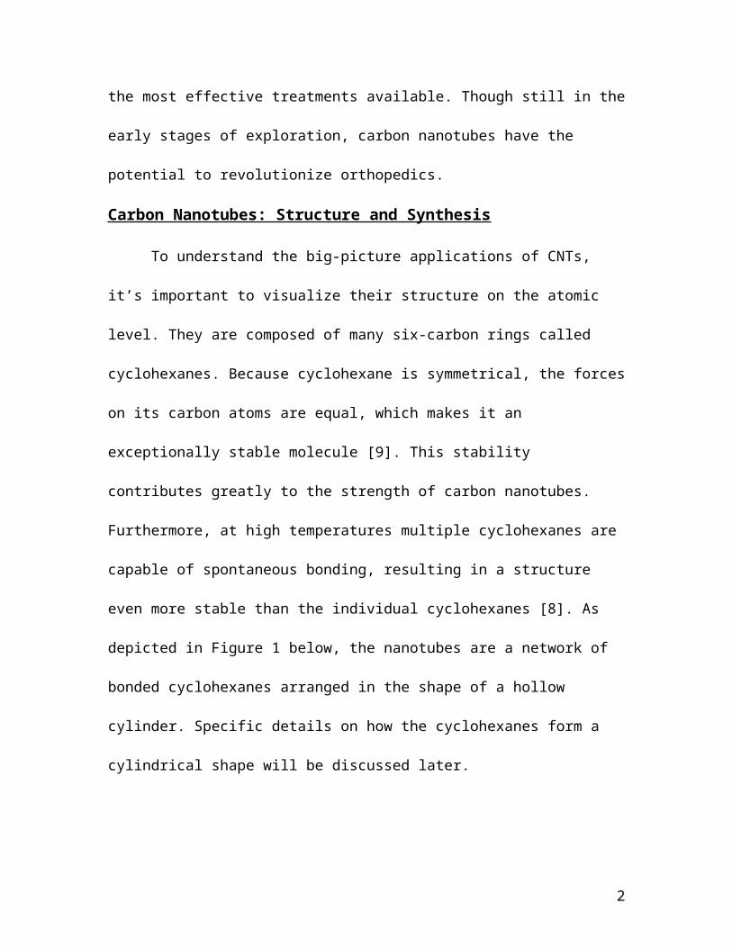
Small amounts of nickel and cobalt are synthetically incorporated into the original
graphite block to speed up the formation of nanotubes by facilitating the bonding of
graphene sheet ends to form cylinders (Figure 4). The process is essentially the same as
Figure 2: Laser ablation model with graphite targetSource: astrobio.net
Figure 3: Three sheets of grapheneSource: www.daviddarling.info
3

taking a piece of paper and taping the two ends together to make a cylinder, and in this
case the cobalt and nickel are what make the ends “sticky” enough to stay together. Note
that all of this happens spontaneously in a temperature and pressure controlled reaction
chamber, with no direct interference from scientists. To maintain a high, uniform rate of
vaporization, the graphite target rotates around a vertical axis within the reaction
chamber. Once vaporized, the graphite collects as cyclohexane soot at the bottom of the
reaction chamber, the soot condenses into the individual graphene sheets, and these
sheets bond with themselves to form nanotubes via the catalytic process described above.
Applications
Now that we’ve discussed why carbon nanotubes integrate seamlessly into the
human body and how they’re made, it is feasible to understand how they are applied to
current orthopedic and tissue engineering research. Bone regeneration scaffolds,
musculoskeletal implants, and bone cement service different needs of the skeletal system;
however, researchers are using CNTs to improve the design and function of each device.
Figure 4: Nanotube formation from graphene sheetSource: siencedirect.com
4

To accomplish this, scientists make CNT composites, materials composed of CNTs and
one or more different materials. In other words, carbon nanotubes are synthesized
separately and then added to existing technology for strength and increased
biocompatibility.
It is important to note that CNT orthopedic applications are currently only
researched in lab settings; however, clinical implementation is on the horizon. Recent
technological breakthroughs have greatly reduced the cost of producing CNTs, from
about $300 per gram to only $35 dollars per gram [5]. A research team from a Malaysian
university discovered the revolutionary production technique in 2012. Team leader Abdul
Rahman Mohamed, Ph.D., stated that their system was capable of producing 1000 grams
of CNTs per day. This economically efficient technique is sure to motivate researchers to
push for FDA approval and subsequent clinical implementation.
Musculoskeletal Implants
Musculoskeletal implants are widely used in the field of orthopedics to mend
broken bones, torn ligaments, and other bony deformities by securing bone to bone, bone
to ligament, or bone to cartilage. Implants are used in both of the two most popular
orthopedic surgeries: knee replacement and hip replacement. They are generally made of
metal like titanium, which is valued for its strength, resistance to corrosion, and inability
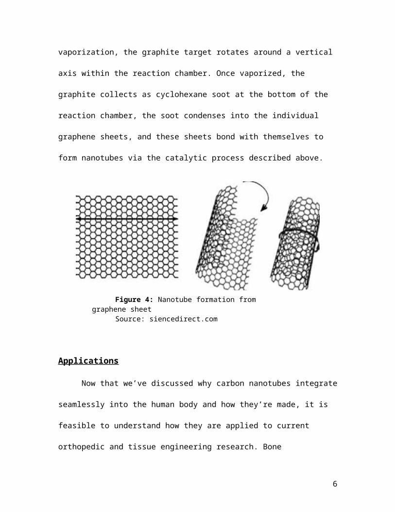
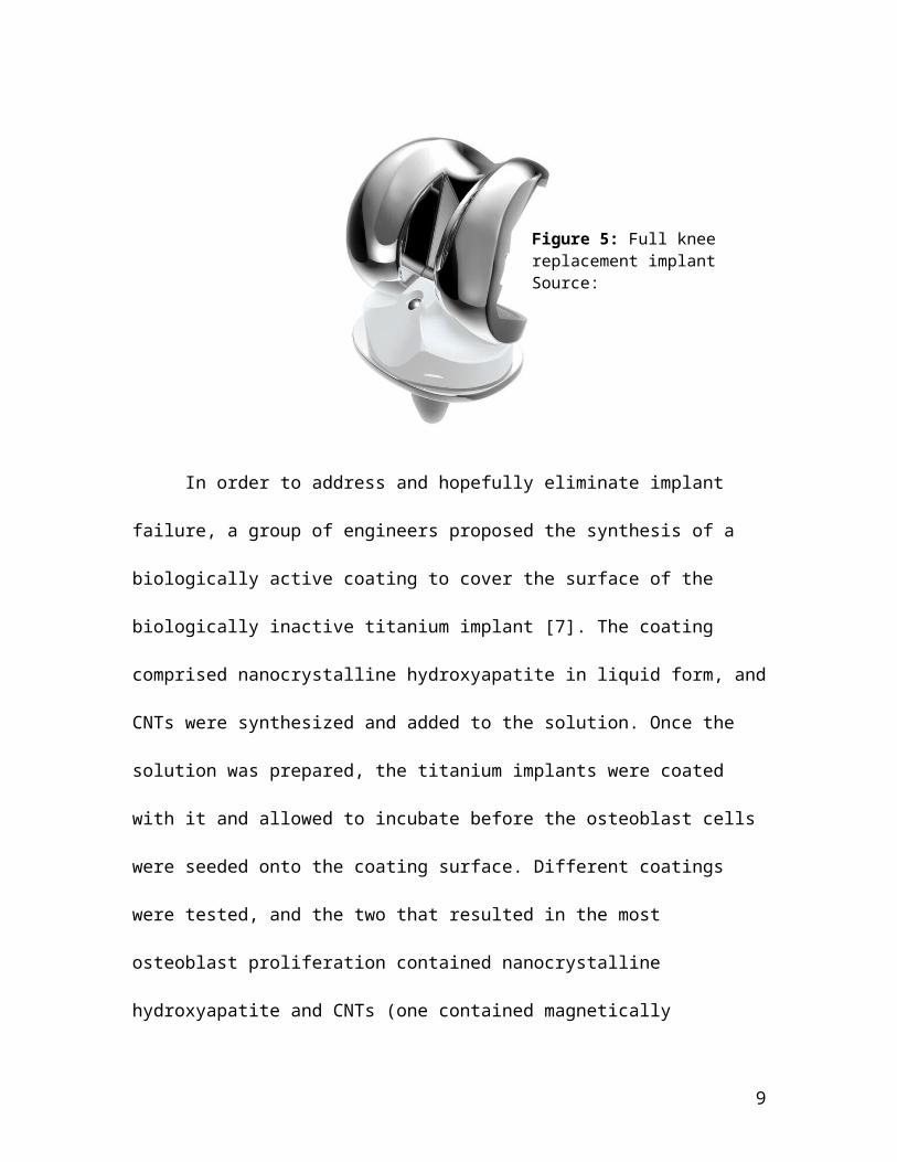
to harm the internal chemistry of the human body. Titanium implants can take the shape
of metal screws, rods, or other more complex shapes like the complete knee replacement
implant depicted in Figure 5 below. Since titanium is merely a metal and has no organic
properties, it is referred to as biologically inactive. This means that osteoblasts will not
bind to it, so the implant is only secured by how tightly it’s drilled into the bone.
5
Complications that lead to implant failure include infection, implant loosening, and poor
host tissue integration (bodily rejection of the implant).
In order to address and hopefully eliminate implant failure, a group of engineers
proposed the synthesis of a biologically active coating to cover the surface of the
biologically inactive titanium implant [7]. The coating comprised nanocrystalline
hydroxyapatite in liquid form, and CNTs were synthesized and added to the solution.
Once the solution was prepared, the titanium implants were coated with it and allowed to
incubate before the osteoblast cells were seeded onto the coating surface. Different
coatings were tested, and the two that resulted in the most osteoblast proliferation
contained nanocrystalline hydroxyapatite and CNTs (one contained magnetically
synthesized CNTs and other non-magnetically synthesized) as depicted in by the
rightmost green bars in Figure 6. Authors of the study see these results as indicators that a
bioactive CNT coating would result in more seamless biointegration and possibly more
secure implantation [7].
Figure 5: Full knee replacement implantSource: www.kneereplacement.com
6

Figure 6: Results for CNT bone implant experiment. The rightmost green bars represent implants coated with CNTs, and they yield the greatest cell proliferation.Source: Journal of Nano science and Nanotechnology
Bone Cement
Bone cement is another commonly applied orthopedic repair treatment, often used
in conjunction with implants. It acts as a grouting agent, which reduces wear between
metal implants and bone. It is also used to secure dentures in the mouth, and has recently
been injected into the back to support collapsing vertebrae [2]. All in all, it’s a very useful
organic compound; however, one of its major flaws is that it’s subject to fatigue-related
cracking and trauma-induced breakage quite frequently. Failure of bone cement is known
as cement-mantle failure, and it is seen in about 5% of post-operative patients within 10
years [2]. The risk of cement-mantle failure is higher in athletes and over-weight patients.
Material scientists have tried adding stainless steel fibers and other reinforcements, but
the fibers were too large and resulted in fracture upon drying.
Nanoscale compounds, specifically CNTs, are the perfect solution to this
dilemma. They’re strong and would increase the toughness of the cement, while at the
7
same time they are small enough to not cause deformities in the cement structure. One
team of engineers combined CNTs and bone cement powder and proceeded to heat and
rotate them together in a 220°C reaction chamber [2]. Through this process bone cement
composites with different concentrations of CNTs were synthesized and allowed to cool
to form solid bone cement composite. The different composites were tested for how many
load cycles they could withstand before failure, and according to Figure 7, the optimum
fatigue life was achieved by 2% weight CNTs, which demonstrates that the addition of
CNTs did contribute to an increase in toughness; however, at a certain point (3 to 5%
weight) there were too many and the structure of the cement deformed. This result is
relatively intuitive, in that a balance between size and strength must be reached so as not
to break the original bonds of the cement, but rather reinforce them.
Bone Regeneration:
Bodily regeneration is one of the most coveted abilities of humankind. When
healthy adults are injured, their cells undergo repair; however, the process leaves the
damaged tissue permanently altered in some way, often in the form of a visible scar.
Regeneration entails complete restoration of the original structure. While the
phenomenon of natural regeneration eludes humans, other animals such as the praying
Figure 7: CNT bone cement experiment data; optimum resistance against failure contained 2% weight CNTsSource: Journal of Biomedical Materials Research
8
mantis or gecko are capable of complete limb regeneration. The greatest motivation for
developing an effective means of human regeneration lies in the potential to restore
injuries that could not otherwise be effectively repaired. Many of these maladies are
inflicted upon the human skeletal structure by severe trauma or disease.
Damaged bones that cannot heal naturally are generally treated with some variety
of surgery. Surgeries are invasive, hard on the body, and pose the risk of infection. Many
surgical procedures involve inserting a graft to replace the damaged bone, and these
grafts can come from the patient’s own body (autografts) or from another resource
outside the patient’s body (allografts) [10]. Potential graft shortage and unnecessary harm
to healthy areas of the body during graft harvest are the primary drawbacks of autografts.
Allografts sometimes result in bodily rejection, which can cause severe infection and
further deformation of the surrounding bony structure.
In order to avoid graft complications, carbon nanotubes have been incorporated
into tissue engineering scaffolds to facilitate bone regeneration. Scaffolds are segments of
material specifically combined to allow cells to grow within and around the segment
(Figure 8). Once the cells have proliferated enough to support and sustain themselves
within the system (the body), the scaffold degrades within the system without causing
harm. This technology with respect to CNTs and orthopedic applications is still in the
early stages of testing and has not yet been applied to a clinical setting; however, the
results of various studies have indicated that the scaffolds yield enough cell proliferation
for probable complete regeneration in the future [6]. As of now carbon nanotube
scaffolds are constructed and tested, and they show very promising osteoblast cell
proliferation. Osteoblasts function directly in bone formation and joining; therefore,
9

Figure 8: Example of a scaffold Source: Int J Nanomedicine. 2012; 7: 2087–2099
CNTs are a particularly advantageous scaffold material because they provide a strong,
biocompatible material to protect and nourish the seeded cells.
One group of researchers in particular made an especially successful CNT
scaffold that contained chitosan and nanocrystalline hydroxyapatite [6]. Chitosan is a
popular hydrogel scaffold material because it contains a naturally occurring compound
called chitin that is biocompatible to humans and easily attainable. Chitin is found in the
exoskeletons of many crustaceans and insects. Nanocrystalline hydroxyapatite is the main
inorganic component in human bone, where inorganic means that it does not contain
carbon. Its presence in the scaffold is important because it increases osteoconductivity
(proliferation of osteoblasts) by providing more sites for calcification, which means the
incorporation of calcium. Calcium is an essential atom for osteoblast cell signaling which
triggers osteoconductivity and therefore the formation of bone. Carbon nanotubes are an
integral aspect of the scaffold because their long, narrow structures allow for osteoblasts
to have the most surface area for bonding, since so many carbon nanotubes can fit within
the scaffold [6]. As a result, the carbon nanotubes would become integrated in the
10
structure of the forming bone, even when the chitosan scaffold begins to degrade. The
hydroxyapatite would also integrate into the developing bone. The research group
compared the results of different scaffold materials to the area of osteoblasts that
proliferated on each scaffold (Figure 9). Upon observing the green bars it is evident that
the scaffold with chitosan, magnetically synthesized CNTs (B-SWCNT), and
nanocrystalline hydroxyapatite (20% nHA) has about 200 more cells/cm2 than the next
scaffold. In other words, the scaffold that incorporated carbon nanotubes showed a
significantly greater proliferation of bone forming cells, thereby displaying the most
potential for regeneration.
Figure 9: Results of CNT scaffold experiment Source: Int J Nanomedicine. 2012; 7: 2087–2099
Closing Thoughts
Human beings are constantly putting themselves at risk for orthopedic injury,
whether they exercise too much or not enough, whether they’re 60 and stepping into the
shower or 16 and walking across the street. On day you might be lined up for an athletic
scholarship, and the next month you could be on the operating table having knee surgery.
The point is, these things are out of our control, but treating the injury is not. As long as
11

there’s any incidence of failure at all in orthopedic procedures, it is imperative that we
strive to eliminate it. Clinical implementation of CNT-reinforced orthopedic composites
could be the solution, but we’ll never know unless it leaves the lab and enters the
operating room.
12

Works Cited
[1] National Center for Health Statistics. Health, United States, 2009: With Special Feature on Medical Technology. Hyattsville, MD. 2010.
[2] Marrs, Brock, et al. "Augmentation of Acrylic Bone Cement with Multiwall Carbon Nanotubes." Journal of Biomedical Materials Research Part A 77A.2 (2006): 269-76.
[3] Anonymous. "Your body contains enough carbon to fill 9000 pencils." Science World. N.p., 07 May 2013. Web. <http://www.scienceworld.ca/we-can-explain/your-body-contains-enough-carbon-to-fill-9000-pencils>.
[4] Laurent, Ch, E. Flahaut, and A. Peigney. "The Weight and Density of Carbon Nanotubes Versus the Number of Walls and Diameter." Carbon 48.10 (2010): 2994-6.
[5] Anonymous. "New Method for Continuous Production of Carbon Nanotubes." Science News. Science Daily, 12 Apr 2012. <http://www.sciencedaily.com/releases/2012/04/120412105109.htm>.
[6] Im, Owen, et al. "Biomimetic Three-Dimensional Nanocrystalline Hydroxyapatite and Magnetically Synthesized Single-Walled Carbon Nanotube Chitosan Nanocomposite for Bone Regeneration." International journal of nanomedicine 7 (2012): 2087-99.
[7] Facca, Sybille, et al. "In Vivo Osseointegration of Nano-Designed Composite Coatings on Titanium Implants." ACS nano 5.6 (2011): 4790.
[8] Aoki, Naofumi, et al. "Carbon Nanotubes as Scaffolds for Cell Culture and Effect on Cellular Functions." Dental materials journal 26.2 (2007): 178.
[9] Jain, K. K. (Kewal K.), SpringerLink. The Handbook of Nanomedicine. Totowa, N.J: Springer; 2008.
[10] Kader, Deiary, et al. “Long-Term Outcome of Endoscopic Anterior Cruciate Ligament Reconstruction with Patellar Tendon Autograft: Minimum 13-Year Review." The American Journal of Sports Medicine 34.5 (2006): 721-32.
[11] Negishi H, Ohashi M, Akita S, Nakayama Y. Orthopedic Treatment of Multiwalled Carbon Nanotube Probes. Japanese Journal of Applied Physics. 2003;42:4866-4868.
[12] Orthopedics; carbon nanotubes help bones mend. Science Letter. 2005:1036.
[13] Prosthetic implant material could outlast the patient.(R&D BREAKTHROUGHS). European Medical Device Technology [serial online]. 2010;1:10.
13

[14] Sirivisoot S. Carbon nanotube-based orthopedic implant sensors. In: New York, NY: Springer New York; 2011:139-160.
[15] Spear RL, Cameron RE. Carbon nanotubes for orthopaedic implants. International Journal of Material Forming. 2008;1:127-133.
[16] Usui Y, Kato H, Taruta S, et al. Carbon nanotubes with high bone-tissue compatibility and bone-formation acceleration effects. Small (Weinheim an der Bergstrasse, Germany). 2008;4:240-246.
[17] Zhang Z, Li Z, Mao X, Wang W. Advances in bone repair with nanobiomaterials: mini-review. Cytotechnology. 2011;63:437-443.
14

Multimedia Suggestions
1. Three dimenstional, rotating carbon nanotube graphic2. Video of bone implant sugery3. Moving graphic of assembling stem cell scaffold4. Video of laser ablation process5. Gaphic of bone cement grouting function
15
Related Documents


