The Journal of Cell Biology © The Rockefeller University Press, 0021-9525/2004/06/775/6 $8.00 The Journal of Cell Biology, Volume 165, Number 6, June 21, 2004 775–780 http://www.jcb.org/cgi/doi/10.1083/jcb.200312030 JCB Report 775 Bcl-2–regulated apoptosis and cytochrome c release can occur independently of both caspase-2 and caspase-9 Vanessa S. Marsden, 1 Paul G. Ekert, 1,2 Mark Van Delft, 1 David L. Vaux, 1 Jerry M. Adams, 1 and Andreas Strasser 1 1 The Walter and Eliza Hall Institute of Medical Research, Melbourne, Victoria 3050, Australia 2 Murdoch Children’s Research Institute, Melbourne, Victoria 3052, Australia poptosis in response to developmental cues and stress stimuli is mediated by caspases that are regu- lated by the Bcl-2 protein family. Although caspases 2 and 9 have each been proposed as the apical caspase in that pathway, neither is indispensable for the apoptosis of leukocytes or fibroblasts. To investigate whether these cas- pases share a redundant role in apoptosis initiation, we generated caspase-2 / 9 / mice. Their overt phenotype, embryonic brain malformation and perinatal lethality mir- rored that of caspase-9 / mice but were not exacerbated. Analysis of adult mice reconstituted with caspase-2 / 9 / A hematopoietic cells revealed that the absence of both caspases did not influence hematopoietic development. Furthermore, lymphocytes and fibroblasts lacking both remained sensitive to diverse apoptotic stimuli. Dying caspase-2 / 9 / lymphocytes displayed multiple hallmarks of caspase-dependent apoptosis, including the release of cytochrome c from mitochondria, and their demise was antagonized by several caspase inhibitors. These findings suggest that caspases other than caspases 2 and 9 can promote cytochrome c release and initiate Bcl-2–regulated apoptosis. Introduction Apoptosis, which is critical for development and tissue homeostasis, is executed by caspases (Adams, 2003). The 10 or so mammalian caspases include both “effectors” (3, 6, and 7), which efficiently digest vital proteins, and “initiators” (e.g., 2, 8, and 9), which proteolytically activate the effectors. Many cell “stress” stimuli, e.g., cytokine deprivation and genome damage, and developmental cues, trigger a common pathway of caspase activation regulated by the Bcl-2 protein family (Adams, 2003). Until recently, the sole apical initiator in that pathway was assumed to be caspase-9, which is acti- vated in a complex termed the “apoptosome” by the scaffold protein Apaf-1 and its cofactor cytochrome c. Evidence that the Bcl-2 family regulates permeabilization of mitochondria argued that cytochrome c release and the ensuing caspase-9 activation were central to the “stress” response. For some neuronal cells, this model is supported, as mice lacking Apaf-1 or caspase-9 die perinatally with brain overgrowth caused by a defect in neuronal apoptosis (Adams, 2003). The apoptosome is not, however, universally essential for Bcl-2–regulated apoptosis, because certain neuronal (Honar- pour et al., 2001), hematopoietic, and fibroblastoid cells (Marsden et al., 2002) lacking Apaf-1 or caspase-9 readily undergo apoptosis in response to diverse insults and, at least in lymphocytes, that apoptosis requires caspase activity (Mars- den et al., 2002). Hence, there must be apoptotic pathways regulated by the Bcl-2 family that require the activation of caspases other than caspase-9 (Adams, 2003). Evidence is also accumulating that certain caspases can contribute to mitochondrial damage and hence may be activated before apoptosome formation (Guo et al., 2002; Lassus et al., 2002; Marsden et al., 2002; Robertson et al., 2002). In particular, caspase-2 has been implicated in cyto- chrome c release (Guo et al., 2002; Lassus et al., 2002; Robertson et al., 2002) and seems to be necessary for cellular demise in some transformed cell lines (Lassus et al., 2002). However, because apoptosis is not markedly impaired in caspase-2–deficient mice (Bergeron et al., 1998; O’Reilly et al., 2002), caspase-2 cannot have a major nonredundant role in apoptosis. Address correspondence to Andreas Strasser, The Walter and Eliza Hall Institute of Medical Research, 1G Royal Parade, Parkville, Victoria 3050, Australia. Tel.: 61-3-9345-2555. Fax: 61-3-9347-0852. email: [email protected] Key words: apoptosis; caspase-2; caspase-9; Bcl-2; cytochrome c Abbreviation used in this paper: PI, propidium iodide. on June 29, 2015 jcb.rupress.org Downloaded from Published June 21, 2004 http://jcb.rupress.org/content/suppl/2004/06/15/jcb.200312030.DC1.html Supplemental Material can be found at:

Welcome message from author
This document is posted to help you gain knowledge. Please leave a comment to let me know what you think about it! Share it to your friends and learn new things together.
Transcript
The
Jour
nal o
f Cel
l Bio
logy
©
The Rockefeller University Press, 0021-9525/2004/06/775/6 $8.00The Journal of Cell Biology, Volume 165, Number 6, June 21, 2004 775–780http://www.jcb.org/cgi/doi/10.1083/jcb.200312030
JCB
Report
775
Bcl-2–regulated apoptosis and cytochrome
c
release can occur independently of both caspase-2 and caspase-9
Vanessa S. Marsden,
1
Paul G. Ekert,
1,2
Mark Van Delft,
1
David L. Vaux,
1
Jerry M. Adams,
1
and Andreas Strasser
1
1
The Walter and Eliza Hall Institute of Medical Research, Melbourne, Victoria 3050, Australia
2
Murdoch Children’s Research Institute, Melbourne, Victoria 3052, Australia
poptosis in response to developmental cues andstress stimuli is mediated by caspases that are regu-lated by the Bcl-2 protein family. Although caspases
2 and 9 have each been proposed as the apical caspase inthat pathway, neither is indispensable for the apoptosis ofleukocytes or fibroblasts. To investigate whether these cas-pases share a redundant role in apoptosis initiation, wegenerated caspase-2
�
/
�
9
�
/
�
mice. Their overt phenotype,embryonic brain malformation and perinatal lethality mir-rored that of caspase-9
�
/
�
mice but were not exacerbated.Analysis of adult mice reconstituted with caspase-2
�
/
�
9
�
/
�
A
hematopoietic cells revealed that the absence of bothcaspases did not influence hematopoietic development.Furthermore, lymphocytes and fibroblasts lacking bothremained sensitive to diverse apoptotic stimuli. Dyingcaspase-2
�
/
�
9
�
/
�
lymphocytes displayed multiple hallmarksof caspase-dependent apoptosis, including the release ofcytochrome
c
from mitochondria, and their demise wasantagonized by several caspase inhibitors. These findingssuggest that caspases other than caspases 2 and 9 canpromote cytochrome
c
release and initiate Bcl-2–regulatedapoptosis.
Introduction
Apoptosis, which is critical for development and tissuehomeostasis, is executed by caspases (Adams, 2003). The 10or so mammalian caspases include both “effectors” (3, 6, and7), which efficiently digest vital proteins, and “initiators”(e.g., 2, 8, and 9), which proteolytically activate the effectors.Many cell “stress” stimuli, e.g., cytokine deprivation andgenome damage, and developmental cues, trigger a commonpathway of caspase activation regulated by the Bcl-2 proteinfamily (Adams, 2003). Until recently, the sole apical initiatorin that pathway was assumed to be caspase-9, which is acti-vated in a complex termed the “apoptosome” by the scaffoldprotein Apaf-1 and its cofactor cytochrome
c
. Evidence thatthe Bcl-2 family regulates permeabilization of mitochondriaargued that cytochrome
c
release and the ensuing caspase-9activation were central to the “stress” response. For someneuronal cells, this model is supported, as mice lackingApaf-1 or caspase-9 die perinatally with brain overgrowthcaused by a defect in neuronal apoptosis (Adams, 2003).
The apoptosome is not, however, universally essential forBcl-2–regulated apoptosis, because certain neuronal (Honar-pour et al., 2001), hematopoietic, and fibroblastoid cells(Marsden et al., 2002) lacking Apaf-1 or caspase-9 readilyundergo apoptosis in response to diverse insults and, at leastin lymphocytes, that apoptosis requires caspase activity (Mars-den et al., 2002). Hence, there must be apoptotic pathwaysregulated by the Bcl-2 family that require the activation ofcaspases other than caspase-9 (Adams, 2003).
Evidence is also accumulating that certain caspases cancontribute to mitochondrial damage and hence may beactivated before apoptosome formation (Guo et al., 2002;Lassus et al., 2002; Marsden et al., 2002; Robertson et al.,2002). In particular, caspase-2 has been implicated in cyto-chrome
c
release (Guo et al., 2002; Lassus et al., 2002;Robertson et al., 2002) and seems to be necessary for cellulardemise in some transformed cell lines (Lassus et al., 2002).However, because apoptosis is not markedly impaired incaspase-2–deficient mice (Bergeron et al., 1998; O’Reilly etal., 2002), caspase-2 cannot have a major nonredundant rolein apoptosis.
Address correspondence to Andreas Strasser, The Walter and Eliza HallInstitute of Medical Research, 1G Royal Parade, Parkville, Victoria3050, Australia. Tel.: 61-3-9345-2555. Fax: 61-3-9347-0852. email:[email protected]
Key words: apoptosis; caspase-2; caspase-9; Bcl-2; cytochrome
c
Abbreviation used in this paper: PI, propidium iodide.
on June 29, 2015jcb.rupress.org
Dow
nloaded from
Published June 21, 2004
http://jcb.rupress.org/content/suppl/2004/06/15/jcb.200312030.DC1.html Supplemental Material can be found at:

776 The Journal of Cell Biology
|
Volume 165, Number 6, 2004
These discordant findings might be reconciled if caspase-2acts redundantly with caspase-9, each activating distinct butconverging pathways. If so, loss of both caspases shouldmarkedly attenuate apoptosis. We address that hypothesishere by studies on mice lacking both caspases 2 and 9.
Results and discussion
To generate mice lacking both caspases 2 and 9, we first inter-crossed animals deficient in caspase-2 (O’Reilly et al., 2002)with caspase-9
�
/
�
mice (Kuida et al., 1998). As expected fromthe severe caspase-9
�
/
�
phenotype (Hakem et al., 1998;Kuida et al., 1998), intercrosses of the resulting caspase-2
�
/
�
9
�
/
�
mice yielded no weaned progeny lacking caspase-9, irre-spective of caspase-2 status (67 progeny genotyped). Mice ofall other genotypes appeared at the expected Mendelian ratiosand were healthy and fertile (unpublished data).
To investigate whether caspase-2 deficiency exacerbatedthe neuronal overgrowth characteristic of the caspase-9 defi-ciency (Hakem et al., 1998; Kuida et al., 1998), embryosfrom the intercrosses were examined at E14.5, when all ge-notypes appeared in the expected ratios. Brain over-growthresembling that previously described and observed in cas-pase-2
�
/
�
9
�
/
�
littermates (Fig. 1) appeared in 6/11 caspase-2
�
/
�
9
�
/
�
and 2/5 caspase-2
�
/
�
9
�
/
�
embryos but never inthose expressing caspase-9 (
n
�
47). All other organs ap-peared normal. Although the brain abnormalities cannot bequantified, we conclude that caspase-2 loss does not substan-tially exacerbate the brain phenotype due to caspase-9 defi-ciency and that other organs develop normally to at leastE14.5 without either caspases 2 or 9.
Bcl-2–regulated apoptosis, which is critical for the physio-logical death of hematopoietic cells (Marsden and Strasser,2003), can occur independently of caspase-9 (Marsden et al.,
2002). To study how caspase-2
�
/
�
9
�
/
�
hematopoietic cellsrespond to the physiological death cues in healthy mice,C57BL/6-Ly5.1 mice were reconstituted with fetal liver–derived hematopoietic stem cells from E14.5 offspring of theintercrosses (Ly5.2
�
). 10 wk later, the thymocytes were allderived from donor (Ly5.2) cells, irrespective of donor geno-type (Fig. 2 A). The absence of caspases 2 and 9 did not aug-ment cell numbers or perturb cell subset composition in thethymus, spleen, lymph nodes, or bone marrow (Fig. 2, B–D;and not depicted). Western blot analysis on reconstituted or-gans (Fig. S1, available at http://www.jcb.org/cgi/content/full/jcb.200312030/DC1) confirmed the absence of cas-
Figure 1. Loss of caspase-2 does not aggravate the overt defects caused by caspase-9 deficiency. (A) Similar overt phenotype of E14.5 caspase-2�/�9�/� and caspase-9�/� embryos. (B) Similar histologic presentation of caspase-2�/�9�/� and caspase-9�/� embryos. Sagittal brain sections were stained with hematoxylin and eosin. For comparison, littermate embryos lacking only caspase-2, which are indistinguishable from wild-type embryos, are shown. O, oral cavity; B, basal ganglia; T, dorsal telencephalon; V, lateral ventricle; S, semicircular canals.
Figure 2. Normal hematopoietic development from caspase-2�/�
9�/� hematopoietic stem cells. Ly5.1 mice that had been lethally irradiated (2 � 5.5Gy) were reconstituted with fetal liver cells and analyzed 10–15 wk later. (A) Staining of thymocytes with antibodies to Ly5.1 (recipient) versus Ly5.2 (donor) cells. (B) Normal cellularities of thymus, lymph nodes (pooled inguinal, axillary, brachial and mesenteric), spleen and bone marrow in animals reconstituted with caspase-2�/�9�/�cells. Results represent means � SD for �3 mice per genotype. (C) Normal composition of the thymus in the reconstituted mice. (D) Normal proportions of B lymphocytes (B220� Thy-1�) and T cells (Thy-1� B220�) in lymph nodes of the reconstituted mice. Results are representative of �3 mice per genotype.
on June 29, 2015jcb.rupress.org
Dow
nloaded from
Published June 21, 2004
Apoptosis independent of caspases 2 and 9 |
Marsden et al. 777
pase-2 and/or -9 and revealed no compensatory up-regulationof caspases 1, 3, 6, 7, 8, or 9. Thus, whereas overexpression ofBcl-2 in the hematopoietic compartment, or absence of itsantagonist Bim, promotes cell accumulation (Bouillet et al.,1999; Marsden et al., 2002), programmed death in that com-partment appears unaffected by loss of both caspases 2 and 9.
To explore further whether caspases 2 and 9 are essentialfor Bcl-2–regulated cell death, we analyzed the responses todiverse cytotoxic insults of donor-derived B and T lympho-cytes and CD4
�
8
�
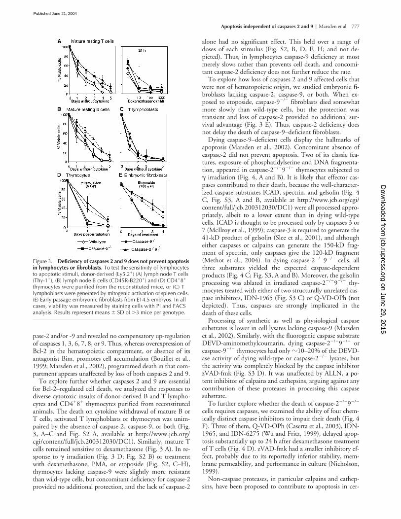
thymocytes purified from reconstitutedanimals. The death on cytokine withdrawal of mature B orT cells, activated T lymphoblasts or thymocytes was unim-paired by the absence of caspase-2, caspase-9, or both (Fig.3, A–C and Fig. S2 A, available at http://www.jcb.org/cgi/content/full/jcb.200312030/DC1). Similarly, mature Tcells remained sensitive to dexamethasone (Fig. 3 A). In re-sponse to
�
irradiation (Fig. 3 D; Fig. S2 B) or treatmentwith dexamethasone, PMA, or etoposide (Fig. S2, C–H),thymocytes lacking caspase-9 were slightly more resistantthan wild-type cells, but concomitant deficiency for caspase-2provided no additional protection, and the lack of caspase-2
alone had no significant effect. This held over a range ofdoses of each stimulus (Fig. S2, B, D, F, H; and not de-picted). Thus, in lymphocytes caspase-9 deficiency at mostmerely slows rather than prevents cell death, and concomi-tant caspase-2 deficiency does not further reduce the rate.
To explore how loss of caspases 2 and 9 affected cells thatwere not of hematopoietic origin, we studied embryonic fi-broblasts lacking caspase-2, caspase-9, or both. When ex-posed to etoposide, caspase-9
�
/
�
fibroblasts died somewhatmore slowly than wild-type cells, but the protection wastransient and loss of caspase-2 provided no additional sur-vival advantage (Fig. 3 E). Thus, caspase-2 deficiency doesnot delay the death of caspase-9–deficient fibroblasts.
Dying caspase-9–deficient cells display the hallmarks ofapoptosis (Marsden et al., 2002). Concomitant absence ofcaspase-2 did not prevent apoptosis. Two of its classic fea-tures, exposure of phosphatidylserine and DNA fragmenta-tion, appeared in caspase-2
�
/
�
9
�
/
�
thymocytes subjected to
�
irradiation (Fig. 4, A and B). It is likely that effector cas-pases contributed to their death, because the well-character-ized caspase substrates ICAD, spectrin, and gelsolin (Fig. 4C, Fig. S3, A and B, available at http://www.jcb.org/cgi/content/full/jcb.200312030/DC1) were all processed appro-priately, albeit to a lower extent than in dying wild-typecells. ICAD is thought to be processed only by caspases 3 or7 (McIlroy et al., 1999); caspase-3 is required to generate the41-kD product of gelsolin (Slee et al., 2001), and althougheither caspases or calpains can generate the 150-kD frag-ment of spectrin, only caspases give the 120-kD fragment(Methot et al., 2004). In dying caspase-2
�
/
�
9
�
/
�
cells, allthree substrates yielded the expected caspase-dependentproducts (Fig. 4 C; Fig. S3, A and B). Moreover, the gelsolinprocessing was ablated in irradiated caspase-2
�
/
�
9
�
/
�
thy-mocytes treated with either of two structurally unrelated cas-pase inhibitors, IDN-1965 (Fig. S3 C) or Q-VD-OPh (notdepicted). Thus, caspases are strongly implicated in thedeath of these cells.
Processing of synthetic as well as physiological caspasesubstrates is lower in cell lysates lacking caspase-9 (Marsdenet al., 2002). Similarly, with the fluorogenic caspase substrateDEVD-aminomethylcoumarin, dying caspase-2
�
/
�
9
�
/
�
orcaspase-9
�
/
�
thymocytes had only
�
10–20% of the DEVD-ase activity of dying wild-type or caspase-2
�
/
�
lysates, butthe activity was completely blocked by the caspase inhibitorzVAD-fmk (Fig. S3 D). It was unaffected by ALLN, a po-tent inhibitor of calpains and cathepsins, arguing against anycontribution of these proteases in processing this caspasesubstrate.
To further explore whether the death of caspase-2
�
/
�
9
�
/
�
cells requires caspases, we examined the ability of four chem-ically distinct caspase inhibitors to impair their death (Fig. 4F). Three of them, Q-VD-OPh (Caserta et al., 2003), IDN-1965, and IDN-6275 (Wu and Fritz, 1999), delayed apop-tosis substantially up to 24 h after dexamethasone treatmentof T cells (Fig. 4 D). zVAD-fmk had a smaller inhibitory ef-fect, probably due to its reportedly inferior stability, mem-brane permeability, and performance in culture (Nicholson,1999).
Non-caspase proteases, in particular calpains and cathep-sins, have been proposed to contribute to apoptosis in cer-
Figure 3. Deficiency of caspases 2 and 9 does not prevent apoptosis in lymphocytes or fibroblasts. To test the sensitivity of lymphocytes to apoptotic stimuli, donor-derived (Ly5.2�) (A) lymph node T cells (Thy-1�), (B) lymph node B cells (CD45R-B220�) and (D) CD4�8� thymocytes were purified from the reconstituted mice, or (C) T lymphoblasts were generated by mitogenic activation of spleen cells. (E) Early passage embryonic fibroblasts from E14.5 embryos. In all cases, viability was measured by staining cells with PI and FACS analysis. Results represent means � SD of �3 mice per genotype.
on June 29, 2015jcb.rupress.org
Dow
nloaded from
Published June 21, 2004

778 The Journal of Cell Biology
|
Volume 165, Number 6, 2004
tain circumstances (Jaattela and Tschopp, 2003). To deter-mine whether either participated in the apoptosis of caspase-2
�
/
�
9
�
/
�
cells, we tested six inhibitors reported to impairapoptosis under certain conditions: the calpain inhibitorsz-VF-CHO and PD150606 (Squier and Cohen, 1997), a cell-permeable peptide of the natural calpain inhibitor calpa-statin (Altznauer et al., 2004), the dual calpain and cathep-sin inhibitors ALLM and ALLN (Ding et al., 2002), and theselective cathepsin inhibitor z-FG-NHO-Bz-
p
OMe. In con-trast to the caspase inhibitors, none of these inhibitors hadany anti-apoptotic activity at doses in the range where othershave reported efficacy (Fig. 4 D), and none cooperated withIDN-1965 to enhance its antagonism of apoptosis (not de-picted). Hence, it appears unlikely that either calpains or ca-thepsins act in tandem with the caspase cascade to causeapoptosis in these cells.
Cytochrome
c
release in thymocytes seems to depend oncaspase activity (Marsden et al., 2002), and caspase-2 hasbeen implicated in mitochondrial disruption in certain cells(Guo et al., 2002; Lassus et al., 2002; Robertson et al.,2002). Hence, we examined whether caspase-2 was thesole caspase responsible for mitochondrial damage in thy-mocytes. Western blotting of fractionated cell lysates re-vealed that cytochrome
c
release from mitochondria did notrequire caspase-2 (Fig. 5 A). Furthermore, mitochondrial
transmembrane potential in dying thymocytes was lost nor-mally in the absence of caspase-2, or both caspases 2 and 9,although its loss in wild-type thymocytes was attenuated bya caspase inhibitor (Fig. 5 B), as shown previously (Bossy-Wetzel et al., 1998). Hence, both the release of pro-apop-totic molecules from mitochondria and the loss of mito-chondrial transmembrane potential can occur independentlyof caspases 2 and 9.
Our results strongly implicate caspases in the apoptosis ofcaspase-2
�
/
�
9
�
/
�
cells and thus imply that there is a Bcl-2–regulated and caspase-mediated pathway that does not re-quire either caspase-2 or -9. Which other caspases might beregulated by Bcl-2? As discussed elsewhere (Adams, 2003),caspases 1, 11, and 12 in mice (or caspases 1, 4, and 5 in hu-mans) are attractive candidates, because, like caspases 2 and9, their NH
2
-terminal CARD domain could interact with acognate scaffold to form an apoptosome-like complex. Forexample, caspase-12, which is implicated in apoptosis in-duced by ER stress (Nakagawa et al., 2000) and in cyto-chrome
c
–independent apoptosis (Morishima et al., 2002;Rao et al., 2002), forms a large complex on serum starvation(Kilic et al., 2002). In other systems, caspase-11 (Hisahara etal., 2001; Kang et al., 2002) or caspase-1 (Hilbi et al., 1998;Marsden et al., 2002; Rowe et al., 2002) have been impli-cated in apoptosis. Hence, in different circumstances, vari-
Figure 4. Dying caspase-2�/�9�/� cells display hallmarks of apoptosis and are protected by caspase inhibitors but not calpain/cathepsin inhibitors. (A–C) Ly5.1�CD4�8� thymocytes sorted from reconstituted mice were cultured follow-ing 5 Gy � irradiation (ir.) (A) Staining with FITC-conjugated Annexin V plus PI reveals phosphatidylserine exposure before loss of membrane integrity irre-spective of genotype. (B) DNA “laddering” indicative of inter-nucleosomal fragmen-tation demonstrated by gel electrophoresis of genomic DNA. (C) Western blots showing processing of ICAD in dying caspase-2�/�9�/� cells. (D) The death of caspase-2�/�9�/� cells is antagonized by inhibitors of caspases but not of calpains or cathepsins. Ly5.1�Thy1� mature T lymphocytes from the lymph nodes of reconstituted mice were cultured for 24 h in the presence of 100 nM dexamethasone plus the caspase inhibitors zVAD-fmk, IDN-6275 (6275), IDN-1965 (1965), or Q-VD-OPh (Q-VD), the calpain inhibitors z-VF-CHO or PD-150606, a cell perme-able peptide of the calpain inhibitor calpastatin, the dual calpain/cathepsin inhibitors ALLM and ALLN or the ca-thepsin inhibitor z-FG-NHO-Bz-pOMe (zFG-NHO) (each at 50 �M). Cell death was quantified by PI staining and FACS analysis. Results represent mean � SD of at least three independent experiments for each genotype. Vehicle: 0.4% DMSO.
on June 29, 2015jcb.rupress.org
Dow
nloaded from
Published June 21, 2004

Apoptosis independent of caspases 2 and 9 | Marsden et al. 779
ous combinations of caspases 1, 11, and 12, and perhaps alsocaspase-8, might act redundantly with caspases 2 and 9 toinitiate apoptosis.
Whereas our results with primary lymphocytes and fibro-blasts implicate caspases in addition to caspases 2 and 9 inthe initiation of apoptosis, the accompanying paper, Ekert etal. (2004) shows that the hallmarks of apoptosis failed to ap-pear when myeloid progenitor cell lines lacking both cas-pases 2 and 9 were deprived of growth factor, but that pro-grammed cell death still prevented their clonogenic survival.These findings and those with transformed human cell lines(Lassus et al., 2002) can be reconciled with our conclusionthat caspases 2 and 9 are not essential for loss of viability perse, if some cell types but not others use these caspases to ac-celerate apoptotic cell demolition.
Materials and methodsMiceCaspase-2�/�9�/� mice were generated by intercrosses of mice deficient incaspase-2 (129/sv) (O’Reilly et al., 2002) with caspase-9�/� (C57BL/6)mice (Kuida et al., 1998). Embryos and 3-wk-old mice were genotyped byPCR. Hematopoietic reconstitution was performed as described previously
(Marsden et al., 2002) from fetal liver cells of embryos with a caspase-2�/�,caspase-9�/�, or caspase-2�/�9�/� genotype (all mixed C57/BL6-129Sv,Ly5.2�) or wild-type (C57/BL6 Ly5.2�) embryos.
Microscopic imagingAll microscopy used either a Stemi SV11 or an Axioplan 2 microscope(Carl Zeiss MicroImaging, Inc.). The latter used objective lenses (magnifi-cation/numerical aperture: 5�/0.15 and 10�/0.30; Carl Zeiss MicroImag-ing, Inc.). Images were recorded with a Axiocam and Axiovision software(Carl Zeiss MicroImaging, Inc.).
Flow cytometryCells stained with fluorochrome- or biotin-conjugated surface marker-spe-cific antibodies (Marsden et al., 2002) were analyzed using a FACScan(Becton Dickinson). Live and dead cells were discriminated by stainingwith 2 �g/ml propidium iodide (PI; Sigma-Aldrich). In cell sorting, host-de-rived (Ly5.1) cells were excluded.
Cell cultureCells were cultured and T lymphoblasts generated as described previously(Marsden et al., 2002). Before death assays, the viable cells in stimulatedspleen cultures were enriched by centrifugation over Ficoll-Paque Plus(Amersham Biosciences). Embryonic fibroblasts, cultured from E14.5 em-bryos, were used at early passage (n 5).
Cell death assaysFor cell death assays, cells were cultured at 0.2–1 � 106 cells/ml andtreated with dexamethasone (Sigma-Aldrich) at 10–8–10–6 M, � radiation (5Gy from a 60Co source), PMA (Sigma-Aldrich) at 0.1–10 ng/ml, or etopo-side (VP-16, David Bull Laboratories) at 0.1–100 �M. Cell viability was de-termined by staining with 2 �g/ml PI and analysis on a FACScan (BectonDickinson). Alternatively, flow cytometry was performed on cells stainedcells with FITC-conjugated Annexin-V. To measure mitochondrial trans-membrane potential, cultured cells stained for 15 min with 40 nM3,3dihexyloxacarbocyanine iodide (DiOC6(3); Molecular Probes) weresubjected to flow cytometry. The caspase inhibitors IDN-1965, IDN-6275(gifts of K. Tomaselli and T. Oltersdorf, IDUN Pharmaceuticals, San Diego,CA), Q-VD-OPh (ICN Biomedicals), and zVAD-fmk (Bachem), as well asthe inhibitors zVF-CHO, PD150606, ALLN, ALLM, and z-FG-NHO-Bz-pOMe (all from Calbiochem), were all solubilized in DMSO. Calpastatinpeptide (Calbiochem) was solubilized in water.
Subcellular fractionation and Western blottingSubcellular fractionation and Western blotting were performed as de-scribed previously (Marsden et al., 2002).
Online supplemental materialWestern blot analysis of thymocytes (Fig. S1 A) and splenocytes (Fig. S1 B)from reconstituted animals demonstrates that no compensatory increase inexpression of other caspases was evident in cells lacking caspase-2 and/orcaspase-9.
Combined deficiency of caspases 2 and 9 in thymocytes does not pre-vent apoptosis in response to a variety of death stimuli. Thymocytes weresubjected to cytokine withdrawal (Fig. S2 A), cultured after graded doses of� irradation (Fig. S2 B), or treated with dexamethasone, PMA, or etoposide(Fig. S2, C–H). Caspase-2 deficiency did not influence the sensitivity tothese stimuli of cells lacking or expressing caspase-9.
Western blotting of thymocytes sorted from reconstituted mice demon-strates that in dying cells the caspase substrates spectrin (Fig. S3 A) andgelsolin (Fig. S3 B) are processed to the expected caspase-generated prod-ucts. Cell extracts were also made from cells of the indicated genotypescultured with or without the caspase inhibitor IDN-1965. Western blottingof these extracts shows the caspase dependence of gelsolin processing indying cells (Fig. S3 C). Caspase-like DEVDase activity was measured byfluorogenic assay (Fig. S3 D). Online supplemental material is available athttp://www.jcb.org/cgi/content/full/jcb.200312030/DC1.
We thank Dr. K. Kuida for caspase-9�/� mice; Drs. K. Tomaselli and T.Oltersdorf for caspase inhibitors; Drs. L. O’Reilly (antibodies to caspase-2;The Walter and Eliza Hall Institute), R. Anderson (antibodies to HSP70; Pe-ter MacCallum Cancer Centre, Melbourne, Australia), Y. Lazebnik (anti-bodies to caspases-3, -7, and -9; Cold Spring Harbor Laboratory, ColdSpring Harbor, NY), P. Vandenabeele and M. Kalai (antibodies to caspase-1;University of Ghent, Ghent, Belgium), and D. Kwiatkowski (antibody togelsolin; Harvard Medical School, Boston, NY); M. Pakusch for technical
Figure 5. Cytochrome c release and mitochondrial depolarization do not require caspases 2 and 9. Ly5.1�CD4�8� thymocytes from the reconstituted mice were cultured following 5 Gy � irradiation (ir.) (A) Subcellular localization of cytochrome c was determined by Western blotting soluble cytosolic (s) and pelleted organelle (p) cell fractions (B). Mitochondrial trans-membrane potential was determined by FACS analysis of cells stained with 40 nM DiOC6(3). The percentages of cells retaining high DiOC6(3) fluorescence are shown. Where indicated, cells were cultured in the presence of IDN-1965 (100 �M).
on June 29, 2015jcb.rupress.org
Dow
nloaded from
Published June 21, 2004
780 The Journal of Cell Biology | Volume 165, Number 6, 2004
assistance; J. Morrow, C. Tilbrook, and A. Naughton for animal care; andDr. F. Battye, D. Kaminaris, V. Lapatis, and C. Tarlinton for cell sorting.We are grateful to Drs. L. Coultas, S. Cory, R. Kluck, T. Thomas, and A.Voss for discussions.
This work was supported by the National Health and Medical ResearchCouncil, the Leukemia and Lymphoma Society (SCOR Center), the CancerCouncil Victoria, the National Institutes of Health, the Cancer Research In-stitute, the Commonwealth Dept. of Education, Science and Training andthe University of Melbourne.
Submitted: 3 December 2003Accepted: 18 May 2004
ReferencesAdams, J.M. 2003. Ways of dying: multiple pathways to apoptosis. Genes Dev. 17:
2481–2495.Altznauer, F., S. Conus, A. Cavalli, G. Folkers, and H.U. Simon. 2004. Calpain-1
regulates Bax and subsequent Smac-dependent caspase-3 activation in neu-trophil apoptosis. J. Biol. Chem. 279:5947–5957.
Bergeron, L., G.I. Perez, G. Macdonald, L. Shi, Y. Sun, A. Jurisicova, S. Varmuza,K.E. Latham, J.A. Flaws, J.C. Salter, et al. 1998. Defects in regulation ofapoptosis in caspase-2-deficient mice. Genes Dev. 12:1304–1314.
Bossy-Wetzel, E., D.D. Newmeyer, and D.R. Green. 1998. Mitochondrial cyto-chrome c release in apoptosis occurs upstream of DEVD-specific caspase ac-tivation and independently of mitochondrial transmembrane depolarization.EMBO J. 17:37–49.
Bouillet, P., D. Metcalf, D.C.S. Huang, D.M. Tarlinton, T.W.H. Kay, F. Könt-gen, J.M. Adams, and A. Strasser. 1999. Proapoptotic Bcl-2 relative Bim re-quired for certain apoptotic responses, leukocyte homeostasis, and to pre-clude autoimmunity. Science. 286:1735–1738.
Caserta, T.M., A.N. Smith, A.D. Gultice, M.A. Reedy, and T.L. Brown. 2003.Q-VDOPh, a broad spectrum caspase inhibitor with potent antiapoptoticproperties. Apoptosis. 8:345–352.
Ding, W.X., H.M. Shen, and C.N. Ong. 2002. Calpain activation after mitochon-drial permeability transition in microcystin-induced cell death in rat hepato-cytes. Biochem. Biophys. Res. Commun. 291:321–331.
Ekert, P.G., S.H. Read, J. Silke, V.S. Marsden, H. Kaufmann, C.J. Hawkins, R.Gerl, S. Kumar, and D.L. Vaux. 2004. Apaf-1 and caspase-9 accelerateapoptosis, but do not determine whether factor-deprived or drug-treatedcells die. J. Cell Biol. 165:835–842.
Guo, Y., S.M. Srinivasula, A. Druilhe, T. Fernandes-Alnemri, and E.S. Alnemri.2002. Caspase-2 induces apoptosis by releasing proapoptotic proteins frommitochondria. J. Biol. Chem. 277:13430–13437.
Hakem, R., A. Hakem, G.S. Duncan, J.T. Henderson, M. Woo, M.S. Soengas, A.Elia, J.L. de la Pompa, D. Kagi, W. Khoo, et al. 1998. Differential require-ment for caspase 9 in apoptotic pathways in vivo. Cell. 94:339–352.
Hilbi, H., J.E. Moss, D. Hersh, Y. Chen, J. Arondel, S. Banerjee, R.A. Flavell, J.Yuan, P.J. Sansonetti, and A. Zychlinsky. 1998. Shigella-induced apopto-sis is dependent on caspase-1 which binds to IpaB. J. Biol. Chem. 273:32895–32900.
Hisahara, S., J. Yuan, T. Momoi, H. Okano, and M. Miura. 2001. Caspase-11 me-diates oligodendrocyte cell death and pathogenesis of autoimmune-mediateddemyelination. J. Exp. Med. 193:111–122.
Honarpour, N., K. Tabuchi, J.M. Stark, R.E. Hammer, T.C. Sudhof, L.F. Parada,X. Wang, J.A. Richardson, and J. Herz. 2001. Embryonic neuronal deathdue to neurotrophin and neurotransmitter deprivation occurs independentof Apaf-1. Neuroscience. 106:263–274.
Jaattela, M., and J. Tschopp. 2003. Caspase-independent cell death in T lympho-cytes. Nat. Immunol. 4:416–423.
Kang, S.J., S. Wang, K. Kuida, and J. Yuan. 2002. Distinct downstream pathwaysof caspase-11 in regulating apoptosis and cytokine maturation during septicshock response. Cell Death Differ. 9:1115–1125.
Kilic, M., R. Schafer, J. Hoppe, and U. Kagerhuber. 2002. Formation of nonca-nonical high molecular weight caspase-3 and -6 complexes and activation ofcaspase-12 during serum starvation induced apoptosis in AKR-2B mouse fi-broblasts. Cell Death Differ. 9:125–137.
Kuida, K., T.F. Haydar, C.-Y. Kuan, Y. Gu, C. Taya, H. Karasuyama, M.S.-S. Su,P. Rakic, and R.A. Flavell. 1998. Reduced apoptosis and cytochrome c-medi-ated caspase activation in mice lacking caspase 9. Cell. 94:325–337.
Lassus, P., X. Opitz-Araya, and Y. Lazebnik. 2002. Requirement for caspase-2 instress induced apoptosis before mitochondrial permeabilization. Science.297:1352–1354.
Marsden, V., and A. Strasser. 2003. Control of apoptosis in the immune system:Bcl-2, BH3-only proteins and more. Annu. Rev. Immunol. 21:71–105.
Marsden, V., L. O’Connor, L.A. O’Reilly, J. Silke, D. Metcalf, P. Ekert, D.C.S.Huang, F. Cecconi, K. Kuida, K.J. Tomaselli, et al. 2002. Apoptosis initi-ated by Bcl-2-regulated caspase activation independently of the cytochromec/Apaf-1/caspase-9 apoptosome. Nature. 419:634–637.
McIlroy, D., H. Sakahira, R.V. Talanian, and S. Nagata. 1999. Involvement of cas-pase 3-activated DNase in internucleosomal DNA cleavage induced by di-verse apoptotic stimuli. Oncogene. 18:4401–4408.
Methot, N., J. Huang, N. Coulombe, J.P. Vaillancourt, D. Rasper, J. Tam, Y.Han, J. Colucci, R. Zamboni, S. Xanthoudakis, et al. 2004. Differential effi-cacy of caspase inhibitors on apoptosis markers during sepsis in rats and im-plication for fractional inhibition requirements for therapeutics. J. Exp. Med.199:199–207.
Morishima, N., K. Nakanishi, H. Takenouchi, T. Shibata, and Y. Yasuhiko. 2002.An endoplasmic reticulum stress-specific caspase cascade in apoptosis. Cyto-chrome c-independent activation of caspase-9 by caspase-12. J. Biol. Chem.277:34287–34294.
Nakagawa, T., H. Zhu, N. Morishima, E. Li, J. Xu, B.A. Yankner, and J. Yuan.2000. Caspase-12 mediates endoplasmic-reticulum-specific apoptosis andcytotoxicity by amyloid-beta. Nature. 403:98–103.
Nicholson, D.W. 1999. Caspase structure, proteolytic substrates, and functionduring apoptotic cell death. Cell Death Differ. 6:1028–1042.
O’Reilly, L.A., P. Ekert, N. Harvey, V. Marsden, L. Cullen, D.L. Vaux, G.Hacker, C. Magnusson, M. Pakusch, F. Cecconi, et al. 2002. Caspase-2 isnot required for thymocyte or neuronal apoptosis even though cleavage ofcaspase-2 is dependent on both Apaf-1 and caspase-9. Cell Death Differ.9:832–841.
Rao, R.V., S. Castro-Obregon, H. Frankowski, M. Schuler, V. Stoka, G. del Rio,D.E. Bredesen, and H.M. Ellerby. 2002. Coupling endoplasmic reticulumstress to the cell death program: an Apaf-1-independent intrinsic pathway. J.Biol. Chem. 277:21836–21842.
Robertson, J.D., M. Enoksson, M. Suomela, B. Zhivotovsky, and S. Orrenius.2002. Caspase-2 acts upstream of mitochondria to promote cytochrome c re-lease during etoposide-induced apoptosis. J. Biol. Chem. 277:29803–29809.
Rowe, S.J., L. Allen, V.C. Ridger, P.G. Hellewell, and M.K.B. Whyte. 2002. Cas-pase-1 deficient mice have delayed neutrophil apoptosis and a prolonged in-flammatory response to lipopolysaccharide-induced acute lung injury. J. Im-munol. 169:6401–6407.
Slee, E.A., C. Adrain, and S.J. Martin. 2001. Executioner caspase-3, -6, and -7 per-form distinct, non-redundant roles during the demolition phase of apopto-sis. J. Biol. Chem. 276:7320–7326.
Squier, M.K.T., and J.J. Cohen. 1997. Calpain, an upstream regulator of thy-mocyte apoptosis. J. Immunol. 158:3690–3697.
Wu, J.C., and L.C. Fritz. 1999. Irreversible caspase inhibitors: tools for studyingapoptosis. Methods. 17:320–328.
on June 29, 2015jcb.rupress.org
Dow
nloaded from
Published June 21, 2004
Related Documents