World Health Organization Geneva B A S I C LABORATORY PROCEDURES I N C L I N I C A L BACTERIOLOGY 2nd edition

Welcome message from author
This document is posted to help you gain knowledge. Please leave a comment to let me know what you think about it! Share it to your friends and learn new things together.
Transcript

Communicable diseases are the most common cause of death indeveloping countries, and their diagnosis and treatment represents asignificant challenge to the health services in those areas. To ensureaccurate identification of causative micro-organisms, laboratories needto use standard procedures for microbiological investigations andsusceptibility testing, and to implement an effective programme ofquality assurance.
This 2nd edition of the Basic Laboratory Procedures in ClinicalBacteriology has been updated in many areas, including a greatlyenhanced section on stool specimens and a new section on serologicaltests.
This manual is a practical guide, for use by laboratory workers in healthcentres and district hospitals, to the procedures to be followed inobtaining specimens, isolating and identifying bacteria, and assessingtheir resistance to antibiotics. It covers bacteriological investigation ofblood, cerebrospinal fluid, urine, stool, sputum, pharyngeal and genitalspecimens, and purulent exudates. Particular attention is given to theneed for quality control of all laboratory procedures. A list of media andreagents needed for the isolation and identification of the most commonbacterial pathogens is included, together with an indication of theirrelative importance for the intermediary laboratory. This list is intendedfor adaptation to local circumstances.
ISBN 92 4 154545 3
World Health OrganizationGeneva
B A S I CLABORATORYP R O C E D U R E S
I N C L I N I C A LBACTERIOLOGY
2nd editionB
ASIC
LAB
OR
ATO
RY
PR
OC
EDU
RES IN
CLIN
ICA
L BA
CTER
IOLO
GY
2n
d ed
ition
WHO

The World Health Organization was established in 1948 as a specialized agency of the United Nations serving as thedirecting and coordinating authority for international health matters and public health. One of WHO’s constitutional func-tions is to provide objective and reliable information and advice in the field of human health, a responsibility that itfulfils in part through its extensive programme of publications.
The Organization seeks through its publications to support national health strategies and address the most pressingpublic health concerns of populations around the world. To respond to the needs of Member States at all levels of devel-opment, WHO publishes practical manuals, handbooks and training material for specific categories of health workers;internationally applicable guidelines and standards; reviews and analyses of health policies, programmes and research;and state-of-the-art consensus reports that offer technical advice and recommendations for decision-makers. These booksare closely tied to the Organization’s priority activities, encompassing disease prevention and control, the developmentof equitable health systems based on primary health care, and health promotion for individuals and communities. Progresstowards better health for all also demands the global dissemination and exchange of information that draws on theknowledge and experience of all WHO’s Member countries and the collaboration of world leaders in public health andthe biomedical sciences.
To ensure the widest possible availability of authoritative information and guidance on health matters, WHO secures thebroad international distribution of its publications and encourages their translation and adaptation. By helping to promoteand protect health and prevent and control disease throughout the world, WHO’s books contribute to achieving the Organization’s principal objective—the attainment by all people of the highest possible level of health.

A
Basiclaboratoryproceduresin clinicalbacteriology


A
World Health OrganizationGeneva2003
Basiclaboratoryproceduresin clinicalbacteriologySecond edition
J. Vandepitte and J. VerhaegenDepartment of MicrobiologySt Rafaël Academic HospitalLeuven, Belgium
K. EngbaekDepartment of Clinical MicrobiologyUniversity of CopenhagenHerlev HospitalHerlev, Denmark
P. RohnerDepartment of Clinical MicrobiologyCantonal University HospitalGeneva, Switzerland
P. PiotJoint United Nations Programme on HIV/AIDSGeneva, Switzerland
C. C. HeuckWorld Health OrganizationGeneva, Switzerland
WHO Library Cataloguing-in-Publication Data
Basic laboratory procedures in clinical bacteriology / J. Vandepitte . . . [et al.].—2nd ed.
1.Bacteriological techniques—standards 2.Laboratory techniques and procedures standards3.Manuals I.Vandepitte, J.
ISBN 92 4 154545 3 (NLM classification: QY 100)
© World Health Organization 2003
All rights reserved. Publications of the World Health Organization can be obtained from Marketingand Dissemination, World Health Organization, 20 Avenue Appia, 1211 Geneva 27, Switzerland (tel:+41 22 791 2476; fax: +41 22 791 4857; email: [email protected]). Requests for permission toreproduce or translate WHO publications–whether for sale or for noncommercial distribution–shouldbe addressed to Publications, at the above address (fax: +41 22 791 4806; email: [email protected]).
The designations employed and the presentation of the material in this publication do not imply theexpression of any opinion whatsoever on the part of the World Health Organization concerning thelegal status of any country, territory, city or area or of its authorities, or concerning the delimitationof its frontiers or boundaries. Dotted lines on maps represent approximate border lines for whichthere may not yet be full agreement.
The mention of specific companies or of certain manufacturers’ products does not imply that theyare endorsed or recommended by the World Health Organization in preference to others of a similarnature that are not mentioned. Errors and omissions excepted, the names of proprietary productsare distinguished by initial capital letters.
The World Health Organization does not warrant that the information contained in this publicationis complete and correct and shall not be liable for any damages incurred as a result of its use.
The named authors alone are responsible for the views expressed in this publication.
Typeset in Hong KongPrinted in Singapore
2001/13712—SNPBest-set/SNPSprint—6000
Contents
Preface viii
Introduction 1
Quality assurance in bacteriology 2
Introduction 2Definitions 2Internal quality control 6External quality assessment 16
PART IBacteriological investigations 19
Blood 20
Introduction 20When and where bacteraemia may occur 20Blood collection 20Blood-culture media 22Processing of blood cultures 23
Cerebrospinal fluid 25
Introduction 25Collection and transportation of specimens 25Macroscopic inspection 26Microscopic examination 26Preliminary identification 28Susceptibility testing 29
Urine 30
Introduction 30Specimen collection 30Culture and interpretation 32Interpretation of quantitative urine culture results 34Identification 35Susceptibility tests 36
Stool 37
Introduction 37Etiological agents and clinical features 37Appropriate use of laboratory resources 39Collection and transport of stool specimens 40Visual examination of stool specimens 41Enrichment and inoculation of stool specimens 41Media for enteric pathogens 42Primary isolation 42Preliminary identification of isolates 44
v
A
Final microbiological identification 50Serological identification 54
Upper respiratory tract infections 60
Introduction 60Normal flora of the pharynx 60Bacterial agents of pharyngitis 61Collection and dispatch of specimens 62Direct microscopy 62Culture and identification 63Susceptibility testing 65
Lower respiratory tract infections 66
Introduction 66The most common infections 66Collection of sputum specimens 68Processing of sputum in the laboratory (for
non-tuberculous infections) 68Culture for Mycobacterium tuberculosis 72Interpretation of cultures for M. tuberculosis 74General note on safety 74
Sexually transmitted diseases 76
Introduction 76Urethritis in men 77Genital specimens from women 79Specimens from genital ulcers 82
Purulent exudates, wounds and abscesses 86
Introduction 86Commonly encountered clinical conditions and the
most frequent etiological agents 86Collection and transportation of specimens 89Macroscopic evaluation 90Microscopic examination 91Culture 92Identification 93Susceptibility testing 97
Anaerobic bacteriology 98
Introduction 98Description of bacteria in relation to oxygen requirement 98Bacteriology 98
Antimicrobial susceptibility testing 103
Introduction 103General principles of antimicrobial susceptibility testing 103Clinical definition of terms “resistant” and “susceptible”:
the three category system 104Indications for routine susceptibility tests 106
vi
CONTENTS

Choice of drugs for routine susceptibility tests in the clinical laboratory 107
The modified Kirby–Bauer method 109Direct versus indirect susceptibility tests 117Technical factors influencing the size of the zone in the
disc-diffusion method 118Quality control 120
Serological tests 122
Introduction 122Quality control measures 122Serological reactions 125Serological tests for syphilis 126Febrile agglutinins tests 133Antistreptolysin O test 135Bacterial antigen tests 137
PART IIEssential media and reagents 141
Introduction 142
Pathogens, media and diagnostic reagents 143
Blood 144Cerebrospinal fluid 144Urine 145Stool 146Upper respiratory tract 147Lower respiratory tract 148Urogenital specimens for exclusion of sexually transmitted
diseases 149Pus and exudates 149List of recommended media and diagnostic reagents
for the intermediate microbiological laboratory 150
Selected further reading 154
Index 155
vii
CONTENTS
A

Preface
Communicable diseases are the most common cause of death in developingcountries, and their diagnosis and treatment represent a significant challengeto the health services in those areas. The World Health Organization has longbeen actively involved in developing and promoting standard techniques forlaboratory investigations of such diseases, a first attempt to standardize sus-ceptibility testing of bacterial pathogens being made in 1960.1 Following onfrom this, in 1976, the WHO Expert Committee on Biological Standardizationdrew up requirements for antibiotic susceptibility testing using the discmethod.2
At the same time, efforts were being made to introduce quality control intolaboratory performance. In 1981, WHO established an International ExternalQuality Assessment Scheme for Microbiology. The laboratories that areinvolved in this scheme are able to play a leading role in the implementationof national quality assessment schemes at all levels of the health care system.
The present publication brings together and updates the various guidelinesproduced by WHO over the years on sampling of specimens for laboratoryinvestigation, identification of bacteria, and testing of antimicrobial resistance.The information included is intended to lead to harmonization of microbio-logical investigations and susceptibility testing, and to improve the quality oflaboratories at both central and intermediate levels. It concentrates on the pro-cedures to be followed, rather than the basic techniques of microscopy andstaining, which have been described in detail in another WHO publication.3
viii
1 The public health aspects of antibiotics in feedstuffs. Report on a Working Group, Bremen, 1–5October 1973. Copenhagen, WHO Regional Office for Europe, 1973 (document no. EURO 3604(2)).2 WHO Expert Committee on Biological Standardization. Twenty-eighth report. Geneva, WorldHealth Organization, 1977 (WHO Technical Report Series, No. 610).3 Manual of basic techniques for a health laboratory, 2nd ed. Geneva, World Health Organiza-tion, 2003.
Introduction
Communicable diseases continue to account for an unduly high proportionof the health budgets of developing countries. According to The world healthreport,1 acute diarrhoea is responsible for as many as 2.2 million deaths annu-ally. Acute respiratory infections (primarily pneumonia) are another impor-tant cause of death, resulting in an estimated 4 million deaths each year.Analysis of data on lung aspirates appears to indicate that, in developingcountries, bacteria such as Haemophilus influenzae and Streptococcus pneumo-niae, rather than viruses, are the predominant pathogens in childhood pneu-monia. b-Lactamase-producing H. influenzae and S. pneumoniae with decreasedsensitivity to benzylpenicillin have appeared in different parts of the world,making the surveillance of these pathogens increasingly important.
Sexually transmitted diseases are on the increase. There are still threats of epidemics and pandemics of viral or bacterial origin, made more likely byinadequate epidemiological surveillance and deficient preventive measures.To prevent and control the main bacterial diseases, there is a need to developsimple tools for use in epidemiological surveillance and disease monitoring,as well as simplified and reliable diagnostic techniques.
To meet the challenge that this situation represents, the health laboratory ser-vices must be based on a network of laboratories carrying out microbiologi-cal diagnostic work for health centres, hospital doctors, and epidemiologists.The complexity of the work will increase from the peripheral to the inter-mediate and central laboratories. Only in this way will it be possible to gather,quickly enough, sufficient relevant information to improve surveillance, andpermit the early recognition of epidemics or unusual infections and the devel-opment, application, and evaluation of specific intervention measures.
1
A1 The world health report 2000. Geneva, World Health Organization, 2000.
BLMIN 1/17/04 2:08 PM Page 1
Quality assurance in bacteriology
Introduction
Quality assurance programmes are an efficient way of maintaining the standards of performance of diagnostic laboratories, and of upgrading thosestandards where necessary. In microbiology, quality goes beyond technicalperfection to take into account the speed, cost, and usefulness or clinical relevance of the test. Laboratory tests in general are expensive and, withprogress in medicine, they tend to use up an increasing proportion of thehealth budget.
Definitions
To be of good quality, a diagnostic test must be clinically relevant, i.e. it musthelp in the prevention or treatment of disease. Other measures of quality in adiagnostic test are:
• Reliability: Is the result correct?• Reproducibility: Is the same result obtained when the test is repeated?• Speed: Is the test rapid enough to be of use to the doctor in prescribing
treatment?• Cost–benefit ratio: Is the cost of the test reasonable in relation to the benefit
to the patient and the community?
Factors that affect the reliability and reproducibilityof laboratory results
Sources of error may include the following:
• Personnel. The performance of the laboratory worker or technician isdirectly related to the quality of education and training received, theperson’s experience, and the conditions of employment.
• Environmental factors. Inadequate working space, lighting, or ventilation,extreme temperatures, excessive noise levels, or unsafe working conditionsmay affect results.
• Specimens. The method and time of sampling and the source of the speci-men are often outside the direct control of the laboratory, but have a directbearing on the ability of the laboratory to achieve reliable results. Otherfactors that the laboratory can control and that affect quality are the trans-port, identification, storage, and preparation (processing) of specimens.The laboratory therefore has a role in educating those taking and trans-porting specimens. Written instructions should be made available and regularly reviewed with the clinical and nursing staff.
• Laboratory materials. The quality of reagents, chemicals, glassware, stains,culture media, and laboratory animals all influence the reliability of testresults.
• Test method. Some methods are more reliable than others.• Equipment. Lack of equipment or the use of substandard or poorly main-
tained instruments will give unreliable results.• Examination and reading. Hurried reading of results, or failure to examine
a sufficient number of microscope fields, can cause errors.• Reporting. Transcription errors, or incomplete reports, cause problems.
2
BLMIN 1/17/04 2:08 PM Page 2
Quality of interpretation of test results
Interpretation is of particular importance in microbiology. At each stage in theexamination of a specimen, the results should be interpreted in order to selectthe optimum test, in terms of speed and reliability, for the next stage of theexamination.
Quality assurance in the microbiology laboratory
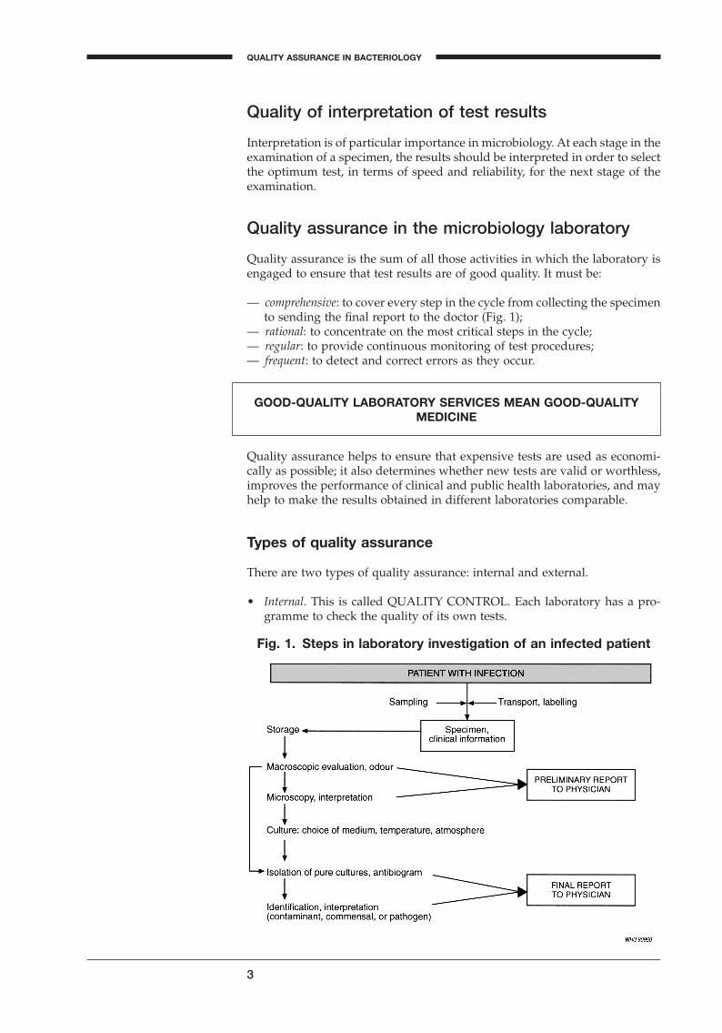
Quality assurance is the sum of all those activities in which the laboratory isengaged to ensure that test results are of good quality. It must be:
— comprehensive: to cover every step in the cycle from collecting the specimento sending the final report to the doctor (Fig. 1);
— rational: to concentrate on the most critical steps in the cycle;— regular: to provide continuous monitoring of test procedures;— frequent: to detect and correct errors as they occur.
3
QUALITY ASSURANCE IN BACTERIOLOGY
A
Fig. 1. Steps in laboratory investigation of an infected patient
Quality assurance helps to ensure that expensive tests are used as economi-cally as possible; it also determines whether new tests are valid or worthless,improves the performance of clinical and public health laboratories, and mayhelp to make the results obtained in different laboratories comparable.
Types of quality assurance
There are two types of quality assurance: internal and external.
• Internal. This is called QUALITY CONTROL. Each laboratory has a pro-gramme to check the quality of its own tests.
GOOD-QUALITY LABORATORY SERVICES MEAN GOOD-QUALITYMEDICINE
BLMIN 1/17/04 2:08 PM Page 3
Internal quality control involves, ideally:
— continuous monitoring of test quality;— comprehensive checking of all steps, from collecting the specimen (whenever
possible) to sending the final report.
Laboratories have an ethical responsibility to the patient to produce accurate,meaningful results.
4
BASIC LABORATORY PROCEDURES IN CLINICAL BACTERIOLOGY
INTERNAL QUALITY CONTROL IS ABSOLUTELY ESSENTIAL FORGOOD OPERATING PROCEDURE
• External. This is called QUALITY ASSESSMENT. Laboratory performanceis controlled by an external agency. In some countries, participation ismandatory (regulated by the government) and required for licensure.
External quality assessment involves:
— periodic monitoring of test quality;— spot checking of identification tests, and sometimes of isolation techniques.
Quality criteria in microbiology
Clinical relevance
An important criterion of quality for a microbiological test is how much it contributes to the prevention or cure of infectious diseases; this is called itsclinical relevance. Clinical relevance can only be ensured when there is good communication between the clinician and the laboratory.
To illustrate clinical relevance, here are some examples:
1. If a few colonies of Gram-negative rods are isolated from the sputum orthroat swab of a hospitalized patient, further identification and an anti-biogram are of no clinical relevance, since neither procedure will have any effect on treatment of the patient.
2. If Streptococcus pyogenes is isolated, a full antibiogram has no clinical rele-vance, since benzylpenicillin is the drug of choice, and this is always activein vitro.
3. If Escherichia coli is isolated from a sporadic case of non-bloody diarrhoea,identification of the serotype is of no clinical relevance, since there is noclearly established correlation between serotype and pathogenicity.
4. If a Gram-stained smear shows “mixed anaerobic flora”, routine identifi-cation of the anaerobes is of no clinical relevance. It would be costly in timeand materials, and would not affect treatment of the patient.
5. If a yeast is isolated from a respiratory tract specimen, an identification testfor Cryptococcus should be done. Further identification tests have no clin-ical relevance, since they would have no effect on patient management.
BLMIN 1/17/04 2:08 PM Page 4
In summary, a test of good quality is one that is accurate and gives usefulresults for the prevention or cure of infection. It is not necessary to isolate andidentify all the different types of organism in the sample.
Reliability
For tests that give quantitative results, reliability is measured by how closethe results are to the true value. Some examples of tests of this kind are:
— antibiotic assay of serum;— measurement of minimal inhibitory concentration (MIC) values of anti-
biotics in vitro;— serum antibody titrations.
For tests that give qualitative results, reliability is measured by whether theresult is correct. Some examples of tests of this kind are:
— identification of pathogens;— antibiotic susceptibility testing of isolates by the disc method.
Standard terminology for microorganisms is essential to reliability. Inter-nationally recognized nomenclature should always be used. For example:Staphylococcus aureus, NOT “pathogenic staphylococci”; Streptococcus pyogenes,NOT “haemolytic streptococci”.
Use of uniform, approved methods is essential. For example, disc suscepti-bility tests should be performed with an internationally recognized technique,such as the modified Kirby–Bauer test (page 109).
Reproducibility
The reproducibility or precision of a microbiological test is reduced by twothings:
1. Lack of homogeneity. A single sample from a patient may contain more thanone organism. Repeat culturing may therefore isolate different organisms.
2. Lack of stability. As time passes, the microorganisms in a specimen multi-ply or die at different rates. Repeat culturing may therefore isolate differ-ent organisms. To improve precision, therefore, specimens should be testedas soon as possible after collection.
Efficiency
The efficiency of a microbiological test is its ability to give the correct diag-nosis of a pathogen or a pathological condition. This is measured by two criteria:
1. Diagnostic sensitivity
The greater the sensitivity of a test, the fewer the number of false-negativeresults.
Sensitivity =total number of positive results
total number of infected patients
5
QUALITY ASSURANCE IN BACTERIOLOGY
A
BLMIN 1/17/04 2:08 PM Page 5
For example, the sensitivity of MacConkey agar is poor for the isolation ofSalmonella typhi from stool. This important enteric pathogen is often missedbecause of overgrowth by nonpathogenic intestinal bacteria.
2. Diagnostic specificity
The greater the specificity of a test, the fewer the number of false-positiveresults.
For example:
• Ziehl–Neelsen staining of sputum is highly specific for diagnosingtuberculosis, because it gives only a few false-positive results.
• Ziehl–Neelsen staining of urine is much less specific, because it givesmany false-positive results (as a result of atypical mycobacteria).
• The Widal test has a very low specificity for the diagnosis of typhoidfever, because cross-agglutinating antibodies remaining from pastinfections with related salmonella serotypes give false-positive results.
The sensitivity and specificity of a test are interrelated. By lowering the levelof discrimination, the sensitivity of a test can be increased at the cost of reduc-ing its specificity, and vice versa. The diagnostic sensitivity and specificity ofa test are also related to the prevalence of the given infection in the popula-tion under investigation.
Internal quality control
Requirements
An internal quality control programme should be practical, realistic, and economical.
An internal quality control programme should not attempt to evaluate everyprocedure, reagent, and culture medium on every working day. It should eval-uate each procedure, reagent, and culture medium according to a practicalschedule, based on the importance of each item to the quality of the test as awhole.
Procedures
Internal quality control begins with proper laboratory operation.
Laboratory operations manual
Each laboratory should have an operations manual that includes the follow-ing subjects:
— cleaning of the working space,— personal hygiene,— safety precautions,— designated eating and smoking areas located outside the laboratory,— handling and disposal of infected material,
Specificity =total number of negative results
total number of unifected patients
6
BASIC LABORATORY PROCEDURES IN CLINICAL BACTERIOLOGY
BLMIN 1/17/04 2:08 PM Page 6
— appropriate vaccinations for workers, e.g. hepatitis B,— care of equipment,— collection of specimens,— registration of specimens,— elimination of unsuitable specimens,— processing of specimens,— recording of results,— reporting of results.
The operations manual should be carefully followed, and regularly revisedand updated.
Care of equipment
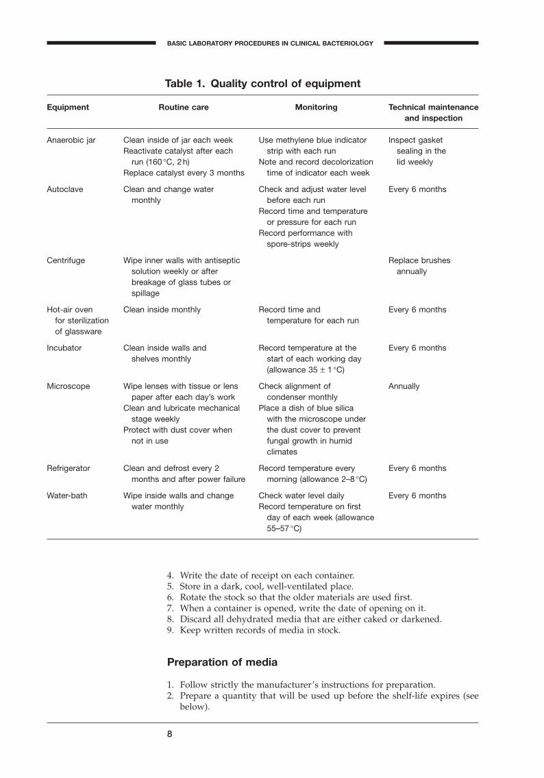
It is particularly important to take good care of laboratory equipment. Goodquality tests cannot be performed if the equipment used is either of poorquality or poorly maintained.
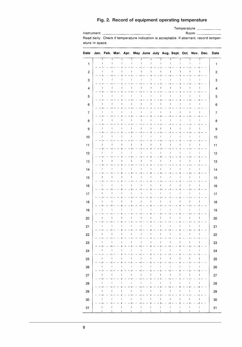
Table 1 is a schedule for the routine care and maintenance of essential equip-ment. Equipment operating temperatures may be recorded on a form such asthe one shown in Fig. 2.
Culture media
Culture media may be prepared in the laboratory from the basic ingredientsor from commercially available dehydrated powders, or they may be pur-chased ready for use. Commercial dehydrated powders are recommendedbecause they are economical to transport and store, and their quality is likelyto be higher than media prepared in the laboratory. For best results, carefulattention is required to the points itemized below.
Selection of media
An efficient laboratory stocks the smallest possible range of media consistentwith the types of test performed. For example, a good agar base can be usedas an all-purpose medium for preparing blood agar, chocolate agar, andseveral selective media.
One highly selective medium (Salmonella–Shigella agar or deoxycholate citrateagar) and one less selective medium (MacConkey agar) are necessary for theisolation of pathogenic Enterobacteriaceae from stools.
A special culture medium should be added for the recovery of Campylobacterspp.
Ordering and storage of dehydrated media
1. Order quantities that will be used up in 6 months, or at most 1 year.2. The overall quantity should be packed in containers that will be used up
in 1–2 months.3. On receipt, tighten caps of all containers securely. Dehydrated media
absorb water from the atmosphere. In a humid climate, seal the tops ofcontainers of dehydrated media with paraffin wax (fill the space betweenthe lid and container with molten wax, and let it harden).
7
QUALITY ASSURANCE IN BACTERIOLOGY
A
BLMIN 1/17/04 2:08 PM Page 7
8
BASIC LABORATORY PROCEDURES IN CLINICAL BACTERIOLOGY
4. Write the date of receipt on each container.5. Store in a dark, cool, well-ventilated place.6. Rotate the stock so that the older materials are used first.7. When a container is opened, write the date of opening on it.8. Discard all dehydrated media that are either caked or darkened.9. Keep written records of media in stock.
Preparation of media
1. Follow strictly the manufacturer’s instructions for preparation.2. Prepare a quantity that will be used up before the shelf-life expires (see
below).
Table 1. Quality control of equipment
Equipment Routine care Monitoring Technical maintenanceand inspection
Anaerobic jar Clean inside of jar each week Use methylene blue indicator Inspect gasketReactivate catalyst after each strip with each run sealing in the
run (160 ∞C, 2h) Note and record decolorization lid weeklyReplace catalyst every 3 months time of indicator each week
Autoclave Clean and change water Check and adjust water level Every 6 monthsmonthly before each run
Record time and temperatureor pressure for each run
Record performance withspore-strips weekly
Centrifuge Wipe inner walls with antiseptic Replace brushessolution weekly or after annuallybreakage of glass tubes orspillage
Hot-air oven Clean inside monthly Record time and Every 6 monthsfor sterilization temperature for each runof glassware
Incubator Clean inside walls and Record temperature at the Every 6 monthsshelves monthly start of each working day
(allowance 35 ± 1 ∞C)
Microscope Wipe lenses with tissue or lens Check alignment of Annuallypaper after each day’s work condenser monthly
Clean and lubricate mechanical Place a dish of blue silicastage weekly with the microscope under
Protect with dust cover when the dust cover to preventnot in use fungal growth in humid
climates
Refrigerator Clean and defrost every 2 Record temperature every Every 6 monthsmonths and after power failure morning (allowance 2–8 ∞C)
Water-bath Wipe inside walls and change Check water level daily Every 6 monthswater monthly Record temperature on first
day of each week (allowance55–57 ∞C)
BLMIN 1/17/04 2:08 PM Page 8
9
A
Fig. 2. Record of equipment operating temperature
BLMIN 1/17/04 2:08 PM Page 9
Storage of prepared media
1. Protect against sunlight.2. Protect against heat. Media containing blood, other organic additives, or
antibiotics should be stored in the refrigerator.3. The shelf-life of prepared media, when stored in a cool, dark place, will
depend on the type of container used. Typical shelf-lives are:— tubes with cotton-wool plugs, 3 weeks;— tubes with loose caps, 2 weeks;— containers with screw-caps, 3 months;— Petri dishes, if sealed in plastic bags, 4 weeks.
Quality control of prepared media
1. pH testing. The pH of the prepared medium need not be checked routinelywhen it is correctly prepared from dehydrated powder. If the medium isprepared from basic ingredients, it should be allowed to cool before thepH is tested. Solid media should be tested with a surface electrode or aftermaceration in distilled water. If the pH differs by more than 0.2 units fromthe specification, adjust with acid or alkali or prepare a new batch.
2. Sterility testing. Carry out routine sterility tests on media to which bloodor other components have been added after autoclaving. Take 3–5% of eachbatch and incubate at 35 ∞C for 2 days. Refrigerate the rest. If more thantwo colonies per plate are seen, discard the whole batch.
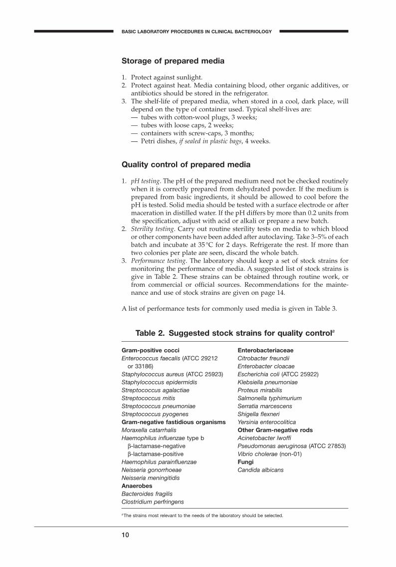
3. Performance testing. The laboratory should keep a set of stock strains formonitoring the performance of media. A suggested list of stock strains isgive in Table 2. These strains can be obtained through routine work, orfrom commercial or official sources. Recommendations for the mainte-nance and use of stock strains are given on page 14.
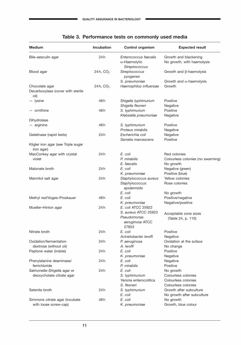
A list of performance tests for commonly used media is given in Table 3.
10
BASIC LABORATORY PROCEDURES IN CLINICAL BACTERIOLOGY
Table 2. Suggested stock strains for quality controla
Gram-positive cocci EnterobacteriaceaeEnterococcus faecalis (ATCC 29212 Citrobacter freundii
or 33186) Enterobacter cloacaeStaphylococcus aureus (ATCC 25923) Escherichia coli (ATCC 25922)Staphylococcus epidermidis Klebsiella pneumoniaeStreptococcus agalactiae Proteus mirabilisStreptococcus mitis Salmonella typhimuriumStreptococcus pneumoniae Serratia marcescensStreptococcus pyogenes Shigella flexneriGram-negative fastidious organisms Yersinia enterocoliticaMoraxella catarrhalis Other Gram-negative rodsHaemophilus influenzae type b Acinetobacter lwoffi
b-lactamase-negative Pseudomonas aeruginosa (ATCC 27853)b-lactamase-positive Vibrio cholerae (non-01)
Haemophilus parainfluenzae FungiNeisseria gonorrhoeae Candida albicansNeisseria meningitidisAnaerobesBacteroides fragilisClostridium perfringens
aThe strains most relevant to the needs of the laboratory should be selected.
BLMIN 1/17/04 2:08 PM Page 10
11
QUALITY ASSURANCE IN BACTERIOLOGY
A
Table 3. Performance tests on commonly used media
Medium Incubation Control organism Expected result
Bile–aesculin agar 24h Enterococcus faecalis Growth and blackeninga-Haemolytic No growth, with haemolysis
StreptococcusBlood agar 24h, CO2 Streptococcus Growth and b-haemolysis
pyogenesS. pneumoniae Growth and a-haemolysis
Chocolate agar 24h, CO2 Haemophilus influenzae GrowthDecarboxylase (cover with sterile
oil)— lysine 48h Shigella typhimurium Positive
Shigella flexneri Negative— ornithine 48h S. typhimurium Positive
Klebsiella pneumoniae NegativeDihydrolase— arginine 48h S. typhimurium Positive
Proteus mirabilis NegativeGelatinase (rapid tests) 24h Escherichia coli Negative
Serratia marcescens PositiveKligler iron agar (see Triple sugar
iron agar)MacConkey agar with crystal 24h E. coli Red colonies
violet P. mirabilis Colourless colonies (no swarming)E. faecalis No growth
Malonate broth 24h E. coli Negative (green)K. pneumoniae Positive (blue)
Mannitol salt agar 24h Staphylococcus aureus Yellow coloniesStaphylococcus Rose colonies
epidermidisE. coli No growth
Methyl red/Voges–Proskauer 48h E. coli Positive/negativeK. pneumoniae Negative/positive
Mueller–Hinton agar 24h E. coli ATCC 25922S. aureus ATCC 25923 Acceptable zone sizesPseudomonas (Table 24, p. 110)
aeruginosa ATCC 27853
Nitrate broth 24h E. coli PositiveAcinetobacter lwoffi Negative
Oxidation/fermentation 24h P. aeruginosa Oxidation at the sufacedextrose (without oil) A. lwoffi No change
Peptone water (indole) 24h E. coli PositiveK. pneumoniae Negative
Phenylalanine deaminase/ 24h E. coli Negativeferrichloride P. mirabilis Positive
Salmonella–Shigella agar or 24h E. coli No growthdeoxycholate citrate agar S. typhimurium Colourless colonies
Yersina enterocolitica Colourless coloniesS. flexneri Colourless colonies
Selenite broth 24h S. typhimurium Growth after subcultureE. coli No growth after subculture
Simmons citrate agar (incubate 48h E. coli No growthwith loose screw-cap) K. pneumoniae Growth, blue colour
BLMIN 1/17/04 2:08 PM Page 11
The procedures to be followed when carrying out performance tests on newbatches of media are:
1. Prepare a suspension of the stock strain with a barely visible turbidity,equivalent to that of the barium sulfate standard used in the modifiedKirby–Bauer method (McFarland 0.5) (see page 109) and use 1 loopful asinoculum.
2. Incubate for the length of time used routinely. Read the plates in the usualway.
3. Keep proper records of results.
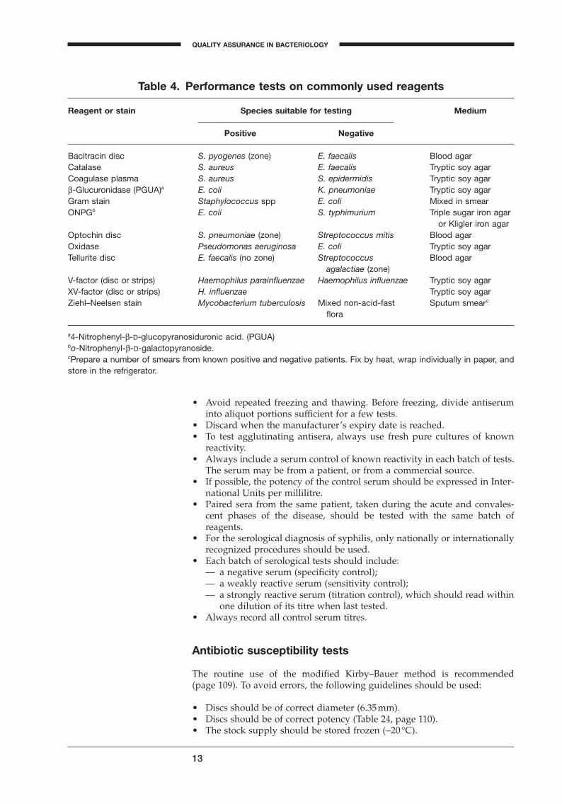
Stains and reagents
Recommendations for testing a number of reagents are given in Table 4.Testing should be carried out:— each time a new batch of working solution is prepared;— every week (this is critical for the cold Ziehl–Neelsen stain: the classical
stain has a shelf-life of several months).
Stains and reagents should be discarded when:
— the manufacturer’s expiry date is reached;— visible signs of deterioration appear (turbidity, precipitate, discoloration).
Diagnostic antigens and antisera
In order to obtain the best results from antigens and antisera:
• Always follow the manufacturer’s instructions.• Store at the recommended temperature. Some serological reagents do not
tolerate freezing.
12
BASIC LABORATORY PROCEDURES IN CLINICAL BACTERIOLOGY
Thiosulfate citrate bile salts 24h Vibrio spp. (non- Yellow coloniesagar agglutinating)
Thayer–Martin agar 24h, CO2 Neisseria meningitidis GrowthNeisseria gonorrhoeae GrowthStaphylococcus spp. No growthE. coli No growthC. albicans No growth
Thioglycollate broth 24h Bacteroides fragilis GrowthTriple sugar iron agar (depth of 24h Citrobacter freundii A/A gasa + H2S
butt should be at least 2.5cm; S. typhimurium K/A gasa + H2Sincubate with loose screw-cap) S. flexneri K/A gasa
A. lwoffi No changeUrea medium 24h E. coli Negative
P. mirabilis Positive (pink)Voges–Proskauer (see Methyl
red/Voges–Proskauer)
aA/A: acid slant; K/A: alkaline slant.
Table 3 (continued)
Medium Incubation Control organism Expected result
BLMIN 1/17/04 2:08 PM Page 12
• Avoid repeated freezing and thawing. Before freezing, divide antiseruminto aliquot portions sufficient for a few tests.
• Discard when the manufacturer’s expiry date is reached.• To test agglutinating antisera, always use fresh pure cultures of known
reactivity.• Always include a serum control of known reactivity in each batch of tests.
The serum may be from a patient, or from a commercial source.• If possible, the potency of the control serum should be expressed in Inter-
national Units per millilitre.• Paired sera from the same patient, taken during the acute and convales-
cent phases of the disease, should be tested with the same batch ofreagents.
• For the serological diagnosis of syphilis, only nationally or internationallyrecognized procedures should be used.
• Each batch of serological tests should include:— a negative serum (specificity control);— a weakly reactive serum (sensitivity control);— a strongly reactive serum (titration control), which should read within
one dilution of its titre when last tested.• Always record all control serum titres.
Antibiotic susceptibility tests
The routine use of the modified Kirby–Bauer method is recommended (page 109). To avoid errors, the following guidelines should be used:
• Discs should be of correct diameter (6.35mm).• Discs should be of correct potency (Table 24, page 110).• The stock supply should be stored frozen (-20 ∞C).
13
QUALITY ASSURANCE IN BACTERIOLOGY
A
Table 4. Performance tests on commonly used reagents
Reagent or stain Species suitable for testing Medium
Positive Negative
Bacitracin disc S. pyogenes (zone) E. faecalis Blood agarCatalase S. aureus E. faecalis Tryptic soy agarCoagulase plasma S. aureus S. epidermidis Tryptic soy agarb-Glucuronidase (PGUA)a E. coli K. pneumoniae Tryptic soy agarGram stain Staphylococcus spp E. coli Mixed in smearONPGb E. coli S. typhimurium Triple sugar iron agar
or Kligler iron agarOptochin disc S. pneumoniae (zone) Streptococcus mitis Blood agarOxidase Pseudomonas aeruginosa E. coli Tryptic soy agarTellurite disc E. faecalis (no zone) Streptococcus Blood agar
agalactiae (zone)V-factor (disc or strips) Haemophilus parainfluenzae Haemophilus influenzae Tryptic soy agarXV-factor (disc or strips) H. influenzae Tryptic soy agarZiehl–Neelsen stain Mycobacterium tuberculosis Mixed non-acid-fast Sputum smearc
flora
a4-Nitrophenyl-b-D-glucopyranosiduronic acid. (PGUA)bo-Nitrophenyl-b-D-galactopyranoside.cPrepare a number of smears from known positive and negative patients. Fix by heat, wrap individually in paper, andstore in the refrigerator.
BLMIN 1/17/04 2:08 PM Page 13
• The working supply should be kept no longer than 1 month in a refriger-ator (2–8 ∞C).
• Only Mueller–Hinton agar of performance-tested quality should be used.• Correct pH (7.2–7.4) of the finished medium is essential for some anti-
biotics.• The inoculum should be standardized against the prescribed turbidity
standard (page 111).• Zone sizes should be measured exactly.• Zone sizes should be interpreted by referring to a table of critical diame-
ters. Zone diameters for each organism should fall within the limits givenin Table 24 (page 110).
• The three standard control strains are:1
— Staphylococcus aureus (ATCC 25923; NCTC 6571);— Escherichia coli (ATCC 25922; NCTC 10418);— Pseudomonas aeruginosa (ATCC 27853; NCTC 10622).
• Tests should be carried out with the three standard strains:— when a new batch of discs is put into use;— when a new batch of medium is put into use;— once a week, in parallel with the routine antibiograms.
• Use the quality control chart shown in Fig. 16 (page 121) for recording andevaluating performance tests.
Maintenance and use of stock cultures
Selection and origin
Select the strains so that the maximum number of morphological, metabolic,and serological characteristics can be tested with the minimum number of cul-tures; a suggested list is given in Table 2. These strains can be obtained froma combination of the following sources:
— properly documented isolates from clinical specimens;— official culture collections;— commercial producers;— external quality assessment surveys;— reference laboratories.
Preservation
Long-term preservationLong-term preservation methods permit intervals of months or even yearsbetween subcultures. The best methods are lyophilization (freeze-drying), orstorage at -70 ∞C or below, in an electric freezer or in liquid nitrogen. Alter-native methods are described below.
Glycerol at -20 ∞C
1. Grow a pure culture on an appropriate solid medium.2. When the culture is fully developed, scrape it off with a loop.3. Suspend small clumps of the culture in sterile neutral glycerol.
14
BASIC LABORATORY PROCEDURES IN CLINICAL BACTERIOLOGY
1 These strains can be obtained from: American Type Culture Collection (ATCC), 10801 Univer-sity Boulevard, Manassas, VA 20110, USA; or National Collection of Type Cultures (NCTC), PHLSCentral Public Health Laboratory, 61 Colindale Avenue, London NW9 5HT, England.
BLMIN 1/17/04 2:08 PM Page 14
4. Distribute in quantities of 1–2ml in screw-capped tubes or vials.5. Store at -20 ∞C. Avoid repeated freezing and thawing. Transfer after 12–18
months.
Mineral oil at room temperature1
1. Prepare tubes of heart infusion agar with a short slant. For fastidiousorganisms, add fresh native or heated blood.
2. Sterilize mineral oil (liquid petrolatum) in hot air (170 ∞C for 1 hour).3. Grow a pure culture on the agar slant.4. When good growth is seen, add sterile mineral oil to about 1cm above the
tip of the slant.5. Subculture when needed by scraping growth from under the oil.6. Store at room temperature. Transfer after 6–12 months.
Stab cultures at room temperature (use for non-fastidious organismsonly, such as staphylococci and Enterobacteriaceae)
1. Prepare tubes with a deep butt of carbohydrate-free agar. Tryptic soy agar(soybean casein digest agar) is recommended.
2. Stab the organism into the agar.3. Incubate overnight at 35 ∞C.4. Close tube with screw-cap or cork. Dip cap or cork into molten paraffin
wax to seal.5. Store at room temperature. Transfer after 1 year.
Stab cultures in cystine trypticase agar (CTA) (for Neisseria andstreptococci)
1. Prepare tubes of CTA basal medium.2. Stab the organism into the medium.3. Incubate overnight at 35 ∞C.4. Close tube with screw-cap or cork. Dip cap or cork into molten paraffin
wax to seal.5. For Neisseria, store at 35 ∞C, and transfer every 2 weeks. For streptococci,
store at room temperature, and transfer every month.
Cooked-meat medium for anaerobes
1. Inoculate tubes.2. Incubate overnight at 35 ∞C.3. Close tube with screw-cap or cork.4. Store at room temperature. Transfer every 2 months.
Short-term preservationWorking cultures for daily routine tests can be prepared in the following ways.
Rapid-growing organisms
1. Inoculate on tryptic soy agar slants in screw-capped tubes.2. Incubate overnight at 35 ∞C.3. Store in a refrigerator. Transfer every 2 weeks.
15
QUALITY ASSURANCE IN BACTERIOLOGY
A
1 Morton HE, Pulaski EJ. The preservation of bacterial cultures. Journal of Bacteriology, 1938,38:163–183.
BLMIN 1/17/04 2:08 PM Page 15
Streptococci
1. Inoculate on blood agar slants in screw-capped tubes.2. Incubate overnight at 35 ∞C.3. Store in a refrigerator. Transfer every 2 weeks.
Meningococci and Haemophilus
1. Inoculate on chocolate agar slants or plates.2. Incubate overnight at 35 ∞C.3. Store at room temperature. Transfer twice a week.
Gonococci
1. Inoculate on chocolate agar.2. Incubate and store at 35 ∞C. Transfer every 2 days.3. Replace the quality control strain by each new clinical isolate.
Use of reference laboratories
The following categories of specimen should be submitted to a regional orcentral reference laboratory:
— specimens for infrequently requested or highly specialized tests (e.g. virol-ogy, serodiagnosis of parasitic infections);
— occasional duplicate specimens, as a check on the submitting laboratory’sown results;
— specimens needing further confirmation, specification, grouping, or typingof pathogens of great public health importance (e.g. Salmonella, Shigella,Vibrio cholerae, Brucella, meningococci, and pneumococci).
Reference laboratories should be able to supply reference cultures for qualitycontrol and training needs, and standard sera and reagents for comparisonwith those in use in the referring laboratory.
If no external quality assessment programme exists, the reference laboratoryshould be asked to supply blind, coded specimens and cultures so that the referring laboratory may test its own proficiency in isolation and identification.
External quality assessment
This section gives information on what is involved in participation in an exter-nal quality assessment scheme (sometimes known as a “proficiency testingscheme”).
Purposes
The purposes of a quality assessment programme are:
— to provide assurance to both physicians and the general public that labo-ratory diagnosis is of good quality;
— to assess and compare the reliability of laboratory performance on anational scale;
— to identify common errors;— to encourage the use of uniform procedures;
16
BASIC LABORATORY PROCEDURES IN CLINICAL BACTERIOLOGY
BLMIN 1/17/04 2:08 PM Page 16
— to encourage the use of standard reagents;— to take administrative measures (which may include revocation of the
operating licence) against substandard laboratories;— to stimulate the implementation of internal quality control programmes.
Organization
A quality assessment programme consists of a number of surveys in whichcoded specimens are distributed by mail to participating laboratories. Thesespecimens should be incorporated into the laboratory routine, and handledand tested in exactly the same way as routine clinical specimens.
The surveys should be conducted in accordance with the following recommendations:
— surveys should be carried out at least 4 times a year;— a minimum of 3 specimens should be included in each survey;— the reporting period should be short, for example 2 weeks following
receipt of the specimens;— instructions and report forms should be included with each survey and
the report sheet should be in duplicate, with a clearly stated deadline.
Cultures
Cultures should be included for identification and for susceptibility testingagainst a limited range of antibiotics; they may be pure cultures or mixturesof two or more cultures.
Cultures should represent at least the first 3 of the following 6 categories:
1. Bacterial species that are of great public health potential, but which are not often seen in routine practice, for example Corynebacterium diphtheriae,Salmonella paratyphi A.NOTE: Brucella and Salmonella typhi should not be used for quality assess-ment schemes, since they may give rise to serious accidental infections.
2. Abnormal biotypes that are often misidentified, for example H2S-positiveEscherichia coli, lactose-negative E. coli, urease-negative Proteus.
3. Newly recognized or opportunistic pathogens, for example Yersinia ente-rocolitica, Vibrio parahaemolyticus, Burkholderia, Pseudomonas cepacia.
4. A mixture of Shigella, Citrobacter, E. coli, and Klebsiella may be used to testthe skill of a laboratory in isolating pathogenic microorganisms from anumber of commensal organisms.
5. A mixture of nonpathogenic organisms may be used to test for ability torecognize negative specimens.
6. Bacteria with special resistance patterns, for example meticillin-resistant S.aureus (MRSA).
Sera
Serological tests for the following infections should be part of an externalquality assessment programme in bacteriology:
— syphilis— rubella— brucellosis
17
QUALITY ASSURANCE IN BACTERIOLOGY
A
BLMIN 1/17/04 2:08 PM Page 17
— streptococcal infections— typhoid fever.
Rating and reporting of results
As soon as all reports of results are received from participating, the correctanswers should be sent to the laboratories. Within one month after that, a finalreport should be sent to the laboratories with an analysis of the results. A per-formance score is given to each laboratory. Each laboratory should have a codenumber known only to itself. Thus it can recognize its own performance inrelation to others, but the other laboratories remain anonymous.
18
BASIC LABORATORY PROCEDURES IN CLINICAL BACTERIOLOGY
BLMIN 1/17/04 2:08 PM Page 18

Part IBacteriological investigations
A
BLM1 1/17/04 2:01 PM Page 19
Blood
Introduction
Blood is cultured to detect and identify bacteria or other cultivable microor-ganisms (yeasts, filamentous fungi). The presence of such organisms in theblood is called bacteraemia or fungaemia, and is usually pathological. Inhealthy subjects, the blood is sterile. However, there are a few exceptions: tran-sient bacteraemia often occurs shortly after a tooth extraction or other dentalor surgical manipulation of contaminated mucous membranes, bronchoscopy,or urethral catheterization. This type of transient bacteraemia is generally dueto commensal bacteria and usually resolves spontaneously through phagocy-tosis of the bacteria in the liver and spleen.
Septicaemia is a clinical term used to describe bacteraemia with clinical manifestations of a severe infection, including chills, fever, malaise, toxicity,and hypotension, the extreme form being shock. Shock can be caused bytoxins produced by Gram-negative rods or Gram-positive cocci.
When and where bacteraemia may occur
Bacteraemia is a feature of some infectious diseases, e.g. brucellosis, lep-tospirosis and typhoid fever. Persistent bacteraemia is a feature of endovas-cular infections, e.g. endocarditis, infected aneurysm and thrombophlebitis.
Transient bacteraemia often accompanies localized infections such as arthri-tis, bed sores, cholecystitis, enterocolitis, meningitis, osteomyelitis, peritoni-tis, pneumonia, pyelonephritis, and traumatic or surgical wound infections.It can arise from various surgical manipulations, but usually resolves spontaneously in healthy subjects.
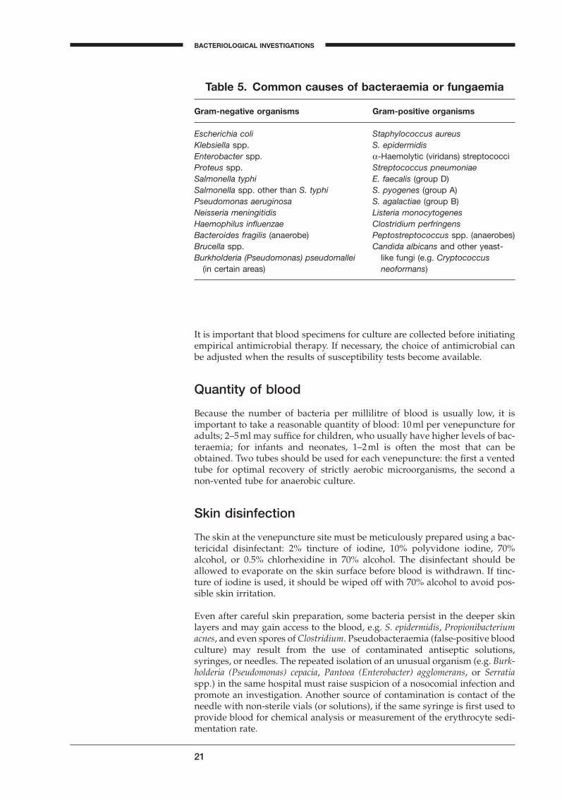
Bacteraemia and fungaemia may result from the iatrogenic introduction ofmicroorganisms by the intravenous route: through contaminated intravenousfluids, catheters, or needle-puncture sites. Both types of infection may developin users of intravenous drugs and in immunosuppressed subjects, includingthose with human immunodeficiency virus/the acquired immunodeficiencysyndrome (HIV/AIDS). They are often caused by “opportunistic” microor-ganisms and may have serious consequences. Table 5 shows the most commoncauses of bacteraemia or fungaemia.
Blood collection
Timing of blood collection
Whenever possible, blood should be taken before antibiotics are administered.The best time is when the patient is expected to have chills or a temperaturespike. It is recommended that two or preferably three blood cultures beobtained, separated by intervals of approximately 1 hour (or less if treatmentcannot be delayed). More than three blood cultures are rarely indicated. Theadvantages of repeated cultures are as follows:
— the chance of missing a transient bacteraemia is reduced;— the pathogenic role of “saprophytic” isolates (e.g. Staphylococcus epider-
midis) is confirmed if they are recovered from multiple venepunctures.
20
BLM1 1/17/04 2:01 PM Page 20
It is important that blood specimens for culture are collected before initiatingempirical antimicrobial therapy. If necessary, the choice of antimicrobial canbe adjusted when the results of susceptibility tests become available.
Quantity of blood
Because the number of bacteria per millilitre of blood is usually low, it isimportant to take a reasonable quantity of blood: 10ml per venepuncture foradults; 2–5ml may suffice for children, who usually have higher levels of bac-teraemia; for infants and neonates, 1–2ml is often the most that can beobtained. Two tubes should be used for each venepuncture: the first a ventedtube for optimal recovery of strictly aerobic microorganisms, the second anon-vented tube for anaerobic culture.
Skin disinfection
The skin at the venepuncture site must be meticulously prepared using a bac-tericidal disinfectant: 2% tincture of iodine, 10% polyvidone iodine, 70%alcohol, or 0.5% chlorhexidine in 70% alcohol. The disinfectant should beallowed to evaporate on the skin surface before blood is withdrawn. If tinc-ture of iodine is used, it should be wiped off with 70% alcohol to avoid pos-sible skin irritation.
Even after careful skin preparation, some bacteria persist in the deeper skinlayers and may gain access to the blood, e.g. S. epidermidis, Propionibacteriumacnes, and even spores of Clostridium. Pseudobacteraemia (false-positive bloodculture) may result from the use of contaminated antiseptic solutions,syringes, or needles. The repeated isolation of an unusual organism (e.g. Burk-holderia (Pseudomonas) cepacia, Pantoea (Enterobacter) agglomerans, or Serratiaspp.) in the same hospital must raise suspicion of a nosocomial infection andpromote an investigation. Another source of contamination is contact of theneedle with non-sterile vials (or solutions), if the same syringe is first used toprovide blood for chemical analysis or measurement of the erythrocyte sedi-mentation rate.
21
BACTERIOLOGICAL INVESTIGATIONS
A
Table 5. Common causes of bacteraemia or fungaemia
Gram-negative organisms Gram-positive organisms
Escherichia coli Staphylococcus aureusKlebsiella spp. S. epidermidisEnterobacter spp. a-Haemolytic (viridans) streptococciProteus spp. Streptococcus pneumoniaeSalmonella typhi E. faecalis (group D)Salmonella spp. other than S. typhi S. pyogenes (group A)Pseudomonas aeruginosa S. agalactiae (group B)Neisseria meningitidis Listeria monocytogenesHaemophilus influenzae Clostridium perfringensBacteroides fragilis (anaerobe) Peptostreptococcus spp. (anaerobes)Brucella spp. Candida albicans and other yeast-Burkholderia (Pseudomonas) pseudomallei like fungi (e.g. Cryptococcus
(in certain areas) neoformans)
BLM1 1/17/04 2:01 PM Page 21
Anticoagulant
The use of sodium polyanethol sulfonate (SPS) as an anticoagulant is recom-mended because it also inhibits the antibacterial effect of serum and phago-cytes. If the blood is immediately added to a sufficient volume (50ml) of brothand thoroughly mixed to prevent clotting, no anticoagulant is needed. It isrecommended that blood-culture bottles be available at all hospitals and majorhealth centres. If blood-culture bottles are not available, blood may be trans-ported to the laboratory in a tube containing a sterile anticoagulant solution(citrate, heparin, or SPS). Upon receipt in the laboratory, such blood samplesmust be transferred immediately to blood-culture bottles using a strict aseptictechnique. Where blood is taken without anticoagulant, the clot can be asep-tically transferred to broth in the laboratory and the serum used for certainserological tests (e.g. Widal).
Blood-culture media
Choice of broth medium
The blood-culture broth and tryptic soy broth (TSB) should be able to supportgrowth of all clinically significant bacteria.
Quantity of broth
Ideally, the blood should be mixed with 10 times its volume of broth (5ml ofblood in 50ml of broth) to dilute any antibiotic present and to reduce the bactericidal effect of human serum.
Blood-culture bottles
Blood-culture bottles (125ml) with a pre-perforated screw-cap and a rubberdiaphragm must be used. Fill the bottle with 50ml of medium and then loosenthe screw-cap half a turn. Cover the cap with a square piece of aluminiumfoil, and autoclave the bottle for 20 minutes at 120 ∞C. Immediately after autoclaving, while the bottle and the medium are still hot, securely tightenthe cap without removing the aluminium foil (otherwise the cap will not be sterile). As the medium cools, a partial vacuum will be created in the bottle, which will facilitate injection of a blood specimen through thediaphragm.
The top of the cap must be carefully disinfected just before the bottle is inoculated.
Prior to distribution and before use, all blood-culture bottles should be carefully examined for clarity. Any medium showing turbidity should not beused.
If strictly aerobic bacteria (Pseudomonas, Neisseria) or yeasts are suspected, thebottle should be vented as soon as it is received in the laboratory, by insert-ing a sterile cotton-wool-plugged needle through the previously disinfecteddiaphragm. The needle can be removed once the pressure in the bottle reachesatmospheric pressure. Commercial blood-culture bottles often also containcarbon dioxide, which has a stimulating effect on growth.
22
BLOOD
BLM1 1/17/04 2:01 PM Page 22
In countries where brucellosis is prevalent, the use of a diphasic blood-culturebottle, with a broth phase and a solid-slant phase on one of the flat surfacesof the bottle (Castaneda bottle), is recommended for the cultivation of Brucellaspp. The presence of carbon dioxide is needed for the isolation of most strainsof B. abortus.
Processing of blood cultures
Incubation time
Blood-culture bottles should be incubated at 35–37 ∞C and routinely inspectedtwice a day (at least for the first 3 days) for signs of microbial growth. A sterileculture usually shows a layer of sedimented red blood covered by a paleyellow transparent broth. Growth is evidenced by:
— a floccular deposit on top of the blood layer— uniform or subsurface turbidity— haemolysis— coagulation of the broth— a surface pellicle— production of gas— white grains on the surface or deep in the blood layer.
Whenever visible growth appears, the bottle should be opened aseptically, asmall amount of broth removed with a sterile loop or Pasteur pipette, and aGram-stained smear examined for the presence of microorganisms.
Subcultures are performed by streaking a loopful on appropriate media:
— for Gram-negative rods: MacConkey agar, Kligler iron agar, motility-indole–urease (MIU) medium, Simmons citrate agar;
— for small Gram-negative rods: blood agar;— for staphylococci: blood agar, mannitol salt agar;— for streptococci: blood agar with optochin, bacitracin, and tellurite discs,
sheep blood agar for the CAMP test, and bile–aesculin agar.
For routine examinations, it is not necessary to incubate blood culturesbeyond 7 days. In some cases, incubation may be prolonged for an additional7 days, e.g. if Brucella or other fastidious organisms are suspected, in cases ofendocarditis, or if the patient has received antimicrobials.
Blind subcultures and final processing
Some microorganisms may grow without producing turbidity or visible alteration of the broth. Other organisms, e.g. pneumococci, tend to undergoautolysis and die very rapidly. For this reason some laboratories performroutine subcultures on chocolate agar after 18–24 hours of incubation. A blindsubculture may be made at the end of 7 days of incubation, by transferringseveral drops of the well-mixed blood culture (using a sterile Pasteur pipette)into a tube of thioglycollate broth, which in turn is incubated and observedfor 3 days.
23
BACTERIOLOGICAL INVESTIGATIONS
A
BLM1 1/17/04 2:01 PM Page 23
Antibiogram
When staphylococci or Gram-negative rods are suspected, precious time canbe saved by performing a direct, non-standardized antibiogram using thepositive broth as an inoculum. A sterile swab is dipped into the turbid broth,excess fluid is expressed, and the swab is used to inoculate Mueller–Hintonmedium as in the standard method (see page 110). A provisional reading canoften be made after 6–8 hours of incubation. In 95% of cases the resultsobtained with this method are in agreement with the standardized test.
Contaminants
Contamination of blood cultures can be avoided by meticulous skin prepara-tion and by adherence to strict aseptic procedures for inoculation and subin-oculation. However, even in ideal conditions, 3–5% of blood cultures grow“contaminants” originating from the skin (S. epidermidis, P. acnes, Clostridiumspp., diphtheroids) or from the environment (Acinetobacter spp., Bacillus spp.). Such organisms, however, may occasionally behave as pathogens and evencause endocarditis. A true infection should be suspected in the following situations:
— if the same organism grows in two bottles of the same blood specimen;— if the same organism grows in cultures from more than one specimen;— if growth is rapid (within 48 hours);— if different isolates of one species show the same biotypes and antimicro-
bial-susceptibility profiles.
All culture results should be reported to the clinician, including the presumedcontaminants. However, for the latter no antibiogram need be performed andappropriate mention should be made on the report slip, e.g. Propionibacteriumacnes (skin commensal), Staphylococcus epidermidis (probable contaminant). Itis to the advantage of all concerned to establish good communication betweenphysicians and laboratory personnel.
The identification of two or more agents may indicate polymicrobial bacte-raemia, which can occur in debilitated patients, but may also be due to con-tamination. “Anaerobic” bacteraemia is often caused by multiple pathogens;for example, one or more anaerobes may be associated with one or more aer-obes in severe fulminating bacteraemia associated with severe trauma or sur-gery involving the large intestine.
24
BLOOD
BLM1 1/17/04 2:01 PM Page 24
Cerebrospinal fluid
Introduction
The examination of cerebrospinal fluid (CSF) is an essential step in the diagnosis of bacterial and fungal meningitis and CSF must always be consid-ered as a priority specimen that requires prompt attention by the laboratorystaff.
Normal CSF is sterile and clear, and usually contains three leukocytes or fewer per mm3 and no erythrocytes. The chemical and cytological compo-sition of CSF is modified by meningeal or cerebral inflammation, i.e. menin-gitis or encephalitis. Only the microbiological examination of CSF will be discussed here, although the CSF leukocyte count is also of paramountimportance.
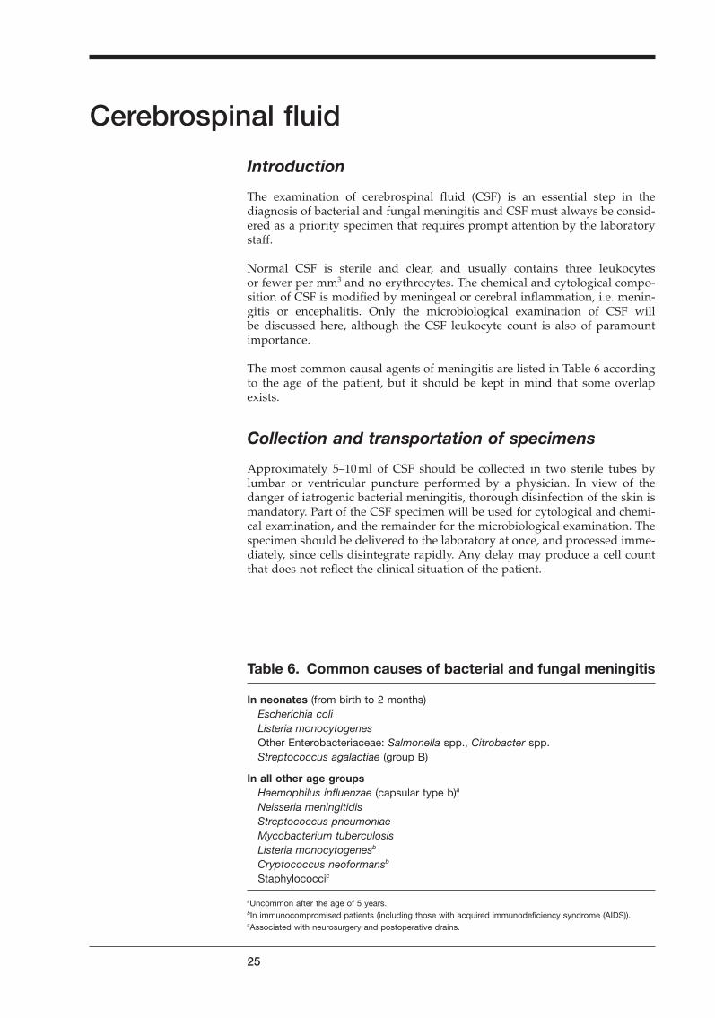
The most common causal agents of meningitis are listed in Table 6 accordingto the age of the patient, but it should be kept in mind that some overlap exists.
Collection and transportation of specimens
Approximately 5–10ml of CSF should be collected in two sterile tubes bylumbar or ventricular puncture performed by a physician. In view of thedanger of iatrogenic bacterial meningitis, thorough disinfection of the skin ismandatory. Part of the CSF specimen will be used for cytological and chemi-cal examination, and the remainder for the microbiological examination. Thespecimen should be delivered to the laboratory at once, and processed imme-diately, since cells disintegrate rapidly. Any delay may produce a cell countthat does not reflect the clinical situation of the patient.
25
A
Table 6. Common causes of bacterial and fungal meningitis
In neonates (from birth to 2 months)Escherichia coliListeria monocytogenesOther Enterobacteriaceae: Salmonella spp., Citrobacter spp.Streptococcus agalactiae (group B)
In all other age groupsHaemophilus influenzae (capsular type b)a
Neisseria meningitidisStreptococcus pneumoniaeMycobacterium tuberculosisListeria monocytogenesb
Cryptococcus neoformansb
Staphylococcic
aUncommon after the age of 5 years.bIn immunocompromised patients (including those with acquired immunodeficiency syndrome (AIDS)).cAssociated with neurosurgery and postoperative drains.
BLM1 1/17/04 2:01 PM Page 25
Macroscopic inspection
The appearance of the CSF should be noted and recorded as: clear, hazy,turbid, purulent, yellow (due to haemolysis or icterus), or blood-tinged, withfibrin web or pellicle.
Microscopic examination
Preparation of specimen
If, on gross examination, the CSF is purulent (very cloudy), it can be exam-ined immediately without centrifugation. In all other cases, the CSF shouldbe centrifuged in a sterile tube (preferably a 15-ml conical tube with screw-cap) at 10000 g for 5–10 minutes. Remove the supernatant using a sterilePasteur pipette fitted with a rubber bulb, and transfer it to another tube for chemical and/or serological tests. Use the sediment for further micro-biological tests.
Direct microscopy
Examine one drop of the sediment microscopically (¥400), between a slide andcoverslip, for:
— leukocytes (polymorphonuclear neutrophils or lymphocytes)— erythrocytes— bacteria— yeasts.
If the yeast-like fungus Cryptococcus neoformans is suspected, mix a loopful ofthe sediment with a loopful of India ink on a slide, place a coverslip on top,and examine microscopically for the typical, encapsulated, spherical, buddingyeast forms.
In areas where African trypanosomiasis occurs, it will also be necessary tosearch carefully for actively motile, flagellated trypanosomes.
A rare and generally fatal type of meningitis is caused by free-living amoebaefound in water (Naegleria fowleri) which enter through the nose and penetratethe central nervous system. They may be seen in the direct wet preparationas active motile amoebae about the size of neutrophilic leukocytes.
Gram-stained smears
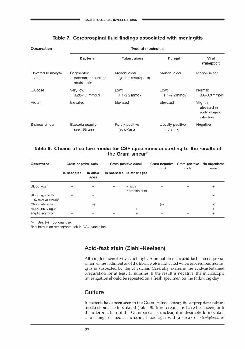
As the causative agent of bacterial meningitis may often be observed in a Gram-stained smear, this examination is extremely important. Air-dry thesmear, fix with gentle heat, and stain it by Gram’s method. Examine at ¥1000(oil-immersion) for at least 10 minutes, or until bacteria are found. Table 7 listsimportant diagnostic findings that are associated with different forms ofmeningitis.
26
CEREBROSPINAL FLUID
BLM1 1/17/04 2:01 PM Page 26
Acid-fast stain (Ziehl–Neelsen)
Although its sensitivity is not high, examination of an acid-fast-stained prepa-ration of the sediment or of the fibrin web is indicated when tuberculous menin-gitis is suspected by the physician. Carefully examine the acid-fast-stainedpreparation for at least 15 minutes. If the result is negative, the microscopicinvestigation should be repeated on a fresh specimen on the following day.
Culture
If bacteria have been seen in the Gram-stained smear, the appropriate culturemedia should be inoculated (Table 8). If no organisms have been seen, or if the interpretation of the Gram smear is unclear, it is desirable to inoculate a full range of media, including blood agar with a streak of Staphylococcus
27
BACTERIOLOGICAL INVESTIGATIONS
A
Table 7. Cerebrospinal fluid findings associated with meningitis
Observation Type of meningitis
Bacterial Tuberculous Fungal Viral(“aseptic”)
Elevated leukocyte Segmented Mononuclear Mononuclear Mononuclearcount polymorphonuclear (young neutrophils)
neutrophils
Glucose Very low: Low: Low: Normal:0.28–1.1mmol/l 1.1–2.2mmol/l 1.1–2.2mmol/l 3.6–3.9mmol/l
Protein Elevated Elevated Elevated Slightlyelevated inearly stage ofinfection
Stained smear Bacteria usually Rarely positive Usually positive Negativeseen (Gram) (acid-fast) (India ink)
Table 8. Choice of culture media for CSF specimens according to the results ofthe Gram smeara
Observation Gram-negative rods Gram-positive cocci Gram-negative Gram-positive No organisms
In neonates In other In neonates In other agescocci rods seen
ages
Blood agarb + + + + with + + +optochin disc
Blood agar with + + +S. aureus streakb
Chocolate agar (+) (+) (+)MacConkey agar + + + + + + +Tryptic soy broth + + + + + + +
a+ = Use; (+) = optional use.bIncubate in an atmosphere rich in CO2 (candle jar).
BLM1 1/17/04 2:01 PM Page 27
aureus to promote growth of H. influenzae. Blood agar and chocolate agar platesshould be incubated at 35 ∞C in an atmosphere enriched with carbon dioxide.All media should be incubated for 3 days, with daily inspections.
When tuberculous meningitis is suspected, at least three tubes of Löwenstein–Jensen medium should be inoculated with a drop of the sediment and incu-bated for 6 weeks. For the first 2–3 days the tubes should be incubated in ahorizontal position with the screw-cap loosened half a turn. Tubes should beinspected for growth at weekly intervals. Smears from any suspicious growthshould be prepared, preferably in a bacteriological safety cabinet, air-dried,heat-fixed, and stained by the Ziehl–Neelsen method. The presence of acid-fast rods is consistent with the diagnosis of tuberculosis. All isolates shouldbe forwarded to a central laboratory for confirmation and for susceptibilitytesting.
When Cryptococcus neoformans is suspected, either from the India ink prepa-ration or on clinical grounds, the sediment should be inoculated on two tubesof Sabouraud dextrose agar, and incubated at 35 ∞C for up to 1 month. C. neoformans also grows on the blood agar plate, which should be incubated at 35 ∞C for 1 week, if indicated.
Preliminary identification
Growth on MacConkey agar is suggestive of Enterobacteriaceae and shouldbe further identified using the methods and media recommended for entericpathogens.
Colonies of Gram-positive cocci with a narrow zone of b-haemolysis may beS. agalactiae (group B streptococci). This should be confirmed with the reverseCAMP test (page 101).
Flat colonies with a concave centre and a slight green zone of a-haemolysisare probably S. pneumoniae. For confirmation, a 6-mm optochin disc should beplaced on a blood agar plate heavily inoculated with a pure culture of the sus-pected strain. After overnight incubation, pneumococci will exhibit an inhi-bition zone of 14 mm or more around the optochin disc. The best results areobtained after incubation on sheep blood agar in a carbon-dioxide-enrichedatmosphere. If the reading of this test on the primary blood agar plate is incon-clusive, the test should be repeated on a subculture.
Colonies of H. influenzae will grow only on chocolate agar, and as satellitecolonies in the vicinity of the staphylococcal streak on blood agar. Furtheridentification may be accomplished using H. influenzae type b antiserum inthe slide agglutination test.
Gram-negative diplococci growing on blood and chocolate agar, and giving arapidly positive oxidase test, may be considered to be meningococci. Confir-mation is accomplished by grouping with appropriate N. meningitidis anti-sera (A, B, C) in the slide agglutination test. A negative agglutination test doesnot rule out meningococci as there are at least four additional serogroups. Ifthe agglutination test is negative, carbohydrate utilization tests should be per-formed and the culture sent to a central reference laboratory. A preliminaryreport should be given to the physician at each stage of identification (Gram-stain, growth, agglutination, etc.), noting that a final report will follow.
Colonies of Gram-positive rods with a narrow zone of b-haemolysis on bloodagar may be Listeria monocytogenes. The following confirmatory tests are
28
CEREBROSPINAL FLUID
BLM1 1/17/04 2:01 PM Page 28
suggested: positive catalase reaction, motility in broth culture or in MIU,growth and black discoloration on bile–aesculin agar.
Susceptibility testing
For Gram-negative rods and staphylococci, the standardized disc-diffusionmethod (Kirby–Bauer) should be used.
No susceptibility testing is needed for Listeria monocytogenes, S. agalactiae orN. meningitidis since resistance to ampicillin and benzylpenicillin is extremelyrare.
All strains of pneumococci should be tested on blood agar for susceptibilityto chloramphenicol and benzylpenicillin. For the latter, the oxacillin (1mg) discis recommended (see page 66, “Lower respiratory tract infections”).
Strains of H. influenzae should be tested for susceptibility to chloramphenicolusing chocolate agar or a supplemented blood agar. Most ampicillin-resistantstrains produce b-lactamase that can be demonstrated using one of the rapidtests recommended for the screening of potential b-lactamase-producingstrains of gonococci (page 79).
29
BACTERIOLOGICAL INVESTIGATIONS
A
BLM1 1/17/04 2:01 PM Page 29
Urine
Introduction
Urine is the specimen most frequently submitted for culture. It also presentsmajor problems in terms of proper specimen collection, transport, culturetechniques, and interpretation of results. As with any other specimen sub-mitted to the laboratory, the more comprehensive the information providedby the submitting physician the more able the laboratory is to provide the bestpossible culture data.
The most common sites of urinary tract infection (UTI) are the urinary bladder (cystitis) and the urethra. From these sites the infection may ascend into the ureters (ureteritis) and subsequently involve the kidney(pyelonephritis). Females are more prone to infection of the urinary tract than are males and also present the greater problem in the proper collectionof specimens.
In both males and females, UTI may be asymptomatic, acute, or chronic.Asymptomatic infection can be diagnosed by culture. Acute UTI is more fre-quently seen in females of all ages; these patients are usually treated on anoutpatient basis and are rarely admitted to hospital. Chronic UTI in bothmales and females of all ages is usually associated with an underlying disease(e.g. pyelonephritis, prostatic disease, or congenital anomaly of the geni-tourinary tract) and these patients are most often hospitalized. Asymptomatic,acute, and chronic UTI are three distinct entities and the laboratory resultsoften require different interpretation.
Asymptomatic pyelonephritis in females may remain undetected for sometime, and is often only diagnosed by carefully performed quantitative urineculture. Chronic prostatitis is common and difficult to cure, and is oftenresponsible for recurring UTI. In most UTI, irrespective of type, enteric bac-teria are the etiological agents, Escherichia coli being isolated far more fre-quently than any other organism. In about 10% of patients with UTI, twoorganisms may be present and both may contribute to the disease process.The presence of three or more different organisms in a urine culture is strongpresumptive evidence of improper collection or handling of the urine speci-men. However, multiple organisms are often seen in UTI in patients withindwelling bladder catheters.
Specimen collection
The importance of the method of collection of urine specimens, their trans-port to the laboratory, and the initial efforts by the laboratory to screen andculture the urine cannot be overemphasized. It is the responsibility of the lab-oratory to provide the physician with sterile, wide-mouthed, glass or plasticjars, beakers, or other suitable receptacles. They should have tight-fitting lids or be covered with aluminium foil prior to sterilization by dry heat orautoclaving.
Urine specimens may have to be collected by a surgical procedure, e.g. supra-pubic aspiration, cystoscopy, or catheterization. If not, the laboratory mustinsist on a clean-catch midstream urine specimen, particularly in females andchildren. Since urine itself is a good culture medium, all specimens should beprocessed by the laboratory within 2 hours of collection, or be kept refriger-
30
BLM1 1/17/04 2:01 PM Page 30
ated at 4 ∞C until delivery to the laboratory and processed no longer than 18hours after collection.
Whenever possible, urine specimens for culture should be collected in the morning. It is advisable to ask the patient the night before to refrain fromurinating until the specimen is to be collected.
A female outpatient should:
1. Wash her hands thoroughly with soap and water and dry them with aclean towel.
2. Spread the labia, and cleanse the vulva and labia thoroughly using sterilecotton gauze pads and warm soapy water, wiping from front to rear. Dis-infectants should not be used.
3. Rinse the vulva and labia thoroughly with warm water and dry with asterile gauze pad. During the entire process the patient should keep thelabia separated and should not touch the cleansed area with the fingers.
4. Pass a small amount of urine. The patient should collect most of theremaining urine in a sterile container, closing the lid as soon as the urinehas been collected. This is a midstream urine specimen.
5. Hand the closed container to the nursing personnel for prompt delivery tothe laboratory.
A male outpatient should:
1. Wash his hands thoroughly with soap and water and dry them with a cleantowel.
2. Pull back the foreskin (if not circumcised) and wash the glans thor-oughly using sterile cotton gauze pads and warm soapy water. Disinfec-tants should not be used.
3. Rinse the glans thoroughly with warm water and dry with a sterile gauzepad. During the entire procedure the patient should not touch the cleansedarea with the fingers.
4. Pull back the foreskin and pass a small amount of urine. Still holding backthe foreskin, the patient should pass most of the remaining urine into asterile container, closing the lid as soon as the urine has been collected.This is a midstream urine specimen.
5. Hand the closed container to the nursing personnel for prompt delivery tothe laboratory.
For bedridden patients, the same procedure is followed, except that a nursemust assist the patient or, if necessary, do the entire cleansing procedure beforerequesting the patient to pass urine.
In both situations every effort must be made to collect a clean-catch urine spe-cimen in a sterile container and to ensure that it is delivered promptly to thelaboratory together with information on the patient, the clinical diagnosis, andthe requested procedures.
Infants and children
Collection of a clean-catch urine specimen from infants and children who areill in bed or uncooperative can be a problem. Give the child water or otherliquid to drink. Clean the external genitalia. The child can be seated on thelap of the mother, nurse, or ward attendant, who should then encourage thechild to urinate and collect as much urine as possible in a sterile container.The container should then be covered and delivered to the laboratory forimmediate processing.
31
BACTERIOLOGICAL INVESTIGATIONS
A
BLM1 1/17/04 2:01 PM Page 31
Culture and interpretation
All urine specimens brought to the microbiology laboratory should be exam-ined at once, or placed in a refrigerator at 4 ∞C until they can be examined.The examination procedure includes the following steps:
1. Examination of a Gram-stained smear.2. A screening test for significant bacteriuria.3. A definitive culture for urine specimens found to be positive in the screen-
ing test, and for all specimens obtained by cystoscopy, suprapubic bladderpuncture (SBP), or catheterization.
4. Susceptibility tests on clinically significant bacterial isolates.
Preparation and examination of a Gram-stained smear is a necessary part ofthe laboratory process. Using a sterile Pasteur pipette (one for each sample),place one drop of well-mixed, uncentrifuged urine on a slide. Allow the dropto dry without spreading, heat-fix and stain. Examine under an oil-immersionlens (¥600 or more) for the presence or absence of bacteria, polymorpho-nuclear leukocytes, and squamous epithelial cells.
One or more bacterial cells per oil-immersion field usually implies that there are 105 or more bacteria per millilitre in the specimen. The presence of oneor more leukocytes per oil-immersion field is a further indication of UTI. Non-infected urine samples will usually show few or no bacteria or leukocytesin the entire preparation. In specimens from females, the presence of manysquamous epithelial cells, with or without a mixture of bacteria, is strong presumptive evidence that the specimen is contaminated with vaginal floraand a repeat specimen is necessary, regardless of the number of bacteria peroil-immersion field. If results are required urgently, the report of the Gram-stain findings should be sent to the physician with a note that the culture report is to follow.
Screening method
The absence of leukocytes and bacteria in a Gram-stained smear of a clean-catch urine sample prepared as described above is good evidence that theurine is not infected. A urine specimen that is “negative” on careful exami-nation of the Gram-stained smear does not need to be cultured. An alterna-tive simple and effective screening test is the test strip for leukocyteesterase/nitrate reduction. The strip is dipped into the urine specimen asinstructed in the package literature. Any pink colour is a positive reactionindicating the presence of leukocyte esterase and/or bacteria in excess of 105 per ml. Urine samples that are positive in the screening test should be cultured as soon as possible to prevent possible overgrowth by non-significant bacteria. If the strip does not develop a pink colour it is interpretedas a negative screening test, is so reported, and no culture is indicated. Thetest strip may not be sensitive enough to detect bacterial counts of less than105 per ml of urine.
Quantitative culture and presumptive identification
Two techniques are recommended here for quantitative culture and presumptive identification: the calibrated loop technique and the filter-paperdip strip method.
32
URINE
BLM1 1/17/04 2:01 PM Page 32
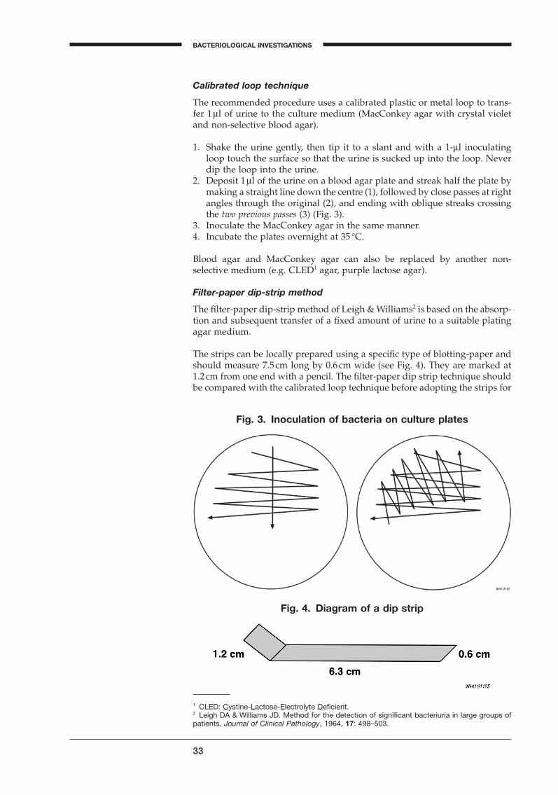
Calibrated loop technique
The recommended procedure uses a calibrated plastic or metal loop to trans-fer 1 ml of urine to the culture medium (MacConkey agar with crystal violetand non-selective blood agar).
1. Shake the urine gently, then tip it to a slant and with a 1-ml inoculatingloop touch the surface so that the urine is sucked up into the loop. Neverdip the loop into the urine.
2. Deposit 1ml of the urine on a blood agar plate and streak half the plate bymaking a straight line down the centre (1), followed by close passes at rightangles through the original (2), and ending with oblique streaks crossingthe two previous passes (3) (Fig. 3).
3. Inoculate the MacConkey agar in the same manner.4. Incubate the plates overnight at 35 ∞C.
Blood agar and MacConkey agar can also be replaced by another non-selective medium (e.g. CLED1 agar, purple lactose agar).
Filter-paper dip-strip method
The filter-paper dip-strip method of Leigh & Williams2 is based on the absorp-tion and subsequent transfer of a fixed amount of urine to a suitable platingagar medium.
The strips can be locally prepared using a specific type of blotting-paper andshould measure 7.5cm long by 0.6cm wide (see Fig. 4). They are marked at1.2cm from one end with a pencil. The filter-paper dip strip technique shouldbe compared with the calibrated loop technique before adopting the strips for
33
BACTERIOLOGICAL INVESTIGATIONS
A
Fig. 3. Inoculation of bacteria on culture plates
1 CLED: Cystine-Lactose-Electrolyte Deficient.2 Leigh DA & Williams JD. Method for the detection of significant bacteriuria in large groups ofpatients. Journal of Clinical Pathology, 1964, 17: 498–503.
Fig. 4. Diagram of a dip strip
BLM1 1/17/04 2:01 PM Page 33
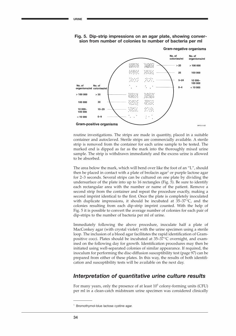
routine investigations. The strips are made in quantity, placed in a suitablecontainer and autoclaved. Sterile strips are commercially available. A sterilestrip is removed from the container for each urine sample to be tested. Themarked end is dipped as far as the mark into the thoroughly mixed urinesample. The strip is withdrawn immediately and the excess urine is allowedto be absorbed.
The area below the mark, which will bend over like the foot of an “L”, shouldthen be placed in contact with a plate of brolacin agar1 or purple lactose agarfor 2–3 seconds. Several strips can be cultured on one plate by dividing theundersurface of the plate into up to 16 rectangles (Fig. 5). Be sure to identifyeach rectangular area with the number or name of the patient. Remove asecond strip from the container and repeat the procedure exactly, making asecond imprint identical to the first. Once the plate is completely inoculatedwith duplicate impressions, it should be incubated at 35–37 ∞C, and thecolonies resulting from each dip-strip imprint counted. With the help of Fig. 5 it is possible to convert the average number of colonies for each pair ofdip-strips to the number of bacteria per ml of urine.
Immediately following the above procedure, inoculate half a plate of MacConkey agar (with crystal violet) with the urine specimen using a sterileloop. The inclusion of a blood agar facilitates the rapid identification of Gram-positive cocci. Plates should be incubated at 35–37 ∞C overnight, and exam-ined on the following day for growth. Identification procedures may then beinitiated using well-separated colonies of similar appearance. If required, theinoculum for performing the disc-diffusion susceptibility test (page 97) can beprepared from either of these plates. In this way, the results of both identifi-cation and susceptibility tests will be available on the next day.
Interpretation of quantitative urine culture results
For many years, only the presence of at least 105 colony-forming units (CFU)per ml in a clean-catch midstream urine specimen was considered clinically
34
Fig. 5. Dip-strip impressions on an agar plate, showing conver-sion from number of colonies to number of bacteria per ml
URINE
1 Bromothymol-blue lactose cystine agar.
BLM1 1/17/04 2:01 PM Page 34
relevant for a diagnosis of urinary tract infection. This assumption has beenchallenged; some experts feel that 104 CFU or even fewer may indicate infec-tion. Others believe that the presence of polymorphonuclear leukocytes playsan important role in the pathology and clinical manifestations of UTI. It is notpossible to define precisely the minimum number of bacteria per millilitre ofurine that is definitely associated with UTI. General recommendations forreporting are given below.
Category 1: fewer than 104 CFU per ml. Report as probable absence of UTI.(Exceptions: if fewer than 104 CFU per ml are present in urinetaken directly from the bladder by suprapubic puncture orcystoscopy, in; symptomatic women, or in the presence ofleukocytura, report the identification and the result of thesusceptibility test.)
Category 2: 104–105 CFU per ml. If the patient is asymptomatic, request asecond urine specimen and repeat the count. If the patient hassymptoms of UTI, proceed with both identification andsusceptibility tests if one or two different colony types ofbacteria are present. Bacterial counts in this range stronglysuggest UTI in symptomatic patients, or in the presence ofleukocyturia. If the count, the quality of the urine specimen, orthe significance of the patient’s symptoms is in doubt, asecond urine specimen should be obtained for retesting.Report the number of CFU.
Category 3: More than 105 CFU per ml. Report the count to the physicianand proceed with identification and susceptibility tests if oneor two different colony types of bacteria are present. Thesebacterial counts are strongly suggestive of UTI in all patients,including asymptomatic females.
If more than two species of bacteria are present in urine samples in categories2 and 3, report as “Probably contaminated; please submit a fresh, clean-catchspecimen”.
Identification
Identification should be performed as rapidly as possible. Since the vastmajority of urinary tract infections are caused by E. coli, a rapid test shouldbe used to identify red colonies from MacConkey agar.
b-Glucuronidase test for rapid identification of E. coli1
This test determines the ability of an organism to produce the enzyme b-glucuronidase. The enzyme hydrolyses the 4-nitrophenyl-b-d-glucopyranosiduronic acid (PGUA) reagent to glucuronic acid and p-nitro-phenol. The development of a yellow colour indicates a positive reaction.
35
BACTERIOLOGICAL INVESTIGATIONS
A1 Kilian M, Borrow P. Rapid diagnosis of Enterobacteriaceae. 1. Detection of bacterial glycosidases. Acta Pathologica et Microbiologica Scandinavica, Section B, 1976, 84:245–251.
BLM1 1/17/04 2:01 PM Page 35
Procedure:
1. Prepare a dense milky suspension of the organism to be tested in a smalltube containing 0.25ml of saline. The suspension should be prepared fromcolonies growing on MacConkey agar.
2. Dissolve 300mg of 4-nitrophenyl-b-d-glucopyranosiduronic acid (PGUA)and 100mg of yeast extract (Oxoid L21)1 in 20ml of phosphate buffer (Trisbuffer, pH 8.5). Adjust the pH to 8.5 ± 0.1. Pour 0.25ml of the medium intoeach of the required number of sterile tubes. Close the tubes with stoppers.Label the tubes PGUA and indicate the date.
3. Inoculate one tube of the PGUA medium with a dense suspension of the organism to be tested. Incubate the tube for 4 hours at 35 ∞C. The devel-opment of a yellow colour indicates the presence of b-glucuronidase (pos-itive result); if no colour change occurs the result is negative. The presenceof a pigmented yellow colour indicates that the result is unreliable; in suchcases other tests must be used.
Quality control organisms:Escherichia coli positive (yellow) resultShigella flexneri negative (clear) result
PGUA tablets are commercially available.
Susceptibility tests
Susceptibility tests (page 97) should only be performed on well-isolatedcolonies of similar appearance that are considered significant according to theguidelines presented above. Susceptibility tests are generally more importanton cultures obtained from patients who are hospitalized or have a history ofrecurring UTI. Cultures from patients a primary UTI may not require a sus-ceptibility test.
36
1 Available from Oxoid Ltd, Wade Road, Basingstoke, Hampshire, RG24 8PW, England.
URINE
BLM1 1/17/04 2:01 PM Page 36
Stool
Introduction
Enteric bacterial infections, causing diarrhoea, dysentery, and enteric fevers,are important health problems throughout the world. Diarrhoeal infectionsare second only to cardiovascular diseases as a cause of death, and they arethe leading cause of childhood death. In developing countries, diarrhoeal; dis-eases account for 1.5 million death each year among children aged 1–4 years.The risk of children in this age group dying from diarrhoeal disease is 600times greater in developing countries than in developed countries. In somedeveloping countries, children suffer ten or more episodes of diarrhoea a year.
Children are frequently infected with multiple pathogens and even childrenwithout diarrhoea quite often carry potential pathogens in their stools. Obser-vations in studies suggested that active immunization through repeated expo-sure and prolonged breastfeeding may protect against the diarrhoeageniceffect of these agents. They have also underlined the difficulties in determin-ing the cause of an episode of diarrhoea by culture of a single stool specimen.
With the increasing prevalence of HIV/AIDS and immunosuppressivechemotherapy, diarrhoea in immunocompromised patients has become agrowing challenge. Patients with HIV/AIDS either present with diarrhoea orhave diarrhoea in a later stage of the disease, and the infection is often life-threatening and difficult to cure. The list of enteric bacterial infections identi-fied in HIV/AIDS patients is long and includes Campylobacter, Salmonella,Shigella, and mycobacteria. Salmonellosis has been estimated to be nearly 20times as common and 5 times more often bacteraemic in patients withHIV/AIDS than in those without the disease. In Zaire, 84% of patients withdiarrhoea of more than a month’s duration were found to be seropositive forHIV, and 40% of patients with HIV/AIDS had persistent diarrhoea.
Etiological agents and clinical features
The genus Salmonella contains more than 2000 serotypes. Many of these may infect both humans and domestic animals. In humans they cause gas-troenteritis, typhoid fever, and bacteraemia with or without metastaticdisease. Salmonella gastroenteritis usually begins with nausea, vomiting,abdominal colic and diarrhoea 8–48 hours after ingestion of the contaminatedfood. The symptoms often persist for 3–5 days before resolving withouttherapy. Antimicrobials will not hasten clinical recovery, and may lengthenthe convalescence and asymptomatic carrier state. Antimicrobial susceptibil-ity testing and antimicrobial therapy are not recommended for uncomplicatedcases. Antimicrobial treatment is only indicated if the patient appears bacter-aemic. Some patients may harbour Salmonella spp. in stool or urine for periodsof 1 year or longer but remain asymptomatic. Approximately 3% of patientswith typhoid fever and 0.2–0.6% of persons with non-typhoid Salmonellagastroenteritis will have positive stool cultures for more than 1 year.
Shigella spp. cause a wide spectrum of clinical diseases, which vary fromasymptomatic infections to diarrhoea without fever to severe dysentery.Symptoms consist of abdominal cramps, ineffectual and painful straining topass stool (tenesmus), and frequent, small-volume, bloodstained inflamma-tory discharge. Shigella spp. are the main cause of bacillary dysentery followedby enteroinvasive and enterohaemorrhagic E. coli. Many cases present as mild
37
A
BLM1 1/17/04 2:01 PM Page 37
illnesses that require no specific treatment. However, for severe dysentery orwhen secondary spread is likely, antimicrobial therapy after susceptibilitytesting is indicated as resistance towards commonly used antimicrobials ishigh in many countries. Four groups of Shigella organisms, with a total of 39serotypes and subtypes, are recognized. Group A (S. dysenteriae), group B (S.flexneri), and group C (S. boydii) contain multiple serotypes; there is only oneserotype for group D (S. sonnei). S. dysenteriae and S. flexneri are the most com-monly isolated Shigella species in developing countries, while S. sonnei is themost commonly isolated species in developed countries.
At least six different classes of diarrhoea-producing Escherichia coli have been identified: enteropathogenic E. coli (EPEC), enterotoxigenic E. coli(ETEC), enterohaemorrhagic or verotoxin-producing E. coli (EHEC or VTEC),enteroinvasive E. coli (EIEC), enteroadhesive E. coli (EAEC), and entero-aggregative E. coli (EAggEC). Four of them are common causes of diarrhoealdisease in developing countries. However, identification of these strainsrequires serological assays, toxicity assays in cell culture, pathogenicitystudies in animals and gene-probe techniques that are beyond the capacity ofintermediate-level clinical laboratories. It may be possible to have a pre-sumptive identification of a VTEC strain, as the most frequent VTEC serotypeO157:H7 is characterized by being sorbitol-negative. However, E. fergusoniiand E. hermanii are also sorbitol-negative. A sorbitol-negative E. coli strain willneed additional identification by serotyping with E. coli O157 antiserum.Demonstration of verotoxin production indicates that it is a VTEC strain.
Cholera is a typical example of a toxigenic infection. All the symptoms can beattributed to the intestinal fluid loss caused by an enterotoxin released byVibrio cholerae in the intestine. The stool is voluminous and watery, and con-tains no inflammatory cells. The main objective of treatment is fluid replace-ment and antimicrobials have only a secondary role.
V. cholerae spreads very rapidly. It can be divided into several serotypes on the basis of variation in the somatic O antigen, and serotype O1 exists in twobiological variants termed “classical” and “El Tor”. Until 1992, only V. choleraeserogroup O1 (classical or El Tor) was known to produce epidemic cholera,and other serogroups were thought to cause sporadic cholera and extrain-testinal infections. However, in 1992 a large outbreak of cholera appeared onthe east coast of India and quickly spread to neighbouring countries. This epi-demic was caused by a previously unrecognized serogroup of V. cholerae O139Bengal. Isolation of V. cholerae O139 has so far been reported from 10 coun-tries in south-east Asia but seems to be on the decline.
V. parahaemolyticus and several other species of Vibrio (V. fluvialis, V. hollisae,V. mimicus) and Aeromonas (A. hydrophila, A. sobria, A. caviae) cause food poi-soning or gastroenteritis in persons who eat raw or undercooked seafood.
Campylobacter jejuni and C. coli have emerged as major enteric pathogens thatcan be isolated as often as Salmonella and Shigella spp. in most parts of theworld. Investigations in Africa and Asia have shown incidence rates ofbetween 19 and 38%, and asymptomatic carrier rates of between 9 and 40%,in children. The intestinal disease varies from a brief, self-limiting enteritis toa fulminant enterocolitis with severe diarrhoea, abdominal colic, fever, andmuscle pain. The stools are at first mucoid and liquid and may progress toprofuse watery diarrhoea containing blood and pus. Symptoms usuallysubside within a week. Relapse occurs in about 25% of patients, but is gener-ally milder than the initial episode. The infection is usually self-limitingwithout antimicrobial therapy, and susceptibility testing is usually not indicated.
38
STOOL
BLM1 1/17/04 2:01 PM Page 38
Arcobacter butzleri, formerly regarded as a low-temperature-growing Campy-lobacter, has recently been recognized as a cause of diarrhoeal disease inpatients, mainly children, in developing countries.
Human infections with Yersinia enterocolitica have been reported mainly fromnorthern Europe, Japan and the United States of America. The majority of iso-lates have been identified from children with sporadic diarrhoea.
Clostridium difficile is the primary cause of enteric disease related to antimi-crobial therapy. It produces a broad spectrum of diseases ranging from milddiarrhoea to potentially fatal pseudomembranous colitis. Observation ofcolonic pseudomembranes by colonoscopy is diagnostic for pseudomembra-nous colitis, in which case laboratory confirmation is unnecessary. Severalcommercial tests are available for the laboratory, including culture, latexagglutination for detection of a cell-associated protein, ELISA assay for cyto-toxin and/or enterotoxin, and cell culture toxicity assay for cytotoxin. Manyhospital patients harbour the organism in the stool in the absence of symp-toms, particularly if they are receiving broad-spectrum antimicrobials. There-fore, a culture without demonstration of toxin production has little diagnosticvalue.
Rotavirus is the only non-bacterial agent to be discussed here. Other virusesare important causes of diarrhoea but rotavirus is common everywhere in theworld with similar rates in both developed and developing countries. Mostinfections occur in children between 6 and 18 months, with a higher preva-lence during the cooler months of the year. The diagnosis is made either withan enzyme immunoassay (ELISA) or more simply and practically with a latexagglutination assay. Reagents for both assays are commercially available butare expensive.
Appropriate use of laboratory resources
The laboratory associated with a busy hospital or clinic in a developingcountry can quickly become overwhelmed with specimens. Cultures are notrequired for effective management of the majority of cases of diarrhoea anddysentery and patients will require only rehydration and not antimicrobials.A few patients (e.g. those with typhoid fever) will need the results of a culturefor appropriate treatment. The problem of how best to utilize scarce resourcesis a constant concern.
Often, the public health aspects are the most important, and the laboratorymust be able to provide data that describe the common enteric pathogens inthe district and the antimicrobial susceptibility patterns of these pathogens,and investigate an epidemy. Clinicians must work closely with the laboratory.A procedure that allows the laboratory to develop a valid database is the collection of specimens from a random systematic sample of patients withdiarrhoea at the hospital or clinic. By testing only a proportion of these, thenumber of specimens is reduced and a more complete investigation can bemade on each specimen. If the sample is a systematic one (e.g. every twenty-fifth patient), then the results can be used to estimate the infection in the entirepopulation of patients. If a typical clinical syndrome is observed in a partic-ular age group, or during a particular season, the laboratory can focus its sam-pling on these specific problems.
The laboratory may decide to test for only certain enteric pathogens. Yersiniaenterocolitica is evidently rare in most tropical areas. If the organism does notoccur in the area served by the laboratory, testing for it can be omitted. When
39
BACTERIOLOGICAL INVESTIGATIONS
A
BLM1 1/17/04 2:01 PM Page 39
the laboratory provides a report, it should specify which organisms wereinvestigated. If Salmonella and Shigella species were the only pathogens ruledout, the report should not state “No pathogens found”. Rather, it should state“Salmonella and Shigella species not found”.
Collection and transport of stool specimens
Faecal specimens should be collected in the early stages of the diarrhoealdisease, when pathogens are present in the highest number, and preferablybefore antimicrobial treatment is started, if appropriate. The specimen shouldbe collected in the morning to reach the laboratory before noon, so that it canbe processed the same day. Formed stools should be rejected. Ideally, a freshstool specimen is preferred to a rectal swab, but a rectal swab is acceptable ifthe collection cannot be made immediately or when transportation of the stoolto the laboratory is delayed.
Procedure for collecting stool samples
Provide the patient with two small wooden sticks and a suitable containerwith a leakproof lid (e.g. a clean glass cup, a plastic or waxed-cardboard box,or a special container with a spoon attached to the lid). The use of penicillinbottles, matchboxes and banana leaves should be discouraged as they exposethe laboratory staff to the risk of infection.
Instruct the patient to collect the stool specimen on a piece of toilet tissue orold newspaper and to transfer it to the container, using the two sticks.
The specimen should contain at least 5g of faeces and, if present, those partsthat contain blood, mucus or pus. It should not be contaminated with urine.Once the specimen has been placed in the specimen container, the lid shouldbe sealed.
The patient should be asked to deliver the specimen to the clinic immediatelyafter collection. If it is not possible for the specimen to be delivered to the lab-oratory within 2 hours of its collection, a small amount of the faecal specimen(together with mucus, blood and epithelial threads, if present) should be col-lected on two or three swabs and placed in a container with transport medium(Cary–Blair, Stuart or Amies) or 33mmol/l of glycerol–phosphate buffer. Forcholera and other Vibrio spp., alkaline peptone water is an excellent transport(and enrichment) medium. Pathogens may survive in such media for up to 1week, even at room temperature, although refrigeration is preferable.
Procedure for collecting rectal swabs
1. Moisten a cotton-tipped swab with sterile water. Insert the swab throughthe rectal sphincter, rotate, and withdraw. Examine the swab for faecalstaining and repeat the procedure until sufficient staining is evident. Thenumber of swabs to be collected will depend on the number and types ofinvestigation required.
2. Place the swab in an empty sterile tube with a cotton plug or screw-cap, ifit is to be processed within 1–2 hours. If the swab must be kept for longerthan 2 hours, place it in transport medium.
40
STOOL
BLM1 1/17/04 2:01 PM Page 40
Visual examination of stool specimens
1. Examine the stool sample visually and record the following:— its consistency (formed, unformed (soft) or liquid)— its colour (white, yellow, brown or black)— the presence of any abnormal components (e.g. mucus or blood).
2. Place a small fleck of the stool specimen or rectal swab together with asmall flake of mucus (if present) in a drop of 0.05% methylene blue solu-tion on a clean slide and mix thoroughly.
3. Place a coverslip on the stained suspension, avoiding the formation of airbubbles. Wait 2–3 minutes to. Examine the slide under the microscopeusing the high-power objective (¥100).
4. Record cells that can be clearly identified as mononuclear or polymor-phonuclear; ignore degenerated cells.
Examination of the cellular exudate of diarrhoeal stools may give an indi-cation of the organism involved:
— clumps of polymorphonuclear leukocytes (>50 cells per high-powerfield), macrophages and erythrocytes are typical of shigellosis;
— smaller numbers of polymorphonuclear leukocytes (<20 cells per high-power field) are found in salmonellosis, and invasive E. coli. In amoebicdysentery the cells are mostly degenerated (ghost cells). Leukocytesand erythrocytes are also found in about half the cases of diarrhoea dueto Campylobacter spp.;
— few leukocytes (2–5 cells per high-power field) are present in cases of cholera, enterotoxigenic and enteropathogenic E. coli, and viral diarrhoea.
Enrichment and inoculation of stool specimens
Enrichment is commonly used for the isolation of Salmonella spp. and Vibriocholerae from faecal specimens. Selenite F or tetrathionate broths are recom-mended for the enrichment of Salmonella spp., and alkaline peptone water(APW) for the enrichment of V. cholerae. Enrichment is not required for Shigellaspp., Campylobacter spp., Yersinia enterocolitica and Clostridium difficile.
Procedure for inoculation of enrichment media
1. Prepare a faecal suspension by suspending approximately 1g of the stoolsample in a tube containing 1ml of sterile saline. If the stool sample isliquid, saline does not need to be added. Rectal swabs received fresh or inCary–Blair medium should be rinsed thoroughly in 1ml of saline. Beforeremoving the swab, press it against the side of the tube to express anyremaining fluid.
2. Add three or more loopfuls of faecal suspension to the enrichment broth.3. Incubate selenite F broth for 18 hours and APW for 6–8 hours. In some lab-
oratories two or three loopfuls of suspension the initial APW are subcul-tured in from fresh APW and incubated for another 6–8 hours.
4. Subculture colonies by streaking a loopful of broth on selective and non-selective plating media.
41
BACTERIOLOGICAL INVESTIGATIONS
A
BLM1 1/17/04 2:01 PM Page 41
V. cholerae grows quickly in APW and will outgrow other organisms in 6–8hours. However, after 8 hours other organisms may overgrow the V. cholerae.Non-O1 V. cholerae grows more quickly than V. cholerae O1 and may overgrowthe latter when both organisms are present.
Media for enteric pathogens
For Shigella spp., Salmonella spp. and Y. enterocolitica a general-purpose platingmedium of low selectivity and a medium of moderate or high selectivity arerecommended. MacConkey agar with crystal violet is recommended as ageneral purpose medium. For Y. enterocolitica incubate the MacConkey agarat 35 ∞C for 1 day and then at room temperature (22–29 ∞C) for another day.
Xylose–lysine–deoxycholate (XLD) agar is recommended as a medium ofmoderate or high selectivity for the isolation of Shigella and Salmonella spp.Deoxycholate citrate agar (DCA), Hektoen enteric agar (HEA) or Salmo-nella–Shigella (SS) agar are suitable alternatives. Shigella dysenteriae type 1, S.sonnei and enteroinvasive E. coli do not grow well on SS agar. However, SSagar may be used for isolating Y. enterocolitica when incubated as describedfor MacConkey agar. Many laboratories include bismuth sulfite agar for iso-lating Salmonella typhi and other species of Salmonella.
For Campylobacter spp. there are several selective media (Blaser, Butzler,Skirrow) containing different antimicrobial supplements. However, a bloodagar base with 5–10% sheep blood containing a combination of a cefalosporin (15 mg/ml), vancomycin (10mg/ml), amphotericin B (2 mg/ml) and 0.05%ferrows sulfate–sodium metabisulfite–sodium pyruvate (FBP) is acceptable.
For Vibrio spp. a selective medium is necessary although many strains maygrow on MacConkey agar. Thiosulfate citrate bile salts sucrose (TCBS) agar isselective for V. cholerae O1 and non-O1 and for V. parahaemolyticus, but it isexpensive. Telluride taurocholate gelatine (TTG) agar is another selectivemedium, but it is not commercially available. Alkaline meat extract agar(MEA) and alkaline bile salt agar (BSA) are inexpensive simple media that canbe locally prepared and give excellent results.
Clostridium difficile was difficult to isolate before the development of cefoxitin–cycloserine–fructose agar (CCFA). Formulations using an egg-yolkagar base are preferally used as C. difficile is negative for both lecithinase andlipase, while other clostridia commonly found in the gut, such as C. perfrin-gens, C. bifermentans and C. sordelli are lecithinase-positive.
Primary isolation
The specimen should be examined and cultured as soon as it is delivered to the laboratory, as this gives the highest isolation rate of Shigellaand Campylobacter spp. If this is not possible, the specimen should be storedat 4 ∞C.
A concentrated inoculum of faeces should be used with high selectivity mediaand a light inoculum of faeces with low selectivity media. In many laborato-ries, the plates are inoculated directly with the rectal swabs, but care shouldbe taken not to make an inoculation that is overloaded.
42
STOOL
BLM1 1/17/04 2:01 PM Page 42
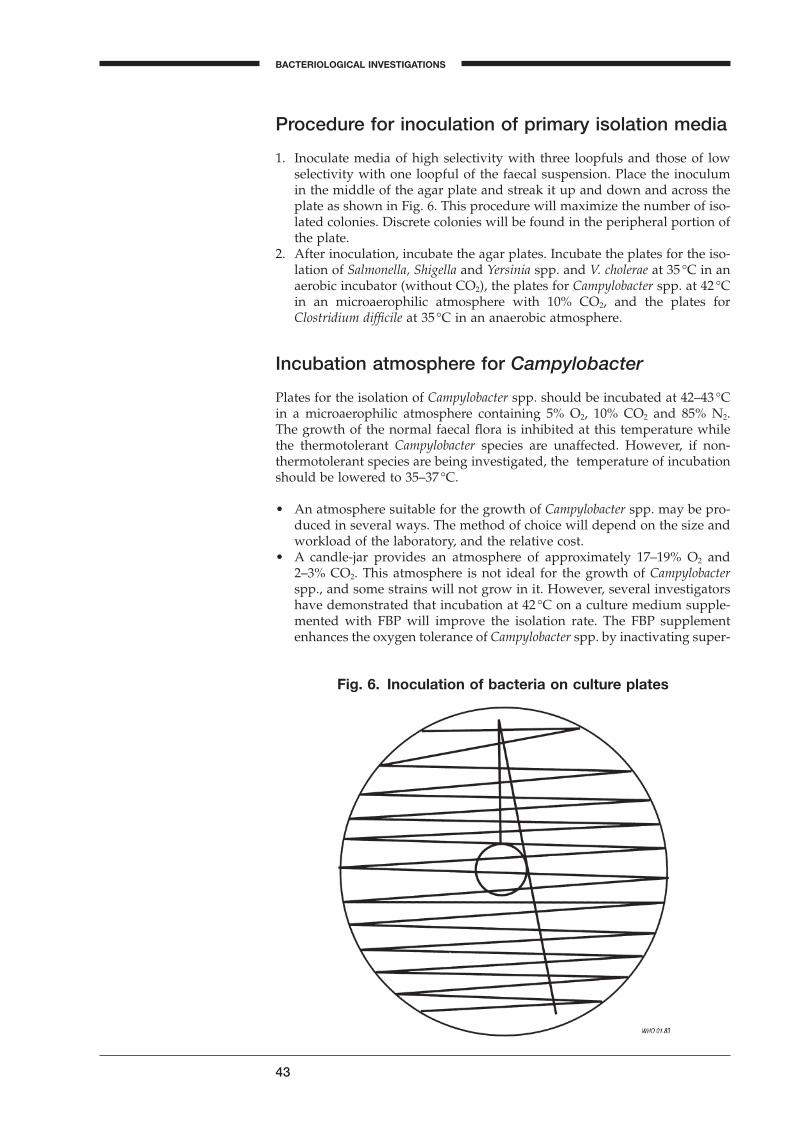
Procedure for inoculation of primary isolation media
1. Inoculate media of high selectivity with three loopfuls and those of lowselectivity with one loopful of the faecal suspension. Place the inoculumin the middle of the agar plate and streak it up and down and across theplate as shown in Fig. 6. This procedure will maximize the number of iso-lated colonies. Discrete colonies will be found in the peripheral portion ofthe plate.
2. After inoculation, incubate the agar plates. Incubate the plates for the iso-lation of Salmonella, Shigella and Yersinia spp. and V. cholerae at 35 ∞C in anaerobic incubator (without CO2), the plates for Campylobacter spp. at 42 ∞Cin an microaerophilic atmosphere with 10% CO2, and the plates forClostridium difficile at 35 ∞C in an anaerobic atmosphere.
Incubation atmosphere for Campylobacter
Plates for the isolation of Campylobacter spp. should be incubated at 42–43 ∞Cin a microaerophilic atmosphere containing 5% O2, 10% CO2 and 85% N2. The growth of the normal faecal flora is inhibited at this temperature whilethe thermotolerant Campylobacter species are unaffected. However, if non-thermotolerant species are being investigated, the temperature of incubationshould be lowered to 35–37 ∞C.
• An atmosphere suitable for the growth of Campylobacter spp. may be pro-duced in several ways. The method of choice will depend on the size andworkload of the laboratory, and the relative cost.
• A candle-jar provides an atmosphere of approximately 17–19% O2 and2–3% CO2. This atmosphere is not ideal for the growth of Campylobacterspp., and some strains will not grow in it. However, several investigatorshave demonstrated that incubation at 42 ∞C on a culture medium supple-mented with FBP will improve the isolation rate. The FBP supplementenhances the oxygen tolerance of Campylobacter spp. by inactivating super-
43
BACTERIOLOGICAL INVESTIGATIONS
A
Fig. 6. Inoculation of bacteria on culture plates
BLM1 1/17/04 2:01 PM Page 43
oxides and hydrogen peroxide. Disadvantages of this system are a longerincubation period and inhibition of some oxygen-sensitive Campylobacterspp.
• Another simple and inexpensive system uses a co-culture technique. Plateswith rapidly growing facultative anaerobic bacteria are incubated with theplates for the isolation of Campylobacter spp. in an airtight container or aplastic bag. As the facultative anaerobic bacteria grow, the oxygen con-tent is lowered and the CO2 content is increased. The disadvantage of thissystem is the longer incubation time usually required for growth of theCampylobacter spp.
• A hydrogen and CO2-generator envelope with a self-contained catalystspecifically for the isolation of Campylobacter spp. is commercially avail-able. The envelope is placed in an anaerobic jar, and a new envelope mustbe used each time the jar is opened. No more than six plates should bestacked in the jar to obtain maximum isolation.
• A plastic bag incubation system is also commercially available. It consistsof a plastic bag, which is collapsed two or three times by hand or vacuum,and refilled each time, with 5% O2, 10% CO2 and 85% N2.
• The evacuation–replacement system uses an anaerobic jar without a cata-lyst. The container is evacuated twice to 38cm (15mmHg) pressure andrefilled each time with 10% H2 and 90% N2 mixture.
Preliminary identification of isolates
Identification involves both biochemical and serological tests, the extent ofwhich will depend on the capacity of the laboratory. Flow charts for guidancein identification of important enteric bacteria are presented in Figs 7a–c.
Identify well-separated colonies of typical appearance on the primary platesby making a mark on the bottom of the Petri dish. These will be transferredfor further testing. If more than one type of colony is present, process at leastone colony of each type.
Lactose-nonfermenting bacteria, such as Salmonella and Shigella spp., give riseto small colourless colonies on MacConkey agar, SS agar, and DCA. Coloniesof Proteus spp. may be confused with Salmonella and Shigella spp., especiallyon MacConkey agar and DCA, because of their lactose-negative appearance.Lactose-fermenting organisms, such as E. coli and Enterobacter/Klebsiella spp.,produce pink to red colonies on MacConkey agar, DCA and SS agar. On XLDagar, Shigella and Salmonella spp. produce small red colonies, most strains ofSalmonella with a black centre. Some strains of Proteus spp. will also give black-centred colonies on XLD agar. On bismuth sulfite agar, Salmonella typhi pro-duces black colonies with a metallic sheen, if the colonies are well separated.Yersinia enterocolitica grows on MacConkey and SS agars as small, pale colour-less colonies that grow most rapidly at 22–29 ∞C.
Salmonella and Shigella spp.
Three differential media are recommended for initial screening of isolates ofSalmonella and Shigella spp.:
— urea broth, weakly buffered (UREA)— motility-indole–lysine medium (MIL)— Kligler’s iron agar (KIA).
44
STOOL
BLM1 1/17/04 2:01 PM Page 44
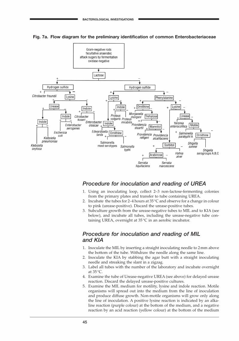
Procedure for inoculation and reading of UREA1. Using an inoculating loop, collect 2–3 non-lactose-fermenting colonies
from the primary plates and transfer to tube containing UREA.2. Incubate the tubes for 2–4 hours at 35 ∞C and observe for a change in colour
to pink (urease-positive). Discard the urease-positive tubes.3. Subculture growth from the urease-negative tubes to MIL and to KIA (see
below), and incubate all tubes, including the urease-negative tube con-taining UREA, overnight at 35 ∞C in an aerobic incubator.
Procedure for inoculation and reading of MIL and KIA1. Inoculate the MIL by inserting a straight inoculating needle to 2mm above
the bottom of the tube. Withdraw the needle along the same line.2. Inoculate the KIA by stabbing the agar butt with a straight inoculating
needle and streaking the slant in a zigzag.3. Label all tubes with the number of the laboratory and incubate overnight
at 35 ∞C.4. Examine the tube of Urease-negative UREA (see above) for delayed urease
reaction. Discard the delayed urease-positive cultures.5. Examine the MIL medium for motility, lysine and indole reaction. Motile
organisms will spread out into the medium from the line of inoculationand produce diffuse growth. Non-motile organisms will grow only alongthe line of inoculation. A positive lysine reaction is indicated by an alka-line reaction (purple colour) at the bottom of the medium, and a negativereaction by an acid reaction (yellow colour) at the bottom of the medium
45
BACTERIOLOGICAL INVESTIGATIONS
A
Fig. 7a. Flow diagram for the preliminary identification of common Enterobacteriaceae
BLM1 1/17/04 2:01 PM Page 45
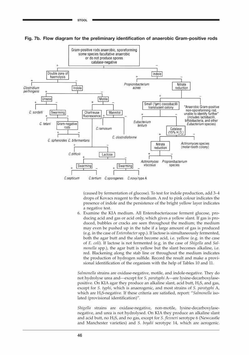
46
Fig. 7b. Flow diagram for the preliminary identification of anaerobic Gram-positive rods
(caused by fermentation of glucose). To test for indole production, add 3–4drops of Kovacs reagent to the medium. A red to pink colour indicates thepresence of indole and the persistence of the bright yellow layer indicatesa negative test.
6. Examine the KIA medium. All Enterobacteriaceae ferment glucose, pro-ducing acid and gas or acid only, which gives a yellow slant. If gas is pro-duced, bubbles or cracks are seen throughout the medium; the mediummay even be pushed up in the tube if a large amount of gas is produced(e.g. in the case of Enterobacter spp.). If lactose is simultaneously fermented,both the agar butt and the slant become acid, i.e. yellow (e.g. in the caseof E. coli). If lactose is not fermented (e.g. in the case of Shigella and Sal-monella spp.), the agar butt is yellow but the slant becomes alkaline, i.e.red. Blackening along the stab line or throughout the medium indicatesthe production of hydrogen sulfide. Record the result and make a provi-sional identification of the organism with the help of Tables 10 and 11.
Salmonella strains are oxidase-negative, motile, and indole-negative. They donot hydrolyse urea and—except for S. paratyphi A—are lysine-decarboxylase-positive. On KIA agar they produce an alkaline slant, acid butt, H2S, and gas,except for S. typhi, which is anaerogenic, and most strains of S. paratyphi A,which are H2S-negative. If these criteria are satisfied, report: “Salmonella iso-lated (provisional identification)”.
Shigella strains are oxidase-negative, non-motile, lysine-decarboxylase-negative, and urea is not hydrolysed. On KIA they produce an alkaline slantand acid butt, no H2S, and no gas, except for S. flexneri serotype 6 (Newcastleand Manchester varieties) and S. boydii serotype 14, which are aerogenic.
STOOL
BLM1 1/17/04 2:01 PM Page 46
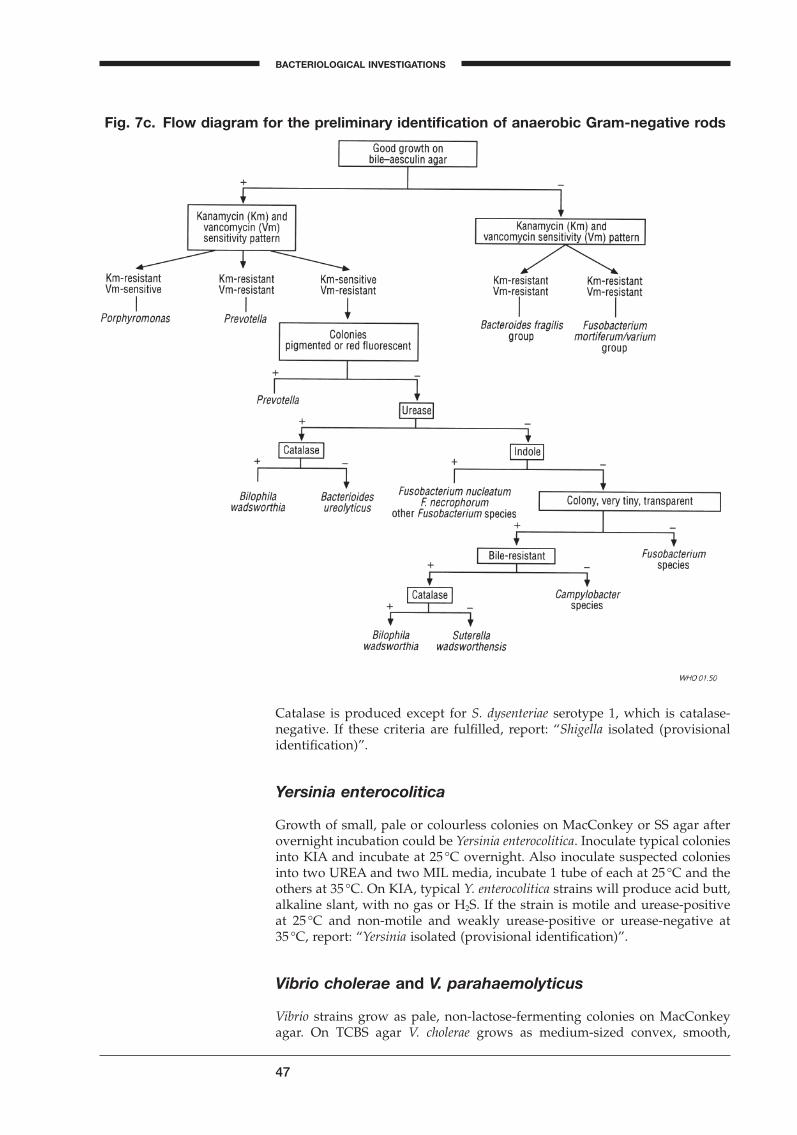
Catalase is produced except for S. dysenteriae serotype 1, which is catalase-negative. If these criteria are fulfilled, report: “Shigella isolated (provisionalidentification)”.
Yersinia enterocolitica
Growth of small, pale or colourless colonies on MacConkey or SS agar afterovernight incubation could be Yersinia enterocolitica. Inoculate typical coloniesinto KIA and incubate at 25 ∞C overnight. Also inoculate suspected coloniesinto two UREA and two MIL media, incubate 1 tube of each at 25 ∞C and theothers at 35 ∞C. On KIA, typical Y. enterocolitica strains will produce acid butt,alkaline slant, with no gas or H2S. If the strain is motile and urease-positiveat 25 ∞C and non-motile and weakly urease-positive or urease-negative at 35 ∞C, report: “Yersinia isolated (provisional identification)”.
Vibrio cholerae and V. parahaemolyticus
Vibrio strains grow as pale, non-lactose-fermenting colonies on MacConkeyagar. On TCBS agar V. cholerae grows as medium-sized convex, smooth,
47
BACTERIOLOGICAL INVESTIGATIONS
A
Fig. 7c. Flow diagram for the preliminary identification of anaerobic Gram-negative rods
BLM1 1/17/04 2:01 PM Page 47
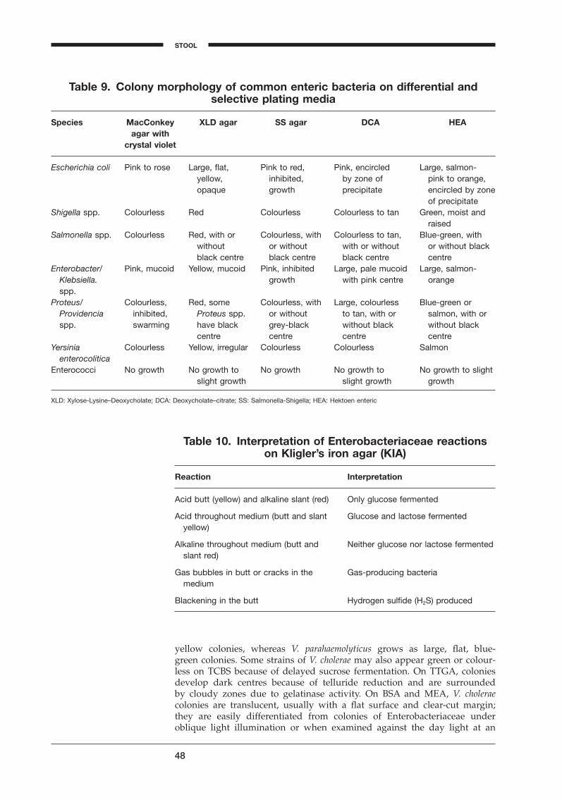
yellow colonies, whereas V. parahaemolyticus grows as large, flat, blue-green colonies. Some strains of V. cholerae may also appear green or colour-less on TCBS because of delayed sucrose fermentation. On TTGA, coloniesdevelop dark centres because of telluride reduction and are surrounded by cloudy zones due to gelatinase activity. On BSA and MEA, V. choleraecolonies are translucent, usually with a flat surface and clear-cut margin; they are easily differentiated from colonies of Enterobacteriaceae underoblique light illumination or when examined against the day light at an
48
Table 9. Colony morphology of common enteric bacteria on differential andselective plating media
Species MacConkey XLD agar SS agar DCA HEAagar with
crystal violet
Escherichia coli Pink to rose Large, flat, Pink to red, Pink, encircled Large, salmon-yellow, inhibited, by zone of pink to orange,opaque growth precipitate encircled by zone
of precipitateShigella spp. Colourless Red Colourless Colourless to tan Green, moist and
raisedSalmonella spp. Colourless Red, with or Colourless, with Colourless to tan, Blue-green, with
without or without with or without or without blackblack centre black centre black centre centre
Enterobacter/ Pink, mucoid Yellow, mucoid Pink, inhibited Large, pale mucoid Large, salmon-Klebsiella. growth with pink centre orangespp.
Proteus/ Colourless, Red, some Colourless, with Large, colourless Blue-green orProvidencia inhibited, Proteus spp. or without to tan, with or salmon, with orspp. swarming have black grey-black without black without black
centre centre centre centreYersinia Colourless Yellow, irregular Colourless Colourless Salmon
enterocoliticaEnterococci No growth No growth to No growth No growth to No growth to slight
slight growth slight growth growth
XLD: Xylose-Lysine–Deoxycholate; DCA: Deoxycholate–citrate; SS: Salmonella-Shigella; HEA: Hektoen enteric
Table 10. Interpretation of Enterobacteriaceae reactionson Kligler’s iron agar (KIA)
Reaction Interpretation
Acid butt (yellow) and alkaline slant (red) Only glucose fermented
Acid throughout medium (butt and slant Glucose and lactose fermentedyellow)
Alkaline throughout medium (butt and Neither glucose nor lactose fermentedslant red)
Gas bubbles in butt or cracks in the Gas-producing bacteriamedium
Blackening in the butt Hydrogen sulfide (H2S) produced
STOOL
BLM1 1/17/04 2:01 PM Page 48
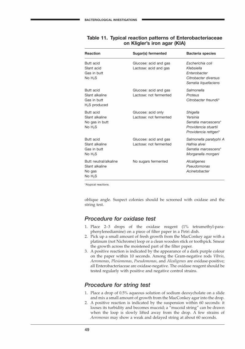
oblique angle. Suspect colonies should be screened with oxidase and the string test.
Procedure for oxidase test1. Place 2–3 drops of the oxidase reagent (1% tetramethyl-para-
phenylenediamine) on a piece of filter paper in a Petri dish.2. Pick up a small amount of fresh growth from the MacConkey agar with a
platinum (not Nichrome) loop or a clean wooden stick or toothpick. Smearthe growth across the moistened part of the filter paper.
3. A positive reaction is indicated by the appearance of a dark purple colouron the paper within 10 seconds. Among the Gram-negative rods Vibrio,Aeromonas, Plesiomonas, Pseudomonas, and Alcaligenes are oxidase-positive;all Enterobacteriaceae are oxidase-negative. The oxidase reagent should betested regularly with positive and negative control strains.
Procedure for string test1. Place a drop of 0.5% aqueous solution of sodium deoxycholate on a slide
and mix a small amount of growth from the MacConkey agar into the drop.2. A positive reaction is indicated by the suspension within 60 seconds: it
looses its turbidity and becomes mucoid; a “mucoid string” can be drawnwhen the loop is slowly lifted away from the drop. A few strains ofAeromonas may show a weak and delayed string at about 60 seconds.
49
BACTERIOLOGICAL INVESTIGATIONS
A
Table 11. Typical reaction patterns of Enterobacteriaceaeon Kligler’s iron agar (KIA)
Reaction Sugar(s) fermented Bacteria species
Butt acid Glucose: acid and gas Escherichia coliSlant acid Lactose: acid and gas KlebsiellaGas in butt EnterobacterNo H2S Citrobacter diversus
Serratia liquefaciens
Butt acid Glucose: acid and gas SalmonellaSlant alkaline Lactose: not fermented ProteusGas in butt Citrobacter freundii*H2S produced
Butt acid Glucose: acid only ShigellaSlant alkaline Lactose: not fermented YersiniaNo gas in butt Serratia marcescens*No H2S Providencia stuartii
Providencia rettgeri*
Butt acid Glucose: acid and gas Salmonella paratyphi ASlant alkaline Lactose: not fermented Hafnia alveiGas in butt Serratia marcescens*No H2S Morganella morgani
Butt neutral/alkaline No sugars fermented AlcaligenesSlant alkaline PseudomonasNo gas AcinetobacterNo H2S
*Atypical reactions.
BLM1 1/17/04 2:01 PM Page 49
If these tests are positive, transfer a part of a colony to KIA and, after overnightincubation, observe for a yellow butt, alkaline slant, and no gas or H2S production. If this is confirmed, report: “Vibrio cholerae isolated (provisionalidentification)”.
Campylobacter jejuni and Campylobacter coli
Examine the Campylobacter plates after 48–72 hours of incubation. Suspectcolonies should be screened with three presumptive tests: oxidase test, wetmount preparation under dark-field or phase-contrast microscope, and Gram-stain. If a dark-field or phase-contrast microscope is not available, coloniesmay be rapidly screened for typical cell morphology by staining with Gram’scrystal-violet solution. For the Gram stain 0.3% carbol fuchsin is recom-mended as counterstain. Campylobacter species are oxidase-positive, they aremotile with a darting, tumbling motility, and they appear as simple curved orspiral-shaped rods (seagull wings or “S”-shape). If this is confirmed, report:“Campylobacter isolated (provisional identification)”.
Clostridium difficile
The C. difficile colonies on CCFA are large, yellow and ground glass in appear-ance. On anaerobic blood agar the colony morphology varies and other fea-tures should be looked for to detect the organism. Typically, colonies are grey,opaque and non-haemolytic at 24–48 hours, but a few strains may be green-ish blue due to a-type haemolysis. After 48–72 hours of incubation, coloniesmay develop a distinctly light grey to white centre. With experience, C. diffi-cile is easily recognized despite the colonial variability, thanks to its charac-teristic odour, which resembles that of horse or elephant manure. If thecolonies are lecithinase- and lipase-negative and show yellow-green fluores-cence when illuminated with Wood’s lamp, report: “Clostridium difficile iso-lated (provisional identification)”.
Final microbiological identification
Before the final report is made, the cultures should always be checked for puregrowth, and the identification should be confirmed with additional biochemi-cal tests.
1. Pick a suspect colony well-separated from other colonies on the plate andsubculture it in nutrient broth for biochemical tests, to an agar slope forserological tests, and plate it on MacConkey agar to confirm the purity ofthe culture.
2. Make additional biochemical tests according to the tables. Examine thereactions after overnight incubation and identify the isolate.
Identification of Shigella and Salmonella may sometimes pose a problem,because some strains vary in their biochemical reactions and may even shareantigens with other Gram-negative organisms. Non-motile, lactose-negative,anaerogenic strains of E. coli are notoriously difficult to differentiate fromShigella, and identification may be further complicated by the fact that someof these strains may cause bacillary dysentery.
50
STOOL
BLM1 1/17/04 2:01 PM Page 50
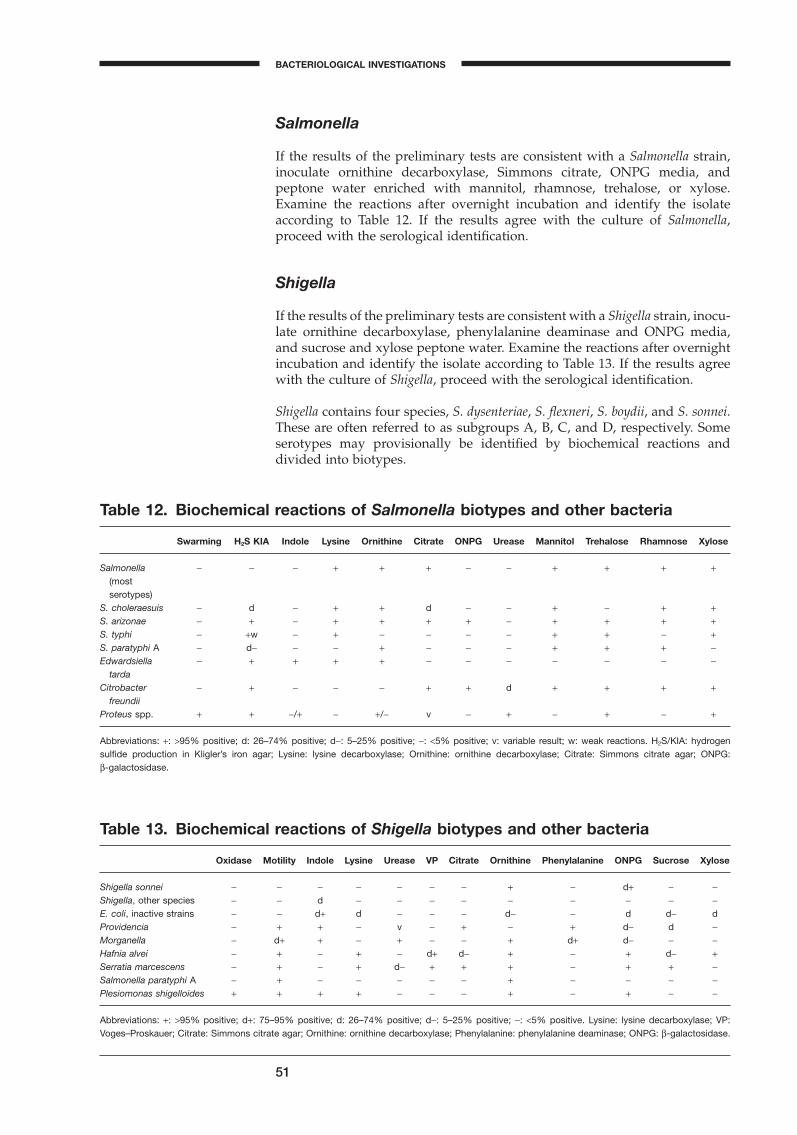
Salmonella
If the results of the preliminary tests are consistent with a Salmonella strain,inoculate ornithine decarboxylase, Simmons citrate, ONPG media, andpeptone water enriched with mannitol, rhamnose, trehalose, or xylose.Examine the reactions after overnight incubation and identify the isolateaccording to Table 12. If the results agree with the culture of Salmonella,proceed with the serological identification.
Shigella
If the results of the preliminary tests are consistent with a Shigella strain, inocu-late ornithine decarboxylase, phenylalanine deaminase and ONPG media,and sucrose and xylose peptone water. Examine the reactions after overnightincubation and identify the isolate according to Table 13. If the results agreewith the culture of Shigella, proceed with the serological identification.
Shigella contains four species, S. dysenteriae, S. flexneri, S. boydii, and S. sonnei.These are often referred to as subgroups A, B, C, and D, respectively. Someserotypes may provisionally be identified by biochemical reactions anddivided into biotypes.
51
BACTERIOLOGICAL INVESTIGATIONS
A
Table 12. Biochemical reactions of Salmonella biotypes and other bacteria
Swarming H2S KIA Indole Lysine Ornithine Citrate ONPG Urease Mannitol Trehalose Rhamnose Xylose
Salmonella - - - + + + - - + + + +(most serotypes)
S. choleraesuis - d - + + d - - + - + +S. arizonae - + - + + + + - + + + +S. typhi - +w - + - - - - + + - +S. paratyphi A - d- - - + - - - + + + -Edwardsiella - + + + + - - - - - - -
tardaCitrobacter - + - - - + + d + + + +
freundiiProteus spp. + + -/+ - +/- v - + - + - +
Abbreviations: +: >95% positive; d: 26–74% positive; d-: 5–25% positive; -: <5% positive; v: variable result; w: weak reactions. H2S/KIA: hydrogen sulfide production in Kligler’s iron agar; Lysine: lysine decarboxylase; Ornithine: ornithine decarboxylase; Citrate: Simmons citrate agar; ONPG: b-galactosidase.
Table 13. Biochemical reactions of Shigella biotypes and other bacteria
Oxidase Motility Indole Lysine Urease VP Citrate Ornithine Phenylalanine ONPG Sucrose Xylose
Shigella sonnei - - - - - - - + - d+ - -Shigella, other species - - d - - - - - - - - -E. coli, inactive strains - - d+ d - - - d- - d d- dProvidencia - + + - v - + - + d- d -Morganella - d+ + - + - - + d+ d- - -Hafnia alvei - + - + - d+ d- + - + d- +Serratia marcescens - + - + d- + + + - + + -Salmonella paratyphi A - + - - - - - + - - - -Plesiomonas shigelloides + + + + - - - + - + - -
Abbreviations: +: >95% positive; d+: 75–95% positive; d: 26–74% positive; d-: 5–25% positive; -: <5% positive. Lysine: lysine decarboxylase; VP:Voges–Proskauer; Citrate: Simmons citrate agar; Ornithine: ornithine decarboxylase; Phenylalanine: phenylalanine deaminase; ONPG: b-galactosidase.
BLM1 1/17/04 2:01 PM Page 51
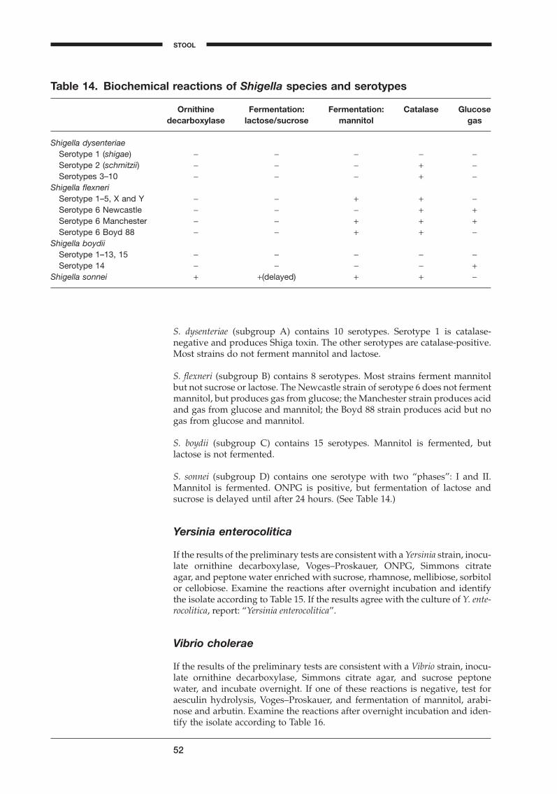
S. dysenteriae (subgroup A) contains 10 serotypes. Serotype 1 is catalase-negative and produces Shiga toxin. The other serotypes are catalase-positive.Most strains do not ferment mannitol and lactose.
S. flexneri (subgroup B) contains 8 serotypes. Most strains ferment mannitolbut not sucrose or lactose. The Newcastle strain of serotype 6 does not fermentmannitol, but produces gas from glucose; the Manchester strain produces acidand gas from glucose and mannitol; the Boyd 88 strain produces acid but nogas from glucose and mannitol.
S. boydii (subgroup C) contains 15 serotypes. Mannitol is fermented, butlactose is not fermented.
S. sonnei (subgroup D) contains one serotype with two “phases”: I and II. Mannitol is fermented. ONPG is positive, but fermentation of lactose andsucrose is delayed until after 24 hours. (See Table 14.)
Yersinia enterocolitica
If the results of the preliminary tests are consistent with a Yersinia strain, inocu-late ornithine decarboxylase, Voges–Proskauer, ONPG, Simmons citrate agar, and peptone water enriched with sucrose, rhamnose, mellibiose, sorbitolor cellobiose. Examine the reactions after overnight incubation and identify the isolate according to Table 15. If the results agree with the culture of Y. ente-rocolitica, report: “Yersinia enterocolitica”.
Vibrio cholerae
If the results of the preliminary tests are consistent with a Vibrio strain, inocu-late ornithine decarboxylase, Simmons citrate agar, and sucrose peptonewater, and incubate overnight. If one of these reactions is negative, test foraesculin hydrolysis, Voges–Proskauer, and fermentation of mannitol, arabi-nose and arbutin. Examine the reactions after overnight incubation and iden-tify the isolate according to Table 16.
52
Table 14. Biochemical reactions of Shigella species and serotypes
Ornithine Fermentation: Fermentation: Catalase Glucosedecarboxylase lactose/sucrose mannitol gas
Shigella dysenteriaeSerotype 1 (shigae) - - - - -Serotype 2 (schmitzii) - - - + -Serotypes 3–10 - - - + -
Shigella flexneriSerotype 1–5, X and Y - - + + -Serotype 6 Newcastle - - - + +Serotype 6 Manchester - - + + +Serotype 6 Boyd 88 - - + + -
Shigella boydiiSerotype 1–13, 15 - - - - -Serotype 14 - - - - +
Shigella sonnei + +(delayed) + + -
STOOL
BLM1 1/17/04 2:01 PM Page 52
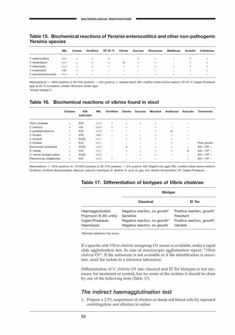
If a specific anti-Vibrio cholerae serogroup O1 serum is available, make a rapidslide agglutination test. In case of macroscopic agglutination report: “Vibriocholerae O1”. If the antiserum is not available or if the identification is uncer-tain, send the isolate to a reference laboratory.
Differentiation of V. cholerae O1 into classical and El Tor biotypes is not nec-essary for treatment or control, but for some of the isolates it should be doneby one of the following tests (Table 17).
The indirect haemagglutination test1. Prepare a 2.5% suspension of chicken or sheep red blood cells by repeated
centrifugation and dilution in saline.
53
BACTERIOLOGICAL INVESTIGATIONS
A
Table 15. Biochemical reactions of Yersinia enterocolitica and other non-pathogenicYersinia species
MIL Urease Ornithine VP 25 ∞C Citrate Sucrose Rhamnose Mellibiose Sorbitol Cellobiose
Y. enterocolitica +/v/- + + v - v - - +* +Y. frederiksenii +/+/- + + + d + + - + +Y. intermedia +/+/- + + + + + + + + +Y. kristensenii +/d/- + + - - - - - + +Y. pseudotuberculosis +/-/- + - - - - + + - -
Abbreviations: +: >95% positive; d: 26–74% positive; -: <5% positive; v: variable result; MIL: motility–indole–lysine medium; VP 25 ∞C: Voges–Proskaueragar at 25 ∞C incubation; Citrate: Simmons citrate agar.*except biotype 5.
Table 16. Biochemical reactions of vibrios found in stool
Oxidase KIA MIL Ornithine Citrate Sucrose Mannitol Arabinose Aesculin Commentsbutt/slant
Vibrio cholerae + K/A +/+/+ + + + + - -V. mimicus + v/A +/+/+ + + - + - -V. parahaemolyticus + K/A +/+/+ + - - + d+ -V. fluvialis + K/A +/d/- - + + + + -V. furnissii + K/AG +/-/- - + + + + -V. hollisae + K/A +/+/- - - - - + - Poor growerAeromonas hydrophila + A/AG +/+/+ - d + + + + Arb +/VP +A. caviae + A/A +/+/- - + + + + d Arb -/VP -A. veronii biotype sobria + A/AG +/+/+ - + + + - - Arb -/VP +Plesiomonas shigelloides + K/A +/+/+ + - - - - + Arb -/VP -
Abbreviations: +: >95% positive; d+: 75–95% positive; d: 26–74% positive; -: <5% positive; KIA: Kligler’s iron agar; MIL: motility–indole–lysine medium;Ornithine: ornithine decarboxylase; Aesculin: aesculin hydrolysis; K: alkaline; A: acid; G: gas; Arb: arbutin fermentation; VP: Voges–Proskauer.
Table 17. Differentiation of biotypes of Vibrio cholerae
Biotype
Classical El Tor
Haemagglutination Negative reaction, no growth* Positive reaction, growth*Polymyxin B (50 units) Sensitive ResistantVoges–Proskauer Negative reaction, no growth* Positive reaction, growth*Haemolysis Negative reaction, no growth Variable
*Aberrant reactions may occur.
BLM1 1/17/04 2:01 PM Page 53
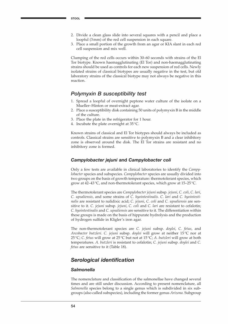
2. Divide a clean glass slide into several squares with a pencil and place aloopful (3mm) of the red cell suspension in each square.
3. Place a small portion of the growth from an agar or KIA slant in each redcell suspension and mix well.
Clumping of the red cells occurs within 30–60 seconds with strains of the ElTor biotype. Known haemagglutinating (El Tor) and non-haemagglutinatingstrains should be used as controls for each new suspension of red cells. Newlyisolated strains of classical biotypes are usually negative in the test, but oldlaboratory strains of the classical biotype may not always be negative in thisreaction.
Polymyxin B susceptibility test1. Spread a loopful of overnight peptone water culture of the isolate on a
Mueller–Hinton or meat-extract agar.2. Place a susceptibility disk containing 50 units of polymyxin B in the middle
of the culture.3. Place the plate in the refrigerator for 1 hour.4. Incubate the plate overnight at 35 ∞C.
Known strains of classical and El Tor biotypes should always be included ascontrols. Classical strains are sensitive to polymyxin B and a clear inhibitoryzone is observed around the disk. The El Tor strains are resistant and noinhibitory zone is formed.
Campylobacter jejuni and Campylobacter coli
Only a few tests are available in clinical laboratories to identify the Campy-lobacter species and subspecies. Campylobacter species are usually divided intotwo groups on the basis of growth temperature: thermotolerant species, whichgrow at 42–43 ∞C, and non-thermotolerant species, which grow at 15–25 ∞C.
The thermotolerant species are Campylobacter jejuni subsp. jejuni, C. coli, C. lari,C. upsaliensis, and some strains of C. hyointestinalis. C. lari and C. hyointesti-nalis are resistant to nalidixic acid; C. jejuni, C. coli and C. upsaliensis are sen-sitive to it. C. jejuni subsp. jejuni, C. coli and C. lari are resistant to cefalotin;C. hyointestinalis and C. upsaliensis are sensitive to it. The differentiation withinthese groups is made on the basis of hippurate hydrolysis and the productionof hydrogen sulfide in Kligler’s iron agar.
The non-thermotolerant species are C. jejuni subsp. doylei, C. fetus, andArcobacter butzleri. C. jejuni subsp. doylei will grow at neither 15 ∞C nor at 25 ∞C; C. fetus will grow at 25 ∞C but not at 15 ∞C; A. butzleri will grow at bothtemperatures. A. butzleri is resistant to cefalotin; C. jejuni subsp. doylei and C.fetus are sensitive to it (Table 18).
Serological identification
Salmonella
The nomenclature and classification of the salmonellae have changed severaltimes and are still under discussion. According to present nomenclature, allSalmonella species belong to a single genus which is subdivided in six sub-groups (also called subspecies), including the former genus Arizona. Subgroup
54
STOOL
BLM1 1/17/04 2:01 PM Page 54
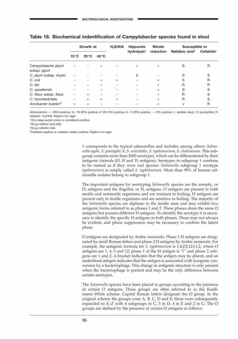
1 corresponds to the typical salmonellae and includes among others: Salmo-nella typhi, S. paratyphi A, S. enteritidis, S. typhimurium, S. choleraesuis. This sub-group contains more than 2000 serotypes, which can be differentiated by theirantigenic formula (O, H and Vi antigens). Serotypes in subgroup 1 continueto be named as if they were real species: Salmonella subgroup 1 serotypetyphimurium is simply called S. typhimurium. More than 99% of human sal-monella isolates belong to subgroup 1.
The important antigens for serotyping Salmonella species are the somatic, orO, antigens and the flagellar, or H, antigens. O antigens are present in bothmotile and nonmotile organisms and are resistant to boiling; H antigens arepresent only in motile organisms and are sensitive to boiling. The majority ofthe Salmonella species are diphasic in the motile state and may exhibit twoantigenic forms referred to as phases 1 and 2. These phases share the same Oantigens but possess different H antigens. To identify the serotype it is neces-sary to identify the specific H antigens in both phases. These may not alwaysbe evident, and phase suppression may be necessary to confirm the latentphase.
O antigens are designated by Arabic numerals. Phase 1 H antigens are desig-nated by small Roman letters and phase 2 H antigens by Arabic numerals. Forexample, the antigenic formula for S. typhimurium is 1,4,[5],12:i:1,2, where Oantigens are 1, 4, 5 and 12; phase 1 of the H antigen is “i” and phase 2 anti-gens are 1 and 2. A bracket indicates that the antigen may be absent, and anunderlined antigen indicates that the antigen is associated with lysogenic con-version by a bacteriophage. This change in antigenic structure is only presentwhen the bacteriophage is present and may be the only difference betweencertain serotypes.
The Salmonella species have been placed in groups according to the presenceof certain O antigens. These groups are often referred to as the Kauff-mann–White scheme. Capital Roman letters designate the O group. In theoriginal scheme the groups were A, B, C, D and E; these were subsequentlyexpanded to A–Z with 4 subgroups in C, 3 in D, 4 in E and 2 in G. The Ogroups are defined by the presence of certain O antigens as follows:
55
BACTERIOLOGICAL INVESTIGATIONS
A
Table 18. Biochemical indentification of Campylobacter species found in stool
Growth at H2S/KIA Hippurate Nitrate Susceptible to
15 ∞C 25 ∞C 42 ∞Chydrolysisa reduction Nalidixic acidb Cefalotinc
Campylobacter jejuni - - + - + + S Rsubsp. jejuniC. jejuni subsp. doylei - - - - d - S SC. coli - - + + - + S RC. lari - - + - - + R RC. upsaliensis - - + - - + S SC. fetus subsp. fetus - + - - - + R SC. hyointestinalis - + v + - + R SArcobacter butzleri d + + - - - + v R
Abbreviations: +: >95% positive; d+: 75–95% positive; d: 26–74% positive; d-: 5–25% positive; -: <5% positive; v: variable result; S: susceptible; R:resistant. H2S/KIA: Kligler’s iron agar.aOnly deep purple colour is considered positive.b30 mg nalidixic acid disk.c30 mg cefalotin disk.dCatalase-negative or catalase weakly positive; Kligler’s iron agar.
BLM1 1/17/04 2:01 PM Page 55
Group: A Ba C1 C2 D E1 FAntigen: 2 4;5 6;7 6;8 9 3;10 11
There are other variations in the antigenic structure: the change in colonyappearance from smooth to rough, and the presence or absence of Vi antigen.When present the Vi antigen prevents agglutination by homologous O anti-serum. The Vi antigen is usually found in fresh isolates and is lost rapidly onstorage of the culture. The Vi antigen is important for the identification of S.typhi, which often is not agglutinated by heterologous H antisera.
Procedure for somatic O antigen analysisDirect slide agglutination test
1. Bring the saline and the reagents to room temperature before performingthe test.
2. Place a drop of saline on a clean microscope slide.3. With a sterile loop, emulsify a small portion of the growth from a moist
agar slope in the drop of saline to produce a uniform and turbid suspension.
4. Examine the bacterial suspensions through a hand-lens or the low-powerobjective (¥10) of a microscope to ascertain that the suspension is not auto-agglutinable in saline.
5. With a 10-ml loop take up 1 drop of Salmonella polyvalent O antiserum (A–Iand Vi), and place it on the slide just beside the bacterial suspension.
6. Mix the antiserum and bacterial suspension and tilt the slide backwardsand forwards for 1 minute. Look for clumping while viewing the suspen-sion under good light. Distinct clumping during this period is a positiveresult.
7. If the result is positive, repeat the slide test with single-factor antiserum.
Some Salmonella possess an envelope (Vi) antigen and, in the live or unheatedform, are non-agglutinable with group C1 (O:6,7) or group D (O:9) antisera.Heat the suspension in boiling water for 20 minutes to remove the Vi antigen,cool, separate the bacteria by centrifugation, re-suspend in fresh saline, andtest with the same antisera.
Procedure for H antigen analysisA preliminary identification of the major flagellar (H) antigen can be made bythe direct slide agglutination test as described for the somatic O antigens.Occasionally it is necessary to increase the motility of the test organism bymaking several consecutive transfers in a semi-solid nutrient medium(“swarm agar”, see below). Antisera against those antigens are commerciallyavailable. However, a phase suppression is often required if identification ofboth flagellar phases is necessary for classification. This often requires a phaseinversion with sera designed for this purpose.1
1. Prepare a semi-solid nutrient medium containing 0.2–0.4% agar. Add 1mlof the medium to test-tubes.
2. Replace the corks from the test-tubes with cotton wool.3. Melt the agar in boiling water and place the test-tubes with the melted
agar in a 45 ∞C water-bath for 30 minutes.
56
a All Salmonellae of subgroup B contain antigen 4, but only some contain antigen 5.1 Available from Statens Serum Institut, 5 Artillerivej, 2300 Copenhagen S, Denmark.
STOOL
BLM1 1/17/04 2:01 PM Page 56
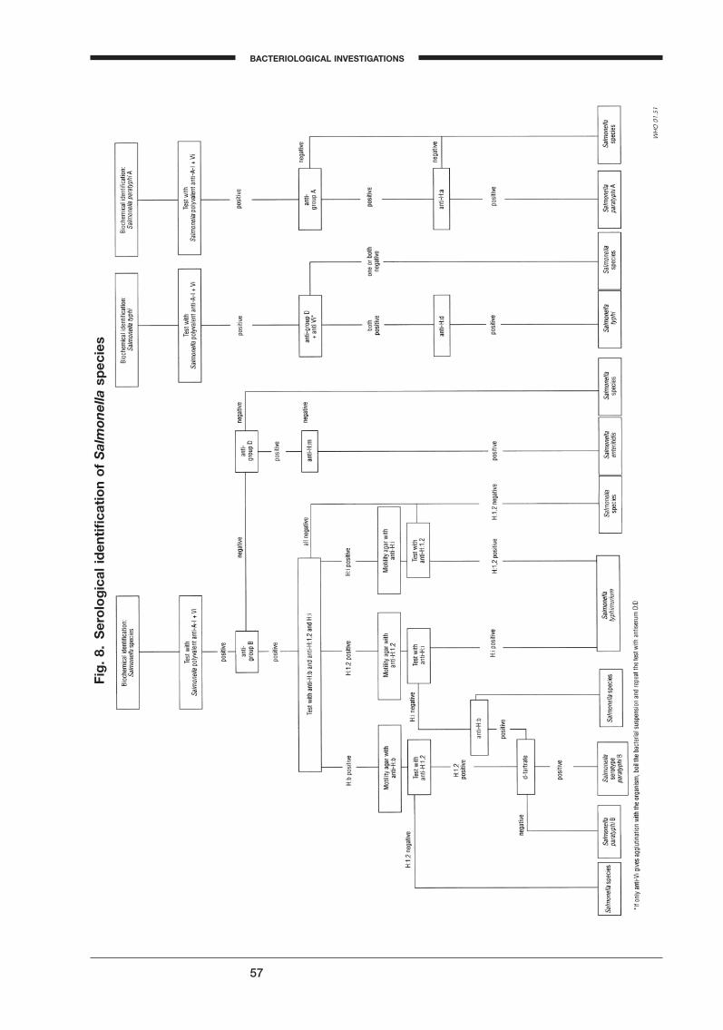
57
BACTERIOLOGICAL INVESTIGATIONS
A
Fig
. 8.
Ser
olo
gic
al id
enti
fica
tio
n o
f S
alm
onel
la s
pec
ies
BLM1 1/17/04 2:01 PM Page 57
58
4. Write the specimen number and the H antisera (H:b, H:i, and H:1,2) oneach test-tube. Include a control test-tube.
5. Add 10 ml of each phase inversion heterologous H antiserum to the corresponding agar, shake the test-tube carefully, and leave the agar tosolidify as a slope.
6. Make a heavy suspension of isolated colonies in saline, streak the slopeusing a loop, and incubate overnight.
7. With an inoculating loop emulsify a speck of the culture from the slopein a drop of saline.
8. With a 10-ml loop, take up 1 drop of one of the heterologous H antiseraand place it on the slide just beside the bacterial suspension.
9. Mix the antiserum and bacterial suspension and tilt the slide backwardsand forwards for 1 minute. Look for clumping while viewing the suspension under a good light. Distinct clumping during this period is apositive result.
10. Repeat the agglutination test with the other heterologous antisera ifrequired, and identify the serotype with the help of the flow diagram onpage 57.
Salmonella typhi
To confirm the biochemical identification, test in antisera Vi, O group D (O:D),and H:d. Cultures agglutinating in Vi may be negative in O:D due to obstruc-tion by the Vi antigen. Remove the Vi antigen by heating the suspension for20 minutes at 100 ∞C and test in O:D antiserum again. If positive in Vi, O:Dand H:d, report: “Salmonella typhi”. If positive only in O:D, report: “Salmonella,group D”.
Salmonella paratyphi A
To confirm the biochemical identification, test with Salmonella group A anti-serum. If positive with group A antiserum, test with H:a antiserum, and ifpositive, report: “Salmonella paratyphi A”. If negative with H:a, report: “Salmonella group A”. If non-motile, they may be S. flexneri type 6 or S. boydiitype 13 or 14, and should be tested with Shigella antisera B and D.
Salmonella, other serotypes
When the presumptive tests indicate typical Salmonella, test with Salmonella Ogroups A, B, C, D, and E antisera.
• If positive in group B, test with flagellar antiserum H:b, H:i and H:1,2. Ifpositive with H:b antiserum, the organism may be S. wien or S. paratyphiB. S. wien can be differentiated from S. paratyphi B by testing with H:1,wand H:2 antisera (if available). S. wien reacts with H:1,w and S. paratyphi Bwith H:2.
• If positive with H:i or H:1,2 antiserum, make a phase inversion and test with heterologous H antiserum. If positive, report: “S. typhimurium”.If negative, report: “Salmonella, group B”.
• If positive with group C antiserum, report: “Salmonella group C”.• If positive with group D antiserum, test with Vi, H:d and H:m antisera. If
positive with Vi or H:d, report: “Salmonella typhi”. If positive with H:m,report: “Salmonella enteritidis”. If negative with Vi, H:d and H:m, report:“Salmonella group D”.
• If biochemically the strain is a Salmonella but is negative with all O-groupantisera, report: “Presumptive Salmonella species” and refer to NationalReference Centre.
STOOL
BLM1 1/17/04 2:01 PM Page 58
Shigella
Shigella can be subdivided into serogroups and serotypes by slide and tubeagglutination tests with specific O-antisera. Slide agglutination will usuallybe sufficient, if the results are clear-cut. The antigen suspension should bemade from a non-selective medium such as a nutrient agar or KIA, and thesuspension should be observed for auto-agglutination before the antiserum isadded.
Tests with Shigella groups A, B, C, and D antisera• If agglutination occurs with group A, report: “Shigella dysenteriae”.
Test with S. dysenteriae type 1 antiserum. If positive, report: “S. dysenteriaetype 1”.
• If agglutination occurs with group B, report: “Shigella flexneri”.• If agglutination occurs with group C, report: “Shigella boydii”.• If agglutination occurs with group D, report: “Shigella sonnei”.
Occasionally Shigella strains may fail to agglutinate in homologous antiserumdue to the presence of a K antigen. Heating a saline suspension of the strainin a boiling water-bath for 20 minutes and repeating the test may reverse this.
Other Gram-negative organisms may share antigens with Shigella strains andgive false-positive agglutination with Shigella typing sera. Well-known exam-ples of bacteria showing this cross-reaction are Plesiomonas shigelloides andShigella sonnei phase 1, and certain strains of Hafnia and Shigella flexneriserotype 4a; but of greater importance is the cross-reaction with some strainsof diarrhoeagenic E. coli.
Yersinia enterocolitica
Y. enterocolitica possesses several somatic (O) antigens, which have been usedfor subdividing the species into at least 17 serogroups. Most human infectionsin Canada, Europe, and Japan are due to serotype O3; Infections due toserotype O9 have been reported mainly from the Scandinavian countries, andinfections with serotype O8 are almost exclusively from the USA. There isserological cross-reaction between Y. enterocolitica O9 and Brucella spp.
59
BACTERIOLOGICAL INVESTIGATIONS
A
BLM1 1/17/04 2:01 PM Page 59
Upper respiratory tract infections
Introduction
The upper respiratory tract extends from the larynx to the nostrils and com-prises the oropharynx and the nasopharynx together with the communicat-ing cavities, the sinuses and the middle ear. The upper respiratory tract canbe the site of several types of infection:
— pharyngitis, sometimes involving tonsillitis, and giving rise to a “sorethroat”
— nasopharyngitis— otitis media— sinusitis— epiglottitis.
Of all those infections, pharyngitis is by far the most frequent; in addition, theuntreated infection may have serious sequelae. Only pharyngitis will be con-sidered here.
Most cases of pharyngitis have a viral etiology and follow a self-limitingcourse. However, approximately 20% are caused by bacteria and usuallyrequire treatment with appropriate antibiotics. As the physician is rarely ableto make a distinction between viral and bacterial pharyngitis on clinicalgrounds alone, treatment should ideally be based on the result of bacterio-logical examination.
Bacteriological diagnosis of pharyngitis is complicated by the fact that theoropharynx contains a heavy, mixed, normal flora of aerobic and anaerobicbacteria. The normal flora generally outnumbers the pathogens and the roleof the bacteriologist is to distinguish between the commensals and thepathogens. Where possible only the latter should be reported to the physician.
Normal flora of the pharynx
The normal flora of the pharynx includes a large number of species thatshould be neither fully identified nor reported when observed in throat cultures:
• viridans (a-haemolytic) streptococci and pneumococci• nonpathogenic Neisseria spp.• Moraxella (formerly Branhamella) catarrhalis (this can also be a respiratory
pathogen)• staphylococci (S. aureus, S. epidermidis)• diphtheroids (with the exception of C. diphtheriae)• Haemophilus spp.• yeasts (Candida spp.) in limited quantity• various strictly anaerobic Gram-positive cocci and Gram-negative rods,
spirochaetes and filamentous forms.
The throats of elderly, immunodeficient, or malnourished patients, particu-larly when they have received antibiotics, may be colonized by Enterobacte-riaceae (Escherichia coli, Klebsiella spp., etc.) and by the nonfermentativeGram-negative groups (Acinetobacter spp. and Pseudomonas spp.). Suchpatients may also have in their pharynx a proliferation of S. aureus or of
60
BLM1 1/17/04 2:01 PM Page 60
Candida spp., or other yeast-like fungi. Although these microorganisms do notcause pharyngitis, except in association with granulocytopenia, it is advisableto report such isolates to the clinician, as they occasionally indicate the exis-tence of (or may sometimes give rise to) a lower respiratory tract infection(e.g. pneumonia) or bacteraemia. However, an antibiogram should not be per-formed routinely on these colonizing microorganisms.
Bacterial agents of pharyngitis
Streptococcus pyogenes (Lancefield group A) is by far the most frequent causeof bacterial pharyngitis and tonsillitis. This infection is particularly prevalentin young children (5–12 years). When streptococcal pharyngitis is associatedwith a characteristic skin rash, the patient is said to have scarlet fever. Ininfants, a streptococcal throat infection may often involve the nasopharynxand be accompanied by a purulent nasal discharge.
Non-group-A, b-haemolytic streptococci (e.g. groups B, C and G) are uncom-mon causes of bacterial pharyngitis and if detected should be reported. Pha-ryngeal infections due to S. pyogenes, if not properly treated, may give rise tosequelae such as rheumatic fever, and, less often, glomerulonephritis. Specificidentification of, and antibacterial treatment directed against, S. pyogenes areprimarily intended to prevent the occurrence of rheumatic fever.
Corynebacterium diphtheriae is the cause of diphtheria, a disease that is endemicin many countries. It can reach epidemic proportions in countries where thevaccination programme has been interrupted. Characteristically (with a fewexceptions), C. diphtheriae causes a typical form of infection, characterized bya greyish-white membrane at the site of infection (pharynx, tonsils, nose, orlarynx). Diphtheria is a serious disease and the diagnosis is made on the basisof clinical findings. The physician would then generally make a specificrequest to culture for diphtheria bacilli.
Gonococcal pharyngitis has been recognized with increasing frequency insome countries, with rates that parallel the incidence of cervical and urethralgonorrhoea. Culture of throat swabs for gonococci should be done on specificrequest from the clinician, using the appropriate selective medium (modifiedThayer–Martin medium).
Necrotizing ulcerative pharyngitis (Vincent angina) is a rare condition char-acterized by a necrotic ulceration of the pharynx with or without formationof a pseudomembrane. It is associated, at the site of infection, with a heavymixed flora of strict anaerobes dominated by Gram-negative fusiform rodsand spirochaetes, generally referred to as Fusobacterium spp. and Treponemavincentii, and possibly others. Although both species belong to the normalmouth flora, their presence in large numbers in a Gram-stained smear of ulcer-ated lesions should be reported as a “fusospirochaetal complex”. This micro-scopic diagnosis need not be confirmed by anaerobic culture, which is difficultand time-consuming. However, the presence of this complex does not excludethe need to search for other pathogens, particularly S. pyogenes.
Although small numbers of C. albicans or other Candida species may be partof the normal oral flora, oral candidiasis results when the number of organ-isms increases considerably in certain pathological conditions, e.g. in mal-nourished premature babies, in immunodeficient adults (e.g. patients withHIV/AIDS), or in patients who have received broad-spectrum antimicrobialsor cancer therapy. The affected area—tongue, tonsils, throat or buccal mucosa—may be extremely red, or covered with white patches or a confluent grey-
61
A
BACTERIOLOGICAL INVESTIGATIONS
BLM1 1/17/04 2:01 PM Page 61
white membrane (thrush). The diagnosis of candidiasis is best made byfinding numerous yeast cells, some of them forming long mycelium-like fila-ments, in a Gram-stained smear of the exudate.
Swabs from the upper respiratory tract may be submitted to the laboratory,not for the diagnosis of a clinical infection, but to detect a potential pathogenin a healthy subject, a pharyngeal or a nasal “carrier”. This should only bedone as part of well-defined epidemiological surveys. The followingpathogens can give rise to a carrier state in the upper respiratory tract:
• Staphylococcus aureus. Sampling of patients and staff for nasal carriers issometimes performed as part of an investigation of hospital outbreaks ofMeticillin-resistant S. aureus (MRSA).
• Neisseria meningitidis. Carriage of meningococci may be very prevalent(20% or more) even at non-epidemic times. Identification of pharyngealcarriers of meningococci is rarely needed, and need not be performed priorto the administration of prophylactic antibiotics to family or other closecontacts of patients with meningococcal disease.
• Streptococcus pyogenes. Carriage of this organism in low numbers may beprevalent, especially among schoolchildren (20–30%).
• Corynebacterium diphtheriae. The carrier rate of the diphtheria bacillus ishigh in non-vaccinated populations. In such communities, it may be justi-fied to identify and treat carriers among the close contacts of a patient withproven diphtheria. Carriers are rare when an immunization programme iscorrectly implemented.
Collection and dispatch of specimens
Ideally, specimens should be collected by a physician or other trained per-sonnel. The patient should sit facing a light source. While the tongue is keptdown with a tongue depressor, a sterile cotton-wool swab is rubbed vigor-ously over each tonsil, over the back wall of the pharynx, and over any otherinflamed area. Care should be taken not to touch the tongue or buccal sur-faces. It is preferable to take two swabs from the same areas. One can be usedto prepare a smear, while the other is placed into a glass or plastic containerand sent to the laboratory. Alternatively, both swabs may be placed in the con-tainer and dispatched to the laboratory. If the specimen cannot be processedwithin 4 hours, the swab should be placed in a transport medium (e.g. Amiesor Stuart).
Direct microscopy
The fusospirochaetal complex of necrotizing ulcerative pharyngitis (Vincentangina) and Candida are best recognized on a Gram-stained smear, whichshould be prepared if the physician makes a special request. The Gram-stained smear is not useful for the detection of streptococci or Neisseria spp.Moreover, the direct smear has poor sensitivity and specificity for the detec-tion of the diphtheria bacillus, unless the specimen has been collected withcare and is examined by an experienced microbiologist. In the absence of aphysician’s request or of clinical information, a Gram-stained smear shouldnot be made for throat swabs.
62
UPPER RESPIRATORY TRACT INFECTIONS
BLM1 1/17/04 2:01 PM Page 62
63
BACTERIOLOGICAL INVESTIGATIONS
A
Culture and identification
Culture for Streptococcus pyogenes
Immediately upon receipt in the laboratory, the swab should be rubbed overone-quarter of a blood agar plate, and the rest of the plate streaked with asterile wire loop. The blood agar should be prepared from a basal agarmedium without glucose (or with a low glucose content), e.g. tryptic soy agar(TSA). Acidification of glucose by S. pyogenes inhibits the production ofhaemolysin. Blood from any species, even human blood (fresh donor blood),can be used at a concentration of 5%. The plates should be filled to a depthof 4–5 mm. Sheep blood is preferred because it does indicate haemolysis ofsome commensal Haemophilus spp. and it gives no haemolysis with the zymo-genes variant of Enterococcus faecalis.
The recognition of b-haemolytic colonies can be improved, and their pre-sumptive identification hastened, by placing a co-trimoxazole disc (as usedfor the susceptibility test) and a special low-concentration bacitracin disc overthe initial streaked area. Because S. pyogenes is resistant and many other bac-teria are susceptible to co-trimoxazole, this disc improves the visibility of b-haemolysis. Incubation in a candle-jar will detect most b-haemolytic strep-tococci. A simple way to increase haemolysis is to stab the agar surface per-pendicularly by inserting the loop deep into the medium to encourage growthof subsurface colonies. After 18 hours and again after 48 hours of incubationat 35–37 ∞C, the blood plates should be examined for the presence of small(0.5–2 mm) colonies surrounded by a relatively wide zone of clear haemoly-sis. After Gram-staining to verify that they are Gram-positive cocci, thecolonies should be submitted to specific identification tests for S. pyogenes. Forclinical purposes, presumptive identification of S. pyogenes is based on its sus-ceptibility to a low concentration of bacitracin. For this purpose, a special dif-ferential disc is used containing 0.02–0.05 IU of bacitracin. The ordinary discsused in the susceptibility test, with a content of 10 units, are not suitable foridentification. A b-haemolytic streptococcus showing any zone of inhibitionaround the disc should be reported as S. pyogenes. If the haemolytic coloniesare sufficiently numerous, the presence or absence of an inhibition zone maybe read directly from the primary blood agar plate. If the colonies are lessnumerous, one or two should be picked from the primary plate, streaked onone-fifth of another plate to obtain confluent growth, and each inoculated areacovered with a bacitracin disc. After overnight incubation, the subculturesshould be read for inhibition zones.
In some laboratories this presumptive identification is confirmed by serolog-ical demonstration of the specific cell wall polysaccharides. This can be doneeither by the classical precipitin method, or more rapidly by using a com-mercial kit for the rapid slide coagglutination or latex agglutination tests. Ifdesirable, bacitracin-resistant b-haemolytic streptococci can be further identi-fied using some simple physiological tests (see Table 19). Minute colonies ofb-haemolytic streptococci may be encountered, which, when grown and sero-logically grouped, react with group A antiserum. These streptococci are notconsidered to be S. pyogenes and are not associated with the serious infectionscaused by group A streptococci.
In reporting the presence of S. pyogenes in a throat culture, a semiquantitativeanswer should be given (rare, +, + +, or + + +). Patients with streptococcalpharyngitis generally show massive growth of S. pyogenes, with colonies overthe entire surface of the plate. Plates of carriers generally show fewer than 20colonies per plate. Even rare colonies of b-haemolytic streptococci should beconfirmed and reported.
BLM1 1/17/04 2:01 PM Page 63
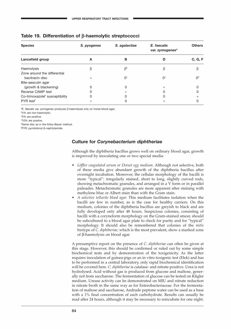
64
Culture for Corynebacterium diphtheriae
Although the diphtheria bacillus grows well on ordinary blood agar, growthis improved by inoculating one or two special media:
• Löffler coagulated serum or Dorset egg medium. Although not selective, bothof these media give abundant growth of the diphtheria bacillus afterovernight incubation. Moreover, the cellular morphology of the bacilli ismore “typical”: irregularly stained, short to long, slightly curved rods,showing metachromatic granules, and arranged in a V form or in parallelpalisades. Metachromatic granules are more apparent after staining withmethylene blue or Albert stain than with the Gram stain.
• A selective tellurite blood agar. This medium facilitates isolation when thebacilli are few in number, as is the case for healthy carriers. On thismedium, colonies of the diphtheria bacillus are greyish to black and arefully developed only after 48 hours. Suspicious colonies, consisting ofbacilli with a coryneform morphology on the Gram-stained smear, shouldbe subcultured to a blood agar plate to check for purity and for “typical”morphology. It should also be remembered that colonies of the mitisbiotype of C. diphtheriae, which is the most prevalent, show a marked zoneof b-haemolysis on blood agar.
A presumptive report on the presence of C. diphtheriae can often be given atthis stage. However, this should be confirmed or ruled out by some simplebiochemical tests and by demonstration of the toxigenicity. As the latterrequires inoculation of guinea-pigs or an in vitro toxigenic test (Elek) and hasto be performed in a central laboratory, only rapid biochemical identificationwill be covered here. C. diphtheriae is catalase- and nitrate-positive. Urea is nothydrolysed. Acid without gas is produced from glucose and maltose, gener-ally not from saccharose. The fermentation of glucose can be tested on Kliglermedium. Urease activity can be demonstrated on MIU and nitrate reductionin nitrate broth in the same way as for Enterobacteriaceae. For the fermenta-tion of maltose and saccharose, Andrade peptone water can be used as a basewith a 1% final concentration of each carbohydrate. Results can usually beread after 24 hours, although it may be necessary to reincubate for one night.
Table 19. Differentiation of b-haemolytic streptococci
Species S. pyogenes S. agalactiae E. faecalis Othersvar. zymogenesa
Lancefield group A B D C, G, F
Haemolysis b bb b bZone around the differential
bacitracin disc + 0c 0c 0d
Bile–aesculin agar(growth & blackening) 0 0 + 0
Reverse CAMP test 0 + 0 0Co-trimoxazolee susceptibility 0 0 0 +PYR testf + 0 + 0
aE. faecalis var. zymogenes produces b-haemolysis only on horse-blood agar.b5% are non-haemolytic.c5% are positive.d10% are positve.eSame disc as in the Kirby–Bauer method.fPYR: pyrrolidonyl-b-naphtylamide.
UPPER RESPIRATORY TRACT INFECTIONS
BLM1 1/17/04 2:01 PM Page 64
65
BACTERIOLOGICAL INVESTIGATIONS
A
It must be emphasized that the microbiology laboratory’s role is to confirmthe clinical diagnosis of diphtheria. Therapy should not be withheld pendingreceipt of laboratory reports. More detailed information on the isolation andidentification of C. diphtheriae is found in Guidelines for the laboratory diagnosisof diphtheria.1
Susceptibility testing
Routine susceptibility tests on throat or pharyngeal isolates are most often notrequired, and may even be misleading. The major pathogens involved in bacterial pharyngitis are S. pyogenes and C. diphtheriae benzylpenicillin anderythromycin are considered as the antimicrobials of choice to treat both typesof infection. In cases of diphtheria, treatment with antitoxin is also indicated.
1 Begg N. Manual for the management and control of diphtheria in the European region. Copen-hagen, WHO Regional Office for Europe, 1994.
BLM1 1/17/04 2:01 PM Page 65
66
Lower respiratory tract infections
Introduction
Lower respiratory tract infections (LRTI) are infections occurring below thelevel of the larynx, i.e. in the trachea, the bronchi, or in the lung tissue (tra-cheitis, bronchitis, lung abscess, pneumonia). Sometimes, in pneumonia, theadjacent membranous covering of the lung is involved, resulting in roughen-ing (pleurisy) and sometimes production of fluid in the pleural cavity (pleuraleffusion).
A special form of LRTI is pulmonary tuberculosis, which is common in manycountries. The patient may cough up aerosols containing tubercle bacilli(Mycobacterium tuberculosis) which can be inhaled by other people. This formof the disease (“open” tuberculosis) is easily spread from person to person,and is therefore a serious communicable disease.
Many patients with LRTI cough up purulent (pus-containing) sputum that isgenerally green or yellowish in colour; this sputum may be cultured andexamined grossly and microscopically.
There are other infections in which little or no sputum is produced: Legion-naire disease (caused by Legionella pneumophila), pneumonia due toMycoplasma pneumoniae (“primary atypical pneumonia”), and Chlamydiapneumonia. These diseases require specialized techniques (serology and iso-lation on special cultures) for their diagnosis and will not be discussed furtherhere. Apart from pulmonary tuberculosis (see below), most requests forsputum microscopy and culture concern patients with respiratory infectionsassociated with purulent sputum.
The most common infections
Acute and chronic bronchitis
In patients with acute bronchitis (usually following an acute viral infection,such as a common cold or influenza), sputum is not usually cultured unlessthe patient fails to show signs of clinical improvement.
Chronic bronchitis is a long-lasting, disabling respiratory disease with periodicacute attacks. Most patients generally cough up sputum every day, which isusually grey and mucoid; the disease also has episodes when the conditionof the patient becomes worse and obviously purulent sputum is coughed up.This is termed an acute exacerbation of chronic bronchitis. The typical respi-ratory pathogens (Haemophilus influenzae, Streptococcus pneumoniae, or lessoften Moraxella (Branhamella) catarrhalis) are frequently found in sputumsamples.
Lung abscess
An abscess may form in the lung following the inhalation of a foreign body,of the stomach contents, or of upper respiratory tract (mouth or throat) secre-tions. This is sometimes termed “aspiration pneumonia”. Attempts may bemade to culture coughed-up sputum (which is often extremely foul-smelling),
BLM1 1/17/04 2:01 PM Page 66
67
A
but when there is an abscess (as demonstrated by radiography) the pus con-tained in it should be examined microscopically and cultured. Unfortunately,there is no medical agreement on how this pus should be obtained, but directpuncture and withdrawal of pus is one of the possibilities. Anaerobic bacte-ria such as Prevotella melaninogenica (formerly Bacteroides melaninogenicus) andPeptostreptococcus spp., derived from the mouth or throat flora, are often veryimportant causative agents. Pus should be collected, transported, and exam-ined according to standard methods for anaerobic culture of pus (see p. 86and pp. 98–102).
Pneumonia and bronchopneumonia
Acute lobar pneumonia usually affects only a single lobe of the lung. Thisinfection is nearly always caused by S. pneumoniae. This form of pneumoniaoccasionally occurs in epidemic form. A rare cause of a rather similar form ofpneumonia is Klebsiella pneumoniae.
While a few patients infected with S. pneumoniae or K. pneumoniae will haveclassical pneumonia, the most frequent form of the disease is bronchopneu-monia, with patches of infiltration and inflammation (termed “consolidation”)distributed over one or often both lungs.
Many different kinds of viruses or bacteria can be associated with bronchop-neumonia. Apart from S. pneumoniae, and sometimes H. influenzae, Staphylo-coccus aureus is a cause of bronchopneumonia, particularly during influenzaor measles epidemics. Gram-negative rods (in particular, E. coli and K. pneu-moniae) and P. aeruginosa are also frequently found. These infections are allcommon in intensive-care departments, especially when broad-spectrumantibiotics are widely used or mechanical respiration is carried out, and areindicative of indiscriminate use of antibiotics and failure to monitor patientscarefully for early signs of infection.
If there is a pleural effusion, the fluid should be examined microscopicallyand cultured according to the procedures described for pus and exudates.
Pulmonary tuberculosis
The sputum of patients with pulmonary tuberculosis is usually not highlypurulent, but should not be rejected for tuberculosis investigation because ofthis. An acid-fast stained smear (Ziehl–Neelsen) should be examined micro-scopically to detect immediately any patients who have acid-fast bacteria intheir sputum1. After the smear has been stained, the sputum should be treatedby a decontamination procedure (see p. 72) in order to kill as many of the non-mycobacterial organisms as possible and to leave the tubercle bacilli viableand thus suitable for culture on Löwenstein–Jensen medium.
Because the bacteriological procedures for the diagnosis of pyogenic respira-tory infections, such as bronchitis and pneumonia, are so fundamentally different from those for tuberculosis, they will be considered separately. The physician must make it clear to the laboratory whether he or she wishesexaminations for:
1 See Manual of basic techniques for a health laboratory, 2nd ed. Geneva, World Health Organization, 2003.
BACTERIOLOGICAL INVESTIGATIONS
BLM1 1/17/04 2:01 PM Page 67
68
LOWER RESPIRATORY TRACT INFECTIONS
• pyogenic bacteria (H. influenzae, S. pneumoniae, etc.),• tubercle bacteria (M. tuberculosis), or• both types of bacteria.
Collection of sputum specimens
The collection of good sputum specimens is an art in itself and has beendescribed in other books1. Examination of a badly collected sputum specimencan give misleading results because of contamination with the normal bacte-rial flora present in the mouth and throat; “sputum” consisting of saliva andfood particles should not be examined.
The sputum should be collected in a sterile wide-mouthed container with asecure, tight-fitting cover and sent to the laboratory without delay. If thesputum is allowed to stand after collection, overgrowth of contaminating bac-teria may take place before the examination is carried out and the results ofsmears and cultures will be highly misleading. For this reason, it is not rec-ommended that sputum specimens be sent to the laboratory by mail. The onlyexceptions are specimens for tuberculosis examination that may have to besent to a district or regional laboratory. The local and national postal regula-tions for the transmission of infected (pathological) material must be strictlyapplied.
Processing of sputum in the laboratory (for non-tuberculous infections)
After collection sputum must be immediately processed or kept in a refrigerator.
Macroscopic evaluation
The macroscopic appearance of the sputum should be recorded. Possibledescriptions include:
purulent, greenpurulent, yellowmucopurulent (i.e. partially mucoid and partially purulent)blood-stainedblood-stained, with green floccules*grey, mucoid*grey, frothy*white, mucoid*white, frothy*white, mucoid, with some food particles*watery (i.e. only saliva present)*watery, with some food particles
1 Specimen collection and transport for microbiological investigation. Alexandria, WHO RegionalOffice for the Eastern Mediterranean, 1995 (WHO Regional Publications, Eastern MediterraneanSeries 8).
Manual of basic techniques for a health laboratory, 2nd ed. Geneva, World Health Organization,2003.
Technical guide for sputum examination for tuberculosis by direct microscopy. Bulletin of theInternational Union Against Tuberculosis and Lung Disease, 4th ed. 1996.
BLM1 1/17/04 2:01 PM Page 68
69
BACTERIOLOGICAL INVESTIGATIONS
A
Sputum specimens marked with an asterisk should not normally be examinedfor non-tuberculous infections.
Microscopic examination
A portion of the purulent or mucopurulent sputum should be used for thepreparation of a Gram-stained smear.
If no floccules of pus can be seen (e.g. in a grey mucoid sputum sample), theGram-stained smear may show only the presence of large, rather square, squa-mous epithelial cells, frequently covered with masses of adherent bacteria.This is an indication that the specimen consists mainly of mouth or throatsecretions, and culture should not be carried out as it is not relevant, andusually highly misleading. An accepted guideline is to reject, for culture, anyspecimen that contains fewer than 10 polymorphonuclear neutrophils perepithelial cell1.
In many patients with acute respiratory infections (e.g. pneumonia) and puru-lent sputum, the emergency examination of a Gram-stained smear mayprovide guidance to the clinician in the choice of antimicrobial chemotherapy.Possible results include:
• Gram-positive diplococci surrounded by an empty space from theunstained capsules (suggestive of S. pneumoniae);
• small Gram-negative coccobacilli (probably H. influenzae);• Gram-negative diplococci, intracellular and extracellular (suggestive of
Movaxella catarrhalis;• Gram-positive cocci in grape-like clusters (suggestive of S. aureus);• Gram-negative rods (suggestive of the presence of Enterobacteriaceae or
Pseudomonas spp.);• large Gram-positive yeast-like cells, often with mycelia (suggestive of the
presence of Candida spp.).
Cultural procedures and interpretation
When microscopy of the specimen demonstrates an acceptable quality of thesputum, select a floccule of purulent material (or of the most nearly purulentmaterial available) using a sterile swap or loop and inoculate on to the variousculture plates.
A suggested routine set of culture media is as follows:
• blood agar, with a streak of S. aureus to facilitate satellite growth of H.influenzae, and with an optochin disc placed in the middle of the secondarystreaking,
• chocolate agar,• MacConkey agar.
The blood agar and chocolate agar plates are incubated at 35–36 ∞C in anatmosphere containing extra carbon dioxide (e.g. in a candle jar) and the MacConkey plate is incubated in air.
1 Heinemann HS & Radano RR. Acceptability and cost savings of selective sputum microbiol-ogy in a community teaching hospital. Journal of Clinical Microbiology, 1979, 10: 567–573.
BLM1 1/17/04 2:01 PM Page 69
70
If grape-like clusters of Gram-positive cocci were present in the stained smear,an extra mannitol salt agar (MSA) plate is suggested. The presence of Gram-positive, yeast-like structures in the stained smear may be an indication for the inoculation of a tube of Sabouraud dextrose agar (which needs to beincubated for at least 3 days at 35–37 ∞C). MSA and Sabouraud cultures do not need to be done routinely for all sputum specimens.
Cultures should be inspected after incubation overnight (18 hours) but reincu-bation for an extra 24 hours may be indicated when growth is less than expectedfrom the microscopic findings, or when only tiny colonies are present.
Typical findings include the following:
• Flat, clear colonies with concave centres and zones of green (a-) haemoly-sis, as well as a zone of inhibition of growth around the optochin disc, may be S. pneumoniae. If the reading of the optochin test result on theprimary plate is inconclusive, the test should be repeated on a subculture.It should not be forgotten that other a-haemolytic colonies (the so-calledviridans streptococci) are normally present in the flora of the mouth andthroat.
• Tiny, water-drop colonies growing as non-haemolytic satellite colonies onthe blood agar plate, but much larger clear colonies on the chocolate agaror enriched blood agar plates, suggest the presence of H. influenzae. Thesecolonies are usually present in large numbers, generally more than 20 perplate. Some laboratories choose to confirm this by X and V factor depen-dence tests, but these have to be very carefully controlled and are notstrictly necessary. Serological typing of respiratory strains is usually nothelpful, as most of them are “rough” and untypable.
• Brittle, dry, grey-white colonies on blood agar and chocolate agar platesthat can be moved intact with a loop may indicate M. catarrhalis. If desired,a set of sugar degradation tests may be set up (all test results negative),but most laboratories do not do this. Moraxella organisms are stronglyoxidase-positive, and their colonial and microscopic appearance is highlycharacteristic. As the morphologcal appearance of Moraxella resemblesNeisseria spp. the tributyrin test may be used for differentiation, sinceMoraxella hydrolyses tributyrin.
• Medium-sized, golden-buff colonies are formed by S. aureus. The coagu-lase and the mannitol fermentation tests are positive, although the slidecoagulase test (“bound” coagulase test) is occasionally negative. If there isa contradiction between the appearance of the colonies and the slide test,then a tube coagulase (“free” coagulase) test should be performed.
• Colonies on MacConkey agar suggest that Enterobacteriaceae orPseudomonas spp. or Acinetobacter spp. are present.
• Whitish, round, matt colonies on the blood agar and chocolate agar platesmay be Candida albicans, which will also grow within 2–3 days on aSabouraud dextrose agar culture.
It should be stressed that rare colonies of any of the above organisms eitherstem from the normal commensal flora of the respiratory tract, or are a resultof colonization (e.g. coliforms, yeasts). As they may not be relevant to themanagement of the patient, they should not be reported, or should be reportedas colonizing flora.
Susceptibility testing
Susceptibility tests should be performed only when the amount of growth isconsidered significant, and not on every bacterial species present in small
LOWER RESPIRATORY TRACT INFECTIONS
BLM1 1/17/04 2:01 PM Page 70
71
BACTERIOLOGICAL INVESTIGATIONS
A
numbers in the culture. Interpretations of some possible results are presentedin Table 20.
For Enterobacteriaceae and staphylococci the standardized disc-diffusionmethod (Kirby–Bauer) should be used. Strains of S. pneumoniae should betested on Mueller–Hinton agar, supplemented with 5% sheep blood, for sus-ceptibility to tetracycline, chloramphenicol, erythromycin, and benzylpeni-cillin. Conventional blood agar can also be used. For benzylpenicillin, a disccontaining 1mg of oxacillin is preferred to a disc containing benzylpenicillinitself as results with this oxacillin agree better with the MIC value for benzyl-penicillin; it is also more stable. Benzylpenicillin discs may deteriorate rapidlyin hot climates and thus produce unreliable results.
H. influenzae strains should be tested for b-lactamase production, using, forexample, the Nitrocefin test. Rare strains of H. influenzae may be ampicillin-resistant without producing b-lactamase. At this stage it is not recommendedto test H. influenzae for antibiotic susceptibility by the disc-diffusion technique.
M. catarrhalis isolates should be tested for b-lactamase production. Testingagainst tetracycline and erythromycin is optional.
Candida albicans cultures need not be tested against any antimicrobial agents.
Most laboratories give a semi-quantitative assessment of the bacteria culturedon solid media, which might be presented as follows:
(+) = few colonies+ = light growth+ + = moderately heavy growth+ + + = heavy growth.
Table 20. Interpretation of susceptibility test results of fastidious organismsa
Total zone diameter (mm)
Resistant Intermediate Susceptible
S. pneumoniae (Mueller–Hintonwith 5% sheep blood in 5% CO2 incubation
Oxacillin (1mg) (for benzylpenicillin) �19b — �20Tetracycline (30mg) �18 19–22 �22Erythromycin (15mg) �15 16–20 �21Chloramphenicol (30mg) �20 — �21Co-trimoxazole (25mg) �15 16–18 �19
M. catarrhalis (Mueller–Hinton)Tetracycline (30mg) �14 15–18 �19Erythromycin (15mg) �13 14–22 �23Co-trimoxazole (25mg) �10 11–15 �16
aNational Committee for Clinical Laboratory Standards (NCCLS). Performance standards for antimicrobial sus-ceptibility testing. M100-S8. Vol 18, 1998.bResistant or intermediate.
BLM1 1/17/04 2:01 PM Page 71
Culture for Mycobacterium tuberculosis
In addition to the preparation of a direct, acid-fast stained smear, material(usually, but not always, sputum) should be cultured for M. tuberculosis, when-ever this disease is clinically suspected. Some patients, in whom pulmonarytuberculosis is suspected, may not cough up any sputum. A little sputum may,in fact, be produced but is immediately swallowed. In this case, the physicianshould collect a specimen of fasting gastric juice (generally obtained early inthe morning) and neutralize the material using sodium bicarbonate (100mg)before sending it to the laboratory. The gastric juice should be treated in thesame way as sputum. It is expensive to culture all sputum samples routinelyfor tubercle bacilli (although some unsuspected patients would be discov-ered); therefore this is not routinely recommended.
Concentration–digestion–decontaminationprocedures
Sputum from patients with tuberculosis infection often contains solid parti-cles of material from the lungs and this material should be selected for culture,whenever it is found. However, as tuberculous sputum is coughed up throughthe throat and mouth, contamination with the normal flora of the pharynx is inevitable. The contaminating bacteria must be killed if the Löwenstein–Jensen culture media are not to become overgrown. A concentration-digestion-decontamination procedure of any specimen collected from a sitewhere there are normal flora is therefore recommended. The following three procedures are widely used:
— Sodium hydroxide (NaOH) (Petroff);— N-acetyl-l-cysteine–sodium hydroxide (NALC–NaOH); and— Zephiran–trisodium phosphate
Sodium hydroxide procedure (Petroff)
This procedure liquifies the sometimes mucoid sputum while destroying thecontaminating organisms. However, sodium hydroxide is also toxic formycobacteria, and care must be taken when using the method to ensure that:
— the final concentration of NaOH does not exceed 2%;— the tubercle bacilli are not exposed to sodium hydroxide for more than 30
minutes, including centrifugation time.
1. Mix equal volumes of sputum and 4% sodium hydroxide 40g/l (previ-ously sterilized by autoclaving) in a sterile, leak-proof, 50-ml glass bottleor jar, or plastic conical centrifuge tube.
2. Incubate at room temperature (25–30 ∞C) for 15 minutes, shaking themixture carefully every 5 minutes using a mechanical shaker. In hot cli-mates some cooling may be needed or the reaction time may be reducedto 10–15 minutes.
3. Centrifuge immediately or dilute the mixture to the 50-ml mark with dis-tilled water or phosphate buffer (pH 6.8) to stop the action of the NaOH.
4. After 15 minutes, centrifuge the specimen at 3000g for 15 minutes. Discardthe supernatant carefully into a splash-proof container filled with a suit-able disinfectant (phenol- or glutaraldehyde-based). Neutralize the sedi-ment drop by drop with a 2-mol/l HCl solution containing 2% of phenolred, combined with shaking, until the colour changes persistently from redto yellow. Alternatively add one drop of indicator solution and then addHCl drop by drop while shaking continuously.
72
LOWER RESPIRATORY TRACT INFECTIONS
BLM1 1/17/04 2:01 PM Page 72
73
BACTERIOLOGICAL INVESTIGATIONS
A
5. If the media is to be inoculated immediately, suspend the neutralizeddeposit in 1–2ml of sterile 0.85% NaCl or sterile distilled water. Otherwise,suspend the sediment in 1–2ml of sterile bovine albumin fraction V.
N-acetyl-L-cysteine–sodium hydroxide procedure
A lower concentration of NaOH in the presence of a mucolytic agent like N-acetyl-l-cysteine (NALC) is less aggressive against tubercle bacilli. Neither theincubation time nor the temperature are as crucial as in the NaOH procedure.However, the short shelf-life of no more than 24 hours of the NALC–NaOHworking solution requires daily preparation.
1. Combine equal volumes of sodium citrate solution (29g sodium citratedihydrate per litre of distilled water) and 4% sodium hydroxide (40g/l),and autoclave the mixture. The solution may be stored at room temperature.
2. Just prior to use, add 0.5g NALC to 100ml of NaOH–sodium citrate solution.
3. Depending on the number of specimens that must be decontaminated,prepare 2.5g of NALC in 500ml of NaOH–sodium citrate solution or 5gNALC in 1000ml of NaOH–sodium citrate solution. After 24 hours thereagent must be discarded.
4. Add an equal volume of NALC–NaOH working solution to the specimenin a sterile, leak-proof, 50-ml glass bottle or jar, or plastic conical centrifugetube. Securely tighten the screw-cap, invert the tube and shake it gentlyfor no longer than 30 seconds.
5. Let the tube stand for 15 minutes at room temperature (20–25 ∞C).6. Dilute the mixture to the 50-ml mark with distilled water or with
67mmol/l phosphate buffer (pH 6.8) to stop the action of the NaOH.Discard the supernatant carefully into a splash-proof container filled witha suitable disinfectant (phenol- or glutaraldehyde-based).
7. If the media is to be inoculated immediately, suspend the deposit in 1–2ml of sterile 0.85% NaCl or sterile distilled water. Otherwise, suspendthe sediment in 1–2ml of sterile bovine albumin fraction V.
Zephiran–trisodium phosphate procedure
Mycobacteria can withstand prolonged treatment with this more gentle pro-cedure. Therefore, incubation time and temperature are not critical.
1. Prepare 1kg of trisodium phosphate (Na3PO4◊12H2O) in 4 litres of hot distilled water, add 7.5ml of 17% benzalkonium chloride (Zephiran), mix well, and store at room temperature.
2. Mix an equal volume of sputum (up to 10ml) with the Zephiran–trisodiumphosphate solution in a 50-ml sterile, leak-proof centrifugation glass bottle.Tighten the cap and vigorously shake the mixture manually or using amechanical shaker for 30 minutes.
3. Leave the mixture to stand for an additional 30 minutes.4. Centrifuge at 3000 g for 15 minutes. Discard the supernatant carefully
into a splash-proof container filled with a suitable disinfectant (phenol- orglutaraldehyde-based), and re-suspend the sediment in 20ml of neutraliz-ing phosphate buffer, pH 6.61.
1 Preparation of neutralizing phosphate buffer 67mmol/l.Stock solutions:A. Dissolve 9.47g of anhydrous disodium phosphate in 1 litre of distilled water.B. Dissolve 9.07g of anhydrous monopotassium phosphate in 1 litre of distilled water.For pH 6.8 buffers: mix 50ml of stock solution A with 50ml of stock solution B.For pH 6.6 buffers. mix 37.5ml of stock solution A with 62.5ml of stock solution B.Check the pH. Add solution A to raise the pH, or solution B to lower the pH as necessary.
BLM1 1/17/04 2:01 PM Page 73
5. Centrifuge once again at 3000g for 15 minutes. Discard the supernatantand inoculate the sediment onto the media.
Culture
1. Inoculate 3 drops (about 0.1ml) of the sediment onto at least three platesof Löwenstein–Jensen medium or equivalent.
2. Determine the contamination rate of the incubated media regularly andrecord the number of contaminated plates.
The rate of contamination should be 3–5%. Excessive contamination (over 5%)of the Löwenstein–Jensen cultures usually indicates that the decontaminationprocedure was not effective enough. Contamination rates of <3% suggest thatthe decontamination procedure was too vigorous and mycobacteria presentin the samples may fail to grow.
Interpretation of cultures for M. tuberculosis
The tubes containing the Löwenstein–Jensen medium should be incubated for2–3 days at 35–37 ∞C in a horizontal position, with the tops loosened half aturn. The culture tubes should then be stored at 37 ∞C for six weeks andinspected for growth at weekly intervals. During these weekly inspections,the growth of any colonies of bacteria on the surface should be noted. A smearshould be carefully made and stained by the Ziehl–Neelsen procedure. If the organisms are not acid-fast bacilli, then the culture may be recorded ascontaminated.
Typical human strains of Mycobacterium tuberculosis are “rough, tough andbuff”, and can sometimes be seen after 2–3 weeks of incubation (but seldomearlier). Bovine strains (M. bovis) are generally smooth and whitish-cream in colour. Other, generally nonpathogenic, mycobacterial species may growmore quickly (sometimes after only several days) and may or may notproduce pigmented growth (red, yellow, or orange). If an isolate has thetypical colonial appearance and the Ziehl–Neelsen stained smear from acolony is also typical, it should be reported as “Mycobacterium spp., probablyM. tuberculosis”; the isolate should also be sent to the national or local reference laboratory for identification and susceptibility testing, as these arespecialized procedures.
General note on safety
Sputum should always be treated with care, and leak-proof specimen con-tainers should be used. This is particularly important if the postal service hasto be used. It is advisable that all procedures involving sputum (even whentuberculosis is not mentioned on the request form) are carried out in a bac-teriological safety box. Even a home-made version is better than none at all.It is strongly recommended that laboratories that process specimens suppos-edly containing pathogenic mycobacteria meet at least the biosafety level 2requirements.1
Particular care must be taken when opening, closing, or shaking bottles, andwhen centrifuging materials. The production of infected aerosols may infect
74
1 Manual of basic techniques for a health laboratory, 2nd ed. Geneva, World Health Organiza-tion, 2003.
LOWER RESPIRATORY TRACT INFECTIONS
BLM1 1/17/04 2:02 PM Page 74
75
BACTERIOLOGICAL INVESTIGATIONS
A
laboratory personnel and appropriate occupational health procedures shouldbe applied.1
Transportation of cultures of M. tuberculosis by mail to the national referencelaboratory presents special risks in the event of accidents or breakage of thecontainer. Only approved containers and dispatch materials conforming topostal requirements should be used.
1 Laboratory services in tuberculosis control Part I: Organization and management. Geneva,World Health Organization, 1998 (unpublished document WHO/TB/98.258).
BLM1 1/17/04 2:02 PM Page 75
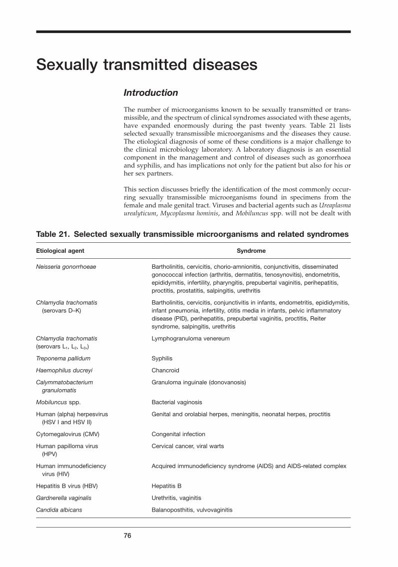
Sexually transmitted diseases
Introduction
The number of microorganisms known to be sexually transmitted or trans-missible, and the spectrum of clinical syndromes associated with these agents,have expanded enormously during the past twenty years. Table 21 listsselected sexually transmissible microorganisms and the diseases they cause.The etiological diagnosis of some of these conditions is a major challenge tothe clinical microbiology laboratory. A laboratory diagnosis is an essentialcomponent in the management and control of diseases such as gonorrhoeaand syphilis, and has implications not only for the patient but also for his orher sex partners.
This section discusses briefly the identification of the most commonly occur-ring sexually transmissible microorganisms found in specimens from thefemale and male genital tract. Viruses and bacterial agents such as Ureaplasmaurealyticum, Mycoplasma hominis, and Mobiluncus spp. will not be dealt with
76
Table 21. Selected sexually transmissible microorganisms and related syndromes
Etiological agent Syndrome
Neisseria gonorrhoeae Bartholinitis, cervicitis, chorio-amnionitis, conjunctivitis, disseminatedgonococcal infection (arthritis, dermatitis, tenosynovitis), endometritis,epididymitis, infertility, pharyngitis, prepubertal vaginitis, perihepatitis,proctitis, prostatitis, salpingitis, urethritis
Chlamydia trachomatis Bartholinitis, cervicitis, conjunctivitis in infants, endometritis, epididymitis,(serovars D–K) infant pneumonia, infertility, otitis media in infants, pelvic inflammatory
disease (PID), perihepatitis, prepubertal vaginitis, proctitis, Reiter syndrome, salpingitis, urethritis
Chlamydia trachomatis Lymphogranuloma venereum(serovars L1, L2, L3,)
Treponema pallidum Syphilis
Haemophilus ducreyi Chancroid
Calymmatobacterium Granuloma inguinale (donovanosis)granulomatis
Mobiluncus spp. Bacterial vaginosis
Human (alpha) herpesvirus Genital and orolabial herpes, meningitis, neonatal herpes, proctitis(HSV I and HSV II)
Cytomegalovirus (CMV) Congenital infection
Human papilloma virus Cervical cancer, viral warts(HPV)
Human immunodeficiency Acquired immunodeficiency syndrome (AIDS) and AIDS-related complexvirus (HIV)
Hepatitis B virus (HBV) Hepatitis B
Gardnerella vaginalis Urethritis, vaginitis
Candida albicans Balanoposthitis, vulvovaginitis
BLM1 1/17/04 2:02 PM Page 76
77
A
here. For more extensive information, the reader is referred to the relevantWHO publication.1
Urethritis in men
Urethritis in men is clinically characterized by a urethral discharge and/ordysuria, but asymptomatic infection with Neisseria gonorrhoeae or Chlamydiatrachomatis occurs frequently. If untreated, gonococcal and chlamydial ure-thritis may progress to epididymitis. Rectal and oropharyngeal infection withN. gonorrhoeae and C. trachomatis may occur in homosexual men.
For the purpose of patient management, urethritis should be divided intogonococcal urethritis and nongonococcal urethritis (NGU). Approximatelyhalf the cases of NGU are caused by C. trachomatis, but the etiology of themajority of the remaining cases has not been fully elucidated. According tosome studies, Ureaplasma urealyticum may be a cause of urethritis, and Tri-chomonas vaginalis can be found in 1–3% of cases of NGU. Intra-urethral infec-tion with human herpesvirus may yield a urethral discharge. Bacterial agentssuch as staphylococci, various Enterobacteriaceae, Acinetobacter spp., andPseudomonas spp. can be isolated from the urethra of healthy men, but havenot been shown to cause urethritis.
The examination of specimens for C. trachomatis is complicated and will notbe discussed in this section. Besides isolation in cell culture systems, non-culture methods for the detection of chlamydial antigens by enzymeimmunoassays, immunofluorescence assays and nucleic acid amplificationtests have recently become available. These methods, while promising, remainprohibitively expensive.
Collection and transport of specimens
For the collection of urethral specimens, a swab with a narrow diameter or asterile bacteriological loop should be inserted 3–4 cm into the urethra andgently rotated before withdrawal. Purulent discharge can be collected directlyon a swab or on the inoculating loop. The composition of both the tip and theshaft of the swab is important. For the culture of N. gonorrhoeae, charcoal-treated cotton tips or calcium alginate or Dacron tips are preferred. If thesespecial, commercially prepared, sampling swabs are not available and regularcotton swabs are used, the specimen should be inoculated immediately. A pro-static massage does not increase the rate of isolation of gonococci or chlamy-diae in cases of urethritis.
Anorectal specimens are obtained by inserting a swab 4–5 cm into the analcanal. For oropharyngeal specimens, the posterior pharynx and the tonsillarcrypts should be swabbed and plated immediately.
Ideally the inoculation of specimens for the isolation of N. gonorrhoeae shouldbe made directly onto the culture medium in the clinic. Inoculated platesshould be placed in a candle jar or into an atmosphere containing 5–10%carbon dioxide, with high humidity. If immediate plating and incubation arenot possible, a transport medium such as Amies or Stuart transport mediumshould be used. The transport time should be as short as possible, and must
1 Van Dyck E, Meheus AZ, Piot P. Laboratory diagnosis of sexually transmitted diseases. Geneva,World Health Organization, 1999.
BACTERIOLOGICAL INVESTIGATIONS
BLM1 1/17/04 2:02 PM Page 77
be less than 12 hours in ambient temperatures up to 30 ∞C. Refrigeration is tobe avoided.
Direct examination and interpretation
Most studies have shown that the presence of four or more polymorphonu-clear (PMN) leukocytes per oil-immersion field is strongly indicative of urethritis in men. This criterion is particularly useful to the clinician who has to decide whether to treat patients with vague urethral complaints.
In most cases of gonorrhoea in the male, the discharge is purulent, and numer-ous polymorphonuclear leukocytes (>10 per oil-immersion field) can be seenin the urethral smear. However, this is not always the case in NGU, whichyields a less severe inflammatory reaction. Smears with more than 4 PMNleukocytes per oil-immersion field, and without intracellular Gram-negativediplococci, are highly suggestive of NGU.
A thinly spread smear, prepared by rolling a swab over a slide, should be heat-fixed and methylene-blue or Gram-stained. The presence of Gram-negativeintracellular diplococci in PMN leukocytes in a urethral smear is strongly sug-gestive of gonorrhoea.
Gram-stained smears of intra-urethral specimens from asymptomatic males,from blind rectal swabs, or from oropharyngeal samples are not recom-mended. However, microscopic examination of purulent material obtainedunder anoscopy has a fairly high diagnostic value.
Culture of Neisseria gonorrhoeae
Inoculated plates with modified Thayer–Martin (MTM)1 agar (or New YorkCity medium (NYC))2 should be incubated at 35 ∞C in a humid atmosphereenriched with carbon dioxide (candle jar), and should be observed daily for two days. Laboratories processing a large number of specimens for N. gonorrhoeae often prefer to use a non-selective chocolate agar enriched withIsoVitaleX, or an equivalent supplement, in addition to the selective MTM,because as many as 3–10% of gonococcal strains in a given area may be sus-ceptible to the concentration of vancomycin used in selective media.
78
SEXUALLY TRANSMITTED DISEASES
1 Modified Thayer–Martin agar is prepared by adding at 50 ∞C a mixture of antimicrobials andIsoVitaleX, or an equivalent supplement, to chocolate agar prepared from GC agar or Columbiaagar as basal medium. Antimicrobial mixtures containing 3 or 4 antimicrobials are commerciallyavailable from several sources; VCN mixture contains vancomycin, colistin, and nystatin; VCNTmixture also contains trimethoprim.The final concentrations of the antimicrobials in the prepared medium are:
— vancomycin: 3mg/ml— colistin: 7.5mg/ml— nystatin: 12.5 IU/ml— trimethoprim lactate: 5mg/ml2 Modified New York City medium is prepared by adding 500ml of sterile GC agar base cooledto 50 ∞C, the following supplements:
— 50ml horse blood lysed by adding 5ml/l saponin,— sterile yeast autolysate,— an antimicrobial mixture containing vancomycin, colistin, amphotericin, and trimethoprim.
The ingredients are commercially available from Oxoid Ltd, Wade Rd, Basingstoke, Hants RG248PW, England.
BLM1 1/17/04 2:02 PM Page 78
79
BACTERIOLOGICAL INVESTIGATIONS
A
Gonococcal colonies may still not be seen after 24 hours. They appear after 48hours as grey to white, opaque, raised, and glistening colonies of differentsizes and morphology.
Identification of Neisseria gonorrhoeae
A presumptive identification of N. gonorrhoeae isolated from urogenital spec-imens is based on a positive oxidase reaction and a Gram-stained smearshowing Gram-negative diplococci. Confirmation of the identification can beobtained by carbohydrate degradation assays or other tests, using methodsand media discussed extensively elsewhere.1
Antimicrobial susceptibility testing
There is considerable geographical variation in the susceptibility of N. gonor-rhoeae strains to benzylpenicillin. In some areas, such as sub-Saharan Africaor South-east Asia, most gonococcal strains are now b-lactamase-producing.Chromosomally mediated resistance to benzylpenicillin not based on b-lactamase production is also becoming more common in many countries.However, the disc-diffusion test is not reliable in detecting such strains.
In areas where benzylpenicillin, ampicillin or amoxicillin is still used for thetreatment of gonococcal infections, N. gonorrhoeae isolates (particularly fromcases of treatment failure) should be routinely screened for b-lactamase pro-duction by one of the recommended tests, such as the Nitrocefin test.2 For theNitrocefin test, a dense suspension from several colonies is prepared in a smalltube with 0.2ml saline; 0.025ml of Nitrocefin is then added to the suspensionand mixed for one minute. A rapid change in the colour, from yellow to pinkor red, indicates that the strain produces b-lactamase.
Antimicrobial susceptibility testing of N. gonorrhoeae by the disc-diffusionassay is not recommended in routine practice.
Genital specimens from women
The vaginal flora of premenopausal women normally consists predominantlyof lactobacilli, and of a wide variety of facultative aerobic and anaerobic bacteria.
Abnormal vaginal discharge may be due to:
— vaginitis: Gardnerella vaginalis, Candida albicans;— bacterial vaginosis: overgrowth of anaerobes and Mobiluncus spp.;— cervicitis: Neisseria gonorrhoeae, Chlamydia trachomatis.
Other bacteria, such as Enterobacteriaceae, are not proven causes of vaginitis.Vaginitis in prepubertal girls may be due to N. gonorrhoeae or C. trachomatis.
1 Van Dyck E, Meheus AZ, Piot P. Laboratory diagnosis of sexually transmitted diseases. Geneva,World Health Organization, 1999.2 The Nitrocefin reagent is obtainable from Oxoid Ltd, Wade Road, Basingstoke, Hants RG248PW, England, and consists of 1mg of Nitrocefin (SR112) and 1 vial of rehydration fluid (SR112A).The tube test can be replaced by a disc test, using Nitrocefin-impregnated paper discs (Cefi-nase discs, available from BD Diagnostic Systems, 7 Loveton Circle, Sparks, MD 21152, USA).
BLM1 1/17/04 2:02 PM Page 79
Bacterial vaginosis (nonspecific vaginitis) is a condition characterized by anexcessive, malodorous, vaginal discharge associated with a significantincrease of Mobiluncus spp. and various obligate anaerobes, and a decrease inthe number of vaginal lactobacilli. A minimum diagnostic requirement forbacterial vaginosis is the presence of at least three of the following signs:abnormal vaginal discharge, vaginal pH > 4.5, clue cells (epithelial cells withso many bacteria attached that the cell border becomes obscured), and a fishy,amine-like odour when a drop of 10% potassium hydroxide is added to thevaginal secretions.
Urethritis in women is also often caused by N. gonorrhoeae and C. trachomatis.
Ascending infections with N. gonorrhoeae, C. trachomatis, vaginal anaerobes,and facultative anaerobic bacteria can cause pelvic inflammatory disease(PID), with infertility or ectopic pregnancy as late sequelae.
Genital infections with bacterial agents, including N. gonorrhoeae and C. trachomatis, during pregnancy may result in complications such as prema-ture delivery, prolonged rupture of membranes, chorio-amnionitis, and post-partum endometritis in the mother, and conjunctivitis, pneumonia, and amniotic infection syndrome in the newborn.
On special request, cervicovaginal specimens may be cultured for bacterialspecies, such as S. aureus (toxic shock syndrome), S. agalactiae (group B strep-tococci, neonatal infection), Listeria monocytogenes (neonatal infection), andClostridium spp. (septic abortion).
Although infections with C. trachomatis and with human herpesvirus are common, and can cause significant morbidity, their laboratory diag-nosis requires expensive equipment and reagents and will not be discussedhere.
Collection and transport of specimens
All specimens should be collected during a pelvic examination using a specu-lum. The speculum may be moistened with warm water before use, but anti-septics or gynaecological exploration cream should not be used, since thesemay be lethal to gonococci.
For examination for yeasts, G. vaginalis, and bacterial vaginosis, samples ofvaginal discharge may be obtained with a swab from the posterior fornix ofthe vagina. Samples for gonococcal and chlamydial culture should be col-lected in the endocervix. After inserting the speculum, cervical mucus shouldbe wiped off with a cotton wool ball. A sampling swab (see page 77) shouldthen be introduced into the cervical canal and rotated for at least 10 secondsbefore withdrawal.
Urethral, anorectal, and oropharyngeal specimens for gonococci may beobtained in a similar manner as from males.
In all cases of pelvic inflammatory disease (PID), as a minimum, the cervixshould be sampled for N. gonorrhoeae. Sampling from the fallopian tubes ismore reliable, but in most areas a cul-de-sac aspirate is the best sample available.
In infants with ophthalmia neonatorum, conjunctival exudate should be col-lected with a swab or a loop.
80
SEXUALLY TRANSMITTED DISEASES
BLM1 1/17/04 2:02 PM Page 80
81
BACTERIOLOGICAL INVESTIGATIONS
A
Amies and Stuart transport media are convenient for transport of cervical andvaginal samples, with the exception of specimens to be tested for C. trachomatis.
Direct examination and interpretation
Direct examination of vaginal secretions is the method of choice for the etio-logical diagnosis of vaginitis, but is much less useful for the diagnosis of cervicitis.
A wet mount is prepared by mixing the vaginal sample with saline on a glassslide, after which a cover slip is added. A diluted preparation is preferred to ensure the separation of the cells, which may otherwise be clumpedtogether. Examine at a magnification of ¥400 for the presence of T. vaginaliswith typical movement, budding yeasts, and clue cells. C. albicans may formpseudomycelia, which may be observed occasionally in vaginal material. Cluecells are found in most women with bacterial vaginosis. A granular or dirtyappearance of the epithelial cell cytoplasm is a less objective criterion than theloss of the cell border. Microscopic examination of a wet mount of cervicalspecimens is not recommended.
Preparation of a Gram-stained smear is the method of choice for the diagno-sis of bacterial vaginosis. The smear should be prepared by gently rolling,rather than smearing, a swab over the glass slide. A normal vaginal smearcontains predominantly lactobacilli (large Gram-positive rods) and fewer than5 leukocytes per field. In typical smears from women with bacterial vaginosis,clue cells covered with small Gram-negative rods are accompanied by a mixedflora consisting of very large numbers of small Gram-negative and Gram-variable rods and coccobacilli, and often Gram-negative curved rods, in the absence of larger Gram-positive rods. Only a few (<5) leukocytes are found per field. This picture is a sensitive and specific diagnostic indicator for bacterial vaginosis.
A large number of white blood cells (>10 cells per field) on the Gram-stainedvaginal smear suggests trichomoniasis or cervicitis.
Gram-staining is not particularly helpful for the diagnosis of gonococcal infection in female patients. The examination of Gram-stained smears of endocervical secretions for intracellular Gram-negative diplococci has a sensitivity of 50–70%, and a specificity of 50–90% for the diagnosis of gono-coccal infection, resulting in a poor predictive value of a positive test in pop-ulations with a low prevalence of gonorrhoea. Gram-negative intracellulardiplococci in cervical smears should be reported as such, and not as N. gonorrhoeae or gonococci. Over-interpretation of cervical smears, which oftencontain Gram-negative coccobacilli and bipolar stained rods, must beavoided.
The main interest in a cervical smear is its validity for the diagnosis of mucopurulent cervicitis: the presence of more than 10 polymorphonu-clear leukocytes per oil-immersion field is a reasonably good indication ofmucopurulent cervicitis, most often due to N. gonorrhoeae and/or C. trachomatis.
The examination of a Gram-stained conjunctival smear is a sensitive and specific technique for the diagnosis of gonococcal conjunctivitis. The pre-sence of intracellular Gram-negative diplococci is diagnostic for gonococcalconjunctivitis.
BLM1 1/17/04 2:02 PM Page 81
Culture
Cervical, rectal, urethral, conjunctival, and cul-de-sac specimens may be cul-tured for N. gonorrhoeae using the methods specified on page 78. Specimensshould be processed as soon as they arrive in the laboratory or, preferably, inthe clinic itself. Unlike in males, culture is essential for the diagnosis of gono-coccal infection in females. The sensitivity of a single culture for the diagno-sis of gonorrhoea in women is 80–90%. The sensitivity is lower for specimenstaken during the peripartum period.
Cultures for G. vaginalis or anaerobes are not recommended for the diagnosisof bacterial vaginosis, since the organisms are recovered from 20–40% ofwomen without vaginal infection. The presence of G. vaginalis in vaginal discharge is in itself not an indication for treatment, and only patients fulfill-ing the diagnostic criteria for bacterial vaginosis should be treated for this condition.
As compared with microscopy, cultures increase the detection of C. albicansby 50–100%. Culture methods are usually more efficient when the number oforganisms is low. However, low numbers of C. albicans can be found in thevagina of 10–30% of women without signs or symptoms of vaginitis, and onlylarge numbers of C. albicans should be considered as evidence of vaginal can-didiasis. Consequently, culture is not recommended. Cultures for G. vaginaliswill mainly detect asymptomatic carriers when performed in addition to a wetmount, and should not be performed.
Specimens from genital ulcers
Genital ulcers are a very common problem in many developing countries.Their etiological diagnosis and management are a challenge to the clinicianas well as to the laboratory. Mixed infections are common. Genital ulcerativelesions can be caused by a variety of sexually transmissible agents:
— human herpesvirus— Treponema pallidum— Haemophilus ducreyi— Calymmatobacterium granulomatis, the agent of granuloma inguinale
(Donovanosis)— Chlamydia trachomatis serovars L1, L2, L3
Genital herpes is the most common cause of genital ulcer disease in mostindustrialized countries, and is a cause of life-threatening complications inimmunodeficient patients and neonates born to women with the infection. Itslaboratory diagnosis will not be discussed here.
Syphilis is still the most serious disease associated with genital lesions, since it can give rise to severe late sequelae and to congenital syphilis. Whileserological tests play an important role in the diagnosis of all stages of syphilis, only the dark-field examination will be discussed here. Techniquesand interpretation of serological tests for syphilis have been extensivelyreviewed elsewhere.1
82
1 Van Dyck E, Meheus AZ, Piot P. Laboratory diagnosis of sexually transmitted diseases. Geneva,World Health Organization, 1999.
SEXUALLY TRANSMITTED DISEASES
BLM1 1/17/04 2:02 PM Page 82
83
BACTERIOLOGICAL INVESTIGATIONS
A
Chancroid (ulcus molle) is the major cause of genital ulceration in many developing areas. The clinical features include painful, purulent ulcer(s)accompanied by painful and occasionally suppurative inguinal buboes. Latesequelae are not known to occur. The clinical differentiation from other genitalulcer diseases is difficult. Chancroid increases the risk of acquiring HIV infection.
Granuloma inguinale is characterized by extensive beefy, red, granulatedgenital ulcers. Bubo formation is rare.
Chlamydial lymphogranuloma is typically associated with inguinal and/orfemoral lymphadenopathy, and, less frequently, with small ulcers which healspontaneously. Its diagnosis is based on serological tests and isolation of C.trachomatis serovars L1, L2, L3.
Collection of specimens
Treponema pallidum: Protective surgical gloves should be worn. Squeeze the ulcer between two fingers and clean the surface of the lesion with saline, using gauze swabs. Crusts should be removed if present. After wipingaway the first drops of blood (if present), collect a sample of serous exudateby touching a completely clean glass slide to the surface of the lesion. Imme-diately place a clean coverslip firmly on the drop of exudate. Alternatively,the specimen may be aspirated from the lesion, or from an enlarged lymph node, using a sterile needle and syringe. The preparation should be examined immediately by a microscopist experienced in dark-fieldmicroscopy.
Haemophilus ducreyi: Specimens should be obtained from the base of the ulcerwith a swab and inoculated directly on to the isolation medium. Material mayalso be aspirated from inguinal buboes, but isolation of H. ducreyi is less suc-cessful than from genital lesions. Transport media for H. ducreyi have not beenevaluated.
If granuloma inguinale is suspected, ideally a biopsy of subsurface tissue froman area of active granulation should be made. Fresh smears should be madefrom a crushed piece of biopsy material. Alternatively, one may make a smearby scraping off the surface of the lesion.
Direct examination
Demonstration of treponemas in lesion material is the method of choice for the diagnosis of primary syphilis. Although T. pallidum can be stained (for instance with silver nitrate), dark-field microscopy is recommendedbecause it is more sensitive and specific. A microscope equipped with a good light source and a dark-field condenser must be available for dark-fieldexamination. Dark-field condensers block out the direct light rays, allowingonly the peripheral rays (deflected by objects such as treponemes) to passthrough.
Place a few drops of immersion oil on the condenser of a dark-field micro-scope. Lower the condenser slightly so that the oil is below the level of thestage. Place the slide on the microscope and raise the condenser until there isgood contact between the oil and the underside of the slide. Carefully avoidtrapping air bubbles in the oil.
BLM1 1/17/04 2:02 PM Page 83
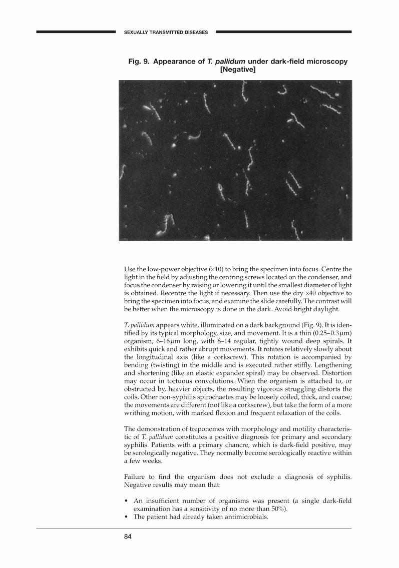
Use the low-power objective (¥10) to bring the specimen into focus. Centre thelight in the field by adjusting the centring screws located on the condenser, andfocus the condenser by raising or lowering it until the smallest diameter of lightis obtained. Recentre the light if necessary. Then use the dry ¥40 objective tobring the specimen into focus, and examine the slide carefully. The contrast willbe better when the microscopy is done in the dark. Avoid bright daylight.
T. pallidum appears white, illuminated on a dark background (Fig. 9). It is iden-tified by its typical morphology, size, and movement. It is a thin (0.25–0.3 mm)organism, 6–16 mm long, with 8–14 regular, tightly wound deep spirals. Itexhibits quick and rather abrupt movements. It rotates relatively slowly aboutthe longitudinal axis (like a corkscrew). This rotation is accompanied bybending (twisting) in the middle and is executed rather stiffly. Lengtheningand shortening (like an elastic expander spiral) may be observed. Distortionmay occur in tortuous convolutions. When the organism is attached to, orobstructed by, heavier objects, the resulting vigorous struggling distorts thecoils. Other non-syphilis spirochaetes may be loosely coiled, thick, and coarse;the movements are different (not like a corkscrew), but take the form of a morewrithing motion, with marked flexion and frequent relaxation of the coils.
The demonstration of treponemes with morphology and motility characteris-tic of T. pallidum constitutes a positive diagnosis for primary and secondarysyphilis. Patients with a primary chancre, which is dark-field positive, maybe serologically negative. They normally become serologically reactive withina few weeks.
Failure to find the organism does not exclude a diagnosis of syphilis. Negative results may mean that:
• An insufficient number of organisms was present (a single dark-fieldexamination has a sensitivity of no more than 50%).
• The patient had already taken antimicrobials.
84
Fig. 9. Appearance of T. pallidum under dark-field microscopy[Negative]
SEXUALLY TRANSMITTED DISEASES
BLM1 1/17/04 2:02 PM Page 84
85
BACTERIOLOGICAL INVESTIGATIONS
A
• The lesion was approaching natural resolution.• The lesion was not syphilitic.
Whatever the result of the dark-field examination, a blood sample shouldalways be taken for serological tests.
In the diagnosis of granuloma inguinale, when acetone-fixed smears arestained with Giemsa, typical, intracellular, encapsulated bacilli can be seen within histiocytes. The diagnosis of this disease is described in full elsewhere.1
For the diagnosis of chancroid, a Gram-stained smear is not recommended,since the sensitivity and specificity are both less than 50%. For the diagnosisof chlamydial lymphogranuloma, Giemsa-stained smears are not recom-mended. However, material from the ulcer should also be examined by dark-field microscopy to search for T. pallidum.
Culture
Gonococci are occasionally isolated from genital ulcers, but their significancein such specimens is unclear. Apart from H. ducreyi, no other bacterialspecies—neither facultative aerobes nor obligate anaerobes—have beenshown to cause genital ulcer disease.
Specimens to be examined for H. ducreyi should be inoculated directly on toa selective, enriched agar plate.2 The medium used should not be older thanone week. The plates should be incubated at 33–35 ∞C in a candle jar with amoistened towel at the bottom. After 48–72 hours of incubation, small, non-mucoid, yellow-grey, semi-opaque or translucent colonies appear that can bepushed intact across the agar surface. The sensitivity of a single culture forthe isolation of H. ducreyi is 70–80%.
A presumptive diagnosis of H. ducreyi can be made on the basis of the char-acteristic morphology of colonies on selective media, and the demonstrationof small Gram-negative, pleomorphic coccobacilli, often in single chains(streptobacilli), parallel chains (“school of fish”), or clumps in the suspectcolonies. Although H. ducreyi is haemin-dependent, most clinical isolates fail to grow on the media used for the determination of X and V factor requirement. Nearly all recent isolates from developing countries produce b-lactamase.
1 Van Dyck E, Meheus AZ, Piot P. Laboratory diagnosis of sexually transmitted diseases. Geneva,World Health Organization, 1999.2 Mueller–Hinton agar base, supplemented with 5% sterile horse blood heated to 75 ∞C, 1% IsoVitaleX, and 3g/ml vancomycin.
BLM1 1/17/04 2:02 PM Page 85
Purulent exudates, wounds, and abscesses
Introduction
One of the most commonly observed infectious disease processes is the production of a purulent (sometimes seropurulent) exudate as the result ofbacterial invasion of a cavity, tissue, or organ of the body. Such infections may be relatively simple, innocuous “pimples” or a series of multiple pocketsof pus found in abscesses in one or more anatomical sites. The exudate consists of white blood cells, predominantly polymorphonuclear leukocytes,the invading organisms, and a mixture of body fluid and fibrin. In someinstances, the exudate may be found as a coating on the surface of an organ,e.g. the surface of the brain in acute bacterial meningitis. In other cases, theexudate may be walled off by layers of fibrin and a network of tissue cells,e.g. a carbuncle or subcutaneous “boil”, while in still other cases the exudatemay be associated with an open wound, which therefore drains thick fluid orpus.
Just as the anatomical site of exudate production can vary considerably, so too can the organisms involved in the underlying infection. All bacteria thatare part of the normal flora, or that gain access to the body, may be involvedin the production of an exudate. Some fungi, particularly those that are ableto multiply in body tissues, can also be involved in the production of an exudate. In contrast, a purulent exudate is rarely produced in a viral infection.
The bench microbiologist should be aware of the diversity of anatomical sites and microorganisms involved, and be prepared to make the appro-priate macroscopic and microscopic examinations and the proper pri-mary media inoculations to recover the major organism(s) involved. Once the organisms have been isolated in pure culture, the identification process and antimicrobial susceptibility tests should be set up as soon as possible.
Communication between the clinician and the microbiologist is particularlyimportant in the diagnosis and management of patients with suppurativeinfectious diseases. The microbiologist must collaborate with the physician toensure proper specimen collection and the expeditious delivery of the speci-men to the laboratory for prompt and proper processing.
Commonly encountered clinical conditions andthe most frequent etiological agents
Surgical specimens
Surgical specimens may be obtained by aspiration of a localized abscess orother surgical procedures. The surgeon should be advised to obtain severalsmall representative tissue samples and any purulent exudate. If possible,cotton swabs should be avoided. The exudate should be collected using aneedle and syringe. If cotton swabs must be used, as much exudate as possi-ble should be collected and dispensed into appropriate containers for dispatchto the laboratory. Upon receipt, the laboratory should review the information
86
BLM1 1/17/04 2:02 PM Page 86
87
A
provided and then plan cultures for the organisms likely to be found in theparticular specimen.
A few examples of conditions and organisms found in different types of surgical specimens are given below:
• The peritoneal cavity is likely to contain Gram-negative enteric bacteria(Enterococci), Gram-negative anaerobic rods (Bacteroides fragilis), andclostridia.
• A walled-off abscess may contain any type of organism, either single ormultiple species: Gram-positive cocci and Gram-negative bacilli are mostfrequently isolated. Anaerobic bacteria and amoebae may also need to beconsidered, depending on the abscess site.
• Lymph nodes are frequently involved in systemic infections. They becomeswollen and often quite tender, and purulent exudate frequently accumu-lates. If the node is fluctuant, the liquid contents can be aspirated by aphysician. Lymph node biopsies or aspirates from children should be cul-tured for Mycobacterium tuberculosis and other mycobacteria. In addition tobeing cultured for staphylococci, streptococci, and Gram-negative entericbacteria, lymph nodes may be good specimens for the diagnosis of sys-temic and subcutaneous mycoses (histoplasmosis, sporotrichosis).
• Skin and subcutaneous tissue are prime targets for both abscesses andwound infections. As a general rule, subcutaneous abscesses are caused bystaphylococci. Open, weeping skin lesions often involve b-haemolyticstreptococci and/or staphylococci, as in impetigo. Another variety of skinlesion requiring some surgical intervention, and often seen as a hospital-acquired infection, is decubitus ulcer or bed sore. The bacteria are fre-quently skin commensals or intestinal flora that have proliferated in themost external part of the ulcer and create an unpleasant odour and appear-ance. Routine culture of those organisms is not clinically relevant. Theorganisms most commonly isolated from biopsy tissue are enteric bacilli;the same organisms may be found in cultures of the superficial exudate. Itis not always possible to evaluate the role of these organisms in decubitusulcers, but healing requires that the ulcer be kept clean, dry, and free ofbacteria. Occasionally organisms in a decubitus ulcer may find their wayinto the blood stream, producing serious complications.
• Burns, especially second- and third-degree burns, are prone to infec-tion with a variety of bacterial species. It is very important that careful surgical débridement be carried out prior to obtaining material for culture. Staphylococci and Pseudomonas aeruginosa are most commonlyencountered.
• Exudates. Sometimes a serous or purulent fluid will collect in a cavity thatnormally possesses a very small amount of sterile fluid, e.g. the pericar-dial sac, pleural cavity, bursa, or joint. Needle aspiration under aseptic con-ditions will yield a laboratory specimen from which the infecting organismmay be isolated and identified. Bacteria are usually the cause, but fungi orviruses may also be responsible. The infections are usually monospecific,but mixed aerobic and anaerobic infections do occur. Aspirates from thepleural cavity may yield pneumococci, streptococci, H. influenzae, anaero-bic streptococci, or anaerobic Gram-negative rods (Prevotella and Porphy-romonas) or M. tuberculosis.
Penetrating wounds
Any lesion caused by a penetrating object that breaks the skin is likely tocontain a mixture of microorganisms; these organisms are generally part ofthe skin flora or of the normal microbial flora of soil and water. A penetrating
BACTERIOLOGICAL INVESTIGATIONS
BLM1 1/17/04 2:02 PM Page 87
wound involving damage to the intestines will lead to an even greater threatbecause the intestinal flora may contribute to infection of the wound and peritoneal cavity.
Penetrating or cutting wounds may be caused by sharp or blunt objects. Metal, glass, wood, etc. are frequently responsible for penetrating wounds,whether caused by accident or deliberately (e.g. stab or gunshot wounds).Tetanus resulting from a penetrating wound is a life-threatening disease in a non-immunized individual. Similarly, wound botulism may go undiag-nosed if the physician and the microbiologist are not aware of this possibil-ity. The diagnoses of tetanus and botulism are best made clinically, andlaboratory support should be provided by a central reference laboratory.People working with animals or their products are at risk of infection with spores of Bacillus anthracis, which may gain access through small woundsor skin abrasions and produce the typical black eschar of anthrax. Other soil organisms, such as Clostridium perfringens, may be involved in deep penetrating wounds and give rise to gas gangrene.
Animal bites or scratches occur frequently in both urban and rural areas. The bite may be from a domestic pet, a farm animal, or a wild animal. Rabiesmust be the prime and immediate concern. Once the possibility of rabies hasbeen eliminated, the other possible etiological agents are many and varied.Rabies diagnosis is too specialized to be discussed in this manual.1
The mouths of all animals contain a heterogeneous flora consisting of aerobic and anaerobic bacteria, yeasts, protozoa, and viruses. Infections result-ing from bites or scratches are predominantly caused by bacteria. A primeexample is infection by Pasteurella multocida, which often follows a dog or catbite if the bite has not been properly cleansed and treated. Human bites may sometimes result in a serious mixed infection of aerobic and anaerobicbacteria.
Nosocomial wound infections
One of the main concerns in the care and treatment of hospitalized patientsis that they should not be harmed in the course of the diagnosis and treat-ment of their illness. Unfortunately, 5–10% of hospitalized patients do acquirean infection while in hospital. Nosocomial infections are costly and canusually be avoided or greatly reduced. Many hospital-acquired infections are known to occur in surgical departments. The rate of postoperative woundinfection varies from hospital to hospital, and within a given hospital is likelyto be highest in patients who have undergone abdominal, thoracic, ororthopaedic surgery. Surgical wound infections may occur shortly aftersurgery or several days postoperatively. The site of infection may be limitedto the suture line or may become extensive in the operative site. Staphylococ-cus aureus (usually benzylpenicillin-resistant, and now often meticillin-resistant) is the biggest offender, followed closely by E. coli and other entericbacteria. Anaerobic bacteria from the patient’s large bowel may gain access tothe operative site, making a mixed infection a serious and fairly frequentoccurrence in hospitals in which the postoperative wound care and infection prevention programmes are weak. Bacteroides fragilis and, occasionally,Clostridium perfringens may invade the bloodstream, resulting in a systemicand frequently fatal postoperative infection.
88
PURULENT EXUDATES, WOUNDS, AND ABSCESSES
1 Meslin F-X, Kaplan MM, Koprowski H, eds. Laboratory techniques in rabies, 4th ed. Geneva,World Health Organization, 1996.
BLM1 1/17/04 2:02 PM Page 88
89
BACTERIOLOGICAL INVESTIGATIONS
A
An infrequent but challenging infection may follow dental or oral surgery,when a sinus tract from the inside works its way to the skin surface on the face or neck and the discharge contains the “sulfur” granules of actinomycosis.
Collection and transportation of specimens
It is not possible, here, to describe in detail the procedures for specimen col-lection from each type of wound, abscess, etc. It should be obvious that thisis a task that requires close cooperation between the laboratory and the physi-cian. In many instances, there is only one opportunity to obtain a specimen;second specimens are unavailable in many cases. Therefore, proper collection,transport, and storage of these specimens are of the greatest importance, andcompromises should be avoided. Once a specimen has been obtained, pack-aged, and dispatched to the laboratory, it should be processed as soon as possible. After the preliminary examinations have been completed and thecultures made, the rest of the specimen should be properly labelled, stop-pered, and refrigerated, until it is certain that no additional laboratory testsare needed.
Abscess
Once an abscess, or multiple abscesses, have been found, the physician orsurgeon and the microbiologist should consult on what is to be done. The technique for collecting pus and pieces of the abscess wall is a surgical pro-cedure. A syringe and needle are used to aspirate as much as possible of thepurulent material, which is then aseptically transferred to sterile specimencontainers. If such containers are not available, the specimen should be keptin the syringe with the needle capped, and the syringe itself should be trans-ported to the laboratory. This material should be processed immediately bythe laboratory; both aerobic and anaerobic cultures can be made from a singlespecimen.
A similar situation exists when the surgeon encounters one or more walled-off abscesses in an organ, or in the thorax, abdomen, or pelvis, during thecourse of a surgical procedure for another purpose. In anticipation of this, the laboratory should arrange to have stored, in the sterile surgical supplies,a kit for obtaining the contents of such abscesses so that specimens can bedelivered promptly to the laboratory for processing. Every effort should bemade to avoid the use of swabs for collecting a small amount of specimen,when a large amount is actually present. A swab can justifiably be used tocollect very small amounts of pus, or pus from sites that require care, e.g. fromthe eye. When pieces of tissue are obtained from the abscess wall, the labora-tory technician should either grind the tissue, using a small amount of sterilebroth as a diluent, or mince the tissue into very small pieces using sterile scissors. Aerobic and anaerobic cultures should be prepared as indicated onpages 92–93.
Infected lacerations, penetrating wounds,postoperative wounds, burns, and decubitus ulcers
No standard procedure for specimen collection can be formulated. However,certain fundamental guidelines should be followed to obtain the best possible specimen for laboratory analysis. After carefully cleaning the site, thesurgeon should look beneath the surface for collections of pus, devitalized
BLM1 1/17/04 2:02 PM Page 89
tissue, the oozing of gas (crepitation), or any other abnormal sign. Segmentsof the tissue involved that are to be used for cultures should be removed andplaced on sterile gauze for processing. Pus or other exudate should be care-fully collected and placed in a sterile tube. Swabs may be used if necessary.
Sinus tract or lymph node drainage
When a sinus tract or lymph node shows evidence of spontaneous drainage,the drainage material should be collected carefully, using a sterile Pasteurpipette fitted with a rubber bulb, and placed in a sterile tube. If discharge isnot evident, the surgeon should obtain the purulent material using a sterilesyringe and needle or probe. Again, swabs should only be used if a sterilePasteur pipette is not available.
Exudates
The abnormal accumulation of fluid within a body cavity such as the pleuralspace, a joint, or the peritoneal space, requires a surgical procedure to aspi-rate the accumulated material into a sterile container for subsequent deliveryto the laboratory for microbiology and cytology. In those cases where the accu-mulation persists and an open drain is put in place, it is necessary to collectthe drainage fluid in an aseptic manner for subsequent culture and other tests.
Macroscopic evaluation
Specimens of pus or wound discharge collected on swabs are difficult to eval-uate macroscopically, particularly when the swab is immersed in a transportmedium. Specimens of pus, received in a syringe or in a sterile container,should be evaluated carefully by an experienced technician for colour, con-sistency, and odour.
Colour
The colour of pus varies from green-yellow to brown-red. A red colour is gen-erally due to admixture with blood or haemoglobin. The aspirate from aprimary amoebic liver abscess has a gelatinous consistency and a dark brownto yellowish brown colour. Pus from postoperative or traumatic wounds(burns) may be stained blue-green by the pyocyanin pigment produced byPseudomonas aeruginosa.
Consistency
The consistency of pus may vary from a turbid liquid to one that is very thickand sticky. Exudates, aspirated from a joint, the pleural cavity, the pericardialsac, or the peritoneal cavity, are generally liquid, with all possible gradationsbetween a serous exudate and frank pus.
Pus originating from a draining sinus tract in the neck should be inspectedfor small yellow “sulfur” granules, which are colonies of the filamentousActinomyces israelii. The presence of sulfur granules suggests a diagnosis ofcervicofacial actinomycosis. Small granules of different colours (white, black,red, or brown) are typical of mycetoma, a granulomatous tumour, generallyinvolving the lower extremities (e.g. madura foot), and characterized by mul-
90
PURULENT EXUDATES, WOUNDS, AND ABSCESSES
BLM1 1/17/04 2:02 PM Page 90
91
BACTERIOLOGICAL INVESTIGATIONS
A
tiple abscesses and draining sinuses. The coloured granules correspond toeither filamentous bacteria or fungal mycelium.
Pus from tuberculous “cold abscesses” (with few signs of inflammation) is sometimes compared with soft cheese and called “caseum” or “caseouspus”.
Odour
A foul feculent odour is one of the most characteristic features of an anaero-bic or a mixed aerobic–anaerobic infection, although it may be lacking in someinstances. The odour, together with the result of the Gram-stained smear,should be reported at once to the clinician as it may be helpful in the empir-ical selection of an appropriate antimicrobial. It will also help in determiningwhether anaerobic cultures are needed.
Microscopic examination
A smear for Gram-staining and examination should be made for every spec-imen. In particular cases, or at the request of the clinician, a direct wet mountmay be prepared and Ziehl–Neelsen staining carried out.
Gram-stained smear
Using a bacteriological loop, make an even smear of the most purulent partof the specimen on a clean slide. If only a swab is available, the slide shouldfirst be sterilized by being passed through the flame of a Bunsen burner andallowed to cool. The cotton swab should then be gently rolled over the glasssurface, without rubbing or excessive pressure. Allow the slide to air-dry, pro-tected from insects, or place it in the incubator. Fix by heat, stain and examinethe smear under the oil-immersion objective (¥100). Inspect carefully and notethe presence and the quantity (use +signs) of:
— polymorphonuclear granulocytes (pus cells);— Gram-positive cocci arranged in clusters, suggestive of staphylococci;— Gram-positive cocci in chains, suggestive of streptococci or enterococci;— Gram-negative rods resembling coliform (Escherichia coli, Klebsiella, etc.),
other Enterobacteriaceae (Proteus, Serratia, etc.), nonfermentative rods(Pseudomonas spp.), or obligate anaerobes (Bacteroides spp.);
— large straight Gram-positive rods with square ends suggestive of Clostrid-ium perfringens, the principal agent of gas gangrene, or Bacillus anthracis,the agent of anthrax;
— an extremely heavy and pleomorphic mixture of bacteria, including strep-tococci, Gram-positive and Gram-negative rods of various sizes, includingfusiform rods; such a picture is suggestive of “mixed anaerobic flora” andshould be reported as such;
— Candida or other yeast cells, which are seen as ovoid Gram-positivebudding spheres, often forming branched pseudomycelia.
Sulfur granules from actinomycosis or granules from a mycetoma should becrushed on a slide, Gram-stained and inspected for thin branched and frag-mented Gram-positive filaments.
BLM1 1/17/04 2:02 PM Page 91
Direct microscopy
When requested, or when a fungal or parasitic infection is suspected, a wetpreparation should be examined. If the pus is thick, a loopful should be mixedin a drop of saline. When looking for fungi, a drop of 10% potassium hydrox-ide should be used to clear the specimen. Apply a coverslip and, using the¥10 and ¥40 objectives, look especially for:
— actively motile amoebae in aspirate from a liver abscess;— yeast cells of Histoplasma capsulatum (including the African var. duboisii,)
Blastomyces dermatitidis (in endemic areas), Candida spp.;— fungal hyphae and bacterial filaments in crushed granules from
mycetoma;— parasites, such as microfilariae, scolices or hooks of Echinococcus, eggs of
Schistosoma, Fasciola, or Paragonimus.
Acid-fast staining (Ziehl–Neelsen)
Ziehl–Neelsen staining should be performed when requested by the clinician.It is also advisable to make an acid-fast stained preparation when the pusshows no bacteria or when only faintly stained Gram-positive “coryneform”rods are seen on the Gram-stained smear. Tubercle bacilli should be suspected,in particular, in pus or purulent exudate from the pleura, joints, boneabscesses, or lymph nodes. Non-tuberculous (so-called “atypical”) acid-fastbacilli are sometimes found in gluteal abscesses at the site of deep intramus-cular injections. Such abscesses are often caused by rapidly growing mycobac-teria belonging to the Mycobacterium fortuitum–chelonei group. In the tropics,discharge scraped from the base of a necrotic skin ulcer situated on a leg oran arm may be due to slow-growing acid-fast rods called M. ulcerans (Buruliulcer). M. marinum is another tuberculous acid-fast rod, which may be foundin chronic, ulcerative, nodular lesions on the hands, arms, and other exposedskin surfaces in swimmers and fishermen.
Culture
If bacteria or fungi are seen on microscopic examination, appropriate culturemedia should be inoculated. Independently from the results of microscopy,all specimens of pus or exudate should preferably be inoculated onto aminimum of three culture media:
— a blood agar plate for the isolation of staphylococci and streptococci;— a MacConkey agar plate for the isolation of Gram-negative rods; and— a tube of broth that can serve as enrichment medium for both aerobes and
anaerobes, e.g. thioglycollate broth or cooked meat medium.
The size of the inoculum should be determined according to the result of themicroscopic examination, and may vary from one loopful to a few drops. Ifmassive numbers of organisms are seen on the Gram-stained smear, the spec-imen may even have to be diluted in a small amount of sterile broth beforeplating out. If a swab is used for the inoculation, it should be applied to asmall area of the plate and the rest of the surface streaked out with a loop. Ifthe swab is dry, it should first be moistened in a small quantity of sterile brothor saline. In all cases, the technique of inoculation should provide singlecolonies for identification and susceptibility tests.
92
PURULENT EXUDATES, WOUNDS, AND ABSCESSES
BLM1 1/17/04 2:02 PM Page 92
93
BACTERIOLOGICAL INVESTIGATIONS
A
Prior to inoculation, the blood agar plate should be dried for 20 minutes inan incubator to minimize the risk of overgrowth by spreading Proteus spp.The inoculated plate should be incubated at 35 ∞C in a candle jar. Routinely,all media should be incubated for two days and inspected daily for growth.If culture for fastidious organisms is requested, longer incubation (1–2 weeksor more) will be necessary. If growth appears in the broth, it should be Gram-stained and subcultured onto appropriate culture media. Additional culturemedia should be used if specially requested, or if indicated by the results ofthe microscopic examination, as in the following examples:
• If staphylococci have been seen, an additional mannitol salt agar is helpfulin obtaining pure growth and in making a preliminary distinction betweenS. aureus and other cocci.
• If streptococci have been observed, their identification may be hastened byplacing a differential bacitracin disc on the initial streaking area.
• If yeasts or fungi have been observed, the specimen should also be inocu-lated onto two tubes of Sabouraud dextrose agar, one to be incubated at35 ∞C, the other at room temperature, both to be observed for up to onemonth. (Blood agar is sufficient for the isolation of Candida spp.)
• If acid-fast rods have been seen in the Ziehl–Neelsen stained smear, up to3 tubes of Löwenstein–Jensen medium should be inoculated. If the speci-men also contains non-acid-fast bacteria, it should first be decontaminated.Rapidly growing mycobacteria, such as M. fortuitum, may be killed by thedecontamination process; they produce growth within 3–7 days on bloodagar and MacConkey agar. Branched, filamentous, partially acid-fast rodsin pus from the pleura or from a brain abscess will probably be Nocardiaasteroides, which grows on blood agar within a few days.
• Pus from patients with arthritis, pleuritis, osteitis, or cellulitis, particularlyfrom children under 5 years of age, should also be inoculated onto a choco-late agar plate for the recovery of H. influenzae.
• Culture in a strictly anaerobic atmosphere is essential when the Gram-stained smear shows mixed anaerobic flora and also when the specimenproduces a typical foul odour. Anaerobic blood agar is also necessary forthe growth of Actinomyces. Anaerobic culture will be requested by the clinician when he or she suspects clostridial gas gangrene. Methods foranaerobic bacteriology are described on pages 98–102.
Identification
With the exception of contaminants from the environment or from the skin(such as Staphylococcus epidermidis), all organisms isolated from wounds, pus,or exudates should be considered significant and efforts made to identifythem. Full identification, however, is not always necessary, particularly in thecase of mixed flora.
Bacteria and fungi isolated from pus and exudates may belong to almost anygroup or species. Identification criteria are given here only for staphylococcicommonly associated with pus (pyogenic), and for two other pathogens, Pas-teurella multocida and Bacillus anthracis, which are rarely isolated from woundsor skin infections, but are very important for the management of the patient.A standard textbook of clinical microbiology should be consulted for a fulldescription of identification methods. In every case, the first step should beto examine well-separated colonies carefully, pick a single colony of each type,prepare a Gram-stained smear, and then characterize the organisms under themicroscope.
BLM1 1/17/04 2:02 PM Page 93
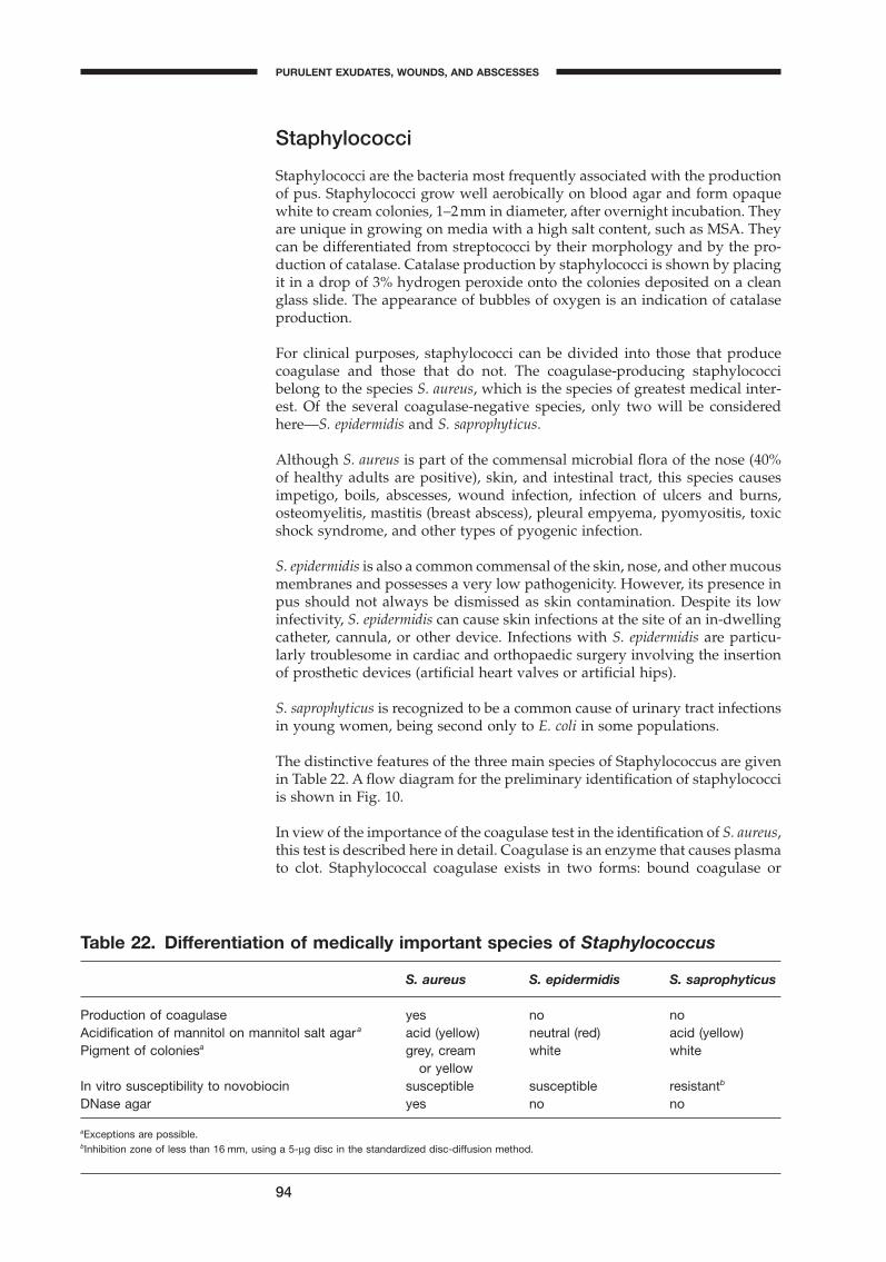
Staphylococci
Staphylococci are the bacteria most frequently associated with the productionof pus. Staphylococci grow well aerobically on blood agar and form opaquewhite to cream colonies, 1–2mm in diameter, after overnight incubation. Theyare unique in growing on media with a high salt content, such as MSA. Theycan be differentiated from streptococci by their morphology and by the pro-duction of catalase. Catalase production by staphylococci is shown by placingit in a drop of 3% hydrogen peroxide onto the colonies deposited on a cleanglass slide. The appearance of bubbles of oxygen is an indication of catalaseproduction.
For clinical purposes, staphylococci can be divided into those that producecoagulase and those that do not. The coagulase-producing staphylococcibelong to the species S. aureus, which is the species of greatest medical inter-est. Of the several coagulase-negative species, only two will be consideredhere—S. epidermidis and S. saprophyticus.
Although S. aureus is part of the commensal microbial flora of the nose (40%of healthy adults are positive), skin, and intestinal tract, this species causesimpetigo, boils, abscesses, wound infection, infection of ulcers and burns,osteomyelitis, mastitis (breast abscess), pleural empyema, pyomyositis, toxicshock syndrome, and other types of pyogenic infection.
S. epidermidis is also a common commensal of the skin, nose, and other mucousmembranes and possesses a very low pathogenicity. However, its presence inpus should not always be dismissed as skin contamination. Despite its lowinfectivity, S. epidermidis can cause skin infections at the site of an in-dwellingcatheter, cannula, or other device. Infections with S. epidermidis are particu-larly troublesome in cardiac and orthopaedic surgery involving the insertionof prosthetic devices (artificial heart valves or artificial hips).
S. saprophyticus is recognized to be a common cause of urinary tract infectionsin young women, being second only to E. coli in some populations.
The distinctive features of the three main species of Staphylococcus are givenin Table 22. A flow diagram for the preliminary identification of staphylococciis shown in Fig. 10.
In view of the importance of the coagulase test in the identification of S. aureus,this test is described here in detail. Coagulase is an enzyme that causes plasmato clot. Staphylococcal coagulase exists in two forms: bound coagulase or
94
Table 22. Differentiation of medically important species of Staphylococcus
S. aureus S. epidermidis S. saprophyticus
Production of coagulase yes no noAcidification of mannitol on mannitol salt agar a acid (yellow) neutral (red) acid (yellow)Pigment of coloniesa grey, cream white white
or yellowIn vitro susceptibility to novobiocin susceptible susceptible resistantb
DNase agar yes no no
aExceptions are possible.bInhibition zone of less than 16 mm, using a 5-mg disc in the standardized disc-diffusion method.
PURULENT EXUDATES, WOUNDS, AND ABSCESSES
BLM1 1/17/04 2:02 PM Page 94
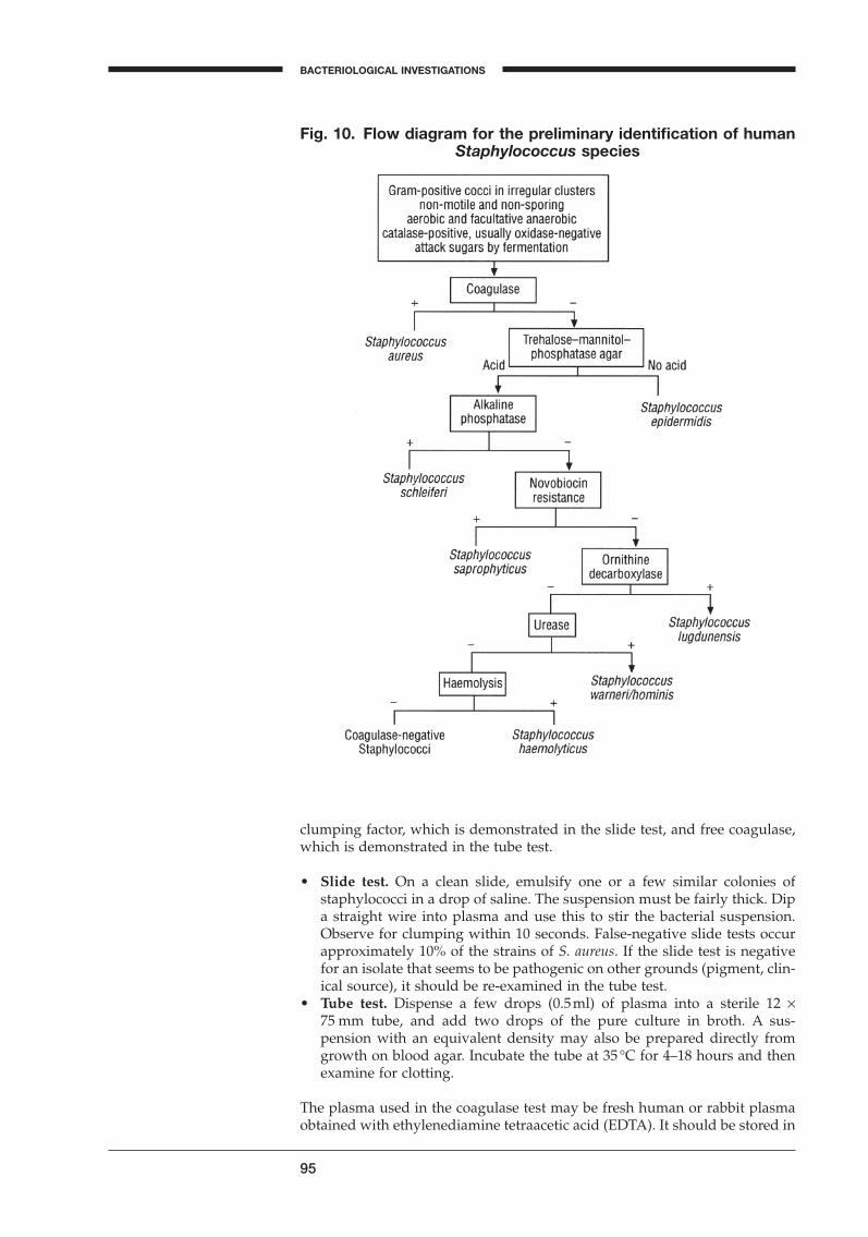
95
BACTERIOLOGICAL INVESTIGATIONS
A
Fig. 10. Flow diagram for the preliminary identification of humanStaphylococcus species
clumping factor, which is demonstrated in the slide test, and free coagulase,which is demonstrated in the tube test.
• Slide test. On a clean slide, emulsify one or a few similar colonies ofstaphylococci in a drop of saline. The suspension must be fairly thick. Dipa straight wire into plasma and use this to stir the bacterial suspension.Observe for clumping within 10 seconds. False-negative slide tests occurapproximately 10% of the strains of S. aureus. If the slide test is negativefor an isolate that seems to be pathogenic on other grounds (pigment, clin-ical source), it should be re-examined in the tube test.
• Tube test. Dispense a few drops (0.5ml) of plasma into a sterile 12 ¥75 mm tube, and add two drops of the pure culture in broth. A sus-pension with an equivalent density may also be prepared directly fromgrowth on blood agar. Incubate the tube at 35 ∞C for 4–18 hours and thenexamine for clotting.
The plasma used in the coagulase test may be fresh human or rabbit plasmaobtained with ethylenediamine tetraacetic acid (EDTA). It should be stored in
BLM1 1/17/04 2:02 PM Page 95
the refrigerator in small amounts (1ml), and its performance checked withcultures of S. aureus and S. epidermidis, run in parallel.
Pasteurella multocida
A number of Gram-negative bacilli are transmitted by animal bites and cancause severe infections in human beings, the most prevalent being Pasteurellamultocida. P. multocida is a commensal found in the normal mouth flora ofmany animals. A bite wound infected with P. multocida may give rise to exten-sive cellulitis, which may extend to a joint, producing arthritis. Osteomyelitis,bacteraemia, and even meningitis have been described.
P. multocida should be looked for specifically in wound discharge if an animalbite is known or suspected of being the cause of the wound. P. multocida is avery small, Gram-negative, nonmotile coccobacillus. It grows well on bloodagar at 35 ∞C but is completely inhibited by the bile salts contained in theenteric selective media, e.g. MacConkey agar. After overnight incubation, thecolonies on blood agar are small, nonhaemolytic, translucent, and mucoid(due to the presence of a capsule in the virulent form).
Biochemical identification is based on the following characteristics:
— fermentation of glucose without gas: P. multocida grows on Kligler ironagar with acidification of the butt;
— oxidase test is weakly positive;— catalase-positive;— nitrate reduced to nitrite (0.1% potassium nitrate in nutrient broth);— urease-negative;— indole-positive—test in tryptic soy broth (TSB) or on MIU after 48 hours
incubation;— highly sensitive to benzylpenicillin in the disc-susceptibility test.
Bacillus anthracis
The genus Bacillus is composed of numerous species of aerobic, spore-forming, Gram-positive rods, widely distributed in the soil. The species B.anthracis is of public health importance in skin infections. The other species,when isolated from wounds or pus, are generally contaminants or, at most,opportunists.
B. anthracis is a major pathogen of cattle, sheep, goats, and other domesticanimals. It also affects wild animals. Anthrax can affect human beings, par-ticularly people in parts of Africa and Asia working or living in close contactwith livestock. Human infection may also derive from animal products con-taining anthrax spores, such as wool, skins, fur, and bones.
The commonest form of human infection is cutaneous anthrax, which mayprogress to septicaemia and meningitis. The spore enters through damagedskin and produces a vesicular lesion with a necrotic centre, surrounded by extensive oedema (“malignant pustule”). Large Gram-positive, square-ended, capsulated rods without spores are seen in smears from the vesicularfluid.
B. anthracis grows aerobically. On blood agar it produces large, flat, greyishcolonies, up to 5 mm in diameter, with a rough surface texture and irregularedges showing hairy outgrowths (Medusa head). It is important at this stage
96
PURULENT EXUDATES, WOUNDS, AND ABSCESSES
BLM1 1/17/04 2:02 PM Page 96
to differentiate the highly pathogenic B. anthracis from the generally harmlesssaprophytic species.
The preliminary distinction should be based on the absence of haemolysis,benzylpenicillin sensitivity, and the lack of motility in B. anthracis. In contrast,most of the saprophytic Bacillus species are motile and strongly haemolytic.These three features can form the basis of a presumptive identification. Fordefinitive diagnosis, a pure culture of the isolate should be sent immediatelyto the central veterinary or public health laboratory.
B. anthracis is a highly infectious organism and specimens and cultures shouldbe handled with the greatest care to avoid contamination of the environmentand infection of laboratory personnel.
Susceptibility testing
Antimicrobials may not always be needed for the management of patientswith wounds, abscesses, or exudates. Proper surgical incision, drainage anddébridement are generally more important than antimicrobial drugs. Theresults of susceptibility tests should be made available, however, within 48hours after receiving the specimen.
Routine susceptibility tests should not be performed on bacteria that have aknown sensitivity pattern, such as streptococci, Pasteurella, and Actinomyces,which are almost without exception susceptible to benzylpenicillins.
For Enterobacteriaceae, non-fermentative Gram-negative rods, and staphylo-cocci, the standardized disc-diffusion test (Kirby–Bauer) should be used. Onlyantimicrobials currently being used by the requesting physicians should betested. New and expensive antimicrobials should only be tested (or reported)on special request, or when the isolate is resistant to other drugs.
Problems are often encountered when testing the susceptibility of staphylo-cocci, both S. aureus and S. epidermidis. Over 80% of isolates, even from thecommunity, produce b-lactamase and are resistant to benzylpenicillin andampicillin. Infections caused by benzylpenicillin-resistant staphylococci areoften treated with b-lactamase-resistant penicillins of the meticillin-group(oxacillin, cloxacillin, etc.). The oxacillin disc (1mg) is currently recommendedfor testing the susceptibility to this group. Oxacillin discs are stable, and aptto detect resistance to the whole group. This resistance is often of the heteroresistant type, i.e. it involves only a part of the bacterial population. Asheteroresistance of staphylococci is easier to recognize at low temperatures,the incubator temperature should not exceed 35 ∞C. A heteroresistant strainshows, within an otherwise definite zone of inhibition, a film of hazy growthor numerous small colonies that are often dismissed as contaminants. If suchgrowth appears, a Gram-stained smear is indicated to exclude contamination.
Heteroresistant strains are clinically resistant to all the b-lactam antimicrobialsincluding cefalosporins, carbapenems, and the meticillin group. For thisreason staphylococci need not be tested for susceptibility to cefalosporins.There is complete cross-resistance between benzylpenicillin and ampicillin.Staphylococcal susceptibility to ampicillin should therefore not be tested separately.
97
BACTERIOLOGICAL INVESTIGATIONS
A
BLM1 1/17/04 2:02 PM Page 97
Anaerobic bacteriology
Introduction
This manual frequently refers to anaerobic bacterial infections and anaerobicbacteria. The former may occur in virtually any body tissue and at any bodysite provided that the prevailing conditions are suitable.
The majority of anaerobic bacterial diseases are caused by endogenous bacte-ria that are part of the normal body flora and that are perfectly compatiblewith health until something happens to disturb the balance of the normalflora, or to allow the passage of bacteria from one anatomical site to another.Exogenous anaerobic bacteria, primarily Clostridium tetani, C. botulinum, and occasionally C. perfringens and other clostridial species, can gain accessthrough wounds, causing tetanus, wound botulism, or gas gangrene.Abscesses of practically any organ, bacteraemia, peritonitis, thoracicempyema, cellulitis, and appendicitis are just a few conditions in whichanaerobic bacteria may play a very significant role in the disease process. It istherefore important that the microbiologist knows when and how to culturefor anaerobic bacteria in a given clinical specimen.
Description of bacteria in relation to oxygenrequirement
A perhaps over-simplistic, but operationally acceptable, description of med-ically important bacteria in relation to their oxygen requirements is as follows:
• Obligate aerobic bacteria require gaseous oxygen to complete their energy-producing cycle; these organisms cannot grow without a source of oxygen.Examples of obligate aerobic bacteria are Micrococcus spp. and Nocardiaasteroides.
• Obligate anaerobic bacteria do not require oxygen for metabolic activity, andin fact oxygen is toxic to many of them. Energy is derived from fermenta-tion reactions, which may produce foul-smelling end-products. Examplesof such anaerobic bacteria are Bacteroides fragilis and Peptostreptococcusmagnus.
• Facultative anaerobic bacteria are those for which there is no absolute require-ment for oxygen for growth or energy production; they can either utilizeoxygen or grow by anaerobic mechanisms. Such bacteria are most versa-tile, and are usually able to adapt to their environment, creating energy forgrowth and multiplication by the most effective mechanism. E. coli and S.aureus are examples of facultative anaerobic organisms.
There are, in addition to the above, microaerophilic bacteria that grow best at reduced oxygen tensions. Campylobacter jejuni is an example of a micro-aerophilic bacterium.
Bacteriology
Four major groups of anaerobic bacteria account for approximately 80% of the diagnosed anaerobic infections. These are Bacteroides, Prevotella and Porphyromonas spp., Peptostreptococcus spp., and Clostridium spp. The most
98
BLM1 1/17/04 2:02 PM Page 98
frequently encountered species in each genus are: Bacteroides fragilis, Pep-tostreptococcus magnus, and Clostridium perfringens. Specific methods for theisolation and identification of these three genera and species should also serveas a model for isolating and initiating the identification process of other clin-ically important anaerobic bacteria.
Specimen collection and transport to the laboratory
This has been covered throughly in previous sections. Swabs are to be avoidedfor the collection of specimens, as anaerobes are very sensitive to air anddrying. Specimens for the culture of anaerobes should be taken carefully fromthe active site of infection. The services of a surgeon may be required for thecollection of some specimens. This is particularly true of needle aspiration ofpus, obtaining tissue and/or pus specimens from infected wounds, empyema,or draining abscesses.
The specimen should be placed in a sterile, tightly closed container, or if oneof these is not available, the entire aspirated specimen should be transportedimmediately to the laboratory in the syringe, with the needle capped or pro-tected by a rubber stopper.
Establishing an anaerobic environment forincubating cultures
A variety of methods exist for creating an anaerobic environment. One that issimple and inexpensive is the use of an anaerobic jar made of thick glass orpolycarbonate, with a capacity of 2.5–3.5 litres, that is equipped with a securegas-proof lid which can be easily removed and replaced. After putting theinoculated Petri dishes into the jar, an anaerobic atmosphere is generated byintroducing a commercially available disposable anaerobiosis-generatingdevice, and closing the lid. Disposable devices for generating anaerobiosistake the form of flat, sealed, foil envelopes that release hydrogen and carbondioxide after addition of water. These devices, however, require a palladiumcatalyst fixed on the underside of the lid of the jar. The catalyst becomes inac-tivated during use and should be regenerated or replaced at regular intervals,as recommended by the manufacturer. Disposable redox-indicator strips,which change from blue (or red) to colorless in an anaerobic atmosphere, areavailable from a number of manufacturers.
Tubes of broth cultures for anaerobes, such as thioglycollate broth or cookedmeat broth, need not be incubated under anaerobic conditions, because theirformulations contain reducing substances that will create an anaerobic envi-ronment. When the volume of broth is sufficient (10–12 ml per 15-mm diam-eter standard screw-top tube) and the medium freshly prepared, anaerobicconditions are produced in the lower part of the tube. If not used on the dayof preparation, the tubes should be regenerated with the screw-top loosenedfor about 15 minutes in a boiling water-bath to remove dissolved oxygen, thescrew-top tightened, and the medium allowed to cool before inoculation.
Anaerobic culture media
Anaerobic cultures should be performed only when requested by the clinician, when the specimen has a foul smell, or when the result of the Gram-stained smear indicates the possibility of an anaerobic infection, e.g.
99
A
BACTERIOLOGICAL INVESTIGATIONS
BLM1 1/17/04 2:02 PM Page 99
the presence of a mixed pleomorphic flora of Gram-positive and Gram-negative rods and cocci, the presence of Gram-negative fusiform rods, or the presence of square-ended thick Gram-positive rods that may be Clostridium.
Routine anaerobic cultures should not be done on urine, genital secretions,faeces, or expectorated sputum; the presence of anaerobic bacteria in thesespecimens is indicative of contamination with the normal commensal flora ofthe respective specimen site. Clinicians should be informed that specimenscontaining normal flora are not acceptable for anaerobic cultures unless thereis strong justification.
Ordinary blood agar is a good plating medium for isolation of the most impor-tant anaerobic pathogens. For isolation of the more fastidious species, a bloodagar base enriched with growth factors (haemin and menadione) is recom-mended. Such a base is commercially available as Wilkins–Chalgren anaerobeagar.
Anaerobic bacteria are often part of a complex microflora, also involvingaerobic organisms, and anaerobic blood agar may be made selective by theaddition of one or more specific antimicrobials. For example, the addition ofan aminoglycoside (neomycin, kanamycin) in a final concentration of 50mg/ml inhibits the majority of aerobic and facultative bacteria. A solution of theaminoglycoside is prepared by dissolving 500mg in 100ml of distilled water.Melt 100ml of anaerobic agar base and when it has cooled to 56 ∞C, add asep-tically 5ml of defibrinated blood and 1ml of the antibiotic solution. Mix well,and aseptically pour about 15–18 ml into sterile Petri plates. These platesshould be used as soon as possible, or kept in a refrigerator, preferably in aplastic bag or sleeve.
Inoculation and isolation procedures
Specimens from suspected anaerobic infections should be inoculated withoutdelay onto the following media:
— an anaerobic blood agar to be incubated in an anaerobic jar;— an aerobic blood agar to be incubated in a candle jar;— a plate of MacConkey agar;— a tube of anaerobic broth (thioglycollate or cooked meat).
The aerobic cultures should be inoculated and processed as usual and exam-ined after 24 and 48 hours for aerobic and facultative organisms. A small areaof the anaerobic blood agar plate should be inoculated and the inoculumstreaked out with a loop. The plates should be incubated and the anaerobicjar opened after 48 hours for inspection. If growth is insufficient, the platesmay be reincubated for a further 24 or 48 hours. The broth culture should beheavily inoculated with a Pasteur pipette so as to distribute the inoculumthroughout the medium in the tube.
After 48 hours, the growth on the anaerobic blood agar should be inspectedand compared with the growth on the aerobic plating media. Each colony type should be examined with Gram stain. Bacteria with the same microscopicappearance that grow on aerobic and anaerobic agar are considered to be facultative anaerobes. Colonies that appear only on anaerobic agar are prob-able anaerobes and should be subcultured on two blood agar plates, one tobe incubated anaerobically and one in a candle jar. If growth appears only in anaerobiosis, identification of a pure culture of the anaerobe should beattempted.
100
ANAEROBIC BACTERIOLOGY
BLM1 1/17/04 2:02 PM Page 100

101
BACTERIOLOGICAL INVESTIGATIONS
A
If growth is observed in the depths of anaerobic broth, it should be sub-cultured to aerobic and anaerobic blood agar and examined in the same way as the primary culture plates. As the liquid culture is inoculated with a larger volume of pus, it may be positive when the primary plates remainsterile.
Identification of medically important anaerobes
Bacteroides fragilis group
This group includes several related species that belong to the normal flora ofthe intestine and the vagina. They are frequently involved in abdominal andpelvic mixed infections and may also cause bacteraemia. B. fragilis is a non-motile Gram-negative rod, often showing some pleomorphism, growingrapidly on anaerobic blood agar. After 48 hours, the colonies are of moderatesize (up to 3 mm in diameter), translucent, grey-white, and non-haemolytic.Rapid identification is possible with the bile stimulation test. A pure cultureof the suspected organism is inoculated in the depth of two tubes of thiogly-collate broth, with one of the tubes containing 20% (2 ml in 10 ml) sterile (auto-claved) ox bile. After 24 hours of incubation, the growth in the two tubesshould be compared: the growth of B. fragilis is clearly stimulated in the brothsupplemented with bile.
Clostridium perfringens
The genus Clostridium contains many species of sporulating Gram-positiverods, some of which belong to the normal gut flora while others are found in dust and soil. The clinically most significant species is C. perfringens. It is commonly associated with gas gangrene and may also cause bacteraemiaand other deep infections. Unlike most other species of Clostridium, it is non-motile and does not form spores in the infected tissues or in young cultures.
C. perfringens grows rapidly in anaerobic broth with the production of abun-dant gas. On anaerobic blood agar, colonies of moderate size (2–3 mm) areseen after 48 hours. Most strains produce a double zone of haemolysis: an inner zone of complete clear haemolysis, and an outer zone of partialhaemolysis.
Rapid identification is possible with the reverse CAMP test,1 which is per-formed as follows (see Fig. 11):2
1. Prepare a blood agar plate with 5% thrice-washed sheep blood.2. Streak a pure culture of Streptococcus agalactiae along the diameter of the
plate. Streak the suspected Clostridium culture in a line perpendicular to,but not touching, the S. agalactiae.
3. Incubate in an anaerobic jar for 24 hours.
C. perfringens forms an arrowhead-shaped area of enhanced haemolysis at the junction of the two streaks. Reverse-CAMP-negative clostridia may bereported as “Clostridium spp. not C. perfringens”.
1 Reverse CAMP test: named after Christy, Adkins and Munch-Peterson who first described thisreaction in group B streptococci.2 Hansen MV & Elliot LP. New presumptive identification test for Clostridium perfringens: reverseCAMP test. Journal of clinical microbiology, 1980, 12:617–619.
BLM1 1/17/04 2:02 PM Page 101
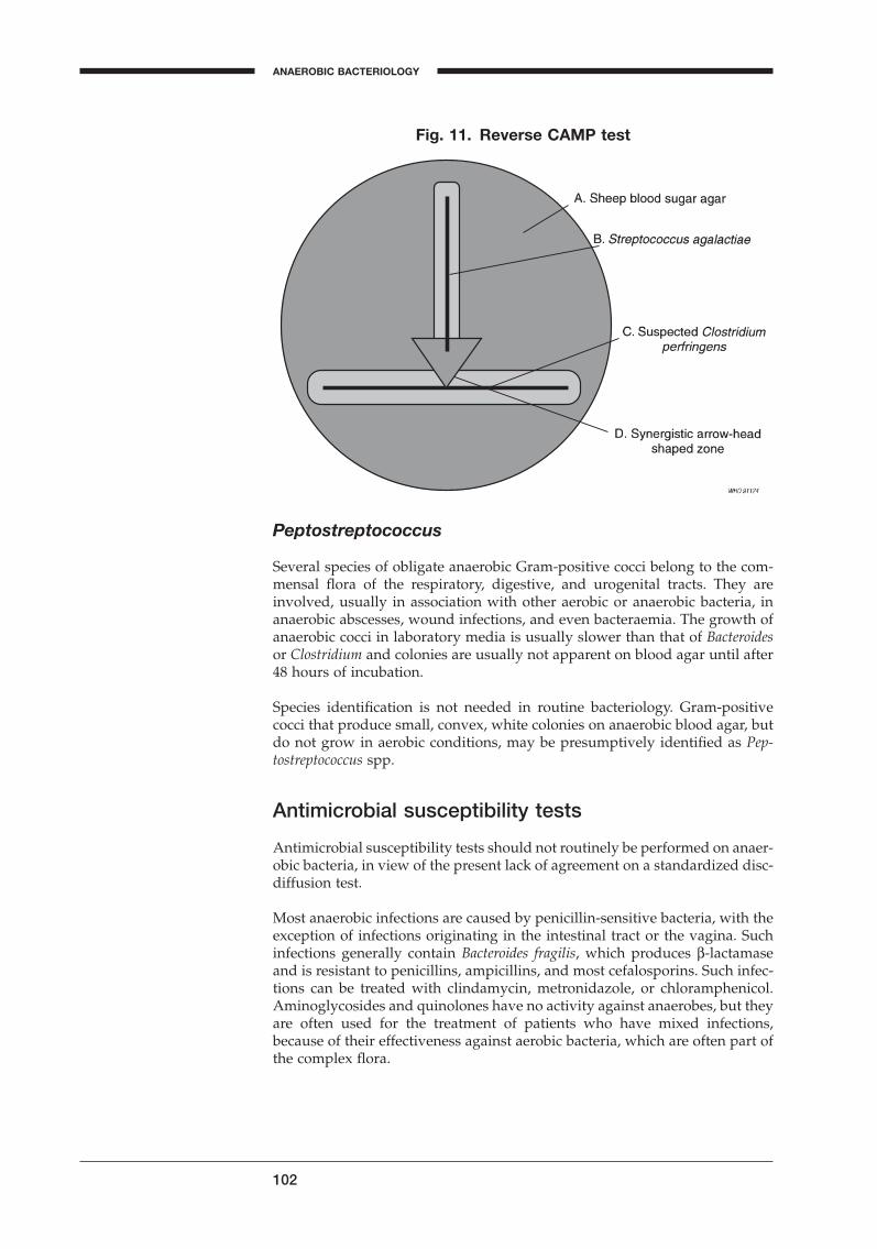
Peptostreptococcus
Several species of obligate anaerobic Gram-positive cocci belong to the com-mensal flora of the respiratory, digestive, and urogenital tracts. They areinvolved, usually in association with other aerobic or anaerobic bacteria, inanaerobic abscesses, wound infections, and even bacteraemia. The growth ofanaerobic cocci in laboratory media is usually slower than that of Bacteroidesor Clostridium and colonies are usually not apparent on blood agar until after48 hours of incubation.
Species identification is not needed in routine bacteriology. Gram-positivecocci that produce small, convex, white colonies on anaerobic blood agar, butdo not grow in aerobic conditions, may be presumptively identified as Pep-tostreptococcus spp.
Antimicrobial susceptibility tests
Antimicrobial susceptibility tests should not routinely be performed on anaer-obic bacteria, in view of the present lack of agreement on a standardized disc-diffusion test.
Most anaerobic infections are caused by penicillin-sensitive bacteria, with theexception of infections originating in the intestinal tract or the vagina. Suchinfections generally contain Bacteroides fragilis, which produces b-lactamaseand is resistant to penicillins, ampicillins, and most cefalosporins. Such infec-tions can be treated with clindamycin, metronidazole, or chloramphenicol.Aminoglycosides and quinolones have no activity against anaerobes, but theyare often used for the treatment of patients who have mixed infections,because of their effectiveness against aerobic bacteria, which are often part ofthe complex flora.
102
Fig. 11. Reverse CAMP test
ANAEROBIC BACTERIOLOGY
BLM1 1/17/04 2:02 PM Page 102
103
A
Antimicrobial susceptibility testing
Introduction
At a meeting organized by WHO in Geneva in 1977,1 concern was expressedabout the worldwide increase in antimicrobial resistance associated with thegrowing, and frequently indiscriminate, use of antimicrobials in both humansand animals. In recent years, drug-resistant bacteria have given rise to severalserious outbreaks of infection, with many deaths. This has led to a need fornational and international surveillance programmes to monitor antibimicro-bial resistance in bacteria by susceptibility testing using reliable methods thatgenerate comparable data. The availability of microbiological and epidemio-logical information would help clinicians in selecting the most appropriateantimicrobial agent for the treatment of a microbial infection.
If predictions are to be valid, the susceptibility test must be performed by anaccurate and reproducible method, the results of which should be directlyapplicable to the clinical situation. The ultimate criterion of the reliability ofany susceptibility testing method is its correlation with the response of thepatient to antimicrobial therapy.
The WHO meeting considered that the modified disc-diffusion technique of Kirby–Bauer2, for which requirements had been established by WHO in1976, could be recommended for clinical and surveillance purposes in viewof its technical simplicity and reproducibility. The method is particularly suit-able for use with bacteria belonging to the family Enterobacteriaceae, but itcan also be recommended as a general purpose method for all rapidlygrowing pathogens. It has been adapted to the clinically most important fastidious bacteria but not to strict anaerobes and mycobacteria. It is thereforerecommended that the details of this test be made available to laboratoryworkers.3
General principles of antimicrobial susceptibilitytesting
Antimicrobial susceptibility tests measure the ability of an antimicrobial agentto inhibit bacterial growth in vitro. This ability may be estimated by either thedilution method or the diffusion method.
The dilution test
For quantitative estimates of antimicrobial activity, dilutions of the antimi-crobial may be incorporated into broth or agar medium, which is then inocu-lated with the test organism. The lowest concentration that prevents growth
1 Surveillance for the prevention and control of health hazards due to antibiotic-resistant entero-bacteria. Geneva, World Health Organization, 1978 (WHO Technical Report Series, No. 624).2 WHO Expert Committee on Biological Standardization. Twenty-eighth report. Geneva, WorldHealth Organization, 1977 (WHO Technical Report Series, No. 610).3. A comparable method, based on similar principles and quality control requirements as the Kirby–Bauer method, is the NEO-SENSITABS method, produced by ROSCO Diagnostica,Taastrup, Denmark. This method uses 9-mm colour-coded, antimicrobial tablets, instead ofpaper discs. The tablet form results in an extraordinary stability with a shelf-life of four years,even at room temperature. This increased stability is very important for laboratories in tropicalcountries.
BLM1 1/17/04 2:02 PM Page 103
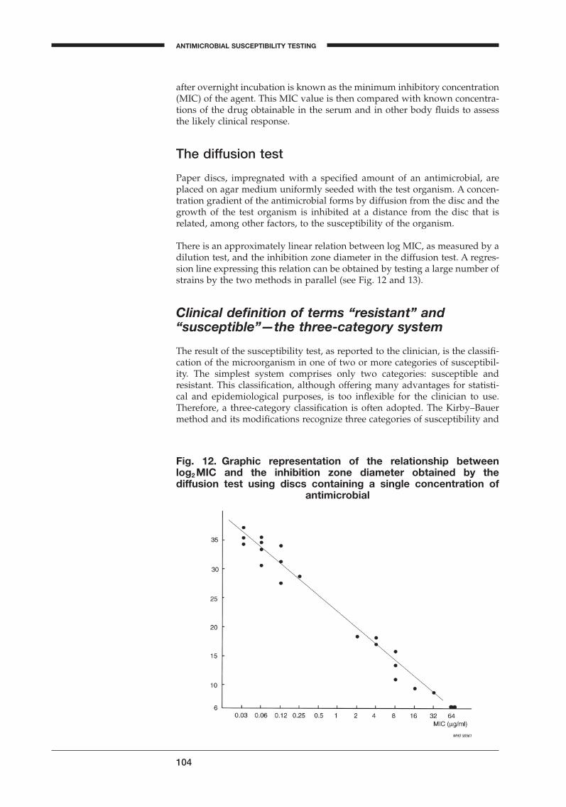
after overnight incubation is known as the minimum inhibitory concentration(MIC) of the agent. This MIC value is then compared with known concentra-tions of the drug obtainable in the serum and in other body fluids to assessthe likely clinical response.
The diffusion test
Paper discs, impregnated with a specified amount of an antimicrobial, areplaced on agar medium uniformly seeded with the test organism. A concen-tration gradient of the antimicrobial forms by diffusion from the disc and thegrowth of the test organism is inhibited at a distance from the disc that isrelated, among other factors, to the susceptibility of the organism.
There is an approximately linear relation between log MIC, as measured by adilution test, and the inhibition zone diameter in the diffusion test. A regres-sion line expressing this relation can be obtained by testing a large number ofstrains by the two methods in parallel (see Fig. 12 and 13).
Clinical definition of terms “resistant” and“susceptible”—the three-category system
The result of the susceptibility test, as reported to the clinician, is the classifi-cation of the microorganism in one of two or more categories of susceptibil-ity. The simplest system comprises only two categories: susceptible andresistant. This classification, although offering many advantages for statisti-cal and epidemiological purposes, is too inflexible for the clinician to use.Therefore, a three-category classification is often adopted. The Kirby–Bauermethod and its modifications recognize three categories of susceptibility and
104
Fig. 12. Graphic representation of the relationship between log2 MIC and the inhibition zone diameter obtained by the diffusion test using discs containing a single concentration of
antimicrobial
ANTIMICROBIAL SUSCEPTIBILITY TESTING
BLM1 1/17/04 2:02 PM Page 104
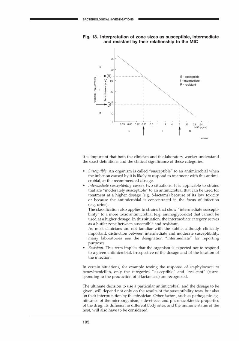
105
BACTERIOLOGICAL INVESTIGATIONS
A
it is important that both the clinician and the laboratory worker understandthe exact definitions and the clinical significance of these categories.
• Susceptible. An organism is called “susceptible” to an antimicrobial whenthe infection caused by it is likely to respond to treatment with this antimi-crobial, at the recommended dosage.
• Intermediate susceptibility covers two situations. It is applicable to strainsthat are “moderately susceptible” to an antimicrobial that can be used fortreatment at a higher dosage (e.g. b-lactams) because of its low toxicity or because the antimicrobial is concentrated in the focus of infection (e.g. urine).The classification also applies to strains that show “intermediate suscepti-bility” to a more toxic antimicrobial (e.g. aminoglycoside) that cannot beused at a higher dosage. In this situation, the intermediate category servesas a buffer zone between susceptible and resistant.As most clinicians are not familiar with the subtle, although clinicallyimportant, distinction between intermediate and moderate susceptibility,many laboratories use the designation “intermediate” for reporting purposes.
• Resistant. This term implies that the organism is expected not to respondto a given antimicrobial, irrespective of the dosage and of the location ofthe infection.
In certain situations, for example testing the response of staphylococci to benzylpenicillin, only the categories “susceptible” and “resistant” (corre-sponding to the production of b-lactamase) are recognized.
The ultimate decision to use a particular antimicrobial, and the dosage to begiven, will depend not only on the results of the susceptibility tests, but alsoon their interpretation by the physician. Other factors, such as pathogenic sig-nificance of the microorganism, side-effects and pharmacokinetic propertiesof the drug, its diffusion in different body sites, and the immune status of thehost, will also have to be considered.
Fig. 13. Interpretation of zone sizes as susceptible, intermediateand resistant by their relationship to the MIC
BLM1 1/17/04 2:02 PM Page 105
Indications for routine susceptibility tests
A susceptibility test may be performed in the clinical laboratory for two mainpurposes:
• to guide the clinician in selecting the best antimicrobial agent for an individual patient;
• to accumulate epidemiological information on the resistance of microor-ganisms of public health importance within the community.
Susceptibility tests as a guide for treatment
Susceptibility tests should never be performed on contaminants or commen-sals belonging to the normal flora, or on other organisms that have no causalrelationship to the infectious process. For example, the presence of Escherichiacoli in the urine in less than significant numbers is not to be regarded ascausing infection, and it would be useless and even misleading to perform anantibiogram.
Susceptibility tests should be carried out only on pure cultures of organismsconsidered to be causing the infectious process. The organisms should also beidentified, as not every microorganism isolated from a patient with an infec-tion requires an antibiogram.
Routine susceptibility tests are not indicated in the following situations:
• When the causative organism belongs to a species with predictable sus-ceptibility to specific drugs. This is the case for Streptococcus pyogenes andNeisseria meningitidis, which are still generally susceptible to benzylpeni-cillin. (However, there have recently been a few reports of sporadic occur-rence of benzylpenicillin-resistant meningococci.) It is also the case forfaecal streptococci (enterococci), which, with few exceptions, are suscepti-ble to ampicillin. If resistance of these microorganisms is suspected on clin-ical grounds, representative strains should be submitted to a competent reference laboratory.
• If the causative organism requires enriched media, e.g. Haemophilus influen-zae and Neisseria gonorrhoeae. Disc-diffusion susceptibility tests may giveunreliable results, if the appropriate technique is not strictly followed. Theemergence of b-lactamase-producing variants of these species has led tothe introduction of special tests, such as the in vitro test for b-lactamaseproduction, described on page 79. It will be the responsibility of the centraland regional laboratories to monitor the susceptibility of pneumococci,gonococci, and Haemophilus. If problems arise with resistant strains, the peripheral laboratories should be alerted and instructions should begiven on appropriate testing methods and on alternative treatmentschemes.
• In uncomplicated intestinal infections caused by salmonellae (other thanS. typhi or S. paratyphi). Antimicrobial treatment of such infections is notjustified, even with drugs showing in vitro activity. There is now ample evidence that antimicrobial treatment of common salmonella gastroenteri-tis (and indeed of most types of diarrhoeal disease of unknown etiology)is of no clinical benefit to the patient. Paradoxically, antimicrobials mayprolong the excretion and dissemination of salmonellae and may lead tothe selection of resistant variants.
106
ANTIMICROBIAL SUSCEPTIBILITY TESTING
BLM1 1/17/04 2:02 PM Page 106
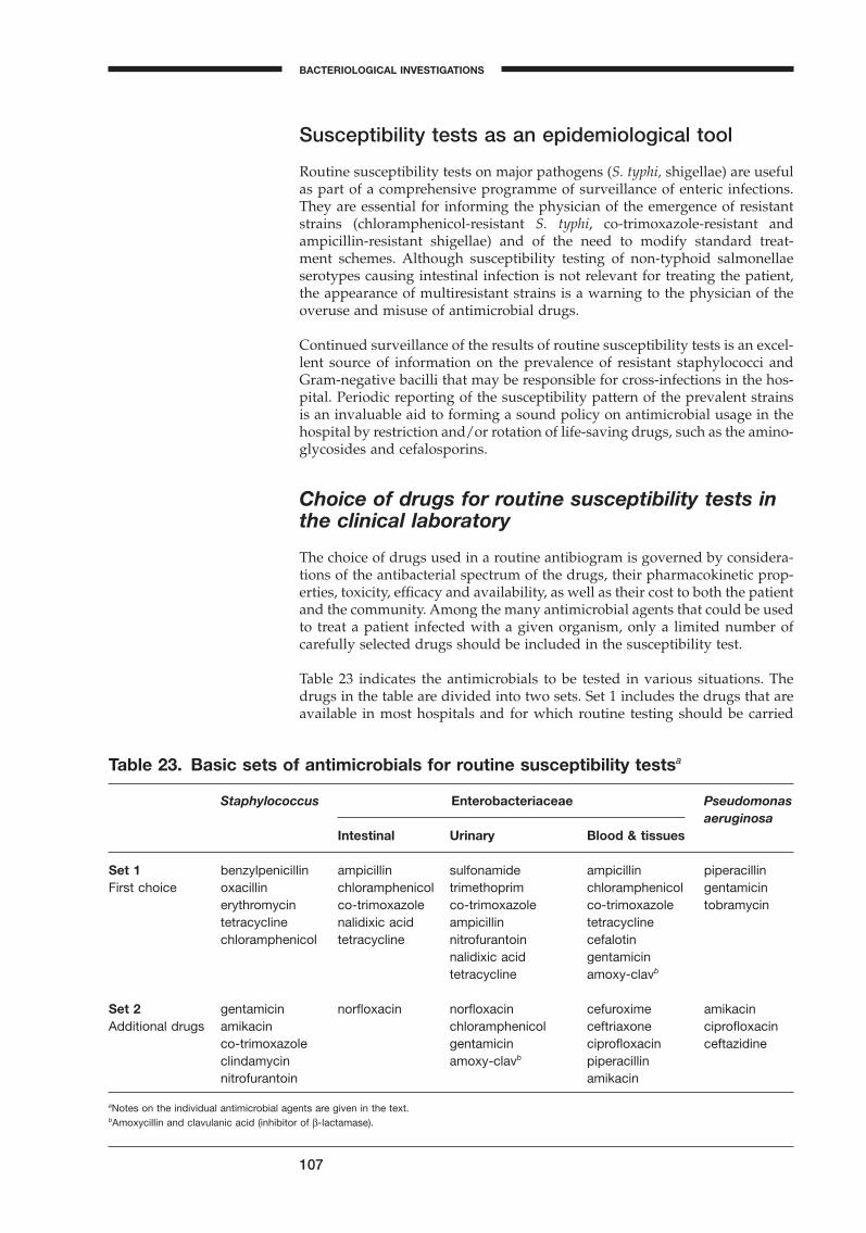
107
BACTERIOLOGICAL INVESTIGATIONS
A
Susceptibility tests as an epidemiological tool
Routine susceptibility tests on major pathogens (S. typhi, shigellae) are usefulas part of a comprehensive programme of surveillance of enteric infections.They are essential for informing the physician of the emergence of resistantstrains (chloramphenicol-resistant S. typhi, co-trimoxazole-resistant and ampicillin-resistant shigellae) and of the need to modify standard treat-ment schemes. Although susceptibility testing of non-typhoid salmonellaeserotypes causing intestinal infection is not relevant for treating the patient,the appearance of multiresistant strains is a warning to the physician of theoveruse and misuse of antimicrobial drugs.
Continued surveillance of the results of routine susceptibility tests is an excel-lent source of information on the prevalence of resistant staphylococci andGram-negative bacilli that may be responsible for cross-infections in the hos-pital. Periodic reporting of the susceptibility pattern of the prevalent strainsis an invaluable aid to forming a sound policy on antimicrobial usage in thehospital by restriction and/or rotation of life-saving drugs, such as the amino-glycosides and cefalosporins.
Choice of drugs for routine susceptibility tests inthe clinical laboratory
The choice of drugs used in a routine antibiogram is governed by considera-tions of the antibacterial spectrum of the drugs, their pharmacokinetic prop-erties, toxicity, efficacy and availability, as well as their cost to both the patientand the community. Among the many antimicrobial agents that could be usedto treat a patient infected with a given organism, only a limited number ofcarefully selected drugs should be included in the susceptibility test.
Table 23 indicates the antimicrobials to be tested in various situations. Thedrugs in the table are divided into two sets. Set 1 includes the drugs that areavailable in most hospitals and for which routine testing should be carried
Table 23. Basic sets of antimicrobials for routine susceptibility testsa
Staphylococcus Enterobacteriaceae Pseudomonas
Intestinal Urinary Blood & tissuesaeruginosa
Set 1 benzylpenicillin ampicillin sulfonamide ampicillin piperacillinFirst choice oxacillin chloramphenicol trimethoprim chloramphenicol gentamicin
erythromycin co-trimoxazole co-trimoxazole co-trimoxazole tobramycintetracycline nalidixic acid ampicillin tetracyclinechloramphenicol tetracycline nitrofurantoin cefalotin
nalidixic acid gentamicintetracycline amoxy-clavb
Set 2 gentamicin norfloxacin norfloxacin cefuroxime amikacinAdditional drugs amikacin chloramphenicol ceftriaxone ciprofloxacin
co-trimoxazole gentamicin ciprofloxacin ceftazidineclindamycin amoxy-clavb piperacillinnitrofurantoin amikacin
aNotes on the individual antimicrobial agents are given in the text.bAmoxycillin and clavulanic acid (inhibitor of b-lactamase).
BLM1 1/17/04 2:02 PM Page 107
out for every strain. Tests for drugs in set 2 are to be performed only at thespecial request of the physician, or when the causative organism is resistantto the first-choice drugs, or when other reasons (allergy to a drug, or itsunavailability) make further testing justified. Many antimicrobials with goodclinical activity have been omitted from the table, but it must be emphasizedthat they are rarely needed in the management of the infected patient. In veryrare cases, one or more additional drugs should be included when there is aspecial reason known to the physician, or when new and better drugs becomeavailable. Periodic revision of this table is therefore desirable, and this shouldbe done after appropriate discussions with clinical staff. Many problems arisein practice, because clinicians are not always aware that only one representa-tive of each group of antimicrobial agents is included in routine tests. Theresult obtained for this particular drug may then be extrapolated to all, ormost, of the other members of the group. Difficulties arise in some countrieswhen the physician is familiar only with the commercial brand name of thedrug and not with its generic nonproprietary name. A serious effort shouldbe made to inform medical personnel about the international nonproprietarynames of pharmaceutical substances, and to encourage their use.1
1. The benzylpenicillin disc is used to test susceptibility to all b-lactamase-sensitive penicillins (such as oral phenoxymethylpenicillin and pheneti-cillin). Isolates of staphylococci that fall into the resistant categoryproduce b-lactamase and should be treated with a b-lactamase-resistantpenicillin G or with another antimicrobial, such as erythromycin.
2. The oxacillin disc is representative of the whole group of b-lactamase-resistant penicillins (including meticillin, nafcillin, cloxacillin,dicloxacillin, and flucloxacillin). There is good clinical evidence that cross-resistance exists between the meticillin and the cefalosporin groups.Therefore, it is useless and misleading to include cefalotin in the antibiogram for staphylococci.Resistance to meticillin and related drugs is often of the heterogeneoustype, i.e. the majority of cells may be fully susceptible and produce a wideinhibition zone, while the resistant part of the population appears in theform of minute discrete colonies growing within the inhibition zone. Thistype of resistance is more apparent when the temperature of the incuba-tor is set at 35 ∞C2 or when the incubation time is prolonged.A serious disadvantage of meticillin, as a representative disc for the b-lactamase-resistant penicillins, is its great lability even under conven-tional storage conditions. The oxacillin disc is much more resistant todeterioration and is therefore preferred for the standardized diffusiontest. The cloxacillin and dicloxacillin discs are not used as they may notindicate the presence of a heteroresistant strain.
3. The results for the tetracycline disc may be applied to chlortetracycline,oxytetracycline, and other members of this group. However, most tetra-cycline-resistant staphylococci remain normally sensitive to minocycline.A disc of minocycline may thus be useful to test multiresistant strains ofstaphylococci.
4. The result for the chloramphenicol disc may be extrapolated to thiam-phenicol, a related drug with a comparable antimicrobial spectrum, butwithout known risk of aplastic anaemia.
5. Only one representative of sulfonamide (sulfafurazole) is required in thetest.
108
1 International Nonproprietary Names (INN) for Pharmaceutical Substances. Cumulative list No. 9. Geneva, World Health Organization, 1996.2 Sahm DF et al. Current concepts and approaches to antimicrobial agent susceptibility testing.In: Cumitech 25, Washington, DC, American Society for Microbiology, 1988.
ANTIMICROBIAL SUSCEPTIBILITY TESTING
BLM1 1/17/04 2:02 PM Page 108
109
BACTERIOLOGICAL INVESTIGATIONS
A
6. The co-trimoxazole disc contains a combination of trimethoprim and a sul-fonamide (sulfamethoxazole). The two components of this synergisticcombination have comparable pharmacokinetic properties and generallyact as a single drug.
7. Ampicillin is the prototype of a group of broad-spectrum penicillins withactivity against many Gram-negative bacteria. As it is susceptible to b-lactamase, it should not be used for testing staphylococci. Generally, thesusceptibility to ampicillin is also valid for other members of this group:amoxycillin, pivampicillin, talampicillin, etc. (although amoxycillin istwice as active against salmonellae and only half as active against shigel-lae and H. influenzae).
8. Only cefalotin needs to be tested routinely as its spectrum is representa-tive of all other first-generation cefalosporins (cefalexin, cefradine,cefaloridine, cefazolin, cefapirin). Where second- and third-generationcefalosporins and related compounds (cefamycins) with an expandedspectrum are available, a separate disc for some of these new drugs maybe justified in selected cases (cefoxitin, cefamandole, cefuroxime, cefo-taxime, ceftriaxone). Although some cefalosporins can be used to treatsevere staphylococcal infections, the susceptibility of the infecting straincan be derived from the result with oxacillin as already mentioned under2 above.
9. Erythromycin is used to test the susceptibility to some other members ofthe macrolide group (oleandomycin, spiramycin).
10. Aminoglycosides form a group of chemically related drugs that includesstreptomycin, gentamicin, kanamycin, netilmicin and tobramycin. Theirantimicrobial spectra are not always closely enough related to permitassumption of cross-resistance, but against susceptible pathogens theseagents have been shown to be equally effective. Numerous studies havecompared the nephrotoxicity and ototoxicity of gentamicin, netilmicinand tobramycin, but there is no conclusive evidence that any one of thedrugs is less toxic than the others. It is strongly recommended that eachlaboratory select a single agent for primary susceptibility testing. Theother agents should be held in reserve for treatment of patients with infec-tions caused by resistant organisms.
11. Nitrofurantoin is limited to use only in the treatment of urinary tract infec-tions, and should not be tested against microorganisms recovered frommaterial other than urine.
Table 24 provides information on zone diameter limits for control strains.
The modified Kirby–Bauer method
The disc-diffusion method, originally described in 1966,1 is well standardizedand has been widely evaluated. Official agencies have recommended it, withminor modifications, as a reference method which could be used as a routinetechnique in the clinical laboratory.
1 Bauer AW et al. Antibiotic susceptibility testing by a standardized single disc method. American journal of clinical pathology, 1966; 45:493–496.
BLM1 1/17/04 2:02 PM Page 109
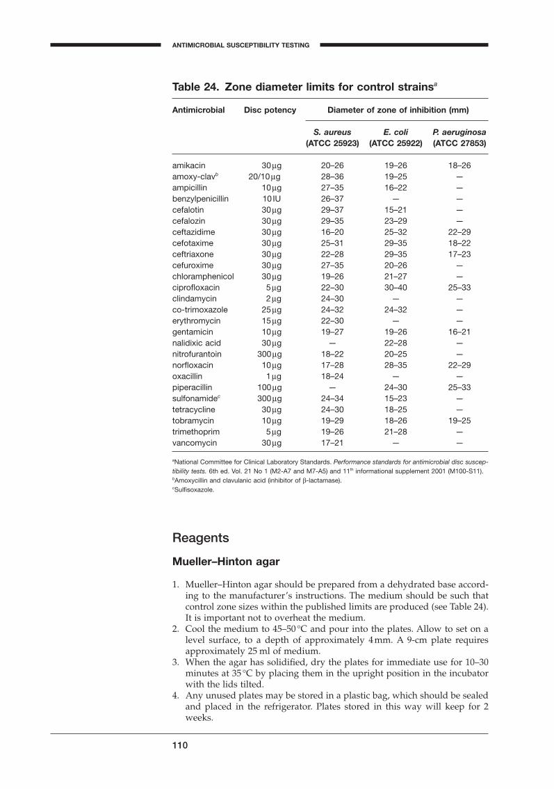
Reagents
Mueller–Hinton agar
1. Mueller–Hinton agar should be prepared from a dehydrated base accord-ing to the manufacturer’s instructions. The medium should be such thatcontrol zone sizes within the published limits are produced (see Table 24).It is important not to overheat the medium.
2. Cool the medium to 45–50 ∞C and pour into the plates. Allow to set on alevel surface, to a depth of approximately 4mm. A 9-cm plate requiresapproximately 25 ml of medium.
3. When the agar has solidified, dry the plates for immediate use for 10–30minutes at 35 ∞C by placing them in the upright position in the incubatorwith the lids tilted.
4. Any unused plates may be stored in a plastic bag, which should be sealedand placed in the refrigerator. Plates stored in this way will keep for 2weeks.
110
Table 24. Zone diameter limits for control strainsa
Antimicrobial Disc potency Diameter of zone of inhibition (mm)
S. aureus E. coli P. aeruginosa(ATCC 25923) (ATCC 25922) (ATCC 27853)
amikacin 30mg 20–26 19–26 18–26amoxy-clavb 20/10mg 28–36 19–25 —ampicillin 10mg 27–35 16–22 —benzylpenicillin 10 IU 26–37 — —cefalotin 30mg 29–37 15–21 —cefalozin 30mg 29–35 23–29 —ceftazidime 30mg 16–20 25–32 22–29cefotaxime 30mg 25–31 29–35 18–22ceftriaxone 30mg 22–28 29–35 17–23cefuroxime 30mg 27–35 20–26 —chloramphenicol 30mg 19–26 21–27 —ciprofloxacin 5mg 22–30 30–40 25–33clindamycin 2mg 24–30 — —co-trimoxazole 25mg 24–32 24–32 —erythromycin 15mg 22–30 — —gentamicin 10mg 19–27 19–26 16–21nalidixic acid 30mg — 22–28 —nitrofurantoin 300mg 18–22 20–25 —norfloxacin 10mg 17–28 28–35 22–29oxacillin 1mg 18–24 — —piperacillin 100mg — 24–30 25–33sulfonamidec 300mg 24–34 15–23 —tetracycline 30mg 24–30 18–25 —tobramycin 10mg 19–29 18–26 19–25trimethoprim 5mg 19–26 21–28 —vancomycin 30mg 17–21 — —
aNational Committee for Clinical Laboratory Standards. Performance standards for antimicrobial disc suscep-tibility tests. 6th ed. Vol. 21 No 1 (M2-A7 and M7-A5) and 11th informational supplement 2001 (M100-S11).bAmoxycillin and clavulanic acid (inhibitor of b-lactamase).cSulfisoxazole.
ANTIMICROBIAL SUSCEPTIBILITY TESTING
BLM1 1/17/04 2:02 PM Page 110
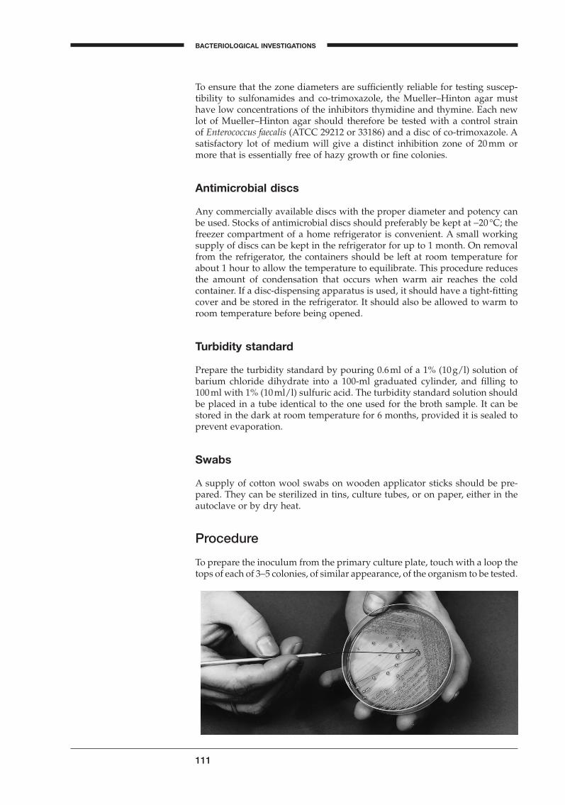
111
BACTERIOLOGICAL INVESTIGATIONS
A
To ensure that the zone diameters are sufficiently reliable for testing suscep-tibility to sulfonamides and co-trimoxazole, the Mueller–Hinton agar musthave low concentrations of the inhibitors thymidine and thymine. Each newlot of Mueller–Hinton agar should therefore be tested with a control strain of Enterococcus faecalis (ATCC 29212 or 33186) and a disc of co-trimoxazole. Asatisfactory lot of medium will give a distinct inhibition zone of 20mm ormore that is essentially free of hazy growth or fine colonies.
Antimicrobial discs
Any commercially available discs with the proper diameter and potency canbe used. Stocks of antimicrobial discs should preferably be kept at -20 ∞C; thefreezer compartment of a home refrigerator is convenient. A small workingsupply of discs can be kept in the refrigerator for up to 1 month. On removalfrom the refrigerator, the containers should be left at room temperature forabout 1 hour to allow the temperature to equilibrate. This procedure reducesthe amount of condensation that occurs when warm air reaches the cold container. If a disc-dispensing apparatus is used, it should have a tight-fittingcover and be stored in the refrigerator. It should also be allowed to warm toroom temperature before being opened.
Turbidity standard
Prepare the turbidity standard by pouring 0.6ml of a 1% (10g/l) solution ofbarium chloride dihydrate into a 100-ml graduated cylinder, and filling to 100ml with 1% (10ml/l) sulfuric acid. The turbidity standard solution shouldbe placed in a tube identical to the one used for the broth sample. It can bestored in the dark at room temperature for 6 months, provided it is sealed toprevent evaporation.
Swabs
A supply of cotton wool swabs on wooden applicator sticks should be pre-pared. They can be sterilized in tins, culture tubes, or on paper, either in theautoclave or by dry heat.
Procedure
To prepare the inoculum from the primary culture plate, touch with a loop thetops of each of 3–5 colonies, of similar appearance, of the organism to be tested.
BLM1 1/17/04 2:02 PM Page 111
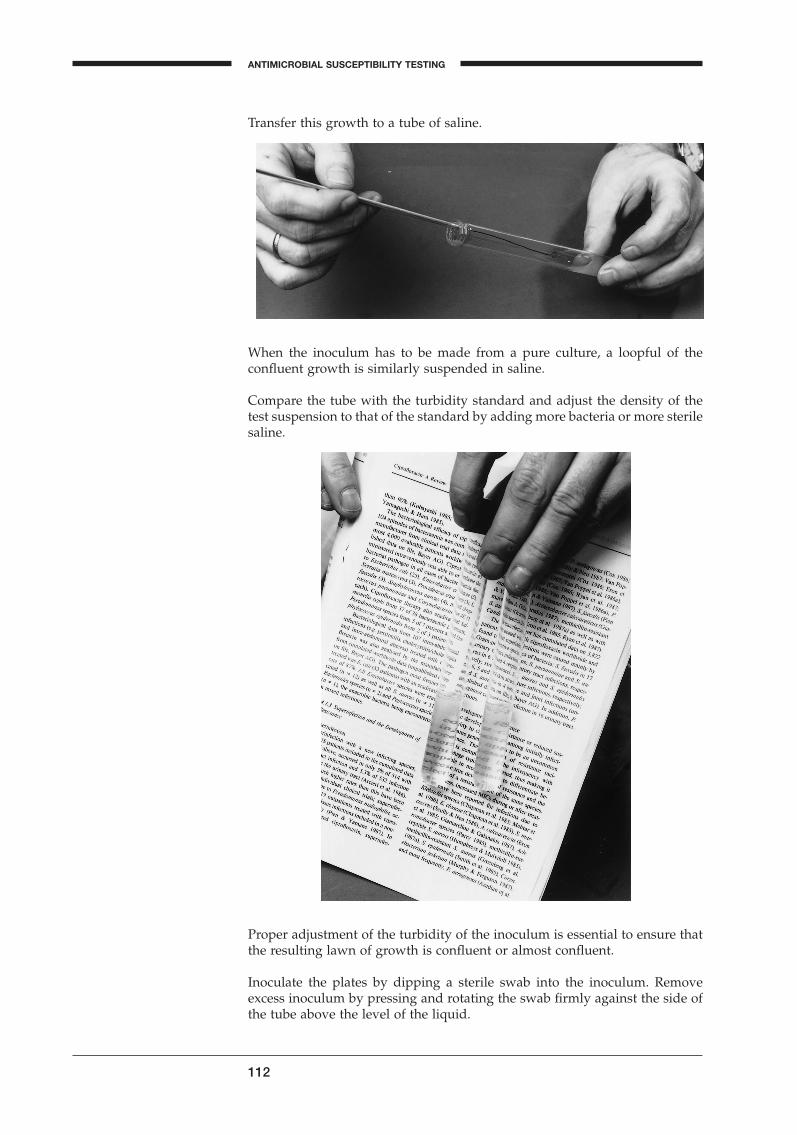
Transfer this growth to a tube of saline.
112
When the inoculum has to be made from a pure culture, a loopful of the confluent growth is similarly suspended in saline.
Compare the tube with the turbidity standard and adjust the density of thetest suspension to that of the standard by adding more bacteria or more sterilesaline.
Proper adjustment of the turbidity of the inoculum is essential to ensure thatthe resulting lawn of growth is confluent or almost confluent.
Inoculate the plates by dipping a sterile swab into the inoculum. Removeexcess inoculum by pressing and rotating the swab firmly against the side ofthe tube above the level of the liquid.
ANTIMICROBIAL SUSCEPTIBILITY TESTING
BLM1 1/17/04 2:02 PM Page 112
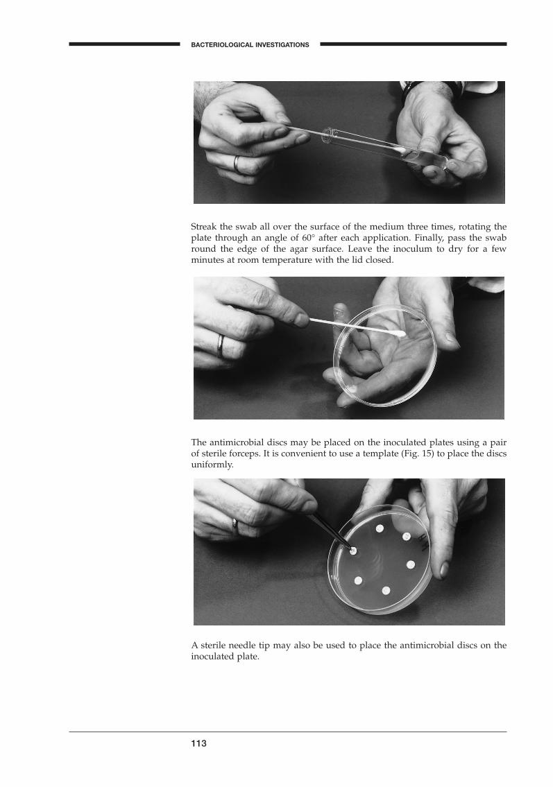
113
BACTERIOLOGICAL INVESTIGATIONS
A
Streak the swab all over the surface of the medium three times, rotating theplate through an angle of 60∞ after each application. Finally, pass the swabround the edge of the agar surface. Leave the inoculum to dry for a fewminutes at room temperature with the lid closed.
The antimicrobial discs may be placed on the inoculated plates using a pairof sterile forceps. It is convenient to use a template (Fig. 15) to place the discsuniformly.
A sterile needle tip may also be used to place the antimicrobial discs on theinoculated plate.
BLM1 1/17/04 2:02 PM Page 113
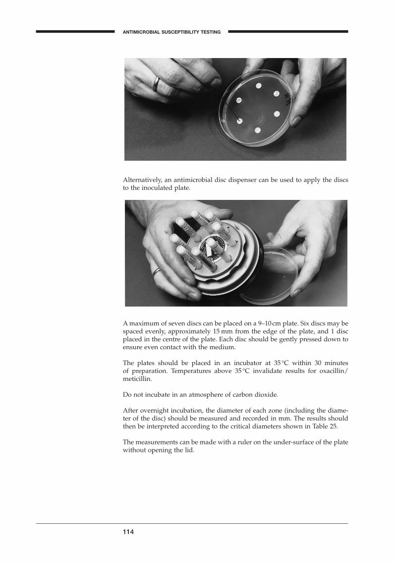
Alternatively, an antimicrobial disc dispenser can be used to apply the discsto the inoculated plate.
114
A maximum of seven discs can be placed on a 9–10cm plate. Six discs may bespaced evenly, approximately 15 mm from the edge of the plate, and 1 discplaced in the centre of the plate. Each disc should be gently pressed down toensure even contact with the medium.
The plates should be placed in an incubator at 35 ∞C within 30 minutes of preparation. Temperatures above 35 ∞C invalidate results for oxacillin/meticillin.
Do not incubate in an atmosphere of carbon dioxide.
After overnight incubation, the diameter of each zone (including the diame-ter of the disc) should be measured and recorded in mm. The results shouldthen be interpreted according to the critical diameters shown in Table 25.
The measurements can be made with a ruler on the under-surface of the platewithout opening the lid.
ANTIMICROBIAL SUSCEPTIBILITY TESTING
BLM1 1/17/04 2:02 PM Page 114
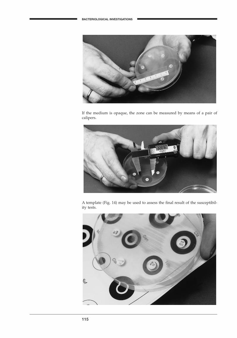
115
BACTERIOLOGICAL INVESTIGATIONS
A
If the medium is opaque, the zone can be measured by means of a pair ofcalipers.
A template (Fig. 14) may be used to assess the final result of the susceptibil-ity tests.
BLM1 1/17/04 2:02 PM Page 115

116
Table 25. Interpretative chart of zone sizes for rapidly growing bacteria using themodified Kirby–Bauer techniquea
Antimicrobial agent Diameter of zone of inhibition (mm)
Disc potency Resistant Intermediate Susceptible
amikacin 30mg <14 15–16 >17amoxy-clavb 20/10mg <13 14–17 >18ampicillin, when testing
— Enterobacteriaceae 10mg <13 14–16 >17— enterococci 10mg <16 — >17
benzylpenicillin, when testing
— staphylococci 10 IU <28 — >29— enterococci 10 IU <14 — >15
cefalotin 30mg <14 15–17 >18cefalozin 30mg <14 15–17 >18cefotaxime 30mg <14 15–22 >23ceftazidime 30mg <14 15–17 >18ceftriaxone 30mg <13 14–20 >21cefuroxime sodium, cefamandole 30mg <14 15–17 >18chloramphenicol 30mg <12 13–17 <18ciprofloxacin 5mg <15 16–20 >21clindamycin 2mg <14 15–20 �21co-trimoxazole 25mg <10 11–15 �16erythromycin 15mg <13 14–22 >23gentamicin 10mg <12 13–14 >15nalidixic acidc 30mg <13 14–18 �19nitrofurantoinc 300mg <14 15–16 >17norfloxacinc 10mg <12 13–16 >17oxacillin 1mg <10 10–12 >13piperacillin, when testing
— P. aeruginosa 100mg <17 — >18— other Gram-negative rods 100mg <17 18–20 >21
sulfonamidec,d 300mg <12 13–16 >17tetracycline 30mg <14 15–18 >19tobramycin 10mg <12 13–14 >15trimethoprimc 5mg <10 11–15 �16vancomycin, when testing
— staphylococci 30mg — — >15— enterococci 30mg <14 15–16 >17
aNational Committee for Clinical Laboratory Standards. Performance standards for antimicrobial disc susceptibility tests, 6th ed., Vol. 21, No. 1 (M2–A7and M7–A5) Wayne, PA, NCCLS, 1997; and 11th informational supplement 2001 (M100–S11).bAmoxycillin and clavulanic acid (inhibitor of b-lactamase).cOnly applicable for testing isolates from urinary tract infection and some enteric pathogens.dSulfisoxazol.
ANTIMICROBIAL SUSCEPTIBILITY TESTING
BLM1 1/17/04 2:02 PM Page 116
117
BACTERIOLOGICAL INVESTIGATIONS
A
The end-point of inhibition is judged by the naked eye at the edge where thegrowth starts, but there are three exceptions:
• With sulfonamides and co-trimoxazole, slight growth occurs within theinhibition zone; such growth should be ignored.
• When b-lactamase-producing staphylococci are tested against benzyl peni-cillin, zones of inhibition are produced with a heaped-up, clearly definededge; these are readily recognizable when compared with the sensitivecontrol, and, regardless of the size of the zone of inhibition, they shouldbe reported as resistant.
• Certain Proteus species may swarm into the area of inhibition around someantimicrobials, but the zone of inhibition is usually clearly outlined andthe thin layer of swarming growth should be ignored.
Interpretation of the zone sizes
Using a template. When the zone sizes are compared with a template, a sepa-rate template must be prepared for each individual antimicrobial agent (seeFig. 14). The result—susceptible, resistant, or intermediate—can be read atonce: “susceptible”, when the zone edge is outside the black circle; “resistant”,when there is no zone, or when it lies within the white circle; and “interme-diate”, when the edge of the zone of inhibition lies on the black circle.
Using a ruler. When the zone sizes are measured in mm, the results should beinterpreted according to the critical diameters given in Table 25.
Direct versus indirect susceptibility tests
In the standardized method outlined above, the inoculum is prepared fromcolonies on a primary culture plate or from a pure culture. This is called an“indirect susceptibility test”. In certain cases, where a rapid answer is impor-tant, the standardized inoculum may be replaced by the pathological speci-men itself, e.g. urine, a positive blood culture, or a swab of pus. For specimensof urine, a microscopical examination of the sediment should first be made inorder to see if there is evidence of infection, i.e. the presence of pus cellsand/or organisms. The urine may then be used as the inoculum in the stan-dard test. Likewise, susceptibility tests may be performed on incubated bloodcultures showing evidence of bacterial growth, or a swab of pus may be usedas a direct inoculum, when a Gram-stained smear shows the presence of largenumbers of a single type of organism. This is called a “direct susceptibilitytest”; its advantage over the indirect test is that a result is obtained 24 hoursearlier. The main disadvantage is that the inoculum cannot be properly con-trolled. When the susceptibility plate shows too light or too heavy growth, orwhen the culture is a mixture, the results should be interpreted with caution,and the test repeated on pure cultures.
Fig. 14. Template for the determination of susceptibility
BLM1 1/17/04 2:02 PM Page 117
Technical factors influencing the size of the zonein the disc-diffusion method
Inoculum density
If the inoculum is too light, the inhibition zones will be larger even thoughthe sensitivity of the organism is unchanged. Relatively resistant strains maythen be reported as susceptible. Conversely, if the inoculum is too heavy, thezone size will be reduced and susceptible strains may be reported as resistant.Usually optimal results are obtained with an inoculum size that produces nearconfluent growth.
Timing of disc application
If the plates, after being seeded with the test strain, are left at room tempera-ture for periods longer than the standard time, multiplication of the inoculummay take place before the discs are applied. This causes a reduction in the zonediameter and may result in a susceptible strain being reported as resistant.
Temperature of incubation
Susceptibility tests are normally incubated at 35 ∞C for optimal growth. If thetemperature is lowered, the time required for effective growth is extended andlarger zones result. When a heterogeneous resistant strain of Staphylococcusaureus is being tested against meticillin (oxacillin), the resistant portion of thepopulation can be detected at 35 ∞C. At higher temperatures the entire cultureappears to be susceptible. At 35 ∞C or lower temperatures, resistant coloniesdevelop within the zone of inhibition. These resistant colonies can be seenmore easily if the plate is left for several hours at room temperature beforethe result is read. Such colonies should always be identified to check whetherthey are contaminants.
Incubation time
Most techniques adopt an incubation period of between 16 and 18 hours. Inemergencies, however, a provisional report may be made after 6 hours. Thisis not routinely recommended and the result should always be confirmed afterthe conventional incubation time.
Size of plate, depth of agar medium, and spacing of the antimicrobial discs
Susceptibility tests are usually carried out with 9–10cm plates and no morethan 6 or 7 antimicrobial discs on each plate. If larger numbers of antimicro-bials have to be tested, two plates, or one 14-cm diameter plate, is to be pre-ferred. Excessively large inhibition zones may be formed on very thin media;the converse is true for thick media. Minor changes in the depth of the agarlayer have negligible effect. Proper spacing of the discs is essential to avoidoverlapping of the inhibition zones or deformation near the edge of the plates(see Fig. 15).
118
ANTIMICROBIAL SUSCEPTIBILITY TESTING
BLM1 1/17/04 2:02 PM Page 118
119
BACTERIOLOGICAL INVESTIGATIONS
A
Potency of the antimicrobial discs
The diameter of the inhibition zone is related to the amount of drug in the disc. If the potency of the drug is reduced owing to deterioration duringstorage, the inhibition zone will show a corresponding reduction in size.
Composition of the medium
The medium influences the size of the zone by its effect on the rate of growthof the organism, the rate of diffusion of the antimicrobial, and the activity ofthe agent. It is essential to use the medium appropriate to the particularmethod.
The many factors influencing the zone diameters that may be obtained for thesame test organism clearly demonstrate the need for standardization of disc-diffusion methods. Only if the conditions laid down in a particular methodare closely followed can valid results be obtained. Alteration of any of thefactors affecting the test can result in grossly misleading reports for the clinician.
The precision and accuracy of the method should be monitored by establish-ing the quality control programme described below. Variations can then beimmediately investigated and corrective action taken to eliminate them.
Fig. 15. Template for uniform placement of susceptibility discson plates of 90mm diameter
BLM1 1/17/04 2:02 PM Page 119
Quality control
The need for quality control in the susceptibility test
The final result of a disc-diffusion test is influenced by a large number of variables. Some of the factors, such as the inoculum density and the incuba-tion temperature, are easy to control, but a laboratory rarely knows the exact composition of a commercial medium or the batch-to-batch variationsin its quality, and it cannot take for granted the antimicrobial content of thediscs. The results of the test must therefore be monitored constantly by aquality control programme, which should be considered part of the procedureitself.
The precision and accuracy of the test are controlled by the parallel use of aset of control strains, with known susceptibility to the antimicrobial agents.These quality control strains are tested using exactly the same procedure asfor the test organisms. The zone sizes shown by the control organisms shouldfall within the range of diameters given in Table 24. When results regularlyfall outside this range, they should be regarded as evidence that a technicalerror has been introduced into the test, or that the reagents are at fault. Eachreagent and each step in the test should then be investigated until the causeof the error has been found and eliminated.
Standard procedure for quality control
The quality control programme should test standard reference strains of bacteria in parallel with the clinical cultures. These tests should preferably be run every week, or with every fifth batch of tests, and, in addition, every time a new batch of Mueller–Hinton agar or a new batch of discs is used.
Standard strains for quality control
Staphylococcus aureus (ATCC 25923)Escherichia coli (ATCC 25922)Pseudomonas aeruginosa (ATCC 27853)
These cultures can be obtained from national culture collections. They arecommercially available in the form of pellets of desiccated pure cultures.
Cultures for day-to-day use should be grown on slants of nutrient agar (trypticsoy agar is convenient) and stored in the refrigerator. They should be sub-cultured on to fresh slants every 2 weeks.
Preparing the inoculum
The cultures may be inoculated into any type of broth, and incubated untilthe broth is turbid. Each broth should be streaked onto an agar plate and incubated overnight. Single colonies should then be picked off and submittedto susceptibility tests as described on pages 111–115.
120
ANTIMICROBIAL SUSCEPTIBILITY TESTING
BLM1 1/17/04 2:02 PM Page 120
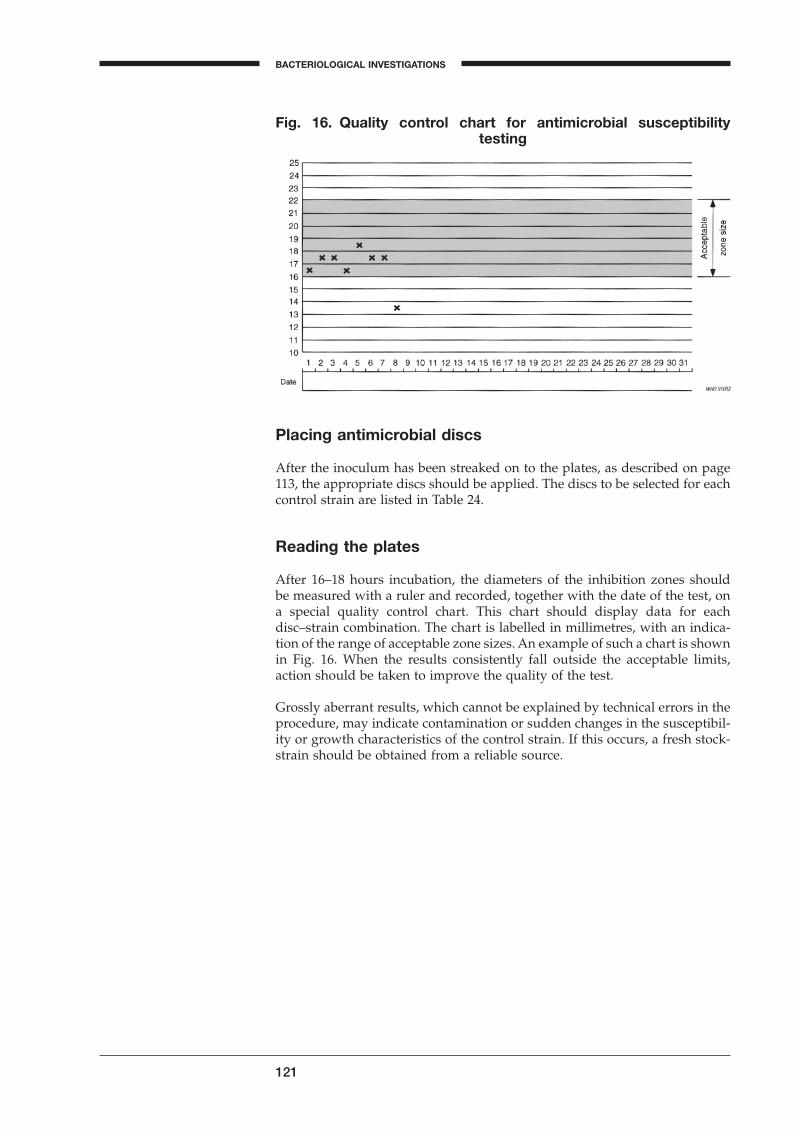
121
BACTERIOLOGICAL INVESTIGATIONS
A
Placing antimicrobial discs
After the inoculum has been streaked on to the plates, as described on page113, the appropriate discs should be applied. The discs to be selected for eachcontrol strain are listed in Table 24.
Reading the plates
After 16–18 hours incubation, the diameters of the inhibition zones should be measured with a ruler and recorded, together with the date of the test, ona special quality control chart. This chart should display data for eachdisc–strain combination. The chart is labelled in millimetres, with an indica-tion of the range of acceptable zone sizes. An example of such a chart is shownin Fig. 16. When the results consistently fall outside the acceptable limits,action should be taken to improve the quality of the test.
Grossly aberrant results, which cannot be explained by technical errors in theprocedure, may indicate contamination or sudden changes in the susceptibil-ity or growth characteristics of the control strain. If this occurs, a fresh stock-strain should be obtained from a reliable source.
Fig. 16. Quality control chart for antimicrobial susceptibility testing
BLM1 1/17/04 2:02 PM Page 121
Serological tests
Introduction
Serological tests, unlike culture and microscopic investigations, provide onlyindirect evidence of infection by detecting bacterial antigens, or antibodiesproduced in response to them, in clinical material. Such tests are now widelyused in microbiology because of their high specificity and sensitivity.
After an initial infection with pathogenic microorganisms, most patients willproduce both IgM and IgG antibodies. After a few weeks, the cells producingIgM antibodies switch to producing IgG antibodies, and subsequently onlyIgG antibodies will be present in the patient’s serum. A second infection withthe same pathogens will elicit only an IgG response. Because the antibody-producing cells have retained the memory of the pathogen, the response willbe more rapid and usually stronger than the initial response; this is ananamnestic response.
The antibody level is usually designated by a “titre”. The diagnostic titre to be reported is the reciprocal value of the highest dilution of the patient’sserum in which antibodies are still detectable. For example, if antibodies are detectable in serum diluted 1 :1024 but not in further dilutions, the titre ofthe serum is 1024. Serum collected during the acute phase of the infection,when the disease is first suspected, is called the acute serum; serum drawnduring convalescence, which is usually 2 weeks later, is called convalescentserum.
Reaction to an antigen will occur regardless of the stage of infection, althoughthis reaction will vary. The presence of IgG antibodies in a single serumsample may indicate past exposure to the agent and therefore cannot be usedto diagnose a current infection. An antigen may also stimulate production of antibodies that cross-react with other antigens. As these antibodies are nonspecific, tests involving only single serum samples may give rise to mis-interpretation of results. For the majority of serological tests, both acute andconvalescent sera should be tested, preferably together in the same test-run,to counteract variables inherent in the test procedure. An increase in the anti-body titre of two doubling dilutions (e.g. from a dilution of 1 : 8 to a dilution1 :32) is usually considered to be diagnostic of current infection. This is calleda fourfold rise in titre. Testing a single serum sample may be useful only inspecial cases, such as for diagnosis of Mycoplasma pneumoniae infection, wherehigh titres may indicate a recent infection, or when the test demonstrates thepresence of IgM antibodies and the result may be taken as evidence of acurrent infection. The type of antigen–antibody reaction depends on the physical state of the antigen.
Quality control measures
The reliability and consistency of serological test results are entirely depen-dent on the quality control measures undertaken before, during and after eachtest. These quality control measures are extremely important as a false-positive or false-negative result might give rise to a medical decision or actionthat could harm the patient. Many variables may affect the quality of sero-logical testing, including the experience of the laboratory personnel, thequality of the kits and equipment, the condition of the specimens, the controlsused in test-runs, and the interpretation and reporting of results. After the test
122
BLM1 1/17/04 2:02 PM Page 122
123
BACTERIOLOGICAL INVESTIGATIONS
A
has been completed, discard used materials into a container filled with disin-fectant, and wash hands and bench surfaces with disinfectant.
Equipment
The equipment in a serology laboratory includes water-baths, incubators,refrigerators, freezers, pH meters, balances, centrifuges, microscopes, androtators. Monitoring and routine maintenance of equipment is a vital part ofthe quality assurance programme in the serology laboratory. A routine main-tenance service should be established with periodic inspection of all equip-ment for minor adjustments. Records showing dates of inspection,maintenance, and repair should be maintained for each piece of equipment.
The water-bath should be kept free of all foreign material and drained andcleaned monthly. The temperatures of the water-bath should be strictly con-trolled and should not vary by more than ±1 ∞C; it should be checked andrecorded daily and during use. A cover should be provided to prevent coolingat the surface of the water. Mixtures of antigen and antibody should not beincubated until the desired temperature has been maintained for at least onehour. The water level should be the same as that of the fluid in the tubes orflasks that are placed in the water-bath.
Microscopes are essential for the Venereal Disease Research Laboratory(VDRL) test and the fluorescent treponemal antigen absorption (FTA-Abs)test, and must be maintained in the best possible working order. After use,the eyepieces, objectives, and condenser of the microscope should be wipedclean of all oil and debris with a non-abrasive tissue. When not in use, themicroscope should be kept covered and protected against moisture. The inten-sity of the mercury lamp of the fluorescence microscope should be checkedrepeatedly with a light meter.
The mechanical rotator for the VDRL and the rapid plasma reagin (RPR) testsshould be checked for speed during use, and any change in speed that mayadversely affect the agglutination reaction should be corrected. The me-chanical rotator should be lubricated on a regular basis.
Materials
Glassware and needles used in serological tests should meet the specificationsrecommended in those tests.
All chipped or scratched glassware, plates and slides should be discarded.The use of dirty or improperly cleaned glassware that may have residues of organic material on it is a major cause of misleading results. The instruc-tions for washing glassware should be clearly written out and displayed inthe washing area. The basic steps should include pre-rinsing, washing withan appropriate laboratory detergent, rinsing with tap water followed by distilled water, drying, and checking to ensure that all detergent has beenremoved.
All pipettes should be soaked upright in a detergent solution so that the solu-tion fills the inside of the pipette. Pipettes should then be rinsed for at least30 minutes under running water, followed by a rinse in distilled water. Glassplates used for the VDRL test must be cleaned thoroughly until all detergentand oil residues are removed. Prolonged soaking in detergent solutionsshould be avoided as this may weaken and flake the ceramic rings on the
BLM1 1/17/04 2:02 PM Page 123
plates. Glass plates with paraffin rings should be cleaned using an appropri-ate organic solvent (e.g. petrol ether).
For the RPR and VDRL tests, calibrated needles are used for dispensingantigen and diluent. The RPR test kit includes a dispensing needle, and theseneedles should be checked before use. Check the needle by determining thenumber of drops/ml: a 20-gauge needle should deliver 90 drops/ml. RPRneedles that are not working properly should be discarded. Wash the needleswith distilled water after use. To prepare the two needles for the VDRL test,break the points with a pair of pliers. Check the needles by determining thenumber of drops/ml: an 18-gauge needle must deliver 60 drops/ml, and a 21-or 22-gauge needle deliver 100 drops/ml. Any needle in the VDRL test kit thatdoes not deliver the exact number of drops should be discarded, or adjustedby either pressing or reaming the end of the needle. Wash the needles withwater, 70% ethanol and acetone after use.
Reagents
Chemicals used in serology must be of reagent quality and must meet thespecifications of the particular procedure. They should be stored in accor-dance with the manufacturers’ instructions.
High-quality distilled water with a pH of 7.0 should be used for the prepara-tion of reagents. Distilled water should be stored in a heat-resistant glass orplastic bottle with a tightly fitting lid and be properly labelled and dated.
Saline is used either as saline or as a buffered solution, such as phosphate-buffered saline. In humid climates, sodium chloride should be dried in a hot-air oven for 30 minutes at 160–180 ∞C to remove any moisture. The salt shouldbe dissolved in distilled or demineralized water and stored in a heat-resistantglass or plastic bottle with a tightly fitting lid and be properly labelled and dated. When buffered saline is prepared, the pH must be determinedbefore use.
Sera that contain particulate debris should be centrifuged at 3000g for 10minutes, and the supernatant used for testing. Haemolysed or contaminatedsera should be discarded. If inactivated serum is required for a test, heat theserum at 56 ∞C for 30 minutes. If the serum has not been used 4 hours afterthe initial inactivation, it should be re-inactivated by heating at 56 ∞C for 10minutes before testing. Bring all sera to room temperature before testing.
A detailed description of some of the serological procedures routinely per-formed in many medical laboratories will be discussed in this chapter. Theseinclude serological tests for the investigation of syphilis, the Wright test forthe diagnosis of brucellosis, and the antistreptolysin O test for the diagnosisof post-streptococcal disease.
Each description refers to the use of a commercial test kit. As these test kitsare available from a number of manufacturers, the user should carefully readthe detailed instructions contained in each package insert.
124
SEROLOGICAL TESTS
BLM1 1/17/04 2:02 PM Page 124
125
BACTERIOLOGICAL INVESTIGATIONS
A
Serological reactions
Flocculation or precipitin reactions
In flocculation tests, the antigen is in solution and the interaction with the anti-body will result in the formation of a precipitate, which can be observed eitherunder the microscope or with the naked eye. When the reagents are mixed,the initial combination of antigen and antibody occurs almost immediately.However, subsequent formation of larger visible clumps requires an hour ormore and is temperature-dependent. The reaction is fastest in the zone ofequivalence, where the antigen–antibody ratio is optimal. The tube with thequickest formation of a precipitate is a good indication of equivalence. Themost widely used flocculation tests are the VDRL and the RPR test. Both areused for the diagnosis of syphilis (caused by Treponema pallidum), and for othertreponemal infections.
Flocculation tests provide qualitative evidence of an antigen–antibody reac-tion but do not indicate whether one or more types of antigen–antibody reac-tions are involved. If, however, the reactions are investigated using a semisolid gel, the different antigens and antibodies are likely to diffuse at different migration rates, and it may be possible to distinguish differentreactions.
Agglutination reactions
In agglutination tests, the reagent, which may be an antigen or an antibody,is fixed or absorbed to a micro-particle. A variety of particles can be used ascarriers of the reagent, e.g. latex particles, gelatin particles, microbeads, bac-teria or red blood cells. This technique is also called passive agglutination.When red blood cells are used as carriers the tests are called coagglutinationtests. When mixed with a specific antiserum, the cells or particles form a latticenetwork that results in clumping and leaves a clear supernatant. If an anti-serum of known specificity is used, the test for the identification of unknownmicroorganisms or their antigens can be applied. This test may be performedon a slide, and the resulting agglutination read macroscopically or under thelow-power objective of microscope. The agglutination reaction is also used toestimate the titre of antibacterial agglutinins in the serum of patients withunknown diseases. A rise in titre during an illness strongly suggests a causalrelationship.
Agglutination is accelerated at higher temperatures (35–56 ∞C) and by move-ment (e.g. shaking, stirring or centrifuging), which increases the contactbetween antigen and antibody. The agglutination process requires the pres-ence of salts. A potentially serious problem with agglutination tests is prozonereaction: if too much antibody is present, the lattice will not form and agglu-tination will be inhibited. A prozone reaction gives the impression that anti-body is absent; this error, however, can be avoided by testing serial dilutionsof the serum.
There is a commonly used agglutination test that uses Staphylococcus aureus,which contains a protein, Protein A, on its surface. This protein binds to theFc fragment of IgG antibodies. IgG antibody-coated staphylococci producevisible agglutination in the presence of a specific antigen. The test is mainlyused to identify organisms cultured from clinical specimens or to detect bac-terial antigens in body fluids of infected patients (cerebrospinal fluid in thecase of meningitis).
BLM1 1/17/04 2:02 PM Page 125
Agglutination tests are used for the diagnosis of Epstein-Barr virus (infectiousmononucleosis), rotavirus, rubella, and for the detection of bacterial antigens(Haemophilus and Streptococcus A and B, among others).
Fluorescent antibody tests
In immunofluorescence tests, the immunoreagent (antigen or antibody) isattached to a fluorescent dye, such as fluorescein or rhodamine, and the reac-tion between the antigen and the antibody is detected by fluorescencemicroscopy. In the direct antigen-detection test, fluorescein-conjugated anti-bodies are used to reveal the presence of a specific antigen. The test is a valu-able aid in the rapid identification of Chlamydia trachomatis, C. psittaci,Rickettsia spp., Streptococcus pyogenes, Bordetella pertussis, Corynebacterium diph-theriae, Legionella pneumophila, and other organisms in clinical specimens.
In the indirect fluorescent antibody (IFA) test, serial dilutions of a patient’sserum are allowed to react with the specific antigen, and antihuman IgG orIgM antibodies conjugated to fluorescein are added to make the reactionvisible. For example, in the serodiagnosis of syphilis, Treponema pallidumantigen is fixed to a slide, overlaid with the patient’s serum, and then washed.Fluorescein-labelled antihuman immunoglobulin is then placed on the prepa-ration, which is washed and examined by fluorescence microscopy. If thepatient’s serum contains specific antibodies to Treponema pallidum, brightly fluorescent spirochaetes are seen. If the spirochaetes do not fluoresce, no spe-cific antitreponemal antibodies are present in the patient’s serum. The IFA testcan also be used to identify other bacteria, including mycobacteria, and thisprocedure is often more sensitive than the direct immunofluorescence test,because more fluorescein-labelled antibodies will attach to each antigen site.
Serological tests for syphilis
The serological tests for the diagnosis of syphilis include non-treponemal andtreponemal tests. The non-treponemal tests are the VDRL and the RPR tests.The antigens used in these tests are prepared from non-treponemal antigens,such as cardiolipin–lecithin, and they detect the antibody-like substancereagin, which is present in the sera of many patients with syphilis, and mayoccasionally be detected in the sera of patients with other acute and chronicdiseases. These tests are practical, inexpensive, and reproducible, althoughthey are not absolutely specific. They may confirm the diagnosis of early orlate symptomatic syphilis or provide diagnostic evidence of latent syphilis.This test is superior to the treponemal tests as a follow-up investigation aftertreatment. Moreover, the VDRL test is an effective tool in epidemiologicalinvestigations of syphilis and other treponemal diseases.
The treponemal tests use Treponema pallidum antigens to detect specific anti-bodies that have developed in serum in response to syphilis infection. Theprocedures are used to verify the specificity of positive reactions in non-treponemal tests. The fluorescent treponemal antibody absorption test (FTA-Abs) and the Treponema pallidum haemagglutination test (TPHA) are highlyspecific and sensitive but they cannot differentiate between active and pastsyphilis infections; neither can they be used for evaluating therapeuticresponse.
126
SEROLOGICAL TESTS
BLM1 1/17/04 2:02 PM Page 126
127
BACTERIOLOGICAL INVESTIGATIONS
A
VDRL test
The VDRL test uses cardiolipin–lecithin-coated cholesterol particles. Inacti-vated serum or cerebrospinal fluid and VDRL antigen emulsion are mixedusing a rotating machine for a prescribed period of time. The VDRL particleswill flocculate if reagin is present in the serum or cerebrospinal fluid.
The VDRL test is strongly reactive for early syphilis infection. After effectivetreatment the titre falls gradually and usually becomes non-reactive within1–2 years. In the late phase of the disease the serum may remain reactive at alow titre (e.g. 1 : 8 or less) for many years, even after effective treatment. Reac-tivity may cease spontaneously in about 20–30% of untreated patients duringthe latent phase of the disease, and even more often during the late phase.
False-positive results may be observed because of the similarity of the VDRLantigen with normal host tissue. Although false-positive reactions can some-times be observed in the serum of healthy persons, they are often associatedwith a specific disease or following vaccination. Acute false-positive reactionsoften have low titres (1 :8 or less) and are mainly seen in persons with viralor bacterial infections (atypical pneumonia, psittacosis, infectious mononu-cleosis and infectious hepatitis), during pregnancy or after recent vaccination.Prolonged false-positive reactions usually have high titres that are caused byautoantibodies (rheumatoid factors) in patients with lepromatous leprosy,tuberculosis, immune disorders (e.g. lupus erythematosus, collagenosis,rheumatic diseases, Sjögren syndrome, dysgammaglobulinaemia) and occa-sionally malaria, or in those who are dependent on heroin. Reactive or weaklyreactive VDRL test results should not be considered conclusive evidence ofsyphilis in a patient and a non-reactive VDRL test by itself does not rule outthe diagnosis of syphilis. Any test sample giving a reactive or weakly reactiveresult in the absence of clinical evidence of syphilis should therefore be sub-jected to a treponemal test such as FTA-Abs or TPHA.
Materials and reagents provided in the VDRL test kit
Buffered saline solutionControl sera set (non-reactive, weakly reactive and reactive)VDRL antigen
Additional materials and reagents required for the VDRL test
Absolute alcohol and acetoneAgglutination slide, approximately 5 ¥ 7.5cm with wells 16mm in diameter
and 1.75mm deep, for cerebrospinal fluid testsAliquot vialsDistilled or deionized waterGlass plate with 12 paraffin or ceramic rings approximately 14mm in diame-
ter for serum testsHumidity coverHypodermic needles without bevels: 18-gauge for serum tests, and 21- or
22-gauge for cerebrospinal fluid testsInterval timerLight microscope with ¥10 ocular and ¥10 objectiveMechanical rotator, circumscribing a circle 2cm in diameter, at a speed of
180rev/min, on a horizontal plane, with an automatic timerpH meterSerological pipettes: 5.0ml, 1.0ml and 0.2mlSterile saline solutions (0.85% and 10%)Syringe, Luer-type, 1 or 2ml
BLM1 1/17/04 2:02 PM Page 127
VDRL antigen emulsion bottles, 30-ml, round, glass-stoppered, narrow-mouth, approximately 35mm in diameter with flat inner bottom surface
Water-bath (56 ∞C)
Rehydration of VDRL antigen and control sera
The VDRL antigen is an alcoholic solution of lipids (cardiolipin and lecithin)and cholesterol. These substances are not soluble in water. VDRL antigen isunstable and a fresh suspension must be prepared on the day of use. Pour thecontents of the antigen ampoule into the storage vial. Ensure that the vial is tightly capped and store in the dark at 15–30 ∞C. Withdraw antigen asrequired.
After the bottle of buffered saline has been opened, it should be stored in therefrigerator. Discard if turbidity appears.
Rehydrate the control sera with 3ml of distilled or deionized water. Dividethe rehydrated sera that are surplus to the day’s use into suitable aliquot por-tions (one day’s use) and store at -20 ∞C for up to one month. Do not thawand refreeze. Store the sera to be used for the day in the refrigerator at 2–8 ∞C.
Preparation of VDRL antigen emulsion
1. Allow the antigen and buffered saline to reach room temperature. Check the pH of the buffered saline and discard if outside the range of pH6.0 ± 0.1.
2. Pipette 0.4ml of buffered saline into an antigen emulsion bottle and gentlytilt the bottle so that the buffered saline covers the bottom the bottle.
3. Measure 0.5ml of antigen solution using a 1-ml pipette graduated to thetip, and add the antigen as follows:• Keep the pipette in the upper one third of the bottle. Do not let it touch
the saline.• While rotating the bottle manually in a circle approximately 5cm in
diameter, add the antigen drop by drop to the buffered saline.• Allow approximately 6 seconds to add the antigen, then discharge the
remaining antigen in the pipette into the bottle.• Continue rotation of the bottle for 10 seconds.
4. Add 4.1ml of buffered saline to the bottle, allowing it to flow down theside of the bottle.
5. Place the glass stopper in the bottle and shake the bottle up and downapproximately 30 times in 10 seconds.
6. Let the antigen emulsion stand for at least 10 minutes before using. Swirlgently prior to use. The antigen emulsion may be used for the next 8 hours.
7. If cerebrospinal fluid is to be tested, dilute the antigen emulsion a further1 :2 with an equal volume of 10% saline solution. Shake the bottle gentlyfor 10 seconds and allow to stand for a minimum of 5 minutes and amaximum of 2 hours before using.
VDRL qualitative test
1. Using a 1.0ml pipette, mix the inactivated serum several times then add0.05ml to the first well of the VDRL glass plate.
2. Spread the serum with a circular motion of the pipette tip so that it coversthe entire inner surface of the paraffin or ceramic well. Use only cleanplates that allow the serum to cover evenly the entire surface within theparaffin or ceramic well.
128
SEROLOGICAL TESTS
BLM1 1/17/04 2:02 PM Page 128
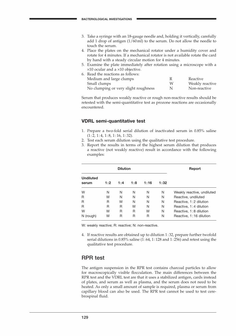
129
BACTERIOLOGICAL INVESTIGATIONS
A
3. Take a syringe with an 18-gauge needle and, holding it vertically, carefullyadd 1 drop of antigen (1/60ml) to the serum. Do not allow the needle totouch the serum.
4. Place the plates on the mechanical rotator under a humidity cover androtate for 4 minutes. If a mechanical rotator is not available rotate the cardby hand with a steady circular motion for 4 minutes.
5. Examine the plate immediately after rotation using a microscope with a¥10 ocular and a ¥10 objective.
6. Read the reactions as follows:Medium and large clumps R ReactiveSmall clumps W Weakly reactiveNo clumping or very slight roughness N Non-reactive
Serum that produces weakly reactive or rough non-reactive results should beretested with the semi-quantitative test as prozone reactions are occasionallyencountered.
VDRL semi-quantitative test
1. Prepare a two-fold serial dilution of inactivated serum in 0.85% saline (1 :2, 1 : 4, 1 : 8, 1 : 16, 1 : 32).
2. Test each serum dilution using the qualitative test procedure.3. Report the results in terms of the highest serum dilution that produces
a reactive (not weakly reactive) result in accordance with the followingexamples:
Dilution Report
Undilutedserum 1:2 1 :4 1 :8 1 :16 1 :32
W N N N N N Weakly reactive, undilutedR W N N N N Reactive, undilutedR R W N N N Reactive, 1 :2 dilutionR R R W N N Reactive, 1 :4 dilutionW W R R W N Reactive, 1 :8 dilutionN (rough) W R R R N Reactive, 1 :16 dilution
W: weakly reactive; R: reactive; N: non-reactive.
4. If reactive results are obtained up to dilution 1 :32, prepare further twofoldserial dilutions in 0.85% saline (1 :64, 1 : 128 and 1 :256) and retest using thequalitative test procedure.
RPR test
The antigen suspension in the RPR test contains charcoal particles to allowfor macroscopically visible flocculation. The main differences between theRPR test and the VDRL test are that it uses a stabilized antigen, cards insteadof plates, and serum as well as plasma, and the serum does not need to beheated. As only a small amount of sample is required, plasma or serum fromcapillary blood can also be used. The RPR test cannot be used to test cere-brospinal fluid.
BLM1 1/17/04 2:02 PM Page 129
In the RPR test the antigen is ready for immediate use. It requires no priorpreparation or dilution. Unopened antigen reagent has a shelf-life of one year;storage in a refrigerator is recommended. Once opened, the antigen reagentmaintains its reactivity for 3 months when stored in the refrigerator in itsplastic dispenser. The RPR test is slightly more sensitive than the VDRL testand it is easier and quicker to perform. False-positive reactions occur slightlymore often with the RPR test than with the VDRL test. Some commercial kitsrequire a mechanical rotator for mixing the reagents, whereas others can berotated manually.
Materials and reagents provided in the RPR test kit
Antigen delivery needle to deliver 60 drops/ml of the antigen suspensionControl sera, positive and negativeDisposable droppers to deliver 50ml of serum or plasmaPlastic-coated RPR test cards, each with two rows of five wellsPrepared RPR antigen suspensionStirrers
Additional materials and reagents required for the RPR test
Disposable dropperGrease pencilHumidity coverMechanical rotator, circumscribing a circle 2cm in diameter, at a speed of
180rev/min, on a horizontal plane, with an automatic timerSterile saline (0.85%)
RPR qualitative test
1. Remove the reagent kit from the refrigerator and allow the reagents towarm to room temperature.
2. Reconstitute the control serum by adding the recommended volume of dis-tilled water.
3. Label each well on the RPR card with the laboratory number of a sampleto be tested, including wells for positive, weakly positive and negativecontrol sera.
4. Use the disposable dropper to add 50ml of unheated serum or plasma tothe corresponding well. Use a new dropper for each sample.
5. Gently shake the antigen suspension and add one free-falling drop to eachwell using the antigen delivery needle provided. Carefully mix the antigensuspension and serum. Use a new stirrer for each sample. Spread to coverthe area of the well.
6. Place the card on the mechanical rotator under a humidity cover and rotatefor 8 minutes. If a mechanical rotator is not available rotate the card byhand with a steady circular motion for 2 minutes, then place it in a moistchamber containing wet tissue or filter-paper for 6 minutes. Remove thecard and rotate briefly to obtain the final reading. Take care to avoid cross-contamination of samples.
7. Remove the card from the rotator and examine it macroscopically in a goodlight. The positive control serum should show clearly visible agglutination.The negative control serum should show no agglutination. A brief rotationand tilting of the card by hand can help to differentiate weakly reactivefrom non-reactive samples.
8. Record the test results:• Small to large flocculated clumps: reactive• Even turbidity of the particle suspension: non-reactive
9. Prepare serial dilutions of any reactive sera to estimate the antibody titre.
130
SEROLOGICAL TESTS
BLM1 1/17/04 2:02 PM Page 130
131
BACTERIOLOGICAL INVESTIGATIONS
A
RPR semi-quantitative test
1. Remove the reagent kit from the refrigerator and allow the reagents towarm to room temperature.
2. Label a row of 5 wells on the RPR card with the laboratory number foreach sample to be tested.
3. Use the disposable dropper to add 1 drop of saline (0.85%) to each well.Do not spread.
4. Use a new dropper to add 1 drop of the serum sample to the first well.Mix by drawing up and down the dropper 5–6 times (avoid bubble for-mation).
5. Transfer 50ml of the mixed sample (1 :2 dilution) to the next well. Mix.Repeat the procedure up to the 5th well (1 : 32 dilution). Discard 50ml fromthe last dilution.
6. Spread the diluted samples over the entire area of the test well startingwith the highest dilution. Use a new stirrer for each sample.
7. Gently shake the antigen add 1 free-falling drop to each well using theantigen delivery needle provided. Carefully mix the antigen suspensionand serum. Spread to cover the area of the well. Use a new stirrer for eachsample.
8. Place the card on the mechanical rotator under a humidity cover androtate for 8 minutes. If a mechanical rotator is not available rotate the cardby hand with a steady circular motion for 2 minutes, then place it in amoist chamber containing wet tissue or filter-paper for 6 minutes.Remove the card and rotate briefly to obtain the final reading. Take careto avoid cross-contamination of samples.
9. Remove the card from the rotator and examine it macroscopically in agood light. The highest dilution to contain macroscopic agglutination isthe titre of the sample.
10. If the sample is positive at 1 : 32, the dilution series should be extended.Prepare a 1 :16 dilution in saline (0.85%) and perform a serial dilutionseries as described previously.
Fluorescent treponemal antibody absorption test (FTA-Abs)
The antigen used for the FTA-Abs test consists of Treponema pallidum (Nicholsstrain) which is fixed with acetone on a slide. Lyophilized T. pallidum cellsreconstituted in saline can also be used. Inactivated patient serum is incubatedwith a sorbent consisting of Reiter treponemes for absorption of nonspecifictreponemal group antibodies. After absorption, patient serum is added to theslide. Specific antibodies in the serum bind to the surface of the treponemalcells. After rinsing, a conjugate of antihuman antibodies with a fluorescentstain (fluorescein isothiocyanate) is added to the treponemes. The conjugatewill bind to the antibodies that have bound to the treponemes and can be visu-alized by fluorescence microscopy.
Reactivity can be observed three weeks after infection and is permanent inuntreated patients. It may be observed several years after successful drugtreatment in the early phase of the disease and may be permanent in patientswho receive adequate drug therapy only in the late phase of the disease. Apositive reaction in the FTA-Abs test indicates a high probability of syphiliticinfection. False-negative results are exceptional and may be due to poorquality antigen. False-positive results may be caused by group antibodies thatwere not eliminated during absorption procedures or by unsatisfactoryreagents. False-positive results have also been reported in patients with
BLM1 1/17/04 2:02 PM Page 131
hepatic cirrhosis, balanitis, collagenosis, herpes gestationis, lupus erythe-matosus, and very occasionally in pregnant women and healthy persons, forunknown reasons.
Cross-reactivity between T. pallidum and Borrelia burgdorferi (Lyme disease) hasbeen demonstrated. In particular, specimens with a disproportionately highantibody titre in an FTA-Abs test, compared with other serological syphilistests, should be tested for antibodies against Borrelia.
The main advantages of the FTA-Abs test is its high specificity and sensitiv-ity as well as the early onset of reactivity. The results are reliable and may bedecisive in doubtful cases. However, the FTA-Abs test is time-consuming andexpensive; it requires highly trained personnel to carry it out and to read theresults. It should therefore be used only as a confirmatory test in cases wherethe diagnosis is in doubt.
Materials and reagents provided in the FTA-Abs test kit
Buffer solutionControl sera, positive and negativeFluorescein-labelled antihuman immunoglobulin (conjugate)Lyophilized extract of Reiter treponemes (sorbent)T. pallidum smears fixed to slides
Additional materials and reagents required for the FTA-Abs test
CoverslipsFluorescence microscope with UV illumination (¥40 objective)Mounting mediumPhosphate-buffered saline, pH 7.2 (PBS)Tween 80 (2%)-PBS
FTA-Abs test
1. Remove the reagent kit from the refrigerator and allow the reagents towarm to room temperature.
2. Allow the required number of slides to warm to room temperature for 15minutes.
3. Dilute 50ml of serum with 0.2ml of sorbent and mix. Dilute positivecontrol and negative control sera in parallel, 1 : 5 in buffer solution and 1 :5 in sorbent.
4. Cover the smear on one slide with 10ml of buffer solution (conjugatecontrol) and the smear on a second slide with 10ml of sorbent (sorbentcontrol).
5. On the remaining slides cover the smears with 10ml each of serum dilu-tion and control serum dilutions.
6. Place the slides in a moist chamber for 30 minutes at 37 ∞C. Wash the slidesfor 5 minutes in Tween-PBS solution. Repeat each wash. Rinse in distilledwater, then drain and leave to dry in a slide box.
7. Cover the smears on all slides with 10ml of antihuman immunoglobulindiluted in buffer solution according to the manufacturer’s instructions.
8. Place the slides in a moist chamber for 30 minutes at 37 ∞C. Wash the slidesfor 5 minutes in Tween-PBS. Repeat each wash. Rinse in distilled water,then drain and leave to dry in a slide box.
9. Cover each smear with 2 drops of mounting medium and a coverslip.10. Check that the conjugate, sorbent and negative serum controls do not
fluoresce:
132
SEROLOGICAL TESTS
BLM1 1/17/04 2:02 PM Page 132
133
BACTERIOLOGICAL INVESTIGATIONS
A
• No fluorescence or slightly greenish treponemes: negative reaction• Varying degree of green fluorescence: positive reaction
The degree of fluorescence can be graded as:
Non-reactive 0Borderline 1+, 2+, 3+Reactive 4+
A reaction of 2+ or greater is regarded as indicative of T. pallidum infection.
Febrile agglutinins tests
Physicians who treat patients with inexplicable fever often request a series ofserological tests, collectively known as febrile agglutinins tests, to demon-strate infections with Brucella (Wright test), Salmonella typhi (Widal test) andsome rickettsias (Weil–Felix reaction). The tests measure agglutinating anti-bodies directed against an O surface antigen and/or an H flagella antigen ofthe suspected organism, or, in the case of the Weil–Felix reaction, a cross-reacting surface antigen of two strains of Proteus vulgaris.
The Widal and Weil–Felix tests are no longer recommended for the screeningof patients as they pose a considerable number of technical and interpreta-tional problems. A negative reaction will not exclude active infection as theinfection may be in the incubation period and the patient has not yet pro-duced detectable antibodies against the organism. The prozone phenomenonalso results in a negative reaction; this can be prevented by using serial dilu-tions of the serum. A positive reaction with a given antigen may not be diag-nostic, as the patient may exhibit a rise in heterologous agglutinins during thecourse of the illness. Such reactions are known as nonspecific anamnestic reac-tions because the patient has responded to an antigenic stimulus with pro-duction of nonspecific agglutinins. This makes serological diagnosis based ona single high antibody titre too uncertain, and only seroconversion with afourfold or greater rise in titre on serial dilutions of sera should be acceptedas an indication of a recent infection.
Most patients with acute brucellosis will have an agglutinin titre of 1 : 320 orgreater by the end of the second week of illness. Even one year after treat-ment, 20% of patients will continue to have a significant Brucella agglutinintitre. High Brucella agglutinin titres have also been recorded in patients withFrancisella tularensis and Yersinia entero-colitica infections, and in patients whohave recently had a Brucella or cholera vaccination or been tested with bru-cellergin skin test. They have also occasionally been recorded in abattoirworkers.
Because blood culture may not demonstrate the organism for many weeks, ifever, the Brucella agglutinin determinations can support a presumptive diag-nosis of acute brucellosis. Isolation of the organism, usually from blood, pro-vides definitive proof of infection. However, in suspected cases where bloodculture does not show infection, sternal bone marrow aspirate may be cul-tured to confirm the diagnosis. Patients with localized brucellosis may beafebrile and may not have significant levels of Brucella agglutinin titre. In thesecases, the infection should be suspected on epidemiological grounds and bydetection of calcified lymph nodes on X-ray, but the diagnosis should be con-firmed by culture.
BLM1 1/17/04 2:02 PM Page 133
The rapid slide test is a screening test designed to detect agglutinins, whereasthe tube test is a confirmatory test designed to measure the agglutinin quan-titatively. Any positive result obtained with the slide test should be verifiedwith the tube test.
Materials and reagents provided in the febrile agglutinins test kit
Antigen suspensions (B. abortus, B. melitensis) in bottles fitted with droppersNegative control serumPositive control serum
Additional materials and reagents required for the febrile agglutinins test
Applicator sticksGlass plateGrease pencilPipettes, 50–1000 mlRulerSterile saline (0.85%)Test-tubes and test-tube racksTimerWater-bath, temperature-controlled
Febrile agglutinins slide test
This test is preferred to the tube test as it is less complicated.
1. The serum sample must be clear and free of visible fat. It should not showhaemolysis or be contaminated by bacteria. It should not be heat inacti-vated, as this may destroy some of the thermolabile agglutinins.
2. Prepare the glass plate by drawing up rows of 2.5-cm squares with theruler and the grease pencil. Each row of 5 squares is sufficient to test oneantigen against serum dilutions up to 1 :320.
3. Use a 0.2ml pipette to add 0.08, 0.04, 0.02, 0.01, and 0.005ml of serum to arow of squares on the glass plate.
4. Place 1 drop of the appropriate well-mixed antigen suspension for the slidetest on each drop of serum.
5. Mix the serum–antigen mixture with an applicator stick, starting from thehighest serum dilution. The final dilutions are correlated approximately tothe macroscopic tube test dilutions and are counted as 1 :20, 1 : 40, 1 : 80, 1 : 160 and 1 :320, respectively.
6. Hold the glass plate with both hands and gently rotate it 15–20 times.Examine the serum–antigen mixture macroscopically for agglutinationwithin 1 minute in a good light. Reactions occurring later may be due tothe reactants drying on the slide and should be verified with the tube test.
7. Record results as follows:Complete agglutination 4+Approximately 75% of the cells are clumped 3+Approximately 50% of the cells are clumped 2+Approximately 25% of the cells are clumped 1+Trace or no agglutination —
Febrile agglutinins tube test
Prepare serial dilutions of serum and the control sera in the following manner:
134
SEROLOGICAL TESTS
BLM1 1/17/04 2:02 PM Page 134
135
BACTERIOLOGICAL INVESTIGATIONS
A
1. Place 8 test-tubes in a rack for each serum to be tested.2. Pipette 1.9ml of saline (0.85%) into the first tube of each row and 0.5ml
into each of the remaining tubes.3. Add 0.1ml of the serum to tube 1 containing 1.9ml of saline.4. Mix well with a pipette and transfer 0.5ml to tube 2. Mix thoroughly.5. Continue down the row of rubes adding 1 drop of serum dilution until
tube 7. Mix thoroughly. Discard 0.5ml from tube 7 after mixing thoroughly.Tube 8 is the antigen control tube.
6. Add 0.5ml of the respective antigen to each of the 8 tubes. Shake the racksto mix the antigen and antiserum. The resultant dilutions are 1 :20 through1 :1280, respectively.
7. Incubate in a water-bath at 37 ∞C for 48 hours or according to the manu-facturer’s instructions.
8. Examine the tubes macroscopically for agglutination within 1 minute in agood light against a black background. The tubes should not be shaken.Positive reactions show obvious agglutination (granulation); negative reac-tions show a cloudy suspension without agglutination. The highest degreeof dilution of serum in a tube showing agglutination is the titre.
Discard an antigen if it does not agglutinate with a known positive controlserum, or if it agglutinates with a known negative control serum.
Agglutinins may be found in healthy individuals, and single sera with titresof less than 80 are of doubtful significance. False-positive results may occurwith sera from patients infected with Francisella tularensis or vaccinatedagainst Vibrio cholerae. It is not possible to differentiate between B. abortus andB. melitensis infections using this test.
Antistreptolysin O (ASO) test
Streptococcal infections are very common in all populations, and a high per-centage of people will have antibodies against streptococci. The b-haemolyticgroup A streptococci produce two haemolysins: oxygen-labile streptolysin Oand oxygen-stable haemolysin S. Only reduced (non-oxidized) streptolysin Ois immunogenic and is used for the test. The antistreptolysin O test is basedon the fact that patients with Streptococcus pyogenes (group A streptococcal)infections develop antibodies that inhibit the haemolytic activity of strep-tolysin O. The antibodies are usually long-lasting and a single increased titreis not an indication of a current infection. Only a fourfold or greater rise intitre on successive serum samples taken 10–14 days apart should be consid-ered indicative of recent infection. This test is mainly used in the diagnosis ofacute rheumatic fever, acute glomerulonephritis and other post-streptococcaldiseases.
There are two types of commercial antistreptolysin O test kits:
• The ASO latex slide agglutination test is used to screen sera to identifythose with raised ASO titres (200 IU or higher).
• The ASO tube test is a haemolysis inhibition test that is used to determineASO antibody titre in serum samples that are positive in the ASO latexslide agglutination test. A titre of less than 50 IU does not confirm the diag-nosis of acute rheumatic fever.
BLM1 1/17/04 2:02 PM Page 135
Materials and reagents provided in the ASO latex slide agglutinationtest kit
Disposable cards, with 6 wells eachDisposable dropperPositive control serumSensitized latex reagent (with streptolysin O)
Additional materials required for the ASO latex slide agglutination test
Applicator sticks
ASO latex slide agglutination test
1. Dilute the serum 1 :20.2. Place 1 drop of the serum solution in a well on the disposable card.3. Use a new dropper to add 1 drop of sensitized latex reagent.4. Use an applicator stick to mix the two drops and spread them over the
entire well.5. Examine for agglutination within 2 minutes.
A positive reaction appears as a fine flocculation (agglutination) within 2minutes.A negative reaction shows no agglutination.
If the flocculation appears within 2 minutes, the serum should be titrated withthe antistreptolysin O tube test.
Materials and reagents provided in the ASO tube test kit
Positive control serumReduced streptolysin O antigen (dried preparation)Sheep red blood cellsStandard antistreptolysin O antibody (dried preparation, 20 IU/bottle)Streptolysin O buffer (25¥ concentrated solution)
Additional materials required for the ASO tube test
Distilled waterPipettes (1ml, 2ml)Test-tubesWater-bath
ASO tube test
1. Reconstitute the reduced streptolysin O antigen (dried preparation) with the appropriate volume of distilled water (stated on the bottle label) to give a potency of 2 IU equivalent per ml. The solution should be used within 6 hours of reconstitution as it does not containpreservatives.
2. Reconstitute the standard antistreptolysin O antibody (dried preparation,20 IU/bottle) with 10ml of streptolysin O buffer. The solution can bestored for six months at 4 ∞C provided it does not become contaminated.
3. Dilute the streptolysin O buffer (25¥ concentrated solution) with 480mlof distilled water before use. Diluted buffer should be discarded after oneweek.
136
SEROLOGICAL TESTS
BLM1 1/17/04 2:02 PM Page 136
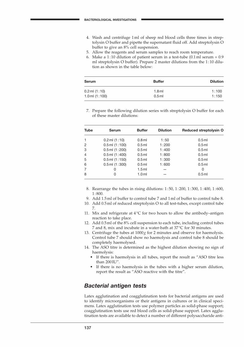
137
BACTERIOLOGICAL INVESTIGATIONS
A
4. Wash and centrifuge 1ml of sheep red blood cells three times in strep-tolysin O buffer and pipette the supernatant fluid off. Add streptolysin Obuffer to give an 8% cell suspension.
5. Allow the reagents and serum samples to reach room temperature.6. Make a 1 :10 dilution of patient serum in a test-tube (0.1ml serum + 0.9
ml streptolysin O buffer). Prepare 2 master dilutions from the 1 :10 dilu-tion as shown in the table below:
Serum Buffer Dilution
0.2ml (1 :10) 1.8ml 1 :1001.0ml (1 :100) 0.5ml 1 :150
7. Prepare the following dilution series with streptolysin O buffer for eachof these master dilutions:
Tube Serum Buffer Dilution Reduced streptolysin O
1 0.2ml (1 :10) 0.8ml 1 :50 0.5ml2 0.5ml (1 :100) 0.5ml 1 :200 0.5ml3 0.5ml (1 :200) 0.5ml 1 :400 0.5ml4 0.5ml (1 :400) 0.5ml 1 :800 0.5ml5 0.5ml (1 :150) 0.5ml 1 :300 0.5ml6 0.5ml (1 :300) 0.5ml 1 :600 0.5ml7 0 1.5ml — 08 0 1.0ml — 0.5ml
8. Rearrange the tubes in rising dilutions: 1 : 50, 1 : 200, 1 : 300, 1 : 400, 1 : 600,1 :800.
9. Add 1.5ml of buffer to control tube 7 and 1ml of buffer to control tube 8.10. Add 0.5ml of reduced streptolysin O to all test-tubes, except control tube
7.11. Mix and refrigerate at 4 ∞C for two hours to allow the antibody–antigen
reaction to take place.12. Add 0.5ml of the 8% cell suspension to each tube, including control tubes
7 and 8, mix and incubate in a water-bath at 37 ∞C for 30 minutes.13. Centrifuge the tubes at 1000 g for 2 minutes and observe for haemolysis.
Control tube 7 should show no haemolysis and control tube 8 should becompletely haemolysed.
14. The ASO titre is determined as the highest dilution showing no sign ofhaemolysis:
• If there is haemolysis in all tubes, report the result as “ASO titre lessthan 200 IU”.
• If there is no haemolysis in the tubes with a higher serum dilution,report the result as “ASO reactive with the titre”.
Bacterial antigen tests
Latex agglutination and coagglutination tests for bacterial antigens are usedto identify microorganisms or their antigens in cultures or in clinical speci-mens. Latex agglutination tests use polymer particles as solid-phase support;coagglutination tests use red blood cells as solid-phase support. Latex agglu-tination tests are available to detect a number of different polysaccharide anti-
BLM1 1/17/04 2:02 PM Page 137
gens of bacteria that cause meningitis including Haemophilus influenzae (typeb), S. pneumoniae (omnivalent), N. meningitidis (group A, B, C, Y and W135),E. coli (type K1), and S. agalactiae (group B). Latex agglutination tests are usefulfor identifying streptococci from Lancefield groups A, B, C, D, F and G. Moreover, latex agglutinating sera are available for use in qualitative slideagglutination tests and quantitative tube agglutination tests for serologicalidentification and typing of Streptococcus groups A–D, N. meningitidis, H.influenzae, Salmonella, Shigella, Vibrio cholerae, etc.
A threshold level of the antigen is required to detect polysaccharide antigensin clinical samples. The threshold level is usually exceeded if organisms canbe seen in Gram-stained preparations, although this may not always be thecase. The number of bacteria found in the cerebrospinal fluid of patients withN. meningitidis infection is significantly lower that that for other types of Neis-seria spp. infection and therefore the threshold level of polysaccharide anti-gens is reached less frequently.
Several serogroups of N. meningitidis may cause meningitis. While serogroupsA, C, Y, and W135 each have a stable antigen that can be detected with a singlepolyvalent reagent, the serogroup B antigen is relatively unstable and con-siderably more difficult to detect. It is also indistinguishable from the K1 poly-saccharide antigen of E. coli, which is the main cause of E. coli meningitis innewborn babies. However, if the Gram-stained preparation demonstratesGram-negative diplococci, infection is most likely to be due to N. meningitidisgroup B; if the Gram-stained preparation demonstrates Gram-negative rods,infection is probably due to E. coli.
For some of the tests a polysaccharide antigen is extracted from the organismbefore testing. This extraction may be carried out chemically or enzymatically.Four different tests can be used for the detection of the antigen:
• In the slide-agglutination tests, the antigen is added to latex particles orstaphylococcal cells coated with specific antibodies. The mixture is rotatedby hand with a steady circular motion for 1–2 minutes and the reaction isobserved macroscopically for agglutination.
• In the ELISA tests, a solution of sample and antigen is first passed over amembrane coated with an antibody (this antibody is usually monoclonal).Subsequently, the membrane is covered with a solution of a second (monoclonal) antibody conjugated with an enzyme. The presence of theantigen–antibody enzyme complex on the membrane is detected by itsreaction with a chromogenic substrate that is added in solution.
• In the gold immunoassay, a solution of sample and antigen is left to diffuseon a membrane, which is then examined.
• In the optical immunoassay, antibodies are attached to a silicon wafer withreflective properties. When the antigen reacts with the specific antibody onthe wafer, a change in the surface layer occurs, which causes a change inthe light reflection.
Typical procedures for latex agglutination orcoagglutination tests
1. For cell-bound antigens: use a sterilized loop to transfer well-isolatedcolonies to a drop of saline on a slide and mix carefully to obtain a slightlyopalescent emulsion. If the saline emulsion shows clumping, it usuallyindicates a rough (R) strain instead of a smooth (S) strain, and the cells willfail to agglutinate with the antibody. If no clumping appears, add thereagent and proceed from step 4.
138
SEROLOGICAL TESTS
BLM1 1/17/04 2:02 PM Page 138
139
BACTERIOLOGICAL INVESTIGATIONS
A
For extracted antigen: use a sterilized loop to transfer well-isolated coloniesto a small test-tube containing extraction solution and emulsify them. Incu-bate the emulsion at 35 ∞C or according to the manufacturer’s instructions.For clinical specimens (CSF, urine): heat the specimen to boiling-point oraccording to the manufacturer’s instructions. Cool and centrifuge at 2000g for 5–10 minutes.
2. Place 1 drop of the antibody-coated particles on the slide.3. Place 1 drop of the antigen suspension beside the antibody-coated
particles.4. Mix the 2 drops on the slide and spread in a square.5. Rotate the slide by hand in a steady circular motion for 1 minute. Take care
that the mixture does not spill outside the boundaries of the square.6. Examine the slide macroscopically for agglutination after the time speci-
fied in the manufacturer’s instructions. For best visibility hold the slidenear a bright light and view against a dark background.
7. A positive result is recorded when the antigen mixed with the antibodyshows agglutination, i.e. the suspension shows clumping or is granular to the point of curdling. Agglutination is best seen by tilting the slideslightly so that the fluid drains down towards the lower boundary of the square.
BLM1 1/17/04 2:02 PM Page 139

BLM1 1/17/04 2:02 PM Page 140

Part IIEssential media and reagents
A
BLM2 1/17/04 3:00 PM Page 141
Introduction
With just a few diagnostic materials, a laboratory can make an important contribution to individual patient care through accurate etiological diagnosis.In most developing countries bacteriological laboratory practice is hamperedby a shortage of culture media and basic reagents, which are very costly toimport. However, the number of culture media and reagents that have to bepurchased can be reduced to the essential ones, through rational selection, ashas been the case with essential drug lists. Additionally, some simple mediaand reagents can be produced or prepared locally. Application of these twoapproaches would greatly reduce the necessity for foreign exchange and makemore readily available the laboratory material necessary for patient care and epidemiological studies.
This chapter has been prepared to enable health laboratory managers to concentrate their resources on the most relevant media and reagents. It comprises two sections both composed of a series of lists.
142
BLM2 1/17/04 3:00 PM Page 142
Pathogens, media and diagnosticreagents
Expected pathogens
Pathogens are listed according to a number of factors:
— frequency of isolation,— clinical relevance,— severity of disease,— epidemic potential,— cost–benefit ratio of isolation and/or identification.
The listing is by no means absolute and will vary from country to country or from laboratory to laboratory, depending on the local disease pattern, thelaboratory capacity, and the resources available.
Media and diagnostic reagent priority gradings
A certain degree of flexibility has been incorporated by adopting a prioritygrading for media and diagnostic reagents as follows:
Grade 1: High priorityGrade 2: Intermediate priorityGrade 3: Low priority
Media and diagnostic reagents are priority graded according to the listing of the pathogens for which they are used for isolation and identification.However, there may be differences. If the medium is used broadly for morethan one pathogen, it may score higher than any one pathogen for which it isused.
Grade 1: High priority media and diagnostic reagents should be available inall laboratories that practise general diagnostic bacteriology. They are mostoften for general purpose use, easy to prepare and few in number.
Grade 2: Intermediate priority media and diagnostic reagents are additionaluseful substances that make laboratory diagnosis more complete and moreuseful for epidemiological studies, although they may not be essential fordirect patient care, e.g. the grouping antisera for meningococci.
Grade 3: Low priority media and diagnostic reagents are substances that are of value only occasionally for patient care, but which are useful for teach-ing, research, and special investigations by a reference laboratory. This category applies to media and diagnostic reagents that are too expensive forgeneral use, or that are needed for the isolation and identification of organ-isms that occur rarely or are difficult to isolate and therefore often not cost-effective.
For common laboratory investigations, the listed pathogens, and the requiredmedia and diagnostic reagents, together with a suggested priority grading,are to be found below. The priority grading should be adapted for each labo-ratory according to local circumstances.
143
A
BLM2 1/17/04 3:00 PM Page 143
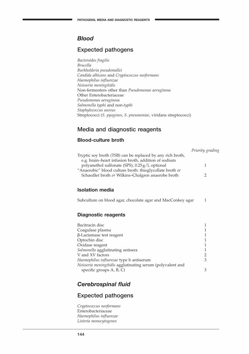
Blood
Expected pathogens
Bacteroides fragilisBrucellaBurkholderia pseudomalleiCandida albicans and Cryptococcus neoformansHaemophilus influenzaeNeisseria meningitidisNon-fermenters other than Pseudomonas aeruginosaOther EnterobacteriaceaePseudomonas aeruginosaSalmonella typhi and non-typhiStaphylococcus aureusStreptococci (S. pyogenes, S. pneumoniae, viridans streptococci)
Media and diagnostic reagents
Blood-culture broth
Priority gradingTryptic soy broth (TSB) can be replaced by any rich broth,
e.g. brain–heart infusion broth, addition of sodiumpolyanethol sulfonate (SPS), 0.25g/l, optional 1
“Anaerobic” blood culture broth: thioglycollate broth orSchaedler broth or Wilkins–Chalgren anaerobe broth 2
Isolation media
Subculture on blood agar, chocolate agar and MacConkey agar 1
Diagnostic reagents
Bacitracin disc 1Coagulase plasma 1b-Lactamase test reagent 1Optochin disc 1Oxidase reagent 1Salmonella agglutinating antisera 1V and XV factors 2Haemophilus influenzae type b antiserum 3Neisseria meningitidis agglutinating serum (polyvalent and
specific groups A, B, C) 3
Cerebrospinal fluid
Expected pathogens
Cryptococcus neoformansEnterobacteriaceaeHaemophilus influenzaeListeria monocytogenes
144
PATHOGENS, MEDIA AND DIAGNOSTIC REAGENTS
BLM2 1/17/04 3:00 PM Page 144
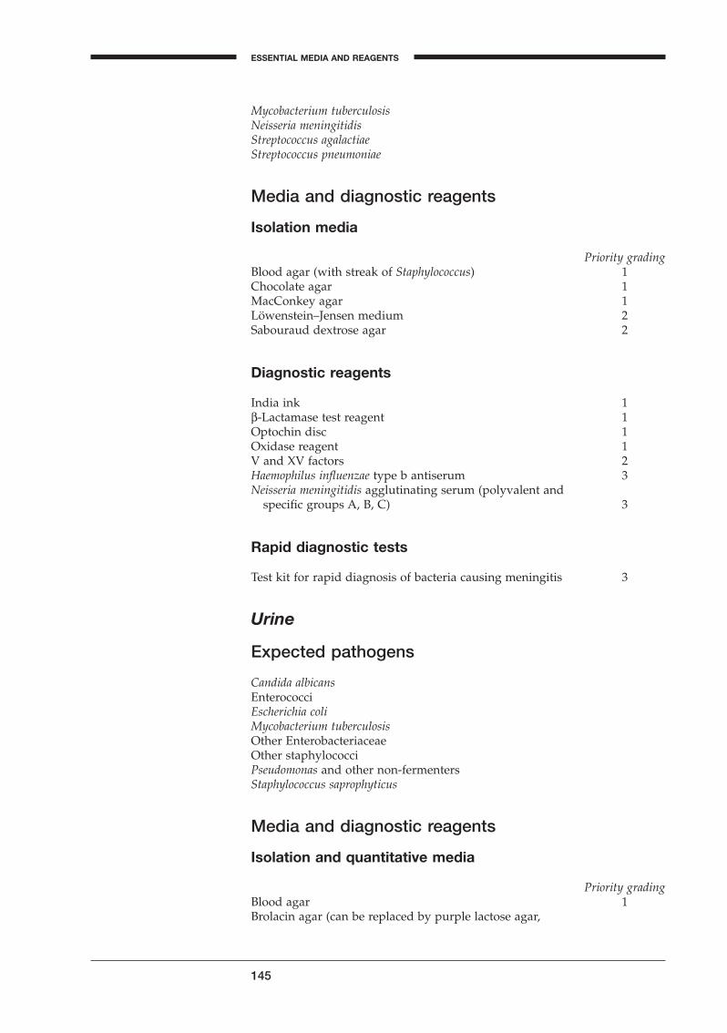
Mycobacterium tuberculosisNeisseria meningitidisStreptococcus agalactiaeStreptococcus pneumoniae
Media and diagnostic reagents
Isolation media
Priority gradingBlood agar (with streak of Staphylococcus) 1Chocolate agar 1MacConkey agar 1Löwenstein–Jensen medium 2Sabouraud dextrose agar 2
Diagnostic reagents
India ink 1b-Lactamase test reagent 1Optochin disc 1Oxidase reagent 1V and XV factors 2Haemophilus influenzae type b antiserum 3Neisseria meningitidis agglutinating serum (polyvalent and
specific groups A, B, C) 3
Rapid diagnostic tests
Test kit for rapid diagnosis of bacteria causing meningitis 3
Urine
Expected pathogens
Candida albicansEnterococciEscherichia coliMycobacterium tuberculosisOther EnterobacteriaceaeOther staphylococciPseudomonas and other non-fermentersStaphylococcus saprophyticus
Media and diagnostic reagents
Isolation and quantitative media
Priority gradingBlood agar 1Brolacin agar (can be replaced by purple lactose agar,
145
ESSENTIAL MEDIA AND REAGENTS
A
BLM2 1/17/04 3:00 PM Page 145
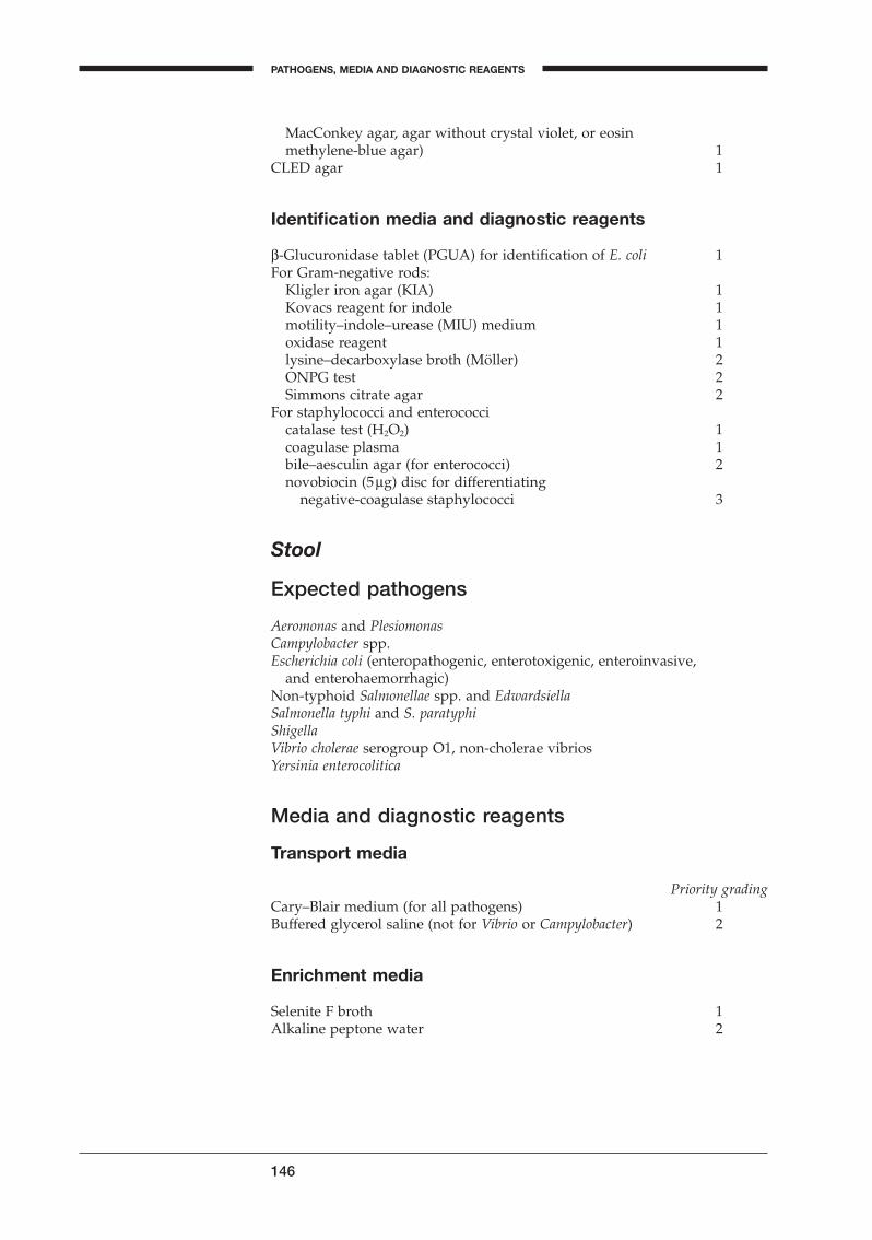
MacConkey agar, agar without crystal violet, or eosinmethylene-blue agar) 1
CLED agar 1
Identification media and diagnostic reagents
b-Glucuronidase tablet (PGUA) for identification of E. coli 1For Gram-negative rods:
Kligler iron agar (KIA) 1Kovacs reagent for indole 1motility–indole–urease (MIU) medium 1oxidase reagent 1lysine–decarboxylase broth (Möller) 2ONPG test 2Simmons citrate agar 2
For staphylococci and enterococcicatalase test (H2O2) 1coagulase plasma 1bile–aesculin agar (for enterococci) 2novobiocin (5mg) disc for differentiating
negative-coagulase staphylococci 3
Stool
Expected pathogens
Aeromonas and PlesiomonasCampylobacter spp.Escherichia coli (enteropathogenic, enterotoxigenic, enteroinvasive,
and enterohaemorrhagic)Non-typhoid Salmonellae spp. and EdwardsiellaSalmonella typhi and S. paratyphiShigellaVibrio cholerae serogroup O1, non-cholerae vibriosYersinia enterocolitica
Media and diagnostic reagents
Transport media
Priority gradingCary–Blair medium (for all pathogens) 1Buffered glycerol saline (not for Vibrio or Campylobacter) 2
Enrichment media
Selenite F broth 1Alkaline peptone water 2
146
PATHOGENS, MEDIA AND DIAGNOSTIC REAGENTS
BLM2 1/17/04 3:00 PM Page 146
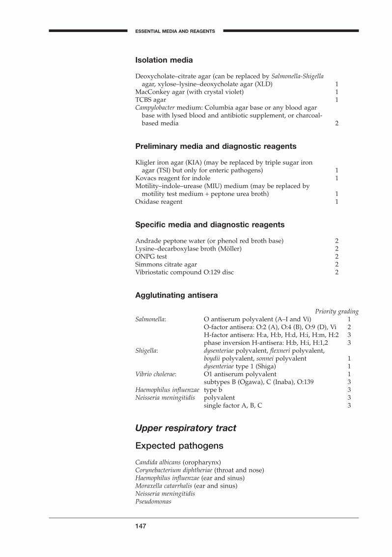
Isolation media
Deoxycholate–citrate agar (can be replaced by Salmonella-Shigellaagar, xylose–lysine–deoxycholate agar (XLD) 1
MacConkey agar (with crystal violet) 1TCBS agar 1Campylobacter medium: Columbia agar base or any blood agar
base with lysed blood and antibiotic supplement, or charcoal-based media 2
Preliminary media and diagnostic reagents
Kligler iron agar (KIA) (may be replaced by triple sugar ironagar (TSI) but only for enteric pathogens) 1
Kovacs reagent for indole 1Motility–indole–urease (MIU) medium (may be replaced by
motility test medium + peptone urea broth) 1Oxidase reagent 1
Specific media and diagnostic reagents
Andrade peptone water (or phenol red broth base) 2Lysine–decarboxylase broth (Möller) 2ONPG test 2Simmons citrate agar 2Vibriostatic compound O:129 disc 2
Agglutinating antisera
Priority gradingSalmonella: O antiserum polyvalent (A–I and Vi) 1
O-factor antisera: O:2 (A), O:4 (B), O:9 (D), Vi 2H-factor antisera: H:a, H:b, H:d, H:i, H:m, H:2 3phase inversion H-antisera: H:b, H:i, H:1,2 3
Shigella: dysenteriae polyvalent, flexneri polyvalent,boydii polyvalent, sonnei polyvalent 1dysenteriae type 1 (Shiga) 1
Vibrio cholerae: O1 antiserum polyvalent 1subtypes B (Ogawa), C (Inaba), O:139 3
Haemophilus influenzae type b 3Neisseria meningitidis polyvalent 3
single factor A, B, C 3
Upper respiratory tract
Expected pathogens
Candida albicans (oropharynx)Corynebacterium diphtheriae (throat and nose)Haemophilus influenzae (ear and sinus)Moraxella catarrhalis (ear and sinus)Neisseria meningitidisPseudomonas
147
ESSENTIAL MEDIA AND REAGENTS
A
BLM2 1/17/04 3:00 PM Page 147
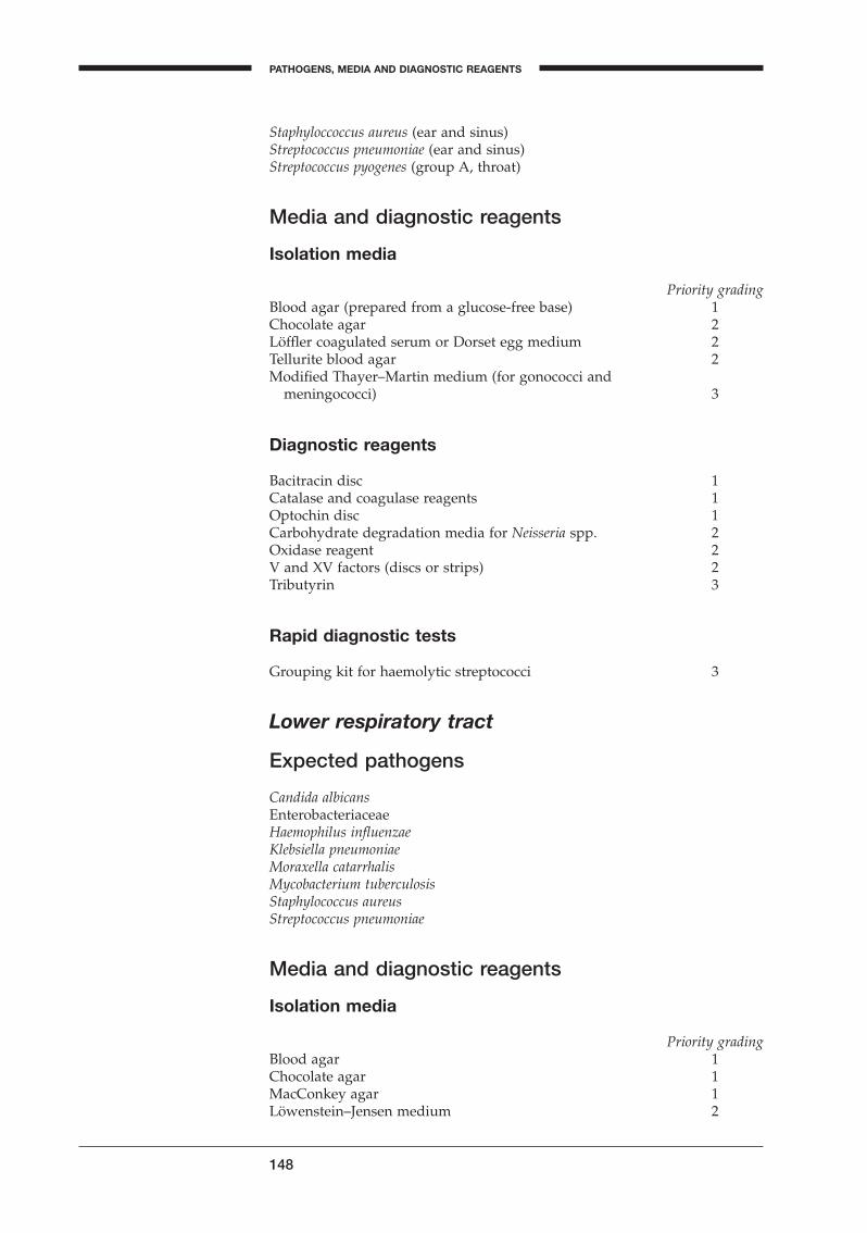
Staphyloccoccus aureus (ear and sinus)Streptococcus pneumoniae (ear and sinus)Streptococcus pyogenes (group A, throat)
Media and diagnostic reagents
Isolation media
Priority gradingBlood agar (prepared from a glucose-free base) 1Chocolate agar 2Löffler coagulated serum or Dorset egg medium 2Tellurite blood agar 2Modified Thayer–Martin medium (for gonococci and
meningococci) 3
Diagnostic reagents
Bacitracin disc 1Catalase and coagulase reagents 1Optochin disc 1Carbohydrate degradation media for Neisseria spp. 2Oxidase reagent 2V and XV factors (discs or strips) 2Tributyrin 3
Rapid diagnostic tests
Grouping kit for haemolytic streptococci 3
Lower respiratory tract
Expected pathogens
Candida albicansEnterobacteriaceaeHaemophilus influenzaeKlebsiella pneumoniaeMoraxella catarrhalisMycobacterium tuberculosisStaphylococcus aureusStreptococcus pneumoniae
Media and diagnostic reagents
Isolation media
Priority gradingBlood agar 1Chocolate agar 1MacConkey agar 1Löwenstein–Jensen medium 2
148
PATHOGENS, MEDIA AND DIAGNOSTIC REAGENTS
BLM2 1/17/04 3:00 PM Page 148
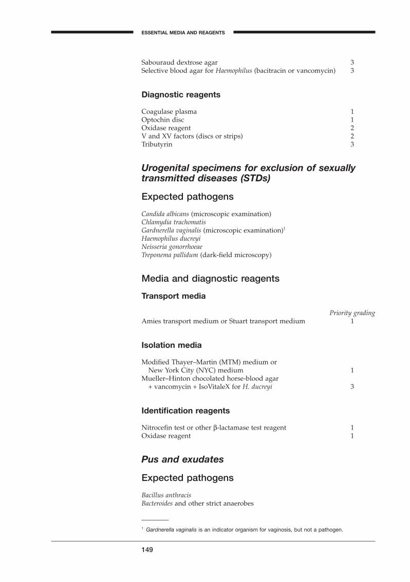
Sabouraud dextrose agar 3Selective blood agar for Haemophilus (bacitracin or vancomycin) 3
Diagnostic reagents
Coagulase plasma 1Optochin disc 1Oxidase reagent 2V and XV factors (discs or strips) 2Tributyrin 3
Urogenital specimens for exclusion of sexuallytransmitted diseases (STDs)
Expected pathogens
Candida albicans (microscopic examination)Chlamydia trachomatisGardnerella vaginalis (microscopic examination)1
Haemophilus ducreyiNeisseria gonorrhoeaeTreponema pallidum (dark-field microscopy)
Media and diagnostic reagents
Transport media
Priority gradingAmies transport medium or Stuart transport medium 1
Isolation media
Modified Thayer–Martin (MTM) medium orNew York City (NYC) medium 1
Mueller–Hinton chocolated horse-blood agar+ vancomycin + IsoVitaleX for H. ducreyi 3
Identification reagents
Nitrocefin test or other b-lactamase test reagent 1Oxidase reagent 1
Pus and exudates
Expected pathogens
Bacillus anthracisBacteroides and other strict anaerobes
149
ESSENTIAL MEDIA AND REAGENTS
A1 Gardnerella vaginalis is an indicator organism for vaginosis, but not a pathogen.
BLM2 1/17/04 3:00 PM Page 149
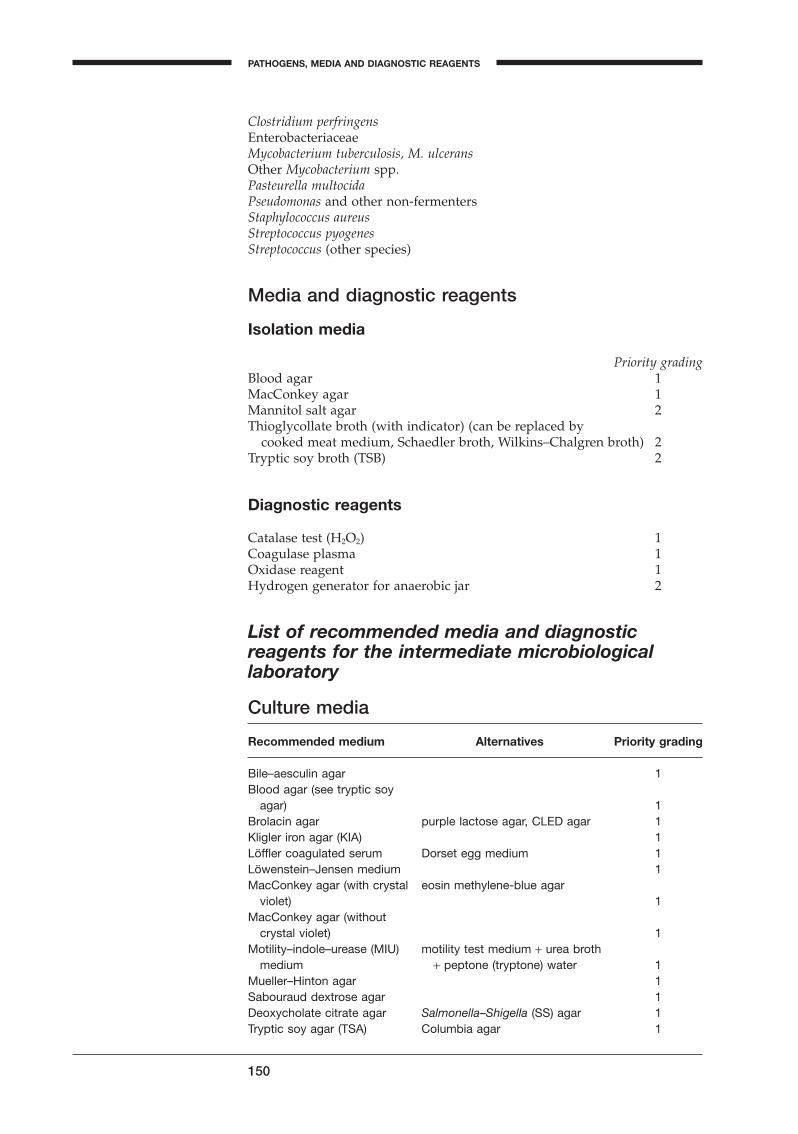
Clostridium perfringensEnterobacteriaceaeMycobacterium tuberculosis, M. ulceransOther Mycobacterium spp.Pasteurella multocidaPseudomonas and other non-fermentersStaphylococcus aureusStreptococcus pyogenesStreptococcus (other species)
Media and diagnostic reagents
Isolation media
Priority gradingBlood agar 1MacConkey agar 1Mannitol salt agar 2Thioglycollate broth (with indicator) (can be replaced by
cooked meat medium, Schaedler broth, Wilkins–Chalgren broth) 2Tryptic soy broth (TSB) 2
Diagnostic reagents
Catalase test (H2O2) 1Coagulase plasma 1Oxidase reagent 1Hydrogen generator for anaerobic jar 2
List of recommended media and diagnosticreagents for the intermediate microbiologicallaboratory
Culture media
Recommended medium Alternatives Priority grading
Bile–aesculin agar 1Blood agar (see tryptic soy
agar) 1Brolacin agar purple lactose agar, CLED agar 1Kligler iron agar (KIA) 1Löffler coagulated serum Dorset egg medium 1Löwenstein–Jensen medium 1MacConkey agar (with crystal eosin methylene-blue agar
violet) 1MacConkey agar (without
crystal violet) 1Motility–indole–urease (MIU) motility test medium + urea broth
medium + peptone (tryptone) water 1Mueller–Hinton agar 1Sabouraud dextrose agar 1Deoxycholate citrate agar Salmonella–Shigella (SS) agar 1Tryptic soy agar (TSA) Columbia agar 1
150
PATHOGENS, MEDIA AND DIAGNOSTIC REAGENTS
BLM2 1/17/04 3:00 PM Page 150
Recommended medium Alternatives Priority grading
Tryptic soy broth (TSB) brain–heart infusion broth 1TCBS 1Transport medium (Amies) transport medium (Stuart or
Cary–Blair) 1Andrade peptone water phenol red broth 2Decarboxylase broth (Möller) 2Mannitol salt agar (MSA) 2Selenite F broth 2Simmons citrate agar 2Thioglycollate medium (with Schaedler broth, Wilkens–
indicator) Chalgren anaerobe broth,cooked meat medium 2
DNase agar 3
Inhibitors or antimicrobials for use in media or as reagents
Chloramphenicol (for isolation of fungi) 1Gonococcal antimicrobial supplement: vancomycin, colistin, nystatin
(trimethoprim): VCN (VCNT) 1Campylobacter antimicrobial supplement 2Tellurite solution (for isolation of Corynebacterium diphtheriae) 2Bacitracin (for isolation of Haemophilus spp.) 3Vancomycin (for isolation of Haemophilus ducreyi or H. influenzae) 3
Enrichments for culture media
IsoVitaleX (Polyvitex, Vitox, supplement B, supplement VX,supplement CVA) 2
Sodium polyanethol sulfonate (SPS) 3
Diagnostic discs, tablets or strips
Bacitracin disc 1Nitrocefin disc (Cefinase) or reagent 1ONPG test 1Optochin disc 1Oxidase reagent 1PGUA (b-glucuronidase) 1V and XV factors 2Novobiocin (5mg) disc 3PYR test 3Tributyrin 3Vibriostatic compound O:129 disc 3
Diagnostic kits
Rapid serodiagnostic kit for identification of bacteria causing meningitis 3
Serological grouping kit for haemolytic streptococci 3
151
ESSENTIAL MEDIA AND REAGENTS
A
BLM2 1/17/04 3:00 PM Page 151
Miscellaneous diagnostic reagents
Barium sulfate standard (for Kirby–Bauer method) 1Gram-stain reagents 1Hydrogen peroxide (H2O2) (catalase) 1Kovacs reagent (for indole) 1Oxidase reagent (dimethyl-p-phenylenediamine) 1Plasma (for coagulase test and germ-tube test) 1Ziehl–Neelsen stain 1Buffered glycerol–saline (for transportation of stool) 2Carbohydrates: glucose, lactose, maltose, mannitol, sucrose 2Hydrogen generator for anaerobic jar 2India ink (for capsule detection) 2Lysine (for decarboxylase test) 2
Sensitivity-testing discs
Antimicrobials included in the WHO list of essential drugs (2002)
amoxicillinampicillinbenzylpenicillinchloramphenicolciprofloxazinco-trimoxazole (sulfamethoxazole–trimethoprim)cloxacillinerythromycingentamicinkanamycinnalidixic acidnitrofurantoinsulfonamidetetracycline (or doxycycline)trimethoprim
Reserved antimicrobials
amoxy-clavamikacincefalotincefazolincefotaximeceftazidimeceftriaxonecefuroximeciprofloxacin or other fluoquinolonesclindamycinpiperacillinvancomycin
152
PATHOGENS, MEDIA AND DIAGNOSTIC REAGENTS
BLM2 1/17/04 3:00 PM Page 152
Agglutinating antisera
Priority gradingSalmonella: O antiserum polyvalent (A–I and Vi) 1
O-factor antisera: O:2 (A), O:4 (B), O:9 (D), Vi 2H-factor antisera: H:a, H:b, H:d, H:i, H:m, H:2 3phase inversion H-antisera: H:b, H:i, H:1,2 3
Shigella: dysenteriae polyvalent, flexneri polyvalent,boydii polyvalent, sonnei polyvalent 1dysenteriae type 1 (Shiga) 1
Vibrio cholerae: O1 antiserum polyvalent 1subtypes B (Ogawa), C (Inaba), O:139 3
Haemophilus influenzae: type b 3Neisseria meningitidis: polyvalent 3
single factor A, B, C 3
153
ESSENTIAL MEDIA AND REAGENTS
A
BLM2 1/17/04 3:00 PM Page 153
Selected further reading
August MJ et al. Quality control and quality assurance practices in clinical microbiol-ogy. Cumitech, 1990, 3A:1–14.
Basics of quality assurance for intermediate and peripheral laboratories, 2nd ed. Alexandria,WHO Regional Office for the Eastern Mediterranean, 2000.
Baron EJ, Finegold SM. Diagnostic microbiology, 8th ed. St Louis, MO, The C.V. MosbyCompany, 1990.
Blazevic DJ et al. Practical quality control procedures for the clinical microbiology lab-oratory. Cumitech, 1976, 3:1–12.
Cheesbrough M. Medical laboratory manual for tropical countries. Vol. II: Microbiology.London, Tropical Health Technology/Butterworths, 1989.
Collins CH, Lyne PM. Microbiological methods, 5th ed. London, Butterworths, 1985.Gillies RP, Paul J. Bacteriology illustrated. Edinburgh, Churchill Livingstone, 1983.Howard BJ et al. Clinical and pathogenic microbiology. St Louis, MO, The C.V. Mosby
Company, 1987.Koneman EW. Color atlas and textbook of diagnostic microbiology, 5th ed. Philadelphia,
Lippincott, 1997.Miller JM. Quality control in microbiology. Atlanta, GA, Centers for Disease Control and
Prevention, 1987.Miller JM, Wentworth BB. Methods for quality control in diagnostic microbiology. Wash-
ington, DC, American Public Health Association, 1985.Montefiore DG et al. Tropical microbiology. Edinburgh, Churchill Livingstone, 1984.Murray P et al. Manual of clinical microbiology, 8th ed. Washington, DC, American
Society for Microbiology, 2003.Stokes EJ et al. Quality control. In: Clinical bacteriology, 7th ed. London, Edward Arnold,
1993.Turk DC et al. A short textbook of medical microbiology. London, Hodder & Stoughton,
1983.Summanen P et al. Wadsworth anaerobic bacteriology manual. Belmont, CA, Star Pub-
lishing Company, 1993.
154
BLM2 1/17/04 3:00 PM Page 154
Antibodies 122Anticoagulant, blood collection 22Antigens 77, 122
diagnostic 12–13latex agglutination test 137, 138–139VDRL test 128
Antimicrobial discs 13–14, 103, 104–105,108–121
potency 119recommended 151, 152
Antimicrobial resistance 17, 62, 97,101–121, 108
Antimicrobial susceptibility, definition104–105
Antimicrobial susceptibility testing, see Susceptibility testing
Antimicrobials (see also Drugs)enteric disease therapy 39generic names 108media/reagent use 151susceptibility testing discs 103,
104–105, 108–121, 151–152Antisera
agglutinating 147, 153diagnostic 12–13external quality assessment 17–18
Antistreptolysin O (ASO) test 135–137Arabinose 53Arcobacter butzleri 39, 54Argenine 38, 54Arthritis 76, 93, 96ASO, see Antistreptolysin O testAspirates 80, 86–87, 92Autoclaves, quality control 8
Bacillus anthracis 88, 91, 93, 96–97, 149Bacitracin 148, 151Bacteraemia 20–21, 24, 37, 101, 102Bacteria, oxygen requirement 98Bacteriology, quality assurance 2–18Bacteriuria, screening test 32Bacteroides
fragilis 12, 21, 87, 88, 91, 144identification 99, 101, 102stock strain 16
melaninogenicus, see Prevotellamelaninogenica
spp. 98–99, 149Balanitis 132Balanoposthitis 76Barium sulfate 12, 152Bartholinitis 76Bed sores 87, 89–90Benzalkonium chloride, see ZephiranBenzylpenicillin 79, 97, 106–108, 110,
116, 152
Abscesses 86, 87, 89, 93, 98liver 92lung 66–67
Acid-fast stain, see Ziehl–Neelsenstaining
AcinetobacterIwoffi 10–12, 24spp. 60, 70
Acquired immunodeficiency syndrome(AIDS) 20, 37, 61, 76
Actinomyces israelii 90, 93Actinomycosis 89, 90Acute bronchitis 66Aeromonas spp. 38, 53, 146Aesculin agar, see Bile–aesculin agarAfrican trypanosomiasis 26Agglutinating antisera 147, 153Agglutination tests 28, 56–59, 63,
125–126, 135–139Agglutinins tests, febrile 133–135AIDS, see Acquired immunodeficiency
syndromeAlbert stain 64Albumin, bovine 73Alcohol 21Alkaline bile salt agar 42Alkaline peptone water 40–42, 146American Type Culture Collection 14Amies transport medium 40, 62, 77, 81,
149, 151Amikacin 107, 110, 116, 152Aminoglycosides 100, 109Amoeba 26, 87, 92Amoebic dysentery 41Amoxicillin 109, 152Amoxy-clav 107, 110, 116, 152Amphotericin B 42, 78Ampicillin 107, 109, 110, 116, 152Anaemia, aplastic 108Anaerobes 10, 80, 87, 98–102
necrotizing ulcerative pharyngitis 61stab cultures 15, 24surgical specimens 91, 93wound infections 88
Anaerobic jars 8, 44Andrade peptone water 64, 147, 151Aneurysms, infected 20Animal bites/scratches 88, 96Anorectal specimens, collection 77Anthrax (see also Bacillus anthracis) 96–97Antibiograms (see also Susceptibility
tests)blood cultures 24
Antibiotic discs, see Antimicrobial discsAntibiotic susceptibility testing,
see Susceptibility testing
155
H
Index
Note: Page numbers in bold type indicate main discussions.
BLMINDEX 1/17/04 2:39 PM Page 155
Bile–aesculin agar 11, 23, 29, 64, 146, 150
Bile stimulation test 101Bismuth sulfite agar (BSA) 44, 48Bites, animal/human 88, 96Blastomyces dermatitidis 92Blind subcultures, blood samples 23Blood agar 27–29,
anaerobic specimens 100Corynebacterium diphtheriae 64CSF specimens 27Haemophilus influenzae 29, 149priority grading 144–145, 148–150,
155sputum culture 69Streptococcus pyogenes culture 63surgical specimens 92–93
Blood specimens 20–24collection 20–22culture 21, 22–24diagnostic reagents/media 144expected pathogens 144
Body cavities, purulent exudates 87, 90Bordetella pertussis 126Borrelia burgdorferi 132Bottles, blood-culture 22–23Botulism, wound 88, 98Brain heart infusion broth 144, 151Branhamella catarrhalis, see Moraxella
catarrhalisBrolacin agar 145, 150Bromothymol blue–lactose–cystine agar
34, 145, 150Bronchitis 66Bronchopneumonia 67Broth
anaerobes culture 99, 100, 101, 144,151
blood culture 22thioglycollate 12, 23, 38, 92, 144, 150tryptic soy (TSB) 22, 27, 96, 144, 150,
151Wilkins–Chalgren 100, 144, 151
Brucellaabortus 23, 135melitensis 133, 135spp. 16, 17, 23, 59, 133, 135, 144
Brucellosis 20, 133BSA, see Bismuth sulfite agarBuboes, inguinal 83Burkholderia pseudomallei 17, 21, 144Burns 87, 89–90
Calcium alginate 77Calibrated loop technique, urine
specimens 32–33Calymmatobacterium granulomatis 76, 82CAMP test 23
reverse 28, 64, 101–102Campylobacter
antimicrobial supplement 151coli 38, 50, 54, 55fetus 54, 55
156
INDEX
hyointestinalis 54, 55jejuni 38, 50, 54, 55lari 54, 55media 147spp. 7, 41, 42, 50, 98
enteric infections 37, 38, 146, 147identification 54, 55incubation 43–44
upsaliensis 54, 55Cancer, cervical 76Candida
albicans 12, 21, 61, 144–149in sputum 69STDs 76, 79, 81, 82stock strain 10, 70, 71
spp. 61, 62, 93Candidiasis, oral 61–62Candle jars 43, 69, 100Carbapenem 97Carbohydrate degradation 79, 148Carbohydrates 152Carbohydrate utilization test 28Carbol fuchsin 50Carbon dioxide 28, 77Cardiolipin lecithin 126, 127Carriers, upper respiratory tract
infections 62Cary–Blair transport medium 40–41,
146, 151Castaneda bottle 23Catalase test 13, 29, 46, 52, 64, 96, 146,
148, 150, 152Causal agents
bacteraemia 21diarrhoeal disease 37–39fungaemia 21meningitis 25pharyngitis 61–62purulent exudates 87STDs 76–77tonsillitis 61
CCFA, see Cefoxitin–cycloserine–fructoseagar
Cefalexin 109Cefalosporins 97, 109Cefalotin 107, 109, 110, 116, 152Cefalozin 110, 116, 152Cefamandole 109Cefamycin 109Cefapirin 109Cefotaxime 109, 116, 152Cefoxitin 109Cefoxitin–cycloserine–fructose (CCFA)
agar 42, 50Cefradine 109Ceftazidime 107, 110, 116, 152Ceftriaxone 107, 110, 116, 152Cefuroxime 107, 109, 110, 152Cellobiose 53Cellulitis 93, 96Centrifuges, quality control 8Cerebrospinal fluid (CSF) 25–29
diagnostic reagents/media 145
BLMINDEX 1/17/04 2:39 PM Page 156
diagnostic tests 29, 125, 127–129, 145expected pathogens 144–145macroscopic inspection 26meningitis 27microscopic examination 26–28Neisseria meningitidis 138pathogen identification 28–29specimen collection/transport 25specimen culture 27–29susceptibility testing 29
Cervical cancer 76Cervicitis 76, 79, 81Cervicofacial actinomycosis 90Cervicovaginal specimens (see also
Sexually transmitted diseases)collection 80culture 82interpretation 81–82microscopic examination 81transport 81
CFUs, see Colony-forming unitsChancre 84Chancroid 76, 83, 85Children
bacterial pharyngitis 61diarrhoeal diseases 37neonates 21, 25, 80, 82surgical specimens 93urine specimens 31
Chlamydiaspp. 66, 83trachomatis 76, 77, 79–80, 82, 83, 126,
149Chloramphenicol 107, 108, 110, 116, 151,
152Chlorhexidine 21Chlortetracycline 108Chocolate agar 11, 27–28, 69, 78, 145,
148Cholera 38, 41Chorio-amnionitis 76, 80Chromosal resistance 79Chronic bronchitis 66Ciprofloxacin 107, 110, 116, 152Citrate 22, 25, 51Citrobacter
freundii 10, 12, 17, 51spp. 25
Classification, Salmonella spp. 54–55CLED agar, see
Cystine–lactose–electrolyte-deficient agar
Clindamycin 107, 110, 116, 152Clinical relevance 4–5Clostridium
bifermentans 42botulinum 98difficile 39, 41, 42, 43, 50perfringens 10, 21, 88, 101–102, 150sordelli 42spp. 21, 24, 42, 80, 98–99, 100tetani 98
Cloxacillin 97, 108, 152
157
INDEX
H
Clue cells 81Coagulase plasma 13, 70, 144, 146, 149,
150Coagulase test, Staphylococcus aureus
94–96Colistin 78, 151Colitis 20, 38–39Collagenosis 127, 132Collection
anaerobic specimens 99anorectal specimens 77blood specimens 20–22cervicovaginal specimens 80CSF specimens 25genital ulcer specimens 83pharyngeal specimens 62sputum specimens 68stool specimens 40surgical specimens 89–90urethral specimens 77–78urine specimens 30–32
Colony-forming units (CFUs) 34–35Concentration, minimal inhibitory (MIC)
5, 71, 104–105Conjunctivitis 76, 80, 81Containers, urine specimens 30Contamination
anaerobic cultures 100blood cultures 21, 24
Control strainsbacteria 14susceptibility testing 110, 120
Cooked meat broth, anaerobes 99, 101Cooked meat medium 92, 150, 151Corynebacterium
diphtheriae 61, 65, 126, 147carriers 62culture/identification 64–65
mitis 64Coryneform rods 92Cost–benefit ratio, diagnostic tests 2Co-trimoxazole 107, 109, 110, 116, 152Crepitation 90Criteria, quality assurance 4–6Cross-reactivity 59, 132Cross-resistance, staphylococci 97Cryptococcus neoformans 4, 21, 25, 26, 28,
144CSF, see Cerebrospinal fluidCulture media (see also Media) 7–8,
10–12blood specimens 22–23CSF specimens 27–28dehydrated 7–8ordering/storage 7–8, 10performance tests 11–12preparation 8priority grading 143, 150–151quality control 10–12selection 7stool specimens 41–42, 44, 48storage 7–8, 10urine specimens 33–34
BLMINDEX 1/17/04 2:39 PM Page 157
Cultures 13–15, 27, 29anaerobic 61, 91, 93, 99–101blood specimens 22–24Candida albicans 82cervicovaginal specimens 182Corynebacterium diphtheriae 64–65CSF specimens 27–28external quality assessment 16, 17Gardnerella vaginalis 82genital ulcer specimens 85Mycobacterium tuberculosis 72–75Neisseria gonorrhoeae 78–79, 82preservation 14–16sputum specimens 69–70stab cultures 15, 24, 29stock 10, 14–16Streptococcus pyogenes 63, 63surgical specimens 92–93susceptibility testing 120urine specimens 32–35URT specimens 63–65
Cystine–lactose–electrolyte-deficient(CLED) agar 33, 146
Cystine trypticase agar (CTA) 15Cystitis 30Cytomegalovirus 76Cytotoxin 39
DCA, see Deoxycholate citrate agarDecarboxylase 11Decarboxylase broth 151Decontamination procedures,
Mycobacterium tuberculosis cultures67, 72–74
Decubitus ulcers 87, 89–90Definitions
antimicrobial resistance/susceptibility104–105
quality assessment 4quality assurance terms 2quality control 3–4
Dehydrated media 7–8Dehydrolase 11Dental surgery 89Deoxycholate citrate agar (DCA) 7, 11,
42, 44, 48, 147, 150Dermatitis 76Dextrose oxidation/fermentation
(see also Glucose) 11Diagnostic discs, see Antimicrobial
discsDiagnostic kits, recommended 151Diagnostic reagents, see ReagentsDiagnostic sensitivity 5–6, 13, 83Diagnostic specificity 6, 13, 83Diagnostic tests (see also Tests)
error sources 2quality assurance 2–18
Diarrhoeal diseases 37–39, 41Dicloxacillin 108Diffusion test, antimicrobial resistance
104Dilution, serological tests 131–133, 137
158
INDEX
Dilution test, antimicrobial resistance103–104
Dimethyl-p-phenylenediamine (oxidase reagent) 52
Diphtheria (see also Corynebacteriumdiphtheriae) 61, 64–65
Disc-diffusion technique, seeKirby–Bauer disc-diffusiontechnique
Discs, antimicrobial 13–14, 103, 104–105,107–121, 151–152
Disinfection, skin 21, 25Dispatch (see also Transport)
Mycobacterium tuberculosis cultures75
pharyngeal specimens 62sputum specimens 68
Distilled water, serological tests 124DNAse agar 94, 151Donovanosis, see Granuloma inguinaleDorset egg medium 64, 151Doxycycline 152Drugs (see also Antimicrobials)
nonproprietary names 108susceptibility testing 107–109WHO list of essential 152
Dysenteriae polyvalent antiserum 147,153
Dysentery 37–38, 41
Edwardsiella tarda 51, 146Egg yolk agar base 64Elek test (in-vitro toxigenic test) 38, 64ELISA test 39, 138Encephalitis 25Endocarditis 20Endometritis 76, 80Endovascular infections 20Enrichment
culture media 151stool specimens 41–42, 146
Enteric bacteria, colony morphology48
Enteric infections (see alsoStool specimens) 37–38
Enterobacteragglomerans, see Pantoea agglomeranscloacae 7, 10, 15, 25, 28, 45–46, 48, 60,
69–71spp. 21, 48
Enterobacteriaceae 4, 7, 10, 15, 28,144–145, 148, 150
colony morphology 48identification 45–50Kirby–Bauer technique 103meningitis 25in pharynx 60, 148sputum cultures 69, 70, 71stab cultures 15susceptibility testing 107, 116
Enterococci 145Enterococcus faecalis 11, 63, 64Enterocolitis 20, 38
BLMINDEX 1/17/04 2:39 PM Page 158
Epidemiology 107Epididymitis 76, 77Epiglottitis 60Epstein–Barr virus 126Equipment
anaerobic jars 8, 44blood-culture 22–23care of 7operating temperature records 9quality control 8–9serological tests 123
Error sources, diagnostic tests 2Erythrocytes 41Erythromycin 71, 107, 109, 110, 116, 152Escherichia
coli 11–12, 17, 88, 91, 145bronchopneumonia 67colony morphology 48control strains 14, 110diarrhoeal disease 38, 41b-glucuronidase test (PGUA)
35–36latex agglutination test 138meningitis 25, 138performance tests 10–11in pharynx 60Shigella differentiation 50–51, 59stool specimens 37–38, 41, 146susceptibility testing 120urinary tract infection 30, 35–36,
94wound infections 88
fergusonii 38hermanii 38
Essential drugs, WHO list 152Ethylenediamine tetraacetic acid (EDTA)
95Exudates, purulent, see Purulent
exudates; Surgical specimens
Facultative anaerobic bacteria 98Faecal specimens, see Stool specimensFalse negatives, blood cultures 21False positives, FTA(Abs) test 131–132Fasciola hepatica 92Febrile agglutinins tests 133–135Females
genital specimens 79–82urine specimens 31
Fevers 37, 133–135Filter-paper dip-strip method, urine
specimens 33–34Flexneri polyvalent antiserum 147, 153Flocculation reactions, serological tests
125Flora
mixed anerobic 4normal pharyngeal 60–61
Flucloxacillin 108Fluorescence microscopy 123, 131–132Fluorescent treponemal antibody
absorption (FTA-Abs) test 123,124, 126, 127, 131–133
159
INDEX
H
Francisella tularensis 133Fungaemia 20–21Fungi 10
fungaemia 20–21meningitis 25–28pharyngitis 61surgical specimens 92, 93
Fusobacterium spp. 61Fusospirochaetal complex 61, 62
Gardnerella vaginalis 76, 79, 80, 82, 149Gas gangrene 88, 93, 98, 101Gastroenteritis 37–38Gelatinase 11, 48Generic names, antimicrobials 108Genital herpes 76, 82Genital specimens 77, 79–82Genital ulcers 82–5Gentamicin 107, 109, 110, 116, 152Ghost cells 41Giemsa stain 85Glassware
equipment 8serological tests 123–124
Glomerulonephritis 61, 135Glucose 27, 46, 52, 63Glucuronic acid 35b-Glucuronidase test, Escherichia coli 13,
35–36, 146, 151Gluteal abscesses 92Glycerol, long-term preservation 14Gold immunoassay 138Gonococcal antimicrobial supplement
151Gonococcal pharyngitis 61Gonococcal urethritis 77Gonococci, culture preservation 16Gonococcus, see Neisseria gonorrhoeaeGonorrhoea 61, 78, 81Gram-negative organisms 10, 20
Bacillus anthracis 96CSF cultures 27meningitis 28, 29piperacillin 116respiratory tract infection 61sputum specimens 69stool cultures 45, 47surgical specimens 87, 91, 96urine cultures 34
Gram-positive organisms 10, 20, 27–28,91
CSF cultures 27sputum specimens 69, 70stool cultures 46–47surgical specimens 91, 95urine cultures 34
Gram-stained smears (see also Gram staining) 64
bacterial vaginosis 81CSF specimens 26–27sputum specimens 69surgical specimens 91, 92, 93urine specimens 32
BLMINDEX 1/17/04 2:39 PM Page 159
Gram staining (see also Gram-stainedsmears) 13, 26, 31, 77, 152
anaerobes 100, 101Granulocytopenia 61Granuloma inguinale (donovanosis) 76,
82, 83, 85Growth factors 64, 100Gunshot wounds 88
H factor antisera 147, 153Haemagglutination tests, stool cultures
53–54Haemin 85, 100Haemolysin 23, 63, 135Haemolysis
alpha 11, 28, 50, 70antibiotic susceptibility 29beta 11, 28, 63, 64performance testing 10–11reverse CAMP test 101
b-Haemolytic streptococci 61, 63–64grouping kit 148
Haemophilusducreyi 76, 82, 83, 85, 149influenzae 10–11, 25, 28, 144, 147, 148
agglutinating antisera 145, 147, 153latex agglutination test 138lower respiratory tract infections
66, 67, 69, 70, 71surgical specimens 93susceptibility tests 106
parainfluenzae 10, 11spp. 16, 60, 63, 126
HBV, see Hepatitis B virusHEA, see Hektoen enteric agarHeart infusion agar 42, 48Hektoen enteric agar (HEA) 42, 48Hepatitis B virus (HBV) 76Heroin 127Herpes, genital 82Herpesvirus, human (HSV) 76, 77, 80, 82Heteroresistance 97Histoplasma capsulatum 92HIV, see Human immunodeficiency virusHospital-acquired infection, see
Nosocomial infectionHot-air ovens, quality control 8HPV, see Human papilloma virusHSV, see Herpesvirus; Human
herpesvirusHuman herpesvirus 76, 77, 80, 82Human immunodeficiency virus (HIV)
20, 37, 61, 76, 83Human papilloma virus (HPV) 76Hydrogen generator 44, 150, 152Hydrogen peroxide 44, 152Hydrogen sulfide 46, 51Hyphae, fungal 92
Iatrogenic infection 20Identification (see also Interpretation)
anaerobic bacteria 101–102Bacillus anthracis 93, 96–97
160
INDEX
Bacillus fragilis 101Clostridium perfringens 101–102Corynebacterium diphtheriae 64–65CSF pathogens 28–29Neisseria gonorrhoeae 79Pasteurella multocida 93, 96Peptostreptococcus spp. 102Salmonella spp. 44–46, 50–51sputum pathogens 70Staphylococcus spp. 94–96stool pathogens 44–59Streptococcus pyogenes 63surgical specimen pathogens 93–97urine pathogens 32–36Vibrio cholerae 47–48, 52–53Yersinia enterocolitica 47, 52–53, 59
IFA, see Indirect fluorescent antibodytest
Immunocompromised patients 20, 60,82
diarrhoea 37oral candidiasis 61
Immunofluorescence assay 77Incubation
anaerobic specimens 99–102blood cultures 23Campylobacter spp. 43–44Clostridium difficile 43Kirby–Bauer method 114stool specimens 43–45susceptibility tests 118Vibrio cholerae 43Yersina spp. 43
Incubators, quality control 8India ink 145, 152Indirect fluorescent antibody (IFA)
test 126Indole 46, 51Indole test 96Infants, urine specimens 31Infertility 76, 80Influenza 67Inhibitors, media/reagent use 151Inoculation
anaerobic specimens 100–101stool specimens 41–43surgical specimens 92–93susceptibility testing 111–114, 118,
120–121urine specimens 33–34
Inspection, equipment 8Intensive-care departments, infections
67Intermediate susceptibility 105Interpretation
cervicovaginal cultures 81Mycobacterium tuberculosis cultures 74sputum cultures 69–70test results 3urethral cultures 78urine cultures 34–35
Iodine 21Isolation, enteric pathogens 42–44
BLMINDEX 1/17/04 2:39 PM Page 160
Isolation media 43, 144–149IsoVitaleX 78, 149, 151
Jarsanaerobic 8, 44candle 43, 69, 100
Kanamycin 100, 109, 152Kirby–Bauer disc-diffusion technique
(modified) 13, 71, 97interpretative chart 116susceptibility testing 103, 104–105,
109–119Kits, diagnostic 151Klebsiella
pneumoniae 10–11, 13, 17, 67, 148spp. 48, 60, 91
Kligler’s iron agar (KIA) 11, 23, 51, 54priority grading 146, 147, 150stool specimens 44–49
Kovacs reagent for indole 46, 146, 147,152
Laboratoriesappropriate resources use 39–40operations manual 6
Lacerations, infected 89–90b-Lactamase production 71, 79, 85, 97,
106, 108test reagent 144–145
Lactobacilli, vaginal 80Lactose 44–45, 46Lactose non-fermenting bacteria 44–45Latex agglutination tests 63, 135–139Lecithinase 42, 50Legionella pneumophila 66, 126Legionnaire disease 66Leigh & Williams method, urine cultures
33Leprosy 127Leptospirosis 20Leukocyte esterase 32Leukocytes
CSF specimens 25–27polymorphonuclear 78stool specimens 41urine specimens 32
Lipase 42, 50Listeria monocytogenes 21, 80, 144
meningitis 25, 28, 29Liver abscesses 92Löffler coagulated serum 64, 148, 150Loop technique, calibrated 32, 33Löwenstein–Jensen medium 28, 67, 72,
93, 145, 148, 150Lower respiratory tract infections 66–75
common 66–68culture/interpretation 69–70, 72–74diagnostic reagents 149expected pathogens 148media 148–149specimen collection/processing 68–69susceptibility testing 70–71
161
INDEX
H
Lumbar puncture 25Lung abscesses 66–67Lyme disease 132Lymph nodes
spontaneous drainage 90surgical specimens 87
Lymphogranuloma venereum 76Lyophilization 14Lysine 46, 151Lysine decarboxylase 46, 152Lysine decarboxylase broth 146, 147
MacConkey agar 11anaerobes 100CSF cultures 27, 28priority grading 144–148, 150stool cultures 42, 47–48, 50surgical cultures 92, 93urine cultures 33–34
Macroscopic evaluationCSF specimens 26sputum specimens 68–69surgical specimens 90–91
Maintenanceequipment 8, 123stock cultures 14–16
Malaria 127Males
urethritis 77–79urine specimens 31
Malonate broth 11Maltose 64Mannitol 51–52, 70Mannitol salt agar 11, 23, 70, 93, 150,
151Manuals, laboratory operations 6–7Mastitis 94Materials
ASO tests 136febrile agglutinins test 134FTA-Abs test 132RPR test 130serological tests 123–124VDRL test 127
Measles 67Media (see also Culture media) 7–12, 42,
142anaerobic specimens 99–101blood specimens 22–23, 144Corynebacterium diphtheriae culture 64CSF specimens 27–28, 145essential 141–53exudates/pus 92–93, 150lower respiratory tract specimens
69–70, 148–149Mycobacterium tuberculosis culture 74Neisseria gonorrhoeae culture 78priority gradings 143, 150–151quality control 10–12sputum specimens 67, 69–71stool specimens 41–42, 146–147storage 10Streptococcus pyogenes culture 63
BLMINDEX 1/17/04 2:39 PM Page 161
surgical specimens 92–93susceptibility testing 110–111upper respiratory tract specimens 64urine specimens 33, 145–146urogenital specimens 149
Medusa head 96Mellibiosa 53Menadione 100Meningitis 25–28, 86
anthrax 96Escherichia coli 138neonates 21, 25Pasteurella multocida 96pathogens 138viral 76
Meningococcus, see Neisseria meningitidisMetacillin, resistance to 17, 62, 97, 108Methyl red/Voges Prosgauer 11Methylene blue 8, 41, 64, 77Meticillin-resistant Staphylococcus aureus
(MRSA) 17, 62Metronidazole 102MIC, see Minimal inhibitory
concentrationMicroaerophilic bacteria 98Microscopes
maintenance 123quality control 8
Microscopic examinationcervicovaginal specimens 81CSF specimens 26–28genital ulcer specimens 83–85pharyngeal specimens 62respiratory tract infections 62sputum specimens 69STD specimens 78, 81, 83–85surgical specimens 86, 91–92Treponema pallidum 83–85urethral specimens 78
Microscopy, dark field 50, 82, 83MIL, see Motility–indole–lysine mediumMIU, see Motility-indole-urease mediumMineral oil 15Minimal inhibitory concentration (MIC)
5, 71, 104–105Minocycline 108Mobiluncus spp. 76, 79–80Monitoring, equipment 8Mononucleosis, infectious 126Moraxella catarrhalis 10, 60, 66, 69, 70, 71,
147, 148Morganella 51Morphology, common enteric bacteria
colonies 48Motility–indole–lysine medium (MIL)
44, 45–46, 47Motility–indole–urease medium (MIU)
146–147, 150MRSA, see Meticillin-resistant
Staphylococcus aureusMTM, see Thayer–Martin mediumMueller–Hinton agar 11, 14, 20, 24, 54,
71, 110–111, 120, 150
162
INDEX
Mueller–Hinton chocolate horse bloodagar 149, 150
Mycetoma 90–92Mycobacteria, HIV/AIDS 37, 87, 92, 93,
126, 150Mycobacterium
bovis 74chelonei 92fortuitum 92, 93marinum 92tuberculosis 25, 66, 72–75, 87, 92, 145,
148, 150ulcerans 92, 150
Mycoplasmahominis 76pneumoniae 66, 122
Mycoses, lymph nodes 87
N-acetyl-L-cysteine-sodium hydroxide(NALC) 72, 73
Naegleria fowleri 26Nafcillin 108NALC, see N-acetyl-L-cysteine-sodium
hydroxideNalidixic acid 107, 110, 116, 152Nasopharyngitis 60Necrotizing ulcerative pharyngitis
(Vincent angina) 61, 62Needles, serological tests 124Neisseria
catarrhalis, see Moraxella catarrhalisgonorrhoeae 10, 12, 76, 77, 79–80, 81,
149culture 78–79, 82identification 79susceptibility testing 79, 106
meningitidis 10, 12, 21, 25, 28, 29,144–145, 147
agglutinating antisera 144–145, 147,153
carriers 62latex agglutination test 138susceptibility 106
Neomycin 100Neonatal infection 80, 82Neonatal meningitis 21, 25Netilmycin 109New York City medium 78, 149NGU, see Nongonococcal urethritisNitrate broth 11Nitrate reduction tests 32, 64, 96Nitrocefin test 79, 149, 151Nitrofurantoin 107, 109, 110, 116, 152o-Nitrophenyl-b-D-galactopyranoside
(ONPG) test 13, 51, 146, 147, 151p-Nitrophenyl-b-D-
glucopyranosiduronic acid(PGUA) test 13, 35–36, 151
Nocardia asteroides 93Nongonococcal urethritis (NGU) 77, 78Nonproprietary names, drugs 108Non-trepanomal tests 126–131Non-tuberculous acid-fast bacilli 92
BLMINDEX 1/17/04 2:39 PM Page 162
Norfloxacin 107, 110, 116Normal flora, pharynx 60–61Nosocomial infections 21, 67, 88–89Novobiocin 146, 151Nystatin 78, 151
O antiserum polyvalent 147, 153Obligate bacteria, aerobic/anaerobic 98Oleandomycin 109ONPG, see o-Nitrophenyl-b-D-
galactopyranoside testOperating temperature, equipment 9Operations manuals 6–7Opthalmia neonatorum 80Optochin discs 13, 23, 28, 69, 70, 144,
145, 148, 149, 151Oral candidiasis 61–62Oral surgery 89Ordering procedures, dehydrated media
7–8Ornithine 11, 28, 51Ornithine decarboxylase 51–52Otitis media 60, 76Ovens, hot-air 8Oxacillin 71, 97, 107, 108, 110, 116Oxidase reaction 79, 96Oxidase reagents 13, 51, 144–153Oxidase test, stool specimens 49Oxygen, bacteria requirement 98Oxytetracycline 108
Pantoea agglomerans 21Paragonimus westermani 92Parasitic infection, surgical specimens 92Pasteurella multocida 88, 150
identification 93, 96Pathogens 143
blood 144CSF 144–145enteric disease 37–39, 42lower respiratory tract 148pus/exudates 149–150STDs 149stool 146upper respiratory tract 147–148urine 145
Pelvic inflammatory disease (PID) 76, 80Penicillins, susceptibility testing 108Peptone urea broth 147, 150Peptone water 11, 51Peptostreptococcus spp. 67, 98–99, 102Performance tests
culture media 11–12prepared media 10stains/reagents 12, 13
Pericardial sac, exudates 87Peritoneal cavity, purulent exudate 87,
90Petroff procedure, see Sodium hydroxide
procedurePGUA, see p-Nitrophenyl-b-D-
glucopyranosiduronic acid testPharyngitis 60–65, 76
163
INDEX
H
Pharynx, normal flora 60–61Phenol red broth 147, 151Phenylalanine 11, 51Phenylalanine deaminase 51pH testing 10Piperacillin 107, 110, 116, 156Pivampicillin 109Plasma, Staphylococcus aureus coagulase
test 95–96, 152Plesiomonas shigelloides 51, 53, 59, 146Pleural cavity, exudates 87, 90Pleural effusion 66, 67Pleurisy 66Pneumococcus, see Streptococcus
pneumoniaePneumonia 61, 66, 67–68
atypical 127Corynebacterium trachomatis 76, 80
Polymorphonuclear leukocytes 78Polymyxin B susceptibility test 53, 54Polysaccharides 63Polyvidone iodine 21Polyvitex 151Porphyromonas spp. 87, 98Potassium hydroxide 80, 92Precipitin reactions, see Flocculation
reactionsPregnancy 80, 127, 132Preservation, stock cultures 14–16Prevotella melaninogenica 67, 87Prevotella spp. 98Priority gradings, media/reagents
143–152Proctitis 76Propionibacterium acnes 21, 24Prostatitis 30, 76Protein A 125Proteus
mirabilis 10–12spp. 10–12, 21, 48, 51, 117vulgaris 133
Providentia spp. 48, 51Prozone reaction 129, 133Pseudobacteraemia 21Pseudomembranous colitis 39Pseudomonas
aeruginosa 10–11, 87, 90, 110, 144bronchopneumonia 67susceptibility tests 107, 116, 120
cepacia 17pseudomalei, see Burkholderia
pseudomaleispp. 22, 91, 145, 147, 150
in pharynx 60sputum specimens 69, 70
Public health, enteric disease 39Pulmonary tuberculosis 66, 67–68Purple lactose agar 145, 150Purulent exudates 77, 86–97, 149–150
abscesses 67, 89collection/transport 86–87, 89culture 89, 92–93diagnostic reagents/media 150
BLMINDEX 1/17/04 2:39 PM Page 163
expected pathogens 149–150pathogen identification 93–97wound infections 87–88
Pus 86, 89, 91, 93lung abscesses 67macroscopic evaluation 90–91
Pyelonephritis 20, 30Pyocyanin 90Pyogenic bacteria 68Pyomyositis 94PYR, see Pyrrolidonyl-b-naphthylamide
testPyrrolidonyl-b-naphthylamide (PYR)
test 64, 151
Quality assessment 4, 16–18Quality assurance 2–18
external 4internal 3–4
Quality controldefinition 3–4equipment 7, 8internal 6–16prepared media 10–12serological tests 122–124stock strains 10susceptibility tests 120–121
Quality criteria, microbiology 4–6Quantitative cultures
serological tests 129, 131urine specimens 32–35
Quinolones 102
Rabies 88Rapid diagnostic tests
CSF specimens 145, 151URT specimens 148
Rapid plasma reagin (RPR) test 123–124,126, 129–131
Reagents 142, 150–153ASO tests 136blood specimens 144CSF specimens 145febrile agglutinins test 134FTA-Abs test 132Kirby–Bauer method 110–111lower respiratory tract 149miscellaneous diagnostic 152performance tests 11–12, 13priority gradings 143pus/exudates 150recommended 150–151RPR test 130serological tests 124stool specimens 147susceptibility testing 110–111upper respiratory tract 148urine specimens 146urogenital specimens 149VDRL test 127–128
Reagin 126, 127Records, equipment operating
temperature 9
164
INDEX
Rectal swabs, collection 40Reference laboratories, use of 16Refrigerators, quality control 8Reiter syndrome 76Reliability, diagnostic tests 2, 5, 16Reproducibility, diagnostic tests 2, 5Resistance
antimicrobial 101–121metacillin and related drugs 17, 62,
97, 108Respiratory tract infections, see Lower
respiratory tract; Upperrespiratory tract
Results, reporting 18Reverse CAMP test 28, 64, 101–102Rhamnose 51, 53Rheumatic disease 127Rheumatic fever 61, 135Rickettsia spp. 126, 133Rotavirus, diarrhoea 39RPR, see Rapid plasma reagin testRubella 17, 126
Sabouraud dextrose agar 28, 70, 93, 145,149–151
Saccharose 64Safety, Mycobacterium tuberculosis
cultures 74–75Saline 112, 124, 127, 128, 132Salmonella
arizonae 51choleraesuis 51groups C and D 58non-typhi 144paratyphi 17, 46, 51, 58, 146spp. 10–12, 25, 37, 40, 41, 43, 106,
146agglutinating antisera 144, 147,
153colony morphology 48identification 44–46, 50–51nomenclature 54–55serological identification 54–58
typhi 17, 21, 42, 51, 107, 133, 144, 146typhimurium 10–12
Salmonella–Shigella agar 7, 11, 42–44, 48,147, 150
Salmonellosis 37, 41Salpingitis 76Schaedler broth 92, 144Screening, urine specimens 32Selenite F broth 11, 41, 146, 151Sensitivity, diagnostic 5–6, 13, 83Sensitivity testing, see Susceptibility
testingSepticaemia 20, 96Sera 122, 131
quality assessment 17–18serological tests 124
Serological tests 12–13, 122–139Salmonella spp. 54–58syphilis 126–133
Serratia marcescens 10, 21, 51, 91
BLMINDEX 1/17/04 2:39 PM Page 164
Sexually transmitted diseases (STDs)76–85
causal agents 76–77diagnostic reagents/media 149expected pathogens 149specimen collection/transport 77–78,
80–81, 83specimen culture 78–79, 82, 85susceptibility tests 79
Shigellaboydii 38, 46dysenteriae 38, 42, 47flexneri 10–12, 36, 38, 46sonnei 38, 51spp. 10–12, 16, 17, 20, 37–38, 146
agglutinating antisera 147, 153final identification 50–52identification 44–47latex agglutination test 138serological identification 59stool specimens 41, 42, 43, 46–47, 51susceptibility tests 107, 109
Shigellosis 41Shock, septic 20Silver nitrate 83Simmons citrate agar 11, 23, 51, 146–147,
151Sinus tract, spontaneous drainage 90Sinusitis 60Sjögren syndrome 127Skin disinfection
blood collection 21CSF collection 25
Skin infections 87–88, 92, 94Slide agglutination tests 28, 63, 138Slide clumping test 95Smears, see Gram-stained smearsSodium citrate 73Sodium hydroxide procedure (Petroff),
Mycobacterium tuberculosis cultures72–73
Sodium metabisulfite–sodium pyruvate42
Sodium polyanethol sulfonate (SPS) 22,144, 151
Sonnei polyvalent antiserum 147, 151Sorbitol 63Soy casein agar, see Tryptic soy agarSpecificity, diagnostic 6, 13, 83, 122, 126,
132Specimen collection, see CollectionSpecimen preparation
blood 22–23CSF 26
Specimen transport, see TransportSpiramycin 109Spirochaetes 61, 84, 126SPS, see Sodium polyanethol sulfonateSputum specimens 72–74
collection 68culture/interpretation 69–70macroscopic evaluation 68–69microscopic examination 69
165
INDEX
H
pulmonary tuberculosis 67, 67–68transport 75
Stab cultures 15, 24, 29Stab wounds 88Stains (see also Gram staining;
Ziehl–Neelsen staining)Albert 64Giemsa 85performance tests 12, 13
Standardization, disc-diffusion methods119
Standard strains, quality control 120Staphylococcus
aureus 10, 12, 21, 80, 94–96, 144, 148bronchopneumonia 67carriers 62identification 70in pharynx 60–61sputum specimens 69susceptibility testing 120wound infections 88
epidermidis 20, 21, 24, 60, 94–96saprophyticus 94–96, 145spp. 11–12, 25, 145
surgical specimens 87, 93, 94–96susceptibility testing 97, 107vancomycin 116
STDs, see Sexually transmitted diseasesSterility testing, prepared media 10Stock cultures 10, 14–16Stool specimens 37–59
anaerobic cultures 100collection and transport 40enrichment and inoculation 41–43expected pathogens 146laboratory resource use 39–40media 42, 146–147pathogen identification 7, 42–43,
44–59reagents 147serological identification 54–59visual examination 41
Storagedehydrated media 7–8prepared media 10stock cultures 14–16
Streptococcusagalactiae 10, 21, 25, 28, 29, 64, 145groups A and B 61, 63, 126, 135mitis 10pneumoniae 10, 21, 25, 66, 67, 69, 70,
71, 145, 148antibiotic susceptibility 28latex agglutination test 138meningitis 25upper respiratory tract infections
60, 148pyogenes 10, 21, 61, 62, 63–64, 126, 135
carriers 62culture/identification 63pharyngitis 61priority grading 144, 148, 150susceptibility 106
BLMINDEX 1/17/04 2:39 PM Page 165
spp. 10, 15, 23, 61, 144ASO test 135–137culture preservation 16surgical specimens 87, 91, 93, 150
Streptolysin O 135Streptomycin 109String test, stool specimens 49–50Strips, diagnostic 151Stuart transport medium 40, 62, 77, 81,
149, 151Subcultures, blood samples 23Subcutaneous tissue, infections 87Sucrose fermentation 48, 51–53Sulfamethoxazole 109Sulfonamide 107, 108, 110, 116, 152Sulfur granules 89Supplements 151Surgical specimens 86–87
collection/transport 89culture 92–93macroscopic evaluation 90–91microscopic examination 91–92pathogen identification 93–97susceptibility testing 97
Surveys, quality assessmentprogrammes 17
Susceptibility testing 13–14, 103–121anaerobic specimens 102basic antimicrobials 107–109benzylpenicillin 108, 110, 116CSF cultures 29direct vs indirect 117general principles 103–105indirect 117Neisseria gonorrhoeae 79plate size 118–119recommended discs 152sputum cultures 70–71stool cultures 54surgical specimens 97throat specimens 65treatment guide 106urine cultures 36
Syphilis (see also Trepanoma pallidum) 76,82, 83–85
serological tests 13, 125, 126–133
Tablets, diagnostic 151Talampicillin 109TCBS, see Thiosulfate citrate bile salts
sucrose agarTellurite agar 42, 48, 64, 148, 151Tellurite discs 13, 23Tellurite taurocholate gelatine agar
(TTGA) 42, 48Temperature
equipment operating records 9susceptibility tests 118
Terminology, microorganisms 5Tests
agglutination 56–59, 125–126, 135–139antimicrobial susceptibility 13–14,
103–121
166
INDEX
ASO 135–137diffusion/dilution 103–104febrile agglutinins 133–135FTA-Abs 123, 124, 126, 127, 131–135media performance 11–12ONPG 13, 51, 146, 147, 151PGUA 13, 35–36rapid diagnostic 145, 148, 151RPR 123–124, 126, 129–131serological 12–13, 122–139stool specimens 49–50
Tetanus 88, 98Tetracycline 71, 107, 108, 110, 116Thayer–Martin medium (MTM),
modified 12, 61, 78, 148, 149Thiamphenicol 108Thioglycollate broth 12, 23, 38, 92, 144,
150, 151Thiosulfate citrate bile salts sucrose
(TCBS) agar 12, 42, 47–48, 147,151
Throat swabscollection 62culture/identification 63–65gonococcal pharyngitis 61
Thrombophlebitis 20Tobramycin 107, 109, 110, 116Tonsillitis 60, 61Toxicity assays 38, 64Toxigenicity 64Toxic shock 20, 80Transport (see also Dispatch)
anaerobic specimens 99cervicovaginal specimens 81CSF specimens 25respiratory tract specimens 62sputum specimens 68STD specimen media 149stool specimens 40, 146surgical specimens 89urethral specimens 77–78urogenital specimens 81
Transport media 81Amies 40, 41, 62, 66, 77, 81, 149, 151Buffered glycerol saline 146, 152Cary–Blair 40–41, 146, 151Stuart 40, 62, 77, 81, 149, 151
Treatment, susceptibility tests as guide106
Treponemapallidum 76, 82, 83–85, 125–126, 149vincentii 61
Tributyrin 70, 148–151Trichomonas vaginalis 77, 81Trimethoprim 78, 107, 110, 116, 152Triple sugar iron agar 12, 70, 147Trisodium phosphate 26, 73Trypanosomiasis, African 26Tryptic soy agar (TSA) 15, 63, 150Tryptic soy broth (TSB) 22, 27, 96, 144,
150, 151Tubercle bacillus, see Mycobacterium
tuberculosis
BLMINDEX 1/17/04 2:39 PM Page 166
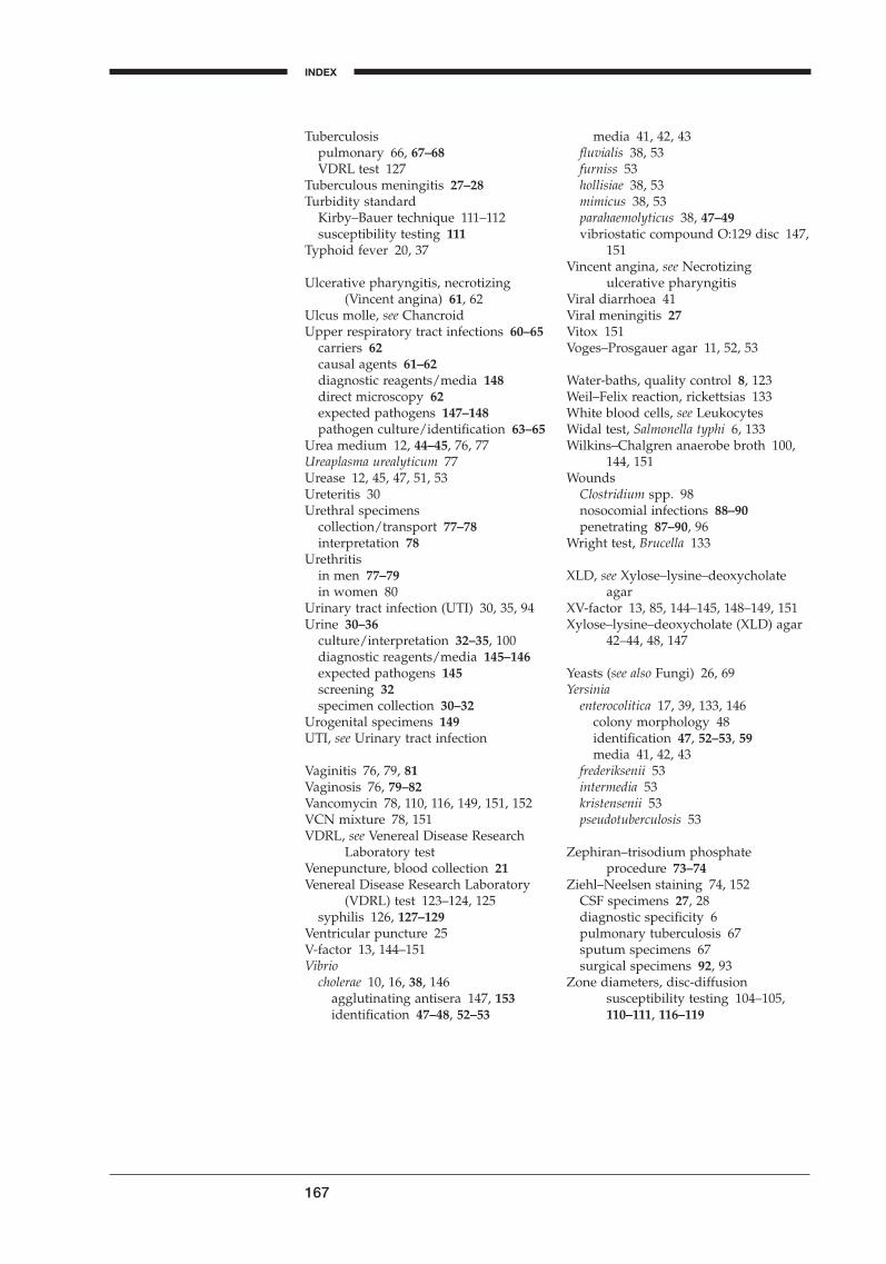
Tuberculosispulmonary 66, 67–68VDRL test 127
Tuberculous meningitis 27–28Turbidity standard
Kirby–Bauer technique 111–112susceptibility testing 111
Typhoid fever 20, 37
Ulcerative pharyngitis, necrotizing(Vincent angina) 61, 62
Ulcus molle, see ChancroidUpper respiratory tract infections 60–65
carriers 62causal agents 61–62diagnostic reagents/media 148direct microscopy 62expected pathogens 147–148pathogen culture/identification 63–65
Urea medium 12, 44–45, 76, 77Ureaplasma urealyticum 77Urease 12, 45, 47, 51, 53Ureteritis 30Urethral specimens
collection/transport 77–78interpretation 78
Urethritisin men 77–79in women 80
Urinary tract infection (UTI) 30, 35, 94Urine 30–36
culture/interpretation 32–35, 100diagnostic reagents/media 145–146expected pathogens 145screening 32specimen collection 30–32
Urogenital specimens 149UTI, see Urinary tract infection
Vaginitis 76, 79, 81Vaginosis 76, 79–82Vancomycin 78, 110, 116, 149, 151, 152VCN mixture 78, 151VDRL, see Venereal Disease Research
Laboratory testVenepuncture, blood collection 21Venereal Disease Research Laboratory
(VDRL) test 123–124, 125syphilis 126, 127–129
Ventricular puncture 25V-factor 13, 144–151Vibrio
cholerae 10, 16, 38, 146agglutinating antisera 147, 153identification 47–48, 52–53
167
INDEX
H
media 41, 42, 43fluvialis 38, 53furniss 53hollisiae 38, 53mimicus 38, 53parahaemolyticus 38, 47–49vibriostatic compound O:129 disc 147,
151Vincent angina, see Necrotizing
ulcerative pharyngitisViral diarrhoea 41Viral meningitis 27Vitox 151Voges–Prosgauer agar 11, 52, 53
Water-baths, quality control 8, 123Weil–Felix reaction, rickettsias 133White blood cells, see LeukocytesWidal test, Salmonella typhi 6, 133Wilkins–Chalgren anaerobe broth 100,
144, 151Wounds
Clostridium spp. 98nosocomial infections 88–90penetrating 87–90, 96
Wright test, Brucella 133
XLD, see Xylose–lysine–deoxycholateagar
XV-factor 13, 85, 144–145, 148–149, 151Xylose–lysine–deoxycholate (XLD) agar
42–44, 48, 147
Yeasts (see also Fungi) 26, 69Yersinia
enterocolitica 17, 39, 133, 146colony morphology 48identification 47, 52–53, 59media 41, 42, 43
frederiksenii 53intermedia 53kristensenii 53pseudotuberculosis 53
Zephiran–trisodium phosphateprocedure 73–74
Ziehl–Neelsen staining 74, 152CSF specimens 27, 28diagnostic specificity 6pulmonary tuberculosis 67sputum specimens 67surgical specimens 92, 93
Zone diameters, disc-diffusionsusceptibility testing 104–105,110–111, 116–119
BLMINDEX 1/17/04 2:39 PM Page 167

BLMINDEX 1/17/04 2:39 PM Page 168
Communicable diseases are the most common cause of death indeveloping countries, and their diagnosis and treatment represents asignificant challenge to the health services in those areas. To ensureaccurate identification of causative micro-organisms, laboratories needto use standard procedures for microbiological investigations andsusceptibility testing, and to implement an effective programme ofquality assurance.
This 2nd edition of the Basic Laboratory Procedures in ClinicalBacteriology has been updated in many areas, including a greatlyenhanced section on stool specimens and a new section on serologicaltests.
This manual is a practical guide, for use by laboratory workers in healthcentres and district hospitals, to the procedures to be followed inobtaining specimens, isolating and identifying bacteria, and assessingtheir resistance to antibiotics. It covers bacteriological investigation ofblood, cerebrospinal fluid, urine, stool, sputum, pharyngeal and genitalspecimens, and purulent exudates. Particular attention is given to theneed for quality control of all laboratory procedures. A list of media andreagents needed for the isolation and identification of the most commonbacterial pathogens is included, together with an indication of theirrelative importance for the intermediary laboratory. This list is intendedfor adaptation to local circumstances.
ISBN 92 4 154545 3
World Health OrganizationGeneva
B A S I CLABORATORYP R O C E D U R E S
I N C L I N I C A LBACTERIOLOGY
2nd edition
BA
SIC LA
BO
RA
TO
RY
PR
OC
EDU
RES IN
CLIN
ICA
L BA
CTER
IOLO
GY
2n
d ed
ition
WHO

The World Health Organization was established in 1948 as a specialized agency of the United Nations serving as thedirecting and coordinating authority for international health matters and public health. One of WHO’s constitutional func-tions is to provide objective and reliable information and advice in the field of human health, a responsibility that itfulfils in part through its extensive programme of publications.
The Organization seeks through its publications to support national health strategies and address the most pressingpublic health concerns of populations around the world. To respond to the needs of Member States at all levels of devel-opment, WHO publishes practical manuals, handbooks and training material for specific categories of health workers;internationally applicable guidelines and standards; reviews and analyses of health policies, programmes and research;and state-of-the-art consensus reports that offer technical advice and recommendations for decision-makers. These booksare closely tied to the Organization’s priority activities, encompassing disease prevention and control, the developmentof equitable health systems based on primary health care, and health promotion for individuals and communities. Progresstowards better health for all also demands the global dissemination and exchange of information that draws on theknowledge and experience of all WHO’s Member countries and the collaboration of world leaders in public health andthe biomedical sciences.
To ensure the widest possible availability of authoritative information and guidance on health matters, WHO secures thebroad international distribution of its publications and encourages their translation and adaptation. By helping to promoteand protect health and prevent and control disease throughout the world, WHO’s books contribute to achieving the Organization’s principal objective—the attainment by all people of the highest possible level of health.

A
Basiclaboratoryproceduresin clinicalbacteriology


A
World Health OrganizationGeneva2003
Basiclaboratoryproceduresin clinicalbacteriologySecond edition
J. Vandepitte and J. VerhaegenDepartment of MicrobiologySt Rafaël Academic HospitalLeuven, Belgium
K. EngbaekDepartment of Clinical MicrobiologyUniversity of CopenhagenHerlev HospitalHerlev, Denmark
P. RohnerDepartment of Clinical MicrobiologyCantonal University HospitalGeneva, Switzerland
P. PiotJoint United Nations Programme on HIV/AIDSGeneva, Switzerland
C. C. HeuckWorld Health OrganizationGeneva, Switzerland

WHO Library Cataloguing-in-Publication Data
Basic laboratory procedures in clinical bacteriology / J. Vandepitte . . . [et al.].—2nd ed.
1.Bacteriological techniques—standards 2.Laboratory techniques and procedures standards3.Manuals I.Vandepitte, J.
ISBN 92 4 154545 3 (NLM classification: QY 100)
© World Health Organization 2003
All rights reserved. Publications of the World Health Organization can be obtained from Marketingand Dissemination, World Health Organization, 20 Avenue Appia, 1211 Geneva 27, Switzerland (tel:+41 22 791 2476; fax: +41 22 791 4857; email: [email protected]). Requests for permission toreproduce or translate WHO publications–whether for sale or for noncommercial distribution–shouldbe addressed to Publications, at the above address (fax: +41 22 791 4806; email: [email protected]).
The designations employed and the presentation of the material in this publication do not imply theexpression of any opinion whatsoever on the part of the World Health Organization concerning thelegal status of any country, territory, city or area or of its authorities, or concerning the delimitationof its frontiers or boundaries. Dotted lines on maps represent approximate border lines for whichthere may not yet be full agreement.
The mention of specific companies or of certain manufacturers’ products does not imply that theyare endorsed or recommended by the World Health Organization in preference to others of a similarnature that are not mentioned. Errors and omissions excepted, the names of proprietary productsare distinguished by initial capital letters.
The World Health Organization does not warrant that the information contained in this publicationis complete and correct and shall not be liable for any damages incurred as a result of its use.
The named authors alone are responsible for the views expressed in this publication.
Typeset in Hong KongPrinted in Singapore
2001/13712—SNPBest-set/SNPSprint—6000

Contents
Preface viii
Introduction 1
Quality assurance in bacteriology 2
Introduction 2Definitions 2Internal quality control 6External quality assessment 16
PART IBacteriological investigations 19
Blood 20
Introduction 20When and where bacteraemia may occur 20Blood collection 20Blood-culture media 22Processing of blood cultures 23
Cerebrospinal fluid 25
Introduction 25Collection and transportation of specimens 25Macroscopic inspection 26Microscopic examination 26Preliminary identification 28Susceptibility testing 29
Urine 30
Introduction 30Specimen collection 30Culture and interpretation 32Interpretation of quantitative urine culture results 34Identification 35Susceptibility tests 36
Stool 37
Introduction 37Etiological agents and clinical features 37Appropriate use of laboratory resources 39Collection and transport of stool specimens 40Visual examination of stool specimens 41Enrichment and inoculation of stool specimens 41Media for enteric pathogens 42Primary isolation 42Preliminary identification of isolates 44
v
A

Final microbiological identification 50Serological identification 54
Upper respiratory tract infections 60
Introduction 60Normal flora of the pharynx 60Bacterial agents of pharyngitis 61Collection and dispatch of specimens 62Direct microscopy 62Culture and identification 63Susceptibility testing 65
Lower respiratory tract infections 66
Introduction 66The most common infections 66Collection of sputum specimens 68Processing of sputum in the laboratory (for
non-tuberculous infections) 68Culture for Mycobacterium tuberculosis 72Interpretation of cultures for M. tuberculosis 74General note on safety 74
Sexually transmitted diseases 76
Introduction 76Urethritis in men 77Genital specimens from women 79Specimens from genital ulcers 82
Purulent exudates, wounds and abscesses 86
Introduction 86Commonly encountered clinical conditions and the
most frequent etiological agents 86Collection and transportation of specimens 89Macroscopic evaluation 90Microscopic examination 91Culture 92Identification 93Susceptibility testing 97
Anaerobic bacteriology 98
Introduction 98Description of bacteria in relation to oxygen requirement 98Bacteriology 98
Antimicrobial susceptibility testing 103
Introduction 103General principles of antimicrobial susceptibility testing 103Clinical definition of terms “resistant” and “susceptible”:
the three category system 104Indications for routine susceptibility tests 106
vi
CONTENTS
Choice of drugs for routine susceptibility tests in the clinical laboratory 107
The modified Kirby–Bauer method 109Direct versus indirect susceptibility tests 117Technical factors influencing the size of the zone in the
disc-diffusion method 118Quality control 120
Serological tests 122
Introduction 122Quality control measures 122Serological reactions 125Serological tests for syphilis 126Febrile agglutinins tests 133Antistreptolysin O test 135Bacterial antigen tests 137
PART IIEssential media and reagents 141
Introduction 142
Pathogens, media and diagnostic reagents 143
Blood 144Cerebrospinal fluid 144Urine 145Stool 146Upper respiratory tract 147Lower respiratory tract 148Urogenital specimens for exclusion of sexually transmitted
diseases 149Pus and exudates 149List of recommended media and diagnostic reagents
for the intermediate microbiological laboratory 150
Selected further reading 154
Index 155
vii
CONTENTS
A
Preface
Communicable diseases are the most common cause of death in developingcountries, and their diagnosis and treatment represent a significant challengeto the health services in those areas. The World Health Organization has longbeen actively involved in developing and promoting standard techniques forlaboratory investigations of such diseases, a first attempt to standardize sus-ceptibility testing of bacterial pathogens being made in 1960.1 Following onfrom this, in 1976, the WHO Expert Committee on Biological Standardizationdrew up requirements for antibiotic susceptibility testing using the discmethod.2
At the same time, efforts were being made to introduce quality control intolaboratory performance. In 1981, WHO established an International ExternalQuality Assessment Scheme for Microbiology. The laboratories that areinvolved in this scheme are able to play a leading role in the implementationof national quality assessment schemes at all levels of the health care system.
The present publication brings together and updates the various guidelinesproduced by WHO over the years on sampling of specimens for laboratoryinvestigation, identification of bacteria, and testing of antimicrobial resistance.The information included is intended to lead to harmonization of microbio-logical investigations and susceptibility testing, and to improve the quality oflaboratories at both central and intermediate levels. It concentrates on the pro-cedures to be followed, rather than the basic techniques of microscopy andstaining, which have been described in detail in another WHO publication.3
viii
1 The public health aspects of antibiotics in feedstuffs. Report on a Working Group, Bremen, 1–5October 1973. Copenhagen, WHO Regional Office for Europe, 1973 (document no. EURO 3604(2)).2 WHO Expert Committee on Biological Standardization. Twenty-eighth report. Geneva, WorldHealth Organization, 1977 (WHO Technical Report Series, No. 610).3 Manual of basic techniques for a health laboratory, 2nd ed. Geneva, World Health Organiza-tion, 2003.
Related Documents