Basic Histology and Connective Tissue Chapter 5 • Histology, the Study of Tissues • Tissue Types • Connective Tissues

Welcome message from author
This document is posted to help you gain knowledge. Please leave a comment to let me know what you think about it! Share it to your friends and learn new things together.
Transcript
Basic Histology and Connective Tissue Chapter 5
• Histology, the Study of Tissues
• Tissue Types
• Connective Tissues
Histology is the Study of Tissues • 200 different types of cells in the human body.
• A Tissue consist of two or more types of cells that function together.
• Four basic types of tissues:
– epithelial tissue
– connective tissue
– muscular tissue
– nervous tissue
• An Organ is a structure with discrete boundaries that is composed of 2 or more tissue types.
• Example: skin is an organ composed of epidermal tissue and dermal tissue.
Distinguishing Features of Tissue Types • Types of cells (shapes and functions)
• Arrangement of cells
• Characteristics of the Extracellular Matrix:
– proportion of water
– types of fibrous proteins
– composition of the ground substance
• ground substance is the gelatinous material between cells in addition to the water and fibrous proteins
• ground substance consistency may be liquid (plasma), rubbery (cartilage), stony (bone), elastic (tendon)
• Amount of space occupied by cells versus extracellular matrix distinguishes connective tissue from other tissues
– cells of connective tissues are widely separated by a large amount of extracellular matrix
– very little extracellular matrix between the cells of epithelia, nerve, and muscle tissue
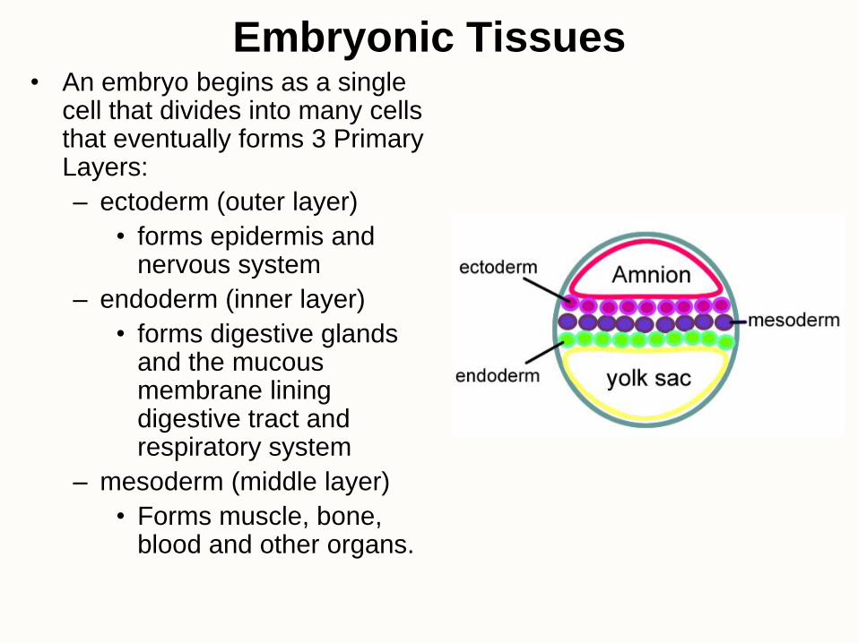
Embryonic Tissues • An embryo begins as a single
cell that divides into many cells that eventually forms 3 Primary Layers:
– ectoderm (outer layer)
• forms epidermis and nervous system
– endoderm (inner layer)
• forms digestive glands and the mucous membrane lining digestive tract and respiratory system
– mesoderm (middle layer)
• Forms muscle, bone, blood and other organs.
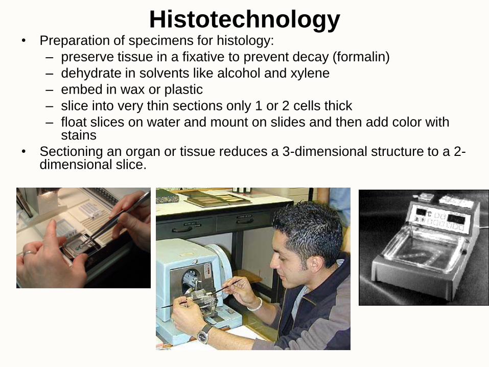
Histotechnology • Preparation of specimens for histology:
– preserve tissue in a fixative to prevent decay (formalin)
– dehydrate in solvents like alcohol and xylene
– embed in wax or plastic
– slice into very thin sections only 1 or 2 cells thick
– float slices on water and mount on slides and then add color with stains
• Sectioning an organ or tissue reduces a 3-dimensional structure to a 2-dimensional slice.
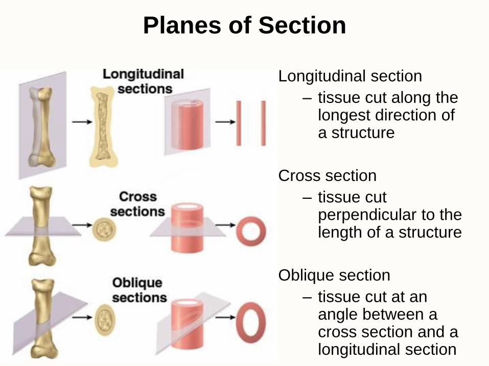
Planes of Section
Longitudinal section
– tissue cut along the longest direction of a structure
Cross section
– tissue cut perpendicular to the length of a structure
Oblique section
– tissue cut at an angle between a cross section and a longitudinal section
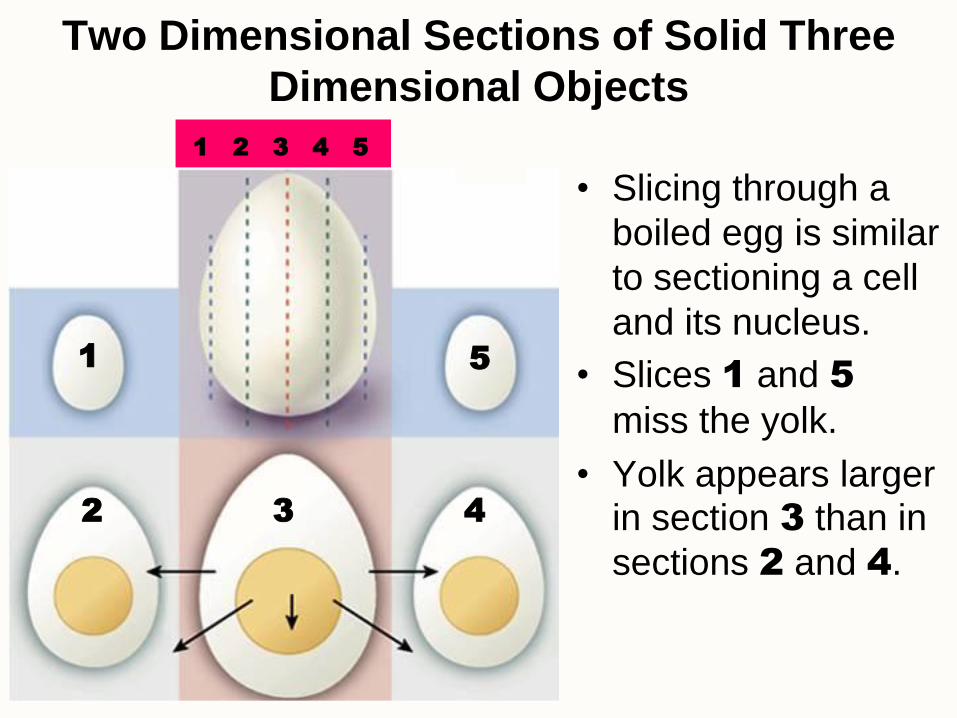
Two Dimensional Sections of Solid Three
Dimensional Objects
• Slicing through a
boiled egg is similar
to sectioning a cell
and its nucleus.
• Slices 1 and 5
miss the yolk.
• Yolk appears larger in section 3 than in
sections 2 and 4.
1 5
1 2 3 4 5
2 3 4
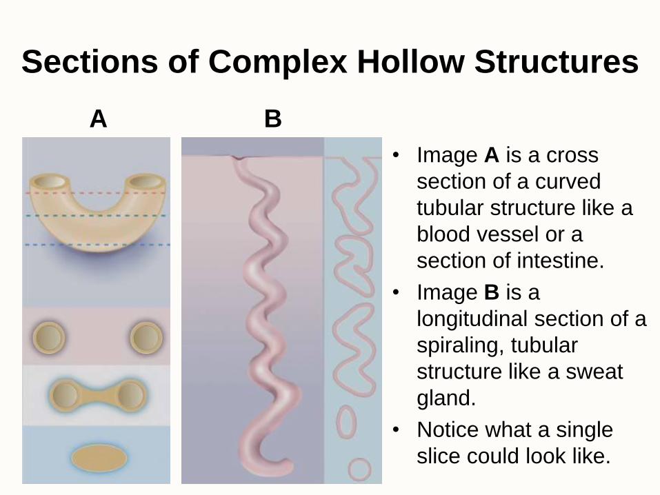
Sections of Complex Hollow Structures
• Image A is a cross
section of a curved
tubular structure like a
blood vessel or a
section of intestine.
• Image B is a
longitudinal section of a
spiraling, tubular
structure like a sweat
gland.
• Notice what a single
slice could look like.
A B
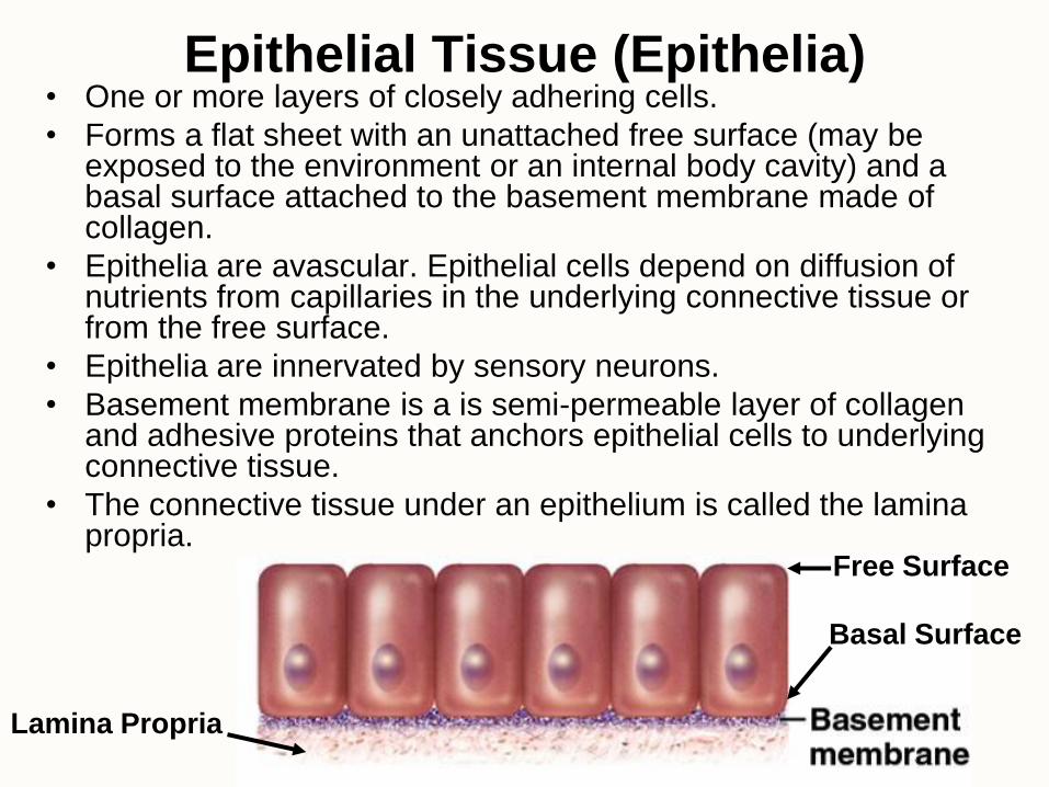
Epithelial Tissue (Epithelia) • One or more layers of closely adhering cells.
• Forms a flat sheet with an unattached free surface (may be exposed to the environment or an internal body cavity) and a basal surface attached to the basement membrane made of collagen.
• Epithelia are avascular. Epithelial cells depend on diffusion of nutrients from capillaries in the underlying connective tissue or from the free surface.
• Epithelia are innervated by sensory neurons.
• Basement membrane is a is semi-permeable layer of collagen and adhesive proteins that anchors epithelial cells to underlying connective tissue.
• The connective tissue under an epithelium is called the lamina propria.
Free Surface
Lamina Propria
Basal Surface
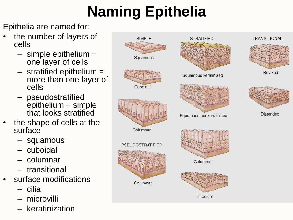
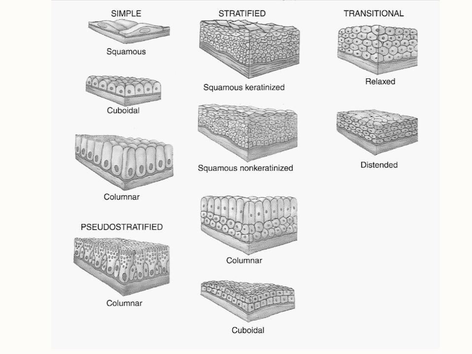
Naming Epithelia Epithelia are named for:
• the number of layers of cells
– simple epithelium = one layer of cells
– stratified epithelium = more than one layer of cells
– pseudostratified epithelium = simple that looks stratified
• the shape of cells at the surface
– squamous
– cuboidal
– columnar
– transitional
• surface modifications
– cilia
– microvilli
– keratinization

Simple
Squamous
Epithelium
• Single row of
squamous (flat)
cells.
• Can allow rapid
diffusion of
substances or
secretion of fluid.
• Example: lining of
blood vessels or
lining of lung
alveoli
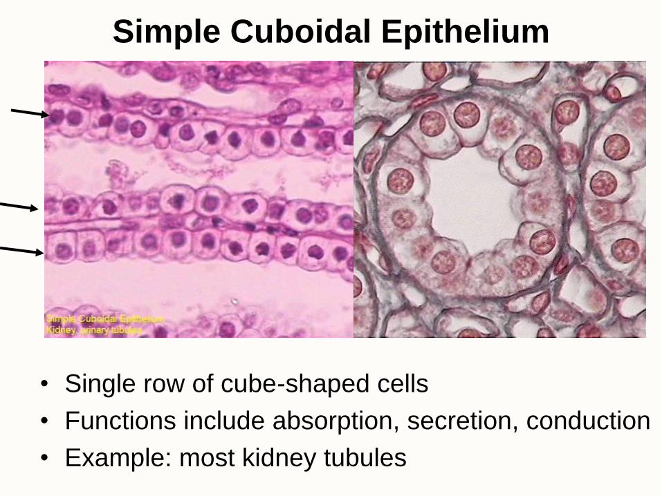
Simple Cuboidal Epithelium
• Single row of cube-shaped cells
• Functions include absorption, secretion, conduction
• Example: most kidney tubules
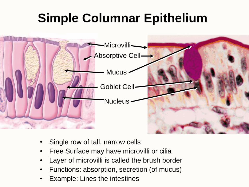
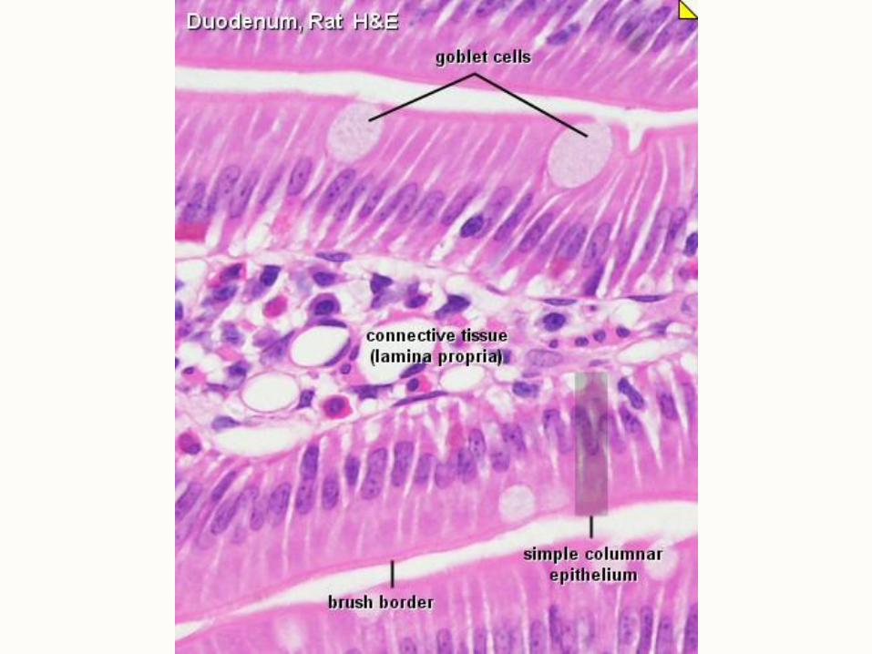
Simple Columnar Epithelium
• Single row of tall, narrow cells
• Free Surface may have microvilli or cilia
• Layer of microvilli is called the brush border
• Functions: absorption, secretion (of mucus)
• Example: Lines the intestines
Mucus
Microvilli
Absorptive Cell
Goblet Cell
Nucleus
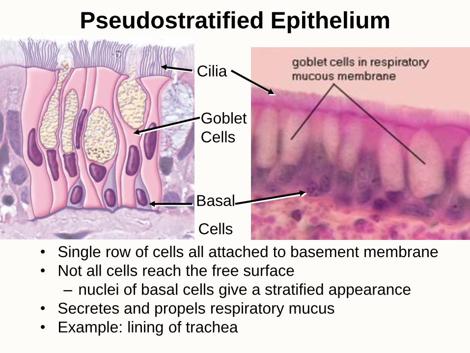
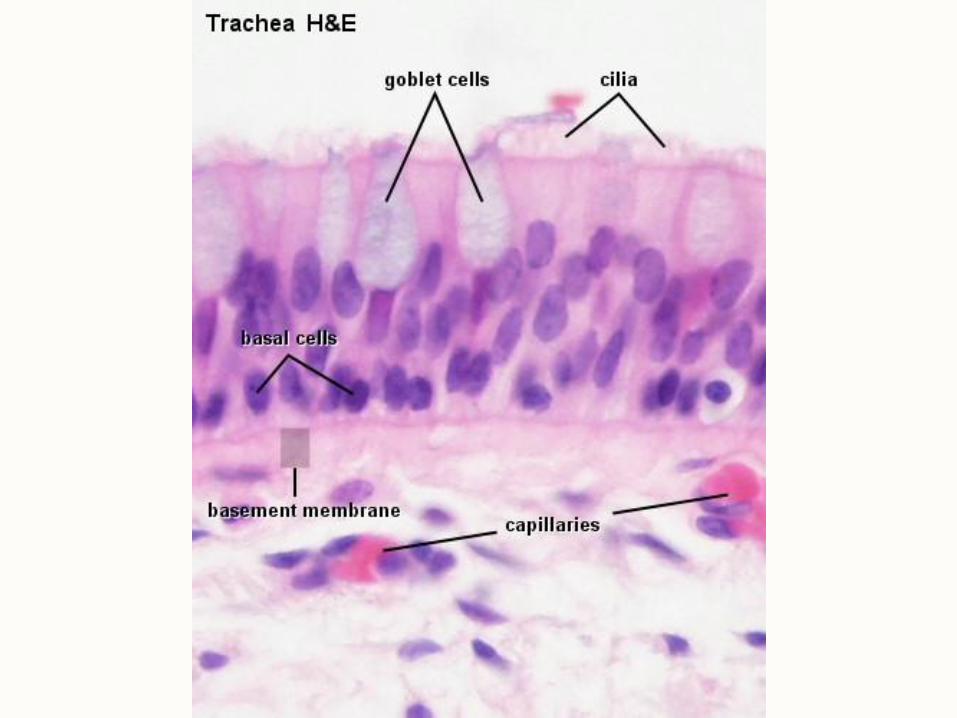
Pseudostratified Epithelium
• Single row of cells all attached to basement membrane
• Not all cells reach the free surface
– nuclei of basal cells give a stratified appearance
• Secretes and propels respiratory mucus
• Example: lining of trachea
Basal
Cells
Goblet
Cells
Cilia
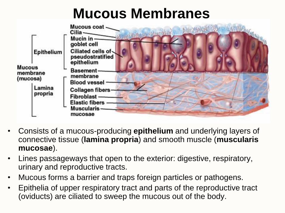
Mucous Membranes
• Consists of a mucous-producing epithelium and underlying layers of connective tissue (lamina propria) and smooth muscle (muscularis mucosae).
• Lines passageways that open to the exterior: digestive, respiratory, urinary and reproductive tracts.
• Mucous forms a barrier and traps foreign particles or pathogens.
• Epithelia of upper respiratory tract and parts of the reproductive tract (oviducts) are ciliated to sweep the mucous out of the body.
Stratified Epithelia
• Composed of more than one layer of cells.
• Always named for shape of surface cells.
• Deepest cells sit on basement membrane and are the source of replacement cells for the epithelium.
• Keratinization:
– keratinized epithelium has surface layer of dead cells that contain abundant protein and are surrounded by lipids
– nonkeratinized epithelium has living cells with nuclei in all layers
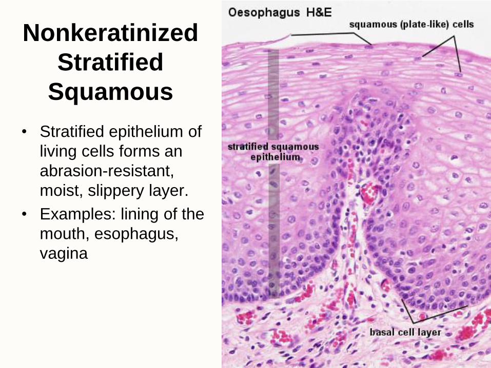
Nonkeratinized
Stratified
Squamous
• Stratified epithelium of
living cells forms an
abrasion-resistant,
moist, slippery layer.
• Examples: lining of the
mouth, esophagus,
vagina
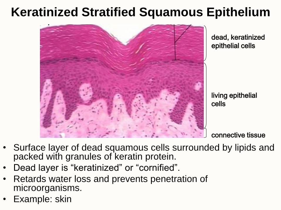
Keratinized Stratified Squamous Epithelium
• Surface layer of dead squamous cells surrounded by lipids and packed with granules of keratin protein.
• Dead layer is “keratinized” or “cornified”.
• Retards water loss and prevents penetration of microorganisms.
• Example: skin
dead, keratinized
epithelial cells
living epithelial
cells
connective tissue
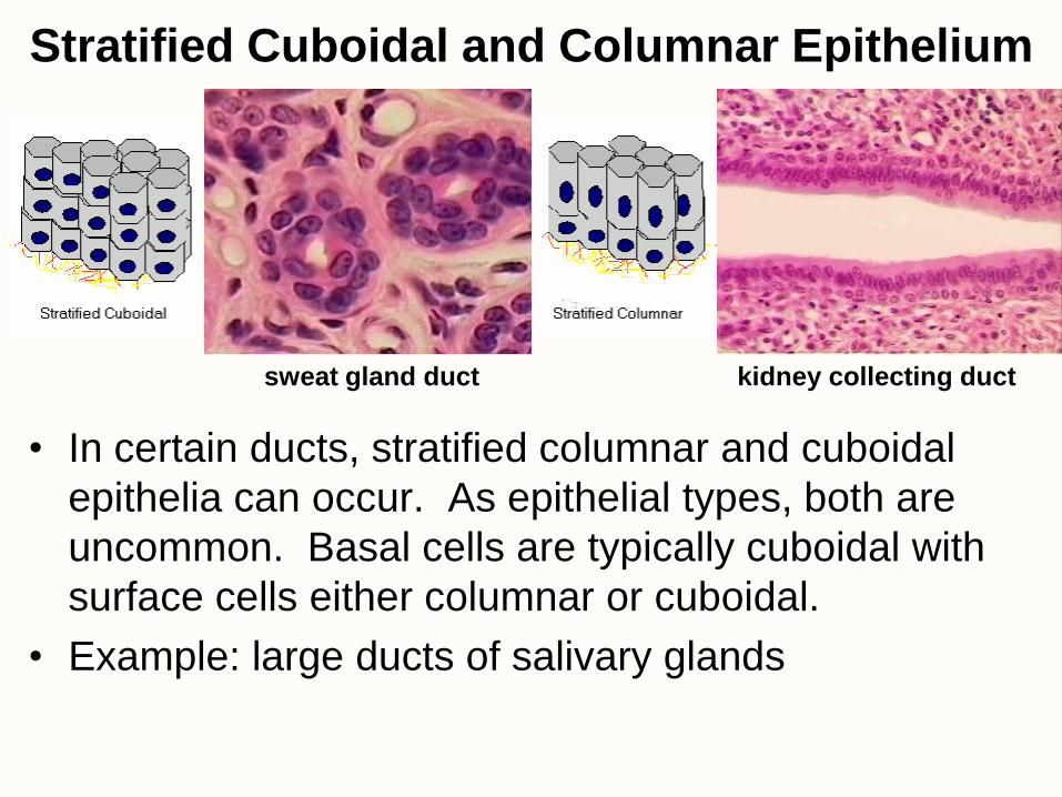
Stratified Cuboidal and Columnar Epithelium
• In certain ducts, stratified columnar and cuboidal
epithelia can occur. As epithelial types, both are
uncommon. Basal cells are typically cuboidal with
surface cells either columnar or cuboidal.
• Example: large ducts of salivary glands
sweat gland duct kidney collecting duct
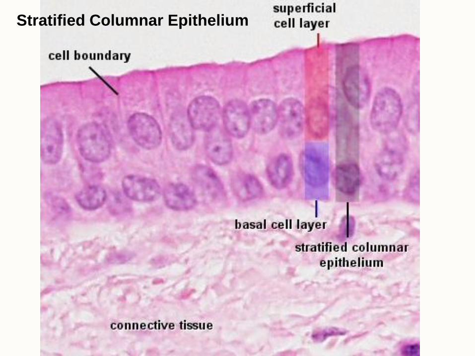
Stratified Columnar Epithelium
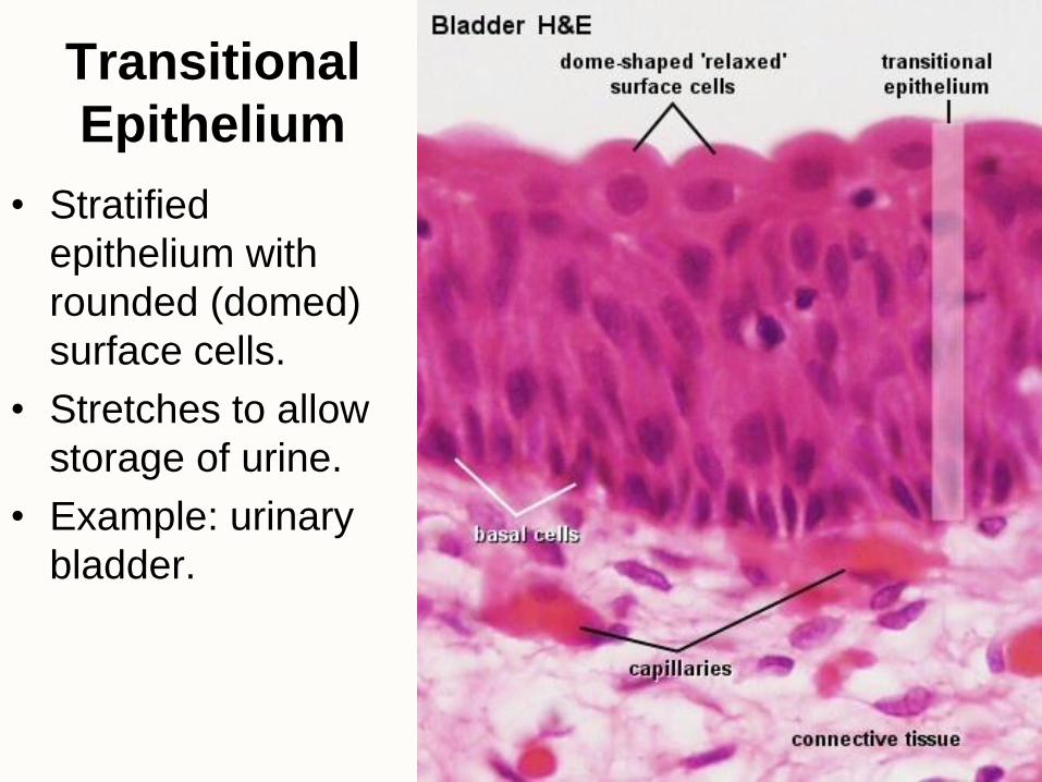
Transitional
Epithelium
• Stratified
epithelium with
rounded (domed)
surface cells.
• Stretches to allow
storage of urine.
• Example: urinary
bladder.

Quiz is on material up to this
point.
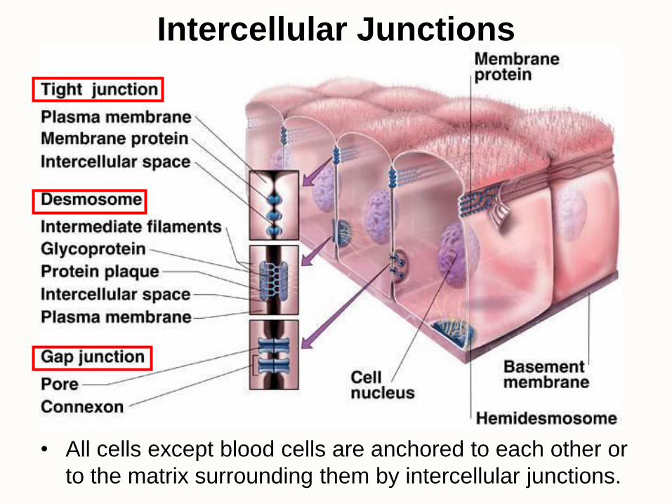
Intercellular Junctions
• All cells except blood cells are anchored to each other or
to the matrix surrounding them by intercellular junctions.
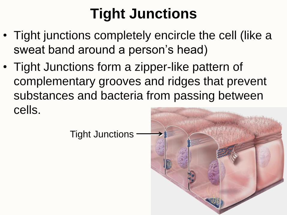
Tight Junctions
• Tight junctions completely encircle the cell (like a
sweat band around a person’s head)
• Tight Junctions form a zipper-like pattern of
complementary grooves and ridges that prevent
substances and bacteria from passing between
cells.
Tight Junctions
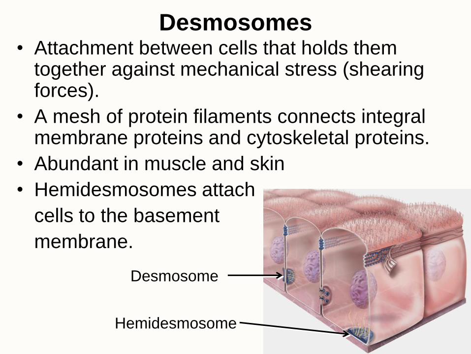
Desmosomes • Attachment between cells that holds them
together against mechanical stress (shearing forces).
• A mesh of protein filaments connects integral membrane proteins and cytoskeletal proteins.
• Abundant in muscle and skin
• Hemidesmosomes attach
cells to the basement
membrane.
Desmosome
Hemidesmosome
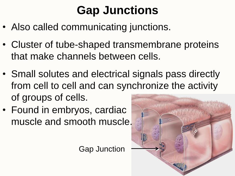
Gap Junctions
• Also called communicating junctions.
• Cluster of tube-shaped transmembrane proteins
that make channels between cells.
• Small solutes and electrical signals pass directly
from cell to cell and can synchronize the activity
of groups of cells.
• Found in embryos, cardiac
muscle and smooth muscle.
Gap Junction

Glands • Glands secrete substances for elimination or for use
elsewhere in the body
• Glands are composed predominantly of epithelial tissue
• Exocrine glands maintain connection to the surface through a duct (examples: sweat glands, salivary glands)
• Endocrine glands have no ducts but secrete their products (hormones) onto capillaries for absorption directly into bloodstream (pituitary, adrenal) or into interstitial fluid
• Mixed organs have both types of glands:
– pancreas secretes digestive enzymes into ducts and hormones into blood
– gonads release gametes into ducts and secrete hormones into blood
Types of Glandular Secretions • Serous
– thin, watery secretions such as sweat, milk, tears
and digestive juices.
• Mucus
– the sticky secretion called mucus is a glycoprotein,
mucin, that absorbs water
• Mixed Glands secrete both serous fluid and mucus
• Note: Mucus is a noun. Mucous is an adjective.
“Mucus is secreted by mucous glands.”
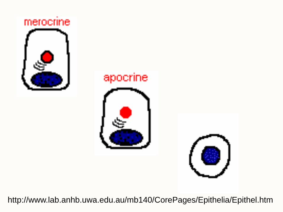
• Cellular mechanisms of glandular secretion include:
1) merocrine
2) apocrine
3) holocrine
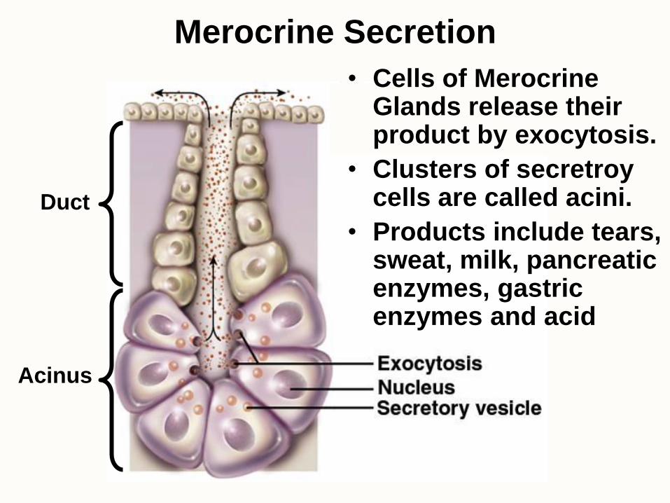
Merocrine Secretion
• Cells of Merocrine Glands release their product by exocytosis.
• Clusters of secretroy cells are called acini.
• Products include tears, sweat, milk, pancreatic enzymes, gastric enzymes and acid
Duct
Acinus
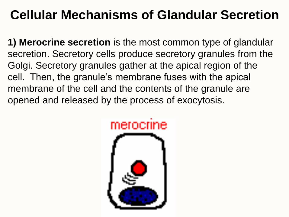
1) Merocrine secretion is the most common type of glandular
secretion. Secretory cells produce secretory granules from the
Golgi. Secretory granules gather at the apical region of the
cell. Then, the granule’s membrane fuses with the apical
membrane of the cell and the contents of the granule are
opened and released by the process of exocytosis.
Cellular Mechanisms of Glandular Secretion
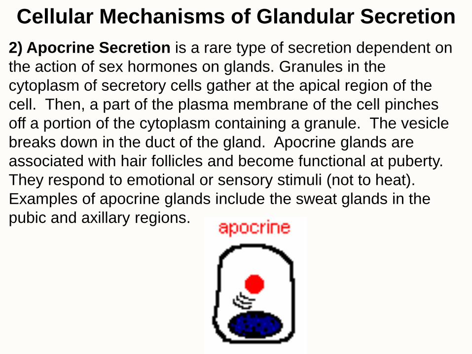
2) Apocrine Secretion is a rare type of secretion dependent on
the action of sex hormones on glands. Granules in the
cytoplasm of secretory cells gather at the apical region of the
cell. Then, a part of the plasma membrane of the cell pinches
off a portion of the cytoplasm containing a granule. The vesicle
breaks down in the duct of the gland. Apocrine glands are
associated with hair follicles and become functional at puberty.
They respond to emotional or sensory stimuli (not to heat).
Examples of apocrine glands include the sweat glands in the
pubic and axillary regions.
Cellular Mechanisms of Glandular Secretion
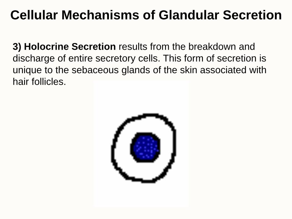
3) Holocrine Secretion results from the breakdown and
discharge of entire secretory cells. This form of secretion is
unique to the sebaceous glands of the skin associated with
hair follicles.
Cellular Mechanisms of Glandular Secretion

Holocrine Secretion
• Secretory cells proliferate at the base of the gland and move towards the duct as they mature. Once the cells are mature, they die and disintegrate. The cellular debris are released as the oily product of the cell.
• Example: sebaceous glands are the oil-producing glands associated with hair follicles.
http://www.lab.anhb.uwa.edu.au/mb140/CorePages/Epithelia/Epithel.htm
Connective Tissue • Connective Tissues consist of widely spaced cells
suspended in an abundant extracellular matrix. – The volume of the extracellular matrix is greater than the
volume of the cells.
• Functions of Connective Tissues – connects organs to each other
– divides body regions into compartments
– provides support, leverage and protection (physical and immune)
– covers and surrounds articular surfaces
– stores nutrients
– thermally insulates
– absorbs shock
– transports materials (water, nutrients, gases, waste, hormones)
A Classification
Scheme for
Connective
Tissues
• Embryonic Connective Tissue – Mucoid Mesenchymal Tissue
• Connective Tissue Proper – Loose (areolar)
– Dense • Dense Regular
• Dense Irregular
– Reticular
– Elastic
• Specialized Connective Tissue – Adipose
• Yellow and White
• Brown
– Hematopoietic
• Supporting Connective Tissue – Cartilage
• Hyaline Cartilage
• Elastic Cartilage
• Fibrocartilage
– Bone
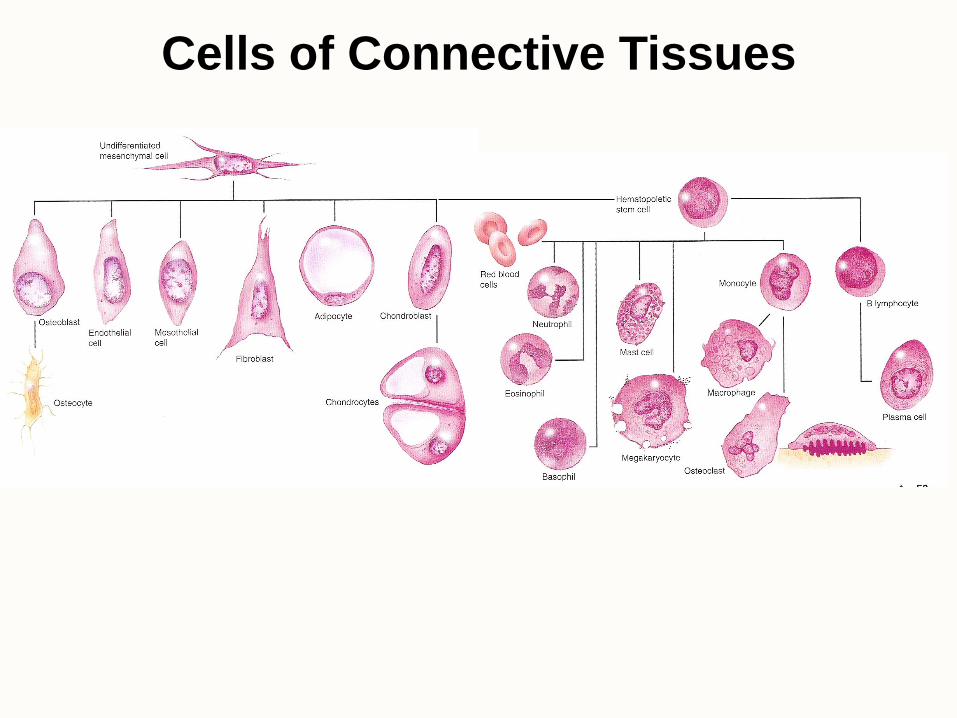
Cells of Connective Tissues
• All connective tissue cells are derived from mesoderm that developes in to mesenchymal cells as embryos develop.
• Fibroblasts are the most abundant CT cell and they produce fibers and ground substance.
• Adipocytes (fat cells) store triglycerides
• Chondroblasts develop into chondrocytes as they produce cartilage
• Osteoblasts develop into osteocytes as they produce bone
• Hematopoietic Cells differentiate into blood cells – Macrophages and Mast Cells can leave the blood and
enter the interstitial fluid between cells
Cells of Connective Tissues
Extracellular Matrix of Connective Tissue
• The Extracellular Matrix is composed of:
– Extracellular Tissue Fluid (mostly water,
similar to blood plasma)
– Protein Fibers
– Ground Substance
Protein Fibers of the ECM • Collagen Fibers
– over a dozen distinct types of protein fibers
– tough, flexible collagen fibers are abundant in tendons, ligaments, dermis of the skin, teeth, cartilage, bone
• Reticular Fibers
– thin, branched fibers that form a loose, interconnected network that hold cells, tissue fluid and ground substance
– Located in distensible or spongy tissues (walls of blood vessels, dermis, lymph nodes, spleen, liver). Stain darkly with silver.
• Elastic Fibers
– also called yellow fibers because of their color in life
– thin, straight fibers made of the protein elastin
– stretch 150% resting length and recoil like a rubber band
– give skin, lungs and arteries ability to stretch and recoil
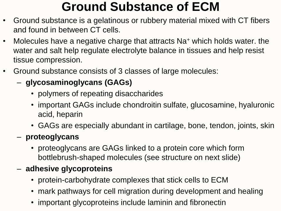
Ground Substance of ECM • Ground substance is a gelatinous or rubbery material mixed with CT fibers
and found in between CT cells.
• Molecules have a negative charge that attracts Na+ which holds water. the
water and salt help regulate electrolyte balance in tissues and help resist
tissue compression.
• Ground substance consists of 3 classes of large molecules:
– glycosaminoglycans (GAGs)
• polymers of repeating disaccharides
• important GAGs include chondroitin sulfate, glucosamine, hyaluronic
acid, heparin
• GAGs are especially abundant in cartilage, bone, tendon, joints, skin
– proteoglycans
• proteoglycans are GAGs linked to a protein core which form
bottlebrush-shaped molecules (see structure on next slide)
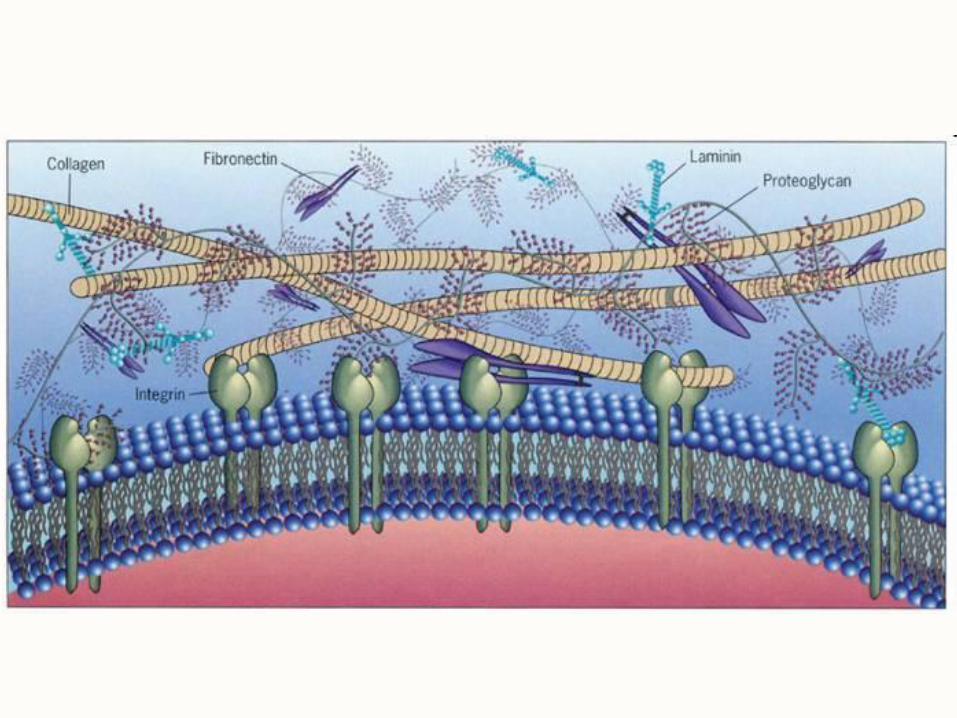
– adhesive glycoproteins
• protein-carbohydrate complexes that stick cells to ECM
• mark pathways for cell migration during development and healing
• important glycoproteins include laminin and fibronectin
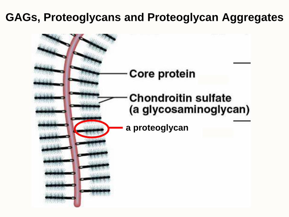
GAGs, Proteoglycans and Proteoglycan Aggregates
a proteoglycan
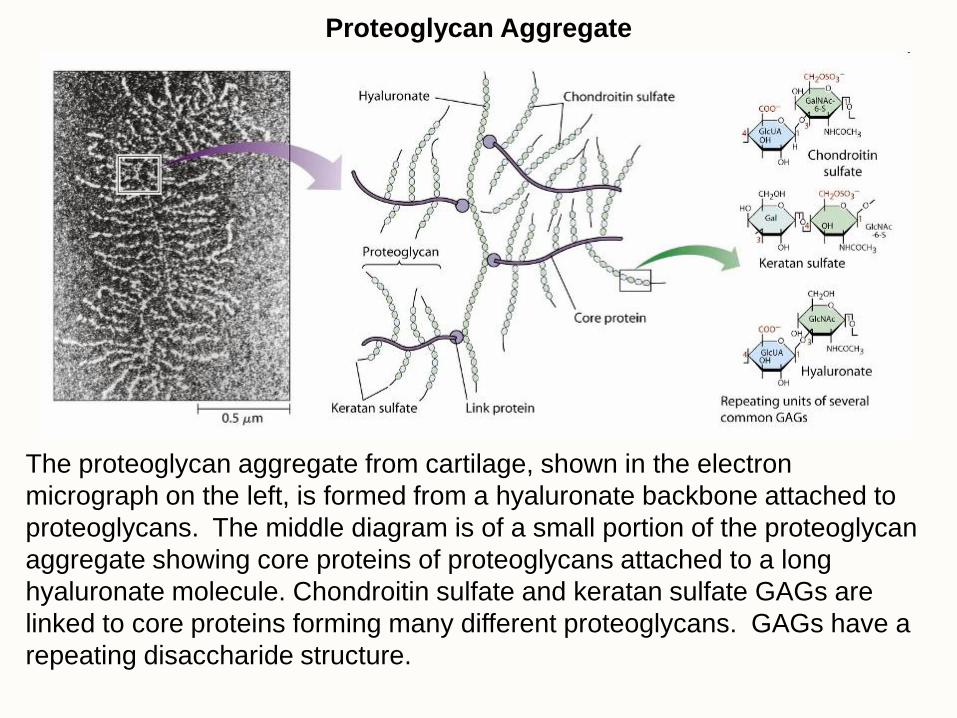
Proteoglycan Aggregate
The proteoglycan aggregate from cartilage, shown in the electron
micrograph on the left, is formed from a hyaluronate backbone attached to
proteoglycans. The middle diagram is of a small portion of the proteoglycan
aggregate showing core proteins of proteoglycans attached to a long
hyaluronate molecule. Chondroitin sulfate and keratan sulfate GAGs are
linked to core proteins forming many different proteoglycans. GAGs have a
repeating disaccharide structure.
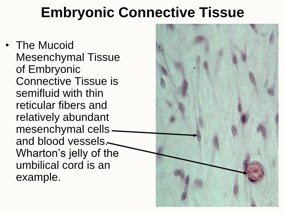
Embryonic Connective Tissue
• The Mucoid Mesenchymal Tissue of Embryonic Connective Tissue is semifluid with thin reticular fibers and relatively abundant mesenchymal cells and blood vessels. Wharton’s jelly of the umbilical cord is an example.
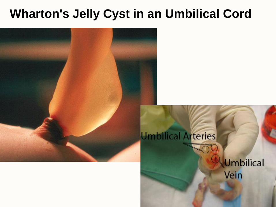
Wharton's Jelly Cyst in an Umbilical Cord
Connective Tissue Proper
• Loose Connective Tissue (Areolar Tissue)
• Dense Connective Tissue
– Dense Connective Tissues
• Dense Regular Connective Tissue
• Dense Irregular Connective Tissue
• Reticular Connective Tissue
• Elastic Connective Tissue
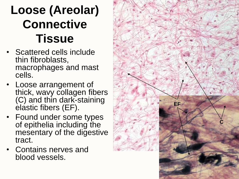
Loose (Areolar)
Connective
Tissue • Scattered cells include
thin fibroblasts, macrophages and mast cells.
• Loose arrangement of thick, wavy collagen fibers (C) and thin dark-staining elastic fibers (EF).
• Found under some types of epithelia including the mesentary of the digestive tract.
• Contains nerves and blood vessels.
C
EF
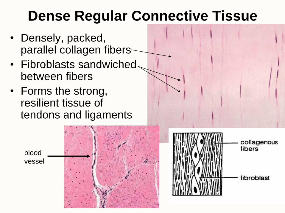
Dense Regular Connective Tissue
• Densely, packed, parallel collagen fibers
• Fibroblasts sandwiched between fibers
• Forms the strong, resilient tissue of tendons and ligaments
blood
vessel
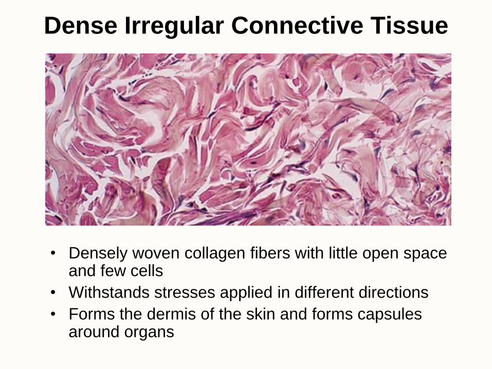
Dense Irregular Connective Tissue
• Densely woven collagen fibers with little open space and few cells
• Withstands stresses applied in different directions
• Forms the dermis of the skin and forms capsules around organs
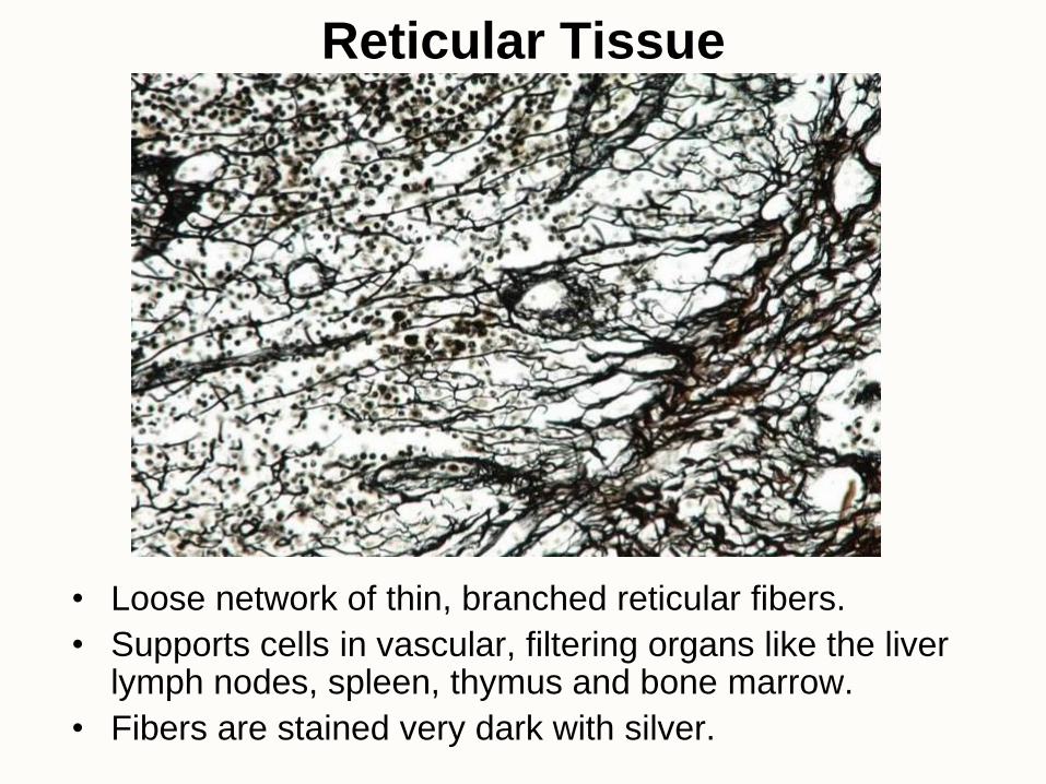
Reticular Tissue
• Loose network of thin, branched reticular fibers.
• Supports cells in vascular, filtering organs like the liver lymph nodes, spleen, thymus and bone marrow.
• Fibers are stained very dark with silver.
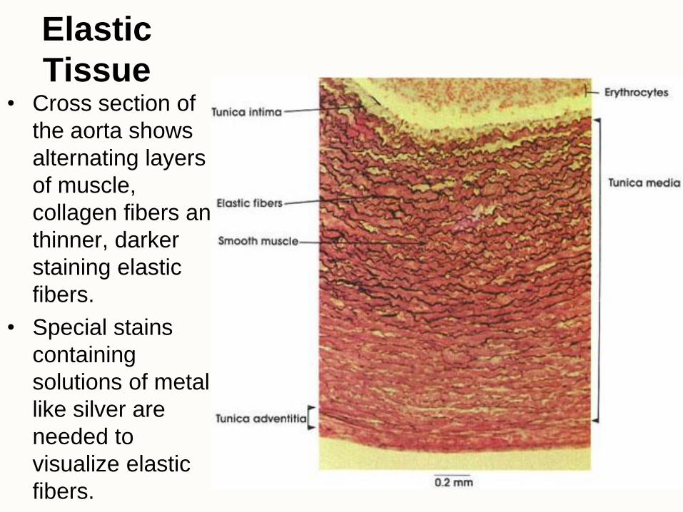
Elastic
Tissue • Cross section of
the aorta shows
alternating layers
of muscle,
collagen fibers and
thinner, darker
staining elastic
fibers.
• Special stains
containing
solutions of metals
like silver are
needed to
visualize elastic
fibers.
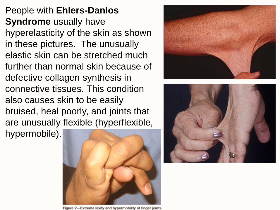
People with Ehlers-Danlos
Syndrome usually have
hyperelasticity of the skin as shown
in these pictures. The unusually
elastic skin can be stretched much
further than normal skin because of
defective collagen synthesis in
connective tissues. This condition
also causes skin to be easily
bruised, heal poorly, and joints that
are unusually flexible (hyperflexible,
hypermobile).


Specialized Connective Tissue
• Adipose Tissues
– Unilocular (Yellow or White Fat)
– Multilocular (Brown Fat)
• Hematopoietic Tissue
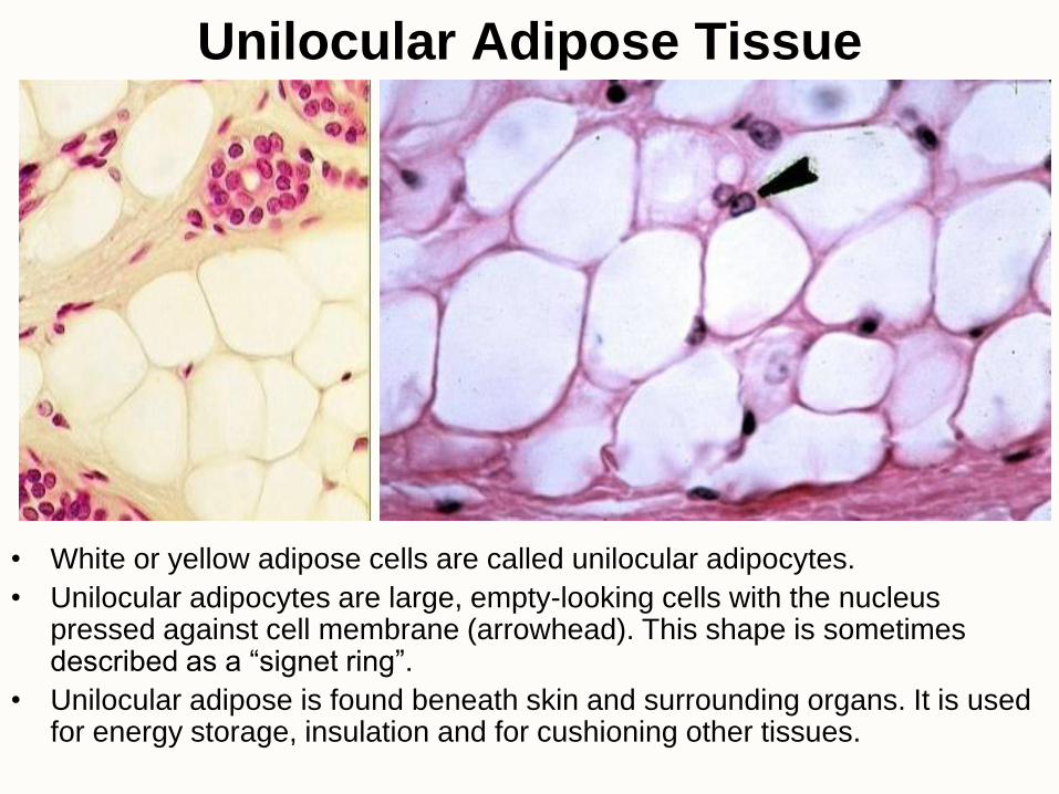
Unilocular Adipose Tissue
• White or yellow adipose cells are called unilocular adipocytes.
• Unilocular adipocytes are large, empty-looking cells with the nucleus pressed against cell membrane (arrowhead). This shape is sometimes described as a “signet ring”.
• Unilocular adipose is found beneath skin and surrounding organs. It is used for energy storage, insulation and for cushioning other tissues.
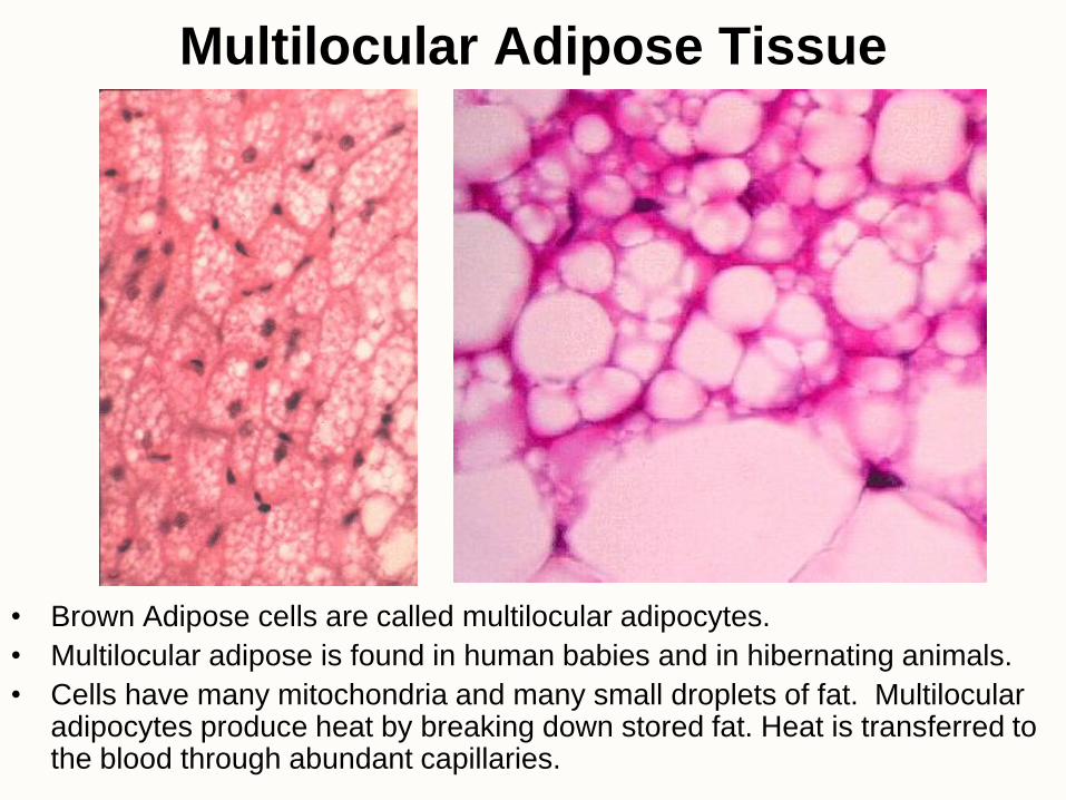
Multilocular Adipose Tissue
• Brown Adipose cells are called multilocular adipocytes.
• Multilocular adipose is found in human babies and in hibernating animals.
• Cells have many mitochondria and many small droplets of fat. Multilocular adipocytes produce heat by breaking down stored fat. Heat is transferred to the blood through abundant capillaries.
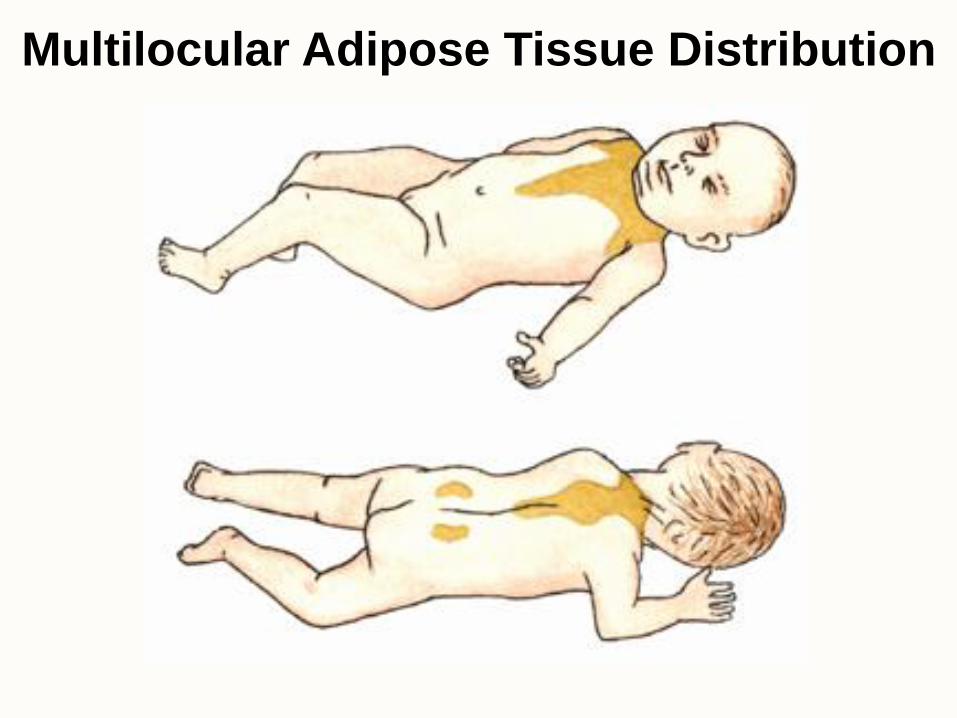
Multilocular Adipose Tissue Distribution
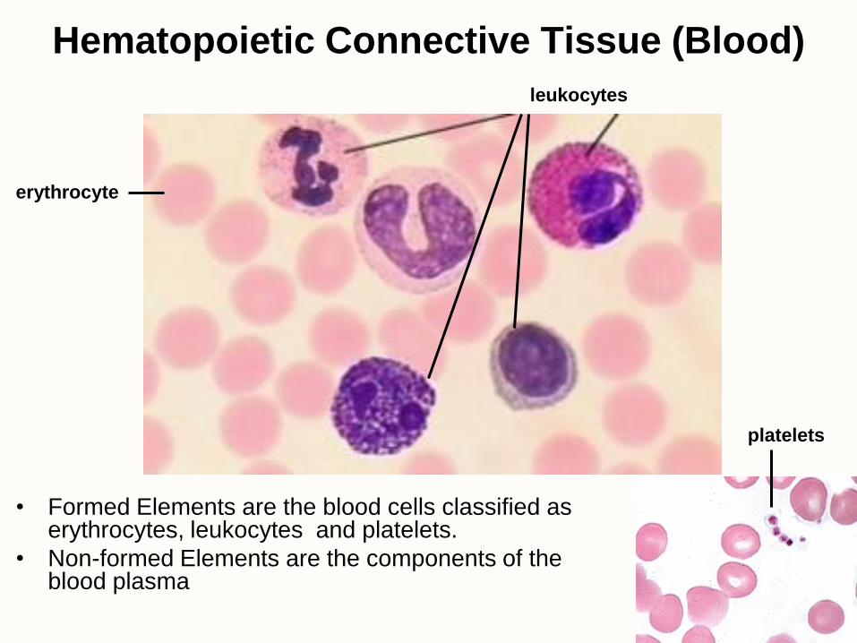
Hematopoietic Connective Tissue (Blood)
• Formed Elements are the blood cells classified as erythrocytes, leukocytes and platelets.
• Non-formed Elements are the components of the blood plasma
erythrocyte
leukocytes
platelets
Supporting Connective Tissue
• Cartilage
– Hyaline Cartilage
– Elastic Cartilage
– Fibrocartilage
• Bone
– Compact Bone
– Spongy Bone
Cartilage
• Supportive connective tissue with a rubbery matrix
• Cartilage is an avascular tissue (no blood vessels in the matrix) so cells rely on diffusion from a surrounding vascular membrane, the perichondrium, to deliver nutrients and remove wastes. (injured cartilage heals slowly if at all)
• 3 types of cartilage distinguished by the fibers of the extracellular matrix:
– hyaline
– elastic
– fibrocartilage
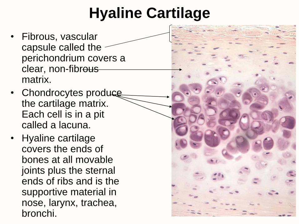
Hyaline Cartilage
• Fibrous, vascular capsule called the perichondrium covers a clear, non-fibrous matrix.
• Chondrocytes produce the cartilage matrix. Each cell is in a pit called a lacuna.
• Hyaline cartilage covers the ends of bones at all movable joints plus the sternal ends of ribs and is the supportive material in nose, larynx, trachea, bronchi.
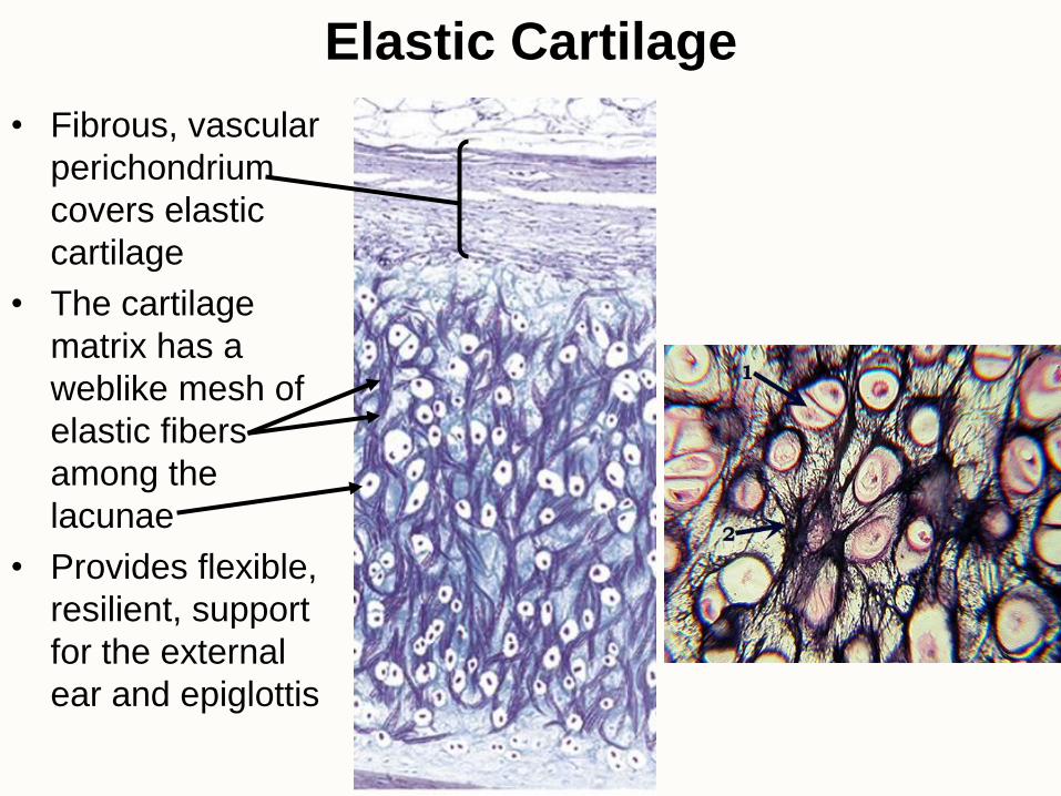
Elastic Cartilage
• Fibrous, vascular
perichondrium
covers elastic
cartilage
• The cartilage
matrix has a
weblike mesh of
elastic fibers
among the
lacunae
• Provides flexible,
resilient, support
for the external
ear and epiglottis
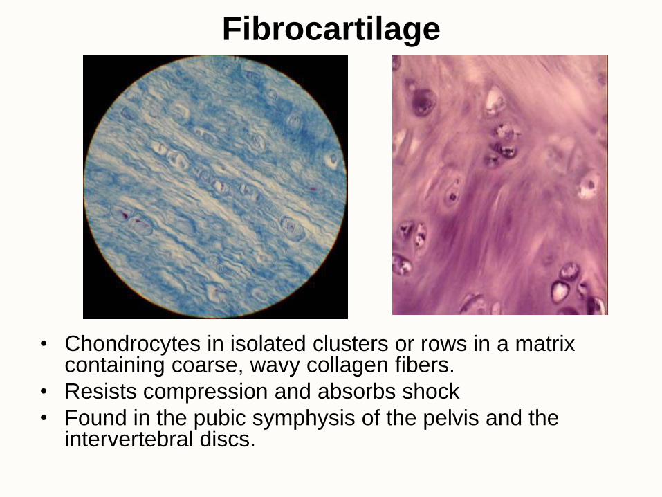
Fibrocartilage
• Chondrocytes in isolated clusters or rows in a matrix containing coarse, wavy collagen fibers.
• Resists compression and absorbs shock
• Found in the pubic symphysis of the pelvis and the intervertebral discs.
Bone
• Bone matrix stores the minerals calcium and phosphorus.
• Compact bone provides physical support for leverage during muscle contraction.
• Spongy bone, also called trabecular or cancellous, fills the ends of long bones and supports the bone marrow.
• Compact bone always covers spongy bone.
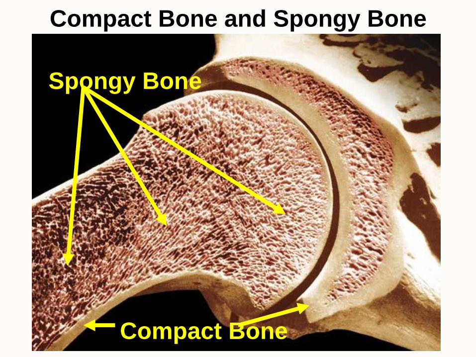
Compact Bone
Spongy Bone
Compact Bone and Spongy Bone
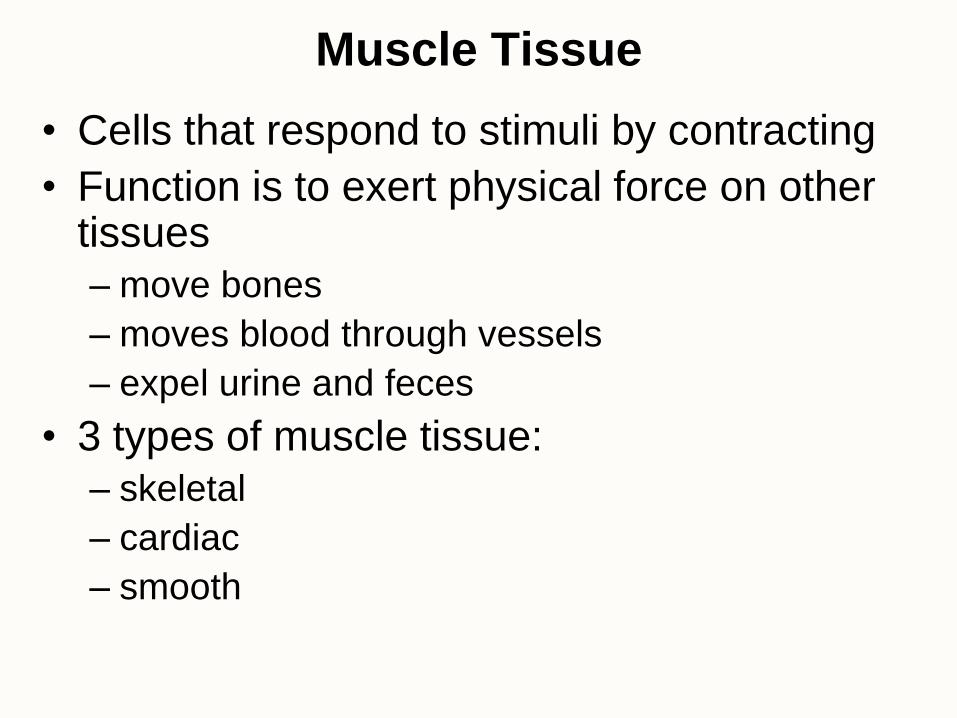
Muscle Tissue
• Cells that respond to stimuli by contracting
• Function is to exert physical force on other tissues
– move bones
– moves blood through vessels
– expel urine and feces
• 3 types of muscle tissue:
– skeletal
– cardiac
– smooth
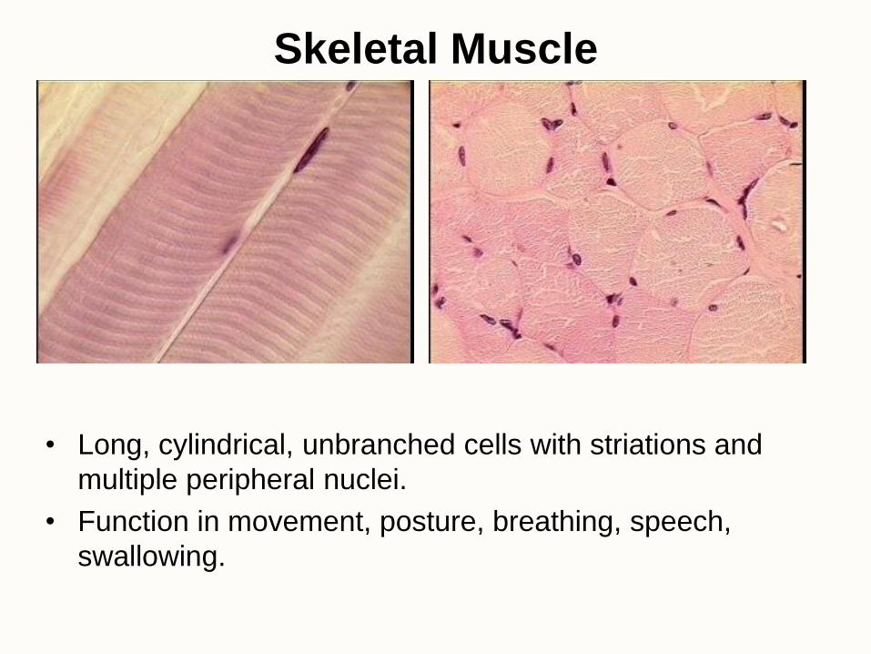
Skeletal Muscle
• Long, cylindrical, unbranched cells with striations and
multiple peripheral nuclei.
• Function in movement, posture, breathing, speech,
swallowing.

Cardiac Muscle
• Short, striated cells connected to each other with intercalated discs.
• Usually one central nucleus per cell.
• Sometimes cells are branched
• Found in heart and functions to pump blood.

Smooth Muscle
• Short, fusiform cells; nonstriated with only one central nucleus.
• Functions to control the diameter of openings in the
gastrointestinal tract, respiratory tract, cardiovascular system
and parts of the reproductive system.
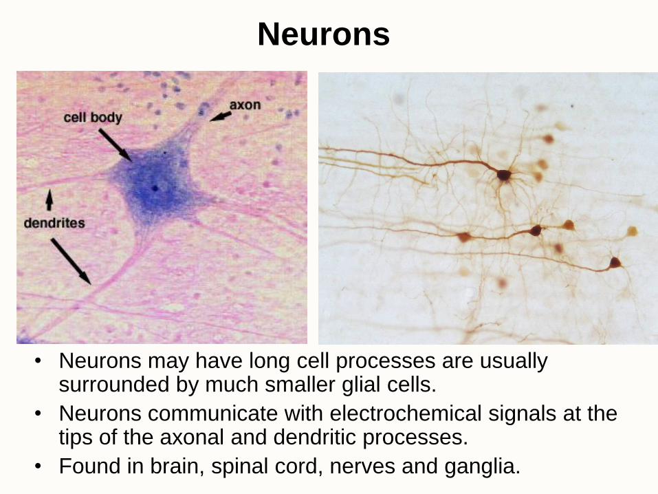
Neurons
• Neurons may have long cell processes are usually surrounded by much smaller glial cells.
• Neurons communicate with electrochemical signals at the tips of the axonal and dendritic processes.
• Found in brain, spinal cord, nerves and ganglia.
Related Documents