O¤print from Advances and Technical Standards in Neurosurgery, Vol. 30 Edited by J.D. Pickard 8 Springer-Verlag/Wien 2005 – Printed in Austria – Not for Sale Basic and Clinical Aspects of Olfaction B. N. Landis1,2, T. Hummel2, and J.-S. Lacroix1 1 Unite ´ de Rhinologie-Olfactologie, Service d’ Oto-Rhinologie-Laryngologie, Hopitaux Universitaires de Gene `ve, Gene `ve, Switzerland 2 Smell and Taste Clinic, Department of Otolaryngology, University of Dresden Medical School, Dresden, Germany With 1 Figure Contents Abstract ...................................................................... 70 Anatomy ..................................................................... 70 Main Olfactory System.................................................... 70 Trigeminal System......................................................... 71 Gustatory System ......................................................... 72 Vomeronasal System ...................................................... 73 Olfactory Coding ............................................................ 73 Measurement of Olfactory Function ........................................ 75 Psychophysical Methods of Olfactory Testing ............................ 75 Electrophysiological/Imaging Techniques Used to Test Olfactory Testing ..................................................................... 77 Causes and Symptoms of Smell Disorders .................................. 79 Most Common Causes .................................................... 80 Olfactory Loss Following Infections of the Upper Respiratory Tract (URTI) ................................................................. 80 Posttraumatic Olfactory Loss ........................................... 81 Sinunasal Causes ........................................................ 82 Neurodegenerative Causes .............................................. 82 Idiopathic ............................................................... 83 Less Frequent Causes ..................................................... 83 Endocrine Diseases ..................................................... 83 Epilepsy ................................................................. 83 General Pathologies..................................................... 84 Post-Surgery/Anesthesia ................................................ 84 Drug-Induced/Toxic .................................................... 84 Congenital .............................................................. 85

Welcome message from author
This document is posted to help you gain knowledge. Please leave a comment to let me know what you think about it! Share it to your friends and learn new things together.
Transcript

O¤print fromAdvances and Technical Standards in Neurosurgery, Vol. 30Edited by J.D. Pickard8 Springer-Verlag/Wien 2005 – Printed in Austria – Not for Sale
Basic and Clinical Aspects of Olfaction
B. N. Landis1,2, T. Hummel2, and J.-S. Lacroix1
1Unite de Rhinologie-Olfactologie, Service d’ Oto-Rhinologie-Laryngologie,
Hopitaux Universitaires de Geneve, Geneve, Switzerland
2Smell and Taste Clinic, Department of Otolaryngology, University of Dresden
Medical School, Dresden, Germany
With 1 Figure
Contents
Abstract . . . . . . . . . . . . . . . . . . . . . . . . . . . . . . . . . . . . . . . . . . . . . . . . . . . . . . . . . . . . . . . . . . . . . . 70
Anatomy . . . . . . . . . . . . . . . . . . . . . . . . . . . . . . . . . . . . . . . . . . . . . . . . . . . . . . . . . . . . . . . . . . . . . 70
Main Olfactory System. . . . . . . . . . . . . . . . . . . . . . . . . . . . . . . . . . . . . . . . . . . . . . . . . . . . 70Trigeminal System. . . . . . . . . . . . . . . . . . . . . . . . . . . . . . . . . . . . . . . . . . . . . . . . . . . . . . . . . 71
Gustatory System . . . . . . . . . . . . . . . . . . . . . . . . . . . . . . . . . . . . . . . . . . . . . . . . . . . . . . . . . 72
Vomeronasal System . . . . . . . . . . . . . . . . . . . . . . . . . . . . . . . . . . . . . . . . . . . . . . . . . . . . . . 73
Olfactory Coding . . . . . . . . . . . . . . . . . . . . . . . . . . . . . . . . . . . . . . . . . . . . . . . . . . . . . . . . . . . . 73
Measurement of Olfactory Function . . . . . . . . . . . . . . . . . . . . . . . . . . . . . . . . . . . . . . . . 75
Psychophysical Methods of Olfactory Testing . . . . . . . . . . . . . . . . . . . . . . . . . . . . 75
Electrophysiological/Imaging Techniques Used to Test Olfactory
Testing. . . . . . . . . . . . . . . . . . . . . . . . . . . . . . . . . . . . . . . . . . . . . . . . . . . . . . . . . . . . . . . . . . . . . 77Causes and Symptoms of Smell Disorders . . . . . . . . . . . . . . . . . . . . . . . . . . . . . . . . . . 79
Most Common Causes . . . . . . . . . . . . . . . . . . . . . . . . . . . . . . . . . . . . . . . . . . . . . . . . . . . . 80
Olfactory Loss Following Infections of the Upper Respiratory Tract
(URTI) . . . . . . . . . . . . . . . . . . . . . . . . . . . . . . . . . . . . . . . . . . . . . . . . . . . . . . . . . . . . . . . . . 80
Posttraumatic Olfactory Loss. . . . . . . . . . . . . . . . . . . . . . . . . . . . . . . . . . . . . . . . . . . 81
Sinunasal Causes. . . . . . . . . . . . . . . . . . . . . . . . . . . . . . . . . . . . . . . . . . . . . . . . . . . . . . . . 82
Neurodegenerative Causes . . . . . . . . . . . . . . . . . . . . . . . . . . . . . . . . . . . . . . . . . . . . . . 82
Idiopathic . . . . . . . . . . . . . . . . . . . . . . . . . . . . . . . . . . . . . . . . . . . . . . . . . . . . . . . . . . . . . . . 83Less Frequent Causes . . . . . . . . . . . . . . . . . . . . . . . . . . . . . . . . . . . . . . . . . . . . . . . . . . . . . 83
Endocrine Diseases . . . . . . . . . . . . . . . . . . . . . . . . . . . . . . . . . . . . . . . . . . . . . . . . . . . . . 83
Epilepsy . . . . . . . . . . . . . . . . . . . . . . . . . . . . . . . . . . . . . . . . . . . . . . . . . . . . . . . . . . . . . . . . . 83
General Pathologies. . . . . . . . . . . . . . . . . . . . . . . . . . . . . . . . . . . . . . . . . . . . . . . . . . . . . 84
Post-Surgery/Anesthesia . . . . . . . . . . . . . . . . . . . . . . . . . . . . . . . . . . . . . . . . . . . . . . . . 84
Drug-Induced/Toxic . . . . . . . . . . . . . . . . . . . . . . . . . . . . . . . . . . . . . . . . . . . . . . . . . . . . 84
Congenital . . . . . . . . . . . . . . . . . . . . . . . . . . . . . . . . . . . . . . . . . . . . . . . . . . . . . . . . . . . . . . 85

Symptoms . . . . . . . . . . . . . . . . . . . . . . . . . . . . . . . . . . . . . . . . . . . . . . . . . . . . . . . . . . . . . . . . . 85
Quantitative Olfactory Disorders . . . . . . . . . . . . . . . . . . . . . . . . . . . . . . . . . . . . . . . 85
Qualitative Olfactory Disorders . . . . . . . . . . . . . . . . . . . . . . . . . . . . . . . . . . . . . . . . 85
Surgical Risks to the Olfactory System. . . . . . . . . . . . . . . . . . . . . . . . . . . . . . . . . . . . . . 86
Endoscopic Sinus/Transnasal Surgery . . . . . . . . . . . . . . . . . . . . . . . . . . . . . . . . . . . . 86
Craniotomy. . . . . . . . . . . . . . . . . . . . . . . . . . . . . . . . . . . . . . . . . . . . . . . . . . . . . . . . . . . . . . . . 87
Recovery of Smell Disorders . . . . . . . . . . . . . . . . . . . . . . . . . . . . . . . . . . . . . . . . . . . . . . . . 88Treatment of Olfactory Disorders . . . . . . . . . . . . . . . . . . . . . . . . . . . . . . . . . . . . . . . . . . . 89
Surgical . . . . . . . . . . . . . . . . . . . . . . . . . . . . . . . . . . . . . . . . . . . . . . . . . . . . . . . . . . . . . . . . . . . . 89
Conservative/Medication. . . . . . . . . . . . . . . . . . . . . . . . . . . . . . . . . . . . . . . . . . . . . . . . . . 89
Acknowledgements . . . . . . . . . . . . . . . . . . . . . . . . . . . . . . . . . . . . . . . . . . . . . . . . . . . . . . . . . . 91
References. . . . . . . . . . . . . . . . . . . . . . . . . . . . . . . . . . . . . . . . . . . . . . . . . . . . . . . . . . . . . . . . . . . . 91
Abstract
Disturbances of olfaction are a common occurrence in many neurologicaland neurosurgical patients and their correct diagnosis might be helpfulin management and enhancement of quality of life. However, olfaction isseldom checked in most neurosurgical units and the ‘‘smell bottles’’ areoften either absent or out of date. This chapter reviews systematically re-cent advances in our understanding of the anatomy, physiology (olfactorycoding) and measurement of olfactory function in the human. The causesand symptoms of smell disorders, risk of damage to the olfactory system byvarious surgical procedures and, finally, the natural history of recovery andtreatment of smell disorders, for example after trauma, are discussed.
Keywords: Olfaction; smell disorder; anatomy; physiology; olfactory coding; mea-
surement of olfactory function; craniotomy.
Anatomy
Although this review focuses on the olfactory system, it is necessary tomention, at least briefly, other sensory channels involved in chemosensoryperception. All the senses can be stimulated by chemicals, which in fact,typically activate not only one but several of the ‘‘chemical senses’’. Forexample, nicotine not only activates the olfactory nerves, but also producesactivation of the intranasal chemosensory trigeminal system.
Main Olfactory System
Olfactory perception starts at the level of the olfactory epithelium in theroof of the nasal cavity. Olfactory receptor neurons (ORN) are embeddedwithin the respiratory epithelium and send their axons through the cribri-form plate towards the olfactory bulbs. ORN carry olfactory receptors(OR) which are the key to olfactory information processing (see below).In the olfactory bulb ORN axons synapse with second order neurons, the
70 B. N. Landis et al.

mitral cells. The wiring between the olfactory epithelium and the olfactorybulb is characterized by a convergence of ORN axons. Specifically, allORN carrying the same OR converge in the same site within the bulb,called ‘‘glomerulus’’. Axons from the mitral cells follow the olfactory tractand divide into two bundles. Most fibers directly project to the pyriformand entorhinal cortices as well as to the amygdalae (all structures formerlysubsumed under the term ‘‘limbic system’’) whereas a minority of fibersproject through the thalamus towards the orbito-frontal cortex [1].
Compared to other sensory modalities the olfactory system has someparticularities. First, the majority of the olfactory fibers do not cross butproject ipsilaterally in the brain. Second, most olfactory fibers bypass thethalamus and project very rapidly and directly in the pyriform cortex,amygdalae, and entorhinal cortex which are implicated in emotional andmemory processing [2]. This di¤erence in central anatomy has beenclaimed to be partly responsible for the emotional load olfactory memoriescan carry [3]. In contrast to other sensory modalities, no main olfactorycortex has yet been found. Numerous works indicate the orbitofrontal cor-tices to be an important relay in olfactory information processing [4].
Trigeminal System
The trigeminal system provides the somato-sensory innervation to thenasal mucosa. Somato-sensory input from the skin, the nasal and oralcavities, respectively, is mediated by the trigeminal system. Since mostodorous compounds stimulate trigeminal nerve endings, at least at higherconcentrations, this system is almost always co-activated in the perceptionof odors. With few exceptions almost all odorants have been shown to ex-hibit trigeminal activation to some extent [5] (e.g., mint has a somewhatfruity odor, but also evokes a typical cooling e¤ect which is mainly trigemi-nally mediated). The main modalities supplied by the trigeminal systemare temperature, pain, touch, and irritation. Testing the chemosensoryintranasal trigeminal system psychophysically is more complex than olfac-tory testing. Since olfactory thresholds are always lower than the trigemi-nal thresholds for a given substance, olfactory biases are obvious.
The olfactory system is unable to localize the site of stimulationwhen one nostril receives clean air and the other nostril simultaneouslyreceives an odor at the same time. In contrast, trigeminal stimulation canbe localized. This di¤erence is used to solve the bias inherent to trigeminaltesting. Using lateralisation paradigms, trigeminal function can be easilyand rapidly measured in a clinical context [6, 7]. Since the olfactory andtrigeminal systems are so closely related anatomically and physiologically,there is a strong interaction between the two systems [8, 9]. In patients witholfactory loss, the trigeminal function is also weakened [10, 11]. Older liter-ature on trigeminal trans-sections also discusses its impact on olfactory
71Basic and Clinical Aspects of Olfaction

function [12]. However, patients with complete trigeminal loss are ex-tremely rare and no large study has so far been conducted to investigatethe e¤ects of trigeminal loss on olfactory function.
Gustatory System
The gustatory system provides the five basic tastes; sweet, sour, salty, bit-ter, and umami (glutamate). The latter, which resembles mainly the taste ofchicken soup, has long been claimed in the Asian literature to be a basictaste quality [13, 14], whereas the western scientific community consideredumami mainly as a ‘‘taste enhancer’’. This controversy was resolved whenmonosodium glutamate receptors were found on the tongue surface actingas specific taste receptors [15]. Molecular biological knowledge about tastereceptors started to emerge a few years ago. Most basic taste qualities arenot mediated by just one receptor type; several receptors act, for example,as sweet receptors. Many other taste modalities have been postulated (me-tallic taste, fat taste) and are currently under investigation. Future researchis expected to clarify the coding mechanisms in taste perception.
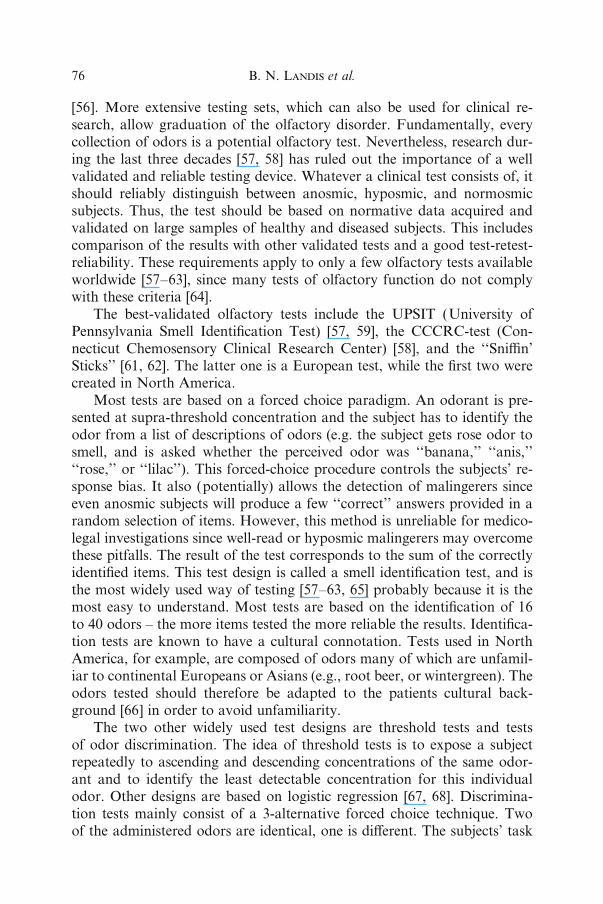
Fig. 1. Schematic diagram illustrating the axonal connectivity pattern between the
nose and the MOB. The OE in mice is divided into four zones (zones I through IV)
that are defined by the expression of odorant receptors. Olfactory sensory neurons in
a given zone of the epithelium project to glomeruli located in a corresponding zone
(zones I through IV ) of the MOB. Axons of sensory neurons expressing the same odor-
ant receptor (red or dark blue) converge to only a few defined glomeruli. NC Neocor-
tex; AOB accessory olfactory bulb. Reprinted (abstracted/excerpted) with permission
from Mori K, Nagao H, Yoshihara Y (1999). The olfactory bulb: coding and process-
ing of odor molecule information. Science 286 (5440): 711–715. Copyright (1999) AAAS
72 B. N. Landis et al.

Taste receptors are located within the taste buds, which are situatedon all papillae except the filiform type. The highest densities of taste budsare found on the tongue and palate but they are also found throughout theentire oral cavity, hypopharynx and subglottic larynx [16]. Like ORNs,gustatory sensory receptor cells have the ability to regenerate [17]. Neuralsupply for these cells is provided by the facial, glossopharyngeal, and vagalnerves. The facial nerve innervates the anterior two thirds of the tongue,while the glossopharyngeal and vagal nerves innervate the posterior thirdof the tongue, as well as the hypopharynx and larynx. Like olfaction, tastefibers project ipsilaterally into the basal ganglia and brain stem. All gus-tatory fibers (facial, glossopharyngeal and vagus) innervating the oral-pharyngeal cavity converge into the nucleus solitarius within the brain stem.
Vomeronasal System
In 1813, Jacobson described a mucosal organ located on each side of thenasal septum and which was subsequently named ‘‘Jacobson’s organ’’ [18,19]. Later, the vomeronasal organ (VNO, consisting of vomeronasal epi-thelium, nerve, and accessory olfactory bulb) was shown to mediate e¤ectsattributed to pheromones [20]. A pheromone is a chemical molecule orcompound which is secreted by one member of a species and, as soon as itis perceived by another member of the same species, elicits physiological,behavioral, or endocrinological e¤ects [21, 22]. According to the originaldefinition of Karlson and Luscher [20] such e¤ects must be species-specific.While the functionality and biological relevance in most animals is welldocumented, there is ongoing debate about the functional significance ofthe vomeronasal pouch in humans. Some authors claim to find neuronalactivity within the vomeronasal epithelium [23] while many other studiessuggest that the vomeronasal duct is nonfunctional in humans, with somevomeronasal nerves missing and lack of accessory olfactory bulbs in adults[24–27]. Furthermore, a vomeronasal duct is not always present in humans;recent investigations revealed that approximately 60% of humans have one[25]. Nevertheless, a few papers indicate that pheromonal-like e¤ects occurin humans [28, 29] and several vomeronasal-like receptor genes have beenfound in the human genome, one of which is expressed in the olfactory ep-ithelium (V1r) [30]. It is not yet known whether these receptors are func-tional or not. Their expression, however, indicates that putative ‘‘humanpheromones’’ may act via the main olfactory system.
Olfactory Coding
The question of olfactory information encoding has been a concern fora long time. One main problem was to find a theory or model that would
73Basic and Clinical Aspects of Olfaction

predict the odorous properties of a given molecule. Although the fragranceindustry spends a lot of money on the creation of new – and hopefully smelly– molecules, no model exists which could predict the smell of any givenmolecule. The search for new odorants is still a very expensive procedurebased on trial and error. Therefore, a universal model of stereochemical –odor interaction would greatly assist the search for new odorants.
Several models have been proposed to explain how the olfactory systemdiscriminates between odorants. In the early sixties Mozell hypothesizedthat the chromatography of a molecule would determine its processing[31, 32]. According to Mozell, the olfactory receptors, which are locatedon the cilia of the olfactory neurons, are covered by a mucous layer andodorants have to cross this mucus before reaching the receptor cell. Histheory was based on experiments using frog olfactory epithelium. Althoughno clear evidence has been presented that absorption of odorants is irrele-vant to its interaction with the receptors, this theory has received less atten-tion during recent years. Nevertheless, recent work on humans suggeststhat absorption could have implications for olfactory perception [33]. An-other model indicates that olfactory recognition is mainly based on a fewbasic odors and that combination of these odors encodes the olfactory in-formation [34, 35]. This model claims that olfaction works according tophysiological principles similar to those governing vision. This assumptionwas mainly based on experiments on specific anosmias to isovaleric acid.Further experiments with other odorants were not able to confirm thismodel. Another theory receiving interest from the media is an old idea[36] reactivated by Turin [37]. According to this theory, olfactory codingcould be based on vibration properties of the odorants. Recent work, how-ever, indicates that this model can not predict the olfactory characteristicsof a given molecule [38].
Since odorants are chemical structures, the existence of a ligand–receptor interaction has been claimed for many years, and was finallysubstantiated in 1991 by the discovery of a large family of seven trans-membrane receptor proteins, expressed exclusively in the olfactory neuro-epithelium. These olfactory receptors (ORs) are encoded by approximately1000 genes in the mouse, or approximately 1% of its genome [39]. Whilethe mouse expresses approximately 850 of these genes, the rest beingpseudogenes, humans have far fewer functional ORs (approximately 350)[40]. Although this seems to indicate a loss in function, the simple equation‘‘less receptors ¼ less function’’ is currently under debate and some studiesargue that humans have a very high preservation rate for specially impor-tant ORs [41–43].
The discovery of the OR superfamily led to a renaissance of olfactoryresearch. During the last decade, potential OR binding sites [44] and thetopographical organization and distribution of the ORs within the olfac-
74 B. N. Landis et al.

tory epithelium have been partly identified [45–47]. A recent finding hasbeen the astonishingly high degree of organization found within the periph-eral olfactory system. The first striking observation was that, among all thepotentially expressible OR, every ORN expresses only one single OR gene[48, 49]. Furthermore, axons from all ORN expressing the same OR, what-ever their location within the olfactory epithelium, project into two glomer-uli in each olfactory bulb. This organization is called glomerular conver-gence [50]. Thus, a large glomerular map in the bulb, containing hundredsof glomeruli, will correspond to all OR expressed in the olfactory neuro-epithelium. Molecular and electrophysiological studies revealed that ORare not selective for only one odorant, but numerous molecules bind withvarying a‰nities to a certain OR. A given receptor may bind to a moleculewith a given carbon chain length, but may lose binding a‰nity as the ago-nist’s chain length increases. Similarly, the OR binding a‰nity for a mole-cule may dramatically change upon modification of the functional groups(aldehydes, ketones, acids, esters, alcohols, etc) of this molecule [51, 52]. Inaddition, every odorant is recognized not by one but by several ORs simul-taneously, depending on its particular chemical properties. At the level ofthe glomerular map this leads to a specific activation pattern for each odor-ant [53]. This odor-specific activation pattern is believed to be responsiblefor the recognition of and distinction between di¤erent odorants [54].
However, as previously mentioned and although the olfactory receptortheory adequately explains how olfactory coding could work, olfactory re-search is still a long way away from predicting the odor of a moleculebased solely on the stereo-chemical properties of the latter.
Measurement of Olfactory Function
Similar to other sensory modalities, olfactory testing procedures will yieldinformation which is either based on subjects’ insights (‘‘subjective’’ or‘‘psychophysical’’ tests) or on more ‘‘objective’’ techniques less biased bythe subjects’ observations. Since the subjects’ self assessment of olfactoryfunction is unreliable, testing of olfactory function is necessary [55].
Psychophysical Methods of Olfactory Testing
The basic principle of psychophysical testing of olfaction is to expose asubject to an olfactory stimulus and to interpret the responses or reactionsof the tested subject.
This procedure has numerous advantages in clinical application, butalso important limitations. The most valuable advantage compared to ob-jective testing methods in daily clinical life is the rapidity which allows psy-chophysical tests to serve as quick screening tools for olfactory dysfunction
75Basic and Clinical Aspects of Olfaction
[56]. More extensive testing sets, which can also be used for clinical re-search, allow graduation of the olfactory disorder. Fundamentally, everycollection of odors is a potential olfactory test. Nevertheless, research dur-ing the last three decades [57, 58] has ruled out the importance of a wellvalidated and reliable testing device. Whatever a clinical test consists of, itshould reliably distinguish between anosmic, hyposmic, and normosmicsubjects. Thus, the test should be based on normative data acquired andvalidated on large samples of healthy and diseased subjects. This includescomparison of the results with other validated tests and a good test-retest-reliability. These requirements apply to only a few olfactory tests availableworldwide [57–63], since many tests of olfactory function do not complywith these criteria [64].
The best-validated olfactory tests include the UPSIT (University ofPennsylvania Smell Identification Test) [57, 59], the CCCRC-test (Con-necticut Chemosensory Clinical Research Center) [58], and the ‘‘Sni‰n’Sticks’’ [61, 62]. The latter one is a European test, while the first two werecreated in North America.
Most tests are based on a forced choice paradigm. An odorant is pre-sented at supra-threshold concentration and the subject has to identify theodor from a list of descriptions of odors (e.g. the subject gets rose odor tosmell, and is asked whether the perceived odor was ‘‘banana,’’ ‘‘anis,’’‘‘rose,’’ or ‘‘lilac’’). This forced-choice procedure controls the subjects’ re-sponse bias. It also (potentially) allows the detection of malingerers sinceeven anosmic subjects will produce a few ‘‘correct’’ answers provided in arandom selection of items. However, this method is unreliable for medico-legal investigations since well-read or hyposmic malingerers may overcomethese pitfalls. The result of the test corresponds to the sum of the correctlyidentified items. This test design is called a smell identification test, and isthe most widely used way of testing [57–63, 65] probably because it is themost easy to understand. Most tests are based on the identification of 16to 40 odors – the more items tested the more reliable the results. Identifica-tion tests are known to have a cultural connotation. Tests used in NorthAmerica, for example, are composed of odors many of which are unfamil-iar to continental Europeans or Asians (e.g., root beer, or wintergreen). Theodors tested should therefore be adapted to the patients cultural back-ground [66] in order to avoid unfamiliarity.
The two other widely used test designs are threshold tests and testsof odor discrimination. The idea of threshold tests is to expose a subjectrepeatedly to ascending and descending concentrations of the same odor-ant and to identify the least detectable concentration for this individualodor. Other designs are based on logistic regression [67, 68]. Discrimina-tion tests mainly consist of a 3-alternative forced choice technique. Twoof the administered odors are identical, one is di¤erent. The subjects’ task
76 B. N. Landis et al.

is to detect the di¤erent one. In principal, tests for odor threshold/odordiscrimination are non-verbal. In addition, they can be used repetitively –which is more di‰cult with odor identification tests.
Generally, identification and discrimination tests are believed to reflectcentral olfactory processing while thresholds are thought rather to reflectperipheral olfactory function. Accordingly, it has been claimed by severalauthors [69–74] that patients with diseases of the central nervous process-ing of odorous information exhibit selective disturbances of discriminationand identification while threshold results are normal. Although this idea ofa certain pattern pathognomonic for ‘‘central’’ olfactory disturbances seemsattractive, the vast majority of studies have yet failed to confirm such typi-cal pathology-associated patterns [75, 76]. The only, so far reliable andrecurrent test pattern in olfactory disturbance is a low threshold and nor-mal identification and discrimination in patients with chronic sinunasalproblems [77].
Besides the solid body of literature and its clinical convenience, the psy-chophysical tests have one main limitation. As soon as the patient’s collab-oration is not guaranteed, interpretation of test results becomes di‰cultor even impossible. Such cases include mainly willful non-collaboration incases of malingering, or for demented, unconscientious, or inexperiencedpatients. In order to acquire olfactory information in such cases, more ob-jective testing methods have been developed which rely less on the subjects’cooperation.
Electrophysiological/Imaging Techniques Used to Test Olfactory Testing
Electro-Olfactogram (EOG)
Electro-olfactograms (EOG) are electrical potentials of the olfactoryepithelium that occur in response to olfactory stimulation. The EOG repre-sents the sum of generator potentials of ORN. While this response has beenused extensively in olfactory research in animals (e.g., [78]), there are onlya handful of reports describing the properties of the human EOG. Amongother results, EOGs have been used to provide evidence for the dominantrole of the central nervous system in olfactory desensitisation [79], for thefunctional characterisation of the olfactory epithelium [80], the specific top-ographical distribution of ORN, the expression of ORN in response to ex-posure to odorants [81], and the characterisation of certain odorants as ORantagonists [82]. However, the EOG so far has not been systematicallyused in patients with olfactory dysfunction. This is partly due to the topo-graphical specificity of EOG responses, meaning that EOGs to certainodorants may be recorded only at certain epithelial sites. Thus, the sub-jects’ odorous impressions may not always be reflected by the presence ofan EOG response. In addition, the presence of an EOG may not always
77Basic and Clinical Aspects of Olfaction

represent an odorous sensation. Specifically, EOGs can be recorded in sub-jects with congenital anosmia [83], or EOGs are present at certain thresh-old levels when the subjects do not yet perceive an odor [84]. Having saidthis, EOGs may be extremely helpful in terms of elucidating pathologicalprocesses at the mucosal level [85].
Chemosensory Event-Related Potentials (CSERP)
Event-related potentials are EEG-derived poly-phasic signals. They arecaused by the activation of cortical neurons which generate electro-magneticfields [86]. As the EEG is a noisy signal which contains activity from manycortical neurons, ERP need to be extracted from this background activity.In other words, the signal-to-noise ratio needs to be improved. The classi-cal approach to this problem involves averaging of individual responsesto olfactory stimuli such that random activity would cancel itself out whileall non-random activation would remain. In addition, stimuli are typicallypresented with a steep onset (<20 ms) in an extremely well-controlled, mo-notonous environment such that stimulus onset synchronizes the activity ofas many cortical neurons as possible.
Olfactory ERP (1) are direct correlates of neuronal activation, unlikethe signals that are seen, for example, in functional MR imaging, (2) havean extremely high temporal resolution in the range of micro-seconds, (3)allow the investigation of the sequential processing of olfactory informa-tion, and (4) can be obtained independently of the subject’s response bias,i.e., they allow the investigation of subjects who have di‰culties to respondproperly (e.g., children, aphasic patients).
Based on a system developed by Kobal [87, 88], odors are applied intra-nasally. Presentation of odorous stimuli does not simultaneously activatemechano- or thermo-receptors in the nasal mucosa since odor pulses areembedded in a constantly flowing air stream. In contrast to audition orvision, to date no early ERP have been recorded in response to olfactorystimuli (for review see [89]) but only late near-field ERP, i.e. responsesfrom cortical neurons. Peaks of the late near-field ERP fall into twogroups. Earlier peaks like N1 encode a greater number of exogenous stim-ulus characteristics than of later, so-called endogenous components. Thatis, earlier components encode stimulus intensity or stimulus quality (e.g.,‘‘What is the nature of this stimulus?’’), whereas later components aremore related to the frequency, or the salience of the stimulus (‘‘What isthe meaning of this stimulus?’’) [86, 90–92].
Olfactory ERP are recorded all over the scalp. In terms of the topo-graphic distribution of olfactory ERP, amplitudes exhibit characteristicpatterns with a centro-parietal maximum for both amplitudes N1 and P2[93] (compare [94–96]). Using magneto-encephalographic techniques [97]
78 B. N. Landis et al.

Kobal and co-workers conducted a series of experiments which addressedthe question of the generation of olfactory ERP. Cortical generators of theresponses to trigeminal stimulation with CO2 were localized in the second-ary somato-sensory cortex [98]. Other work indicated [99, 100] that olfac-tory stimuli activate anterior-central parts of the insula, the para-insularcortex, and the superior temporal sulcus [101].
Clinical testing with chemosensory ERP [89] typically includes therecording of responses to olfactory (e.g., hydrogen sulfide, and phenylethyl alcohol) and trigeminal (e.g., CO2) stimuli. This procedure has beenadopted by the working group ‘‘Olfaction and Gustation’’ of the GermanENT Society [102]. So far, in all investigated anosmic patients, intranasaltrigeminal ERPs could be obtained after stimulation with CO2 – althoughwith significantly smaller amplitudes than in healthy controls [10]. In con-trast, no olfactory ERPs could be detected in anosmic patients after stimu-lation with the odorants hydrogen sulfide and vanillin [103, 104]. Resultsfrom ERP investigations provide significant information in the testing ofmalingering patients. In a recent study, olfactory short-term recognitionmemory was assessed in patients with unilateral temporal lobe epilepsyand stereotactic electroencephalography (SEEG) recordings prior to sur-gery. Such recordings from the amygdala indicated the presence ofchemosensory evoked potentials [267].
FMRI, PET, and MSI
Recent progress in the field of imaging presented the opportunity to studythe functional topography of the human olfactory system in detail [106–108]. There are three major techniques being used: positron emissiontomography (PET) [109–111], functional magnetic resonance imaging(FMRI) [112–114], and magnetic source imaging (MSI) based on magne-to-encephalography [99, 101]. While bio-magnetic fields directly reflectelectrophysiological events, PET and FMRI reflect either changes in bloodflow or changes in metabolism which are epiphenomena of neuronal activ-ity. Other major di¤erences between these techniques relate to the temporaland spatial resolution. All three techniques have been used extensively toperform basic research, e.g., on olfactory induced emotions, odor memory,mechanisms of sni‰ng [109], or age- and sex-related di¤erences in terms ofolfactory function [115]. However, in order to become relevant for routineclinical investigations [116], these intriguing techniques await further stan-dardization.
Causes and Symptoms of Smell Disorders
Since olfactory disorders or even total olfactory loss are far less of a hand-icap than blindness or deafness for the person concerned, there have not
79Basic and Clinical Aspects of Olfaction

been many attempts to estimate the percentage of people with olfactoryproblems. Initial surveys were done with questionnaires and rapid smelltests. They revealed that approximately 1–3% of the population has anolfactory problem [117, 118]. Since most of the decrease in olfactory func-tion, like any other sensory function, is due to aging [119], this high in-cidence was not so astonishing in an aging society with an increasingmean age. In these first attempts to evaluate the epidemiology of olfactoryproblems, olfactory function was tested rather rudimentarily. Consecutivestudies yielded much higher percentages of the population concerned by ol-factory dysfunctions [120]. Importantly, olfactory disorders seem to a¤ectmore younger people than previously thought and most anosmic or hypos-mic subjects either do not realize that they have an olfactory disorder orare simply not su‰ciently handicapped to consult a physician [121, 122].Current consensus is that approximately 5 percent of the general popula-tion su¤er from anosmia, unrelated to chronic nasal problems. Althoughthe highest incidence is found in the age group above 65 years, anosmia isastonishingly frequent in subjects between 45 and 65 years of age. Resultsare similar for the distribution of hyposmia, with a mean percentage of ap-proximately 20% of the general population exhibiting mild or moderatesmell dysfunction [120–122]. Recent studies underlined the potential alter-ation of quality of life consecutive to olfactory impairment [123–125]. Al-though not all patients with olfactory impairment seek medical help due todecreased quality of life, some may experience hazardous events in dailylife like eating spoiled food or undetected smoke or gas leaks [126].
Most Common Causes
Several reports have been published on the frequency of the diverse originsof olfactory dysfunction. A recent survey conducted in Austria, Switzer-land and Germany [127] revealed that approximately 50% of patients witholfactory dysfunction seen in ENT clinics are due to sinunasal problems.Further frequent causes of olfactory dysfunction are related to traumaticand post-URTI events.
Olfactory Loss Following Infections of the Upper RespiratoryTract (URTI)
As mentioned above, epidemiological questions surrounding olfactorydisorders within the general population have only recently been addressed.Previous contributions to the epidemiology of olfactory loss included retro-spective analyses of specialized ‘‘Smell and Taste Centers’’ on their respec-tive data bases [128–130]. With some minor di¤erences, these reports showsimilar findings about the main causes of olfactory disorders. Apart from
80 B. N. Landis et al.

posttraumatic and sinunasal origin, post-URTI olfactory loss is among themajor causes of olfactory dysfunction.
The patient’s history typically starts with a cold, during which he loseshis sense of smell. Not particularly bothered during the cold, the patientbecomes suspicious about the smell loss when, one or two months after allsinunasal symptoms have abated, normal olfactory function does not re-turn. This is usually the moment when the patient seeks medical advice,either from their general practitioner or from an ENT specialist. Unfor-tunately, very few studies focused on the epidemiology and prognostic out-come of post-URTI olfactory disorders [131–134]. Currently, no good dataindicate which agent in such upper tract respiratory infections (URTI)leads to olfactory lesions. It is not even clear whether toxicity originatesfrom a virus or bacteria, or from the immune response directed againstolfactory neuroepithelium. Some authors claim that viral rather than bac-terial infections are responsible for olfactory disturbances, and observe ahigher incidence of dysosmias after spring and summer URTI [132]. Fur-thermore, women above 45 years of age seem to be a¤ected at a higher per-centage than men [132, 135, 136] – which brings up the potential olfactoryprotective e¤ect of estrogens [137]. Nevertheless, the e¤ect of estrogen onolfactory function remains an open debate [122, 138].
Clinically, it is important to know, and to inform patients with post-URTI olfactory anosmia or hyposmia, about the possibility of parosmia.Parosmia (also termed troposmia), the unpleasant distortion of odoroussensations, tends to occur two to three month after the URTI, although itappears sometimes to occur directly after the URTI. The real frequency ofparosmia is probably higher than previously believed, in particular becausepatients do not always mention it to their physician [139, 140]. Accordingto our clinical experience, up to 25% of subjects with URTI olfactory dys-function experience parosmia or phantosmia.
Posttraumatic Olfactory Loss
Posttraumatic olfactory disorders represent approximately 20% of thepatients seen in ‘‘Smell and Taste Clinics’’ [124]. Most posttraumatic olfac-tory dysfunctions are said to occur after occipital trauma, although noclear data on olfactory dysfunctions after lateral impacts exist. The currentexplanation is that ‘‘coup-contre-coup’’ lesions or tearing of the filae olfac-toriae leads to anosmia or hyposmia. Although the entity of posttraumaticolfactory loss had already been described by the end of the last centuryit has, like most olfactory disorders received little systematic attention[141]. This might also be due to the modest olfactory complaints of severelypoly-traumatized patients during their hospitalization. Olfactory loss seemsto correlate with the severity of the trauma [142–145], although several
81Basic and Clinical Aspects of Olfaction

authors pointed out the fact that there is considerable individual variabilityin terms of the vulnerability of olfactory structures [145, 146]. Thus, evenminor trauma can lead to anosmia whereas severe brain injuries may notalter olfaction. Probably, the injured parts of the olfactory system aremost often the filae olfactoriae which cross the cribriform plate. However,central structures such as the orbitofrontal cortex and gyrus rectus havealso been found to be a¤ected after head trauma [142]. Similar to post-URTI olfactory impairment, these patients are prone to develop parosmiaand phantosmia several months after the trauma. Clinical experience showsthat most patients with posttraumatic olfactory disturbance typically be-come aware of the alteration after some delay. It is usually several weeksafter the injury, when the major health problems have resolved andpatients are discharged from the hospital, that they begin to complain oftaste or smell loss. This is probably due to increased attention to olfactiononce the general health status improves.
Sinunasal Causes
The third large group of patients who seek counseling for olfactory prob-lems are patients su¤ering from concomitant sinunasal problems. Approx-imately 20% of all patients in smell and taste consultations have lost orimpaired olfactory function due to a nasal problem [124]. Nasal polyposishas been known for a long time to decrease olfactory abilities due to themechanical obstruction of nasal cavity restricting the airflow to the olfac-tory cleft [77, 129, 147–151]. During the last two decades, as a result ofbetter olfactory tests, mild olfactory impairments could also be identifiedin other groups of patients with sinunasal diseases such as allergic anduncomplicated chronic rhinosinusitis [77, 152, 153]. In contrast to posttrau-matic and post-URTI olfactory dysfunctions, these patients rarely exhibitparosmia or phantosmia.
Neurodegenerative Causes
Olfactory loss is common in patients with idiopathic Parkinson’s disease(IPD) [154–156]. While a decreased sni¤ volume seems to contribute tothe diminution of olfactory function [157], electrophysiological recordingsin response to passive olfactory stimulation clearly established the presenceof olfactory impairment in IPD [158, 159]. This olfactory deficit is so reli-able that it can be used as a marker of IPD [75]. In other words; if a patientwith normal olfactory function presents with IPD symptoms the diagnosisshould be re-investigated [160, 161]. It can also be assumed that olfactoryloss precedes the onset of motor symptoms by 4–6 years [162, 163] so thatIPD may be the reason for ‘‘idiopathic olfactory loss’’ in some patients. Ol-
82 B. N. Landis et al.

factory loss is also observed regularly in Alzheimer’s disease, but at a muchlower frequency and is less pronounced in multiple system atrophy, Hun-tington’s disease, and motor neuron disease [161]. Little or no olfactorydeficit is seen in cortico-basal degeneration, progressive supranuclear palsy,or essential tremor [161, 164].
Idiopathic
According to the retrospective epidemiological studies of Taste and SmellClinics, the diagnosis of idiopathic – unknown – origin of smell impairmentaccounts for almost 20% of the cases, with the sampling bias discussedabove. This seems simply to reflect the poor understanding of factors in-terfering with olfaction. With further insight and research this percentageshould logically decrease. A considerable number of these idiopathic causesmight be due to sinunasal disease, post URTI dysosmias following an al-most undetected URTI, or neurodegenerative diseases [165].
Less Frequent Causes
Endocrine Diseases
Diabetes is probably one of the best investigated endocrine diseases con-cerning olfactory disorders [121, 166–168]. Most studies reveal slight olfac-tory deficiencies in diabetic patients especially at threshold levels indicatinga peripheral patho-mechanism compatible with a possible diabetic micro-angiopathy or peripheral polyneuropathy. However, olfactory impairmentin diabetes is relatively mild. Two recent studies conducted with identifica-tion tests in large study samples did not find that diabetic patients exhibit adecreased ability to identify odors compared to healthy controls [121, 122].Several other endocrine diseases such as hypothyroidism [169, 170], adre-nocortical insu‰ciency (Addison’s disease) [171] or pseudohypoparathyr-oidism [172], have been reported to cause olfactory disorders. Many en-docrine diseases have been reported to cause hyposmia but rarely lead toanosmia.
Epilepsy
Epileptic patients have been repeatedly tested with all possible olfactorytesting modalities, and the general findings were that epileptic patientsperform similar to controls with regard to odor thresholds [173, 174].In contrast, more centrally believed tasks such as odor identification, dis-crimination or memory tests revealed that epileptic patients have olfactoryimpairments predominating on the side of the epileptic focus [74, 174–176].
83Basic and Clinical Aspects of Olfaction

Furthermore, olfactory evoked potentials have been shown to be altered inepileptic patients [177]. This latter study showed increased latencies in ol-factory ERP ipsilateral to the epileptic lesion. These latencies were evenlonger when the lesion was right-sided. Taken together the data indicatethat decreased olfactory function in epileptic patients is primarily due tocentrally altered olfactory structures whereby the temporal lobe is themain lesion site. Studies on olfactory function in patients with frontal epi-leptic lesions, however, are lacking.
General Pathologies
Long lists of general pathologies causing olfactory disorders can be foundin most reviews and textbooks of smell and taste disorders [172, 178]. Nev-ertheless, only few studies on specific pathologies have been conducted,sometimes on small sample sizes using unreliable olfactory tests, and some-times with contradictory outcomes. Besides the above mentioned endocrinediseases, metabolic disorders such as kidney [72] and liver [122, 179, 180]a¤ections have been associated with decreased olfactory function. Olfac-tory disturbances in those patients are especially interesting, since they arediscussed as a potential cause of malnutrition with a more general impacton the patients’ health [181].
Post-Surgery/Anesthesia
Anosmia may occur after general anesthesia during the course of surgi-cal interventions not necessarily associated with nasal surgery [122, 182].Further research should clarify whether surgery under general anesthesiapresents a risk of anosmia. For surgical interventions in the sinu-nasal re-gion, anosmia as complication has been estimated to occur in 1% of thecases [183] although this risk has probably been overestimated as indicatedby the results of two large studies [184, 185].
Drug-Induced/Toxic
Numerous toxins have been implicated as causes of olfactory disorders[186]. Nevertheless, this information has been mainly accumulated on thebasis of case reports. Knowledge about drugs inducing smell and taste dis-orders is also mainly based on case reports, but several major groups ofdrugs have been identified as likely to cause problems. Among these, car-diovascular drugs [187], anti-hypertensive drugs [188, 189], and antibiotics[172, 190] are the most frequently mentioned. Usually, the chemosensoryside e¤ects disappear when the medication is discontinued.
84 B. N. Landis et al.

Congenital
Currently we distinguish between congenital anosmia occurring as an iso-lated defect or occurring within the context of a syndrome [191]. Isolatedcongenital anosmia seems to occur more often than previously believed.Apart from the typical patient history of no odor memories, only MRimaging leads to a more definitive diagnosis [192, 193]. In the frontal imag-ing planes just tangential to the eye bulbs, hypoplastic or aplastic olfactorybulbs can be visualized. This plane also allows an evaluation of the olfac-tory sulcus which is flattened if the olfactory bulb is absent or aplastic. Thisis a useful indicator of congenital anosmia, especially since the bulb is notalways easy to identify. Among cases of congenital anosmia as part of asyndrome, the Kallmann-Syndrom [194] is the disorder in which it is mostfrequently encountered. This is an anosmia associated with hypogonado-tropic hypogonadism clinically characterized by infertility and anosmia,where infertility can be reversed by substitution of gonadotropins [195].
Congenital anosmia is typically discovered during early puberty. It is amatter of speculation whether olfaction starts to be more important in thisperiod compared to younger years.
Symptoms
Although this distinction is a matter of debate, the discrimination betweenqualitative and quantitative olfactory disorder have proven helpful in clin-ical practice. This distinction is mainly based on the patient’s history andpsychophysical test results.
Quantitative Olfactory Disorders
Normosmia/Hyposmia/Anosmia: Normosmia is the subjectively perceivednormal olfactory function, usually defined as the ability to detect the greatmajority of tested odors in a given olfactory test. Hyposmia means the de-crease of this olfactory function and anosmia the total loss of any olfactoryfunction. Beside total anosmia, specific anosmias have been described,where only certain odors are not perceived and most odors are smelt nor-mally [196]. The term functional anosmia was chosen since many subjectswith severe olfactory loss appear to be able to still perceive a few singleodors. Nevertheless, those rare and weak olfactory impressions are toopoor to be of any help to these patients in daily life.
Qualitative Olfactory Disorders
The term ‘‘qualitative olfactory disorder’’ reflects the qualitatively changedperception of odorous sensation. They are frequently, but not necessarily,associated with quantitative olfactory disorders.
85Basic and Clinical Aspects of Olfaction

Parosmia describes the distorted perception of smells in presence of anodor source. In other words, parosmias are triggered by odors. This is asymptom occurring particularly often in post-URTI or posttraumatic ol-factory disorders. Mostly odors are distorted into unpleasant odors (al-though some exceptions seem to exist: TH, personal communication). Forexample, to parosmic patients, co¤ee smells like burnt plastic. The exactexplanation of the molecular modifications leading to parosmia is as yetunknown. Even the site of parosmia generation (olfactory epithelium, ol-factory bulb, or other central-nervous olfactory structures) is not clear. Im-portant clinically, is the observation that most parosmic impressions tendto diminish over months and finally disappear after years.
Phantosmia describes the distorted perception of smells in the ab-sence of an odor source. Most often, phantosmias occur after trauma orURTI and consist of unpleasant odors occurring without being elicitedthrough environmental odor sources. Phantosmias are rarely triggered butmenstruation- and stress-related phantosmias have been reported [197].Similar to parosmia, there is no exact explanation as yet of the molecularmodifications leading to phantosmia; also, the site of its generation remainsunclear. Phantosmias also have a tendency to disappear over the course ofyears.
Surgical Risks to the Olfactory System
Endoscopic Sinus/Transnasal Surgery
Chronic rhinosinusitis is the most common chronic inflammatory diseaseand is frequently associated with impaired sense of smell [198, 199]. Whensymptomatic patients do not improve on medical treatment, endoscopicsinus surgery (ESS) may be proposed. Nasal polyposis is considered asthe ultimate stage of chronic rhinosinusitis for which the mainstay oftreatment is medical, but in which ESS plays a part in the majority of casesresistant to medication. Assessment of preoperative olfactory function isimportant since patients su¤ering from chronic rhinosinusitis are not al-ways aware of their olfactory dysfunction, and occurrence of olfactoryloss or disorders after endonasal surgery has been reported to be as highas 1% [183, 200, 201]. Nevertheless, this may be an overestimation, as re-cent studies suggested [184, 185]. Regarding bilateral choanal atresia, sur-gical repair at relatively advanced ages (8–10 years) was not associatedwith olfactory improvement [202]. This observation suggests that early sen-sory exposure could be important for the normal development of olfactoryfunction.
In most cases, ESS is associated with significant improvement of rhino-sinusitis symptoms and olfactory function [184, 185]. However, absence
86 B. N. Landis et al.

or deterioration of olfactory detection thresholds in patients with chronicrhinosinusitis after ESS have been reported [203, 204]. Post-ESS olfactorydysfunction could be due to several mechanisms with persistent mucosalinflammation/edema in the region of the olfactory epithelium being onepossible explanation [205]. In addition to post-operative edema, local polyprecurrence, scar tissue, or granulation could also contribute to the absenceof improvement in the sense of smell [206].
The olfactory mucosa of patients su¤ering from long lasting chronicrhinosinusitis could be altered by a variety of toxic inflammatory media-tors. In parallel, repetitive URTIs probably alter the neuroepitheliumeven further [131]. Furthermore, the olfactory epithelium can degeneratein chronic rhinosinusitis and may be replaced by the respiratory epithelium[207]. Furthermore, all surgeons performing ESS should be aware of therisk of iatrogenic injuries of the olfactory epithelium associated with exten-sive ethmoidectomy [208].
Craniotomy
This paragraph focuses only on the interventions with access to the ante-rior fossa, since these are most likely to a¤ect olfaction. As stated by Pas-sagia [209], the olfactory structures constitute a natural obstacle to theexploration of the anterior fossa. Therefore, anosmia is a frequent compli-cation of surgical approaches to this region [209]. Nevertheless, techniqueshave been described which potentially preserve olfaction [210, 211]. Onecrucial point in preservation of olfactory structures is to respect the bloodsupply to the olfactory bulb [209]. Whereas leaks of cerebrospinal fluid canbe treated without destruction of olfactory structures, oncologic surgeryfor ethmoidal adenocarcinoma or esthesioneuroblastoma usually leads toanosmia [212, 213]. Meningiomas, which preferably grow in midline struc-tures and especially within the olfactory groove region, are potentially dis-sectible with preservation of olfaction [209]. However, most reports on ol-factory impairment after surgery of the anterior fossa have been conductedon small samples [214] and olfactory function has rarely been measuredproperly [209]. Welge-Luessen et al. [215] have recently published a studyfocusing on the olfactory outcome after meningeoma surgery. They pointedout that preservation of olfaction ipsilateral to the tumor is extremely dif-ficult. They also showed a correlation between preserved postoperativeolfactory function and tumor size. Overall, it seems that preservation ofolfactorily eloquent structures might be possible when the tumor size issmall. Nevertheless, olfactory function seems to be very vulnerable andseems sometimes altered even though the surgeon did not touch the olfac-tory structures. This corroborates findings by Delank [146] on posttrau-
87Basic and Clinical Aspects of Olfaction

matic cadavers, that olfactory tracts and bulbs in certain people are severedeven after minor tearing.
Recovery of Smell Disorders
Age-related and congenital anosmia do not usually exhibit recovery. Sinu-nasal smell disorders are treatable and will be extensively discussed in thenext paragraph. Toxic- and drug-induced smell disorders may recover oncethe drug intake is interrupted [190]. Two of the most important causes ofolfactory dysfunction, post-URTI and posttraumatic causes, have receivedrelatively little attention concerning their recovery rate. This is partly dueto di‰culties obtaining reliable epidemiological data on the real frequencyof post-URTI olfactory disorders. Most patients with transitory or re-covered post-URTI smell disorders probably do not seek medical help.The following recovery data apply to patients seen in Smell and TasteClinics and are usually the ones with the most tenacious smell disorders.Several authors described recovery rates for post-URTI and posttraumaticdisorders to be highest within the first year [133, 216–218]. According tothis literature post-URTI disorders have a slightly better prognosis com-pared to posttraumatic disorders, mainly because they often cause hypo-smia rather than anosmia. Total recovery is observed in approximately 5%of the cases, while up to 60% of all patients experience partial recovery ofsome olfactory function over the following years. The remaining patientsdo not have any improvement of chemosensory function. Although olfac-tory neurons have the ability to regenerate [17, 219], the exact mechanismsfavoring such spontaneous recovery are not understood.
It is currently impossible to predict an individual outcome with regardto recovery. Clinically, one has the impression that younger patients mighthave better recovery rates but no solid data support this hypothesis [136].For quite a while the presence of parosmia and phantosmia has been inter-preted as a sign of plasticity and regeneration within the olfactory system.Recent retrospective data, however, do not support this clinical impression[220]. In contrast to the quantitative olfactory disorders, the qualitativedisorders have a far better prognosis of spontaneous disappearance. Paros-mias tend to decrease to a bearable level after approximately one year[139]. However, recent work revealed that more than 50% of the parosmiasare still present after 2 years [220]. Over time, parosmia seems to lose itsdevastating e¤ect on quality of life. To summarize, the best current thera-peutic attitude towards post-URTI and posttraumatic olfactory disordersis to correctly inform the patient, without removing all hope of recovery,but not promising a quick and complete recovery. The patients should re-ceive satisfactory olfactory testing. Follow-up investigations give both thephysician and the patient the possibility to observe improvements.
88 B. N. Landis et al.

Treatment of Olfactory Disorders
Surgical
The e¤ect of surgery on quantitative olfactory disorders has already beenmentioned above. Beside the routine surgery indicated in advanced andmedication resistant nasal polyposis, ESS has also been proposed in veryparticular cases of qualitative olfactory disorders [221, 222]. Leopold wasthe first to describe the selective excision of the olfactory epithelium inpatients su¤ering from very handicapping phantosmias. These cases, lessthan two dozen so far, have been carefully selected, and ESS in phantosmiais far from being routinely indicated. Interestingly, the histological analysisof these epithelia revealed numerous neuromas within the olfactory epithe-lium. Whether these neuromas are the substrate of the phantosmia is notclear. One report also treated parosmia with selective resection of the olfac-tory bulb [223] and a recent paper rediscovered the technique used by Leo-pold to treat parosmia [224]. These latter authors were unable to analyzethe excised tissue and apparently ignored the existence of Leopold’s work.This underlines the fact that this procedure should be reserved to experi-enced surgeons and is far from being a routine operation.
Conservative/Medication
Conservative Therapy of SND Related Olfactory Loss
Antibiotics: Putrid acute sinusitis is most frequently the result of infec-tion by streptococcus pneumoniae, haemophilus influenzae, and moraxellacatarrhalis which are relatively sensitive to antibiotic therapy. However, inthe chronic form of putrid sinusitis, staphylococcus aureus and pseudomo-nas aeruginosa are much more important. Whenever possible, antibiotictherapy should only be started after the bacteria have been identified andtested for resistance to antibiotics. It is important to note that in chronicputrid sinusitis antibiotic treatment is not always successful.
Steroids: Among many other e¤ects corticosteroids act as anti-inflammatory drugs, the anti-inflammatory e¤ects being produced via anumber of di¤erent pathways including inhibition of phospholipase A2through induction of lipocortin [225]. They reduce submucosal edema andmucosal hypersecretion and thereby increase nasal patency. Systemicallyadministered steroids are of help in many sinu-nasal disease (SND) patients[129, 226–228]. For example, Stevens reported that systemic adminis-tration of steroids was e¤ective in 12 of 24 patients with SND-related ol-factory loss [229]. In addition to the anti-inflammatory activity it has beenpostulated that corticosteroids directly improve olfactory function [230,231] by modulating the function of ORN through e¤ects on olfactory Na,K-ATPase [225]. In fact, also based on our own experience, systemic ste-
89Basic and Clinical Aspects of Olfaction

roids are often helpful even in patients without nasal obstruction due topolyps or obvious inflammatory changes (compare [229, 232]).
Steroids may be administered systemically or topically. With regard toidiopathic olfactory dysfunction, systemic administration is often appliedfor diagnostic purposes [233]. If systemic steroids improve olfactory func-tion, treatment is typically continued with locally administered steroids.Although systemic steroids are usually more e¤ective than locally adminis-tered steroids [230, 234], prescription of systemic steroids over an extendedperiod of time is rarely warranted due to their side e¤ects [150, 232]. Whilethere are no exact recommendations, it is possible, however, to repeatedlyadminister short courses of systemic steroids with an interval of 6–12months between courses.
A number of studies indicate the usefulness of topical steroids [153, 226,228, 235]; however, the role of topical steroids in the treatment of SND re-lated olfactory loss has been questioned [230, 233, 234, 236–239]. So far,no factors predicting a favorable response to topical steroids have beenidentified. It is not entirely clear why systemic steroids have a higher thera-peutic e‰cacy compared to topical steroids [129, 234]. One reason may re-late to the deposition of the spray in the nasal cavity. In fact, it has beenshown that only a small amount of nasally applied drugs reaches the olfac-tory epithelium which is situated in an e¤ectively protected area of the na-sal cavity [240–242]. This situation can be slightly improved by the appli-cation of sprays in ‘‘head-down-forward position’’ [230, 239].
Other treatments: In addition to the use of steroids there are other ther-apeutic approaches to restoration of olfactory loss. They include the use ofanti-leukotrienes [243], saline lavages [244], or approaches which havereceived less vigorous scientific investigation, e.g., dietary changes [245],acupuncture [246], anti-allergy immunotherapy [247] and herbal treatments.
Conservative Therapy of Post-URTI/Posttraumatic Olfactory Loss
Post-URTI smell dysfunction seems to be due to an impairment of ORN,both in function and in numbers [248, 249]. While numerous treatmentshave been tried in post-URTI anosmia (e.g., zinc, vitamin A; see below),no pharmacological therapy has been established so far (see [250–252]).The situation is similar for posttraumatic olfactory loss where therapeu-tic options are lacking. The absence of conservative treatment for certainforms of olfactory dysfunction is underlined by the fact that, when ‘‘paros-mia’’ is present [253, 254], in some patients surgical removal of the olfac-tory epithelium may be considered as a cure [255].
Having said this, there are still numerous candidates for the pharma-cological treatment of olfactory dysfunction, one being alpha-lipoic acid(aLA) which is used in the treatment of diabetic neuropathy [256]. The ef-
90 B. N. Landis et al.

fect of aLA is well described both in experimental animals and in humans(for review see [257]). It is known to stimulate the expression of nervegrowth factor, substance P, and neuropeptide Y [258–260]. It enhancesmotor nerve conduction velocity as well as microcirculation [261, 262].Further, due to its potent anti-oxidative e¤ects, aLA also has neuropro-tective capabilities indicating that aLA is suited to treat neural damageinvolving free radicals [263]. Preliminary work has already indicated thatit may be useful in post-URTI olfactory loss when administered at a doseof 600 mg/d over a period of 4–7 months [136]. Other encouraging pilotstudies have been performed with the NMDA-antagonist caroverine [135]administered at a dose of 120 mg/d for 4 weeks. Potential mechanisms forthe hypothesized e¤ect included reduced feedback inhibition in the olfac-tory bulb as a consequence of NMDA-antagonistic actions, or antagonismof an excitotoxic action of glutamate.
Although frequently mentioned as a therapeutic option, studies on zinctreatment for olfactory dysfunction have produced negative results [135,250] (see also [264]). It may, however, be of therapeutic value in patientswith severe zinc deficiency, e.g., in hemodialysis. In studies in postmeno-pausal women estrogens have been reported to provide a certain protectionagainst olfactory disturbances [130]. However, as mentioned above, recentstudies [138] indicate that estrogens are probably ine¤ective in the treat-ment of olfactory loss. Finally, although discussed frequently, the potentialtherapeutic use of orally administered vitamin A [251, 265] is questionableunless appropriate double-blinded studies become available.
A di¤erent approach to the treatment of olfactory disorders is the de-tection and treatment of underlying causes. This approach may also in-volve the replacement of drugs suspected of a¤ecting the sense of smell[172, 266, 267]. Other possible treatments may include, for example, acu-puncture [246].
Acknowledgements
The authors would like to thank Professor Ivan Rodriguez for his helpful com-
ments on the basic olfactory sections (one and two). This work was partly sup-
ported by a grant from the Deutsche Forschungsgemeinschaft (DFG HU441/2-
1) to TH and by a grant from the Swiss National Fund for Scientific Research
FNSRS (n� 3100A0-100621-1) to JSL.
References
1. Dodd J, Castellucci VF (1991) Smell and taste: The chemical senses. Prin-
ciples of neural sciences. In: Kandel ER, Schwartz JH, Jessel TM (eds)Elsevier Science Publishing Co, New York, p 512–529
2. Turner BH, Mishkin M, Knapp M (1980) Organization of the amygdalope-
91Basic and Clinical Aspects of Olfaction

tal projections from modality-specific cortical association areas in the mon-
key. J Comp Neurol 191: 515–543
3. Herz RS (2000) Scents of time. The Sciences 34–39
4. Zatorre RJ et al (1992) Functional localization and lateralization of human
olfactory cortex. Nature 360: 339–340
5. Doty RL et al (1978) Intranasal trigeminal stimulation from odorous vola-
tiles: psychometric responses from anosmic and normal humans. PhysiolBehav 20: 175–185
6. Hummel T et al (2003) E¤ects of olfactory function, age, and gender on tri-
geminally mediated sensations: a study based on the lateralization of chemo-
sensory stimuli. Toxicol Lett 140–141: 273–280
7. Hummel T (2000) Assessment of intranasal trigeminal function. Int J Psy-
chophysiol 36: 147–155
8. Cain WS (1974) Contribution of the trigeminal nerve to perceived odor mag-
nitude. Ann NY Acad Sci 237: 28–349. Cain WS, Murphy CL (1980) Interaction between chemoreceptive modalities
of odour and irritation. Nature 284: 255–257
10. Hummel T et al (1996) Loss of olfactory function leads to a decrease of tri-
geminal sensitivity. Chem Senses 21: 75–79
11. Gudziol H, Schubert M, Hummel T (2001) Decreased trigeminal sensitivity
in anosmia. ORL J Otorhinolaryngol Relat Spec 63: 72–75
12. Krause F (1895) Die Physiologie des Trigeminus nach Untersuchungen an
Menschen, bei denen das Ganglion Gasseri entfernt worden ist (Schluss).Munch Med Wochenschr 27: 628–631
13. Ikeda I (1909) On a new seasoning. J Tokyo Chem Soc 30: 820–836
14. Ikeda K (2002) New seasonings. Chem Senses 27: 847–849
15. Chaudhari N, Landin AM, Roper SD (2000) A metabotropic glutamate re-
ceptor variant functions as a taste receptor. Nat Neurosci 3: 113–119
16. Witt M, Reutter K, Miller IJ Jr (2003) Morphology of the peripheral taste
system. Handbook of olfaction and gustation. In: Doty RL (ed) Marcel
Dekker, Inc, New York, p 651–67817. Beidler LM, Smallman RL (1965) Renewal of cells within taste buds. J Cell
Bio 27: 263–272
18. Jacobson L, Trotier D, Døving KB (1998) Anatomical description of a new
organ in the nose of domesticated animals by Ludvig Jacobson (1813). Chem
Senses 23: 743–754
19. Cuvier G (1811) Rapport – Fait a l’Institut, sur un Memoire de M. Jacob-
son, intitule: Descripion anatomique d’un organe observe dans les mammi-
feres. Annales du Museum d’Histoire Naturelle Paris. Tome 18: 412–42420. Karlson P, Luscher M (1959) ‘‘Pheromones’’: a new term for a class of bio-
logically active substances. Nature 183: 55–56
21. Schaal B et al (2003) Chemical and behavioural characterization of the rab-
bit mammary pheromone. Nature 424: 68–72
22. Dorries KM, Adkins-Regan E, Halpern BP (1997) Sensitivity and behavioral
responses to the pheromone androstenone are not mediated by the vomero-
nasal organ in domestic pigs. Brain Behav Evol 49: 53–62
92 B. N. Landis et al.

23. Monti-Bloch L, Grosser BI (1991) E¤ect of putative pheromones on the elec-
trical activity of the human vomeronasal organ and olfactory epithelium. J
Steroid Biochem Molec Biol 39: 573–582
24. Witt M et al (2002) On the chemosensory nature of the vomeronasal epithe-
lium in adult humans. Histochem Cell Biol 117: 493–509
25. Knecht M et al (2001) Frequency and localization of the putative vomer-
onasal organ in humans in relation to age and gender. Laryngoscope 111:448–452
26. Witt M et al (2000) Characterization of the adult human vomeronasal organ
using immunohistochemical and electrophysiological measures. Chem Senses
25: 668
27. Knecht M et al (2003) Assessment of olfactory function and androstenone
odor thresholds in humans with or without functional occlusion of the
vomeronasal duct. Behav Neurosci 117: 1135–1141
28. Stern K, McClintock MK (1998) Regulation of ovulation by human phero-mones. Nature 392: 177–179
29. Savic I et al (2001) Smelling of odorous sex hormone-like compounds
causes sex-di¤erentiated hypothalamic activations in humans. Neuron 31:
661–668
30. Rodriguez I et al (2000) A putative pheromone receptor gene expressed in
human olfactory mucosa. Nat Genet 26: 18–19
31. Mozell MM (1964) Evidence for sorption as a mechanism of the olfactory
analysis of vapours. Nature 203: 1181–118232. Mozell MM, Jagodowicz M (1973) Chromatographic separation of odorants
by the nose: retention times measured across in vivo olfactory mucosa.
Science 181: 1247–1249
33. Sobel N et al (1999) The world smells di¤erent to each nostril. Nature 402:
35
34. Amoore JE (1967) Specific anosmia: a clue to the olfactory code. Nature 214:
1095–1098
35. Henning H (1916) Der Geruch. Johann Ambrosius Barth, Leipzig36. Dyson GM (1938) The scientific basis of odour. Chem Ind 57: 647–651
37. Turin L (1996) A spectroscopic mechanism for primary olfactory reception.
Chem Senses 21: 773–791
38. Keller A, Vosshall LB (2004) A psychophysical test of the vibration theory of
olfaction. Nat Neurosci 7: 337–338
39. Buck L, Axel R (1991) A novel multigene family may encode odorant recep-
tors: a molecular basis for odor recognition. Cell 65: 175–187
40. Gilad Y et al (2003) Human specific loss of olfactory receptor genes. ProcNatl Acad Sci USA 28: 28
41. Menashe I et al (2003) Di¤erent noses for di¤erent people. Nat Genet 34:
143–144
42. Gilad Y et al (2003) Natural selection on the olfactory receptor gene family
in humans and chimpanzees. Am J Hum Genet 73: 489–501
43. Gilad Y et al (2004) Loss of olfactory receptor genes coincides with the ac-
quisition of full trichromatic vision in primates. PLoS Biol 2: E5
93Basic and Clinical Aspects of Olfaction

44. Man O, Gilad Y, Lancet D (2004) Prediction of the odorant binding site of
olfactory receptor proteins by human-mouse comparisons. Protein Sci 13:
240–254
45. Strotmann J et al (1994) Olfactory neurones expressing distinct odorant
receptor subtypes are spatially segregated in the nasal neuroepithelium. Cell
Tissue Res 276: 429–438
46. Vassar R, Ngai J, Axel R (1993) Spatial segregation of odorant receptor ex-pression in the mammalian olfactory epithelium. Cell 74: 309–318
47. Ressler KJ, Sullivan SL, Buck LB (1993) A zonal organization of odorant
receptor gene expression in the olfactory epithelium. Cell 73: 597–609
48. Vassar R et al (1994) Topographic organization of sensory projections to the
olfactory bulb. Cell 79: 981–991
49. Nef P et al (1992) Spatial pattern of receptor expression in the olfactory epi-
thelium. Proc Natl Acad Sci USA 89: 8948–8952
50. Mombaerts P et al (1996) Visualizing an olfactory sensory map. Cell 87:675–686
51. Zhao H et al (1998) Functional expression of a mammalian odorant recep-
tor. Science 279: 237–242
52. Araneda RC, Kini AD, Firestein S (2000) The molecular receptive range of
an odorant receptor. Nat Neurosci 3: 1248–1255
53. Uchida N et al (2000) Odor maps in the mammalian olfactory bulb: domain
organization and odorant structural features. Nat Neurosci 3: 1035–1043
54. Malnic B et al (1999) Combinatorial receptor codes for odors. Cell 96: 713–723
55. Landis BN et al (2003) Ratings of overall olfactory function. Chem Senses
28: 691–694
56. Hummel T et al (2001) Screening of olfactory function with a four-minute
odor identification test: reliability, normative data, and investigations in
patients with olfactory loss. Ann Otol Rhinol Laryngol 110: 976–981
57. Doty RL, Shaman P, Dann M (1984) Development of the University of
Pennsylvania Smell Identification Test: a standardized microencapsulatedtest of olfactory function. Physiol Behav 32: 489–502
58. Cain WS et al (1988) Evaluation of olfactory dysfunction in the Connecticut
Chemosensory Clinical Research Center. Laryngoscope 98: 83–88
59. Doty RL et al (1984) University of Pennsylvania Smell Identification Test:
a rapid quantitative olfactory function test for the clinic. Laryngoscope 94:
176–178
60. Kobal G et al (1996) ‘‘Sni‰n’ sticks’’: screening of olfactory performance.
Rhinology 34: 222–22661. Hummel T et al (1997) ‘Sni‰n’ sticks’: olfactory performance assessed by the
combined testing of odor identification, odor discrimination and olfactory
threshold. Chem Senses 22: 39–52
62. Kobal G et al (2000) Multicenter investigation of 1,036 subjects using a
standardized method for the assessment of olfactory function combining tests
of odor identification, odor discrimination, and olfactory thresholds. Eur
Arch Otorhinolaryngol 257: 205–211
94 B. N. Landis et al.

63. Kondo H et al (1998) A study of the relationship between the T&T olfac-
tometer and the University of Pennsylvania Smell Identification Test in a
Japanese population. Am J Rhinol 12: 353–358
64. Lecanu JB et al (2002) Valeurs normatives du test olfactometrique Biolfa.
Ann Otolaryngol Chir Cervicofac 119: 164–169
65. Briner HR, Simmen D (1999) Smell diskettes as screening test of olfaction.
Rhinology 37: 145–14866. Ho WK et al (2002) Change in olfaction after radiotherapy for nasopharyn-
geal cancer – a prospective study. Am J Otolaryngol 23: 209–214
67. Lotsch J, Lange C, Hummel T (2004) A simple and reliable method for clin-
ical assessment of odor thresholds. Chem Senses 29: 311–317
68. Linschoten MR et al (2001) Fast and accurate measurement of taste and
smell thresholds using a maximum-likelihood adaptive staircase procedure.
Percept Psychophys 63: 1330–1347
69. Hawkes CH, Shephard BC (1993) Selective anosmia in Parkinson’s disease?Lancet 341: 435–436
70. Koss E et al (1987) Olfactory detection and recognition in Alzheimer’s dis-
ease. Lancet 1: 622
71. Koss E et al (1988) Olfactory detection and identification performance are
dissociated in early Alzheimer’s disease. Neurology 38: 1228–1232
72. Frasnelli JA et al (2002) Olfactory function in chronic renal failure. Am J
Rhinol 16: 275–279
73. Hornung DE et al (1998) The olfactory loss that accompanies an HIV infec-tion. Physiol Behav 15: 549–556
74. Jones-Gotman M, Zatorre RJ (1988) Olfactory identification deficits in
patients with focal cerebral excision. Neuropsychologia 26: 387–400
75. Mesholam RI et al (1998) Olfaction in neurodegenerative disease: a meta-
analysis of olfactory functioning in Alzheimer’s and Parkinson’s diseases.
Arch Neurol 55: 84–90
76. Daum RF et al (2000) Riechprufung mit ‘‘Sni‰n’ Sticks’’ zur klinischen
Diagnostik des Morbus Parkinson. Nervenarzt 71: 643–65077. Klimek L et al (1998) Lateralized and bilateral olfactory function in patients
with chronic sinusitis compared with healthy control subjects. Laryngoscope
108: 111–114
78. Ottoson D (1956) Analysis of the electrical activity of the olfactory epithe-
lium. Acta Physiol Scand 35: 1–83
79. Hummel T, Knecht M, Kobal G (1996) Peripherally obtained electrophy-
siological responses to olfactory stimulation in man: electro-olfactograms ex-
hibit a smaller degree of desensitization compared with subjective intensityestimates. Brain Res 717: 160–164
80. Leopold DA et al (2000) Anterior distribution of human olfactory epithe-
lium. Laryngoscope 110: 417–421
81. Wang L, Chen L, Jacob T (2004) Evidence for peripheral plasticity in human
odour response. J Physiol 554: 236–244
82. Spehr M et al (2004) The HOR17-4 signalling system – one receptor, dual
capacity. Chem Sens (in press)
95Basic and Clinical Aspects of Olfaction

83. Rawson NE et al (1995) Functionally mature olfactory receptor neurons
from two anosmic patients with Kallmann syndrome. Brain Res 681: 58–
64
84. Hummel T, Mojet J, Kobal G (1997) Electro-olfactograms are present when
odorous stimuli have not been perceived. Chem Senses 22: 196
85. Knecht M, Hummel T (2004) Recording of the human electro-olfactogram.
Physiol Behav 83: 13–1986. Picton TW, Hillyard SA (1988) Endogenous event-related potentials. EEG-
handbook, revised series, vol. 3. In: Picton TW (ed) Elsevier, Amsterdam,
p 361–426
87. Kobal G, Plattig KH (1978) Methodische Anmerkungen zur Gewinnung
olfaktorischer EEG-Antworten des wachen Menschen (objektive Olfaktome-
trie). Z EEG-EMG 9: 135–145
88. Kobal G (1981) Elektrophysiologische Untersuchungen des menschlichen
Geruchssinns. Thieme Verlag, Stuttgart89. Kobal G, Hummel T (1991) Olfactory evoked potentials in humans. Smell
and taste in health and disease. In: Getchell TV et al (eds) Raven Press,
New York, p 255–275
90. Pause BM et al (1996) The nature of the late positive complex within the
olfactory event-related potential. Psychophysiology 33: 168–172
91. Krauel K et al (1998) Attentional modulation of central odor processing.
Chem Senses 23: 423–432
92. Donchin E et al (1986) Cognitive psychophysiology and human informationprocessing. Psychophysiology: systems, processes and applications. In: Coles
MGH, Donchin E, Porges SW (eds) Guilford Press, New York
93. Kobal G, Hummel T, Van Toller S (1992) Di¤erences in chemosensory
evoked potentials to olfactory and somatosensory chemical stimuli presented
to left and right nostrils. Chem Senses 17: 233–244
94. Pause B, Sojka B, Ferstl R (1996) The latency but not the amplitude of the
olfactory event-related potential (OERP) varies with the odor concentration.
Chem Senses 21: 48595. Murphy C et al (1998) Age e¤ects on central nervous system activity
reflected in the olfactory event-related potential. Evidence for decline in mid-
dle age. Ann NY Acad Sci 855: 598–607
96. Lorig TS et al (1996) The e¤ects of active and passive stimulation on chemo-
sensory event-related potentials. Int J Psychophysiol 23: 199–205
97. Williamson SJ, Kaufman L (1987) Analysis of neuromagnetic signals. Hand-
book of electroencephalography and clinical neurophysiologgy, volume 1,
Methods of brain electrical and magnetical signals. In: Gevins AS, RemondAA (eds) Elsevier, Amsterdam, p 405–448
98. Huttunen J et al (1986) Cortical responses to painful CO2-stimulation of
nasal mucosa: a magnetencephalographic study in man. Electroenceph Clin
Neurophysiol 64: 347–349
99. Kettenmann B et al (1996) Magnetoencephalographical recordings: sepa-
ration of cortical responses to di¤erent chemical stimulation in man. Funct
Neurosci [EEG Suppl] 46: 287–290
96 B. N. Landis et al.

100. Kettenmann B et al (1997) Multiple olfactory activity in the human neocor-
tex identified by magnetic source imaging. Chem Senses 22: 493–502
101. Ayabe-Kanamura S et al (1997) Measurement of olfactory evoked magnetic
fields by a 64-channel whole-head SQUID system. Chem Senses 22: 214–
215
102. Welge-Lussen A (1999) Chemosensorisch evozierte Potentiale – Anwendung
und Bedeutung im klinischen Alltag. HNO 47: 453–455103. Kobal G, Hummel T (1998) Olfactory and intranasal trigeminal event-
related potentials in anosmic patients. Laryngoscope 108: 1033–1035
104. Peters JM et al (2003) Olfactory function in mild cognitive impairment and
Alzheimer’s disease: an investigation using psychophysical and electrophy-
siological techniques. Am J Psychiatry 160: 1995–2002
105. Hudry J et al (2003) Olfactory short-term memory and related amygdala
recordings in patients with temporal lobe epilepsy. Brain 126: 1851–1863
106. Savic I (2002) Imaging of brain activation by odorants in humans. Curr OpinNeurobiol 12: 455–461
107. Zald DH, Pardo JV (2000) Functional neuroimaging of the olfactory system
in humans. Int J Psychophysiol 36: 165–181
108. Kettenmann B, Hummel T, Kobal G (2001) Functional imaging of olfactory
activation in the human brain. Methods and frontiers in chemosensory re-
search. In: Simon SA, Nicolelis MAL (eds) CRC Press, Baco Raton, Flor-
ida, USA, p 477–506
109. Small DM et al (1997) Flavor processing: more than the sum of its parts.Neuroreport 8: 3913–3917
110. Kareken DA et al (2004) Olfactory system activation from sni‰ng: e¤ects in
piriform and orbitofrontal cortex. Neuroimage 22: 456–465
111. Savic I, Berglund H (2004) Passive perception of odors and semantic circuits.
Hum Brain Mapp 21: 271–278
112. Sobel N et al (2000) Time course of odorant-induced activation in the human
primary olfactory cortex. J Neurophysiol 83: 537–551
113. Poellinger A et al (2001) Activation and habituation in olfaction – an fMRIstudy. Neuroimage 13: 547–560
114. Anderson AK et al (2003) Dissociated neural representations of intensity and
valence in human olfaction. Nat Neurosci 6: 196–202
115. Yousem DM et al (1999) The e¤ect of age on odor-stimulated functional
MR imaging. Am J Neuroradiol 20: 600–608
116. Henkin RI, Levy LM, Lin CS (2000) Taste and smell phantoms revealed by
brain functional MRI (fMRI). J Comput Assist Tomogr 24: 106–123
117. Wysocki CJ, Gilbert AN (1989) National Geographic Smell Survey: e¤ectsof age are heterogenous. Ann NY Acad Sci 561: 12–28
118. Ho¤man HJ, Ishii EK, MacTurk RH (1998) Age-related changes in the
prevalence of smell/taste problems among the United States adult popula-
tion. Results of the 1994 disability supplement to the National Health Inter-
view Survey (NHIS). Ann NY Acad Sci 855: 716–722
119. Doty RL et al (1984) Smell identification ability: changes with age. Science
226: 1441–1443
97Basic and Clinical Aspects of Olfaction

120. Murphy C et al (2002) Prevalence of olfactory impairment in older adults.
JAMA 288: 2307–2312
121. Bramerson A et al (2004) Prevalence of olfactory dysfunction: the skovde
population-based study. Laryngoscope 114: 733–737
122. Landis BN, Konnerth CG, Hummel T (2004) A study on the frequency of
olfactory dysfunction. Laryngoscope 114: 1764–1769
123. Miwa T et al (2001) Impact of olfactory impairment on quality of life anddisability. Arch Otolaryngol Head Neck Surg 127: 497–503
124. Temmel AF et al (2002) Characteristics of olfactory disorders in relation to
major causes of olfactory loss. Arch Otolaryngol Head Neck Surg 128: 635–
641
125. Hummel T, Nordin S (2004) Olfactory disorders and their consequences for
quality of life – A review. Acta Oto-Laryngologica, in press
126. Santos DV et al (2004) Hazardous events associated with impaired olfactory
function. Arch Otolaryngol Head Neck Surg 130: 317–319127. Damm M et al (2004) Riechstorungen – Epidemiologie und Therapie in
Deutschland, Osterreich und der Schweiz. HNO 52: 112–120
128. Quint C et al (2001) Patterns of non-conductive olfactory disorders in eastern
Austria: a study of 120 patients from the Department of Otorhinolaryngol-
ogy at the University of Vienna. Wien Klin Wochenschr 113: 52–57
129. Seiden AM, Duncan HJ (2001) The diagnosis of a conductive olfactory loss.
Laryngoscope 111: 9–14
130. Deems DA et al (1991) Smell and taste disorders: a study of 750 patientsfrom the University of Pennsylvania Smell and Taste Center. Arch Otorhino-
laryngol Head Neck Surg 117: 519–528
131. Jafek BW et al (1990) Postviral olfactory dysfunction. Am J Rhinol 4: 91–
100
132. Sugiura M et al (1998) An epidemiological study of postviral olfactory dis-
order. Acta Otolaryngol [Suppl] 538: 191–196
133. Faulcon P et al (1996) Anosmie secondaire a une rhinite aigue: semiologie et
evolution a propos d’une serie de 118 patients. Ann Otolaryngol Chir Cervi-cofac 116: 351–357
134. Duncan HJ, Seiden AM (1995) Long-term follow-up of olfactory loss sec-
ondary to head trauma and upper respiratory tract infection. Arch Otolary-
ngol Head Neck Surg 121: 1183–1187
135. Quint C et al (2002) The quinoxaline derivative caroverine in the treatment
of sensorineural smell disorders: a proof-of-concept study. Acta Otolaryngol
122: 877–881
136. Hummel T, Heilmann S, Huttenbrink KB (2002) Lipoic acid in the treat-ment of smell dysfunction following viral infection of the upper respiratory
tract. Laryngoscope 112: 2076–2080
137. Dhong HJ, Chung SK, Doty RL (1999) Estrogen protects against 3-
methylindole-induced olfactory loss. Brain Res 824: 312–315
138. Hughes LF et al (2002) E¤ects of hormone replacement therapy on olfac4-
tory sensitivity: cross-sectional and longitudinal studies. Climacteric 5: 140–
150
98 B. N. Landis et al.

139. Portier F et al (2000) Semiologie, etiologie et evolution des parosmies: a pro-
pos de 84 cas. Ann Otolaryngol Chir Cervicofac 117: 12–18
140. Frasnelli J, Hummel T (2004) Olfactory dysfunction and daily life. Eur Arch
Otorhinolaryngol 5: 5
141. Legg JW (1873) A case of anosmia following a blow. Lancet 2: 659–660
142. Yousem DM et al (1999) Posttraumatic smell loss: relationship of psycho-
physical tests and volumes of the olfactory bulbs and tracts and the temporallobes. Acad Radiol 6: 264–272
143. Zusho H (1982) Posttraumatic anosmia. Arch Otolaryngol 108: 90–92
144. Frank Grei¤enstein M, John Baker W, Gola T (2002) Brief report: anosmia
and remote outcome in closed head injury. J Clin Exp Neuropsychol 24:
705–709
145. Sumner D (1964) Post-traumatic anosmia. Brain 87: 107–120
146. Delank KW, Fechner G (1996) Zur Pathophysiologie der posttraumatischen
Riechstorungen. Laryngorhinootologie 75: 154–159147. Fein BT, Kamin PB, Fein NN (1966) The loss of sense of smell in nasal al-
lergy. Ann Allergy 24: 278–283
148. Doty RL (1997) Olfaction. Nasal Polyposis – An inflammatory disease and
its treatement. In: Mygind N, Lildholdt T (eds) Munksgaard, Copenhagen,
p 153–159
149. Landis BN et al (2003) Retronasal olfactory function in nasal polyposis.
Laryngoscope 113: 1993–1997
150. Hotchkiss WT (1956) Influence of Prednisone on nasal polyposis with anos-mia. Arch Otolaryngol 64: 478–479
151. Klimek L et al (1997) Olfactory function after microscopic endonasal surgery
in patients with nasal polyps. Am J Rhinol 11: 251–255
152. Apter AJ et al (1995) Allergic rhinitis and olfactory loss. Ann Allergy
Asthma Immunol 75: 311–316
153. Stuck BA et al (2003) Mometasone furoate nasal spray improves olfactory
performance in seasonal allergic rhinitis. Allergy 58: 1195
154. Ansari KA, Johnson A (1975) Olfactory function in patients with Parkin-son’s disease. J Chron Dis 28: 493–497
155. Ward CD, Hess WA, Calne DB (1983) Olfactory impairment in Parkinson’s
disease. Neurology 33: 943–946
156. Doty RL, Deems D, Steller S (1988) Olfactory dysfunction in Parkinson’s
disease: A general deficit unrelated to neurologic signs, disease stage, or dis-
ease duration. Neurology 38: 1237–1244
157. Sobel N et al (2001) An impairment in sni‰ng contributes to the olfactory
impairment in Parkinson’s disease. Proc Natl Acad Sci USA 98: 4154–4159158. Barz S et al (1997) Chemosensory event-related potentials in response to tri-
geminal and olfactory stimulation in idiopathic Parkinson’s disease. Neurol-
ogy 49: 1424–1431
159. Hawkes CH, Shephard BC (1998) Olfactory evoked responses and identifica-
tion tests in neurological disease. Ann NY Acad Sci 855: 608–615
160. Hawkes CH, Shephard BC, Daniel SE (1999) Is Parkinson’s disease a pri-
mary olfactory disorder? QJM 92: 473–480
99Basic and Clinical Aspects of Olfaction

161. Hawkes C (2003) Olfaction in neurodegenerative disorder. Mov Disord 18:
364–372
162. Berendse HW et al (2001) Subclinical dopaminergic dysfunction in asympto-
matic Parkinson’s disease patients’ relatives with a decreased sense of smell.
Ann Neurol 50: 34–41
163. Sommer U et al (2004) Detection of presymptomatic Parkinson’s disease:
combination of olfactory tests, transcranial sonography, and 123 I-FP-CIT-SPECT. Mov Disord, in press
164. Muller A et al (2002) Olfactory function in idiopathic Parkinson’s disease
(IPD): results from cross-sectional studies in IPD patients and long-term
follow-up of de-novo IPD patients. J Neural Transm 109: 805–811
165. Heilmann S, Huettenbrink KB, Hummel T (2004) Local and systemic ad-
ministration of corticosteroids in the treatment of olfactory loss. Am J Rhi-
nol 18: 29–33
166. Jorgensen MB, Buch NH (1961) Studies on the sense of smell and taste indiabetics. Arch Otolaryngol 53: 539–545
167. Weinstock RS, Wright HN, Smith DU (1993) Olfactory dysfunction in dia-
betes mellitus. Physiol Behav 53: 17–21
168. Le Floch JP et al (1993) Smell dysfunction and related factors in diabetic
patients. Diabetes Care 16: 934–937
169. Doty RL (1986) Gender and endocrine-related influences on human olfac-
tory perception. Clinical Measurement of Taste and Smell. In: Meiselman
R (ed) MacMillan, New York, p 377–413170. McConnell RJ et al (1975) Defects of taste and smell in patients with hypo-
thyroidism. Am J Med 59: 354–364
171. Henkin RI, Bartter FC (1966) Studies on olfactory thresholds in normal
man and in patients with adrenal cortical insu‰ciency: the role of adrenal
cortical steroids and of serum sodium concentration. J Clin Invest 45:
1631–1639
172. Schi¤man SS (1983) Taste and smell in disease (first of two parts). N Engl J
Med 308: 1275–1279173. Campanella G, Filla A, De Michele G (1978) Smell and taste acuity in epi-
leptic syndromes. Eur Neurol 17: 136–141
174. Eskenazi B et al (1986) Odor perception in temporal lobe epilepsy patients
with and without temporal lobectomy. Neuropsychologia p 553–562
175. Jones-Gotman M et al (1997) Contribution of medial versus lateral
temporal-lobe structures to human odour identification. Brain 120: 1845–
1856
176. Kohler CG et al (2001) Olfactory dysfunction in schizophrenia and temporallobe epilepsy. Neuropsychiatry Neuropsychol Behav Neurol 14: 83–88
177. Hummel T et al (1995) Chemosensory event-related potentials in patients
with temporal lobe epilepsy. Epilepsia 36: 79–85
178. Schi¤man SS (1983) Taste and smell in disease (second of two parts). N Engl
J Med 308: 1337–1343
179. Henkin RI, Smith FR (1971) Hyposmia in acute viral hepatitis. Lancet 1:
823–826
100 B. N. Landis et al.

180. Kleinschmidt EG, Kramp B, Schwager A (1976) Functional study on the
sense of smell in patients with chronic liver disease. Z Gesamte Inn Med 31:
853–856
181. Reaich D (1997) Odour perception in chronic renal disease. Lancet 350: 1191
182. Adelman BT (1995) Altered taste and smell after anesthesia: cause and ef-
fect? Anesthesiology 83: 647–649
183. Kimmelman CP (1994) The risk to olfaction from nasal surgery. Laryngo-scope 104: 981–988
184. Damm M et al (2003) Olfactory changes at threshold and suprathreshold
levels following septoplasty with partial inferior turbinectomy. Ann Otol
Rhinol Laryngol 112: 91–97
185. Briner HR, Simmen D, Jones N (2003) Impaired sense of smell in patients
with nasal surgery. Clin Otolaryngol 28: 417–419
186. Hastings L, Miller ML (1997) Olfactory loss to toxic exposure. Taste and
smell disorders. In: Seiden AM (ed) Thieme, New York, p 88–106187. Doty RL et al (2003) Influences of antihypertensive and antihyperlipidemic
drugs on the senses of taste and smell: a review. J Hypertens 21: 1805–1813
188. Kharoubi S (2003) Anosmie toxi-medicamenteuse a la nifedipine. Presse
Med 32: 1269–1272
189. Levenson JL, Kennedy K (1985) Dysosmia, dysgeusia, and nifedipine. Ann
Intern Med 102: 135–136
190. Welge-Luessen A, Wolfensberger M (2003) Reversible anosmia after amika-
cin therapy. Arch Otolaryngol Head Neck Surg 129: 1331–1333191. Jafek BW et al (1990) Congenital anosmia. Ear Nose Throat J 69: 331–337
192. Abolmaali ND et al (2002) MR evaluation in patients with isolated anosmia
since birth or early childhood. AJNR Am J Neuroradiol 23: 157–164
193. Yousem DM et al (1996) MR evaluation of patients with congenital hypo-
smia or anosmia. Am J Radiol 166: 439–443
194. Kallmann FJ, Schoenfeld WA, Barrera SE (1944) The genetic aspects of pri-
mary eunuchoidism. Am J Ment Defic 48: 203–236
195. Wustenberg EG et al (2001) Normosmie bei Kallmann Syndrom – Ein Fall-bericht. Laryngorhinootologie 80: 85–89
196. Amoore JE (1991) Specific anosmias. Smell and taste in health and disease.
In: Getchell TV et al (eds) Raven Press, New York, p 655–664
197. Kaufman MD, Lassiter KR, Shenoy BV (1988) Paroxysmal unilateral dysos-
mia: a cured patient. Ann Neurol 24: 450–451
198. Lanza DC, Kennedy DW (1997) Adult rhinosinusitis defined. Otolaryngol
Head Neck Surg 117: S1–7
199. Togias A (1999) Mechanisms of nose-lung interaction. Allergy 54[Suppl] 57:94–105
200. Stevens CN, Stevens MH (1985) Quantitative e¤ects of nasal surgery on ol-
faction. Am J Otolaryngol 6: 264–267
201. Haddad FS et al (1985) Intracranial complications of submucous resection of
the nasal septum. Am J Otolaryngol 6: 443–447
202. Gross-Issero¤ R et al (1989) Olfactory function following late repair of choa-
nal atresia. Laryngoscope 99: 1165–1166
101Basic and Clinical Aspects of Olfaction

203. Hosemann W et al (1993) Olfaction after endoscopic endonasal ethmoidec-
tomy. Am J Rhinology 7: 11–15
204. Rowe-Jones JM, Mackay IS (1997) A prospective study of olfaction follow-
ing endoscopic sinus surgery with adjuvant medical treatment. Clin Otolary-
ngol 22: 377–381
205. Downey LL, Jacobs JB, Lebowitz RA (1996) Anosmia and chronic sinus dis-
ease. Otolaryngol Head Neck Surg 115: 24–28206. Min YG et al (1995) Recovery of nasal physiology after functional endo-
scopic sinus surgery: olfaction and mucociliary transport. ORL J Otorhino-
laryngol Relat Spec 57: 264–268
207. Lee SH et al (2000) Olfactory mucosal findings in patients with persistent
anosmia after endoscopic sinus surgery. Ann Otol Rhinol Laryngol 109:
720–725
208. Jafek BW, Murrow B, Johnson EW (1994) Olfaction and endoscopic sinus
surgery. Ear Nose Throat J 73: 548–552209. Passagia JG et al (1999) Surgical approaches to the anterior fossa, and pres-
ervation of olfaction. Adv Tech Stand Neurosurg 25: 195–241
210. Spetzler RF et al (1993) Preservation of olfaction in anterior craniofacial
approaches. J Neurosurg 79: 48–52
211. Suzuki J, Yoshimoto T, Mizoi K (1981) Preservation of the olfactory tract in
bifrontal craniotomy for anterior communicating artery aneurysms, and the
functional prognosis. J Neurosurg 54: 342–345
212. Dulguerov P, Allal AS, Calcaterra TC (2001) Esthesioneuroblastoma: ameta-analysis and review. Lancet Oncol 2: 683–690
213. Dias FL et al (2003) Patterns of failure and outcome in esthesioneuroblas-
toma. Arch Otolaryngol Head Neck Surg 129: 1186–1192
214. Bakay L, Cares HL (1972) Olfactory meningiomas. Report on a series of
twenty-five cases. Acta Neurochir (Wien) 26: 1–12
215. Welge-Luessen A et al (2001) Olfactory function in patients with olfactory
groove meningioma. J Neurol Neurosurg Psychiatry 70: 218–221
216. Bonfils P, Corre FL, Biacabe B (1999) Semiologie et etiologie desanosmies: a propos de 306 patients. Ann Otolaryngol Chir Cervicofac 116:
198–206
217. Costanzo RM, DiNardo LJ, Zasler ND (1995) Head injury and olfaction.
Handbook of Olfaction and Gustation. In: Doty RL (ed) Marcel Dekker,
New York, p 493–502
218. Murphy C, Doty RL, Duncan HJ (2003) Clinical disorders of olfaction.
Handbook of olfaction and gustation. In: Doty RL (ed) Marcel Dekker,
New York, p 461–478219. Gradziadei PPC, Monti-Graziadei GA (1978) Continuous nerve cell renewal
in the olfactory system. Handbook of sensory physiology, vol. IX. In: Jacob-
son M (ed) Springer, Wien, New York, p 55
220. Hummel T et al (2004) Qualitative olfactory dysfunction: frequency and
prognostic significance. Abstractbook AchemS p 89 (Poster 339)
221. Leopold DA et al (1991) Successful treatment of phantosmia with preser-
vation of olfaction. Arch Otolaryngol Head Neck Surg 117: 1402–1406
102 B. N. Landis et al.

222. Leopold DA, Loehrl TA, Schwob JE (2002) Long-term follow-up of surgi-
cally treated phantosmia. Arch Otolaryngol Head Neck Surg 128: 642–647
223. Markert JM, Hartshorn DO, Farhat SM (1993) Paroxysmal bilateral dysos-
mia treated by resection of the olfactory bulbs. Surg Neurol 40: 160–163
224. Bonfils P et al (2004) Traitement chirurgical d’une parosmie post-rhinitique.
Ann Otolaryngol Chir Cervicofac 121: 47–50
225. Fong KJ et al (1999) Olfactory secretion and sodium, potassium-adenosinetriphosphatase: regulation by corticosteroids. Laryngoscope 109: 383–388
226. Golding-Wood DG et al (1996) The treatment of hyposmia with intranasal
steroids. J Laryngol Otol 110: 132–135
227. Tos M et al (1998) E‰cacy of an aqueous and a powder formulation of nasal
budesonide compared in patients with nasal polyps. Am J Rhinol 12: 183–
189
228. Mott AE et al (1997) Topical corticosteroid treatment of anosmia associated
with nasal and sinus disease. Arch Otolaryngol Head Neck Surg 123: 367–372
229. Stevens MH (2001) Steroid-dependent anosmia. Laryngoscope 111: 200–203
230. Mott AE, Leopold DA (1991) Disorders in taste and smell. Med Clin North
Am 75: 1321–1353
231. Klimek L, Eggers G (1997) Olfactory dysfunction in allergic rhinitis is re-
lated to nasal eosinophilc inflammation. J Allergy Clin Immunol 100: 159–
164
232. Jafek BW et al (1987) Steroid-dependent anosmia. Arch Otolaryngol HeadNeck Surg 113: 547–549
233. Heilmann S, Huttenbrink KB, Hummel T (2004) Local and systemic admin-
istration of corticosteroids in the treatment of olfactory loss. Am J Rhinol
18: 29–33
234. Ikeda K et al (1995) E‰cacy of systemic corticosteroid treatment for anos-
mia with nasal and paranasal sinus disease. Rhinology 33: 162–165
235. Meltzer EO et al (1998) Subjecive and objective assessments in patients with
seasonal allergic rhinitis: e¤ects of therapy with mometasone furoate nasalspray. J Allergy Clin Immunol 102: 39–49
236. El Naggar M et al (1995) E¤ect of Beconase nasal spray on olfactory func-
tion in post-nasal polypectomy patients: a prospective controlled trial. J Oto-
laryngol 109: 941–944
237. Blomqvist EH et al (2003) Placebo-controlled, randomized, double-blind
study evaluating the e‰cacy of fluticasone propionate nasal spray for the
treatment of patients with hyposmia/anosmia. Acta Otolaryngol 123: 862–
868238. Heilmann S et al (2004) Untersuchung der Wirksamkeit von systemischen
bzw. topischen Corticoiden und Vitamin B bei Riechstorungen. Laryngo-
Rhino-Otol 86: 1–6
239. Benninger MS et al (2004) Techniques of intranasal steroid use. Otolaryngol
Head Neck Surg 130: 5–24
240. Hardy JG, Lee SW, Wilson CG (1985) Intranasal drug delivery by spray and
drops. J Pharmacy Pharmacol 37: 294–297
103Basic and Clinical Aspects of Olfaction

241. Newman SP, Moren F, Clarke SW (1987) Deposition pattern from a nasal
pump spray. Rhinology 25: 77–82
242. McGarry GW, Swan IR (1992) Endoscopic photographic comparison of
drug delivery by ear-drops and by aerosol spray. Clinical Otolaryngology
17: 359–360
243. Parnes SM, Chuma AV (2000) Acute e¤ects of antileukotrienes on sinonasal
polyposis and sinusitis. Ear Nose Throat J 79: 18–20, 24–25244. Bachmann G, Hommel G, Michel O (2000) E¤ect of irrigation of the nose
with isotonic salt solution on adult patients with chronic paranasal sinus dis-
ease. Eur Arch Otorhinolaryngol 257: 537–541
245. Rundles W (1946) Prognosis in the neurologic manifestations of pernicious
anemia. Blood 1: 209–219
246. Tanaka O, Mukaino Y (1999) The e¤ect of auricular acupuncture on olfac-
tory acuity. Am J Chin Med 27: 19–24
247. Stevenson DD et al (1996) Aspirin desensitization treatment of aspirin-sensitive patients with rhinosinusitis-asthma: long-term outcomes. J Allergy
Clin Immunol 98: 751–758
248. Moran DT et al (1992) Ultrastructural histopathology of human olfactory
dysfunction. Microsc Res Tech 23: 103–110
249. Yamagishi M, Fujiwara M, Nakamura H (1994) Olfactory mucosal findings
and clinical course in patients with olfactory disorders following upper respi-
ratory viral infection. Rhinology 32: 113–118
250. Henkin RI et al (1976) A double-blind study of the e¤ects of zinc sulfate ontaste and smell dysfunction. Am J Med Sci 272: 285–299
251. Yee KK, Rawson NE (2000) Retinoic acid enhances the rate of olfactory re-
covery after olfactory nerve transection. Brain Res Dev Brain Res 124: 129–
132
252. Hendriks APJ (1988) Olfactory dysfunction. Rhinology 26: 229–251
253. Nordin S et al (1996) Prevalence and assessment of qualitative olfactory dys-
function in di¤erent age groups. Laryngoscope 106: 739–744
254. Leopold D (1995) Distorted olfactory perception. Handbook of olfactionand gustation. In: Doty RL (ed) Marcel Dekker Inc. New York, p 441–454
255. Jafek BW, Murrow B, Linschoten M (2000) Evaluation and treatment of
anosmia. Curr Opin Otol Head Neck Surg 8: 63–67
256. Reljanovic M et al (1999) Treatment of diabetic polyneuropathy with the
antioxidant thioctic acid (alpha-lipoic acid): a two year multicenter ran-
domized double-blind placebo-controlled trial (ALADIN II). Alpha Lipoic
Acid in Diabetic Neuropathy. Free Radic Res 31: 171–179
257. Packer L, Kraemer K, Rimbach G (2001) Molecular aspects of lipoic acid inthe prevention of diabetes complications. Nutrition 17: 888–895
258. Hounsom L et al (1998) A lipoic acid-gamma linolenic acid conjugate is ef-
fective against multiple indices of experimental diabetic neuropathy. Diabe-
tologia 41: 839–843
259. Hounsom L et al (2001) Oxidative stress participates in the breakdown of
neuronal phenotype in experimental diabetic neuropathy. Diabetologia 44:
424–428
104 B. N. Landis et al.

260. Garrett NE et al (1997) alpha-Lipoic acid corrects neuropeptide deficits in
diabetic rats via induction of trophic support. Neurosci Lett 222: 191–194
261. Coppey LJ et al (2001) E¤ect of antioxidant treatment of streptozotocin-
induced diabetic rats on endoneurial blood flow, motor nerve conduction ve-
locity, and vascular reactivity of epineurial arterioles of the sciatic nerve. Di-
abetes 50: 1927–1937
262. van Dam PS et al (2001) Glutathione and alpha-lipoate in diabetic rats:nerve function, blood flow and oxidative state. Eur J Clin Invest 31: 417–424
263. Lynch MA (2001) Lipoic acid confers protection against oxidative injury in
non-neuronal and neuronal tissue. Nutr Neurosci 4: 419–438
264. Seiden AM (1997) The intial assessment of patients with taste and smell dis-
orders. Taste and smell disorders. In: Seiden AM (ed) Thieme, New York,
p 4–19
265. Garrett-Laster M, Russell RM, Jacques PF (1984) Impairment of taste and
olfaction in patients with cirrhosis: the role of vitamin A. Hum Nutr ClinNutr 38: 203–214
266. Henkin RI (1994) Drug-induced taste and smell disorders. Incidence, mech-
anisms and management related primarily to treatment of sensory receptor
dysfunction. Drug Saf 11: 318–377
267. Ackerman BH, Kasbekar N (1997) Disturbances of taste and smell induced
by drugs. Pharmacotherapy 17: 482–496
268. Hudry J, Perrin F, Ryvlin P, Mauguiere F, Royet J-P (2003) Olfactory short-
term memory and related amygdala recordings in patients with temporallobe epilepsy. Brain 126: 1851–1863
105Basic and Clinical Aspects of Olfaction
Related Documents