Basement membrane antigens in preneoplastic and neoplastic conditions Citation for published version (APA): Visser, R. (1993). Basement membrane antigens in preneoplastic and neoplastic conditions. [Doctoral Thesis, Maastricht University]. Datawyse / Universitaire Pers Maastricht. https://doi.org/10.26481/dis.19930513rv Document status and date: Published: 01/01/1993 DOI: 10.26481/dis.19930513rv Document Version: Publisher's PDF, also known as Version of record Please check the document version of this publication: • A submitted manuscript is the version of the article upon submission and before peer-review. There can be important differences between the submitted version and the official published version of record. People interested in the research are advised to contact the author for the final version of the publication, or visit the DOI to the publisher's website. • The final author version and the galley proof are versions of the publication after peer review. • The final published version features the final layout of the paper including the volume, issue and page numbers. Link to publication General rights Copyright and moral rights for the publications made accessible in the public portal are retained by the authors and/or other copyright owners and it is a condition of accessing publications that users recognise and abide by the legal requirements associated with these rights. • Users may download and print one copy of any publication from the public portal for the purpose of private study or research. • You may not further distribute the material or use it for any profit-making activity or commercial gain • You may freely distribute the URL identifying the publication in the public portal. If the publication is distributed under the terms of Article 25fa of the Dutch Copyright Act, indicated by the “Taverne” license above, please follow below link for the End User Agreement: www.umlib.nl/taverne-license Take down policy If you believe that this document breaches copyright please contact us at: [email protected] providing details and we will investigate your claim. Download date: 23 Jul. 2022

Welcome message from author
This document is posted to help you gain knowledge. Please leave a comment to let me know what you think about it! Share it to your friends and learn new things together.
Transcript
Basement membrane antigens in preneoplastic andneoplastic conditionsCitation for published version (APA):
Visser, R. (1993). Basement membrane antigens in preneoplastic and neoplastic conditions. [DoctoralThesis, Maastricht University]. Datawyse / Universitaire Pers Maastricht.https://doi.org/10.26481/dis.19930513rv
Document status and date:Published: 01/01/1993
DOI:10.26481/dis.19930513rv
Document Version:Publisher's PDF, also known as Version of record
Please check the document version of this publication:
• A submitted manuscript is the version of the article upon submission and before peer-review. There canbe important differences between the submitted version and the official published version of record.People interested in the research are advised to contact the author for the final version of the publication,or visit the DOI to the publisher's website.• The final author version and the galley proof are versions of the publication after peer review.• The final published version features the final layout of the paper including the volume, issue and pagenumbers.Link to publication
General rightsCopyright and moral rights for the publications made accessible in the public portal are retained by the authors and/or other copyrightowners and it is a condition of accessing publications that users recognise and abide by the legal requirements associated with theserights.
• Users may download and print one copy of any publication from the public portal for the purpose of private study or research.• You may not further distribute the material or use it for any profit-making activity or commercial gain• You may freely distribute the URL identifying the publication in the public portal.
If the publication is distributed under the terms of Article 25fa of the Dutch Copyright Act, indicated by the “Taverne” license above,please follow below link for the End User Agreement:
www.umlib.nl/taverne-license
Take down policyIf you believe that this document breaches copyright please contact us at:
providing details and we will investigate your claim.
Download date: 23 Jul. 2022

BASEMENT MEMBRANE ANTIGENS INPRENEOPLASTIC AND NEOPLASTIC CONDITIONS
CIP GEGEVENS KONINKUJKE BIBLIOTHEEK, DEN HAAG
Visser, Robbert
Basement membrane antigens in preneoplastic and neoplasticconditions / Robbert Visser, - Maastricht : UniversitairePers Maastricht. - III.Proefschrift Maastricht. - Met lit. opg. • Metsamenvatting in het NederlandsISBN 90-5278-074-9Trefw.: basaal membraan / immuun histochemie / borderlinetumoren.
Produktie: Datawyse | Universitaire Pers MaastrichtDruk: Krips Repro Meppel
This project was financially supported by the Dutch Cancer Society "Het Koningin Wilhelmina Fonds", grant RUL85-2.Financial support for the publication of this thesis was given by:
- De Nederlandse Kankerbestrijding - Koningin Wilhelmina Fonds (Dutch Cancer Society)• De Stichting Bevordering Klinische Pathologie Limburg- Bank F van Lanschot

Basement membrane antigens inpreneoplastic and neoplastic conditions
PROEFSCHRIFT
ter verkrijging van de graad van doctoraan de Rijksuniversiteit Limburg te Maastricht,
op gezag van de Rector Magnificus, Prof.Mr. M.J. Cohenvolgens het besluit van het College van Dekanen,
in het openbaar te verdedigenop donderdag, 13 mei 1993 om 16.00 uur
door
Robbert Visser
UPMUNIVERSITAIRE PERS MAASTRICHT
Promotores:
prof.dr. F.T. Bosmanprof.dr. J.W. Arends
Beoordelingscommissle:
prof.dr. F.C.S. Ramaekers, voorzitterprof.dr. R.W. Stockbruggerprof.dr. G. Kootstraprof.dr. J. Drukkerprof.dr. D.J. Ruiter, Katholieke Universiteit Nijmegen
To /he memory o^ my fe//jer
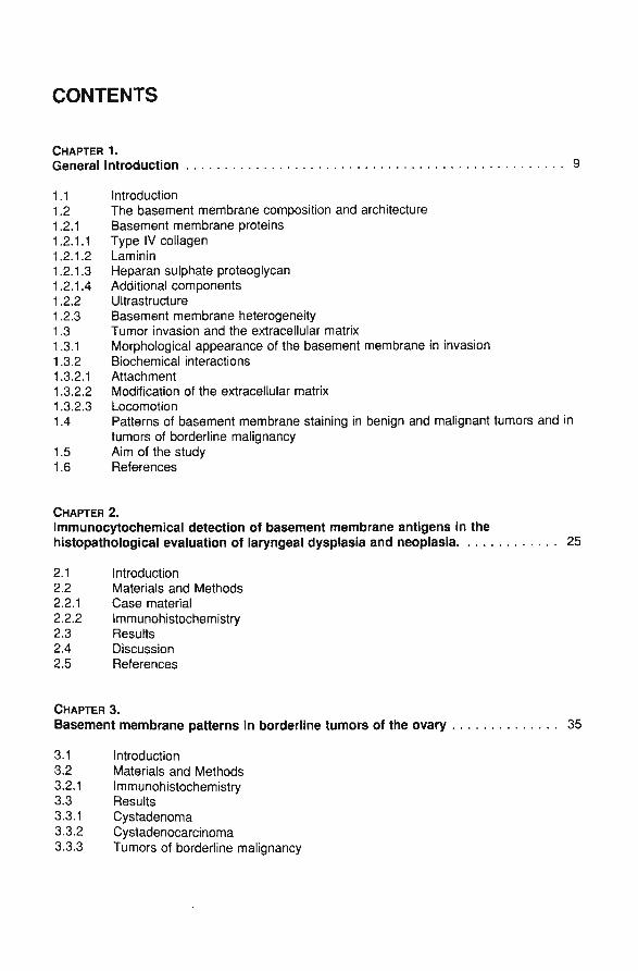
CONTENTS
CHAPTER 1.General Introduction
1.1 Introduction1.2 The basement membrane composition and architecture1.2.1 Basement membrane proteins1.2.1.1 Type IV collagen1.2.1.2 Laminin1.2.1.3 Heparan sulphate proteoglycan1.2.1.4 Additional components1.2.2 Ultrastructure1.2.3 Basement membrane heterogeneity1.3 Tumor invasion and the extracellular matrix1.3.1 Morphological appearance of the basement membrane in invasion1.3.2 Biochemical interactions1.3.2.1 Attachment1.3.2.2 Modification of the extracellular matrix1.3.2.3 Locomotion1.4 Patterns of basement membrane staining in benign and malignant tumors and in
tumors of borderline malignancy1.5 Aim of the study1.6 References
CHAPTER 2.Immunocytochemical detection of basement membrane antigens in thehistopathological evaluation of laryngeal dysplasia and neoplasia 25
2.12.22.2.12.2.22.32.42.5
IntroductionMaterials and MethodsCase materialImmunohistochemistryResultsDiscussionReferences
CHAPTER 3.
Basement membrane patterns in borderline tumors of the ovary 35
3.13.23.2.13.33.3.13.3.23.3.3
IntroductionMaterials and MethodsImmunohistochemistryResultsCystadenomaCystadenocarcinomaTumors of borderline malignancy
3.4 Discussion3.5 References
CHAPTER 4.
Basement membrane immunohistochemistry in renal cell adenocarcinoma 45
4.14.24.2.14.2.24.2.34.34.3.14.3.24.44.54.6
IntroductionMaterials and MethodsTissueImmunohistochemistryStatisticsResultsHistological findingsImmunohistochemical patterns in renal tumorsDiscussionTablesReferences
CHAPTER 5.
Pattern and composition of basement membranes in squamous cell carcinomasof the upper aerodigestive tract 57
5.15.25.2.15.2.25.35.45.55.6
IntroductionMaterials and MethodsTissueImmunohistochemistryResultsDiscussionTablesReferences
CHAPTER 6.
Patterns and composition of basement membranes in colon adenomas andcarcinoma 69
6.16.26.2.16.2.26.2.36.36.3.16.3.26.3.36.46.5
IntroductionMaterials and MethodsTissueAntibodiesImmunohistochemistryResultsNormal mucosaAdenomasAdenocarcinomasDiscussionReferences

CHAPTER 7.Type VII collagen expression in the normal, hyperplastfc and neoplasticendometrium 79
7.17.27.2.17.2.27.2.37.2.47.37.3.17.3.27.3.37.47.5
IntroductionMaterials and MethodsEndometrial mucosaHyperplasia and adenocarcinomaAntibodiesImmunohistochemistryResultsNormal mucosaHyperplasiaAdenocarcinomaDiscussionReferences
CHAPTER 8.General discussion 918.1 Basement membrane patterns in benign and in malignant neoplasms8.2 Potential significance of basement membrane antigens in tumors of borderline
malignancy8.3 Observed basement membrane patterns in tumors of borderline malignancy8.4 Future perspective
SUMMARY 95
SAMENVATTING 99
DANKWOORD 103
CURRICULUM VITAE 105

CHAPTER 1
GENERAL INTRODUCTION*
1.1 INTRODUCTION
"Simple, homogeneous, and perfectly transparent membranes, in which no structure can bediscovered" were described for the first time by Bowman in 1842 in an article entitled " Thestructure and use of the Malpighian bodies of the kidney, with observations on thecirculation through that gland" (1).Since the original description of Bowman, the term basement membrane has becomewidely accepted in the literature, although alternatives such as basal lamina, basementlamina and boundary membrane have been proposed (2).Although originally described in the uriniferous tube of the kidney, basement membranesare present ubiquitously in most multicellular organisms. In linings of epithelial andendothelial cells basement membranes are found at the basal side of the cell, facing thestroma. In addition they envelope cells in mesenchymal tissues: individual fat cells andcontractile cells of heart, smooth muscle and skeletal muscle and also Schwann cells aresurrounded by a basement membrane. In contrast, mesenchymal cells such as fibroblasts,histiocytes and blood cells are not surrounded by a basement membrane, and also theendothelium of lymph vessels and the sinusoids of the spleen, lymph nodes and liver lack acontinuous basement membrane. The hepatocyte is in fact the only epithelial cell in directcontact with the plasma (3). Myofibroblasts, which play an important role in the mainten-ance of the extracellular matrix (ECM) do not have a normal, intact basement membrane,but are surrounded by patches of basement membrane material containing type IVcollagen, laminin and heparan sulphate proteoglycan (4,5).In the past, histochemical procedures have been developed to visualize the basementmembrane, including the periodic acid Schiff method, which stains carbohydrates (6), aswell as silver staining procedures (7). Although the basement membrane zone is visualizedin this way, these histochemical reactions are not specific for basement membrane relatedcomponents, but stain other structures of the extracellular matrix and stroma, such ascollagen type I and type III, as well. Immunohistochemical methods with antibodies directed
" partially based on: Bosman FT, Havenith MG, Visser R, Cleutjens JPM.Basement membranes in neoplasia.Progress in Histochemistry and Cytochemistry 1992; Volume 24 No 2: 1-93
against basement membrane specific components have overcome this disadvantage andprovided a powerful and specific tool for fundamental and diagnostic investigations, whichled to unravelling of the basement membrane structure and composition and the establish-ment of basement membrane staining patterns in normal and pathological conditions.The basement membrane is a specialized compartment of the extracellular matrix (ECM),which contains at least four classes of macromolecules: the family of collagen proteins;structural glycoproteins such as laminin, fibronectin and entactin; elastin; and proteogly-cans. The ECM forms a dynamic environment with major functions in cell differentiation,tissue architecture and function, repair mechanisms, and in pathological conditions likeatherosclerosis, diabetes and malignant disease (3,8,9).The basement membrane is a complex structure, with two distinct layers when visualizedelectronmicroscopically, by staining with uranyl and osmium combinations. Adjacent to theunderlying stroma, a layer with high electron density, the "lamina densa", occurs. A secondlayer of low electron density occurs adjacent to the cell membrane and is designated"lamina lucida" or "lamina rara". A third layer has been described recently: this part of thebasement membrane contains perpendicular fibrils, which contain type VII collagen as amajor component. As this part of the basement membrane is in fact not a continuous layer,the term "pars fibroreticularis" is preferred (2). It is suggested that they anchor thebasement membrane to the underlying connective tissue.
1.2 THE BASEMENT MEMBRANE COMPOSITION AND ARCHITECTURE
Until recently the ECM was regarded as a static framework, supporting and accommodatingepithelial and mesenchymal cells. During the past decade much knowledge concerning itsstructure and function has become available (for recent reviews see 10-13).Basement membranes are complex and dynamic molecular structures, with a moleculararrangement differing in various organs, according to locally required functions. Compo-nents of the basement membrane are synthesized and deposited by cells of both epithelialand mesenchymal derivation (14, 15, 16). Some aspects of the composition and architec-ture will be more extensively discussed in the following sections.
1.2.1 Basement membrane proteins
ECM components are in general divided into three categories:(a) co//agens, which are the major structural elements;(b) proteog/ycans, highly charged molecules regulating fibre size, hydration and tissuepermeability, and(c) g/ycoprofe/ns which link matrix components and cells.The collagens of the extracellular matrix form a heterogeneous family of proteins, which arechemically and immunologically distinct.The basement membrane, as a specialized compartment of the extracellular matrix,contains a variety of other specific proteins. Martinez-Hernandez and Amenta (3) proposeda distinction between "intrinsic" components, i.e. type IV collagen, laminin and heparansulphate proteoglycan, proteins which occur (almost) exclusively in basement membranes,and "extrinsic" components, which are present in basement membranes with specializedfunctions, but can also be found in other (extracellular matrix) structures.
1.2.1.1 Type IV collagen
Type IV collagen is the most important structural component of the basement membrane.The formation of a stable network, serving as a supportive structure, is the major functionof this protein. Other functions include binding sites for other components like laminin,proteoglycan and nidogen.
10
The type IV collagen triple helix molecule is composed of four distinct polypeptide chains.Originally two a chains were identified: oil (IV) and oc2(IV), which are arranged in a 390 nmlong triple helical rod, with a globular domain ("NC1-domain") at the carboxy-terminus anda 30 nm long segment at the amino-terminus of the triple helix ("7S domain") (17). Recentlytwo additional type IV collagen polypeptide chains have been described, i.e. a3(IV) anda4(IV) (18, 19).These domains are the sites of the molecule where oligomers are generated by interactionbetween the NC1 domains, leading to dimers, and of lateral association of the 7S domainsand resulting in tetramers (17,20,21).
1.2.1.2 Laminin
Laminin is a noncollagenous glycoprotein with a molecular weight of 900 kD. It formscomplexes with other basement membrane components such as type IV collagen (22-24)and nidogen/entactin (25). It possesses distinct cell-binding properties and influences celldifferentiation and movement (26-31). The molecule has a cross-shaped structure withthree similar short arms and one long arm terminated by globular domains (32-34). Theprotein consists of three polypeptide chains: two chains with a molecular mass of about200 kD (B1 and B2 chains) and a large chain of about 400 kD (A chain). These chains areattached to each other by disulphide bonds.
1.2.1.3 Heparan sulphate proteoglycan
Basement membranes contain at least three different proteoglycans, with heparan sulphateproteoglycan (35) as the most important, and two smaller high density proteoglycans, witheither heparan sulphate or chondroitin sulphate side chains (36).Heparan sulphate proteoglycan was found in in the lamina lucida, where it forms clusteredaggrgates and in the lamina densa.It has been suggested, that proteoglycans have affinity for laminin and for the NC1 domainof type IV collagen and may be involved in the attachment of cells to basement membranesand in the maintenance and remodelling of basement membranes (8).
1.2.1.4 Additional components
Fibronectin is a widespread glycoprotein, and occurs in the extracellular matrix as well asin fibrous tissues and in plasma. It is capable of multiple interactions with cell surfaces andother matrix components (37). Fibronectin may promote extracellular matrix assembly andcell adhesion to these matrices. Together with laminin, fibronectin plays a major role intumor invasion.Type VII collagen is the largest member of the collagen family. It consists of three identicala-chains with a 150 kD globular domain at the carboxyl-terminus that is attached to ahelical section of 170 kD (38).The protein, that is secreted by keratinocytes (39), is now thought to be the major structuralcomponent of the anchoring fibrils which link the basement membrane to the underlyingstroma. Monoclonal antibodies have been raised against type VII collagen (40,41). Theseantibodies bind exclusively to the dermal/epidermal junction, to the basement membrane ofepithelia in tissues such as the breast, prostate, amnion and bronchus and stratifiedepithelia of the larynx, esophagus, trachea, vagina and ectocervix (40-43). In the epithelialbasement membrane of the major part of the gastrointestinal tract (liver, stomach andintestine) and in the basement membrane of bloodvessels, muscle and nerve fibers type VIIcollagen was not observed (43).Nidogen, a sulphated glycoprotein, was described in 1983 by Timpl et al (44). It consists ofa single polypeptide chain with a molecular weight of approximately 150 kD and is
11

distributed ubiquitously, similar to the distribution of laminin and type IV collagen. It hasbeen suggested that nidogen mediates the binding of laminin to type IV collagen (8).By immunoelectronmicroscopy it was shown that the basement membranes may differ intheir content and distribution of laminin and nidogen (45).A recently described extracellular matrix glycoprotein is tenascin, a 150 - 240 kD molecule,which appears to play a major role in tissue development. It is distributed in themesenchyme surrounding developing epithelia during embryogenesis of various organs(46). Tenascin was also found in neoplasms, where the molecule is expressed throughoutthe stroma, with increasing intensity in relation to an increasing degree of atypia andmalignancy (47, 48, 49).
1.2.2 Infrastructure
Examination of the basement membrane by light microscopy shows a thin, continuous andapparently homogeneous structure, after routine staining for example by the periodic acid-Schiff reaction. Ultrastructurally the typical basement membrane appears to be composedof three distinct layers (50-53). The /am/na cfensa, which is the most prominent layer, iscomposed of a network of anastomosing linear fibrils which show a diameter ranging from1.8-5.3 nm (53-55).The /am/na /uc/da, situated between the cell membrane and the lamina densa, is electronlucent and is crossed by fine fibrils from the lamina densa which are in continuity with theadjacent cell. Extensions of the lamina densa are found in the pars //£>rore?;cu/ar/s, togetherwith anchoring fibrils and microfibrils. These structures secure an intimate connectionbetween the basement membrane and connective tissue. Anchoring fibrils, which are foundalong the interface between the basement membrane in some epithelia and the adjacentstroma, connect the lamina densa with anchoring plaques, which are electron-denseislands in the stroma. Type VII collagen is a major structural component of these anchoringfibrils.
At sites where fusion of two basement membranes has occurred, as in the renalglomerulus, a lamina lucida is present on both sides of the lamina densa.
1.2.3 Basement membrane heterogeneity
In view of the diverse functions of the cell types, that are surrounded by a basementmembrane, and the contribution of this structure to many of these specialized functions(56), it is conceivable, that the biochemical composition of the basement membrane differsfrom organ to organ and that disease might influence this composition as well.Ultrastructural and immunohistochemical studies using monoclonal antibodies has indeedshown considerable heterogeneity and organ specificity during embryonal development,between different anatomical sites, and according to physiological functions (3, 57-59). Inseveral pathological conditions including diabetes and glomerular disease, morphologicaland biochemical changes have been described. In neoplasia, basement membranes arefound that are morphologically and biochemically indistinguishable from the tissues oforigin; however, in invasive tumors, a variety of structural alterations have been described(60,61)
1.3 TUMOR INVASION AND THE EXTRACELLULAR MATRIX
When tumor cells acquire the capacity of invasive growth, a complex sequence of events isinvolved. Although features morphologically characteristic for invasion may not be observedat an early stage, the expression of a variety of proteases, involved in degradation of theextracellular matrix, may be encountered (table 1).
12
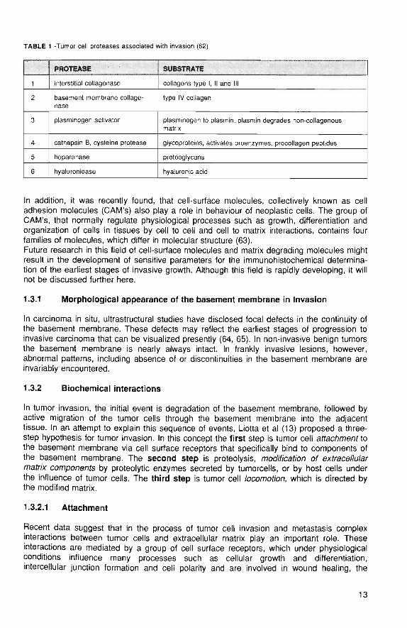
TABLE 1 -Tumor cell proteases associated with invasion (62)
1
2
3
4
5
6
PROTEASE
interstitial collagenase
basement membrane collage-nase
plasminogen activator
cathepsin B, cysteine protease
heparanase
hyaluronidase
SUBSTRATE
collagens type I, II and III
type IV collagen
plasminogen to plasmin; plasmin degrades non-collagenousmatrix
glycoproteins, activates proenzymes, procollagen peptides
proteoglycans
hyaluronic acid
In addition, it was recently found, that cell-surface molecules, collectively known as celladhesion molecules (CAM'S) also play a role in behaviour of neoplastic cells. The group ofCAM's, that normally regulate physiological processes such as growth, differentiation andorganization of cells in tissues by cell to cell and cell to matrix interactions, contains fourfamilies of molecules, which differ in molecular structure (63).Future research in this field of cell-surface molecules and matrix degrading molecules mightresult in the development of sensitive parameters for the immunohistochemical determina-tion of the earliest stages of invasive growth. Although this field is rapidly developing, it willnot be discussed further here.
1.3.1 Morphological appearance of the basement membrane in invasion
In carcinoma in situ, ultrastructural studies have disclosed focal defects in the continuity ofthe basement membrane. These defects may reflect the earliest stages of progression toinvasive carcinoma that can be visualized presently (64, 65). In non-invasive benign tumorsthe basement membrane is nearly always intact. In frankly invasive lesions, however,abnormal patterns, including absence of or discontinuities in the basement membrane areinvariably encountered.
1.3.2 Biochemical interactions
In tumor invasion, the initial event is degradation of the basement membrane, followed byactive migration of the tumor cells through the basement membrane into the adjacenttissue. In an attempt to explain this sequence of events, Liotta et al (13) proposed a three-step hypothesis for tumor invasion. In this concept the first step is tumor cell atfacnmenr tothe basement membrane via cell surface receptors that specifically bind to components ofthe basement membrane. The second step is proteolysis, moa7/7ca//on o/ ex/race//t//armafnx components by proteolytic enzymes secreted by tumorcells, or by host cells underthe influence of tumor cells. The third step is tumor cell /ocomof/on, which is directed bythe modified matrix.
1.3.2.1 Attachment
Recent data suggest that in the process of tumor cell invasion and metastasis complexinteractions between tumor cells and extracellular matrix play an important role. Theseinteractions are mediated by a group of cell surface receptors, which under physiologicalconditions influence many processes such as cellular growth and differentiation,intercellular junction formation and cell polarity and are involved in wound healing, the
13
inflammatory response and also in the host reaction to neoplasia (66-72). Four families ofcell surface molecules or cell adhesion molecules (CAM'S) have been identified:1- The group of integrins. This family of about 10 different types of adhesion molecules iscomposed of two subfamilies (alpha and beta), which form heterodimers.2- A large family of molecules that structurally resemble immunoglobulins. Only a fewmolecules of this family are CAM's.3- A group of calcium dependent membrane glycoproteins called Cadherins, and4- A more recently identified family of lectin-like giycoproteins.
During the past years much research has been devoted to the structure and function ofintegrins and these efforts have made them among the best understood in the family of celladhesion molecules (66, 67).Because of their mediating role in cell-extracellular matrix interactions as well as cell-celladhesion, it is likely that integrins are involved in each of the three steps of invasion:attachment of the tumor cell (by integrin receptors for the basement membrane compo-nents laminin and type IV collagen), proteolytic degradation of the basement membrane(through modulation of the expression of proteolytic enzymes) and, finally, migration intothe underlying stroma through adhesion to interstitial collagens (type I and III).
1.3.2.2 Modification of the extracellular matrix
Following attachment, the tumor cell secretes enzymes which can locally degrade theextracellular matrix. In addition, the tumor cell may be capable to stimulate host cells toproduce proteolytic enzymes. The release of these proteolytic enzymes may be mediatedby cell-extracellular matrix interactions: binding extracellular matrix ligands to their integrinreceptors may result in synthesis and release of proteases, which are involved in basementmembrane degradation.The target substrates are the matriceal components and the result is local degradation ofthe extracellular matrix with hydratation and expansion of the viscoelastic ground subs-tance. In this way an environment is created, that is conducive for invading tumor cells.Several proteases, involved in this process have already been identified (table 1).Local degradation of the basement membrane, through lysis of type IV collagen occursthrough specific collagenases and other proteases such as elastase, plasmin and cathep-sins.Non-collagenous macromolecules of the extracellular matrix such as glycoproteins andproteoglycans are degraded by glycosidases, which cleave the glycosaminoglycan side-chains.The plasminogen activator/plasmin system also plays a role in modification of the extracel-lular matrix. Two types of plasminogen activator (PA) have been identified in neoplastictissue: a urokinase-like PA, which is secreted by most carcinomas, and a tissue activatortype PA (62,71). The tissue activator type PA plays a major role in thrombolysis; as such itmay participate in fibrin degradation at the primary tumor site and contribute to tumor cellrelease.In addition, plasmin may be involved in the activation of type IV collagenase (73) andfurthermore it can degrade both laminin and fibronectin (74, 75).
1.3.2.3 Locomotion
The third step in the process of invasion is tumor cell motility, which follows attachment andlocal proteolysis of the extracellular matrix. Cell motility is an essential step for tumor cellsto reach neighbouring compartments such as the adjacent stroma, vascular walls anddistant organs and probably involves chemotactic mechanisms.It has been suggested, that an autocrine motility factor is secreted by the tumor cell andrecognizes a tumor cell surface receptor (76). This results in stimulation of cell movement,
14
a major function in this phase of invasion. For active movement of the tumor cells throughthe enzymatically altered extracellular matrix, attachment to extracellular matrix elements ismediated by receptors, including those of the integrin family.
1.4 PATTERNS OF BASEMENT MEMBRANE STAINING IN BENIGN AND MALIG-NANT TUMORS AND IN TUMORS OF BORDERLINE MALIGNANCY
Normal tissue patterns
In general, the tissues of every organ of the body contain continuous and regular basementmembranes, as has already been outlined. The epithelia of the skin, the endocrine systemand the respiratory, genitourinary and gastrointestinal tracts are separated from theadjacent stroma by a basement membrane. The central nervous system, in contrast,contains only vascular basement membranes, whereas Schwann cells of the peripheralnervous system are enveloped by basement membranes. Mesenchymal cells such asadipocytes, cardiac, skeletal and smooth muscle cells also are surrounded by a basementmembrane. However, fibroblasts, histiocytes, blood cells and, exceptionally, epithelial cellssuch as hepatocytes lack a basement membrane. Myofibroblasts are surrounded bypatches of basement membrane components (5).
Basement membranes in benign tumors
Although most investigations have focused mainly on basement membrane characteristicsin connection with tumor invasion and metastasis, several studies have reported patterns ofbasement membrane staining in benign tumors, a summary of which is given in Table 2.In general terms, the results of these studies reconfirm the continuity of basementmembranes in benign, reactive proliferations and in benign tumors. However, basementmembrane interruptions have been found in benign lesions (85, 102, 108), but theseinterruptions were mostly related to an adjacent inflammatory infiltrate. As inflammatorycells are known to secrete collagenase, it can be assumed that these are responsible forthe local breakdown of the basement membrane (104).Some benign tumors may be very difficult to distinguish from malignant tumors because oftheir morphological similarity. An example, familiar to every diagnostic pathologist, issclerosing adenosis of the female breast, which can be very difficult to distinguish fromtubular carcinoma. Basement membrane immunohistochemistry may facilitate a solution ofthis diagnostic problem, because in sclerosing adenosis the basement membranes areintact and continuous, but in the tubular carcinoma they are typically interrupted or evencompletely absent.In addition, basement membrane immunohistochemistry may assist in discriminatingbetween inflammatory lesions and malignant tumors. An example is chronic pancreatitis,which leads to abundant fibrous tissue, surrounding irregular ducts. This can be difficult todistinguish from pancreatic carcinoma. Also in the pancreas the inflammatory lesion showsintact epithelial basement membranes, whereas in carcinoma these may be interrupted oreven absent (84, 105, 106).
15
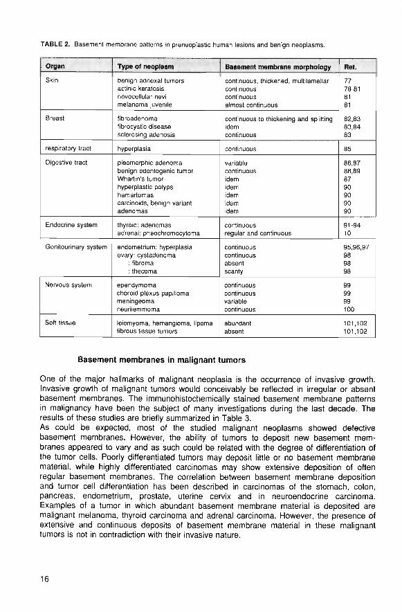
TABLE 2. Basement membrane patterns in preneoplastic human lesions and benign neoplasms.
Organ
Skin
Breast
respiratory tract
Digestive tract
Endocrine system
Genitourinary system
Nervous system
Soft tissue
Type of neoplasm
benign adnexal tumorsactinic keratosisnevocellular nevimelanoma juvenile
fibroadenomafibrocystic diseasesclerosing adenosis
hyperplasia
pleomorphic adenomabenign odontogenic tumorWhartin's tumorhyperplastic polypshamartomascarcinoids, benign variantadenomas
thyroid: adenomasadrenal: pheochromocytoma
endometrium: hyperplasiaovary: cystadenoma
: fibroma: thecoma
ependymomachoroid plexus papillomameningeomaneurilemmoma
leiomyoma, hemangiorna, lipomafibrous tissue tumors
Basement membrane morphology
continuous, thickened, multilarnellarcontinuouscontinuousalmost continuous
continuous to thickening and splittingidemcontinuous
continuous
variablecontinuousidemidemidemidemidem
continuousregular and continuous
continuouscontinuousabsentscanty
continuouscontinuousvariablecontinuous
abundantabsent
Re*.
7778-818181
82,8383,8483
85
86,8788,898790909090
91-9410
95,96,97989898
999999100
101,102101,102
Basement membranes in malignant tumors
One of the major hallmarks of malignant neoplasia is the occurrence of invasive growth.Invasive growth of malignant tumors would conceivably be reflected in irregular or absentbasement membranes. The immunohistochemically stained basement membrane patternsin malignancy have been the subject of many investigations during the last decade. Theresults of these studies are briefly summarized in Table 3.As could be expected, most of the studied malignant neoplasms showed defectivebasement membranes. However, the ability of tumors to deposit new basement mem-branes appeared to vary and as such could be related with the degree of differentiation ofthe tumor cells. Poorly differentiated tumors may deposit little or no basement membranematerial, while highly differentiated carcinomas may show extensive deposition of oftenregular basement membranes. The correlation between basement membrane depositionand tumor cell differentiation has been described in carcinomas of the stomach, colon,pancreas, endometrium, prostate, uterine cervix and in neuroendocrine carcinoma.Examples of a tumor in which abundant basement membrane material is deposited aremalignant melanoma, thyroid carcinoma and adrenal carcinoma. However, the presence ofextensive and continuous deposits of basement membrane material in these malignanttumors is not in contradiction with their invasive nature.
16
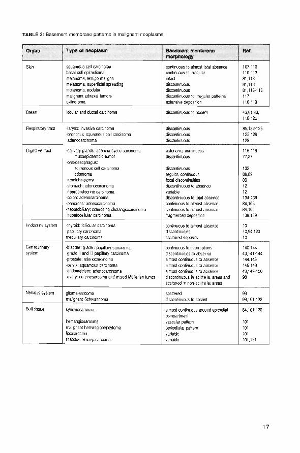
TABLE 3: Basement membrane patterns in malignant neoplasms.
Organ
Skin
Breast
Respiratory tract
Digestive tract
Endocrine system
Genitourinarysystem
Nervous system
Soft tissue
Type of neoplasm
squamous cell carcinomabasal cell epithelioma,melanoma, lentigo malignamelanoma, superficial spreadingmelanoma, nodular
malignant adnexal tumorscyiindroma
lobular and ductal carcinoma
-larynx: invasive carcinoma
•bronchus: squamous cell carcinomaadenocarcinoma
-salivary glands: adenoid cystic carcinomamucoepidermoid tumor
•oral/oesophagus:squamous cell carcinomaodontoma
ameloblastoma
-stomach: adenocarcinomaneuroendocrine carcinoma
-colon: adenocarcinoma-pancreas: adenocarcinoma•hepatobiliary: sclerosing cholangiocarcinomahepatocellular carcinoma
•thyroid: follicular carcinomapapillary carcinoma
medullary carcinoma
-bladder: grade 1 papillary carcinomagrade II and III papillary carcinoma
-prostate: adenocarcinoma-cervix: squamous carcinoma
-endometrium: adenocarcinoma
-ovary: carcinosarcoma and mixed Mulierian tumor
glioma-sarcomamalignant Schwannoma
synoviosarcoma
hemangiosarcoma
malignant hemangiopericytomaliposarcoma
rhabdo-, leiomyosarcoma
Basement membranemorphology
continuous to almost total absencecontinuous to irregular
intactdiscontinuousdiscontinuousdiscontinuous to irregular patternsextensive deposition
discontinuous to absent
discontinuousdiscontinuous
discontinuous
extensive, continuousdiscontinuous
discontinuousregular, continuousfocal discontinuities
discontinuous to absencevariablediscontinuous to total absence
continuous to almost absence
continuous to almost absencefragmented deposition
continuous to almost absencediscontinuitiesscattered deposits
continuous to interruptionsdiscontinuities to absence
almost continuous to absencealmost continuous to absencealmost continuous to absence
discontinuous in epithelial areas and
scattered in non-epithelial areas
scattered
discontinuous to absent
almost continuous around epithelialcompartmentvascular pattern
pericellular pattern
variable
variable
Ref.
107-110110-113
81,11381,11381,113-116117116-119
43,61,83,118-122
85,122-125125-129
129
116-11977,87
13288,89
891212134-138
84,10584,106
138,139
1010,94.120
10
140-14443,141-144
144,145146-149
43,149-150
98
9999,101,102
84,101,120
101101101101,151
17
The propensity of these tumors to synthesize and deposit basement membranes mightreflect their biological potential and, as such, may contain prognostic information. Studiesconcerning this possibility in carcinomas of the lung, colon and bladder have indeed showna positive correlation between extensive deposition of basement membranes by the tumorand a more favourable course of the disease (128,134,135,140).Also in soft tissue tumors basement membrane immunohistochemistry may yield importantinformation. Certain sarcomas may be diagnosed more readily, owing to the ability of thetumor cells to deposit basement membrane material. For instance, leiomyosarcomas arecomposed of cells that are individually enveloped by a basement membrane. Fibrosarcomacells, however, do not deposit basement membrane material. In this way, basementmembrane immunohistochemistry can be a valuable tool in distinguishing these categoriesof spindle cell tumors.
Basement membranes in tumors of borderline malignancy
Unlike the situation in benign or malignant tumors, basement membrane immunohistoche-mistry has not been extensively explored in tumors of borderline malignancy. As summar-ized in Table 4, studies have been largely restricted to epithelial dysplasia and in situcarcinoma in the uterine cervix and also to adenomas in the colorectum. Series of lesionsusually contained borderline tumors with intact basement membranes in addition to tumorswith focally interrupted basement membranes.
Table 4: Basement membrane patterns in premalignant lesions and tumors of borderline malignancy.
Organ
Skin
Breast
Respiratory tract
Digestive tract
Endocrine system
Genitourinarysystem
Type of neoplasm
Bowen's diseaseactinic keratosis
intraduct and intralobularcarcinoma
-larynx: hyperplasiadysplasia
-colon: adenoma with dysplasia
carcinoid, islet cell tumor
-prostate: severe dysplasia-endometrium: adenomatoushyperplasia-cervix: dysplasiacarcinoma in situ
Basement membranemorphology
continuouscontinuous
continuous
continuouscontinuous
local interruptions
continuous
discontinuousintact or disruptions
intactintact or focal disruptions
Ret.
77,78,10777-80
82
85,79,12379,84
152,153
10,152
10,14496
95,145,14795,145,147
1.5 AIM OF THE STUDY
In the last decade many new facts were unveiled regarding the physiological andbiochemical properties of the extracellular matrix includingthe basement membrane. The literature reviewed in the previous paragraphs hashighlighted current opinions regarding the biochemical aspects of the tumor cell-extracellular matrix interaction. Isolation and purification of basement membranecomponents and the production ofpolyclonal and monoclonal antibodies directed against these components, in particularagainst laminin, type IV collagen and type VII collagen, has allowed visualization of the
18
basement membrane in normal and pathological conditions. Especially in the field ofneoplastic disease the use of immunohistochemistry with antibodies directed againstbasement membrane components has resulted in a large number of publications that haveexpanded our knowledge of the basement membrane structure in normal tissues and inneoplasia, in particular in invading malignant tumors. Relatively few investigations havefocused on basement membrane immunohistochemistry in preneoplastic lesions andtumors of borderline malignancy.Therefore we initiated a series of studies to investigate the characteristics of the basementmembrane immunohistochemically in the transition from benign to malignant. In thesestudies antibodies were used against basement membrane proteins laminin, type IVcollagen and type VII collagen.The following questions were addressed:1-Are basement membrane patterns in epithelial dysplasia of varying grades of severity,different from those in in situ carcinoma? Can in situ carcinomas be subdivided accordingto their basement membrane status (continuous or discontinuous)?2-Do basement membrane patterns in tumors, that are classified as of borderlinemalignancy, allow a distinction between benign (continuous) and incipiently malignant(discontinuous)?3-Does type VII collagen play a role in the transition from benign to malignant neoplasia?
In chapter 2 the basement membrane patterns in laryngeal carcinoma in situ, dysplasiaand hyperplasia are described using immunohistochemistry and applying antibodiesdirected against type IV collagen and laminin.In chapter 3 basement membrane patterns in a series of borderline tumors of the ovaryare reported in comparison with patterns in benign cystadenomas of the ovary andcystadenocarcinomas.Another example of a class of tumor that stands on the borderline between benign andmalignant is the renal cortical (tubular) adenoma. Some authors rely mainly on the diameterof the tumor to determine the nature of the neoplasm.In chapter 4 we report on the results of a study to determine the nature of renal corticalepithelial tumors with a diameter ranging from millimetres to centimetres, with emphasis onpatterns of the basement membrane staining using antibodies against type IV collagen andlaminin. The aim of this study was to find criteria for benign and malignant renal celltumors, based on basement membrane characteristics.In chapter 5 we report on the results of a study regarding the patterns of type IV collagenand type VII collagen staining in squamous cell carcinoma of the upper aerodigestive tract,with emphasis on the relation of these patterns with tumor differentiation.In chapters 6 and 7 studies are described concerning basement membrane patterns in thenormal mucosa of the colon and endometrium and in preneoplastic lesions such asadenomas and hyperplasia. Especially the presence and distribution of type VII collagenwas addressed, as this protein has been mainly associated with anchoring fibrils, which donot normally occur in these epithelia.Finally, in chapter 8, the results of these studies are discussed in more general terms.Some remarks are made regarding applicability of basement membraneimmunohistochemistry in diagnostic histopathology.
19

1.6 REFERENCES
1. Bowman W. On the structure and use of the malpighian bodies of the kidney, with observations on thecirculation through that gland. Phil Transactions Royal Soc London 1842; 132: 57-80.
2. Inoue S. Ultrastructure of basement membranes. Int Rev Cytol 1989; 117: 57-99.3. Martinez-Hernandez A, Amenta PS. The basement membrane in pathology. Lab Invest 1983; 48: 656-677.4. Lipper S, Kahn LB, Reddick RL. The myofibroblast. Pathol Annu 1980; 15: 409- 413.5. SeemayerTA, Schurch W, Lagace R. Myofibroblasts in human pathology. Hum Pathol 1981; 12: 491-4936. Lillie RD. Reticulum staining with Schiff reagent after oxidation by acidified sodium periodate. J Lab Clin
Med 1947; 32: 910-912.7. Jones DB. Inflammation and repair of the glomerulus. Am J Pathol 1951; 27: 991- 1009.8. Timpl R. Structure and biological activity of basement membrane proteins. Eur J Biochem 1989- 180- 487-
502.9. Weber L. Krieg T. Timpl R. Basal membranen: Struktur, Funktion, Pathologie. Hautarzt 35; 1964: 279-286.10. Bosman FT, Havenith MG, Visser R, Cleutjens JPM. Basement membranes in neoplasia. Progr Histochem
Cytochem 1992; 24 (4): 1-9411. Rubin E, Farber JL. (eds). Pathology. 1988. Lippincott Comp, Philadelphia USA; 66.-77.12. d'Ardenne AJ. Use of basement membrane markers in tumour diagnosis. J Clin Pathol 1989; 42: 449-457.13. Liotta LA, Rao CN, Barsky SH. Tumor invasion and the extracellular matrix. Lab Invest 1983; 49: 636-650.14. Weiser MM, Sykes DE, Killen PD. Rat intestinal basement membrane synthesis. Epithelial versus
nonepithelial contributions. Lab Invest 1990; 62: 325-330.15. Cleutjens JPM, Havenith MG, Beek C, Vallinga M, ten Kate J, Bosman FT. Origin of basement membrane
type IV collagen in xenografted human epithelial tumor cell lines. Am J Pathol 1990; 136: 1165-1172.16. Damjanov I, Damjanov N, Knowles BB, Engvall E. Origin of laminin in the extracellular matrix of human
tumor xenografts in nude mice. Virchows Arch(B) 1985; 49: 45-52.17. Timpl R, Oberbaumer I, Furthmayr H, Kuehn K. Macromolecular organization of type IV collagen. 1982 In:
New trends in basement membrane research. H.Kuehn, H. Schoene and R. Timpl eds. Raven Press NewYork, 57-67.
18. Butkowski R, Langeveld JP, Wieslander J, Hamilton J, Hudson BG. Localization of the Goodpasture epitopeto a novel chain of basement membrane collagen. J Biol Chem 1987; 262: 7874-7877.
19. Saus J, Wieslander J, Langeveld JP, Quinones S, Hudson BG. Identification of the Goodpasture antigen asthe alpha 3(IV) chain of collagen IV. J Biol Chem 1988; 263: 13374-13380.
20. Yurchenco PD and Furthmayr H. Self-assembly of basement membrane collagen. Biochemistry 1984; 23;1839-1850.
21. Laurie GW, Bing JT, Kleinman HK, Hassell JR, Aumailley M, Martin GR, Feldmann RJ. Localization ofbinding sites for laminin, heparan sulfate proteoglycan and fibronectin on basement membrane (type IV)collagen. J Mol Biol 1986; 189: 205-216.
22. Terranova VP, Rohrbach DH, Martin GR. Role of laminin in the attachment of PAM 212 (epithelial) cells tobasement membrane collagen. Cell 1980; 22: 719-726.
23. Rao CN, Margulies IMK, Tralka TS, Terranova VP, Madri JA, Liotta LA. Isolation of a subunit of laminin andits role in molecular structure and tumor cell attach ment. J Biol Chem 1982; 57: 9740-9744.
24. Woodley DT, Rao CN, Hassell JR, Liotta LA, Martin GR, Kleinman HK. Interactions of basement membranecomponents. Biochim Biophys Acta 1983; 761: 278-283.
25. Carlin BE, Durkin ME, Bender B Jatfe R, Chung AE. Synthesis of laminin and entactin by F9 cells inducedwith retinoic acid and dibutyryl cyclic AMP. J Biol Chem 1983; 258: 7729-7737.
26. Kleinman HK, McCarvey ML, Hassell JR, Martin GR, Baron van Evercooren A, Dubois-Dalcq M. The role oflaminin in basement membranes and in the growth, adhesion and differentiation of cells. Role ofextracellular matrix in development. 1984; 14: 123-130.
27. Kleinman HK, Cannon FB, Laurie GW, Hassell JR, Aumailley M, Terranova VP, Martin GR, Dubois-Dalcq M.Biological activities of laminin. J Cell Biochem 1985; 27: 317-326.
28. Zagris N, Chung AE. Distribution and functional role of laminin during induction of the embryonic axis in thechick embryo. Differentiation 1990; 43: 81-86.
29. Shah KD, Gerber MA. Development of intrahepatic bile ducts in humans. Possible role of laminin. ArchPathol Lab Med 1990; 114; 597-600.
30. Aumailley M, Wiedemann H, Mann K, Timpl R. Binding of nidogen and the laminin-nidogen complex tobasement membrane collagen type IV. Eur J Biochem 1989; 184: 241-248.
31. Kleinman HK, Ogle RC, Cannon FB, Little CD, Sweeney TM, Luckenbill-Edds L. Laminin receptors forneurite formation. Proc Natl Acad Sci USA 1988; 85: 1282- 1286.
32. Timpl R, Rohde H, Gehron Robey P, Rennart SI, Foidart JM, Martin GR. Laminin. A glycoprotein frombasement membranes. J Biol Chem 1979; 254: 9933-9937.
33. Engel J, Odermatt E, Engel A, Madri JA, Furthmayr H, Rohde H, Timpl R. Shapes, domain organizationsand flexibility of laminin and fibronectin, two multifunctional proteins of the extracellular matrix. J Mol Biol1981;150:97-120.
20
34. Cooper AR, Kurkinen M, Taylor A, Hogan BLM, Studies on the biosynthesis of laminin by murine parietalendoderm cells. Eur J Biochem 1981; 119: 189-197.
35. Liotta LA, Rao CN, Wewer UM: Biochemical interactions of tumor cells with the basement membrane. AnnuRev Biochem 1986; 55: 1037-1057.
36. Paulsson M, Fujiwara S, Dziadek M, Timpl R, Pejler G, Backstrom G, Lindahl U, Engel J. Structure andfunction of basement membrane proteoglycans. Ciba- Found-Symp 1986; 124: 189-203.
37. Couchman JR, Austria MR, Woods A. Fibronectin-cell interactions. J Invest Dermatol 1990; 94: 7S.-4S.38. Burgeson RE, Morris NP, Murray LW, Duncan KG, Keene DR, Sakai LY. The structure of type VII collagen.
Ann N Y Acad Sci 1986; 460: 47-57.39. Regauer S, Seller GR, Barrandon Y, Easley KW, Compton CC. Epithelial origin of cutaneous anchoring
fibrils. J Cell Biol 1990; 111: 2109-2115.40. Leigh IM, Purkis PE, Bruckner-Tuderman L. LH 7.2 monoclonal antibody detects type VII collagen in the
sublamina densa zone of ectodermally-derived epithelia, including skin. Epithelia 1987; 1: 17-29.41. Sakai LY, Keene DR, Morris NP, Burgeson RE. Type VII collagen is a major structural component of
anchoring fibrils. J Cell Biol 1986; 103: 1577-1586.42. Keene DR, Sakai LY, Lunstrurn GP, Morris NP, Burgeson RE. Type VII collagen forms an extended network
of anchoring fibrils. J Cell Biol 1987; 104: 611-621.43. Wetzels RHW, Robben HCM, Leigh IM, Schaafsma E., Vooys GP, Ramaekers FCS. Distribution patterns of
type VII collagen in normal and malignant human tissues. Am J Pathol 1991; 139: 451-45944. Timpl R, Dziadek M, Fujiwara S, Nowackm H, Wick G. Nidogen, a new self aggregating basement
membrane protein. Eur J Biochem 1983; 137: 455-456.45. McCarthy JB, Basara ML, Palm SL, Sas DF, Furcht LT. The role of cell adhesion proteins-laminin and
fibronectin-in the movement of malignant and metastatic cells. Cancer Metastatis Rev 1985; 4: 125-152.46. Chiquet-Ehrismann R, Mackie EJ, Pearson CA, Sakakura T. Tenascin, an extracellular matrix protein
involved in tissue interactions during fetal development and oncogenesis. Cell 1986; 47; 131-139.47. Mackie EJ, Chiquet-Ehrismann R, Pearson CA, Inaguma Y. Tenascin is a stromal marker for epithelial
malignancy in the mammary gland. Proc Natl Acad Sci USA 1987; 84: 4621-4625.48. Vollmer G, Siegal GP, Chiquet-Ehrismann R, Lightner VA, Arnholdt H, Knuppen R. Tenascin expression in
the human endometrium and in endometrial adenocarcinomas. Lab Invest 1990; 62: 725-730.49. Howeedy AA, Virtanen I, Laitinen L, Gould NS, Koukoulis GK, Gould VE. Differential distribution of tenascin
in the normal, hyperplastic and neoplastic breast. Lab Invest 1990; 63: 798-806.50. Kefalides NA, Alper R, Clark CC. Biochemistry and metabolism of basement membranes. Int Rev Cytol
1979; 61: 169-228.51. Vracko R. Basal lamina scaffold-anatomy and significance for maintenance of orderly tissue structure. Am J
Pathol 1974; 77: 314-346.52. Madri JA, Pratt BM, Yurchenco PD, Furthmayr H. The ultrastructural organization and architecture of
basement membranes. In; Basement membranes and cell movement. CIBA Found symp 108 1984; 6-24.53. Inoue S, Leblond CP, Lauri GW. Ultrastructure of Reichert's membrane, a multilayered basement membrane
in the parietal wall of the rat yolk sac. J Cell Biol 1983; 97: 1524-1537.54. Laurie GW, Leblond CP, Inoue S, Martin GR, Chung AE. Fine structure of the glomerular basement
membrane components to the lamina densa (basal lamina) and its extensions in both glomeruli and tubulesof the rat kidney. Am J Anat 1984; 169: 463-481.
55. Inoue S, Leblond CP. Three-dimensional network of cords: the main component of basement membranes.Am J Anat 1988; 181; 341-358.
56. Gorstein F. The dynamic extracellular matrix. Editorial. Hum Pathol 1988; 19: 751- 752.57. Bosman FT, Cleutjens JPM, Beek C, Havenith MG. Basement membrane heterogeneity. Histochem J 1989;
21:629-633.58. Damjanov I. Heterogeneity of basement membranes in normal and pathologically altered tissues. Virchow's
Arch A Pathol Anat 1990; 416: 185-188.59. Leu FJ, Engvall E, Damjanov I, Heterogeneity of basement membranes of the human genitourinary tract
revealed by sequential immunofluorescence staining with monoclonal antibodies to laminin. J HistochemCytochem 1986; 34: 483-489.
60. Schmoeckel C, Stolz W, Sakai LY, Burgeson RE, Timpl R, Krieg T. Structure of basement membranes inmalignant melanoma and nevocytic nevi. J Invest Dermatol 1989; 92: 663-669.
61. Wetzels RHW, Holland R, van Haelst UJGM, Lane EB. Leigh IM, Ramaekers FCS. Detection of basementmembrane components and basalcell keratin 14 in non- invasive and invasive carcinoma ol the breast. Am JPathol 1989; 134: 571-579.
62. Liotta LA. Mechanisms of cancer invasion and metastasis. Volume 3: Influence of Tumor Development onthe Host. In: Cancer Growth and Progression, edited by HE Kaiser Dordrecht; Kluwer Academic Publishers,1989:58-71.
63. Katz AM, Rosenthal D, Sauder DN. Cell adhesion molecules. Structure, function, and implication in a varietyof cutaneous and other pathologic conditions. Intern J Dermatol 1991; 30: 153-160.
64. Niedbala MJ, Crickard K, Bernacki RJ. In vitro degradation of extracellular matrix by human ovariancarcinoma cells. Clin Exp Metastasis 1987; 5: 181-197.
21
65. Dingemans KP. What's new in the ultrastructure of tumor invasion in vivo? Pathol Res Pract 1988' 183- 792-808.
66. Hynes RO. Integrins: a family of cell surface receptors. Ceil 1987; 48: 549-554.67. Virtanen I, Korhonen M, Kariniemi AL, Gould VE, Laitinen L, Ylanne J. Integrins in human cells and tumors
Cell Differ Dev 1990; 32: 215-227.68. Cunningham BA. Cell adhesion molecules and the regulation of development Am J Obstet Gynecol 1991-
164(4): 939-948.69. Takeichi M. Cadherin cell adhesion receptors as a morphogenetic regulator. Science 1991; 251: 1451-1455.70. Crossin KL. Cell adhesion molecules in embryogenesis and disease. Ann N Y Acad Sci 1991; 615: 172-186.71. Fleming S. Cell adhesion and epithelial differentiation. J Pathol 1991; 164: 95-100.72. Dana K, Andreasen PA, Grondahl-Hansen J, Kristensen P, Nielsen LS, Skriver L Plasminogen activators,
tissue degradation and cancer. Adv Cancer Res 1985; 44: 139-266.73. Salo T, Liotta LA, Keski-Oja J, Turpeenniemi-Hujanen T, Tryggvason K. Secretion of basement membrane
collagen degrading enzyme and plasminogen activator by transformed cells - role in metastasis Int JCancer 1982; 30: 669-673.
74. Balian G, Click EM, Crouch E, Davidson JM, Bornstein P. Isolation of a collagen- binding fragment fromfibronectin and cold-insoluble globulin. J Biol Chem 1979; 254: 1429-1433.
75. Liotta LA, Goldfarb RH, Brundage R, Siegel GP, Terranova VP, Garbisa S. Effect of plasminogen activator(urokinase), plasmin and thrombin on glycoprotein and collagenous components of basement membraneCancer Res 1981; 41: 4629- 4636.
76. Liotta LA, Wewer U, Rao NC, Schiffmann E, Stracke M, Guirguis R, Thorgeirsson U, Muschel R, Sobel M.Biochemical mechanisms of tumor invasion and metas tases. Adv Exp Med Biol 1988; 233: 161-169.
77. Kallioinen M, Autio Harmainen H, Dammert K, Risteli J, Risteli L Basement membrane laminin and type IVcollagen in various benign and malignant adnexal tumors of the skin: an immunohistochemical study. JInvest Dermatol 1984; 83: 276-280.
78. Gusterson BA, Warburton MJ, Mitchell D, Kraft N, Hancock WW. Invading squamous cell carcinoma canretain a basal lamina. An immunohistochemical study using a monoclonal antibody to type IV collagen LabInvest 1984; 51: 82-87.
79. Gusterson BA, Clinton S, Cough G. Studies of early invasive and intraepithelial squamous cell carcinomausing an antibody to type IV collagen. Histopathology 1986; 10: 161-169.
80. Cam Y, Bellon G, Poulin G, Caron Y, Birembaut P. Distribution of type IV collagen in benign and malignantepithelial proliferations. An indirect immunofluorescence study on the breasts, the lungs and the skin.Invasion Metastasis 1984; 4; 61-72.
81. Havenith MG, van Zandvoort EHM, Cleutjens JPM, Bosman FT. Basement membrane deposition in benignand malignant nevo-melanocytic lesions: an immunohistochemical study with antibodies to type IV collagenand laminin. His topathology 1989; 15: 137-146.
82. Natali PG, Giacomini P, Bigotti G, Nicotra MR, Bellocci M, De Martino C. Heterogeneous distribution ofactin, myosin, fibronectin and basement membrane antigens in primary and metastatic human breastcancer. Virchows Arch A (Pathol anat) 1984; 405: 69-83.
83. Willebrand D, Bosman FT, De Goeij AF. Patterns of basement membrane deposition in benign andmalignant breast tumours. Histopathology 1986; 10: 1231-1241.
84. Barsky SH, Siegal GP, Jannotta F, Liotta LA. Loss of basement membrane components by invasive tumorsbut not by their benign counterparts. Lab Invest 1983; 49: 140-147.
85. Visser R, van der Beek JMH, Havenith MG, Cleutjens JPM, Bosman FT. Im munocytochemical detection ofbasement membrane antigens in the histopathological evaluation of laryngea! dysplasia and neoplasiaHistopathology 1986; 10: 171-180.
86. Erlandson RA, Cardon-Cardo C, Higgins PJ. Histogenesis of benign pleomorphic adenoma (mixed tumor) ofthe major salivary glands. Am J Surg Pathol 1984; 8: 803-820.
87. Toida M, Takeuchi J, Hara K, Sobue M, Tsukidate K, Goto K, Nakashima N. Histochemical studies ofintercellular components of salivary gland tumors with special reference to glycosaminoglycans, laminin andvascular elements. Virchows Arch (Path Anat) 1984; 403: 15-26.
88. Heikinheimo K, Morgan PR, Happonen RP, Stenman G, Virtanen I. Distribution of extracellular matrixproteins in odontogenic tumors and developing teeth. Virchows Arch B Cell Pathol 1991; 61:101-109.
89. Thesleff I, Ekblom P. Distribution of keratin and laminin in ameloblastoma. Comparison with developing toothand epidermoid carcinoma. J Oral Pathol 1984; 13: 85-96.
90. Burtin P, Chavanel G, Foidart JM, Martin E. Antigens of the basement membrane and the peritumoralstroma in human colonic adenocarcinomas: an immunofluore scence study. Int J Cancer 1982; 30: 13-20.
91. Katoh R, Jasani B, Williams E.D. Hyalinizing trabecular adenoma of the thyroid. A report of three oases withimmunohistochemical and ultrastructural studies. Histopatholy 1989; 15: 211-224.
92. Miettinen M, Virtanen I. Expression of laminin in thyroid gland and thyroid tumors: an immunohistologicstudy. Int J Cancer 1984; 34: 27-30.
93. Kendall CH, Sanderson PR, Cope J, Talbot IC. Follicular thyroid tumors: a study of laminin and type IVcollagen in basement membrane and endothelium. J Oin Pathol 1985; 38: 1100-1105.
94. Charpin C, Kopp F, Pourreau-Schneider N et al. Laminin immunodetection in tumorous and non-tumorousdisorders of human thyroid. Bull Cancer (Paris) 1985; 72: 6-15,
22
95. Bulletti C, Galassi A, Jasonni VM, Martinelli G, Tabanelli S, Flamigni C. Basement membrane componentsin normal, hyperplastic and neoplastic endometrium. Cancer 1988; 62: 142-149.
96. Vogel HP, Mendelsohn G. Laminin immunostaining in hyperplastic, dysplastic and neoplastic lesions of theendometrium and the uterine cervix. Obstet Gynecol 1987; 69: 794-799.
97. Furness PN, Lam EW. Patterns of basement membrane deposition in benign, pre-malignant and malignantendometrium. J Clin Pathol 1987; 40: 1320-1323.
98. Stenback F, Wasenius VM. Basement membrane structures in tumors of the ovary. Eur J Obstet GynecolReprod Biol 1985; 20: 357-371.
99. McComb RD, Bigner DD. Immunolocalization of laminin in neoplasms of the central and peripheral nervoussystems. J Neuropath Exp Neurol 1985; 44: 242- 253.
100. Fleischmajer R, Timpl R, Dziadek M, Lebwohl M. Basement membrane proteins, interstitial collagens andfibronectin in neurofibroma. J Invest Dermatol 1985; 85: 54-59.
101. Miettinen M, Foidart JM, Ekblom P. Immunohistochemical demonstration of laminin, the major glycoproteinof basement membranes, as an aid in the diagnosis of soft tissue tumors. Am J Clin Pathol 1983; 79: 306.
102. Ogawa K. Oguchi M, Yamabe H, Nakashima Y, Hamashima Y. Distribution of collagen type IV in soft tissuetumors. An immunohistochemical study. Cancer 1986; 58: 269-277.
103. Richards CJ, Furness PN. Basement membrane continuity in benign, premalignant and malignant epithelialconditions of the uterine cervix. Histopathology 1990; 16: 47-52.
104. Uitto UT, Schwartz D, Vers A. Degradation of basement membrane collagen by neutral proteases fromhuman granulocytes. Eur J Biochem 1980; 105: 409-417.
105. Haglund C, Nordling S, Roberts PJ, Ekblom P. Expression of laminin in pancreatic neoplasms and in chronicpancreatitis. Am J Surg Pathol 1984; 8: 669-676.
106. Haglund C, Roberts PJ, Nordling S. Expression of laminin in benign and malignant sclerosing lesions ofextrahepatic bile ducts. J Clin Pathol 1989; 42: 927-930.
107. Gusterson BA, Warburton MJ, Mitchell D, Kraft N, Hancock WW. Invading squamous cell carcinoma canretain a basal lamina. An immunohistochemical study using a monoclonal antibody to type IV collagen. LabInvest 1984; 51: 82- 87.
108. Stenback F, Wasenius VM. Basement membranes in ultraviolet-light induced skin lesions and tumors.Photodermatol 1985; 2: 347-358.
109. Gusterson BA, Clinton S, Cough G. Studies of early invasive and intraepithelial squamous cell carcinomausing an antibody to type IV collagen. Histopathology 1986; 10: 161-169.
110. van Cauwenberge Pierard GE, Foidart JM, Lapiere ChM. Immunohistochemical localization of laminin, typeIV and type V collagen in basal cell carcinoma. Br J of Dermatol 1983; 108: 163-170.
111. Cam Y, Bellon G, Poulin G, Caron Y, Birembaut P. Distribution of type IV collagen in benign and malignantepithelial proliferations. An indirect immunofluorescence study on the breasts, the lungs and the skin.Invasion Metastasis 1984; 4: 61-72.
112. Kallioinen M, Autio Harmainen H, Dammert K, Risteli J, Risteli L. Discontinuity of the basement membranein fibrosing basocellular carcinomas and basosquamous carcinomas of the skin: an immunohistochemicalstudy. J Invest Dermatol 1984; 82: 248-251.
113. Stenback F, Wasenius VM. Occurrence of basement membranes in pigment cell tumors of the skin. Relationto cell type and clinical behaviour. J Cutan Pathol 1986; 13: 175-186.
114. Schmoeckel C, Stolz W, Sakai LY, Burgeson RE, Timpl R, Krieg T. Structure of basement membranes inmalignant melanoma and nevocytic nevi. J Invest Dermatol 1989; 92: 663-668.
115. Kirkham N, Price ML, Gibson B, Leigh IM, Coburn P, Darley CR. Type VII collagen antibody LH 7-2identifies basement membrane characteristics of thin malignant melanomas. J Pathol 1989; 157: 243-247.
116. Weber L, Wick G, Gebhart W, Krieg T, Timpl R. Basement membrane components outline the tumourislands in cylindroma. Br J Dermatol 1984; 111: 45-51.
117. Kallioinen M. Immuno-electron microscope demonstration of the basement membrane components lamininand type IV collagen in the dermal cylindroma. J Pathol 1985; 147: 97-102.
118. Barsky SH, Hannah JB. Extracellular hyaline bodies are basement membrane accumulations. Am J ClinPathol 1987; 87: 455-460.
119. Natali PG, Giacomini P, Bigotti G, Nicotra MR, Bellocci M, De Martino C. Heterogeneous distribution ofactin, myosin, fibronectin and basement membrane antigens in primary and metastatic human breastcancer. Virchows Arch A (Pathol anat) 1984; 405: 69-3.
120. Birembaut P, Caron Y, Adnet JJ, Foidart JM, Usefulness of basement membrane markers in tumouralpathology. J Pathol 1985; 145: 283-296.
121. Charpin C, Lissitzky JC, Jacquemier J, Lavaut MN, Kopp F, Pourreau-Schneider N, Martin PM, Toga M.Immunohistochemical detection of laminin in 98 human breast carcinomas: a light and electron microscopicstudy. Hum Pathol 1986; 17: 355-365.
122. Cam Y, Caulet T, Bellon G. Poulin G, Legros M, Pytlinska M. Immunohistochemical localization ofmacromolecules of the basement membrane and the peritumoral stroma in human laryngeal carcinomas. JPathol 1984; 144: 35-44.
123. Carter RL, Burman JF, Barr L. Immunohistochemical localization of basement membrane type IV collagen ininvasive and metastatic squamous carcinoma of the head and neck. J Pathol 1985; 147: 159-164.
23
124. Sakr WA, Zarbo RJ, Jacobs JR, Crissman JD. Distribution of basement membrane in squamous cellcarcinoma of the head and neck. Hum Pathol 1987; 18: 1043- 1050.
125. Havenith MG, Dingemans KP, Cleutjens JPM, Wagenaar SjSc, Bosman FT. Basement membranes inbronchogenic squamous cell carcinoma: an immunohis tochemical and ultrastructural study. UltrastructPathol 1990; 14: 51-63.
126. Dingemans KP, Mooi WJ. Invasion of lung tissue by bronchogenic squamous cell carcinomas: interaction oftumor cells and lung parenchyma in the tumor periphery. Int J Cancer 1986; 37: 11-9.
127. Dingemans KP, Mooi WJ. Ultrastructure of tumor invasion and desmoplastic response of bronchogenicsquamous cell carcinoma. Virchows Arch A Pathol Anat 1987; 411: 283-291.
128. Ten Velde GPM. Havenith MG, Volovics A, Bosman FT. Prognostic significance of basement membranedeposition in operable squamous cell carcinoma of the lung. Cancer 1991; 67: 3001-3005.
129. Ten Velde GPM, Thunnissen FBJM, Kuypers-Engelen BTMJ, Wagenaar Sj Sc, Bosman FT. Basementmembranes in adenocarcinomas of the lung: an immunohistochemical and ultrastructural study. Submitted.
130. Wetzels RHW, Schaafsma HE, Leigh IM, Lane EB, Troyanovsky SM, Wagenaar SSC, Vooys GP,Ramaekers FCS. Laminin and type VII collagen distribution in different types of human lung carcinoma:correlation with expression of keratins 14, 16, 17 and 18. Histopathology 1992; 20: 295-303.
131. Caselitz J, Schulze I, Seifert G. Adenoid cystic carcinoma of the salivary glands: an immunohistochemicalstudy. J Oral Pathol 1986; 15: 308-318.
132. Meyer JR, Silverman S, Daniels TE, Kramer RH, Greenspan JS. Distribution of fibronectin and laminin inoral leukoplakia and carcinoma. J Oral Pathol 1985; 14: 247-255.
133. Sauk JJ. Basement membrane confinement of epithelial tumor islands in benign and malignantameloblastomas. J Oral Pathol 1985; 14: 307-314.
134. Havenith MG, Arends JW, Simon R, Volovics A, Wiggers T, Bosman FT. Type IV collagen immunoreactivityin colorectal cancer. Prognostic value of basement membrane deposition. Cancer 1988: 62; 2207-2211.
135. Forster SJ, Talbot IC, Clayton DG, Critchley DR. Tumour basement membrane laminin in adenocarcinomaof the rectum: an immunohistochemical study of biological and clinical significance. Int J Cancer 1986- 37-813-817.
136. Daneker GW, Mercurio AM, Guerra L, Wolf B, Salem RR, Bagli DJ, Steele GD. Laminin expression incolorectal carcinoma varying in degree of differentiation. Arch Surg 1987; 122: 1470-1474.
137. Burtin P, Chavanel G, Foidart JM, Martin E. Antigens of the basement membrane and the peritumoralstroma in human colonic adenocarcinomas: an immunofluore scence study. Int J Cancer 1982; 30: 13-20.
138. Donato MF, Colombo M, Matarazzo M, Paronetto F. Distribution of basement membrane components inhuman hepatocellular carcinoma. Cancer 1989; 63: 272- 279.
139. Tabarin A, Bioulac-Sage P, Boussarie L, Balabaud C, De Mascarel A, Grimaud JA. Hepatocellularcarcinoma developed on noncirrhotic livers. Sinusoids in hepatocellular carcinoma. Arch Pathol Lab Med1987; 111: 174-180.
140. Schapers RFM, Pauwels RPE, Havenith MG, Smeets AWBG, van den Brandt PA, Bosman FT. Prognosticsignificance of type IV collagen and laminin immunoreactivity in urothelial carcinomas of the bladder Cancer1990; 66: 2583-2588.
141. Conn IG, Crocker J, Wallace DMA, Hughes MA, Hilton CJ. Basement membranes in urothelial carcinoma BrJ Urol 1987; 60: 536-542.
142. Hashimoto H, Sakashita S. Laminin- a basement membrane specific glycoprotein in bladder carcinomasUrol Int 1986; 41: 248-253.
143. Daher N, Abourachid H, Bove N, Petit J, Burtin P. Collagen IV staining pattern in bladder carcinomas,relationship to prognosis. Br J Cancer 1987; 55: 665-671.
144. Sinha AA, Gleason DF, Wilson MJ, Staley NA, Furcht LT, Palm SL, Reddy PK, Sibley RK, Martinez-Hernandez A. Immunohistochemical localization of laminin in the basement membranes of normal,hyperplastic and neoplastic human prostate. Prostate 1989; 15: 299-313.
145. Bostwick DG, Brawer MK. Prostatic intraepithelial neoplasia and early invasion in prostate cancer Cancer1987; 59: 788-794.
146. Richards CJ, Furness PN. Basement membrane continuity in benign, premalignant and malignant epithelialconditions of the uterine cervix. Histopathology 1990; 16: 47-52.
147. Pitt MA, Hale RJ, Buckley CH. The distribution of type IV collagen in invasive carcinoma of the uterinecervix. Histopathology 1992; 20: 139-143.
148. Stenback F, Wasenius VM, Risteli J, Risteli L. Basement membranes in progressing intraepithelial cervicalneoplasia. An ultrastructural and immunohistochemical study with antibodies against human type IV collagenand laminin. Gynecol Obstet Invest 1985; 20: 158-166.
149. Stenback F, Risteli J, Risteli L, Wasenius VM. Basement membrane laminin and type IV collagen inendometrial adenocarcinoma: relation to differentiation and treatment. Oncology 1985; 42: 370-376.
150. Faber M, Wewer UM, Berthelsen JG, Liotta LA, Albrechtsen R. Laminin production by human endometrialstromal cells relates to the cyclic and pathologic state of the endometrium. Am J Pathol 1986; 124: 384-391.
151. Autio-Harmainen H, Apaja-Sarkkinen M, Martikainen J, Taipale A, Rapola J. Production of basementmembrane laminin and type IV collagen by tumors of striated muscle. Human Pathol 1986; 17: 1218-1224.
152. D'Ardenne AJ. Use of basement membrane markers in tumour diagnosis. J Clin Pathol 1989; 42: 429-57.153. Bosman FT, Havenith MG, Cleutjens JPM. Basement membranes in cancer. Ultrastruct Pathol 1985; 8: 291.
24
CHAPTER 2
IMMUNOCYTOCHEMICAL DETECTION OF BASEMENTMEMBRANE ANTIGENS IN THE HISTOPATHOLOGICALEVALUATION OF LARYNGEAL DYSPLASIA ANDNEOPLASIA
R. Visser, J.M.H. van der Beek, M.G. Havenith, J.P.M. Cleutjens, F.T BosmanHistopathology 1986; 10: 171-180
25
2.1 INTRODUCTION
In the histopathological assessment of epithelial laryngeal neoplasia it is of paramountimportance to distinguish between severe dysplasia or carcinoma in situ and microinvasivecarcinoma, because the optimal therapeutic approach is significantly different. Thediagnosis depends on the presence or absence of invasion of neoplastic epithelial cells intothe adjacent mesenchymal stroma. In borderline situations it is, however, often difficult toestablish infiltrative expansion of epithelial tumor cells into the adjacent stroma without thehelp of special staining procedures.Conventional methods to overcome this problem are reticulin and PAS stains for basementmembrane identification, and serial sections. Even when these techniques are applied,histopathologists are confronted from time to time with laryngeal biopsies, showing severeepithelial dysplasia with irregular budding of epithelial cells, in which microinvasive growthis difficult to establish or exclude.The laryngeal basement membrane, as elsewhere in the body, is composed of intrinsiccomponents, including type IV collagen and laminin and extrinsic components such as typeV collagen and fibronectin (1). The intrinsic components are produced by the adjacentepithelial cells. Immunohistochemical procedures using antibodies specific for laminin andtype IV collagen allow selective staining of the basement membrane and thereforeevaluation of its composition and continuity (2). Against this background we studied theepithelial basement membrane in a series of laryngeal biopsies with epithelial dysplasia ofvarying severity, and neoplasia. The results indicate that basement membranes, in theabsence of inflammation, form a continuous structure at the epithelial-stromal interface innormal and hyperplastic mucosa, but are progressively disrupted in some cases ofcarcinoma in situ and in all cases of invasive carcinoma. This phenomenon may be poten-tially useful for the histopathological diagnosis of laryngeal neoplasms.
2.2 MATERIALS AND METHODS
2.2.1 Case material
From the files of the Department of Pathology of the University Hospital (Maastricht) andthe Netherlands Cancer Institute (Amsterdam) we collected 27 laryngeal biopsies withsimple hyperplasia, dysplasia, carcinoma in situ and invasive carcinoma. All tissues hadbeen fixed in 4% neutral buffered formaldehyde and were paraffin embedded. Onhaematoxylin and eosin stained sections the lesions were classified independently by twopathologists according to the criteria outlined in Table 1.
Table 1. Histopathological criteria for classification
Simple hyperplasia Epithelial hyperplasiaHyper- and parakeratosisIncreased mitotic activity in the basal cell layerNo cytonuclear atypia
Dysplasia acanthosisHyper- and parakeratosisModerate cellular and nuclear atypiaDyskeratosisIncreased number ol mitoses
Carcinoma in situ Severe cellular atypiaMarked nuclear hyperchromasia and pleomorphismNumerous mitoses in all cell layersAbsence of infiltrating growth
26
Based on these criteria the cases were diagnosed as hyperplasia (n=8), dysplasia (n=7),carcinoma in situ (n=8) and invasive carcinoma (n=4). In addition, three lymph nodes withmetastases of squamous cell carcinoma of the larynx were studied.Serial 4 (im sections were cut and stained according to the periodic-acid-Schiff method(PAS) and the Jones method for reticulin.
2.2.2 Immunohistochemistry
Immunocytochemical detection of type IV collagen and laminin in 17 biopsies withhyperplasia, dysplasia, carcinoma in situ and invasive carcinoma and type IV collagen in 11biopsies with hyperplasia and dysplasia was performed using an indirect peroxidaselabeled antibody method. A rabbit anti-rat laminin antiserum was obtained from Dr. A.Martinez-Hernandez (Hahnemann Medical School, Philadelphia). This antiserum was raisedagainst laminin isolated from the rat EHS sarcoma. The specificity of this antiserum hasbeen described previously (3). In addition, we excluded cross reactivity with type I, III and Vcollagens, by solid phase enzyme immunoassay performed as described previously (4).Human type IV collagen was isolated from placenta using the method described by Sage,Wordberg & Bronstein (5). Purity of this preparation was monitored by sodium dodecylsulphate polyacrylamide gel electrophoresis (SDS-PAGE). Antibodies were induced inrabbits by multiple intradermal injections with the antigen, emulsified with Freund'scomplete adjuvant. The titre of the antiserum was monitored by solid phase enzymeimmunoassay. Antiserum specificity was analyzed by SDS-PAGE and immunoblottingaccording to Towbin, Staehelin & Gordin (6). As second antibody a peroxidase labeled goatanti-rabbit antiserum was used (Miles. Yeda lot no. 456).Immunocytochemistry was performed on rehydrated paraffin sections. Before incubation,the sections were exposed to pepsin as described by Barsky et al. (2).The incubations (60 min at room temperature) were done with antiserum appropriatelydiluted in phosphate buffered saline (PBS) with 1% bovine serum albumin. After incubationwith primary antiserum and conjugate the sections were repeatedly washed in PBS.Peroxidase activity was visualized using diaminobenzidine. Specificity of the obtainedimmune reaction was controlled by staining of parallel sections with preimmune serum orwith immune serum preincubated with the appropriate antigen. In these control sectionsimmunoreactivity was not detected.
2.3 RESULTS
The results of the histological classification and basement membrane immunocytochemistryare summarized in Table 2. Unlike routine stains, sections processed with the immunoper-oxidase method showed clearly detectable basement membranes using laminin and type IVcollagen antisera. Basement membranes were prominently stained at the junction betweenepithelium and stroma, and around blood vessels and muscle fibres (Figure 1). In all casesimmunoreactivity for type IV collagen and laminin showed an identical pattern, althoughusually laminin staining was less intense. As expected, the epithelial basement membranestaining was continuous in cases of hyperplasia and dysplasia (Figure 2). In one case ofhyperplasia with severe inflammation the basement membrane showed several small, welldefined interruptions strictly confined to areas of epithelial invasion by inflammatory cells(Figure 3).
27
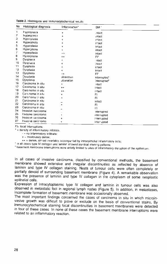
Table 2. Histological and immunocytochemical results
No Histological diagnosis Inflammation* BM*
1 Hyperplasia2 Hyperplasia3 Hyperplasia4 Hyperplasia5 Hyperplasia6 Hyperplasia7 Hyperplasia8 Hyperplasia9 Dysplasia10 Dysplasia11 Dysplasia12 Dysplasia13 Dysplasia14 Dysplasia15 Dysplasia16 Carcinoma in situ17 Carcinoma in situ18 Carcinoma in situ19 Carcinoma in situ20 Carcinoma in situ21 Carcinoma in situ22 Carcinoma in situ23 Carcinoma in situ24 Invasive carcinoma25 Invasive carcinoma26 Invasive carcinoma27 Invasive carcinoma
ulcerationulceration
intactintactintactintactintactintactintactFl"intactintactintactintactFl"interrupted*interrupted'intactintactintactFlFlintactFlFlinterruptedinterruptedinterruptedinterrupted
Fl= focal interruptions;* = density of inflammatory infiltrate:
- = no infammatory infiltrate;+ = moderately dense;++ = dense, almost invariably accompanied by intraepithelial inflammatory cells;
* in all cases type IV collagen and laminin showed identical staining patterns;" basement membrane interruptions were strictly limited to sites of inflammatory disruption of the epithelium.
In all cases of invasive carcinoma, classified by conventional methods, the basementmembrane showed extensive and irregular discontinuities as reflected by absence oflaminin and type IV collagen staining. Nests of tumour cells were often completely orpartially devoid of surrounding basement membrane (Figure 4). A remarkable observationwas the presence of laminin and type IV collagen in the cytoplasm of some neoplasticepithelial cells.Expression of intracytoplasmic type IV collagen and laminin in tumour cells was alsoobserved in metastatic foci in regional lymph nodes (Figure 5). In addition, in metastases,incomplete formation of basement membrane was occasionally observed.The most important findings concerned the cases of carcinoma in situ in which microin-vasive growth was difficult to prove or exclude on the basis of conventional stains. Byimmunocytochemical staining focal discontinuities in basement membranes were detectedin four of these cases. In none of these cases the basement membrane interruptions wererelated to an inflammatory reaction.
28
2.4 DISCUSSION
In all mucosae, basement membranes occur at the interface between epithelium andstroma and furthermore around capillaries, muscle fibres and glandular structures.Basement membranes are not found around lymphatic vessels (2).As shown by electron microscopy, basement membranes consist of an electron lucid layer(lamina rara), which is in close proximity to the basal cell surface, and an electron denselayer (lamina densa). Biochemically, basement membranes are composed of intrinsic andextrinsic components (1).Type IV collagen and laminin are intrinsic components, occurring only in basementmembranes and deposited by the adjacent cell. Substances such as fibronectin and type Vcollagen occur also in other structures, are not exclusively produced by the adjacent cells,and are therefore regarded as extrinsic components. In recent years many studies havedemonstrated that basement membrane interruptions are characteristic of infiltrative growthin carcinoma (7).The study of basement membranes in laryngeal neoplasia is not entirely new. Schenk (8)investigated the tumour/stroma relationship in invasive squamous cell carcinomas of thelarynx at the ultrastructural level. He found that basement membranes are produced bymalignant epithelial cells and concluded that penetration of basement membranes bytumour cells is not conclusive evidence of invasive carcinoma, but may be an indication ofincreased catabolic cell surface activity. Another interesting finding in this study was theinappropriate formation of basement membrane material in spaces created by invaginationof the plasma membrane into the cytoplasm. These observations largely agree with ourresults. In our material we noted intact membranes in normal laryngeal mucosa and incases of simple epithelial hyperplasia and dysplasia without severe inflammation. In thecases with severe inflammation there were several rather regular and small interruptions ofbasement membranes, related to the site of inflammation and the presence of intraepithelialinflammatory cells. This pattern of basement membrane disintegration was distinctly dif-ferent from the more irregular pattern of basement membrane interruptions in obviouslyinvasive carcinoma.In four cases of carcinoma in situ, interruptions of the basement membrane were found. Inthree of these cases moderately active inflammation was also observed but not in the areawhere basement membrane interruptions were detected. This finding can be explained byassuming that decreased assembly or increased breakdown of basement membranes, orboth, may be early signs of imminent infiltrative growth. Additional studies on larger seriesof borderline cases and including long term follow-up data will have to be performed toestablish the biological significance of basement membrane interruptions in morphologicallynon-invasive carcinoma in situ. Four cases of carcinoma with extensive invasive growthshowed advanced and irregular interruptions of the basement membrane in areas ofinvasion.Strikingly, also in foci of metastatic cancer in regional lymph nodes we found tumour cellswith inappropriate intracytoplasmic expression of laminin and type IV collagen.These results agree largely with those of Cam et al. (9), who found weak and irregularpatterns of basement membranes for laminin and type IV collagen immunofluorescence inundifferentiated carcinomas and in differentiated carcinomas. The immunofluorescencestaining was almost entirely absent at the periphery of the infiltrating tumour trabeculae. Onthe other hand, there was well defined linear staining of basement membranes aroundnests of well differentiated squamous cell carcinoma.The mechanisms underlying basement membrane penetration during neoplastic invasivegrowth are complex and not completely understood. Basement membrane penetration maybe due to deficient synthesis of basement membrane components or by the inability of theneoplastic cell to assemble the components into a structured membrane. This lattermechanism is supported by the intracytoplasmic appearance of laminin and type IVcollagen. An alternative possibility is proteolytic breakdown of basement membranes by the
29
invasive neoplastic cells. Evidence in support of this mechanism was provided by Liotta etal. (7) who demonstrated production of type IV collagen specific collagenases by neoplasticcells in several types of carcinoma.In conclusion, we have demonstrated that the epithelial basement membrane showscharacteristic interruptions in some cases of carcinoma in situ and in invasive carcinoma ofthe larynx. We therefore postulate that immunocytochemical detection of specific basementmembrane components, laminin and/or type IV collagen, may be a valuable tool for thedetermination of early invasive growth.Additional studies on larger series of laryngeal neoplasms, including long term follow up,are necessary to establish the practical value of this approach for the histopathologicalclassification of laryngeal neoplasia.
30

2.5 REFERENCES
1. Martinez-Hernandez A, Amenta P. The basement membrane in pathology. Lab Invest 1983; 48: 656-678.2. Barsky SH, Rao NC, Restrepo C, Liotta LA. Immunocytochemical enhancement of basement membrane
antigens by pepsin: applications in diagnostic pathology. Am J Clin Pathol 1984; 82(2): 191-194.3. Martinez-Hernandez A, Amenta PS. Laminin-secreting yolk sac carcinoma of the rat. Lab Invest 1982; 47:
247-257.4. Bosman FT. Some recent developments in immunocytochemistry. Am J Clin Path 1983; 15: 189-200.5. Sage H, Wordberg RG, Bomstein P. Structural studies on human type IV collagen. J Biol Chem 1979; 254-
9893-9900.6. Towbin H, Staehelin T, Gordin J. Electrophoretic transfer of proteins from polyacrylamide gels to
nitrocellulose sheets. Proc Natl Acad Sci USA 1979; 76: 4350-354.7. Liotta LA, Rao CN, Barsky SH. Tumor invasion and the extracellular matrix. Lab Invest 1983; 49: 636-650.8. Schenk P. Uitrastrukturelle Basallamina-alterationen an der Tumor-Stromagrenze von invasive Larynxkar-
zinomen. Laryngologie, Rhinologie, Otologie (Stuttgart) 1981; 60: 573-576.9. Cam Y, Caulet T, Bellon G, Poulin G, Legros M, Pytlinska M. Immunohistochemi cal localization of
macromolecules of the basement membrane and the peritum oral stroma in human laryngeal carcinomas. JPathol 1984; 144:35-44.
31
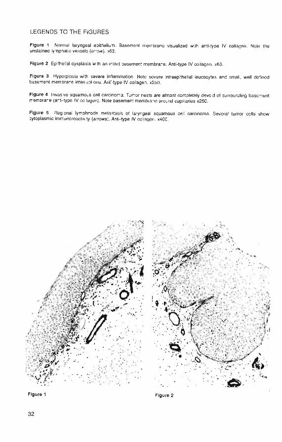
LEGENDS TO THE FIGURES
Figure 1 Normal laryngeal epithelium. Basement membrane visualized with anti-type IV collagen. Note theunstained lymphatic vessels (arrow). x63.
Figure 2 Epithelial dysplasia with an intact basement membrane. Anti-type IV collagen. x63.
Figure 3 Hyperplasia with severe inflammation. Note severe intraepithelial leucocytes and small, well definedbasement membrane interruptions. Anti-type IV collagen. x250.
Figure 4 Invasive squamous cell carcinoma. Tumor nests are almost completely devoid of surrounding basementmembrane (anti-type IV collagen). Note basement membrane around capillaries x250.
Figure 5 Regional lymphnode metastasis of laryngeal squamous cell carcinoma. Several tumor cells showcytoplasmic immunoreactivity (arrows). Anti-type IV collagen. x400.
Figure 1 Figure 2
32
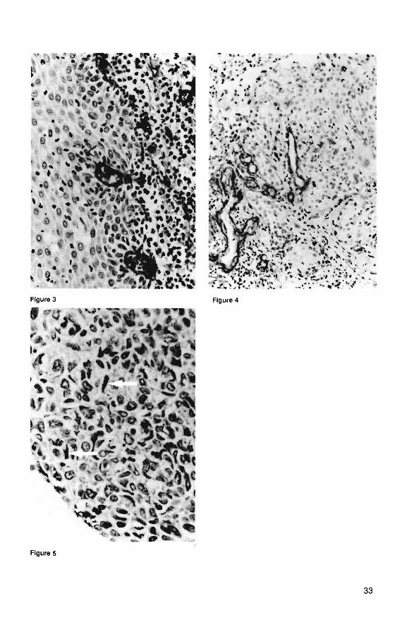
Figure 3
Figure S
33

CHAPTER 3
BASEMENT MEMBRANE PATTERNS IN BORDERLINETUMORS OF THE OVARY.
An immunohistochemicai study with antibodies toIaminin and type IV collagen.
R Visser, JW Arends, FT BosmanInt. J. Gyn. Cancer; accepted for publication
35
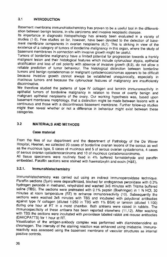
3.1 INTRODUCTION
Basement membrane immunohistochemistry has proven to be a useful tool in the differenti-ation between benign lesions, in-situ carcinoma and invasive neoplastic disease.Its importance in diagnostic histopathology has already been evaluated in a variety ofstudies (1-5). Few studies so far, however, have been devoted to the detection of base-ment membrane components in ovarian neoplasms (6,7). This is striking in view of theexistence of a category of tumors of borderline malignancy in this organ, where the study ofbasement membranes in connection with invasive growth might be useful.Tumors of borderline malignancy have a limited potential for progression towards a franklymalignant lesion and their histological features which include cytonuclear atypia, epithelialstratification and loss of cell polarity with absence of invasive growth (8,9), do not allow areliable prediction of tumor behaviour. The histological distinction between borderlinetumors and benign cystadenomas or malignant cystadenocarcinomas appears to be difficultbecause invasive growth cannot always be established unequivocally, especially inmucinous tumors and because the cytonuclear features of malignancy are insufficientlydiscriminative.We therefore studied the patterns of type IV collagen and laminin immunoreactivity inepithelial tumors of borderline malignancy in relation to those of overtly benign andmalignant epithelial neoplasms of the ovary. Our working hypothesis was, based uponbasement membrane morphology, that a distinction might be made between lesions with acontinuous and those with a discontinuous basement membrane. Further follow-up studiesmight then reveal whether or not a difference in behaviour might exist between thesecategories.
3.2 MATERIALS AND METHODS
Case material
From the files of our department and the department of Pathology of the De WeverHospital, Heerlen, we collected 20 cases of borderline ovarian lesions of the serous as wellas the mucinous type, 5 cases of mucinous and 5 of serous ovarian cystadenoma, 4 casesof serous ovarian cystadenocarcinoma and 10 of mucinous cystadenocarcinoma.All tissue specimens were routinely fixed in 4% buffered formaldehyde and paraffinembedded. Paraffin sections were stained with haematoxylin and eosin (H&E).
3.2.1. Immunohistochemistry
Immunohistochemistry was carried out using an indirect immunoperoxidase technique.Paraffin sections (3|am) were deparaffinized, blocked for endogenous peroxidase with 0.3%hydrogen peroxide in methanol, rehydrated and washed 3x5 minutes with Trizma bufferedsaline (TBS). The sections were pretreated with 0.1% pepsin (Boehringer) in 1 N HCI, 30minutes at room temperature (RT) to enhance immunoreactivity (10). Subsequently thesections were washed 3x5 minutes with TBS and incubated with polyclonal antibodiesagainst type IV collagen (diluted 1:250 in TBS with 1% BSA) or laminin (diluted 1:100)during one hour at RT in a moist chamber. Both antisera were raised in rabbits. Theimmunospecificity of these antisera has been reported elsewhere (11,12). After washingwith TBS the sections were incubated with peroxidase labeled rabbit anti-mouse antibodies(DAKOPATTS) for 1 hour at RT.Visualization of the antigen-antibody complex was performed with diaminobenzidine aschromogen. The intensity of the staining reaction was enhanced using imidazole. Immuno-reactivity was assessed using the basement membrane of vascular structures as internalpositive controls.
36
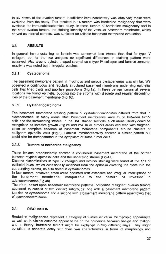
In six cases of the ovarian tumors insufficient immunoreactivity was obtained; these wereexcluded from the study. This resulted in 14 tumors with borderline malignancy that wereavailable for immunohistochemical study. In these tumors of borderline malignancy and inthe other ovarian tumors, the staining intensity of the vascular basement membrane, whichserved as internal controls, was sufficient for reliable basement membrane evaluation.
3.3 RESULTS
In general, immunostaining for laminin was somewhat less intense than that for type IVcollagen, but for the two antigens no significant differences in staining pattern wereobserved. Also around spindle shaped stromal cells type IV collagen and laminin immuno-reactivity was noted but in irregular patches.
3.3.1 Cystadenoma
The basement membrane pattern in mucinous and serous cystadenomas was similar. Weobserved a continuous and regularly structured basement membrane underlying epithelialcells that lined cysts and papillary projections (Fig. 1a). In these benign tumors at severallocations we found epithelial budding into the stroma with discrete and regular discontinu-ities of the basement membrane (Fig.1b).
3.3.2 Cystadenocarcinoma
The basement membrane staining pattern of cystadenocarcinomas differed from that incystadenomas. In many areas intact basement membranes were found between tumorcells and the surrounding stroma. In the H&E stained sections, such areas usually could berecognized as invasive growth (Fig.2a and 2b). In all tumors areas occurred with fragmen-tation or complete absence of basement membrane components around clusters ofmalignant epithelial cells (Fig.3). Laminin immunoreactivity showed a similar pattern butcould also be demonstrated in the cytoplasm of some tumor cells.
3.3.3. Tumors of borderline malignancy
These lesions predominantly showed a continuous basement membrane at the borderbetween atypical epithelial cells and the underlying stroma (Fig.4a).Discrete discontinuities in type IV collagen and laminin staining were found at the tips ofepithelial buds, which occasionally extended from the epithelia covering the cysts into thesurrounding stroma, as also noted in cystadenomas.In four tumors, however, small areas occurred with extensive and irregular interruptions ofthe basement membrane, comparable to the pattern of invasion inadenocarcinomas(Fig.4b).Therefore, based upon basement membrane patterns, borderline malignant ovarian tumorsappeared to consist of two distinct subgroups: one with a basement membrane patternidentical to cystadenoma and a second with a basement membrane pattern resembling thatof cystadenocarcinoma.
3.4. DISCUSSION
Borderline malignancies represent a category of tumors which in microscopic appearanceas well as in clinical outcome appear to be on the borderline between benign and malign-ant. In theory, borderline tumors might be explained in two different ways. They mightconstitute a separate entity with their own characteristics in terms of morphology and
37
clinical behaviour. Alternatively, this category may be regarded as to consist of lesions,which on the basis of histomorphology cannot be classified with regard to their behaviour.In the latter case, the borderline tumor group should consist of two groups, one with anessentially benign and an one with an essentially malignant character.Since one of the essential features of malignancy is invasive growth, we regarded itworthwhile to study the integrity of the basement membrane. We hypothesized that ifborderline tumors constitute a distinct and uniform entity a uniform basement membranepattern would be found. If, however, the borderline group contains essentially benign andessentially malignant tumors, this would be reflected by differences in basement membranepatterns.Although basement membranes in cancer have been studied extensively, relatively fewstudies have been devoted to basement membrane qualities in borderline tumors of theovary. Frappart et al (7) investigated only three cases of borderline tumors in a series ofepithelial ovarian tumors and found discontinuous immunofluorescence at the basementmembrane region using antibodies directed against laminin. They concluded that thesediscontinuities in the basement membrane indicated a beginning of infiltrative growth.Contrasting with the findings of Frappart et al, we observed regular and continuousbasement membranes in the majority of the borderline tumors in a pattern identical to thatin cystadenomas. Basement membrane interruptions were seen, but in a rather regular andpredictable way, surrounding epithelial buds or small epithelial islands in the stroma. Fox(8) previously described these invaginations in serous and mucinous tumors of borderlinemalignancy and emphasized the difficulties in differentiating these from stromal invasion.In 4 of our 14 tumors, originally diagnosed as of borderline malignancy, areas withextensive and irregular interruptions of the basement membrane were found. As thispattern was similar to that in invasive areas in cystadenocarcinoma, we are tempted to con-rlliHo that thoco four bordorlino himgro i/uiilaii 10J fuof uf Invasive yiUWlll add merefOre
should be regarded as adenocarcinoma.On the basis of these findings we propose that borderline ovarian tumors are not a distinctentity but consist of a group of intrinsically benign and a group of intrinsically malignanttumors.Further immunohistochemical studies on larger patient series and including follow up dataare necessary to assess the clinical validity of this assumption.
38

3.5 REFERENCES
1. D'Ardenne AJ. Use of basement membrane markers in tumour diagnosis. J Clin Pathol 1989 ;42: 429-57.2. Bosman FT, Havenith MG, Cleutjens JPM. Basement membrane in cancer, Ultrastruct Pathol 1985; 8: 291-
304.3. Martinez-Hernandez A, Amenta PS. The basement membrane in pathology. Lab Invest 1983; 6: 656-78.4. Storad CJ. Membrane changes in epithelial neoplasms. JAMA 1984; 252: 1665-1666.5. Bosman FT, Cleutjens JPM, Beek C, Havenith MG. Basement membrane heterogeneity. Histochem J 1989;
21: 629-33.6. Stenback F, Wasenius VM. Basement membrane structures in tumors of the ovary. Eur J Obstet Gynecol
Reprod Biol 1985; 20; 357-371.7. Frappart L, Agard C, Hu Chi Lin, Grimaud JA, Palayer C, Rochet Y, Bremont A. Les membranes basales
des tumeurs epitheliales de I'ovaire. J Gynecol Obstet Biol Reprod 1984; 13: 117-23.8. Fox H. The concept of borderline malignancy in ovarian tumours: a reappraisal. In: Current topics in
pathology Berlin: Springer, Vol 78: 1989: 111-34.9. Serov SF, Scully RE, Sobin LH. International classification of tumors. No 9. Histologic typing of ovarian
tumours. Geneva: World Health Organization, 1973.10. Barsky SH, Rao NC, Restrepo C, Liotta LA. Immunocytochemical enhancement of basement membrane
antigens by pepsin: applications in diagnostic pathology. Am J Clin Pathol 1984; 822: 191-194.11. Havenith MG, Cleutjens JPM, Beek C, v.d.Linden E, de Goeij AFPM, Bosman FT. Human specific anti type
IV collagen antibodies, characterization and immunohistochemical application. Histochemistry 1987; 87: 123-128.
12. Havenith MG, Dingemans KP, Cleutjens JPM, Wagenaar SjSc, Bosman FT. Basement membranes inbronchogenic squamous cell carcinoma: an immunohistochemical and ultrastructural study. Ultrastruct Pathol1990; 14(1): 51-63.
39
LEGENDS TO THE FIGURES
Figure 1a. Serous cystadenoma with intact basement membrane, immunoreactive for type IV collagen (125x).Figure 1b. Mutinous cystadenoma with focal absence of the basement membrane around epithelial budding(arrow). Type IV collagen, immunoperoxidase (250x).
Figure 2. Cystadenocarcinoma. Two consecutive sections, H&E (a) and immunostained for type IV collagen (b).The H&E stained section suggests tumor invasion, whereas the type IV collagen immunostaining (b) shows anintact basement membrane between tumor and stroma (63x).
Figure 3. Cystadenocarcinoma. Periphery of the tumor. Irregular, fragmented basement membrane material at theinterface between tumor and stroma. Type IV collagen immunoperoxidase (250x).
Figure 4. Borderline malignant ovarian tumors, a: Epithelium lined with a continuous basement membrane (typeIV collagen immunoperoxidase, 63x). b: Atypical epithelia only focally lined with a basement membrane (arrows;type IV collagen, 63x).
.£" i
Figure 1a
40
Figure 1b

Figure 2a Figure 2b
» V
^ • « ^s * - - - *̂f
> .»•
•*v>
V . . . . . ,
Figure 3
41

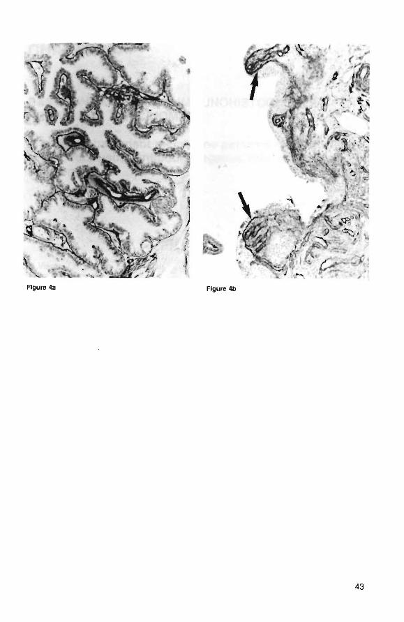
Figure 4a Figure 4b
43

CHAPTER 4
BASEMENT MEMBRANE IMMUNOHISTOCHEMISTRY INRENAL CELL CARCINOMA
A study of basement membrane patterns in small renalcell tumors using antibodies against type IV collagenand laminin.
R. Visser, J.W, Arends, FT. BosmanJ Urol Pathology 1993 (in press)
45
4.1 INTRODUCTION
Renal cell carcinomas are tumors of varying histological appearance and clinical behaviour.In many studies various morphologic parameters have been correlated with biologicalproperties of these tumors. Tumor stage appeared to be adversely related with prognosis(1,2) and with regard to nuclear grade it appeared that high grade tumors are generallyassociated with shorter disease-free interval (2,3,4). Furthermore the tumor type is relevant:papillary and spindle cell tumors show a less favourable prognosis than tumors with a solidpattern (2).Of small renal cortical adenomas the biological behaviour is still a matter of debate. Basedon the low metastatic propensity of tumors measuring less than 3 cm in diameter, Bell (5)classified these tumors as adenomas. Larger tumors, which more frequently developsubsequent metastasis, were classified as adenocarcinoma. It is generally recognized thathistological characteristics can not distinguish between adenoma and adenocarcinoma ofthe kidney.Because metastasis is preceded by invasive growth, it is not unlikely that the varyingmetastatic capacity of small renal cell tumors might be correlated with the presence orabsence of invasive growth. Immunohistochemical staining of basement membraneantigens has been advocated as a tool in the study of invasive growth.Although interruptions in the basement membrane are not always caused by invasivelygrowing neoplastic cells (6,7,8), and, on the other hand, invading cells may be surroundedby basement membranes (9), immunohistochemical visualization of the basement mem-brane might be indicative of invasive growth and thus might constitute a useful parameterto distinguish between non-invasive (with intact basement membranes) and invasive renalcortical tumors (with interrupted basement membranes). To investigate this possibility weexecuted an immunohistochemical study with antibodies directed against the basementmembrane proteins laminin and type IV collagen.The significance of basement membrane staining patterns in predicting the behaviour ofsmall renal cortical neoplasms can only be established in case material with follow-upinformation, which was not at our disposal, partly due to the fact that small renal corticalneoplasms are usually encountered in autopsy specimens and rarely in nephrectomyspecimens.Therefore we compared basement membrane expression patterns with tumor size and withnuclear grade, which both have been shown prognostically valuable.
4.2 MATERIALS & METHODS
4.2.1 Tissue
Renal cell tumors (n=41), diagnosed as renal cell carcinoma or renal cortical adenoma andwith a maximal diameter of 60 mm, were selected from our own files and from those of thedept of Pathology, Erasmus University, Rotterdam (courtesy of dr FWJ ten Kate) andclassified into 5 groups, according to the tumor diameter: < 10 mm (n=12); 11 - 20 mm(n=7), 21 - 30 mm (n=15), 31 - 40 mm (n=4), and > 40 mm (n=3).Thirty of these tumors were encountered in nephrectomy specimens and were classified asrenal cell carcinoma. Eleven tumors (measuring less than 10 mm in diameter), wereobtained at autopsy, which precluded clinico-pathological correlation in these cases.Tissue blocks were routinely fixed in 4% buffered formaldehyde and paraffin embedded.For orientation and for diagnostic purposes 3|i sections were stained with haematoxylin andeosin (H&E).Histological classification was performed according to the criteria of the World HealthOrganization (10) and nuclear grading according to the protocol of Fuhrman (3).
46

4.2.2. Immunohistochemistry
For immunohistochemical evaluation of every tumor one section from the tumor peripheryand one section from the centre of the tumor was selected. These were immunostained forlaminin and type IV collagen.Polyclonal antibodies against mouse type IV collagen and laminin were raised in rabbitsand affinity purified as described earlier (11,12). Paraffin sections (3u) of these tumors weredeparaffinized and blocked for endogenous peroxidase with 0,6% hydrogen peroxide inmethanol during 15 minutes and washed 3 x 5 minutes in demineralized water.Tissue sections were treated with 0.1% pepsin (Sigma) in 0.1 N HCI at room temperatureduring 30 minutes to unmask immunoreactivity for the two basement membrane compo-nents (13). After rinsing (3x 5 min) in demineralized water the sections were preincubatedwith normal swine serum and subsequently exposed to polyclonai anti-type IV collagen(1:250 diluted in Trizma buffered saline (TBS) with 0.1% BSA) or anti-laminin antibodies(1:100 diluted in TBS with 0.1% BSA). After washing and incubation with a horseradishperoxidase conjugated swine anti-rabbit antibody (DAKO PATTS) (45 minutes, room temp)the slides were washed again in TBS before a diaminobenzidine reaction was executed.Imidazole was added to the substrate solution in order to enhance the staining intensity.Finally, tissue sections were counterstained with Mayer's haematoxylin for 1 minute.Basement membrane patterns were scored in the centre of the tumor and at the peripheryusing a semiquantitative scoring scale. When basement membrane interruptions were notobserved this was scored as "intact" (+) when interruptions were seen or the basementmembrane appeared to be absent, this was classified as "not intact" (-).
4.2.3 Statistics
To compare the observed frequencies with the frequencies expected in case of a homo-geneous distribution, the Chi-squared test (two-sided), corrected according Yates was used(14).
4.3 RESULTS
4.3.1 Histological findings
According to the criteria of the WHO, 29 of the renal tumors were predominantly of theclear cell type, while 11 tumors appeared to be composed of a combination of clear cellsand granular cells. One tumor was composed of oncocytic cells. Spindle cell tumors werenot present in our series. The tumors showed a variety of glandular patterns with solid,tubular, acinar, papillary and cystic formations, or combinations of these patterns. Thetumors were graded according to the criteria of Fuhrman (3). Each tumor was graded bythe highest nuclear grade, even when the higher grade occurred only focally. In our series9 tumors had grade 1,15 tumors grade 2 and 17 tumors grade 3. When the tumor gradewas compared with tumor size, grade 1 tumors occurred significantly more often in thegroup of tumors with a diameter of less than 10 mm than in the larger tumors. However,grades 2 and 3 were found in all tumor sizes (Table 1).
4.3.2 Immunohistochemical patterns in renal tumors
For assessment of basement membrane immunoreactivity in the tumors, neighbouringtubules and vascular structures served as a positive control. In general, staining intensityfor laminin and type IV collagen was sufficient for scoring purposes, although a fewspecimens displayed weak laminin immunoreactivity; these were not included in this series.Post mortem specimens showed excellent immunoreactivity.
47
Immunoreactivity of basement membrane components at the periphery of the tumor couldbe determined in 38 cases and in the central parts of the tumor in 36 cases. Tumorsmeasuring less than 10 mm in diameter significantly more often showed intact central andperipheral basement membranes than larger tumors (Tables 2 and 3, fig. 1 and 2). Twotumors showed focal basement membrane absence in the periphery and 1 showed thisphenomenon in the centre of the tumor. A more heterogeneous pattern was found intumors with a diameter of more than 10 mm. The majority of these tumors displayed limitedbasement membrane expression in the tumor centre as well as in the periphery (fig. 3 and4). Only 3 tumors showed intact basement membranes in the periphery (fig. 5) and only 5tumors showed intact basement membranes in the tumor centre (fig. 6). One tumor did notshow any peripheral basement membrane immunoreactivity. All but one of the tumors witha diameter exceeding 30 mm showed focal absences of the basement membrane both inthe tumor centre and periphery and were classified as "not intact". Only one of these largertumors contained intact basement membranes in the centre.When the basement membrane patterns of the tumors were compared with the nucleargrade according to the criteria of Fuhrman, a significant correlation was found betweennuclear grade and basement membrane patterns in the centre and at the periphery of thetumor (Tables 4 and 5).
4.4 DISCUSSION
The most frequent neoplasm of the kidney in adults is renal cell adenocarcinoma. Thesetumors, which supposedly originate from tubular epithelium (15,16) have attracted attentionbecause of their variable biological behaviour. Tumor behaviour can, to a certain extent, bepredicted by the stage of the tumor, the tumor grade and by the histological subtype. Forexample, the sarcomatoid variant carries a poor prognosis (17).Bell (5) introduced a classification based on the size of the tumor. Tumors less than 3centimeters in diameter appeared to metastasize rarely and most, but not all, of the largertumors subsequently developed metastases. Therefore, it has become customary toclassify tumors less than 3 cm as adenoma and those larger than 3 cm as carcinoma. Thisclassification has been used widely since then, but has also been criticized. Severalauthors have discarded this classification and consider all tumors as renal cell carcinoma,but small tumors as "renal adenocarcinoma of low metastatic potential" (17, 18). However!other authors have held the view that renal adenomas do exist and can even be distin-guished by histological criteria (19, 20). This view is supported by Dal Cin and coworkers(21), who found chromosomal aberrations in tumors that were histopathologically diagnosedas cortical adenomas. These aberrations were different from those commonly found inrenal cell carcinomas. Small renal cortical neoplasms might therefore consist of two sub-groups, adenomas and small adenocarcinomas, that cannot be reliably distinguished on thebasis of histological characteristics, immunohistochemistry or ultrastructure (17,22), but witha different biological potential in terms of invasive growth and subsequent metastasis.The basement membrane plays a crucial role in malignant neoplasia, in particular as abarrier to be taken by invasively growing tumor cells. Therefore, basement membraneimmunohistochemistry might help to discriminate between (non invasive) adenoma and(invasive) adenocarcinoma.
Our results on renal cortical neoplasms indicate that tumor size, tumor grade and basementmembrane patterns are closely related. Tumors larger than 10 mm were almost always ofhigher nuclear grade, whereas smaller tumors were heterogeneous with regard to nucleargrade. This subdivision is supported by the basement membrane immunohistochemistryresults. Tumors smaller than 10 mm mostly showed intact basement membranes, in thetumor centre as well as in the periphery. Larger tumors mostly showed a heterogeneouspattern with defects in the basement membranes of most of the tumors. These dataindicate, in agreement with Bell (5), that tumor size may be an important parameter of
48
malignant potential, but with a cut-of point of 1 cm rather than 3 cm. Taking into consider-ation the data provided by Fuhrman et al (3), who found in stage I tumors a significantdifference in metastatic rate between grade 1 tumors and the tumors of grades 2-4, andour finding, that grade I tumors contained intact basement membranes in the centre andthe periphery, we propose that all tumors larger than 10 mm should be consideredpotentially malignant. It would be preferable to ascertain this contention in additionalstudies, including follow-up data, but the rarity of tumors smaller than 10 mm did not allowus to perform such an analysis on our material.In conclusion, renal cortical tumors smaller than 10 mm have a somewhat variable nucleargrade, but mostly intact basement membranes. This finding supports the benign nature ofthese tumors.
49
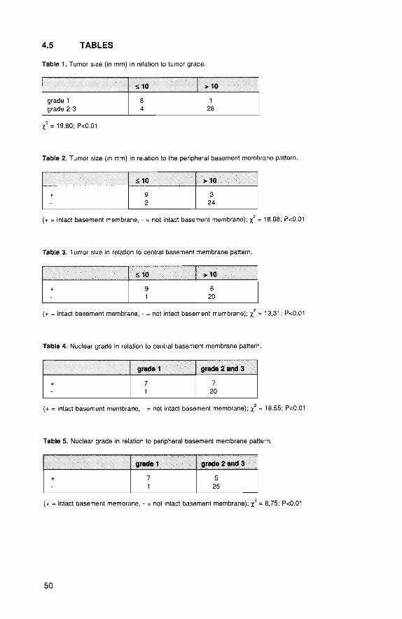
4.5 TABLES
Table 1. Tumor size (in mm) in relation to tumor grade.
grade 1grade 2-3
510
84
>10
128
X = 19.80; P<0.01
Table 2. Tumor size (in mm) in relation to the peripheral basement membrane pattern.
+
£ 1 0
92
>10
324
(+ = intact basement membrane, - = not intact basement membrane); x* = 18.08; P<0.01
Table 3. Tumor size in relation to central basement membrane pattern.
+
sto
91
>10
620
(+ = intact basement membrane, - = not intact basement membrane); x = 13,31; P<0.01
Table 4. Nuclear grade in relation to central basement membrane pattern.
+
grade 1
71
grads 2 and 3
720
(+ = intact basement membrane, - = not intact basement membrane); x = 18.55; P<0.01
Table 5. Nuclear grade in relation to peripheral basement membrane pattern.
+ 71
grade 2 and 3 '
525
(+ = intact basement membrane, • = not intact basement membrane); x = 8,75; P<0.01
50
4.5 REFERENCES
1. Robson CJ, Churchill BM, Anderson W. The results ot radical nephrectomy for renal cell carcinoma. J Urol1969; 101:297-301,
2. Medeiros LJ, Gelb AB, Weiss LM. Renal cell carcinoma. Prognostic significance of morphologic parametersin 121 cases. Cancer 1988; 61: 1639-1651.
3. Fuhrman SA, Lasky LC, Limas C. Prognostic significance of morphologic parameters in renal cell carcinoma.Am J Surg Pathol 1982; 6: 655-663.
4. Green LK, Ayala AG, Ro JY, Swanson DA, Grignon DJ, Giacco GG, Guinee VF. Role of nuclear grading instage I renal cell carcinoma. Urology 1989; 34: 310-315.
5. Bell ET. Tumors of the kidney. In Bell ET (ed): Renal diseases (ed 2), Philadelphia, Lea & Febiger, 1950.6. Martinez-Hernandez A, Amenta PS. The basement membrane in pathology. Lab Invest 1983; 48: 656-678.7. Timpl R. Structure and biological activity of basement membrane proteins. Eur J Biochem 1989; 180: 487-
502.8. Visser R, van der Beek JM, Havenith MG, Cleutjens JPM, Bosman FT. Immunocytochemical detection of
basement membrane antigens in the histopathological evaluation of laryngeal dysplasia and neoplasia.Histopathology 1986; 10: 171-180.
9. Bosman FT, Havenith MG, Cleutjens JPM. Basement membrane in cancer. Ultrastruct Pathol 1985; 8: 291-304.
10. Mostofi FK, Sesterhenn IA, Sobin LH. Histological typing of kidney tumours. In: International histologicalclassification of tumours. No 25 World Health Organization 1981; 17-18.
11. Havenith MG, Cleutjens JPM, Beek C, v.d.Linden E, de Goeij AFPM, Bosman FT. Human specific anti-typeIV collagen monoclonal antibodies, characterization and immunohistochemical application. Histochemistry1987; 87: 123-128.
12. Havenith MG, Dingemans KP, Cleutjens JPM, Wagenaar Sj Sc, Bosman FT. Basement membranes inbronchogenic squamous cell carcinoma: an immunohistochemical and ultrastructural study. Ultrastruct Pathol1990; 14(1): 51-63.
13. Barsky SH, Rao NC, Restrepo C, Liotta LA. Immunocytochemical enhancement of basement membraneantigens by pepsin: applications in diagnostic pathology. Am J Clin Pathol 1984; 82(2): 191-94.
14. Kirkwood BR. Essentials of medical statistics. Oxford, Blackwell Scientific Publications. 1988.15. Storkel S, Jacobi GH. Systematik, Histogenese und Prognose der Nierenzellkarzinome und des renalen
Onkozytoms. Verh Dtsch Ges Pathol 1989; 73: 321-338.16. Cohen C, McCue PA, Derose PB. Histogenesis of renal cell carcinoma and renal oncocytoma. An
immunohistochemical study. Cancer 1988; 62: 1946-1951.17. Petersen RO. Urologic Pathology. Philadelphia, JB Lippincott, 1986.18. Bennington JL, Beckwith JB. Tumors of the kidney, renal pelvis and ureter (fascicle 12). In: Atlas ot tumor
pathology, 2nd series, Washington DC, Armed Forces Institute of Pathology, 1975.19. Mostofi FK, Sesterhenn IA, Davis Jr CJ. Benign tumors of the kidney. EORTC Genitourinary Group
Monograph 5: Progress and controversies in oncological urology II. 1988; 329-344: Alan R. Liss, Inc.20. Davis Jr CJ. Pathology of renal neoplasms. Seminars in Roentgenology 1987; 22:233-240.21. Dal Cin P, Gaeta J, Huben R, Li FP, Prout GR, Sandberg AA. Renal cortical tumors. Cytogenetic
characterization. Am J Clin Pathol 1989; 92: 408-414.22. Fisher ER, Morvat B. Comparative ultrastructural study of so called renal adenoma and carcinoma. J. Urol
1972; 108: 382-386.
51
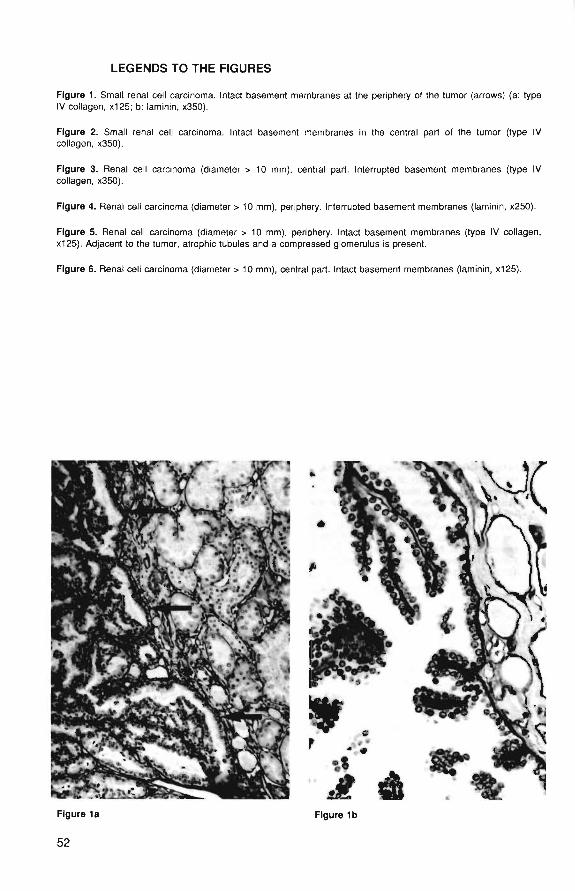
LEGENDS TO THE FIGURES
Figure 1. Small renal cell carcinoma. Intact basement membranes at the periphery of the tumor (arrows) (a: typeIV collagen, x125; b: laminin, x350).
Figure 2. Small renal cell carcinoma. Intact basement membranes in the central part of the tumor (type IVcollagen, x350).
Figure 3. Renal cell carcinoma (diameter > 10 mm), central part. Interrupted basement membranes (type IVcollagen, x350).
Figure 4. Renal cell carcinoma (diameter > 10 mm), periphery. Interrupted basement membranes (laminin, x250).
Figure 5. Renal cell carcinoma (diameter > 10 mm), periphery. Intact basement membranes (type IV collagen,x125). Adjacent to the tumor, atrophic tubules and a compressed glomerulus is present.
Figure 6. Renal cell carcinoma (diameter > 10 mm), central part. Intact basement membranes (laminin, x125).
1
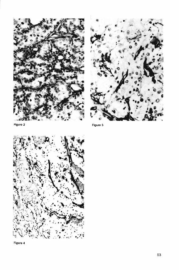
Figure 3
Figure 4
53


Figure 5 Figure 6
55

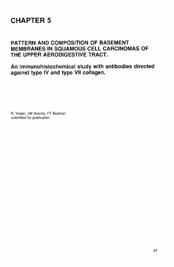
CHAPTER 5
PATTERN AND COMPOSITION OF BASEMENTMEMBRANES IN SQUAMOUS CELL CARCINOMAS OFTHE UPPER AERODIGESTIVE TRACT.
An immunohistochemical study with antibodies directedagainst type IV and type VII collagen.
R. Visser, JW Arends, FT Bosmansubmitted for publication
57
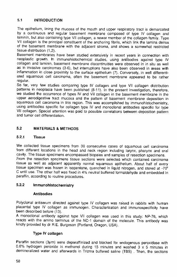
5.1 INTRODUCTION
The epithelium, lining the mucosa of the mouth and upper respiratory tract is demarcatedby a continuous and regular basement membrane composed of type IV collagen andlaminin, but also containing type VII collagen, a newer member of the collagen family. TypeVII collagen is the principal constituent of the anchoring fibrils, which link the lamina densaof the basement membrane with the adjacent stroma, and shows a somewhat restrictedtissue distribution (1,2).Basement membranes have been studied extensively in recent years in connection withneoplastic growth. In immunohistochemical studies, using antibodies against type IVcollagen and laminin, basement membrane discontinuities were observed in in situ as wellas in invasive carcinomas (3-6), but interruptions have also been observed in areas withinflammation in close proximity to the surface epithelium (7). Conversely, in well differenti-ated squamous cell carcinoma, often the basement membrane appeared to be ratherregular.So far, very few studies comparing type IV collagen and type VII collagen distributionpatterns in neoplasia have been published (8-11). In the present investigation, therefore,we studied the occurrence of types IV and VII collagen in the basement membrane in theupper aerodigestive tract mucosa and the pattern of basement membrane deposition insquamous cell carcinoma in this region. This was accomplished by immunohistochemistry,using antibodies specific for collagen type IV and monoclonal antibodies specific for typeVII collagen. Special attention was paid to possible correlations between deposition patternand tumor cell differentiation.
5.2 MATERIALS & METHODS
5.2.1 Tissue
We collected tissue specimens from 30 consecutive cases of squamous cell carcinomafrom different locations in the head and neck region including larynx, pharynx and oralcavity. The tissue specimens encompassed biopsies and samples of resection specimens.From the resection specimens tissue sections were selected which contained carcinomatissue as well as adjacent apparently normal squamous epithelium. About half of everytissue specimen was frozen in isopentane, quenched in liquid nitrogen, and stored at -70°C until use. The other half was fixed in 4% neutral buffered formaldehyde and embedded inparaffin, according to routine procedures.
5.2.2 Immunohistochemistry
Antibodies
Polyclonal antiserum directed against type IV collagen was raised in rabbits with humanplacental type IV collagen as immunogen. Characterization and immunospecificity havebeen described before (13).A monoclonal antibody against type VII collagen was used in this study: NP-76, whichreacts with the amino terminus of the NC-1 domain of the molecule. This antibody waskindly provided by dr R.E. Burgeson (Portland, Oregon, USA).
Type IV collagen
Paraffin sections (3nm) were deparaffinized and blocked for endogenous peroxidase with0.6% hydrogen peroxide in methanol during 15 minutes and washed 3 x 5 minutes indemineralized water and afterwards in Trizma buffered saline (TBS) . Then, the sections
58

were treated with 0.1% pepsin (Sigma) in 0.1 N HCI at room temperature (RT) during 30minutes to unmask immunoreactivity for type IV collagen (12).After rinsing ( 3 x 5 min) in demineralized water the sections were exposed to a rabbit anti-type IV collagen antiserum (diluted 1:250 in TBS with 1% BSA and 0.1% Tween). Afterwashing, the sections were incubated with a horseradish peroxidase conjugated swine anti-rabbit antibody (DAKO PATTS) (45 min) at RT, washed again in TBS, and a diaminobenzi-dine/HjOj reaction was executed. Imidazole (0.1 M) was added to the substrate solution inorder to enhance the staining intensity. The slides were counterstained with Mayer'shaematoxylin during 1 minute.
Type VII collagen
Frozen sections (4 (i) were cut, airdried and fixed subsequently in 4% buffered formalinduring 10 minutes. Then the sections were washed with demineralized water and dehy-drated in graded alcohols. After blocking endogenous peroxidase with 0.3% HjO^ (15 min),sections were again rinsed in demineralized water (3x 5 min) and briefly in TBS. Then thesections were incubated with the antibody against type VII collagen (diluted 1:500 in TBSwith 1% BSA and 0.1% TWEEN), 45 min at RT. Afterwards the sections were washed (3x5min) and incubated (45 min, RT) with horseradish peroxidase conjugated rabbit anti-mouseantibody.Subsequently the sections were washed again in TBS and treated with diaminoben-zidin/HjOz. Imidazole was added as described above. The amount of immunoreactivity atthe tumor cell-stroma interface was scored semi-quantitatively in terms of presence ofimmunoreactivity in or around the cell or clusters of cells and in terms of co-expression ofthe two basement membrane components.
Indirect immunofluorescence doublestaining
For type IV collagen and type VII collagen doublestaining the sections were first incubatedwith a mixture of anti-type IV collagen (1:250 diluted) and anti-type VII collagen (1:500 dilu-ted), in TBS/1% BSA/0,1% Tween.As second layer a mixture was used of FITC labeled goat anti- rabbit IgG (Nordic, 1:300diluted) and rhodamine labeled goat anti- mouse IgG (TAGO Inc, Burlingame, Cal., USA;diluted 1:300).Specificity of the obtained immune reaction was controlled by identical incubation of parallelsections, but with an irrelevant primary antibody or with TBS instead of the primaryantibody.The fluorescence patterns were photographed with a Leitz dialux fluorescence microscopeusing alternately FITC and rhodamin filter combinations.
5.3 RESULTS
The histologically normal epithelium showed intact basement membranes, with a linear typeIV collagen immunoreactivity pattern and a similar but somewhat granular pattern of typeVII collagen (Fig. 1). A similar pattern was also observed at the interface between theepithelium of the salivary gland ductules and the adjacent stroma. However, type VIIcollagen was lacking in the basement membrane lining the epithelium of the glandularacini. Unlike type IV collagen, type VII collagen immunoreactivity was not found in thebasement membrane around vascular structures, muscle fibres, nerve bundles andadipocytes.The majority of the squamous cell carcinomas showed moderate differentiation (Table I). Inparticular the available pharyngeal carcinomas showed a homogeneous, moderatedifferentiation, while our laryngeal tumors contained areas with poorly and moderately
59

differentiated carcinoma. Most oral cavity carcinomas were moderately differentiated butcontained focal areas with either poor or high differentiation.When we compared tumor differentiation with expression of the two basement membraneantigens it appeared that in only one of the five tumors with poor differentiation co-expression of both components occurred. Of tumors with moderate differentiation howevera majority (16 of 21 tumors) showed immunoreactivity for the two basement membranecomponents in an identical pattern (fig 2). In well differentiated carcinomas both compo-nents showed identical expression patterns.As is shown in Table II we found two patterns of immunoreactivity. All of the poorlydifferentiated tumors showed type VII collagen staining in the cytoplasm (fig 3); one ofthese five tumors in addition showed areas with extracellular deposition of type VII collagen(fig 4). This type of staining pattern, both intra- and extracellular, was observed in 9 of the21 moderately differentiated carcinomas. The other tumors contained either intracellular orextracellular deposition. In the group of well differentiated carcinomas all but one showedonly extracellular deposition of type VII collagen.
5.4 DISCUSSION
The mucosa of the upper aerodigestive tract is lined partly with ciliated columnar, andpartly with stratified squamous epithelium. These cells all rest on a basement membrane,which is in direct continuity with the basement membrane of the epithelia of the mucosalglands and their ducts. One of the major constituents of the basement membrane is type IVcollagen, which forms, together with laminin, the basic structure of the basement mem-brane (14-16). A newer member of the collagen family, tvoe VII collaaen is a maior^uMotiiucnc ui ine aiicnunng iiDrns (,i,ifc>,l/). Inese structures, which occur in the parsreticularis of the basement membrane, form a network together with anchoring plaques,which entraps large stromal collagen fibres. As a result the basement membrane iseffectively anchored to the underlying stroma (18). The distribution of type VII collagencontaining anchoring fibrils is somewhat tissue restricted. Type VII collagen has been foundin the skin, chorioamnion, cornea (Bowman's membrane), ectocervix, mucosa of the upperaerodigestive tract and around ducts and lobules of the breast (1,2,8). During theembryonal development of the skin, the occurrence of anchoring fibrils is preceded by thedeposition of a basement membrane (19,20).In invasive carcinoma of the upper aerodigestive tract, but also in some in situ carcinomas,disruption of the basement membrane has been described (4,5,6). In these studiesimmunohistochemical methods were employed, using antibodies directed against lamininor/and type IV collagen. Basement membrane disruption is not specific for tumor invasion:it occurs also when epithelia are invaded by inflammatory cells (7). Deposition of basementmembranes around nests of tumor cells, in co-operation with stromal myofibroblasts hasalso been reported (21-24).Studies of type VII collagen deposition in neoplasia as yet are limited to carcinoma of thebreast (8), melanoma (9,10) and basal cell carcinoma (11). The distribution patterns of typeVII collagen in a variety of malignant tumors were reported by Wetzels et al (2). In thebreast it was found that the majority of carcinomas does not contain a basement memb-rane. However, a minority showed basement membrane staining with a different pattern fortype VII collagen, which differed from that of laminin and type IV collagen.Kirkham et al (10) reported in a study of nevomelanocytic lesions that type IV collagen andtype VII collagen line the epidermal basement membrane and this staining pattern iscontinued around dermal nevus cells. In invasive melanoma gradually gaps were found tooccur in the epidermal basement membrane in the invasive front when stained withantibodies against type VII collagen. Type IV collagen often remained detectable in theform of a continuous basement membrane surrounding invading melanoma cells.
60
In basal cell carcinomas type VII collagen is deposited around tumor cell nests. Thispattern corresponds with that of type IV collagen and laminin (3), although in the desmo-plastic or sclerosing type extensive irregularities to sometimes absences of type IV collagendeposition were described (25).Our findings indicate that in highly differentiated squamous cell carcinoma almost continu-ous basement membranes occur which contain type IV collagen and type VII collagen.These results are in line with the findings of Wetzels et al (2). With decreasing level ofdifferentiation the tendency of cancer cells to deposit a basement membrane decreases. Inthis process, loss of deposition of type VII collagen occurs first and is followed by loss oftype IV collagen deposition. Strikingly, some poorly differentiated tumor cells retain thepossibility to synthesize type VII collagen, but fail to deposit it extracellularly, presumably inrelation to the absence of a basal lamina.These findings suggest, in line with the findings reported by Kirkham et al (10) in melanom-a, that type VII collagen might be a more sensitive indicator for invasive growth than typeIV collagen because it disappears earlier. Whether or not this might be of diagnostic usewill have to be established in clinicopathological studies of early neoplastic lesions. Thefindings furthermore indicate that type VII collagen synthesis is regulated independent fromtype IV collagen synthesis, but its extracellular deposition appears to depend on theavailability of a type IV collagen matrix. Intracytoplasmic type VII collagen expression mightbe used as a progression marker for squamous cell carcinoma. The clinical use of thisparameter will have to be established in clinicopathological follow-up studies.
61
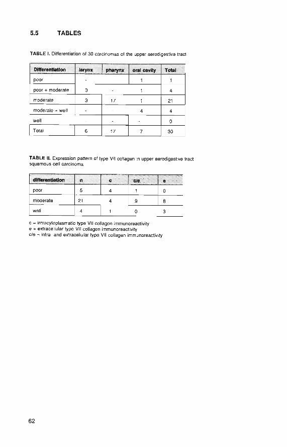
5.5 TABLES
TABLE I. Differentiation of 30 carcinomas of the upper aerodigestive tract
Differentiation
poor
poor + moderate
moderate
moderate + well
well
Total
larynx
-
3
3
-
6
pharynx
-
17
-
17
oral cavity
1
1
1
4
7
Total *
1
4
21
4
0
30
TABLE II. Expression pattern of type VII collagen in upper aerodigestive tractsquamous cell carcinoma
differentiation
poor
moderate
well
n
5
21
4
c
4
4
1
c / e ; : -'
1
g
0
e >'•
0
8
3
c = intracytoplasmatic type VII collagen immunoreactivitye = extracellular type VII collagen immunoreactivityc/e = intra- and extracellular type VII collagen immunoreactivity
62

5.6 REFERENCES
1. Sakai LY, Keene DR, Morris NP, Burgsson RE. Type VII collagen is a major structural component ofanchoring fibrils. J Cell Biol 1986; 103: 1577-1586.
2. Wetzels RHW, Robben HCW, Leigh IM, Schaafsma E, Vooys GP, Ramaekers FCS. Distribution patterns oftype VII collagen in normal and malignant human tissues. Am J Pathol 1991; 139: 451-459.
3. Bosman FT, Havenith MG, Visser R, Cleutjens JPM. Basement membranes in neoplasia. Progr HistochemCytochem 1992; 24 (4): 1-92.
4. Cam Y, Caulet T, Bellon G, Poulin G, Legros M, Pytlinska M. Immunohistochemical localization ofmacromolecules of the basement membrane and the peritumoral stroma in human laryngeal carcinomas. JPathol 1984; 144: 35-44.
5. Gusterson BA, Warburton MJ, Mitchell D, Kraft N, Hancock WW. Invading squamous cell carcinoma canretain a basal lamina. An immunohistochemical study using a monoclonal antibody to type IV collagen. LabInvest 1984; 51:82-88.
6. Sakr WA, Zarbo RJ, Jacobs JR, Crissman JD. Distribution of basement membrane in squamous cellcarcinoma of the head and neck. Hum Pathol 1987; 18: 1043- 1050.
7. Visser R, van der Beek JMH, Havenith MG, Cleutjens JPM, Bosman FT. Im munocytochemical detection ofbasement membrane antigens in the histopatho logical evaluation of laryngeal dysplasia and neoplasia.Histopathology 1986; 10: 171-180.
8. Wetzels RHW, Holland R, van Haelst UJGM, Lane EB, Leigh IM, Ramaekers FCS. Detection of basementmembrane components and basalcell keratin 14 in non- invasive and invasive carcinoma of the breast. Am JPathol 1989; 134:571-579.
9. Schmoeckel C, Stolz W, Sakai LY, Burgeson RE, Timpl R, Krieg T. Structure of basement membranes inmalignant melanomas and nevocytic nevi. J Invest Dermatol 1989; 92: 663-668.
10. Kirkham N, Price ML, Gibson B, Leigh IM, Coburn P, Darley CR. Type VII collagen antibody LH 7.2identifies basement membrane characteristics of thin malignant melanomas. J Pathol 1989; 157: 243-247.
11. Jones JCR, Steinman HK, Goldsmith BA. Hemidesmosomes, collagen VII and intermediate filaments inbasal cell carcinoma. J Invest Dermatol 1989; 93: 662-671.
12. Barsky SH, Rao NC, Restrepo C, Liotta LA. Immunocytochemical enhancement of basement membraneantigens by pepsin: applications in diagnostic pathology. Am J Clin Pathol 1984; 82(2); 191-94.
13. Havenith MG, Cleutjens JPM, Beek C, v.d.Linden E, de Goeij AFPM, Bosman FT. Human specific anti-typeIV collagen monoclonal antibodies, characterization and immunohistochemical application. Histochemistry1987; 87: 123-28.
14. Timpl R. Structure and biological activity of basement membrane proteins. Eur J Biochem 1989; 180: 487-502.
15. Martinez-Hernandez A, Amenta PS. The basement membrane in pathology. Lab Invest 1983; 48: 656-678.16. Leblond CP, Inoue S. Structure, composition and assembly of basement membrane. Am J Anat 1989; 185:
367-390.17. Burgeson RE. The junction zone and the dermis. The collagens of skin. Curr Probl Derm 1987; 17: 61.18. Keene DR, Sakai LY, Lunstrum GP, Morris NP, Burgeson RE. Type VII collagen forms an extended network
of anchoring fibrils. J Cell Biol 1987; 104: 611-622.19. Tisdale AS, Spurr-Michaud SJ, Rodriguez M, Hacked J, Ktachmer J, Gipson IK. Development of the
anchoring structures in the epithelium in rabbit and human fetal corneas. Invest Ophthalmol Vis Sci 1988;29: 727-736.
20. Smith LT, Sakai LY, Burgeson RE, Holbrook KA. Ontogeny of structural components at the dermal-epidermal junction in human embryonic and fetal skin: the appearance of anchoring fibrils and type VIIcollagen. J Invest Dermatol 1988; 90: 480-485.
21. Gould VE, Battifiora H. Origin and significance of the basal lamina and some interstitial tibrillar componentsin epithelial neoplasms. Pathol Annual 1976; 11: 353-386.
22. Bosman FT, Havenith MG, Cleutjens JPM. Basement membranes in cancer. Ultrastruct Pathol 1985; 8: 291-304.
23. Damjanov I, Damjanov N, Knowless BB, Engvall D. Origin of laminin in the extracellular matrix of humantumor xenografts in nude mice. Virchows Arch (B) 1985; 49: 45-52.
24. Cleutjens JPM, Havenith MG, Beek C, Vallinga M, ten Kate J, Bosman FT. Origin of basement membranetype IV collagen in xenogratled human epithelial tumor cell lines. Am J Path 1990; 136: 1165-1173.
25. Kallioinen M, Autio Harmainen H, Dammert K, Risteli J, Risteli L. Discontinuity of the basement membranein fibrosing basocellular carcinomas and basosquamous carcinomas of the skin, an immunohistochemicalstudy. J Invest Dermatol 1984; 82: 248-251.
63

LEGENDS TO THE FIGURES
Figure 1. Regular basement membranes lining the surface epithelium. Type IV collagen (a), type VII collagen (b);no immunoreactivity between antibodies to type VII collagen and capillary basement membranes (b). 250x,immunofluorescence (doublestaining)
Figure 2. Squamous cell carcinoma, moderately differentiated. Consecutive sections. Islands of tumor cellssurrounded by focally interrupted basement membranes (arrow), with identical patterns of type IV collagen (a) andtype VII collagen (b). Antobodies to type VII collagen do not react with vascular basement membranes. 250x,immunoperoxidase.
Figure 3. Squamous cell carcinoma, poorly differentiated. Consecutive sections. Only capillary basementmembranes in type IV collagen staining (a). Only expression of type VII collagen in the cytoplasm of tumor cells(b). 250x (inset, 400x), immunoperoxidase.
Figure 4. Squamous cell carcinoma, poorly differentiated. Intracytoplasmic and extracellular deposition of type VIIcollagen. 300x, immunoperoxidase.
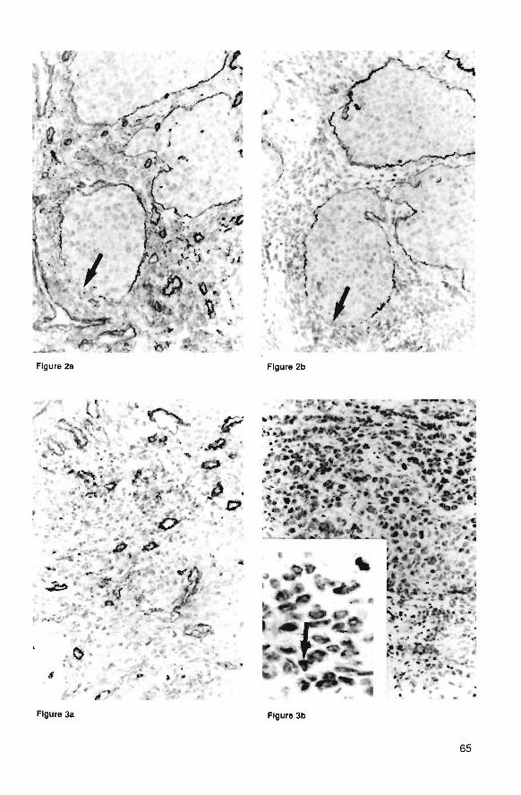
V *» .J*
, w * < # • * • . • . ; . .
Figure 2a Figure 2b
Figure 3b
65

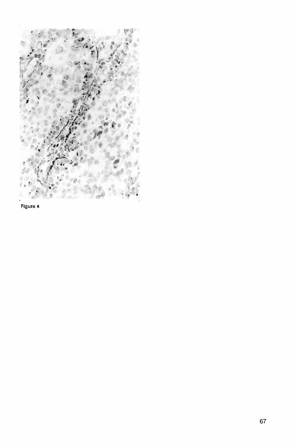
Ml.^ . v'*{ #.;
/>..
t.
*
Figure 4
67

CHAPTER 6
PATTERNS AND COMPOSITION OF BASEMENTMEMBRANES IN COLON ADENOMAS ANDADENOCARCINOMAS.
R Visser, JW Arends, IM Leigh, FT BosmanJ Pathol 1993 (in press)
69
6.1 INTRODUCTION
Invasive growth is a decisive hallmark of malignancy in epithelial cancers. It is a complexmultistep process with as initial event the dissolution of the extracellular matrix, includingthe basement membrane.In developing neoplastic epithelial lesions progressing towards invasion, a complex anddynamic interaction occurs between epithelial tumor cells and extracellular matrix prior tobreakdown of the basement membrane, as seen by morphological methods (1-4). Downregulation of the expression and cell-surface redistribution of integrins and degradation ofbasement membrane components by proteolytic enzymes such as type IV collagenase,cathepsins, plasminogen activators and plasmin, elastases and heparinases (5) occur. As aresult a passage is created for tumour cell migration into the surrounding stroma. The siteof basement membrane degradation can be visualized by immunohistochemical staining forbasement membrane antigens. Discontinuities are usually evident in carcinomas (1,6),although interruptions have also been reported in inflammatory conditions (7). Basementmembranes are, however, also deposited in growing neoplasms and in fact in mostneoplasms the observed basement membrane patterns reflect the balance betweenbasement membrane degradation and basement membrane deposition.In the last decade, many immunohistochemical studies have been performed documentingbasement membrane patterns in invasive malignant neoplasia (8,9). In a majority of thesestudies, antibodies were used against laminin and type IV collagen, two omnipresentcomponents of the basement membrane. These studies have reported that, although thebasement membrane structure may be focally or extensively interrupted, the co-expressionof type IV collagen and laminin, which is characteristic of a normal basement membrane, isgenerally maintained.The situation appears to be different when immunoreactivity for types IV and VII collagenare compared. Type VII collagen is a recently characterized member of the collagen family(10,11). It is related with anchoring fibrils and occurs in connection with a limited number ofepithelia: epidermis, chorioamnion, cornea, oral and cervical mucosa. It has recently beenreported that in carcinomas of the breast, naevocytic naevi and malignant melanomas, typeIV and type VII collagen do not occur in identical patterns, which is indicative of majoralterations in the composition of the basement membrane, as a result of invasive qrowth(12,13).To further explore a possible role for type VII collagen in the development of invasion incolon cancer we carried out an immunohistochemical study in a series of adenomas andadenocarcinomas of the colon.The aims of this study were to determine whether or not type VII collagen occurs in thebasement membrane of normal and neoplastic colon mucosa, and if so, how it is distrib-uted and furthermore, whether or not type IV collagen and type VII collagen would besimilarly distributed in adenomas and carcinomas of the colon.
6.2 MATERIALS & METHODS
6.2.1 Tissue
The tissues used for this study comprised of three groups. The first group contained normalmucosa samples from the descending colon and rectum from 4 patients under observationfor rectal blood loss, but with normal mucosa by coloscopy. The second group contained28 samples (resection specimens and biopsies) of hyperplastic polyps and adenomas.The third group consisted of 22 surgically resected colorectal adenocarcinomas.From each specimen tissue samples were selected and fixed in 4% neutral bufferedformaldehyde and embedded in paraffin according to routine procedures. Sections (4 urn)were stained with haematoxylin and eosin and subjected to immunoperoxidase staining
70
with antibodies directed against type IV collagen and type VII collagen. In addition, sampleswere frozen in isopentane, quenched in liquid nitrogen, and stored at -70°C. Cryostatsections were cut at 4 u.m, air dried at room temperature and stored at -20"C for immuno-fluorescence double staining with antibodies directed against type IV collagen and type VIIcollagen.
6.2.2 Antibodies
Antibodies against type IV collagen were raised in rabbits with human placental type IVcollagen. Characteristics of the obtained antibodies have been described before (14).Two monoclonal antibodies against type VII collagen were used in this study: NP-76, whichreacts with the amino terminus of the NC-1 domain of the molecule, and LH 7.2 (15).The antibodies were kindly provided by Drs R.E. Burgeson (Portland, Oregon, USA; NP-76)and I.M. Leigh (London, G.B.; LH 7.2).Two monoclonal antibodies against cytokeratins were used: RCK 102 directed againstkeratins 5 and 8 and NCL 5D3 against keratins 8, 18 and 19. Both antibodies wereobtained from Organon (Oss, The Netherlands).
6.2.3 Immunohistochemistry
For indirect immunoperoxidase staining cryostat sections (fixed in 2.5% buffered formalde-hyde for 10 min) or rehydrated paraffin sections were immersed in 0.6% hydrogen peroxidein methanol (15 min) to block endogenous peroxidase. Incubation with the appropriateantiserum was performed for 45 minutes at room temperature (anti type IV collagen diluted1:250, NP-76 1:500 and LH 7,2 1:10 in Trizma buffered saline (TBS) with 1%BSA and0,1% Tween). After washing in TBS a peroxidase-labelled swine-anti-rabbit conjugate forthe polyclonal antibody (diluted 1:500 in TBS/BSA/Tween) and peroxidase-labelled rabbit-anti-mouse for the monoclonal antibody (diluted 1:200 in TBS/BSA/Tween) were used(Dakopatts, Denmark). Peroxidase activity was visualized in a substrate solution containingdiaminobenzidine (DAB). Imidazole (0.1 M in Triss/HCI) was added to enhance the stainingintensity. The sections were counterstained with Mayer's haematoxylin.For indirect immunofluorescence double staining of types IV and VII collagen the sameprocedure was followed but tissue sections were incubated with a mixture of anti-type IVcollagen and anti-type VII collagen, in TBS/1 %BSA/0,1% Tween.As second layer a mixture of FITC labelled goat anti rabbit IgG (Nordic, diluted 1:300) andrhodamine labelled goat anti mouse IgG, (TAGO Inc, Burlingame, Ca, USA; diluted 1:300)was used. The fluorescence patterns were photographed with a Leitz Dialux fluorescencemicroscope using FITC and rhodamine filter combinations alternately.
Histological evaluation
In adenomas, dysplasia was classified as mild, moderate or severe, according to thehighest grade of dysplasia, even when the higher grade occurred only focally. Fouradenomas contained foci of adenocarcinoma with superficial invasion, Adenocarcinomaswere graded according to their level of differentiation as poorly, moderately and welldifferentiated, taking the area of lowest differentiation as criterion.
6.3 RESULTS
Immunostaining for type IV collagen by immunoperoxidase and immunofluorescenceshowed consistent immunoreactivity; vascular basement membranes were taken as aninternal positive control. Normal epithelia in all specimens displayed a continuous basementmembrane.
71
By immunoperoxidase staining type VII collagen reactivity was not very intense, but byimmunofluorescence a more intense reactivity was observed. The patterns of immunore-activity for both monoclonal antibodies (NP-76 and LH 7.2) were identical.
6.3.1 Normal mucosa
In the specimens containing normal mucosa, continuous basement membranes wereobserved adjacent to the surface and crypt epithelium when stained for type IV collagen.When stained for type VII collagen, however, no immunoreactivity was seen, neither invascular nor in epithelial basement membranes.
6.3.2 Adenomas
All adenomas showed a normal and structurally intact basement membrane by type IVcollagen immunohistochemistry, regardless of the histological type or grade of theadenoma. Type VII collagen immunoreactivity occurred extensively in adenomas and wasrestricted to the basement membrane zone (Fig 1). Although type IV collagen washomogeneously distributed with a linear pattern at the interface between epithelium andstroma, type VII collagen immunoreactivity was more intense in the basement membrane ofepithelia at the luminal surface of the adenomas and displaying a granular pattern (Fig 2).In the four adenomas with focal adenocarcinoma the latter showed distinctly less intensetype VII collagen immunoreactivity. The normal epithelium adjacent to an adenomaoccasionally showed type VII collagen reactivity, especially at the luminal surface (Fig 3).
6.3.3 Adenocarcinomas
The majority of the adenocarcinomas showed moderate differentiation. The basementmembrane staining patterns for type IV collagen in all carcinomas were remarkably similar.The majority of the neoplastic tubules was surrounded by a fairly regular basementmembrane, whereas scattered tubules, especially at areas where stroma was scanty,lacked type IV collagen immunoreactivity entirely or showed a discontinuous stainingpattern (Fig.4). Dysplastic or normal crypt epithelium, adjacent to the carcinoma, showed aregular basement membrane.Type VII collagen was found only in well or moderately differentiated adenocarcinomas.Only a few tubules showed basement membranes with type VII collagen immunoreactivity.In poorly differentiated adenocarcinomas no type VII collagen immunoreactivity occurred.In some tumors the mucosal epithelium adjacent to the tumor showed slight to moderatedysplasia. In these areas types IV and VII collagen appeared to be co-distributed as acontinuous basement membrane. Type VII collagen immunoreactivity was consistentlymore intensive in the superficial parts of these crypts, than in the deeper parts.Of all specimens containing areas with type VII collagen immunoreactivity, parallel sectionswere stained for cytokeratins. The pattern of immunoreactivity for both cytokeratin anti-bodies did not differ between type VII collagen positive and type VII collagen negativespecimens.
6.4 DISCUSSION
In this study basement membrane patterns were investigated in normal mucosa of thecolon and in colon adenomas and adenocarcinomas, using antibodies directed against typeIV collagen and type VII collagen. Type VII collagen is a 320 kD molecule consisting of atriple helical domain of 170 kD and a 150 kD non-helical domain at the carboxy-terminus(16,17). Both basal keratinocytes and a small population of fibroblastic cells synthesize typeVII procollagen in the developing skin (10). Type VII collagen, anchoring fibrils and
72
hemidesmosomes appear simultaneously at an estimated gestational age of 7-8 weeks,which is 2 weeks later than the basement membrane components type IV collagen andlaminin (18,19). Type Vll collagen is the major constituent of the anchoring fibrils, whichanchor the basement membrane to the adjacent collagenous stroma. Together withhemidesmosomes and anchoring filaments, which attach epithelial cells to the basementmembrane, anchoring fibrils mediate attachment of epithelial cells to the stroma. Type Vllcollagen has been found in the basement membrane zone of the amnion and the basementmembrane of stratified epithelia such as oesophageal, oral and cervical mucosa, cornealepithelium, urothelium of the urinary bladder and around ducts of the breast (11,12).In a small number of studies type VII collagen has been localized in neoplastic tissues. Themost notable findings were reported by Kirkham et al (20) in thin cutaneous melanomas. Inbenign naevi type Vll collagen co-localized with type IV collagen and laminin in basementmembrane structures surrounding nests of naevus cells. In thin malignant melanoma thetumor cell nests were usually outlined by a basement membrane which also contained typeIV collagen and laminin as well as type Vll collagen. In malignant melanomas thicker than0.9 mm, type Vll collagen was very irregularly distributed, in spite of almost continuousstaining for type IV collagen and laminin. The authors suggest on the basis of thesefindings that type Vll collagen is a better marker for invasive growth in nevomelanocyticlesions than type IV collagen and that the better prognosis of thin melanomas is reflected inintegrity of the basement membrane as judged by type Vll collagen staining. These findingsare in keeping with those reported by Schmoeckel et al (13), who found that aroundmalignant melanomas the basement membranes do contain type IV collagen and laminin,but not type Vll collagen.Against this background we investigated the expression of type IV and type Vll collagen incolon mucosa and in colonic neoplasms. We could confirm earlier results reported by Sakaiet al (11) that in the normal colon type Vll collagen does not occur in the basementmembrane. In transitional and dysplastic colonic epithelium a striking pattern of type Vllcollagen deposition was noted. In mucosa adjacent to adenomas and carcinomas and inadenomatous epithelium, type Vll collagen expression appeared in the epithelia close tothe luminal surface. What causes the expression of type Vll collagen remains unknown.Considering the mechanical functions of type Vll collagen it could be hypothesized thatincreased shear forces on the epithelial surface might induce its expression.In any case, our findings indicate that the interaction between epithelium and stroma in anadenoma is different from that in the normal mucosa. Whether or not the presence of typeVll collagen in adenomas reflects the presence of structurally identifiable anchoring fibrilsremains to be further investigated.Type Vll collagen deposition was not related to altered epithelial differentiation as thepattern of cytokeratin expression did not differ between type Vll collagen positive and typeVll collagen negative epithelia. Also in some carcinomas type Vll collagen expression wasfound, but only in well differentiated carcinomas; in moderately well and poorly differenti-ated carcinomas this no longer occurred. Furthermore, the extent of type Vll collagendeposition was distinctly less in carcinomas than in adenomas. It appears, therefore, thatearly in the development of colonic neoplasia (e.g. in adenomas) type Vll collagen isexpressed, whereas in progressed lesions this phenomenon no longer occurs.
73

6.5 REFERENCES
1. Liotta LA, Rao CN, Barsky SH. Tumor invasion and the extra cellular matrix. Lab Invest 1983; 49: 636-650.2. Liotta LA, Wewer U, Rao NC, Schiffmann E, Stracke M, Guirguis R, Thorgeirsson U, Muschel R, Sobel M.
Biochemical mechanisms of tumor invasion and metastases. Adv Exp Med Biol 1988; 233; 161-169.3. Gorstein F. The dynamic extracellular matrix. Editorial. Hum Pathol 1988; 19: 751- 753.4. Pauli BU, Knudson W. Tumor invasion: a consequence ot destructive and compositional matrix alterations
Human Pathol 1988; 19: 628-640.5. Liotta LA. Gene products which play a role in cancer invasion and metastasis. Breast Cancer Res Treat
1988; 11: 113-124.6. Bosman FT, Havenith MG, Cleutjens JPM. Basement membranes in cancer. Ultrastruct Pathol 1985' 8- 291-
304.
7. Visser R, van der Beek JMH, Havenith MG, Cleutjens JPM, Bosman FT. Im munocytochemicaf detection ofbasement membrane antigens in the histopathological evaluation of laryngeal dysplasia and neoplasia His-topathology 1986; 10: 171-180.
8. Caselitz J. Basal membrane antigens as tumour markers. Curr Top Pathol 1987; 77: 223-243.9. d'Ardenne AJ. The use of basement membrane markers in tumour diagnosis. J Clin Pathol 1989; 42: 449-
457.
10. Burgeson RE, Lunstrum GP, Rokosova B, Rimberg CS, Rosenbaum LM, Keene DR. The structure andfunction ot type VII collagen. Ann N-Y- Acad Sci 1990; 580: 32-43.
11. Sakai LY, Keene DR, Morris NP, Burgeson RE. Type VII collagen is a major structural component ofanchoring fibrils. J Cell Biol 1986; 103: 1577-1587.
12. Wetzels RHW, Holland R, van Haelst UJGM, Lane EB, Leigh IM, Ramaekers FCS. Detection of basementmembrane components and basal cell keratin 14 in noninvasive and invasive carcinoma of the breast Am JPathol 1989; 134: 571- 580.
13. Schmoeckel C, Stolz W, Sakai LY, Burgeson RE, Timpl R, Krieg T. Structure of basement membranes inmalignant melanoma and nevocytic nevi. J Invest Dermatol 1989; 92: 663-669.
14. Havenith MG, Cleutjens JPM, Beek C, v.d.Linden E, de Goey AFPM, Bosman FT. Human specific anti-typeIV collagen monoclonal antibodies, characterization and immunohistochemical application. Histochemistrv1987; 87-. 123-128.
15. Leigh IM, Purkis PE, Bruckner-Tuderman. LH 7:2 Monoclonal antibodies detects type VII collagen in thebasement membrane of ectodermally-derived epithelia, including skin. Epithelia 1988; 1: 17-29
16. Lunstrum GP, Sakai LY, Keene DR, Morris NP, Burgeson RE. Large complex globular domains of type VIIprocollagen contribute to the structure of anchoring fibrils. J Biol Chem 1986; 261: 9042-9048.
17. Burgeson RE, Morris NP, Murray LW, Duncan KG, Keene DR Sakai LY. The structure of type VII collagenAnn N Y Acad Sci 1986; 460: 47-57.
18. Smith LT, Sakai LY, Burgeson RE, Holbrook KA. Ontogeny of structural components at the dermal-epidermal junction in human embryonic and fetal skin: the appearance of anchoring fibrils and type VIIcollagen. J Invest Dermatol 1988; 90: 480-485.
19. Gipson IK, Spurr-Michaud SJ, Tisdale AS. Hemidesmosomes and anchoring fibril collagen appearsynchronously during development and wound healing. Develop Biol 1988; 126: 253-262.
20. Kirkham N, Price ML, Gibson B, Leigh IM, Coburn P, Darley CR. Type VII collagen antibody LH 7.2identifies basement membrane characteristics of thin malignant melanomas. J Pathol 1989; 157: 243-247.
74
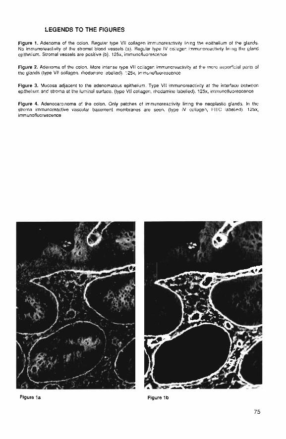
LEGENDS TO THE FIGURES
Figure 1. Adenoma of the colon. Regular type VII collagen immunoreactivity lining the epithelium of the glands.No immunoreactivity of the stromal blood vessels (a). Regular type IV colfagan immunoreaciivity lining the glandepithelium. Stromal vessels are positive (b). 125x, immunofluorescence
Figure 2. Adenoma of the colon. More intense type VII collagen immunoreactivity at the more superficial parts ofthe glands (type VII collagen, rhodamine labelled). 125x, immunofluorescence
Figure 3. Mucosa adjacent to the adenomatous epithelium. Type VII immunoreactivity at the interface betweenepithelium and stroma at the luminal surface, (type VII collagen, rhodamine labelled). 125x, immunofluorescence
Figure 4. Adenocarcinoma of the colon. Only patches of immunoreactivity lining the neoplastic glands. In thestroma immunoreactive vascular basement membranes are seen, (type IV collagen, FITC labelled). 125x,immunofluorescence
Figure 1a Figure 1b
75

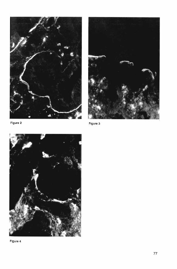
Figure 4
77

CHAPTER 7
TYPE VII COLLAGEN EXPRESSION IN THE NORMAL,HYPERPLASTIC AND NEOPLASTIC ENDOMETRIUM
An immunohistochemical study with type VII collagenand type IV collagen antibodies in preneoplastic andneoplastic lesions of the endometrium.
R. Visser, MPML Snijders, JW Arends , FT BosmanSubmitted for publication
79
7.1 INTRODUCTION
The glandular epithelium and the vascular endothelium in the stroma of the endometriumare supported by intact and continuous basement membranes throughout the menstrualcycle.These basement membranes contain intrinsic components such as laminin, type IVcollagen and heparan sulphate proteoglycan (1-4). In addition, it has been shown that inthe secretory phase and in pregnancy pericellular deposition of these matrix componentsoccurs around decidualized stromal cells (2). In benign (pre)neoplastic conditions, theepithelial basement membranes remain patent as for example in simple hyperplasia and inatypical hyperplasia of the endometrium.Some authors, however, observed minor interruptions in the epithelial basement membranein simple hyperplasia (1,3,4), but this phenomenon was also found in association withinflammation and stromal fibrosis (3). Interruptions in the basement membrane of neoplasticglands in endometrial carcinoma have been observed (3). The extent of these interruptionshas been correlated with the grade of differentiation of the tumor: in poorly differentiatedadenocarcinomas of the endometrium little or no basement membranes were detectable,whereas in well differentiated adenocarcinomas extensive basement membrane depositionoccurred. These studies were executed by immunohistochemistry using antibodies againsttype IV collagen and laminin.Type VII collagen, which is a major component of the anchoring fibrils, which bind thebasement membrane to the underlying extracellular matrix, has been found almostexclusively in the basement membrane zones of stratified epithelia (7). In the female genitaltract, type VII collagen expression has only been described in the cervical mucosa (8,9).The occurrence and distribution of this anchoring fibril associated protein in non-stratifiedepithelia such as the glands of the endometrium has not been extensively described yet.We therefore studied type VII collagen distribution in normal and neoplastic endometrium,addressing the following questions:1- Does type VII collagen occur in the endometrium and if so, is the expression patternrelated to the menstrual cycle.2- Is the expression pattern of type VII collagen in the basement membrane altered inhyperplasia, atypical hyperplasia and adenocarcinoma of the endometrium in comparisonwith that of type IV collagen.
7.2 MATERIALS & METHODS
7.2.1 Endometrial mucosa
For this study we used endometrial tissues obtained from patients which were treatedbecause of essential menorrhagia, dysmenorrhoea or uterine descensus. Details onmenstrual history, date of last menstrual period, hormonal therapy and gynaecologicalsurgery were recorded. All 15 premenopausal women (between 29 and 46 years of age)had a regular menstrual cycle; the 5 postmenopausal women were between 51 and 66years of age. None of the patients had a history of exogenous hormonal therapy ordistortion of the endometrial cavity (curettage, IUD etc.) for at least two cycles beforesurgery. Immediately after surgery (De Wever Hospital, Heerlen), the removed uteri wereopened. Besides sections for routine histopathological evaluation, another two samplesincluding endometrium and adjacent myometrium were obtained from the uterine fundusand corpus. These samples were frozen in isopentane quenched in liquid nitrogen andstored at -70°C. Cryostat sections were cut at 4 |am, airdried at room temperature andstored at -20°C for immunofluorescence doublestaining with antibodies directed againsttype IV collagen and type VII collagen.
80
Routinely processed haematoxylin and eosin (H&E) stains were used for histopathologicalevaluation. None of the uteri revealed any abnormalities. The endometrial dating criteria asdescribed by Noyes et al (9) were used to assess the phase of the menstrual cycle insections of the midfundus region. The histological appearance of the endometrium wasclassified as follows:I : menstruation(days 1-5)II : early proliferative (days 6-9)III: late proliferative (days 10-14)IV : early secretory (days 15-18)V : midsecretory (days 19-22)VI : late secretory(days 23-28)VII: postmenopausal
7.2.2 Hyperplasia and adenocarcinoma
Tissue sampling was performed as described for the group with normal endometrium. H&Estaining was performed in order to classify the endometrial hyperplasia andadenocarcinoma. Hyperplasia was classified according to the histopathological criteriadescribed by Kurman and Norris (11). By definition, the glandular lining epithelium wasdevoid of cytological atypia. Hyperplastic lesions in which the lining epithelium did showatypia were classified as atypical hyperplasia. Endometrial adenocarcinomas were gradedby two pathologists by consensus, according to the revised FIGO definitions (12).
7.2.3 Antibodies
Polyclonal antiserum directed against type IV collagen was raised in rabbits with humanplacental type IV collagen as immunogen. Characterization and immunospecificity havebeen described before (13).Two monoclonal antibodies against type VII collagen were used in this study: NP-76, whichreacts with the amino terminus of the NC-1 domain of the molecule, and LH 7.2 (14). Theantibodies were kindly provided by drs R.E. Burgeson (Portland, Oregon, USA; NP-76) andI.M. Leigh (London, G.B.; LH 7.2).
7.2.4 Immunohistochemistry
1. indirect immunoperoxidase staining:Cryostat sections were fixed in 2.5% buffered formaldehyde for 10 minutes and washed indemineralized water. After dehydration in graded alcohol, endogenous peroxidase wasblocked 15 minutes in 0.3% hydrogenperoxide in methanol. After washing in demineralizedwater the sections were incubated with the appropriate antiserum during 45 minutes atroom temperature (anti-type IV collagen diluted 1:250, NP-76 1:500 and LH 7,2 1:10 in Trisbuffered saline (TBS) with 1% bovine serum albumin (BSA) and 0,1% Tween to reducebackground staining). After washing in TBS the peroxidase-labeled swine-anti-rabbitconjugate, detecting the polyclonal antibody, (1:150 diluted in TBS/BSA/Tween) andperoxidase-labeled rabbit-anti-mouse (both obtained from Dakopatts, Denmark) detectingthe monoclonal antibody (1:200 diluted in TBS/BSA/Tween) were applied. After washing inTBS the complex was visualized in a substrate solution containing 5 mg DAB, dissolved in10 ml Tris/HCI (pH 7.6), 4uJ of 30% hydrogenperoxide and 1 ml Imidazole (0.1 M inTris/HCL). The sections were then counterstained with Mayer's haematoxylin during 1minute, dehydrated and mounted with Entellan (Merck, Germany).
81
2. Indirect immunofluorescence doublestaining.For type IV collagen and type VII collagen doublestaining the sections were first incubatedwith a mixture of anti-type IV collagen (1:250 diluted) and anti-type VII collagen(1:500diluted), in TBS/1% BSA/0,1% Tween.As second layer a mixture was used of FITC labeled goat anti rabbit IgG (Nordic, 1:300diluted) and rhodamine labeled goat anti- mouse IgG (TAGO Inc, Burlingame, Cal, USA;diluted 1:300). Specificity of the obtained immune reaction was controlled by identicalincubation of parallel sections, but with TBS instead of the primary antibody.The fluorescence patterns were photographed with a Leitz dialux fluorescence microscopeusing alternately FITC and rhodamine filter combinations.
7.3 RESULTS
All specimens immunostained for type IV collagen showed consistent staining of epithelialand vascular basement membranes. Moreover, pericellular immunoreactivity was observedaround the smooth muscle cells of the myometrium. These patterns were taken as internalpositive control.
7.3.1 Normal mucosa
Regular and continuous basement membranes were found bordering the epithelial cells ofthe glands, at the interface between epithelial cells and adjacent stroma (Fig 1). Thispattern, observed with antibodies directed against type IV collagen, was consistentthroughout the menstrual cycle and in the menopausal period.The expression pattern of type VII collagen was different. Throughout the different phasesof the menstrual cycle and postmenopausal phase the glands in the basal part of themucosa lacked type VII collagen immunoreactivity completely. During the early proliferativephase no type VII collagen immunoreactivity was observed around any of the endometrialglands. From the late proliferative phase on, throughout the rest of the menstrual cycle, anincreasing type VII collagen immunoreactivity was observed, only focal in the superficialparts of the glands and more intense and extensive with continuous immunoreactivity in thebasement membrane of the epithelial cells at the luminal surface of the mucosa (Fig. 2). Inthe specimens of the postmenopausal endometrium, the deeper parts of the endometrialglands showed focal immunoreactivity, while the surface epithelium showed continuoustype VII collagen immunoreactivity.
7.3.2 Hyperplasia
In hyperplasia, stained for type IV collagen, the epithelial basement membrane appeared tobe continuous, irrespective of the presence of epithelial atypia. The same pattern ofgenerally regular and intact epithelial basement membranes was observed by type VIIcollagen immunostaining. However, in specimina with cellular atypia of the tubules, areaswith absent basement membrane immunoreactivity for type VII collagen were observed,while type IV collagen staining showed intact basement membranes (Fig. 3).
7.3.3 Adenocarcinoma
Adenocarcinomas of the endometrium showed irregular patterns of epithelial basementmembrane staining, with interruptions up to total absence, when stained for type IVcollagen (Fig 4). In 1 case of papillary adenocarcinoma, type IV collagen immunoreactivitywas not observed around tumor epithelium.In these specimina of endometrial adenocarcinoma type VII collagen immunoreactivity wasnot observed.
82
7.4 DISCUSSION
In this study, we investigated basement membrane patterns in normal endometrium, and inendometrial hyperplasia and adenocarcinoma. Antibodies against type IV coiiagen and typeVII collagen were used. Type Vil collagen is the major constituent of anchoring fibrils(7,15). These fibrils, which reinforce attachment of epithelial cells to the stroma, originate atthe basement membrane and project into the stroma. Type VII coiiagen has been found inthe basement membrane of amnion epithelium and in the basement membrane of stratifiedepithelia such as oesophageal, oral and cervical mucosa, corneal epithelium, uroiheitum ofthe urinary bladder and around ducts of the breast (8,9,16).Patterns of type VII collagen expression have been studied in a limited spectrum ofneoplasms. Kirkham et al (17) described type VII collagen containing basement mem-branes surrounding tumor cell nests in malignant melanoma. In malignant melanomasexceeding 0.9 mm in thickness, these basement membranes appeared to be fragmented,suggesting a relationship between the thickness of the tumor, basement membraneintegrity and a prognosis in thin malignant melanomas. Schmoeckel et al (18) found intactbasement membranes around junctional tumor nests of melanomas, including weakstaining with type VII collagen. In deeper, dermal regions they observed positive stainingfor laminin around the tumor nests, but weak staining for type IV collagen and absent typeVII collagen immunoreactivity, suggesting a gradual disappearance of basement membranecomponents during the penetration of malignant melanoma cells in the dermis. Recently,we studied the presence and localization of type VII collagen in the normal mucosa,adenomas and adenocarcinomas of the large bowel (reported in Chapter VI of this thesis).Type VII collagen appeared to be transiently expressed in the development of neoplasia:no expression in normal epithelium, extensive expression in dysplastic, limited expressionin well differentiated malignant and no expression in poorly differentiated malignantepithelium.In the present investigation we studied the expression of type IV and type VII collagen inthe normal endometrium and in endometrial hyperplasia and adenocarcinoma.Our observations of type IV collagen immunoreactivity patterns are in line with findings ofothers (1,2,3,4,19), who reported a homogeneous distribution of type IV collagen in thebasement membrane of vascular structures in the stroma and of tubules and epithelia atthe luminal surface.The results of our study indicate, that an increasing amount of type VII collagen isexpressed during the course of the menstrual cycle, with focal immunoreactivity in theepithelial basement membrane in the proliferative phase and continuous type VII collagenimmunoreactivity in the basement membranes of the glands in the secretory phase, whichis more intense at the luminal surface than around the deeper glands. This pattern differsfrom that of type IV collagen, which is expressed ubiquitously along the epithelial lining ofglands and surface. This different expression pattern might reflect specific functions of typeIV and VII collagen in the basement membrane. The association of type VII collagen withanchoring fibrils, which are found in the subbasal lamina and reinforce the attachment ofepithelial cells to the underlying stroma of tissues subject to external frictional forces sug-gests that the presence of type VII collagen in the secretory phase of the menstrual cyclemight be associated with shear forces in the uterine cavity. Alternatively, it could beenvisaged that the function of type VII collagen is not merely mechanical, but also includesregulating properties.In adenomatous hyperplasia various authors have reported intact basement membranes,although Furness and Lam (4) found disruptions in the basement membrane in some casesof adenomatous hyperplasia. In our series of adenomatous hyperplasia we did not find anybasement membrane discontinuity in type IV collagen stained sections, even in thepresence of cellular atypia. However, type VII collagen staining showed focal absence incases of hyperplasia with atypia.
83
This heterogeneous expression pattern of type IV and type VII collagen was morepronounced in adenocarcinoma of the endometrium. Basement membrane breaks up tototal absence was observed in sections stained for type IV collagen, confirming results fromthe literature (1-4,19). Type VII collagen expression, on the other hand, lacked completelyin adenocarcinoma. The biological significance of this finding remains unanswered.Our findings might have some diagnostic applications. The irregular type VII collagenstaining we observed in atypical hyperplasia might be used to delineate the boundarybetween hyperplasia and adenocarcinoma. The disappearance of type VII collagenimmunoreactivity in adenocarcinoma might be used in the same way.In conclusion, we have found type VII collagen expression in the late proliferative andsecretory endometrium, especially in connection with basement membranes of superficialglands. In hyperplasia this pattern remains, but in atypical hyperplasia type VII collagengradually disappears and type VII collagen is no longer observed in endometrialadenocarcinoma.
84
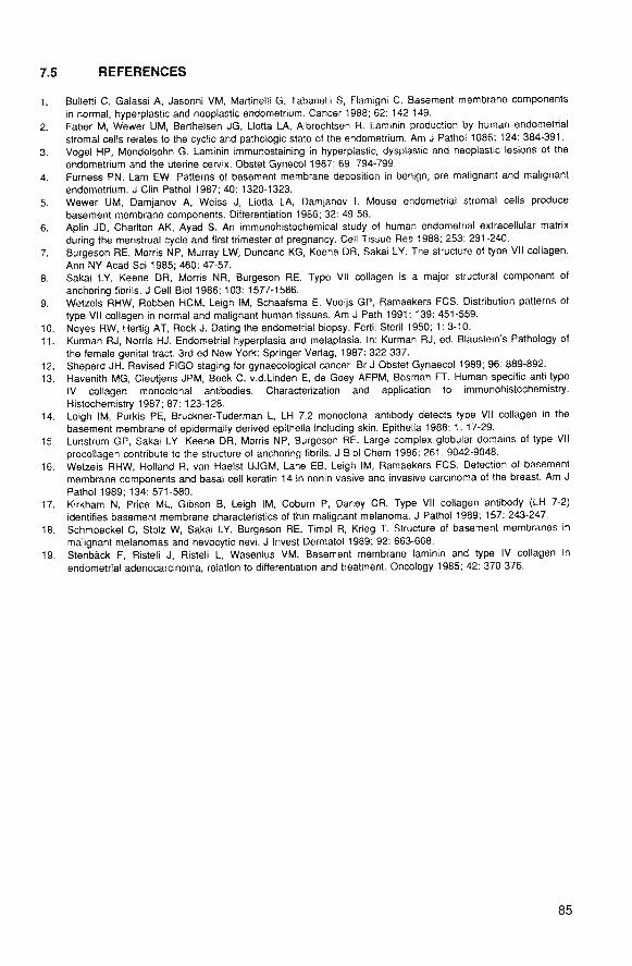
7.5 REFERENCES
1. Bulletti C, Galassi A, Jasonni VM, Martineili G, Tabanelli S, Flamigni C. Basement membrane componentsin normal, hyperplastic and neoplastic endometrium. Cancer 1988; 62: 142-149.
2. Faber M, Wewer UM, Berthelsen JG, Liotta LA, Albrechtsen R. Laminin production by human endometriaistromal cells relates to the cyclic and pathologic state of ihe endometrium. Am J Pathol 1086; 124: 384-391.
3. Vogel HP, Mendelsohn G. Laminin immunostaining in hyperplastic, dysplastic and neoplastic lesions of theendometrium and the uterine cervix. Obstet Gynecol 1987; 69: 794-799.
4. Furness PN, Lam EW. Patterns ot basement membrane deposition in benign, pre malignant and malignantendometrium. J Clin Pathol 1987; 40: 1320-1323.
5. Wewer UM, Damjanov A, Weiss J, Liotta LA, Damjanov I. Mouse endometriai stromal cells producebasement membrane components. Differentiation 1986; 32: 49-58.
6. Aplin JD, Charlton AK, Ayad S. An immunohistochemical study of human endometriai extracellular matrixduring the menstrual cycle and lirst trimester of pregnancy. Cell Tissue Res 1988; 253: 231-240.
7. Burgeson RE, Morris NP, Murray LW, Duncanc KG, Keene DR, Sakai LY. The structure of type VII collagen.Ann NY Acad Sci 1985; 460: 47-57.
8. Sakai LY, Keene DR, Morris NR, Burgeson RE. Type VII collagen is a major structural component ofanchoring fibrils. J Cell Biol 1986; 103: 1577-1586.
9. Wetzels RHW, Robben HCM, Leigh IM, Schaafsma E, Vooijs GP, Ramaekers FCS. Distribution patterns oftype VII collagen in normal and malignant human tissues. Am J Path 1991; 139: 451-559.
10. Noyes RW, Hertig AT, Rock J. Dating the endometriai biopsy. Fertil Steril 1950; 1: 3-10.11. Kurman RJ, Norris HJ. Endometriai hyperplasia and metaplasia. In: Kurman RJ, ed. Blaustein's Pathology of
the female genital tract. 3rd ed New York: Springer Verlag, 1987: 322-337.12. Sheperd JH. Revised FIGO staging for gynaecological cancer. Br J Obstet Gynaecol 1989; 96: 889-892.13. Havenith MG, Cleutjens JPM, Beek C, v.d.Linden E, de Goey AFPM, Bosman FT. Human specific anti-type
IV collagen monoclonal antibodies. Characterization and application to immunohistochemistry.Histochemistry 1987; 87: 123-128.
14. Leigh IM, Purkis PE, Bruckner-Tuderman L, LH 7.2 monoclonal antibody detects type VII collagen in thebasement membrane of epidermally derived epithelia including skin. Epithelia 1988; 1: 17-29.
15. Lunstrum GP, Sakai LY, Keene DR, Morris NP, Burgeson RE. Large complex globular domains of type VIIprocollagen contribute to the structure of anchoring fibrils. J Biol Chem 1986; 261: 9042-9048.
16. Wetzels RHW, Holland R, van Haelst UJGM, Lane EB, Leigh IM, Ramaekers FCS. Detection of basementmembrane components and basal cell keratin 14 in nonin vasive and invasive carcinoma ot the breast. Am JPathol 1989; 134: 571-580.
17. Kirkham N, Price ML, Gibson B, Leigh IM, Coburn P, Darley CR. Type VII collagen antibody (LH 7-2)identifies basement membrane characteristics of thin malignant melanoma. J Pathol 1989; 157: 243-247.
18. Schmoeckel C, Stolz W, Sakai LY, Burgeson RE, Timpl R, Krieg T. Structure of basement membranes inmalignant melanomas and nevocytic nevi. J Invest Dermatol 1989; 92: 663-668.
19. Stenback F, Risteli J, Risteli L, Wasenius VM. Basement membrane laminin and type IV collagen inendometriai adenocarcinoma, relation to differentiation and treatment. Oncology 1985; 42; 370-376.
85
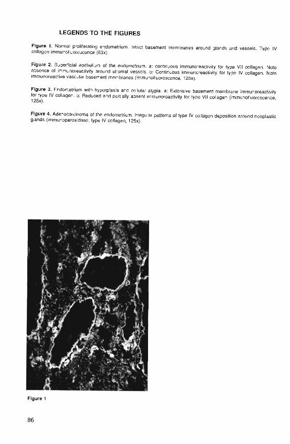
LEGENDS TO THE FIGURES
Figure 1. Normal proliferating endometrium. Intact basement membranes around glands and vessels Type IVcollagen immunofluorescence (63x).
Figure 2. Superficial epithelium of the endometrium. a: continuous immunoreactivity for type VII collagen Noteabsence of immunoreactivity around stromal vessels, b: Continuous immunoreactivity for type IV collagen. Noteimmunoreactive vascular basement membranes (immunofluorescence, 125x).
Figure 3. Endometrium with hyperplasia and cellular atypia. a; Extensive basement membrane immunoreactivityfor type IV collagen, b: Reduced and partially absent immunoreactivity for type VII collagen (immunofluorescence,
Figure 4. Adenocarcinoma of the endometrium. Irregular patterns of type IV collagen deposition around neoplasticglands (immunoperoxidase; type IV collagen, 125x).
Figure 1
86
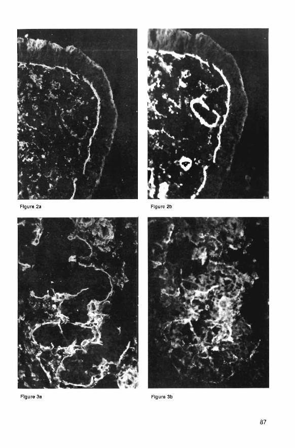
Figure 2a Figure 2b
Figure 3a Figure 3b
87

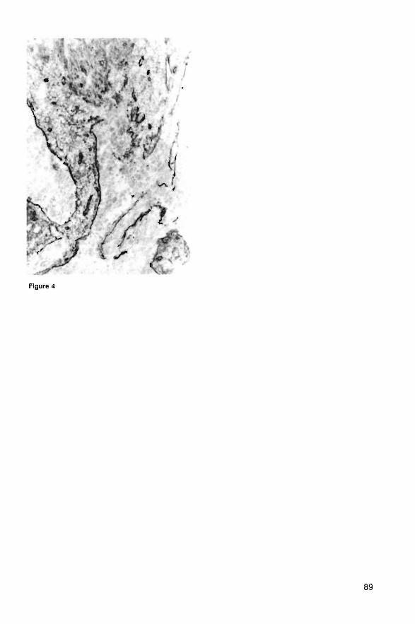
Figure 4
89

CHAPTER 8
GENERAL DISCUSSION
8.1 BASEMENT MEMBRANE PATTERNS IN BENIGN AND MALIGNANT NEO-PLASMS
An essential feature of benign epithelial neoplasia is its restriction to the epithelial compart-ment which implies absence of invasion. Consequently, it may be expected that immuno-histochemical staining of the basement membrane will invariably reveal intact basementmembranes in benign tumors. This has indeed been confirmed in many studies on non-invasive benign epithelial tumors of various tissues including the skin, breast, uterine cervixand larynx. Two examples of this principle, which illustrate the use in daily practice, arebreast neoplasms, where intact basement membranes allow the distinction betweensclerosing adenosis and tubular carcinoma and pancreatic lesions, where basementmembrane imrnunohistochemistry facilitates the discrimination between chronic fibrosingpancreatitis and adenocarcinoma of the pancreas. However, it has also been reported thatin benign conditions, such as epithelial dysplasia, interruptions occur. Also in inflammatoryconditions basement membrane gaps are found, presumably due to proteolytic propertiesof migrating inflammatory cells.In contrast, basement membrane degradation is regarded as a decisive step towards thedevelopment of malignant neoplasia, because it precedes tumor invasion. Invasive tumorsmay show variable basement membrane patterns, ranging from almost total absence, ashas been described in tubular carcinoma of the breast, to almost complete enveloping oftumor cell nests with an irregular, but largely intact basement membrane, as in malignantmelanomas.
8.2 POTENTIAL SIGNIFICANCE OF BASEMENT MEMBRANE PATTERNS INTUMORS OF BORDERLINE MALIGNANCY
Contrasting with the extensive biochemical and cell biological research in the field of tumorcell/extracellular matrix interaction, and with the numerous studies reporting basementmembrane patterns in malignant neoplasms, relatively few immunohistochemical studieshave been executed to investigate basement membrane characteristics in neoplasms onthe borderline between benign and malignant. Structural basement membrane alterations,such as reduplication and focal interruption, have been described in epithelia with severedysplasia or in in situ carcinomas as well as in epithelial tumors of borderline malignancy.
91

In this stage of tumor cell evolution, when the neoplastic cells may transit from a non-invasive into an invasive phenotype, complex alterations occur in the relation between theneoplastic cell and its surrounding extracellular matrix, which creates a suitable microen-vironment for invasive growth. Gaps in the basement membrane are caused by basementmembrane remodelling due to decreased synthesis and assembly of its components, or,alternatively, basement membrane dissolution may be due to matrix-degrading enzymessuch as collagenases, cathepsins, plasmins and heparanases, which are secreted by tumorcells.If in this initial phase of tumor progression the basement membrane is visualized, adistinction might be envisioned between lesions with an intact basement membrane, to beregarded as benign, and lesions with basement membrane defects, to be regarded asmalignant. This would indicate that the term borderline lesion signifies the inability of thepathologist to determine the biological potential of a lesion by routine histology. Thatproblem might then be solved by basement membrane immunohistochemistry. Analternative view is that borderline lesions constitute a biological intermediate betweenbenign and malignant, undetermined in their biological behaviour. The primary aim of ourstudy was to test the first hypothesis i.e. that borderline lesions can be distinguished bybasement membrane immunohistochemistry in an intrinsically benign and a potentiallymalignant group.Using antibodies directed against specific basement membrane components, includinglaminin, type IV collagen and type VII collagen immunohistochemical studies were executedin tumors of the larynx, ovary, kidney, colon and endometrium.
8.3 OBSERVED BASEMENT MEMBRANE PATTERNS IN TUMORS OF BORDER-LINE MALIGNANCY
In the larynx we investigated hyperplasia, dysplasia and in situ carcinoma of the mucosa.Distinct and continuous basement membranes were found with anti laminin and type IVcollagen antibodies in normal epithelium as well as in hyperplasia and dysplasia. Besidesbasement membrane discontinuity in invasive carcinomas but also in some cases of in situcarcinoma, focal interruptions were observed in areas with adjacent inflammation.The first observation supports our hypothesis, although follow-up studies have to beperformed to test the validity of the assumption that in situ carcinomata with basementmembrane interruptions stand a higher chance to progress towards invasive carcinomathan those with intact basement membranes. The second observation indicates thatbasement membrane interruptions only, can never be taken as convincing evidence ofmalignant behaviour.We furthermore studied borderline malignant tumors of the ovary. We studied basementmembrane patterns in these tumors and compared them with those in ovarian cystade-nomas and cystadenocarcinomas.In cystadenomas regular and intact basement membranes were found. Cystadenocarcino-mas displayed irregular and interrupted basement membranes intermingled with areas ofintact basement membranes. Of the borderline tumors 70% shared a regular and intactbasement membrane pattern with cystadenomas, However, 30% of the borderline malig-nant tumors contained small areas with an irregular and interrupted basement membranepattern, resembling that of invasive carcinoma.We conclude that also in ovarian tumors of borderline malignancy basement membranepatterns mostly are intact and therefore similar to those of benign epithelial tumors. Focalirregular defects, possibly suggesting early invasive growth, however, occur in 30% of thecases.As in the laryngeal in situ carcinoma, follow-up studies have to confirm the predictive valueof basement membrane staining patterns for the prediction of the behaviour of theneoplasm. We next investigated basement membrane patterns in small renal adeno-
92
carcinomas. Based on these patterns, two groups could be discriminated. One group oftumors contained intact and regular basement membranes and the other group showedirregular and/or focally absent basement membranes. A statistically significant correlationwas found between basement membrane pattern and tumor size as well as nuclear grade,both parameters correlating with metastatic potential. Therefore, even though also in thesetumors follow-up studies were not conducted, we conclude that in small renal cortical neo-plasms basement membrane interruptions might be indicative of malignant potential.Taken together, these three studies indicate that histopathological borderline categoriesmight contain distinct subpopulations with, on the basis of basement membrane patterns,identifiable differences in behaviour. Our studies also illustrate a practical problem in valida-tion of this assumption in obtaining sufficient material to conduct adequate follow-upstudies. Two other studies were conducted in order to elucidate the potential role of typeVII collagen depositions in lesions progressing from non-invasive to invasive.Type VII collagen, the major component of anchoring fibrils, is mainly found in associationwith surface epithelium. Its expression has been studied by other investigators in carcinomaof larynx, endometrium and large bowel. We noted in squamous cell carcinoma of thelarynx a difference in the patterns of expression of type IV collagen and type VII collagen,which was most pronounced in poorly differentiated tumors. Basement membranes insections stained for type VII collagen showed discontinuities whereas staining for type IVcollagen showed intact basement membranes, suggesting that type VII collagen is a betterindicator for early invasive growth. It also appeared that in poorly differentiated carcinomaintracytoplasmic expression of type VII collagen occurs in the absence of type IV collagenreactive basement membranes.
In the endometrium type VII collagen appeared gradually in the course of the menstrualcycle, along with intact basement membranes as reflected in the pattern of staining for typeIV collagen. In atypical hyperplasia, extensive deposition of type VII collagen was found,which gradually disappeared in adenocarcinoma. As in squamous carcinoma of the larynx,type VII collagen deposition therefore might be related to epithelial differentiation.Although the epithelial basement membrane of normal mucosa of the large bowel lackstype VII collagen, this component was evidently transiently expressed in areas of dysplasiaas for example in adenomas.The overall conclusions of these studies is that the relationship of basement membranestaining and morphologically detectable tumor cell invasion is highly complex, withbasement membrane interruptions in inflammation, severe epithelial dysplasia, in situcarcinoma and in invasive carcinoma. Therefore, invasive malignant neoplasia cannot bediagnosed solely on disruptions in the basement membrane.
8.4 FUTURE PERSPECTIVE
Basement membrane morphology is only one aspect of the complex interaction betweentumor cells and the extracellular matrix and provides a fairly static view on a dynamicprocess: altered balance between basement membrane assembly and degradation. Recentstudies have revealed ample information concerning the proteases and glycosidases (a.o.)that are synthesized and released by tumor cells and stromal cells and are associated withinitiation of tumor invasion. Also the matrix, adjacent to the tumor cells is changed andbecomes permissive for the tumor cells to migrate. In this sequence of events during initialinvasion of tumor cells cell adhesion molecules and motility factors play an important role.It is this area, that future studies might result in new insight in the interaction betweentumor cells and their surrounding matrix. Identification of new matrix-degrading enzymes,unravelling of the role of plasminogen activators and plasmin, the significance of motilityfactors and new information on cell-adhesion molecules and integrin extracellular matrixreceptors will extend our understanding of tumor cell invasion and provide us with tools formore accurate tumor diagnosis and determination of tumor behaviour.
93

SUMMARY
is fhss thesis 6 sts ies are reported concerning the eondSsfi af the basement imem&fafw Ine p i c a l iteapiasiss, «iiteh reflects the intemstisn between the tumef eeite, the sterna!eeis srxi the ssteoefiylar f^attm. l i re appearance of the basement membrane eatlH feewsaafeeci i>y ifjwmnshfstashejrastry, using poiydonal and monoclonal antibodies directedagassst bminai, ^pe IV esiagen and type Vil collagen. These proteins occur an!y in the
As this method has been extensively used to study invasive tumors, we limited ourinvssigaions to tenors that afe on the borderline between benign and malignant. As thehistoiogical diagnosis "tumor of borderline malignancy" generally is made by routine stains,which do not visualize the basement membrane, the aim of our studies was te subdividetumors of borderline malignancy in a group with intact baseroent membranes and a §f©ypwith interrupted basement membranes. If the difference would correlate with a different,more aggressive biological behaviour, being reflected by interruptions of the basementmembrane, this would be of diagnostic importance.In Chapter 1 the aims of the studies are defined and the structure, composition andfunction of the basement membrane is outlined. Presently, it is generally accepted that thebasement membrane is not a static border between epithelial cells and the adjacentstroma, but a dynamic structure, which appears to be continuously synthesized anddegraded. The balance between synthesis and degradation is grossly disturbed inmalignant conditions.Finally, an overview is presented of the basement membrane patterns in benign andmalignant tumors and a review of the literature concerning basement membrane patterns intumors of borderline malignancy.Chapter 2 describes basement membrane patterns in the larynx: biopsies with normalmucosa, dysplasia and in situ carcinoma were studied. As could be anticipated, intactbasement membranes were found in the normal mucosa, both with laminin and type IV col-lagen. Basement membranes in dysplastic epithelium also appeared to be continuous, butdefects were observed in carcinoma, in some cases of in situ carcinoma and in areas ofinflammation. It was concluded that basement membrane immunohistochemistry could beof value for the demonstration of microinvasive growth in laryngeal cancer, but that thebasement membrane can also be degraded by inflammatory cells.Chapter 3 reports basement membrane patterns in 3 groups of tumors of the ovary:cystadenomas, cystadenocarcinomas and tumors of borderline malignancy. Antisera wereused against laminin and type IV collagen. Cystadenomas displayed intact basementmembranes. In cystadenocarcinomas, serous and mucinous, a different pattern wasobserved: areas with irregular or absent basement membranes but also deposition ofbasement membranes between tumor cells and stroma. Tumors with borderline malignancyshared a regular basement membrane pattern with cystadenomas. However, thirty percentof this group contained small areas where basement membranes were lacking, resemblingthe pattern of cystadenocarcinomas.
95
From the results of this study, we conclude, that the basement membranes in cystadeno-mas are intact, while cystadenocarcinomas contain irregular basement membranes. Tumorsof borderline malignancy can be subdivided into a group that contains intact basementmembranes (like cystadenomas) and a group in which irregular basement membranes arefound (resembling patterns in cystadenocarcinomas).Chapter 4 describes a study of basement membranes patterns in a group of small renalcortical tumors of which the biological behaviour is unpredictable. Several investigatorsstudied the possibility of a correlation between tumor size, stage, histological subtype andkaryotype on the one side and clinical parameters of tumor behaviour on the other. This,however, has not resulted in the identification of parameters, which unambiguouslydistinguish benign from malignant lesions.In principle the presence or absence of invasive growth might provide a more reliableparameter of tumor behaviour. In order to investigate this possibility, we executed animmunohistochemical study of basement membrane patterns in small renal corticalneoplasms with emphasis on patterns in the centre and the periphery of the tumor. Wefurthermore studied the relation between deposition of basement membrane componentsand nuclear grade, which in stage 1 tumors can distinguish between high grade, more fre-quently metastasizing tumors and low grade, less frequently metastasizing tumors.The results of our study show that, on the basis of basement membrane patterns, twogroups can be distinguished. One group of tumors contains intact basement membranessimilar to in situ carcinomas and the other group shows focally or totally absent basementmembranes, corresponding with tumors with invasive growth. Tumors with low nucleargrade mostly contain regular and intact basement membranes, in contrast to high gradetumors.In Chapter 5 the presence of type IV collagen and type VII collagen was assessed in thebasement membrane of the upper respiratory tract mucosa and the distribution pattern ofthese components was evaluated in squamous cell carcinoma of this region.Basement membrane patterns were investigated, using immunohistochemical methods, andtheir presence was assessed semi-quantitatively in relation to tumor cell differentiation. Co-expression of type IV collagen and type VII collagen was studied using double immuno-fluorescence.The results indicate that the expression of the two basement membrane components iscorrelated and is also dependent on tumor cell differentiation: with decreasing level ofdifferentiation the tendency of tumor cells to deposit basement membrane materialdecreases. In poorly differentiated tumors some cells with intracellular deposition of type VIIcollagen fail to deposit this component extracellularly.The findings of this study suggest that type VII collagen might be a more sensitive indicatorfor invasive growth than type IV collagen, because it disappears earlier. Furthermore theresults indicate that type VII collagen synthesis is regulated independent from type IVcollagen synthesis, but its extracellular deposition appears to depend on the availability of atype IV collagen matrix. Intracytoplasmic type VII collagen expression might be used as aprogression marker for squamous cell carcinoma.In Chapter 6 the distribution of type IV collagen and type VII collagen in the basementmembranes of normal mucosa of the colon, adenomas and adenocarcinomas is described,using immunoperoxidase and immunofluorescence techniques. In normal mucosa we foundregular type IV collagen positive basement membranes, lining vascular structures andmucosal epithelia. These basement membranes, however, lacked type VII collagen.In adenomas of the colon intact basement membranes were observed through type IVcollagen staining. Type VII collagen staining was also detected, but only in connection withdysplastic epithelium. Adjacent to the dysplastic epithelium in adenomas, histologicallynormal epithelium also showed type VII collagen staining along the basement membrane,but restricted to the epithelium of the luminal surface. These areas were also investigatedfor expression of keratins 8, 18 and 19 and keratins 5 and 8 (monoclonal antibodies NCL-
96
5D3 and RCK 102 respectively), but altered differentiation was not detected using thistechnique.In adenocarcinomas of the colon, type IV collagen was irregularly deposited in thebasement membrane of neoplastic tubules. Type VII collagen staining was only detected inwell or moderately differentiated carcinomas and in higher amounts.Our findings, therefore, reveal a transient expression of type VII collagen in the transition ofdysplastic epithelium into carcinoma, suggesting the involvement of type VII collagen in theprocess of early invasion.In Chapter 7 the staining patterns for the basement membrane components type IV andtype VII collagen in the different phases of the menstrual cycle were studied immunohisto-chemically using an indirect immunoperoxidase technique as well as immunofluorescencedoublestaining. In addition the staining patterns in hyperplasia and adenocarcinoma of theendometrium were investigated.The normal endometrium showed epithelial basement membranes with continuous stainingfor type IV collagen. However, type VII collagen was only present in epithelial basementmembranes in the superficial layer of the lamina functionalis of the endometrium only in thelate proliferative phase and in the secretory phase. Hyperplastic endometrium showedcontinuous basement membranes when stained both for type IV collagen and type VIIcollagen. However, in areas with cellular atypia discontinuities were found in the basementmembranes when stained for type VII collagen. Staining for basement membrane compo-nents was even more abnormal in adenocarcinomas of the endometrium, where type IVcollagen staining was discontinuous, while type VII collagen was completely absent.Our results indicate that type VII collagen is more widely distributed than previouslyassumed. Furthermore, in the development of endometrial neoplasia basement membraneabnormalities are reflected earlier in type VII collagen than in type IV collagen deposition.The potential for diagnostic use of this finding needs to be further investigated.In chapter 8 an overview is given of basement membrane characteristics in benign andmalignant neoplasms. Basement membrane patterns in tumors of borderline malignancyare discussed with respect to their possible biological behaviour and potential diagnosticsignificance. Finally, the potential role of type VII collagen depositions in lesions progress-ing from non-invasive to invasive were discussed.
97

SAMENVATTING
In dit proefschrift worden de resultaten beschreven van een zestal immunohistochemischestudies op het gebied van veranderingen van de basaalmembraan in epitheliale tumoren.Omdat, blijkens omvangrijke literatuur, reeds vrij veel onderzoek werd verricht naar de ver-schijningsvormen van de basaal membraan bij invasief groeiende tumoren, hebben wij denadruk bij onze studies gelegd bij processen met beginnende en (bij conventionelekleuringsmethoden) onzekere kwaadaardigheid, respectievelijk "carcinoma in situ" en"borderline tumoren".De basaal membraan kon hierbij specifiek zichtbaar worden gemaakt met behulp vanimmunohistochemie op weefselcoupes, waarbij gebruik werd gemaakt van polyclonale enmonoclonale antilichamen, gericht tegen basaalmembraan-specifieke eiwitten als laminine,type IV collageen en type VII collageen.In hoofdstuk 1 wordt het onderzoeksgebied ingeleid. De structuur en samenstelling van debasaalmembraan wordt beschreven op geleide van de in de literatuur beschikbaregegevens. Ook wordt gewezen op de dynamiek van de basaalmembraan en de relatie eninteractie met de extracellulaire matrix en de cellen, die met de basaalmembraan geasso-cieerd zijn. Deze onderlinge beinvloeding vindt ook plaats onder omstandigheden, waarbijde normale ontwikkeling van de cellen verstoord is. In dit gebied is reeds veel onderzoekverricht, de laatste tien jaar. Er wordt een uitgebreid overzicht gegeven over de resultatenvan dit onderzoek, zowel bij goedaardige als kwaadaardige tumoren en er wordt eenoverzicht gegeven van de reeds verschenen publikaties op het gebied van grensvlaktu-moren en tumoren van onzekere kwaadaardigheid. Het hoofdstuk wordt besloten met eenuitgebreide lijst van publikaties op dit gebied.Hoofdstuk 2 bevat de beschrijving en resultaten van een onderzoek naar het patroon vande basaal membraan in normaal slijmvlies van de larynx, bij hyperplasie, dysplasie encarcinoma in situ, waarbij gebruik werd gemaakt van antilichamen gericht tegen laminineen type IV collageen. Een regelmatig patroon van de basaal membraan werd gezien innormaal slijmvlies. Hierbij werd de basaal membraan gezien als een smalle, streepvormigeen ononderbroken structuur. De patronen bij de immunohistochemische kleuringen metlaminine en type IV collageen waren identiek. Ononderbroken basaal membranen werdenook aangetroffen bij dysplastisch epitheel, terwijl defecten waargenomen werden bij hetcarcinoom. Bij het carcinoma in situ bleek de basaal membraan in sommige gevallen en opgelokaliseerde plaatsen defecten te bevatten. Daarnaast is gebleken, dat basaal mem-braandefecten ook konden ontstaan in gebieden met ontstekingsactiviteit. In de gevallen,waar dit waargenomen werd, toonde het bekledende epitheel hyperplastische of dysplas-tische kenmerken, maar ook in gevallen met carcinoma in situ, waarbij een ontstekings-component aanwezig was.De waarde van deze methode bij de histopathologische diagnostiek van deze groep vanaandoeningen in de larynx werd bediscussieerd.In hoofdstuk 3 is het onderzoek gericht op basaal membraanpatronen bij ovariumtumorenvan "borderline" maligniteitsgraad. De verkregen resultaten werden vergeleken met basaal
99
membraan patronen bij cystadenomen en cystadenocarcinomen van het ovarium. Er werdgebruik gemaakt van antilichamen gericht tegen laminine en type IV collageen. In tegen-stelling tot de groep van de cystadenomen, waarbij de basaal membraan regelmatiggevormd en overal intact bleek, was het expressiepatroon bij de groep van de cystadeno-carcinomen onregelmatig en op talrijke plaatsen afwezig.Het patroon van de basaal membraan bij de groep van ovariumtumoren met "borderline"maligniteit toonde in principe een identiek beeld als in de groep cystadenomen. Echterbleek in 30% van de gevallen de basaal membraan onregelmatige defecten te bevatten. Deresultaten wijzen er op, dat -uitgaande van het basaal membraanexpressiepatroon- degroepen met cystadenomen en cystadenocarcinomen homogeen zijn, terwijl in de groepovariumtumoren met "borderline" maligniteit twee subgroepen zijn te onderscheiden,waarbij het basaal membraanexpressiepatroon van de ene subgroep overeenkomt met datvan cystadenomen, en van de tweede subgroep lokaal overeenkomt met het irregulairepatroon in cystadenocarcinomen.In hoofdstuk 4 worden de resultaten vermeld van een studie naar basaal membraanpatro-nen bij een groep van kleine tumoren van de nier, waarvan het biologische gedrag moeilijkvoorpelbaar is. Gebruik makend van antistoffen, gericht tegen laminine en type IVcollageen werden 41 tumoren van de nier, gediagnostiseerd als "niercel carcinoom" of"adenoom van de nierschors" en in diameter varierend van enkele millimeters tot 6 cm, ophet basaal membraan expressiepatroon onderzocht. De vraagstelling richtte zich met nameop een mogelijke relatie tussen basaal membraanpatronen en morfologische parametersals kernpolymorfie (uitgedrukt in "nuclear grade") en diameter van de tumor, die beide vanprognostische waarde zijn.De bevindingen wezen uit, dat in deze groep tumoren voorkomen met intacte basaalmembranen en tumoren, waarbij de basaal membraan onregelmatig gevormde defectenbevat. Een intacte basaal membraan werd significant meer aangetroffen in kleine niertumo-ren, met een diameter minder dan 10 mm, terwijl de meeste van de grotere tumorendefecten lieten zien. Beoordeling van de "nuclear grade" in relatie tot deze diametercate-gorieen leverde op, dat laaggradige tumoren significant meer voorkwamen in de groeptumoren met een diameter minder dan 10 mm.Aangezien uit de literatuur bekend is, dat laaggradige tumoren significant minder metasta-seren dan hooggradige tumoren en gezien de intacte basaal membranen in deze categorie,concluderen wij, dat voor het biologisch gedrag van een niertumor de diameter van meer ofminder dan 10 mm van meer belang is, dan de diameter van 3 cm, zoals totnogtoe regelwerd aangenomen. Deze conclusie zou nog in een grotere meerjarenstudie moeten wordengetoetst, waarin ook klinische follow-up gegevens konden worden betrokken.Hoofdstuk 5 bevat de resultaten van een studie naar de expressiepatronen van type IVcollageen en type VII collageen in het slijmvlies van mond- en keelholte en in het plaveisel-celcarcinoom van deze regio. Bij dit onderzoek werd gebruik gemaakt van een dubbelfluor-escentiemethode. Ook werd gezocht naar een mogelijke relatie tussen expressie van dezebeide basaal membraaneiwitten en tumordifferentiatie. De resultaten van het onderzoekwijzen uit, dat de depositiepatronen van type IV collageen en type VII collageen identiekzijn in de basaal membraan van het slijmvlies epitheel en de afvoerbuisjes van despeekselkliertjes, maar verschillend bij het klierepitheel, bloedvaatjes, spiervezels,zenuwvezels en vetcellen, waar type IV collageen wel aanwezig was in de basaalmem-braan in tegenstelling tot type VII collageen. Wanneer de expressiepatronen van beideantigenen werden vergeleken, dan bleek, dat er een toenemende co-expressie was,naarmate het carcinoom beter gedifferentieerd was. Bovendien bleek er intracytoplasma-tische expressie van type VII collageen in alle slecht gedifferentieerde en sommige van dematig gedifferentieerde carcinomen, waarbij het vermogen om type VII collageen extracel-lulair af te zetten kennelijk niet aanwezig was. De resultaten suggereren, dat type VIIcollageenexpressie een gevoelige indicator is voor invasieve groei bij maligne tumoren.
100
In hoofdstuk 6 wordt het expressiepatroon van type IV collageen en type VII collageenbeschreven in het normale slijmvlies van de dikke darm, in adenomen en adenocar-cinomen, ontstaan uit dit slijmvliesEr bleek een discrepantie in het vdorkomen van deze componenten, waarbij type IVcollageen in regelmatige patronen gezien werd in de basaal membraan van het epitheel,van bloedvaatjes, spier- en zenuwvezels en type VII collageen geheel ontbrak. Bijadenomen werden, bij gebruik van type IV collageen, regelmatige basaal membraanpatronen waargenomen. Type VII collageen expressie werd uitsluitend aangetroffen in debasaal membraan grenzend aan dysplastisch epitheel en het oppervlakkige, histologischenormaal gebouwde epitheel in de onmiddellijke omgeving van het adenoom. Met behulpvan antilichamen, gericht tegen hoog- en laagmoleculaire keratines werd geen aanwijzinggevonden voor gewijzigde differentiatie van cellen grenzend aan de basaal membraan mettype VII collageen expressie.Bij adenocarcinomen van de dikke darm werd onregelmatige expressie gezien van type IVcollageen, wijzend op uitgebreide defecten. Ook hier was het expressiepatroon niet identiekmet type VII collageen, omdat deze component alleen in goed en matig gedifferentieerdetumoren werd aangetroffen.De in dit hoofdstuk vermelde bevindingen wijzen op een voorbijgaande expressie van typeVII collageen, welke alleen waargenomen wordt bij epitheel met dysplastische veran-deringen en carcinomen van matige of hoge differentiatiegraad. Ook hier zou type VIIcollageen een waardevolle indicator kunnen zijn bij vaststelling van vroeg invasieve groeibij adenocarcinomen van de dikke darm.In hoofdstuk 7 worden de resultaten beschreven van een studie naar de expressiepatro-nen van type IV collageen en type VII collageen in het endometrium bij de fysiologische,hormonaal bepaalde fasen, bij hyperplasie van het endometrium en bij het adenocarci-noom.Omdat onderlinge vergelijking van deze expressiepatronen tot het onderwerp van de studiebehoorde, werd ook immunofluorescentie dubbelkleuring aangewend.Bij het normale endometrium werden met type IV collageen normaal gevormde basaalmembranen gezien. Type VII collageen werd uitsluitend waargenomen in de meeroppervlakkige niveau's van het endometrium en dan aan het eind van de proliferatiefase enin de secretiefase. Bij hyperplasie zonder epitheelatypie bleek er een vrijwel identiekexpressiepatroon van type IV en type VII collageen; in gevallen van endometriumhyper-plasie met epitheelatypie bleek de basaal membraan ononderbroken en regelmatigeimmunoreactiviteit voor type IV collageen te bezitten, terwijl immunoreactiviteit voor type VIIcollageen op sommige plaatsen afwezig bleek.Bij het adenocarcinoom werden onregelmatige type IV collageen expressiepatronen gezien,terwijl type VII collageen immunoreactiviteit geheel ontbrak.De resultaten van deze studie tonen aan, dat type VII collageen tijdens het verloop van demenstruele cyclus in het oppervlakkige niveau tot expressie komt. Dit fenomeen, en deverschillende type IV en type VII collageen expressie berust mogelijk op de specifieke, envan type IV collageen afwijkende functie. Aangezien type VII collageen geassocieerd wordtmet de verankeringsfibrillen tussen basaal membraan en aangrenzend stroma, zouexpressie van dit eiwit kunnen wijzen op de noodzaak van steviger hechting van epitheelaan het stroma, bijvoorbeeld bij inwerking van uitwendige wrijvingskrachten op hetoppervlakkige epitheel van het endometriumslijmvlies. De discrepante expressie van beidebasaal membraan componenten bij endometriumhyperplasie zou mogelijk van belangkunnen zijn bij histologische diagnostiek in het grensgebied tussen atypie en invasievegroei.Hoofdstuk 8, tenslotte, geeft een overzicht van de resultaten van de studies tegen deachtergrond van interacties tussen tumorcellen, basaal membraan en extracellulaire matrixbij invasief groeiende tumoren en in het bizonder tumoren met beginnende invasieve groeien tumoren met "borderline" maligniteit.
101
Er wordt op gewezen, dat de resultaten van de studies aanwijzingen geven voor hetbiologische gedrag van deze tumoren en van belang kunnen zijn voor de diagnostischehistopathologie. Om deze aanwijzingen om te zetten in concreet toepasbare, wetenschap-pelijk gefundeerde criteria, is uitbreiding met klinisch-pathologische follow-up studiesgeindiceerd en noodzakelijk.
102
DANKWOORD
Het is nu circa 4 jaar geleden, dat, tijdens een wandeltocht langs de Jeker en op weg naarde bron van deze rivier, de kiel werd gelegd voor dit proefschrift. Na met Jan WillemArends en Henny Peltenburg, vanaf Maastricht een dag lang de rivier stroomopwaartsgevolgd te hebben en in Tongeren (Belgie) in een Italiaans restaurant te hebben gegeten("wenst U de spaghetti met of zonder frites, heren?"), kwam tijdens het natafelen in cafe"Relax", nabij het station van Tongeren, het onderwerp "promoveren" ter sprake. Hierbijsuggereerden mijn wandelgenoten om de reeds verrichtte inspanningen op het gebied vanbasaal membraan immunohistochemie uit te breiden tot een promotiewaardig onderzoek.Het resultaat ligt voor U. Het is tot stand gekomen, mede dankzij de directe of indirectebetrokkenheid van velen en voor een belangrijk deel bewerkt binnen de vakgroep Patholo-gie van de Rijksuniversiteit Limburg te Maastricht. Hier waren het Margriet Pijls, BirgitteKuypers-Engelen en Annick Moors, die op voortreffelijke wijze voor de immunohisto-chemische bewerkingen zorgdroegen. Het materiaal werd grotendeels verzameld uit hetarchief van de vakgroep Pathologie van het Academisch Ziekenhuis Maastricht, maar ookwerd materiaal verkregen van de afdeling Pathologie van het "de Wever" ziekenhuis teHeerlen (hoofd: dr Joh Koudstaal) en door de medewerking van dr Johan van der Beek(vakgroep Keel-, neus- en oorheelkunde), prof dr RW Stockbrugger (vakgroep Internegeneeskunde, gastro-enterologie), de medewerksters van de polikliniek gastro-enterologie,dr Delemarre (destijds hoofd van de afd Pathologie van het Nederlands Kanker Instituut teAmsterdam) en dr Fibo ten Kate (toentertijd verbonden aan de afd Pathologie van deErasmus Universiteit te Rotterdam). Bij de statistische berekeningen ben ik geholpen doorGeert Jan Tangelder (vakgroep Fysiologie) en Erik Thunnissen (vakgroep Pathologie).Bij de fotografische opnamen werd geadviseerd door de Audiovisuele Dienst van hetAcademisch Ziekenhuis Maastricht en de foto's werden door Eline Huiben afgedrukt op deafdeling Fotografie van het Gerechtelijk Laboratorium (Hr van den Heuvel).Bij de tekstverwerking heb ik nuttige en bruikbare tips en adviezen gekregen van de herenvan der Wulp en Oeij (afd Informatie & Automatisering van het G.L.) en voor het uitprintenheb ik meermalen van hun faciliteiten gebruik mogen maken. Bij de afwerking van deopmaak van het manuscript ben ik op uitstekende wijze bijgestaan door Ruud Leliveld vanDatawyse (Maastricht), die op welhaast virtuoze wijze met de computer weet om te gaan.Ik wilde dit dankwoord niet beeindigen zonder prof dr Roelof Willighagen genoemd tehebben, die, als coordinator van het keuze-onderwijs Pathologie in Leiden, mijn belangstel-ling voor dit specialisme wist te op te wekken, hetgeen resulteerde in een opleiding teMaastricht bij prof dr GJV Swaen.
Gedurende deze voor mij belangrijke en bijzondere Maastrichtse periode heb in diversefuncties met velen samengewerkt. Ook ben ik, gesuperviseerd door prof dr Fre Bosman, inde gelegenheid geweest wetenschappelijk onderzoek te verrichten. Dit proefschrift is danook in nauwe samenwerking met hem en prof dr Jan Willem Arends tot stand gekomen,met hulp c.q. adviezen van (binnenkort dr) Adriaan de Brume (vakgroep Pathologie) en drMiek Havenith (Streeklaboratorium voor Pathologie te Enschede).
103

De kritische commentaren en suggesties van de beoordelingscommissie, bestaande uit profdr FCS Ramaekers, prof dr RW Stockbrugger, prof dr G Kootstra, prof dr J Drukker en profdr DJ Ruiter hebben tot verbetering van vorm en inhoud geleid.Op enkele, voor mij belangrijke momenten kon ik in alle rust en daardoor op efficientswijze, delen van dit proefschrift schrijven in de inspirerende atmosfeer en omgeving van hetGroot Seminarie te Brugge (prof dr Adelbert Denaux).Een belangrijk deel van het proefschrift werd geschreven tijdens mijn huidige aanstelling bijhet Laboratorium voor Gerechtelijke Pathologie te Rijswijk. Ik hoop, dat ik de ervaring, diebij het schrijven van dit proefschrift is opgedaan, kan aanwenden bij het verder ontwikkelenvan de forensische pathologie in Rijswijk.Tenslotte wilde ik Sonja, Rinske, Maarten en Iris noemen, die door hun creatieve aanpas-sing in de nieuwe omgeving en inzet bij verhuizing en verbouwing op indirecte wijze eenbijzondere aandeel hebben gehad bij de totstandkoming van dit proefschrift.
104
CURRICULUM VITAE
7947 geboren te 's-Gravenhage
7967-7966 Maerlant Lyceum (H.B.S.-b) te 's-Gravenhage
7966-/974 studie Geneeskunde aan de Rijksuniversiteit te Leiden, keuze-onderwijsPathologie (dr R.G.J. Willighagen) en een half jaar doctoraal-assistentschappathologie
7975 wetenschappelijk ambtenaar bij het ministerie van Onderwijs en Wetenschap-pen ter standplaats Maastricht (Rijksuniversiteit Limburg i.o.)
7975-7980 opleiding tot patholoog-anatoom (prof. dr. G.J.V. Swaen), Annadal Ziekenhuiste Maastricht
79SO-7997 patholoog(-anatoom), universitair docent rijksuniversiteit Limburg
7997- patholoog bij het Laboratorium voor Gerechtelijke Pathologie (ministerie vanJustitie) te Rijswijk
105

Datawyse I Universitaire Pers MaastrichtISBN 90 5278 074 9
Related Documents