Bacterial persisters tolerate antibiotics by not producing hydroxyl radicals Jun-Seob Kim a , Paul Heo a , Tae-Jun Yang a , Ki-Sung Lee a , Yong-Su Jin b , Sung-Koo Kim c , Dongwoo Shin d,⇑ , Dae-Hyuk Kweon a,⇑ a School of Life Science and Biotechnology and Center for Human Interface Nano Technology, Sungkyunkwan University, Suwon, Gyeonggi-do 440-746, South Korea b Dept. of Food Science and Human Nutrition, University of Illinois, Urbana, IL 61801, USA c Dept. of Biotechnology and Bioengineering, Pukyong National University, Busan 608-737, South Korea d Dept. of Molecular Cell Biology, Sungkyunkwan University School of Medicine, Suwon, Gyeonggi-do 440-746, South Korea article info Article history: Received 23 July 2011 Available online 22 August 2011 Keywords: Persistence Resistance Antibiotics Hydroxyl radical Flow cytometer abstract In a phenomenon called persistence, small numbers of bacterial cells survive even after exposure to anti- biotics. Recently, bactericidal antibiotics have been demonstrated to kill bacteria by increasing the levels of hydroxyl radicals inside cells. In the present study, we report a direct correlation between intracellular hydroxyl radical formation and bacterial persistence. By conducting flow cytometric analysis in a three- dimensional space, we resolved distinct bacterial populations in terms of intracellular hydroxyl radical levels, morphology and viability. We determined that, upon antibiotic treatment, a small sub-population of Escherichia coli survivors do not overproduce hydroxyl radicals and maintain normal morphology, whereas most bacterial cells were killed by accumulating hydroxyl radicals and displayed filamentous morphology. Our results suggest that bacterial persisters can be formed once they have transient defects in mediating reactions involved in the hydroxyl radical formation pathway. Thus, it is highly probable that persisters do not share a common mechanism but each persister cell respond to antibiotics in differ- ent ways, while they all commonly show lowered hydroxyl radical formation and enhanced tolerance to antibiotics. Ó 2011 Elsevier Inc. All rights reserved. 1. Introduction Within a population of bacteria, a sub-population of multidrug- tolerant cells exists. These cells, termed persisters, are believed to be in a state of dormancy [1], which enables them to tolerate anti- biotics. Upon reinoculation of cells that survived from antibiotics, the persisters give rise to new populations that have the same vulnerability to antibiotics as the ancestral population. Unlike anti- biotic-resistant cells, the persisters are not genetically different from normal antibiotic-sensitive cells but are phenotypic variants of the wild-type cells [2,3]. This non-inherited bacterial resistance to antibiotics, called persistence, can be considered as an insurance policy that permits survival of a sub-population from an antibiotics encounter [4]. Alter- natively, persistence may be a social trait of bacteria that benefits other individuals [5]. Paradoxically, this phenomenon can be disas- trous to humans because it disarms antibiotics which have been the strongest weapon that humans have developed to cure bacterial infection. Some well-known examples are tuberculosis, syphilis, typhoid fever and gastric ulcer. The pathogens linger in the host for long periods of time in spite of prolonged antibiotics treatment. More importantly, persisters are a potential source for the emer- gence of inheritable antibiotics resistance [6]. Thus, the problems posed by persistence are no less intractable than those by the resis- tance [2,7–11]. Although persistence was first described 70 years ago, its mechanism still remains unknown. A major hurdle for studying persistence is in the fact that bacterial persisters are formed at a very low rate (i.e. 10 6 ) [7]. Moreover, the persisters, which are phenotypic variants of the normal population, have a transient nature. While it is still a mystery how persisters survive from an antibiotic encounter, several studies have suggested that toxin– antitoxin (TA) modules are important for persister formation. In the 1980s, the hip (high frequency of persisters) mutant was iden- tified [12]. The hipA7 allelic strain produced 1000 more persisters than the wild-type strain. The hipBA operon was shown to act as a TA module, in which the HipA toxin is tightly regulated by the HipB antitoxin [13]. Lewis and colleagues proposed that various TA modules such as HipBA and RelBE could lead to multidrug tolerance on the basis of microarray analysis of the Escherichia coli transcriptome [14,15]. Recently, a mechanism for HipA-mediated persistence and its neutralization by HipB was suggested based on the HipA and HipA–HipB–DNA crystal structures [16]. However, the DnaJ, PmrC and DskS proteins, which are unrelated to TA modules, changed persister frequency [17]. Moreover, several genes that do not show direct relevance to TA modules but display 0006-291X/$ - see front matter Ó 2011 Elsevier Inc. All rights reserved. doi:10.1016/j.bbrc.2011.08.063 ⇑ Corresponding authors. Fax: +82 31 299 6229 (D. Shin), +82 31 290 7870 (D.-H. Kweon). E-mail addresses: [email protected] (D. Shin), [email protected] (D.-H. Kweon). Biochemical and Biophysical Research Communications 413 (2011) 105–110 Contents lists available at SciVerse ScienceDirect Biochemical and Biophysical Research Communications journal homepage: www.elsevier.com/locate/ybbrc

Welcome message from author
This document is posted to help you gain knowledge. Please leave a comment to let me know what you think about it! Share it to your friends and learn new things together.
Transcript
Biochemical and Biophysical Research Communications 413 (2011) 105–110
Contents lists available at SciVerse ScienceDirect
Biochemical and Biophysical Research Communications
journal homepage: www.elsevier .com/locate /ybbrc
Bacterial persisters tolerate antibiotics by not producing hydroxyl radicals
Jun-Seob Kim a, Paul Heo a, Tae-Jun Yang a, Ki-Sung Lee a, Yong-Su Jin b, Sung-Koo Kim c, Dongwoo Shin d,⇑,Dae-Hyuk Kweon a,⇑a School of Life Science and Biotechnology and Center for Human Interface Nano Technology, Sungkyunkwan University, Suwon, Gyeonggi-do 440-746, South Koreab Dept. of Food Science and Human Nutrition, University of Illinois, Urbana, IL 61801, USAc Dept. of Biotechnology and Bioengineering, Pukyong National University, Busan 608-737, South Koread Dept. of Molecular Cell Biology, Sungkyunkwan University School of Medicine, Suwon, Gyeonggi-do 440-746, South Korea
a r t i c l e i n f o
Article history:Received 23 July 2011Available online 22 August 2011
Keywords:PersistenceResistanceAntibioticsHydroxyl radicalFlow cytometer
0006-291X/$ - see front matter � 2011 Elsevier Inc. Adoi:10.1016/j.bbrc.2011.08.063
⇑ Corresponding authors. Fax: +82 31 299 6229 (D.Kweon).
E-mail addresses: [email protected] (D. Shin), dhkw
a b s t r a c t
In a phenomenon called persistence, small numbers of bacterial cells survive even after exposure to anti-biotics. Recently, bactericidal antibiotics have been demonstrated to kill bacteria by increasing the levelsof hydroxyl radicals inside cells. In the present study, we report a direct correlation between intracellularhydroxyl radical formation and bacterial persistence. By conducting flow cytometric analysis in a three-dimensional space, we resolved distinct bacterial populations in terms of intracellular hydroxyl radicallevels, morphology and viability. We determined that, upon antibiotic treatment, a small sub-populationof Escherichia coli survivors do not overproduce hydroxyl radicals and maintain normal morphology,whereas most bacterial cells were killed by accumulating hydroxyl radicals and displayed filamentousmorphology. Our results suggest that bacterial persisters can be formed once they have transient defectsin mediating reactions involved in the hydroxyl radical formation pathway. Thus, it is highly probablethat persisters do not share a common mechanism but each persister cell respond to antibiotics in differ-ent ways, while they all commonly show lowered hydroxyl radical formation and enhanced tolerance toantibiotics.
� 2011 Elsevier Inc. All rights reserved.
1. Introduction
Within a population of bacteria, a sub-population of multidrug-tolerant cells exists. These cells, termed persisters, are believed tobe in a state of dormancy [1], which enables them to tolerate anti-biotics. Upon reinoculation of cells that survived from antibiotics,the persisters give rise to new populations that have the samevulnerability to antibiotics as the ancestral population. Unlike anti-biotic-resistant cells, the persisters are not genetically differentfrom normal antibiotic-sensitive cells but are phenotypic variantsof the wild-type cells [2,3].
This non-inherited bacterial resistance to antibiotics, calledpersistence, can be considered as an insurance policy that permitssurvival of a sub-population from an antibiotics encounter [4]. Alter-natively, persistence may be a social trait of bacteria that benefitsother individuals [5]. Paradoxically, this phenomenon can be disas-trous to humans because it disarms antibiotics which have been thestrongest weapon that humans have developed to cure bacterialinfection. Some well-known examples are tuberculosis, syphilis,typhoid fever and gastric ulcer. The pathogens linger in the hostfor long periods of time in spite of prolonged antibiotics treatment.
ll rights reserved.
Shin), +82 31 290 7870 (D.-H.
[email protected] (D.-H. Kweon).
More importantly, persisters are a potential source for the emer-gence of inheritable antibiotics resistance [6]. Thus, the problemsposed by persistence are no less intractable than those by the resis-tance [2,7–11].
Although persistence was first described �70 years ago, itsmechanism still remains unknown. A major hurdle for studyingpersistence is in the fact that bacterial persisters are formed at avery low rate (i.e. �10�6) [7]. Moreover, the persisters, which arephenotypic variants of the normal population, have a transientnature. While it is still a mystery how persisters survive from anantibiotic encounter, several studies have suggested that toxin–antitoxin (TA) modules are important for persister formation. Inthe 1980s, the hip (high frequency of persisters) mutant was iden-tified [12]. The hipA7 allelic strain produced 1000�more persistersthan the wild-type strain. The hipBA operon was shown to act as aTA module, in which the HipA toxin is tightly regulated by the HipBantitoxin [13]. Lewis and colleagues proposed that various TAmodules such as HipBA and RelBE could lead to multidrugtolerance on the basis of microarray analysis of the Escherichia colitranscriptome [14,15]. Recently, a mechanism for HipA-mediatedpersistence and its neutralization by HipB was suggested basedon the HipA and HipA–HipB–DNA crystal structures [16]. However,the DnaJ, PmrC and DskS proteins, which are unrelated to TAmodules, changed persister frequency [17]. Moreover, severalgenes that do not show direct relevance to TA modules but display
106 J.-S. Kim et al. / Biochemical and Biophysical Research Communications 413 (2011) 105–110
altered persister formation were identified from genetic screens[18–20]. Thus, persistence may be a result of the coincidence ofmany different events.
Kohanski et al. suggested an intriguing mechanism of antibioticsfor killing bacteria [21]. They showed that three major bactericidalantibiotics stimulate hydroxyl radical formation in bacteria, and thistoxic chemical contributes to the killing efficiency of lethal antibiot-ics. This process was accompanied by hyperactivation of NADHdehydrogenases and a depletion of NADH. Based on the results, theyconcluded that generation of hydroxyl radicals is a common mech-anism of bacterial cell death caused by antibiotics. Hydroxyurea,which is an inhibitor of class I ribonucleotide reductase, also inducedhydroxyl radical-mediated cell death in E. coli [22]. Based on thesefindings, we questioned whether a lack of hydroxyl radical genera-tion inside a bacterial cell upon antibiotic treatment could lead tothe cell becoming a persister. Consistent with this idea, our experi-ments using flow cytometer and fluorescence microscopy revealthat upon bactericidal antibiotic treatment, the majority of E. colicells which are killed display morphological changes (i.e. filamenta-tion) and intracellular accumulation of hydroxyl radicals, whereaspersisters maintain normal morphology and do not overproducehydroxyl radicals. Based on the result, we conclude that persistersdo not share a common mechanism of antibiotic-tolerance becausehydroxyl radical formation can be blocked in various ways.
2. Materials and methods
2.1. Bacterial strains and chemicals
E. coli K12 BW25113 was obtained from the Korean Culture Cen-ter of Microorganisms (Seoul, Korea). All antibiotics were purchasedfrom Sigma. The following concentrations of antibiotics were used:10 lg/ml ampicillin, 250 ng/ml norfloxacin. 5 lM hydroxyphenylfluorescein (HPF) and 1 lg/ml propidium iodide (PI) were used toanalyze persister cells by flow cytometry and confocal microscope.
2.2. Determination of colony forming unit (cfu) in a liquid culture
Cells were pre-cultured in 10 ml LB overnight. The pre-culturewas inoculated into fresh LB (50 ml) and cultured at 37 �C untilthe cfu reached �1 � 108/ml. 3 ml of bacterial culture was trans-ferred to a 14 ml polypropylene round-bottom tube. After addingantibiotics at the indicated concentrations, bacterial cells wereremoved at the indicated times. The cells were washed twice withphosphate buffered saline (PBS), serially diluted and plated onto LBagar plates. After incubating the plates overnight, visible colonieswere counted to determine cfu.
2.3. Flow cytometry analysis
To observe hydroxyl radical formation and morphologicalchanges of E. coli upon antibiotic treatment, 3 ml of bacterialculture (�108 cells) was transferred to a 14 ml round-bottom tube.After adding indicated concentrations of antibiotics and HPF,bacterial cells were incubated at 37 �C for 3 h. Cells (200 ll) wereremoved at the indicated times and washed twice and diluted withphosphate buffered saline (PBS). The diluted cells were stainedwith propidium iodide (PI) for 10 min and analyzed with a FACSCalibur flow cytometer.
2.4. Confocal microscopy
The relationship between hydroxyl radical formation and bacte-rial cell death was examined by fluorescence microscopy. A bacterialculture (100 ll) treated with antibiotics and HPF was washed twice
with PBS. Washed cells were stained in a dark chamber with PI for10 min. The stained cells (5 ll) were dropped onto an L-lysine coatedslide glass and analyzed by a Zeiss LSM510 Meta DuoScan SpectralConfocal microscope. Emission laser length was 488 nm, and HPFand PI were detected by the green/red filter.
3. Results and discussion
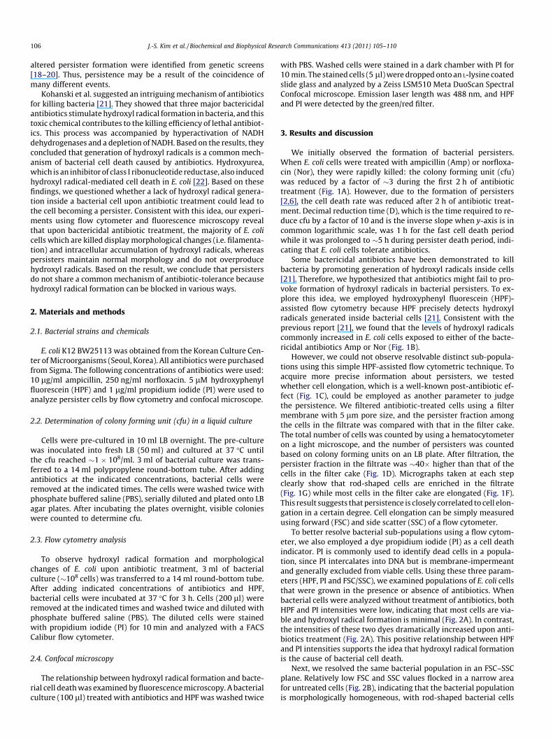
We initially observed the formation of bacterial persisters.When E. coli cells were treated with ampicillin (Amp) or norfloxa-cin (Nor), they were rapidly killed: the colony forming unit (cfu)was reduced by a factor of �3 during the first 2 h of antibiotictreatment (Fig. 1A). However, due to the formation of persisters[2,6], the cell death rate was reduced after 2 h of antibiotic treat-ment. Decimal reduction time (D), which is the time required to re-duce cfu by a factor of 10 and is the inverse slope when y-axis is incommon logarithmic scale, was 1 h for the fast cell death periodwhile it was prolonged to �5 h during persister death period, indi-cating that E. coli cells tolerate antibiotics.
Some bactericidal antibiotics have been demonstrated to killbacteria by promoting generation of hydroxyl radicals inside cells[21]. Therefore, we hypothesized that antibiotics might fail to pro-voke formation of hydroxyl radicals in bacterial persisters. To ex-plore this idea, we employed hydroxyphenyl fluorescein (HPF)-assisted flow cytometry because HPF precisely detects hydroxylradicals generated inside bacterial cells [21]. Consistent with theprevious report [21], we found that the levels of hydroxyl radicalscommonly increased in E. coli cells exposed to either of the bacte-ricidal antibiotics Amp or Nor (Fig. 1B).
However, we could not observe resolvable distinct sub-popula-tions using this simple HPF-assisted flow cytometric technique. Toacquire more precise information about persisters, we testedwhether cell elongation, which is a well-known post-antibiotic ef-fect (Fig. 1C), could be employed as another parameter to judgethe persistence. We filtered antibiotic-treated cells using a filtermembrane with 5 lm pore size, and the persister fraction amongthe cells in the filtrate was compared with that in the filter cake.The total number of cells was counted by using a hematocytometeron a light microscope, and the number of persisters was countedbased on colony forming units on an LB plate. After filtration, thepersister fraction in the filtrate was �40� higher than that of thecells in the filter cake (Fig. 1D). Micrographs taken at each stepclearly show that rod-shaped cells are enriched in the filtrate(Fig. 1G) while most cells in the filter cake are elongated (Fig. 1F).This result suggests that persistence is closely correlated to cell elon-gation in a certain degree. Cell elongation can be simply measuredusing forward (FSC) and side scatter (SSC) of a flow cytometer.
To better resolve bacterial sub-populations using a flow cytom-eter, we also employed a dye propidium iodide (PI) as a cell deathindicator. PI is commonly used to identify dead cells in a popula-tion, since PI intercalates into DNA but is membrane-impermeantand generally excluded from viable cells. Using these three param-eters (HPF, PI and FSC/SSC), we examined populations of E. coli cellsthat were grown in the presence or absence of antibiotics. Whenbacterial cells were analyzed without treatment of antibiotics, bothHPF and PI intensities were low, indicating that most cells are via-ble and hydroxyl radical formation is minimal (Fig. 2A). In contrast,the intensities of these two dyes dramatically increased upon anti-biotics treatment (Fig. 2A). This positive relationship between HPFand PI intensities supports the idea that hydroxyl radical formationis the cause of bacterial cell death.
Next, we resolved the same bacterial population in an FSC–SSCplane. Relatively low FSC and SSC values flocked in a narrow areafor untreated cells (Fig. 2B), indicating that the bacterial populationis morphologically homogeneous, with rod-shaped bacterial cells
Before Filter cake Filtrate
Rel
ativ
e su
rviv
al f
ract
oin
10-1
100
101
Time (hr)0 1 2 3 4 5 6
cfu/
ml
103
104
105
106
107
108
109
NorAmp
B
C
NoneAmp
Nor
None Amp
A
D
E
F
G
Fig. 1. Formation of persisters, hydroxyl radicals, and morphological changes upon antibiotic treatment. (A) Persister formation. (B) Flow cytometric analysis of hydroxylradical contents using HPF. (C) Filamention of E. coli after Amp treatment. (D) Filtration of Amp-treated cells. After treating E. coli with 5 lg/ml Amp for 3 h, cells were filteredusing the membrane with 5 lm pore size and colony forming units were counted. (E) Total cells before filtration are largely elongated but contain a few rod-shaped cells(arrows). (F) Morphology of cells in the filter cake. (G) Filtrate exhibited higher frequency of rod-shaped cells compared to cells before filtration.
J.-S. Kim et al. / Biochemical and Biophysical Research Communications 413 (2011) 105–110 107
(Fig. 2B). Both FSC and SSC values increased upon antibiotic treat-ment as a result of cell filamentation. While antibiotics treatmentshifted the majority of the bacterial population to the area withhigh FSC and SSC values (Fig. 2B), a small fraction of cells remainedwith the same FSC and SSC values as those of untreated cells(Fig. 2B, indicated as gate 1). When the cells in gate 1 were ana-lyzed for hydroxyl radical contents (Fig. 2C) and cell death(Fig. 2D), they showed a much lesser increase in both HPF and PIvalues than the majority of the population.
Fluorescence microscopy conducted on bacterial cells that werestained with HPF and PI further supports the flow cytometry data.Most of the E. coli cells were elongated and overproduced hydroxylradicals upon antibiotic treatment, and the micrographs for HPFand PI signals are superimposed perfectly (Fig. 3). Furthermore,we obtained a few images containing several rod-shaped cellsout of tens of micrographs, and these cells were not stained byHPF or PI (Fig. 3 and S1, indicated as arrows). Cumulatively, our re-sults suggest that bacterial persisters are the cells that do not over-produce hydroxyl radicals and retain normal morphology whilethe dead cells contain high levels of hydroxyl radicals and displayfilamentous morphology.
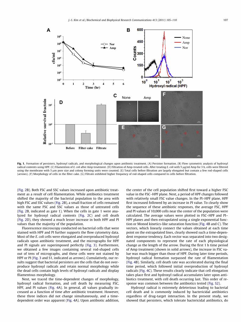
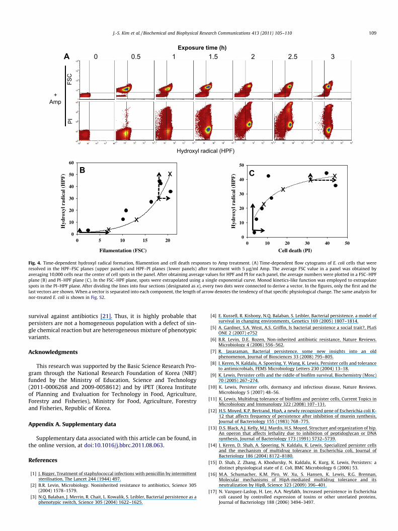
Next, we traced the time-dependent changes of morphology,hydroxyl radical formation, and cell death by measuring FSC,HPF, and PI values (Fig. 4A). In general, all values gradually in-creased as a function of time after antibiotic treatment. However,these three indices did not change simultaneously, and a time-dependent order was apparent (Fig. 4A). Upon antibiotic addition,
the center of the cell population shifted first toward a higher FSCvalue in the FSC–HPF plane. Next, a period of HPF changes followedwith relatively small FSC value changes. In the PI–HPF plane, HPFfirst increased followed by an increase in PI value. To clearly showthe sequence of these antibiotic responses, the average FSC, HPFand PI values of 10,000 cells near the center of the population werecalculated. The average values were plotted in FSC–HPF and PI–HPF planes and then extrapolated using a single exponential func-tion or Monod kinetics-like saturation function (Fig. 4B and C). Thevectors, which linearly connect the values obtained at each timepoint on the extrapolated lines, clearly showed such a time-depen-dent response tendency. Each vector was separated into two desig-nated components to represent the rate of each physiologicalchange as the length of the arrow. During the first 1 h time periodof Amp treatment (shown in solid arrows), the increase in FSC va-lue was much bigger than those of HPF. During later time periods,hydroxyl radical formation surpassed the rate of filamentation(Fig. 4B). Similarly, cell death rate was accelerated during the finaltime period, which followed initial overproduction of hydroxylradicals (Fig. 4C). These results clearly indicate that cell elongationtakes place first and hydroxyl radical accumulates later upon anti-biotics treatment, with cell death occurring last. This order of re-sponse was common between the antibiotics tested (Fig. S2).
Hydroxyl radical is extremely deleterious leading to bacterialcell death and is commonly induced by bactericidal antibioticsregardless of drug-target interaction. In the present study, weshowed that persisters, which tolerate bactericidal antibiotics, do
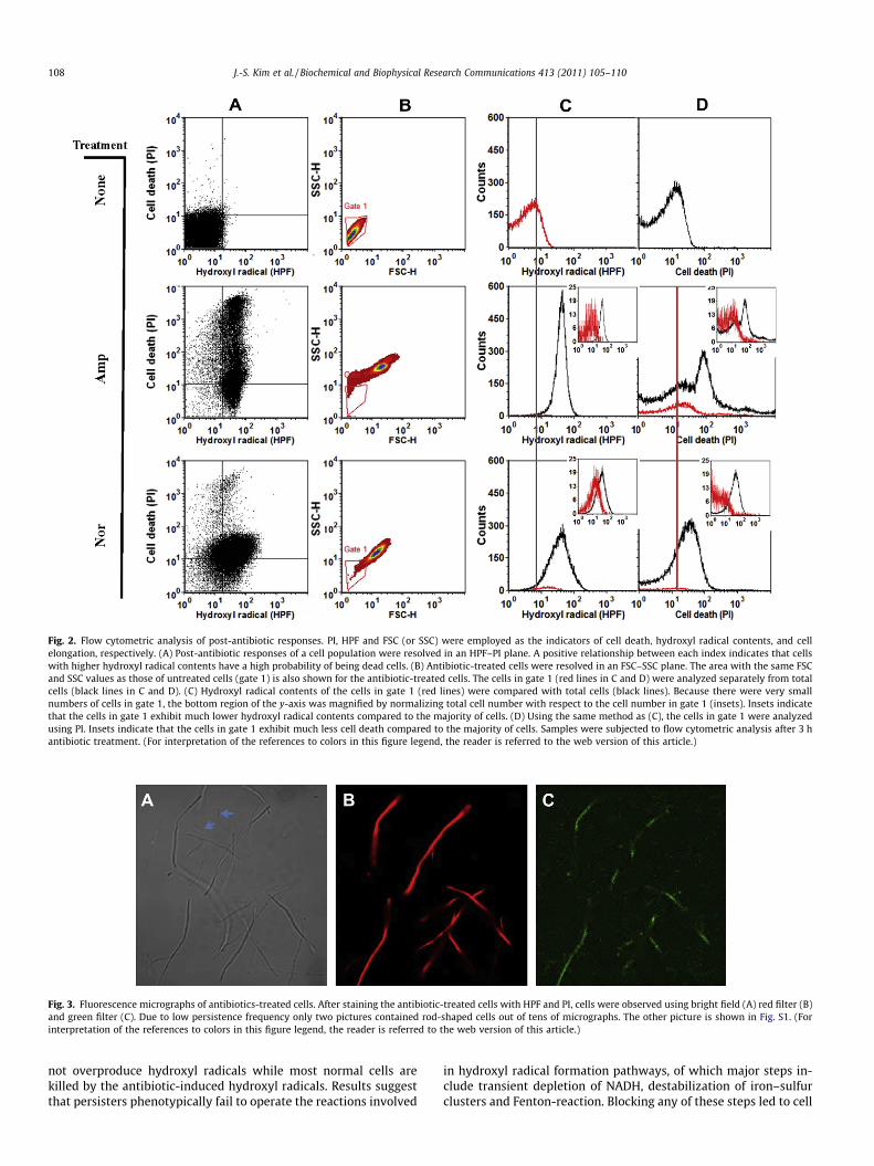
Fig. 2. Flow cytometric analysis of post-antibiotic responses. PI, HPF and FSC (or SSC) were employed as the indicators of cell death, hydroxyl radical contents, and cellelongation, respectively. (A) Post-antibiotic responses of a cell population were resolved in an HPF–PI plane. A positive relationship between each index indicates that cellswith higher hydroxyl radical contents have a high probability of being dead cells. (B) Antibiotic-treated cells were resolved in an FSC–SSC plane. The area with the same FSCand SSC values as those of untreated cells (gate 1) is also shown for the antibiotic-treated cells. The cells in gate 1 (red lines in C and D) were analyzed separately from totalcells (black lines in C and D). (C) Hydroxyl radical contents of the cells in gate 1 (red lines) were compared with total cells (black lines). Because there were very smallnumbers of cells in gate 1, the bottom region of the y-axis was magnified by normalizing total cell number with respect to the cell number in gate 1 (insets). Insets indicatethat the cells in gate 1 exhibit much lower hydroxyl radical contents compared to the majority of cells. (D) Using the same method as (C), the cells in gate 1 were analyzedusing PI. Insets indicate that the cells in gate 1 exhibit much less cell death compared to the majority of cells. Samples were subjected to flow cytometric analysis after 3 hantibiotic treatment. (For interpretation of the references to colors in this figure legend, the reader is referred to the web version of this article.)
Fig. 3. Fluorescence micrographs of antibiotics-treated cells. After staining the antibiotic-treated cells with HPF and PI, cells were observed using bright field (A) red filter (B)and green filter (C). Due to low persistence frequency only two pictures contained rod-shaped cells out of tens of micrographs. The other picture is shown in Fig. S1. (Forinterpretation of the references to colors in this figure legend, the reader is referred to the web version of this article.)
108 J.-S. Kim et al. / Biochemical and Biophysical Research Communications 413 (2011) 105–110
not overproduce hydroxyl radicals while most normal cells arekilled by the antibiotic-induced hydroxyl radicals. Results suggestthat persisters phenotypically fail to operate the reactions involved
in hydroxyl radical formation pathways, of which major steps in-clude transient depletion of NADH, destabilization of iron–sulfurclusters and Fenton-reaction. Blocking any of these steps led to cell
+Amp
Exposure time (h)0.5 1 1.5 2 2.5 30
Hydroxyl radical (HPF)
PIFS
C
Filamentation (FSC)
0 5 10 15 20
Hyd
roxy
l rad
ical
(H
PF
)
0
10
20
30
40
50
60
Cell death (PI)0 10 20 30 40 50
Hyd
roxy
l rad
ical
(H
PF
)0
10
20
30
40
50
A
B C
x
x
x
xx
x
x
x
Fig. 4. Time-dependent hydroxyl radical formation, filamention and cell death responses to Amp treatment. (A) Time-dependent flow cytograms of E. coli cells that wereresolved in the HPF–FSC planes (upper panels) and HPF–PI planes (lower panels) after treatment with 5 lg/ml Amp. The average FSC value in a panel was obtained byaveraging 10,000 cells near the center of cell spots in the panel. After obtaining average values for HPF and PI for each panel, the average numbers were plotted in a FSC–HPFplane (B) and PI–HPF plane (C). In the FSC–HPF plane, spots were extrapolated using a single exponential curve. Monod kinetics-like function was employed to extrapolatespots in the PI–HPF plane. After dividing the lines into four sections (designated as x), every two dots were connected to derive a vector. In the figures, only the first and thelast vectors are shown. When a vector is separated into each component, the length of arrow denotes the tendency of that specific physiological change. The same analysis fornor-treated E. coli is shown in Fig. S2.
J.-S. Kim et al. / Biochemical and Biophysical Research Communications 413 (2011) 105–110 109
survival against antibiotics [21]. Thus, it is highly probable thatpersisters are not a homogeneous population with a defect of sin-gle chemical reaction but are heterogeneous mixture of phenotypicvariants.
Acknowledgments
This research was supported by the Basic Science Research Pro-gram through the National Research Foundation of Korea (NRF)funded by the Ministry of Education, Science and Technology(2011-0006268 and 2009-0058612) and by iPET (Korea Instituteof Planning and Evaluation for Technology in Food, Agriculture,Forestry and Fisheries), Ministry for Food, Agriculture, Forestryand Fisheries, Republic of Korea.
Appendix A. Supplementary data
Supplementary data associated with this article can be found, inthe online version, at doi:10.1016/j.bbrc.2011.08.063.
References
[1] J. Bigger, Treatment of staphylococcal infections with penicillin by intermittentsterilisation, The Lancet 244 (1944) 497.
[2] B.R. Levin, Microbiology. Noninherited resistance to antibiotics, Science 305(2004) 1578–1579.
[3] N.Q. Balaban, J. Merrin, R. Chait, L. Kowalik, S. Leibler, Bacterial persistence as aphenotypic switch, Science 305 (2004) 1622–1625.
[4] E. Kussell, R. Kishony, N.Q. Balaban, S. Leibler, Bacterial persistence. a model ofsurvival in changing environments, Genetics 169 (2005) 1807–1814.
[5] A. Gardner, S.A. West, A.S. Griffin, Is bacterial persistence a social trait?, PLoSONE 2 (2007) e752
[6] B.R. Levin, D.E. Rozen, Non-inherited antibiotic resistance, Nature Reviews.Microbiology 4 (2006) 556–562.
[7] R. Jayaraman, Bacterial persistence. some new insights into an oldphenomenon, Journal of Biosciences 33 (2008) 795–805.
[8] I. Keren, N. Kaldalu, A. Spoering, Y. Wang, K. Lewis, Persister cells and toleranceto antimicrobials, FEMS Microbiology Letters 230 (2004) 13–18.
[9] K. Lewis, Persister cells and the riddle of biofilm survival, Biochemistry (Mosc)70 (2005) 267–274.
[10] K. Lewis, Persister cells, dormancy and infectious disease, Nature Reviews.Microbiology 5 (2007) 48–56.
[11] K. Lewis, Multidrug tolerance of biofilms and persister cells, Current Topics inMicrobiology and Immunology 322 (2008) 107–131.
[12] H.S. Moyed, K.P. Bertrand, HipA, a newly recognized gene of Escherichia coli K-12 that affects frequency of persistence after inhibition of murein synthesis,Journal of Bacteriology 155 (1983) 768–775.
[13] D.S. Black, A.J. Kelly, M.J. Mardis, H.S. Moyed, Structure and organization of hip.An operon that affects lethality due to inhibition of peptidoglycan or DNAsynthesis, Journal of Bacteriology 173 (1991) 5732–5739.
[14] I. Keren, D. Shah, A. Spoering, N. Kaldalu, K. Lewis, Specialized persister cellsand the mechanism of multidrug tolerance in Escherichia coli, Journal ofBacteriology 186 (2004) 8172–8180.
[15] D. Shah, Z. Zhang, A. Khodursky, N. Kaldalu, K. Kurg, K. Lewis, Persisters: adistinct physiological state of E. Coli, BMC Microbiology 6 (2006) 53.
[16] M.A. Schumacher, K.M. Piro, W. Xu, S. Hansen, K. Lewis, R.G. Brennan,Molecular mechanisms of HipA-mediated multidrug tolerance and itsneutralization by HipB, Science 323 (2009) 396–401.
[17] N. Vazquez-Laslop, H. Lee, A.A. Neyfakh, Increased persistence in Escherichiacoli caused by controlled expression of toxins or other unrelated proteins,Journal of Bacteriology 188 (2006) 3494–3497.

110 J.-S. Kim et al. / Biochemical and Biophysical Research Communications 413 (2011) 105–110
[18] Y. Hu, A.R. Coates, Transposon mutagenesis identifies genes which controlantimicrobial drug tolerance in stationary-phase Escherichia coli, FEMSmicrobiology letters 243 (2005) 117–124.
[19] A.L. Spoering, M. Vulic, K. Lewis, GlpD and PlsB participate in persister cellformation in Escherichia coli, Journal of Bacteriology 188 (2006) 5136–5144.
[20] Y. Li, Y. Zhang, PhoU is a persistence switch involved in persister formation andtolerance to multiple antibiotics and stresses in Escherichia coli, AntimicrobialAgents and Chemotherapy 51 (2007) 2092–2099.
[21] M.A. Kohanski, D.J. Dwyer, B. Hayete, C.A. Lawrence, J.J. Collins, A commonmechanism of cellular death induced by bactericidal antibiotics, Cell 130(2007) 797–810.
[22] B.W. Davies, M.A. Kohanski, L.A. Simmons, J.A. Winkler, J.J. Collins, G.C. Walker,Hydroxyurea induces hydroxyl radical-mediated cell death in Escherichia coli,Molecular Cell 36 (2009) 845–860.
Related Documents