*For correspondence: [email protected] Competing interest: See page 17 Funding: See page 17 Received: 15 June 2016 Accepted: 23 September 2016 Published: 24 September 2016 Reviewing editor: Richard M Berry, University of Oxford, United Kingdom Copyright Postel et al. This article is distributed under the terms of the Creative Commons Attribution License, which permits unrestricted use and redistribution provided that the original author and source are credited. Bacterial flagellar capping proteins adopt diverse oligomeric states Sandra Postel 1 , Daniel Deredge 2 , Daniel A Bonsor 1 , Xiong Yu 3 , Kay Diederichs 4 , Saskia Helmsing 5 , Aviv Vromen 6 , Assaf Friedler 6 , Michael Hust 5 , Edward H Egelman 3 , Dorothy Beckett 7 , Patrick L Wintrode 2 , Eric J Sundberg 1,8,9 * 1 Institute of Human Virology, University of Maryland School of Medicine, Baltimore, United States; 2 Department of Pharmaceutical Sciences, University of Maryland School of Pharmacy, Baltimore, United States; 3 Department of Biochemistry and Molecular Genetics, University of Virginia, Charlottesville, United States; 4 Department of Biology, University of Konstanz, Konstanz, Germany; 5 Department of Biotechnology, Institute of Biochemistry, Biotechnology and Bioinformatics, Technische Universita ¨ t Braunschweig, Braunschweig, Germany; 6 Institute of Chemistry, The Hebrew University of Jerusalem, Jerusalem, Israel; 7 Department of Chemistry and Biochemistry, University of Maryland College Park, Baltimore, United States; 8 Department of Medicine, University of Maryland School of Medicine, Baltimore, United States; 9 Department of Microbiology and Immunology, University of Maryland School of Medicine, Baltimore, United States Abstract Flagella are crucial for bacterial motility and pathogenesis. The flagellar capping protein (FliD) regulates filament assembly by chaperoning and sorting flagellin (FliC) proteins after they traverse the hollow filament and exit the growing flagellum tip. In the absence of FliD, flagella are not formed, resulting in impaired motility and infectivity. Here, we report the 2.2 A ˚ resolution X-ray crystal structure of FliD from Pseudomonas aeruginosa, the first high-resolution structure of any FliD protein from any bacterium. Using this evidence in combination with a multitude of biophysical and functional analyses, we find that Pseudomonas FliD exhibits unexpected structural similarity to other flagellar proteins at the domain level, adopts a unique hexameric oligomeric state, and depends on flexible determinants for oligomerization. Considering that the flagellin filaments on which FliD oligomers are affixed vary in protofilament number between bacteria, our results suggest that FliD oligomer stoichiometries vary across bacteria to complement their filament assemblies. DOI: 10.7554/eLife.18857.001 Introduction Pathogenic bacteria cause a multitude of deadly human diseases. Many of these microbes possess flagella, molecular machines responsible for cell motility, adherence to host cells, and pathogenicity (Duan et al., 2013; Haiko and Westerlund-Wikstrom, 2013). Flagella are helix-shaped hollow attachments formed predominantly by thousands of copies of the protein flagellin (also called FliC), anchored in the bacterial membrane by a hook (or joint) that is attached to the basal body and that is composed of rotary motor proteins (Arora et al., 1998). A proton motive force typically drives the propeller motion of flagella (Berg, 2003), resulting in swimming motility. A FliD (also called HAP2) oligomer forms the cap protein complex that is located at the tip of the flagellar filament (Yonekura et al., 2000). This complex controls the distal growth of the filament by regulating the Postel et al. eLife 2016;5:e18857. DOI: 10.7554/eLife.18857 1 of 20 RESEARCH ARTICLE

Welcome message from author
This document is posted to help you gain knowledge. Please leave a comment to let me know what you think about it! Share it to your friends and learn new things together.
Transcript

*For correspondence:
Competing interest: See
page 17
Funding: See page 17
Received: 15 June 2016
Accepted: 23 September 2016
Published: 24 September 2016
Reviewing editor: Richard M
Berry, University of Oxford,
United Kingdom
Copyright Postel et al. This
article is distributed under the
terms of the Creative Commons
Attribution License, which
permits unrestricted use and
redistribution provided that the
original author and source are
credited.
Bacterial flagellar capping proteins adoptdiverse oligomeric statesSandra Postel1, Daniel Deredge2, Daniel A Bonsor1, Xiong Yu3, Kay Diederichs4,Saskia Helmsing5, Aviv Vromen6, Assaf Friedler6, Michael Hust5,Edward H Egelman3, Dorothy Beckett7, Patrick L Wintrode2, Eric J Sundberg1,8,9*
1Institute of Human Virology, University of Maryland School of Medicine, Baltimore,United States; 2Department of Pharmaceutical Sciences, University of MarylandSchool of Pharmacy, Baltimore, United States; 3Department of Biochemistry andMolecular Genetics, University of Virginia, Charlottesville, United States;4Department of Biology, University of Konstanz, Konstanz, Germany; 5Departmentof Biotechnology, Institute of Biochemistry, Biotechnology and Bioinformatics,Technische Universitat Braunschweig, Braunschweig, Germany; 6Institute ofChemistry, The Hebrew University of Jerusalem, Jerusalem, Israel; 7Department ofChemistry and Biochemistry, University of Maryland College Park, Baltimore, UnitedStates; 8Department of Medicine, University of Maryland School of Medicine,Baltimore, United States; 9Department of Microbiology and Immunology, Universityof Maryland School of Medicine, Baltimore, United States
Abstract Flagella are crucial for bacterial motility and pathogenesis. The flagellar capping
protein (FliD) regulates filament assembly by chaperoning and sorting flagellin (FliC) proteins after
they traverse the hollow filament and exit the growing flagellum tip. In the absence of FliD, flagella
are not formed, resulting in impaired motility and infectivity. Here, we report the 2.2 A resolution
X-ray crystal structure of FliD from Pseudomonas aeruginosa, the first high-resolution structure of
any FliD protein from any bacterium. Using this evidence in combination with a multitude of
biophysical and functional analyses, we find that Pseudomonas FliD exhibits unexpected structural
similarity to other flagellar proteins at the domain level, adopts a unique hexameric oligomeric
state, and depends on flexible determinants for oligomerization. Considering that the flagellin
filaments on which FliD oligomers are affixed vary in protofilament number between bacteria, our
results suggest that FliD oligomer stoichiometries vary across bacteria to complement their
filament assemblies.
DOI: 10.7554/eLife.18857.001
IntroductionPathogenic bacteria cause a multitude of deadly human diseases. Many of these microbes possess
flagella, molecular machines responsible for cell motility, adherence to host cells, and pathogenicity
(Duan et al., 2013; Haiko and Westerlund-Wikstrom, 2013). Flagella are helix-shaped hollow
attachments formed predominantly by thousands of copies of the protein flagellin (also called FliC),
anchored in the bacterial membrane by a hook (or joint) that is attached to the basal body and that
is composed of rotary motor proteins (Arora et al., 1998). A proton motive force typically drives the
propeller motion of flagella (Berg, 2003), resulting in swimming motility. A FliD (also called HAP2)
oligomer forms the cap protein complex that is located at the tip of the flagellar filament
(Yonekura et al., 2000). This complex controls the distal growth of the filament by regulating the
Postel et al. eLife 2016;5:e18857. DOI: 10.7554/eLife.18857 1 of 20
RESEARCH ARTICLE
assembly of FliC molecules, which are transported through the hollow filament from the cytoplasm
to the tip of the flagellum.
The dynamic movement of FliD in this assembly was modeled based on low (~26 A)-resolution
cryo-electron microscopic (EM) structures of the Salmonella serovar Typhimurium flagellum-cap com-
plex (Maki-Yonekura et al., 2003; Yonekura et al., 2000, 2003), which adopts the shape of a five-
legged stool with flexible leg domains that regulate the assembly of new FliC molecules onto the tip
of the growing flagellum (Maki-Yonekura et al., 2003). It has been suggested that the plate of the
stool is formed by core regions of the FliD molecule, and that disordered/flexible regions form the
five leg structures (Vonderviszt et al., 1998) that are known to interact with the FliC filament. FliD
exhibits low sequence similarity to the flagellar hook proteins and to FliC. Nevertheless, it shares the
disordered terminal regions of these flagellar proteins, a common structural characteristic that is
thought to control the polymerization of flagellar proteins and to play an important role in interac-
tion with the FliC filament (Vonderviszt et al., 1998). These regions are the most conserved in FliD
sequences across bacteria. Flagellum-mediated motility is crucial for the virulence and pathogenicity
of numerous bacteria, including Campylobacter jejuni (Black et al., 1988), Salmonella (Allen-
Vercoe and Woodward, 1999; Marchetti et al., 2004), Escherichia coli (La Ragione et al., 2000),
Vibrio cholera (Krukonis and DiRita, 2003), and Pseudomonas aeruginosa (Arora et al., 2005), as
well as the major causative agent of gastric cancer Helicobacter pylori (Kim et al., 1999). To date,
however, no high-resolution structure of any FliD protein exists. To better define the roles of FliD in
bacterial motility and pathogenesis, we determined the first X-ray crystal structure of FliD at 2.2 A
resolution, and assessed the structural contributions of its flexible regions using a multitude of com-
plementary biophysical and functional analyses.
eLife digest Many bacteria, including several that cause diseases in people, have long whip-like
appendages called flagella that extend well beyond their cell walls. Flagella can rotate and propel
the bacteria through liquids, such as water or blood, and they are constructed primarily from
thousands of copies of a single protein called flagellin. When flagella are built, the flagellin proteins
are placed in their proper positions by another protein called FliD, several copies of which form a
cap on the end of flagella. Without FliD, bacteria cannot properly assemble flagella and, thus, can
no longer swim; this also hinders their ability to cause disease.
Determining the three-dimensional structure of a protein, down to the level of its individual
atoms, can provide unique insights into how the protein operates. However, no one had resolved
the structure of a FliD protein from any bacterium to this level of detail before.
Now, Postel et al. report the high-resolution structure of a large fragment of FliD from the
bacterium Pseudomonas aeruginosa. The structure reveals that parts of this FliD protein are shaped
like parts of other proteins from which flagella are constructed, including the flagellin protein that
FliD places into position. Some parts of the FliD protein are also very flexible and these parts of the
protein are responsible for holding numerous FliD proteins together as a cap. Finally, Postel et al.
saw that six copies of FliD bind to one another to form a protein complex on the end of flagella.
This last finding was particularly unexpected since it was thought that all FliD proteins formed five-
membered cap complexes, an assumption that was based largely on studies of FliD from another
bacterium called Salmonella.
The current structure covers about half of the FliD protein, and so the next challenge is to
determine the structure of the full-length protein. An improved understanding of the structure of
FliD may, in future, help researchers to design drugs that stop bacteria from building flagella and,
therefore, from swimming and causing disease.
DOI: 10.7554/eLife.18857.002
Postel et al. eLife 2016;5:e18857. DOI: 10.7554/eLife.18857 2 of 20
Research article Biophysics and Structural Biology Microbiology and Infectious Disease
Results
Crystal structure of the FliD protein from P. aeruginosa PAO1To facilitate crystallization of FliD from the P. aeruginosa PAO1 strain, we deleted the predicted
coiled-coil domains on both the N- and C-termini of full length FliD, which has 474 residues (FliD1–
474), to generate the truncated FliD78-405 (Figure 1a, Figure 1—source data 1). We expressed
FliD78–405 in E. coli with an N-terminal His6-tag and purified it to homogeneity by Ni2+-NTA, size
exclusion and anion exchange chromatography. We improved initially weakly diffracting crystals of
FliD78–405 by random matrix microseed screening (Bergfors, 2003), yielding crystals that diffracted
to 2.2 A resolution. In the absence of any homologous protein that could be used as a model for
molecular replacement, we crystallized a seleno-methionine derivative of FliD78–405 that included
four leucine-to-methionine mutations (FliD78–405/L4–M4). This crystal provided phase information suf-
ficient to build an initial model, which we used subsequently for molecular replacement with the
native FliD78–405 dataset (Figure 1—source data 1). We modeled residues 80–273 into clear electron
density, including all side chains, but observed density of increasingly poor quality in the C-terminus
beyond residue 273 (Figure 1—figure supplement 2a). Thus, we were able to model with
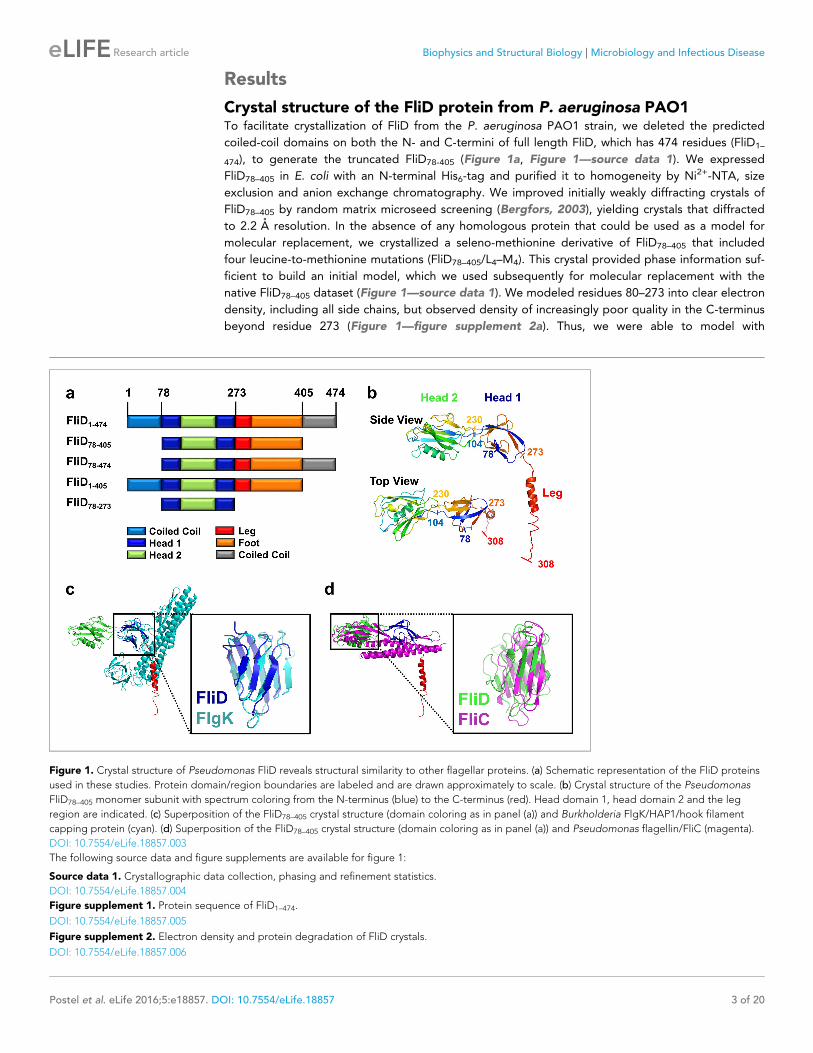
Figure 1. Crystal structure of Pseudomonas FliD reveals structural similarity to other flagellar proteins. (a) Schematic representation of the FliD proteins
used in these studies. Protein domain/region boundaries are labeled and are drawn approximately to scale. (b) Crystal structure of the Pseudomonas
FliD78–405 monomer subunit with spectrum coloring from the N-terminus (blue) to the C-terminus (red). Head domain 1, head domain 2 and the leg
region are indicated. (c) Superposition of the FliD78–405 crystal structure (domain coloring as in panel (a)) and Burkholderia FlgK/HAP1/hook filament
capping protein (cyan). (d) Superposition of the FliD78–405 crystal structure (domain coloring as in panel (a)) and Pseudomonas flagellin/FliC (magenta).
DOI: 10.7554/eLife.18857.003
The following source data and figure supplements are available for figure 1:
Source data 1. Crystallographic data collection, phasing and refinement statistics.
DOI: 10.7554/eLife.18857.004
Figure supplement 1. Protein sequence of FliD1–474.
DOI: 10.7554/eLife.18857.005
Figure supplement 2. Electron density and protein degradation of FliD crystals.
DOI: 10.7554/eLife.18857.006
Postel et al. eLife 2016;5:e18857. DOI: 10.7554/eLife.18857 3 of 20
Research article Biophysics and Structural Biology Microbiology and Infectious Disease

confidence only a single a helix in this region, corresponding to residues 274–308, with incomplete
side chain structures. To determine whether the remaining region of the protein actually existed in
the crystals and not just in the protein preparation used for crystallization, we analyzed crystals using
liquid chromatography-mass spectrometry (LC-MS) and SDS-PAGE. Both analyses indicated that the
crystals consisted of an approximate 50:50 mixture of the FliD78–405 protein used for crystallization
and a further proteolyzed version with a molecular weight of about 27 kDa. The N-terminal His6-tag
is still detectable by Western blot (Figure 1—figure supplement 2b). Thus, the proteolyzed form
corresponds approximately to residues 78–319 of FliD. The 86 residues absent from the C-terminus
in a population of FliD proteins are clearly not required for crystal packing, suggesting that they are
highly flexible even in a crystalline environment.
FliD is structurally similar on the domain level to FliC and FlgKOur crystal structure of FliD78–405 reveals that it consists of two discreet regions with distinct confor-
mational properties, corresponding to a stable head region and a flexible and/or disordered leg
region (Figure 1b). The head region is itself comprised of two separate, but entwined, protein
domains. Residues 80–101 form two b strands that belong to the first domain (Head 1), the second
domain (Head 2) is formed in its entirety by the contiguous residues 104–230, and residues 231–273
then complete the first domain. The second domain is, thus, a loop insertion of the first domain. We
searched for structural homologs of these domains in the Protein Data Bank and found that the first
domain of the head region (Head 1) exhibits high structural similarity (RMSD=2.5 A), despite low
sequence identity (14%), to the FlgK/HAP1/hook filament capping protein of Burkholderia pseudo-
mallei (PDB code 4UT1; Figure 1c). Likewise, the second head region domain (Head 2) exhibits high
structural similarity (RMSD=2.7 A), despite low sequence identity (17%), to the FliC/flagellin protein
of P. aeruginosa (PDB code 4NX9; Figure 1d). In contrast to the head region, the leg region of FliD
is highly flexible, as indicated by the paucity of electron density corresponding to residues 274–405
(Figure 1—figure supplement 2a). Despite this, we were able to model the initial a helical structural
element, corresponding to residues 274–308, of this region. This helix extends from the axis of the
head region at a nearly perpendicular angle, resulting in an L-shaped monomer subunit structure
(Figure 1b).
FliD from P. aeruginosa PAO1 forms a hexamerIn the crystal, FliD78–405 monomer subunits are arranged in hexamers, resulting in a shape akin to a
six-pointed star when viewed from the top of the FliD oligomer (Figure 2a), which corresponds to
the distal end of the growing flagellum. This star shape has a minimum inner diameter of 48 A and a
maximal outer diameter of 136 A. When viewed from the side (Figure 2b), the FliD hexamer appears
as a six-legged stool, the legs of which extend 55 A below the bottom of the head region. Addi-
tional crystallographic symmetry results in the stacking of hexamers in alternating head-to-head and
leg-to-leg orientations (Figure 2c). The leg-to-leg stacking forms dodecamers, resulting from the
helix–helix interaction of the residues 274–302 of stacked molecules and the interaction of residues
303–308 of one FliD78–405 molecule with Head 1 domain of a stacked molecule, burying a surface
area of 1362 A2. The formation of dodecamers may be unique to the FliD78–405 fragment, as this
strand could potentially be replaced by additional N-terminal residues in the full-length FliD1–474
protein. All of the morphologies observed for Pseudomonas FliD are highly reminiscent of the pen-
tamer/decamer oligomeric organization of Salmonella FliD as determined by low-resolution cryo-EM
analysis (Maki-Yonekura et al., 2003) (Figure 2d). Indeed, despite the difference in
the stoichiometries of the Pseudomonas and Salmonella FliD oligomers, the gross measurements are
nearly identical for these two proteins of similar molecular weight. In our crystal structure, Pseudo-
monas FliD measures 136 A in diameter with a head region that is 30 A deep and a leg region
that is 55 A long; whereas, in the cryo-EM structure, Salmonella FliD measures 145 A in diameter
with a head region that is 30 A deep and a leg region that is 55 A long (Maki-Yonekura et al.,
2003).
The stoichiometry of Pseudomonas FliD differs from that of Salmonella FliD and our Pseudomo-
nas FliD crystals belong to the P6 space group, which could possibly force a non-physiological oligo-
meric organization of subunits. Thus, we confirmed that the hexameric assembly of FliD78–405 occurs
not only in the crystalline environment but also in solution using negative stain EM class averaging
Postel et al. eLife 2016;5:e18857. DOI: 10.7554/eLife.18857 4 of 20
Research article Biophysics and Structural Biology Microbiology and Infectious Disease

(Tang et al., 2007), resulting in an image that clearly exhibits a hexameric assembly (Figure 3a).
These data are consistent with the oligomerization state that we detected in the crystal structure, in
which a view looking down the a-helices of the leg reveals a six-membered ring organization. We
also verified that both FliD78–405 and full-length FliD1–474 form oligomers by both analytical ultracen-
trifugation (AUC) and crosslinking analyses. We found that the FliD78–405 fragment that we crystal-
lized oligomerizes up to a dodecameric state (Figure 3b,c), similar to the crystallographic assembly
(Figure 2c). Additionally, we collected small-angle X-ray scattering (SAXS) data of FliD78–405, for
which the calculated radial distribution function (Figure 3d) is characteristic of an oligomeric assem-
bly forming a hollow sphere (Svergun and Koch, 2003). These data produce a molecular envelope
that superimposes well with our dodecameric FliD78–405 X-ray crystal structure (Figure 3d). Full-
length FliD1–474, by contrast, forms up to hexamers in solution (Figure 3e,f), which are likely to rep-
resent the physiologically relevant oligomerization state of this protein on the tip of the flagellum.
The predominant tetrameric species in solution identified by AUC may be a stable intermediate on
the path to hexamer formation (Figure 3e).
As the hexameric assembly of full-length Pseudomonas FliD1–474 proved to be unstable in solution
in the absence of the flagellar filament, we sought to stabilize it using our newfound understanding
of its structure. We used our crystal structure of FliD78–405, as input to Disulfide by Design 2.0
(Craig and Dombkowski, 2013), to identify cysteine mutations that would lead to a stable, disul-
fide-bridged hexameric FliD1–474. We found that when two residues within neighboring head domain
subunits, I167 and D253, were each mutated to a cysteine residue (Figure 4a) a stable, hexameric
full-length FliD1–474(I167C/D253C) resulted under non-reducing conditions, as shown by SDS-PAGE
Figure 2. Pseudomonas FliD forms hexamers in crystals. (a) Top view, cartoon representation of the FliD78–405 hexamer. Each monomer subunit is
colored distinctly and inner diameter dimension is indicated. (b) Side view, cartoon representation of the FliD78–405 hexamer. Each monomer subunit is
colored distinctly. Outer dimensions are indicated. (c) FliD78–405 hexamers as arranged in the crystal are stacked head-to-head and leg-to-leg (shown) in
an alternating fashion, with residues 303–308 assembling in the Head 1 domain of an opposing molecule (close-up views) leading to a dodecameric
crystal packing. (d) Cryo-EM structure of Salmonella FliD from (Maki-Yonekura et al., 2003) for comparison.
DOI: 10.7554/eLife.18857.007
Postel et al. eLife 2016;5:e18857. DOI: 10.7554/eLife.18857 5 of 20
Research article Biophysics and Structural Biology Microbiology and Infectious Disease
(Figure 4b) and SAXS (Figure 4c) analyses. We also confirmed that the cysteine bridges resulted in
the expected interfaces between head domains by employing mass spectrometry to compare the
peptide coverage under reducing and non-reducing conditions (Figure 4—figure supplement 1a)
and by successfully detecting the correct cysteine bridges (C167–C253) while ruling out non-specific
cysteine bridging (C167–C167 and C253–C253) (Figure 4—figure supplement 1b–e).
To show that the hexameric form of Pseudomonas FliD is functional in vivo, we complemented
the fliD transposon strain PW2975 (DfliD) with wildtype fliD1–474 and hexamer-stabilized fliD1–474
(I167C/D253C), resulting in Pseudomonas PAO1 strains DfliD/fliD1–474 and DfliD/fliD1–474(I167C/D253C),
respectively. We found that swimming motility that was lost in the DfliD strain was restored in both
DfliD/fliD1–474 and DfliD/fliD1–474(I167C/D253C) complementation strains, similar to our observations in
the wildtype Pseudomonas PAO1 strain (Figure 4d). Using antibodies that we generated by phage
display to Pseudomonas PAO1 FliD, we confirmed the expression of full-length FliD proteins by
Western blot analysis from preparations of flagella isolated from live bacteria from both DfliD/fliD1–
474 and DfliD/fliD1–474(I167C/D253C) complementation strains (Figure 4e). In flagella preparations from
the DfliD/fliD1–474(I167C/D253C) complementation strain, the FliD1–474(I167C/D253C) protein produced by
Pseudomonas maintains its hexameric oligomeric state (Figure 4e). In contrast, subsequent to fla-
gella isolation from live bacteria, FliD1–474 does not maintain a stable hexameric complex in either
the wildtype Pseudomonas PAO1 strain or in the DfliD/fliD1–474 complementation strain (Figure 4e).
The DfliD transposon strain does not form flagella, as indicated by the lack of FliC in the analyzed
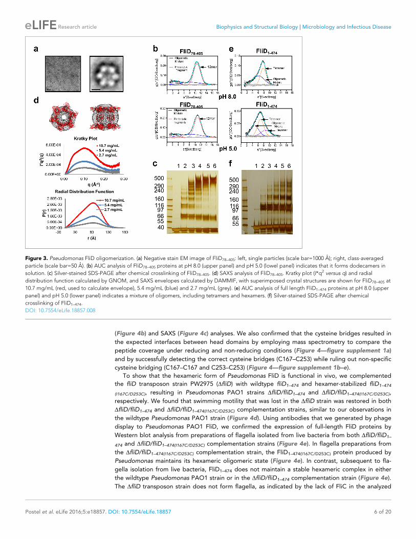
Figure 3. Pseudomonas FliD oligomerization. (a) Negative stain EM image of FliD78–405: left, single particles (scale bar=1000 A); right, class-averaged
particle (scale bar=50 A). (b) AUC analysis of FliD78–405 proteins at pH 8.0 (upper panel) and pH 5.0 (lowel panel) indicates that it forms dodecamers in
solution. (c) Silver-stained SDS-PAGE after chemical crosslinking of FliD78–405. (d) SAXS analysis of FliD78–405. Kratky plot (I*q2 versus q) and radial
distribution function calculated by GNOM, and SAXS envelopes calculated by DAMMIF, with superimposed crystal structures are shown for FliD78–405 at
10.7 mg/mL (red, used to calculate envelope), 5.4 mg/mL (blue) and 2.7 mg/mL (grey). (e) AUC analysis of full length FliD1–474 proteins at pH 8.0 (upper
panel) and pH 5.0 (lower panel) indicates a mixture of oligomers, including tetramers and hexamers. (f) Silver-stained SDS-PAGE after chemical
crosslinking of FliD1–474.
DOI: 10.7554/eLife.18857.008
Postel et al. eLife 2016;5:e18857. DOI: 10.7554/eLife.18857 6 of 20
Research article Biophysics and Structural Biology Microbiology and Infectious Disease
flagella preparations (lane 5 on the stained Western blot membrane, Figure 4—figure supplement
2). In comparison, the wildtype and all complementation strains form flagella as indicated by the
presence of flagellin/FliC in the purified flagella samples (Figure 4—figure supplement 2).
Together, these data indicate that FliD that is covalently locked in its hexameric assembly can form
functional flagella that allow Pseudomonas bacteria to swim like Pseudomonas with wildtype FliD.
Thus, the hexamer oligomeric state of Pseudomonas FliD is functional in vivo.
We also tested whether Salmonella FliD, which is known to form pentamers when capping the fla-
gellar filament, could function as a capping protein for Pseudomonas flagella. In contrast to the in
vivo functional hexameric forms of P. aeruginosa FliD–474, complementation of the PAO1 PW2975
transposon strain with fliD from Salmonella typhimurium (DfliD/fliDStyFliDe) did not restore swimming
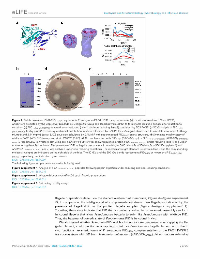
Figure 4. Stable hexameric DM1-FliD1–474 complements P. aeruginosa PAO1 dFliD transposon strain. (a) Location of residues I167 and D253,
which were predicted by the web server Disulfide by Design 2.0 (Craig and Dombkowski, 2013) to form stable disulfide bridges after mutation to
cysteines. (b) FliD1–474(I167C/D253C) analyzed under reducing (lane 1) and non-reducing (lane 2) conditions by SDS-PAGE. (c) SAXS analysis of FliD1–474
(I167C/D253C). Kratky plot (I*q2 versus q) and radial distribution function calculated by GNOM for 9.75 mg/mL (blue, used to calculate envelope), 4.88 mg/
mL (red) and 2.44 mg/mL (grey). SAXS envelope calculated by DAMMIF with superimposed FliD78–405 crystal structure. (d) Swimming motility assay of
wildtype PAO1 (WT), FliD transposon strain PW2975 (DfliD), DfliD complemented with FliD1–474 (DfliD/fliD1–474) or FliD1–474(I167C/D253C) (DfliD/fliD1–474(I167C/
D253C)), respectively. (e) Western blot using anti-FliD scFv-Fc SH1579-B7 showing purified protein FliD1–474(I167C/D253C) under reducing (lane 1) and under
non-reducing (lane 2) conditions. The presence of FliD in flagella preparations from wildtype PAO1 (lane 4), DfliD (lane 5), DfliD/fliD1–474(lane 6) and
DfliD/fliD1–474(I167C/D253C) (lane 7) was analyzed under non-reducing conditions. The molecular weight standard is shown in lane 3 and the corresponding
molecular weights are indicated on the right side of the blot. The 50 kDa and the 300 kDa bands representing FliD1–474 or hexameric FliD1–474(I167C/
D253C), respectively, are indicated by red arrows.
DOI: 10.7554/eLife.18857.009
The following figure supplements are available for figure 4:
Figure supplement 1. Analysis of FliD1–474(I167C/D253C) peptides following pepsin digestion under reducing and non-reducing conditions.
DOI: 10.7554/eLife.18857.010
Figure supplement 2. Western blot analysis of PAO1 strain flagella preparations.
DOI: 10.7554/eLife.18857.011
Figure supplement 3. Swimming motility assay.
DOI: 10.7554/eLife.18857.012
Postel et al. eLife 2016;5:e18857. DOI: 10.7554/eLife.18857 7 of 20
Research article Biophysics and Structural Biology Microbiology and Infectious Disease
motility (Figure 4—figure supplement 3). As this clone was codon-optimized for expression in
Escherichia coli, we also confirmed that a wildtype PAO1 full-length FliD1–474 encoded by a similarly
codon-optimized gene did restore swimming motility in the DfliD strain (DfliD/fliDPAOfliDe; Figure 4—
figure supplement 3). Although there exist many possible reasons other than oligomeric state that
could explain the inability of Salmonella FliD to functionally complement Pseudomonas, these data
suggest that Pseudomonas flagella may prefer FliD proteins that adopt hexameric rather than pen-
tameric states.
Regions outside of the head domains drive FliD oligomerizationAlthough the FliD78–405 crystal structure exhibits intermolecular contacts between the head regions
of FliD78–405 subunits comprising the hexamer, each of these interfaces is small, with a buried surface
area of only 665 A2, and contains few intermolecular contacts (Figure 5a) relative to typical protein–
protein interactions (Jones and Thornton, 1996). To determine whether these interactions were suf-
ficient to drive oligomerization of FliD, we expressed and purified the head region only, FliD78–273.
By AUC (Figure 5b), chemical crosslinking (Figure 5c) and SAXS analysis (Figure 5d), we observed
that when FliD lacks the leg region and the N- and C-terminal coiled-coil domains, it is present pre-
dominantly in the form of monomers (and dimers to a lesser extent) in solution, but fails to form
higher-order oligomers as do the longer versions of FliD that we analyzed. Because different buffer
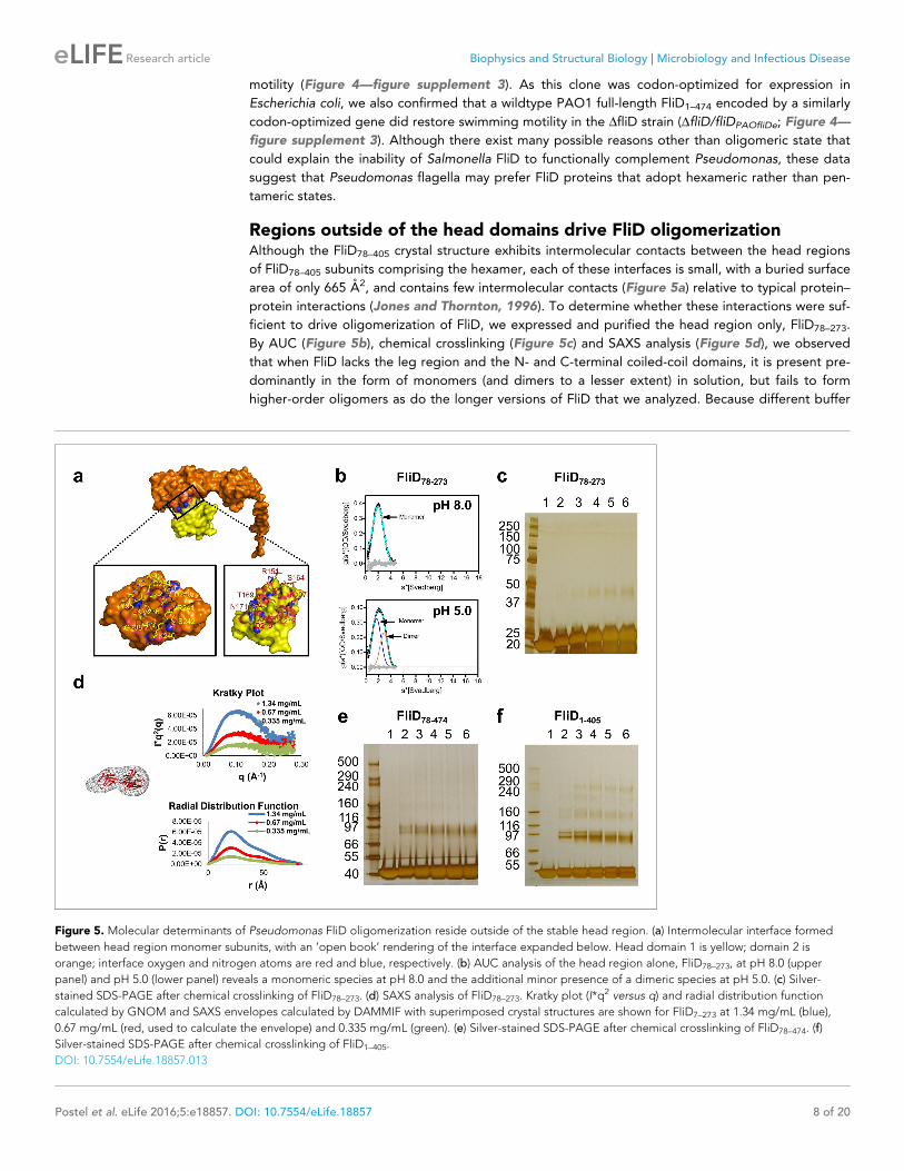
Figure 5. Molecular determinants of Pseudomonas FliD oligomerization reside outside of the stable head region. (a) Intermolecular interface formed
between head region monomer subunits, with an ’open book’ rendering of the interface expanded below. Head domain 1 is yellow; domain 2 is
orange; interface oxygen and nitrogen atoms are red and blue, respectively. (b) AUC analysis of the head region alone, FliD78–273, at pH 8.0 (upper
panel) and pH 5.0 (lower panel) reveals a monomeric species at pH 8.0 and the additional minor presence of a dimeric species at pH 5.0. (c) Silver-
stained SDS-PAGE after chemical crosslinking of FliD78–273. (d) SAXS analysis of FliD78–273. Kratky plot (I*q2 versus q) and radial distribution function
calculated by GNOM and SAXS envelopes calculated by DAMMIF with superimposed crystal structures are shown for FliD7–273 at 1.34 mg/mL (blue),
0.67 mg/mL (red, used to calculate the envelope) and 0.335 mg/mL (green). (e) Silver-stained SDS-PAGE after chemical crosslinking of FliD78–474. (f)
Silver-stained SDS-PAGE after chemical crosslinking of FliD1–405.
DOI: 10.7554/eLife.18857.013
Postel et al. eLife 2016;5:e18857. DOI: 10.7554/eLife.18857 8 of 20
Research article Biophysics and Structural Biology Microbiology and Infectious Disease

conditions, including changes in pH, have been shown to affect the polymerization states of flagellar
filaments (Shibata et al., 2005) and capping proteins (Imada et al., 1998), we performed additional
AUC experiments and found that FliD78–273 is entirely monomeric at pH 8.0 and becomes approxi-
mately one-third dimeric at pH 5.0; we observed no higher-order oligomers of FliD78–273 regardless
of buffer conditions (Figure 5b). We also assessed, by chemical cross-linking, the oligomerization
states of FliD variants lacking only the N-terminal coiled-coil domain (FliD78–474; Figure 5e) or the
C-terminal coiled-coil domain (FliD1–405; Figure 5f). We found them to be mainly monomeric with a
minority of species appearing to dimerize, although the latter exhibit weak higher-order oligomeriza-
tion potential. Kratky plots and radial distribution functions calculated from SAXS data of the variants
lacking either the N- or C-terminal coiled-coil domain, FliD78–474 or FliD1–405 respectively, reveal that
these proteins adopt extended shapes with flexible regions that are clearly represented in the result-
ing molecular envelopes (Figure 6a,b). These data indicate that the driving force for hexamerization
of Pseudomonas FliD resides in molecular determinants outside of the head region and, at a mini-
mum, involves residues in the N-terminal and C-terminal coiled-coil domains. FliD78–405 is lacking the
C-terminal and N-terminal coiled-coil domain but still assembles into dodecamers, as shown in the
crystal structure, cross-linking experiments and AUC, which is likely caused by strand replacement in
the head region domain 1 and helix–helix (residues 274–308) interaction of stacked molecules
(Figure 2c).
The N- and C-terminal regions of FliD are highly flexibleA large extent of FliD sequence currently remains inaccessible to high-resolution structural analysis,
including Pseudomonas FliD residues 1–79 and 309–474. Consequently, we performed hydrogen/
deuterium (H/D) exchange-mass spectrometry (HDX-MS) experiments with FliD78–405 to define its
solvent accessible regions and to evaluate its dynamic behavior. We subjected FliD78–405 to H/D
exchange for 10 s to 2 hr and observed that the head region (residues 80–273) of FliD78–405 adopts
a largely stable exchange-protected fold with greater stability observed for domain 2 relative to that
Figure 6. Small angle X-ray scattering (SAXS) data of FliD1–405, FliD78–474 and FliD1–474. Log-scale intensity SAXS profiles, Kratky Plot (I � q2 versus q),
radial distribution function calculated by GNOM and SAXS envelopes calculated by DAMMIF are shown for: (a) FliD1–405 at 10.4 mg/mL (blue), 5.2 mg/
mL (red) and 2.6 mg/mL (grey); (b) FliD78–474 at 9.9 mg/mL (blue), 4.95 mg/mL (red) and 2.5 mg/mL (grey); and (c) FliD1–474 at 11 mg/mL (blue), 5.5 mg/
mL (red), 2.7 mg/mL (grey), 1.38 mg/mL (green) and 0.69 mg/mL (yellow).
DOI: 10.7554/eLife.18857.014
The following figure supplement is available for figure 6:
Figure supplement 1. Analytical ultracentrifugation (AUC) analysis of FliD1–474 at pH 11.0.
DOI: 10.7554/eLife.18857.015
Postel et al. eLife 2016;5:e18857. DOI: 10.7554/eLife.18857 9 of 20
Research article Biophysics and Structural Biology Microbiology and Infectious Disease
of domain 1. The leg region, particularly residues C-terminal to the a helix observed in the crystal
structure, is more disordered or less stable (Figure 7a). Residues linking head domain 1 to the leg
helix display cooperative unfolding behavior as indicated by EX1 kinetics (Weis et al., 2006) that
result in double isotopic envelopes (Figure 7b). The FliD78–405 protein used in these experiments
was folded properly as shown by circular dichroism, as were all other FliD protein fragments that we
produced (Figure 7—figure supplement 1). When mapped to our crystal structure of FliD78–405, the
degree of H/D exchange over time on the peptide level corresponds to the degree of conforma-
tional stability on the residue level (Figure 7c). When we used a difference plot to compare the
extent of H/D exchange of full length FliD1–474 with that of FliD78–405, we observed that residues
165–225 exhibit relatively greater stability in FliD78–405 (Figure 8a). Within this stretch of residues in
head domain 2 are those residues, 165–171, which form the interface between the two head region
domains in the hexameric complex (Figure 8b). We observed an additional region of relative stabili-
zation for residues 298–324, part of which, residues 300–308, correspond to the b strand replace-
ment in the opposing head region domain 1 that drives dodecamer formation of FliD78–405
(Figure 8a). Kratky plots calculated from SAXS data of full-length FliD1–474 at pH 11, which is mono-
meric under these conditions (Figure 6—figure supplement 1), confirms the overall flexible nature
of this protein. Accordingly, heterogeneous molecular envelopes calculated from SAXS data col-
lected at different concentrations of FliD1–474 also exhibit significant conformational flexibility
(Figure 6c).
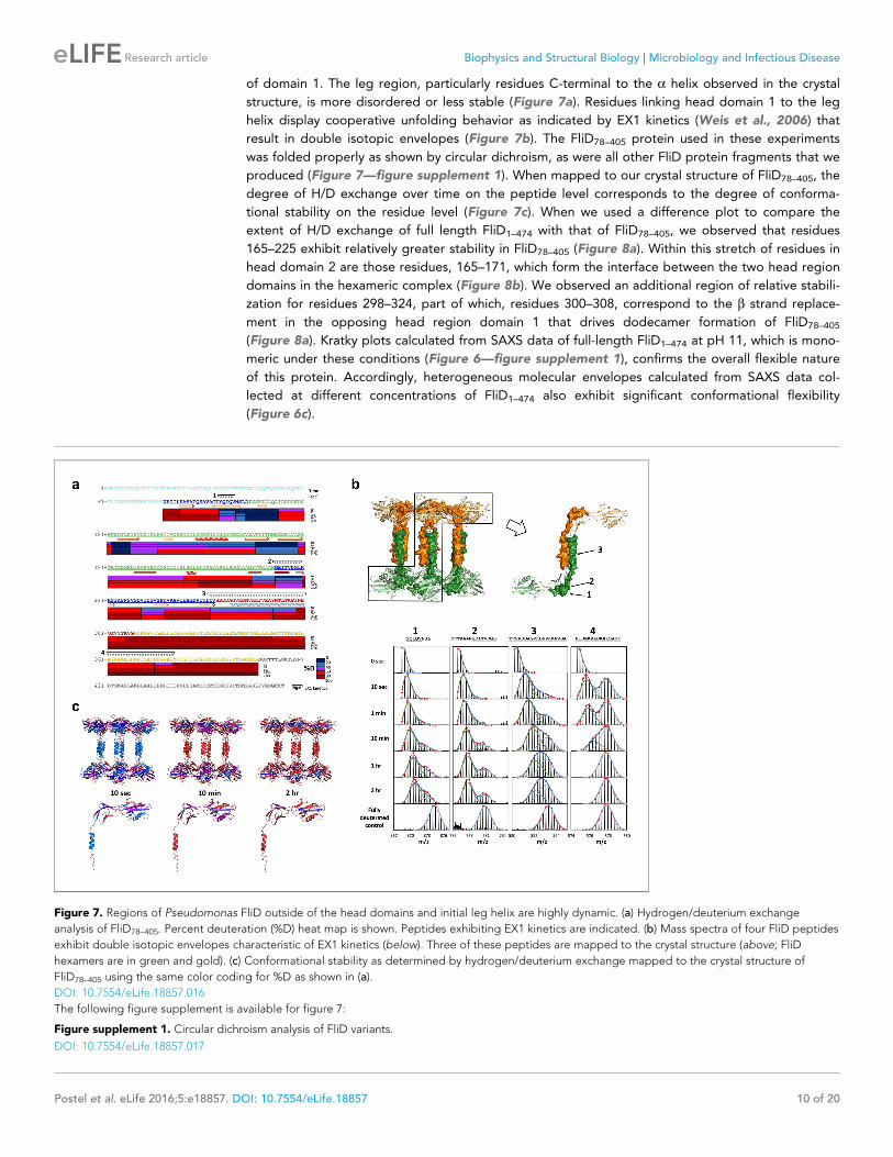
Figure 7. Regions of Pseudomonas FliD outside of the head domains and initial leg helix are highly dynamic. (a) Hydrogen/deuterium exchange
analysis of FliD78–405. Percent deuteration (%D) heat map is shown. Peptides exhibiting EX1 kinetics are indicated. (b) Mass spectra of four FliD peptides
exhibit double isotopic envelopes characteristic of EX1 kinetics (below). Three of these peptides are mapped to the crystal structure (above; FliD
hexamers are in green and gold). (c) Conformational stability as determined by hydrogen/deuterium exchange mapped to the crystal structure of
FliD78–405 using the same color coding for %D as shown in (a).
DOI: 10.7554/eLife.18857.016
The following figure supplement is available for figure 7:
Figure supplement 1. Circular dichroism analysis of FliD variants.
DOI: 10.7554/eLife.18857.017
Postel et al. eLife 2016;5:e18857. DOI: 10.7554/eLife.18857 10 of 20
Research article Biophysics and Structural Biology Microbiology and Infectious Disease
Multiple regions of FliD interact with one anotherTo gain further insight to the dynamics and interactions of the different regions of FliD, we performed
HDX-MS experiments with the remaining Pseudomonas FliD fragments that we had generated
(Figure 1a, Figure 1—figure supplement 1). HDX-MS heat maps show the overall high degree of
flexibility of FliD regions outside the head domain (Figure 8—figure supplement 1), and difference
plots of FliD truncations in comparison to the full length FliD1–474 indicate that the head region, and
more specifically head region domain 1, is stabilized by FliD regions outside of the head region
(Figure 8c). Indeed, the N-terminal coiled coil is responsible for this stabilization of head region
domain 1, in addition to stabilization of residues between the leg region a helix and the C-terminal
coiled coil (Figure 8c). Finally, the C-terminal coiled coil predominantly stabilizes residues in the
N-terminal coiled coil (Figure 8c). The stabilization of one FliD region by another is most likely to be
direct and intramolecular, rather than allosteric and intermolecular, as the full-length protein FliD1–474
adopts predominantly monomeric species under the conditions that we used for HDX-MS analysis
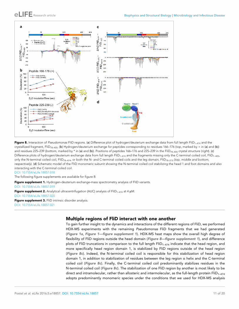
Figure 8. Interaction of Pseudomonas FliD regions. (a) Difference plot of hydrogen/deuterium exchange data from full length FliD1–474 and the
crystallized fragment, FliD78–405. (b) Hydrogen/deuterium exchange for peptides corresponding to residues 166–176 (top, marked by + in (a) and (b))
and residues 225–239 (bottom, marked by * in (a) and (b)). Positions of peptides 166–176 and 225–239 in the FliD78–405 crystal structure (right). (c)
Difference plots of hydrogen/deuterium exchange data from full length FliD1–474 and the fragments missing only the C-terminal coiled coil, FliD1–405,
only the N-terminal coiled coil, FliD78–474, or both the N- and C-terminal coiled coils and the leg domain, FliD78–273 (top, middle and bottom,
respectively). (d) Schematic model of the FliD monomeric subunit showing the N-terminal coiled coil stabilizing the head 1 and foot domains and also
interacting with the C-terminal coiled coil.
DOI: 10.7554/eLife.18857.018
The following figure supplements are available for figure 8:
Figure supplement 1. Hydrogen-deuterium exchange-mass spectrometry analysis of FliD variants.
DOI: 10.7554/eLife.18857.019
Figure supplement 2. Analytical ultracentrifugation (AUC) analysis of FliD1–474 at 4 mM.
DOI: 10.7554/eLife.18857.020
Figure supplement 3. FliD intrinsic disorder analysis.
DOI: 10.7554/eLife.18857.021
Postel et al. eLife 2016;5:e18857. DOI: 10.7554/eLife.18857 11 of 20
Research article Biophysics and Structural Biology Microbiology and Infectious Disease

(Figure 8—figure supplement 2). This leads to a transient structural model of the entire FliD mono-
mer subunit (Figure 8d) in which the N-terminal coiled coil bridges the head and foot regions and is
itself pinned to these structural elements by the C-terminal coiled coil. Notably, none of the flexible
regions of FliD, save the C-terminal 20 residues, are consistently predicted by sequence analysis to
be intrinsically disordered (Figure 8—figure supplement 3); rather, they are inherently capable of
adopting a limited number of conformations required to be in an ’on-state’ (actively chaperoning and
sorting a FliC protein) or an ’off-state’ (engaging only structured and previously positioned FliC
proteins).
DiscussionAt the gross structural level, our studies show that the oligomeric states differ between FliD protein
assemblies in diverse bacteria. Salmonella has long served as the model for bacterial flagellum struc-
ture and function. Since Salmonella FliD performs its native flagellar capping function as a pentamer
(Imada et al., 1998; Maki et al., 1998; Vonderviszt et al., 1998; Yonekura et al., 2000), it could
reasonably be assumed that all FliD proteins form pentamers at the distal ends of all bacterial fla-
gella. We found that Pseudomonas FliD instead forms hexamers both in crystals and in solution.
Additionally, we showed that Pseudomonas FliD constrained to its hexameric state by inter-subunit
disulfide bonds is functional in vivo, resulting in the formation of flagella and swimming motility.
Conversely, Salmonella FliD that assembles as pentamers does not allow flagellar formation and
swimming motility in Pseudomonas bacteria.
Much like cryo-EM studies of flagellar filaments from diverse bacteria unequivocally showed that
the number of protofilaments ranges, at least, from 11 in Salmonella (Yonekura et al., 2003) to
seven in Campylobacter (Galkin et al., 2008), our structural analyses show that the proteins that cap
these filaments also vary in their oligomeric states. The end of the Salmonella filament exhibits a
non-planar surface with five indentations (Yonekura et al., 2000) into which the five legs of the cor-
responding FliD pentamer have been modeled (Maki-Yonekura et al., 2003). Although no structure
of the Pseudomonas filament has been determined even at low resolution, our hexameric FliD struc-
ture suggests that it may incorporate an even greater number of protofilaments than does the Sal-
monella filament. This would allow the formation of an additional molecular cavity on the distal end
of FliD that accommodates its unique capping protein hexamer. Indeed, Pseudomonas flagellar fila-
ments have been found to be macroscopically different from those of Salmonella (Shibata et al.,
2005).
At 2.2 A resolution, our crystal structure reveals several previously unknown structural features of
FliD that are likely to be critical to its function. First, each one of the six FliD head regions that
appear as discreet lobes when visualized by EM analysis is actually composed of the first and second
domains of neighboring monomer subunits, as opposed to both domains belonging to the same
subunit. This is likely the case for FliD oligomers from diverse bacteria, including Salmonella FliD for
which cryo-EM analyses showed five head region lobes (Maki-Yonekura et al., 2003;
Yonekura et al., 2000, 2003). Second, despite vanishingly low sequence similarity, each domain
within the head region exhibits high structural similarity to the corresponding domains of other fla-
gellar proteins, including the hook-filament junction protein/FlgK and flagellin/FliC. Thus, at least
one protein domain from the proteins that occupy the distal ends of the hook (FlgK) and the cap
(FliD) adopts a common structural fold, which may be required for and unique to their function at
these related positions within the flagellum. This domain conservation among flagellar proteins con-
firms a previous study proposing a structural relationship of FliD to other flagellar proteins despite
their low sequence similarities (Vonderviszt et al., 1998). This structural conservation suggests that
a structurally similar cap complex may form at both hook and on the distal end. It also suggests that
FliD may play a functional role earlier in the flagellar assembly process, prior to its known
FliC sorting function. Notably, in mutants of Salmonella that lack filaments, FlgK (HAP1), FliD (HAP2)
and FlgL (HAP3) form a layered structure at the distal end of the hook-basal body (Homma and Iino,
1985). In addition, the shared domain structure of portions of FliC and FliD suggests that the
chaperoning activity of FliD could derive, at least in part, from its presentation of a like-structured
template against which FliC subunits may fold prior to being positioned into the growing filament.
Owing to the conformational flexibility of FliD necessitated by its FliC sorting function, we still
lack high-resolution structural information for certain regions of this protein. However, using a
Postel et al. eLife 2016;5:e18857. DOI: 10.7554/eLife.18857 12 of 20
Research article Biophysics and Structural Biology Microbiology and Infectious Disease

number of biophysical methods, we found that molecular determinants outside of the conformation-
ally stable head regions control FliD oligomerization. Previous analysis of a trypsinized fragment of
Salmonella FliD that lacks the N-terminal 42 residues and C-terminal 51 residues, which is similar to
the Pseudomonas FliD78–405 protein that we crystallized, showed that this fragment can form pen-
tamers but not decamers in solution and dissociates into smaller oligomers at low temperatures
(Vonderviszt et al., 1998). This suggests that determinants outside of the head regions of FliD pro-
teins of diverse bacteria control their oligomeric states. Accordingly, we showed that FliD, when con-
strained to its hexameric state by disulfide bonds formed between neighboring head domain
subunits, is fully functional in vivo. Thus,conformational flexibility in or relative repositioning of the
head regions are not functional requirements of FliD but flexibility in regions outside of the head
regions undoubtedly is. Considering that inhibiting protein–protein interfaces with small molecules
remains a major technical challenge (Arkin et al., 2014), the dependence of FliD oligomeric assem-
bly on its flexible regions enhances the prospects of developing small molecule inhibitors of FliD
oligomerization, and consequently of flagellar function, as a novel class of antibiotic agents.
Materials and methods
PlasmidsFor crystallization, the coding sequence optimized for expression in E. coli of FliD78–405 from the
PAO1 strain of P. aeruginosa was synthesized and cloned into the pET-28b vector (Novagen) with
the inclusion of an N-terminal His6-tag followed by a tobacco etch virus (TEV) protease recognition
site. A FliD78–405 mutant encoding four leucine to methionine mutations at positions L135, L239,
L347 and L350 (FliD78–405/L4-M4) was synthesized and likewise cloned into the pET-28b vector with
an N-terminal His6-tag. For all other experiments, FliD78–405, full-length FliD (FliD1 –474), head
domain-only FliD (FliD78 –273), and FliD lacking the N-terminal (FliD78 –474) or C-terminal (FliD1 –405)
coiled-coil domains were codon-optimized for expression in E. coli and cloned into pGEX5x2 in
frame with an N-terminal GST-tag followed by a TEV protease site. To obtain a stable hexameric
full-length FliD mutant (FliD1 –474(I167C/D253C)), two residues (I167 and D253) located at the interface
between neighboring head domains were mutated to cysteine residues in wildtype FliD1–474.
Recombinant protein expression and purificationAll FliD constructs from the P. aeruginosa PAO1 strain were expressed in LB medium for 4 hr at
37˚C in E. coli BL21(DE3)pLysS cells after induction with 1 mM IPTG at an OD600nm of 0.6. Seleno-
methionine (SeMet)-labeled FliD78 –405/L4-M4 was produced using metabolic inhibition of methionine
biosynthesis (Van Duyne et al., 1993) and growth in M9 medium containing 60 mg/L SeMet as the
sole source of methionine for 6 hr after induction with 1 mM IPTG. Cells were harvested (5000 g for
15 min) and lysed in PBS including 5 mM b-mercaptoethanol by sonication. For crystallization of His-
tagged FliD78 –405 and His-tagged FliD78 –405/L4-M4,the soluble fraction was purified using HisPur
NiNTA Resin (Thermo Scientific). The protein was further purified by size exclusion chromatography
(Superdex 200 10/300 GL, GE Healthcare) in PBS followed by anion exchange chromatography
(MonoQ 5/50 GL, GE Healthcare). For crystallization the protein was dialyzed into 30 mM Tris pH
8.0, 80 mM sodium chloride and concentrated to approximately 13 mg/mL. FliD-GST-fusion con-
structs were purified using a Glutathione Sepharose (BioVision) column. Following 16 hr digestion
with TEV protease, the GST tag was removed by Glutathione Sepharose and TEV was removed by
NiNTA (Thermo Scientific) chromatography. Cleaved FliD constructs were further purified using size
exclusion (Superdex 200 10/300 GL, GE Healthcare) in PBS followed by anion exchange chromatog-
raphy (MonoQ 5/50 GL, GE Healthcare) using 20 mM CHES pH 9.0 and a linear salt gradient from 0
to 1 M NaCl over 12 min.
Protein crystallizationCrystals of FliD78 –405 obtained in 0.25 M L-Arginine, 0.1 M Tris/HCl pH 8.0, 8% PGA diffracted
poorly and were subsequently used for random microseeding matrix screening (rMMS)
(D’Arcy et al., 2007). Improved crystals of FliD78 –405 were grown in 0.8 M NaK Tartrate, 0.1 M
Hepes pH 7.5 and diffracted to 2.2 A. Crystals of SeMet-labeled FliD78 –405/L4-M4 were also
obtained by employing rMMS with the initial, poorly diffracting crystals of FliD78 –405, which resulted
Postel et al. eLife 2016;5:e18857. DOI: 10.7554/eLife.18857 13 of 20
Research article Biophysics and Structural Biology Microbiology and Infectious Disease
in FliD78 –405/L4-M4 crystals grown in 1.5 M ammonium sulfate, 0.1 M Tris pH 8.5, 10% glycerol dif-
fracting to 3.6 A (anomalous signal cuttoff). Crystals were harvested and flash cooled in liquid nitro-
gen in mother liquor supplemented with 25% to 30% glycerol as cryo-protectant.
X-ray diffraction data processing, structure determination andrefinementX-ray diffraction data for the SeMet-labeled FliD78 –405/L4-M4 crystal were collected using a Dectris
6M PILATUS detector on the 12–2 beamline at the Stanford Synchrotron Radiation Lightsource,
SSRL, processed using XDS (Kabsch, 2010b), scaled in AIMLESS (Evans and Murshudov, 2013;
Winn et al., 2011), and phases obtained using the SSRL multi-wavelength anomalous dispersion
(MAD) script by A. Gonzalez with SHELX options based on a script by Qingping Xu, including the
programs SHELX (Schneider and Sheldrick, 2002), SOLVE (Terwilliger and Berendzen, 1999) and
RESOLV (Terwilliger, 2000). The initial FliD78 –405/L4-M4 model was improved manually by rebuilding
the peptide chain backbone in Coot (Emsley and Cowtan, 2004) and refining using Phenix
(Adams et al., 2010). Diffraction data for native, wildtype FliD78 –405 were collected using a MARmo-
saic 300 CCD detector on the 23ID-B beamline at the Advanced Photon Source, Argonne National
Laboratory, APS, and processed using XDS (Kabsch, 2010b) and XSCALE (Kabsch, 2010a). The par-
tially built and refined SeMet-FliD78 –405/L4-M4 model was used as a molecular replacement model
for phasing the native FliD78–405 data using Phaser (McCoy et al., 2007). The initial native FliD78–405
model was build using Autobuild and improved by manual model rebuilding in Coot (Emsley and
Cowtan, 2004) and by iterative rounds of refinement using Phenix (Adams et al., 2010).
Mass spectrometryFliD78–405 crystals were crosslinked using 2% formaldehyde, harvested and washed in mother liquor,
dissolved in water and the crosslinking reversed by heating the samples to 95˚C for 20 min. The sam-
ples were analyzed by liquid chromatography (LC)-electrospray ionization (ESI)-mass spectrometry
(MS) using a gradient of mobile phase A (0.1% formic acid in water) and mobile phase B (0.1% formic
acid in acetonitrile) increasing from 0% B to 90% B in 20 min. The Accela LC System was attached to
a LXQ linear ion trap mass spectrometer (Thermo Scientific). Raw MS data were analyzed using Xcali-
bus Qual Browser (Thermo Scientific) and deconvoluted using BioWorks (Thermo Scientific, Wal-
tham, MA).
Circular dichroism10 mM FliD protein in 10 mM sodium phosphate pH 7.0 was used to record a spectrum ranging
from 190 nm to 260 nm at 15˚C. CD melting curves were analyzed at 222 nm or 205 nm by increas-
ing the temperature by 1˚C per minute starting at 15˚C using a JASCO J810 CD instrument accord-
ing to the manufacturer’s instructions.
Electron microscopyAn aliquot of a FliD78–405 protein sample was negatively stained with 2% (weight/volume) uranyl ace-
tate and imaged using a Tecnai F20 (FEI) electron microscope operating at 120 keV. Approximately
3500 particles were selected from 70 micrographs and used to generate class averages in EMAN2
(Tang et al., 2007). Six classes were generated, and Figure 3a shows the single largest class.
Small angle X-ray scattering (SAXS)Small angel x-ray scattering data were collected using a dual Pilatus 100K-S SAXS/WAXS detector at
beamline G-1 of the Macromolecular Diffraction Facility at the Cornell High Energy Synchrotron
Source (MacCHESS). Scattering was measured in 30 mM Tris pH 8.0, 80 mM NaCl of FliD78–405 at
10.7 mg/mL, 5.4 mg/mL and 2.7 mg/mL, of FliD78–273 at 1.34 mg/mL, 0.67 mg/mL and 0.335 mg/
mL, of FliD1–405 at 10.4 mg/mL, 5.2 mg/mL and 2.6 mg/mL, of FliD78–474 at 9.9 mg/mL, 4.95 mg/mL
and 2.6 mg/mL and of FliD1–474(I167C/D253C) at 9.75 mg/ml, 4.88 mg/ml and 2.44 mg/ml. Scattering of
monomeric full-length FliD1–474 at 11 mg/mL, 5.5 mg/mL, 2.75 mg/mL, 1.38 mg/mL and 0.69 mg/mL
was measured in 20 mM CAPS pH 11.0, 80 mM NaCl. The SAXS data were processed using the Bio-
XTAS RAW software (Nielsen et al., 2009) and radial distribution functions calculated using GNOM
(Svergun, 1992). Molecular envelopes were generated using GASBOR (Svergun et al., 2001) and
Postel et al. eLife 2016;5:e18857. DOI: 10.7554/eLife.18857 14 of 20
Research article Biophysics and Structural Biology Microbiology and Infectious Disease

DAMMIF (Lammie et al., 2007). FoXS (Schneidman-Duhovny et al., 2010) was used to verify the
calculated intensity plots of the structures of the head domain FliD78–273 and the dodecameric
FliD78–405. The X-ray crystal structures of FliD78–273 and FliD78–405 were superimposed onto the
envelopes.
Hydrogen/deuterium exchange-mass spectrometryThe coverage maps for FliD1–474 and FliD78–405 were obtained from undeuterated controls as follows:
3.5 mL of ~40 mM FliD in 30 mM TrisHCl, 150 mM NaCl pH 8.0 was diluted with 31.5 mL of the same
buffer at room temperature followed by the addition of 100 mL of ice cold quench (100 mM Phos-
phate buffer, 1.5 M Guanidine-HCl, pH 2.4). The quenched samples were injected into a Waters
HDX nanoAcquity UPLC (Waters, Milford, MA) with in-line pepsin digestion (Waters Enzymate BEH
pepsin column). Peptic fragments were trapped on an Acquity UPLC BEH C18 peptide trap and sep-
arated on an Acquity UPLC BEH C18 column. A 7 min, 5% to 35% acetonitrile (0.1% formic acid) gra-
dient was used to elute peptides directly into a Waters Synapt G2 mass spectrometer (Waters,
Milford, MA). MSE data were acquired with a 20 to 30 V ramp trap CE for high energy acquisition of
product ions as well as continuous lock mass (Leu-Enk) for mass accuracy correction. Peptides were
identified using the ProteinLynx Global Server 2.5.1 (PLGS) from Waters. Further filtering of 0.3 frag-
ments per residues was applied in DynamX.
For each construct, the HD exchange reactions were performed as follows: 3.5 mL of ~40 mM FliD
in 30 mM TrisHCl, 150 mM NaCl pH 8.0 was incubated in 31.5 mL of 30 mM TrisDCl, 99.99% D2O,
pD 8.0, 150 mM NaCl. All reactions were performed at 25˚C. Prior to injection, deuteration reactions
were quenched at various times (10 s, 1 min, 10 min, 1 hr and 2 hr) with 100 mL of 100 mM Phos-
phate buffer, 1.5 Guanidine-HCl, pH 2.4. Back exchange correction was performed against fully deu-
terated controls acquired by incubating 3.5 mL of 40 mM FliD1–474 in 31.5 mL 30 mM TrisDCl, 99.99%
D2O, pD 8.0, 150 mM NaCl containing 6 M deuterated Guanidine DCl for 2 hr at 25˚C prior to
quenching (without guanidine HCl). All deuteration time points and controls were acquired in
triplicates.
The deuterium uptake by the identified peptides through increasing deuteration time and for the
fully deuterated control was determined using Water’s DynamX 2.0 software. The normalized per-
centage of deuterium uptake (%D) at an incubation time t for a given peptide was calculated as
follows:
%D¼100 � mt �m0ð Þ
mf �m0
With mt the centroid mass at incubation time t;m0 the centroid mass of the undeuterated control,
and mf the centroid mass of the fully deuterated control. Heat maps and percent deuteration differ-
ence plots (D%D) were generated using the percent deuteration calculated. Confidence intervals for
the D%D plots were determined using the method outlined by Houde et al. (2011), adjusted to per-
cent deuteration using the fully deuterated controls. Confidence intervals (98%) were plotted on the
D%D plots as horizontal dashed lines. EX1 type cooperative unfolding was analyzed using HX-
Express2 (Guttman et al., 2013).
Determination of peptide coverage of FliD1–474(I167C/D253C) underreducing and non-reducing conditionsCoverage maps of FliD1–474(I167C/D253C) in the presence and absence of reducing agent were
obtained similarly as above except for the following: 3 mL of 66 mM FliD1–474(I167C/D253C) were incu-
bated for 2 hr with 15 mL of 8 M Guanidine-HCl and 2 mL of 1 M TCEP (reducing conditions) or 2 mL
H2O (non-reducing conditions). Subsequently, 180 mL of quench buffer (100 mM potassium buffer,
pH 2.4) was added and the mixture immediately injected into the Waters HDX nanoAcquity UPLC.
The remainder of the workflow, MS method, peptide identification and coverage map determination
was unchanged. In addition, Biopharmalynx 1.3.5 (Waters) was used to search for and to identify
disulfide-bridged peptides. A filter of 15% b/y ions identified was applied. The search was per-
formed both in the context of the expected C167–C253 disulfide bridge and forthe C167–C167 and
C253–C253 disulfide bridges as negative controls.
Postel et al. eLife 2016;5:e18857. DOI: 10.7554/eLife.18857 15 of 20
Research article Biophysics and Structural Biology Microbiology and Infectious Disease

Analytical ultracentrifugationThe oligomeric states of FliD78–405, FliD78–273 and FliD1–474 in buffers containing 30 mM Tris, 80 mM
NaCl, pH 8.0 or 20 mM sodium citrate, 80 mM NaCl, pH 5.0 respectively, were analyzed by sedi-
mentation velocity using a Beckman-Coulter XL-I analytical ultracentrifuge equipped with a 4- or 8-
hole An-60Ti Rotor at 20˚C. SedenTerp (http://sednterp.unh.edu) was used to calculate protein par-
tial specific volumes and solvent densities and viscosities from the protein amino acid sequences and
buffer compositions. For sedimentation velocity measurements, samples of 295 mM (pH 8.0) or
325 mM (pH 5.0) FliD78–405, 124 mM FliD78–273, and 168 mM FliD1–474 were prepared in each buffer.
FliD1–474 was also analyzed by AUC at a low concentration of 4 mM in 30 mM Tris, 80 mM NaCl, pH
8.0 and at a concentration of 43 mM at high pH in 20 mM CAPS, 80 mM NaCl, pH 11.0. After
exhaustive dialysis to ensure chemical equilibrium, the samples were loaded into cells equipped with
2-hole charcoal-filled epon centerpieces (either 1.2 or 0.3 mm path length) with sapphire windows.
Prior to centrifugation, samples were equilibrated in the rotor for at least 2 hr at the desired experi-
mental temperature. Centrifugation was performed at 50,000 (FliD78–273), 40,000 (FliD1–474) or
30,000 (FliD78–273) rpm and scans were acquired at 280 nm. The resulting data were analyzed using
DCDT+ version 2.2.1 (Philo, 2006; Stafford, 1997, 1992) to determine the number of species, their
sedimentation coefficients, and their fractional contributions to the species populations. All sedimen-
tation coefficients were corrected to s20,w values.
Chemical crosslinkingApproximately 0.4 mg/mL of protein in 20 mM Hepes pH 8.0, 10 mM sodium chloride was cross-
linked using 20 mM 1-ethyl-3-(3-dimethylaminopropyl)carbodiimide hydrochloride (EDC) and 20 mM
N-hydroxysuccinimide (NHS) in 20 mM sodium phosphate, pH 7.0, 150 mM NaCl for various time
points. The reaction was stopped by the addition of 0.5 M Tris-HCl pH 8.0 to a final concentration of
0.25 M. The products were analyzed on NuPAGE 3–8% Tris-Acetate gels (Life
Technologies, Carlsabd, CA) or an Any kD Mini-PROTEAN TGX gels (BioRad) using a silver-staining
kit (Thermo Scientific).
Complementation of Pseudomonas aeruginosa PAO1Wildtype fliD1474 and fliD1474(I167C/D253C) were cloned into pUCP20 and transformed by electropora-
tion (Cadoret et al., 2014) into the DfliD transposon strain PW2975 (obtained from the Manoil Lab at
the University of Washington), resulting in the strains DfliD/fliD1–474 and DfliD/fliD1–474(I167C/
D253C), respectively. Wildtype fliD1–474 (fliD1–474e) and full-length fliD from Salmonella typhimurium
(fliDStyFliDe) both with genes codon-optimized for E. coli expression were also transformed into
PW2975 resulting in DfliD/fliD1–474e and DfliD/fliDStyFliDe, respectively.
Swimming motility assaysSwimming motility assays of Pseudomonas aeruginosa strains were performed as described by
Ha et al. (2014).
Isolation of flagella and FliD detectionPseudomonas aeruginosa PAO1 was grown overnight in LB liquid culture, cells were spun down and
resuspended in PBS. Flagella were sheared off the cells by passing the suspension through a 23
gauge needle approximately 25 times. After centrifugation, the supernatant containing flagella was
concentrated, proteins separated by SDS-PAGE and analyzed by Western blot using anti-FliD scFv-
Fc SH1579-B7 and an anti-human-IgG-HRP conjugate secondary antibody.
Generation of anti-FliD antibodiesHuman antibodies were generated as described by Frenzel et al. (2014). In brief, recombinant head
region only FliD78 –273 was immobilized on Costar High Binding plates and incubated with the Hyper-
phage packaged human antibody gene libraries HAL9/10 (Kugler et al., 2015) for negative selec-
tion. The non-binding scFv phages were incubated with recombinant full-length FliD1 –474 to select
binders specific for the leg region of FliD. In total, three panning rounds were performed and mono-
clonal antibodies were identified as described by Frenzel et al. (2014). The antibody SH1579-B7
Postel et al. eLife 2016;5:e18857. DOI: 10.7554/eLife.18857 16 of 20
Research article Biophysics and Structural Biology Microbiology and Infectious Disease

was recloned as an scFv-Fc (Yumab) with a human IgG1 Fc region and produced in mammalian cell
culture as described by Jager et al. (2013).
Intrinsic disorderThe sequence of the full-length FliD was submitted to twelve publicly available servers implementing
different algorithms for protein disorder prediction. In all cases, we used the default parameters.
The servers used were as follows: disEMBL (Linding et al., 2003a), GlobProt (Linding et al., 2003b),
IUPred (Dosztanyi et al., 2005), RONN (Yang et al., 2005), DisPro (Cheng et al., 2005), PONDR
(Romero et al., 2001), Spine-D (Zhang et al., 2012), OnD-CRF (Wang and Sauer, 2008), Foldindex
(Prilusky et al., 2005), MFDp (Mizianty et al., 2010), MFDp2 (Mizianty et al., 2010), and MD2
(Kozlowski and Bujnicki, 2012). Averaging of the results gave all servers equal weight.
Accession codeCoordinates and structure factors have been deposited in the Protein Data Bank under accession
code 5FHY.
AcknowledgementsWe thank the staff of the Stanford Synchrotron Radiation Lightsource (SSRL) beamline 12-2, SLAC
National Accelerator Laboratory, San Francisco, USA, of the Advanced Photon Source (APS) GM/CA
CAT beamline 23-ID-B, Argonne National Laboratory, Illinois, USA and of the beamline G-1 of the
Macromolecular Diffraction facility at Cornell High Energy Synchrotron Source (MacCHESS), Ithaca,
USA for their support. This work is supported in part by the University of Maryland Baltimore, School
of Pharmacy Mass Spectrometry Center (SOP1841-IQB2014).
Additional information
Competing interests
EHE: Reviewing editor, eLife. The other authors declare that no competing interests exist.
Funding
Funder Grant reference number Author
National Center for ResearchResources
NIH S10 RR15899 Dorothy Beckett
The funders had no role in study design, data collection and interpretation, or the decision tosubmit the work for publication.
Author contributions
SP, Designed the project, Performed experiments, Analysis of data, Writing the manuscript, Concep-
tion and design, Acquisition of data, Analysis and interpretation of data, Drafting or revising the arti-
cle; DD, Performed experiments, Analysis of data, Reviewed the manuscript, Acquisition of data,
Analysis and interpretation of data; DAB, KD, PLW, Analysis of data, Analysis and interpretation of
data; XY, MH, DB, Performed experiments, Analysis of data, Acquisition of data, Analysis and inter-
pretation of data; SH, Performing experiments, Acquisition of data; AV, AF, Intrinsic disorder predic-
tion, Analysis and interpretation of data; EHE, Performed experiments, Analysis of data, Analysis and
interpretation of data; EJS, Designed the project, Analysis of data, Writing the manuscript, Concep-
tion and design, Analysis and interpretation of data, Drafting or revising the article
Author ORCIDs
Sandra Postel, http://orcid.org/0000-0002-6717-1870
Edward H Egelman, http://orcid.org/0000-0003-4844-5212
Eric J Sundberg, http://orcid.org/0000-0003-0478-3033
Postel et al. eLife 2016;5:e18857. DOI: 10.7554/eLife.18857 17 of 20
Research article Biophysics and Structural Biology Microbiology and Infectious Disease

Additional files
Major datasets
The following dataset was generated:
Author(s) Year Dataset title Dataset URL
Database, license,and accessibilityinformation
Postel S, Bonsor D,Diederichs K,Sundberg EJ
2016 Crystal structure of FliD (HAP2) fromPseudomonas aeruginosa PAO1
http://www.rcsb.org/pdb/explore/explore.do?structureId=5FHY
Publicly available atthe RCSB ProteinData Bank (accessionno. 5FHY)
ReferencesAdams PD, Afonine PV, Bunkoczi G, Chen VB, Davis IW, Echols N, Headd JJ, Hung LW, Kapral GJ, Grosse-Kunstleve RW, McCoy AJ, Moriarty NW, Oeffner R, Read RJ, Richardson DC, Richardson JS, Terwilliger TC,Zwart PH. 2010. PHENIX: a comprehensive Python-based system for macromolecular structure solution. ActaCrystallographica Section D Biological Crystallography 66:213–221. doi: 10.1107/S0907444909052925
Allen-Vercoe E, Woodward MJ. 1999. The role of flagella, but not fimbriae, in the adherence of Salmonellaenterica serotype Enteritidis to chick gut explant. Journal of Medical Microbiology 48:771–780. doi: 10.1099/00222615-48-8-771
Arkin MR, Tang Y, Wells JA. 2014. Small-molecule inhibitors of protein-protein interactions: progressing towardthe reality. Chemistry & Biology 21:1102–1114. doi: 10.1016/j.chembiol.2014.09.001
Arora SK, Neely AN, Blair B, Lory S, Ramphal R. 2005. Role of motility and flagellin glycosylation in thepathogenesis of Pseudomonas aeruginosa burn wound infections. Infection and Immunity 73:4395–4398. doi:10.1128/IAI.73.7.4395-4398.2005
Arora SK, Ritchings BW, Almira EC, Lory S, Ramphal R. 1998. The Pseudomonas aeruginosa flagellar cap protein,FliD, is responsible for mucin adhesion. Infection and Immunity 66:1000–1007.
Berg HC. 2003. The rotary motor of bacterial flagella. Annual Review of Biochemistry 72:19–54. doi: 10.1146/annurev.biochem.72.121801.161737
Bergfors T. 2003. Seeds to crystals. Journal of Structural Biology 142:66–76. doi: 10.1016/S1047-8477(03)00039-X
Black RE, Levine MM, Clements ML, Hughes TP, Blaser MJ. 1988. Experimental Campylobacter jejuni infection inhumans. Journal of Infectious Diseases 157:472–479. doi: 10.1093/infdis/157.3.472
Cadoret F, Soscia C, Voulhoux R. 2014. Gene transfer: transformation/electroporation. Methods in MolecularBiology 1149:11–15. doi: 10.1007/978-1-4939-0473-0_2
Cheng J, Sweredoski MJ, Baldi P. 2005. Accurate prediction of protein disordered regions by mining proteinstructure data. Data Mining and Knowledge Discovery 11:213–222. doi: 10.1007/s10618-005-0001-y
Craig DB, Dombkowski AA. 2013. Disulfide by Design 2.0: a web-based tool for disulfide engineering in proteins.BMC Bioinformatics 14:346. doi: 10.1186/1471-2105-14-346
D’Arcy A, Villard F, Marsh M. 2007. An automated microseed matrix-screening method for protein crystallization.Acta Crystallographica Section D Biological Crystallography 63:550–554. doi: 10.1107/S0907444907007652
Dosztanyi Z, Csizmok V, Tompa P, Simon I. 2005. IUPred: web server for the prediction of intrinsicallyunstructured regions of proteins based on estimated energy content. Bioinformatics 21:3433–3434. doi: 10.1093/bioinformatics/bti541
Duan Q, Zhou M, Zhu L, Zhu G. 2013. Flagella and bacterial pathogenicity. Journal of Basic Microbiology 53:1–8.doi: 10.1002/jobm.201100335
Emsley P, Cowtan K. 2004. Coot: model-building tools for molecular graphics. Acta Crystallographica Section DBiological Crystallography 60:2126–2132. doi: 10.1107/S0907444904019158
Evans PR, Murshudov GN. 2013. How good are my data and what is the resolution? Acta CrystallographicaSection D Biological Crystallography 69:1204–1214. doi: 10.1107/S0907444913000061
Frenzel A, Kugler J, Wilke S, Schirrmann T, Hust M. 2014. Construction of human antibody gene libraries andselection of antibodies by phage display. Methods in Molecular Biology 1060:215–243. doi: 10.1007/978-1-62703-586-6_12
Galkin VE, Yu X, Bielnicki J, Heuser J, Ewing CP, Guerry P, Egelman EH. 2008. Divergence of quaternarystructures among bacterial flagellar filaments. Science 320:382–385. doi: 10.1126/science.1155307
Guttman M, Weis DD, Engen JR, Lee KK. 2013. Analysis of overlapped and noisy hydrogen/deuterium exchangemass spectra. Journal of the American Society for Mass Spectrometry 24:1906–1912. doi: 10.1007/s13361-013-0727-5
Ha DG, Kuchma SL, O’Toole GA. 2014. Plate-based assay for swimming motility in Pseudomonas aeruginosa.Methods in Molecular Biology 1149:59–65. doi: 10.1007/978-1-4939-0473-0_7
Haiko J, Westerlund-Wikstrom B. 2013. The role of the bacterial flagellum in adhesion and virulence. Biology 2:1242–1267. doi: 10.3390/biology2041242
Postel et al. eLife 2016;5:e18857. DOI: 10.7554/eLife.18857 18 of 20
Research article Biophysics and Structural Biology Microbiology and Infectious Disease

Homma M, Iino T. 1985. Locations of hook-associated proteins in flagellar structures of Salmonella typhimurium.Journal of Bacteriology 162:183–189.
Houde D, Berkowitz SA, Engen JR. 2011. The utility of hydrogen/deuterium exchange mass spectrometry inbiopharmaceutical comparability studies. Journal of Pharmaceutical Sciences 100:2071–2086. doi: 10.1002/jps.22432
Imada K, Vonderviszt F, Furukawa Y, Oosawa K, Namba K. 1998. Assembly characteristics of flagellar cap proteinHAP2 of Salmonella: decamer and pentamer in the pH-sensitive equilibrium. Journal of Molecular Biology 277:883–891. doi: 10.1006/jmbi.1998.1662
Jones S, Thornton JM. 1996. Principles of protein-protein interactions. PNAS 93:13–20. doi: 10.1073/pnas.93.1.13
Jager V, Bussow K, Wagner A, Weber S, Hust M, Frenzel A, Schirrmann T. 2013. High level transient productionof recombinant antibodies and antibody fusion proteins in HEK293 cells. BMC Biotechnology 13:52. doi: 10.1186/1472-6750-13-52
Kabsch W. 2010a. Integration, scaling, space-group assignment and post-refinement. Acta CrystallographicaSection D Biological Crystallography 66:133–144. doi: 10.1107/S0907444909047374
Kabsch W. 2010b. Xds. Acta Crystallographica Section D Biological Crystallography 66:125–132. doi: 10.1107/s0907444909047337
Kim JS, Chang JH, Chung SI, Yum JS. 1999. Molecular cloning and characterization of the Helicobacter pylori fliDgene, an essential factor in flagellar structure and motility. Journal of Bacteriology 181:6969–6976.
Kozlowski LP, Bujnicki JM. 2012. MetaDisorder: a meta-server for the prediction of intrinsic disorder in proteins.BMC Bioinformatics 13:111. doi: 10.1186/1471-2105-13-111
Krukonis ES, DiRita VJ. 2003. From motility to virulence: Sensing and responding to environmental signals inVibrio cholerae. Current Opinion in Microbiology 6:186–190. doi: 10.1016/S1369-5274(03)00032-8
Kugler J, Wilke S, Meier D, Tomszak F, Frenzel A, Schirrmann T, Dubel S, Garritsen H, Hock B, Toleikis L, SchutteM, Hust M. 2015. Generation and analysis of the improved human HAL9/10 antibody phage display libraries.BMC Biotechnology 15:10. doi: 10.1186/s12896-015-0125-0
La Ragione RM, Sayers AR, Woodward MJ. 2000. The role of fimbriae and flagella in the colonization, invasionand persistence of Escherichia coli O78:K80 in the day-old-chick model. Epidemiology and Infection 124:351–363. doi: 10.1017/S0950268899004045
Lammie D, Osborne J, Aeschlimann D, Wess TJ. 2007. Rapid shape determination of tissue transglutaminaseusing high-throughput computing. Acta Crystallographica Section D Biological Crystallography 63:1022–1024.doi: 10.1107/S0907444907032933
Linding R, Jensen LJ, Diella F, Bork P, Gibson TJ, Russell RB. 2003a. Protein disorder prediction: implications forstructural proteomics. Structure 11:1453–1459. doi: 10.1016/j.str.2003.10.002
Linding R, Russell RB, Neduva V, Gibson TJ. 2003b. GlobPlot: Exploring protein sequences for globularity anddisorder. Nucleic Acids Research 31:3701–3708. doi: 10.1093/nar/gkg519
Maki S, Vonderviszt F, Furukawa Y, Imada K, Namba K. 1998. Plugging interactions of HAP2 pentamer into thedistal end of flagellar filament revealed by electron microscopy. Journal of Molecular Biology 277:771–777. doi:10.1006/jmbi.1998.1663
Maki-Yonekura S, Yonekura K, Namba K. 2003. Domain movements of HAP2 in the cap-filament complexformation and growth process of the bacterial flagellum. PNAS 100:15528–15533. doi: 10.1073/pnas.2534343100
Marchetti M, Sirard JC, Sansonetti P, Pringault E, Kerneis S. 2004. Interaction of pathogenic bacteria with rabbitappendix M cells: bacterial motility is a key feature in vivo. Microbes and Infection 6:521–528. doi: 10.1016/j.micinf.2004.02.009
McCoy AJ, Grosse-Kunstleve RW, Adams PD, Winn MD, Storoni LC, Read RJ. 2007. Phaser crystallographicsoftware. Journal of Applied Crystallography 40:658–674. doi: 10.1107/S0021889807021206
Mizianty MJ, Stach W, Chen K, Kedarisetti KD, Disfani FM, Kurgan L. 2010. Improved sequence-based predictionof disordered regions with multilayer fusion of multiple information sources. Bioinformatics 26:i489–496. doi:10.1093/bioinformatics/btq373
Nielsen SS, Toft KN, Snakenborg D, Jeppesen MG, Jacobsen JK, Vestergaard B, Kutter JP, Arleth L. 2009.BioXTAS RAW , a software program for high-throughput automated small-angle X-ray scattering data reductionand preliminary analysis. Journal of Applied Crystallography 42:959–964. doi: 10.1107/S0021889809023863
Philo JS. 2006. Improved methods for fitting sedimentation coefficient distributions derived by time-derivativetechniques. Analytical Biochemistry 354:238–246. doi: 10.1016/j.ab.2006.04.053
Prilusky J, Felder CE, Zeev-Ben-Mordehai T, Rydberg EH, Man O, Beckmann JS, Silman I, Sussman JL. 2005.FoldIndex: a simple tool to predict whether a given protein sequence is intrinsically unfolded. Bioinformatics21:3435–3438. doi: 10.1093/bioinformatics/bti537
Romero P, Obradovic Z, Li X, Garner EC, Brown CJ, Dunker AK. 2001. Sequence complexity of disorderedprotein. Proteins 42:38–48. doi: 10.1002/1097-0134(20010101)42:1<38::AID-PROT50>3.0.CO;2-3
Schneider TR, Sheldrick GM. 2002. Substructure solution with SHELXD. Acta Crystallographica Section DBiological Crystallography 58:1772–1779. doi: 10.1107/S0907444902011678
Schneidman-Duhovny D, Hammel M, Sali A. 2010. FoXS: a web server for rapid computation and fitting of SAXSprofiles. Nucleic Acids Research 38:W540–544. doi: 10.1093/nar/gkq461
Shibata S, Alam M, Aizawa S. 2005. Flagellar filaments of the deep-sea bacteria Idiomarina loihiensis belong to afamily different from those of Salmonella typhimurium. Journal of Molecular Biology 352:510–516. doi: 10.1016/j.jmb.2005.07.023
Postel et al. eLife 2016;5:e18857. DOI: 10.7554/eLife.18857 19 of 20
Research article Biophysics and Structural Biology Microbiology and Infectious Disease
Stafford WF. 1992. Boundary analysis in sedimentation transport experiments: a procedure for obtainingsedimentation coefficient distributions using the time derivative of the concentration profile. AnalyticalBiochemistry 203:295–301. doi: 10.1016/0003-2697(92)90316-Y
Stafford WF. 1997. Sedimentation velocity spins a new weave for an old fabric. Current Opinion inBiotechnology 8:14–24. doi: 10.1016/S0958-1669(97)80152-8
Svergun DI, Koch MHJ. 2003. Small-angle scattering studies of biological macromolecules in solution. Reports onProgress in Physics 66:1735–1782. doi: 10.1088/0034-4885/66/10/R05
Svergun DI, Petoukhov MV, Koch MH. 2001. Determination of domain structure of proteins from X-ray solutionscattering. Biophysical Journal 80:2946–2953. doi: 10.1016/S0006-3495(01)76260-1
Svergun DI. 1992. Determination of the regularization parameter in indirect-transform methods using perceptualcriteria. Journal of Applied Crystallography 25:495–503. doi: 10.1107/S0021889892001663
Tang G, Peng L, Baldwin PR, Mann DS, Jiang W, Rees I, Ludtke SJ. 2007. EMAN2: an extensible imageprocessing suite for electron microscopy. Journal of Structural Biology 157:38–46. doi: 10.1016/j.jsb.2006.05.009
Terwilliger TC, Berendzen J. 1999. Automated MAD and MIR structure solution. Acta Crystallographica SectionD Biological Crystallography 55:849–861. doi: 10.1107/S0907444999000839
Terwilliger TC. 2000. Maximum-likelihood density modification. Acta Crystallographica Section D BiologicalCrystallography 56:965–972. doi: 10.1107/S0907444900005072
Van Duyne GD, Standaert RF, Karplus PA, Schreiber SL, Clardy J. 1993. Atomic structures of the humanimmunophilin FKBP-12 complexes with FK506 and rapamycin. Journal of Molecular Biology 229:105–124. doi:10.1006/jmbi.1993.1012
Vonderviszt F, Imada K, Furukawa Y, Uedaira H, Taniguchi H, Namba K. 1998. Mechanism of self-association andfilament capping by flagellar HAP2. Journal of Molecular Biology 284:1399–1416. doi: 10.1006/jmbi.1998.2274
Wang L, Sauer UH. 2008. OnD-CRF: predicting order and disorder in proteins using [corrected] conditionalrandom fields. Bioinformatics 24:1401–1402. doi: 10.1093/bioinformatics/btn132
Weis DD, Wales TE, Engen JR, Hotchko M, Ten Eyck LF. 2006. Identification and characterization of EX1 kineticsin H/D exchange mass spectrometry by peak width analysis. Journal of the American Society for MassSpectrometry 17:1498–1509. doi: 10.1016/j.jasms.2006.05.014
Winn MD, Ballard CC, Cowtan KD, Dodson EJ, Emsley P, Evans PR, Keegan RM, Krissinel EB, Leslie AG, McCoyA, McNicholas SJ, Murshudov GN, Pannu NS, Potterton EA, Powell HR, Read RJ, Vagin A, Wilson KS. 2011.Overview of the CCP4 suite and current developments. Acta Crystallographica Section D BiologicalCrystallography 67:235–242. doi: 10.1107/S0907444910045749
Yang ZR, Thomson R, McNeil P, Esnouf RM. 2005. RONN: the bio-basis function neural network techniqueapplied to the detection of natively disordered regions in proteins. Bioinformatics 21:3369–3376. doi: 10.1093/bioinformatics/bti534
Yonekura K, Maki S, Morgan DG, DeRosier DJ, Vonderviszt F, Imada K, Namba K. 2000. The bacterial flagellarcap as the rotary promoter of flagellin self-assembly. Science 290:2148–2152. doi: 10.1126/science.290.5499.2148
Yonekura K, Maki-Yonekura S, Namba K. 2003. Complete atomic model of the bacterial flagellar filament byelectron cryomicroscopy. Nature 424:643–650. doi: 10.1038/nature01830
Zhang T, Faraggi E, Xue B, Dunker AK, Uversky VN, Zhou Y. 2012. SPINE-D: accurate prediction of short andlong disordered regions by a single neural-network based method. Journal of Biomolecular Structure andDynamics 29:799–813. doi: 10.1080/073911012010525022
Postel et al. eLife 2016;5:e18857. DOI: 10.7554/eLife.18857 20 of 20
Research article Biophysics and Structural Biology Microbiology and Infectious Disease
Related Documents


