1 *iTero Element 5D is not yet available for sale in the US.*iTero Element 5D is currently available in European Union countries with exception in Switzerland and Norway. This clinical guide presents the promising features of the iTero Element 5D Imaging System designed with NIRI technology and its application into every day dentistry. NIRI technology of the iTero Element 5D aids in detection and monitoring of interproximal caries lesions above the gingiva without using harmful radiation. Author: Dr. Priyanka Keshav BDS, iTero Global Education Background In 2001, the National Institutes of Health Consensus Conference on the Diagnosis and Management of Dental Caries throughout life stated that “Dental caries is an infectious, communicable disease resulting in destruction of tooth structure by acid-forming bacteria found in dental plaque, an intraoral biofilm, in the presence of sugar. The infection results in the loss of tooth minerals that begins with the outer surface of the tooth and can progress through the dentin to the pulp, ultimately compromising the vitality of the tooth” 1 . Although largely preventable, dental caries is one of the two biggest threats to oral health and is amongst the most common chronic diseases in the United States. Dental caries is the most common chronic disease in children; it is about five times as common as asthma and seven times as common as hay fever 2 . Majorities of adults today live with untreated tooth decay in their permanent teeth; this makes the early detection of caries vital to identify and combat these pathological lesions in the early stages. The World Health Organization estimates that 60-90% of school children and nearly 100% of adults have or have had caries 3 . The concept of dental caries has changed significantly over the last decade. While the only way of managing caries used to be the complete removal of the demineralized tissues, today, caries is considered a dynamic process, which, if diagnosed in time, could be reversed. The current treatment philosophy is to prevent and detect dental disease at the earliest stage in order to avoid invasive treatment. With the current understanding of the nature of dental disease and its process, the treatment philosophy is now changing to a more conservative approach and the concept of minimal intervention is gaining popularity in modern dentistry throughout the world. Early caries detection is essential for minimal intervention dentistry because it could give the opportunity to reverse the process and eliminate or at least postpone the surgical treatment. The ideal caries detection device should be able to detect the caries from the earliest stages, when the organic matrix is still not damaged, to the latest stages of cavitated lesion 4 . Current conventional diagnostic methods rely mainly on visual, tactile methods paired with radiographs. Each of these methods have significant drawbacks; Visual examination is highly technique sensitive and subjective, and tactile methods of examination are unreliable for examining proximal areas due to lack of eye contact with the proximal surface itself and some studies have indicated that the tip of the probe may cause micro abrasions of the enamel or damage to areas of remineralization if present. Additonally, radiographs are known to expose the patient to harmful ionizing radiation present with technique sensitivity cannot be used frequently. New imaging technologies are in demand for the early detection of such lesions. Moreover, the treatment for early dental decay or caries is shifting away from aggressive cavity preparations that attempt aggressive removal of demineralized tooth structure toward non-surgical or minimally invasive restorative techniques 5 . Near infrared imaging technology Near Infrared Imaging serves as a valuable diagnostic aid in the early detection of interproximal caries. The near infrared (NIR) is the region of the electromagnetic spectrum between 0.7 to 2.0 micrometers (µm) 6 . The iTero Element 5D Imaging System uses light of wavelength (= 850nm) in the electromagnetic spectrum which on interaction with the hard tissue of the tooth provides additional data of its structure. Enamel is transparent to NIRI due to the reduced scattering co-efficient of light, allowing it to pass through its entire thickness and present as a dark area, whereas the dentin appears bright due to the scattering effect of light caused by the orientation of the dentinal tubules, any interferences/pathological lesions/ areas of demineralization appear as bright areas in a NIRI image due to the increased scattering within the region. iTero Element 5D Imaging System is an innovative integrated optical diagnostic aid (uses class 1 laser) and is the first 3D intraoral scanner with NIRI technology. With one scan, it is possible to view multiple layers of data: 3D model, 2D color images and NIRI images mapped to the 3D model. The user can rotate a 3D model of the teeth on the computer monitor and without looking at the patient to evaluate it from different angles and review the corresponding color and NIRI images at the same time to gather a comprehensive view of the situation. The system digitally captures the 3D geometry and color of the patient's intraoral dental structures using a proprietary optical, non-contact, focus detection technique. The device also includes capabilities of NIRI function that captures data beneath the tooth surface using NIRI illumination during routine scanning. Incorporating both the NIRI images and the color images captured by the system can aid in the detection of caries. Images are available in real time on the screen, can be enlarged, and contrasts can be adjusted based on preference. Additionally, scans can be saved and viewed later as desired or paired with tools such as TimeLapse to monitor areas of interest. Optical methods have the advantage that they do not use ionizing radiation. For this reason, these procedures can be used as often as desired to monitor caries. Several clinical studies have showed NIRI sensitivity to be as potent as radiographic examinations and are well suited for the detection and imaging of interproximal caries 7 . Near infrared imaging (NIRI) technology in dentistry - iTero Element 5D.

Welcome message from author
This document is posted to help you gain knowledge. Please leave a comment to let me know what you think about it! Share it to your friends and learn new things together.
Transcript

1*iTero Element 5D is not yet available for sale in the US.*iTero Element 5D is currently available in European Union countries with exception in Switzerland and Norway.
This clinical guide presents the promising features of the iTero Element 5D Imaging System designed with NIRI technology and its application into every day dentistry. NIRI technology of the iTero Element 5D aids in detection and monitoring of interproximal caries lesions above the gingiva without using harmful radiation. Author: Dr. Priyanka Keshav BDS, iTero Global Education
BackgroundIn 2001, the National Institutes of Health Consensus Conference on the Diagnosis and Management of Dental Caries throughout life stated that “Dental caries is an infectious, communicable disease resulting in destruction of tooth structure by acid-forming bacteria found in dental plaque, an intraoral biofilm, in the presence of sugar. The infection results in the loss of tooth minerals that begins with the outer surface of the tooth and can progress through the dentin to the pulp, ultimately compromising the vitality of the tooth”1.
Although largely preventable, dental caries is one of the two biggest threats to oral health and is amongst the most common chronic diseases in the United States. Dental caries is the most common chronic disease in children; it is about five times as common as asthma and seven times as common as hay fever2. Majorities of adults today live with untreated tooth decay in their permanent teeth; this makes the early detection of caries vital to identify and combat these pathological lesions in the early stages. The World Health Organization estimates that 60-90% of school children and nearly 100% of adults have or have had caries3.
The concept of dental caries has changed significantly over the last decade. While the only way of managing caries used to be the complete removal of the demineralized tissues, today, caries is considered a dynamic process, which, if diagnosed in time, could be reversed. The current treatment philosophy is to prevent and detect dental disease at the earliest stage in order to avoid invasive treatment. With the current understanding of the nature of dental disease and its process, the treatment philosophy is now changing to a more conservative approach and the concept of minimal intervention is gaining popularity in modern dentistry throughout the world. Early caries detection is essential for minimal intervention dentistry because it could give the opportunity to reverse the process and eliminate or at least postpone the surgical treatment. The ideal caries detection device should be able to detect the caries from the earliest stages, when the organic matrix is still not damaged, to the latest stages of cavitated lesion4. Current conventional diagnostic methods rely mainly on visual, tactile methods paired with radiographs. Each of these methods have significant drawbacks; Visual examination is highly technique sensitive and subjective, and tactile methods of examination are unreliable for examining proximal areas due to lack of eye contact with the proximal surface itself and some studies have indicated that the tip of the probe may cause micro abrasions of the enamel or damage to areas of remineralization if present.
Additonally, radiographs are known to expose the patient to harmful ionizing radiation present with technique sensitivity cannot be used frequently. New imaging technologies are in demand for the early
detection of such lesions. Moreover, the treatment for early dental decay or caries is shifting away from aggressive cavity preparations that attempt aggressive removal of demineralized tooth structure toward non-surgical or minimally invasive restorative techniques5.
Near infrared imaging technologyNear Infrared Imaging serves as a valuable diagnostic aid in the early detection of interproximal caries. The near infrared (NIR) is the region of the electromagnetic spectrum between 0.7 to 2.0 micrometers (µm)6. The iTero Element 5D Imaging System uses light of wavelength (= 850nm) in the electromagnetic spectrum which on interaction with the hard tissue of the tooth provides additional data of its structure. Enamel is transparent to NIRI due to the reduced scattering co-efficient of light, allowing it to pass through its entire thickness and present as a dark area, whereas the dentin appears bright due to the scattering effect of light caused by the orientation of the dentinal tubules, any interferences/pathological lesions/areas of demineralization appear as bright areas in a NIRI image due to the increased scattering within the region.
iTero Element 5D Imaging System is an innovative integrated optical diagnostic aid (uses class 1 laser) and is the first 3D intraoral scanner with NIRI technology. With one scan, it is possible to view multiple layers of data: 3D model, 2D color images and NIRI images mapped to the 3D model. The user can rotate a 3D model of the teeth on the computer monitor and without looking at the patient to evaluate it from different angles and review the corresponding color and NIRI images at the same time to gather a comprehensive view of the situation. The system digitally captures the 3D geometry and color of the patient's intraoral dental structures using a proprietary optical, non-contact, focus detection technique.
The device also includes capabilities of NIRI function that captures data beneath the tooth surface using NIRI illumination during routine scanning. Incorporating both the NIRI images and the color images captured by the system can aid in the detection of caries. Images are available in real time on the screen, can be enlarged, and contrasts can be adjusted based on preference. Additionally, scans can be saved and viewed later as desired or paired with tools such as TimeLapse to monitor areas of interest.
Optical methods have the advantage that they do not use ionizing radiation. For this reason, these procedures can be used as often as desired to monitor caries. Several clinical studies have showed NIRI sensitivity to be as potent as radiographic examinations and are well suited for the detection and imaging of interproximal caries7.
Near infrared imaging (NIRI) technology in dentistry - iTero Element 5D.

2*iTero Element 5D is not yet available for sale in the US.*iTero Element 5D is currently available in European Union countries with exception in Switzerland and Norway.
LiteratureNumerous studies have been conducted concerning near infrared imaging that can be traced back to the early 1990s. Some noteworthy articles have been mentioned as follows:
1. Fried D, Glena RE, Featherstone JD, Seka W. Nature of light scattering in dental enamel and dentin at visible and nearinfrared wavelengths. Applied Optics. 1995;34(7):127812868
Objective: In this study, Fried et al. measured the optical properties of fully index-matched samples of enamel and dentin as a step in calculating the distribution of deposited energy in teeth. The light-scattering properties of dental enamel and dentin were measured at 543, 632, and 1053 nm between 0° and 180° in appropriate index-matching baths. From the measured distributions and comparison with Monte Carlo 1MC2 simulations of light scattering in these tissues, the optical coefficients, the nature of the phase function, and the scattering anisotropy were derived for dentin and enamel at these wavelengths.
Results: In the visible and NIR wavelengths, dentin and enamel weakly absorb light, and light scattering plays an important role in determining the deposited energy distribution in the tissue. The scattering and absorption coefficients of enamel compare favorably with literature values measured using an integrating sphere. The measured scattering and absorption coefficients of dentin are both almost an order of magnitude larger than for enamel. Preliminary, two-dimensional, spatially resolved MC simulations using the optical parameters determined in this study indicate that the use of visible and NIR laser beams of, 1-mm diameter on the enamel surface may lead to preferential energy deposition near the dentin–enamel interface. This may have negative consequences such as subsurface heating and cracking.
Relevance: Use of NIRI has been studied in enamel, which shows high transparency. There is published data available regarding this technology in teeth, and more specifically in enamel and dentin. There is substantial evidence dating from 1990 for the potential use of NIR light for detecting caries in enamel, due to its high transparency when illuminated by Near Infra-Red light.
2. Comparison of diagnostic methods for early interproximal caries detection with near-infrared light transillumination: an in vivo study Ismail Hakki Baltacioglu and Kaan Orhan9
Background: Although numerous studies have used digital intraoral imaging, only a few studies have used photo-optical methods for the diagnosis of caries. Moreover, several limitations exist in terms of observers (experience and specialty) and the caries lesion itself. Hence, the aims of this study were to evaluate the diagnostic capability of near-infrared light transillumination
(NILT) and PSP-Bitewing radiographs and to compare the interobserver and intraobserver differences in addition to observers’ experience level to detect early interproximal caries lesions in vivo
Methods: A total of 52 untreated posterior teeth with and without varying degrees of early interproximal carious lesions were included. Bitewing radiographs using digital phosphor plates (PSP-Bitewing) and NILT were used to clarify the diagnosis. An oral and maxillofacial radiologist and a restorative dentistry consultant evaluated the images twice. A separate appointment for clinical validation and restoration was made. Kappa coefficients were calculated to assess both intraobserver and interobserver agreements for each evaluation method. Scores obtained from PSP-Bitewing and NILT were compared with the clinical validation via receiver operating characteristic (ROC) analysis.
Results: No significant differences were found between PSP-Bitewing radiography and NILT for detecting early interproximal carious lesions with high average Az results. Both intraobserver and interobserver agreement values were relatively higher for NILT evaluation. The Az values increased at second evaluations for both caries detection methods.
Conclusion: NILT examination has an appropriate sensitivity and diagnostic accuracy for detecting early interproximal caries lesions and can be considered as a method of choice for detecting caries without the use of ionizing radiation.
3. Evaluation of two imaging techniques: near-infrared transillumination and dental radiographs for the detection of early approximal enamel caries. Maia AM, Karlsson L, Margulis W, Gomes AS.10 Objective: The aim of this paper was to evaluate a transillumination (TI) system using near-infrared (NIR) light and bitewing radiographs for the detection of early approximal enamel caries lesions.
Methods: Mesiodistal sections of teeth (n = 14) were cut with various thicknesses from 1.5 mm to 4.75 mm. Both sides of each section were included, 17 approximal surfaces with natural enamel caries and 11 surfaces considered intact. The approximal surfaces were illuminated by NIR light and X-ray. Captured images were analysed by two calibrated specialists in radiology, and re-analysed after 6 months using stereomicroscope images as a gold standard.
Results: The interexaminer reliability (Kappa test statistic) for the NIR TI technique showed moderate agreement on first (0.55) and second (0.48) evaluation, and low agreement for bitewing radiographs on first (0.26) and second (0.32) evaluation. In terms of accuracy, the sensitivity for the NIR TI system was 0.88 and the specificity was 0.72. For the bitewing radiographs the sensitivity ranged from 0.35 to 0.53 and the specificity ranged from 0.50 to 0.72.

3*iTero Element 5D is not yet available for sale in the US.*iTero Element 5D is currently available in European Union countries with exception in Switzerland and Norway.
Conclusion: In the same samples and conditions tested, NIR TI images showed reliability and the enamel caries surfaces were better identified than on dental radiographs.
4. Russotto, F, Tirone, F, Salzano, S, Borga, FC, Paolino, D, Ferraro, A, Botasso, S. Clinical evaluation of near-infrared light transillumination (NIRT) as an interproximal caries detection tool in a large sample of patients in a private practice. J Radiol Imaging. 2016;1(1):1-511
Background: A study has been carried out in order to evaluate in vivo the diagnostic performance of near-infrared light transillumination (NIRT) compared to digital radiographic examination (RE) in the detection of class II carious lesions. Methods: A total of 114 patients were included, and 2957 proximal surfaces were considered. Surfaces were imaged by means of NIRT and radiographed with a photostimulable phosphor system. NIRT and radiographic images were observed by two blinded operators. Their diagnoses were compared with those made while visiting the patients, when visual-tactile, radiographic and NIRT data were matched by expert operators to obtain the reference diagnoses. Sensitivity, specificity and inter-observer consistency were calculated.
Results: Throughout the visits, 395 caries were detected. When investigating without clinical information and in a blind manner, RE performed significantly better than NIRT regarding sensitivity analysis (0.591 vs. 0.456, p<0.001), and NIRT performed significantly better than Radiographic examination (RE) regarding specificity analysis (0.980 vs 0.933,p<0.001). However, NIRT showed sensitivity similar to RE when only enamel caries were concerned. With regard to no agreement between the two positives for enamel caries (95% from 0.699 to 0.791) was observed in RE. NIRT was very likely to detect and correct the erroneous positive diagnosis of enamel carious lesions obtained using RE (955 CI for probability from 0.938 to 0.979).
Conclusions: NIRT should be used in caries diagnosis in combination with radiographic images. In fact, NIRT can help to correct a false positive diagnosis of enamel caries. Furthermore, NIRT could be used to detect caries in patients for whom non-urgent radiographic exposition is contraindicated and to monitor caries in medically treated patients.
5. Caries Detecion and Diagnostics with near – infrared light transillumination : Clinical experiences .Friederike Sochtig, DDS/Reinhard Hickel,DDS./Jan Kuhnisch,DDS,MDS12 The aim of the study was to present the function and potential of diagnosing caries lesions using a recently introduced near-infrared(NIR) transillumination technique (DIAGNOcam, KaVO).
Materials and Methods: The study included 130 adolescents and adults with complete permanent dentition (age >12). All patients underwent visual examination and, if necessary, bitewing radiographs. Proximal and occlusal surfaces, which had not yet been restored, were photographed by a NIR transillumination camera system using light of 780nm rather than ionizing radiation. OF the study patients.85 showed 127 proximal dentin caries lesions that were treated operatively.
Results: Based on the practical experiences to date by the authors, a possible classification of diagnosis was introduced. The main result of the study was that NIR light was able to visualize caries lesions on proximal and occlusal surfaces.
Conclusion: The study suggests that NIR Trans illumination is a method that may help to avoid bitewing radiographs for diagnosis of caries in everyday clinical practice.

4*iTero Element 5D is not yet available for sale in the US.*iTero Element 5D is currently available in European Union countries with exception in Switzerland and Norway.
NIRI - A reflective concept of light and its mechanism of action
Image interpretation - Tooth with caries
NIRI as a diagnostic aid for interproximal caries detection above the gingiva without use of radiation:
Interproximal carious lesions are clinically apparent as a chalky white discoloration. It is estimated that it takes about 4 years for an inital proximal lesion to be seen clinically13. Effective diagnosis of interproximal carious lesions is affected by the natural anatomy of the tooth, alignment within the arch and technique sensitivity involved with radiographs.
A study conducted at the University of California (UCLA) School of Dentistry found that when using traditional film radiographs, caries presence and depth are misdiagnosed up to 40% of the time. In addition, healthy teeth are misdiagnosed as having caries up to 20% of the time.
Hence, using effective tools that aid in confirming the presence of a lesion at it's earliest stage can prove to be a major advantage while treating patients.
The iTero Element 5D intraoral scanner uses light of 850nm that penetrates into the tooth structure to produce a NIRI image.
NIRI image of a healthy tooth
Image interpretation - Healthy tooth
Enamel is mostly transparent to NIRI and appears dark
Dentin is mostlyscattering to NIRI and appears bright
Healthy enamel appears dark
Proximal carious lesions of the enamel appear bright

5*iTero Element 5D is not yet available for sale in the US.*iTero Element 5D is currently available in European Union countries with exception in Switzerland and Norway.
Case presentation 1: Healthy tooth structure (maxillary premolar #12)
Figure 1: Image demonstrating the left maxillary premolar #12 as seen in NIRI. A uniformly dark outer enamel layer with a bright center indicating the dentin is a classic example of a healthy tooth structure with no apparent lesions, note the constrast between the enamel-dentin provides a clear, appreciable demaraction between the two.
When examined in multiple modes (color view, intraoral camera view and NIRI) comparisons can be made to aid in differential diagnosis; in this case, uniform color of the tooth with no apparent discoloration or loss of structural integrity indicates the presence of a healthy tooth.
Fig. 1
Color view Intraoral camera NIRI Graphic NIRI image
Case presentation 2: Healthy tooth #10 with an Invisalign attachment
Figure 2: Image showing (#10) left maxillary lateral incisor with an Invisalign attachment on the buccal. Inspection of the occlusal surface under NIRI suggests a healthy tooth structure with no evidence of carious lesions or enamel demineralization.
Note: The presence of attachments in this case does not have any negative effect on the NIRI image.
Fig. 2
Color view Intraoral camera NIRI Graphic NIRI image

6*iTero Element 5D is not yet available for sale in the US.*iTero Element 5D is currently available in European Union countries with exception in Switzerland and Norway.
Case presentation 4: Proximal carious lesion and composite filling (Maxillary premolar #13)
Figure 4: A mesial bright spot in the left maxillary premolar (#13) indicates the presence of a carious lesion. Note the distal of #13 presents with a dark area, on comparison with the color image from the intraoral camera, the presence of an existing composite restoration is confirmed.
Fig. 4
Color view Intraoral camera NIRI Graphic NIRI image
Case presentation 3: Proximal carious lesion (maxillary premolar #13)
Figure 3: A bright spot in the mesial aspect of the left maxillary premolar indicates the presence of a proximal carious lesion. The position of #12 (rotated and inclined) in relation to #13 creates a narrow area which is difficult to clean and may favor accumulation of food and debris over time. Note in the image from the intraoral camera there is no evidence of underlying carious activity.
Fig. 3
Color view Intraoral camera NIRI Graphic NIRI image

7*iTero Element 5D is not yet available for sale in the US.*iTero Element 5D is currently available in European Union countries with exception in Switzerland and Norway.
Case presentation 5: Proximal carious lesion (maxillary premolar #4
Figure 5: NIRI image of #4 indicates a bright wedge shaped area advancing towards the DEJ suggesting the presence of carious activity.
Fig. 5
Color view Intraoral camera NIRI Graphic NIRI image
Case presentation 6: Proximal carious lesion (maxillary premolar #12)
Figure 6: NIRI image of #12 indicates the presence of a proximal carious lesion (distal).
Fig. 6
Color view Intraoral camera NIRI Graphic NIRI image

8*iTero Element 5D is not yet available for sale in the US.*iTero Element 5D is currently available in European Union countries with exception in Switzerland and Norway.
Case Presentation 7: Healthy tooth (maxillary left premolar #12)
Figure 7: Image showing (#12) left maxillary premolar, corresponding NIRI image suggests a healthy tooth structure with no evidence of carious lesions or enamel demineralization
Case presentation 8: Dental fluorosis (mandibular left canine #22), distal interproximal carious lesion (#21)
Figure 8: Dental flurosis is one of the most common disorders of the enamel presenting with characteristic permanent discoloration. This case is particularly interesting as is it shows the ability of NIRI to detect the changes in the structural integrity of enamel. Note: Instances like these may mimick the presence of caries, in such instances it is valueable to make comparisons with color images before arriving at a conclusion. Also seen in this image is a distal interproximal carious lesion on #21.
Fig. 7
Fig. 8
Color view Intraoral camera NIRI
Color view Intraoral camera NIRI Graphic NIRI image

9*iTero Element 5D is not yet available for sale in the US.*iTero Element 5D is currently available in European Union countries with exception in Switzerland and Norway.
Case presentation 9: Bonded mandibular lingual arch wire
Figure 9: Image shows a good example of a bonded lingual arch wire in the mandibular anteriors. Note: The NIRI image remains abolsutely clear of any obstacles and ready for interpretation.
Fig. 9
Color view Intraoral camera NIRI
Case presentation 10: Stains in the mandibular anteriors (lingual)
Figure 10: Stains are commonly seen in the mouth especially in individuals who have a habit of smoking. The above image suggests that stains do not have any significant effect on the resultant NIRI image.
Fig. 10
Color view Intraoral camera NIRI

10*iTero Element 5D is not yet available for sale in the US.*iTero Element 5D is currently available in European Union countries with exception in Switzerland and Norway.
Case presentation 11: Proximal carious lesion (mesial#4 and distal #5) with treatment plan
Figure 11: Image on the left shows a patient scan from a routine dental check-up appointment. Patient had no visual intraoral signs of caries or any associated pain. Find below a detailed summary of the steps taken in the diagnosis and treatment planning which lead to successfully restoring a proximal carious lesion in #5 in the early stages completed in a single visit.
Fig. 11
Image from the intraoral camera
On visual examination, small white surface spots were present on #5.
Patient did not feel any pain associated with #5.
Graphic representation of #501
Periapical x-rays were prescribed as a part of routine check-up.
Radiograph suggested no significant findings.
Periapical radiographs01
iTero scan in colour
Findings from the scan were same as that from the intra oral camera.
Graphic representation
Based on the findings from NIRI, on removal of superficial tooth structure, brown, decayed carious lesion in the distal aspect was found.
Treatment procedure photograph
Based on the findings from NIRI, on removal of superficial tooth structure, brown, decayed carious lesion in the distal aspect was found.
Post treatment photograph
The NIRI image of the same area shows bright spots in the distal area of #5 suggesting the presence of a proximal carious lesion advancing towards the DEJ.
NIRI image
02
03
04
05
06
07
11*iTero Element 5D is not yet available for sale in the US.*iTero Element 5D is currently available in European Union countries with exception in Switzerland and Norway.
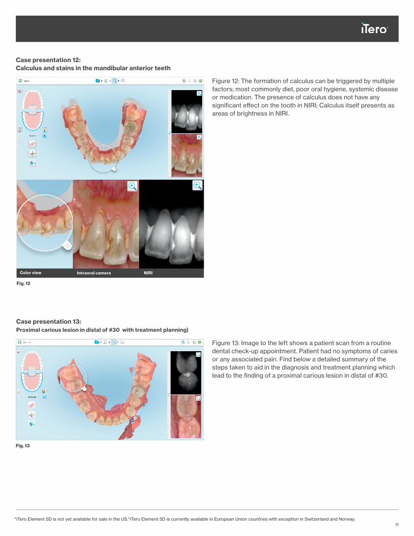
Case presentation 13: Proximal carious lesion in distal of #30 with treatment planning)
Figure 13: Image to the left shows a patient scan from a routine dental check-up appointment. Patient had no symptoms of caries or any associated pain. Find below a detailed summary of the steps taken to aid in the diagnosis and treatment planning which lead to the finding of a proximal carious lesion in distal of #30.
Fig. 13
Case presentation 12: Calculus and stains in the mandibular anterior teeth
Figure 12: The formation of calculus can be triggered by multiple factors; most commonly diet, poor oral hygiene, systemic disease or medication. The presence of calculus does not have any significant effect on the tooth in NIRI; Calculus itself presents as areas of brightness in NIRI.
Fig. 12
Color view Intraoral camera NIRI
12*iTero Element 5D is not yet available for sale in the US.*iTero Element 5D is currently available in European Union countries with exception in Switzerland and Norway.
Limitations of the technology: Current limitations of the technology are mostly around existing restorations. In the presence of restorations such as amalgam or composite resins, NIRI is unable to penetrate through the structure of the tooth. The insufficient data from the scan in these scenarios causes a blurry, dark and ill-defined resultant image that is not suitable for examination.
Instances mimicking interproximal caries: Teeth involving enamel demineralization conditions such as tooth wear, enamel hypoplasia and fluorosis (as seen in case 7) may mimic the presence of interproximal caries under NIRI; some dental cements (such as oxides and phosphates) may also exhibit the same behavior on interaction with NIRI, best practices to avoid misinterpretation in such cases would be to compare the NIRI images with the color images from the scan and other applicable examination techniques.
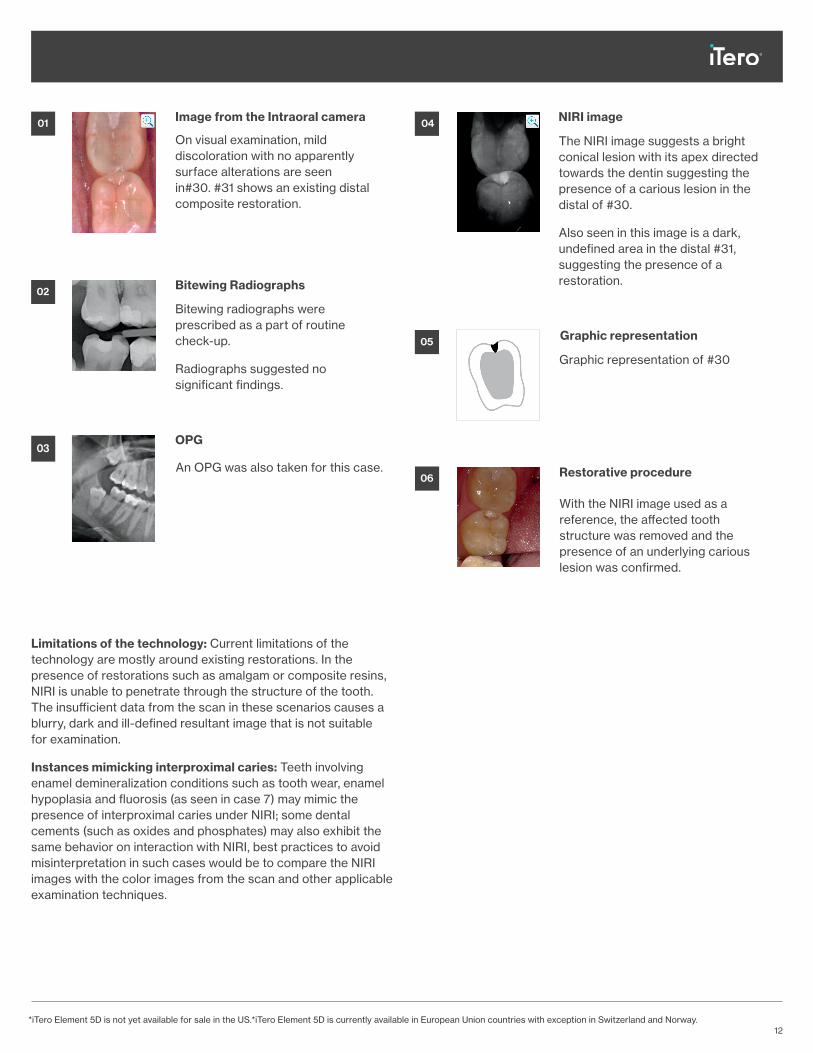
Image from the Intraoral camera
On visual examination, mild discoloration with no apparently surface alterations are seen in#30. #31 shows an existing distal composite restoration.
Bitewing radiographs were prescribed as a part of routine check-up.
Radiographs suggested no significant findings.
Bitewing Radiographs01
OPG
An OPG was also taken for this case.
The NIRI image suggests a bright conical lesion with its apex directed towards the dentin suggesting the presence of a carious lesion in the distal of #30.
Also seen in this image is a dark, undefined area in the distal #31, suggesting the presence of a restoration.
NIRI image
Graphic representation of #30
Graphic representation
With the NIRI image used as a reference, the affected tooth structure was removed and the presence of an underlying carious lesion was confirmed.
Restorative procedure
02
03
04
05
06
01
03

13*iTero Element 5D is not yet available for sale in the US.*iTero Element 5D is currently available in European Union countries with exception in Switzerland and Norway.
Case presentation 14: Composite restoration (mandibular right #29#30)
Figure 14: Composite restoration in the distal of #29 and mesial of #30 presents as a dark area which is comparitively dull in constrast when compared with the adjacent structures. The inabiltiy of Near infrared light to pass through existing restoration results in the presentation of a dark area.
Fig. 14
Color view Intraoral camera NIRI Graphic NIRI image
Case presentation 15: Amalgam restoration (maxillary right molar #3)
Figure 15: Exisiting amalgam restorations (as seen to the left). Amalgam being an alloy creates a highly scattering effect on Near infrared light resulting in a dark image with ill defined anatomical landmarks which makes the image unsuitable for interpretation. In such cases, comparison with other available data is recommended.
Fig. 15
Color view Intraoral camera NIRI Graphic NIRI image
14*iTero Element 5D is not yet available for sale in the US.*iTero Element 5D is currently available in European Union countries with exception in Switzerland and Norway.
Conclusion: Constant improvements in dental technology are shaping the way clinicians practice across the globe. Interactive technology also serves as an added benefit to patients of all ages who may be apprehensive about their dental visits.
As seen from all the case presentations in this article, NIRI has demonstrated to be an effective tool in aiding the diagnosis and monitoring early stages of interproximal caries above the gingiva in a wide array of clinical scenarios, ultimately leading towards the successful management of caries even in its earliest stages. NIRI, which is non-invasive by nature, can be used as frequently as required to monitor the patient’s oral health and provide the patient with chairside education, which enables patients to appreciate and understand the finer details associated with their oral health.
The iTero Element 5D imaging system helps turn the concept of comprehensive dentistry into a reality in every dental practice.

15*iTero Element 5D is not yet available for sale in the US.*iTero Element 5D is currently available in European Union countries with exception in Switzerland and Norway.
References 1Diagnosis and Management of Dental Caries Throughout Life National Institutes of Health Consensus Development Conference Statement, March 26–28, 2001
2Oral Health: The Silent Epidemic; the Surgeon Generals Perspective
Regina M. Benjamin, MD, MBA, VADM, USPHS
3The Global Burden of Oral Diseases and Risks to Oral Health, W.H.o Policy and Practice
4Dalli M, Çolac H, Hamidi MM. Minimal intervention concept: a new paradigm for operative dentistry. J Invest Clin Dent. 2012;3(3):167–175
5J. D. B. Featherstone and D. Young, "The need for new caries detection methods," Lasers in Dentistry V, San Jose, CA, Proc. SPIE 3593, 134-140 (1999).
6: Near-Infrared Imaging of Dental Decay at 1310 nm Daniel Fried, PhD*, Michal Staninec, DDS, Cynthia L. Darling, PhD University of California San Francisco (UCSF) School of Dentistry, San Francisco, California
7Effectiveness of Near-Infrared transillumination in early caries diagnosis
Mirela-Marinova – Tokorova
Clinical Evaluation of Near Infrared light transillumination as an interproximal caries detection tool in a large sample of patients in a private practice – Francesco Russotto,F Tirone,Stepho Salzano, Borga, Ferraro,S.Botasso 2016
DIAGNOcam--a Near Infrared Digital Imaging Transillumination (NIDIT) technology.
Abdelaziz M, Krejci I
8Fried D, Glena RE, Featherstone JD, Seka W. Nature of light scattering in dental enamel and dentin at visible and nearinfrared wavelengths. Applied Optics. 1995;34(7):12781286.
9Comparison of diagnostic methods for early interproximal caries detection with near-infrared light transillumination: an in vivo study Ismail Hakki Baltacioglu and Kaan Orhan
10Evaluation of two imaging techniques: near-infrared transillumination and dental radiographs for the detection of early approximal enamel caries. Maia AM1, Karlsson L, Margulis W, Gomes AS.
11Clinical evaluation of near-infrared light transillumination (NIRT) as an interproximal caries detection tool in a large sample of patients in a private practice Francesco Russotto1 , Federico Tirone1,*, Stefano Salzano1 , Francesco Coero Borga1,*, Davide Paolino2 , Alberto Ferraro1 , and Samanta Botasso3
Journal of Radiology and Imaging
12Caries Detecion and Diagnostics with near – infrared light transillumination : Clinical experiences
Friederike Sochtig,DDS/Reinhard Hickel,DDS./Jan Kuhnisch,DDS,MDS
13Elsevier Textbook of Oral Medicine Oral diagnosis and Oral radiology edition 2, Editiors Ravikiran Ongole BDS,MDS,Praveen BN, BDS,MDS
14White SC, Hollender L, Gratt BM. Comparison of xeroradiographs and film for detection of proximal surface caries. J Am Dent Assoc. 1984;108:755-759.
Special acknowledgement: Align would like to thank Dr. Ingo Baresel, Dr.Olivier Boujenah, Dr. Timo Weihard for their contribution to this article.
© 2019 Align Technology, Inc. All rights reserved. Invisalign, iTero, iTero Element, iTero elemnent 5D, the iTero logo, among others, are trademarks and/or service marks of Align Technology, Inc. or one of its subsidiaries or affiliated companies and may be registered in the U.S. and/or other countries. www.iTero.com | MKT-0003041 Rev A
Related Documents