Azerbaijan Medical University PROCEEDİNGS OF THE FIRST INTERNATIONAL SCIENTIFIC PRACTICAL VIRTUAL CONFERENCE HUMAN GENETICS AND GENETIC DISEASES: PROBLEMS AND DEVELOPMENT PERSPECTIVES İNSAN GENETIKASI VƏ GENETİK XƏSTƏLIKLƏR: PROBLEMLƏR VƏ İNKIŞAF PERSPEKTIVLƏRI BIRINCI BEYNƏLXALQ ELMI PRAKTIK VIRTUAL KONFRANSININ XƏBƏRLƏRİ AZERBAIJAN, BAKU MAY 30-31, 2020 AZƏRBAYCAN, BAKI MAY 30-31, 2020 DOI 10.36962/HGGD01 E-ISBN: 978-9949-7486-1-7

Welcome message from author
This document is posted to help you gain knowledge. Please leave a comment to let me know what you think about it! Share it to your friends and learn new things together.
Transcript
Azerbaijan
Medical University
PROCEEDİNGS OF THE FIRST INTERNATIONAL SCIENTIFIC PRACTICAL
VIRTUAL CONFERENCE HUMAN GENETICS AND GENETIC DISEASES:
PROBLEMS AND DEVELOPMENT PERSPECTIVES
İNSAN GENETIKASI VƏ GENETİK XƏSTƏLIKLƏR: PROBLEMLƏR VƏ
İNKIŞAF PERSPEKTIVLƏRI BIRINCI BEYNƏLXALQ ELMI PRAKTIK
VIRTUAL KONFRANSININ XƏBƏRLƏRİ
AZERBAIJAN, BAKU MAY 30-31, 2020
AZƏRBAYCAN, BAKI MAY 30-31, 2020
DOI 10.36962/HGGD01
E-ISBN: 978-9949-7486-1-7
PROCEEDİNGS OF THE FIRST INTERNATIONAL SCIENTIFIC PRACTICAL
VIRTUAL CONFERENCE HUMAN GENETICS AND GENETIC DISEASES:
PROBLEMS AND DEVELOPMENT PERSPECTIVES
İNSAN GENETIKASI VƏ GENETİK XƏSTƏLIKLƏR: PROBLEMLƏR VƏ
İNKIŞAF PERSPEKTIVLƏRI BIRINCI BEYNƏLXALQ ELMI PRAKTIK
VIRTUAL KONFRANSININ XƏBƏRLƏRİ
AZERBAIJAN, BAKU MAY 30-31, 2020
AZƏRBAYCAN, BAKI MAY 30-31, 2020
E-ISBN: 978-9949-7486-1-7
AZERBAIJAN, BAKU 2020
2
THE FIRST INTERNATIONAL SCIENTIFIC PRACTICAL VIRTUAL CONFERENCE − HUMAN GENETICS AND GENETİC DISEASES:
PROBLEMS AND DEVELOPMENT PERSPECTIVES
Organizer of the conference: Azerbaijan Medical University. Invited organizations: Molecular Biology & Biotechnologies institute of Azerbaijan National Academy of Sciences (Azerbaijan).
Genetic resources institute of Azerbaijan National Academy of Sciences (Azerbaijan).
İnternational Research, Education & Training Center LTD (UK, London).
İnternational Research, Education & Training Center LTD (Estonia, Tallinn).
Afgen Genetic Diagnostic Center (Azerbaijan).
Tbilisi State Medical University (Georgia).
School of Medicine, New Vision University (Georgia).
Department of Biophysics, Iv. Beritashvili Center of Experimental Biomedicine (Georgia).
Institute of Genetic Resources of Azerbaijan National Academy of Sciences Institute of Microbiology.
Academy of Sciences of the Republic of Uzbekistan (Uzbekistan)
West Kazakhstan Marat Ospanov Medical University (Kazakhstan, Aktobe)
Semey Medical University (Kazakhstan, Semey)
Ege University Faculty of Medicine (Turkey)
Celal Bayar University Faculty of Medicine (Turkey)
3
BIRINCI BEYNƏLXALQ ELMI PRAKTIK VIRTUAL KONFRANS − İNSAN GENETIKASI VƏ GENETİK XƏSTƏLIKLƏR: PROBLEMLƏR VƏ İNKIŞAF PERSPEKTIVLƏRI
TABLE OF CONTENTS
Organizing Committee ...………………………..…………………………………………………………………………. 04
Scientific Committee ………………………..…...……..……………..…………………………………………………… 05
Publishing committee ..……….………………………..……………..………………………………………..………...... 06
Editorial Board .………………………………………………..…..……………………………………………………...... 07
Program at a Glance ………………………..…………………………………...……………………………………….... 08
Abstracts and Theses ……………………….……………..……………….. ……………………………………………. 10
4
THE FIRST INTERNATIONAL SCIENTIFIC PRACTICAL VIRTUAL CONFERENCE − HUMAN GENETICS AND GENETİC DISEASES:
PROBLEMS AND DEVELOPMENT PERSPECTIVES
ORGANIZING COMMITTEE
Aytakin Hasanova Azerbaijan Medical University. I Preventive Medicine Faculty. Deputy of Head of the Department of Medical Biology and Genetics for Scientific Direction. Senior teacher. PhD in Biology. Leyla Suleymanova Azerbaijan Medical University, Deputy Head of the Department of Medical Biology and Genetics for Educational Direction. Associate Professor. Baku, Azerbaijan Maia Matoshvili Tbilisi State Medical University. The First University Clinic. Dermato-Venereologist. Assistant Professor. PhD in DAPS. Marziya Mammadova Scientific Research Institute of Medical Rehabilitation, MD, Baku, Azerbaijan Namig Isazade International Research, Education & Training Center. MTÜ. PhD in Business Administration. Namig Khalilov Azerbaijan Medical University, PhD in Medicine, Baku, Azerbaijan
Nino Didbaridze
Tbilisi State Medical University. Microbiology and Immunology Department. Immunology Direction. PhD MD.
Nino Pirtskhelani Tbilisi State Medical University. Associated Professor. Department of Molecular and Medical Genetics. Rauf Baylarov Azerbaijan Medical University, Vice-rector, Associate Professor, Baku, Azerbaijan. Rusudan Sujashvili New Vision University. School of Medicine. Professor. Tbilisi, Georgia. Sevil Asadova Azerbaijan Medical University, MD, Baku, Azerbaijan Sevinj Mammadova Azerbaijan Medical University, Associate Professor, Baku, Azerbaijan Vagif Karimov Head of the Department of Medical Biology and Genetics, Azerbaijan Medical University, Associate Professor. Baku, Azerbaijan Zhanargul Smailova S.O. Tapbergenova NAC Medical University of city Semey. Head of the Department of Biochemistry and Chemical Disciplines named after MD, professor. Semey, Kazakhstan.
5
BIRINCI BEYNƏLXALQ ELMI PRAKTIK VIRTUAL KONFRANS − İNSAN GENETIKASI VƏ GENETİK XƏSTƏLIKLƏR: PROBLEMLƏR VƏ İNKIŞAF PERSPEKTIVLƏRI
SCIENTIFIC COMMITTEE
Aflatun Azizov Azerbaijan Medical University, Associate Professor, Baku, Azerbaijan. Aytakin Hasanova Azerbaijan Medical University. I Preventive Medicine Faculty. Deputy Head of the Department of Medical Biology and Genetics for Scientific Direction. Senior teacher. PhD in Biology. Lala Huseynova Azerbaijan Medical University, Senior teacher, PhD in Pedagogy, Baku, Azerbaijan. Leyla Suleymanova Azerbaijan Medical University, Deputy Head of the Department of Medical Biology and Genetics for Educational Direction. Associate Professor Baku, Azerbaijan. Manzar Aliyeva Azerbaijan Medical University, Associate Professor, Baku, Azerbaijan. Namig Isazade International Research, Education & Training Center. MTÜ. PhD in Business Administration. Nino Didbaridze
Microbiology and Immunology Department. Immunology Direction. Tbilisi State Medical University. PhD MD.
Nino Pirtskhelani Associated Professor of Department of Molecular and Medical Genetics of Tbilisi State Medical University. Rahib Aliyev Azerbaijan Medical University, Associate Professor, Baku, Azerbaijan. Rena Karimova Azerbaijan Medical University, Associate Professor, Baku, Azerbaijan. Rusudan Sujashvili New Vision University. School of Medicine. Professor, Tbilisi, Georgia. Sevda Alizade Azerbaijan Medical University, Associate Professor, Baku, Azerbaijan. Tamar Didbaridze Tbilisi State Medical University, First University Clinic. PhD in MD, Tbilisi, Georgia. Vagif Karimov Head of the Department of Medical Biology and Genetics, Azerbaijan Medical University, Associate Professor Baku, Azerbaijan. Vladimer Papava Tbilisi State Medical University. Assistant-Professor. PhD. MD, Tbilisi, Georgia.

6
THE FIRST INTERNATIONAL SCIENTIFIC PRACTICAL VIRTUAL CONFERENCE − HUMAN GENETICS AND GENETİC DISEASES:
PROBLEMS AND DEVELOPMENT PERSPECTIVES
PUBLISHING COMMITTE
Aytakin Hasanova Azerbaijan Medical University. I Preventive Medicine Faculty. Deputy of Dean of the Department of Medical Biology and Genetics for Scientific Direction. Senior teacher. PhD in Biology. Namig Isazade International Research, Education & Training Center. MTÜ. PhD in Business Administration.
Nino Didbaridze
Microbiology and Immunology Department. Immunology Direction. Tbilisi State Medical University. PhD MD.
Tamar Didbaridze Tbilisi State Medical University, First University Clinic. PhD in MD.
7
BIRINCI BEYNƏLXALQ ELMI PRAKTIK VIRTUAL KONFRANS − İNSAN GENETIKASI VƏ GENETİK XƏSTƏLIKLƏR: PROBLEMLƏR VƏ İNKIŞAF PERSPEKTIVLƏRI
EDITORIAL BOARD
Aytakin Hasanova Azerbaijan Medical University. I Preventive Medicine Faculty. Deputy of Head of the Department of Medical Biology and Genetics for Scientific Direction. Senior teacher. PhD in Biology. Davit Tophuria Tbilisi State Medical University. Head of International Students Academic Department, Associate Professor. PhD in HNA. Maia Matoshvili Tbilisi State Medical University. The First University Clinic. Dermato-Venereologist. Assistant Professor. PhD in DAPS. Mariam Darbaidze Davit Aghmashenebeli National Defense Academy of Georgia. The Head of Education Division. PhD in Biology. Mariam Kharaishvili Ilia State University. Asistent Professor. PhD MD. Namig Isazade International Reserch, Education & Training Center. MTÜ. PhDin Business Administration. Nino Didbaridze
Microbiology and Immunology Department. Immunology Direction. Tbilisi State Medical University. PhD MD.
Nino Gogokhia Tbilisi State Medical University. Head of Laboratory The First University Clinic. Professor Nino Pirtskhelani Associated Professor of Department of Molecular and Medical Genetics of Tbilisi State Medical University. Rusudan Sujashvili New Vision University. School of Medicine. Professor, Tamar Didbaridze Tbilisi State Medical University, First University Clinic. PhD in MD. Tamar Giorgadze Tbilisi State Medical University. Department of Histology, Cytology and Embryology. Assistant Professor. Vladimer Papava Tbilisi State Medical University. Assistant-Professor. PhD. MD. Zhanargul Smailova Head of the Department of Biochemistry and Chemical Disciplines named after MD, professor S.O. Tapbergenova NAC Medical University of city Semey.
8
THE FIRST INTERNATIONAL SCIENTIFIC PRACTICAL VIRTUAL CONFERENCE − HUMAN GENETICS AND GENETİC DISEASES:
PROBLEMS AND DEVELOPMENT PERSPECTIVES
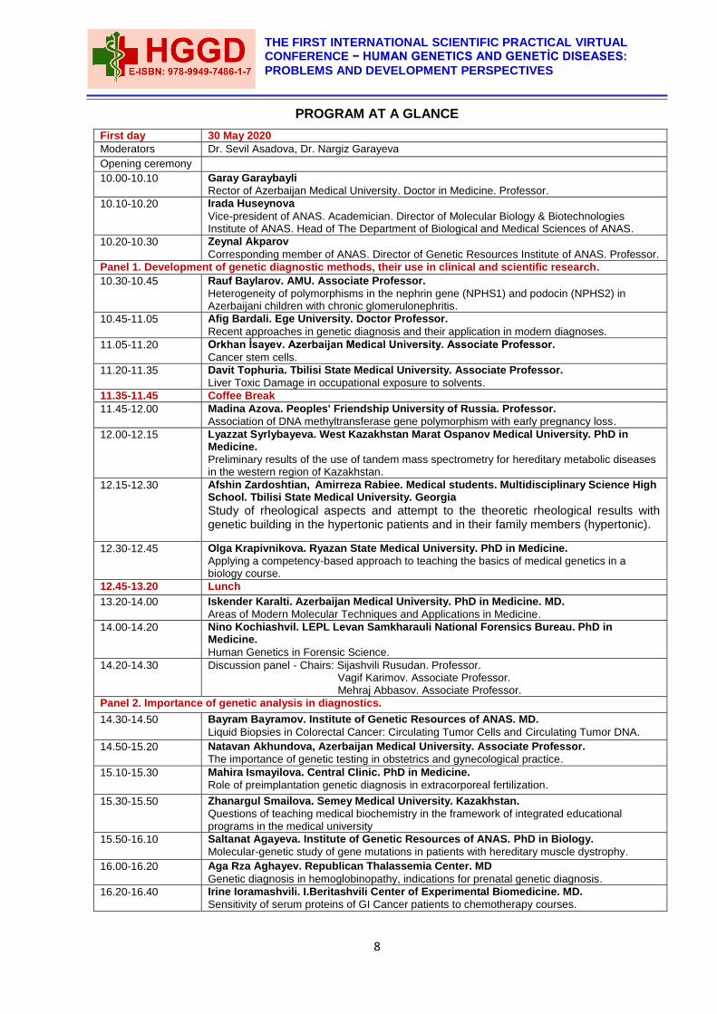
PROGRAM AT A GLANCE
First day 30 May 2020
Moderators Dr. Sevil Asadova, Dr. Nargiz Garayeva
Opening ceremony
10.00-10.10 Garay Garaybayli
Rector of Azerbaijan Medical University. Doctor in Medicine. Professor.
10.10-10.20 Irada Huseynova
Vice-president of ANAS. Academician. Director of Molecular Biology & Biotechnologies Institute of ANAS. Head of The Department of Biological and Medical Sciences of ANAS.
10.20-10.30 Zeynal Akparov
Corresponding member of ANAS. Director of Genetic Resources Institute of ANAS. Professor.
Panel 1. Development of genetic diagnostic methods, their use in clinical and scientific research.
10.30-10.45 Rauf Baylarov. AMU. Associate Professor.
Heterogeneity of polymorphisms in the nephrin gene (NPHS1) and podocin (NPHS2) in Azerbaijani children with chronic glomerulonephritis.
10.45-11.05
Afig Bardali. Ege University. Doctor Professor.
Recent approaches in genetic diagnosis and their application in modern diagnoses.
11.05-11.20 Orkhan İsayev. Azerbaijan Medical University. Associate Professor.
Cancer stem cells.
11.20-11.35 Davit Tophuria. Tbilisi State Medical University. Associate Professor.
Liver Toxic Damage in occupational exposure to solvents.
11.35-11.45 Coffee Break
11.45-12.00 Madina Azova. Peoples' Friendship University of Russia. Professor.
Association of DNA methyltransferase gene polymorphism with early pregnancy loss.
12.00-12.15 Lyazzat Syrlybayeva. West Kazakhstan Marat Ospanov Medical University. PhD in Medicine.
Preliminary results of the use of tandem mass spectrometry for hereditary metabolic diseases in the western region of Kazakhstan.
12.15-12.30 Afshin Zardoshtian, Amirreza Rabiee. Medical students. Multidisciplinary Science High School. Tbilisi State Medical University. Georgia
Study of rheological aspects and attempt to the theoretic rheological results with genetic building in the hypertonic patients and in their family members (hypertonic).
12.30-12.45 Olga Krapivnikova. Ryazan State Medical University. PhD in Medicine.
Applying a competency-based approach to teaching the basics of medical genetics in a biology course.
12.45-13.20 Lunch
13.20-14.00 Iskender Karalti. Azerbaijan Medical University. PhD in Medicine. MD.
Areas of Modern Molecular Techniques and Applications in Medicine.
14.00-14.20 Nino Kochiashvil. LEPL Levan Samkharauli National Forensics Bureau. PhD in Medicine.
Human Genetics in Forensic Science.
14.20-14.30
Discussion panel - Chairs: Sijashvili Rusudan. Professor. Vagif Karimov. Associate Professor. Mehraj Abbasov. Associate Professor.
Panel 2. Importance of genetic analysis in diagnostics.
14.30-14.50
Bayram Bayramov. Institute of Genetic Resources of ANAS. MD.
Liquid Biopsies in Colorectal Cancer: Circulating Tumor Cells and Circulating Tumor DNA.
14.50-15.20
Natavan Akhundova, Azerbaijan Medical University. Associate Professor.
The importance of genetic testing in obstetrics and gynecological practice.
15.10-15.30 Mahira Ismayilova. Central Clinic. PhD in Medicine.
Role of preimplantation genetic diagnosis in extracorporeal fertilization.
15.30-15.50 Zhanargul Smailova. Semey Medical University. Kazakhstan.
Questions of teaching medical biochemistry in the framework of integrated educational programs in the medical university
15.50-16.10 Saltanat Agayeva. Institute of Genetic Resources of ANAS. PhD in Biology.
Molecular-genetic study of gene mutations in patients with hereditary muscle dystrophy.
16.00-16.20 Aga Rza Aghayev. Republican Thalassemia Center. MD
Genetic diagnosis in hemoglobinopathy, indications for prenatal genetic diagnosis.
16.20-16.40 Irine Ioramashvili. I.Beritashvili Center of Experimental Biomedicine. MD.
Sensitivity of serum proteins of GI Cancer patients to chemotherapy courses.
9
BIRINCI BEYNƏLXALQ ELMI PRAKTIK VIRTUAL KONFRANS − İNSAN GENETIKASI VƏ GENETİK XƏSTƏLIKLƏR: PROBLEMLƏR VƏ İNKIŞAF PERSPEKTIVLƏRI
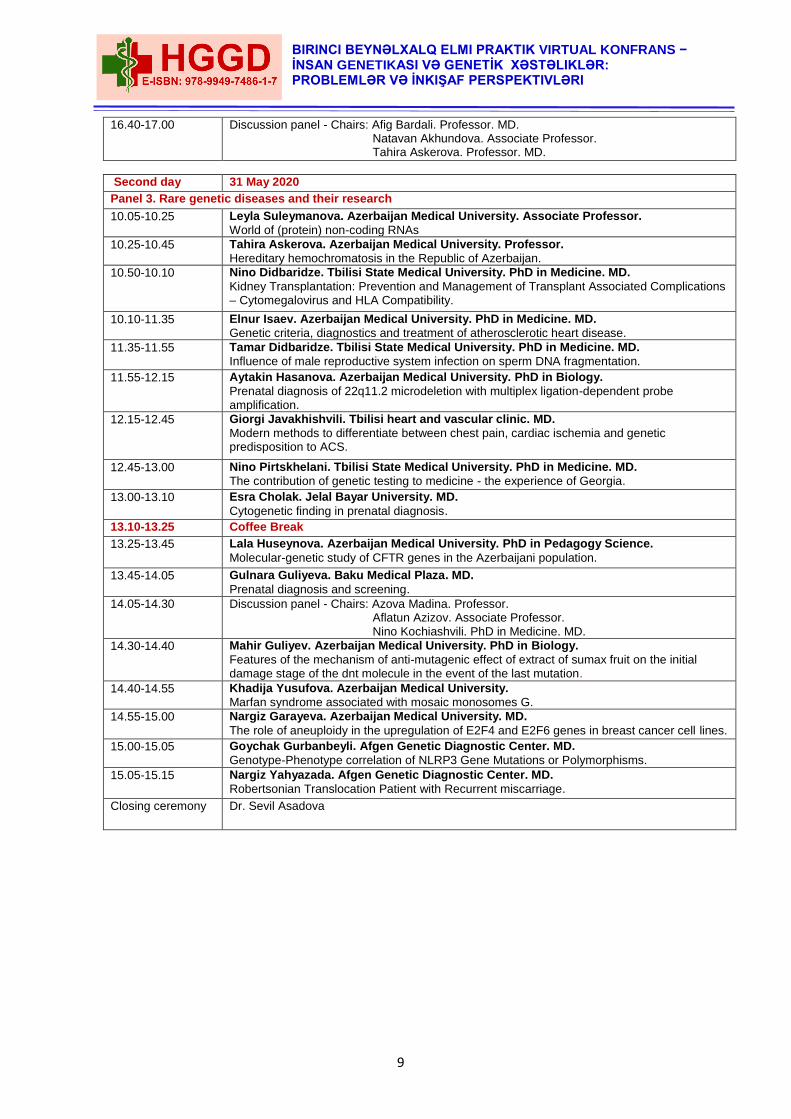
16.40-17.00 Discussion panel - Chairs: Afig Bardali. Professor. MD. Natavan Akhundova. Associate Professor. Tahira Askerova. Professor. MD.
Second day 31 May 2020
Panel 3. Rare genetic diseases and their research
10.05-10.25 Leyla Suleymanova. Azerbaijan Medical University. Associate Professor.
World of (protein) non-coding RNAs
10.25-10.45 Tahira Askerova. Azerbaijan Medical University. Professor.
Hereditary hemochromatosis in the Republic of Azerbaijan.
10.50-10.10 Nino Didbaridze. Tbilisi State Medical University. PhD in Medicine. MD.
Kidney Transplantation: Prevention and Management of Transplant Associated Complications – Cytomegalovirus and HLA Compatibility.
10.10-11.35 Elnur Isaev. Azerbaijan Medical University. PhD in Medicine. MD.
Genetic criteria, diagnostics and treatment of atherosclerotic heart disease.
11.35-11.55 Tamar Didbaridze. Tbilisi State Medical University. PhD in Medicine. MD.
Influence of male reproductive system infection on sperm DNA fragmentation.
11.55-12.15 Aytakin Hasanova. Azerbaijan Medical University. PhD in Biology.
Prenatal diagnosis of 22q11.2 microdeletion with multiplex ligation-dependent probe amplification.
12.15-12.45 Giorgi Javakhishvili. Tbilisi heart and vascular clinic. MD.
Modern methods to differentiate between chest pain, cardiac ischemia and genetic predisposition to ACS.
12.45-13.00
Nino Pirtskhelani. Tbilisi State Medical University. PhD in Medicine. MD.
The contribution of genetic testing to medicine - the experience of Georgia.
13.00-13.10 Esra Cholak. Jelal Bayar University. MD.
Cytogenetic finding in prenatal diagnosis.
13.10-13.25 Coffee Break
13.25-13.45 Lala Huseynova. Azerbaijan Medical University. PhD in Pedagogy Science.
Molecular-genetic study of CFTR genes in the Azerbaijani population.
13.45-14.05 Gulnara Guliyeva. Baku Medical Plaza. MD.
Prenatal diagnosis and screening.
14.05-14.30 Discussion panel - Chairs: Azova Madina. Professor. Aflatun Azizov. Associate Professor. Nino Kochiashvili. PhD in Medicine. MD.
14.30-14.40 Mahir Guliyev. Azerbaijan Medical University. PhD in Biology.
Features of the mechanism of anti-mutagenic effect of extract of sumax fruit on the initial damage stage of the dnt molecule in the event of the last mutation.
14.40-14.55 Khadija Yusufova. Azerbaijan Medical University.
Marfan syndrome associated with mosaic monosomes G.
14.55-15.00 Nargiz Garayeva. Azerbaijan Medical University. MD.
The role of aneuploidy in the upregulation of E2F4 and E2F6 genes in breast cancer cell lines.
15.00-15.05 Goychak Gurbanbeyli. Afgen Genetic Diagnostic Center. MD.
Genotype-Phenotype correlation of NLRP3 Gene Mutations or Polymorphisms.
15.05-15.15 Nargiz Yahyazada. Afgen Genetic Diagnostic Center. MD.
Robertsonian Translocation Patient with Recurrent miscarriage.
Closing ceremony Dr. Sevil Asadova
10
THE FIRST INTERNATIONAL SCIENTIFIC PRACTICAL VIRTUAL CONFERENCE − HUMAN GENETICS AND GENETİC DISEASES:
PROBLEMS AND DEVELOPMENT PERSPECTIVES
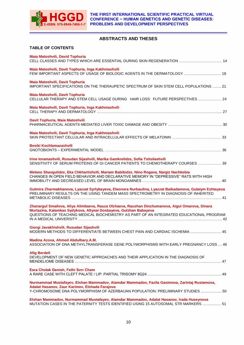
ABSTRACTS AND THESES
TABLE OF CONTENTS Maia Matoshvili, David Tophuria
CELL CLASSES AND TYPES WHICH ARE ESSENTIAL DURING SKIN REGENERATION …………………….………. 14 Maia Matoshvili, Davit Tophuria, Inga Kakhniashvili
FEW IMPORTANT ASPECTS OF USAGE OF BIOLOGIC AGENTS IN THE DERMATOLOGY ………………………… 18 Maia Matoshvili, Davit Tophuria
IMPORTANT SPECIFICATIONS ON THE THERAUPETIC SPECTRUM OF SKIN STEM CELL POPULATIONS ……. 21 Maia Matoshvili, Davit Tophuria
CELLULAR THERAPY AND STEM CELL USAGE DURING HAIR LOSS: FUTURE PERSPECTIVES …………….… 24 Maia Matoshvili, Davit Tophuria, Inga Kakhniashvili
CELL THERAPY AND DERMATOLOGY ………………………………………………………………………………………... 27 Davit Tophuria, Maia Matoshvili
PHARMACEUTICAL AGENTS-MEDIATED LIVER TOXIC DAMAGE AND OBESITY …………………………………….. 30 Maia Matoshvili, Davit Tophuria, Inga Kakhniashvili
SKIN PROTECTANT CELLULAR AND INTRACELLULAR EFFECTS OF MELATONIN ……………………………….… 33 Besiki Kochlamazashvili
GNOTOBIONTS – EXPERIMENTAL MODEL ………………………………………………………………………………….. 36
Irine Ioramashvili, Rusudan Sijashvili, Marika Gamkrelidze, Sofia Tsitsilashvili SENSITIVITY OF SERUM PROTEINS OF GI CANCER PATIENTS TO CHEMOTHERAPY COURSES ………………. 39
Melano Shavgulidze, Eka Chkhartishvili, Mariam Babilodze, Nino Rogava, Nargiz Nachkebia
CHANGES IN OPEN FIELD BEHAVIOR AND DECLARATIVE MEMORY IN “DEPRESSIVE” RATS WITH HIGH IMMOBILITY AND DECREASED LEVEL OF BRAIN MONOAMINES ……………………………….……………………… 40 Gulmira Zharmakhanova, Lyazzat Syrlybayeva, Eleonora Nurbaulina, Lyazzat Baikadamova, Gulaiym Eshtayeva
PRELIMINARY RESULTS ON THE USING TANDEM MASS SPECTROMETRY IN DIAGNOSIS OF INHERITED METABOLIC DISEASES ………………………………………………………………………………………………………..… 41 Zhanargul Smailova, Aliya Alimbaeva, Rauza Olzhaeva, Raushan Dinzhumanova, Aigul Omarova, Dinara Murtazina, Kalamkas Sydykova, Altynai Dosbayeva, Gulzhan Babayeva
QUESTIONS OF TEACHING MEDICAL BIOCHEMISTRY AS PART OF AN INTEGRATED EDUCATIONAL PROGRAM IN A MEDICAL UNIVERSITY ……………………………………………………………………………………………………... 42 Giorgi Javakhishvili, Rusudan Sijashvili
MODERN METHODS TO DIFFERENTIATE BETWEEN CHEST PAIN AND CARDIAC ISCHEMIA ……………………. 45 Madina Azova, Ahmed Abdulbary.A.M.
ASSOCIATION OF DNA METHYLTRANSFERASE GENE POLYMORPHISMS WITH EARLY PREGNANCY LOSS … 46 Afig Berdeli
DEVELOPMENT OF NEW GENETIC APPROACHES AND THEIR APPLICATION IN THE DIAGNOSIS OF MENDELIOME DISEASES ……………………………………………………………………………………………..………… 47 Esra Cholak Genish, Fethi Sırrı Cham
A RARE CASE WITH CLEFT PALATE / LIP: PARTIAL TRISOMY 8Q24 ………………………………………………..…. 49 Nurmammad Mustafayev, Elshan Mammadov, Alamdar Mammadov, Fazila Gasimova, Zarintaj Rustamova, Adalat Hasanov, Zaur Karimov, Elshada Farajova
Y-CHROMOSOME DNA POLYMORPHISM OF AZERBAIJAN POPULATION: PRELIMINARY STUDIES …………….. 50 Elshan Mammadov, Nurmammad Mustafayev, Alamdar Mammadov, Adalat Hasanov, Irada Huseynova
MUTATION CASES IN THE PATERNITY TESTS IDENTIFIED USING 15 AUTOSOMAL STR MARKERS …………… 51
11
BIRINCI BEYNƏLXALQ ELMI PRAKTIK VIRTUAL KONFRANS − İNSAN GENETIKASI VƏ GENETİK XƏSTƏLIKLƏR: PROBLEMLƏR VƏ İNKIŞAF PERSPEKTIVLƏRI
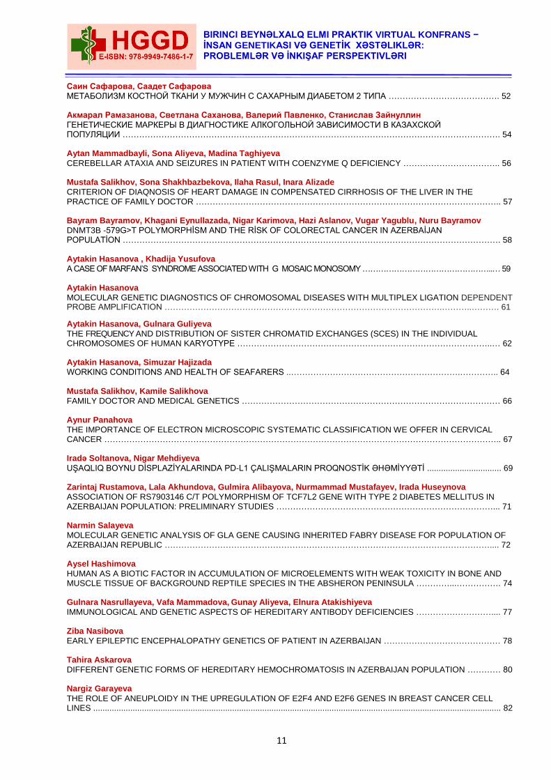
Саин Сафарова, Саадет Сафарова
МЕТАБОЛИЗМ КОСТНОЙ ТКАНИ У МУЖЧИН С САХАРНЫМ ДИАБЕТОМ 2 ТИПА …………………………………. 52
Aкмарал Рамазанова, Cветлана Саханова, Bалерий Павленко, Cтанислав Зайнуллин
ГЕНЕТИЧЕСКИЕ МАРКЕРЫ В ДИАГНОСТИКЕ АЛКОГОЛЬНОЙ ЗАВИСИМОСТИ В КАЗАХСКОЙ ПОПУЛЯЦИИ ………………………………………………………………………………………………………………………. 54
Aytan Mammadbayli, Sona Aliyeva, Madina Taghiyeva
CEREBELLAR ATAXIA AND SEIZURES IN PATIENT WITH COENZYME Q DEFICIENCY …………………………….. 56
Mustafa Salikhov, Sona Shakhbazbekova, Ilaha Rasul, Inara Alizade
CRITERION OF DIAQNOSIS OF HEART DAMAGE IN COMPENSATED CIRRHOSIS OF THE LIVER IN THE PRACTICE OF FAMILY DOCTOR ……………………………………………………………………………………………….. 57
Bayram Bayramov, Khagani Eynullazada, Nigar Karimova, Hazi Aslanov, Vugar Yagublu, Nuru Bayramov
DNMT3B -579G>T POLYMORPHİSM AND THE RİSK OF COLORECTAL CANCER IN AZERBAİJAN POPULATİON ………………………………………………………………………………………………………………………. 58
Aytakin Hasanova , Khadija Yusufova
A CASE OF MARFAN’S SYNDROME ASSOCIATED WITH G MOSAIC MONOSOMY …………………………………………..… 59
Aytakin Hasanova
MOLECULAR GENETIC DIAGNOSTICS OF CHROMOSOMAL DISEASES WITH MULTIPLEX LIGATION DEPENDENT PROBE AMPLIFICATION ……………………………………………………………………………………….………..………. 61
Aytakin Hasanova, Gulnara Guliyeva
THE FREQUENCY AND DISTRIBUTION OF SISTER CHROMATID EXCHANGES (SCES) IN THE INDIVIDUAL CHROMOSOMES OF HUMAN KARYOTYPE ………………………………………………………………………………..… 62
Aytakin Hasanova, Simuzar Hajizada
WORKING CONDITIONS AND HEALTH OF SEAFARERS ..…………………………………………………….………….. 64
Mustafa Salikhov, Kamile Salikhova
FAMILY DOCTOR AND MEDICAL GENETICS ………………………………………………………………………………… 66
Aynur Panahova
THE IMPORTANCE OF ELECTRON MICROSCOPIC SYSTEMATIC CLASSIFICATION WE OFFER IN CERVICAL CANCER …………………………………………………………………………………………………………………………….. 67
Iradə Soltanova, Nigar Mehdiyeva
UŞAQLIQ BOYNU DİSPLAZİYALARINDA PD-L1 ÇALIŞMALARIN PROQNOSTİK ƏHƏMİYYƏTİ ................................ 69
Zarintaj Rustamova, Lala Akhundova, Gulmira Alibayova, Nurmammad Mustafayev, Irada Huseynova
ASSOCIATION OF RS7903146 C/T POLYMORPHISM OF TCF7L2 GENE WITH TYPE 2 DIABETES MELLITUS IN AZERBAIJAN POPULATION: PRELIMINARY STUDIES ……………………………………………………………………... 71
Narmin Salayeva
MOLECULAR GENETIC ANALYSIS OF GLA GENE CAUSING INHERITED FABRY DISEASE FOR POPULATION OF AZERBAIJAN REPUBLIC ……………………………………………………………………………………………………….... 72
Aysel Hashimova
HUMAN AS A BIOTIC FACTOR IN ACCUMULATION OF MICROELEMENTS WITH WEAK TOXICITY IN BONE AND MUSCLE TISSUE OF BACKGROUND REPTILE SPECIES IN THE ABSHERON PENINSULA …………...……………. 74
Gulnara Nasrullayeva, Vafa Mammadova, Gunay Aliyeva, Elnura Atakishiyeva
IMMUNOLOGICAL AND GENETIC ASPECTS OF HEREDITARY ANTIBODY DEFICIENCIES ……………………….... 77
Ziba Nasibova
EARLY EPILEPTIC ENCEPHALOPATHY GENETICS OF PATIENT IN AZERBAIJAN …………………………………… 78
Tahira Askarova
DIFFERENT GENETIC FORMS OF HEREDITARY HEMOCHROMATOSIS IN AZERBAIJAN POPULATION ………… 80
Nargiz Garayeva
THE ROLE OF ANEUPLOIDY IN THE UPREGULATION OF E2F4 AND E2F6 GENES IN BREAST CANCER CELL LINES ................................................................................................................................................................................ 82
12
THE FIRST INTERNATIONAL SCIENTIFIC PRACTICAL VIRTUAL CONFERENCE − HUMAN GENETICS AND GENETİC DISEASES:
PROBLEMS AND DEVELOPMENT PERSPECTIVES
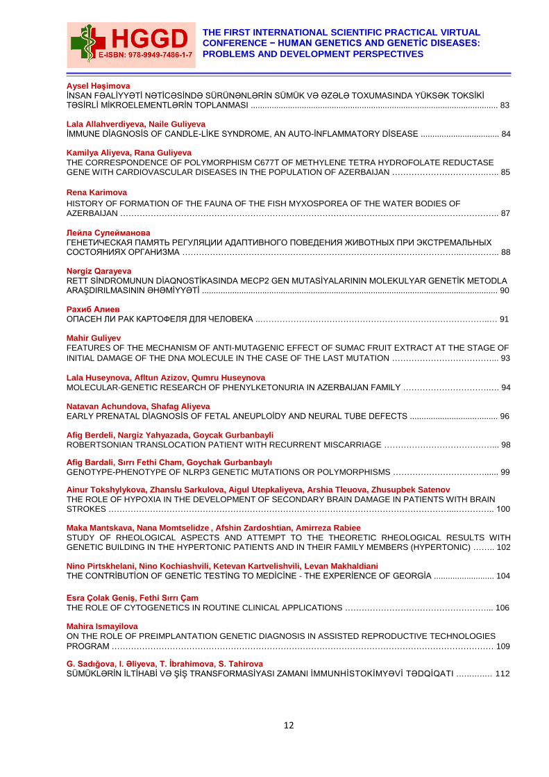
Aysel Həşimova
İNSAN FƏALİYYƏTİ NƏTİCƏSİNDƏ SÜRÜNƏNLƏRİN SÜMÜK VƏ ƏZƏLƏ TOXUMASINDA YÜKSƏK TOKSİKİ TƏSİRLİ MİKROELEMENTLƏRİN TOPLANMASI ........................................................................................................... 83 Lala Allahverdiyeva, Naile Guliyeva
İMMUNE DİAGNOSİS OF CANDLE-LİKE SYNDROME, AN AUTO-İNFLAMMATORY DİSEASE .................................. 84 Kamilya Aliyeva, Rana Guliyeva
THE CORRESPONDENCE OF POLYMORPHISM С677Т OF METHYLENE TETRA HYDROFOLATE REDUCTASE GENE WITH CARDIOVASCULAR DISEASES IN THE POPULATION OF AZERBAIJAN …………………………….….. 85
Rena Karimova
HISTORY OF FORMATION OF THE FAUNA OF THE FISH MYXOSPOREA OF THE WATER BODIES OF AZERBAIJAN ……………………………………………………………………………………………………………………….. 87
Лейла Сулейманова
ГЕНЕТИЧЕСКАЯ ПАМЯТЬ РЕГУЛЯЦИИ АДАПТИВНОГО ПОВЕДЕНИЯ ЖИВОТНЫХ ПРИ ЭКСТРЕМАЛЬНЫХ СОСТОЯНИЯХ ОРГАНИЗМА ………………………………………………………………………………………..………….. 88
Nərgiz Qarayeva
RETT SİNDROMUNUN DİAQNOSTİKASINDA MECP2 GEN MUTASİYALARININ MOLEKULYAR GENETİK METODLA ARAŞDIRILMASININ ƏHƏMİYYƏTİ ................................................................................................................................ 90 Pахиб Алиев
ОПАСЕН ЛИ РАК КАРТОФЕЛЯ ДЛЯ ЧЕЛОВЕКА ..………………………………………………………………………..… 91 Mahir Guliyev
FEATURES OF THE MECHANISM OF ANTI-MUTAGENIC EFFECT OF SUMAC FRUIT EXTRACT AT THE STAGE OF
INITIAL DAMAGE OF THE DNA MOLECULE IN THE CASE OF THE LAST MUTATION ………………………………... 93
Lala Huseynova, Afltun Azizov, Qumru Huseynova
MOLECULAR-GENETIC RESEARCH OF PHENYLKETONURIA IN AZERBAIJAN FAMILY ………………………….…. 94 Natavan Achundova, Shafag Aliyeva
EARLY PRENATAL DİAGNOSİS OF FETAL ANEUPLOİDY AND NEURAL TUBE DEFECTS ...................................... 96 Afig Berdeli, Nargiz Yahyazada, Goycak Gurbanbayli ROBERTSONIAN TRANSLOCATION PATIENT WITH RECURRENT MISCARRIAGE …………………………………... 98
Afig Bardali, Sırrı Fethi Cham, Goychak Gurbanbaylı GENOTYPE-PHENOTYPE OF NLRP3 GENETIC MUTATIONS OR POLYMORPHISMS ……………………………...... 99
Ainur Tokshylykova, Zhanslu Sarkulova, Aigul Utepkaliyeva, Arshia Tleuova, Zhusupbek Satenov
THE ROLE OF HYPOXIA IN THE DEVELOPMENT OF SECONDARY BRAIN DAMAGE IN PATIENTS WITH BRAIN STROKES ……………………………………………………………………………………………………………...………….. 100 Maka Mantskava, Nana Momtselidze , Afshin Zardoshtian, Amirreza Rabiee
STUDY OF RHEOLOGICAL ASPECTS AND ATTEMPT TO THE THEORETIC RHEOLOGICAL RESULTS WITH GENETIC BUILDING IN THE HYPERTONIC PATIENTS AND IN THEIR FAMILY MEMBERS (HYPERTONIC) …….. 102 Nino Pirtskhelani, Nino Kochiashvili, Ketevan Kartvelishvili, Levan Makhaldiani
THE CONTRİBUTİON OF GENETİC TESTİNG TO MEDİCİNE - THE EXPERİENCE OF GEORGİA .......................... 104
Esra Çolak Geniş, Fethi Sırrı Çam
THE ROLE OF CYTOGENETICS IN ROUTINE CLINICAL APPLICATIONS ……………………………………………... 106
Mahira Ismayilova
ON THE ROLE OF PREIMPLANTATION GENETIC DIAGNOSIS IN ASSISTED REPRODUCTIVE TECHNOLOGIES
PROGRAM ………………………………………………………………………………………………………………………… 109
G. Sadığova, I. Əliyeva, T. İbrahimova, S. Tahirova
SÜMÜKLƏRİN İLTİHABİ VƏ ŞİŞ TRANSFORMASİYASI ZAMANI İMMUNHİSTOKİMYƏVİ TƏDQİQATI .............. 112
13
BIRINCI BEYNƏLXALQ ELMI PRAKTIK VIRTUAL KONFRANS − İNSAN GENETIKASI VƏ GENETİK XƏSTƏLIKLƏR: PROBLEMLƏR VƏ İNKIŞAF PERSPEKTIVLƏRI
Gülnarə Əzizova, Arzu Dadaşova, Şəymən Həsənova, Şəfiqə Ələkbərzadə
SİTOKİNLƏRİN VƏ ANTİMİKROB PEPTİDLƏRİN ŞD-2 XƏSTƏLƏRDƏ QAN SERUMUNDA SƏVİYYƏSİNİN TƏTQİQİ ......................................................................................................................................................................... 113 Aynur Ənsərova
SU ANBARLARININ BİOLOJİ MƏHSULDARLIĞI .......................................................................................................... 115 Aynur Ənsərova, G. Vəliyeva, P. Mahmudova, N. Məmmədova
TÜRKİYƏ RESPUBLİKASI ƏRAZİSİNDƏ ARAZ ÇAYININ EKOLOJİ MİKROBİOLOGİYASI ........................................ 116 Aynur Ənsərova, G. Vəliyeva, P. Mahmudova, N. Məmmədova
SU ANBARLARININ ƏTRAF MÜHİTƏ VƏ ƏHALİNİN SAĞLAMLIĞINA TƏSİRİ ........................................................... 118 Nurana Baghirova
THE SMPD1 GENE MUTATIONS IN TWO SIBLINGS WITH NIMANN-PEAK TYPE A/B DISEASE …………………... 119
Sevda Alizada, Kamile Aliyeva, Elkhan Rasulov
BIOCHEMICAL POLYMORPHISM, GENETIC HETEROGENICITY, GENE MAPING AND PREVENTION OF MUCOPOLYSACCHARIDOSIS HEREDITARY DISEASE IN THE POPULATION OF THE REPUBLIC OF AZERBAIJAN ………………………………………………………………………………………………………………..... 120 Irada Soltanova, Nigar Mehdiyeva
PROGNOSTİC SİGNİFİCANCE OF PD-L1 STUDİES İN CERVİCAL DYSPLASİA ...................................................... 121 Agharza Aghayev, Valeh Huseynov, Ramin Bayramli, Oya Uyguner
MOLECULAR BASIS OF HEMOGLOBINOPATHIES IN AZERBAIJAN ……………………………………………………. 122
14
THE FIRST INTERNATIONAL SCIENTIFIC PRACTICAL VIRTUAL CONFERENCE − HUMAN GENETICS AND GENETİC DISEASES:
PROBLEMS AND DEVELOPMENT PERSPECTIVES
CELL CLASSES AND TYPES WHICH ARE ESSENTIAL DURING SKIN REGENERATION
1Maia Matoshvili, 2Davit Tophuria
1,2Dermato-Venerology Department, Human Normal Anatomy Department, Tbilisi State Medical University ABSTRACT
The Skin has the natural ability to heal and replace damaged and dead cells regulated by a network of complex immune processes. This ability is conferred by the population of resident immune cells that act in coordination with other players to provide a homeostatic environment under constant challenge. In this article we conclude that near future discoveries using such innovative strategies will not only help us achieve better therapeutic products for skin-related immune disorders but will also foster ideas toward novel cosmetic formulations and topical applications for improving skin’s regenerative potentia l. Keywords: damaged and dead cells, resident immune cells, therapeutic products.
The stem cells are involved in the renewal and regeneration of the epithelium of various organs. The largest reservoir of epithelial stem cells in the human body is the skin. This organ is a specialized interior barrier protecting the body from the influence of physical, chemical, environmental and biological factors[1], ensuring at the same time the reception of signals from the external environment. Skin is also involved in numerous physiological processes which determine the homeostasis of the body. Renewal and regeneration of the epidermis which is the outer layer of the skin, is possible by the presence of different populations of stem cells that reside in microenvironments (niches),that creates specific conditions to preserve the biological properties of these cells. Because divisions of cells in niches are quite rare, it became possible to distinguish them from other rapidly proliferating cells of the skin. On this basis, the stem cells in the interfollicular epidermis, bulge region of the hair follicles, and within the sebaceous glands were located. Tissues have a natural capacity to replace dying cells and to heal wounds. This ability resides in resident stem cells, which self-renew, preserve, and repair their tissue during homeostasis and following injury. The skin epidermis and its appendages are subjected to daily assaults from the external environment [2.3]. A high demand is placed on renewal and regeneration of the skin's barrier in order to protect the body from infection and dehydration and to heal wounds. This review focuses on the epithelial stem cells of skin, where they come from, where they reside, and how they function in normal homeostasis and wound repair. Moreover hair follicles are suggested to be a niche for melanocyte progenitor cells and other multipotent stem cells derived from the neural crest, as well as mesenchymal stem cells. The presence of stem cells that are characterized by high proliferative potential and the ability to self-renew allow maintaing homeostasis and regeneration of epidermis. Identification [4], isolation and characterization of epithelial stem cells is necessary to understand skin diseases background, develop effective methods for their treatment and for wider use of stem cells in regenerative medicine, gene therapy or cosmetology. The stem cells variations and types represent a novel hope for regenerative medicine. In adult life, stem cell deposits are kept in organ niches; the need for tissue or organ regeneration mobilizes stem cells via the SDF-1-CXCR4 regulation axis. Constant regeneration of the skin is achieved due to stem cell differentiation within the epidermis and the hair follicle; thus, skin may serve as an excellent source of stem cells. This is of paramount importance in the treatment of chronic skin wounds and burns The enormous interest in the biology of stem cells (SC) is related to their capacity for self-renewal, replication and differentiation to other cells that build different tissues and organs. SCs replenish lost cells throughout an organism’s lifespan. SCs have the capacity for unlimited replication that gives a population of ‘sister’ SCs. These cells are responsible for self-renewal and differentiate into tissue-specific cells[5]. This process maintains the constant number of aging somatic cells, which become apoptotic. In the future, SCs could be used in the treatment and regeneration of organs and tissues. The implantation of SCs could be applied instead of the transplantation of tissue and organs [6.7]. This would be a huge step in regenerative medicine. There are several types of SCs, which differ one from another in their proliferation and differentiation capacity. The less mature SCs have greater possibilities of differentiation and replication. Previous research suggested that tissue-committed stem cells (TCSCs) showed plasticity i.e. the possibility of these cells transdifferentiating into other TCSCs under the control of environmental factors. For example, hematopoietic stem cells (HSCs) could differentiate into heart stem cells, hepatic stem cells or pancreas stem cells. The new hypothesis on this subject is that stem cell niches are not only colonized by TCSCs but also contain pluripotent stem cells (PSCs)[8.9], which can differentiate into specific tissue. PSCs express embryonic markers such as Oct4, Nanong and Rex-1 and give rise to SCs specific for various tissues and organs; some of them are deposited during embryogenesis in organs and can survive in these localizations to adulthood. Kucia and Ratajczak confirmed that bone marrow (BM) and other tissue of adults is equipped with PSCs — the very small embryonic-like stem cells (VSELs). The morphology of these cells and their immunohistochemical features are similar to those of early embryonic SCs. They were initially isolated from murine BM as a homogenic lineage Sca-1+lin- -CD45– which demonstrates coexpression of PSCs markers such as SSEA-1, Oct-4, Nanog and Rex-1. Direct electron microscopy showed cells with a large nucleus with euchromatin and narrow cytoplasm. The cells are isolated from human BM, circulating blood and umbilical cord blood as CD34+CD133+CXCR4+lin-CD45– , small size cells (7 μm in diameter ) [19–21]. In vitro, they differentiate into all three germ layers [16]. These cells are enriched for mRNA for skin epidermis like Trp63, Krt2-5, BNC. During ontogenesis, VSELs are deposited in BM and in other organs (tissues)
15
BIRINCI BEYNƏLXALQ ELMI PRAKTIK VIRTUAL KONFRANS − İNSAN GENETIKASI VƏ GENETİK XƏSTƏLIKLƏR: PROBLEMLƏR VƏ İNKIŞAF PERSPEKTIVLƏRI
and are mobilized in cases of organ and tissue damage for their regeneration. Their number is higher in young individuals and decreases with age. The stem cells migration, which is the key process in their development and regeneration, is regulated by the axis CXCR4-SDF-1. CXCR4 receptor has been described in many types of tissue-specific SCs including nervous tissue, skeletal muscles, heart, liver, endothelium, tubules of nephron, pigment cells of retina and embryonic PSC. SCs follow the SDF-1 gradient. The SDF-1 is expressed in stromal, endothelial, cardiac, skeletal muscle, liver, brain and renal cells. Recently, the alternative receptor for SDF-1, CXCR7, was described. The damage of tissue increases expression of SDF-1 that attracts CXCR4+ SCs, which are necessary for organ reparation. The expression of SDF-1 can be up-regulated by HIF-1a and down-regulated by steroids, granulocytes colony stimulating factor (G-CSF) and transforming growth factor (TGF-b1) [10]. Investigators have found SCs in the niches of the epidermis. PSCs as Oct4+ embryonic cells or as non-epidermal non-melanocyte Oct4+Nanog+ cells have been identified in the same niche. The association of these cells with VSELs requires further investigation. Dyce et al. determined that SCs isolated from the skin include a population capable of differentiating into oocyte-like cells expressing Oct4 and other markers characteristic for oocytes. The hypothesis on the migration of cells of epiblast — primordial germ cells (PGCs) — into nongonadal niches during early embryogenesis may explain their presence in the skin. Obtaining PSCs from the skin may potentially give us new uses for these cells in terms of treatment, and become a new experimental model for in vitro studies. Cells are the main component of the tissue-engineered skin used for burn therapies. They include both stem and somatic cells and can be divided into three main groups: autologous, allogeneic and xenogeneic. One of the main trends in choosing a cell type for patient treatment is the use of autologous cells as they do not cause immune rejection and their tumorigenicity is low due to the absence of epigenetic manipulations. Nowadays, animal cells are not widely used for skin tissue regeneration, only ECM or its components that they synthesize. Plant stem cells, which are commonly applied in cosmetics, can be interesting as they have no use limitations when compared to animal and human cells. Of course, they cannot be used in skin substitute development as a cell component; but they can provide bioactive substances, which can improve the wound healing processes. Fibroblasts and keratinocytes are common cells used in products for wound and burn healing. Keratinocytes are the major cell component of the epidermis and responsible for its stratified structure and form numerous tight intercellular junctions. Fibroblasts are the main cell type of the dermis and produce ECM components and secrete various growth factors (TGF-β), cytokines (TNF-α), and matrix metalloproteinases, which ensure the ECM formation and keratinocyte proliferation and differentiation. Commercial products such as Epicel, Cryoskin, and BioSeed-S contain keratinocytes; Dermagraft, TransCyte and Hyalograft 3D—fibroblasts; and Apligraf, Theraskin, and OrCell—a combination. The use of these cells enables the large-scale production of standardized product batches. However, these materials are mostly non-permanent bioactive dressings, which provide cytokines, ECM, and growth factors for the successful skin reparation [11]. Immune rejection is commonly reported with allogeneic fibroblasts and keratinocytes, but this is mostly shown for allogeneic keratinocytes that can be explained by the difference in HLA expression and cytokine production. Progenitors of mast cells, myeloid cells and lymphoid DC travel through the bloodstream and migrate to the dermis where they mature as a result of resident elements. The predominant tissues in the bone marrow are MSCs and fibroblasts. MSCs from hair follicles arising from the neural crest are the closest to this sort judging by their properties.[12] It Is worth a mention to state that the quantity of platelets increases during an immune response due to their special properties. The dermis is the structural and functional backbone of the epidermis. It provides access to cells within circulation which are strictly regulated. These selective cells can practically only be found within this area. This is primarily keratinocytes, LC and γδ T lymphocytes. The last two function as part of the innate as well as the adaptive immunity, which links both the skin compartments with the entire body. This is required to present antigens traveling through the lymphatic path in the dermis followed by the lymphatic follicle. γδ T lymphocytes support keratinocytes and promote their regeneration. Fetal fibroblasts are of particular interest because they can significantly improve skin repair due to the high expansion ability, low immunogenicity, and intense secretion of bioactive substances such as basic fibroblast growth factor, vascular endothelial growth factor, and keratinocyte growth factor. However, ethical issues limit their application.Epidermal stem cells (ESC) are of particular interest for skin tissue regeneration as they have favorable features such as high proliferation rate and easy access and keep their potency and differentiation potential for long periods. They are one of the skin stem cell types, either heterogeneous or autogenous origins. ESC are mostly connected to the process of skin regeneration. They are rare, infrequently divide and generate short-lived and rapidly dividing cells, which are involved in the regeneration process. Their main population, responsible for skin repair, is located in the basal layer of the epidermis; however, they can also be revealed in the base of sebaceous glands and the bulge region of hair follicles. Moreover, as they can be easily derived from the patient’s skin and transplanted to the same patient, ESC are not restricted by ethical issues. Grafts containing autologous holoclones ESC have proven to be effective in treating vast skin defects: epidermolysis, skin and ocular burns [13]. Mesenchymal stromal cells (MSC) have similar (not identical) features as ESC and can be derived from various tissues, even the skin as mentioned previously. They have a high differentiation potential and a certain degree of plasticity and may generate cells of mesodermal, ectodermal, and endodermal lineages. Moreover, paracrine, trophic, and immunomodulatory MSC properties enable their clinical use. MSC can migrate to the injured tissues, differentiate, and regulate the tissue regeneration by the production of growth factors, cytokines, and chemokines. Their immunomodulatory activity is based on the release of anti-inflammatory cytokines and the inhibition of proliferation of CD4+ and CD8+ natural killer cells, T cells, and B cells. MSC are considered to be hypoimmunogenic because they do not express class I and II molecules of the major histocompatibility complex (MHC) and co-stimulatory proteins (e.g., CD40, CD80, CD86)[14]. Therefore, the transplantation of allogenic MSC has a low risk of the immune rejection. In burn therapy, adipose-derived stromal cells refined from the stromal vascular fraction are widely applied because of their easy access and isolation procedure and inspiring improvement of the healing processes. They are showed to preserve their therapeutic effects after
16
THE FIRST INTERNATIONAL SCIENTIFIC PRACTICAL VIRTUAL CONFERENCE − HUMAN GENETICS AND GENETİC DISEASES:
PROBLEMS AND DEVELOPMENT PERSPECTIVES
freezing that ensures their multiple use. It is worth mentioning that even the freshly isolated stromal vascular fraction is showed to be effective in burn therapy, but compared to adipose-derived stromal cells, it can release high concentrations of inflammatory mediators. However, the number of randomized controlled preclinical and clinical trials remains insufficient.Among the MSC derived from other tissues (adipose tissue, umbilical cord, etc.) the MSC derived from bone marrow (BMSC) requires special attention. They also possess plasticity and can differentiate into tissues of mesodermal, ectodermal, and endodermal origin. BMSC are considered to participate in the skin development. It has been reported that bone marrow can generate not only hematopoietic and mesenchymal cells but also fibroblast-like cells that are located in the dermis and actively proliferate in the skin during the regeneration processes. The possible disadvantages of BMSC are that the tumor microenvironment may induce changes in the angiogenesis ability and anti-tumor response[15]. Moreover, they may generate tumor-associated fibroblasts and shift a normal immune cell phenotype to an immunosuppressive and tumor promoting one.Nowadays, the greatest interest in tissue regeneration belongs to induced pluripotent stem cells (iPSC); using somatic cell reprogramming like a magic wand, we can develop patient-specific cells with a tailored phenotype and apply them in clinics. The most commonly used cells for cell reprogramming are dermal fibroblasts, melanocytes, and keratinocytes since they can be easily accessed and isolated from punch biopsies[16]. Research has shown that both murine and human iPSC can be differentiated into dermal fibroblasts, keratinocytes, and melanocytes, opening a door for iPSC technology into dermatology applications. The interesting fact is that fibroblasts achieved via this technique may show increased properties compared to those of the parental fibroblasts, e.g., the exceeded ECM production. This might be related to the changed epigenetic signature that occurs during iPSC differentiation and is critical for their use in skin tissue regeneration [17]. However, when cells are reprogrammed with tumorigenic c-Myc and this transgene remains in iPSC, the risk of tumor formation increases, because c-Myc might be reactivated. Since modern methods for cell purification cannot ensure the full separation of differentiated cells from iPSC, undifferentiated and partly differentiated cells may be implanted into a patient and increase the possibility of tumor formation. Further, use of lineage-tracing concepts in such regenerative experimental model systems will enhance our understanding of molecular events and trigger factors that are responsible for immune cell trafficking to sites of regeneration, post skin injury. Taken together, we conclude that near future discoveries using such innovative strategies will not only help us achieve better therapeutic products for skin-related immune disorders but will also foster ideas toward novel cosmetic formulations and topical applications for improving skin’s regenerative potential. REFERENCES
1. Fuchs E. Skin and its regenerative powers: an alliance between stem cells and their niche. Dev Cell.
2017;43(4):387–401. 2. Streilein JW. Lymphocyte traffic, T-cell malignancies and the skin. J Invest Dermatol. 1978;71(3):167–171.
doi:10.1111/1523-1747.ep12547071 3. Gallo RL. Nizet V, Skin microbiota: a source of disease or defence? Br J Dermatol. 2008;158(3):442–455.
doi:10.1111/j.1365-2133.2008.08437. 4. Segre JA. The skin microbiome. Nat Rev Microbiol. 2011;9(4):244. doi:10.1038/nrmicro2537 5. Matejuk MA. Skin Immunity. Arch Immunol Ther Exp (Warsz). 2017;66(1):45–54. doi:10.1007/s00005-017 6. Koning HD, Simon A, Zeeuwen PLJM, Schalkwijk J. Pattern recognition receptors in infectious skin diseases.
Microbes Infect. 2012;14(11):881–893. doi:10.1016/j.micinf.2012.03.004 7. Medzhitov R. Janeway CA. Innate immune recognition. Annu Rev Immunol. 2002;20(1):197–216.
doi:10.1146/annurev.immunol. 8. Friedenstein AJ, Gorskaja JF, Kulagina NN. Fibroblast precursors in normal and irradiated mouse hematopoietic
organs. Exp Hematol. 1976;4(5) 1. Bautista-Hernández LA, Gómez-Olivares JL, Buentello-Volante B, Bautista-de Lucio VM. Fibroblasts: the
unknown sentinels eliciting immune responses against microorganisms. Eur J Microbiol Immunol (Bp). 2017:7(3):151–157. Published 2017 Aug 19. doi:10.1556/1886.2017.00009
2. Huang Q, Fei J, Yu HJ, Gou YB, Huang XK. Effects of human β-defensin-3 on biofilm formation-regulating genes dltB and icaA in Staphylococcus aureus. Mol Med Resp. 2014;10:825–831.
3. Mahanonda R, SardAIam N, Montreekachon P, et al. IL-8 and IDO expression by human gingival fibroblasts via TLRs. J Immunol. 2007;178(2):1157. doi:10.4049/jimmunol.178.2.1151
4. Kirker KR, James GA, Fleckman P, Orelund JE, Stewart PS. Differential effects of planktonic and biofilm MRSA on human fibroblasts. Wound Repair Regen. 2012;20:253–261. doi:10.1111/j.1524-475X.2012.00769.
5. Stappers MHT, Thys Y, Oosting M, et al. TLR1, TLR2, and TLR6 gene polymorphisms are associated with increased susceptibility to complicated skin and skin structure infections. J Infect Dis. 2014
6. Dominici MLBK, Le Blanc K, Mueller I, et al. Minimal criteria for defining multipotent mesenchymal stromal cells. The International society for cellular therapy position statement. Cytotherapy. 2006;8(4):315–317.
7. Rietveld M, Mahé C, Saintigny G, El Ghalbzouri A. Differential effect of extracellular matrix derived from papillary and reticular fibroblasts on epidermal development in vitro. Eur J Dermatol. 2017;27(3):237–246.
8. Wang X, Jiang D. Immune tolerance of mesenchymal stem cells and induction of skin allograft tolerance. Curr Stem Cell Res Ther. 2017;12(5):409–415.

17
BIRINCI BEYNƏLXALQ ELMI PRAKTIK VIRTUAL KONFRANS − İNSAN GENETIKASI VƏ GENETİK XƏSTƏLIKLƏR: PROBLEMLƏR VƏ İNKIŞAF PERSPEKTIVLƏRI
9. Kühbacher A, Henkel H, Stevens P, et al. Dermal fibroblasts play a central role in skin model protection against C. albicans invasion. J Infect Dis. 2017;215. doi:
DATACITE
18
THE FIRST INTERNATIONAL SCIENTIFIC PRACTICAL VIRTUAL CONFERENCE − HUMAN GENETICS AND GENETİC DISEASES:
PROBLEMS AND DEVELOPMENT PERSPECTIVES
FEW IMPORTANT ASPECTS OF USAGE OF BIOLOGIC AGENTS IN THE DERMATOLOGY 1Maia Matoshvili, 2Davit Tophuria, 3Inga Kakhniashvili
1,2,3Dermato-Venerology Department, Human Normal Anatomy Department, Tbilisi State Medical University.
ABSTRACT
The Biologic therapy has dramatically changed the way medicine, and specifically dermatology, is practiced today. The use of biologic agents in dermatology is evolving, with psoriasis being the most common indication for which biologics are used currently. However, several other dermatologic diseases seem to be responsive to biologic therapy, and continuing research and development efforts are elucidating the benefit-risk profiles of various biologic medications in these dermatologic conditions. Understanding their mechanisms of action, labeled and off-label uses in dermatology, and common adverse effects helps to inform clinical decision making and improve patient outcomes. Keywords: biologic agents in dermatology, dermatologic diseases, various biologic medications.
Biologics currently represent a new hope for managing psoriasis. Dermatologists might find themselves faced with an opportunity to experience a novel step in therapy. Biologics are medications made from human or animal proteins. They are designed to specifically target biologic pathways that cause inflammation in the skin and other organs. Biologics have been used in many people worldwide to treat severe psoriasis [1], psoriatic arthritis, other types of arthritis and inflammatory bowel diseases (e.g. Crohn’s disease). Biologic medications are given as injections. Acondition such as psoriasis develops in people who are genetically predisposed. Immune cells are triggered and become overactive, creating inflammation in the skin (which we recognise as psoriasis) and, in some cases, the joints (psoriatic arthritis). Biologics work in different ways to traditional treatments by blocking the activation and behaviour of immune cells that play a role in a disease such as psoriasis. Examples of biologic drugs currently used in Australia to treat psoriasis include etanercept (enbrel), adalimumab (humira), infliximab (remicade), ustekinumab (stelara) and secukinumab (cosentyx) [2]. Biologics are associated with an increased risk of new infections or reactivation of old infections. With long-term treatment there may be an increased risk of lymphoma. Prior to starting on a biologic drug, your dermatologist will carefully review your medical history and examine or test you for evidence of tuberculosis, HIV and other chronic infections, significant heart disease or significant evidence of atherosclerosis and past history of cancer. It may be necessary to have a booster for some childhood diseases such as whooping cough, diphtheria or polio. You should be immunised against hepatitis A and B as well. Necessary live vaccines will be given prior to commencing treatment. A number of blood tests, a chest X-ray and other investigations will be required. Biologic drugs are rated category B or C in pregnancy, and planned pregnancy needs to be discussed with the treating doctor. Live or attenuated vaccines should not be given while taking biologics. These include vaccines such as herpes zoster,
influenza (including nasal form), measles, mumps, rubella rotavirus, oral polio vaccine, smallpox, varicella, yellow fever, typhoid (oral form) and BCG injection. It is important to remember that all systemic medications, whether traditional or the newer biologics, have broad effects and people undergoing treatment have to be carefully monitored. The target-specific mediators of inflammation have become an important and useful part of the dermatologists’ treatment armamentarium. They modulate the immune system through stimulatory or inhibitory actions, acting at only specific parts of the immune system; hence, their safety profile is generally considered to be more favorable than that of traditional systemic immuno-suppressive agents. Nevertheless, they are not devoid of adverse reactions, a few of which are associated with significant morbidity[3]. The initial over enthusiasm though has been replaced by a guarded and cautious approach now, with increasing years of experience with these drugs. The following account focuses attention to the biologics, which are/or may become useful in dermatological diseases. Broadly, these include agents acting against tumor necrosis factor-α (TNF-α), those acting on cell surface receptors, fuspion proteins and intravenous immunoglobulins (IVIG). The tumour necrosis factor-alpha (TNF-α) is a potent proinflammatory cytokine exerting pleiotropic effects on various cell types and plays a critical role in the pathogenesis of chronic inflammatory diseases, such as psoriasis. Accumulating evidence suggests that not only soluble TNF-α (sTNF, a homotrimer of 17 kDa monomers), but also its precursor form (transmembrane TNF [tTNF], a homotrimer of 26 kDa monomers) is involved in the inflammatory response. sTNF is released as a soluble cytokine after being enzymatically cleaved from its cell-surface-bound form (tTNF) by TNF-α-converting enzyme, TNF is produced by numerous cell types, including immune cells (B cells and T cells, basophils, eosinophils, dendritic cells, natural killer cells, neutrophils and mast cells), nonimmune cells (astrocytes, fibroblasts, glial cells, granuloma cells and keratinocytes) and many kinds of tumor cells[4]. The biological activity of TNF-α is triggered by binding to one of two structurally distinct receptors: TNF receptor type I (TNFRI [or p55 or CD120a]) and TNF receptor type II (TNFRII [or p75 or CD120b]). TNFRI and TNFRII are present in all cell types except erythrocytes. Upon binding to TNF receptors, both transmembrane and soluble TNF-α mediate pleiotropic effects (apoptosis, cell proliferation and cytokine production). Three anti-TNF agents, Infliximab (INF), Adalimumab (ADA) and Etanercept (ETN) are approved worldwide for the treatment of psoriasis. INF and ADA are anti-TNF monoclonal
19
BIRINCI BEYNƏLXALQ ELMI PRAKTIK VIRTUAL KONFRANS − İNSAN GENETIKASI VƏ GENETİK XƏSTƏLIKLƏR: PROBLEMLƏR VƏ İNKIŞAF PERSPEKTIVLƏRI
antibodies. INF is a human-murine chimeric monoclonal antibody with a constant human region (Fc) and a variable mouse region and ADA is a fully human IgG1 monoclonal anti-TNF antibody. [5]. Both have two binding sites for TNF-α and present high specificity, affinity and avidity for the citokyne. ETN is composed of the extracellular portion of two human TNFRII linked to a Fc portion (CH2 and CH3 domains) of human IgG1. ETN is supposed to form 1:1 complex with the TNF-α trimer. INF and ADA form stable complexes with TNF-α, while ETN forms relatively unstable complexes. The TNF-α-producing cells temporarily express TNF-α in their plasma membranes (tTNF). INF, ADA and ETN bind to transmembrane TNF-α with similar affinities that are lower (weaker) than for soluble TNF-α. Since INF and ADA are IgG1 antibodies, binding to tTNF, they are capable of complement fixation and also can produce the destruction of the TNF-α-bearing cell by antibody dependent cell cytotoxicity (ADCC). ETN possess the Fc portion of IgG1 that can induce ADCC, but it does not carry the CH1 domain of IgG1 which is important for the activation of C3. Thus, differential clinical efficacies of anti-TNF agents may be explained by their different action on transmembrane TNF-α-bearing cells. The two main sources of TNF-α in the body are lymphocytes and macrophages (cells which form the granulomas) and the sTNF is essential for the maintenance of granulomas architecture[6]. ADA and INF induce complement-dependent cytotoxicity, ADCC and outside-to-inside signalling through transmembrane TNF-α and seem to be more potent than ETN in the elimination of transmembrane TNF-α-bearing macrophages and transmembrane TNF-α-bearing T cells. Therefore, they are very effective in disrupting granulomas, being indicated in the treatment of granulomatous diseases such as Crohn’s disease. However, the occurrence of reactivation of latent TB is more common in patients receiving ADA and INF than in patients treated with ETN or with other biologics that do not directly inhibit the TNF-α (for example, the interleukin inhibitors). ADA and INF are more effective than the ETN in the treatment of plaque psoriasis. The most common adverse effects of ADA and ETN (subcutaneous use) are reactions in application sites while the most common adverse effects related to INF (intravenous use) are infusion reactions. Another common side effect of anti-TNF agents are common infections (especially of the upper respiratory tract). As a result of the immunological alterations provoked by TNF-α inhibitors, the use of these drugs has been associated with severe infections of viral, bacterial and fungal etiology so it is essential to properly monitor the patients using these drugs and to know all their common and rare possible adverse effects. The inhibitors of interleukins (ustekinumab - UST and secukinumab - SEC) are more effective in the treatment of plaque psoriasis than the anti-TNF agents as they act directly on the Interleukin (IL) -23/17 axis (the protagonist of the immunopathogenesis of psoriasis)[7]. By the way, the main mechanism of action of the anti-TNF agents in plaque psoriasis appears to be the inhibition of sTNF involved in the activation of dermal dendritic cells (which are potent sources of IL-23). Thus, the anti-TNF agents would also inhibit the IL-23/17 axis. Ustekinumab is a fully human IgG1-k monoclonal antibody that binds specifically to the p40 subunit of the cytokines interleukin (IL)-12 (p40+p35) and IL-23 (p40+p19). It binds to the same epitope within the D1 domain of the p40 subunit of each cytokine. Binding of ustekinumab to IL-12 and IL-23 prevents their association with IL-12Rβ1, which is expressed on the surface of a variety of immune cells such as natural killer and T cells. By directly neutralizing their biological activity, ustekinumab attenuates the immune cell activation properties of IL-12 and IL-23. Ustekinumab is unable to bind to IL-12 or IL-23 that is already bound to IL12Rβ1; therefore, it is unlikely to contribute to complement- or antibody-mediated cytotoxicity. By inhibiting the soluble IL12 and IL-23, UST inhibits, respectively, the differentiation in the lymph node of naive T-helper lymphocytes (LThθ) into Th1 and Th17. In psoriasis patients, these activated Th17 and Th1 cells fall in circulation and are captured by the activated endothelial cells on the skin. In the dermis, Th17 and Th1 interact with the antigen-presenting cells - APC (i.e, macrophage and dermal dendritic cells) and under the influence of IL-12 and IL-23 (produced by the APC), they proliferate and release their specific repertoire of cytokines (TNF-α, INFγ, IL-17, IL-22 and others). By inhibiting IL-23 and IL-12, UST does not allow the prolif eration of the Th1 and Th17 in the dermis. Besides, UST may also act in skin lesions preventing IL-17 release by various cells of the innate immune system (neutrophils, mast cells, LTYδ cells and innate lymphoid cells). Finally, it is a very effective drug and has an excellent safety profile for the treatment of plaque psoriasis. Nasopharyngitis, upper respiratory tract infection and headache are reported as the most common adverse effect of UST. IL-17 (or IL-17A) is a key cytokine in the immunopathogenesis of psoriasis[8]. It acts on keratinocytes altering their differentiation and proliferation, and stimulating them to produce various proteins (cytokines, chemokines and antimicrobial peptides), which attracts more immune cells to the skin. IL-17A acts on keratinocytes individually and together with the TNF-α and IL-22. The inhibition of IL-17 by SEC (human IgG1κ monoclonal antibody that binds to soluble interleukin IL-17A) reduces the production of various chemokines by keratinocytes including the ones responsible for the arrival of neutrophils in the skin. SEC causes rapid disappearance of neutrophils (potent sources of IL17A) in psoriasis lesions. The disappearance of neutrophils correlates with the decrease in proliferation of keratinocytes, demonstrating a strong interaction between these cells in the immune response. IL-17A is important in defense against extracellular pathogens and candidiasis has been reported in patients using SEC (cases controlled with classical treatments without systemic infection report). The effectiveness of SEC is greater than the UST in the treatment of plaque psoriasis, but rare adverse effects of SEC include neutropenia and isolated reports of Crohn’s disease activation. For conclusion, it is important to know the immunopathogenesis of psoriasis and the mechanisms of action of the biologics in order to understand better their indications and possible adverse effects. In this way, we can individualize the treatment of those patients who need this type of medication. Usage of biologics represents a novel therapeutic approach in dermatology. It has been used for a few years in other specialties, like rheumatology. Dermatologists must nonetheless be vigilant regarding the toxicity of biologics, whether it be renal, hepatic or on the bone marrow, in addition to immunosuppression, teratogenicity and carcinogenesis. Clinical studies with biologic agents indicate a net improvement of the clinical condition of psoriasis, referred to as PASI 75, to the
20
THE FIRST INTERNATIONAL SCIENTIFIC PRACTICAL VIRTUAL CONFERENCE − HUMAN GENETICS AND GENETİC DISEASES:
PROBLEMS AND DEVELOPMENT PERSPECTIVES
effect of 40% to 60%.Since psoriasis is a chronic disease and the medications for treating it are expensive, the traditional medications, like methotrexate (MTX), cyclosporine, retinoids and phototherapy, will probably keep being used. In this situation, the effects of each drug will have been potentialized, and their toxic effects and the cost of treatment reduced. Nonetheless, the toxic effects of the traditional drugs-which are well known-must be recalled when performing a combination with biologics. It is also known that they are not nephrotoxic or hepatotoxic and that they seem to be useful in associations with MTX or cyclosporine[8]. Another aspect to be considered is increased immunosuppression when in combination with traditional drugs, like with azathioprine, hydroxyurea and mycophenolatemofetil. Current studies had shown that, biologics have less impact on immunosuppression compared to traditional drugs, because they act in accordance with specific steps of the immune process. Still, the carcinogenic potential must considered mainly with prolonged use, or in patients with an increased risk of cutaneous neoplasia, like in those who have already been using phototherapy for a long time. In rheumatology, infliximab is used in association with MTX in an attempt to prevent the formation of anti-chimeric antibodies[9]. There were no reports of increased carcinogenesis with this association or with etanercept and MTX.60 More studies are required, though, as is more time for these drugs to obtain definitive conclusions. Apart from these associations, sequential therapy must be considered as well. They are the drugs of choice for treating psoriasis. On the other hand, etanercept has proved to keep its effects for up to six months after the end of therapy. Alefacept also induces periods of greater remission and better efficacy in subsequent therapeutic cycles. Therefore, it seems as though they will be useful in a second phase of sequential therapy[10]. More complete comparative and long-lasting studies must be performed in order to upgrade the best indication for this new class of medications in dermatology. Biologics currently represent a new hope for managing psoriasis. Dermatologists might find themselves faced with an opportunity to experience a novel step in therapy. Indeed, this step might be as important as the introduction of corticosteroids was in its time-or more. It is important and necessary for dermatologists to upgrade their knowledge for this new era, whose signs can already be glimpsed. This is how dermatologists will ensure their place in further research and not be left on the side-lines from other medical and scientific specialties. REFERENCES
1. Griffiths, C.E.M.; Ashcroft, D.M. Systematic review of the incidence and prevalence of psoriasis. Brit. J. Dermatol. 2011, 165, e5.
2. Parisi, R.; Symmons, D.P.; Griffiths, C.E.; Ashcroft, D.M. Global Epidemiology of Psoriasis: A Systematic Review of Incidence and Prevalence. J. Invest. Dermatol. 2013, 133, 377–385.
3. Zachariae, H. Prevalence of joint disease in patients with psoriasis: Implications for therapy. Am. J. Clin. Dermatol. 2003, 4, 441–447.
4. Ibrahim, G.; Waxman, R.; Helliwell, P.S. The prevalence of psoriatic arthritis in people with psoriasis. Arthritis. Rheum. 2009, 61, 1373–1378.
5. Lowes, M.A; Kikuchi, T.; Fuentes-Duculan, J.; Cardinale, I.; Zaba, L.C.; Haider, A.S.; Bowman, E.P.; Krueger, J.G. Psoriasis vulgaris lesions contain discrete populations of Th1 and Th17 T cells. J. Invest. Dermatol. 2008
6. Di Meglio, P.; Perera, G.K.; Nestle, F.O. The multitasking organ: Recent insights into skin immune function. Immunity 2011, 35, 857–869.
7. Bravo, V.Y.; Hawkins, N.; Kainth, A.; Khadjesari, Z.; Misso, K.; Light, K.; Asseburg, C.; Palmer, S.; Claxton, K.; Bruce, I.; Sculpher, M.; Riemsma, R. Etanercept and infliximab for the treatment of psoriatic arthritis: A systematic review and economic evaluation. Health Technol. Assess. 2016, 10, 1–239.
8. Antoni, C.E. Psoriatic arthritis treatment: Biological response modifiers. Ann. Rheum Dis. 2015, 64(Suppl. 2), 78–82.
9. Langley, R.G.; Gottlieb, A.B.; Strober, B.E.; Papp, K.A.; Klekotka, P.; Creamer, K.; Thompson, E.H.; Hooper, M.; Kricorian, G. A randomized, double-blind, placebo-controlled study to evaluate the addition of methotrexate to etanercept in patients with moderate to severe plaque psoriasis. Br. J. Dermatol. 2012, 167, 649–657
10. Sobell, J.M.; Duffin, K.C.; Bao, Y.; Guérin, A.; Yang, H.; Goldblum, O.; Okun, M.M.; Mulani, P.M. Sleep quality and other patient-reported outcomes improve after patients with psoriasis with suboptimal response to other systemic therapies are switched to adalimumab: Results from PROGRESS, an open-label Phase IIIB trial. Br. J. Dermatol. 2012, 167, 1374–1381.
DATACITE
21
BIRINCI BEYNƏLXALQ ELMI PRAKTIK VIRTUAL KONFRANS − İNSAN GENETIKASI VƏ GENETİK XƏSTƏLIKLƏR: PROBLEMLƏR VƏ İNKIŞAF PERSPEKTIVLƏRI
IMPORTANT SPECIFICATIONS ON THE THERAUPETIC SPECTRUM OF SKIN STEM CELL
POPULATIONS
Maia Matoshvili, Davit Tophuria
Dermato-Venerology Department, Human Normal Anatomy Department, Tbilisi State Medical University.
ABSTRACT
Stem cell therapy has become a very promising and advanced scientific research topic. The development of treatment methods has evoked great expectations. This review focused on the discovery of different stem cells and the potential therapies based on these cells. The genesis of stem cells is followed by laboratory steps of controlled stem cell culturing and derivation. Quality control and teratoma formation assays are important procedures in assessing the properties of the stem cells tested. Among many types of stem tissue applications, the use of graphene scaffolds and the potential of extracellular vesicle-based therapies require attention due to their versatility. The review is summarized by challenges that stem cell therapy must overcome to be accepted worldwide. A wide variety of possibilities makes this cutting edge therapy a turning point in modern medicine, providing hope for untreatable diseases. Keywords: different stem cells, extracellular vesicle-based therapies.
1) Epidermal stem cells (EpSC). Advantages of using EpSC for research, diagnostic and therapeutic purposes include
their readily accessibility and relatively simple isolation from (bioptic) skin tissues in comparison to ESCs. EpSC are further considered to be less “artificial” than iPSCs. Immune rejection following autologous transplantation is not expected and the tumorigenicity of these cells is considered to be low, due to their lesser degree of potency and absence of (epi-)genetic manipulations. In contrast, iPSCs reprogramming with tumorigenic c-Myc increases the frequency of transformed cells during iPSC generation [1]. Tumor formation risk increases when the c-Myc transgene remains in establishediPSCs and becomes reactivated. EpSCs demonstrate further favorable features, such as their high proliferation rate with the ability to double their number within 3~4 days of culture. At the same time they are able to keep their potency and differentiation potential for longer periods, although progressive aneuploidy (a state in which cells have abnormal numbers of chromosomes) and polyploidy (a state in which cells have one or more extra fully duplicated sets of chromosomes) as well as accumulation of mutations occur after several passages in cell culture. Notably, ethical issues do not restrict their use. This is in contrast to the serious ethical concerns that arise in ESCs research when referring to human dignity and ideas of personhood along the creation as well as destruction of embryos as the earliest forms of human life specifically for research purposes [2]. All these characteristics make skin derived adult SCs an ideal population for the use in SC-based therapies. Grafts generated from autologous epithelial cultures that encase an appropriate number of EpSCs as holoclones were shown to permanently recover massive epithelial defects (e.g., in skin and ocular burns or epidermolysisbullosa). Therewith, EpSCs also prove to provide both, a cellular environment and normal ECM to mediate restoration of a normal dermal-epidermal junction. 2) Multipotent mesenchymal stromal cells (MSCs). Since their first identification as fibroblast precursors in bone marrow
in the 1950s, mesenchymal (stem) stromal cells (MSCs) have been obtained from several tissues, including adipose tissue, skin, umbilical cord blood, placenta, peripheral blood, endometrium, dental pulp, dermis, amniotic fluid, as well as from tumors. MSC of different origin share similar features but are not identical. Even in the skin several MSC subtypes exist. Regardless of their origin, MSCs possess a broad differentiating potential and some degree of plasticity, since they generate cells of not only mesodermal origin (i.e., osteocytes, adipocytes, chondrocytes, myoblasts, and tenocytes) but also of ectodermal (e.g., neurocytes, melanocytes) and endodermal lineages (e.g., hepatocytes, thyroid cells)[3]. In 2006, the International Society for Cellular Therapy established guidelines for MSC characterization to counteract controversies concerning its name, definition, isolation and characterization criteria. The name “multipotentmesenchymal stromal cells” was favoured and three minimal criteria were delineated: i) adherence to plastic in culture; ii) expressing a combination of surface antigens (CD73+, CD90+, CD105+, CD34−, CD45−, CD11b−, CD14−, CD19−, CD79a− and HLA-DR−); and iii) in vitro differentiation-capability into adipocytes, osteoblasts and chondrocytes. However, MSC populations isolated from different tissues significantly differ in their proliferation, differentiation and molecular phenotype. Besides the differentiation-capability in vitro, the trophic, paracrine and immunomodulatory functions of MSCs are those that hitherto may have the biggest therapeutic implication in vivo. One of the main functions of MSCs is to support repair of damaged tissues. In response to inflammation MSCs migrate towards injured sites, differentiate into cells (mainly fibroblasts) and operate through the release of molecules participating in tissue regeneration such as cytokines (i.e., PGE2, GM-CSF, interleukin [IL]-1, RA, IL-7, IL-8, IL-10, and IL-11), growth factors and chemokines. In addition, MSCs modify tissue healing through pro-angiogenic, anti-fibrotic, and anti-apoptotic pathways. In stromal vascularized tissues, their perivascular amount correlates with the blood vessel density and the number of pericytes as mesenchymal progeny. The immunomodulatory abilities of MSCs reside on the secretion of anti-inflammatory cytokines and the inhibition of CD4+ and CD8+ T cell, B-cell, and natural killer (NK) cell proliferation. These features depend on the microenvironmental milieu that MSCs encounter after their application[4]. Thus, MSCs have been shown to exert even opposite effects in response to different inflammatory cues. Although it is not fully determined whether MSCs are immunoprivileged or immunoevasive, they are specified as hypo-immunogenic due to their menial expression of major histocompatibility complex (MHC) class I molecules, as well as lack of MHC class II and co-stimulatory molecules, inclusive CD80, CD86, and CD40. These characteristics reduce the risk of immune rejection so that MSCs are considered to be safe when used in an allogeneic environment. Various methods have been applied to generate “optimized” MSCs,
22
THE FIRST INTERNATIONAL SCIENTIFIC PRACTICAL VIRTUAL CONFERENCE − HUMAN GENETICS AND GENETİC DISEASES:
PROBLEMS AND DEVELOPMENT PERSPECTIVES
including genetic modification through viral and non-viral modifications, bioengineering of surface receptors, and priming with biological agents. For example, MSCs activated by nucleotide oligomerization domain 2 (NOD2; involved in the regulation of differentiation of umbilical cord derived MSCs and able to modulate inflammatory responses) or MSCs overexpressing SOD3 (a powerful antioxidant molecule) have been shown to exert much higher therapeutic efficacy than naïve MSCs in experimental immune modulatory models of atopic eczema and psoriasis, respectively. Although a confirmation of these results in the clinic is still missing, development of exceedingly efficient MSCs with augmented benefit and minimum risk along genetic modifications gives promising therapeutic perspectives[5]. 3) Bone marrow stem cells. Bone marrow comprises at least two different lineages of cells: hematopoietic and associated
supporting stroma with mesenchymal cells. Hematopoietic cells are produced by hematopoietic stem cells (HSCs), which are situated in the bone marrow SC niche. The mesenchymal compartment contains a subset of cells (1 in 107 to 108) with probably (pluri-)multipotent differentiation capacity, referred to as MSCs. 4) Bone marrow derived mesenchymal stromal cells (BM-MSCs). The BM-MSCs are similar but somewhat different to
mesenchymal stromal cells isolated from other tissues. The former can be isolated, enriched and transfused into allogeneic or autologous recipients along bone marrow transplantation (BMT) and exert a substantial role in producing erythrocytes, leukocytes, and platelets. They show also plasticity with their ability to differentiate into tissues of mesodermal, endodermal, and ectodermal origin, including skin and have been implicated to contribute to skin development. Nevertheless, the nature and function of these cells is still beeing controversial discussed[6]. In addition, BM SCs may also serve as a reservoir for skin epithelial cells. After BMT, donor cells differentiating into keratinocytes were detected in human epidermis of recipients for at least 3 years before vanishing. However, such BM-derived keratinocytes seem to be an extremely rare finding, perhaps contributing to only ~0.0001%~0.0003% of all epidermal cells in this setting. Since BM-derived epithelial cells are sparse, the physiological role of BM cells in regeneration of the skin has been called into question. Potential drawbacks of BM-MSC therapy refer to immune modulating abilities in context of a tumor microenvironment leading to an unfavourably alteration of anti-tumor response and angiogenesis[7]. Furthermor MSCs may serve as precursors of tumor-associated fibroblasts and possess the capability to skew neutrophils and inflammatory monocytes or tissue macrophages into an immunosuppressive and tumor-promoting phenotype. 5) Induced pluripotent stem cells. Reprogramming of somatic cells to iPSCs provides an important (and ex vivo infinitely
expandable) cell source to develop customized, patient-specific cells with a broad spectrum of cellular phenotypes for potential therapeutic applications. Skin cells like dermal fibroblasts, keratinocytes, dermal papilla cells or melanocytes are preferentially used for this technique, since they are easily accessible in the patient via isolation from punch biopsies. Especially fibroblasts further have plain culture conditions. Adult adipose SCs, yielded via lipoaspiration, pose another source for iPSCs. The differentiation of both, mouse and human iPSCs into keratinocytes, melanocytes, and fibroblasts has already been successfully shown [8]. This thus opens the possibility of extending iPSC technology into the field of dermatology. Interestingly, fibroblasts differentiated from iPSCs may display specific properties that exceed those of the parental fibroblasts from which these iPSCs were originally reprogrammed, such as an increased production and assembly of ECM. Acquisition of an augmented biological potency of modified cells when compared to their parental origin is probably related to a modified epigenetic signature following differentiation of iPSCs and is an important functional feature for using these cells in regenerative therapies. Fibroblasts are essential in maintaining normal tissue homeostasis and wound repair through their synthesis of ECM proteins and secretion of growth factors. Their incorporation into tissue-engineered biomaterials seems promising for the use in repairing damaged or diseased tissues by fabricating dermal substitutes. In this context, iPSC-derived fibroblasts offer a novel source of autologous cells for dermal regeneration. Although iPSCs have enormous potential for cell-based drug designs, cell therapy, and disease modeling, their transition into the clinic is still hindered by the missing evidence of safety and reliability of the reprogramming technology. Although cell identity can be modified by the exogenous expression of transcription factors, the efficiency of nuclear reprogramming remains low (0.1% to 3%). This low outcome is probably associated with residual epigenetic memory of the tissue from which iPSCs were derived, detected via gene profiling studies in iPSCs. It is known that differentiated somatic cells have distinctive epigenetic patterns to maintain their cell identity. Cellular reprogramming works to change this epigenetic status of differentiated cells back to an undifferentiated state. Further, there is evidence that through the reprogramming process a restructuring of the existing somatic epigenetic memory takes place, followed by the generation of a new “epigenetic signature” adapted to the type of cell to be differentiated. In addition, currently available cell purification technologies may not fully succeed in separating the differentiated cells from undifferentiated iPSCs[9]. Undifferentiated or partly differentiated iPSC could consequently be transplanted into the patient, carrying an increased risk of tumor/teratoma formation. Furthermore, it remains unclear to what extent the reprogramming process affects the genomic integrity of a cell. Several recent genomic analyses have signified that genomic abnormalities such as the accumulation of mutations and aberrant DNA methylation of distinct single bases emerge in iPSCs, either by the reprogramming process or following culture conditions[10]. To address this issue, genome integration-free approaches are already widely used aiming at the reduction of the tumorigenic risk of insertion mutagenesis. However, it is necessary to perform more extensive and thorough genomic and epigenetic studies before using iPSCs in the clinic. Interestingly not only iPSCs but also dermal fibroblasts themselves were demonstrated to have features of in vitro pluripotency without the necessity to be reprogrammed back to immaturity via activation of embryonic stage genes. Again, more studies will be needed to definitively exclude aiPSC-mediated
23
BIRINCI BEYNƏLXALQ ELMI PRAKTIK VIRTUAL KONFRANS − İNSAN GENETIKASI VƏ GENETİK XƏSTƏLIKLƏR: PROBLEMLƏR VƏ İNKIŞAF PERSPEKTIVLƏRI
immune response in patients. Immune rejection related to iPSC-based genetic correction is another problematic aspect, especially in skin diseases with homozygous null mutations of relevant genes. REFERENCES
1. Larijani B, Esfahani EN, Amini P, Nikbin B, Alimoghaddam K, Amiri S, Malekzadeh R, Yazdi NM, Ghodsi M,
Dowlati Y, Sahraian MA, Ghavamzadeh A. Stem cell therapy in treatment of different diseases. Acta Medica Iranica. 2012:79–96
2. Sullivan S, Stacey GN, Akazawa C, et al. Quality guidelines for clinical-grade human induced pluripotent stem cell lines. Regenerative Med. 2018
3. Andrews PW, et al. Screening ethnically diverse human embryonic stem cells identifies a chromosome 20 minimal amplicon conferring growth advantage. Nat. Biotechnol. 2011;29(12):1121–44.
4. Amit M, Itskovitz-Eldor J. Atlas of human pluripotent stem cells: derivation and culturing. New York: Humana Press; 2012.
5. Levenstein ME, Yu J, Probasco MD, Thomson JA. Feeder-independent culture of human embryonic stem cells. Nat Methods. 2006;3:637–46.
6. Kang MI. Transitional CpG methylation between promoters and retroelements of tissue-specific genes during human mesenchymal cell differentiation. J. Cell Biochem. 2007;102:224–39.
7. Craeye D, Pinxteren J. Quality control during manufacture of a stem cell therapeutic. BioProcess Int. 2012;10:55. 8. Bloushtain-Qimron N. Epigenetic patterns of embryonic and adult stem cells. Cell Cycle. 2009;8:809–17. 9. Brindley DA. Peak serum: implications of serum supply for cell therapy manufacturing. Regenerative Medicine.
2012;7:809–17. 10. Hoepfl G, Gassmann M, Desbaillets I. Differentiating embryonic stem cells into embryoid bodies. Methods Mole
Biol. 2004;254:79–98
DATACITE
24
THE FIRST INTERNATIONAL SCIENTIFIC PRACTICAL VIRTUAL CONFERENCE − HUMAN GENETICS AND GENETİC DISEASES:
PROBLEMS AND DEVELOPMENT PERSPECTIVES
CELLULAR THERAPY AND STEM CELL USAGE DURING HAIR LOSS: FUTURE PERSPECTIVES
Maia Matoshvili, Davit Tophuria
Dermato-Venerology Department, Human Normal Anatomy Department, Tbilisi State Medical University.
ABSTRACT
Stem cells may have potential as a treatment for regenerating hair. Initially, methods to obtain stem cells have concentrated on isolating the primary cells from the tissue of interest through biopsy and growing these cells outside the body to be transplanted into the patient. Stem cell treatment of nonautoimmune hair loss like androgenetic alopecia is promising. Although an autologous transplant is viewed as the standard, its use is limited because of a lack of data and the diminished viability of cells that are made available using this method. Adipose-derived stem cells are a promising alternative because of their limited immunogenicity. They are easy to obtain, are multipotent, and can differentiate into different cell lines. They also have significant potential for angiogenesis. More studies are needed to establish the efficacy of the various types of stem cell-based treatments for people with hair loss. Keywords: treatment for regenerating hair, autologous transplant, adipose-derived stem cells.
Alopecia is a common hair loss disorder that may be due to hereditary factors, medical conditions, hormonal imbalances, autoimmune disorders, nutritional problems, environmental factors, psychological stress, and aging. All these damaging factors affect the hair cycle and reduce stem cell activity and the regeneration of hair follicles.[1] Alopecia is not painful or life-threatening; however, there can be skin irritation and physical problems brought about by the loss of hair, not only on the scalp but also in the eyelashes and eyebrows.[3] Alopecia that is due to chemotherapy, though having a different etiology, can also bring about anxiety and identity issues.Hair follicles have mature epithelial and melanocyte stem cells, also known as hair follicular stem cells (HFSCs), contained in a bulge in the attachment area of arrectorpilimuscles [2]. In addition, HFSCs are likewise located inside the outer root sheath inside the area of the proximal end of the isthmus. HFSCs are involved in the regeneration of epidermal cells and the structure of hair follicles and sebaceous glands.[4] In the scalp of those with hair loss, the numbers of hair follicle stem cells remain unaltered, although there is a decrease in actively proliferating progenitor cells.[5] Thus, hair stem cell treatments are promising new treatments for hair loss. Hair stem cell treatments include the advancement of new autologous advances to include hair regrowth in vitro and in vivo through regeneration and stimulation. Stem cells may have potential as a treatment for regenerating hair. Initially, methods have concentrated on isolating the primary cells from the tissue of interest through biopsy and growing these cells outside the body to be transplanted back into the patient. Stem cells are a promising approach for the treatment of nonautoimmune hair loss like androgenetic alopecia. First, hair follicles are easily accessible and observable. Next, the anatomy and physiology of hair follicles are well studied. In addition, hair follicles and its derived cells have been cultured in vivo and autologous transplantation of hair follicles is widely done. The hair follicle is a self-renewing miniorgan with numerous stem cells at the bulge area and dermal sheath. Due to this, pluripotent, multipotent, and adipose-derived stem cells (ADSCs) have potential as cell-based treatments for hair loss.[6] The regeneration of hair follicles relies upon well-organized interactions between epithelial receptors and mesenchymal parts. In the past, various epithelial and mesenchymal parts were consolidated and grafted in vivo to enhance interactions between them. Hypothetically, hair follicle stem cells could effectively yield hair follicles in typical assays. Thus, hair follicle stem cells were demonstrated to be beneficial materials for the regeneration of hair follicles. This article aims to review the use and potential of hair follicle stem cell treatment in alopecia. Only free full-text articles were included. The author narratively described the major findings and conclusions from individual studies. Out of the 849 studies reviewed, only 24 studies fit the criteria. Stem cells types for hair renegeneration
Stem cells are classified according to their plasticity. The classes into which they fall include: the multipotent stem cells, pluripotent stem cells, totipotent stem cells, and the adult stem cells which are a certain type of multipotent stem cell. Studies on hair regeneration as of present have dwelt more on the use of pluripotent and multipotent stem cells and adipose tissue-derived stem cells. Autologous stem cells: Cellular therapy is being studied for alopecia in the form of autologous dermal papillae (DP) cells
to induce hair follicular regeneration. Pluripotent stem cells may be coaxed into hair follicle lineages to promote hair growth.[8],[10] Although an autologous transplant is viewed as the standard, its use is limited because of a lack of data and the diminished viability of cells that are made available using this method. As of now, techniques are being improved which improve the viability of autologous stem cells of the hair follicle.[1]Cells can retain phenotypes and the ability to create hair follicles even after passing through bioreactors. The potential for regeneration of cultured dermal papilla to encourage the growth of a hair follicle was studied in the skin of mice. At first, dermal papilla cells (DPCs) were seen to grow with the expression of CD200, and these fusiform cells formed colonies in three to 5 days. After 2 weeks, they gained a passaging capability and formed an extracellular matrix after the third passaging. Histopathological examination in rodents showed that structures changed into hair follicles at the areas of infusion in the dermis. Autologous-induced pluripotent stem cells (iPSCs) are engineered stem cells that have been created from mature body cells by way of transduction of four reprogramming transcription factors which are mostly found in embryonic stem cells (ESCs). These factors are cMYC, SOX2, OCT4, and KLF4. iPSCs have certain growth characteristics and can differentiate in a manner that is similar to those of ESCs. These stem cells can be genetically modified to treat hair loss and are able to provide an unlimited source of specific cells for hair regeneration.
25
BIRINCI BEYNƏLXALQ ELMI PRAKTIK VIRTUAL KONFRANS − İNSAN GENETIKASI VƏ GENETİK XƏSTƏLIKLƏR: PROBLEMLƏR VƏ İNKIŞAF PERSPEKTIVLƏRI
Adipose-derived stem cells: ADSCs appear as an ideal cell population in regenerative medicine because there may be
minimal immunogenic properties. They are also easy to obtain, are multipotent, and can easily differentiate into different cell lines. They also have significant potential for angiogenesis. These cells have appeared to be from mural cells situated in the perivascular areas, vascular smooth muscle cells, and pericytes. These cells are involved in the development of blood vessels and are receptive to vesicular endothelial growth factor (VEGF). Embryonic stem cells: Human ESCs (hESCs) were activated to first create neural cells and then into hair-inducing DP-
like cells in culture. hESC-derived DP-like cells express markers typically found in adult human DP cells and are able to encourage the growth of hair follicles when transplanted under the skin of mice. These hESC-derived dermal papilla-like cells were placed into the dermal papilla of newly formed hair follicles, and appropriate markers were expressed. Prior to the study, the knowledge that DP cells were proposed as the cell-based treatment for hair loss diseases initially struck the researchers; however, they are not suitable for this purpose because they cannot be obtained in needed amounts, and they can rapidly lose their ability to induce hair follicle formation when they are cultured. Functional hESC-DP cells are capable of inducing greater hair growth for the treatment of alopecia. Cord blood stem cells: Wharton's jelly is a gel-like substance that is present inside the umbilical cord and in the vitreous
humor. It has become a good source of stem cells because it is widely available from many donors, it is noninvasive and painless and offers no risk to the donor. There are also no ethical considerations, has a weak immunogenic potential, and can grow and differentiate easily. Furthermore, it carries a minimal risk for infections. In 2013, two studies exhibited that it is possible to get cells with cytokeratin 19 (CK19) expression and hair-like structures from WJMSC in vitro. CK19 is a marker of bulge stem cells which reflects the regeneration capability of altered skin.[14],Yoo et al. analyzed the impact of human Wharton's jelly stem cell (hWJSC) on faster wound healing and the growth of hair follicles. Enriched hWJSC cells were able to create new hair follicles. Growth factors may be added to the culture medium, such as hepatocyte growth factor which enhances hair follicle growth, basic fibroblast growth factor (bFGF) which enhances DPC growth, and VEGF which also enhances hair follicle growth. In addition, the effects of bone marrow and umbilical cord stem cells to dermal papilla-like tissue growth were examined. Cells of the outer sheath of the hair were utilized for incubation and infused into the skin of mice. The mice were then studied after 6 weeks. Accordingly, hair follicle development was observed.Wu et al. showed that the potential for hMSC from human embryos to DPCs in hMSC cultures utilizing DPCs acquired from patients. Versican, CD133, stem cell factor, endothelin-1, and fibroblast growth factor expressions were seen during differentiation. Li et al. in 2015 have previously described a new type of stem cell from human umbilical cord blood which is known as cord blood-derived multipotent stem cell (CB-SC). CB-SCs are different from other types of stem cells functionally and genetically such as monocyte-derived stem cells hematopoietic stem cells, endothelial progenitor cells (EPCs), and mesenchymal stem cells (MSCs). According to the authors, clinical data have demonstrated that a single treatment was able to provide balanced immune responses that allowed the regeneration of hair cells. Their study focused on the therapeutic potential of Stem Cell Educator therapy in alopecia areatapatients [15]. The authors created a Stem Cell Educator therapy, wherein patient's blood is circulated through a closed-loop system that could separate mononuclear cells from whole blood further allowing cells to briefly interact with human CB-SCs and to return the “educated” cells to the patient's blood circulation. The results showed that patients with severe alopecia areata achieved improved hair regrowth and quality of life after they received Stem Cell Educator therapy. Immunohistochemistry revealed the formation of a “ring of transforming growth factor-beta 1” around hair follicles, leading to the restoration of immune balance in the hair follicles and the protection of newly created hair follicles against destruction by the body's own cells. The hair follicle is an interesting organ. The application of stem cells in hair regeneration is promising because these stem cells can lead to follicle regeneration. Stem cell regeneration for the treatment of nonautoimmune hair loss such as androgenetic alopecia or FPHL is very feasible for various reasons. Although an autologous transplant is viewed as the standard, its use is limited because of a lack of data and the diminished viability of cells that are made available using this method. ADSCs are easy to obtain, are multipotent, and can easily differentiate into different cell lines, along with their significant potential for angiogenesis. More studies are needed to establish the efficacy of the various types of stem cell-based treatments for people with hair loss. REFERENCES
1. A, Krajewska-Włodarczyk M, Kruszewska A, Banasiak Ł Placek W, Maksymowicz W, et al. Therapeutic potential of stem cells in follicle regeneration. Stem Cells Int 2018;2018:1049641.
2. Hunt N, McHale S. The psychological impact of alopecia. BMJ 2005;331:951-3. 3. Purba TS, Haslam IS, Poblet E, Jiménez F, Gandarillas A, Izeta A, et al. Human epithelial hair follicle stem cells
and their progeny: Current state of knowledge, the widening gap in translational research and future challenges. Bioessays 2014;36:513-25.
4. Scioli MG, Bielli A, De Angelis B, De Sio C, De Fazio D, et al. Platelet-rich plasma and micrografts enriched with autologous human follicle mesenchymal stem cells improve hair re-growth in androgenetic alopecia. Biomolecular pathway analysis and clinical evaluation. Biomedicines 2019;7. pii: E27.
5. Ohyama M. Use of human intra-tissue stem/progenitor cells and induced pluripotent stem cells for hair follicle regeneration. InflammRegen 2019;39:4.
6. Mosler EL. Advances in a rapidly emerging field of hair follicle stem cell research. CollAntropol 2014;38:373-8.
26
THE FIRST INTERNATIONAL SCIENTIFIC PRACTICAL VIRTUAL CONFERENCE − HUMAN GENETICS AND GENETİC DISEASES:
PROBLEMS AND DEVELOPMENT PERSPECTIVES
7. T, Kromminga A, Bettermann A, Takigawa M, Kees F, et al. Human hair follicles display a functional equivalent of the hypothalamic-pituitary-adrenal axis and synthesize cortisol. FASEB J 2005;19:1332-4.
8. Kobayashi T. Isolation and characterization of stem cell-enriched human and canine hair follicle keratinocytes. Methods MolBiol 2012;879:389-401.
9. Clavel C, Rendl M, Watt FM. Hair follicle dermal papilla cells at a glance. J Cell Sci 2011;124:1179-82. 10. Borys BS, Sparks HD, Boon K, Raharjo EW, Abbasi S, et al. Enhanced expansion and sustained inductive
function of skin-derived precursor cells in computer-controlled stirred suspension bioreactors. Stem Cells Transl Med 2017;6:434-43.
11. Scioli MG, Bielli A, Orlandi A, Cervelli V. Stem cells from human hair follicles:First mechanical isolation for immediate autologous clinical use in androgenetic alopecia and hair loss. Stem Cell Investig 2017;4:47.
12. RahimiJameh E, Jaffary F, Abolhasani E, Keshtmand G, Zarkob H, et al. Hair follicle generation by injections of adult human follicular epithelial and dermal papilla cells into nude mice. Cell J 2017;19:259-65.
13. Mohamed BM, Ibrahim ZA, Abdou SM, El Attar YA, Youssef A, et al. Stem cell therapy as a novel therapeutic intervention for resistant cases of alopecia areata and androgenetic alopecia. J Dermatolog Treat 2018;29:431-40.
14. Veraitch OK, Kobayashi T, Imaizumi Y, Akamatsu W, Amagai M, Okano H, et al. Human Induced Pluripotent Stem Cell-Derived Keratinocyte Precursors Contribute to Hair Follicle Regeneration in vivo. Raleigh, NC: Society for Investigative Dermatology Annual Meeting; 2012.
15. Dobke M, Lunyak VV. Mesenchymal stem cells from adipose tissue in clinical applications for dermatological indications and skin aging. Int J MolSci 2017;18. pii: E208.
DATACITE
27
BIRINCI BEYNƏLXALQ ELMI PRAKTIK VIRTUAL KONFRANS − İNSAN GENETIKASI VƏ GENETİK XƏSTƏLIKLƏR: PROBLEMLƏR VƏ İNKIŞAF PERSPEKTIVLƏRI
CELL THERAPY AND DERMATOLOGY
Maia Matoshvili, Davit Tophuria, Inga Kakhniashvili
Dermato-Venerology Department, Human Normal Anatomy Department, Tbilisi State Medical University.
E-mail: [email protected]
ABSTRACT
The stem cells are undifferentiated cells capable of generating, sustaining, and replacing terminally differentiated cells and tissues. They can be isolated from embryonic as well as almost all adult tissues including skin, but are also generated through genetic reprogramming of differentiated cells. Preclinical and clinical research has recently tremendously improved stem cell therapy, being a promising treatment option for various diseases in which current medical therapies fail to cure, prevent progression or relieve symptoms. This review summarizes general aspects as well as current and future perspectives of stem cell therapy in dermatology. Keywords: genetic reprogramming, undifferentiated cells, terminally differentiated cells.
Stem cells generally have two major characteristics that they can give rise to specialized cell lineages or cells and are capable of self-renewing for long periods. Traditionally, stem cells can be categorized into two different groups, embryonic stem cells and somatic stem cells. Embryonic stem cells are obtained from the inner cell mass of blastocyst in mammalian embryos. Embryonic stem cells are pluripotent; therefore, they have the potential to derive progeny cells belonged to all three germ layers including ectoderm, endoderm and mesoderm. Unlike embryonic stem cells, somatic stem cells are typically found in mature organs or tissues. Some somatic stem cells might be multipotent but majority of them are lineage limited, i.e. hematopoietic stem cells can only give rise to mature blood cells, whereas neural stem cells can only divide into neuronal and glial cells, differentiated, adult somatic cells can be reprogrammed to generate induced pluripotent stem cells (iPSCs), and now iPSCs become a new emerging group of stem cells[1]. The reprograming is achieved by exogenous addition of four transcription factors (Oct-3/4, Sox2, c-Myc, and Klf4) using retroviral transduction. IPSCs have been shown to be pluripotent and can give rise to a wide range of mature cell types. Skin stem cells as well fall into the classification as somatic stem cells, however, due to the cellular heterogeneity of skin, various types of skin stem cells were found in past decades. Recently, significant advances have been made in identifying different types of skin stem cells with the aid of molecular tools. Subgroups of skin stem cells are listed as below:
Stem cell therapies are at the forefront of regenerative aesthetic medicine. Multipotent stem cells and induced pluripotent stem cells (iPSCs), progenitor cells that result from the dedifferentiation of specialized adult cells, have demonstrated promise in tissue regeneration for a wide range of dermatologic conditions and aesthetic applications. Herein, the potential of stem cells as a new frontier in aesthetic dermatology. Regenerative medicine encompasses innovative therapies that allow the body to repair or regenerate aging cells, tissues, and organs. The skin is a particularly attractive organ for the application of novel regenerative therapies due to its easy accessibility [2]. Among these therapies, stem cells and platelet-rich plasma (PRP) have garnered interest based on their therapeutic potential in scar reduction, antiaging effects, and treatment of alopecia. Stem cells possess the cardinal features of self-renewal and plasticity. Self-renewal refers to symmetric cell division generating daughter cells identical to the parent cell. Plasticity is the ability to generate cell types other than the germ line or tissue lineage from which stem cells derive [3]. Stem cells can be categorized according to their differentiation potential. Totipotent stem cells may develop into any primary germ cell layer (ectoderm, mesoderm, endoderm) of the embryo, as well as extraembryonic tissue such as the trophoblast, which gives rise to the placenta. Pluripotent stem cells such as embryonic stem cells have the capacity to differentiate into any derivative of the 3 germ cell layers but have lost their ability to differentiate into the trophoblast. Adults lack totipotent or pluripotent cells; they have multipotent or unipotent cells. Multipotent stem cells are able to differentiate into multiple cell types from similar lineages; mesenchymal stem cells (MSCs), for example, can differentiate into adipogenic, osteogenic, chondrogenic, and myogenic cells. Unipotent stem cells have the lowest differentiation potential and can only self-regenerate. Herein, we review stem cell sources and their therapeutic potential in aesthetic dermatology. Multipotent stem cells derived from the bone marrow, umbilical cord, adipose tissue, dermis, or hair follicle bulge have various clinical applications in dermatology [4]. Stem cells from these sources are primarily utilized in an autologous manner in which they are processed outside the body and reintroduced into the donor. Autologous multipotent hematopoietic bone marrow cells were first successfully used for the treatment of chronic wounds and show promise for the treatment of atrophic scars. However, due to the invasive nature of extracting bone marrow stem cells and their declining number with age, other sources of multipotent stem cells have fallen into favor. Umbilical cord blood is a source of multipotent hematopoietic stem cells for which surgical intervention is not necessary because they are retrieved after umbilical cord clamping. Advantages of sourcing stem cells from umbilical cord blood includes high regenerative power compared to a newborn’s skin and low immunogenicity given that the newborn is immunologically immature.
28
THE FIRST INTERNATIONAL SCIENTIFIC PRACTICAL VIRTUAL CONFERENCE − HUMAN GENETICS AND GENETİC DISEASES:
PROBLEMS AND DEVELOPMENT PERSPECTIVES
Another popular source for autologous stem cells is adipose tissue due to its ease of accessibility and relative abundance. Given that adipose tissue–derived stem cells (ASCs) are capable of differentiating into adipocytes that help maintain volume over time, they are being used for midface contouring, lip augmentation, facial rejuvenation, facial scarring, lipodystrophy, penile girth enhancement, and vaginal augmentation. Adipose tissue–derived stem cells also are capable of differentiating into other types of tissue, including cartilage and bone[5]. Thus, they have been successfully harnessed in the treatment of patients affected by systemic sclerosis and Parry-Romberg syndrome as well in the functional and aesthetic reconstruction of various military combat–related deformities. Adipose tissue–derived stem cells are commonly harvested from lipoaspirate of the abdomen and are combined with supportive mechanical scaffolds such as hydrogels. Lipoaspirate itself can serve as a scaffold for ASCs. Accordingly, ASCs also are being utilized as a scaffold for autologous fat transfer procedures in an effort to increase the viability of transplanted donor tissue, a process known as cell-assisted lipotransfer (CAL). In CAL, a fraction of the aspirated fat is processed for isolation of ASCs, which are then recombined with the remainder of the aspirated fat prior to grafting. However, there is conflicting evidence as to whether CAL leads to improved graft success relative to conventional autologous fat transfer. The skin also serves as an easily accessible and abundant autologous source of stem cells. A subtype of dermal fibroblasts has been proven to have multipotent potential. These dermal fibroblasts are harvested from one area of the skin using punch biopsy and are processed and reinjected into another desired area of the skin. Autologous human fibroblasts have proven to be effective for the treatment of wrinkles, rhytides, and acne scars[6].
In June 2011, the US Food and Drug Administration approved azficel-T, an autologous cellular product created by harvesting fibroblasts from a patient’s own postauricular skin, culture-expanding them in vitro for 3 months, and reinjecting the cells into the desired area of dermis in a series of treatments. This product was the first personalized cell therapy approved by the US Food and Drug Administration for aesthetic uses, specifically for the improvement of nasolabial fold wrinkles[7]. In adults, hair follicles contain an area known as the bulge, which is a site rich in epithelial and melanocytic stem cells. Bulge stem cells have the ability to reproduce the interfollicular epidermis, hair follicle structures, and sebaceous glands, and they have been used to construct entirely new hair follicles in an artificial in vivo system. Hair follicle epithelium and interfollicular epidermis can be regenerated using cultured bulge stem cells. The cultured bulge stem cells were mixed with dermal papilla cells from neonatal rat vibrissae and engrafted into a silicone chamber implanted on the backs of severe combined immune deficient (SCID) mice. The grafts exhibited tufts of hair as well as a complete interfollicular epidermis at 4 weeks after transplantation. Thus, these bulge stem cells have the potential to treat male androgenic alopecia and female pattern hair loss. Bulge stem cells also have been shown to accelerate wound healing [8]. Additionally, autologous melanocytic stem cells located at the hair follicle bulge are effective for treating vitiligo and are being investigated for the treatment of hair graying.
The regenerative capacity of keratinocytes and fibroblasts from human skin has created new opportunities to develop cell-based therapies for patients. Cultured cells and bioengineered skin products are being used to treat patients with inherited and acquired skin disorders associated with defective skin, and further clinical trials of new products are in progress. The capacity of extracutaneous sources of cells such as bone marrow is also being investigated for its plasticity in regenerating skin, and new strategies, such as the derivation of inducible pluripotent stem cells, also hold great promise for future cell therapies in dermatology. The future directions relating to cell therapy in dermatology are dedicated particularly for inherited skin diseases associated with fragile skin and poor wound healing. One of the key functions of skin is to provide a mechanical barrier against the external environment. In several inherited and acquired dermatological disorders, however, this resilience is broken. Loss of a functional epidermis can have profound biological and clinical consequences including loss of water and electrolytes, cutaneous and systemic infections, as well as impaired thermoregulation [9]. Epidermal failure can occur from burns, trauma, and adverse drug reactions. Several inherited diseases associated with inherent mechanical weaknesses in epidermal or dermal structural proteins can all be associated with extensive skin wounds and chronic erosions. Ulceration of the skin caused bycommon pathologies such as venous hypertension, arterial impairment, diabetes mellitus, or neuropathies creates an enormous clinical and health economic burden. Therapeutic interventions to restore an intact epithelium and recover skin function have therefore been an important long-term focus of both traditional and translational medicine, and one in which a number of key advances and clinical benefits have occurred in recent years. The cellular therapy to repair or restore a defective epithelium and possibly deeper skin layers represents an attractive area of translational research that could have significant health benefits for many people. The development and application of cell therapy in dermatology, with a special focus on inherited skin disorders in which chronic ulceration has a major impact on quality of life. The main emphasis of the text is on recent clinical studies as well as new and emerging strategies that can exploit and harness the regenerative potential of human cells to restore skin tissue, although an overview of the clinical applications of cell therapy across a range of skin diseases is under study. With regard to the focus of this research topic, it is hoped that cell therapy lessons learned from studies on rare skin diseases will also be relevant to improving future healthcare of patients with more common disorders associated with different kind and type of skin diseases[10]. There remain some differences in skin composition between ethnic groups, e.g. the stratum corneum structure. Investigations on transepidermal water loss in patients of different types of skin have unfortunately reported conflicting results. However, when collectively interpreting all available data, most studies indicate differences between ntypes of the skin. The findings have important implications for the ability of different skin types to endure and recover from exogenous insults, absorb topical therapeutic agents and maintain moisture under various physiological conditions. Topical dermatological formulations aim to deliver therapeutically effective concentration of drugs to the skin layers, which are also the target site. The barrier function of skin is mostly mediated by the stratum corneum. The stratum corneum consists of 15–20 layers of acutely flattened, metabolically inactive, polygonal cells. The process of drug or chemical
29
BIRINCI BEYNƏLXALQ ELMI PRAKTIK VIRTUAL KONFRANS − İNSAN GENETIKASI VƏ GENETİK XƏSTƏLIKLƏR: PROBLEMLƏR VƏ İNKIŞAF PERSPEKTIVLƏRI
absorption into the skin is influenced by several factors. These include molecular size, lipophilicity, pH of formulation, penetrant concentration, temperature and formulation compositions among others. Although differences in morphology and physiology do not fully determine differences in efficacy and safety, variability between ethnic groups warrants further study. Besides, skin contains all the major enzymes found in the liver and other tissues capable of catalyzing a number of metabolic reactions. Metabolism of topically applied compounds results in altered pharmacological and toxicological effects. There are a number of chemical groups that are particularly susceptible to skin metabolism, including alcohols, acids, primary amines and esters, among others. Thus, the skin has unique and complicated dermatokinetics similar to pharmacokinetics in plasma. Assessment of the dermatokinetics of topical dermatological formulations is of utmost importance in assessing the safety and efficacy of dermatological products. Numerous approaches are reportedly being used to determine the real-time measurement of molecules in the skin layers. Regulatory agencies, such as the U.S. FDA, are still exploring different techniques for characterizing drug dermatopharmacokinetics. Certain dermatological products applied to the skin surface may penetrate into deeper tissue layers and reach the systemic circulation. The issue of efficacy must also be considered. RESULT
As to the stem cell therapy on skin, although initially most clinical trials were mainly designed as autologous engraftment, nowadays already some of them aimed for allogeneic indications. Similar to the topical formulations applied in the dermatological fields, reviewing policies should be more dedicated on potential safety concerns, especially on ethnic bridging issues as mentioned above. Based upon the differences in morphology and physiology between different types of the skin, the possible variation in efficacy and/or safety of allogeneic skin cell products should not be ignored Therefore, both regulatory bodies and pharmaceutic companies should work together to set the standard bridging criterion for skin stem cell products, especially those of allogenic indications. The best solution will be always to enroll adequate numbers of non-Caucasian subjects into future clinical trials. For developing ideal medications, we definitely have to verify the characteristics of proposed skin stem cell products and clarify the differences in efficacy and safety across different races, hence to actually promote public health. REFERENCES
1. Talluri TR, Anand T, Kues WA. Transposon-based reprogramming to induced pluripotency. Histol Histopathol. 2015;30:1397–1409.
2. Roop DR. Induced pluripotent stem cells in dermatology: potentials, advances, and limitations.. 2014;4:a015164.] 3. Beaver CM, Ahmed A, Masters JR. Clonogenicity: holoclones and meroclones contain stem cells. PLoS One.
2014;9:e89834. 4. Gola M, Czajkowski R, Bajek A, Dura A, Drewa T. Melanocyte stem cells: biology and current aspects. Med Sci
Monit. 2012;18:RA155–RA159. 5. Mezey E. Bone marrow stromal cells as immunomodulators. A primer for dermatologists. J Dermatol Sci.
2015;77:11–20. 6. Tanabe S. Signaling involved in stem cell reprogramming and differentiation. World J Stem Cells. 2015;7:992–
998. 7. Van Camp JK, Beckers S, Zegers D, Van Hul W. Wnt signaling and the control of human stem cell fate. Stem
Cell Rev. 2014;10:207–229. 8. Kim YM, Kahn M. Wnt/catenin signaling in adult stem cell physiology and disease. Stem Cell Rev. 2014;10:512–
525. 9. Fuchs E. Epidermal stem cells of the skin. Annu Rev Cell Dev Biol. 2006;22:339–373. 10. Senoo M. Epidermal stem cells in homeostasis and wound repair of the skin. Adv Wound Care (New Rochelle)
2013;2:273–282.
DATACITE
30
THE FIRST INTERNATIONAL SCIENTIFIC PRACTICAL VIRTUAL CONFERENCE − HUMAN GENETICS AND GENETİC DISEASES:
PROBLEMS AND DEVELOPMENT PERSPECTIVES
PHARMACEUTICAL AGENTS-MEDIATED LIVER TOXIC DAMAGE AND OBESITY 1Davit Tophuria, 2Maia Matoshvili 1,2Departments Human Normal Anatomy, Dermatology and Venereology, Tbilisi State Medical University. ABSTRACT
Studies have shown that drug-induced liver injury, which involves the parent drug or a reactive metabolite generated through cytochromes P450, Microvesicular steatosis, a potentially severe liver lesion usually associated with liver failure and profound hypoglycemia, is due to a major inhibition of mitochondrial fatty acid oxidation (FAO). Moreover, recent investigations suggest that some drugs could favor lipid deposition in the liver through primary alterations of white adipose tissue (WAT) homeostasis. Numerous factors could favor drug-induced mitochondrial and metabolic toxicity, such as the structure of the parent molecule, genetic predispositions (in particular those involving mitochondrial enzymes), alcohol intoxication, hepatitis virus C infection, and obesity. In obese and diabetic patients, some drugs may induce acute liver injury more frequently. World many countries are facing an epidemic of obesity that can be explained, at least in part, by a sedentary life style and calorie overconsumption. This poses a major issue for public health since obesity primarily enhances the risk of various illnesses such as type 2 diabetes, coronary heart disease, some cancers and non-alcoholic fatty liver disease (NAFLD). Consequently, obese patients are consuming on average more drugs than non-obese individuals, some medications in obese individuals can cause severe liver damage [1]. Keywords: microvesicular steatosis, profound hypoglycaemia, alcohol intoxication.
ABSTRACT
Studies have shown that drug-induced liver injury, which involves the parent drug or a reactive metabolite generated through cytochromes P450, Microvesicular steatosis, a potentially severe liver lesion usually associated with liver failure and profound hypoglycemia, is due to a major inhibition of mitochondrial fatty acid oxidation (FAO). Moreover, recent investigations suggest that some drugs could favor lipid deposition in the liver through primary alterations of white adipose tissue (WAT) homeostasis. Numerous factors could favor drug-induced mitochondrial and metabolic toxicity, such as the structure of the parent molecule, genetic predispositions (in particular those involving mitochondrial enzymes), alcohol intoxication, hepatitis virus C infection, and obesity. In obese and diabetic patients, some drugs may induce acute liver injury more frequently. World many countries are facing an epidemic of obesity that can be explained, at least in part, by a sedentary life style and calorie overconsumption. This poses a major issue for public health since obesity primarily enhances the risk of various illnesses such as type 2 diabetes, coronary heart disease, some cancers and non-alcoholic fatty liver disease (NAFLD). Consequently, obese patients are consuming on average more drugs than non-obese individuals, some medications in obese individuals can cause severe liver damage [1]. Keywords: microvesicular steatosis, profound hypoglycaemia, alcohol intoxication.
There is limited sensitivity of non-invasive testing in the diagnosis of NAFLD, and generally speaking, a liver biopsy is required for confirmation. Thus, the prevalence of NASH in a general population has been difficult to estimate. In a 1977 study, steatosis was noted in 24% of random patients who died in motor vehicle accidents, and NASH was present in 2.4%. In 1990, using histological guidelines, NASH was found on autopsy in 6.3% of 351 non-alcoholic, obese, and nonobese patients. In 2011, Williams et al noted an astounding prevalence of biopsy‐proven NASH of 12.2% in a group of
random volunteers at an army base. Overall, the prevalence of NASH in Western populations appears to be between 3% and 10%. Initial studies had suggested a female predominance of NASH, with women accounting for 60%–83% of diagnoses. Subsequent study of morbidly obese patients pointed to a male predominance of NASH [2]. However, more recent data suggest that men and women may be affected equally Ethnicity has been noted to be a factor in the United States, with higher prevalence of NAFLD in Hispanics (45%) compared with whites (33%) and African Americans (24%). The prevalence of NASH is also likely to be higher in Hispanics, assuming similar rates of progression from NAFLD to NASH across ethnic groups. Albeit obesity rates are highest in the Hispanic population, obesity does not explain the higher prevalence of NAFLD in the white population compared to African Americans. Obesity has been documented to have a strong association with NASH and NAFLD. Some of the earliest reports of the two have been described in cohorts of obese patients [3]. NASH has been found in up to 36% of patients with morbid obesity undergoing weight loss surgery. The strongest association of NASH is with central and not overall obesity, and some individuals, labelled as “nonobese” NAFLD on the basis of BMI, have been found to have central obesity. Accordingly, central obesity, determined by a waist‐to‐hip ratio, is strongly associated with insulin resistance and has been
added to the diagnostic criteria of the metabolic syndrome (Adult Treatment Panel [ATP] III guidelines; . That said, obesity is by no means necessary for NAFLD/NASH to occur, as noted by Wanless and Lentz,4 who found steatohepatitis in 2.7%
of lean individuals. Disorders of glucose metabolism, including type 2 diabetes mellitus (type 2 DM), hyperglycemia, and glucose intolerance, have a strong association with NAFLD and confer an independent risk for the development of steatohepatitis, a risk that is
31
BIRINCI BEYNƏLXALQ ELMI PRAKTIK VIRTUAL KONFRANS − İNSAN GENETIKASI VƏ GENETİK XƏSTƏLIKLƏR: PROBLEMLƏR VƏ İNKIŞAF PERSPEKTIVLƏRI
further amplified by the presence of obesity[4]. Diabetes mellitus, hyperglycemia, and insulin resistance have been described as ranging from 20%–75% of adults with NASH to as high as 91% in some study groups. Central obesity, as outlined above, is an independent risk factor for insulin resistance and, as such, can be considered a contributing factor to the formation of NASH. Dyslipidemia (hypertriglyceridemia, hypercholesterolemia, or both) has been reported in 20%–92% of patients with NAFLD. Most of these patients had other components of the metabolic syndrome, highlighting the importance of diagnosing the metabolic syndrome and its effect on NASH. NAFLD has been associated with multiple other conditions such as surgical interventions, medications, and metabolic diseases. NASH was observed in obese patients who underwent a jejunoileal bypass (J‐I bypass), a once‐popular weight
loss surgery. This group of patients is particularly prone to progressive liver disease and cirrhosis. Subsequently, the procedure has been abandoned because of the association with liver failure, and reversal of the operation has been recommended. NAFLD has also been described in patients with other surgical procedures, including extensive small bowel resection, gastroplasty, and biliopancreatic diversion. Small intestinal bacterial overgrowth and diverticulosis have been found in a higher prevalence in NASH patients. Acute starvation, rapid weight loss, and hypothyroidism are associated with NASH. NASH also has been noted in patients with rare errors of metabolism such as abetalipoproteinemia and Wilson disease, and hepatic steatosis is a feature of Weber‐Christian syndrome.
The liver, located between the absorptive surface of the gastrointestinal tract and drug targets throughout the body, is central to the metabolism of virtually every foreign substance. Most drugs and xenobiotics are lipophilic, enabling them to cross the membranes of intestinal cells. Drugs are rendered more hydrophilic by biochemical processes in the hepatocyte, yielding water-soluble products that are excreted in urine or bile.This hepatic biotransformation involves oxidative pathways, primarily by way of the cytochrome P-450 enzyme system.6 After further metabolic steps, which usually include conjugation to a glucuronide or a sulfate or glutathione, the hydrophilic product is exported into plasma or bile by transport proteins located on the hepatocyte membrane, and it is subsequently excreted by the kidney or the gastrointestinal tract[2,4]. Mitochondrial dysfunction is a major mechanism of liver injury. A parent drug or its reactive metabolite can trigger outer mitochondrial membrane permeabilization or rupture due to mitochondrial permeability transition. The latter can severely deplete ATP and cause liver cell necrosis, or it can instead lead to apoptosis by releasing cytochrome c, which activates caspases in the cytosol. Necrosis and apoptosis can trigger cytolytic hepatitis resulting in lethal fulminant hepatitis in some patients. Other drugs severely inhibit mitochondrial function and trigger extensive microvesicular steatosis, hypoglycaemia, coma, and death. Milder and more prolonged forms of drug-induced mitochondrial dysfunction can also cause macrovacuolar steatosis. Although this is a benign liver lesion in the short-term, it can progress to steatohepatitis and then to cirrhosis. Patient susceptibility to drug-induced mitochondrial dysfunction and liver injury can sometimes be explained by genetic or acquired variations in drug metabolism and/or elimination that increase the concentration of the toxic species (parent drug or metabolite). Susceptibility may also be increased by the presence of another condition, which also impairs mitochondrial function, such as an inborn mitochondrial cytopathy, β-oxidation defect, certain viral infections, pregnancy, or the obesity-associated metabolic syndrome. Liver injury due to mitochondrial dysfunction can have important consequences for pharmaceutical companies. It has led to the interruption of clinical trials, the recall of several drugs after marketing, or the introduction of severe black box warnings by drug agencies. Pharmaceutical companies should systematically investigate mitochondrial effects during lead selection or preclinical safety studies [5]. Nowdays sedentary life style, consumption of junk food and excessive caloric consumption is leading to one of the world health society challenges, to the Obesity [1]. This could pose another medical issue, in particular for hepatologists, since many drugs are able to induce liver injury. Moreover, there is growing evidence that obesity and NAFLD can increase the risk of drug-induced liver injury (DILI), at least for some drugs. Thus, obese patients could be more prone to develop DILI as a consequence of drug overconsumption and an intrinsic susceptibility of their diseased liver to drug-induced hepatotoxicity. Actually, DILI in obese patients could occur as two distinct clinical settings. Indeed, in the context of obesity and related metabolic diseases, some drugs seem to aggravate pre-existing NAFLD whereas others could induce more frequently an acute hepatitis. Drugs that could aggravate NAFLD in obese patients are tamoxifen, irinotecan, methotrexate and nucleoside reverse transcriptase inhibitors (NRTIs) such as stavudine and didanosine. Aggravation of NAFLD has also been documented in different animal models with rosiglitazone, tetracycline, phenobarbital and pentoxifylline. Drugs that could induce acute liver injury more often in obese individuals are the volatile halogenated anesthetic halothane and isoflurane, acetaminophen(APAP), and other drugs such as losartan, ticlopidine and omeprazole. However, it is noteworthy that the list of drugs in temporary and should expand in the future as DILI in the context of obesity is gaining growing attention. Since these mechanisms have mostly been discovered during experimental studies, any extrapolation to humans should be done with caution.
32
THE FIRST INTERNATIONAL SCIENTIFIC PRACTICAL VIRTUAL CONFERENCE − HUMAN GENETICS AND GENETİC DISEASES:
PROBLEMS AND DEVELOPMENT PERSPECTIVES
Drug-induced worsening of NAFLD could be explained by different mechanisms. Regarding fatty liver, some drugs could be able to stimulate lipogenesis in steatotic liver, but not in normal liver, by activating lipogenic transcription factors such as peroxisomeproliferator is activated receptor-γ (PPARγ) (e.g. rosiglitazone), pregnane X receptor (e.g. tamoxifen) and carbohydrate response element-binding protein (e.g. pentoxifylline). Because numerous drugs are able to alter mitochondrial function [2,5], it is conceivable that impaired mitochondrial fatty acid oxidation (mtFAO) could also be involved in drug-induced aggravation of fatty liver. Importantly, increased FAO during NAFLD is a key adaptive mechanism in order to restrain fat accretion, and thus any impediment to this adaptation could significantly aggravate fatty liver. Reduced VLDL secretion could be involved, as several drugs were shown to interfere with VLDL synthesis by inhibiting microsomal triglyceride transfer protein (MTP) activity (e.g. amiodarone, tianeptine) or apolipoprotein B-100 synthesis (e.g. mipomersen). Some drugs are also able to induce oxidative stress, which can trigger the progression of simple steatosis to non-alcoholic steatohepatitis (NASH). Drug-induced oxidative stress could have several origins, in particular through glutathione (GSH) depletion and inhibition of the mitochondrial respiratory chain (MRC). The pre-existent reduction of GSH levels and MRC activity in NAFLD could prime drug-induced oxidative stress and accelerate the progression of fatty liver to NASH. Unlike ethanol overconsumption, which is known to aggravate NAFLD, it is still unknown whether drugs able to aggravate NAFLD can stimulate the production of proinflammatory and fibrogenic cytokines such as TNFα and TGFβ, respectively. It is worth mentioning that drug-induced aggravation of NAFLD can also be secondary to the worsening of insulin resistance (IR), a key mechanism leading to hepatic lipid deposition. Indeed, worsening of IR exacerbates not only hepatic lipogenesis secondary to hyperinsulinemia but also the delivery of free fatty acids (FFA) to the liver due to adipose triacylglycerol hydrolysis. Drugs known to trigger (or worsen) IR are, for instance, synthetic corticosteroids, antipsychotic drugs (e.g. clozapine, olanzapine), NRTIs, protease inhibitorsand thiazide diuretics (e.g. hydrochlorothiazide). Higher risk of drug-induced acute hepatitis in obesity could be related to increased activity of several cytochromes P450 (CYPs), which could enhance the generation of toxic metabolites. Indeed, increased activity of several CYPs such as CYP1A2, CYP2C9, CYP2D6and CYP2E1 has been documented in obese individuals. Higher CYP2E1 activity could explain why drugs such as halothane and APAP seem to be more hepatotoxic in the context of obesity and NAFLD since CYP2E1 transforms these drugs into the highly reactive metabolites trichloroacetyl chloride and N-acetyl-p-benzoquinone imine (NAPQI), respectively.
When generated in excess, these reactive metabolites can induce hepatic oxidative stress, severe mitochondrial dysfunction and cytolysis. It is noteworthy that higher risk of APAP-induced acute liver injury in obese individuals with NAFLD is mostly suspected in the context of APAP overdose, although therapeutic doses of this pain killer could also be involved. Another mechanism that could explain higher risk of drug-induced acute hepatitis in obesity is reduced levels of GSH, in particular at the mitochondrial level, which could impair the removal of CYP-generated reactive metabolites. Because obesity is associated with reduced activity of some CYPs such as CYP3A4, higher risk of acute hepatitis is not expected with all drugs able to generate toxic metabolites. Moreover, enhanced glucuronosyltransferase activity seems to be common in obesity, which may favor the detoxication of some compounds. Finally, it is also noteworthy that under-dosing is expected with drugs whose dosage is not adapted to higher body mass index. Clearly, more investigations are needed in order to decipher the mechanisms whereby some drugs are more toxic on the obese liver. From a clinical viewpoint, a better identification of the drugs presenting such harmful effects is urgently warranted. This should prompt physicians to carry out a regular monitoring of liver function in obese patients treated with these drugs in order to detect any deterioration of the pre-existing NAFLD, or the occurrence of acute hepatitis. REFERENCES
1. J. Lloyd, L. Zhao, S. Kamal-Bahl Obesity, disease burden, and prescription spending by community-dwelling
Medicare beneficiaries. Curr Med Res Opin, 24 (2018), pp. 2377-2387 2. N. Buron, C. Roussel, G. Labbe, B. Fromenty, A. Borgne-Sanchez Prediction of liver injury induced by chemicals
in human with a multiparametric assay on isolated mouse liver mitochondria Toxicol Sci, 129 (2013), pp. 332-345 3. K. Begriche, J. Massart, M.A. Robin, A. Borgne-Sanchez, B. Fromenty Drug-induced toxicity on mitochondria and
lipid metabolism: mechanistic diversity and deleterious consequences for the liver. J Hepatol, 54 (2015), pp. 773-794
4. K. Begriche, L. Knockaert, M.A. Robin, B. Fromenty Increased expression of cytochrome P450 2E1 in nonalcoholic fatty liver disease: mechanisms and pathophysiological role. Clin Res Hepatol Gastroenterol, 35 (2011), pp. 630-637
5. G.C. Farrell Drugs and steatohepatitis
DATACITE
33
BIRINCI BEYNƏLXALQ ELMI PRAKTIK VIRTUAL KONFRANS − İNSAN GENETIKASI VƏ GENETİK XƏSTƏLIKLƏR: PROBLEMLƏR VƏ İNKIŞAF PERSPEKTIVLƏRI
SKIN PROTECTANT CELLULAR AND INTRACELLULAR EFFECTS OF MELATONIN 1Maia Matoshvili, 2Davit Tophuria, 3Inga Kakhniashvili Dermato-Venerology Department, Human Normal Anatomy Department, Tbilisi State Medical University. ABSTRACT
The environmental factors as radiation, physical injuries, chemicals, pollution, and microorganisms, the skin requires protective chemical molecules and pathways. Melatonin, a highly conserved ancient molecule, plays a crucial role in the maintenance of skin. As human skin has functional melatonin receptors and also acts as a complete system that is capable of producing and regulating melatonin synthesis, melatonin is a promising candidate for its maintenance and protection. Below, we review the studies of new metabolic pathways involved in the protective functions of melatonin in dermal cells. We also discuss the advantages of the topical use of melatonin for therapeutic purposes and skin protection. In our view, endogenous intracutaneous melatonin production, together with topically-applied exogenous melatonin and its metabolites, represent two of the most potent defense systems against external damage to the skin. Keywords: protective chemical molecules and pathways, functional melatonin receptors.
The pineal gland secretes melatonin into the blood circulation to exert a range of well‐documented physiological functions. Classical chronobiology considers melatonin exclusively a hormone that regulates the circadian day–night rhythm and seasonal biorhythms. At least in part, these effects of melatonin are indirectly mediated by coupling to other endocrine systems, whose output/signalling activity is modulated by the photoperiod‐dependent pineal secretion of melatonin.
Additionally, currently recognized physiological melatonin activities in the mammalian system include the modulation of immune defense responses, body weight and reproduction, tumor growth inhibitory and anti‐jet‐lag effects [1].
Independent of these effects, melatonin exerts many direct, receptor‐independent activities, acting for example as a potent
direct antioxidant, as a chemotoxicity reducing agent and a putative anti‐aging substance Melatonin is a highly lipophilic
substance that easily penetrates organic membranes and therefore is able to protect important intracellular structures including mitochondria and DNA from oxidative damage directly at the sites where such damage occurs. Intriguingly, melatonin also up‐regulates gene expression and activity of several antioxidative enzymes such as Cu/Zn‐superoxide dismutase (CuZn‐SOD), Mn‐superoxide dismutase (Mn‐SOD), catalase and glutathione peroxidase (GPx)[2].
Thus, melatonin not only acts as a potent antioxidant itself, but also is capable of activating an entire endogenous enzymatic protective system against oxidative stress t is now evident that the physiological level of melatonin has to be defined individually for each tissue, since the body liquids, tissues or organs mentioned above reveal melatonin levels which are 10‐ to 1000‐fold higher than plasma melatonin concentrations which formerly might have been considered as
‘pharmacological’. However, this observation throughout several completely different body compartments is highly suggestive for local tissue‐specific melatonin synthesis since plasma levels would be too low to build this high tissue
levels[3]. Therefore, the presence of tissue‐specific, local melatoninergic systems have been suggested that would have
the biological role of counteracting specific, tissue‐related regional stressors exactly at the place where they occur.
In fact, such a melatoninergic antioxidative system (MAS) has been discovered recently in a highly differentiated manner in the skin Since changes in skin and coat phenotype/function represent a major form of mammalian adaptation to changing environmental challenges, it is not surprising that melatonin – the major neuroendocrine regulator that couples photoperiod changes to complex endocrine responses – impacts on mammalian skin physiology. In fact, indications that melatonin is involved in the regulation of seasonal hair growth and pigmentation can already be traced back several decades[4]. For example, in several mammalian species, melatonin can alter wool and cashmere production, the development and frequency of pelage cycling and seasonal moulting as well as coat colour. While the effects of melatonin on hair follicle biology have long been most obvious, yet are still insufficiently understood. This should not detract from the accumulating body of evidence that melatonin’s functions in skin biology and skin pathology extend far beyond the modulation of hair growth and/or pigmentation. A few examples may suffice to illustrate this wide range of – at times, seemingly contradictory – functions. Melatonin suppresses apoptosis and stimulates growth in both serum‐starving HaCaT keratinocytes and serum‐free‐cultured fibroblasts. In contrast, the growth of serum‐supplemented HaCaT keratinocytes is inhibited by melatonin at low concentrations, whereas very high concentrations of melatonin (4–20 × 10−6 MOL) were found to stimulate cell growth under the same serum‐supplemented culture condition.
Strikingly, pinealectomized (i.e. melatonin‐deficient) rats have been reported to show markedly reduced back, abdominal
and thoracic skin thickness, along with an increase of lipid peroxidation and a decrease in the number of dermal papillae and hair follicles as well as of antioxidative enzymes (CAT, GPx). Melatonin substitution to these rats reportedly restored skin thickness, reduced lipid peroxidation and enhanced antioxidative enzyme activity These results were later supplemented by the same group by ultrastructural evidence: compared to unsubstituted animals, melatonin‐treated,
pinealectomized rats showed reduced cytological atypia, decrease of nuclear irregularity, normalization of tonofilament distribution and mitochondrial integrity as well as of dermal collagen fibre structure[4]. Collagen synthesis is controlled by proline hydroxylase which uses superoxide anion radical as the specific substrate together with L‐proline yielding hydroxyproline on the precollagens. The removal of the ROS superoxide anion radical by
melatonin would therefore prevent collagen synthesis. This corresponds well to the finding that melatonin also protects against pressure‐induced ulcer formation in rat skin, as reflected by reduced lipid peroxidation, tissue neutrophil infiltration,
along with increased glutathione (GSH) levels and reduced degenerative skin changes. One of many arguments that
34
THE FIRST INTERNATIONAL SCIENTIFIC PRACTICAL VIRTUAL CONFERENCE − HUMAN GENETICS AND GENETİC DISEASES:
PROBLEMS AND DEVELOPMENT PERSPECTIVES
advocate the administration of melatonin as a therapeutic adjuvant in burns patients is that skin damage induced by thermal injury is reduced by melatonin, likely by limiting oxidative damage[5]. Oxidative damage is also a key pathogeneic element in skin flap necrosis after plastic surgery: in pinealectomized rats, skin flaps of melatonin‐treated animals exhibited
reduced lipid peroxidation, nitric oxide formation and ratio of skin flap necrosis, along with increasing levels of GSH, GPx and superoxide dismutase (SOD) compared to non‐melatonin‐treated rats.
Clinically, topically applied 0.5% melatonin reduces UV‐erythema when administered before, but not when applied after
UV‐irradiation. This was confirmed by another group showing that not only melatonin but also other antioxidants (vitamin
E and vitamin C) have no effect on UV‐erythema when administered after UV‐irradiation, irrespective of the time course of
application. Associated immunological skin responses, as exemplified by UV‐induced suppression of the Mantoux response, are also
not inhibited by melatonin when applied after UV‐exposure. This indicates that the UV‐induced free radical formation in
skin is an immediate event which can only be antagonized by antioxidants that are already present at the target sites and at the time point of UV‐exposure.
The antioxidant and DNA repair properties of melatonin raise the theoretical possibility that it may also prevent or reduce cutaneous photo‐aging. In healthy skin, melatonin reduces the collagen accumulation, an indicator of skin aging. Melatonin
also inhibits chemically induced carcinogenesis in rat skin, represented by reduction of the number of benzo(a)pyrene‐induced papillomas; this is paralleled by attenuated lipid peroxidation and prevention of the binding of benzo(a)pyrene or its metabolites to DNA [6].Indeed, melatonin treatment reportedly reduced benzo(a)pyrene‐induced tumor frequency by 30% in mice. Melatonin may also play a role in the thermoregulatory control of human skin blood flow, at least in healthy males. A few selected aspects of melatonin’s proposed role as a major skin protectant deserve to be discussed in more detail, since they are of particular clinical and/or pharmaceutical interest. The photo‐induced melatonin metabolism leading to the
generation of antioxidant melatonin metabolites in human keratinocytes represents an antioxidative cascade which has been described earlier for chemical or other tissue homogenate systems and has now been identified in the skin to protect this important barrier organ against UVR‐induced oxidative stress‐mediated damaging events on DNA subcellular, protein
and cell morphology level. This newly identified MAS of the skin likely extends to skin compartments beyond the epidermis, namely to the dermis and the hair follicle, and may have evolved as a defense mechanism against the multi‐facetted threats
of environmental stress, especially UVR, to which the skin is life‐long exposed.
The UV‐induced melatonin metabolites, especially AFMK, are themselves potent antioxidants . ROS – mainly the hydroxyl
radical – occurring under UV‐irradiation in the skin react directly with melatonin. The latter is either autonomously produced
in epidermal and/or hair follicle keratinocytes where it engages in intracrine signalling/interactions or released into the extracellular space to regulate auto‐, para‐ or endocrine signalling. The reaction of melatonin with hydroxyl radicals induces
the formation of 2‐OH‐melatonin and 4‐OH‐melatonin which are then further metabolized to AFMK and by arylamine
formamidase or catalase to AMK[7]. During this process, hydroxyl radicals are scavenged, and resulting damaging events are either indirectly or directly reduced via decrease of lipid peroxidation, protein oxidation, mitochondrial damage and DNA damage. For application in clinical dermatology, exogenous melatonin should rather be used topically than orally, since orally administered melatonin appears in rather low levels in the blood due to prominent first‐pass degradation in the liver, thus
limiting skin access. Topical administration circumvents this problem. In addition, as we could show in our own investigations, melatonin can penetrate into the stratum corneum and build there a depot due to its distinct lipophilic chemical structure. Therefore, endogenous intracutaneous melatonin production, together with topically applied exogenous melatonin, can be expected to provide the most potent defense system against cutaneous photodamage and multiple other pathologic conditions that produce oxidative stress (e.g. in chronic skin inflammation, such as atopic dermatitis) In chemotherapy‐induced damage, melatonin significantly reduces cisplatin‐induced testicular toxicity in rats. Also,
amicacin‐ or cisplatin‐induced nephrotoxicity in rats is prevented by melatonin through enhancement of the GSH (reduced
glutathione)/GSSG (oxidized glutathione) ratio, reduction of lipid peroxidation and restoration of the enzymatic antioxidant GPx. In primary rat renal tubular cisplatin‐treated epithelial cells, melatonin exerts its protective effects via scavenging
ROS and reducing DNA fragmentation, much stronger than its precursors or metabolites such as tryptophan, serotonin or 6‐hydroxymelatonin[8]. Melatonin also protects against doxorubicin‐induced cardiotoxicity in rats by stimulating the activity
of antioxidative enzymes (CAT, GSH), reducing lipid peroxidation and protecting against mitochondrial damage. This suggests that melatonin can potently protect against chemotherapy‐induced damage through different biological
mechanisms in a number of organs. Unfortunately, this has not yet been investigated in a dermatological context. Melatonin may even protect the skin against the highly destructive effects of IR. The skin ranks among the chief target tissues for the well‐recognized undesired effects of IR (years, while SCC development is strongly correlated with IR in
combination with cumulative UV‐irradiation exposure[9], with basal cell carcinoma (BCC) and squamous cell carcinoma
(SCC) representing the most common IR‐induced skin malignancies. BCC specially develops when IR occurs before the
age of 20). The molecular precondition for IR‐induced skin cancer development is severe and widespread DNA damage,
predominantly due to IR‐mediated hydroxyl radical generation. Hydroxyl radicals are a result of IR‐induced radiolysis of water, leading to formation of oxidized bases, DNA–DNA
intrastrand adducts, DNA single‐ and double‐strand breaks and DNA–protein cross‐linking which all lead to genomic
instability, a prerequisite for tumor promotion and development . Since melatonin is a highly efficient hydroxyl radical scavenger, it is not unexpected that it acts highly protective against IR‐induced damage at a single time point or from lymphocytes which were preincubated).
35
BIRINCI BEYNƏLXALQ ELMI PRAKTIK VIRTUAL KONFRANS − İNSAN GENETIKASI VƏ GENETİK XƏSTƏLIKLƏR: PROBLEMLƏR VƏ İNKIŞAF PERSPEKTIVLƏRI
Melatonin markedly inhibited formation of chromosome aberrations and micronuclei in IR‐exposed lymphocytes separated before IR from healthy volunteers who orally took melatonin (300 in vitro m with melatonin at the concentration of 2MOL (Gy of IR, the cell survival rate was reduced to 37%, whereas preincubation with melatonin. When cultured human fibroblasts were exposed to 8μMOL) led to an increased survival rate of 68%. These survival enhancing effects of melatonin correlated with reduced lipid peroxidation of the cell membranes (represented by lowered malondialdehyde levels) and decreased apoptotic pre‐G1 peak. Of note, the pathways influenced by melatonin were not
p53‐ nor p21‐dependent. Interestingly, the use of different antioxidants (including trolox, the water‐soluble analogue of α‐tocopherol) has shown that the antioxidant must be applied before IR‐exposure in order to effectively scavenge ROS
formed during IR, just as it is true for the antioxidant effects of melatonin in connection with UVR. Since the discovery of the strong antioxidant properties of melatonin, which until then had exclusively been appreciated as a circadian and seasonal biorhythm regulator, a tremendously wide spectrum of targets and effects of melatonin has evolved in a great variety of tissues and organisms. The predominant feature of melatonin that has surfaced in consequence is that of a potent cytoprotective substance on multiple different levels of cell damage, both in physiological and pharmacological concentrations. The presence of specific and functionally active membrane, cytosolic and nuclear melatonin receptors in mammalian (including human) skin and its appendages suggests the skin to be a major melatonin target. Parallely the demonstration of AANAT activity in hamster skin of transcripts for melatonin‐synthesizing enzymes in human skin and hair follicle cells
as well as in cutaneous tissues and of inducible melatonin synthesis and metabolism in keratinocytes and hair follicles identifies mammalian skin and its appendages as major extrapineal sites of melatonin synthesis and metabolism[10]. A steadily growing body of evidence now supports that the functional role of melatonin and its metabolites fully extends to skin and hair biology/pathology including the effects of melatonin on heat‐ and pressure‐induced skin injury, ulcer
formation, apoptosis, necrosis, melanogenesis, hair shaft growth and hair follicle receptor modulation as well as tumor growth suppression. Finally, the main environmental skin stressors (UVR, IR) are effectively counteracted by melatonin in the context of a complex intracutaneous MAS.In fact, in human biology, the skin may be unrivalled as a model organ for elucidating the full range of melatonin functions, targets, metabolism, receptors and regulation in health and disease. Moreover, growing evidence suggests that ligands of membrane, nuclear and cytosolic melatonin receptors (including antioxidant melatonin photoproducts) may be recruited as adjuvant therapy in a wide range of problems in clinical dermatology, ranging from wound healing via vitiligo, atopic eczema, sarcoidosis, diabetic foot syndrome and pruritus to carcinoma and melanoma. REFERENCES
1. Hardeland R., Zmijewski M.A., Slominski R.M., Reiter R.J., Paus R. Melatonin: A cutaneous perspective on its production, metabolism, and functions. J. Investig. Dermatol. 2018;138:490–499. doi: 10.1016/j.jid.2017.10.025.
2. Slominski A., Tobin D.J., Zmijewski M.A., Wortsman J., Paus R. Melatonin in the skin: Synthesis, metabolism and functions. Trends Endocrinol. Metab. 2008;19:17–24. doi: 10.1016
3. Sayed R.K.A., Fernández-Ortiz M., Diaz-Casado M.E., Aranda-Martínez P., Fernández-Martínez J., Guerra-Librero A., Escames G., López L.C., Alsaadawy R.M., Acuña-Castroviejo D. Lack of NLRP3 inflammasome activation reduces age-dependentsSarcopenia and mitochondrial dysfunction, favoring the prophylactic effect of melatonin. J. Gerontol. Ser. A. 2019 doi:
4. Kleszczynśki K., Janjetovic Z., Sweatman T., Lin Z., Li W., Reiter R.J., Fischer T.W., Slominski A.T. Metabolism of melatonin and biological activity of intermediates of melatoninergic pathway in human skin cells. FASEB J. 2013;27:2742–2755. doi: 10.1096/fj.12-224691.
5. Sparavigna A. Antiaging efficacy of melatonin-based day and night creams: A randomized, split-face, assessor-blinded proof-of-concept trial. Clin. Cosmet. Investig. Dermatol. 2018;11:51–57. doi: 10.2147/CCID.S153905.
6. Pugazhenthi K., Kapoor M., Clarkson A.N., Hall I., Appleton I. Melatonin accelerates the process of wound repair in full-thickness incisional wounds. J. Pineal Res. 2008;44:387–396. doi: 10.1111/j.1600-079X.2007.00541
7. Ren L., Ma H., Hu R., Gao H., Wang L., Chen X., Zhao Z., Liu J. Melatonin promotes diabetic wound healing in vitro by regulating keratinocyte activity. Am. J. Transl. Res. 2016;8:4682–4693.
8. García-Moreno H., González-Yanes C., Calvo J.R. Possible involvement of the inhibition of NF-κB factor in anti-inflammatory actions that melatonin exerts on mast cells. J. Cell. Biochem. 2016;117:1926
9. D’Angelo G., Barberi I., Manti S., Salpietro C., Arrigo T., Reiter R.J., Gitto E. Melatonin and atopy: Role in atopic dermatitis and asthma. Int. J. Mol. Sci. 2014;15:13482–13493.
10. Cuppari C., Manti S., D’Angelo G., Salpietro C., Reiter R.J., Gitto E. Atopic dermatitis: Melatonin as potential treatment. J. Biol. Regul. Homeost. Agents. 2015;29:142–149
DATACITE
36
THE FIRST INTERNATIONAL SCIENTIFIC PRACTICAL VIRTUAL CONFERENCE − HUMAN GENETICS AND GENETİC DISEASES:
PROBLEMS AND DEVELOPMENT PERSPECTIVES
ГНОТОБИОНТЫ – ЭКСПЕРИМЕНТАЛЬНАЯ МОДЕЛЬ Бесики Кочламазашвили2, Лиана Гогиашвили1, Кетеван Джандиери1, Елене Никобадзе1
1Тбилисский Государственный Университет им. И.А.Джавахишвили, Институт морфологии им. А.Н.Натишвили. (Грузия)
2Тбилисский Государственный Медицинский Университет, департамент патологической анатомии и цитопатологии. (Грузия) В настоящее время широко используются различные медико-биологические экспериментальные модели, с целью установления общебиологических морфофункциональных особенностей организма в норме и патологии. Следует отметить, что эти модели создаются с учетом современных технологий и основных параметров объекта моделирования на различных (генетических, микробиологических, экологических и др.) уровнях исследования; они легко контролируемы, а результаты, полученные с их помощью, достоверны и вполне адекватны. В последнее время в качестве экспериментальной модели все чаще используются лабораторные животные с контролируемой микрофлорой – так называемые гнотобионты, которые по своему микробиологическому статусу полностью соответствуют требованиям экологической медицины. Существуют различного типа гнотобионты – от полностью безмикробных до полиассоциированных (имеющих известную непатогенную форму). Гнотобионты содержатся в специальных пластиковых изоляторах, дышат воздухом, поступающим через микробонепроницаемые мембраны, пьют пропущенную через миллипоровый фильтр дистиллированную воду, питаются термически (в автоклавах) и химически (надуксусной кислотой и др.) обработанной пищей [1, 2]. Несмотря на то, что в литературе имеется немало сведений об экспериментах, проведенных на гнотобионтах, они все еще требуют подкрепления фундаментальными экспериментальными и научно обоснованными фактами. В течение последних двадцати лет в экспериментах, проводимых ТГМУ в Департаменте клинической и экспериментальной патологии Института морфологии им. А.Н.Натишвили Тбилисского Государственного Университета им. И.Джавахишвили используются крысы гнотобионты, так называемые «Germ free» животные. Гнотобионты получены из лаборатории экспериментально-биологических моделей “Iffa-Credo” (Лион, Франция). Животные, рождались с помощью кесарева сечения, содержались в специальных пластиковых изоляторах типа “Trexler”, в определенных, строго соблюдаемых условиях вивария. Питание, уход и микробиологический контроль гнотобионтов осуществлялся по специально подобранной методике, в полном соответствии со всеми технологическими требованиями гнотобиологии, исключающей возможность их случайной контаминации (загрязнения непатогенной микрофлорой). Данные каждого эксперимента сравнивались с результатами, полученными при использовании обычных, так называемых конвенциональных животных, полученных тем же путем, то есть в результате кесарева сечения, однако, в отличие от гнотобионтов, проживающих в стандартных условиях микробного окружения вивария той же экспериментально-биологической клиники (контрольная группа). В настоящей работе нами представлен неполный перечень исследований, проведеных на крысах-гнотобионтах.
1. Исследование возрастных изменений центрального органа лимфоидной системы – тимуса привлекает особое внимание, так как на фоне минимализации антигенной нагрузки, в отличие от конвенциональных животных, у гнотобионтов происходит задержка созревания Т-зависимых лимфоцитов, что вызывает ингибицию реакций клеточного иммунитета [2].
Проведенные исследования показали, что лимфоидная ткань тимуса гнотобионтов, пропорционально уменьшению антигенной нагрузки, является менее созревшей и развитой, о чем свидетельствует инволюция Т-лимфоцитов, а также интенсивная соединительнотканная субституция тимуса. Анализ проведенных исследований дает основание предположить, что несмотря на отставании в развитии и созревании, возрастные измениения лимфоидной ткани как конвенциональных (контрольная группа), так и животных – гнотобионтов, имеют однонаправленный характер [3].
I. Сравнительное исследование цитологического профиля и морфологии периферических лимфоидных органов (селезенка, висцеральные и соматические лимфатические узлы) у животных с различной антигенной нагрузкой с учетом возраста дало возможность заключить, что для периферических лимфоидных органов как гнотобионтов, так и конвенциональных животных в зрелом возрасте характерно превалирование малых лимфоцитов I типа по сравнению со II типом; в тоже время как у старых животных обеих групп выявляется совершенно противоположное соотношение отмеченных клеток. В первом периоде старости возрастные изменения в селезенке и брыжеечных лимфатических узлах гнотобионтов выражены сравнительно в меньшей степени, чем у конвенциональных животных, что проявляется в лучшей сохранности, цитоархитектоники и морфологических особенностей этих органов. Содержание и степень зрелости клеток-эффекторов гуморального звена иммунитета в селезенке и брыжеечных узлах, в основном, не зависит от возрастного фактора и уровня антигенного окружения, клеточное же звено иммунитета в паховых лимфатических узлах подавлено в соответствие с уменьшением микробного статуса. Иммунный статус висцеральных (брыжеечных) лимфатических узлов, по сравнению с соматическими (паховыми), в меньшей степени подвержен возрастным изменениям и воздействию факторов внешней среды. Зависимость морфологических особенностей и клеточного состава лимфоидной ткани от антигенной нагрузки и возраста, по нашим данным не имеет однозначрый характер [4].

37
BIRINCI BEYNƏLXALQ ELMI PRAKTIK VIRTUAL KONFRANS − İNSAN GENETIKASI VƏ GENETİK XƏSTƏLIKLƏR: PROBLEMLƏR VƏ İNKIŞAF PERSPEKTIVLƏRI
II. Представляет интерес использование в качестве экспериментальной модели безмикробных организмов – гнотобионтов для выяснения ведущих морфофункциональных механизмов, ответственных за патогенез некоронарогенных повреждений миокарда и его микроциркуляционного русла, а также дифференциации так называемых «чистых» морфологических изменениий от первычных аутоиммунных при моделировании аутоиммунного поражения сердца как в нативном состоянии, так и при введении гетеро- и гомологичных субстратов [5].
III. Наиболее интересный и наглядный результат использования в качастве экспериментальной модели гнотобионтов был получен при сравнительном морфологическом исследовании слизистой подвздошной кишки гнотобионтов и конвенциональных животных с прицельной детализацией цитологического профиля. Известно, что механизмы формирования микробиоценоза и его взаимоотношения с иммунной системой у человека и других млекопитающих до сегодняшнего дня недостаточно изучены, так как все исследования должны касаться не одного, а сотен видов микроорганизмов, что довольно затруднительно. Именно поэтому в качестве экспериментальной модели многими исследователями используются безмикробные животные – гнотобионты. Гнотобионты, развивающиеся в стерильных условиях, имеют незрелую лимфоидную ткань кишечника, (MALT) [6, 7].
Нами было высказано предположение, что у гнотобионтов должны быть в наличи и сравнительно хорошо развитые
морфологические структуры, обеспечивающие «акцептивный» иммунитет (взаимодействие иммунной системы с
нормальным микроокружением), аналогичный с конвенциальными животными, однако должны отсутствовать
структуры, ответственные за «протективный» иммунитет, имеющийся у лишь у конвенциальных животных. Термин
«акцепсивный» иммунитет был предложен в 2002 году В.Б.Климовичем [8] для обозначения взаимодействия
иммуной системы с нормальным микроокружением организма.
Таким образом, гомеостатический механизм, обеспечивающий симбионтные взаимоотношения на уровне реакции врожденного и адаптивного иммунитета отличается сложностью структурного обеспечения. Выявилось, что иммунная система кишечника созревает после взаимодействия с кишечной микрофлорой. Данная экспериментальная модель отражает также нормальный процесс параллельного становления биоценоза иммунной системы у новорожденных. Проведенные исследования также выявили существенные различия «акцептивного» звена иммунитета от «протективного» по структуре: весь сложный комплекс иммунных реакций протекает в пределах нормального физиологического процесса, важно подчеркнуть, что отсутствуют признаки воспаления. Данный феномен объясняется тем, что взаимодействие с комменсалами представляет собой физиологическую норму. В слизистой кишечника симбионтные взаимоотношения обеспечиваются гомеостатическим механизмом, происходящем на уровне клеток эпителиального покрова. Следовательно, в задачи «акцептивного» звена иммунитета входят сложные процессы: изоляция бактерий и создание условий для их проживания, ограничение трансэпителиального проникновения бактерий во внутреннюю среду организма, учет и контроль проживающих микроорганизмов, создание и постоянное поддержание иммунологической толерантности к антигенам нормальной микробиоты, а также сохранение и передача полезных бактерий своему потомству. В частности, имеется в виду активная роль симбионтов в формировании иммунорезистентности организма, обмена веществ, синтеза витаминов и основных аминокислот, в целом ряде биологически активных соединений [8]. Наши исследования фвляются ещн одним подтверждением в пользу существования «акцепторного» иммунитета. Все вышеотмеченное усиливает интерес к гнотобионтам («наивных» организмах, не имеющих контакта с
антигенами), которые отличаются от обычных конвенциональных животных рядом основных механизмов
морфогенеза. Результаты таких исследований могут быть приняты за «эталон» контроля особенностей,
характерных для той или иной патологии. Гнотобиология дает стимул новым идеям и интересным перспективам,
которые, конечно, в объязательном порядке должы быть подкреплены фундаментальными экспериментами и
научно обоснованными фактами.
ЛИТЕРАТУРА
1. Подопригора Г.И., Кафарская Л.И., Байнов Н.А. – Гнотобиология в медико-биологических исследованиях
– Вестник РАМН, 2012, №5, 63-70 2. Luckey Thomas – Germfree Life and Gnotobiology, 2012, 526 pages 3. Kochlamasashvili B., Gogiashvili L., Jandieri K. - Thymus cells specificity of gnotodionts in regard to age -
Georgian Medical News, 2014, 2 (227), 94-96 4. Кочламазашвили Б.К., Гогиашвили Л.Е., Джандиери К.Н. – Морфологические особенности
периферических лимфоидных органов крыс при различных уровнях антигенной нагрузки с учетом возраста - Georgian Medical News, 2013, №12 (225), 104-109
5. Кочламазашвили Б.К., Гогиашвили Л.Е., Джандиери К.Н. – Субмикроскопические особенности сердца при различных уровнях антигенной нагрузки крыс в возрастном аспекте – GULUSTAN-BSSJAR ACCEPTED BY JOURNAL, 2013, 18. 10
6. Hill D.A., Artis D. – Intestinal bacteria and the regulation of immune cell homeostasis – Annu. Rev. Immunol., 2010, vоl. 28, pp. 623-667
7. Hooper J.V., Littman D.R., Macpherson A.J. – Interaction between the microbiota and the immune system – Science, 2012, vol. 336, pp. 1268-1273
38
THE FIRST INTERNATIONAL SCIENTIFIC PRACTICAL VIRTUAL CONFERENCE − HUMAN GENETICS AND GENETİC DISEASES:
PROBLEMS AND DEVELOPMENT PERSPECTIVES
8. Климович В.Б. – Актуальные проблемы эволюционной иммунологии – Журнал эволюционной биохимии и физиологии, 2002, т.38, №5, с.442-451
9. Кочламазашвили Б.К., Гогиашвили Л.Е., Джандиери К.Н. - Взаимодействие иммунной системы с симбионтными микроорганизмами в эпителии подвздошной кишки гнотобионтов – Georgian Medical News, 2017, №11б 126-133
GNOTOBIONTS – EXPERIMENTAL MODEL
1Kochlamazashvili Besiki, 2Gogiashvili Liana, 3Jandieri Ketevan, 4Nikobadze Elene
1Tbilisi State Medical University, department of pathological anatomy and cytopathology. (Georgia)
2,3,4Tbilisi State University, A. Natishvili Institute of Morphology. (Georgia)
ABSTRACT
This paper presents the results of the studies of biological behavior and morphology of the lymphoid system in gnotobionts–animals with controlled microflora, namely: 1) age-related changes are determined in the thymus – the central organ of the lymphoid system in gnotobionts, in which maturation of the T-dependent lymphocytes is delayed, causing inhibition of the cellular immunity reactions; 2) comparative cytological profile and morphology of the peripheral lymphoid organs (spleen, visceral and somatic lymph nodes) are analyzed with regard to age; 3) the leading morphofunctional mechanisms responsible for development of non-coronary damages of the myocardium and the microcirculatory bed are indicated; 4) test morphological studies of the mucous membrane of the ileum are performed with a detailed cytological profile of the lymphoid tissue followed by determination of presence or absence of the structures of both “acceptor” and “protective” immunity. Key words: gnotobiotic animal’s, microflora, acceptive and protective immunity.
DATACITE
39
BIRINCI BEYNƏLXALQ ELMI PRAKTIK VIRTUAL KONFRANS − İNSAN GENETIKASI VƏ GENETİK XƏSTƏLIKLƏR: PROBLEMLƏR VƏ İNKIŞAF PERSPEKTIVLƏRI
SENSITIVITY OF SERUM PROTEINS OF GI CANCER PATIENTS TO CHEMOTHERAPY COURSES 1Irine Ioramashvili, 2Rusudan Sujashvili, 3Marika Gamkrelidze, 4Sofia Tsitsilashvili 1,2,3,4 Iv.Beritashvili Center of Experimental Biomedicine, New Vision University. ABSTRACT
Gastrointestinal cancers (GI) are one of the most abundant types of cancers among the world population, though statistical data indicate that in eastern Asia these types of cancer occur 4 times more often than in Western Europe. Absence of treatment of bacterial infections, obesity, and lack of vegetable food in a diet can be the case of GI cancer. All pathologies are inevitably connected to the changes in cell cycle, abnormal protein amount and their dysfunction. Serum proteins are widely used as an additional source of information about body condition, also changes in protein composition can point out the mechanism of disease development and effectiveness of treatment. In the presented work we studied protein composition of GI cancer patients in different stages of cancer development, after and before chemotherapy and compared these data to protein composition of healthy control group of voluntaries. Treatment of patients was performed according the guidelines appropriate for the GI cancer. Association of the effectiveness of treatment at the different stages of chemotherapeutic courses and changes of protein composition of blood serum has been assessed. Proteins composition was studies by SDS-PAGE electrophoresis and densitometry analysis. Experimentally gained molecular and statistical information exposed the most vulnerable groups of proteins affected by chemotherapeutic agents indicating targets for searching new biomarkers for treatment effectiveness. Research involving human patients performed in accordance with the requirements of the Council of Europe Convention on Human Rights and Biomedicine, Biomedical Research, as well as the UNESCO Declaration of Bioethics and Human Rights. Key wards: Gastrointestinal cancer, chemotherapy, proteins, biomarkers. Acknowledgement: Project granted from Shota Rustaveli National Science Foundation 2016-2019, N217342
REFERENCES
1. R.Sujashvili, Tsitsilashvili S., M.Abuladze, “Elisa test of changes of serum ubiquitin levels in GI cancer patients at the different stages of treatment” International Conference “Cancer Metastasis”, Seefeld-in-Tirol, Austria; December 11 – 14, 2019;
2. M. Arnold, A. Colquhoun, M. B. Cook, J. Ferlay, D. Forman, and I. Soerjomataram, "Obesity and the incidence of upper gastrointestinal cancers: an ecological approach to examine differences across age and sex", Cancer Epidemiol Biomarkers Prev. 2016 January ; 25(1): 90–97. doi:10.1158/1055-9965.EPI-15-0753
3. GBD 2017 Colorectal Cancer Collaborators, "The global, regional, and national burden of colorectal cancer and its attributable risk factors in 195 countries and territories 1990-2017: a systematic analysis for the Global Burden of Disease Study 2017", Lancet Gastroenterol Hepatol. 2019 Published Online October 21, 2019. https://doi.org/10.1016/ S2468-1253(19)30345-0
4. M.N. Gunathilake, J. Lee, I.J. Choi et al., "Association between the relative abundance of gastric microbiota and the risk of gastric cancer: a case-control study", Sci Rep 9, 13589 (2019). https://doi.org/10.1038/s41598-019-50054-x
5. Kalniņa, Zane et al., “Emerging blood-based biomarkers for detection of gastric cancer”, World journal of gastroenterology vol. 21,41 (2015): 11636-53. doi:10.3748/wjg.v21.i41.11636
DATACITE
40
THE FIRST INTERNATIONAL SCIENTIFIC PRACTICAL VIRTUAL CONFERENCE − HUMAN GENETICS AND GENETİC DISEASES:
PROBLEMS AND DEVELOPMENT PERSPECTIVES
CHANGES IN OPEN FIELD BEHAVIOR AND DECLARATIVE MEMORY IN “DEPRESSIVE” RATS WITH HIGH IMMOBILITY AND DECREASED LEVEL OF BRAIN MONOAMINES
1Melano Shavgulidze, 2Eka Chkhartishvili, 3Mariam Babilodze, 4Nino Rogava, 5Nargiz Nachkebia 1,2.3.4.5Lab Neurobiology of sleep-wakefulness cycle, I. Beritashvili Center of experimental Biomedicine. (Georgia) Email: [email protected] INTRODUCTION
Changes in some forms of motivational-emotional behavior, learning and memory are thought to be characteristic for major depressive disease. However, results existing until today about the character of changes in motivational-emotional and exploratory behavior as well as character of disorders in declarative memory, accompanying major depressive disease, are not unambiguous. Therefore, studying them in animal models of depression is very topical and important. METHODS
Experiments were conducted on adult white wild rats (with 250-300 g weight). “Depressive” and “non- depressive” rats were selected according to the level of immobility in forced swim test. Rats with low level of immobility, “non-depressive” rats, constituted control group and rats with high level of immobility, “depressive” rats, constituted the experimental group (10 rats in each). Changes of motivational-emotional and exploratory behavior were studied in open field test. The changes of learning and memory were studied in the fear motivated one trial passive avoidance test considered as the declarative memory test. Experiments were carried out on “non-depressive”, control and “depressive”, experimental groups (10 rats in each). Obtained results were processed statistically by Student’s t-test. RESULTS
Sharp decrease in locomotion was found in rats with high level of immobility. It was manifested in a significant decrease of the number of crossed squares. The quantitative indices of vertical activity, vertical standings, head risings, were also sharply decreased. Fear reaction was considerably increased in “depressive” rats, manifested in the significant decrease of the number of entering in the center of open field and grooming and sharp increase in defecation rate. Investigation of the changes of learning and memory in the passive avoidance test has shown that the latency of entering from the light into dark section of passive avoidance camera, in the learning session, was sharply increased in “depressive” rats. They revealed an impaired ability to evaluate the level of danger coming from the brightly illuminated open area and therefore they do not hurry to escape from the dangerous section. The difference between “depressive” and “non-depressive” rats was maintained even after 24 hours from receiving a painful stimulation. In particular, the animals of control group remember that they have received a painful stimulation in dark section during learning session and do not enter there during testing session, whereas the experimental animals with considerable delay but still enter in the dark section during testing session, therefore, they show significant impairment of declarative memory in passive avoidance task. CONCLUSIONS
Locomotor and exploratory behavior are impaired and fear motivation is increased in the open field in “depressive” rats with high immobility and low level of monoamines content in the brain. Learning and memory in one of the tests of declarative memory, so called passive avoidance task, is disturbed. Keywords: “Depressive” rats, Open field Behavior, Declarative Memory and Monoamines Deficiency.
REFERENCES
1. M.N. Gunathilake, J. Lee, I.J. Choi et al., "Association between the relative abundance of gastric microbiota and the risk of gastric cancer: a case-control study", Sci Rep 9, 13589 (2019). https://doi.org/10.1038/s41598-019-50054-x
2. Kalniņa, Zane et al., “Emerging blood-based biomarkers for detection of gastric cancer”, World journal of gastroenterology vol. 21,41 (2015): 11636-53. doi:10.3748/wjg.v21.i41.11636
3. GBD 2017 Colorectal Cancer Collaborators, "The global, regional, and national burden of colorectal cancer and its attributable risk factors in 195 countries and territories 1990-2017: a systematic analysis for the Global Burden of Disease Study 2017", Lancet Gastroenterol Hepatol. 2019 Published Online October 21, 2019. https://doi.org/10.1016/ S2468-1253(19)30345-0
4. R.Sujashvili, Tsitsilashvili S., M.Abuladze, “Elisa test of changes of serum ubiquitin levels in GI cancer patients at the different stages of treatment” International Conference “Cancer Metastasis”, Seefeld-in-Tirol, Austria; December 11 – 14, 2019;
5. M. Arnold, A. Colquhoun, M. B. Cook, J. Ferlay, D. Forman, and I. Soerjomataram, "Obesity and the incidence of upper gastrointestinal cancers: an ecological approach to examine differences across age and sex", Cancer Epidemiol Biomarkers Prev. 2016 January ; 25(1): 90–97. doi:10.1158/1055-9965.EPI-15-0753
DATACITE
41
BIRINCI BEYNƏLXALQ ELMI PRAKTIK VIRTUAL KONFRANS − İNSAN GENETIKASI VƏ GENETİK XƏSTƏLIKLƏR: PROBLEMLƏR VƏ İNKIŞAF PERSPEKTIVLƏRI
PRELIMINARY RESULTS ON THE USING TANDEM MASS SPECTROMETRY IN DIAGNOSIS OF INHERITED METABOLIC DISEASES
Gulmira Zharmakhanova1, Lyazzat Syrlybayeva2, Eleonora Nurbaulina3, Lyazzat Baikadamova4, Gulaiym Eshtayeva5
1Head of the department of molecular biology and medical genetics, West Kazakhstan Marat Ospanov Medical University, Aktobe, Kazakhstan, 2Senior lecturer of the department of molecular biology and medical genetics, West Kazakhstan Marat Ospanov Medical University, Aktobe, Kazakhstan, 3Senior lecturer of the department of general medical practice, West Kazakhstan Marat Ospanov Medical University, Aktobe, Kazakhstan, 4Obstetrician-gynecologist, Medical Center Aktobe, Kazakhstan, 5Senior lecturer, Higher Medical college, Almaty, Kazakhstan BACKGROUND
According to the results of the researches common indexes of the prevalence of inherited metabolic diseases (IMD) varies from 1 to 800 on 1 to 2500 alive newborns. IMD are taking one of the first places among children pathology, early children death (40%) and disability[1]. According to systematic review of the 43 forms of the inborn errors of metabolism are related to unexpected death of newborns. For IMD it is common to have a wide spectrum of the unusual clinical manifestation, often they are not diagnosed, while well timed diagnoses and proper treatment are able to prevent severe systematic lesions, which lead to death and disability[2]. For that reason one of the most significant problems of the modern pediatrics is to early diagnosis of IMD. The only way to diagnosis of orphan metabolic diseases is the tandem mass spectrometry (TMS) [3]. AIM
Scientifically substantiate the need for implementation of selective screening IMD of children using TMS method in Republic of Kazakhstan (RK) for early diagnosis, therapy of the inherited metabolic diseases, to reduce disability and death rate. MATERIALS AND METHODS
Material of the research – dry blood spots, taken using standard methodology on filtered DBS papers, which are used in RK in the program of neonatal screening (for retrospective research – archived samples of the dry blood spots of the children dead during first year of life). Method of the research is tandem mass spectometry (QSight Perkin Elmer). RESULTS
Analysis of the archived dry blood spot samples showed metabolic deviations in 20.4% of the cases. The detected changes are related to amino acids metabolic disorders, defects of β-oxidation of the fat acids, decrease activity of the
glucocerebrosidase (Gaucher’s disease) and sphingomyelinase (Nimman – Pick disease). Results of the selective screening have shown metabolic disorders in 5% of the cases (defects of β-oxidation of the fat acids, aminoacidopathy,
organic aciduria). CONCLUSIONS
The preliminary results of the using TMS for the diagnosis of IMD have shown the need for implementation of selective screening IMD using TMS, which is able to conduct diagnosis of 75 metabolites of 49 IMD in single blood spot, which were not detected in RK previously. Taking into the consideration economic expenses of the government, related to the costs of the systematic treatment, medical service, life expectancy and lifelong support of the disabled children with IMD, early detection of orphan metabolic diseases is the vital condition of the decrease of newborn and children death rate, sickness rate and disability. This research study was carried out as a part of a scientific project funded by West Kazakhstan Marat Ospanov Medical University. REFERENCE
1. G.V. Baydakova, T.A. Ivanova, E.Yu. Zakharova, O.S. Kokorina, «The role of tandem mass spectrometry in the
diagnosis of inherited metabolic diseases»// Российский журнал детской гематологии и онкологии, с.96-104 2. Е.А. Николаева, «Значение достижений медицинской генетики для решения проблемы нарушения
развития у детей»// Российский вестник перинатологии и педиатрии, 2, 2016 г., с.5-10 3. А.А. Баранов, Л.С. Намазова-Баранова, Т.Э. Боровик, Т.В. Бушуева, «Гомоцистинурия у детей»//
Вопросы современной педиатрии, 2017, том 16 № 6, с. 457-467 DATACITE
42
THE FIRST INTERNATIONAL SCIENTIFIC PRACTICAL VIRTUAL CONFERENCE − HUMAN GENETICS AND GENETİC DISEASES:
PROBLEMS AND DEVELOPMENT PERSPECTIVES
QUESTIONS OF TEACHING MEDICAL BIOCHEMISTRY AS PART OF AN INTEGRATED EDUCATIONAL PROGRAM IN A MEDICAL UNIVERSITY 1 Zhanargul Smailova, 2Aliya Alimbaeva, 3 Rauza Olzhaeva, 4 Raushan Dinzhumanova, 5 Aigul Omarova, 5 Dinara Murtazina, 6 Kalamkas Sydykova, 7 Altynai Dosbayeva, 8 Gulzhan Babayeva
1PhD, 2PhD, 3PhD, 4PhD, 5PhD, 6Master in Chemistry, 7Master in Chemistry, 8Master in Medicine, 9Master in Pharmacy 1,2,3,4,5,6,7,8,9Department of biochemistry and chemical disciplines, department of pediatric NCJSC «Semey Medical University», (Kazakhstan) ABSTRACT
Current global trends in the development of medical education determine the need to develop integrated educational programs that focus on the needs of the healthcare system and to achieve the final results of training. An integrated approach to the teaching and teaching of medical biochemistry ensures the integrity and systematic study of the biochemical processes of the body. In this article, the authors share their experience in teaching medical biochemistry as part of an integrated educational program developed in conjunction with a strategic partner - Bashkent University. A new educational program in medical biochemistry according to a certain system is combined into modules and studied in integration with several basic disciplines. Examples of the development of learning outcomes based on Bloom's taxonomy and depending on the level of complexity are given. The use of situational tasks in the form of mini-cases to achieve high-level end results and apply knowledge in solving certain problems is considered. The experience of using innovative teaching methods to improve teaching methods is described. This ensures the formation of a theoretical basis for the further assimilation of clinical disciplines and thereby ensures a close relationship between basic and clinical disciplines; a basis is formed for applying the obtained theoretical knowledge to the solution of a specific clinical problem. Keywords: medical biochemistry, integrated educational program, learning outcomes, Bloom's taxonomy, innovative
teaching methods. РЕЗЮМЕ
Современные мировые тенденции развития медицинского образования определяют необходимость разработки интегрированных образовательных программ, ориентированных на потребности системы здравоохранения и на достижение конечных результатов обучения. Интегрированный подход к обучению и преподаванию медицинской биохимии обеспечивает целостность и системность изучения биохимических процессов организма. В данной статье авторы делятся опытом преподавания медицинской биохимии в рамках интегрированной образовательной программы, где знания по определенной системе объединены в один модуль и изучаются в интеграции с несколькими дисциплинами. Приводятся примеры разработки конечных результатов обучения на основе таксономии Блума и в зависимости от уровня сложностей. Рассматривается использование ситуационных задач в виде мини-кейсов для конечных результатов высокого уровня для применение знаний в решении определенных задач. Для совершенствования методов достижения конечных результатов обучения, описывается использование инновационных методов обучения. Это обеспечивает формирование теоретической основы для дальнейшего усвоения клинических дисциплин. Ключевые слова: медицинская биохимия, интегрированная образовательная программа, конечные результаты
обучения, таксономия Блума, инновационные методы обучения
Основная цель высшего медицинского образования – достижение качества подготовки кадров здравоохранения. Как отметил Елбасы Н.А.Назарбаев в Стратегии «Казахстан-2050: новый политический курс состоявшегося государства», знания и профессиональные навыки- ключевые ориентиры современной системы образования, подготовки и переподготовки кадров [1,2]. На сегодняшний день перед высшими медицинскими учебными заведениями стоит задача не только дать хорошие знания студентам, но и подготовить специалистов нового формата, обладающих умением использовать полученные теоретические знания для решения профессиональных задач. Данная задача определяет необходимость ориентации образовательных программ вузов на потребности практического здравоохранения и на достижение конечных результатов обучения, определения наиболее эффективных образовательных методик для подготовки конкурентоспособных специалистов на рынке труда. Анализ мировых тенденций развития медицинского образования показывает, что во всем мире идет постепенный переход от традиционного дисциплинарно-ориентированного образования к интегрированному, так как дисциплинарно-ориентированное обучение характеризуется разрывом между теоретическими знаниями и возможностью использовать эти знания в практической деятельности врача, что приводит к необходимости усовершенствования образовательного процесса [3, 4]. Новый Государственный общеобязательный стандарт высшего образования определяет, что программы дисциплин и модулей должны иметь междисциплинарный и мультидисциплинарный характер, обеспечивающий подготовку кадров на стыке ряда областей знаний [5]. В связи с переходом на новую модель интегрированного образования, преподавание дисциплины «Медицинская биохимия» на кафедре биохимии и химических дисциплина НАО «Медицинский университет Семей» претерпел
43
BIRINCI BEYNƏLXALQ ELMI PRAKTIK VIRTUAL KONFRANS − İNSAN GENETIKASI VƏ GENETİK XƏSTƏLIKLƏR: PROBLEMLƏR VƏ İNKIŞAF PERSPEKTIVLƏRI
ряд новых преобразований. Прежде всего изменилось содержание дисциплины, которое направлено на обеспечение взаимосвязи между фундаментальными и клиническими науками. При этом учитывалось, что медицинская биохимия является базовой дисциплиной и способствует формированию базовых знаний основных закономерностей живого организма и содержит элементы патохимии, необходимой для изучения клинических дисциплин [6,7]. В соответствии новой образовательной программой, в первую очередь были определены интегрированые модули с другими базовыми дисциплинами, конечные результаты обучения. Известный европейский ученый С. Адам определяет, что результаты обучения – это письменная формулировка того, что успешный студент, как ожидается, будет в состоянии делать по итогам обучения» [8]. И здесь надо заметить, что акцент делается на понятие «делать», а не «знать» или «уметь». Для дифференцировки конечных результатов обучения по трем уровням сложности мы руководствовались таксономией Блума. С точки зрения Блума, цели обучения напрямую зависят от иерархии мыслительных процессов, таких как запоминание (remembering), понимание (understanding), применение (applying), анализ (analizing), синтез (evaluating) и оценка (creating) [9]. Соответственно данному подходу конечные результаты обучения по каждой теме модулей были разделены по уровням сложности: высокий, средний, низкий. К каждому уровню с помощью соответствующих глаголов определены задачи. Темы занятий были разработаны на основании анализа образовательной программы стратегического партнера – Башкент университета совместно с преподавателями других дисциплин в интеграции и с учетом потребностей клинических кафедр. На первом курсе дисциплина «Медицинская биохимия» изучается по нескольким модулям. Например, модуль «Клеточный метаболизм» изучается в интеграции с такими базовыми дисциплинами, как физиология, микробиология. По теме «Введение в метаболизм. Биологическое окисление. Общие пути катаболизма» для низкого уровня конечных результатов поставлены задачи, соответствующие уровню запоминание: называет определение метаболизма и его основные этапы, экзергонические и эндергонические реакции, записывает схему катаболизма основных пищевых веществ, называет конечные продукты обмена веществ и макроэргические соединения и т.д. Конечные результаты обучения среднего уровня достигаются путем объяснения, описания, определения, обсуждения, формулирования, иллюстрирования, демонстрации определенных задач, например, объясняет значение обмена веществ для жизнедеятельности организма, механизм дегидрирования субстратов и окисление водорода как источника энергии в клетке и т.д. Высокий уровень конечных результатов нацелен на применение знаний: обосновывает механизмы трансформации энергии в живом организме для обеспечения метаболических процессов, тем самым на первом курсе закладываются базовые знания для понимания биохимических механизмов развития патологического состояния. Для этого модули 2 курса были определены по основным системам, которые изучаются в интеграции с другими дисциплинами, как нормальная физиология, анатомия, гистология, медицинская биология с овновами генетики, и здесь по высокому уровню сложности на первый план выходит применение знаний для решения определенных задач, например, по теме «Механизмы переваривания липидов и всасывание в отделах желудочно-кишечного тракта» задачей высокого уровня определено обоснование биохимических механизмов переваривания и всасывания липидов для объяснения причин нарушений в клинических случаях, особенности данных механизмов у взрослых и детей. Для этого на кафедре используются ситуационные задачи, моделирующие биохимические процессы, протекающие в живом организме. Высокий уровень конечных результатов достигается после освоения задач среднего и низкого уровней. На занятиях используются мини-кейсы, то есть мини-ситуационные задачи, которые являются небольшие по объему (от 0,5 до 1 страницы). Данные задачи представляют собой клиническую ситуацию с полным описанием клинико-биохимических показателей, которые в достаточном объеме представляют проблему и её решение [10,11]. Ситуационные задачи предназначены преимущественно для проблематизации и иллюстрации конечных результатов высокого уровня, рассматриваемых в ходе аудиторных занятий. Они не требуют предварительной подготовки, а их обсуждение, как правило, занимает менее половины практического занятия. В конце изучения каждого модуля или раздела для углубления интеграции базовых и клинических дисциплин проводится занятие по решению интегрированного кейса, разработанного клиническими кафедрами на примере конкретных клинических проблем. Содержание кейса разработано непосредственно с клиническим контекстом, чтобы продемонстрировать студентам актуальность изучения фундаментальных наук для их будущей практической деятельности. Кроме этого на кафедре активно используются такие инновационные методы обучения, как работа в малых группах, «мозговой штурм», биохимический диктант, занятия по методике TBL (командно-ориентированное обучение) и другие. Таким образом, для изучения дисциплины «Медицинская биохимия» в рамках интегрированной программы мы определили ожидаемые конечные результаты обучения, разработали учебный план, определили методы преподавания, обучения и оценивания таким образом, чтобы сделать возможным достижение результатов. Интегрированное обучение дисциплин способствует формированию целостной системы знаний, собирать различные факты по определенной системе или модулю в единую цепь, с тем, чтобы получить полную картину о клинической ситуации и формировать основу для последующего освоения клинических дисциплин. Данная стратегия образовательной деятельности обеспечивает тесную взаимосвязь медицинской биохимии с будущей профессиональной деятельностью врача. Разработанные новые программы по медицинской биохимии позволяют установить междисциплинарные связи как по вертикали с кафедрами клинического профилей, так и по горизонтали, проведя интеграцию преподавания всех базовых дисциплин.
44
THE FIRST INTERNATIONAL SCIENTIFIC PRACTICAL VIRTUAL CONFERENCE − HUMAN GENETICS AND GENETİC DISEASES:
PROBLEMS AND DEVELOPMENT PERSPECTIVES
ЛИТЕРАТУРА
1. Н.А.Назарбаев в Стратегии «Казахстан-2050: новый политический курс состоявшегося государства»,
[Электронный ресурс]. - (URL: https://www.akorda.kz/ru/official_documents/strategies_and_programs) 2. Государственная программа развития образования Республики Казахстан на 2011-2020 годы.
[Электронный ресурс]. - URL: http://control.edu.gov.kz/ru/gosudarstvennaya-programma-razvitiya-obrazovaniya-na-2011-2020-gody
3. Омирбаев С.М. Ключевые тренды Болонского процесса в казахстанских вузах: адаптация и развитие. // Высшая школа Казахстана. 2014. №1. С.29-37
4. Отчет о реализации принципов Болонского процесса в Казахстане// Министерство образования и науки Республики Казахстан. Центр Болонского процесса и академической мобильности. Астана, 2012. С. 12.
5. Об утверждении государственных общеобязательных стандартов образования всех уровней образования. Приказ Министра образования и науки Республики Казахстан от 31 октября 2018 года № 604. (URL: http://adilet.zan.kz/rus/docs/V1800017669)
6. Bonde M. Improving biotech education through gamified laboratory simulations // FEBS Journal, 2015. Berlin, Germany.V. 282. Supplement 1. P38-028. P. 360.
7. Eksioglu S., Sepici-Dincel A., Atik A.D., Erkoc F. Effective teaching and learning of biochemistry and molecular life sciences with action-oriented and e-learning approaches versus instructor-dominated lecture methods // FEBS Journal, 2015. Berlin, Germany.V. 282. Supplement 1. P38-005. P. 354.
8. Адам С. Использование результатов обучения // Болонский процесс:середина пути / под науч. ред. В. И. Байденко. М., 2005. С. 100-139.
9. Bloom B.S. (Ed.). 1956. Taxonomy of educational objectives: The classification of educational goals: Handbook I, cognitive domain. New York: Longman.
10. Смаилова Ж.К и соавторы. Опыт применения метода case-study для формирования конечных результатов обучения. Наука и здравоохранение. 2019. Т. 21. № 6. С. 67-72.
11. Смаилова Ж.К. и соавторы. К вопросу преподавания базовых дисциплин в свете модернизации медицинского образования. Наука и здравоохранение. 2018. Т. 20. № 5. С. 176-183.
ВОПРОСЫ ПРЕПОДАВАНИЯ МЕДИЦИНСКОЙ БИОХИМИИ В РАМКАХ ИНТЕГРИРОВАННОЙ ОБРАЗОВАТЕЛЬНОЙ ПРОГРАММЫ В МЕДИЦИНСКОМ ВУЗЕ
Смаилова Ж.К., Алимбаева А.Р., Олжаева Р.Р., Динжуманова Р.Т., Омарова А.Ш., Сыдыкова К.Т., Муртазина Д.Д., Советов Б.С., Досбаева А.М., Бабаева Г.С. НАО «Медицинский университет Семей», Республика Казахстан
DATACITE
45
BIRINCI BEYNƏLXALQ ELMI PRAKTIK VIRTUAL KONFRANS − İNSAN GENETIKASI VƏ GENETİK XƏSTƏLIKLƏR: PROBLEMLƏR VƏ İNKIŞAF PERSPEKTIVLƏRI
MODERN METHODS TO DIFFERENTIATE BETWEEN CHEST PAIN AND CARDIAC ISCHEMIA
1Giorgi Javakhishvili, 2Rusudan Sujashvili 1,2New Vision University. Beritashvili Center of Experimental Biomedicine. Tbilisi Heart and Vascular Clinic. (Georgia) Acute coronary syndrome (ACS) is a group of conditions which often present with similar signs and symptoms while having different outcomes and complications. Therefore, it is essential to differentiate between them as soon as possible and provide appropriate management. Acute coronary syndromes are classified into two categories: STE-ACS (ST segment Elevation Acute Coronary Syndrome) and NSTE-ACS (Non ST segment Elevation Acute Coronary Syndrome). STE-ACS stands for ST Elevation Acute Coronary Syndrome all of which demonstrate significant ST elevations on ECG due to complete blockage of artery by thrombus, while NSTE-ACS is due to partial occlusion of artery which exhibit ST segment depression and/or T wave inversions. Patients with NSTE-ACS who do not develop infarction are diagnosed with unstable angina, which itself is a precursor of myocardial infarction. Acute coronary syndromes are considered multifactorial and risk factors most commonly associated with development of acute coronary syndromes include: hypertension, smoking, diabetes, obesity, sedentary life-style, hereditary conditions etc. Chronic stress to the coronary endothelium eventually leads to inflammation and atherosclerotic plaque formation. Plaque at some point with additional stress will rupture and trigger thrombus formation. Probability of plaque rupture depends on its composition: stable plaques contain small fatty core and thick fibrous cap, unstable plaque have larger fatty cores and thin fibrous cap. Patients with acute coronary syndromes present with chest pain and/or discomfort and may experience tightness and pressure sensation; pain may radiate to left or both arms, jaw, back or stomach, sweating, dyspnea and dizziness are also common complaints. Whenever we suspect ACS first diagnostic tests is always ECG (Electrocardiography). If ST segment is persistently elevated STEMI (ST Elevation Myocardial Infarction) can be diagnosed and reperfusion therapy is indicated; but if ST segment is depressed and/or T wave inversion is present laboratory tests are necessary for diagnosis. Cardiac biomarkers mainly used in the clinic are Troponins and CK-MB (Creatine Kinase MB), yet LDH (lactate dehydrogenase), B-type natriuretic peptide and C-reactive protein can be used additionally. Several studies have been conducted in hopes to find other myocardial markers useful for diagnosis of ACS, one of which tested candidate biomarkers such as hFABP (Heart-type fatty acid binding protein), GPBB (Glycogen Phosphorylase Isoenzyme BB), S100, PAPP-A (Pregnancy-associated plasma protein A), TNF (Tumor Necrosis Factor), IL6 (Interleukin 6), IL18 (Interleukin 18), CD40 (Cluster of differentiation 40) ligand, MPO (Myeloperoxidase), MMP9 (Matrix metallopeptidase 9), cell-adhesion molecules, oxidized LDL (Low Density Lipoprotein), glutathione, homocysteine, fibrinogen, and D-dimer, procalcitonin. The idea of this study was to estimate usefulness of combining enzymatic markers with nonenzymatic ones in the clinical settings. Keywords: cardiac ischemia, enzymatic biomarkers, STEMI. REFERENCE
1. Suleyman Aydin, Biomarkers in acute myocardial infarction: current perspectives, Vasc Health Risk Manag. 2019; 15: 1–10., Published online 2019 Jan 17. doi: 10.2147/VHRM.S166157
2. YvonneDöring, LukasPawig, ChristianWeber, The CXCL12/CXCR4 chemokine ligand/receptor axis in cardiovascular disease, REVIEW ARTICLE Frontiers, Physiology, 11June2014, doi: 10.3389/fphys.2014.00212
3. Ola Hammarstena, Johannes Mairb, Martin M€ockelc, Bertil Lindahld and Allan S. Jaffee, Possible mechanisms behind cardiac troponin elevations, BIOMARKERS, 2018, VOL. 23, NO. 8, 725–734, https://doi.org/10.1080/1354750X.2018.1490969
4. Y Ishikawa, Y Akasaka, T Ishii, K Itoh, T Masuda, L Zhang & H Kiguchi, Sequential changes in localization of repair-related proteins (heat shock protein 70, ubiquitin and vascular endothelial growth factor) in the different stages of myocardial infarction, Histopathology 2000, 37, 546±554
DATACITE
46
THE FIRST INTERNATIONAL SCIENTIFIC PRACTICAL VIRTUAL CONFERENCE − HUMAN GENETICS AND GENETİC DISEASES:
PROBLEMS AND DEVELOPMENT PERSPECTIVES
ASSOCIATION OF DNA METHYLTRANSFERASE GENE POLYMORPHISMS WITH EARLY PREGNANCY LOSS 1Madina Azova, 2Ahmed Abdulbary.A.M. 1,2Peoples’ Friendship University of Russia (RUDN University). (Russia) INTRODUCTION
Infertility has become one of the main healthcare issues in the modern society. Miscarriage affects numerous couples every year due to various reasons including genetic and environmental factors. Pregnancy loss represents 31% of total pregnancies. This rate could rise to 50–75% if losses occurring within the first 4 weeks of pregnancy are included. Approximately 80% of all miscarriage cases occur before the 12th week of gestation. Abnormal DNA methylation in conceptus, embryonic chromosome abnormalities, placental thrombophilia may lead to impairment of growth and development of embryo. Recurrent miscarriage arises due to miscellaneous risk factors among which some maternal genetic factors tend to be more significant than others. The objective of the present research was to study association between some DNA methyltransferase gene
polymorphisms and early pregnancy loss in Russian women. MATERIAL AND METHODS
The patient group involved 100 women with early pregnancy loss before the 12th week of gestation with a mean age of 31.5±4.9 years. It was classified into two subgroups: with sporadic pregnancy loss (SPL, n=50), and recurrent pregnancy loss (RPL, n=50). The control group included 56 healthy women with a mean age of 29.2±3.5 years. Genotyping for DNMT3B rs2424913, DNMT3B rs1569686, DNMT3A rs7590760, DNMT1 rs2228611, DNMT1 rs8101626 SNPs was performed using the PCR-RFLP method. DNA fragments were separated by agarose gel electrophoresis. For statistical analysis of data we used chi-square test or Fisher's exact test and Odds ratio (OR) with 95% confidence intervals (CIs), which were calculated using software SPSS, version 22. RESULTS AND DISCUSSION
The distributions of the DNMT3B rs2424913 polymorphism genotypes and alleles were significantly different in the women with SPL and RPL when compared with those in the healthy women. Furthermore, the results revealed an increase in the risk of early pregnancy loss under the minor homozygous genotype TT (OR: 4.44, CI: 1.48 – 13.32). It is known that the minor allele results in a 30% increase in promoter activity of DNMT3B gene, which may change establishing de novo DNA
methylation patterns. DNMT1 is crucial for maintaining methylation patterns during the cell division. It is suggested that the analyzed SNPs (rs2228611 and rs8101626) may influence splicing of pre-mRNA. Statistically significant differences were detected in the different genotype and allele frequencies in RPL group when compared with healthy women for DNMT1 rs2228611 polymorphism. The risk of RPL was highly elevated under the GG genotype (OR: 3.94, 95% CI: 1.92 – 8.09). The frequency of DNMT1 rs8101626 homozygous minor genotype GG in both patient groups was significantly higher than in the control group. Thereby the women carrying GG genotype are at higher risk for the early pregnancy loss than the non-carriers (OR: 2.64, 95% CI: 1.2 – 5.76). We did not find differences for other studied SNPs. CONCLUSION
The minor homozygous genotypes for DNMT3B rs2424913 and DNMT1 rs8101626 polymorphisms are associated with the increased risk of both sporadic and recurrent early pregnancy loss, whereas the minor homozygous genotype for DNMT1 rs2228611 shows a significant association with only recurrent early miscarriage. REFERENCES
1. ACOG Practice Bulletin No. 200: Early pregnancy loss. Obstetrics & Gynecology. 2018; 132(5): e197 - e207. 2. Abdelazim I.A., AbuFaza M., Purohit P., Farag R.H. Miscarriage Definitions, Causes and Management: Review
of Literature. ARC Journal of Gynecology and Obstetrics. 2017; 2(3): pp 20-31.
DATACITE
47
BIRINCI BEYNƏLXALQ ELMI PRAKTIK VIRTUAL KONFRANS − İNSAN GENETIKASI VƏ GENETİK XƏSTƏLIKLƏR: PROBLEMLƏR VƏ İNKIŞAF PERSPEKTIVLƏRI
DEVELOPMENT OF NEW GENETIC APPROACHES AND THEIR APPLICATION IN THE DIAGNOSIS OF MENDELIOME DISEASES
Afig Berdeli
Professor, PhD MD.
Ege University Faculty of Medicine, Department of Pediatrics, Molecular Medicine Laboratory. (Turkey)
Since the existence of human kind, they wondered what was happening around them, tried to understand and tried to overcome the difficulties they faced by doing new discoveries. Supportive and complementary researches carried out by scientists from past to present form the basis of today's technologies. In 1977, two different DNA sequence analysis methods were found by Allan MAXAM- Walter GILBERT and Frederick SANGER. In 1982, Akiyoshi proposed automatic analysis of DNA sequence and robots have begun to be developed. These types of studies have brought up the sequencing of the entire human genome. The Human Genome Project was a 13-year-long, publicly funded project initiated in 1990 with the objective of determining the DNA sequence of the entire euchromatic human genome within 15 years. In its early days, the Human Genome Project was met with skepticism by many people, including scientists and nonscientists alike. One prominent question was whether the huge cost of the project would outweigh the potential benefits. Today, however, the overwhelming success of the Human Genome Project is readily apparent. Not only did the completion of this project usher in a new era in medicine, but it also led to significant advances in the types of technology used to sequence DNA. The Human Genome Project, the mapping of our 30 000 genes and the sequencing of all of our DNA, will have major impact on biomedical research and the whole of therapeutic and preventive health care. The purpose of the human genome project is to find genes and describe what they do. This is an important distinction from some of the previous approaches to genetics because the aim is not to define function at this stage but to focus on mapping the genes and clarifying their sequence. It was announced that the participants of the Human Genome Project and Celera completed the draft of the human gene map in June 2000. A genetic disorder is a disease caused in whole or in part by a change in the DNA sequence away from the normal sequence. Genetic disorders can be caused by a mutation in one gene (monogenic disorder) or by mutations in multiple genes (genetic heterogeneicity). Genetic Diagnostic testing is used to identify or rule out a specific genetic or chromosomal condition. In many cases, genetic testing is used to confirm a diagnosis when a particular condition is suspected based on physical signs and symptoms. Diagnostic testing can be performed before birth or at any time during a person's life, but is not available for all genes or all genetic conditions. The results of a diagnostic test can influence a person's choices about health care and the management of the disorder. In the years 2000 and thereafter different molecular methods have been used in the diagnosis of monogenic diseases with Mendel inheritance (Mendeliome.) Among these methods, Sanger sequencing method has been accepted as the gold standard which is the most frequently applied and allows to read the entire sequence of genes. Molecular Medicine Laboratory, which I have managed since 2001, has molecular diagnosis of 512 different genetic diseases in 42000 patients. Over the years, this approach has led to answer some of our questions in the group of diseases or syndromes that show genetic hetogenicity (proteinuria, cystic kidney diseases, collagen diseases, autoinflammatory diseases, etc.). In these years, new DNA sequencing methods have been developed and these have led to the development of new technologies in genetics. The emergence of next generation sequencing (NGS) techniques has made the sequencing of whole genomes, transcriptomes, and epigenomes faster and more readily available than previous methods such as Sanger sequencing, which was developed in the 1970s. The commercialisation of next generation sequencing (NGS), approximately 10 years ago, led to major improvements in both research and the diagnosis of genetic diseases. NGS makes it possible to sequence the whole genome, exome, or predetermined panel of a patient’s genes in a single sequencing reaction and in a much more time efficient manner. NGS also has the capacity to characterize all steps of transcription, translation, and methylation of DNA. Next generation sequencing (NGS), massively parallel or deep sequencing are related terms that describe a DNA sequencing technology which has revolutionized genomic research. Using NGS an entire human genome can be sequenced within a single day. In contrast, the previous Sanger sequencing technology, used to decipher the human genome, required over a decade to deliver the final draft. Although in genome research NGS has mostly superseded conventional Sanger sequencing, it has not yet translated into routine clinical practice. The National Human Genome Research Institute subsequently initiated and funded a sequencing technology development program with the aim of reducing the duration and cost of genome sequencing. This led to a wave of new projects, and finally to the introduction of the contemporary commercial sequencing platforms. NGS permits the simultaneous analysis of multiple genetic aberrations, including single nucleotide variants (SNVs), small insertions/deletions (indels) as well as copy number variants (CNVs) or complex genomic rearrangements. Although nowadays sequencing-by-synthesis is the predominant sequencing technology in use, multiple technologies and platforms have been developed and are commercially available. The scope of available strategies for GENOME sequencing ranges from targeted gene panels encompassing several thousand base calls through whole-exome sequencing (WES) analysis of the ∼ 22 000 human
protein-coding genes (40– 50 million bases) to WGS across all 3.3 billion bases of the human genome. NGS found its first clinical application in germline testing for known monogenic and rare diseases by targeted panels, while it was shown that WES is ideally suited for the diagnosis of suspected novel Mendelian diseases. We are studying on hereditary autoinflammatory disease panels in our molecular medicine laboratory with the NGS method. The monogenic autoinflammatory diseases are a group of illnesses with prominent rheumatic manifestations that
48
THE FIRST INTERNATIONAL SCIENTIFIC PRACTICAL VIRTUAL CONFERENCE − HUMAN GENETICS AND GENETİC DISEASES:
PROBLEMS AND DEVELOPMENT PERSPECTIVES
are characterized by genetically-determined recurrent sterile inflammation, and are thus inborn errors of innate immunity. These are hereditary disease analysis the mutations of FMF, HIDS, TRAPS, PFAPA, DIRA, Majeed Syndrome, CRMO, PAPA, Schnitzler Syndrome Blau (NOD2) (PGA), NLRP12, CANDLE syndrome, Behcet’s, SJIA, CAPS, FCAS2, FCAS3,FCAS4, interferonopathies and ubiqutination disorders. More than 3000 patients was analyzed with AID targeted NGS panel and genetic diagnosis was made in more than 60% of patients. 1200 patients were analyzed with nephrotic syndrome panel consisting of 29 genes and molecular classification was performed in 29% of patients. Atypical HUS panel consisting of 12 genes was made in 400 patients and 111 variants were detected in these patients. Of these, 58 (52%) were identified in our study, and 53 (48%) were previously reported in the HGMD database. In our Alport disease panel, we studied COL4A3, COL4A4, COL4A5 genes on 130 patients. We detected 61 different variant on these patients. 9 of the total 61 variants evaluated are the new mutations detected in this study. 21 of all mutations detected were classified as benign, 3 as possible pathogens and 28 as pathogens according to bioinformatics databases. In this study, in addition to the mutations detected in the literature, 9 new mutations were detected. PKD1, PKD2 and PKHD1 genes which responsible for polycystic kidney disease were analyzed in 190 patients by targeted NGS method. There were 134 missense mutations and 18 nonsense mutations in the patients. 35 of them are new mutations detected in this study. 142 mutations reported in these genes in the literature were also detected in this study. Our renal channel disease panel consist of 17 genes and urolithiasis panel consist of 30 genes. We use these panel on routine laboratory tests. In conclusion, Sanger genetic mutation analysis method is the gold standard in single gene diseases that molecular pathogenesis is well known. It’s important to confirmation in clinical differential diagnosis and establishing new diagnoses in single gene diseases. This method is important in the re-molecular classification of genetic diseases, in monitoring and managing treatment, in planning prognosis, in the emergence of target therapies, in reproductive genetic consultations, and in rapid predictive genetic consultations in family members both symptomatic and non-symptomatic. NGS, the new technological approach, should be applied in kidney diseases which phenotype has genetic heterogeneity. Mutation classifications and bioinformatics data should be used wisely, and accurate interpretation of NGS findings requires a multidisciplinary approach, often involving specialist physicians, clinical geneticists and laboratory specialists. REFERANCES
1. F. Sanger, S. Nicklen, and A. R. Coulson. DNA sequencing with chain-terminating inhibitors. PNAS:
1977 74 (12) 5463-5467. 2. A M Maxam ; W Gilbert, A new method for sequencing DNA.PNAS, 1977 74 (2) 560-564 3. Francis S. Collins*, Ari Patrinos, Elke Jordan, Aravinda Chakravarti, Raymond Gesteland, LeRoy Walters, the
members of the DOE and NIH planning groups. New Goals for the U.S. Human Genome Project: 1998-2003, Science 23 Oct 1998:Vol. 282, Issue 5389, pp. 682-689.
4. Nelson Freimer & Chiara Sabatti : The Human Phenome Project . Nature 2003: Genetics, volume 34, pages15–21.
5. Leslie Roberts. Timeline: A History of the Human Genome Project. 6. Science,2001, Vol. 291, No. 5507 ; pp. 1195-1200. 7. K. V. Voelkerding, Sh. A. Dames,J. D. Durtschi. Next-Generation Sequencing: From Basic Research to
Diagnostics . Clin. Chem , 2009; 55:4 641–658. 8. Berdeli A, Mir S, Yavascan O, Serdaroglu E, Bak M, Aksu N, Oner A, Anarat A, Donmez O, Yildiz N, Sever L,
Tabel Y, Dusunsel R, Sonmez F, Cakar N NPHS2, (podicin) mutations in Turkish children with idiopathic nephrotic syndrome.Pediatr Nephrol. 2007 Dec;22(12):2031-40.
9. Berdeli A, Mir S, Nalbantoglu S, Kutukculer N, Sozeri B, Kabasakal Y, Cam S, Solak . Comprehensive analysis of a large-scale screen for MEFV gene mutations: do they truly provide a "heterozygote advantage" in Turkey?
10. M.Genet Test Mol Biomarkers. 2011 Jul-Aug;15(7-8):475-82. 11. Broz P, Dixit VM. Inflammasomes: mechanism of assembly, regulation and signalling. Nat Rev Immunol.
2016;16(7):407-420. 12. Hal M. Hoffman, Lori Broderick, The role of the inflammasome in patients with autoinflammatory diseases. (J
Allergy Clin Immunol, 2016;138:3-14. 13. Berdeli A, Şenol Ö, Talay G. Treatment of familial mediterranean fever with canakinumab in patients who are
unresponsive to colchicine. Eur J Rheumatol. 2019 Apr 1;6(2):85-88.
DATACITE
49
BIRINCI BEYNƏLXALQ ELMI PRAKTIK VIRTUAL KONFRANS − İNSAN GENETIKASI VƏ GENETİK XƏSTƏLIKLƏR: PROBLEMLƏR VƏ İNKIŞAF PERSPEKTIVLƏRI
A RARE CASE WITH CLEFT PALATE / LIP: PARTIAL TRISOMY 8Q24
1Esra Cholak Genish, 2Fethi Sırrı Cham
1,2Manisa Celal Bayar University Faculty of Medicine, medical genetics, Manisa (Turkey)
ABSTRACT
OBJECTIVES
Cleft palate / lip is one of the common congenital craniofacial anomalies and its prevelance is 1-2 / 1000. This anomaly requires a multidisciplinary approach because it is important in the physical and psychological development of the patients. Cleft palate / lip may be accompanied by a syndrome or may be seen as isolated. In this study, we aimed to present a rare partial trisomy 8 in a syndromic patient with cleft palate / lip and accompanying congenital anomalies occurring as a result of maternal balanced translocation. MATERIAL AND METHODS
Peripheral venous blood chromosome analysis was performed on proband and the parents'. Phytohemagglutinin (PHA) -induced peripheral blood lymphocytes cultures were used for the study. The chromosomes of 30 G-banded metaphases (500-550 band level) were examined for numerical and structural chromosome abnormalities. The thirty metaphase areas stained with giemsa trypsin banding technique were evaluated according to the 2016 International System for Human Cytogenetic Nomenclature (ISCN). RESULTS
On the physical examination of the newborn, cleft palate / lips and umbilical hernia were detected. There was no consanguineous marriage between his parents. Chromosome analysis showed that the mother had a balanced reciprocal translocation carrier, 46, XX, t (8; 17) (q24.1; p13). Patient 46, XY, der (17), t (8; 17) (q24.1; p17) mat chromosome was detected. CONCLUSIONS
Conventional karyotyping analysis revealed a rare trisomy 8q24 syndrome. Chromosomal anomalies are a condition that must be kept in mind with various findings from prenatal period to adulthood. Individuals with a balanced chromosomal translocation carrier may transmit this condition unsteadily to children with abnormal segregation during gametogenesis. This balanced translocation, which is also found in the mother, has clinical findings consistent with trisomy 8q24 syndrome, which is a rare disorder resulting in unbalanced transfer of the patient. Keywords: Cleft lip and palate (CL / P), reciprocal translocation, trisomy 8q24. REFERENCES
1. Rezazadeh A, Borlot F, Faghfoury H, Andrade D. M. Genetic generalized epilepsy in three siblings with 8q21.13-
q22.2 duplication. Seizure. 2017; 48:57‐61. doi:10.1016/j.seizure.2017.04.004
2. Cassina M, Calò A, Salviati L, Alghisi A, Montaldi A, Clementi M. Prenatal detection of trisomy 8 mosaicism: Pregnancy outcome and follow up of a series of 17 consecutive cases. Eur J Obstet Gynecol Reprod Biol. 2018; 221:23‐27. doi:10.1016/j.ejogrb.2017.12.012
3. Chen C. P, Chen M, Pan Y. J, Su Y. N, Chern S. R, Tsai F. J, Wang W. Prenatal diagnosis of mosaic trisomy 8: clinical report and literature review. Taiwanese Journal of Obstetrics and Gynecology, 2011; 50(3), 331-338.
DATACITE
50
THE FIRST INTERNATIONAL SCIENTIFIC PRACTICAL VIRTUAL CONFERENCE − HUMAN GENETICS AND GENETİC DISEASES:
PROBLEMS AND DEVELOPMENT PERSPECTIVES
Y-CHROMOSOME DNA POLYMORPHISM OF AZERBAIJAN POPULATION: PRELIMINARY STUDIES Nurmammad Mustafayev1,2, Elshan Mammadov2, Alamdar Mammadov1,2, Fazila Gasimova1, Zarintaj Rustamova1, Adalat Hasanov2, Zaur Karimov2, Elshada Farajova2
1Institute of Molecular Biology & Biotechnologies, Azerbaijan National Academy of Sciences. Azerbaijan 2Biology Department, Unit of Forensic Medical Expertize and Pathological Anatomy, Ministry of Health of the Azerbaijan. AIM
Among various groups of molecular-genetic markers which have found their wide application for studying the structure and genealogy of human populations inhabited in different parts of our planet, for ethno-genetic haplotyping of various ethnic groups etc. high conservative Y-chromosome STR markers consisting of short tandem repeats (STR) and constituting a separate family of STR-markers have great importance. The aim of this research was to study the Y-DNA polymorphism of Azerbaijan population sample covering the whole republic. MATERIALS AND METHODS
The object of the study was DNA samples isolated from 184 aborigine male Azerbaijanis living on the territory of the republic for several generations. DNA profiles obtained by PCR using multiplex AmpFLSTR™ Yfiler™ PCR Amplification Kit consisting of 16 Y-STR markers. Allele frequencies of SRT loci calculated by Power Stat software, haplogroups were identified by the help of haplogroup predictors NEVGEN (http://www.nevgen.org/) as main predictor, and Whit Athey’s haplogroup predictor as alternative predictor (http://www.hprg.com/ hapest5/ hapest5a/ hapest5.htm?order=num) etc. RESULTS
Analysis showed that the allele frequencies of Y-STR loci vary over a wide range - 0.005-0.679. An allele 10 of the DYS391 locus had highest frequency (0.679). At the definition of haplogroups by different predictors revealed following haplogroup branches (frequencies in %): E (E1b1b – 10.32), G (G1 – 1.09; G2 – 7.61), J (J1 – 15.22; J2 – 25.54), I (I1 – 1.09; I2 – 3.26), H (0.54), L (3.80), N (1.63), Q (1.63), R (R1a – 8.70; R1b – 11.96; R2 – 0.54) and T (7.07). The possible times of
origin of the main branches of Y-DNA tree vary within ~12,00048,500 years ago. CONCLUSIONS
It is established that in formation of Y-DNA gene pool of the Azerbaijan population play a major role haplogroups concerning to North African, Caucasian, Mediterranean, European and Asian origin E, G, J, R and T branches, which make up about 90.0% of all observations. Keywords: molecular-genetic markers, polymorphism, population, Y-DNA STR locus, haplotype, haplogroup, allele,
frequency, subclade. REFERENCES
1. Rootsi S. et al. (2012) Distinguishing the co-ancestries of haplogroup G Y-chromosomes in the populations of Europe and the Caucasus. European Journal of Human Genetics, 20 (12): 1275-1282.
2. Skhaljaho R.A. et al. (2013) Turkic speakers of the Caucasus: a comparative analysis of their gene pools from the Y-chromosomal perspective. Moscow University Anthropology Bulletin, Serija XXIII, 2: 34-48 (In Russ.).
3. Aliev A.A. et al. (2018) Evaluation of Y-DNA diversity of Azerbaijanis. Moscow University Anthropology Bulletin, No 4: 49-55
4. http://www.nevgen.org/ 5. http://www.hprg.com/hapest5/hapest5a/hapest5.htm?order=num
DATACITE
51
BIRINCI BEYNƏLXALQ ELMI PRAKTIK VIRTUAL KONFRANS − İNSAN GENETIKASI VƏ GENETİK XƏSTƏLIKLƏR: PROBLEMLƏR VƏ İNKIŞAF PERSPEKTIVLƏRI
MUTATION CASES IN THE PATERNITY TESTS IDENTIFIED USING 15 AUTOSOMAL STR MARKERS
Elshan Mammadov2, Nurmammad Mustafayev1,2, Alamdar Mammadov1,2, Adalat Hasanov2, Irada Huseynova1
1Institute of Molecular Biology & Biotechnologies, Azerbaijan National Academy of Sciences. Azerbaijan.
2Biology Department, Unit of Forensic Medical Expertise and Pathological Anatomy, Ministry of Health of the
Azerbaijan.
AIM
As well known, the mutations of the STR loci revealed in resolving the identification, disputed paternity/mothernity, kinship, etc. cases reduce to some extent the reliability of the results and deliver a certain difficulty in preparation of an accurate expert opinion. Therefore, information on the facts of detection of such allelic variations has great practical importance. MATERIAL AND METHODS
DNA profiles of 250 family cases of the disputed paternity tests obtained by PCR amplification. RESULTS
In this study we found mutated alleles in two cases on FGA, in two cases on D19S433, in one case on D13S317 and in one case on D5S818 locus. The mutation cases at the FGA locus can be represented as: (1) Ch1(17)=AF1(20) – 3 repeat or Ch1(17)=AF1(23) – 6 repeat (multi-step “del” mutations), (2) Ch2(25)=AF2(26) – 1 repeat (one-step “del” mutation) (hereinafter Ch – child, AF – alleged father, AM – alleged mother); at the D19S433 locus as: (1) Ch1(15.2)=AF1(16.2) – 1 repeat, (2) Ch2(15.2)=AM2(13) + 2 repeat+2 b.p. or Ch2(15.2)=AM2(13) + 3 repeat – 2 b.p.; mutation case at the D13S317 locus can be represented as deletion leading to reduction of paternal allele size: Ch1(12)=AM(13) – 1 repeat. The mutaton case at the D5S818 locus can be explained as follows: (1) Ch(12)=AF(11) + 1 repeat (allele size extension) and (2) Ch(12)=AF(13) – 1 repeat (allele size reduction). Moreover, in one case three-allelic profile on D21S11 locus has been observed indicating three copies of chromosome 21, which supported existing Down’s syndrome phenotype. CONCLUSIONS
In five cases more likely these mutations affected the paternal alleles, in one case the maternal allele. For each case possible mutation formation ways scheme was proposed. It is supposed that these meiotic mutations occurred during replication according to the gene conversion or DNA crossover models (Immel et al., 2004) or strand-slippage replication mechanism (Jobling, 2004) as stepwise mutation process. Keywords: STR marker, crossover, gene conversion, strand-slippage replication, stepwise mutation, paternity testing,
paternal, maternal. REFERENCES
1. Immel U.-D., Kleiber M., Neerman-Arbez M., Klintschar M. (2004) An isolated exclusion in the FGA system. International Congress Series, 1261: 148-150.
2. Jobling M.A. (2004) The diversity of the human genome. In: Human Evolutionary Genetics: Origins, Peoples & Disease (eds. Jobling, M.A., et al.). USA: New York, Garland Science, pp. 67.
DATACITE
52
THE FIRST INTERNATIONAL SCIENTIFIC PRACTICAL VIRTUAL CONFERENCE − HUMAN GENETICS AND GENETİC DISEASES:
PROBLEMS AND DEVELOPMENT PERSPECTIVES
МЕТАБОЛИЗМ КОСТНОЙ ТКАНИ У МУЖЧИН С САХАРНЫМ ДИАБЕТОМ 2 ТИПА Саин Сафарова1, Саадет Сафарова2
1Эндокринолог, доцент кафедры внутренних болезней II Азербайджанского Медицинского Университета, (Азербайджан)
2Акушер-гинеколог, доцент кафедры акушерства и гинекологии I Азербайджанского Медицинского Университета. (Азербайджан)
E-mail: [email protected], [email protected]
ABSTRACT
The presented study includes a systematic analysis aimed at assessing the correlation between bone metabolism markers and bone mineral density in male patients with a history of type 2 diabetes. This analysis helps to identify informative markers for early prediction of destructive changes in bone tissue. Clinically, both markers of bone remodeling and bone mineral density are independent predictors of fragility fracture in patients with diabetes. However, the results of a systematic analysis showed that changes in the values of the procollagen type I N-terminal propeptide (PINP) and C-terminal telopeptide of type 1 collagen (CTX) tended to be more informative in predicting long-term destructive changes in bone tissue compared with x-ray osteodensitometry. Keywords: systematic analysis, bone metabolism markers, bone mineral density РЕЗЮМЕ
Представленное исследование включает системный анализ, направленный на оценку корреляции между маркерами обмена костной ткани и минеральной плотностью кости у пациентов мужского пола с сахарным диабетом 2 типа в анамнезе. Данный анализ способствует выявлению информативных предикторов для раннего прогнозирования деструктивных изменений в костной ткани. Клинически, как маркеры костного ремоделирования, так и минеральная плоьность кости, являются независимыми предикторами риска переломов у пациентов с сахарным диабетом в анамнезе. Однако, результаты систематического анализа показали, что изменения значений N –концевого пропептида проколлагена I типа (PINP) и С-концевого сшивающего телопептида коллагена I типа (СTX) имели тенденцию быть более информативными при прогнозировании долгосрочных деструктивных изменений в костной ткани по сравнению с рентгеностеоденситометрией. Во всем мире растёт распространенность сахарного диабета 2 типа. Все больше внимания начинает уделяться проблеме диабетических осложнений [1,2]. Снижение прочности кости является одним из наиболее серьезных осложнений диабета, приводящих к инвалидности и увеличивающих с возрастом уровень смертности [3]. Параллельно с улучшением понимания биохимических процессов, происходящих в костной ткани на фоне сахарного диабета, выявляются маркеры способные точно характеризовать процессы образования и резорбции костного матрикса [2,3]. Цель исследования. Выявить предикторы сахарного диабета 2 типа, сопряженные с риском развития
диабетической остеопатии. Материал и методы исследования. В исследование были включены 52 пациента мужского пола с сахарным
диабетом 2 типа в возрастной группе от 40 до 70 лет. Группа контроля состояла из 43 здоровых лиц, совпадающих по возрасту и полу. Пациенты с поздними стадиями осложнений на и хроническими заболеваниями были исключены из исследования. Проведено исследование с определением Т-критерия МПК (методом DXA) и сывороточных маркеры костного ремоделирования (щелочная фосфатаза, P1NP, CTХ) и 25(ОН)D, паратирина, кальцитонина, инсулина, HbA1c, электролитов (Са2+, Pi). Результаты исследования и их обсуждение. Анализ показал значимые различия между группами по величине
ионизированного Ca2+, паратирина и 25(OH)D (p =0,002, p =0,037 и p =0,018 соответственно). У мужчин с диабетом 2 типа выявлена положительная ассоциация между HbA1c и b-CTх (r=0,253; р=0,012); отрицательная ассоциация между 25(OH)D и HbA1c / длительностью диабета (r=-0,487; р=0,003 и r=-0,464; р=0,001); положительная корреляция между паратирином и длительностью СД2 / b-CTX (r = 0,218, p = 0,01 и r=0,603; р=0,001); отрицательная ассоциация между 25(OH)D и HbA1c / длительностью диабета (r=-0,487; р=0,003 и r=-0,464; р=0,001); положительная корреляция между P1NP и индексом HOMA-IR (r = 0,218, p = 0,03); положительная корреляция между уровнем паратирина и кальцитонином (r = 0,536, p = 0,001). При СД2 с увеличением длительности заболевания выявляется связь уровня кальцитонина и значений индекса HOMA-IR (r = 0,615, р = 0,03), так, кальцитонин ингибирует глюкозо-стимулированную секрецию инсулина. Предположительно, СТ приводит к снижению чувствительности мышц и жировой ткани к инсулину, усиливает гликогенолиз и инсулинорезистентность периферических тканей [3]. Анализ данных DXA показал, что у мужчин при СД2 изменения Т-критерия в области шейки бедренной кости менее выражены, чем в области L1-L4 (r=0,361; р=0,002).
53
BIRINCI BEYNƏLXALQ ELMI PRAKTIK VIRTUAL KONFRANS − İNSAN GENETIKASI VƏ GENETİK XƏSTƏLIKLƏR: PROBLEMLƏR VƏ İNKIŞAF PERSPEKTIVLƏRI
С увеличение длительности сахарного диабета гипергликемия способна влиять на количество и функцию как остеобластов, так и остеокластов, на что указывает снижение уровня маркера костеобразования P1NP и рост значений маркера костной резорбции b-CTх. Кроме того, снижение уровня витамина D в сыворотке крови влияет на гомеостаз кальция и фосфата. В результате страдает как органический матрикс, так и минеральный компонент костной ткани, что в итоге приводит к деструкции костной ткани и прогрессированию диабетической остеопатии, повышая риск возникновения низкотравматических переломов. Изменения в значениях маркеров костного ремоделирования более значимы в сравнении с данными рентгенденситометрии. Учитывая статистически более высокий риск переломов у лиц с сахарным диабетом, тем самым указывая на высокую информативность биохимических маркеров кости. ЗАКЛЮЧЕНИЕ
Гипергликемия, инсулинорезистентность и нарушения электролитного гомеостаза приводят к изменениям костного ремоделирования, лежащим в основе деструктивных изменений микроархитектоники костной ткани и связанным с развитием диабетической остеопатии. Данные изменения проявляются ускорением процессов костной резорбции и наиболее выражены у мужчин в возрасте до 50 лет. ЛИТЕРАТУРА
1. Afsar B., Karaca H. The relationship between insulin, insulin resistance, parathyroid hormone, cortisol, testosterone, and thyroid function tests in the presence of nephrolithiasis: a comprehensive analysis // Cent European J Urol., 2014, v. 67, N 1, p. 58–64.
2. Jennings A., Cashman K.D., Gillings R, Cassidy A., et al. A Mediterranean-like dietary pattern with vitamin D3 (10 µg/d) supplements reduced the rate of bone loss in older Europeans with osteoporosis at baseline: results of a 1-y randomized controlled trial // Am J Clin Nutr., 2018, v.108, p.1–8.
3. Vestergaard P. Discrepancies in bone mineral density and fracture risk in patients with type 1 and type 2 diabetes—a meta-analysis // Osteoporosis International., 2007, v. 18, N 4, p. 427–444.
BONE TISSUE METABOLISM IN MEN WITH TYPE 2 DIABETES Sain Safarova, Saadet Safarova
Azerbaijan Medical University, Azerbaijan.
ABSTRACT
The presented study includes a systematic analysis aimed at assessing the correlation between bone metabolism markers and bone mineral density in male patients with a history of type 2 diabetes. This analysis helps to identify informative markers for early prediction of destructive changes in bone tissue. Clinically, both markers of bone remodeling and bone mineral density are independent predictors of fragility fracture in patients with diabetes. However, the results of a systematic analysis showed that changes in the values of the procollagen type I N-terminal propeptide (PINP) and C-terminal telopeptide of type 1 collagen (CTX) tended to be more informative in predicting long-term destructive changes in bone tissue compared with x-ray absorptiometry. Keywords: diabetes mellitus; bone remodeling markers; osteopathy.
DATACITE
54
THE FIRST INTERNATIONAL SCIENTIFIC PRACTICAL VIRTUAL CONFERENCE − HUMAN GENETICS AND GENETİC DISEASES:
PROBLEMS AND DEVELOPMENT PERSPECTIVES
ГЕНЕТИЧЕСКИЕ МАРКЕРЫ В ДИАГНОСТИКЕ АЛКОГОЛЬНОЙ ЗАВИСИМОСТИ В КАЗАХСКОЙ
ПОПУЛЯЦИИ
Акмарал Рамазанова, Светлана Саханова, Валерий Павленко, Станислав Зайнуллин
НАО «Западно-Казахстанский медицинский университет имени Марата Оспанова» (Казахстан)
АКТУАЛЬНОСТЬ ПРОБЛЕМЫ
Злоупотребление алкоголем и наркотиками становится все более острой проблемой не только медицинского, но и социально-экономического характера (Иванец Н.Н. и др., 2002). На современном уровне знаний факт генетической предрасположенности к алкоголю считается доказанным (Анохина И.П., 1997, 2000, 2001; Rothhammer F. et al., 2000; Hallman J. et al.,. 2001). По данным ВОЗ, из-за злоупотребления алкоголем ежегодно в мире умирает до 2,5 миллиона человек. По уровню потребления алкоголя Казахстан занимает 34 место из 188 стран мира (10.96 литров спирта на душу населения) и первое место среди стран Центральной Азии. Как показали исследования Нашкеновой А. с соавторами (2015), наибольший удельный вес алкоголизма в Казахстане отмечается в 18-29 лет, показано преобладание городских жителей над сельскими (в 6 раз), уменьшение разницы в соотношении больных алкоголизмом мужчин и женщин в 15-17 лет (5,8:1) и после 60 лет (2,3:1). По данным агентства по статистике РК за 2018 год в Актюбинской области отмечается повышение употребления ПАВ в Актюбинской области. Среди мужчин высокие показатели употребления ПАВ в Актюбинской области 33,4%. Среди женщин в Актюбинской области 22,7%. Согласно мировой статистике аддиктивные расстройства входят в первую десятку причин смертности и представляют важную социальную проблему в большинстве стран мира. Алкоголизм - это вызванное злоупотреблением спиртными напитками хроническое психическое заболевание, характеризующееся патологическим влечением к алкоголю и связанными с ним физическими и психическими последствиями алкогольной интоксикации [Альтшуллер, 1999]. В основе формирования данной патологии, наряду с социальными, важную роль играют и генетические
факторы, которые отражают индивидуальные особенности деятельности нейромедиаторных систем и ферментов метаболизма алкоголя [Tyndale, 2003; Goldman, 2005; Hiroi, 2005]. Особая роль наследственности в формировании ХА состоит в возникновении компенсаторных механизмов, обеспечивающих нормальное функционирование нейромедиаторных систем при длительном влиянии алкоголя на организм. Наиболее плодотворным подходом к исследованию наследственной предрасположенности к хроническому алкоголизму является изучение ассоциаций между полиморфными локусами генов-кандидатов и заболеванием с учетом этнической принадлежности исследованных индивидов [Bishop et al., 2000]. В Республике Казахстан молекулярно-генетическое изучение алкоголизма ранее не проводилось и выявление полиморфных вариантов генов-кандидатов, наиболее значимых в развитии заболевания, представляет собой актуальную задачу, как для фундаментальной науки, так и практической медицины. Целью работы является изучение генетического вклада полиморфных вариантов генов нейромедиаторных систем, ферментов метаболизма этанола в развитие хронического алкоголизма в популяции казахов. Дизайн исследования – одномоментное поперечное. Критерии отбора пациентов – наличие клинически установленного и верифицированного диагноза «Психические и поведенческие расстройства в результате употребления алкоголя (F10)» в соответствии с диагностическими критериями МКБ-10 и чистая родословная в трех поколениях казахской национальности. Исследование проводится на базе (АОЦПЗ) Актюбинского областного центра психического здоровья. В
основную группу были отобраны пациенты с клинически установленным и верифицированным диагнозом «Психические и поведенческие расстройства в результате употребление алкоголя (F10)» в соответствии с диагностическими критериями МКБ-10 в количестве 100 человек, мужчины и женщины в возрасте 18 лет и старше. Проводился анкетирование по МПА, заполнены информированное согласие и генетическое анкетирование. Для проведения молекулярно-генетических исследований у всех обследуемых проводился забор венозной крови. Проводилось генотипирование по полиморфизму гена ADH1B (rs l229984, rs2066701) у 102 больных
хроническим алкоголизмом методом полимеразной цепной реакции в режиме реального времени. Выделение геномной ДНК из периферической крови исследуемых проводилось с использованием комплектов реагентов «ДНК-Кровь-М-100» компании ООО «ТестГен» (Россия). Принцип используемого в наборе метода основан на обратимом связывании нуклеиновых кислот на поверхности магнитных частиц. Анализ генотипов гена ADH1B (rs2066701) показал, что гомозиготный генотип ADH1B*GG и гетерозиготный генотип ADH1B*GА встречаются чаще, чем гомозиготный генотип ADH1B*АА. При изучении полиморфизма гена ADH1B (rsl229984) у всех исследуемых определен генотип ADH1B*GG. ЛИТЕРАТУРА
1. Анохин П.К., Шагиахметов Ф.Ш., Проскурякова Т.В., Шамакина И.Ю. Алкогольная интоксикация в адолесцентном (подростковом) периоде: экспериментальное исследование. // Вопросы наркологии. – 2016. – №3. – С. 40–54.
55
BIRINCI BEYNƏLXALQ ELMI PRAKTIK VIRTUAL KONFRANS − İNSAN GENETIKASI VƏ GENETİK XƏSTƏLIKLƏR: PROBLEMLƏR VƏ İNKIŞAF PERSPEKTIVLƏRI
2. Анохина И.П. Основные биологические механизмы болезней зависимости от психоактивных веществ. // Вопросы наркологии. – 2017. –№2-3. – С. 15–41.
3. Анохина И.П., Шамакина И.Ю. Фундаментальные механизмы зависимости от психоактивных веществ. // Наркология. Национальное руководство. – 2-е изд. – М.: ГЭОТАР-Медиа, 2016. – С. 96–115.
4. Арзуманов Ю.Л., Лапин И.А., Мещеряков А.Ф., Бродянский В.М., Абакумова А.А., Винникова М.А., Кибитов А.О. Электрофизиологические корреляты генетического риска развития алкогольной зависимости. // Вопросы наркологии. – 2016. – №2. – С. 5–23.
5. Векшина Н.Л., Анохин П.К., Веретинская А.Г., Шамакина И.Ю. Гетеромерные комплексы D1-, D2-дофаминовых рецепторов: обзор литературных данных. // Биомедицинская химия. – 2017. – Т. 63. – Вып. 1. – С. 5–12.
6. Кибитов А.О. Генетические и фармакогенетические аспекты наркологических заболеваний: профилактика, терапия, реабилитация. //Вопросы наркологии. – 2017. – №8. – С. 89–92.
7. Курылев А.А., Бродянский В.М., Андреев Б.В., Кибитов А.О. Влияние полиморфизмов гена цитохрома CYP2D6 на дозы антипсихотиков, длительность госпитализации и безопасность психофармакотерапии шизофрении в условиях реальной клинической практики. // Современная терапия психических расстройств. – 2017. – №1. –С. 14–1.
8. Лапин И.А., Абакумова А.А., Арзуманов Ю.Л. Возможности ранней объективной диагностики тяжелого течения алкоголизма по данным спектрального анализа ЭЭГ. // Вопросы наркологии. – 2017. – №6. – С. 74–76.
9. Незнанов Н.Г., Мазо Г.Э., Рукавишников Г.В., Кибитов А.О. Депрессия как предиктор соматических заболеваний: патофизиологические предпосылки и генетический риск лаборатория молекулярной генетики. // Успехи физиологических наук. – 2017. – Т. 48. – №4. – С.29–39.
GENETIC MARKERS IN THE DIAGNOSTICS OF ALCOHOL DEPENDENCE IN THE KAZAKH
POPULATION
Akmaral Ramazanova, Svetlana Sakhanova, Valery Pavlenko, Stanislav Zainullin
NJSC "West Kazakhstan Medical University named after Marat Ospanov" (Kazakhstan)
ABSTRACT
The abuse of alcohol and drugs is becoming an increasingly acute problem not only of a medical, but also of a socio-economic nature (Ivanets N.N. et al., 2002). At the current level of knowledge, the fact of a genetic predisposition to alcohol is considered proved (Anokhina I.P., 1997, 2000, 2001; Rothhammer F. et al., 2000; Hallman J. et al.,. 2001). According to WHO, up to 2.5 million people die every year due to alcohol abuse. In terms of alcohol consumption, Kazakhstan ranks 34th out of 188 countries of the world (10.96 liters of alcohol per capita) and the first place among the countries of Central Asia. According to world statistics, addictive disorders are among the top ten causes of death and represent an important social problem in most countries of the world. Alcoholism is a chronic mental illness caused by alcohol abuse, characterized by a pathological craving for alcohol and the physical and mental consequences of alcohol intoxication associated with it [Altshuller, 1999]. Keywords: an increasingly acute problem, due to alcohol abuse.
DATACITE
56
THE FIRST INTERNATIONAL SCIENTIFIC PRACTICAL VIRTUAL CONFERENCE − HUMAN GENETICS AND GENETİC DISEASES:
PROBLEMS AND DEVELOPMENT PERSPECTIVES
CEREBELLAR ATAXIA AND SEIZURES IN PATIENT WITH COENZYME Q DEFICIENCY 1Aytan Mammadbayli, 2Sona Aliyeva, 3Madina Taghiyeva 1Department of Neurology. Professor. 2Department of Neurology. Child Neurology Hospital. MD. 3Department of Neurology. MD. Doctoral candidate. 1,2,3Azerbaijan Medical University. (Azerbaijan) ABSTRACT OBJECTIVE
Primary coenzyme Q(10) deficiency represents a clinically heterogeneous condition suggestive of genetic heterogeneity, and several disease genes have been previously identified. These patients presented a similar progressive neurological disorder with cerebellar atrophy and seizures. Cerebellar ataxia is a common symptom of coenzyme Q10 (CoQ10) deficiency associated with COQ8A mutations. METHODS
The patient is a boy born at term, parents are cousins. Pregnancy, birth history and developmental milestones were unremarkable. Before neurological manifestation, they had night recurrent vomiting(1-2 times per month) not associated with food intake. He developed his first focal seizure at 9 years. Since that time , he continued to had focal and generalized seizures which were responsive to levetiracetam ,and also had epileptic status . Neurologically, he was noted to have
additional symptoms after epileptic status that included left sided spastic hemiparesis, facial left sided palsy, positive oral automatism reflexes, positive pathological reflexes in the left , clonus in both legs and cerebellar symptoms( ataxia-dynamic and static, slurred speech, intention tremor).His cognitive functions were good before epileptic status. Metabolic workup revealed persistently elevated lactate and low levels of pyruvate. Magnetic resonance imaging (MRI) of the brain was normal.EEG shows focal epileptic activity of the occipital lobe. RESULTS Using whole exome sequencing, we identified compound heterozygous variants in the COQ8A gene p.R271L/p. L506W heterozygous mutation was identified. CONCLUSIONS
This patient presenting with seizures and cerebellar symptoms (ataxia-dynamic and static, slurred speech, intention tremor) represents rare mitochondrial disease caused by biallelic COQ8A mutations. The response to CoQ10 supplement is good
and patient remains stable. Keywords: coenzyme Q10, COQ8A, cerebellar ataxia, seizures.
REFERENCE
1. Emmanuele V, Lopez LC, Berardo A, et al Heterogeneity of coenzyme Q10 deficiency: patient study and literature review. Arch Neurol 2013;69:978–83.
2. Vazquez Fonseca L, DoimoM, Calderan C, et al. Mutations in COQ8B (ADCK4) found in patients with steroid resistant nephrotic syndrome alter COQ8B function. Human Mutation. 2018;39:406–414
3. Rötig A, Mollet J, Rio M, Munnich A. Infantile and pediatric quinone deficiency diseases. Mitochondrion. 2007;7:S112-S121
4. Kwong AK-Y, Chiu AT-G, Tsang MH-Y, et al. A fatal case of COQ7-associated primary coenzyme Q10 deficiency. JIMD Reports. 2019;47:23–29.
5. Lagier-Tourenne C, Tazir M, Lopez LC, et al ADCK3, an ancestral kinase, is mutated in a form of recessive ataxia associated with coenzyme Q10 deficiency. Am J Hum Genet 2008;82:661–72.
6. Liu Y, Hersheson J, Plagnol V, et al Autosomal-recessive cerebellar ataxia caused by a novel ADCK3 mutation that elongates the protein: clinical, genetic and biochemical characterization Journal of Neurology, Neurosurgery & Psychiatry 2014;85:493-498.
DATACITE
57
BIRINCI BEYNƏLXALQ ELMI PRAKTIK VIRTUAL KONFRANS − İNSAN GENETIKASI VƏ GENETİK XƏSTƏLIKLƏR: PROBLEMLƏR VƏ İNKIŞAF PERSPEKTIVLƏRI
CRITERION OF DIAQNOSIS OF HEART DAMAGE IN COMPENSATED CIRRHOSIS OF THE LIVER IN
THE PRACTICE OF FAMILY DOCTOR
1Mustafa Salikhov, 2Sona Shakhbazbekova, 3Ilaha Rasul, 4Inara Alizade
1Head of the Department of Family Medicine, Associate Professor. 2Department of Family Medicine, Associate Professor. 3Assistant of the Department of Family Medicine. 4Assistant of the Department of Family Medicine. 1,2,3,4Azerbaijan Medical University. ABSTRACT
With the intention of seeking preclinical clinical manifestations of heart damage we examined 34 patients aged 40-45 years with compensated cirrhosis of the liver in an outpatient settingIn 65% of patients determined hypertrophy and dilatation of left atrium, counting diametrical and longitudinal measurement, and also disturbance of diastolic function of left ventricule. In order to indentify heart damage in patients with compensated cirrhosis of the liver it is, necessary carrying out ECG and doppler echocardiography, which allow at early stage to identify damage of the heart muscle and timely correct disfunction of the heart, which in turn prevents transition of cirrhosis of the liver to the decompensatory stage. TARGET OF RESEARCH
to search preclinical manifestations of heart damage in patients with compensated cirrhosis of the liver in out-patient condition. METHODS
We have examined 34 patients with cirrhosis of the liver, age 40-45 years, who were registered in the polyclinic. All patients have been carried out liver function tests, also fasting blood glucose, levels of albumin, creatinine and lipid profile. Criterion of exclusion was availability of decompensated cirrhosis. Retained 30 patients with compensated cirrosis performed ECG, taking into consideration prolongation of QT interval on Bazetts formula and doppler echocardiography. RESULTS
Echocardiography revealed -20 patients have sinus tachycardia, 10% have left bundle brunch block and ventricular extrasystoles; average duration of QT interval -40.0±8.2mc. Echocardiography revealed ejection fraction at rest within
normal limits (62.4±6.1%), increased density of ventricular septum and back wall of left ventricule 0.91 ± 0.9 sm and 0.98
± 0.12 sm accordingly. In 65% of patients determined hypertrophy and dilatation of left atrium, counting diametrical and
longitudinal measurement, and also disturbance of diastolic function of left ventricule (dilatation of left atrium up to 40 mm and increase of diastolic pressure in left ventricule) CONCLUSION
In order to identify heart damage in patients with compensated cirrhosis of the liver it is, necessary carrying out ECG and doppler echocardiography, which allow at early stage to identify damage of the heart muscle and timely correct disfunction of the heart, which in turn prevents transition of cirrhosis of the liver to the decompensatory stage REFERENCES
1. Cirrhosis: Diagnosis and Management. Andrew Smith, MD; Katrina Baumgartner, MD; and Christopher Bositis,
MD, Greater Lawrence Family Health Center 2019 2. EASL Clinical Practice Guidelines for the management of patients with decompensated cirrhosis 2018 3. Современные принципы диагностики и лечения осложнений цирроза печени, Москва 2018
DATACITE
58
THE FIRST INTERNATIONAL SCIENTIFIC PRACTICAL VIRTUAL CONFERENCE − HUMAN GENETICS AND GENETİC DISEASES:
PROBLEMS AND DEVELOPMENT PERSPECTIVES
DNMT3B -579G>T POLYMORPHİSM AND THE RİSK OF COLORECTAL CANCER IN AZERBAİJAN
POPULATİON
Bayram Bayramov1, Khagani Eynullazada2, Nigar Karimova3, Hazi Aslanov4, Vugar Yagublu5, Nuru Bayramov6
1,2,3Genetic Resources Institute of Azerbaijan National Academy of Sciences. (Azerbaijan)
4Scientific Surgical Center named after Academician Topchubashov. (Azerbaijan)
5Hospital Frankfurt Hoechst, Frankfurt am Main. (Germany)
6Department of surgical diseases I, Azerbaijan Medical University.
BACKGROUND
DNA methylation is one of the important mechanisms for epigenetic modifications and under the control of DNA methyltransferases (DNMTs). The family of DNMTs consists of five different enzymes (DNMT1, DNMT2, DNMT3A, DNMT3B, and DNMT3L). The purpose of this case-control study was to determine the association between DNMT3B −579
G>T polymorphism and the risk of colorectal cancer in the Azerbaijan population. MATERİALS AND METHODS
Blood samples were collected from the Center of Scientific Surgery Named After Academician Topchubashov. One hundred and fifteen patients with colorectal cancers and 115 controls were included in the study. Genomic DNA isolation was performed with the Qiagen kit (QIAamp DNA Mini Kit) in the Human Genetics Laboratories of the Institute of Genetic Resources. The genotyping of DNMT3B -579G>T was performed by PCR-RFLP. RESULTS The frequencies of the GG, GT, and TT genotypes of DNMT3B were 41.7%, 40%, and 18.3% in the patients with colorectal cancer and 28.7%, 54.8%, and 16.5% in the healthy control group respectively. The GT genotype was significantly different between patients with colorectal cancer and control subjects. Mutant TT genotype was more frequent in the patient group compared to the control group. CONCLUSİON Our findings suggest that DNMT3B -579G>T polymorphism represents a genetic risk factor that may play an important role in colorectal cancer development. Keywords: DNA Methyltransferase; Genes; Colorectal; Cancer; Polymorphism
REFERENCES
1. Khoram-Abadi KM, Forat-Yazdi M, Kheirandish S, Saeidi N, Zarezade Z, Mehrabi N, Neamatzadeh H. DNMT3B -149 C>T and -579 G>T Polymorphisms and Risk of Gastric and Colorectal Cancer: a Meta-analysis. Asian Pac J Cancer Prev. 2016;17(6):3015-20.
2. Duan F, Cui S, Song C, Dai L, Zhao X, Zhang X. Systematic evaluation of cancer risk associated with DNMT3B polymorphisms. J Cancer Res Clin Oncol. 2015 Jul;141(7):1205-20
3. Xia Z, Duan F, Jing C, Guo Z, Nie C, Song C. Quantitative assessment of the association between DNMT3B-579G>T polymorphism and cancer risk. Cancer Biomark. 2015;15(5):707-16
4. Fan H, Zhang F, Hu J, Liu D, Zhao Z. Promoter polymorphisms of DNMT3B and the risk of colorectal cancer in Chinese: a case-control study. J Exp Clin Cancer Res. 2008 Jul 28;27:24.
5. Ho V, Ashbury JE, Taylor S, Vanner S, King WD. Genetic and epigenetic variation in the DNMT3B and MTHFR genes and colorectal adenoma risk. Environ Mol Mutagen. 2016 May;57(4):261-8.
DATACITE
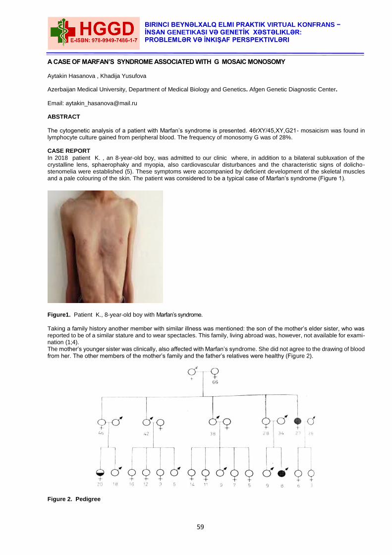
59
BIRINCI BEYNƏLXALQ ELMI PRAKTIK VIRTUAL KONFRANS − İNSAN GENETIKASI VƏ GENETİK XƏSTƏLIKLƏR: PROBLEMLƏR VƏ İNKIŞAF PERSPEKTIVLƏRI
A CASE OF MARFAN’S SYNDROME ASSOCIATED WITH G MOSAIC MONOSOMY Aytakin Hasanova , Khadija Yusufova Azerbaijan Medical University, Department of Medical Biology and Genetics. Afgen Genetic Diagnostic Center.
Email: [email protected] ABSTRACT
The cytogenetic analysis of a patient with Marfan’s syndrome is presented. 46rXY/45,XY,G21- mosaicism was found in lymphocyte culture gained from peripheral blood. The frequency of monosomy G was of 28%. CASE REPORT
In 2018 patient K. , an 8-year-old boy, was admitted to our clinic where, in addition to a bilateral subluxation of the crystalline lens, sphaerophaky and myopia, also cardiovascular disturbances and the characteristic signs of dolicho- stenomelia were established (5). These symptoms were accompanied by deficient development of the skeletal muscles and a pale colouring of the skin. The patient was considered to be a typical case of Marfan’s syndrome (Figure 1).
Figure1. Patient K., 8-year-old boy with Marfan’s syndrome.
Taking a family history another member with similar illness was mentioned: the son of the mother’s elder sister, who was reported to be of a similar stature and to wear spectacles. This family, living abroad was, however, not available for exami-nation (1;4). The mother’s younger sister was clinically, also affected with Marfan’s syndrome. She did not agree to the drawing of blood from her. The other members of the mother’s family and the father’s relatives were healthy (Figure 2). Figure 2. Pedigree
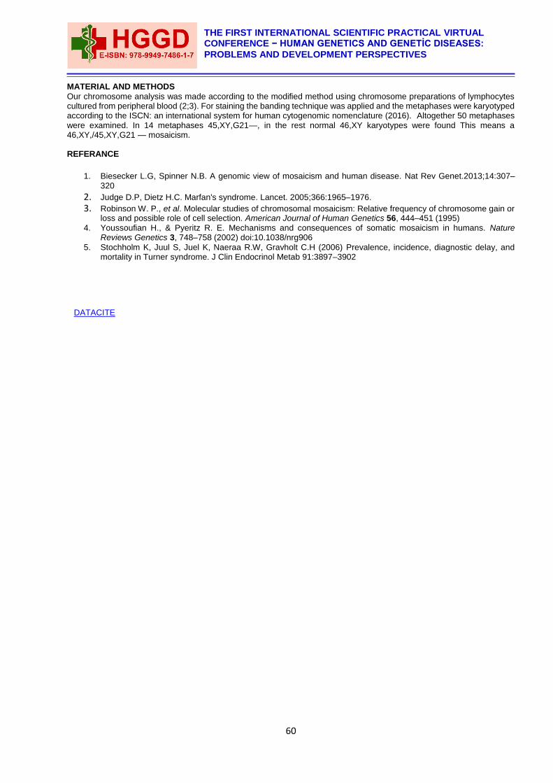
60
THE FIRST INTERNATIONAL SCIENTIFIC PRACTICAL VIRTUAL CONFERENCE − HUMAN GENETICS AND GENETİC DISEASES:
PROBLEMS AND DEVELOPMENT PERSPECTIVES
MATERIAL AND METHODS
Our chromosome analysis was made according to the modified method using chromosome preparations of lymphocytes cultured from peripheral blood (2;3). For staining the banding technique was applied and the metaphases were karyotyped according to the ISCN: an international system for human cytogenomic nomenclature (2016). Altogether 50 metaphases were examined. In 14 metaphases 45,XY,G21—, in the rest normal 46,XY karyotypes were found This means a 46,XY,/45,XY,G21 — mosaicism. REFERANCE
1. Biesecker L.G, Spinner N.B. A genomic view of mosaicism and human disease. Nat Rev Genet.2013;14:307–320
2. Judge D.P, Dietz H.C. Marfan's syndrome. Lancet. 2005;366:1965–1976. 3. Robinson W. P., et al. Molecular studies of chromosomal mosaicism: Relative frequency of chromosome gain or
loss and possible role of cell selection. American Journal of Human Genetics 56, 444–451 (1995) 4. Youssoufian H., & Pyeritz R. E. Mechanisms and consequences of somatic mosaicism in humans. Nature
Reviews Genetics 3, 748–758 (2002) doi:10.1038/nrg906
5. Stochholm K, Juul S, Juel K, Naeraa R.W, Gravholt C.H (2006) Prevalence, incidence, diagnostic delay, and mortality in Turner syndrome. J Clin Endocrinol Metab 91:3897–3902
DATACITE
61
BIRINCI BEYNƏLXALQ ELMI PRAKTIK VIRTUAL KONFRANS − İNSAN GENETIKASI VƏ GENETİK XƏSTƏLIKLƏR: PROBLEMLƏR VƏ İNKIŞAF PERSPEKTIVLƏRI
MOLECULAR GENETIC DIAGNOSTICS OF CHROMOSOMAL DISEASES WITH MULTIPLEX LIGATION-DEPENDENT PROBE AMPLIFICATION
Aytakin Hasanova Azerbaijan Medical University, Department of Medical Biology and Genetics, Azerbaijan ABSTRACT
Chromosomal diseases are birth defects caused by changes in the number or structure of chromosomes. Among newborn children, the frequency of chromosomal pathology is up to 1.0%. Trisomy of 21 chromosomes, which leads to Down syndrome, is most common. Polysomies on X and Y chromosomes are also noted, with an abnormal number of sex chromosomes often showing themselves only at pubertic age. Changes in the structure of chromosomes (aberrations) are less common, but cause profound disorders in the development of many organ systems. The most severe clinical picture is seen in the deletion (loss) of part of the chromosome, it may be the deletion an entire shoulder or only a chromosome locus. Lack of genetic material leads to severe defects. The duplication (doubling) of the chromosome section may, among other things, affect the mental and psychological development of the patient, but usually does not lead to pronounced development abnormalities. The main method for analyzing the number and structure of chromosomes is karyotyping. This study involves several steps: cell culture; Preparation of preparations of metaphase chromosomes and interphase nuclei, colouring of the preparation; microscopic analysis. To detect numerical and structural changes in chromosomes, it is sufficient to carry out karyotyping using differential staining methods. In order to detect small deletions or duplications on the chromosome, it is generally necessary to use an in situ fluorescent hybridization technique that is sufficiently complex and cost-effective. economically costly Among molecular genetic methods for analysis of chromosomal anomalies, the multiplex ligation-dependent probe amplification (MLPA) method is identified. MLPA is a variation of the multiplex polymerase chain reaction that permits amplification of multiple targets with only a single primer pair. It detects copy number changes at the molecular level, and software programs are used for analysis. Identification of deletions or duplications can indicate pathogenic mutations, thus MLPA is an important diagnostic tool used in clinical pathology laboratories worldwide. This technique allows to estimate the number of copies of the gene, detect point mutations, prolonged deletions or duplication of chromosomes. In a single assay, it is possible to determine the number of copies of up to 40 sites of different genes. It is not necessary to use living cells for analysis, which gives a time advantage and allows the detection of pathologies after long storage of the material. The aim of the work was to determine the number of sexual and some somatic chromosomes and to analyze for common microdelection syndrome in patients with various pathologies and a control group of people by MLPA method. Commercial sets from MRC-Holland (Amsterdam, Netherlands) were used. The analysis was carried out using an automatic CEQ8800 capillary electrophoresis system (Beckman Coulter, USA). The following chromosome structure disorders were identified: deletion 22q11.2(Di Georgie syndrome), deletion 17p13.3 (Miller-Dicker syndrome), X chromosome dysomy in male XXY (Klinefelter syndrome). The use of molecular genetic methods for the diagnosis of hereditary diseases has a number of advantages over cytogenetic methods and allows the effective diagnosis of chromosomal diseases in both the prenatal and postnatal period. Keywords: multiplex, ligation-dependent probe amplification, aneuploidy, microdeletion syndrome.
REFERENCES
1. Moeschler JB, Shevell M: American academy of pediatrics committee on genetics. Clinical genetic evaluation of the child with mental retardation or developmental delays. Pediatrics 2006, 117:2304–2316.
2. Knight SJ, Horsley SW, Regan R, Lawrie NM, Maher EJ, Cardy DL, Flint J, Kearney L: Development and clinical application of an innovative fluorescence in situ hybridization technique which detects submicroscopic rearrangements involving telomeres. Eur J Hum Genet 1997, 5(1):1–8.
3. Shaffer LG, Bejjani BA, Torchia B, Kirkpatrick S, Coppinger J, Ballif BC: The identification of microdeletion syndromes and other chromosome abnormalities: cytogenetic methods of the past, new technologies for the future. Am J Med Genet C Semin Med Genet 2007, 145:335–345.
4. Rooms L, Vandeweyer G, Reyniers E, van Mol K, de Canck I, Van der Aa N, Rossau R, Kooy RF: Array-based MLPA to detect recurrent copy number variations in patients with idiopathic mental retardation. Am J Med Genet A 2011, 155A(2):343–348.
5. Su Z, Ning B, Fang H, Hong H, Perkins R, Tong W, Shi L: Next-generation sequencing and its applications in molecular diagnostics. Expert Rev Mol Diagn 2011, 11(3):333–343.
DATACITE
62
THE FIRST INTERNATIONAL SCIENTIFIC PRACTICAL VIRTUAL CONFERENCE − HUMAN GENETICS AND GENETİC DISEASES:
PROBLEMS AND DEVELOPMENT PERSPECTIVES
THE FREQUENCY AND DISTRIBUTION OF SISTER CHROMATID EXCHANGES (SCES) IN THE
INDIVIDUAL CHROMOSOMES OF HUMAN KARYOTYPE
1Aytakin Hasanova, 2Gulnara Kuliyeva
1Azerbaijan Medical University, 2Baku Medical Plaza Clinic.
The aim of this study was to determine the frequency and distribution of sister chromatid exchanges (SCEs) in the individual chromosomes of human karyotype, Chromosomes were studied in (i) healthy subjects, (ii) subjects with rearrangements of X chromosome, (iii) in lymphoblastoid cell lines isolated from the peripheral blood of patients with acute leukaemias. 5-bromo- 2-deoxyuridine (BUdR) was added for 48, 72 and 96 hours, respectively, in a concentration of 30 µg per ml. The slides were stained according to the technique of Perry and Wolff (4). The average number of SCEs was 9,2 with no statistically significant differences between the individual groups. Out of the total number of SCEs 20% was found in the centromeric region with no difference between the cells in the 2nd and 3rd divisions. The observed distribution of breakpoints was approximately proportional to the relative length of individual chromosomes with a higher number in long chromosomes and a lower number in the small ones. Non-random distribution of SCEs was only found in the B group of chromosomes of lymphoblastoid cell lines, which showed an excess compared with the SCEs of both the controls and the expected frequency based on the relative length of chromosomes. Neither in the late replicating i(Xq) nor in the early replicating Xq— did the number of SCEs significantly exceed the expected value.
MATERIAL AND METHODS
The evaluation of the number and distribution of SCEs was performed in Seventy-five mitoses with karyotype 46,X,I (Xq) derived from 4 patients. In one of these subjects a double centromere in the i(Xq) was identified by means of C banding. Twenty mitoses of one patient with reciprocal translocation 46, XX, t (X,4) (Xqter→Xq22: :4pl6→4qter). Autoradiography proved that both the deleted X and the B4 chromosome with translocation were early replicating. Fifty-six mitoses obtained from three lymphoblastoid cell lines, derived from the peripheral blood of patients suffering from acute leukaemias. These were classified as dedifferentiated (Epstein-Barr virus positive), lymphatic and myeloid (both EBV-negative). One hundred and seventy mitoses from the control group consisting of 3 males with karyotype 46,XY and 4 females with 46,XX karyotype. The mean age of controls was 29.5 years. The peripheral blood leukocytes were cultivated for 48, 72 and 96 hours in EPL or Parker medium (Usol, Prague), enriched by 20% calf serum, with PHA (Welcome) and protected by streptomycin and penicillin. Bromodeoxyuridine (Sigma) was added in a concentration of 30 µg/ml since the beginning of cultivation. The tubes were protected from light to avoid photolysis. Colcemid (Ciba) in a concentration of 10 µg/ml was added two hours before harvesting. The cells were hypotonized by 0.075 M KC1 and methanol: acetic acid (3:1) were used for fixation(1). The lymphoblastoid cell lines were grown as permanent suspension cultures from peripheral blood. They were established and subcultured in RPMI 1,640 medium enriched by 20% fetal calf serum, protected by streptomycin and penicillin, without PHA. BUdR was added in a concentration of 30 µg/ml for 48 or 72 hours following 48 hours of subcultivation. The examination of SCEs was carried out in the 15th, 3rd, and 38th, and in the 16th passages in three individual lines. The harvesting of chromosomes was the same as with the above-mentioned short-term peripheral blood cultures(2). Chromosomal preparations were stained according to the FPG staining technique of Perry and Wolff (4). Intact mitoses with harlequin chromosomes were photographed and SCEs evaluated first directly under the microscope, then from enlarged negatives or karyotypes. Exchanges occurring in short arms, long arms and in centromeric regions were counted separately. The number of SCEs was examined independently by two experienced observers and expressed as the number of breakpoints. Statistical evaluation was performed by means of the test and χ2 test.
RESULTS
The difference in the number of SCEs found per cell in 50 mitoses of control subjects when evaluated from photomicrographs or karyotypes is small but significant (p< 0.01). Therefore, the data described have been obtained from karyotypes only. The mean value of 9.5 SCEs per mitosis found in the control group was in no way significantly different from mean values of the other groups, i.e. those consisting of pathological karyotypes and lymphoblastoid cell lines (p >0.05). High frequency of SCEs (20%) was found in the centromeric region of chromosomes after the 2nd and 3rd divisions in BUdR medium(6). The distribution of SCEs both observed and expected on the basis of the relative length of chromosomal groups in the karyotype with results of χ2 test is shown in Table II. The changes were similar in all observed groups. The only exception were B group chromosomes of the lymphoblastoid ceil lines, where the number of breakpoints increased significantly (p<0.01). In this particular chromosomal group the breakpoints leading to exchanges were distributed proportionally along the whole length of all chromosomes. Significant difference was found not only as against the expected number of SCEs but also as against the control group. The increased number of breakpoints on the long chromosomes was naturally matched by their decrease in small chromosomes(5). We preferred to evaluate the non-banded chromosomes (G-banding considerably interferes with the accuracy of SCE calculation), the regions of breakpoints are only roughly delineated. Even so it is clear that some regions are more often
63
BIRINCI BEYNƏLXALQ ELMI PRAKTIK VIRTUAL KONFRANS − İNSAN GENETIKASI VƏ GENETİK XƏSTƏLIKLƏR: PROBLEMLƏR VƏ İNKIŞAF PERSPEKTIVLƏRI
involved in SCEs than others. This is the case especially with regions 1q1, 3q2, 4q2, 8c and 16c. SCEs seem to be preferentially located on G-negative bands, as mentioned also by Morad, Jonasson and Lindsten (3).
REFERENCES
1. Burkholder G. D. (1993): The basis of chromosome banding. Appl Cytogenet.,19, 181-186. 2. Craig J.M. Bickmorc W.A. (1994): The distribution of CpG islands in mammalian chromosomes. Nature Genetics,
7, 376-382. 3. Morad M., Jonasson J. and Lindsten J. (1973): Distribution of Mitomycin C induced breaks on human
chromosomes. Hereditas (Lund), 74, 273-282. 4. Perry P. and Wolff S. (1974): A new Giemsa method for the differential staining of sister chromatids. Nature,251,
156-158. 5. Sumner A.T., de la Torre J., Stuppia I.(1993):The distribution of genes on chromosomes: A cytological
approach. J Mol Evol, 37,117-122. 6. Sumner A.T.(1994) Functional aspects of the longitudinal differentiation of chromoso mes. Eur J Histochem, 38,
91-109.
DATACITE
64
THE FIRST INTERNATIONAL SCIENTIFIC PRACTICAL VIRTUAL CONFERENCE − HUMAN GENETICS AND GENETİC DISEASES:
PROBLEMS AND DEVELOPMENT PERSPECTIVES
WORKING CONDITIONS AND HEALTH OF SEAFARERS
1Aytakin Hasanova, 2Simuzar Hajizada 1Azerbaijan Medical University. I Preventive Medicine Faculty. PhD in Medical Biology. 2The Ministry of Health of the Republic of Azerbaijan. Azerbaijan State Institute for the Improvement of Physicians named Aziz Aliyev. PhD dissertant. ABSTRACT
Relevance of a problem
Modern development of the fleet is inextricably connected with the solution of the problem of preservation and improvement of seafarers' health, improvement of their working, living and rest conditions. The most important condition for the health of seafarers is the provision of optimal habitat on the vessel. At the same time, the vessel should be considered as an artificial ecological closed system, which provides the crew with a long-term active existence. A complex of interconnected environmental factors (climatic conditions of the navigation area, microclimate of ship 's rooms, noise, vibration, electrostatic radiation, electromagnetic radiation, harmful substances in the air, microflora in the room, psychophysiological factors, etc.) simultaneously acts on the human body in the conditions of buoyancy. The person in the final outcome responds to the environment as a whole. Therefore, the criterion reflecting the influence of the ship's environment on the human body is the level of functional state of the worker and his health [Lane T, et al., 2002; Rohrer JE. 2004; Heistaro S, et al., 2001; Jensen OC, et al., 2001; Thomas M, 2003]. There are currently poor working conditions and a high rate of occupational morbidity among maritime workers. The intensification of labor in the flight is increasing. There is poor quality of pre-trial and pre-trial medical examinations, reduction of medical positions on ships, leading to a deterioration of the quality of medical care or its complete absence. Obsolete vessels with expired service life are used. These circumstances lead to a deterioration of the habitat on ships and pose a threat to the health of workers. However, research on comprehensive hygienic assessment of working conditions and on the state of health of seafarers in modern conditions is scarce and often contradictory. This justifies the relevance of conducting a scientific study on the further study of the complex of factors that form the conditions of habitation on ships. Work purpose
Develop a modern system of science-based measures to prevent adverse effects of ship environment factors on the human body to preserve the health and high efficiency of seafarers. Research problems
To carry out a hygienic assessment of the physical and chemical factors forming the conditions of habitation on sea-going vessels, to assess the severity and intensity of work on ships of the sea-going fleet; Undertake a comprehensive assessment of seafarers health; To carry out an analysis of industrial injuries on ships of the marine fleet; Provide scientific justification for the system of preventive measures and recommendations for the protection of labour and health of marine vessels in the modern period. Methods of research
included hygienic, psychophysiological, sociological (questionnaire and interviewing) and analytical. Provisions for protection
The leading adverse production factors on ships should be considered noise, vibration, microclimate parameters, lack of recovery, tension and severity of work; Work in harmful conditions of production contributes to the development of changes in the state of health of seafarers, determines the structure of production - due to and occupational morbidity, increases the risk of occupational injuries; The condition of labour and health of ship specialists justifies the need to organize a system of labour protection and health of seafarers, the main purpose of which is to create safe working conditions that exclude or minimize the risk of an employee receiving a professional disease or accident, preserving the life and health of ship specialists. Scientific novelty of work
For the first time in the conditions of the Azerbaijani region, a comprehensive sanitary and hygienic assessment of working conditions, an analysis of the state of health and industrial injuries of specialists working on sea vessels was carried out. Leading harmful production factors have been identified, and features of occupational and production-related morbidity have been identified. For the first time, an assessment of the natural resistance of the body of seafarers was given, as well as an analysis of individual risk factors. On this basis, a system of labour protection and health of seafarers is justified and developed.
65
BIRINCI BEYNƏLXALQ ELMI PRAKTIK VIRTUAL KONFRANS − İNSAN GENETIKASI VƏ GENETİK XƏSTƏLIKLƏR: PROBLEMLƏR VƏ İNKIŞAF PERSPEKTIVLƏRI
The practical value of the work
lies in the fact that the comprehensive assessment of working conditions and the state of health of seafarers proposes a system of labour protection and health of persons working on sea vessels, which will ensure working conditions that meet the requirements of safety, which will contribute to the preservation of the life and health of maritime transport workers. REFERENCES
1. Lane T, Obando-Rojas B, Sorensen M, Wu B, Tasiran A. Crewing the International Merchant Fleet. Surrey:
Lloyd's Register–Fairplay Ltd, 2002 2. Rohrer JE. Medical care usage and self-rated mental health. BMC Public Health 2004;4:3 3. Heistaro S, Jousilahti P, Lahelma E, Vartiainen E, Puska P. Self rated health and mortality: a long term prospective
study in eastern Finland. J Epidemiol Community Health 2001;55:227–232. 4. Jensen OC, Laursen FV, Sørensen JFL. International surveillance of seafarers' health and working environment.
A pilot study of the method. Preliminary report. Int Marit Health 2001; 52:59–67. 5. Thomas M, Sampson H, Zhao M. Finding a balance: companies, seafarers and family life. Marit Policy Manage
2003;30:59–76..
DOI: 10.36962/hggd0164
DATACITE
66
THE FIRST INTERNATIONAL SCIENTIFIC PRACTICAL VIRTUAL CONFERENCE − HUMAN GENETICS AND GENETİC DISEASES:
PROBLEMS AND DEVELOPMENT PERSPECTIVES
FAMILY DOCTOR AND MEDICAL GENETICS
1Mustafa Salikhov, 2Kamile Salikhova 1Head of the Department of Family Medicine. Associate Professor.
2Assistant of the Department of Family Medicine.
1,2Azerbaijan Medical University
ABSTRACT
Introduction of the concept of family doctor as a primary medical care, involves his methods of work with a family as with a patient, the concept “family-patient”, introducing absolutely new state of things in organization and professional work of family doctor, as known, that structure of the family includes- size of the family, social composition and number of generations. Now, it is very actual the medical insurance and preventive medicine in Azerbaijan, so importance of medical genetics is increasing. The family doctor should do clinical and geneological investigations to the patient and to the members of his family, find out the group of the patients with the high risk and involve the preventative measures. Keywords: family doctor, preventive medicine, clinical and geneological investigations.
Clinical genetics is one of the chapters of clinical medicine and human genetics. Clinical genetics solving problem of diagnosis, prognosis and treatment of different hereditary diseases. Introduction of the concept of family doctor as a primary medical care, involves his methods of work with a family as with a patient, the concept “family-patient”, introducing absolutely new state of things in organization and professional work of family doctor, as known, that structure of the family includes- size of the family, social composition and number of generations. Now, it is very actual the medical insurance and preventive medicine in Azerbaijan, so importance of medical genetics is increasing. The family doctor should do clinical and geneological investigations to the patient and to the members of his family, find out the group of the patients with the high risk and involve the preventative measures.
Therefore, knowledge of medical and clinical genetics one of the necessary condition in professional activity of a family doctor. Family doctor must have certain skills in taking patient’s anamnesis, knowledge of signs in hereditary pathology and should inform families of inappropriateness of close blood related marriages, because children born from close blood related marriages have more possibilities of in hereting genetic diseases and birth defects and newborn mortality is more higher.
Certainly, invasion of family doctor to the area of the patient’s hereditary structure possible only with one aim to treat the patient and to create condition, when the birth defect gen wouldn’t transfer to the children. And finally, theoretical calculations of family geneticist and experience in genetical consultations give an opinion regarding rise of having a sick baby in every particular family and find people, who need medical genetic consultation. Important part of care in patients with genetic disease, in addition to therapeutic and preventative measures, are also psyhotherapy correction and social rehabilitation. Based on above, it is important to note: qualification of the doctor and his participation in the life of the family affect not only the health of people who live now, but also the health and prosperity of future generations. REFERENCES
1. Integrating Genetic Counseling into Family Medicine JEFFREY R. MARTIN, M.D. ADAM S. WILIKOFSKY,
PH.D.Lancaster General Hospital Family Medicine Residency Program Lancaster, Pennsylvania Am Fam Physician. 2005
2. Genetics for Family Physicians W. Gregory Feero, MD, PhD 3. Clinical genetics issues encountered by family physicians Louise S Acheson, Kurt C Stange & Stephen
Zyzanski Genetics in Medicine 2005
DATACITE
67
BIRINCI BEYNƏLXALQ ELMI PRAKTIK VIRTUAL KONFRANS − İNSAN GENETIKASI VƏ GENETİK XƏSTƏLIKLƏR: PROBLEMLƏR VƏ İNKIŞAF PERSPEKTIVLƏRI
THE IMPORTANCE OF ELECTRON MICROSCOPIC SYSTEMATIC CLASSIFICATION WE OFFER IN CERVICAL CANCER Aynur Panahova Department of Oncology, Azerbaijan Medical University. ABSTRACT
Based on the electron microscopic diversity in tumour cells in cervical cancer, we systematized the results obtained in our study, considering the difference in the various cells of bioptates taken from the same patient. The scientific novelty of the work is the development of an ultrastructural microscopic classification of neoplastic cells in cervical cancer by summarizing electron microscopic signs. The said classification allows us to explain the electron microscopic theoretical basis of differential diagnosis based on the organ-tissue and cytospecific ultrastructural signs (groups and types of tumour cells) of tumour tissue, on the one hand. On the other hand, it helps to specify the degree of ultrastructural differentiation of neoplasms, comparison between cells, types of tumour cell and options of tumours. The main purpose of the use of electron microscope in practical medicine is related to the histogenetic diagnosis of tumours. From this point of view, the systematic classification we offer allows us to systematize electron microscopic signs of tumour cells and tumour cell groups, on the one hand, and, on the other hand, it allows us to detect the degree of ultrastructural differentiation of new derivatives (tumour cells, tumour cell groups, ratio of tumour options). Thus, the systematic classification we provided as a result of our study allows us to explain the theoretical basis of the general principles of tumour cells. This system explains the specialization of ultrastructural organ-tissue-cell during specific electron microscopic differentiation of tumour cells. We can also get information, at electron microscopic level, on the morphogenetic potential of cambial cells, which are the source for creation of tumour cells as a result of such a systematic analysis. Thus, in the irregular and numerous ultrastructural options of tumour cells in the tumour mass, precise regularities can be obtained, and we can say something about their cell composition based on these regularities. Based on the stated and obtained certain results, it is possible to substantiate the great importance of electron microscopy in practical activity and theoretic studies, diagnostics, differential diagnostics of tumours, differentiation, histogenesis, biological features of neoplastic cells.
OBJECTIVE
The research aims to optimize the prognostic criteria of the disease based on a comprehensive clinical, instrumental, morpho-functional, and statistic analysis of cervical cancer, as well as electron microscopic indicators. MATERIAL AND METHODS
The study covered 330 patients with in cervical background diseases, precancerous diseases and cancer from 2007 to 2017. Main group: 220 patients with cervical cancer; Control group: 110 patients - erosions, cervical polyps, CIN 1-3 Electron microscopic studies were carried out and based on these studies, cytological (Pap-Smear and Bethesda) and histological and electron microscopic study of the ultrasound classification of Cervical Neoplasia was carried out. Electron microscopic studies were conducted on 93 cancer patients in an electron microscopic laboratory at the Department of Histology of AMU.
RESULTS
Differentiated tumour cells rich (60%) with ultrastructural organo-tissue and organoids with cytospecific signs are predominant in cervical squamous cell corned cancer (option 1) [6, 62, 117]. The amount of organoids in cervical squamous cell non-corned cancer is at medium level (50%), the activity of 2 groups of non-differentiated tumour cells, ultrastructural cataplasias noticed in one or more types of cell indicate a worse prognosis (option 2) [6, 62, 117]. In non-differentiated cervical cancer, only non-differentiated tumour cells with less ultrastructural organoids (30%) were noticed (option 3) [6, 62, 117]. The same number of differentiated and non-differentiated tumor cells is detected in cervical adenocarcinomas (variant IV). The said elements are related to heterogeneous cells and contribute to progress, development of tumours, and metastasis [6, 62, 117]. Based on the ultrastructural classification we applied in cervical neoplasms, different types, species of tumour cells, and comparative tumour options in the groups to which they belong were identified [2, 3, 117]. Based on calculation of prognostic score, it was detected that those over the age of 45 years (informativeness - 0,821) had more abortions in their anamnesis (1,640) had HPV positive (1,145), high stage (2,529), low degree of differentiation (1,422), lymph nodes (4,773) and gave distant metastases (4,823), and patients with less organelles (2,404) were found to be more likely to have recurrence during electron microscopic examination [3].
68
THE FIRST INTERNATIONAL SCIENTIFIC PRACTICAL VIRTUAL CONFERENCE − HUMAN GENETICS AND GENETİC DISEASES:
PROBLEMS AND DEVELOPMENT PERSPECTIVES
REFERENCES
1. Amiraslanov A.T. Panahova A.M/ Aspects of etiopathogenesis of background, precanserous diseases and cancer of the cervix / Вестник Xирургим Казаxстана, Алматы – 2017, №4 (53), səh. 17-22
2. Alemany L., de Sanjose S., Tous S., Quint W. et al. Time trends of human papilloma virus types in invasive cervical cancer, from 1940 to 2007 // International Journal of Cancer, 2014, vol. 135 (1), p. 88-95
3. Al-Darwish A.A., Al-Naim A.F., Al-Mulhim K.S., Al-Otaibiet N.K. al. Knowledge about cervical cancer early warning signs and symptoms, risk factors and vacination among students at a medical school in Al-Ahsa, Kingdom of Saudi Arabia // Asian Pacific Journal Cancer Prevention, 2014, vol. 15(6), p. 2529-2532
4. Bahmanyar R.E., Paavonen, J., Naud P. et al. Prevalence and risk factors for cervical HPV infection and abnormalities in young adult women at enrolment in the multinational PATRICIA trial // Gynecologic Oncology, 2012, vol. 127 (3), p. 440-450
5. Chen A.A., Heideman D.A., Boon D., Gheit T. et al. Human papilloma virus 45 genetic variation and cervical cancer risk worldwide // Journal of Virology, 2014, vol. 88 (8), p. 4514-4521
DATACITE
69
BIRINCI BEYNƏLXALQ ELMI PRAKTIK VIRTUAL KONFRANS − İNSAN GENETIKASI VƏ GENETİK XƏSTƏLIKLƏR: PROBLEMLƏR VƏ İNKIŞAF PERSPEKTIVLƏRI
UŞAQLIQ BOYNU DİSPLAZİYALARINDA PD-L1 ÇALIŞMALARIN PROQNOSTİK ƏHƏMİYYƏTİ
İradə Soltanova, Nigar Mehdiyeva
Azərbaycan Tibb Universitetin Onkoloji kafedrası.
ABSTRACT
The article is included the clinical and morphological features of 35 patients who had been diagnosed and treated with cervical dysplasia and reflected the immununistochemical results of PD-L1 expression in atypical cells. By standardized immunochemical methods, receptor levels vary depending on the degree of intraepitelial dysplasia. In patients with CIN I, cells were not stained at all (negative PD-L1 reaction), PD-L1 expression in CIN II was 1+ and 2+ in 76.9±5.6% , and 3+ in 23.1±5.3% of patients (p<0.05). In patients with CIN III, 3+ cases (severe staining) were detected in 66.7±4.2% and cases of 2+ (moderate staining) in 33.3±4.9% of women. Thus, expression of the PD-L1 receptor, known as anti tumor immunitite regulator is closely related to dysplastic processes that begin in the tissue. By other words, the detection this receptor expression in patients with CIN diagnosis(especially CIN III) can play an important role in the selection of treatment tactics and assessment of prognosis,
REZÜME
Məqalədə uşaqlıq boynu displaziyası diaqnozu ilə müşahidə və müalicə almış 35 xəstənin kliniki-morfoloji xüsusiyyətləri və PD-L1 ekspressiyasının imuunhistokimyəvi nəticələri əks etdirilmişdir. Standartlaşdırılmış immunhistokimyəvi işləmə üsulu ilə təyin edilmişdir ki, reseptor səviyyəsi intraepitelial displaziyanın dərəcəsindən asılı olaraq dəyişir. CIN I olan xəstələrin preparatları ümumi olaraq boyanmadığı halda (negativ PD-L1 reaksiya), CIN II zamanı PD-L1 ekspressiyası 76,9±5.6% hallarda 1+ və 2+, 23,1±5,3% xəstələrdə isə 3+ kimi qiymətləndirilmişdirp<0,05. CIN III olan xəstələrdə 3+ (şiddətli boyanma)halları 66,7±4,2%, 2+ (orta səviyyədə boyanma)hadisələri isə 33,3±4.9% qadınlarda müəyyən edilmişdir. Beləliklə, şişəleyhinə immunitetin tənzimləyicisi və tumor hüceyrələrinə xas olduöu bilinən PD-L1 reseptorunun ekspressiyası toxumada başlayan displastik proseslərlə sıx əlaqəlidir. Başqa sözlə desək, CIN diaqnozu qoyulmuş xəstələrdə (xüsusən də CIN III) bu reseptorun təyini müalicə taktikasının seçilməsində və proqnozun qiymətləndirilməsində mühüm əhəmiyyət kəsb edə bilər. Uşaqlıq boynu xərçəngi (UBX) qadınlar arasında rast gəlinən bədxassəli şişlərin 6,6% təşkil edir, hər il dünyada 570000-dən çox yeni xəstələnmə halları qeydə alınır, və bu say ildən-ilə artmaqdadır (1,2,3). UBX-nin skrininqində xərçəngönü xəstəliklərin aşkarlanması bu problemlə mübarizənin əsas mexanizmlərindən biridir (4). Bu səbəbdən uşaqlıq boynu displaziyalarının (cervical intraepithelial neoplasia, CİN) diaqnostikası, adekvat müalicəsi və proqnostik dəyərləndirilməsi UBX ilə mübarizədə mühüm yer tutur.
TƏDQİQATIN MƏQSƏDİ
CIN displaziyalarında PD-L1 reseptorun ekspessiyasının histoloji qradasiya ilə əlaqəsinin öyrənilməsi. MATERİAL VƏ METODLAR Tədqiqata CİN diaqnozu qoyulmuş və konizasiyaya məruz qalmış 35 xəstənin preparatlarından daxil olmuşur. Əldə olunmuş yeni kəsiklərdə deparafinizasiyadan sonra PD-L1-monoklonal dovşan anticisimlərindən (VD21R, Medaysis firması) istifadə etməklə standartlaşdırılmış immunhistokimyəvi işləmə üsulu (VENTANA Bench Mark Ultra) tətbiq edilmişdir. Reaksiya nəticələri boyanan hüceyrə sayına görə aşağıdakı kimi dəyərləndirilir: 1+ - zəif boyanan hüceyrə sayı 10%-dən azdır; 2+ - ≥10% hüceyrələrdə orta dərəcəli boyanma; 3+ - ≥10% hüceyrələrdə qüvvətli boyanma. NƏTİCƏLƏR
Xəstələrin əksəriyyətini 28-49 yaş arası qadınlar təşkil etmişdir – 32 nəfər (91,4±1,9%). Onlardan 20-si (57,1±2,4%) 28-39 yaş arası qadınlardır. 60-69 yaş arasında isə UBD diaqnozu ilə yalnız 1 xəstə təsadüf etmişdir (2,9±3,2%,p<0,05). Kontingentə daxil olan xəstələrin 30-u (85,7±3,7%) reproduktiv fəal qadınlar idi, 5-i isə (14,3±4,0%, p<0,05) pre- və menopauza dövründə idi. Qadınların 31-də (88,6±2,6%) heç bir şikayət olmamış, qalanları isə uşaqlıq yolundan periodik gələn selikli-qanlı ifrazatı qeyd etmişlər (11,4±2,9%). Bütün xəstələrə sitoloji müayinə, kolposkopiya və uşaqlıq boynundan biopsiya götürülmüşdür. Aparılan sitoloji müayinənin nəticələri 30 halda (85,7±4,1%) sonrakı histoloji rəylə üst-üstə düşmüşdür. Onlardan 26-da (74,3±2,8%) HSİL (high grade intraepithelial squamous lesion), 4-də (11,4±2,7%) isə LSİL (low grade intraepithelial squamous lesion) təsbit edilmişdir. Histoloji müayinəyə əsasən xəstələr 3 qrupa bölünmüşdür: CİN I – 7 (20±3,5%), CİN II – 13(37,1±4,5%) və CİN III – 15 (42,9±2,8%) xəstə. Xəstələrin 12-də (34,2±3,4%) epitelial zədələnmənin dərinliyi 1,4 mm-ə qədər olmuşdur, onların 9-u(75,0±5,1%) CIN III qrupuna daxil olan qadınlardır. Bu isə həmin qrupdakı hadisələrin 60,0±3,9%-ni təşkil edir. İmmunhistokimyəvi olaraq CİN I zamanı boyanma qeyd edilməmiş; CIN II zamanı - PD-L1 ekspressiyası 6 halda (46,1±5,2%)1+, 4-də (30,8±3,7%) 2+ və 3-də(23,1±5,3%, p<0,05) 3+ kimi qiymətləndirilmişdir. CIN IIIxəstələrin10-da (66,7±4,2%,p<0,05) 3+, 5-də isə (33,3±4.9%) 2+ səviyyədə müəyyən edilmişdir, zəif boyanma hadisəsi qeydə alınmamışdır. PD-L1-müsbət hallarda kəskin ifadə olunmuş koylositoz müşahidə edilirdi. İşıq mikroskopunda təhlil zamanı həmçinin limfositar infiltrasiya halları da qeydə alınmışdır. YEKUN
CIN əksər hallarda cavan, reproduktiv fəal qadınlarda rast gəlinir (85,7±3,7%,p<0,05). İntraepitelial invaziyanın dərinlik dərəcəsi displaziyanın səviyyəsindən asılıdır, və CIN III olan xəstələrin 60,0±3,9%-də 1,4 mm olmuşdur. UBD zamanı
70
THE FIRST INTERNATIONAL SCIENTIFIC PRACTICAL VIRTUAL CONFERENCE − HUMAN GENETICS AND GENETİC DISEASES:
PROBLEMS AND DEVELOPMENT PERSPECTIVES
atipik hüceyrələrdə PD-L1 ekspressiyası müşahidə olunur ki, bu displaziya dərəcəsindən asılı olaraq dəyişir. CIN I-də neqativ reaksiya qeyd edildiyi halda, CIN II-də güclü boyanma (3+) 3 hadisədə (23,1±5,3%), CIN III-də isə 66,7±4,2%-də (10 hal) güclü boyanma (p<0,05) və şiddətli koylositoz aşkarlanır. Bundan əlavə, CIN III zamanı zəif boyanma halları ümumiyyətlə qeyd edilməmişdir. Mikroskopik görüntüdə limfositar infiltrasiya halları ilə rastlaşsaq da, bu əlamətin kəmiyyət göstəriciləri və displaziya dərəcəsi ilə əlaqəsi ətraflı tədqiq edilməlidir. ƏDƏBİYYAT
1. F. Bray, J. Ferlay et al. Global cancer statistics 2018: GLOBOCAN estimates of incidence and mortality worldwide for 36 cancers in 185 countries. CA CANCER J CLIN 2018; 68:394–424
2. Р.В. Пак Вестник КазНМУЭпидемиологические особенности рака шейки матки в мире2019, с 78-81 3. https://www.who.int/reproductivehealth/publications/cancers/9241547006/ru 4. M.Feng, L. XuClinical significance of PD-L1 (CD274) enhanced expression in cervical squamous cell
carcinomaInt J ClinExpPathol. 2018; 11(11): 5370–5378.
PROGNOSTİC SİGNİFİCANCE OF PD-L1 STUDİES İN CERVİCAL DYSPLASİA
İrada Soltanova, Nigar Mehdiyeva
Department of Oncology, Azerbaijan Medical University.
ABSTRACT
The article is included the clinical and morphological features of 35 patients who had been diagnosed and treated with cervical dysplasia and reflected the immununistochemical results of PD-L1 expression in atypical cells. By standardized immunochemical methods, receptor levels vary depending on the degree of intraepitelial dysplasia. In patients with CIN I, cells were not stained at all (negative PD-L1 reaction), PD-L1 expression in CIN II was 1+ and 2+ in 76.9±5.6% , and 3+ in 23.1±5.3% of patients (p<0.05). In patients with CIN III, 3+ cases (severe staining) were detected in 66.7±4.2% and cases of 2+ (moderate staining) in 33.3±4.9% of women. Thus, expression of the PD-L1 receptor, known as anti tumor immunitite regulator is closely related to dysplastic processes that begin in the tissue. By other words, the detection this receptor expression in patients with CIN diagnosis(especially CIN III) can play an important role in the selection of treatment tactics and assessment of prognosis. Keywords: clinical and morphological features, cervical dysplasia and reflected the immununistochemical results.
DATACITE
71
BIRINCI BEYNƏLXALQ ELMI PRAKTIK VIRTUAL KONFRANS − İNSAN GENETIKASI VƏ GENETİK XƏSTƏLIKLƏR: PROBLEMLƏR VƏ İNKIŞAF PERSPEKTIVLƏRI
ASSOCIATION OF RS7903146 C/T POLYMORPHISM OF TCF7L2 GENE WITH TYPE 2 DIABETES MELLITUS IN AZERBAIJAN POPULATION: PRELIMINARY STUDIES
Zarintaj Rustamova, Lala Akhundova, Gulmira Alibayova, Nurmammad Mustafayev, Irada Huseynova
Institute of Molecular Biology & Biotechnologies, Azerbaijan National Academy of Sciences.
AIM
Nowadays, type 2 diabetes mellitus (T2DM) is one of the most propagative endocrine diseases. Prediagnosis of T2DM, which is developing due to the combination of genetic and lifestyle factors, is an urgent problem of the diabetology. The aim of this work was to study the association of the single nucleotide polymorphism (SNP) rs7903146 C/T of TCF7L2 gene with a risk of developing of the T2DM. MATERIALS AND METHODS
The object of the study were DNA samples, isolated from patients with T2DM and healthy individuals (approximately equal amount in each group of ~100). DNA profiles were obtained by PCR using primer pairs (marker rs7903146): forward 1 (C allele): 5′-GAACAATTAGAGAGCTAAG CACTTTTTAGAAAC-3′; forward 2 (T allele): 5′-GAACAATTAGAGAGCTAAGCACTTTTT AGAGAT-3′ and common reverse: 5′-AGATGAA ATGTAGCAGTGAAGTGC-3′. PCR conditions were determined by gradient PCR. RESULTS
The results of the current study revealed the following gene variants (in %): homozygotes CC 22.7 and 40.9, homozygotes TT 45.5 and 22.7, heterozygotes CT 31.8 and 36.4 in experimental and control groups respectively. Allele frequencies: Cexp=0.386, Texp=0.614; Ccontrol=0.591, Tcontrol=0.409. The values of the ratios Cexp:Texp=0.63; Ccontrol:Tcontrol=1.44; Cexp:Ccontrol=0.65; Texp:Tcontrol=1.5 indicate that the presence of the C allele in the control and the T allele in the experimental group is ~1.5 times greater than that in experimental and control group respectively. In addition, TT homozygotes in the experimental group are 2 times more than in control group. The number of heterozygotes (CT) is almost equally. CONCLUSIONS
Despite that the frequency of the T allele and the number of TT homozygotes in the group with T2DM are noticeably higher than that in the control, statistically significant correlation between rs7903146 polymorphism and T2DM was lower than that was expected (p<0.1). Since T2DM is a polygenic disease, this may be because of the sample size, which was not enough to make a clear conclusion about the association. Moreover, studies of additional markers should be included. Keywords: molecular-genetic markers, SNP polymorphism, type 2 diabetes mellitus (T2DM), gene transcription factor 7
like 2 (TCF7L2), homozygote, heterozygote. REFERENCE
1. Acharya. S, Al-Elq. A, Al-Nafaie. A, Muzaheed. M, Al-Ali. A. (2015) Type 2 diabetes mellitus susceptibility gene TCF7L2 is strongly associated with hyperglycemia in the Saudi Arabia Population of the eastern province of Saudi Arabia. European Review for Medical and Pharmacological Sciences, 19:3100-3106
DATACITE
72
THE FIRST INTERNATIONAL SCIENTIFIC PRACTICAL VIRTUAL CONFERENCE − HUMAN GENETICS AND GENETİC DISEASES:
PROBLEMS AND DEVELOPMENT PERSPECTIVES
MOLECULAR GENETIC ANALYSIS OF GLA GENE CAUSING INHERITED FABRY DISEASE FOR POPULATION OF AZERBAIJAN REPUBLIC Narmin Salayeva Doctoral student of the Department of Biology and Basics of Medical Knowledge. Lankaran State University,Lankaran,Azerbaijan.
ABSTRACT For the first time in Lenkoran-Astara administrative area of Azerbaijan Republic, patients with cardiomiopathies were genetically screened for Fabry metabolic disease. Screening was carried out by means of identification of α-galactosidase enzyme activity and quantity of globotriasylsphingosine. In 12 out of 29 examined persons we got activity deficit of α-galactosidase enzyme, and amount of globotriasylsphingosine was higher than the norm which were specific for Fabry disease.In 8 women manifested X-linked inheritance type as heterozygotes for Fabry disease, and 4 men were identified as hemizygotes. Molecular genetic analysis identified two different mutations of GLA gene: 801+3A>G mutation in intron 5 and, substitution of Adenine nucleotide with Guanine nucleotide in position 137 (137,A>G) of GLA gene. To prevent Fabry disease it is recommended toscreen affected persons’ family members for α-galactosidase enzyme activities. Keywords: Fabry, inherited disease, mutation, α-galactosidase,lisosome,globotriasylsphingosine (lyso-Gb3), GLA
INTRODUCTION
Fabry disease (Anderson-Fabry disease, inherited distonic lipoidosis etc.) being a lisosome metabolic disease relates to a group of orphan (rare) diseases[1]. GLA gene, modifying Fabry disease clinic, locates on the X-sex chromosome long
shoulder - Хq22. GLAgene encodes α-galactosidaseenzyme. Up to nowadays more than 640 mutations were identified.Mutations 70% are related to missense or nonsense mutations group. The most of mutations have family specificity[5]. Fabry disease being one out of 60 diseases of lisosome storage diseases is a result of complete or partial
α-galactosidase A enzyme activity deficiency. Alpha-galactosidaseA enzyme activity reduction causing sphyngolipidoses metabolism damage, leads to storage of globotriasylsphingosines. The disease has broad spectrum of simptoms. In the course of disease one organ is mainly damaged, problems are in heart or kidney [4].
It should be mentioned that because population of Azerbaijan Republic have neverbeen diagnosed for Fabry disease, the disease has never been identified, there is no data on clinic, biochemistry and genetics of the disease. The goal of the article was to screen patients with cardiomiopathies for Fabry inherited lisosome metabolic disease and to carry out molecular genetic analysis of GLA gene for identified patients. MATERİAL AND METHODS
29 patient suspicious for Fabry inherited lisosome disease venous blood samples 1ml each were taken into EDTA anticoaggulant sample tube and then absorbed into special DBS (dry blood spots) cards.Cards with absorbed blood samples stay at room temperature for 1 hour, and then are analysed at the Chair of Laboratory Science (Azerbaijan State Doctors’ Advanced Training Institute after A.Aliyev) and CENTOGENE laboratories (Germany, Rostock city). 29 cardiologic patients from Lenkoran, Masalli and Astara Central Regional Hospitals were suspicious for Fabry disease to develop those cardiomiopathies. For that purpose α-galactosidase enzyme activities were measured, and in case of enzymatic deficit amounts of globotriasylsphingosine(lyso-Gb3) were identified[2].
Fluorimetric method and liquid chromatography were usedin genetic screening.Sanger methodique was applied for direct sequensing of GLA gene.Testing of the existing mutation inGLA gene became possible with this method.The method was developed in CENTOGENE laboratories, Rostock, Germany[3].
RESULTS
In Masalli region 4 patients identified were 2 men and 2 women.In brothers T.I.and T.A. α-galactosidase enzyme activityshowed lower thanthe norm activity as 0.8 mkmol/l/s (N ≥15.3 mkmol/l/s). To verify the diagnostics, another test was carried out for lyso-Gb3 amount, and happened to be higher than the norm - 106.0 ng/ml (T.I.) and 106.0 ng/ml (T.A.). Fabry disease having X-linked chromosome dominant inheritance type, both of brothers were hemizygous carriers of GLA gene.Alpha-galactosidase enzyme deficiency for sisters T.G. andA.G. being 3.2 mkmol/l/s, 1.9 mkmol/l/s,relatively, amounts of Lyso-Gb3 for them were viсe versa higher – 15.6 ng/ml (T.G) and 8.3 ng/ml (A.G). For both of sisters X-linked autosome dominant inheritance type in heterozygous state was identified. Patient M.A. originated from Lerkoran had got alpha-galactosidase activity lower than the norm–2.3 mkmol/l/s, but amount of Lyso-Gb3 was higher than the norm (109 ng/ml). In Astara area in the family of 6 persons were women and one man born on 12.06.1978. Alpha-galactosidase enzyme showed “0” activity for patient K.A., and amount of Lyso-Gb3 was much higher than the norm (218.0 ng/ml).In six women of the family, activity of alpha-galactosidase enzyme was in range of1.4-1.8 mkmol/l/s. The mean amount of Lyso-Gb3was registered as 16.0 ng/ml (11.0-21.0 ng/ml).
73
BIRINCI BEYNƏLXALQ ELMI PRAKTIK VIRTUAL KONFRANS − İNSAN GENETIKASI VƏ GENETİK XƏSTƏLIKLƏR: PROBLEMLƏR VƏ İNKIŞAF PERSPEKTIVLƏRI
Masalli area all patients were members of one and the same family, thus they had got the same mutation 801+3A>G of GLA gene. In patient M.A. molecular genetic diagnostics of GLA gene identified substitution of Adenine nucleotide withGuanine nucleotide in position 137 (137,A>G). This mutation in position 137 of GLA gene at the time of Adenine nucleotide substitution with Guanine nucleotide causes substitution of Histidine amino acid with Arginine amino acid in position 46 in the courseof protein (enzyme) biosynthesis (46 His>Arg). One and the same GLA gene mutation was identified in one family in Astara area. Adenine nucleotide was substituted with Guanine nucleotidein position 137(137,A>G) of GLA gene.Mutation happened in the gene caused the change of Histidine amino acid to Arginine amino acid (46 His>Arg) in position 46 while protein biosynthesising. Because of mutation relates to missense (nonsense) mutation type, the synthesis of enzyme was completely destroyed. So, twelve patients with Fabry inherited metabolic disease were identified resultingfrom genetic screening in Astara, Lenkoran and Masalli areas population especially among cardiomiopathic patients for α-galactosidase enzyme and globotriasylsphingosine.Molecular genetic analysis of GLA gene identified two different mutations: 801+3A>G and 137, A>G.Both mutations of GLA gene were found for the first time for Republic population.To prevent Fabry disease, index patients family members are recommended to pass through screening of α-galactosidase enzyme activity. REFERENCES
A. Волгина С. Я. Болезнь Фабри // Практическая медицина (научно-практический медицинский журнал), 2012, 62, 7, 75-79.
B. Bouwien E Smidt et al. Plasma globotriaosylsphingosine in relation to phenotypes of Fabry disease (ENG) // J Med Genet. - 2015. - Jan.
C. Daniel Trujillano, Aida M Bertoli-Avella, Arndt Rolfs .Clinical exome sequencing: results from 2819 samples reflecting 1000 families. Eur J Hum Genet. 2017 Feb; 25(2): 176–182. Published online 2016 Nov 16.
D. Gutiérrez-Amavizca B. E., Gal A., Ortíz-Orozco R., et al., Mutational analysis of the GLA gene in Mexican families with Fabry disease. 2017. Genet. 96.
E. Shabbeer A.Mutations of the GLA gene in young patients with stroke: the PORTYSTROKE study--screening genetic conditions in Portuguese young stroke patients. (PMID: 20110537) BaptistaMVPORTuguese Young STROKE Investigators Stroke 2010, 3, 23, 41-54
DATACITE
74
THE FIRST INTERNATIONAL SCIENTIFIC PRACTICAL VIRTUAL CONFERENCE − HUMAN GENETICS AND GENETİC DISEASES:
PROBLEMS AND DEVELOPMENT PERSPECTIVES
HUMAN AS A BIOTIC FACTOR IN ACCUMULATION OF MICROELEMENTS WITH WEAK TOXICITY IN BONE AND MUSCLE TISSUE OF BACKGROUND REPTILE SPECIES IN THE ABSHERON PENINSULA Aysel Hashimova Azerbaijan Medical University, The Department of Medical Biology and Genetics. Azerbaijan. ABSTRACT
The accumulation and amount of microelements with weak toxic effects have been studied in bone and muscle tissue of following Reptile species - Water snake (Natrix tessellata Laurenti, 1768), Mediterranean turtle (Testudo graeca L.1758) and Caspian bent-toed gecko (C.caspius E. 1831).Studied microelements are cobalt Co, molybdenum Mo, manganese Mn, and chromium Cr. All the four microelements that we have studied are mostly accumulated in the body of Caspian bent-toed gecko. It should be noted that the Caspian bent toed gecko is very small and more functionally active among background reptile species that we studied. Its occurrence in oil, gas wells and in areas exposed to technogenic and antropogenic pollution, indicates its plasticity and resistance to toxic microelements. INTRODUCTION In recent years the Absheron peninsula has been subjected to anthropogenic and technogenic pollution in addition to the intense urbanization process.[5] So, it affects all components of the biocenosis in the peninsula and especially, to the flora and fauna. The reptiles that have historically been settled here cannot stay away from these effects, as they feed on existing plants, native invertebrates and vertebrates and drink the water of this area. Therefore, their bodies are rich in microelements of the peninsula, respectively. The problem of deficiency and excess of microelements in human and animal organism is the important issue of world scientists. Thus in both cases, pathological processes occur. The reason for accumulation of toxic microelements in the body of research objects is anthropogenic and technogenic pollution and its directly related to human activity.In this research we aimed to determine how human impacts on the research objects and which of these objects is mostly exposed to this impact. MATERIALS AND METHODS
The objects of our research are the Mediterranean turtle (Testudo graeca L.1758) from the order Turtles (Testudines), the water snake (Natrix tessellata Laurenti, 1768) from the order Snakes (Serpentes) and the Caspian bent-toed gecko (C.caspius E. 1831), from the order Lizards (Sauria). All these orders belong to the class of Reptiles. The route method was used for the collection of materials. The amount of the following microelements -cobalt, molybdenum, chromium, and manganese, which have weak toxic effects and are needed for the normal functioning of the body, have been studied in the muscle and bone tissue of the selected reptile species. [1,2,6] These microelements have toxic effects when their amount is higher than the norm, and in the case of their deficiency, the functional activities of the body are impaired. However, the proper amount of these microelements is actively involved in regulating the physiological and biochemical functions of the body. For the biochemical analysis 10 specimens were taken from each of the Caspian bent-toed geckoes and water snakes, as well as 7 specimens from the Mediterranean turtles. It should be noted that, taking into consideration the sharp decline in the number of Mediterranean turtles in recent years and their inclusion in International Union for Conservation of Nature’s Red List of Threatened Species, samples of the turtles have been taken from bone and muscle tissue of damaged or dead bodies that were found on the roadside. Furthermore, during the extraction of the material from the Caspian bent-toed gecko, it was sampled from both muscle and bone tissues for the chemical analysis of microelements, because, due to its small quantity it was not well anatomized. Accordingly, the muscle and bone tissue of the water snakes that we investigated were analyzed together. Since both tissues are a major component of the supporting apparatus, it is more advisable to perform this analysis by using both muscle and bone tissue. Bone tissue and somatic muscles (that form a separate tissue group) are important for the body due to their biological and physiological properties. Considering this point, we aimed to study the amount of microelements with weak toxicity that have been found in both tissues. The quantitative analysis of the collected material was carried out at the Institute of Radiation Problems of ANAS in the Laboratory of “Physics and Chemistry of Harmful Impact on the Environment”. The analysis was performed using the AAS-Atomic Absorption Spectroscopy method. Atomic Absorption Spectrometer 220 FS was used for determining the accumulation and quantity of toxic microelements in the bone and muscle tissue of the animals we investigated. The advantage of Atomic Absorption Spectrometer is that it is possible to identify several elements in the same solution with high sensitivity, selectivity, and spending little time. RESULTS
Cobalt is one of the most important micronutrients for the vital functions of the body. Interest in this micronutrient started in the 30s of the last century. Large horned animals and sheep in various countries around the world, including Russia, Canada, New Zealand, Scotland and Australia, have been suffered from severe diseases such as weight loss, loss of appetite, fatigue and anemia, and sometimes it resulted in death. Scientists initially attributed the occurrence of these symptoms to the lack of iron in the diet. In the late twentieth century, it was discovered that the addition of small amounts
75
BIRINCI BEYNƏLXALQ ELMI PRAKTIK VIRTUAL KONFRANS − İNSAN GENETIKASI VƏ GENETİK XƏSTƏLIKLƏR: PROBLEMLƏR VƏ İNKIŞAF PERSPEKTIVLƏRI
of cobalt to the diet would help to cure all the above-mentioned diseases. Cobalt micronutrients have been included in the list of cancerous factors by the International Health Organization because of their involvement in the development of malignant tumors.[7] Among the reptiles we investigate, cobalt microelement is more commonly found in muscle and bone tissue of the Caspian bent-toed gecko (1,240 mg / kg). In the second place are water snakes (0.139 mg / kg), and in the third place are turtles (0.076 mg / kg). As it seems, the large amount of cobalt in bone and muscle tissue of the Caspian bent-toed gecko is due to its organism's highly adaptive ability to these microelement. On the other hand, as the cobalt microelement actively takes part in the synthesis of muscle proteins and geckoes are more active among reptiles we investigated, it is logical that the cobalt mostly accumulated in the muscles of geckoes. Manganese is one of the most important microelements for the organism, it is widely distributed in the air, water, soil, plants and animals.[3] It actively participates in many biological processes during the normal life and activity of both plants and animals. Manganese stimulates growth and development of the body, activates many enzymes, participates in the respiratory process, as well as in the metabolism of minerals. People should have 0.4-10 mg of manganese daily with food. Manganese microelement is highly concentrated in the bone and muscle tissue of the reptiles we investigate. Thus, the amount of manganese in the muscle and bone tissue of the Caspian bent-toed geckoes is 147.09 mg / kg, in water snakes 29.850 mg / kg and in Mediterranean turtles was 8.529 mg / kg. Comparison of the animals studied, it can be concluded that the high concentration of the manganese in the muscle and bone tissue of Caspian bent-toed gecko and water snake is depending on their daily nutrition, and on the other hand, both species are mostly associated with water (especially snakes). The normal amount of chromium microelement has a high value for the organism.[4,6] The essential role of chromium in the body is to take part in synthesis of fats, to play an important role in the metabolism of sugar and its stability in the blood. The compounds of chromium with metals have no toxic effect, but its compounds in the form of solution are very toxic. The leading position for the amount of chromium in the muscle and bone tissue of the reptiles we investigate is the Caspian bent-toed gecko - 14.912 mg / kg. In the second place, the Mediterranean turtle -4,461 mg / kg, and the last - the water snake - 0.965 mg / kg. The high concentration of chromium microelement in geckoes and turtles is consistent with the literature, however the reason of small amount of chromium in the snakes can be related to their nutrition with fish and other marine animals that contain small amount of chromium. Molybdenum microelement is actively takes part in the activation of enzymes involved in detoxication of foreign substances.[3,7] It also regulates iron metabolism in the liver, and low doses stimulate the formation of hemoglobin, but large amounts of this microelement prevent hemoglobin formation. Molybdenum is most commonly found in the organs involved in the metabolism of the liver, kidneys, lymph nodes, and less concentrated in the muscle tissue. Another interesting feature of this microelement is that it, as an antagonist of copper, it is involved in the exclusion of the copper from the liver and as an antagonist of phosphorus it takes part in exclusion of phosphorus from the bone tissue. The demand for molybdenum among animals varies depending on the species that it belongs to, its age and the amount of copper, zinc, lead and inorganic compounds in the food it receives. Usually the young organism is more susceptible to molybdenum than the elder one. Quantitative analysis of the molybdenum microelement in muscle and bone tissue of the reptiles collected from the urbanized areas of the Absheron peninsula has resulted in the following findings: 0,557 mg / kg in Caspian bent-toed geckoes, 0,224 mg /kg in Mediterranean turtles and 0,254 mg / kg in water snakes. When comparing the amount of micronutrients with low toxicity in bone and muscle tissue, it is becoming clear that the molybdenum micronutrient is less common in the animals studied. CONCLUSION
As mentioned above, the amount of microelements, that we studied in bone and muscle tissue of reptiles differs depending on species and microelements. Some of them are below, while others are above the standard average and it is related to disturbance of the ecological balance. First of all, these microelements are excessive in external environment and on the other hand, the amount of some microelements exceeds the norm in muscle and bone tissue of reptiles. So, it helps us to come to conclusion that these organisms have a high ability to accumulate microelements and adapt to them. All the four microelements that we have studied are mostly accumulated in the body of Caspian bent-toed gecko. It should be noted that the Caspian bent toed gecko is very small and more functionally active among background reptile species that we studied. Therefore, high levels of microelements detected in the Caspian bent-toed gecko do not cause lethal effects. Toxic microelements in bone and muscle tissue of studied reptiles are higher than the standard average and one of the reasons why they do not have a lethal effect is that, these micronutrients are accumulated in functionally active bone and muscle tissue. REFERENCES
1. Diqova MP., Kubalova LM // The biological role of molybdenum// The International Scientific Student news.-2015- № 3-4. -p. 550-551.
2. Yermakov VV., Tyutikov SF., Safonov VA // Biochemical induction of microelements// Moscow -2018, p-386 3. Ivanova TM //Biological role and toxic effects of manganese// 2010, p-115-120 4. Iskra RY //The impact of the different concentrations of chrome chloride on antioxidant defence system in mice//
Krasnoyarsk, The journal of Syberian Federal Univercity,-2013, № 3, -p. 246-256.

76
THE FIRST INTERNATIONAL SCIENTIFIC PRACTICAL VIRTUAL CONFERENCE − HUMAN GENETICS AND GENETİC DISEASES:
PROBLEMS AND DEVELOPMENT PERSPECTIVES
5. Kahramanova ShSh //Technogenic pollution of the soil in Absheron// Yekaterinburg: J. The Academic news of Ural ISR, -2012, № 1, -p. 25-30.
6. Kist AA //Phenomenology of biochemistry and bioorganic chemistry// Tashkent, -1987. p-236 7. Kovalenkov YK //The impact of chelates of cobalt, zinc, copper and iron on laboratory animals and large horned
tur// The news of TAA-2011, vol.-1, p.-139-149
DATACITE
77
BIRINCI BEYNƏLXALQ ELMI PRAKTIK VIRTUAL KONFRANS − İNSAN GENETIKASI VƏ GENETİK XƏSTƏLIKLƏR: PROBLEMLƏR VƏ İNKIŞAF PERSPEKTIVLƏRI
IMMUNOLOGICAL AND GENETIC ASPECTS OF HEREDITARY ANTIBODY DEFICIENCIES
1Gulnara Nasrullayeva, 2Vafa Mammadova, 3Gunay Aliyeva, 4Elnura Atakishiyeva
1Head of Scientific-Research Immunology Laboratory. Professor.
2Leading specialist of the Scientific-Research Immunology Laboratory. PhD.
3Leading specialist of the Scientific-Research Immunology Laboratory.
4Leading specialist of the Scientific-Research Immunology Laboratory.
ABSTRACT INTRODUCTION AND AIM
Primary Immune Deficiencies (PID) are rare but very severe hereditary pathologies associated with disfunction of immune system. The most common type of PID is Antibody Deficiencies (AD) that characterized by the recurrent bacterial infections in gastrointestinal, respiratory and urinary tract. The aim of the study was to analyze the data of pediatric and adult patients diagnosed as AD in our PID center between 2010-2019. METHODS
All patients with PID were examinated by blood chemistry and phenotyping of immune cells in peripheral blood, measurement of IgM, IgG, IgA, IgE and sIgA levels in serum and saliva, phagocytic activity by NBT, and detection of CIC by photometric method. Additional X-ray and ultrasound examinations were also performed. RESULT
In 2010-2020 years in Research Immunology laboratory of Azerbaijan Medical University patients suspected to immune disorders were examinated and the Antibody deficiency was detected in 46 patients: 3 adult and 6 children in age of 2-8 years were diagnosed with sIgA deficiency, Hyper IgM syndrome was rare deficiency-only in 2 chidren in age 1-2 years. The biggest group of patients with common variable immune deficiency and agammaglobulinemia included children of different ages and adults. All the patients who had frequent and severe infections-lung, skin, gastrointestinal diseases, were receiving replacement therapy-IVIG. Immune disbalance show decrease of quantity and function of B-cell, hypogammaglobulinemia. Detaled diagnosis in 5 patients based on the genetic tests which were carried out. The following results show genetic mutations: 2 case –Btk deficiency, 2 cases - CD40 LG deficiency, 1 case - BLNK gene mutation. CONCLUSION
The children with repeated, severe bacterial infections and hypoglobulinemia have to be suspected on PID especially to AD type. Immune examination should be including serum immunoglobulin levels and B and T-cell subsets in peripheral blood as the first stage of detection. REFERENCE
1. Perez E., Bonilla F., Orange J., Ballow M. Specific Antibody Deficiency: Controversies in Diagnosis and Management. Front Immunol. 2017; 8: 586.
2. Bonilla FA, Barlan I, Chapel H, Costa-Carvalho BT, Cunningham-Rundles C, de la Morena MT, et al. International consensus document (ICON): common variable immunodeficiency disorders. J Allergy Clin Immunol Pract (2016) 4(1):38–59.10.1016
3. Hajjar J., Nguyen An., Constantine G, et all. Prophylactic Antibiotics Versus Immunoglobulin Replacement in Specific Antibody Deficiency. Journal of Clinical Immunology, Shpringer link/ Published: 22 November 2019
4. Fazal N., Nasrullayeva G., Ibrahimova Sh., Mammadova V., Aliyeva G. Genetic Immune Pathologies Are Common in Azerbaijan . J Immunol May 1, 2019, 202 (1 Supplement) 182.5;
5. Nasrullayeva G. Molekular genetic testing in immune disorders in Azerbaijan children. ESID – 2019 Meeting of the Society for Immunodeficiecies, Brussels, Belgium 18-21 September 2019, 89/#152
DATACITE
78
THE FIRST INTERNATIONAL SCIENTIFIC PRACTICAL VIRTUAL CONFERENCE − HUMAN GENETICS AND GENETİC DISEASES:
PROBLEMS AND DEVELOPMENT PERSPECTIVES
EARLY EPILEPTIC ENCEPHALOPATHY GENETICS OF PATIENT IN AZERBAIJAN Ziba Nasibova Dr. Laboratory of Human Genetics. ANAS Genetic Resources Institute. Azerbaijan. ABSTRACT
Blood genetic analysis of the patient M.D. identified mutation existence in four genes. M.D. The following synonyms are for ATP7A. The ATP7A variant c.3632G>A p. (Arg1211Gln) causes an amino acid change from Arg to Gln at position 1211. Itis classified as variant of uncertain significance (class 3) according to the recommendations of Centogene and ACMG (please, see additional information below). The second gene - The FGF23 variant c.61G>A p. (Val21Ile) causes an amino acid change from Val to Ile at position 21. It is classified as variant of uncertain significance (class 3) according to the recommendations of Centogene and ACMG (please, see additional information below). The third gene - The CTSA gene provides instructions for making a protein called cathepsin A. Cathepsin A can act as a protease, cutting apart other proteins in order to break them down. Cathepsin A can also act as a protective protein, interacting with other enzymes to prevent them from breaking down prematurely. Based on this protective function, this enzyme is also called protective protein/cathepsin A or PPCA. The fourth gene - The PKLR gene is active (expressed) in the liver and in red blood cells, where it provides instructions for producing an enzyme called pyruvate kinase. This enzyme is involved in a critical energy-producing process known as glycolysis. During glycolysis, the simple sugar glucose is broken down to produce energy. Specifically, pyruvate kinase is involved in the last step of the glycolytic pathway. In this step, a cluster of oxygen and phosphorus atoms (a phosphate group) is moved from a molecule called phosphoenolpyruvate to another molecule called adenosine diphosphate (ADP), resulting in molecules called pyruvate and adenosine triphosphate (ATP). ATP is the cell's main energy source. Thus, apparently, combination of gene mutations as ATP7A, PKLR, CTSA, FGF23, in the child lead to disease – early epileptic encephalopathy. Keywords: genetics, molecular genetic analysis, epileptic encephalopathies, ATP7A gene, FGF23, CTSA gene, PKLR
gene. INTRODUCTION
Epileptic encephalopathies (EE) are a group of progressing diseases of different etiology which is expressed as neurocognitive deficit and epileptiform activity on the electroencephalogram. EE make 15% of all epilepsy forms in childhood age and up to 40% of all epileptic onsets in their first 3 years of life. 10 syndromic forms of EE are outlined [5]. Genetic factors play special role in pathologies development in around 70—80% patients, and not less than 40% of all idiopathic epilepsies have got monogenic nature. 35 genes responsible for EE occurrence are identified, and the search is still continued. Severe genetic heterogeneity of early EEs is showed, 16 of which are inherited autosome-dominant, 13 –autosome –recessive, 4 – X-linked recessive and 2 – X-linked dominant. Differential approaches to some EE syndromes cure are presented [7,10]. Totally for nowadays the OMIM catalogue includes more than 400 genes, where mutations lead to occurrence of monogenic diseases with following seizures. Except that seizure syndrome is included in symptomatic complex of significant number of chromosomic syndromes diagnosed by means of standard karyotype as well as chromosomal micro matrix analysis [1-4]. Goal of our researches is modern molecular genetic diagnostics study of one patient with epileptic encephalopathy diagnosis from Azerbaijani family. MATERIAL AND METHODS
Patient M.D. is a girl of 9 months with diagnosis of epileptic encephalopathy who was born from consanguine marriage where parents are first cousin sibs (their fathers are brothers). Patient M.D. is the third child and has got two sound elder brothers (17 and 19 years of age). Blood sampling for genetic diagnostics was done on DBS (Dry Blood Spots) cards. After drying up blood spots, the sample was sent to CENTOGENE laboratories (Germany) for molecular genetic analysis. Analysis was performed as follows: A custom double stranded DNA capture bait pool was used to selectively enrich the coding regions, 10 bp of flanking intronic sequences, and known relevant variants beyond the coding regions, based on HGMD® and CentoMD® for the 166 panel genes. Libraries are generated with Illumina compatible adaptors and sequenced on an Illumina platform to obtain ≥ 20x coverage depth for >99,5% of the targeted bases. An in house bioinformatics pipeline, including read alignment to GRCh37/hg19 genome assembly, variant calling and annotation, and comprehensive variant filtering is applied. All potential disease-causing variants, including the ones reported in HGMD®, in ClinVar and in CentoMD® are considered. Centogene has established stringent quality criteria and validation processes for variants detected by NGS. Low quality single nucleotide variants and all relevant deletion/insertion variants are confirmed by Sanger sequencing [8,9].
79
BIRINCI BEYNƏLXALQ ELMI PRAKTIK VIRTUAL KONFRANS − İNSAN GENETIKASI VƏ GENETİK XƏSTƏLIKLƏR: PROBLEMLƏR VƏ İNKIŞAF PERSPEKTIVLƏRI
Consequently, we warrant a specificity of >99.9% for all reported variants. In case relevant variants are detected for a gene with availablebiomarker and/or enzyme activity testing at CENTOGENE, the test will be performed and included in the medical report. RESULTS
Blood genetic analysis of the patient M.D. identified mutation existence in four genes. M.D. The following synonyms are for ATP7A: ATPase copper transporting alpha also known as Menkes' protein - MNK, CTSA (Cathepsin A, beta-galactosidase 2, beta-galactosidase protective protein, GSL, PPCA, PPGB, PPGB Human, FGF23 (Recombinant Human Fibroblast growth factor 23 protein) and PKLR (Pyruvate kinase L/R). The ATP7A variant c.3632G>A p.(Arg1211Gln) causes an amino acid change from Arg to Gln at position 1211. Itis classified as variant of uncertain significance (class 3) according to the recommendations of Centogene and ACMG (please, see additional information below).Pathogenic variants in the ATP7A gene are associated with X-linked recessive Menkes disease (OMIM®: 309400), X-linked recessive occipital horn syndrome (OMIM®: 304150), and X-linked recessive distal spinal muscular atrophy type 3 (OMIM®: 300489). The second gene - The FGF23 variant c.61G>A p.(Val21Ile) causes an amino acid change from Val to Ile at position 21. It is classified as variant of uncertain significance (class 3) according to the recommendations of Centogene and ACMG (please, see additional information below). Pathogenic variants in the FGF23 gene are associated with autosomal dominant hypophosphatemic rickets (OMIM®: 193100). The third gene - The CTSA gene provides instructions for making a protein called cathepsin A. Cathepsin A can act as a protease, cutting apart other proteins in order to break them down. Cathepsin A can also act as a protective protein, interacting with other enzymes to prevent them from breaking down prematurely. Based on this protective function, this enzyme is also called protective protein/cathepsin A or PPCA. The fourth gene - The PKLR gene is active (expressed) in the liver and in red blood cells, where it provides instructions for producing an enzyme called pyruvate kinase. This enzyme is involved in a critical energy-producing process known as glycolysis. During glycolysis, the simple sugar glucose is broken down to produce energy. Specifically, pyruvate kinase is involved in the last step of the glycolytic pathway. In this step, a cluster of oxygen and phosphorus atoms (a phosphate group) is moved from a molecule called phosphoenolpyruvate to another molecule called adenosine diphosphate (ADP), resulting in molecules called pyruvate and adenosine triphosphate (ATP). ATP is the cell's main energy source. Thus, apparently, combination of gene mutations as ATP7A, PKLR, CTSA, FGF23, in the child lead to disease – early epileptic encephalopathy. REFERENCES
1. Bailey SD. Variation at the NFATC2 locus increases the risk of thiazolidinedione-induced edema in the Diabetes REduction Assessment with ramipril and rosiglitazone Medication (DREAM) study. (PMID: 20628086) DREAM investigators Diabetes care 2013. 6,12-16.
2. Daniel Trujillano, Aida M Bertoli-Avella, Arndt Rolfs. (2017) Clinical exome sequencing: results from 2819 samples reflecting 1000 families. Eur J Hum Genet. Feb; 25(2): 176–182. Published online 2016 Nov 16
3. de Ligt J, Willemsen MH, van Bon BW, Kleefstra T, Yntema HG, Kroes T, et al. Diagnostic exome sequencing in persons with severe intellectual disability. N Engl J Med. 2012;367(20):1921–9.
4. Hamdan, F. F., Saitsu, H., Nishiyama, K., Gauthier, J., Dobrzeniecka, S., Spiegelman, D., Lacaille, J.-C., Decarie, J.-C., Matsumoto, N., Rouleau, G. A., Michaud, J. L. Identification of a novel in-frame de novo mutation in SPTAN1 in intellectual disability and pontocerebellar atrophy. Europ. J. Hum. Genet. 20: 796-800, 2012.
5. Nonoda, Y., Saito, Y., Nagai, S., Sasaki, M., Iwasaki, T., Matsumoto, N., Ishii, M., Saitsu, H. Progressive diffuse brain atrophy in West syndrome with marked hypomyelination due to SPTAN1 gene mutation. Brain Dev. 35:
280-283, 2013. 6. Rose JE,Uhl GR . Personalized smoking cessation: interactions between nicotine dose, dependence and quit-
success genotype score. (PMID: 20379614) Molecular medicine (Cambridge, Mass.) 2010 , 7, 9, 10-13. 7. Saitsu, H., Tohyama, J., Kumada, T., Egawa, K., Hamada, K., Okada, I., Mizuguchi, T., Osaka, H., Miyata, R.,
Furukawa, T., Haginoya, K., Hoshino, H., and 15 others. Dominant-negative mutations in alpha-II spectrin cause West syndrome with severe cerebral hypomyelination, spastic quadriplegia, and developmental delay. Am. J. Hum. Genet. 86: 881-891, 2010.
8. Vissers LE, Gilissen C, Veltman JA. Genetic studies in intellectual disability and related disorders. Nat Rev Genet. 2016;17(1):9–18
9. Writzl, K., Primec, Z. R., Strazisar, B. G., Osredkar, D., Pecaric-Meglic, N., Kranjc, B. S., Nishiyama, K., Matsumoto, N., Saitsu, H. Early onset West syndrome with severe hypomyelination and coloboma-like optic discs in a girl with SPTAN1 mutation. Epilepsia 53: e106-e110, 2012. Note: Electronic Article.
10. X.Zhu, R. Padmanabhan, BCopeland, J.Bridgers, Z.Ren, et al., A Case-Control Collapsing Analysis Identifies Epilepsy Genes Implicated in Trio Sequencing Studies Focused on De Novo Mutations. Genet. 2017 Nov 29;13(11)
DATACITE
80
THE FIRST INTERNATIONAL SCIENTIFIC PRACTICAL VIRTUAL CONFERENCE − HUMAN GENETICS AND GENETİC DISEASES:
PROBLEMS AND DEVELOPMENT PERSPECTIVES
DIFFERENT GENETIC FORMS OF HEREDITARY HEMOCHROMATOSIS IN AZERBAIJAN POPULATION Tahira Askarova Azerbaijan Medical University. Hemochromatoses are genetically determined or acquired forms of pathology due to excess iron in the organism [1,2,3]. There are primary and secondary forms of hemochromatosis. I type primary hereditary hemochromatosis, due to genetically deterministic hyper absorption of food iron with accumulation in liver, pancreas, myocardium, skin, endocrine glands and other tissues [4,5,6]. Secondary forms of hemochromatosis are formed in hemolytic anemia, hemotransfusion, hereditary ferrotherapy, and chronic liver diseases, especially at the stage of its cirrhosis. According to the data of WHO, hereditary hemochromatosis (HH), the most frequent hereditary disease associated with primary iron overload, identified the I type hemochromatosis (HFE) gene, and later mutations were established in genes responsible for the development of type II-IV hemochromatosis. In Europe, the population frequency of major mutations of HH reaches 24%, in Russia 16%, and among patients with initial signs of iron overload increases to 26%. Iron accumulation has several stages: from asymptomatic period of iron overload, which is characteristic for childhood; minimal manifestations (2-3 decade of life), formed hemochromatosis (from the 4th decade of life) to multi-organ insufficiency, the main causes of mortality of untreated type I HH patients are cardiac 10-33% and hepatic insufficiency (25-32%) portal hypertension (15%) and hepato carcinoma 23-30%. There is information in the literature that hemochromatosis is also
observed in populations where blood disease is common. In Azerbaijan, as the spread of -thalassemia is 15%, hereditary hemochromatosis can be observed in this population also. The aim of the research. The aim of the research is the establishment of different genetic forms of inherited
hemochromatosis. 29 patients with HH, homozygous heterozygous in the HH gene (17 women and 12 men) were identified aged 23 to 63 years old, the control group consists of 20 healthy people (8 women and 12 men, average age - 45 years old). A triad of clinical symptoms was observed in the patients. Iron values were examined by spectrophotometric and immunoenzyme method. The level of serum iron at patients was increased and fluctuated from 38.4 to 81.36 µmol/l (the
norm is 1.870.17 µmol/l). The increasing constant of transferrin has consistently exceeded 60%, often reaches 157%.
Level of spare iron (SF – serum ferritin, 548.476.94) in some cases fluctuated from 92 to 1100.5 mkg/l. The level of serum
transferrin equaled 2714,2 of mg / 100. The serum iron in homozygotes was increased 1.6 times higher than in heterozygotes FSC (ferritin saturation constant) was high 1.7 times. The level of SF - in homozygotes 2.1 times was higher than in heterozygotes. The family-geneological analysis confirmed the bearing of HH in the identified persons. An autosomal-recessive type of inheritance has been established. The phenotypic frequency of HH was 12.3%. Hereditary hemochromatosis in the Republic of Azerbaijan
Numerous scientific studies carried out in different countries in recent years have revealed that the incidence of hereditary hemochromatosis (HH) is the most prevalent in human populations, currently not being treated, and causing serious disorders and mortality in humans. There are many countries on the planet where hereditary hemochromatosis is spreading rapidly. Hemochromatosis can often be attributed to people living in Europe, America, Australia and sometimes in Africa and Asia. The prevalence of hereditary hemochromatosis in the European population is 10% in the heterozygous state. Genogeographical study of hereditary hemochromatosis has shown that it is widespread among the population of the Caucasus. According to the authors, the frequency of mutations C282 Y and H63D is between 1% and 24% among Caucasians. The study of hereditary hemochromatosis in the Russian Federation and Uzbekistan has not begun in the past. Earlier hereditary hemochromatosis has not been studied in our republic. Hereditary hemochromatosis is caused by severe diseases, genetically programmed by iron deficiency in the intestines and iron loading by the body. Iron is accumulated mainly in the liver, pancreas, and heart, then damages the cells of these organs and causes the growth of the connective tissue in them. This leads to liver cirrhosis, fibrosis, and later to malignant tumors in the liver. Also, the accumulation of iron in the organs causes diabetes, other endocrine diseases, melanodermia, myocardiopathy. Although many aspects of the disease have been studied by scientists among adults, neonatal hemochromatosis is poorly described in children, including genetic aspects of the disease, family and hereditary hemochromatosis. Due to glucose-
6-phosphate dehydrogenase enzyme deficiency (11%) and high prevalence of -thalassemia (15%) in the Republic of Azerbaijan, there are favorable conditions for the presence of carriers of two pathological genes in the presence of hereditary hemochromatosis. Also, there is little literature on the disruption of iron metabolism in the hereditary hemochromatosis in combination with structural-anomalous HbS. İt is very interesting by scientifically and practically to study of hereditary hemochromatosis among people with diabetes, liver cirrhosis, and cardiovascular diseases. The study of hereditary hemochromatosis among the Azerbaijani population can determine the frequency of identified and theoretically expected gene frequencies.
81
BIRINCI BEYNƏLXALQ ELMI PRAKTIK VIRTUAL KONFRANS − İNSAN GENETIKASI VƏ GENETİK XƏSTƏLIKLƏR: PROBLEMLƏR VƏ İNKIŞAF PERSPEKTIVLƏRI
REFERENCES
1. Ikuta K., Fujimoto Y., Suzuki Y., Tanaka K. Expression of hemochromatosis protein action transferrin recycling
process in human hepatomacells. Biochem., Biophys., Acta. 2000, 11, N. 2-3, 221-231. 2. Lee J.Y., Yoo K.H., Hahn S.H. HFE gene mutation, C282Y causing hereditary hemochromatosis in Caucasian
is extremely rare in Korean population. J Korean Med Sci. 2000 Apr;15(2):179-82. 3. Волошина Н.Б., Осипенко М.Ф., Литвинова Н.В., Волошин Ф.Н. Гемохроматоз - современное состояние
проблемы. Терапевтический архив, 2018, № 3, с. 107-111. 4. Аскерова Т.А., Гасанзаде Н.Ч. Возрастные особенности обмена железа у больных с наследственным
гемохроматозом. Вестник проблемы биологии и медицины, 2016, вып.2, том 3, с. 90-93. 5. Rombout-Sestrienkova E., van Kraaij M.G., Koek G.H. How we manage patients with hereditary
haemochromatosis. Br J Haematol. 2016 Dec; 175(5): 759-770. doi: 10.1111/bjh.14376. 6. Warne C.D., Zaloumis S.G., Bertalli N.A., Delatycki M.B. HFE p.C282Y homozygosity predisposes to rapid serum
ferritin rise after menopause: A genotype-stratified cohort study of hemochromatosis in Australian women. J Gastroenterol Hepatol. 2017 Apr; 32(4):797-802. doi: 10.1111/jgh.13621.
DATACITE
82
THE FIRST INTERNATIONAL SCIENTIFIC PRACTICAL VIRTUAL CONFERENCE − HUMAN GENETICS AND GENETİC DISEASES:
PROBLEMS AND DEVELOPMENT PERSPECTIVES
THE ROLE OF ANEUPLOIDY IN THE UPREGULATION OF E2F4 AND E2F6 GENES IN BREAST CANCER CELL LINES
Nargiz Garayeva PhD candidate. Azerbaijan Medical University. Department of Medical biology and genetics. Strange expression pattern of stromal antigen 3 (STAG3) in breast cancer has prompted researchers to study it further as a possible breast cancer biomarker. In somatic cells there is no need for STAG3 as they divide through mitosis which has only one segregation round (segregation of sister chromatids). The expression of STAG3 gene in these cells is controlled by its negative regulators. One of them is E2F6 which is the member of E2F transcription factor family. It is encoded by the E2F6 gene located on chromosome 2p25.1 [1]. In the promoter region of STAG3 gene there is E2F site which has very high affinity to E2F6. It was proposed that to repress STAG3 gene expression in non-meiotic cells, E2F6 binds to its promoter region and recruits proteins with histone methyltransferase or histone deacetylase activity [2]. E2F4 is another member of E2F transcription family. The gene encoding E2F4 transcription factor is located on chromosome 16q22.1[1]. E2F4 also functions as a transcriptional repressor by binding to the promoter regions of its target genes in G0 and G1 phases. After that the recruitment of tumor suppressor of the pocket protein family, p130, leads to the formation of E2F4-p130 complex. This complex reduces the acetylation of histones and inhibits transcription by interacting with histone deacetylases [3]. Purpose
The purpose of current research project was to identify whether the upregulation of transcription factors E2F4 and E2F6 in breast cancer cell lines are due to aneuploidy. Materials and methods
In this study, a control (MCF-10A) and four different human breast cancer cell lines - MCF-7, T-47D, MDA-231 and MDA-468 were used. Cells were fixed in methanol/acetic acid (50:50) on microscopic slides. Each slide was added FISH probes (Vysis CEP16[D16Z3] or Vysis CEP2[D2Z1]).Then slides were washed in 0.4xSSCT, 2xSSCT and were dehydrated sequentially in 70%, 95% and 100% ethanol series. Cell nuclei were stained by applying DAPI. Finally, cell analysis and image acquisition were performed by using OlympusBX61 fluorescence microscope equipped with Hamamatsu ORCA-ER1394 camera. FISH probe signals were scored in approximately 100 non-overlapped nuclei with clear boundaries in the chosen areas. Results
All nuclei analyzed possessed two copies of both chromosomes in control cells. The great number of MCF-7 cell nuclei had three (46%) and four (39%) copies of chromosome 2 whereas for chromosome 16 the highest proportion of nuclei showed three copies. The number of fluorescent signals for chromosome 2 were two in 98% of tested T-47D cell nuclei. However, for chromosome 16, results were 68% with three signals and 27% with two signals. MDA-231 cell line showed four signals for both chromosomes each accounting for 93% of all nuclei examined. Copy numbers in MDA-468 cell line were very variable ranging from two to five copies, possibly showing tumor cell heterogeneity. Conclusion
This research project showed that aneuploidy can be the key mechanism leading to the upregulation of E2F4 and E2F6, in the cell lines having more than two copies of corresponding chromosomes. However, further experiments addressed learning the other mechanisms including gene amplification and epigenetic regulation are required. REFERENCES
1. Flicek, P., Amode, M.R., Barrell, D., Beal, K., Billis, K., Brent, S., Carvalho-Silva, D., Clapham, P., Coates, G., Fitzgerald, S., et al. (2014). Ensembl 2014. Nucleic Acids Res 42, D749-755.
2. Storre, J., Schafer, A., Reichert, N., Barbero, J.L., Hauser, S., Eilers, M., and Gaubatz, S. (2005). Silencing of the meiotic genes SMC1beta and STAG3 in somatic cells by E2F6. J Biol Chem 280, 41380-41386.
3. Takahashi, Y., Rayman, J.B., and Dynlacht, B.D. (2000). Analysis of promoter binding by the E2F and pRB families in vivo: distinct E2F proteins mediate activation and repression. Genes Dev 14, 804-816.
DATACITE
83
BIRINCI BEYNƏLXALQ ELMI PRAKTIK VIRTUAL KONFRANS − İNSAN GENETIKASI VƏ GENETİK XƏSTƏLIKLƏR: PROBLEMLƏR VƏ İNKIŞAF PERSPEKTIVLƏRI
İNSAN FƏALİYYƏTİ NƏTİCƏSİNDƏ SÜRÜNƏNLƏRİN SÜMÜK VƏ ƏZƏLƏ TOXUMASINDA YÜKSƏK TOKSİKİ TƏSİRLİ MİKROELEMENTLƏRİN TOPLANMASI
Aysel Həşimova
Azərbaycan Tibb Universiteti, Tibbi biologiya və genetika kafedrası.
İşin məqsədi
Son illər Abşeron yarımadası texnogen və antropogen faktorlarla daha çox çirklənməyə məruz qalıb və bu prossesdə insan amili və onun fəaliyyəti mühüm rol oynayır. Tədqiqat işinin məqsədi yarımada üçün səciyyəvi olan 3 növ sürünənin sümük və əzələ toxumasında güclü toksiki təsirə malik olan mikroelementlərin miqdari analizinin aparılmasıdır. Material və metodlar
Tədqiqat obyekti kimi Sürünənlər sinfinin (Reptilia) nümayəndəsi olan Aralıqdənizi tısbağası (Testudo graeca L.1758), Xəzər nazikbarmaq gekkonu (C.caspius E. 1831), Su ilanı (Natrix tessellata Laurenti, 1768) seçilmişdir. Material toplamaq üçün marşrut üsulundan istifadə olunmuşdur. Seçilmiş fon reptili növlərinin əzələ və sümük toxumasında toksiki təsirə malik olan mikroelementlərdən nikel Ni, mis Cu, qurğuşun Pb, kadmium Cd və sinkin Zn miqdarı öyrənilmişdir. Toplanmış materialın miqdari analizi Atom Absorbsiya Spektroskopiya metodu ilə həyata keçirilmişdir. Nəticələr
Nəticələr göstərir ki, mis mikroelementi Xəzər nazikbarmaq gekkonunun sümük və əzələ toxumasında 9,426 mq/ kq, Aralıqdənizi tısbağalarında 4.364 mq/kq , Su ilanında 1,322 mq/kq müəyyən olunmuşdur.Öyrənilən sürünənlərin sümük və əzələ toxumasında nikelin miqdarı Xəzər nazukbarmaq gekkonunda -2,499 mkq/kq, su ilanında 1,392 mkq/kq, Aralıqdənizi tısbağalarında 1,122 mkq/kq olmuşdur. Tədqiqat obyektimiz olan sürünənlər arasında qurğuşun mikroelementi Xəzər nazik barmaq gekkonunun sümük və əzələ toxumasında 5,060 mq/kq, Su ilanında 0,564, Aralıqdənizi tısbağasında 0,547 mq/kq həddindədir.Tədqiqat apardığımız sürünənlərin sümük və əzələ toxumasında kadmiumun miqdarı Xəzər nazikbarmaq gekkonunda 0,103 mq/kq, su ilanında 0,059 mq/kq, Aralıqdənizi tısbağalarında isə 0, 055 mq/kq səviyyəsindədir. Xəzər nazikbarmaq gekkonunun sümük və əzələ toxumasında sinkin miqdarı 554, 2 mq/kq, Su ilanında 274,7 mq/kq, Aralıqdənizi tısbağalarında isə 212,4 mq/kq olmuşdur. Yekun
Öyrəndiyimiz beş mikroelementin hamısı Xəzər nazikbarmaq gekkonunun bədənində üstünlük təşkil edir. Qeyd etməliyik ki, Xəzər naziikbarmaq gekkonu fon növlər içərisində kütləcə az və funksional baxımdan isə çox aktiv heyvandır. Onun texnogen ərazilərdə neft və qazçıxarma buruqlarında rast gəlinməsi həm plastikliyini, həm də toksiki mikroelementlərə qarşı dözümlü olmasını göstərir. Ona görə də müəyyən olunan mikroelementlərin miqdarnın Xəzər nazikbarmaq gekkonunda yüksək olması ona letal təsir göstərmir.Toksiki mikroelementlərin öyrənilən sürünənlərin sümük və əzələ toxumasında miqdarı standart orta miqdardan yüksək olması və onlara letal təsir göstərməməsinin bir səbəbi də funksional baxımdan daha aktiv olan sümük və əzələ toxumasında olmasıdır. ƏDƏBİYYAT
1. Baumova SR, Redkina NN,Lıkasova NA //Başqırd Ural ərazisindəki heyvanların toxuma və orqanlarında ağıır metalların toplanması// Başqırdstan Universitetinin Xəbərləri, 2007, cild.12, № 2, səh.27-28.
2. Bıkova EA //Daşkənd şəhərində ev siçanınının orqanizmində toksiki elementlərin və ağır metalların tərkibi və toplanma miqdarı // Özbəkistanın heyvanlar aləminin öyrənilməsi və saxlanmasının aktual problemləri-Daşkənd, 2011. – səh. 23-25
3. Bıkova EA //Urbanizasiya şəraitində kiçik məməlilərin orqanizmində mikroelementlərin toplanma xüsusiyyətləri// REA-nın Samara elmi mərkəzinin xəbərləri- 2014. - cild.16. - № 1 (4), 2014. – səh.1144-1148.
4. Qrajdankina Eİ //Daşkənd vilayətinin iri sənaye mərkəzləri ərazisində insan və heyvanların biosubstratlarında toksiki elementlər və ağır metallar// Daşkənd, 2001. səh 245-246.
5. Dobryanskaya AN, Şvets TM, Yanoviç DA və b.//Balıqçılıq gölü ekosisteminin komponentlərində misin toplanmasının xüsusiyyətləri//Lvov Milli Universitetinin elmi xəbərləri -2014, cild.16, № 3 (60). –səh.296-300.
6. Duskayev QK, Miroşnikov SA, Sizova EA //Ağır metalların heyvan orqanizminə və yaşayış mühitinə təsiri//2014. № 3. С. 7–11.
DATACITE
84
THE FIRST INTERNATIONAL SCIENTIFIC PRACTICAL VIRTUAL CONFERENCE − HUMAN GENETICS AND GENETİC DISEASES:
PROBLEMS AND DEVELOPMENT PERSPECTIVES
İMMUNE DİAGNOSİS OF CANDLE-LİKE SYNDROME, AN AUTO-İNFLAMMATORY DİSEASE
Lala Allahverdiyeva, Naile Guliyeva Azerbaijan Medical University, Department of Allergology and Immunology. Azerbaijan.
Auto-inflammatory diseases are genetically caused heterogeneous diseases. These pathologies are mainly associated with disorders of the mechanism of non-specific immunity. In some cases, this may occur with a violation of the regulation of specific immunity. One of these pathologies is the CANDLE-like syndrome. The disease occurs mainly in the first months and progresses. This is mainly due to the autosomal recessive mutation in the PSMB8 gene. This pathology is a chronic atypical dermatosis characterized by weight loss due to lipodystrophy, fever, joint pain, subcutaneous nodules, subcutaneous fat and muscle dystrophy. During the first few months, the patient had a high fever, small nodules under the skin for 2 months, and then large painful nodules, erythema throughout the body, lipoatrophy, joint pain, bloating, hepatosplenomegaly. A general analysis of blood, urine and feces, a biochemical study, blood coagulation, some hormonal tests, an extensive immunological study, and determination of auto-specific antibodies were performed during laboratory studies. Due to on the results, it can be noted that the number of leukocytes, ECS, ALT, AST, QF, QQT, bilirubin and its fractions, total protein, albumin, Na, K, CL, Ca, Mg, P, uric acid, urea, sugar, creatinine kinase, ASO, reticulocyte count, prothrombin index and fibrinogen, free forms of the hormones TSH, T3- and T4 were within normal limits. ANA, Anti-dsDNA and Anti-ssDNA were negative for autoantibodies, EBV IgG and IgM were negative for specific antibodies, and Streptococc A was negative for yawning. Red blood cells, platelets, cholesterol, LDL, triglycerides, transferrin, erythropoietin, CRZ were above normal. Hb, both the relative and the absolute number of lymphocytes, Fe and its absorption, ferritin is 2.5-3 times higher than normal, HDL, creatinine and vit. D was below normal. Extensive immunological studies have revealed a decrease in IgA, IgE, CD3, CD4 and CD19, an increase in absolute IgG, CD8, CD16 / CD56, CD4 / CD8 and HLA-DR. Although genetic testing did not reveal the PSMB4, PSMB8, PSMB9, and PSMA3 genes, the patient was diagnosed CANDLE-like because all clinical signs and course of the disease resembled suppository syndrome. Keywords: Auto-inflammatory diseases, genetic defects, CANDLE-like syndrome REFERENCES
1. Nəsrullayeva G., Əyyubova A., Sultanova N,, İbrahimova Ş., Quliyeva N.və həmmüəlliflər., /Anadangəlmə immun patologiyalar/. 2017, 134-140
2. Fietta P/Autoinflammatory disease: thevhereditarybperiodic feverbsyndromes//nActa Biol.AteneonParmense.-2004.-V.75.-P.92-99.
3. Özgür Kasapçopur., Kenan Barut., Sezgin Şahin., Amra Adroviç/ Çocukluk çağında otoenflamatuar hastalıklar/2016., p 2-10
DATACITE

85
BIRINCI BEYNƏLXALQ ELMI PRAKTIK VIRTUAL KONFRANS − İNSAN GENETIKASI VƏ GENETİK XƏSTƏLIKLƏR: PROBLEMLƏR VƏ İNKIŞAF PERSPEKTIVLƏRI
THE CORRESPONDENCE OF POLYMORPHISM С677Т OF METHYLENE TETRA HYDROFOLATE REDUCTASE GENE WITH CARDIOVASCULAR DISEASES IN THE POPULATION OF AZERBAIJAN 1Kamilya Aliyeva, 2Rana Guliyeva 1Baku State University. Head of the Department of Genetics and Evolution Theory. Professor. Azerbaijan. 2Baku State University. Lecturer of the Department of Genetics and Evolution Theory. Azerbaijan. ABSTRACT OBJECTIVE: The research is devoted to the studying of the C677T polymorphism of the MTHFR gene among patients
with cardiovascular diseases - coronary heart disease, myocardial infarction and hypertension METHODS: We used a complex of molecular genetic methods including: polymerase chain reaction in different execution
modes, electrophoresis of genomic DNA and amplified gene fragments on agarose plates and nucleotides of individual fragments to determine the type of mutation. RESULTS: As a result of sequencing of a fragment of the MTHFR gene in 72 patients, in 27 cases (37.5%), the mutation
C677T MTHFR (Ala677Val) in the heterozygous state (C / T) was identified. When comparing the obtained mutation values among people in the control and experimental groups, it was found that the frequency of occurrence of the C677T MTHFR mutation (Ala677Val) among the group of patients was 15.3% higher than that in people in the control group (p <0, 05). CONCLUSION: As a result, a high frequency of these mutations was established within the group of patients with severe
forms of cardiovascular disorders. For the first time there also were determined the frequencies of these mutations in the population of Azerbaijan and was postulated that these frequencies are mainly correlated with the frequencies described in other populations of the world. Proper prophylaxis by detecting the C677T mutation of the MTHFR gene will allow doctors to conduct qualified treatment of cardiovascular diseases Keywords: genetic polymorphism, reductase, polymerase chain reaction, cardiovascular diseases.
INTRODUCTION
Methylentetrahydrofolate reductase (MTGFR) is a key enzyme in the folate cycle and catalyzes the reaction of converting folic acid into the active form, which is involved in the synthesis of the amino acid methionine, which is responsible for DNA methylation during cell division and eliminating the excess of the amino acid homocysteine, which has a pronounced toxic effect [6, 25]. An increased concentration of homocysteine in the blood leads to the development of early myocardial infarction and thrombovascular disease. Now it has been proven that there are several allelic variants of the MTFRH enzyme. However, only two variants of the allele have practical significance; one of them is the C677T allele. Replacement of nucleotides cytosine (C) to thymine nucleotide (T) at position 677 of the gene have resulted in a decrease of enzyme activity to about 35% of the average value. Persons who inherit this variant of the genotype from both parents are significantly more (14-21%) susceptible to diseases of the cardiovascular system and also congenital pathologies in the offspring [1, 124]; [2, 27]; [5, 44]; [3, 145]; [4, 17] . The purpose of our study is to establish the relationship of polymorphism С677Т of methylenetetrahydrofolate reductase (MTFRG) gene with cardiovascular diseases in the population of Azerbaijan Material and methods: To study the genetic polymorphism of the MTFRG gene we used a complex of molecular
genetic methods including: polymerase chain reaction in different execution modes, electrophoresis of genomic DNA and amplified gene fragments on agarose plates and nucleotides of individual fragments to determine the type of mutation. To identify mutations and establish the frequency of occurrence among individuals, we have examined 180 people. The research material was venous blood on the anticoagulant EDTA (or heparin) in an amount of 2 ml from 72 individuals (39 men and 33 women) with diseases of the cardiovascular system (coronary heart disease, myocardial infarction, hypertension) from 18 to 67 years. The history of arterial hypertension, established by history, ranged from 2 to 26 years. As a control group, venous blood of 108 -x practically healthy individuals from 20 to 52 years old (60 men and 48 women) was used. CONCLUSION
In 24 cases out of 108 practically healthy individuals included in the control group, the mutation С677Т MTHFR was identified. All of 24 healthy individuals had a heterozygous mutation (C/T). The phenotypic mutation rate was 22.2%. The phenotypic frequency of the normal genotype (C/C) without this mutation was 77.8%. The genotypic frequency had a fraction of unity equal to 0.2222. The frequency of the T and C alleles in unit fractions was 0.1111 and 0.8889, respectively.
86
THE FIRST INTERNATIONAL SCIENTIFIC PRACTICAL VIRTUAL CONFERENCE − HUMAN GENETICS AND GENETİC DISEASES:
PROBLEMS AND DEVELOPMENT PERSPECTIVES
72 patients with CVD also have been examined, and in 27 cases (37.5%) the C677T MTHFR mutation in the heterozygous state (C/T) was identified. When comparing the obtained mutation values among individuals in the control and experimental groups, it was found that the frequency of occurrence of the C677T MTHFR mutation among the patient group was 15.3% higher than this indicator in people in the control group (p <0.05). . Also, the frequency of the mutant gene (T - 0.1806) among patients was 0.0695 units higher than those in the control group (0.1111; p > 0.05). Therefore, there is a relationship between the presence of the C677T MTHFR mutation and CVD. Persons with CVD are divided into the following groups: 1. patients with a mild clinic; 2. patients with a moderate clinic; 3. patients with a severe form of the disease. The frequency of the C677T mutation in patients with mild hypertension is 22.22% (4 cases out of 18). For individuals with a moderate form of hypertension, the mutation rate was 30.0% (9 cases out of 30). Among patients with severe hypertension, the highest incidence was 54.17% (13 cases out of 24). The frequency of mutations of C677T in patients with mild form of hypertension is 22.22% (4 cases out of 18). For individuals with a moderate form of hypertension, the mutation rate was 30.0% (9 cases out of 30). Among patients with severe hypertension, the highest incidence was 54.17 % (13 cases out of 24). The frequency of the severe C677T allele (0.0903) was on average 3.3 times highe r than in the group of patients with mild (0.0278) and 1.3 times higher than with a moderate form of hypertension separately (0.0694). REFERENCES
1. Гнездилова И.В., Ахмадишина Л. Исследование роли гена метилентетрагидрофолатредуктазы (mthfr) в формировании предрасположенности к тромбофилии // Молодой ученый, № 1-2, том 1, 2010, стр. 123-126
2. Мяндина Г.И., Касапова Е.Н. Роль полиморфизма C677T гена mthfr в развитии осложненного течения ИБС и гипертонической болезни у пациентов с дислипидемиями // Вестник РУДН, серия «Медицина», 2012, № 2, стр. 23-27
3. Панкова Е.Е., Зинченко Л.В., Матулевич С.А., Голубцов В.И. Полиморфизм C677Tгена MTHFR как фактор риска врожденной патологии у потомства // Кубанский научный медицинский вестник, № 6 (111), 2009, стр. 144-147
4. Фетисова И.Н., Перетятко Л.П., Дюжев Ж.А., Липин М.А., Голубева А.Л., Проценко Е.В. Полиморфизм генов фолатного цикла в семьях с привычным невынашиванием беременности, пороками развития плода и анэмбрионией // Вестник РУДН, серия «Медицина», 2009, № 6, стр. 11-17
5. Шейдина А.М., Сироткина О.В., Пчелина С.Н., Вавилова Т.В. Генетические факторы риска развития венозных тромбозов в молодом возрасте // Вопросы современной педиатрии, т. 4, № 2, 2005, стр. 42-46
6. Brattstrom, L. et al.Common MTHFR gene mutation leads to hyperhomocysteninemia bu to vascular disease: the result of a meta-analysis /L. Brattstrom [et al.] // Circulation, 1998, V. 198 (23). P. 2520-2526
DATACITE
87
BIRINCI BEYNƏLXALQ ELMI PRAKTIK VIRTUAL KONFRANS − İNSAN GENETIKASI VƏ GENETİK XƏSTƏLIKLƏR: PROBLEMLƏR VƏ İNKIŞAF PERSPEKTIVLƏRI
HISTORY OF FORMATION OF THE FAUNA OF THE FISH MYXOSPOREA OF THE WATER BODIES OF
AZERBAIJAN
Rena Karimova Azerbaijan Medical University, Department of Medical Biology and Genetics. Despite the fact that all water bodies of Azerbaijan belong to the basin of Caspian Sea, among the myxosporeas of our fish, there are only 3 species from Ponto-Caspian marine faunistic complex formed by brackish-water of Pontic Lake-Sea. In the Neogene, in the drainless water bodies of Near East, Western Asian faunistic complex arose. Its representatives began to penetrate into the water bodies of Caucasus, after it joined its southernmost tip to the mainland and turned from an island into a peninsula. From the myxosporeas of our fauna, this complex includes species that parasitize mainly on Labeobarbus and barbel. In the Neogene, in the water bodies of Northern Palaearctic, the most widespread freshwater faunistic complex, i.e. boreal plain complex (in the sense of Yakovlev, 1964) was formed. In our fauna, it is represented by three environmental groups. From these, palearctic group includes specific myxosporea of northern pike and a number of species with a wide range of hosts. Representatives of the next, Ponto-Caspian group are inferior to the Palaearctic species in the degree of eurythermy and are therefore more numerous in the southern and low-land areas than in the northern and mountainous. Among the myxosporeas of the fauna of the republic there are 16 of them. They are found in fish belonging to various faunal complexes. Our amphiboreal group consists of 6 species with interrupted habitat, including Europe and the Far East. The vast majority of myxosporeas of boreal low-land origin is euryhaline and can parasitize in the body of many fishes. Therefore, these forms could easily penetrate from the north, for example, from Volga basin, into the brackish waters of the Caspian Sea. Passing the sea, through rivers, on migratory fish, they also got into the freshwater bodies of our region. Due to periodically occurring phases of desalination, this process has been significantly simplified. In Neogene, when the mountain range of the Greater Caucasus did not reach its present height, boreal forms could pass into the freshwater bodies of the Caucasus also through the upper reaches of the rivers. In the era of productive strata, as a result of strong regression, the northern boundary of the sea water body corresponding to modern Caspian Sea receded far to the south and the rivers flowing now into the Middle Caspian Sea temporarily turned into tributaries of the so-called Paleo-Volga. In such conditions, typically freshwater boreal forms of the fish and ichthyoparasites penetrated the southern freshwater tributaries. Approximately in the Neogene, in the lowlands of Palearctic, a brackish-water faunistic complex was formed, in particular, sticklebacks and their highly specific parasites, including 3 species of myxosporidia. Their penetration into our region could follow the same paths as boreal lowland forms. Moreover, given the limnophility nature of the former, apparently, the possibility of their crossing the upper reaches of the rivers should be excluded. In the post-tertiary period, when the Khvalyn (Caspian) basin renewed its connection with the Ancient Euxin (Black Sea) basin, the entrance of the Mediterranean into atherins, Common goby and pipefish, as well the latter parasite, myxosporea Sinuolinea sakinachanumae was happened in the Caspian Sea. Salmon fish and 2 species of their myxosporeas in the water bodies of Azerbaijan are representatives of boreal piedmont-faunistic complex, which formed in Siberia and Northern Europe from some of boreal low-land part. Their penetration, apparently, should be attributed to already post-glacial period, when the nelma moved into the Caspian Sea from the Arctic basin, which then turned into a passage inconnu.
REFERENCES
1. Yakovlev V.N. History of the formation of faunistic complexes of freshwater fishes. Ichthyol questionnaire. - 1964 - vol. 4. - Iss. 1. (30) - Page 10 - 20.
2. Dogel V. A., Bykhovsky B. E. Parasites of fishes of the Caspian Sea. - Tr. Commission for the Study of the Caspian Sea - 1939 - Iss. 7. - Page 1 -150.
3. Mikailov T. K., Ibragimov Sh. R. Myxozoic type - Myxozoa. - Animal world of Azerbaijan. - Vol. 1. Unicellular and multicellular (without arthropods and chordates) - Baku - Elm - 1995 - p. 67 - 73.
DATACITE
88
THE FIRST INTERNATIONAL SCIENTIFIC PRACTICAL VIRTUAL CONFERENCE − HUMAN GENETICS AND GENETİC DISEASES:
PROBLEMS AND DEVELOPMENT PERSPECTIVES
ГЕНЕТИЧЕСКАЯ ПАМЯТЬ РЕГУЛЯЦИИ АДАПТИВНОГО ПОВЕДЕНИЯ ЖИВОТНЫХ ПРИ ЭКСТРЕМАЛЬНЫХ СОСТОЯНИЯХ ОРГАНИЗМА
Лейла Сулейманова
Азербайджанский Медицинский Университет. Кафедра Медицинской биологии и генетики.
Белки в центральной нервной системе играют ключевую роль в механизме восприятия, консолидации и воспроизведения нейрохимических основ генетической памяти и обучения. Они принимают участие в адаптационно-компенсаторных реакциях целого организма, а также составляют пластическую основу нейрохимических процессов, протекающих в центральной нервной системе (Бродский В.Я., Нечаева А.В. и др. 2005). Многочисленные исследования показали, что торможение синтеза белка приводит к нарушениям длительного формирования памяти и синаптической пластичности межнейронных отношений (Alberini, 2008; Costa-Mattioli et al., 2009). Целью работы явилось изучение влияния месячного безбелкового питания на выработку и сохранение условного рефлекса активного и пассивного избегания и сопутствующие поведенческие показатели у животных различных возрастов. Материалы и методы: Эксперименты проводились на 120 белых лабораторных крысах в возрасте 3 и 12 месяцев.
Животным в течении одного месяца давали пищу по следующему рецепту: крахмал - 65,0 гр; подсолнечное масло - 5,0 гр; целлюлоза - 5,0 гр; витаминная смесь - 1,0 гр; солевая смесь - 0,4 гр (Nikinorov M., Urbanek-Karlowska B., Karlowska K., 1973). В качестве условных рефлексов применяли методы условной реакции активного избегания (УРАИ) и условной реакции пассивного избегания (УРПИ). УРАИ вырабатывалась в камере размером 50х30х50. В камере на одной из боковых стенок на расстоянии 10,5 см от пола находилась выдвигающаяся платформа площадью 15х13 см, над которой на высоте 10 см помещался условный стимул – осветительная лампочка мощностью 100 вт. Передняя стенка камеры была сделана из плесксиглаза. Сетчатый металлический пол был подключен к источнику постоянного тока. Используемый метод выработки УРПИ основан на врожденной реакции животными предпочтения темноты, которая очень хорошо развита у крыс (Jarvik M.E., Koop R. 1967). Камера состояла из двух отсеков: освещенного, «безопасного», размером 26х16х17 см, и затемненного, «наказуемого», размером 38х28х31 см. Сетчатый, металлический пол темного отсека камеры через небольшое отверстие соединялся с освещенным отсеком. Результаты исследований: Результаты экспериментов с выработкой УРАИ у 3-х месячных крыс показали, что
обучение УРАИ у опытных животных, которые получали безбелковое питание, шло быстрее по сравнению с контрольной группой, получавшие белковое питание. Опытная группа достигла критерия обученности начиная с 3-его дня обучения, в то время как контрольная группа – с 7 дня. Тестирование на сохранение рефлекса проводили в режиме острого угасания, которое показало, что у подопытных животных рефлекс быстрее угашается (50 предъявлений УС) по сравнению с контрольными (70 предъявлений УС). Эксперименты с тестировнием УРПИ через 24 часа у 3-х месячных крыс показали, что обучение контрольной и опытной групп животных шло одинаково. Если контрольные животыне не входили в темный отсек камеры в течении тестируемого времени (90 сек) и у них наблюдались такие реакции как грумминг, поисково-исследовательская активность как вертикального, так и горизонтального типа, то у опытных животных таковых не было зарегистрировано, поскольку они после вхождения в темный отсек камеры все тестируемое время сидели в нем. Результаты экспериментоы с выработкой УРАИ и УРПИ у 12-ти месячных крыс показали, что как во время обучения, так и во время тестирования на воспроизведение и сохранение рефлекса опытная группа ничем не отличается от контрольной. Таким образом, безбелковая пища существенно влияет на выработку и сохранение УРАИ и нарушает формирование и воспроизведение УРПИ у животных молодого возраста (3-х месячные), а у взрослых животных (12-ти месячных) – не наблюдается различие в выработке, сохранении и воспроизведении УРАИ и УРПИ между контрольными и опытными животными. Выводы: Результаты исследований условнорефлекторной деятельности крыс различных возрастов на фоне
безбелкового питания позволяет сделать вывод о том, что в ходе индивидуального развития животных белки играют ключевую роль в формировании взаимосвязи между врожденными и приобретенными рефлексами. У молодых животных безбелковая пища, нарушая эту взаимосвязь, усиливает врожденные формы реакции самосохранения, что является проявлением следов генетической памяти, сформировавшейся в ходе филогенетического развития животных.
89
BIRINCI BEYNƏLXALQ ELMI PRAKTIK VIRTUAL KONFRANS − İNSAN GENETIKASI VƏ GENETİK XƏSTƏLIKLƏR: PROBLEMLƏR VƏ İNKIŞAF PERSPEKTIVLƏRI
ЛИТЕРАТУРА
1. Бродский В.Я., Нечаева А.В. и др. Возрастные особенности ритма синтеза белка в гепатоцитах. Влияние межклеточной среды. Онтогенез. 2005, №1, с.9-17.
2. Alberini CM. The role of protein synthesis during the labile phases of memory: revisiting the skepticism. Neurobiol Learn Mem. 2008; 89:234–246.
3. Costa-Mattioli M, Sossin WS, Klann E, Sonenberg N. Translational control of long-lasting synaptic plasticity and memory. Neuron. 2009; 61:10–26.
4. Nikinorov M., Urbanek-Karlowska B, Karlowska K. Protein deficient diets. Activity of selected enzymes of protein and carbohydrate metabolism. Toxicology. 1973, v.1, p.263-276.
5. Jarvik M.E., Koop R. An Improved One-Trial Passive Avoidance Learning Situation. Psychol Rep. 1967 Aug;21(1):221-4.
GENETIC MEMORY OF REGULATION OF ADAPTIVE BEHAVIOR OF ANIMALS IN EXTREME CONDITIONS OF ORGANISM Leyla Suleymanova Azerbaijan Medical University, Department of Medical biology and genetics. PhD in biology. The results of studies of conditioned reflex activity of rats of various ages against a background of proteinless food allow us to conclude that proteins play a key role in the formation of the relationship between congenital and acquired reflexes during the individual development of animals. In young animals, proteinless feeding, breaking this relationship, enhances the innate forms of the self-preservation reaction, which is a manifestation of genetic memory traces formed in the course of phylogenetic development of animals. Keywords: protein-free nutrition, conditional reaction of avoidance response (CAAR), conditional reaction of passive
avoidance (CPAR), genetic memory.
DATACITE
90
THE FIRST INTERNATIONAL SCIENTIFIC PRACTICAL VIRTUAL CONFERENCE − HUMAN GENETICS AND GENETİC DISEASES:
PROBLEMS AND DEVELOPMENT PERSPECTIVES
RETT SİNDROMUNUN DİAQNOSTİKASINDA MECP2 GEN MUTASİYALARININ MOLEKULYAR GENETİK METODLA ARAŞDIRILMASININ ƏHƏMİYYƏTİ
Nərgiz Qarayeva
PhD namizəd. Azərbaycan Tibb Universiteti, Tibbi biologiya və genetika kafedrası, Bakı, Azərbaycan,
REZÜME
Rett sindromu ağır və proqressiv nevroloji xəstəlik olub əsasən qız uşaqlarında rast gəlinir. Rett sindromu zamanı 6-18 aya kimi normal inkişaf müşahidə edilir, sonrakı yaşlarda isə beyin inkişafında geriləmə, ünsiyyət bacarığının zəifləməsi, stereotipik əl hərəkətlərinin yaranması, tənəffüsdə çətinlik və s. kimi əlamətlər özünü göstərir [1]. Metil-CpG bağlanan protein 2-nin (methyl-CpG binding protein 2-MECP2) de novo mutasiyaları klassik Rett sindromunun ən başlıca səbəbidir [2]. Bu genin mutasiyalarının və ya ekspressiyasındakı dəyişikliklərin autism spectrum pozuntusu kimi digər sinir sistemi xəstəliklərində də rol oynaması MECP2-nin beynin inkişaf və funksiyasına təsirinin molekulyar mexanizmlərinin
öyrənilməsinin əhəmiyyətini göstərir. İşin məqsədi: Klinik təzahür edən simptomlara əsasən Rett sindromu şübhəsi olan 4 yaşlı qız uşağında MECP2 gen
mutasiyalarının molekulyar genetik analizlə araşdırılması olmuşdur. Material və metodlar: Xəstənin periferik qan materialından genomik DNT ekstraksiyası aparılmışdır. MECP2 geninin
ekzonlarına müvafiq oliqonukleotid primerlərdən istifadə edərək polimeraza zəncir reaksiyası (PCR) aparılmışdır. Alınmış PCR məhsullarında Sanger sequencing üsulu ilə nukleotid ardıcıllıqları oxunmuş və nukleotid yerdəyişmələri müəyyənləşdirilmişdir. Nəticə: Xəstədə İVS2+27delTHet, c.455C>GHet (p.Pro152ArgHet), c750C>THet (p.Arg250Arg), 3’UTR+905C>THet,
3’UTR+1276A>GHom dəyişiklikləri aşkar olunmuşdur. Yekun: Xəstədə aşkar olunmuş c.455C>GHet mutasiyası sintez olunan proteinin quruluşunda 152-ci yerdə prolin amin
turşusunun arginin amin turşusu ilə əvəz olunmasına səbəb olmuşdur. Bu mutasiya patogen təsirə malikdir və Rett sindromunun yaranmasına səbəb olur [3]. De novo, yəni cinsi hüceyrələrdə yaranan mutasiyadır. Bu mutasiyanın aşkarlanması MECP2 geninin molekulyar genetik analizinin Rett sindromunun diaqnostikasında önəmli rola malik olduğunu göstərir. Açar sözlər: Rett sindromu, MECP2 geni, mutasiya, sequencing. ƏDƏBİYYAT
1. Ehinger, Y., Matagne, V., Villard, L., & Roux, J. C. (2018). Rett syndrome from bench to bedside: recent
advances. F1000Research, 7, 398.
2. https://doi.org/10.12688/f1000research.14056.1 3. Amir, R. E., Van den Veyver, I. B., Wan, M., Tran, C. Q., Francke, U., & Zoghbi, H. Y. (1999). Rett syndrome is
caused by mutations in X-linked MECP2, encoding methyl-CpG-binding protein 2. Nature genetics, 23(2), 185-188.
4. https://www.ncbi.nlm.nih.gov/clinvar/33478153/#clinical-assertions
THE IMPORTANCE OF MOLECULAR GENETIC ANALYSIS OF MECP2 GENE MUTATIONS IN DIAGNOSIS OF RETT SYNDROME
Nargiz Garayeva
Azerbaijan Medical University, Department of Medical biology and genetics, PhD candidate. Azerbaijan.
Email: [email protected]
ABSTRACT
Rett syndrome is a severe and progressive neurological disease that mainly affects girls. In Rett syndrome, normal development is observed for 6-18 months, but in later years patients show different symptoms like delay in brain development, impaired communication skills, the emergence of stereotypical hand movements, difficult breathing and so on [1]. De novo mutations in methyl-CpG binding protein 2-MECP2 are the leading cause of classical Rett syndrome [2]. The role of mutations or changes in the expression of this gene in other diseases of the nervous system such as autism spectrum disorder, show the importance of studying the molecular mechanisms by which MECP2 affects brain
development and function. Purpose: MECP2 gene mutations were studied by molecular genetic analysis in a 4-year-old girl suspected of having Rett
syndrome based on clinical symptoms. Keywords: Rett syndrome, MECP2 gene, mutation, sequencing.
DATACITE
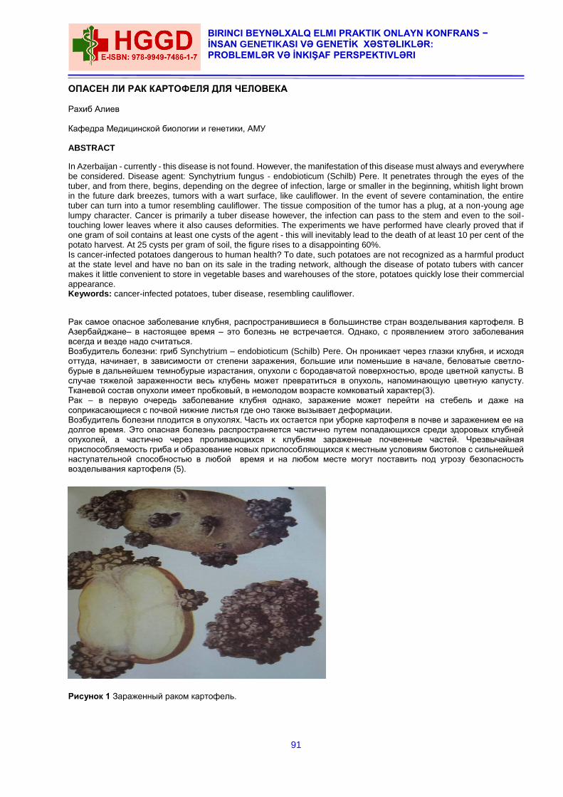
91
BIRINCI BEYNƏLXALQ ELMI PRAKTIK ONLAYN KONFRANS − İNSAN GENETIKASI VƏ GENETİK XƏSTƏLIKLƏR: PROBLEMLƏR VƏ İNKIŞAF PERSPEKTIVLƏRI
ОПАСЕН ЛИ РАК КАРТОФЕЛЯ ДЛЯ ЧЕЛОВЕКА Рахиб Алиев Кафедра Медицинской биологии и генетики, АМУ ABSTRACT
In Azerbaijan - currently - this disease is not found. However, the manifestation of this disease must always and everywhere be considered. Disease agent: Synchytrium fungus - endobioticum (Schilb) Pere. It penetrates through the eyes of the tuber, and from there, begins, depending on the degree of infection, large or smaller in the beginning, whitish light brown in the future dark breezes, tumors with a wart surface, like cauliflower. In the event of severe contamination, the entire tuber can turn into a tumor resembling cauliflower. The tissue composition of the tumor has a plug, at a non-young age lumpy character. Cancer is primarily a tuber disease however, the infection can pass to the stem and even to the soil-touching lower leaves where it also causes deformities. The experiments we have performed have clearly proved that if one gram of soil contains at least one cysts of the agent - this will inevitably lead to the death of at least 10 per cent of the potato harvest. At 25 cysts per gram of soil, the figure rises to a disappointing 60%. Is cancer-infected potatoes dangerous to human health? To date, such potatoes are not recognized as a harmful product at the state level and have no ban on its sale in the trading network, although the disease of potato tubers with cancer makes it little convenient to store in vegetable bases and warehouses of the store, potatoes quickly lose their commercial appearance. Keywords: cancer-infected potatoes, tuber disease, resembling cauliflower.
Рак самое опасное заболевание клубня, распространившиеся в большинстве стран возделывания картофеля. В Азербайджане– в настоящее время – это болезнь не встречается. Однако, с проявлением этого заболевания всегда и везде надо считаться. Возбудитель болезни: гриб Synchytrium – endobioticum (Schilb) Pere. Он проникает через глазки клубня, и исходя оттуда, начинает, в зависимости от степени заражения, большие или поменьшие в начале, беловатые светло-бурые в дальнейшем темнобурые израстания, опухоли с бородавчатой поверхностью, вроде цветной капусты. В случае тяжелой зараженности весь клубень может превратиться в опухоль, напоминающую цветную капусту. Тканевой состав опухоли имеет пробковый, в немолодом возрасте комковатый характер(3). Рак – в первую очередь заболевание клубня однако, заражение может перейти на стебель и даже на соприкасающиеся с почвой нижние листья где оно также вызывает деформации. Возбудитель болезни плодится в опухолях. Часть их остается при уборке картофеля в почве и заражением ее на долгое время. Это опасная болезнь распространяется частично путем попадающихся среди здоровых клубней опухолей, а частично через проливающихся к клубням зараженные почвенные частей. Чрезвычайная приспособляемость гриба и образование новых приспособляющихся к местным условиям биотопов с сильнейшей наступательной способностью в любой время и на любом месте могут поставить под угрозу безопасность возделывания картофеля (5).
Рисунок 1 Зараженный раком картофель.
92
THE FIRST INTERNATIONAL SCIENTIFIC PRACTICAL VIRTUAL CONFERENCE − HUMAN GENETICS AND GENETİC DISEASES:
PROBLEMS AND DEVELOPMENT PERSPECTIVES
Прежде всего надо внимательно следить за картофелем точки зрения зараженности раком в местах, где для посева применяются импортированные или сорта неизвестного происхождения(1). По отношению рака картофеля в большинстве стран были приняты карантинные мероприятия. Меры борьбы направленные на предотвращение заноса, на предотвращение заноса на немедленное обнаруживание и уничтожение очагов заразы и на предупреждение дальнейшего распространения болезни. На полях, где зараза уже появилась или на подозрительных местах. Для предупреждения возможного вреда следует выращивать исключительно только устойчивые к раки сорта. Зараженный раком картофель, ни в коем случае нельзя употреблять в пищу. Следует помнить, что отдельные зооспорангии погибают только после предварительного кипячения (при 100°С), при этом продолжительность обработки в кипящей воде должна составлять не менее одного (!) часа(2). К счастью, споры картофельного рака плохо адаптируются в условиях сильной жары или чрезмерного холода. Например, в регионах, где почва на протяжении длительного времени промерзает до -11°C или прогревается до +30°C, данная инфекция полностью отсутствует. Оптимальной для быстрого размножения грибных споров является температура грунта от +15° до 18°С, при влажности 80%. Увы, именно при подобных условиях всходы картофеля начинают интенсивно формировать клубни. Зимует патоген в форме зооспорангиев (одноклеточных органов бесполого размножения). При этом из одного зооспорангия, с наступлением весны может выйти до 300 крошечных зооспор. Примечательно, что для того, чтобы найти и внедрится в клетку растения-хозяина, у зооспоры существует временной лимит в размере двенадцати часов. Если в течение этого срока спора не находит растение, благодаря которому будет паразитировать, она погибает(4). При попадании в растительную клетку зооспора начинает стремительно расти и развиваться, а под действием выделяемых ею токсинов клетки картофеля интенсивно делятся, образуя, таким образом, характерные шишковатого вида наросты. В почве зооспорангии могут существовать до 30 (!) лет. К счастью, с каждым началом нового сезона «приживается» лишь около 30 процентов зооспорангиев. При этом их жизненный цикл продолжается в течение двух недель, а на зараженном участке может развиваться до 17 поколений патогенных грибов(6). Эксперименты, произведенные нами наглядно доказали, что если в одном грамме грунта содержится, хотя бы одна циста возбудителя – это неминуемо приведет к гибели минимум 10 процентов урожая картофеля. При количестве 25 цист на грамм почвы цифра возрастает до неутешительных 60%. Опасен ли для здоровья человека зараженный раком картофель? На сегодняшний день такой картофель не признается вредным продуктом на государственном уровне и не имеет запрета на продажу его в торговой сети, хотя заболевание клубней картофеля раком делает его мало удобным для хранения на овощных базах и складах магазина, картофель быстро теряет товарный вид, гниет и портится, что может нанести большой урон скупщикам больного картофеля. ЛИТЕРАТУРА
1. Алиев Р.А Нематоды картофеля Шеки Закатальской зоны Азербайджана 1969 Издательство «Элм». Бакы 1971
2. Кальман Гинфнер. Заболевания и повреждения клубня картофеля издательство АН Венгрии Будапешт – 1958
3. Яковлева В.А. Методические рекомендации по тестированию почвы из очагов возбудителя рака картофеля Synchytrium endobioticum (Schilb.) Perc. для снятия карантина. ФГБУ «ВНИИКР», Москва, 2009.
4. Яковлева В.А.Методические рекомендации по выявлению и идентификации возбудителя рака картофеля Synchytrium endobioticum (Schilb.) ФГБУ «ВНИИКР» Москва, 2014.
5. Langerfeld E. & Stachewicz H. (1994) Оценка реакций картофеля разных сортов на рак картофеля (Synchytrium endobioticum) в Германии».Бюллетень ЕОКЗР 24, 793-798.
6. Langerfeld E., Stachewicz H. & Rintelen J. (1994) Патотипы Synchytrium endobioticum. Германия. Бюллетень ЕОКЗР 24, 799-804. 8. Noble M. & Glynne M.D. (1970) Рак картофеля. Бюллетень ФАО по защите растений 18, 125-135.
DATACITE
93
BIRINCI BEYNƏLXALQ ELMI PRAKTIK ONLAYN KONFRANS − İNSAN GENETIKASI VƏ GENETİK XƏSTƏLIKLƏR: PROBLEMLƏR VƏ İNKIŞAF PERSPEKTIVLƏRI
FEATURES OF THE MECHANISM OF ANTI-MUTAGENIC EFFECT OF SUMAC FRUIT EXTRACT AT
THE STAGE OF INITIAL DAMAGE OF THE DNA MOLECULE IN THE CASE OF THE LAST MUTATION Mahir Guliyev Azerbaijan Medical University, Department of Histology, Embryology and Cytology. During the last mutation at the stage of primary DNA damage in wild-type E.Coli K-12 cells and AB 1885 (uvrB), JC 5519 (recBC), JC 9238 (recF) V and JC 7689 (sbc B) strains that are its derivatives, sumakh extract was used in a apriori detected dose of 0.01 mkq/ml. In the experiments, mutagens UV, 4 nitrocholine -1 – oxide (4NXO) V and mitomycin C were used, which differ in the spectrum of primary damage to the DNA molecule formed by them. All this allows us to believe that the main factor of mutagenesis modification and cell death by sumakh fruit extract is the position of reparative enzymes, which, being in a state of defect, encode the genes of uvr B, rec BC V, and rec F. In the schemes of experiments using the studied extract, cells with a reparatively damaged genome are more sensitive to the mutagen compared to wild-type cells. Increased sensitivity of the E. Coli uvr B strain to influencing factors, in comparison with wild-type cells, is associated with the presence of a nuclease independent of uvr AB in cells of this series, which is a product of uvr A and uvr B genes (Howard-Flanders, Boyce, 1996). Increased sensitivity and relatively equal sensitivity in E. Coli strains rec BC and rec F in the first case is associated with the activity of rec BC – dependent nuclease (exonuclease V) (Witkin,1974), and in the second case – with a partial weakening of the process of post-replication repair of the rec F gene defect (Ganesan, Seawell,1975). In turn, increased sensitivity of sbc B cells in the series is associated with inactivation of exonuclease I activity (Тарасов.1982).The results given below were obtained by adding an extract from sumakh fruit to the schemes of experiments with the influence of mutagens. In experiments using damaging factors UV and 4NXO in wild cell mutants, as well as rec BC and sbc B strains, a decrease in mutagenesis is observed. Thus, in uvr B and rec F cells of the series, the genome-protecting properties of sumakh fruit extract are observed in practice. In experiments where an artificial mutation was achieved using Mitomycin C, the positive effect of the extract was observed in wild-type cells and sbc B mutants, but no positive effect was observed in uvr B-, V - rec BC – rec F-genotype cells. REFERANCE
1. Howard-Flanders P., Boyce R.P., DNA repair and genetic recombination studies of mutants of Esherichia coli
defective in these prosses //Radiat.Res.-1996.-v.6.-P.156-184. 2. Witkin, E.M. Thermal enhancement of ultraviolet mutabillity in tif-1 uvrA derivative of I coli B/r: evicible function
//Proc. Nat. Acad.Sci. USA.-1974-v.71.-P.1930-1934. 3. Ganesan A.K. Seawell P.C The effekct of lex A and rec F mutations on postreplication repair and DNA syntheasis
in Esherichia coli K-12 // Mol. And Gen. Genet.-1975.-v. 141.-№3.-P.189-205.
DATACITE

94
THE FIRST INTERNATIONAL SCIENTIFIC PRACTICAL VIRTUAL CONFERENCE − HUMAN GENETICS AND GENETİC DISEASES:
PROBLEMS AND DEVELOPMENT PERSPECTIVES
MOLECULAR-GENETIC RESEARCH OF PHENYLKETONURIA IN AZERBAIJAN FAMILY
Lala Huseynova, Aflatun Azizov, Qumru Huseynova
Azerbaijan Medical University, Department of Medical Biology and Genetics.
ABSTRACT
Phenylketonuria gene has an identified R261G (G-A) mutation. The study of erythrocyte enzyme preparation for family members have shown low electophoretic mobility for G6PD which was unknown in the world studies. A new biochemical variant was identified on the basis of physic-chemical indications of G6PD enzyme, and PKU inherited metabolic disease with R261G (G-A) mutation were found in the family G.M., who live in Tekle village of Masally area. Phenylketonuria and G6PD enzyme deficiency were identified in one family in Tekle village of Masally area of Azerbaijan Republic. Heterozygous and homozygous genetic types of phenylalanine-4-hydroxilase gene mutation R261G (G-A) were identified.A new unknown to scientific literature biochemical polymorphism of G6PD enzyme was identified. Keywords: disease, polymerase chain reaction, enzyme, biochemical polymorphism, family, identification
INTRODUCTION
The family with identified inherited metabolic disease of phenylketonuria lives in Masally administrative area. Masally area itself is located in South-East of Azerbaijan Republic on the slopes of Talysh mountains in subtropical zone. Members of proband’s family possess deficiency of glucose-6-phosphatedehydrogenase. Phenylketonuria gene has an identified R261G (G-A) mutation. The study of erythrocyte enzyme preparation for family members have shown low electophoretic mobility for G6PD which was unknown in the world studies. Based on the Michaelis-Menten constant (Km) applied to G6P, substrate values have manifested high biochemical polymorphism. The goal of nutrition management of phenylketonuria (PKU) is to maintain blood phenylalanine concentrations between 120 and 360 μmol/L. The diet for PKU includes medical foods low in or devoid of phenylalanine and limited quantities of phenylalanine from intact protein sources. Frequent monitoring of blood phenylalanine concentrations is key to successful diet management. Frequent adjustments in the diet are needed to achieve desired blood phenylalanine concentrations as well as to promote normal growth and feeding development. A variety of PKU medical foods and modified low-protein foods are available to accommodate different nutrient needs and taste preferences throughout the life span. Maintaining the diet is challenging for many patients with PKU; alternative therapies are available, but most still require some degree of diet modification [6,9]. The phenylalanine-4-hydroxilase (PAH) gene is located on the long shoulder of chromosome 12 in q22-24.1 site. The lendth is 90 thousand nb and consists of 13 exons. Synthesized protein consists of 451 amino acid residues. Phenylalanine amino acid coming with food in oxidation process turns into different amino acide -thyrosine as a result of phenylalanine hydroxidation process. In the result of mutation in PAH gene this phenylalanine into thyrosine transformation fails[7,9]. Up to 1 % cases of phenylketonuria are presented with atypical forms. The disease is inherited as to autosome-recessive type. The prevalence rate differs in different population groups. For example, in Europeoid inhabitants in the USA it is 1 to 10000. The highest rate is in Turkey, which is 1 to 2600. In Finland and Japan the rate of phenylketonuria is extremely low: even less 1 newborn to 100000 births. In Slovakia in some gypsy populations there were found ultrahigh rates of phenylketonuria because of inbreeding: 1 case for 40 newborns. According to the World Health Organisation data, there are around 100 million people suffering from glucose-6-phosphatedehydrogenase (G6PD) enzyme activity deficiency. More than 400 abnormal variants were identified, and around ¼ of them are endemically different. One part of those abnormal G6PD variants could be characteristic for only one certain ethnic group, and another part - for several ethnic groups. A group of people with enzyme deficiency resulted with hemolytic crisis after some specific medicines, and other people - just after eating food cooked with beans[7]. Thus, the goal of our studies was to identify gene mutations in people with PKU diagnosis and to study the physic-chemical specificities of abnormal G6PD enzyme in the proband’s family members[8]. MATERIAL AND METHODS
Venous blood samples with heparine anticoagulant were used as the study subjects. Blood was sampled from G.M. (proband) family members, who are inhabitants of Tekle village of Masally area, Azerbaijan Republic. PKU diagnostics was carried out by means of IFA method. In identification of PKU gene mutations, complex of molecular-genetic methods were used [1,5]. Genomic DNA was isolated from venous blood, using readymade kits by QIAGEN (Germany) company. Intactness and quantity of isolated genomic DNA were identified by means of electrophoresis in 1.7% agarose gel, as well as gene fragments after polymerase chain reaction (PCR). Electrophoretic apparatus and power source were BioRad (USA) manufactured. Marker for identification of synthesized DNA fragments was DNA Ladder 100 bp . The content of PCR: 0,1-1,0 µg of genomic DNA, 0,25 µM of each dNTP, 25 µl buffer (67 mM Tris-HCL, pH 8,8: 16,6 M (NH4)2SO4, 0,01% Twin-20, 1,5 unit DNA-polymerase. 2 µg of primers for each of exons 3,5,7,11 and 12. Regime of PCR for PKU gene was as follows: 95оС-2 minutes, (94оС-45I, 58оС-45I, 72оС-45İ 30 cycles), 72оС-7 minutes and pause at 4оС for 10 minutes. PCR was conducted in amplifier – Professional Thermocycler, Biometra, (Germany). Purification of DNA fragments after the first PCR stage a set of magnets was used: «AgencourtAMPure XP PCR purification» and SPRIPlate 96 Super Magnet Plate. After that purified DNA fragments were used for the further researches. The second PCR was conducted in the regime: 95оС-2 minutes, (95оС-30I, 52оС-58оС - 30I, 78оС-2 minutes 30 cycles), 72оС-10 minutes and pause on the amplifier at 4оС for 10 minutes. Then the standard procedure on the
95
BIRINCI BEYNƏLXALQ ELMI PRAKTIK ONLAYN KONFRANS − İNSAN GENETIKASI VƏ GENETİK XƏSTƏLIKLƏR: PROBLEMLƏR VƏ İNKIŞAF PERSPEKTIVLƏRI
apparatus GENOMELabGeXPTM Sequencing for the identification of nucleotide sequence of each DNA fragment was carried out. G6PD enzyme activity was measured by means of modified fluorescent method [2,3]. Purification of enzyme prerarations and study of the characteristics were carried out according to the WHO standardized methods [4]. RESULTS AND DISCUSSION
The world scientific literature researches show that European populations have mainly R408W, P281L, R261Q, R158Q, R252W, I65T, IVS10nt546, IVS12ntl. PAH gene mutations prevail over the others. These mutations are located in 3,5,7,11 and 12 exons of the gene [4,10]. With this purpose we have done amplification of PAH gene exons 3,5,7,11 and 12 genomic DNA fragments, got from lymphocytes of the G.M. family members: two parents and six their children, by means of polymerase chain reaction with 5 primer groups. Fragments of exon 3 of 112 nb, exon 5 of 162 nb, exon 7 of 218 nb, exon 11 of 222 nb and exon 12 of 177 nb were amplified. Total nucleotide sequencing was done only for exon 7 as an example, where R261G mutation was identified. Being a point mutation we have found a substitution of guanine with adenine. The result of mutation was on protein level, and arginine amino acid was substituted with gluthamine amino acid. Homozygous form was identified in 4-year-old girl. Heterozygous form carriers were both parents and one sibling. So, family members manifested one homozygous and three heterozygous forms of R261G mutation. It’s worthwhile noting, that proband’s parents are children of two sisters. Marriage is identified as a 3rd cosanguineous parallel marriage type. G6PD enzyme deficiency was as low as 5.2-12.5% of the normal activity in three brothers in the family. According to the obtained activity it was relates to the II activity class. All three enzyme preparations showed low enzyme (0-10%) activity. Each of the three kids had erythrocyte hemolysis and anaemia after eating food with beans. All three enzyme preparations have the indication of pH-optimum in the normal range (pH 7,5-8,5). All enzyme preparations under research have shown low electrophoretic movbility. Based on the G6P substrate, constant of Michaelis-Menten (Km) indication of all the enzyme preparations was high (146,7µm). For analogue of 2dG6P substrate was high disposal degree identified. Enzyme preparations, obtained from the inhabitants of Masally area Tekle village, have manifested such physic-chemical characteristics of G6PD deficiency which was new and had no analogue in the world scientific literature. Thus, a new biochemical variant was identified on the basis of physic-chemical indications of G6PD enzyme, and PKU inherited metabolic disease with R261G (G-A) mutation were found in the family G.M., who live in Tekle village of Masally area. CONCLUSION
Phenylketonuria and G6PD enzyme deficiency were identified in one family in Tekle village of Masally area of Azerbaijan Republic. Heterozygous and homozygous genetic types of phenylalanine-4-hydroxilase gene mutation R261G (G-A) were identified. A new unknown to scientific literature biochemical polymorphism of G6PD enzyme was identified. REFERENCES
1. Beutler E. G6FD deficiency. Blood.1994. 84. P. 3615-3636. 2. Beutler E. The genetics of glukoze-6-phosphatedehydrogenase deficiency. Seminars in Hematol. 1998. v.27.
3.137-164. 3. Charles R.,Seriver N., The PAH Gene Rhenylketonuria and a Paradigm Shift. Human Mutation. 28(9), 831-845,
2007. 4. De Vita G., Alkalay M., Sampietro M. et al., Two points mutations are responsible for G6FD polumorfizm in
Sardinia. Amer.J.Hum.Genet. 1989. V.44. P.233-238. 5. Du C.S., Xu Y.K., Wu Q.L.. Glucose-6-phosphatedehydrogenase variants and polymorphic frequency in
Guangdog. China. Hum.Genet.1988. v.60. P.385-388. 6. Fuji H., Miwa S., Takegawa S., Takahashi K., et al., Two new variants of glukoze-6-phosphatedehydrogenase
found Japan. Hum. Genet. 1984. V.66. 2. P.276-278. 7. Hirono A., Ishii A., Kere N. et al., Molecular analysis of glucose-6-phosphatedehydrogenase variants in the
Solomon Islands. Amer. J. Hum. Genet.1995. V.56. 5. 8. Krasnopolskaya X.D., Shatskaya T.L., Distribution of Gd alleles in some ethnic group in the USSR.
Hum.Genet.1987. V.75.3.P.258-263. 9. Nowacki P., Byck S., Prevost l., The PAH mutation analysis consortium database: update 1996, NAR,1997, 25,
№1, 138-142.
DATACITE
96
THE FIRST INTERNATIONAL SCIENTIFIC PRACTICAL VIRTUAL CONFERENCE − HUMAN GENETICS AND GENETİC DISEASES:
PROBLEMS AND DEVELOPMENT PERSPECTIVES
EARLY PRENATAL DİAGNOSİS OF FETAL ANEUPLOİDY AND NEURAL TUBE DEFECTS
1Natavan Achundova, 2Shafag Aliyeva
1Associate Professor of the Department of Obstetrics and Gynecology I, MD–PhD
2Assistant doctor of the Department of Pathological Anatomy.
1,2Azerbaijan Medical University.
10-15% of fertilized eggs cannot be implanted (1). Pregnancy losses are observed in 22% of cases before the diagnosis of clinical pregnancy (2). Cytogenetic study of spontaneous miscarriages reveals chromosomal abnormalities in 50-65% of cases (3, 4, 5). 17.4% of Polyploidy, 23.7% of X monosomy, 49.8% of trisomy (mainly, Chromosome 16 trisomy) were found in miscarriage specimens in which chromosomal abnormalities were detected (6). 50% of fertilized human eggs undergo reproductive loss, and chromosomal and gene mutations play a significant role in the occurrence of these losses (7). The use of genetic testing methods allows to explain the causes of reproductive losses: spontaneous abortions, perinatal death and disease. If a woman has a history of 2 or more miscarriages, genetic counseling is crucial. In this case, genealogical examination should be carried out taking into account not only, the family history of the couple, but, also miscarriages, stillbirth, intrauterine growth retardation, congenital malformation, mental weakness, infertility. During genetic counseling and cytogenetic examination:
1. Reasons identified as a result of genealogical and cytogenetic analysis should be explained to the couple.
2. The risk of miscarriages and congenital malformations of the fetus should be assessed;
3. It is necessary to explain the importance of prenatal diagnosis in subsequent pregnancies and the possibility of
in vitro fertilization with donor eggs and sperm in the presence of a gross pathology in the family.
4. The importance of cytogenetic testing in cases of miscarriage, stillbirth and neonatal death should be emphasized
(8).
Screening tests with a comprehensive assessment based on several indicators allow early detection of some fetal chromosomal abnormalities, defects of the neural tube during the first and second trimester of pregnancy.The improvement of non-invasive diagnostic techniques plays an important role in early detection of chromosomal abnormalities in the fetus. Of these, the screening test used in the first trimester of pregnancy is of particular importance.
Indications for biochemical markers in the first trimester of pregnancy:
When the mother's age is over 35 and the father's age is over 40.
History of ≥2 spontaneous miscarriage
Taking medication before fertilization or in the first weeks of pregnancy
Presence of bacterial and viral infections (TORCH syndrome)
The birth of children with chromosomal abnormalities in the family or close relatives
Probability of radiation from one or both parents
The desire to have a healthy child, etc. (8, 9).
The risk of fetal trisomy increases with advancing maternal age. In prenatal diagnosis of aneuploidy (trisomy 13, 18, 21) and neural tube defects, ultrasound data are combined with the age of the mother, two or more biochemical indicators. It should also be noted that prenatal diagnosis of aneuploidy should not be carried out taking into account only the mother's age. Amniocentesis should not be performed on women over 40 years without prenatal screening. Because the risk of chromosomal abnormalities with a negative test may be < 1/200 (10, 11). According to the UK National Screening Committee (UK NSC) diagnostic tests with a sensitivity of> 75% and a false positivity of <3% can be used. These tests should be used between 10 and 20 weeks of gestation. First trimester screening should be done before 3 weeks and 6 days of pregnancy. There is still no reliable source for prenatal diagnosis of Down's syndrome before the week 10 of pregnancy. A blood test combined with an ultrasound (which checks the thickness at the back of the fetus's neck, known as its nuchal translucency), human chorionic gonadotropin (hCG) and pregnancy-associated protein A (PAPP-A) are evaluated simultaneously in the 1st trimester screening. Early diagnosis makes it possible to identify the risk before week 14 of gestation and make a timely decision. At a meeting, in 2007, it was stated that the sensitivity values of a diagnostic test conducted only with biochemical or ultrasound examination were lower (8, 11, 13). Triple or quad screening are performed in the second trimester. As 15% of pregnant women seek antenatal care in the final weeks of pregnancy, triple or quadruple screening is recommended in the second trimester. Human Chorionic Gonadotropin (hCG) and unconjugated estriol (uE3) and α-fetoprotein (AFP) are detected by triple marker screening tests in the maternal plasma. The quadruple tests includes hCG,AFP, uE3 and additionally Inhibin A. Screening may be done in the following weeks of pregnancy:
PAPP-A test is measured between 10 weeks 0 days and 13 weeks 6 days in the first trimester
Second trimester screening is done between 15 weeks 0 days and 20 weeks 0 days;
Weeks 9 to 10 of gestation are more suitable for PAPP-A determination. From 10 to 13 weeks of gestation, its
level gradually decreases.
97
BIRINCI BEYNƏLXALQ ELMI PRAKTIK ONLAYN KONFRANS − İNSAN GENETIKASI VƏ GENETİK XƏSTƏLIKLƏR: PROBLEMLƏR VƏ İNKIŞAF PERSPEKTIVLƏRI
The first trimester combined test is performed between weeks 11 and 14 of pregnancy, taking into account NT
and PAPP-A.
Pregnant women seeking antenatal care in the last weeks of pregnancy, a quadruple (possibly triple screening
) test is performed between weeks 15 - 20 of pregnancy.
The cut-off is 1/150 for the first trimester screening and 1/200 for the second trimester screening (13).
Invasive tests such as amniocentesis and Chorionic villus sampling (CVS) are recommended if a high risk is identified. A karyotype can be defined in a sample obtained in this way and a final diagnosis can be made. CVS is performed at week 11-13 of pregnancy and amniocentesis is done from 15 weeks. NIPS (Prenatal Cell-Free DNA Screening -cfDNA) is a non-invasive screening test. It is designed to detect chromosomal abnormalities without risk during pregnancy. Fetal DNA is found in the mother's blood and if any changes are detected, invasive confirmation and genetic counselling is recommended (12). False-negative response is 1% for trisomy 21, 3.6% for trisomy 18 and 9.4% for trisomy 13. When the Y chromosome is found, the sex is determined. The main objective of genetic examinations –development of healthy generation through family planning. The main purpose of genetic research is to develop a healthy generation through family planning. REFERENCES
1. Weathersbee P.S. –Early reproductive loss and the factors that may infeuence its occurrence –J. Reprod. Med.,
1980, 25, 6, 315-318. 2. Wilcox A. J., Weinberg C.R. et al. – Incidence of early pregnancy loss – N. Engl. J. Med., 1988, 319, 189. 3. Boue J., Boue A., Lasar P. – The epidemiology of human spontaneous abortions with chromosomal anomalies-
In: Aqing gametes Basel, 1975, 330-348. 4. Boue J., Boue A., Lasar P. – Retrospective and prospective epidemiologic studies of 1500 Karyotyped
spontaneous abortions- Teratology, 1975, 11, 11-26. 5. Boue A., Boue J., Grapp A.- Cytogenetics of pregnancy was tage advances in human genetics –New. York,
Plenum Press, 1985, 30-31. 6. В. И. Сидельникова -Привычная потеря беременности-Москва, 2005, стр. 32-36 7. Алипов В. И., Головачев Г. Д. –Репродуктивные потери и хромосомные аномалии-Акуш. и гинек., 1983. 1,
38-41. 8. Prenatal Screening for Fetal Aneuploidy in Singleton Pregnancies; 2011; Joint SOGC- CCMG Clinical Practice
Guidelines. 9. Screening for Down’s syndrome: UK NSC Policy recommendations 2011-2014; Model of Best Practise; Anomaly
Screening Programme. 10. Antenatal Care- NICE Clinical Guidelines 62; 2008. 11. Non-invasive prenatal testing for chromosomal abnormality using maternal plasma DNA. RCOG Scientific Impact-
Paper No. 15; March 2014. 12. Yunus Emre Purut- Kadın Doğum Board 2. Cilt 550-560-Modern kitap evi, 2018. 13. Jyotsna Pundir, Arri Coomarasamy-Obstetrik, kanıta dayalı algoritmalar 2-9.
DATACITE
98
THE FIRST INTERNATIONAL SCIENTIFIC PRACTICAL VIRTUAL CONFERENCE − HUMAN GENETICS AND GENETİC DISEASES:
PROBLEMS AND DEVELOPMENT PERSPECTIVES
ROBERTSONIAN TRANSLOCATION PATIENT WITH RECURRENT MISCARRIAGE
Afig Berdeli, Nargiz Yahyazada, Goycak Gurbanbayli AFGEN Genetic Diagnosis Center ABSTRACT
Translocation is a type of chromosomal abnormality, which results in a chromosome breaks where the portion of it reattaches to a different chromosome. This includes balanced and unbalanced translocation, with two main types: reciprocal -and Robertsonian translocation. Robertsonian translocation is a type of translocation caused by breaks at or near the centromeres of two acrocentric chromosomes. The reciprocal exchange of parts gives rise to one large metacentric chromosome and one extremely small chromosome that may be lost from the organism with little effect because it contains few genes. The resulting karyotype in humans leaves only 45 chromosomes, since two chromosomes have fused together. Robertsonian translocation is one of the major chromosomal rearrangements with a prevalence rate of 0.1% of the general population and 1% of the infertile population. These chromosomal translocations are mainly observed in group D including 13, 14, 15 and group G including 21 and 22 chromosomes CASE: A non-consanguineous couple (32-year old male and 26-year old female) with the complaint of repeated
miscarriages attended to the “Afgen Genetic Diagnosis Center” for cytogenetic evaluation. They had a h istory of three repeated miscarriages in the past four years of their marriage. The first abortion was two months from pregnancy the second was a tubal pregnancy. The third was a missed abortion from a of 7-weeks pregnancy. None of the abortions got cytogenetically evaluated and there were no such histories of repeated abortions in any other family member. MATERIALS AND METHODS: Two milliliters of peripheral blood was obtained from both partners in heparinized tubes to
harvest white blood cells for karyotyping 30 to 40 metaphases were analyzed and the karyotype was interpreted using the Smarttype Karyotyper Analizer. The chromosomes were identified and classified according to the guidelines by the International System for human Cytogenetic Nomenclature (ISCN, 2011) RESULTS: In this study, we report a Robertsonian translocation rob (14; 15) in a female patient with a history of repeated
miscarriages. The resulting balanced karyotype has only 45 chromosomes including the translocated one, which is the result of a fusion of the long arms of two acrocentric chromosomes. Chromosomal analysis of the male partner showed normal 46, XY karyotype. Chromosomal analysis revealed an abnormality in the female partner with 45, XX, rob (14; 15) (q10; q10) chromosomal constitution DISGUSSION: The presence of a balanced chromosomal rearrangement in a parent results in an increased risk for
structural chromosomal defects in future pregnancies. It is estimated that in about 70% of couples with at least two spontaneous abortions, one parent carries a balanced chromosomal rearrangement such as inversions, translocation, etc. The carrier of a Robertsonian translocation has a normal phenotype but is at risk of producing unbalanced gametes and, therefore, unbalanced offspring. In general, the prevalence of chromosomal abnormalities is higher in females than in males. Cytogenetic analysis of couples with recurrent abortions is mandatory to evaluate the probable presence of any chromosomal aberrations. This will offer valuable data for the appropriate genetic counseling strategies. Physicians should be aware of the condition as at least 5% of these couples with repeated abortions exhibit chromosomal abnormalities as the cause. Such cases have to be analyzed as early as possible to arrange for adequate genetic counseling and to allow couples to make an informed reproductive decision regarding subsequent pregnancies. Prenatal diagnosis should be offered to these couples in the case of future pregnancies. REFERENCE
1. Hassanzadeh Nazarabadi M, Raoofian R, Abutorabi R. Balanced Chromosomal Translocations of Parents in Relation to Spontaneous Abortions. J Sci. 2005;16:203–208
2. Ananthapur V, Avvari S, Tella S, Nallari P, Akka J. A Robertsonian Translocation rob (14;15) (q10:q10) in a Patient with Recurrent Abortions Infertil 2010; 11: 197-200.
3. Shaffer LG, Spikes AS, Macha M, Dunn R. Identification of a subtle chromosomal translocation in a family with recurrent miscarriages and a child with multiple congenital anomalies. Med 1996; 41: 367-371.
DATACITE
99
BIRINCI BEYNƏLXALQ ELMI PRAKTIK ONLAYN KONFRANS − İNSAN GENETIKASI VƏ GENETİK XƏSTƏLIKLƏR: PROBLEMLƏR VƏ İNKIŞAF PERSPEKTIVLƏRI
GENOTYPE-PHENOTYPE OF NLRP3 GENETIC MUTATIONS OR POLYMORPHISMS
1Afig Bardali , 2Sırrı Fethi Cham, 3Goychak Gurbanbaylı
1Izmir Ege University, 2Manisa Celal Bayar University
3AFGEN Genetic Diagnosis Center. INTRODUCTION
Inflammasomes - are the essential part of the natural immune response and are multiple intracellular protein complexes that severely control different aspect of adaptive immunity. Generally, sensory molecules recognize different pathogenic(PAMP)or endogenous sterile distress signals(DAMP)they activate caspase-1 using adapter proteins such as ASC, leading to the processing and release of pro-inflammatory cytokines such as interleukin(IL)-1β and IL-18. NLRP3 inflammasome pathway is known to be the most researched among 7 different inflammasomes. Mutations detected in the NLRP3 gene which encodes the sensor protein chyrphoria, are known to cause 3 different diseases. Materials and Methods: Venial blood was obtained from 50 different patients and DNA were isolated. Mutagenesis was
performed by sanger DNA sequencing methods and results were read by Finch TV, CLC Sequence Viewer – QIAGEN Bioinformatics and MEGA7 biomorpho -logical programs. Mortality evaluations were done according to NCBI, PolyPhen-2, INFEVERS and many different databases. Results: 2 synonymous mutations (Ala240Ala and Arg260Arg) were found in both of the study groups which classified as
polymorphism 6 different mutations which were detected from the study group, were found to be related with conditions. 10 patients carried Gln703Lys mutation, 4 patients carried Ser726Gly mutation, 3 patients carried Val198Met mutation, 2 patients carried Ile313Val mutation, 2 patients carried Thr913Met mutation, and 2 patients carried Ser331Arg mutation.No mutations were fond on NLRP3 gene in the healthy control group. Discussion: One of the associated mutation found by us (Thr913Met) was new, and had been reported to INFEVERS
database. Most of the mutations found to be located on the 3rd exon of NLRP3 gene as well as 4th and 8th exons.Mutations cause increase in function of NACHT, NAD, and LRR parts of the protein structure. The effect of each mutation on protein structure was further investigated. REFERENCE
1. E.I. Elliott, F.S. (2015), Sutterwala Initiation and perpetuation of NLRP3 inflammasome activation and
assembly Immunol. Rev., 265 pp. 35-52 2. K. Izawa, A. Hijikata, N. Tanaka, T. Kawai, M.K. Saito, R. Goldbach-Mansky, et al. (2012), Detection of base
substitution-type somatic mosaicism of the NLRP3 gene with >99.9% statistical confidence by massively parallel sequencing DNA Res, 19 pp. 143-152
3. Halle A, et al. (2008) The NALP3 inflammasome is involved in the innate immune response to amyloid-beta. Nat Immunol 9:857–865.
DATACITE
100
THE FIRST INTERNATIONAL SCIENTIFIC PRACTICAL VIRTUAL CONFERENCE − HUMAN GENETICS AND GENETİC DISEASES:
PROBLEMS AND DEVELOPMENT PERSPECTIVES
THE ROLE OF HYPOXIA IN THE DEVELOPMENT OF SECONDARY BRAIN DAMAGE IN PATIENTS
WITH BRAIN STROKES
Ainur Tokshylykova1*, Zhanslu Sarkulova1, Aigul Utepkaliyeva2, Arshia Tleuova2, Zhusupbek Satenov2
1Department of Anesthesiology and Intensive Care, 2Department of Neurology,
West Kazakhstan Marat Ospanov State Medical University. Kazakhstan.
E-mail: [email protected],[email protected]., [email protected]., [email protected]
ABSTRACT
Adverse outcomes of treatment of acute brain strokes are largely due, first of all, to the action of a hypoxia. The brain's own oxygen reserves are negligible, and even the short-term hypoxy can lead to hard consequences. There is no clear definition of parameters of assessing the oxygen starvation of the cell in the literature, which could unambiguously and reliably indicate the presence or absence of it. This parameters would exactly could determine the outcome of brain damage, remain insufficiently studied. The aim of the study was to assess the diagnostic and prognostic significance of hypoxia criteria in patients with acute cerebral strokes. METHODS
A prospective method was used in the work. The study involves 75 patients. There are 62.6% with hemorrhagic strokes, and 37.3% with ischemic strokes. According to the outcome of the disease, regardless of the diagnosis,the groups of patients were identified: with a favorable outcome (54.6%) and with an unfavorable outcome (45.3%). The results were compared. RESULTS
The diagnostic criteria of hypoxia - indicators of LDH and CPK enzymes, lactate and glucose, parameters of ABS and blood gas composition by clinical groups at admission and at treatment stages. It was noticed that the level of LDH on the 2nd day compared to the initial values was statistically significantly higher in both groups of patients: in the 1st day -by 6.5% (p = 0.005), in the 2nd day -by 5.25% (p = 0.005).On the 3rd day of the disease, the increase in LDH in the 1st group remained at 15.0% (p = 0.021), while on the 2nd -at 31.5% (p = 0.05). Although there was a slight decrease in LDH on the 4th day of the disease in the 1st group, but it exceeded the initial indicators by 11% (p = 0.022). The same pattern had a place to be in the 2nd group of patients, where the LDH level decreased slightly, but remained elevated by 24.9% (p = 0.012) on the 4th day, compared to the baseline values. The interesting fact is that during the study of CPK indicators, was noted that at admission of patients from both groups, there were significantly high levels of this enzyme (468.97±192.0 and 256.17±110.5 g/l), compared to acceptable levels (less than 190 g/l). On the 2nd day, the 1st group had a statistically significant increase of 22.5% (p=0.040), on the 3rd day of the disease, its level was 13.75%, higher than the initial one. Dynamically, on the 4th day, though there was a decrease of 67.36% from the initial level, this indicator remained above acceptable standards. In the 2nd group of patients with ischemic stroke, the CPK index compared to the baseline levels was as follows: on the 2nd day-there was a statistically significant increase of 26.4% (p=0.012), on the 3rd day-it exceeded the baseline by 15.2% (p=0.003), on the 4th day – a statistically significant decrease of 30.53% (p=0.009). Thus, the study of LDH indicators showed statistically significant increases in both study groups of patients, both at their admission to the hospital and in the dynamics of their treatment. In the study of CPK, there was noted more significant increase in the 1st group of patients. However, more statistically dependent relationships were found in the group 2 of patients. Studying the parameters of the CBS and gas composition, the following results were revealed: patients with compensated forms of acidosis were admitted in both groups, but in the 1st group, against the background of a slight increase in the level of carbonate ions to 31.37±16.2 mmol/l, the pH level was 7.36±0.055, in the 2nd group: with acceptable HCO3 indicators, 26.31±6.17, the pH level was 7.38±0.05. In dynamics, in the 1st group for 3rd -4th days there was a decrease of the partial pressure of oxygen in both to 45.11±9.05 mmHg.CT-48.17±11.03 mmHg.St, and a statistically significant decrease in HCO3 to 26.70±3.04 - 27.13±3.60 mmol / l. However, the pH remained at the level of reliable compensation. In the 2nd group of the studied patients, the indicators of the gas composition of the blood underwent more significant changes. Against the background of reducing the oxygen partial pressure to 44.24±13.43 - 47.93±10.19 mmHg, according to the acceptable indicators of pCO2 and HCO3, the pH level on day 3 reflected the presence of a decompensated form of respiratory acidosis in patients with pH of 7.23±0.7, but this indicator was statistically insignificant. Analysis of the diagnostic data of lactate and blood glucose shows that at the admission to the hospitalthey was at the initial state, all patients also had elevated levels of both lactate – 2.41±1.5 and 2.06±1.58 mmol/l, and blood glucose – 8.78±2.94 and 7.97±2.32 mmol/l. There was a gradual statistically significant decrease in the level of both lactate to 1.32±0.7 and 1.03±0.3 mmol/l, (p=0.001) and glucose: up to 7.31±1.29 and 6.56±1.02 mmol/l (p= 0.001), dynamically on the 2-3-4 day of the disease in the 1st group of patients. Results of the study of LDH and CKD, lactate and glucose, CBS parameters and blood gas composition in patients with acute brain strokes with favorable and unfavorable outcomes.All admitted patients with brain strokes were diagnosed with initial hypoxia with hyperfermentemia, lactatemia, and hyperglycemia against the background of acidosis. In dynamics, during the treatment in a group of patients with a favorable outcome of the disease, the decrease in creatinephosphokinase was observed by 52.5%. On the contrary, there is an
101
BIRINCI BEYNƏLXALQ ELMI PRAKTIK ONLAYN KONFRANS − İNSAN GENETIKASI VƏ GENETİK XƏSTƏLIKLƏR: PROBLEMLƏR VƏ İNKIŞAF PERSPEKTIVLƏRI
increase of LHD on 38.1 per cent, CPK by 34%, saving excess lactate in 2.2, glucose in 1.3 times against the background of decompensated acidosis from 7.33±0,69 in the group of patients with unfavorable outcome, which was due to more severe disease in this group of patients and the subsequent fatal outcome in the progression of the disease. CONCLUSION
Analyzing the dynamics of treatment of patients, with favorable and unfavorable outcomes, it was found out that the results of increased activity of diagnostic criteria for hypoxia (creatinephosphokinase, lactatedehydrogenase, lactate, glucose and decompensated acidosis indicated a severe degree of hypoxic damage to brain tissue. Subsequently, they can serve as prognostically considerably diagnostic criteria for the adverse outcome of the disease in patients with acute brain strokes. REFERENCES
1. Nelson J.E., Cox C.E., Hope A.A., Carson S.S. Chronic critical illness, Am J Respir Crit Care Med 2010;182(4):446-454.
2. Carson S.S., Definitions and epidemiology of the chronically critically ill, Respiratory Care June 2012, 57 (6) 848- 858.
3. Carson S.S., Bach P.B. The epidemiology and costs of chronic criticalillness. Crit Care Clin 2002;18(3):461-476.
4. Carson S.S., Kahn J.M., Hough C.L., Seeley E.J., White D.B., Douglas I.S., et al. A multicenter mortality prediction model for patients receiving prolonged mechanical ventilation. Crit Care Med 2012;40(4):1171-1176.
5. Shannon S. Carson, Definitions and Epidemiology of the Chronically Critically Ill, Respiratory Care June 2012, 57 (6) 848-858.
6. Parfenov A.L., Solovyova N.N., Pichugina I.M., Vorontsova V.S. " Comorbidity in severe brain injuries of various etiologies with an outcome in a low level of consciousness." Materials of the All-Russian conference "Providing emergency and emergency medical care at the present stage. Achievements and prospects." Kazan, 2017. P. 25.
DATACITE
102
THE FIRST INTERNATIONAL SCIENTIFIC PRACTICAL VIRTUAL CONFERENCE − HUMAN GENETICS AND GENETİC DISEASES:
PROBLEMS AND DEVELOPMENT PERSPECTIVES
STUDY OF RHEOLOGICAL ASPECTS AND ATTEMPT TO THE THEORETIC RHEOLOGICAL RESULTS WITH GENETIC BUILDING IN THE HYPERTONIC PATIENTS AND IN THEIR FAMILY MEMBERS (HYPERTONIC)
1Maka Mantskava, MD, PHD, 2Nana Momtselidze , PHD, 3Afshin Zardoshtian, 4Amirreza Rabiee
European University, Tbilisi, Georgia1
Multidisciplinary Science High School. Georgia1,2,3,4 Ivane Beritashvili Center of Experimental Biomedicine, Department of Rheology and Diagnostic Analytical Services. Georgia1,2,
1,2,3,4Multidisciplinary Science High School. Tbilisi State Medical University. Georgia
ABSTRACT
We made a small clinical laboratory research with compliance with the Helsinki Declaration; we have used of a special method of informed consent between patients, doctor, which was approved by administration of research institutions. We selected patients who had hypertension within the age of 60 to 80 years old. We selected 12 families which consist of (mother, father, one child). Therefore, we have two group. I group - 19 patients with hypertension (some mother and father were not alive at moment of our research) and II group - the children of the patients with hypertension, III group - control (this group contents was 20 healthy adults same age). We used an innovation Georgian method for counting RBC aggregation index. This method due on count the ratio of the area of aggregated of RBCs to the area of non-aggregated of RBCs in unit volume. Process of aging is one of the most interesting issues for scientists, many different researches had been done to identify how different agents affects human body to decrease or loose the abilities which had before. In this article we tried to explain this process from rheological view we, rheologists will try to find the answers through the way of fluid flow and their aspects in body, and the role of high blood pressure on patients with hypertension and their family members in this process. First we should consider that the process of aging in human body is not fully recognized yet, but there are some important factors such as DNA methylation which has big affect on this. Keywords: aging, rheology, RBC aggregation, hypertonia
INTRODUCTION
To start the research, we have made small clinical laboratory work with the compliance of the Helsinki Declaration [1]; We have used a special form of informed consent between patients, doctor, which was approved by administration of research institutions. We selected hypertension 12 families which consist of (mother, father, one child) and patients within the age of 60 to 80 years old. Therefore, we had two group. I group – consist of 19 patients with hypertension (some mother and father were die to moment of our research) and II group – which consist of their children - patients with hypertension, III group - control group (this group contents was 20 healthy adults in the same age). Detailing by age in table 1. We used an innovation Georgian method for counting RBC aggregation index. This method work is due to the counting the ratio of the aggregated area of RBCs to the non-aggregated area of RBCs per unit volume [2,3].
RESULTS
In the table 1
Table 1: Аveraged age in groups. М±m
Group Years
Control 71±3.2
Parents (men) 81±2.7
Parents (women) 82±5.3
Children (without differentiation of sex) 63±2.8
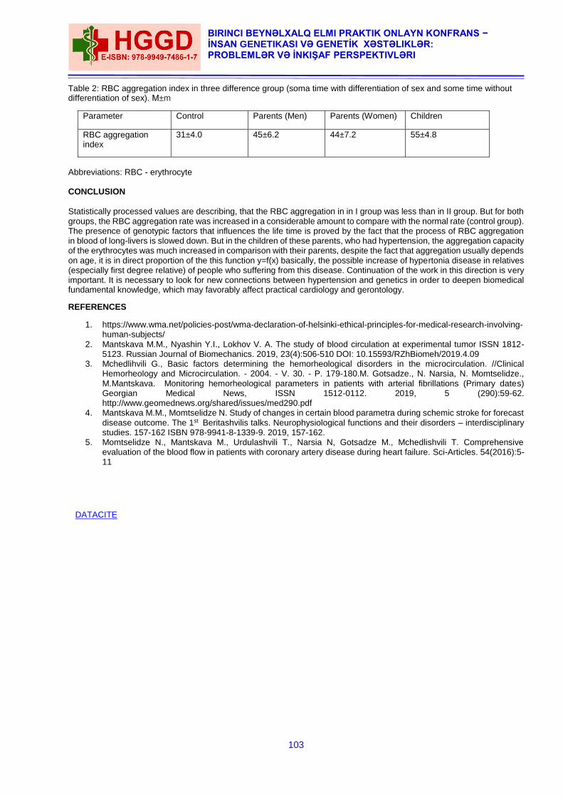
103
BIRINCI BEYNƏLXALQ ELMI PRAKTIK ONLAYN KONFRANS − İNSAN GENETIKASI VƏ GENETİK XƏSTƏLIKLƏR: PROBLEMLƏR VƏ İNKIŞAF PERSPEKTIVLƏRI
Table 2: RBC aggregation index in three difference group (soma time with differentiation of sex and some time without differentiation of sex). М±m
Parameter Control Parents (Men) Parents (Women) Children
RBC aggregation index
31±4.0 45±6.2 44±7.2 55±4.8
Abbreviations: RBC - erythrocyte CONCLUSION
Statistically processed values are describing, that the RBC aggregation in in I group was less than in II group. But for both groups, the RBC aggregation rate was increased in a considerable amount to compare with the normal rate (control group). The presence of genotypic factors that influences the life time is proved by the fact that the process of RBC aggregation in blood of long-livers is slowed down. But in the children of these parents, who had hypertension, the aggregation capacity of the erythrocytes was much increased in comparison with their parents, despite the fact that aggregation usually depends on age, it is in direct proportion of the this function y=f(x) basically, the possible increase of hypertonia disease in relatives (especially first degree relative) of people who suffering from this disease. Continuation of the work in this direction is very important. It is necessary to look for new connections between hypertension and genetics in order to deepen biomedical fundamental knowledge, which may favorably affect practical cardiology and gerontology.
REFERENCES
1. https://www.wma.net/policies-post/wma-declaration-of-helsinki-ethical-principles-for-medical-research-involving-human-subjects/
2. Mantskava M.M., Nyashin Y.I., Lokhov V. A. The study of blood circulation at experimental tumor ISSN 1812-5123. Russian Journal of Biomechanics. 2019, 23(4):506-510 DOI: 10.15593/RZhBiomeh/2019.4.09
3. Mchedlihvili G., Basic factors determining the hemorheological disorders in the microcirculation. //Clinical Hemorheology and Microcirculation. - 2004. - V. 30. - P. 179-180.M. Gotsadze., N. Narsia, N. Momtselidze., M.Mantskava. Monitoring hemorheological parameters in patients with arterial fibrillations (Primary dates) Georgian Medical News, ISSN 1512-0112. 2019, 5 (290):59-62. http://www.geomednews.org/shared/issues/med290.pdf
4. Mantskava M.M., Momtselidze N. Study of changes in certain blood parametra during schemic stroke for forecast disease outcome. The 1st Beritashvilis talks. Neurophysiological functions and their disorders – interdisciplinary studies. 157-162 ISBN 978-9941-8-1339-9. 2019, 157-162.
5. Momtselidze N., Mantskava M., Urdulashvili T., Narsia N, Gotsadze M., Mchedlishvili T. Comprehensive evaluation of the blood flow in patients with coronary artery disease during heart failure. Sci-Articles. 54(2016):5-11
DATACITE
104
THE FIRST INTERNATIONAL SCIENTIFIC PRACTICAL VIRTUAL CONFERENCE − HUMAN GENETICS AND GENETİC DISEASES:
PROBLEMS AND DEVELOPMENT PERSPECTIVES
THE CONTRİBUTİON OF GENETİC TESTİNG TO MEDİCİNE - THE EXPERİENCE OF GEORGİA
Nino Pirtskhelani1, Nino Kochiashvili2, Ketevan Kartvelishvili3, Levan Makhaldiani4
1Expert of Forensic Biology (DNA) Department, National Forensics Bureau. Professor of Tbilisi Open University; MD,
PhD. (Tbilisi, Georgia);
2Head of Biology (DNA) Department, National Forensics Bureau, MD, PhD (Tbilisi, Georgia),
3Expert of Biology (DNA) Department, National Forensics Bureau, PhD Student, TSMU, (Tbilisi, Georgia)
4Head of Hemophilia and Thrombosis Centre, K. Eristavi National Center of Experimental and Clinical Surgery, MD
(Tbilisi, Georgia)
INTRODUCTION
Since the completion of the Human Genome Project, remarkable advances have been made in understanding the human genome's contribution to health and disease; the wealth of scientific discovery generated over the past 10 years is unparalleled in the history of biomedicine, and the rate of discovery is accelerating [1]. The traditional study of single gene disorders (genetic medicine) has developed into an understanding of how multiple genes interact with environmental factors: genomics [2]. Personalized medicine is health care that tailors interventions to individual variation in risk and treatment response. Although medicine has long strived to achieve this goal, advances in genomics promise to facilitate this process. Relevant to present-day practice is the use of genomic information to classify individuals according to disease susceptibility or expected responsiveness to a pharmacologic treatment and to provide targeted interventions. [3] Inherited thrombophilia is a genetic disorder of blood coagulation resulting in a hypercoagulable state, which has been suggested as a possible cause of recurrent thromboembolism. Family and twin studies have established a heritable component to venous and arterial thrombosis. For the vast majority of patients, thrombosis is a complex, multifactorial disease caused by a combination of numerous, often unknown, environmental and genetic factors [4]. Since the placenta is rich in blood vessels, it is reasonable to infer that clot formation within the placental vasculature may result in placental insufficiency, intrauterine growth restriction and other complications. Intrauterine growth restriction (IUGR) is defined as estimated fetal weight below gestational age, according to ultrasound data, and birth weight below the 10th percentile of the birth weight for gestational age reference curve. Fetal growth restriction (FGR) affects 5–10% of pregnancies, leading to clinically significant fetal morbidity and mortality. Aim: The aim of this study was to determine the intensity of connection between inherited thrombophilia (Factor V Leiden,
Prothrombin G20210A and MTHFR C677T gene mutations) and IUGR.
Materials and Methods:48 Georgian women with pregnancy complication (IUGR of unknown origin) and 100 controls
(women with three or more uncomplicated pregnancies) were investigated for detection of inherited thrombophilia (Factor V Leiden (FVL), Prothrombin (PTH G20210A) and Methylenetetrahydrofolatereductase (MTHFR C677T) gene mutations) by PCR analyses. Studied gene mutation were detected by the molecular-genetics methods, which implied the following stages: I. Extraction of genomic DNA: The genomic (nuclear) DNA was isolated from the peripheral blood leukocytes by a commercially available DNA extraction kit (Pronto Diagnostics). For the detection of mutation in the extracted DNA, was used Pronto kits (Pronto Diagnostics, Israel) [5], which detects Single Nucleotide Substitution by a single nucleotide primer extension reaction, followed by Enzyme Linked Immuno-Sorbent Assay (ELISA). II. Identification of mutation stages in genomic DNA 1. DNA amplification by Polymerase Chain Reaction (PCR), Gene Amp PCR System 9700 (Applied Biosystems) and Pronto BRCA Amplification Mix; 2. Detection of amplified DNA by gel-electrophoreses 3. Wild type and mutation-positive allele detection by a single nucleotide primer extension reaction using Gene Amp PCR System 9700 (Applied Biosystems) thermocycler; 4. Wild type and mutation-positive allele detection by Enzyme Linked Immuno-Sorbent Assay (ELISA); 5. Date detection by photometer-reader. The PRONTO Product line is for in vitro diagnostic use and is accredited to the highest international quality standards of production including GLP/GMP, EN46001, ISO 9001 and ISO 13485 and is CE certified. Statistical Analysis: Statistical analysis was performed on SPSS v. 21 statistical software.. The difference was considered
to be significant when p<0.05. Results: Relationships between IUGR and FVL (12.5% in patients and 0% in control; χ2(1,N=148)=13.028, p=0.001) and
Prothrombin (8.33% in patients and 1% in control; χ2(1,N=148)=5.343, p=0.038) mutations were significant. Relationship between IUGR and MTHFR mutation (6.25% in patients and 1% in control; χ2(1,N=148)=3.399, p=0.1) was weak. The combined double and triple mutations were seen in 4 cases, which was not seen in control group. It is important to note that 46(95.83%) patients had a history of other pregnancy complications and thrombosis, 19(39.53%) patients had a history of stillbirth.
105
BIRINCI BEYNƏLXALQ ELMI PRAKTIK ONLAYN KONFRANS − İNSAN GENETIKASI VƏ GENETİK XƏSTƏLIKLƏR: PROBLEMLƏR VƏ İNKIŞAF PERSPEKTIVLƏRI
Conclusion: There are few studies concerning the association between IUGR and thrombophilia. Our study reconfirms
role of studied mutations (totally 23%) in IUGR and also in other pregnancy complications. Anticoagulation therapy in pregnant women produces specific risk at the time of delivery, where bleeding and clotting risks interface. Altered metabolism rates of anticoagulants in pregnant women should also be considered. Low-molecular-weight heparins (LMWHs) and unfractionated heparin are the mainstay of treatment, as they don’t cross placenta. Further research should help to clarify who should receive thromboprophylaxis, how to prevent adverse pregnancy outcomes in women with inherited thrombophilia. Clinical trials on treatment are essential since they will provide physicians with the information to determine whether or how they should modify their clinical practice. REFERENCES
1. Feero WG, Guttmacher AE, Collins FS. Genomic medicine — an updated primer. New Engl J Med. 2010;362(21) : 2001–2011.
2. Fiona M Walter, Jon D Emery. Genetic advances in medicine: has the promise been fulfilled in general practice? Br J Gen Pract. 2012 Mar; 62(596): 120–121.
3. Rena Conti, David L Veenstra, at all. Personalized Medicine and Genomics: Challenges and Opportunities in Assessing Effectiveness, Cost-Effectiveness, and Future Research Priorities. Med Decis Making. May-Jun 2010;30(3):328-40.
4. Srikanth Nagalla and Paul F. Bray. Personalized medicine in thrombosis: back to the future. 2018. BLOOD, 2 JUNE 2016 x VOLUME 127, NUMBER 22
5. Carmi N, Cohen D, Zvang E, Naparstek E, Deutsch V. Pronto ThromboRisk-a novel primer-extension ELISA based assay for the detection of mutations associated with increased risk for thrombophilia. JClin Lab Anal. 2004;18(5):259-64.
DATACITE
106
THE FIRST INTERNATIONAL SCIENTIFIC PRACTICAL VIRTUAL CONFERENCE − HUMAN GENETICS AND GENETİC DISEASES:
PROBLEMS AND DEVELOPMENT PERSPECTIVES
THE ROLE OF CYTOGENETICS IN ROUTINE CLINICAL APPLICATIONS
Esra Çolak Geniş1, Fethi Sırrı Çam1
Manisa Celal Bayar University Faculty of Medicine, medical genetics, Manisa, Turkey
Email: [email protected]
ABSTRACT
Introduction: Chromosomal irregularities include numerical and structural chromosomal anomalies and are examined by
cytogenetic methods. These methods include conventional cytogenetic and molecular cytogenetic methods. In this study, it was aimed to detect numerical and structural anomalies that are encountered more frequently with cytogenetic methods and to discuss possible consequences of genetic diseases after clinical evaluation. Method: In this study, 286 cases with postnatal chromosome analysis indication were included. Phytohemagglutinin (PHA)
-induced peripheral blood lymphocytes cultures were used for the study. The chromosomes of 20 G-banded metaphases (500-550 band level) were examined for numerical and structural chromosome abnormalities. Twenty G banded metaphases were evaluated according to the 2016 International System for Human Cytogenetic Nomenclature (ISCN). Results: Chromosome anomalies were detected in 30 cases (9%). 20 cases (6,9%) of them were trisomy 21 (down
syndrome), 8 cases (2,7%) were gender anomaly, and 2 cases (0.6%) were balanced translocation carriers. Conclusions: Cytogenetic analysis results are consistent with the rates reported in the literature.Balanced translocation
carriers are healthy but unbalanced gametes may occur during gametogenesis. Therefore, preimplantation (PGD) genetic diagnosis is recommended in all pregnancies and pregnancy follow-up is recommended. The family is given genetic counseling. Keywords: cytogenetic, traslocations, choromosome analysis,
INTRODUCTION
Clinical cytogenetics is the study of the number, structure and heredity of chromosomes. Chromosome disorders form a major category of genetic disease. They account for a large proportion of all reproductive wastage, congenital malformations, and mental retardation and play an important role in the pathogenesis of malignant disease(1). Clinical Indications for Chromosome Analysis;
Problems of early growth and development
Stillbirth and neonatal death
Fertility problems
Family history
Neoplasia
Pregnancy in a woman of advanced age Mutations in the genetic material can sometimes cover a very large area of the chromosome. If this irregularity is large enough to be observed in the light microscope, it is defined as "chromosomal anomaly or irregularity". There are 2 types of chromosomal anomalies; numerical and structural anomalies. They occur at different stages of the cell cycle. Numerical chromosomal anomalies are the number of chromosomes in the cells more or less and these are polyploidy and aneuploidy. Polyploidy is the increase in the number of chromosomes in cells by the exact number of the haploid number. Haploid chromosome number is expressed as "n" and is equal to the number in gamet cells. Aneuploidy, on the other hand, is called the increase or decrease of the basic chromosome number that is not the multiple of it (2). Aneuploidy is the most common and clinically significant type of human chromosome disorder, occurring in at least 5% of all clinically recognized pregnancies. Most aneuploid patients have either trisomy (three instead of the normal pair of a particular chromosome) or, less often, monosomy (only one representative of a particular chromosome). Either trisomy or monosomy can have severe phenotypic consequences. trisomy for a whole chromosome is rarely compatible with life. By far the most common type of trisomy in liveborn infants is trisomy 21 (karyotype 47,XX or XY,+21), the chromosome constitution seen in 95% of patients with Down syndrome (1,3). Monosomy for an entire chromosome is almost always lethal; an important exception is monosomy for the X chromosome, as seen in Turner syndrome. It is known that the most common chromosomal mechanism is meiotic nondisjunction. This refers to the failure of a pair of chromosomes to disjoin properly during one of the two meiotic divisions, usually during meiosis I. The consequences of nondisjunction during meiosis I and meiosis II are different (4). Structural rearrangements result from chromosome breakage. Structural abnormalities are present in about 1 in 375 newborns. Structural anomalies constitute 21% of all chromosomal anomalies. Structural rearrangements are defined as balanced, if the chromosome set has the normal complement of chromosomal material, or unbalanced, if there is additional or missing material (5). Unbalanced rearrangements; deletions, duplications, marker, ring chromosomes, isochromosomes and dicentric chromosomes. Duplication of part of a chromosome leads to partial trisomy; deletion leads to partial monosomy. Any change that disturbs the normal balance of functional genes can result in abnormal development (6). Large deletions or duplications involving imbalance of at least a few million base pairs can be detected at the level of routine chromosome banding, including highresolution karyotyping. Detection of smaller deletions or duplications generally requires more sophisticated analysis, involving FISH or microarray analysis (7). Balanced rearrangements; inversions (paracentric or pericentric), translocations (reciprocal translocations or robertsonian translocations) and insertions. Reciprocal translocations are mutual displacement of fragments broken in homologous and non-homologous chromosomes. Robertsonian translocation is the changes that occur between acrocentric chromosomes (13,14,15,21,22). Molecular cytogenetics provides a much higher-resolution study of chromosome structure and allows chromosomal rearrangements to be identified in nondividing cells. It is likely that new syndromes will be delineated due to
107
BIRINCI BEYNƏLXALQ ELMI PRAKTIK ONLAYN KONFRANS − İNSAN GENETIKASI VƏ GENETİK XƏSTƏLIKLƏR: PROBLEMLƏR VƏ İNKIŞAF PERSPEKTIVLƏRI
very subtle changes of chromosome structure in the near future, further expanding the clinical role of cytogenetic analysis(8). The most common indications for postnatal analysis are the occurrence of a recognized chromosomal syndrome, the occurrence of multiple congenital anomalies or developmental problems suggestive of a syndrome, a history of recurrent miscarriage, or a family history suggestive of a possible chromosomal abnormality(4). METHOD
Chromosomal analysis is integrated into the routine practice of medicine. The identification of a chromosomal abnormality may provide a diagnosis, prognostic information, and knowledge of genetic recurrence risks for a family. Cytogenetic analysis is most effectively performed if the cytogeneticist is aware of the indications for testing and can customize the study to the clinical question. Although a standard level of scrutiny is expected for all routine studies, in many instances special studies or focus on specific chromosomal regions may be clinically appropriate. “Routine” chromosomal analysis consists of counting chromosomes in 20 cells and karyotyping two cells (1). These karyotypes are banded at the 400 band level or better. Most laboratories use G-banding for routine analysis, although Q- or R-banding may be used in some laboratories (9). Chromosome analysis was performed from the peripheral blood sample according to the appropriate protocol. Phytohemagglutinin (PHA) -induced peripheral blood lymphocytes cultures were used for the study. The chromosomes of 20 G-banded metaphases (500-550 band level) were examined for numerical and structural chromosome abnormalities. The twenty metaphase areas stained with giemsa trypsin banding technique were evaluated according to the 2016 International System for Human Cytogenetic Nomenclature (ISCN). The karyotype results are normal but those with continuing clinical suspicion were examined by advanced methods (fısh, array). RESULTS
Chromosome anomalies were detected in 30 cases (9%). 20 cases (6,9%) of them were trisomy 21 (down syndrome), 8 cases (2,7%) were gender anomaly, and 2 cases (0.6%) were balanced translocation carriers. Only one of 20 cases with down syndrome showed mosaic structure (46, XX / 47, XX + 21). Two of the Turner syndrome-related cases show mosaicism (45, XO / 46, XX). In the cases where mosaic was detected, the number and the ratios of 100 cells were determined by means of the FISH method. The karyotype of 45, XO / 46, XX del (X) in one case and 45, X / 46, X, i(X) karyotype in one case were detected. 46 XX,chromosome structure were detected in one male patient. In this case, 21 hydroxylase enzyme deficiency was detected. Klinefelter's syndrome was found in two identical twins brothers (47, XXY).The karyotype of 46, XX, t (12; X)(q24.3;q22) and the other 45, XY, t (13; 14) (q10; q10) were detected in one of the two cases with balanced translocation carrier.The chromosome analysis of the parents who were translocation carriers was also performed, it was seen to appear as de novo, there was no transition from the family. In cases with different clinical additional findings, microarrays were planned for cases that could not be explained by chromosome analysis. CONCLUSION
Chromosome analysis is the first step for genetic diseases. Cytogenetic analysis results are consistent with the rates reported in the literature. In this study, clinical features of Down syndrome patients were also evaluated. Almost all of the cases were given genetic counseling in our center. With the definitive clinical diagnosis, consultancy should be given to the family appropriately. Clinical violence may be variable in cases with mosaic. In this case, a milder phenotypic effect is expected in mosaic patients and disease course includes variability. Detection of numerical and structural chromosomal anomalies is informative in terms of risk assessment while having pregnancies and children. Cytogenetic study was performed in cases with Down Syndrome pre-diagnosed and down syndrome was detected in 78.1%. Similarly, in our study, we evaluated down syndrome by cytogenetic analysis and found it compatible(10). In a study, 21% chromosomal anomaly was detected in a cytogenetic study in primary amenorrhoea patients(11). We detected chromosome anomaly in patients with suspected gender anomaly similar to this study. Karyotype analysis is required for patients with suspected chromosomal anomaly or whose cause cannot be elucidated. Thanks to cytogenetic studies, early diagnosis and treatment can increase the quality of life of the patients, but also reduce the problems they will experience to the lowest possible level. Translocations occur best with cytogenetic methods, especially they can be detected by chromosome analysis, the conventional cytogenetic method. It is very important to detect balanced translocation carriers. Because, balanced translocation carriers are healthy but unbalanced gametes may occur during gametogenesis. Therefore, preimplantation (PGD) genetic diagnosis is recommended in all pregnancies and pregnancy follow-up is recommended. The family is given genetic counseling. REFERENCES
1. Nussbasum RL, McInnes RR, Willard HF. THOMPSON & THOMPSON GENETICS IN MEDICINE. In: chapter 5; Principles of Clinical Cytogenetics. 2005. 7th Seventh edition.
2. Gersen S, Keagle M. The principles of clinical cytogenetics. New York: Springer, 2013. 3. Roizen, Nancy J, PATTERSON David. Down's syndrome. The Lancet. 2003:361.9365: 1281-1289. 4. Gardner R. M, Sutherland G. R,Shaffer L. G. Chromosome abnormalities and genetic counseling 2011: (No. 61)
OUP USA. 5. Pandiyan N, Jequier AM. Mitotic chromosomal anomalies among 1210 infertile men. Hum Reprod.1996:11:2604-
8. 6. Oral D, ALP M. N, BUDAK T. A Familial Reciprocal Translocation Case and Recurrent Abortions.Dicle Tıp journal,
2006:33.3: 182-8.
108
THE FIRST INTERNATIONAL SCIENTIFIC PRACTICAL VIRTUAL CONFERENCE − HUMAN GENETICS AND GENETİC DISEASES:
PROBLEMS AND DEVELOPMENT PERSPECTIVES
7. Mark H E , Jenkins R, Miller WA. Current applications of molecular cytogenetic technologies. Ann Clin Lab Sci 1997; 47-56.
8. KAISER, Peter. Pericentric inversions. Human genetics. 1984: 68.1: 1-47. 9. American College of Medical Genetics. Standards and Guidelines: Clinical Genetics Laboratories. Bethesda, MD:
American College of Medical Genetics, 1993. 10. Alp M. N, Oral D, BudakT. Cytogenetic Analysis of 584 Cases with Clinical Diagnosis of Down Syndrome. Dicle
Tıp journal, 2007:34.4: 283-289. 11. Saatçi Ç, Özkul Y, Müderris İ. İ, Kiraz A, Taşdemir Ş, Çağlayan A. O, Munis D. Cytogenetic Analysis of Patients
with Amenorrhea. Journal of Clinical Obstetrics & Gynecology. 2008:18(2), 83-87.
DATACITE
109
BIRINCI BEYNƏLXALQ ELMI PRAKTIK ONLAYN KONFRANS − İNSAN GENETIKASI VƏ GENETİK XƏSTƏLIKLƏR: PROBLEMLƏR VƏ İNKIŞAF PERSPEKTIVLƏRI
ON THE ROLE OF PREIMPLANTATION GENETIC DIAGNOSIS IN ASSISTED REPRODUCTIVE
TECHNOLOGIES PROGRAM
Mahira Ismayilova Central Clinic. Azerbaijan. ABSTRACT
The correct selection of embryos is one of the main predictors of the success of extracorporeal fertilization. To date, this is performed visually by an embryologist using a high-resolution microscope, that is, a so-called "morphological" analysis of embryos is carried out. But the introduction in recent years of the procedure of preimplantation genetic diagnosis shows that not always morphologically qualitative embryos can be genetically complete or vice versa. Preimplantation genetic diagnosis is the diagnosis of genetic diseases in a human embryo before implantation into the uterine wall, that is, before the stage of the transver in the program of extracorporeal fertilization. Typically, for analysis, a single blastomer biopsy is performed in an embryo in the division stage (6-10 blastomers) or a trophectoderma (outer layer of cells) biopsy in the blastocyst stage (fifth day of embryo development). The main advantage of preimplantation genetic diagnostics is that when using it there is no selective termination of pregnancy, and the probability of having a child without a diagnosed genetic disease is quite high [3 .8]. The literature presents contradictory data on the plating effect of preimplantational genetic diagnosis on outcomes of extracorporeal fertilization [1, 5, 7]. Some authors say that preimplantational genetic diagnosis does not increase the chances of pregnancies in extracorporeal fertilization, while others note a high frequency of detection of genetic abnormalities in this procedure. But world statistics show that despite the increasing use of preplantation genetic diagnostics, the rate of pregnancy in assisted reproductive technology does not exceed 35-40% [2, 4, 6]. Therefore, it is of interest to study the structure of chromosomal disorders of embryos on the basis of preimplantationary genetic diagnosis in the program of assisted reproductive technologies, as well as the impact of this procedure on the results of pregnancies. Material and methods. The chromosomal pathology of embryos was studied in 86 women with different outcomes of
extracorporeal fertilization. Preimplantation study of embryos was performed by FISH method in 42 women with positive results of extracorporeal fertilization and in 44 women with negative results of extracorporeal fertilization. The quality of the embryos was evaluated on the third day of cultivation. All patients were subjected to a special examination before extracorporeal fertilization: hormonal profile, infectious status, peripheral karyotype, hysterosalpingography, hysteroscopy with pathohistological examination of endometrial biopsy were studied. In men, it was mandatory to study sperm with morphological sperm index, genetic sperm analysis (FISH) and DNA fragmentation. The criteria for exclusion were women with monogenic diseases and men with pronounced forms of pathozoospermia. Controlled ovarian hyperstimulation was carried out according to standard antogonist protocol from the 2nd-3rd day of menstrual cycle with recombinant follicle-stimulating hormone preparations in combination with human menopausal hormone preparations. The eggs were taken 35-36 hours after the ovulation trigger was introduced. All patients received intracytoplasmic sperm injection (icsi method). The embryo biopsy was performed on day 3 after fertilization at the blastomer or blastocyst 6-10 stage. In-situ fluorescence hybridization was used to detect numerical and structural chromosomal disorders. Statistical data processing was performed using the SPSS statistics 17.0 application package. To assess the significance of intergroup differences of several independent samples, the Kruskal-Wallis test was investigated. In the case of two samples, the Manna- Whitney U-criterion for unrelated sets was applied. Evaluation of compliance of inserted parts of genotypes with Hardy-Vastiberg law was carried out by Х2 criterion in comparison with expected frequencies of genotypes of equilibrium distribution. The reliability of differences in the frequency of occurrence of qualitative signs was determined by the Х2 criterion. Results: The results of the study on the characteristics of embryos subjected to preimplantation diagnosis are shown in
Table 1. A total of 220:111 embryos in group A and 109 embryos in group B. The patients of each of the study groups were divided into subgroups by age under 35 years and over 35 years. In Group A, the number of embryos subjected to preimplantation diagnosis was 52 among women under 35 and 59 for women over 35. In Group B, women under 35 had 48 embryos subjected to preimplantation diagnosis, and women over 35 had 61 embryos. Table 1. Characterization of embryos subjected to preimplantation diagnosis
Indicator
Group А n=42 Group B n=44 Total n=86
< 35 years > 35 years < 35 years > 35 years
abc % abc % abc % abc % abc %
In total embryos, the undergone PD
52 59 48 61 220
No embryo pathologies by chromosomes
36 69,2 35 59,3 20 41,7** 15 24,6*** 106
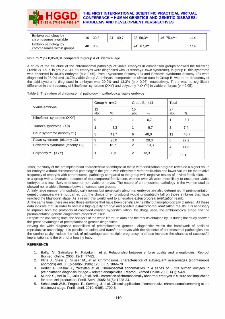
110
THE FIRST INTERNATIONAL SCIENTIFIC PRACTICAL VIRTUAL CONFERENCE − HUMAN GENETICS AND GENETİC DISEASES:
PROBLEMS AND DEVELOPMENT PERSPECTIVES
Embryo pathology by chromosomes available
16 30,8 24 40,7 28 58,3** 46 75,4*** 114
Embryo pathology by chromosomes within groups
40 36,0 74 67,9** 114
Note: *- ** р< 0,05-0,01 compared to group A of identical age
A study of the structure of the chromosomal pathology of viable embryos in comparison groups showed the following (Table 2). Thus, in group A, 41.7% embryos were diagnosed with 21 trisomy (Down syndrome), in group B, this syndrome was observed in 40.0% embryos (p > 0.05). Patau syndrome (trisomy 13) and Edwards syndrome (trisomy 18) were diagnosed in 25.0% and 16.7% viable Group A embryos, comparable to similar data in Group B, where the frequency of the said syndrome diagnosed in embryos was 20.0% and 13.3% (p > 0.05), respectively. There was no significant difference in the frequency of Klinefelter syndrome (XXY) and polysomy Y (XYY) in viable embryos (p > 0.05). Table 2. The nature of chromosomal pathology in pathological viable embryos
Viable embryos
Group А n=42 Group B n=44 Total
12 abc %
15 аbc %
27 аbc %
Klinefelter syndrome (ХХY) 0 0 1 6,7 1 3,7
Turner's syndrome (Х0) 1 8,3 1 6,7 2 7,4
Daun syndrome (trisomy 21) 5 41,7 6 40,0 11 40,7
Patau syndrome (trisomy 13) 3 25,0 3 20,0 6 22,2
Edwards's syndrome (trisomy 18) 2 16,7
2 13,3
4 14,8
Polysomy Y (XYY) 1 8,3
2 13,3
3 11,1
Thus, the study of the preimplantation characteristic of embryos in the in vitro fertilization program revealed a higher value for embryos without chromosomal pathology in the group with effective in vitro fertilization and lower values for the relative frequency of embryos with chromosomal pathology compared to the group with negative results of in vitro fertilization. In a group with a favorable outcome of extracorporeal fertilization, women over 35 were more likely to encounter viable embryos and less likely to encounter non-viable embryos. The nature of chromosomal pathology in the women studied showed no reliable difference between comparison groups. A fairly large number of morphologically normal but genetically abnormal embryos are also determined. If preimplantation genetic diagnosis were not carried out, the choice of embryologist would undoubtedly fall on those embryos that have reached the blastocyst stage. As a result, this would lead to a negative extracorporeal fertilization result. At the same time, there are also those embryos that have been genetically healthy but morphologically disabled. All these data indicate that, in order to obtain a high-quality embryo and positive extracorporeal fertilization results, it is necessary
to improve both the protocols of controlled ovarian hyperstimulation, the drugs used, the embryological stage and the preimplantation genetic diagnostics procedure itself. Despite the conflicting data, the analysis of the world literature data and the results obtained by us during the study showed the great advantages of preimplantation genetic diagnostics. Having the wide diagnostic capabilities of preimplantation genetic diagnostics within the framework of assisted reproductive technology, it is possible to select and transfer embryos with the absence of chromosomal pathologies into the uterine cavity, reduce the risk of miscarriage and multiple pregnancy, and also increase the chances of successful implantation and the birth of a healthy baby.
REFERENCE
1. Balfari V., Satirofgler H., Kabukem., et al. Relationship between embryo quality and aneuploidies. Reprod. Biomed. Online. 2006, 12(1), 77-82.
2. Kline J., Stein Z., Susser M., et al. Chromosomal characteristics of subsequent miscarriages (spontaneous abortions) Am. J. Epidemiol. 1986; 123 (6), p/ 1066-79.
3. Kerliet A. Cieslak J., Ylkeviteh et al. Chromosomal abnormalities in a series of 6,733 human oocytes in preinplatation diagnosis for age – related aneuploidies. Reprod. Biomed Online.2003; 6(1); 54-9.
4. Munne S., Velilla E., Colls P., et al. self – correction of chromosomally abnormal embryos in culture and implication for stern cell production. Fertil. Steril. 2005; 84(5): 1328-34.
5. Schoolcraft W.B., Fragouli E., Steveeg J. et al. Clinical application of compressive chromosomal screening at the blastocyst stage. Fertil. steril. 2010; 94(5): 1700-6.

111
BIRINCI BEYNƏLXALQ ELMI PRAKTIK ONLAYN KONFRANS − İNSAN GENETIKASI VƏ GENETİK XƏSTƏLIKLƏR: PROBLEMLƏR VƏ İNKIŞAF PERSPEKTIVLƏRI
6. Shinawi M., Cheung S.W. The array CGH and its clinical applications. DrengDisscow. Today. 2008; 13 (17-18); 760-70.
7. Staessen C., plattean P., van Assche E. et al. Comparison of blastocyst transfer with or without preimplantation genetic diagnosis for aneuploidy screening in couples with adzancedneabernal age aprogretivezanddomized controlled trial. Klm. Reprod. 2004; 19 (12); 2849-58.
8. Treff N.B., Levy B., Su J. et al., SNP microarray –baged 24 chromosome aneuploidy screening is significantly more cousistent than FISH. Mol.HumanReprod. 2010, 16 (8).
DATACITE
112
THE FIRST INTERNATIONAL SCIENTIFIC PRACTICAL VIRTUAL CONFERENCE − HUMAN GENETICS AND GENETİC DISEASES:
PROBLEMS AND DEVELOPMENT PERSPECTIVES
SÜMÜKLƏRİN İLTİHABİ VƏ ŞİŞ TRANSFORMASİYASI ZAMANI İMMUNHİSTOKİMYƏVİ TƏDQİQATI
G. Sadığova, İ. Əliyeva, T. İbrahimova, S. Tahirova
ABSRTACT
Qeyri-şiş mənşəli zədələnmələrin, xoşxassəli və bədxassəli sümükyaradıcı və sümük iliyi şişlərin sitospesifikliyinin müəyyənləşdirilməsi üçün istifadə edilən immunhistokimyəvi markerlər bizim tərəfimizdən dörd kateqoriyaya bölünmüşdür (əsas, faydalı, dəqiqləşdirici və əlavə). Bunlardan ümumi leykositar antigeni biz əsas, neyronspesifik enolazanı və neyrofilamentləri – faydalı, S-100 zülalını –dəqiqləşdirici, HBA-71 – əlavə markerlərə aid etmişik. Osteomielit zamanı immunhistokimyəvi tədqiqatlar üçün 5 immunohisto-kimyəvi markerdən osteoblastlar yalnız ümumi leykositrar antigenə pozitivdirlər. İmmunhistokimyəvi tədqiqatın nəticələri aşağıdakı şəkildə veril-mişdir. Osteomielit zamanı immunhistokimyəvi tədqiqatlar üçün 5 immunohisto-kimyəvi markerdən osteoblastlar yalnız ümumi leykositrar antigenə pozitivdirlər. Yuinq sarkoması zamanı HBA-71 və S-100 zülalı kimi markerlərlə immunhistokimyəvi tədqiqatlarla yüksək pozitivlik dərəcəsi (++) aşkar edilmişdir. PNET-in immunfenotipləşməsi zamanı neyronspesifik enolazanın, membran antigeni HBA-71, neyrofilamentlərin və S-100 zülalının yüksək pozitivlik dərəcəsi aşkar edilmişdir (++). İmmunfenotipləşmə zamanı sümüyün bədxassəli limfomasının 12 müşahidəsindən yüksək müsbət reaksiya yalnız ümumi leykositar antigenlə aşkar edilmişdir (++). Beləliklə, osteomielit üçün immunhisto-kimyəvi marker rolunu vimentin, aktin, ümumi leykositar antigen, Yuinq sarkoması üçün– HBA-71, S-100 zülalı, vimentin, həmçinin sitokeratinlər, neyronspesifik enolaza oynayır. PNET hüceyrələri S-100 zülalını, neyron-spesifik enolazanı, HBA-71-i, vimentini, neyrofilamentləri, sitokeratinləri və desmini ekspressiya edə bilər. Sümüyün bədxassəli limfoması ümumi leykositar antigenə və yalnız nadir hallarda vimentinə yüksək pozitivdilər.
IMMUNOHISTOCHEMICAL STADY OF BONES DURING THEIR INFLAMMATORY AND TUMOR TRANSFORMATION.
DATACITE
113
BIRINCI BEYNƏLXALQ ELMI PRAKTIK ONLAYN KONFRANS − İNSAN GENETIKASI VƏ GENETİK XƏSTƏLIKLƏR: PROBLEMLƏR VƏ İNKIŞAF PERSPEKTIVLƏRI
SİTOKİNLƏRİN VƏ ANTİMİKROB PEPTİDLƏRİN ŞD-2 XƏSTƏLƏRDƏ QAN SERUMUNDA SƏVİYYƏSİNİN TƏTQİQİ Gülnarə Əzizova, Arzu Dadaşova, Şəymən Həsənova, Şəfiqə Ələkbərzadə
Azərbaycan Tibb Universetiti, biokimya kafedrası Hal-hazırda şəkərli diabet (ŞD) dünyada geniş yayılmış xəstəliklərdən hesab olunur. Belə ki, Ümumdünya Səhiyyə Təşkilatın (ÜST) verdiyi statistik məlumatlara görə 2025-ci ildə isə 300 mln olacağını proqnozlaşdırılır. Bu xəstələrin 80-90%-i II tip şəkərli diabetdir Son illərin tədqiqatlarından aydın olur ki, II tip ŞD-in patogenezində müxtəlif növ sitokinlər və antimikrob peptidlər mühüm rol oynayır. Xüsusi farmakoloji aktiv, xırdamolekullu polipeptidlər olan sitokinlər autokrin, parakrin və yukstakrin təsirlərə malikdir [8]. Onlar bir sıra xəstəliklərin patofiziologiyasını, o cümlədən şəkərli diabeti mühüm siqnallarla təmin edir. TƏDQİQATIN MƏQSƏDİ
II tip şəkərli diabet zamanı IL-8, ŞNA- və TBF-1 sitokinlərin və bəzi antimikrob peptidlərin qan zərdabında öyrənilməsi idi. Tədqiqat 60 nəfər II tip şəkərli diabet xəstələri üzərində aparılmışdır. ŞD xəstəliyinin müddətinə görə 2 qrup xəstələr götürülmüşdür: I qrup – 1 ildən 5 ilə qədər – 34 xəstə (subkompensasiya); II qrup – 5 ildən çox – 26 xəstə
(dekompensasiya). Nəzərət qrupu 10 sağlam şəxs təşkil etmişdir. Qan zərdabında sitokinlərin (IL-8, ŞNA- və TBF-1) miqdarı Bender Medsystems (Avstriya) kommersiya dəstindən istifadə edərək təyin edilmişdir. Hycult Biotech dəsti ilə ELISA üsulu ilə endogen antimikrob peptidlərin qatılıqları (defenzin, endotoksin) öyrənilmişdir. Statistik təhlil Vilkokson (Mann-Utni) kriteriyasına əsasən aparılıb. NƏTİCƏLƏR
Alınan nəticələr aşkar etdi ki , nəzarətlə müqayisədə hər 2 qrupda zərdab sitokinləri, IL-8, ŞNA- və TBF-1 artmışdır.Oxşar nəticələr antimikrob peptidlərə səviyyəsində aşkar olunmuşdur.
I qrupda (II tip ŞD-in subkompensasiya mərhələsi) defenzinlərin qatılığı 120,0 0,9 nq/ml olmuşdur. Bu rəqəm nəzarət qrupundan 17,6%, yəni 1,2 dəfə çoxdur. II tip ŞD xəstələrində dekompensasiya mərhələsində (II qrup) defenzinin qatılığı I qrup xəstələrdə olduğundan nəzərəçarpacaq dərəcədə – 401,0%, yəni 4 dəfə çox olmuşdur. Endotoksinin səviyyəsi qan zərdabında I qrupda 1.18 ± 0.05 ng/ml, II qrupda isə 1.57 ± 0.14 ng/ml olmışdur ( nəzərətdə 0.33 ± 0.12 ng/ml). Qanda bakterial endotoksin dövr etməsi nəticəsində qrammənfi bakteriyaların dağıdılması endotoksemiya adlanır. Bu bakteriyaların deqradasiyası və ya ölümü geniş iltihabi reaksiyalarla nəticələnir. Əlaqəli sitokin sistemi makrofaqların iltihabönü sitokinlərinin qana keçməsini aktivləşdirir və leykositlərin endotelial adqeziyasını artırmaqla neytrofillərin komplemendən asılı reaksiyasını sürətləndirir və bütün bunlar sonda defenzinlərin qanda qatılığının yüksəlməsinə səbəb olur.
SECRETION LEVELS OF CYTOKINES AND ANTIMICROBIAL PEPTIDES IN BLOOD SERUM OF PATIENTS WITH TYPE 2 DIABETES MELLITUS Gulnara Azizova, Arzu Dadashova, Shayman Hasanova, Shafiga Alekberzade
Azerbaijan Medical University, biochemistry department
Aim. To assess the secretion levels of cytokines (interleukin-8, tumor necrosis factor α and transforming growth factor β1)
and some antimicrobial peptides in blood serum in type 2 diabetes mellitus. Material and methods. Blood samples of 60 patients with type 2 diabetes mellitus were examined. All patients were
assigned to two groups depending on the duration of diabetes mellitus: I group – from 1 to 5 years – 34 subjects (subcompensation); II group – more than 5 years – 26 patients (decompensation). Control group consisted of 10 healthy patients. The levels of cytokines (interleukin-8, tumor necrosis factor α and transforming growth factor β1) were measured with commercial kit «Bender Medsystems» kits (Austria). The levels of endogenous antimicrobial peptides (defensin, endotoxin) were studied with ELISA method using set «Hycult Biotech». Statistical analysis was performed with Wilcoxon (Mann–Whitney) test. Results. Obtained results revealed that in both groups the serum levels of cytokines, interleukin-8, tumor necrosis factor
α and transforming growth factor β1 increased compared to the control group. Similar results were obtained for antimicrobial peptides.
In I group (subcompensation stage of II type DM), defensin level was 0,0 0,9 ng/ml. This is 17,6%, i.e. 1,2 times, higher than in the control group. In the group of type 2 diabetes mellitus patients in decompensation stage (II group) the level of defensin was by 401,0%, i.e. 4-fold, higher compared to the I group. Endotoxin concentration in blood serum was 1,18 ± 0,05 ng/ml in I group, 1,57 ± 0,14 ng/ml in II group (control value was 0,33 ± 0,12 ng/ml). An association was revealed between the secretion of antimicrobial peptides and cytokines. Concentration of antimicrobial peptides and cytokines reflects the stage of type 2 diabetes mellitus. Keywords: Antimicrobial peptides, type 2 diabetes mellitus, cytokines, defensin, endotoxin
114
THE FIRST INTERNATIONAL SCIENTIFIC PRACTICAL VIRTUAL CONFERENCE − HUMAN GENETICS AND GENETİC DISEASES:
PROBLEMS AND DEVELOPMENT PERSPECTIVES
ƏDƏBİYYAT
1. World Health Organization. HEARTS D: diagnosis and management of type 2 diabetes. 22 April 2020
[https://www.who.int/publications/i/item/who-ucn-ncd-20.1]; 2. International Diabetes Federation. IDF Diabetes Atlas. 9th edition, 2019
[http://www.diabetesatlas.org/content/foreword]; 3. Е. А. Литвиненко (Lytvynenko E.A.), П. Н. Боднар (Bodnar P.N.), Н. И. Лисяний (Lysianuy N.I.), Л. Н. Бельська
(Belska L.N.). Уровень эндотоксемии и цитокиновый статус у пациентов с сахарным диабетом 2 типа и неалкогольной жировой болезнью печени. Журнал Гродненского государственного медицинского университета, No 4, 2014: стр.86-90;
4. Shehan N. Randeria, Greig J. A. Thomson, Theo A. Nell, Timothy Roberts, and Etheresia Pretorius. Inflammatory cytokines in type 2 diabetes mellitus as facilitators of hypercoagulation and abnormal clot formation. Cardiovasc
Diabetol., 2019 Jun 4, 18(1): p. 72; 5. Balázs Csaba Németh, Tamás Várkonyi, Ferenc Somogyvári, Csaba Lengyel, Katalin Fehértemplomi, Szabolcs
Nyiraty, Péter Kempler, and Yvette Mándi. Relevance of α-defensins (HNP1-3) and defensin β-1 in diabetes. World J Gastroenterol., 2014 Jul 21, 20(27): 9128–9137;
6. Omar S Al-Attas, Nasser M Al-Daghri, Khalid Al-Rubeaan, Nancy F da Silva, Shaun L Sabico, et al. Changes in endotoxin levels in T2DM subjects on anti-diabetic therapies. Cardiovasc Diabetol., 2009, 8: p. 20;
7. А. А. Тишкина, Н. В. Ворохобина, А. Ю. Барановский. Роль изменений микрофлоры кишечника в патогенезе сахарного диабета 2-го типа и ожирения. Возможные пути коррекции. Лечащий врач, 2010, 3.
DATACITE
115
BIRINCI BEYNƏLXALQ ELMI PRAKTIK ONLAYN KONFRANS − İNSAN GENETIKASI VƏ GENETİK XƏSTƏLIKLƏR: PROBLEMLƏR VƏ İNKIŞAF PERSPEKTIVLƏRI
SU ANBARLARININ BİOLOJİ MƏHSULDARLIĞI Aynur Ənsərova Tibbi biologiya və genetika kafedrası. Azərbaycan Tibb Universiteti. İşin məqsədi. Azərbaycanın əsas su anbarlarının müasir ekoloji vəziyyəti haqqında 2012-2017-ci ildə fəsillər üzrə apardığımız tədqiqatın nəticələrini təqdim etməkdir.
Material və metodlar. Su nümunələri steril şüşə qablarla Y.İ.Sorokinin batometri ilə , lil-qrunt isə kiçik Peterson
cihazı ilə əldə edilmişdir. Suyun şəffaflığını təyin etmək üçün ağ rəngli Sekki lövhəsindən istifadə olunmuşdur. Sularda və lil-qruntda mikrobların ümumi sayı, müvafiq olaraq A.C.Razumovun və S.N.Vinoqradskinin metodlarına əsasən müəyyən olunmuşdur. Saprofit bakteriyaların miqdarı balıq peptonlu aqar qida mühitinə əkməklə təyin olunmuşdur. Spor əmələ gətirən saprofit bakteriyalar termiki (80°C) üsula əsasən, fizioloji qrupa və koli-enterobakter cinsinə aid olan bakteriyalar V.İ.Romanenko, S.İ.Kuznetsov və A.Q.Rodinanın metodik göstəricilərinə müvafiq əldə edilmişlər. Mikrobioloji əkmə-analizlər, nümunələr götürüləndən 1,5-2 saatdan gec olmamaq şərtilə aparılmışdır.
Nəticələr. Bioloji məhsuldarlıq dedikdə ilk olaraq balıq və balıq məhsulları yada düşür. Əslində isə vətəgə əhəmiyyətli
məhsullar bitki və heyvan mənşəli məhsullardan-biokütlədən asılıdır. Qida silsiləsi su mühitində 5-6 mərhələli olduğu halda, məsələn: nərə balığında: 1)yosun; 2)zooplankton; 3)xərçəngkimilər; 4)infuzorlar; 5)aterina kilkə balıqları; 6)siyənəklər; 7)bölgə balıqları. Litosferdə (quruda) isə qida silsiləsi 2 mərhələlidir; (məsələn; ot-at; at-qoyun) nəzərə alınmalıdır ki, su anbarına çatanda çay sularının keyfiyyəti tam dəyişir. Məsələn Araz kimi bulanıq şay sularda olan biogen elementlər fitoplankton az mənimsənilir və fotosintez getmir, bulanıq suda “mürgüləyən” biogen elementlərin 1 kq-mı su anbarına çatana “ayılaraq” 1 tona qədər plankton kütləsi əmələ gəlir. Mikrobioloji və hidrobioloji tədqiqatlarla müəyyən edilmişdir ki, çay sularda orta hesabla mikrobların sayı, hetorotrof bakteriyaların miqdarı müvafiq olaraq yəni- a) mikrobların sayı -0,4-0,75 min\ml olarsa- oliqotrof, 0,8-1,5 min\ml 0,9-1,2 min\ml olarsa – mezotrof , 5-6 min\ml və 8-15 min\ml olarsa – evtrof su anbarlarına aid edilir. Balıqçılığa görə evtrof – su anbarı zəngin hesab edilir. Yeni yaradılandan Mingəçevir su anbarında 2 il sonra hövzədə ixtiofaunada külmə balıqları üstünlük təşkil etsə də sonralar ovlanan balıqların 20-25% - dən çox hissəsini çəki, çapaq və sif balıqları üstün olub – ali su bitkilərinin təmizlənməsində ağ amur (belamor) balığından istifadə edilir.
Yekun.Təcrübələrlə sübut edilmişdir ki, su hövzələrində anaerobioz hadisəsini yaradan sulfatlaşdırıcı bakteriyaların
intensiv inkişafı üçün 3 amil; a) üzvi maddələr; b) sulfat-sulfid birləşmələri; c) oksigen qıtlığı lazımdı. Su mühitində fəaliyyət göstərən hidrofauna və hidroflora yerləşdikləri ərazi, qidalanma və hidroflora yerləşdikləri ərazi, qidalanma artıb-çoxalma xüsusiyyətinə görə a)planktonlara; b)bentoslara ayrılır. Planktonlara aid olan bəsit orqanizmlər su qatında passiv halda asılı vəziyyətdə , bentos adlanır. Hidrobiontlar isə dibdə -lil qruntda fəaliyyət göstərirlər. Su qatlarənda fəaliyyət göstərən orqanizmlər – pelagial adlanır.
Açar sözlər. Anaerobioz,sulfatlaşdırıcı bakteriyalar,evtrof su anbarı.
DATACITE

116
THE FIRST INTERNATIONAL SCIENTIFIC PRACTICAL VIRTUAL CONFERENCE − HUMAN GENETICS AND GENETİC DISEASES:
PROBLEMS AND DEVELOPMENT PERSPECTIVES
TÜRKİYƏ RESPUBLİKASI ƏRAZİSİNDƏ ARAZ ÇAYININ EKOLOJİ MİKROBİOLOGİYASI
Ənsərova Aynur, Gülnarə Vəliyeva, Pərvanə Mahmudova, Nailə Məmmədova
Azərbaycan Tibb Universitetinin, Tibbi biologiya və genetika kafedrası. Azərbaycan.
ABSTRACT
It was the first time identified ecological microbiology at the river Araz for seasons in the Turkish Republic. It was found that the upper part of the river Araz quantitative and qualitative microbiot are different, but there is nt neqative impact on the stable anthropogenetic ecological sustainability of water source. At the same time? Was noted that allachton organic substances episodic discharges from large settlements in areas of the river. It was determined that in the research the household and livestocic farms sewage is restored to a stable condition water below the 18-20 km involved in the avas. Tədqiqatın məqsədi
İlk dəfə olaraq, Türkiyyə Respublikası ərazisində Araz çayı ilk mənbədən qonşu dövlətlərlə sərhəd sahələrdə ilin fəsilləri üzrə mikrobioloji cəhətdən müfəssəl öyrənilmişdir. Tədqiqatın material və metodları
Bütün nümunələr aseptik qaydalara riayət olunmaqla – sular Y.İ.Sorokin batometri və lil-qrunt isə steril şpatel (çayda), kiçik QOIN borucuğu (su anbarında) ilə götürülmüşdür . İlkin mikrobioloji analiz-əkmələr nümunə götürülən vaxtdan 2 saatdan gec olmadan aparılmışdır (çöl-ekspedisiya şəraiti və stasionar-laboratoriya). Suyun temperaturu civə sütunlu dərinlik termometri ilə ölçülmüşdür. Üzvi maddələrin biodestruksiyası Vinkler-Vinberq (1960) üsulları ilə təyin edilmişdir. Fitoplanktonun (yosunların) ilkin məhsulu karbon 14 izotopu vasitəsilə (Stemann Nilsen, 1952; Sorokin,1956) ənənəvi klassik üsullarla təyin edilmişdir. 2010-2016-ci illərdə ilin fəsilləri üzrə 2296 məntə-stansiyada 1897-su, lil-qurunt nümunələri toplanmış və həmin nümunələrdən istifadə etməklə 5319 analiz-əkmələr tərəfimizdən aparılmışdır.Bütün alınan rəqəmlər nəticələr statistik işlənmişdir. Tədqiqatın nəticələri
Öz başlanğıcını Türkiyə Respublikasının Şərqi Anadolu dağlarından götürən Araz (Bingöl dağları) çayının Ermənistan, Azərbaycan və İİR ilə sərhəd ərazilərinə kimi məcraları yuxarı axar hesab olunur. Son 50 ildə aparılan tədqiqatlardan məlum olmuşdur ki, Araz çayının orta və aşağı axarları, ərazilərindən keçdikləri dövlətlər tərəfindən uzun illərdən bəri aramsız davam edən antropogen təsirlərə məruzdur. İlk dəfə olaraq , Türkiyyə Respublikası ərazisində Araz çayı ilk mənbədən qonşu dövlətlərlə sərhəd sahələrdə ilin fəsilləri üzrə mikrobioloji cəhətdən müfəssəl öyrənilmişdir.Araz çayı hövzəsinin Türkiyyə Respublikasna aid olan ərazisində tədqiqatların aparılması və müşahidələrin həyata keçirilməsi üçün , onun əsas axar qollarını əhatə edən 16-17 məntəqə nəzərdə tutulmuşdur. Bundan başqa Araz çayının ən iri qolları Pasinçaydan, Qərbi Arpaçaydan və Karsçaydan da nümunələr toplanmışdır. Ümumiyyətlə, tədqiqatlar aparılan müddətdə 286 su və lil-qrunt nümunələri toplanmış və onlarda mikroorqanizmlərin ümumi miqdarı A.Razumov və S.Vinoqradskinin metoduna əsasən hesablanmışdır. Saprofit, koliform və fizioloji qrupa aid bakteriyalar V.Romanenko, S.Kuznetsovun “Mikrobioloji laborator tədqiqatları” əsərinə əsasən əldə edilmişdir. Həmçinin hər dəfə suların fiziki xassələrinə aid göstəricilərdən suyun temperaturu, şəffaflıq dərəcəsi axın sürəti ölçülmüşdür. Sularda öz-özünə təmizlənmə prosesinin intensivliyini , saprobluq dərəcəsini, axın boyu çirklənmə məntəqələrini müəyyən etmək üzrədə müşahidələr aparılmışdır. Sularda şəffaflıq Sekki lövhəsi ilə oksigenin miqdarı və üzvi maddələrin biodestruksiya olunması intensivlyi Vinkler-Vinberq üsulu ilə öyrənilmişdir. Yekun. Beləliklə ilin fəsilləri üzrə Araz çayı hövzəsində başa çatdırılan mikrobioloji və sanitar- hidrobioloji tədqiqatlardan
aydın olmuşdur ki, Türkiyə Respublikası ərazisində Ermənistanla sərhəddə Qərbi Arpaçaya Araz çayı hövzəsində bir neçə yaşayış məntəqələri, heyvandarlıq ferma-birlikləri tərəfindən lokal çirklənmə qeyd edilir. Eyni zamanda da axın boyu hövzədə 18-20 km məsafədə suların ekoloji sabitliyi bərpa olunur. Ona görə etiraf etmək lazımdır ki, Araz çayı Türkiyə Respublikası ərazisindən qonşu dövlətlərlə sərhəd sahələrə kimi təmizdir. Bununla belə, Arazın Ermənistan ərazisində ilkin və kəskin dərəcədə çirklənməsi-zəhərlənməsi müəyyən olmuşdur. Açar sözlər: Antropogen təsir, lokal çirklənmə.
COLOGİCAL MİCROBİOLOGY OF ARAZ RİVER İN THE TERRİTORY OF THE REPUBLİC OF TURKEY. Aynur Ansarova, Gulnara Valiyeva, Parvana Mahmudova, Naila Mammadova Azerbaijan Medical University.Department of medical biology and genetics. Azerbaijan ABSTRACT
The purpose of the study. For the first time in the territory of the Republic of Turkey, the Araz River was studied in detail
from the first source in the areas of the border with neighboring countries from a microbiological point of view. It was the first time identified ecological microbiology at the river Araz fo seasons in the Turkish Republic. It was found that the upper part of the river Araz quantitative and qualitative microbiot are different, but there is nt neqative impact on the

117
BIRINCI BEYNƏLXALQ ELMI PRAKTIK ONLAYN KONFRANS − İNSAN GENETIKASI VƏ GENETİK XƏSTƏLIKLƏR: PROBLEMLƏR VƏ İNKIŞAF PERSPEKTIVLƏRI
stable anthropogenetic ecological sustainability of water source. At the same time? Was noted that allachton organic substrances episodic discharges from large settlements in areas of the river. It was determined that in the research the household and livestocic farms sewage is restored to a stable condition water below the 18-20 km involved in the avas. Keywords. Anthropogenic impact, local pollution.
DATACITE
118
THE FIRST INTERNATIONAL SCIENTIFIC PRACTICAL VIRTUAL CONFERENCE − HUMAN GENETICS AND GENETİC DISEASES:
PROBLEMS AND DEVELOPMENT PERSPECTIVES
SU ANBARLARININ ƏTRAF MÜHİTƏ VƏ ƏHALİNİN SAĞLAMLIĞINA TƏSİRİ
Ənsərova Aynur, Gülnarə Vəliyeva, Pərvanə Mahmudova, Nailə Məmmədova
Azərbaycan Tibb Universiteti, Tibbi biologiya və genetika kafedrası, Bakı,Azərbaycan.
E.mail:[email protected] Tədqiqatın məqsədi. Su anbarlarında fitoplanktonun fotosintez prosesində sintez etdiyi ilkin məhsulun, destruksiya
olunan ümumi üzvi maddələrin miqdarının təyin edilməsi ilə bioloji məhsuldarlığın formalaşmasının əsaslandırılması. Tədqiqatın material və metodları. Üzvi maddələrin biodestruksiyası Vinkler-Vinberq üsulları ilə təyin edilmişdir.
Fitoplanktonun (yosunların) ilkin məhsulu karbon 14 izotopu vasitəsilə (Stemann Nilsen; Sorokin) ənənəvi klassik üsullarla təyin edilmişdir. 2010-2016-ci illərdə ilin fəsilləri üzrə 2296 məntə-stansiyada 1897-su, lil-qurunt nümunələri toplanmış və həmin nümunələrdən istifadə etməklə 5319 analiz-əkmələr tərəfimizdən aparılmışdır.
Tədqiqatın nəticələri. Mingəçevir və Şəmkir su anbarında güclü küləklər əsir.Şəmkir su anbarının sağ sahilinə
qonşuluqda yerləşən Sarı təpədən əsən güclü küləklər son vaxtlar azalıb deyə qurunt suların üst qata miqrasiyası güclənib. Bu səbəbdən Mingəçevir, Yenikənd su anbarının sahil əraziləri uçulur, su anbarına min kub metrlərlə torpaq tökülür, su tutumu azalır. Bu yolla Mingəçevir su anbarında son 60 ildə sahil ərazilər 150-350 m artmış anbarın sahəsi 1 km2 artmış, su tutumu isə 1,9 km3 azlmış, son illərdə hər il Mingəçevir su anbarında 3,84 milyon ton lil torpaq əlavə olunmuşdur. Mingəçevir su anbarında istifadə olunmayan su hövzədə cəmləşən suyun 50%- dən çoxunu təşkil edir. Su anbarlarından çəkilən Yuxarı Qarabağ və Şirvan su kanalları məcrası ekranlaşmadığından 30-45% su itkisinə yol verilir. Xəstəliklərin yayaılmasında su anbarları 3 zəmin yaradır. 1) Xəstəliklərin və mikrobdaşıyıcıların ifrazatları ilə xəstəlik törədicilərinin sulara düşməsi; 2)Xəstəliktörədicilərin virulentli formalarının su mühitində uzun müddət saxlanılması; 3)Xəstəliktörədicilərlə çirklənmiş sudan məişətdə istifadə edilməli, su anbarları ətrafında yaşayan əhali arasında yayılan
xəstəliklərdən- malyariya –anofeles ağcaqanadlarının artması səsəbindən çoxalır. Sənaye sahələrinin çirkabının su hövzələrdə balıqları, xərçəngi, ilbizləri yeyilməz dərəcədə zəhərləyir.
Yekun. İri su anbarları əhalinin iqtisadi, məişət, sosial-ictimai, həyat tərzinə, sağlamlığına müsbət təsir etməklə yanaşı
epidemioloji vəziyyətinə, gərginləşməsinə və bəzi xəstəliklərin yayılmasında heyvanlar arasında invazion xarakterli parazitlərin artmasına da səbəb ola bilər. Açar sözlər.Fitoplankton,destruksiya,qrunt sular.
DATACITE
119
BIRINCI BEYNƏLXALQ ELMI PRAKTIK ONLAYN KONFRANS − İNSAN GENETIKASI VƏ GENETİK XƏSTƏLIKLƏR: PROBLEMLƏR VƏ İNKIŞAF PERSPEKTIVLƏRI
THE SMPD1 GENE MUTATIONS IN TWO SIBLINGS WITH NIEMANN-PEAK TYPE A/B DISEASE Baghirova Nurana Baku Health Center Purpose of work. Niemann–Pick type A/B disease in children was investigated. Niemann–Pick type A disease is the most severe form that begins in infants, characterized by an increase in the hepatosplenomegaly and progressive damage to the nervous system. Type B is characterized by a chronic course, hepatosplenomegaly, the nervous system is usually not involved. Both types are associated with mutations in the SMPD1 gene and sphingomyelinase deficiency. In addition to these, there is an increased level of cholesterol and lipids in the blood and thrombocytopenia. The sphingomyelinase breaks down sphingomyelin into ceramide and phosphocholine in lysosome membranes, and its deficiency leads to excessive accumulation of sphingomyelin. The SMPD1 gene occupies a locus on chromosome 11p15.4-15.1. The disease is inherited as an autosomal recessive trait (1, 2, 3, 4). Material and methods. Study materials were collected on DBS cards as dried venous blood spots from Republic Children’s Clinical Hospital departments and Baku Health Center. The method of tandem mass spectrophotometry was used to determine the activity of acid sphingomyelinase in dried blood stains. Using direct automatic sequencing, all 6 coding exons of SMPD1 gene were studied in the ARCHIMED Life laboratory (Austria). General blood analysis and ultrasonic scan were made with common methods. Patients A.L. (born 2000) and A.Z. (born 2006) live in the city of Ganja. Those born from a blood-related marriage are a cousin of a parallel type. Low activity values of the acid sphingomyelinase enzyme in dry blood spots were obtained for A.L.- 0.29 μmol/liter hour for A.Z.- 0.3 μmol/liter/hour (norm> 0.9 μmol/liter/hour). Results. Genetic analysis of a DNA sample extracted from the blood of patients A.L. and A.Z., revealed two mutations of the SMPD1 gene: G→A transversion at position 1345 (1345G→A) and cytosine nucleotide duplication at 188 position (188dup. C). Both siblings have a double heterozygous state for two different mutations - 1345 G→A/188 dup. C, which are characteristic of Niemann–Pick disease type A/B. The replacement of the guanine nucleotide with the adenine nucleotide at position 1345 in exon 5 of the SMPD1 gene in the heterozygous state leads to the replacement of the glutamine amino acid with lysine at position 449 (Glu449 Lys) in the patient protein. A mutation of 188 dup. C in the first exon of the SMPD1 gene in the heterozygous state was also identified. Mutation in the protein leads to the replacement of the amino acid leucine with serine at position 64 (Leu 64 Ser). Conclusion. Thus, both siblings have a double heterozygous state for two different mutations of the SMPD1 gene - 1345 G→A/188 dup. C. Therefore, the identified mutations of the SMPD1 gene indicate Niemann–Pick type A/B disease. REFERENCES
1. Baghirova N.A., Rasulov E.М. et al. The first case of Niemann-Pick A/B type molecular characteristics for the patient from Azerbaijan, Pediatrics and Children’s Surgery, Almaty, №3 (97) 2019, p. 62-65.
2. Galehdari H., Tangestani R., Ghasemian S. New single nucleotide deletion in the SMPD1 gene causes Niemann-Pick disease type A in a child from Southwest Iran: a case report // Iran J. Pediatr. 2013. Vol. 23. № 2. p. 233–236.
3. Hua R., Wu H., Cui Z. et al. A novel SMPD1 mutation in two Chinese sibling patients with type B Niemann Pick disease // Chin. Med. J. (Engl.). 2012. Vol. 125. № 8. p. 1511–1512.
4. Внутренние болезни: учебник: в 2 т./под ред. В.С. Моисеева, А.И. Мартынова, Н.А. Мухина, 2013, Т 2, 896 с.
DATACITE
120
THE FIRST INTERNATIONAL SCIENTIFIC PRACTICAL VIRTUAL CONFERENCE − HUMAN GENETICS AND GENETİC DISEASES:
PROBLEMS AND DEVELOPMENT PERSPECTIVES
BIOCHEMICAL POLYMORPHISM, GENETIC HETEROGENICITY, GENE MAPING AND PREVENTION OF MUCOPOLYSACCHARIDOSIS HEREDITARY DISEASE IN THE POPULATION OF THE REPUBLIC OF AZERBAIJAN 1Sevda Alizada, 2Kamile Aliyeva, 3Elkhan Rasulov
1PhD in Medical Biology., 2Professor, 3Professor. 1Azerbaijan Medical University, 2Baku State University, 3Azerbaijan State Advanced Training Institute for Doctors named after A.Aliyev. The purpose of the work: is to study the biochemical polymorphism, genetic diversity of mucopolysaccharidosis
lysosomal hereditary disease in the population of the Republic of Azerbaijan, to compile a gene map of the disease for the population of the Republic and to develop the ways to prevent this disease. Materials and methods: It was discovered within expeditions in the regions of the Republic of Azerbaijan, during genetic
screening among sick children treated at the CRH. Fluorimetric method and liquid chromatography were used for genetic screening. Results: Lysosomal enzymes for genetic screening of mygppolysaccharidosis hereditary diseases: α - L - iduronidase,
Iduronatsulfatase, Heparan - N - sulfatase, α - N - acetylglucosaminidase, α - N - acetyl – CoA: Glycosamine - acetyltransferase, N - acetylglucosamine - 6 sulfatase, Galactosamine - 6 sulfatase, β - galactosidazole, Arylsulfatase B, β glucuronidase, N - acetylglucosamine -1- phosphotransferase activity were studied. Direct sequencing of IDS, HGSNA, GALNS and ARBS genes was performed by the Senger method. By using the method, disease-causing mutations within the IDS, HGSNA, GALNS, and ARBS genes were identified. The method was developed in the laboratory of GENTOGENE, Rostock, Germany. Conclusion: Based on the results of our biochemical and molecular genetic research, treatment and prevention methods
will be developed separately for each type of mucopolysaccharidosis. REFERENCES
1. В.И. Иванов, Н.В. Барышникова, Дж.С. Билева, Е.Л. Дадали, Л.М. Константинова, О.В. Кузнецова, А.В. Поляков «Генетика» Москва ИКЦ «Академкнига»-2007.
2. Filocamo M, Morrone A (2011) Lysosomal storage disorders: molecular basis and laboratory testing. Hum Genomics 5(3):156-169.
3. Khan SA, Peracha H, Ballhausen D, Wiesbauer A, Rohrbach M, Gautschi M, Mason RW, Giugliani R, Suzuki Y, Orii KE, Orii T, Tomatsu S (Jul. 2017) Epidemiology of mucopolysaccharidoses. Mol Genet Metab 121(3):227-240.
DATACITE
121
BIRINCI BEYNƏLXALQ ELMI PRAKTIK ONLAYN KONFRANS − İNSAN GENETIKASI VƏ GENETİK XƏSTƏLIKLƏR: PROBLEMLƏR VƏ İNKIŞAF PERSPEKTIVLƏRI
PROGNOSTİC SİGNİFİCANCE OF PD-L1 STUDİES İN CERVİCAL DYSPLASİA Irada Soltanova, Nigar Mehdiyeva Department of Oncology. Azerbaijan Medical University. ABSTRACT
The article is included the clinical and morphological features of 35 patients who had been diagnosed and treated with cervical dysplasia and reflected the immununistochemical results of PD-L1 expression in atypical cells. By standardized immunochemical methods, receptor levels vary depending on the degree of intraepitelial dysplasia. In patients with CIN I, cells were not stained at all (negative PD-L1 reaction), PD-L1 expression in CIN II was 1+ and 2+ in 76.9±5.6% , and 3+ in 23.1±5.3% of patients (p<0.05). In patients with CIN III, 3+ cases (severe staining) were detected in 66.7±4.2% and cases of 2+ (moderate staining) in 33.3±4.9% of women. Thus, expression of the PD-L1 receptor, known as anti tumor immunitite regulator is closely related to dysplastic processes that begin in the tissue. By other words, the detection this receptor expression in patients with CIN diagnosis(especially CIN III) can play an important role in the selection of treatment tactics and assessment of prognosis. Cervical cancer(CC) accounts for 6.6% of malignant neoplasms among women, with more than 57,000,000 new cases being diagnosed worldwide each year, and the number is steadily increasing (1,2,3). Detection of precancerous diseases in CC screening is one of the key mechanisms to combat this problem (4). For this reason, diagnosis, adequate treatment, and prognostic assessment of cervical intraepithelial neoplasia (CIN) are of great importance in the management of CC. The purpose of the study. To determine the relationship between PD-L1 receptor expression detection and histological
grade in cervical dysplasia.
Materials and methods. Results of diagnosis of 35 patients with CIN that conizated were included in to the study. There
was performed standart immunocytochemical method (VENTANA Bench Mark Ultra) using PD-L1-monoclonal rabbit antibodies (VD21R, Medaysis firm) was applied newly obtained incisions after deparaffinization. The reaction results are evaluated by the number of staining cells as follows: 1+ - the number of poorly stained cells is less than 10%; 2+ - moderate staining in 10% of cells; 3+ - Strong staining in ≥10% of cells.
Results. Patients ages 28 to 61 years, with an average age of 37.3 ± 2.6. The majority of patients were women between
the ages of 28 and 49 - 32 (91.4 ± 1.9%), 20 of them (57.1 ± 2.4%) were women between the ages of 28-39. Only one woman had a diagnosis of CIN between the ages of 60-69 (2.9 ± 3.2%, p <0.05). Thirty (85.7 ± 3.7%) of the patients were reproductively activite, and 5 (14.3 ± 4.0%, p <0.05) were in the pre- or menopausal period. In 31 of the women (88.6 ± 2.6%), no complaints were reported, and the rest reported mucosal-bloody vaginal discharge (11.4 ± 2.9%). All patients underwent cytological examination, colposcopy and biopsy on their cervix. The results of the cytological examination in 30 cases (85.7 ± 4.1%) coincided with subsequent histological findings. Of these, 26 (74.3 ± 2.8%) were diagnosed with HSIL (high grade intraepithelial squamous lesion), and 4 (11.4 ± 2.7%) were diagnosed with LSIL (low grade intraepithelial squamous lesion). After histological examination, patients were divided into 3 groups: CIN I - 7 (20 ± 3.5%), CIN II - 13 (37.1 ± 4.5%), and CIN III - 15 (42.9 ± 2.8%). ill. In 12 (34.2 ± 3.4%) of patients, the depth of epithelial damage was up to 1.4 mm, 9 of them (75.0 ± 5.1%) were women in the CIN III group. This is 60.0 ± 3.9% of the incidents in the group. Immunohistochemical staining was not recorded during CIN I; About in CIN II - PD-L1 expression was 1+ in 6 cases (46.1 ± 5.2%), 2+ in 4 (30.8 ± 3.7%) and 3 (23.1 ± 5.3) %, p <0.05) was rated as 3+. CIN III was defined as 3+ in 10 patients (66.7 ± 4.2%, p <0.05) and 2+ in 5 (33.3 ± 4.9%), with no staining reported. In PD-L1-positive cases, acute expressed koylocytosis was observed. Lymphocytar infiltration was also reported in light microscopy analysis.
Final. In our study CIN was most commonly found in young, reproductively-active women (85.7 ± 3.7%, p <0.05). The
depth of intraepithelial invasion depends on the degree of dysplasia, and in 60.0 ± 3.9% of patients with CIN III it was 1.4 mm. PD-L1 expression is observed in atypical cells, which varies depending on the degree of dysplasia. In case of negative reaction in CIN I, strong staining (3+) in CIN II was observed in 3 cases (23.1 ± 5.3%) and in CIN III at 66.7 ± 4.2% (10 cases) shows strong staining (p <0.05) and severe koylocytosis. In addition, during CIN III, poor staining cases were not reported at all. Although we encounter lymphocytic infiltration in microscopic imaging, the relationship between this symptom and degree of dysplasia should be investigated in detail.
DATACITE
122
THE FIRST INTERNATIONAL SCIENTIFIC PRACTICAL VIRTUAL CONFERENCE − HUMAN GENETICS AND GENETİC DISEASES:
PROBLEMS AND DEVELOPMENT PERSPECTIVES
MOLECULAR BASIS OF HEMOGLOBINOPATHIES IN AZERBAIJAN
1Agharza Aghayev, 2Valeh Huseynov, 3Ramin Bayramli, 4Oya Uyguner Z.
1,2,3National Hematology and Transfusiology Center, Genetic Diagnostics Center. Azerbaijan.
4Department of Medical Genetics, Istanbul Medical Faculty, Istanbul University. Turkey.
Introduction: Thalassemias are defined by the absence or decrease of globin chain(s), which may lead imbalances of
globin chains, ineffective erythropoiesis, hemolysis, and eventually to a variable degree of anemia. With the estimated
carrier frequency of β-thalassemia is 4%–8.6% in country, the importance of premarital genetic testing, informed family
counseling and preventive medical treatment cannot be overstated 1-4. Screening for traits by hemoglobin electrophoresis
and biochemical analysis has become a routine application test prior to the issuance of a marriage license throughout
Azerbaijan.
Methods: We aimed to evaluate prevalent mutations spectrum, the effect and their co-inheritance and/or role of
polymorphisms (BCL11A, HBS1L-MYB and Xmn1 SNP) on disease phenotype by using reverse dot-blot hybridization
using customized strips, sequencing analysis of the complete genes and MLPA.
Results: We report here a summary finding of HBB gene analysis for 265 patients and HBA1-HBA2 genes analysis for 28
patients, along with their family members totaling 570 individuals.
𝛃 thalassemia - 66 were found to have homozygous, 76 possessed compound heterozygous and 80 possessed
heterozygous mutations. Overall, a total of 416 alleles were found to have 34 mutations. The first 20 frequented mutations
covered 87% of the entirety of all mutations. The summary range is as following: c.25_26delAA (p.Lys9Valfs) 27.9%; c.93-
21G>A (IVS1+110G>A) 11.3%; c.315+1G>A (IVS2+1G>A) 8.9%.
𝛂 thalassemia - Genotyping of 45 alleles revealed 9 mutations, the 3.7 kb deletion is the most frequent mutation (35.6%),
followed by 20.5 kb deletion (24.4%) and α2 polyA2 (HBA2: c.*92A >G, 13.3%).
Conclusions: The results may help inform decisions in the design and implementation of prevention strategies and
diagnostic approaches. Through examination of the cumulative results we established the distribution pattern of hemoglobinopathies in Azerbaijan which facilitated a more focused molecular genetic approach to prevention. Wide range sequence analysis of HBB gene from 5’ promoter (-150) to 3’ promoter region (*150) provide additional contribution to diagnose and whole gene sequencing is important to clarify of patient with thalasemia intermedia cilinical findings. We will demonstrate in presentation that despite the high degree of molecular heterogeneity, the above approach, combined with the advent of PCR-based techniques and improved methodologies of early fetal sampling have made heterozygote screening and prenatal diagnosis of feasible in Azerbaijan. REFERENCES
1. Sabath, D. E. (2017). Molecular diagnosis of thalassemias and hemoglobinopathies: an ACLPS critical review.
American journal of clinical pathology, 148(1), 6-15.
2. Giardine, Belinda, et al. (2014). Updates of the HbVar database of human hemoglobin variants and thalassemia
mutations. Nucleic acids research 42.D1 D1063-D1069.
3. Altay, Ç (2002). The frequency and distribution pattern of β-thalassemia mutations in Turkey. Turkish Journal of
Haematology, 19(2), 309–315.
4. Asadov, C., et al. (2017). Genotype–phenotype correlations of β-thalassemia mutations in an Azerbaijani
population. Turkish Journal of Hematology, 34(3), 258–263.
DATACITE
©Publisher: LLC Azerbaijan Medical University (Azerbaijan, Baku), R/C 1400457111
©Nəşriyyat: Azərbaycan Tibb Universiteti. MMC (Azərbaycan, Bakı)
Rector: Garay Garabayli. Doctor of Medical Science. Professor
Rektor: Gəray Gəraybəyli Tibb Elmləri Doktoru. Professor
Registered address: 14, A.Gasimzade street. AZ1022
Qeydiyyat ünvanı: 14, Ə. Qasımzadə küç., AZ1022
©Publisher: LTD International Research, Education & Training Center. (UK, London), C/N 12224486
©Nəşriyyat: LTD Beynəlxalq Tədqiqat, Təhsil & Təlim Mərkəzi.
Director and shareholder: Alexandra Cuco. Lawyer. Portugal.
Direktor və Payçı: Aleksandra Kuko. Hüquqşünas. Portuqaliya.
Deputy and shareholder: Namig Isazade. PhD in Business Administration.
Direktor müavini və Payçı: Namig Isazade. PhD in Business Administration.
Registered address/Qeydiyyat Ünvnı: 71-75 Shelton Street, Covent Garden, London, WC2H 9JQ, UK.
©Editorial office / Redaksiya: 71-75 Shelton Street, Covent Garden, London, WC2H 9JQ, UK.
©Typography/Mətbəə: LTD International Research, Education & Training Center. (UK, London).
Telephones / Telefonlar: +994 55 241 70 12; +994 51 864 88 94
Website/Veb səhifə: http://sc-media.org/
E-mail: [email protected], [email protected]
Organizer of the conference:
Azerbaijan Medical University.
Invited organizations:
Molecular Biology & Biotechnologies institute of Azerbaijan National Academy of Sciences (Azerbaijan).
Genetic resources institute of Azerbaijan National Academy of Sciences (Azerbaijan).
İnternational Research, Education & Training Center LTD (UK, London).
İnternational Research, Education & Training Center LTD (Estonia, Tallinn).
Afgen Genetic Diagnostic Center (Azerbaijan).
Tbilisi State Medical University (Georgia).
School of Medicine, New Vision University (Georgia).
Department of Biophysics, Iv. Beritashvili Center of Experimental Biomedicine (Georgia).
Institute of Genetic Resources of Azerbaijan National Academy of Sciences Institute of Microbiology.
Academy of Sciences of the Republic of Uzbekistan (Uzbekistan)
West Kazakhstan Marat Ospanov Medical University (Kazakhstan, Aktobe)
Semey Medical University (Kazakhstan, Semey)
Ege University Faculty of Medicine (Turkey)
Celal Bayar University Faculty of Medicine (Turkey)
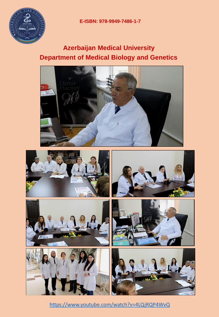
Department of Medical Biology and Genetics
Azerbaijan Medical University
https://www.youtube.com/watch?v=4LQjRQP4WvQ
E-ISBN: 978-9949-7486-1-7
Related Documents