Comprehensive Summaries of Uppsala Dissertations from the Faculty of Medicine 1116 _____________________________ _____________________________ Studies on Prediction of Axillary Lymph Node Status in Invasive Breast Cancer BY JOHAN AHLGREN ACTA UNIVERSITATIS UPSALIENSIS UPPSALA 2002

Welcome message from author
This document is posted to help you gain knowledge. Please leave a comment to let me know what you think about it! Share it to your friends and learn new things together.
Transcript
Comprehensive Summaries of Uppsala Dissertationsfrom the Faculty of Medicine 1116
_____________________________ _____________________________
Studies on Prediction ofAxillary Lymph Node Status in
Invasive Breast Cancer
BY
JOHAN AHLGREN
ACTA UNIVERSITATIS UPSALIENSISUPPSALA 2002

2
Dissertation for the Degree of Doctor of Philosophy (Faculty of Medicine) in Oncology presented at UppsalaUniversity in 2002
ABSTRACT
Ahlgren, J. 2002. Studies on prediction of axillary lymph node status in invasive breast
cancer. Acta Universitatis Upsaliensis. Comprehensive Summaries of Uppsala Dissertations
from the Faculty of Medicine 1116. 63pp. Uppsala. ISBN 91-554-5221-3.
Breast cancer is the most common malignancy among females in Sweden. Axillarylymph-node dissection is a standard procedure in the management of breast cancer,aiming at obtaining prognostic information for adjuvant therapy decisions. Axillarydissection entails considerable morbidity. The aims of this study were to establish moreselective surgical approaches and to investigate angiogenesis, a potential predictor forlymph-node metastases and prognosis.
Clinical nodal status, tumour size and S-phase were associated with nodal metastasesin cohort of 1145 women. The proportion of nodal metastases was 13% in the subgroupwith the lowest risk.
In a study from two registries, 675 and 1035 breast cancers ≤ 10 mm diagnosed byscreening mammography had nodal metastases in 6,5% and 7%, respectively. Clinicallydetected cancers had a risk of 16% and 14%, respectively.
In a study on 415 women, a 5-node biopsy of the axilla had a sensitivity of 97,3%and a false negative rate of 2,7% in comparison with axillary dissection.
Six sections from 21 breast cancers were analysed for microvessel density (MVD).The inter-section variation contributed more to the total variance than inter-tumourvariation, 45,0% and 37,3%, respectively.
In a cohort of 315 women, breast cancers with high MVD more frequently had p53mutations (27,1%) compared with cases with low MVD (18,4%). This difference wasnot statistically significant (p=0,075). p53 mutations were associated with a worseoutcome, whereas MVD was not.
In conclusion, women with screening detected ≤10 mm breast cancers have a lowrisk of lymph node metastases and some may not need axillary dissection in the future.The 5-node biopsy could be an alternative to axillary dissection. MVD is associatedwith methodological weaknesses and routine use is not recommended.
Key words: Breast cancer, lymph node metastases, prognostic factors, predictive factors,axillary surgery, angiogenesis, immunohistochemistry.
Johan Ahlgren, Department of Oncology, Radiology and Clinical Immunology, Sectionof Oncology. Uppsala University Hospital, SE-75185 Uppsala, Sweden
Johan Ahlgren 2002
ISSN 0282-7476ISBN 91-554-5221-3
Printed in Sweden by Uppsala University, Tryck&Medier, Uppsala 2002
3
This thesis is based on the following papers, which in the text will be referred to by their
Roman numerals:
I Ahlgren J, Ståhl O, Westman G, Arnesson LG. Prediction of axillary lymph
node metasases in a screened breast cancer population.
Acta Oncol 1994;33:603-8.
II Arnesson LG, Ahlgren J. Omitting axillary surgery for low-risk breast cancer
patients. A Swedish prospective cohort study. Acta Oncol 2000;39:291-4.
III Ahlgren J, Holmberg L, Bergh J, Liljegren G. Five node biopsy of the axilla. An
alternative to axillarydissection of level I-II in operable breast cancer. Eur J Surg
Oncol 2001; (Accepted).
IV Ahlgren J, Risberg B, Villman K, Bergh J. Angiogenesis in inasive breast
carcinoma – A prospective study of tumour heterogeneity. Eur J Cancer 2001;
(In press).
V Ahlgren J, Lindgren A, Lindahl T, Klaar S, Bergh J. Angiogenesis by Chalkley
counting in primary breast cancer – Relationship with p53 mutations. 2002
(Submitted).
Reprints have been made with the permission of the publishers.
4
Table of contents
Abstract 2
Main references 3
Abbreviations 5
Introduction 6
Background 7
Incidence 7
Diagnostic procedures 8
Staging of breast cancer 9
Prognostic and predictive factors in clinical use 10
New prognostic and predictive factors 12
Treatment 15
Aims of the study 20
Materials and methods 21
Results 24
General discussion 40
Paper I-III 40
Paper IV-V 45
General conclusions 51
Acknowledgements 52
References 53
5
Abbreviations
BCS Breast conserving surgery
BCSS Breast cancer specific survival
CI Confidence interval
CMF Cyclophosphamide, methotrexate, 5-fluorouracil
CV Coefficient of variation
ER Oestrogen receptors
FVIIIRag Factor VII related antigen
H&E Hematoxilin and Eosin
HPF High power field
OS Overall survival
PR Progesterone receptors
IHC Immunohistochemistry
LR Likelihood ratio
MRM Modified radical mastectomy
MVD Microvessel density
RH Relative hazard
RFS Recurrence-free survival
ROC Regional Oncologic Centre, Uppsala/Örebro
SD Standard deviation
SPF S-phase fraction
6
IntroductionBreast cancer is the most common female malignancy in the western societies. The incidence
of breast cancer in Sweden has increased steadily by 1-1,5% annually during the recent
decades and more than 6300 women contracted the disease in 1999 [1]. The good news are
that major improvements have been achieved in the management of breast cancer during
recent decades. Breast cancer surgery has been refined, the cosmetic results have been
improved by the widespread use of breast conserving surgery (BCS), in combination with
radiotherapy, that to a large extent has replaced the more traumatic mastectomy without
compromising survival rates [2].
The use of radiotherapy as an adjunct to surgery reduces the local and regional relapse rates by
about two thirds whereas the impact on survival is small but statistically significant [2, 3]. The
increasing use of adjuvant polychemotherapy, to eradicate micro metastases, gives a relative
reduction of the mortality of about 25% [4]. The use of adjuvant tamoxifen to oestrogen
receptor (ER) positive subsets results in similar benefits [5].
A great deal of the laboratory-based research has been focused on finding new prognostic- and
predictive factors as well us expanding the understanding of the biology of breast cancer.
Important results from this research field is the increased knowledge on the c-erbB-2
oncogene [6] and the tumour suppressor gene p53 [7]. The application of microarray
technique, which enables the mapping of thousands of different genes in a tumour, represents
the most recent progress in this research area [8, 9]. The development of molecular biology
has also resulted in the description of two breast cancer susceptibility genes (BRCA1 and
BRCA2) [10, 11]. The achievements of molecular biology will almost certainly improve the
medical management of increasing numbers of women with or at risk for breast cancer in the
near future.
Although a multitude of new prognostic factors has been proposed, the strongest one is still
axillary lymph-node status [12]. Nodal status is obtained by histopathological examination of
lymph nodes that are retrieved by the means of axillary surgery. The down side of this
procedure is the morbidity associated with axillary surgery such as arm oedema, numbness,
pain and weakness of the arm [13, 14]. The introduction of mammography screening
throughout Sweden has led to earlier diagnosis of breast cancer. Thereby the proportion of

7
women with small tumours without lymph node metastases has increased. In the Swedish
Two-County study the proportion of lymph node positivity was 27% in the group that was
invited to mammography and 42% in the control group [15]. This means that axillary surgery
is over-treatment in the vast majority of women with breast cancer.
Establishing alternatives to axillary clearence is therefore of great importance for the majority
of women with breast cancer. One potential alternative may be the extended use of prognostic
factors. One such factor that deserves further evaluation is angiogenesis, i.e. a tumour’s ability
to induce the formation of new blood vessels. This process is a necessary step in tumour
progression [16] that can be estimated in the primary tumour [17, 18].
BackgroundIncidence
In Sweden a total of 6311 women were diagnosed with breast cancer during 1999 according to
the Swedish Cancer Registry [1]. The age corrected incidence of breast cancer has increased
from 65-70/100.000 women at the beginning of the 1960ies to 120-125 at the end of the
1990ies [1]. This corresponds to an annual increase of 1-1,5% during the recent decades. The
number of deaths attributable to breast cancer was 1485 in 1999 [19]. The mortality seems to
have decreased slightly during the last decade, in the years 1987-98 the average death rate was
32,9/100.000 women compared with an average of 29,8/100.000 during 1997-99 [19]. In USA
and the UK the breast cancer mortality in year 2000, at ages 20-69, is predicted to have
decreased by 25-30% compared with the mid-eighties [20]. The most likely explanation for
this positive trend is the broad application of adjuvant systemic therapy and earlier diagnosis
due to screening mammography.
The reasons for the increase in incidence are not completely understood but it seems safe to
postulate that breast cancer is a disease with multifactorial etiology. The risk of contracting
breast cancer is positively correlated with increasing age, being a rare disease during the first
four decades of life. In 1999 only 3,5% of all new breast cancers in Sweden were diagnosed in
women under the age of 40, and 40% were diagnosed at age 40-59 [1]. Endogenous and
exogenous hormonal factors also influence the risk of developing breast cancer. For example
8
early menarche, late menopause, obesity among postmenopausal women and hormone
replacement therapy are factors associated with an increased exposure of the breast
parenchyma to estrogens and thereby increasing the breast cancer risk [21]. Ionising radiation
to the breast parenchyma is another established risk factor for breast cancer [22].
A large number of studies have been focused on dietary factors and the risk of breast cancer.
Especially fat intake has attracted much attention in this context. However, results from
studies on total fat intake have been equivocal [23-25]. Restrictions on total energy intake
during childhood and adolescence has been hypothesised to be a protective factor for breast
cancer [26]. Among other dietary constituents alcohol has consistently showed a correlation
with increased breast cancer risk. In a meta-analysis of 12 case-control studies [27] a
consumption of two drinks per day was estimated to induce a relative risk of 1,4 compared to
non-drinkers. There is also some evidence that vitamin A may protect against breast cancer
[23].
In recent years the breakthrough in molecular biology has resulted in increased knowledge on
hereditary forms of breast cancer. It has been estimated that 5–10% of all breast cancers are
caused by inherited mutations [28]. The majority of breast cancers caused by genetic
predisposition are caused by mutations in BRCA1 or BRCA2, two breast cancer susceptibility
genes that have been identified [10, 11]. Individuals with a family history of breast cancer can
nowadays undergo genetic testing with the aim of detecting mutations in either of these two
genes. This has opened up possibilities to offer preventive measures to bearers of these
genetic abnormalities and.
Diagnostic procedures
A palpable lump, tenderness, oedema or secretion from the mamilla are signs and symptoms
that often lead a woman with breast cancer to a physician. The recommended work-up in this
situation is: 1) physical examination of the breasts and regional lymph-nodes, 2)
mammography and 3) morphological diagnosis by the means of fine needle aspiration
cytology or a core needle biopsy for histopathological examination.
A large proportion of all breast cancers in Sweden are detected by screening mammography,
which was implemented during the last years of the 1980ies and in the beginning of the
9
1990ies in most Swedish counties. In the Uppsala-Örebro health care region with 1,9 million
residents, 38,6% of 10.777 invasive breast cancers diagnosed 1992-2001 were detected by
screening mammography [29]. Controlled studies on screening mammography indicate a
reduction of breast cancer mortality with about 25% [30, 31]. Earlier detection by screening
has also led to a change of the stage-distribution. The tumours are smaller and the proportion
of patients with lymph-node metastases in the axilla has decreased. In the Swedish Two-
County study the proportion of lymph node positivity was 27% and 42% in the invited group
and the control group respectively [15]. A current estimate from the Uppsala-Örebro health
care region, based on data from 8389 axillary dissections, is a node positivity rate of 35,4%
[29]. Screening mammography has thus created a challenge to physicians to adopt the
treatment strategies to the changed spectrum of breast cancer.
Staging of breast cancer
The TNM staging [32] is derived from the size of the primary tumour (T), lymph node status
(N) and distant metastases (M). Clinical staging (cTNM) is based on the clinical findings prior
to surgery whereas pathological staging according to the TNM system (pTNM) includes
information from the histopathological examination of the resected breast and axillary tissue.
The primary tumour (T) is classified as follows: Tx, primary tumour cannot be assessed; T0,
no evidence of primary tumour; Tis, carcinoma in situ or Paget disease of the nipple; T1,
tumour 20 mm or less; T2, tumour more than 20 mm but not more than 50 mm; T3, tumour
more than 50 mm. T4, tumour of any size with direct extension to the chest wall or the skin or
tumour with oedema of the breast or inflammatory breast cancer.
The regional lymph nodes (N) are classified as follows: Nx, regional lymph nodes cannot be
assessed; N0, no regional lymph node metastases; N1, metastasis to one or more movable
ipsilateral axillary node; N2, metastasis to one or more ipsilateral axillary node fixed to one
another or to other structures; N3, metastases to ipsilateral internal mammary lymph node(s).
Distant metastasis (M) is classified as follows: Mx, presence of distant metastasis cannot be
assessed; M0, no distant metastasis; M1, distant metastasis including metastases to one or
more ipsilateral supraclavicular node.
10
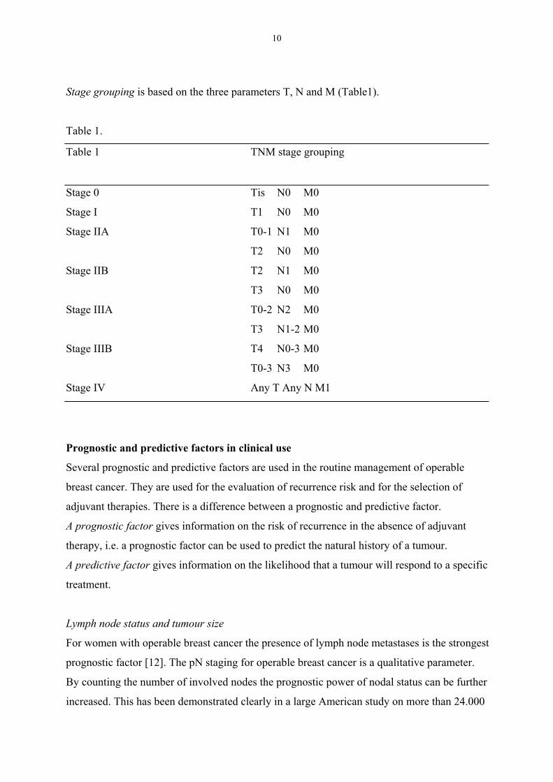
Stage grouping is based on the three parameters T, N and M (Table1).
Table 1.
Table 1 TNM stage grouping
Stage 0 Tis N0 M0
Stage I T1 N0 M0
Stage IIA T0-1 N1 M0
T2 N0 M0
Stage IIB T2 N1 M0
T3 N0 M0
Stage IIIA T0-2 N2 M0
T3 N1-2 M0
Stage IIIB T4 N0-3 M0
T0-3 N3 M0
Stage IV Any T Any N M1
Prognostic and predictive factors in clinical use
Several prognostic and predictive factors are used in the routine management of operable
breast cancer. They are used for the evaluation of recurrence risk and for the selection of
adjuvant therapies. There is a difference between a prognostic and predictive factor.
A prognostic factor gives information on the risk of recurrence in the absence of adjuvant
therapy, i.e. a prognostic factor can be used to predict the natural history of a tumour.
A predictive factor gives information on the likelihood that a tumour will respond to a specific
treatment.
Lymph node status and tumour size
For women with operable breast cancer the presence of lymph node metastases is the strongest
prognostic factor [12]. The pN staging for operable breast cancer is a qualitative parameter.
By counting the number of involved nodes the prognostic power of nodal status can be further
increased. This has been demonstrated clearly in a large American study on more than 24.000
11
women [33]. In order to correctly establish pN status at least 10 lymph nodes should be
examined [34]. The number of examined lymph nodes, i.e. the quality of the procedure, is
dependent on both the surgeon and the pathologist [35].
The size of the primary tumour is also a powerful prognostic factor and readily available in
almost all cases. The risk of recurrence is positively correlated to tumour size [33, 36, 37].
Tumour grade
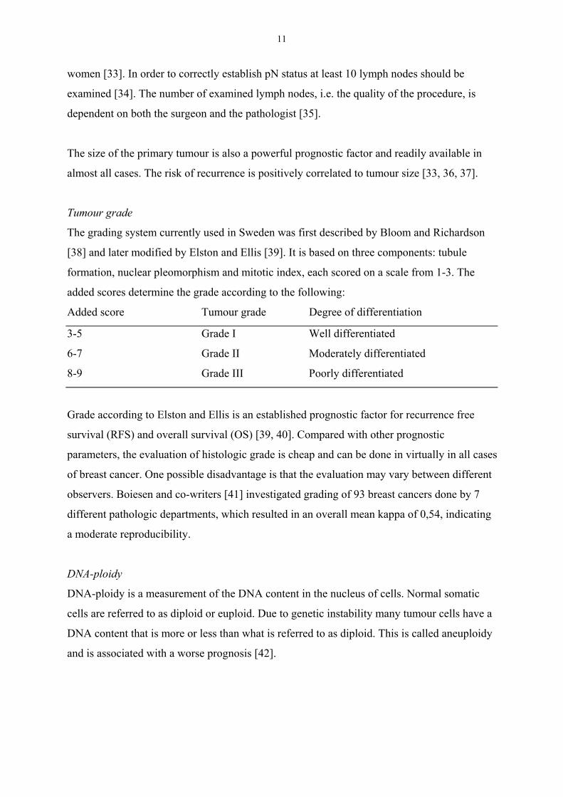
The grading system currently used in Sweden was first described by Bloom and Richardson
[38] and later modified by Elston and Ellis [39]. It is based on three components: tubule
formation, nuclear pleomorphism and mitotic index, each scored on a scale from 1-3. The
added scores determine the grade according to the following:
Added score Tumour grade Degree of differentiation
3-5 Grade I Well differentiated
6-7 Grade II Moderately differentiated
8-9 Grade III Poorly differentiated
Grade according to Elston and Ellis is an established prognostic factor for recurrence free
survival (RFS) and overall survival (OS) [39, 40]. Compared with other prognostic
parameters, the evaluation of histologic grade is cheap and can be done in virtually in all cases
of breast cancer. One possible disadvantage is that the evaluation may vary between different
observers. Boiesen and co-writers [41] investigated grading of 93 breast cancers done by 7
different pathologic departments, which resulted in an overall mean kappa of 0,54, indicating
a moderate reproducibility.
DNA-ploidy
DNA-ploidy is a measurement of the DNA content in the nucleus of cells. Normal somatic
cells are referred to as diploid or euploid. Due to genetic instability many tumour cells have a
DNA content that is more or less than what is referred to as diploid. This is called aneuploidy
and is associated with a worse prognosis [42].
12
S-phase fraction (SPF)
The S-phase is the phase of DNA-synthesis that takes place before cell division and thereby a
measure of cell proliferation. The fraction of cells in S-phase can be estimated with flow
cytometry. The estimate is a percentage that usually is dichotomised. High SPF correlates with
increased risk of recurrence and death, and is regarded as a useful prognostic factor [43]. A
high S-phase fraction is defined as > 7% in diploid tumours and >12% in non-diploid tumours
at the department of pathology at Akademiska Sjukhuset Uppsala. At the University hospital
in Linköping a cut-off value of 10% is used for all breast cancers, irrespective of DNA-ploidy.
Estrogen- and progesterone receptor status
Oestrogen receptors (ER) and progesterone receptors (PR) are expressed in a majority of
breast cancer tumours. The first report on the prognostic value of ER was published more than
20 years ago [44]. However, with longer follow-up time ER and PR are not strong prognostic
factors, although women with receptor positive cancers have a somewhat better prognosis
during the first years after diagnosis [45]. The greatest utility of ER and PR is as predictive
factors and this was demonstrated in the late 1970ies [46]. Tumours expressing both ER and
PR are the most likely to benefit from endocrine therapy but those who express either ER or
PR still have significant responses [45]. About 50-60% of women with receptor positive
advanced breast cancer will benefit from endocrine therapy while responses among receptor
negative women are rare [45]. The predictive value of receptor status has also been
established in the adjuvant setting [5].
In recent years, the biochemical analyses of ER and PR, that requires fresh frozen tissue, have
to a large extent been replaced by immunohistochemical (IHC) methods employed on paraffin
sections. Immunohistochemistry is more suitable for small tumours and possibly gives
superior prognostic information [47-49].
New prognostic and predictive factors
The prognostic- and predictive markers of today aim at giving information on prognosis and
therapy selection. The implications of these markers are known on group levels but they are
not sufficient to identify the individual woman at risk. A prognostic factor gives information
on the outcome, at best completely unrelated to different therapies. Ideally, a predictive factor
enables the rational use of drugs, i.e., treatment only of those who benefit from a selected drug

13
or a combination of drugs. The sex-hormone receptor status (ER and PR) is so far the only
established predictive factor for the management of women with breast cancer. Therefore,
there has been a demand for further markers, especially prognostic- and predictive factors
working at the individual level. Indeed, numerous potential prognostic factors identifying
different risk groups have been reported [12]. Three factors with special interest will be
discussed below: the c-erbB-2 oncogene, the p53 tumour suppressor gene and angiogenesis.
These three factors have all been claimed to have prognostic properties. One of them, c-erbB-
2, is already a routinely used predictive factor, whereas p53 and angiogenesis in the future
may be of value in optimising the decision making in breast cancer therapy.
c-erbB-2
c-erbB-2 (ERBB2,HER2/neu) is an oncogene located on the long arm of chromosome 17 [50].
C-erbB-2 encodes a transmembrane tyrosine growth factor receptor belonging to the
epidermal growth factor receptor (EGFR) family .[51]. In a Swedish population based study,
including screening detected tumours, the prevalence of overexpression of c-erbB-2 was 19%
[52]. Overexpression of this gene has been associated with a poor prognosis in breast cancer
[6, 52, 53], although not all studies have shown an independent prognostic value [54].
Furthermore, it has been suggested that overexpression of c-erbB-2 is associated with
decreased sensitivity to tamoxifen and CMF-like chemotherapy [55]. The most interesting
aspect of c-erbB-2 is the possibility to target the gene product with trastuzumab, a monoclonal
antibody. Trastuzumab in combination with chemotherapy results in prolonged survival
compared with chemotherapy alone in women with advanced breast cancer overexpressing c-
erbB-2 [56]. As a consequence, determination of c-erbB-2 status with IHC, to be confirmed by
fluorescence in situ hybridisation (FISH) on paraffin embedded tumour sections is a routine
analysis for women with recurrent breast cancer.
p53
p53 is a tumour supressor gene located on the short arm of chromosome 17 [57]. Due to its
central role in cell cycle control, execution of programmed cell death (apoptosis) and defence
mechanisms after DNA damage, p53 has been called “the guardian of the genome” [58].
Mutation of p53 is found in 20-25% of breast cancers [7, 59]. The most common method of
assessing p53 status is immunohistochemistry, which is done on paraffin sections. This
method detects intracellular accumulation of altered p53 protein, or enhanced levels of normal
14
p53 protein, that is interpreted as an evidence for an altered p53 function. Although IHC is a
fast method with low costs, the prognostic and predictive value is inferior to p53 status
obtained by gene sequencing [60, 61]. The potential use of p53 in the future seems to be as a
predictive factor. p53 mutations have been associated with increased resistance to FEC
chemotherapy [62] and tamoxifen [63], while taxane based chemotherapy seems to have
increased efficacy in women with p53 mutated breast cancers [62]. Thus, if these findings can
be reproduced in large randomised controlled trials, determination of p53 status has the
potential to substantially improve the clinical management of breast cancer.
Angiogenesis
Angiogenesis, i.e. the ability of a tumour to induce formation of new microvessels, was
reported to be of importance for tumour growth more than 30 years ago [64]. Angiogenesis
stimulates progression of both primary and metastatic tumours by several mechamisms.
Firstly, the growth of tumours beyond the size of 1-2 mm3 is dependent on angiogenesis [65].
Without access to vasculature tumour, their growth is limited by diffusion of nutrients and
accumulation of waste products. Secondly, the presence of microvessels facilitates tumour cell
access to the blood circulation [66, 67], a prerequisite for the establishment of distant
metastases. Thirdly, endothelial cells in the microvasculature release growth factors that can
stimulate tumour growth [68]. Moreover, angiogenesis seems to be involved in the production
of proteolytic enzymes [69], which is of importance in tumour invasiveness.
Angiogenesis is regulated by numerous angiogenic factors, one of the most important being
vascular endothelial growth factor (VEGF) [70], which is a potent mitogen for endothelial
cells. High levels of VEGF correlate with a poor prognosis in breast cancer [71-73].
Interestingly, the tumour suppressor gene p53 putatively exerts a negative effect on
angiogenesis, wild-type p53 protein seems to down regulate VEGF [74]. This finding is
corroborated by the findings of Linderholm and co-workers, showing that mutation of p53
correlates with increased levels of VEGF [75, 76].
Another link between p53 status and angiogenesis that has been postulated is that wild-type
p53 protein induces the formation of thrombospondin 1, which is a potent inhibitor of
angiogenesis [77]. This pathway is affected when mutant p53 protein is present, leading to
decreased formation of thrombospondin 1 and consequently increased angiogenesis [78, 79].
15
Estimation of tumour angiogenesis by a histological grading system and that a high grade of
neovascularisation correlates with tumour aggressiveness was described 3 decades ago [80].
The presently most widely used method for estimation of tumour angiogenesis was developed
by Weidner and co-workers [17]. They showed that tumour blood vessels can be visualised
with immunohistochemical staining of tumour sections, using a monoclonal antibody to an
antigen expressed by endothelial cells. The microvessel density (MVD) is then estimated by
counting these highlighted microvessels within the areas with the most intense
neovascularisation (also called hotspots). Several studies have demonstrated that MVD
independently can predict poor prognosis in operable breast cancer [18, 81-85] including
lymph-node negative patients [86-92]. In contrast, other authors could not find any prognostic
value of MVD [93-99]. One important reason for the contradictory results may well be
methodological problems.
Despite contradictory results regarding MVD and prognosis in breast cancer, several authors
have reported a correlation between high MVD and lymph-node metastases [17, 18, 82, 83].
Although some authors report a lack of relationship between MVD and nodal status [100-
102], this putative relationship is of interest since factors associated with lymph-node
metastases could potentially be used for prediction of axillary lymph-node metastases. A
predictive factor for nodal metastases could be used for identifying women with high risk of
node positivity, and inversely, also help to define a subgroup of women with a low enough
risk of lymph-node metastases to allow omission of axillary surgery.
Finally, there have been some reports on the predictive value of angiogenesis [102-104] but
most of the interest, in the therapeutical aspect of angiogenesis, has been focused on the
development of specific anti-angiogenic drugs [105, 106]. Though, the clinical breakthrough
of this conceptually appealing quest for a new class of anticancer drugs is still to be awaited.
Treatment
Surgery
Until the 1980ies mastectomy was considered to be the operation of choice for almost all
patients. The increasing numbers of patients with small tumours created a need for less

16
extensive surgery and breast conserving operations became increasingly more common at the
end of the 1980ies. No survival differences have been demonstrated in comparisons between
BCS combined with radiotherapy and larger operations where the whole breast is removed
[2]. The type of BCS used in Sweden is sector resection [107]. In the Uppsala-Örebro health
care region, registry data from 1992 –2001 on 9912 breast cancers treated with primary
surgery, shows that sector resection constitutes 60,0% of the operations [29].
Surgical management of the axilla
The axilla is anatomically a triangle shaped area with the axillary vein forming the superior
margin and the latissimus dorsi and serratus anterior muscles forming the posterior and medial
borders. This anatomic region is divided into three levels: level I - lateral and below the lateral
margin of the minor pectoral muscle; level II - the lymph nodes under the minor pectoral
muscle; and level III - the lymph nodes medial and above the medial margin of the minor
pectoral muscle. The extent of axillary surgery is usually defined according to these three
levels of the axilla.
The radical mastectomy, originally described by Halsted [108], included not only removal of
the whole breast and the pectoral muscles but also clearance of the axillary lymph nodes of
level I-III and removal of the intrapectoral lymph nodes. The underlying hypothesis of the
radical mastectomy was that breast cancer is a localised disease that disseminates first to the
regional lymph nodes and then to distant organs in an orderly fashion, thus, by extensive
surgery the chances for cure would be maximised. This view was challenged by results from
randomised studies showing that the extent of lymph node removal did not seem to affect
survival [109, 110]. The interpretation of these data was that failure after surgery usually is
because of the systemic dissemination of cancer cells before surgery, rather than an inadequate
operative technique. Since the impact on survival by axillary surgery is unclear, the main
reasons for this part of the operation is to ensure a correct staging of the disease and to achieve
local tumour control. The second reason for axillary surgery is to ensure local control of the
disease in the axilla. After level I-III clearance of the axilla, isolated axillary recurrences are
rare (1-1,4%) despite no further treatment [109].
The recommended type of axillary surgery is dissection of level I-II [111] since this procedure
meets the requirements of accurately staging the patient and ensuring local control. Axelsson
17
and colleagues found in a large study [34] comprising 7145 lymph node negative women, that
at least 10 lymph nodes should be removed in order to correctly establish node negativity.
Women with lymph node negative tumours and less than 10 removed lymph nodes had a
worse prognosis compared with those who had at least 10 nodes removed. The obvious reason
for this is that a subset of women with <10 removed nodes were wrongly classified as being
node negative. The Danish group [34] also reported a low local relapse rate of 3% after level
I-II dissection without further treatment in node negative patients if at least 5 lymph nodes
were removed. Recht and co-writers reported data from 1624 patients that underwent level I-II
dissection [112]. The local relapse rate was 2,2% in women not given radiation to the axilla.
Other studies have shown that the frequency of “skip” metastases to level III when levels I-II
are negative is about 1% [113, 114], thus, the risk of leaving metastases behind in level III is
very low.
Complications to present axillary staging procedures
Axillary surgery is in the long run one of the major sources of treatment associated morbidity.
Lymphoedema of the arm, the most widely recognised complication of axillary dissection, is
commonly defined as an increase of arm circumference of >2 cm or increase of arm volume
>200 ml. The incidence of lymphoedema is 10-25% [115]. The extent of axillary surgery
correlates with the risk of lymphoedema [14, 116, 117]. Other complications are also
common. In a report on 126 patients after axillary dissection without radiotherapy [13], 70%
of the women complained of numbness, 33% of pain and 25% experienced weakness of the
arm. Although these complications mostly were described as mild, 39% of the women
experienced an effect upon their daily lives [13].
Less extensive surgical procedures
Due to complications associated with axillary dissection, efforts have been made to reduce the
extent of axillary surgery. In a Scottish study [118] patients were randomised between axillary
clearance and lower axillary sampling. The latter method consisted of surgical removal of 4-5
nodes near the lateral border of the breast. In this study, the sampling procedure was followed
by axillary clearence during the same operation in a subset of 67 patients. This experiment
resulted in a sensitivity of the sampling procedure of 100%. Later, the same group published a
report from the whole randomised study including 417 women [119]. Patients that underwent
sampling were treated with radiation therapy in case of node positivity. The long-term results

18
with reference to survival were identical with the two methods. A second study from the same
research group included 466 women treated with BCT [120]. Once again, no survival
difference could be detected when sampling and clearence were compared. In contrast, Kissin
and co-authors [121] showed that 24% were erroneously staged with sampling compared with
results obtained with axillary dissection. Moreover, data from the large Danish registry study
[34] question the use of sampling. According to their results at least 10 lymph nodes should be
removed to eliminate the risk of misclassification.
The sentinel node biopsy is a new method for minimally invasive axillary surgery. By the
means of peritumoural injections of blue dye and/or a radiolabeled colloid a few hours before
surgery the lymph node that first receives the drainage from the breast can be identified
visually and/or by a gamma probe. The false negative rate in one of the largest series from a
single institution was 6,7% [122], whereas the corresponding figure in two multicenter studies
was 11% [123, 124]. Data from randomised comparisons between axillary dissection and the
sentinel node procedure alone, with reference to local control and preferably survival, should
be awaited before this promising method can be adopted in clinical practise.
Adjuvant radiotherapy
The use of ionising radiation given as an adjunct to surgery reduces the risk of local
recurrence by two thirds [125]. Radiotherapy also reduces breast cancer mortality with an
absolute reduction of about 5% at 20 years of follow-up [125]. This positive effect is counter
balanced by an absolute survival reduction due to deaths from other diseases than breast
cancer of about 4% at 20 years. The resulting absolute increase of overall survival at 20 years
is about 1% [125]. However, the total survival benefit is greater among young women and
those with high risk of recurrence [125]. Recent studies on post-mastectomy radiotherapy
given in combination with chemotherapy indicate that the survival gains might be
significantly larger [126-128] if the radiotherapy is given with a more modern technique. The
view that the survival benefit of adjuvant radiotherapy at most is marginal is now being
challenged [3].
Radiotherapy to the axilla increases the risk of sequelae such as lymphoedema and impaired
mobility of the arm [129]. The highest risk of arm-lymphoedema is seen among women that
are treated with both axillary surgery and radiotherapy to the axilla [130]. The radiotherapy

19
technique is of outmost importance for avoiding toxicity. This should include adequate dose
planning, preferably based on computerised tomography, and modern fractionation schedules.
The standard treatment is 2Gy/fraction to a total dose of 50-54Gy over 5-5,5 weeks to the
breast after BCS and 2Gy/fraction to a total dose of 50Gy over 5 weeks to the chest wall after
mastectomy. In lymph node positive women the axilla, supraclacivular fossa and the internal
mammary chain are usually treated with 2Gy/fraction to a total dose of 50Gy over 5 weeks.
Adjuvant systemic therapy
The rationale for using adjuvant polychemotherapy is that systemic treatment early in the
course of breast cancer can eliminate micrometastases and thereby increase the chance of cure.
Meta analyses have shown that polychemotherapy gives a relative risk reduction for death by
about 25 % [4]. Recent data show that chemotherapy regimens including an anthracycline
(doxorubicin or epirubicin) are superior (relative risk reduction for death 11%) to older
regimens based on alkylating agents [4]. Hence, the standard treatment currently in Sweden is
a combination of 5-fluorouracil, epirubicin and cyclophosphamide (FEC regimen) given in
seven courses. Tamoxifen is a selective estrogen receptor modifier (SERM) with activity
against ER or PR positive breast cancer. The use of this drug in the adjuvant setting gives a
relative risk reduction for recurrence by almost 50% and a relative risk reduction for death by
about 25% when taken daily for five years [5]. The combination of polychemotherapy and
tamoxifen to receptor positive women produces additional benefit [4]. Thus, the relative risk
reduction for death for most women treated with chemo-endocrine therapy clearly must be
considerably greater than 25%.
Treatment of recurrent breast cancer
Once distant metastases are clinically detected the disease is incurable and expected median
survival in this situation is typically in the range of 2-3 years [21]. Women with metastatic
disease are treated with chemotherapy and/or hormonal therapy which can alleviate symptoms
and also improve median survival [131-134]. Recently the addition of the monoclonal
antibody trastuzumab to chemotherapy has been demonstrated to prolong survival for women
with c-erbB-2 positive advanced breast cancer [56]. Palliative radiotherapy, adequate
analgesics, bisphosphonates in case of bone metastases, erythropoeitin, and psychological
support are other important means of good palliation in the treatment of metastatic breast
cancer [21].
20
Aims of the studyThe overall aim of the study is to investigate different strategies to decrease the need for
axillary dissection in women with breast cancer. The study consists of two parts where papers
I-III are aiming at a more selective surgical approach. Papers IV-V investigate angiogenesis, a
potential predictor not only for prognosis but also for lymph-node metastases.
Specific aims
Paper I:
To delineate a subgroup of women with breast cancer, from a population with ongoing
screening, with a low enough risk of lymph node metastases to allow omission of axillary
surgery.
Paper II:
To define a subgroup of women by, the use of small tumour size and detection by screening,
with a low enough risk of lymph node metastases to allow omission of axillary surgery within
the framework of a prospective cohort study.
Paper III:
To test whether a biopsy of five lymph nodes is as informative on histopathological lymph
node status as a level I-II dissection of the axilla in operable breast cancer.
Paper IV:
To investigate if intra tumoural heterogeneity of angiogenesis demonstrates a significant
variation which may influence the results of this parameter in primary breast cancer.
Paper V:
To investigate the relationship between MVD and cDNA sequenced determined p53
mutations for exons 2-11 in breast cancer and to analyse the correlation between MVD and
recurrence free survival, breast cancer corrected survival and overall survival.
21
Materials and methods
Paper I
A database in which all new cases of breast cancer in the South-East Sweden Health Care
Region are to be reported was used. We analysed data from 1145 women with primary breast
cancer retrospectively in order to define a subgroup in which axillary dissection could be
omitted. Women with tumours larger than 50 mm and/or with less than 5 examined lymph
nodes were excluded. We used the clinical and pathological features of the tumours that are
used in clinical practice: clinical nodal status, age, tumour size, hormonal receptors, DNA
ploidy and SPF and that had been prospectively reported to the database.
Paper II
The second paper is based on two registry studies that were made as a part of the preparations
for a multicentric Swedish prospective cohort-study for omission of axillary surgery in women
with low risk of having lymph-node metastases. Data from the breast cancer registries from
the South-East Swedish Region and the Uppsala-Örebro Region was used. All newly
diagnosed breast cancers within the two health care regions are to be reported to their
respective registry. No restrictions were made on the number of removed lymph-nodes.
Paper III
Four hundred and fifteen females from Örebro and Uppsala were entered in a prospective
study on a 5-node biopsy of the axilla. Women with clinical stage T0-3N0-1M0 breast cancer
were eligible. They were included in the study after informed consent. All patients were
operated by the same surgeon using a standardised technique. Each patient underwent sector
resection or mastectomy. The axillary surgery consisted of a 5-node biopsy followed by a
dissection of the remaining axillary tissue in level I-II in the same operation.
The 5-node biopsy was begun with dissection at the axillary tail of the breast until five
lymph nodes had been removed. Each of these lymph nodes were submitted to the
pathologist in separate boxes and labelled lymph node 1 to 5. After the first five lymph
nodes had been retrieved, the dissection of the axilla continued until the axillary fat in
level I-II had been excised. This material was also submitted in a separate box to the
pathologist. The aim of the five-node biopsy and the level I-II dissection was to remove
22
a total of at least 10 axillary lymph nodes for histopathological examination. In this way
the experimental method could be compared with the gold standard procedure in each
patient.
All the removed tissue was fixed in formaldehyde and stained with van Gieson or H&E. The
axillary level I-II specimens were carefully palpated in order to find as many lymph nodes as
possible. Sections of all nodes were examined with routine pathology. Immunohistochemical
staining was not used in the examination of the nodes. The breast tumours were also stained
with van Gieson or H&E and examined with routine pathology.
Paper IV
Twenty-one consecutive invasive breast cancers were recieved as fresh specimens at the
Department of Pathology in Örebro from May to October 1994. No preoperative treatment of
breast cancer was allowed. The whole tumour was sectioned in 5mm slices. Fixation was run
overnight in 4% buffered formaldehyde solution. After dehydration all tumour tissue was
embedded in paraffin. H&E staining of all tumours was done for routine assessment of type
and grade. Six 4µ sections were cut from each tumour. In all cases sections were separated as
widely and evenly as possible within the tumour. The position of all sections within the
tumour was registered and labelled A-F. The sections (n=126) were stained
immunohistochemically with a CD31 monoclonal antibody (JC70). In each section the areas
with the most intense neovascularisation (hotspots) were identified and the MVD was
obtained by counting vessels in 200 X fields (0,72mm2) in three such hotspots. All slides were
assessed simultaneously by two observers.
Statistical methods
We used Statistica software (Statsoft OK, USA) for calculation of standard deviation (SD),
confidence intervals, and the independent two-sided t-test for comparisons between groups.
Variability was expressed by the coefficient of variation (CV) which is defined as 100*SD
divided by the mean. A nested ANOVA (Statistica software) was used to analyse the
proportion of the total variance that each sampling level contributed to. For this analysis we
considered cases, intersection and intrasection as three levels in a hierarchic model.
23
Paper V
Tumour material was analysed from 315 consecutive women with primary breast cancer who
underwent surgery in Uppsala County, Sweden, during the period January 1st 1987 to
December 31st 1989 and from whom fresh frozen material was saved, which was the routine
procedure. Routine histopathological examination and determination of ER, PR and S-phase
fraction was performed.
Regular follow-up visits were performed during 5-10 years. In November 1999 the patient
records were re-examined and updated. Survival information on the women by the 1st of
November 1999 was retrieved from the Swedish Population Registry. To get information on
death causes we used data both in medical files and from death certificates. The follow-up of
recurrences was obtained by reviewing the patient records. When women had been referred to
another hospital or to a GP for further follow-up, we retrieved information about the date of
the latest check-up and status concerning signs of recurrence.
MVD analysis
From 305 tumours we were able to retrieve fresh frozen tumour material. A piece of the
frozen tumour tissue was thawed and then fixed. After dehydration the specimens were
embedded in paraffin. Sections with a diameter of 4-5 mm were thus obtained and mounted
on slides. We used a monoclonal antibody to CD31 (JC70A, Dako AS, Glostrup, Denmark) in
a 1:10 dilution. The sections were predigested with protease. The quality of the intratumoural
staining was judged using blood vessels in adjacent benign breast tissue as internal positive
control. The most vascularised areas of the tumour tissue were located at low magnification
(10X oculars with 4X and 10X objectives). Thereafter a 25-point Chalkley eyepiece graticule
was employed [135] over the same tumour region and orientated so that the maximum number
of points at 200X (0,95 mm2) were on or within areas of stained microvessels. Thus the
highest graticule count was recorded for each tumour.
p53 analysis
The p53 status in tumours was analysed by sequencing with cDNA for exons 2 to 11 on
homogenised fresh frozen tumour samples. The sequencing products generated were analysed
with an automated laser fluorescence sequencer. The sequence was finally compared with the
wild-type p53 sequence. Nucleotide alterations that had an impact on he protein were
24
considered mutations. Immunohistochemical determination of p53 status was performed on
paraffin sections using the monoclonal mouse antibody Pab 1801.
Statistical methods
For non-parametric comparisons between groups we used the χ2-test. For estimation of overall
survival (OS), breast cancer specific survival (BCSS) and recurrence free survival (RFS) we
used life table analyses and the Kaplan Meier method. The log-rank test was employed for
analysis of differences between groups. For OS all deaths were counted as events. For BCSS
only deaths from breast cancer were considered to be events. Events for estimation of RFS
were all breast cancer relapses or death from breast cancer. Relative hazards (RH) for OS and
BCSS were estimated by Cox’s proportional hazards method in univariate and multivariate
models. We used Statistica Software (StatSoft Inc., Tulsa, OK) for all analyses except for
Cox’s proportional hazards analyses for which the PHREG procedure in SAS for PC
(Armonk, NY) was used.
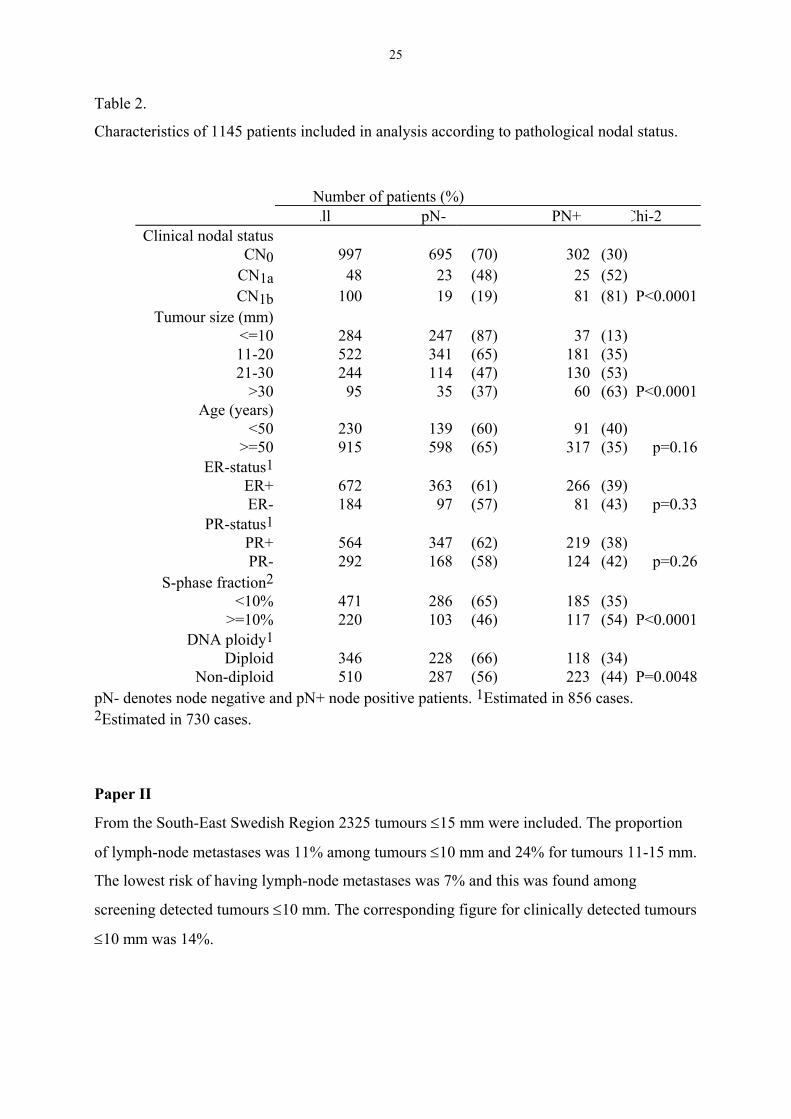
ResultsPaper I
Both clinical nodal status and tumour size were strongly correlated with pathological nodal
status. Also SPF >10% was strongly correlated with node positivity in univariate analysis
(Table 2).
In multivariate analysis there was a correlation between high SPF and nodal metastases among
tumours up to 20 mm but not with tumours greater than 20 mm. Women with clinically
negative nodal status and tumour size <=20 mm and <=10 mm had pathologically positive
nodes in 25% and 13%, respectively (in the original publication the latter estimate was
miscalculated, the correct estimate is 13%, not 15%). If tumours with high SPF were
excluded, the corresponding estimates were 24% and 14%.
25
Table 2.
Characteristics of 1145 patients included in analysis according to pathological nodal status.
Number of patients (%)All pN- PN+ Chi-2
Clinical nodal statusCN0 997 695 (70) 302 (30)
CN1a 48 23 (48) 25 (52)CN1b 100 19 (19) 81 (81) P<0.0001
Tumour size (mm)<=10 284 247 (87) 37 (13)11-20 522 341 (65) 181 (35)21-30 244 114 (47) 130 (53)
>30 95 35 (37) 60 (63) P<0.0001Age (years)
<50 230 139 (60) 91 (40)>=50 915 598 (65) 317 (35) p=0.16
ER-status1ER+ 672 363 (61) 266 (39)ER- 184 97 (57) 81 (43) p=0.33
PR-status1PR+ 564 347 (62) 219 (38)PR- 292 168 (58) 124 (42) p=0.26
S-phase fraction2<10% 471 286 (65) 185 (35)
>=10% 220 103 (46) 117 (54) P<0.0001DNA ploidy1
Diploid 346 228 (66) 118 (34)Non-diploid 510 287 (56) 223 (44) P=0.0048
pN- denotes node negative and pN+ node positive patients. 1Estimated in 856 cases.2Estimated in 730 cases.
Paper II
From the South-East Swedish Region 2325 tumours ≤15 mm were included. The proportion
of lymph-node metastases was 11% among tumours ≤10 mm and 24% for tumours 11-15 mm.
The lowest risk of having lymph-node metastases was 7% and this was found among
screening detected tumours ≤10 mm. The corresponding figure for clinically detected tumours
≤10 mm was 14%.

26
The figure for screening detected tumours ≤10 mm from the Uppsala-Örebro Region was
6,5% (30/464), whereas the proportion of lymph node positivity among patients with
clinically detected breast cancers ≤10 mm was 14% (34/211).
Results from extended analysis made in December 2001
It is reasonable to believe that the subgroup of clinically detected tumours has a greater mean
tumour size compared with tumours detected by screening mammography. Moreover, since
the screening detected subgroup consists of women aged 40-70, the clinically detected
subgroup will have a different age distribution. In December 2001 we performed a
multivariate analysis on the relation between positive lymph node status and detection mode
adjusted for tumour size and age using data from the registry at the Regional Oncologic
Centre of Uppsala/Örebro. Data from 803 tumours with a maximum diameter of 10 mm
diagnosed from September 1992 to December 1996 was selected. Detection mode, tumour
size and age were entered in a logistic regression model with lymph node status as response
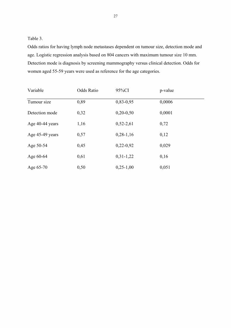
variable (Table 3). The results from this analysis show that the mode of detection is the
strongest predictor of lymph node metastases, clinically detected tumours being more likely to
have positive lymph nodes. Both tumour size and age are also independent risk factors for
nodal metastases, whereas decreasing tumour size as expected shows a correlation with
decreasing risk for metastases, age have a non-linear relationship. Age 50-54 years was the
only age group with an odds ratio that was statistically significant.
27
Table 3.
Odds ratios for having lymph node metastases dependent on tumour size, detection mode and
age. Logistic regression analysis based on 804 cancers with maximum tumour size 10 mm.
Detection mode is diagnosis by screening mammography versus clinical detection. Odds for
women aged 55-59 years were used as reference for the age categories.
Variable Odds Ratio 95%CI p-value
Tumour size 0,89 0,83-0,95 0,0006
Detection mode 0,32 0,20-0,50 0,0001
Age 40-44 years 1,16 0,52-2,61 0,72
Age 45-49 years 0,57 0,28-1,16 0,12
Age 50-54 0,45 0,22-0,92 0,029
Age 60-64 0,61 0,31-1,22 0,16
Age 65-70 0,50 0,25-1,00 0,051
28
Paper III
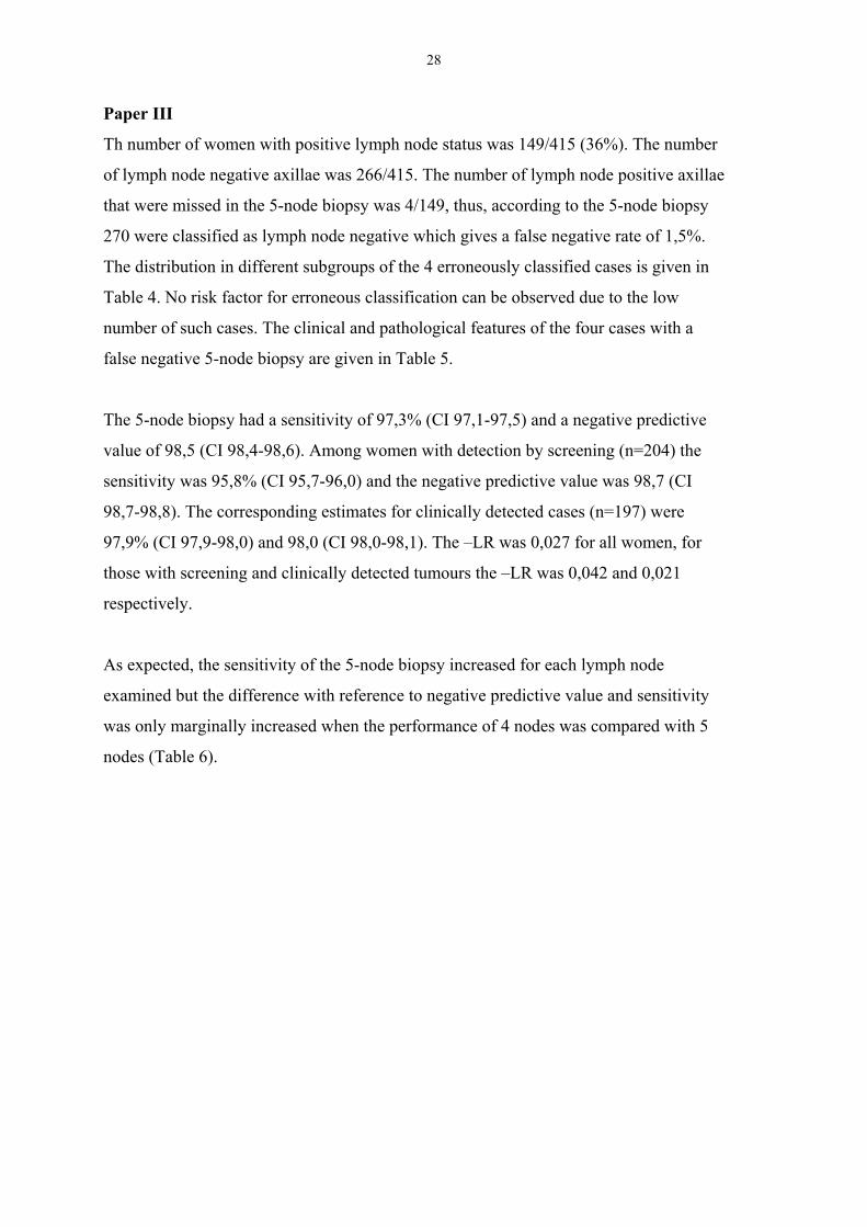
Th number of women with positive lymph node status was 149/415 (36%). The number
of lymph node negative axillae was 266/415. The number of lymph node positive axillae
that were missed in the 5-node biopsy was 4/149, thus, according to the 5-node biopsy
270 were classified as lymph node negative which gives a false negative rate of 1,5%.
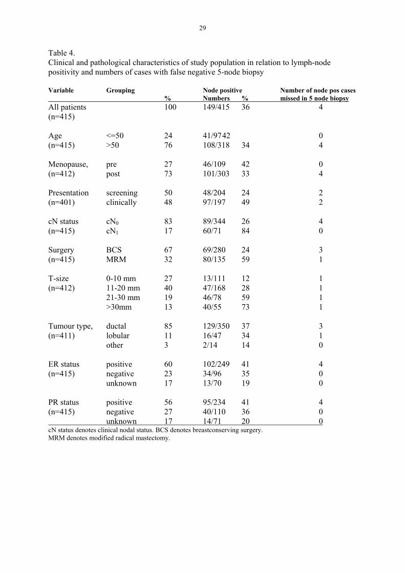
The distribution in different subgroups of the 4 erroneously classified cases is given in
Table 4. No risk factor for erroneous classification can be observed due to the low
number of such cases. The clinical and pathological features of the four cases with a
false negative 5-node biopsy are given in Table 5.
The 5-node biopsy had a sensitivity of 97,3% (CI 97,1-97,5) and a negative predictive
value of 98,5 (CI 98,4-98,6). Among women with detection by screening (n=204) the
sensitivity was 95,8% (CI 95,7-96,0) and the negative predictive value was 98,7 (CI
98,7-98,8). The corresponding estimates for clinically detected cases (n=197) were
97,9% (CI 97,9-98,0) and 98,0 (CI 98,0-98,1). The –LR was 0,027 for all women, for
those with screening and clinically detected tumours the –LR was 0,042 and 0,021
respectively.
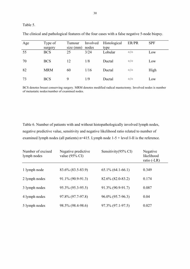
As expected, the sensitivity of the 5-node biopsy increased for each lymph node
examined but the difference with reference to negative predictive value and sensitivity
was only marginally increased when the performance of 4 nodes was compared with 5
nodes (Table 6).
29
Table 4.Clinical and pathological characteristics of study population in relation to lymph-nodepositivity and numbers of cases with false negative 5-node biopsy
Variable Grouping Node positive Number of node pos cases % Numbers % missed in 5 node biopsyAll patients 100 149/415 36 4(n=415)
Age <=50 24 41/9742 0(n=415) >50 76 108/318 34 4
Menopause, pre 27 46/109 42 0(n=412) post 73 101/303 33 4
Presentation screening 50 48/204 24 2(n=401) clinically 48 97/197 49 2
cN status cN0 83 89/344 26 4(n=415) cN1 17 60/71 84 0
Surgery BCS 67 69/280 24 3(n=415) MRM 32 80/135 59 1
T-size 0-10 mm 27 13/111 12 1(n=412) 11-20 mm 40 47/168 28 1
21-30 mm 19 46/78 59 1>30mm 13 40/55 73 1
Tumour type, ductal 85 129/350 37 3(n=411) lobular 11 16/47 34 1
other 3 2/14 14 0
ER status positive 60 102/249 41 4(n=415) negative 23 34/96 35 0
unknown 17 13/70 19 0
PR status positive 56 95/234 41 4(n=415) negative 27 40/110 36 0 unknown 17 14/71 20 0cN status denotes clinical nodal status. BCS denotes breastconserving surgery.MRM denotes modified radical mastectomy.
30
Table 5.
The clinical and pathological features of the four cases with a false negative 5-node biopsy.
Age Type ofsurgery
Tumoursize (mm)
Involvednodes
Histologicaltype
ER/PR SPF
55 BCS 25 3/24 Lobular +/+ Low
70 BCS 12 1/8 Ductal +/+ Low
82 MRM 60 1/16 Ductal +/+ High
73 BCS 9 1/9 Ductal +/+ Low
BCS denotes breast conserving surgery. MRM denotes modified radical mastectomy. Involved nodes is numberof metastatic nodes/number of examined nodes.
Table 6. Number of patients with and without histopathologically involved lymph nodes,
negative predictive value, sensitivity and negative likelihood ratio related to number of
examined lymph nodes (all patients) n=415. Lymph node 1-5 + level I-II is the reference.
Number of excised Negative predictive Sensitivity(95% CI) Negative lymph nodes value (95% CI) likelihood
ratio (-LR)
1 lymph node 83.6% (83.5-83.9) 65.1% (64.1-66.1) 0.349
2 lymph nodes 91.1% (90.9-91.3) 82.6% (82.0-83.2) 0.174
3 lymph nodes 95.3% (95.3-95.5) 91.3% (90.9-91.7) 0.087
4 lymph nodes 97.8% (97.7-97.8) 96.0% (95.7-96.3) 0.04
5 lymph nodes 98.5% (98.4-98.6) 97.3% (97.1-97.5) 0.027
31
Paper IV
Median age of the patients was 69 years (range 35 - 88). The median size of the 21 tumours
was 20 mm (range 10 - 40 mm). Lymph-node status was positive in 8 cases and negative in
11. Nodal status was not assessed in two elderly women.
In six patients the quality of immunostaining was not judged as satisfactory in a proportion of
the six sections (Table 7). All 8 sections with questionable staining were stained a second time
with a highly vascularised tumour-section as positive control, none of them turned to be
assessable by this procedure.
Measures of variation
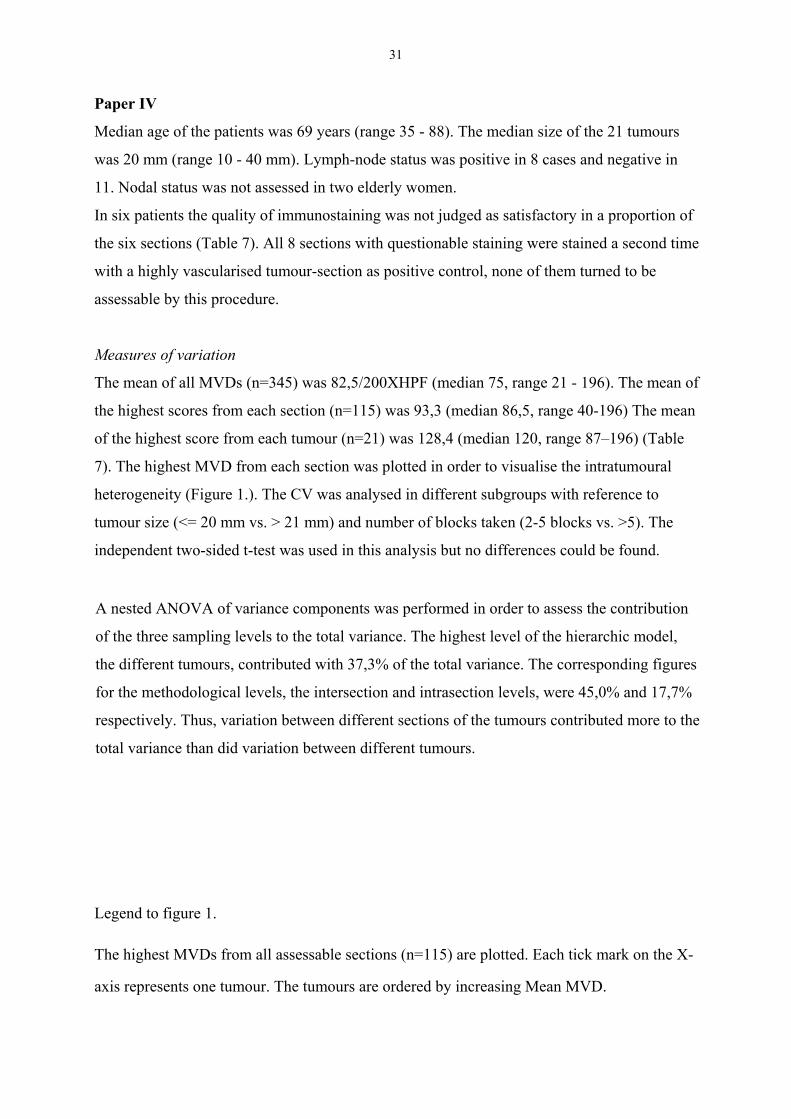
The mean of all MVDs (n=345) was 82,5/200XHPF (median 75, range 21 - 196). The mean of
the highest scores from each section (n=115) was 93,3 (median 86,5, range 40-196) The mean
of the highest score from each tumour (n=21) was 128,4 (median 120, range 87–196) (Table
7). The highest MVD from each section was plotted in order to visualise the intratumoural
heterogeneity (Figure 1.). The CV was analysed in different subgroups with reference to
tumour size (<= 20 mm vs. > 21 mm) and number of blocks taken (2-5 blocks vs. >5). The
independent two-sided t-test was used in this analysis but no differences could be found.
A nested ANOVA of variance components was performed in order to assess the contribution
of the three sampling levels to the total variance. The highest level of the hierarchic model,
the different tumours, contributed with 37,3% of the total variance. The corresponding figures
for the methodological levels, the intersection and intrasection levels, were 45,0% and 17,7%
respectively. Thus, variation between different sections of the tumours contributed more to the
total variance than did variation between different tumours.
Legend to figure 1.
The highest MVDs from all assessable sections (n=115) are plotted. Each tick mark on the X-
axis represents one tumour. The tumours are ordered by increasing Mean MVD.
32
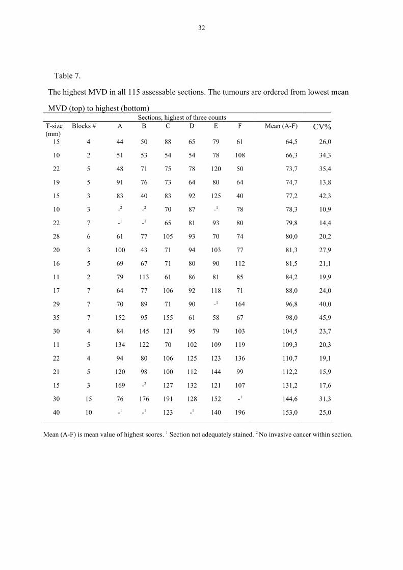
Table 7.
The highest MVD in all 115 assessable sections. The tumours are ordered from lowest mean
MVD (top) to highest (bottom)Sections, highest of three counts
T-size(mm)
Blocks # A B C D E F Mean (A-F) CV%
15 4 44 50 88 65 79 61 64,5 26,0
10 2 51 53 54 54 78 108 66,3 34,3
22 5 48 71 75 78 120 50 73,7 35,4
19 5 91 76 73 64 80 64 74,7 13,8
15 3 83 40 83 92 125 40 77,2 42,3
10 3 -2 -2 70 87 -1 78 78,3 10,9
22 7 -1 -1 65 81 93 80 79,8 14,4
28 6 61 77 105 93 70 74 80,0 20,2
20 3 100 43 71 94 103 77 81,3 27,9
16 5 69 67 71 80 90 112 81,5 21,1
11 2 79 113 61 86 81 85 84,2 19,9
17 7 64 77 106 92 118 71 88,0 24,0
29 7 70 89 71 90 -1 164 96,8 40,0
35 7 152 95 155 61 58 67 98,0 45,9
30 4 84 145 121 95 79 103 104,5 23,7
11 5 134 122 70 102 109 119 109,3 20,3
22 4 94 80 106 125 123 136 110,7 19,1
21 5 120 98 100 112 144 99 112,2 15,9
15 3 169 -2 127 132 121 107 131,2 17,6
30 15 76 176 191 128 152 -1 144,6 31,3
40 10 -1 -1 123 -1 140 196 153,0 25,0
Mean (A-F) is mean value of highest scores. 1 Section not adequately stained. 2 No invasive cancer within section.
Figu
re 1
.
050100
150
200
250
Highest MVD/200XHPF
Cas
es
34
Application of potential cut-offs.
In this analysis we chose the median of all the highest scores (86,5/200XHPF) from each
section (n=115) as a tentative cut-off level, which was applied to the scores from each
sectioning level (A-F). In this way we had 6 sets of dichotomised scores, each representing a
different part of the tumours. We then compared the results from one set of sections with a
second set of sections (A-B; A-C; A-D; A-E; A-F; B-C; etc.) and calculated the proportion of
tumours for which both results of a pair were concordant with reference to the cut-off level.
Fifteen comparisons were thus made for each of the 21 tumours. In these paired comparisons
the mean proportion of concordant results was 59,0% (95% CI (55,3:62,8)). The result was
similar if the upper tertile (101,3/200XHPF) was chosen as a cut-off level, 64,7% (95% CI
(60,0:69,5)). This example shows that more than one third of the dichotomised estimates will
change from high to low or from low to high if the analysis is made on a second section from
another part of the tumour.
Paper V
The median follow-up for survival and recurrence was 122 and 73 months, respectively. The
number of women that had died was 137, of these 74 died of breast cancer, 61 died of
unrelated causes and in 2 cases the cause of death was unknown. Recurrence was observed in
116 women with one individual lost to follow-up.
Two hundred ninety-six of the 315 tumours were assessable for MVD, in 10 cases there was
no frozen tissue and in another 9 there was no invasive cancer in the new section. The median
of all Chalkley counts was 3 with a range from 1-10. The relationship between MVD as a
dichotomous variable (the median was used as cut-off level) and clinical and pathological
parameters was investigated (Table 8). A Chalkley count above median was associated with
nodal involvement and high SPF.
The cDNA-based sequencing method for analysis of p53 mutations was successful in all but 4
tumours. The number of tumours with a mutation was 69. Thus, in the whole material the
proportion of tumours with a p53 mutation was 21,9%. Among the 311 sequenced tumours 19
were missing data on MVD. That left 292 cases for comparisons between p53 status and

35
MVD. Among tumours with MVD above the median the proportion of mutations was 27,1%.
The corresponding figure for those with low MVD was 18,4%. This difference was near the
level of statistical significance (p=0,075). We also found that there was a statistically
significant correlation between high MVD and p53 mutations (p=0,037), if tumours (n10)
with a mutation in the evolutionary conserved regions 2 or 5 were excluded. This was due to
the fact that 6/10 tumours with mutation in region 2 or 5 had a low MVD. We then analysed
MVD with reference to p53 IHC but no correlation could be established.

36
Table 8.
Relationship between MVD and other clinical and pathological features
Variable Grouping % high MVD χ2 testAge ≤50 vs ≥51 50,0 vs 47,2 p=0,71
Tumor size T1 vs T2-3 44,4 vs 51,7 p=0,21
Lymph-node status N0 vs N1 43,2 vs 56,5 p=0,036
Estrogen receptors pos vs neg 45,5 vs 58,6 p=0,073
Progesterone receptors pos vs neg 45,9 vs 60,0 p=0,082
S-phase fraction low vs high 45,7 vs 60,0 p=0,049
Vascular invasion neg vs pos 44,7 vs 57,4 p=0,094
p53-mutation neg vs pos 45,1 vs 57,6 p=0,075
p53 IHC neg vs pos 46,2 vs 54,4 p=0,26
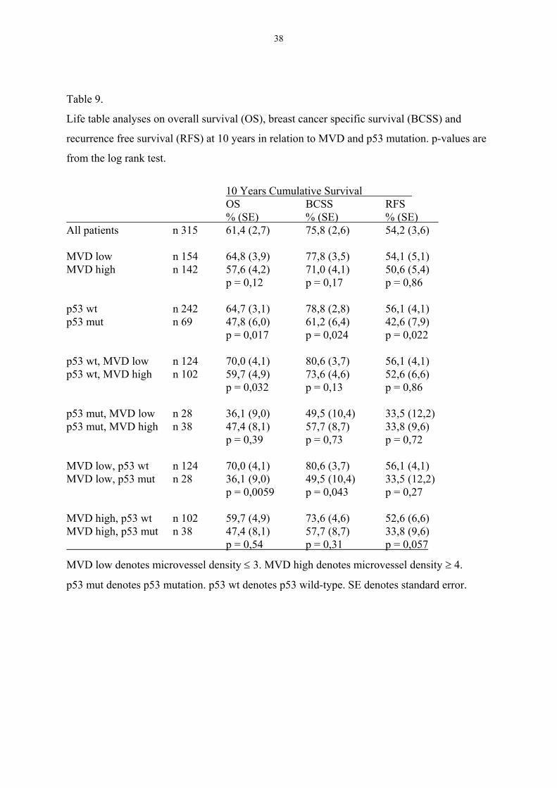
Analyses of OS, BCSS and RFS
There was no statistically significant correlation between MVD and outcome when analysis
was done on all patients or on subgroups of node negative and node positive patients.
Tumours with a mutation of the p53 gene entailed a statistically significantly worse prognosis
with reference to OS, BCSS and RFS (Table 9). A possible prognostic value of MVD among
tumours without p53 mutation was not consistent, a worse outcome in terms of OS (p=0,032)
was reduced to a trend when BCSS (p=0,13) was analysed and the RFS did not differ (p=0,86)
(Table 9).
Multivariate analysis showed that p53 was an independent risk factor for OS and BCSS with
RHs of 1,91 (1,23-2,97) and 1,76 (1,01-3,08), respectively (Table 10). The RH of MVD did
not reach the level of significance. Nodal status was the most powerful risk factor with a RH
of 2,72 (CI 1,79-4,12) and 3,35 (1,98-5,67) for OS and BCSS, respectively. The relative
hazards for MVD and p53 were stable through the models (Table 10), indicating that they did

37
not mutually confound each other or were confounded by tumour stage. However, due to the
findings in Table 9 indicating a different effect of p53 mutation depending on MVD status, we
allowed for an interaction between MVD and p53 mutation in one model. The interaction term
was not statistically significant regardless if MVD was treated as a continuous or dichotomous
variable, but the RH for p53 mutation was shifted upwards to a RH of 2,5-3,5 when the
interaction term was introduced in the model (data not shown).
38
Table 9.
Life table analyses on overall survival (OS), breast cancer specific survival (BCSS) and
recurrence free survival (RFS) at 10 years in relation to MVD and p53 mutation. p-values are
from the log rank test.
10 Years Cumulative Survival OS BCSS RFS
% (SE) % (SE) % (SE) All patients n 315 61,4 (2,7) 75,8 (2,6) 54,2 (3,6)
MVD low n 154 64,8 (3,9) 77,8 (3,5) 54,1 (5,1)MVD high n 142 57,6 (4,2) 71,0 (4,1) 50,6 (5,4)
p = 0,12 p = 0,17 p = 0,86
p53 wt n 242 64,7 (3,1) 78,8 (2,8) 56,1 (4,1)p53 mut n 69 47,8 (6,0) 61,2 (6,4) 42,6 (7,9)
p = 0,017 p = 0,024 p = 0,022
p53 wt, MVD low n 124 70,0 (4,1) 80,6 (3,7) 56,1 (4,1)p53 wt, MVD high n 102 59,7 (4,9) 73,6 (4,6) 52,6 (6,6)
p = 0,032 p = 0,13 p = 0,86
p53 mut, MVD low n 28 36,1 (9,0) 49,5 (10,4) 33,5 (12,2)p53 mut, MVD high n 38 47,4 (8,1) 57,7 (8,7) 33,8 (9,6)
p = 0,39 p = 0,73 p = 0,72
MVD low, p53 wt n 124 70,0 (4,1) 80,6 (3,7) 56,1 (4,1)MVD low, p53 mut n 28 36,1 (9,0) 49,5 (10,4) 33,5 (12,2)
p = 0,0059 p = 0,043 p = 0,27
MVD high, p53 wt n 102 59,7 (4,9) 73,6 (4,6) 52,6 (6,6)MVD high, p53 mut n 38 47,4 (8,1) 57,7 (8,7) 33,8 (9,6) p = 0,54 p = 0,31 p = 0,057
MVD low denotes microvessel density ≤ 3. MVD high denotes microvessel density ≥ 4.
p53 mut denotes p53 mutation. p53 wt denotes p53 wild-type. SE denotes standard error.

39
Table 10.Multivariate survival analyses showing relative hazards (RH). All analyses corrected for age.
Overall Survival RH (95%CI)
Variable univariate model 1 model 2 MVD (continuous) 1.1 (0.98-1.3) 1.1 (0.96-1.2) 1.1 (0.92-1.2)
p53 mut (yes vs no) 1.9 (1.3-2.8) 2.0 (1.3-3.0) 1.9 (1.2-3.0)
T size (mm) - - 1.0 (0.99-1.0)
N+ (yes vs no) - - 2.7 (1.8-4.1)
S-phase (high vs low) - - 1.6 (1.0-2.4)
Breast Cancer Specific Survival RH(95%CI)
Variable univariate model 1 model 2 MVD (continuous) 1.2 (0.99-1.4) 1.1 (0.96-1.3) 1.1 (0.89-1.3)
p53 mut (yes vs no) 1.8 (1.1-3.1) 1.8 (1.1-3.09) 1.8 (1.0-3.1)
T size (mm) - - 1.0 (0.99-1.0)
N+ (yes vs no) - - 3.4 (2.0-5.7)
S-phase (high vs low) - - 1.2 (0.67-2.2)MVD denotes microvessel density. Mut denotes mutation T size is tumour size. N+ is axillarylymph-node positive. High S-phase: Diploid tumours >7%; non-diploid tumours >12%. CIdenotes confidence interval.

40
General DiscussionPaper I-III
Clinical palpation as the only means of deciding whether a patient must undergo axillary
surgery or not is inadequate. Fisher and co-workers [136] reported a false negative rate of 39%
and a false positive rate of 27%. The corresponding figures in our study (I) were 31% and
19%. These somewhat lower figures are probably partly due to a lower prevalence of nodal
metastases in a screened population. Clinical node negativity can not be considered sufficient
for allowing omission of axillary surgery. However, the current praxis that all women with
preoperatively palpable lymph nodes are recommended axillary operation is justifiable, given
a proportion of positive nodes of 52% for cN1a, and 81% for cN1b (Paper I).
Larger tumour size was strongly correlated with the presence of lymph node metastases in our
study (Paper I). This correlation has been reported several times before [33, 34]. However,
tumour size alone cannot be considered as a sufficient means to delineate a low risk group
since the proportion with lymph node metastases was 13% among women with cN0 status and
tumour size ≤10 mm (Paper I). This risk is too high to allow omission of axillary surgery.
The third factor that correlated with lymph node metastases was SPF (Paper I). The
correlation was limited to tumours ≤20mm. Stål and co-writers [42] reviewed 16 articles
regarding the relation between SPF and nodal status, 13 did not show a statistically significant
correlation whereas 3 did. The majority of cases in our study (70%) had a tumour size of 20
mm or less, most likely because of screening, and this could explain why most other
investigators did not find a correlation. Data from 605 women was entered into a logistic
regression analysis of correlation between high SPF, adjusted for tumour size among cN0
tumours (Paper I). The frequency of pN1 in the ≤10mm subgroup was 14% and 21% in the
low SPF and high SPF groups respectively. The clinical consequence of this finding is that
one should not consider omitting axillary surgery in women with high SPF tumours. On the
other hand, low SPF is not useful as a low risk criterion for a tentative subgroup in which
axillary surgery could be omitted. The reason for this is that the correlation between high SPF
and pN1 is not strong enough and that small tumours with high SPF are rare.
41
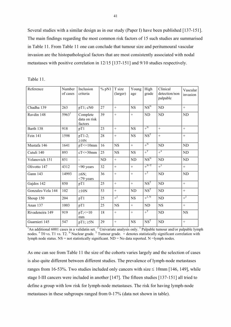
Several studies with a similar design as in our study (Paper I) have been published [137-151].
The main findings regarding the most common risk factors of 15 such studies are summarised
in Table 11. From Table 11 one can conclude that tumour size and peritumoural vascular
invasion are the histopathological factors that are most consistently associated with nodal
metastases with positive correlation in 12/15 [137-151] and 9/10 studies respectively.
Table 11.
Reference Numberof cases
Inclusioncriteria
% pN1 T size(larger)
Youngage
Highgrade
Clinicaldetection/nonpalpable
Vascularinvasion
Chadha 139 263 pT1; cN0 27 + NS NSN ND +
Ravdin 148 59631 Completedata on riskfactors
39 + + ND ND ND
Barth 138 918 pT1 23 + NS +N + +
Fein 141 1598 pT1-2;≥10N
28 + NS NST + +
Mustafa 146 1641 pT<=10mm 16 NS + +N ND ND
Cutuli 140 893 cT<=30mm 25 NS NS +T +4 ND
Velanovich 151 851 - ND + ND NSN ND ND
Olivotto 147 4312 <90 years 32 + + +N+T +3 +
Gann 143 14993 ≥6N;<79 years
36 + + +T ND ND
Gajdos 142 850 pT1 25 + + NST ND +
Gonzales-Vela 144 102 ≥10N 53 + ND NST ND +
Shoup 150 204 pT1 25 +2 NS +2, N ND +2
Anan 137 1003 pT1 25 NS + ND NS +
Rivadeneira 149 919 pT;<=10mm
18 + + +T ND NS
Guarnieri 145 547 pT1; ≥5N 29 + NS NST ND +1An additional 6001 cases in a validatin set. 2 Univariate analysis only. 3 Palpable tumour and/or palpable lymphnodes. 4 T0 vs. T1 vs. T2. N Nuclear grade. T Tumour grade. + denotes statistically significant correlation withlymph node status. NS = not statistically significant. ND = No data reported. N =lymph nodes.
As one can see from Table 11 the size of the cohorts varies largely and the selection of cases
is also quite different between different studies. The prevalence of lymph-node metastases
ranges from 16-53%. Two studies included only cancers with size ≤ 10mm [146, 149], while
stage I-III cancers were included in another [147]. The fifteen studies [137-151] all tried to
define a group with low risk for lymph-node metastases. The risk for having lymph-node
metastases in these subgroups ranged from 0-17% (data not shown in table).
42
Since increasing tumour size is the most consistent risk factor for lymph node metastasis, our
second study (Paper II) was focused on women with a tumour size of 10 mm or less. The use
of detection mode (screening mammography vs. clinical) was based on findings in a study by
Arnesson and co-workers [152] who reported that 9% of 229 screening detected cancers were
lymph node positive compared to 20% of 89 clinically detected tumours. Our study (Paper II),
based on data from two large breast cancer registries, showed that detection by screening
mammography is associated with a considerable lower risk of positive lymph nodes when
compared with clinically detected tumours. This finding was later confirmed with a
multivariate analysis (Table 3), in which detection mode was adjusted for age and tumour
size. This is in accordance with a study reported by Fein and co-writers [141], who also
identified mammographical detection as a predictor of low risk for nodal metastases. The
correlation between mode of detection and node positivity was retained in multivariate
analysis [141]. The proportion of lymph node metastases was 15,8% among 487 women with
mammographically detected tumours whereas 32,7% had lymph node metastases if the
tumour was detected by physical examination. Fein and colleagues reported a 0% risk of
lymph node metastases among mammographically detected tumours ≤ 5 mm and a 5-10% risk
for mammographically detected tumours with histopathologic size 6-10 mm and age > 40.
Other groups [138, 147] have used palpable versus non-palpable breast tumour as a potential
predictor for nodal metastases. Both these studies showed that palpability is an independent
risk factor for axillary metastases. A French study [140] reported that clinical tumour size (T0
vs. T1 vs. T2) was independently correlated with pN status.
Thus, the reason for mammographical detection being a predictor for node negativity seems to
be something more than a mere matter of tumour size. This phenomenon is probably partly
due to the tendency for mammography to detect biologically less aggressive cancers. Duffy
and co-workers found that interval cancers and cancers among non-attenders had a
significantly higher proportion of grade 3 compared with incident cases detected with
mammography [153]. Hakama and co-writers [154] found that the proportion of diploid
tumours was higher among cancers detected in incident screens compared with interval cases
and cancers among non-attenders.
43
Based on the findings in our study (Paper II), we proposed a prospective Swedish multicentric
cohort-study. After careful ethical and medical considerations, the study started accrual of
patients in 1997. In this study axillary dissection is omitted in screening detected tumours with
size ≤10 mm and grade I-II according to Elston and Ellis and/or low SPF. Women with
multifocality or a prior history of cancer are excluded. The primary end point is axillary
recurrence and a total of 1500 women are to be recruited. The accrual is estimated to stop
early 2002. No results are yet available from this study.
The five node biopsy
Although there is hope that it will be possible to forgo axillary surgery in a low risk
group, this subset comprises only a minority of newly diagnosed breast cancers. Thus, in
order to improve the management of the axilla for the vast majority of women with
breast cancer other strategies are needed. One alternative is lymph-node sampling of the
axilla, a method that is less extensive than axillary dissection. The Scottish trial on
sampling versus axillary clearance showed a sensitivity of 100% of a four-node biopsy
from the axilla [118]. However, the estimation of sensitivity, which is a key parameter,
was based on only 67 women. Moreover, the Scottish trial included patients with on
average larger tumours than currently are seen in areas with well functioning screening.
In contrast, our study of the 5-node biopsy (Paper III) included a large number of women
(n 415) and half of them had screening detected cancers. The false negative rate in our
study was 2,7% (Paper III), this estimate is encouraging since several studies of the
sentinel node procedure have shown false negative rates of 6,7-11,4% [122-124]. The
negative predictive value in our series was 98,5%, the corresponding estimate reported
from the sentinel node procedure is 93-96%. The reproducibility of our study can be
questioned, since one surgeon made all operations. However, for a specialised team a
training period with 30-50 operations with the same technique as in this trial would be
easily accomplished. The recommendations for the sentinel node procedure are similar.
The only direct comparison between a sampling procedure and the sentinel node technique
that has been published [155] included 200 women. Ten out of 60 node positive patients were
not detected with the sentinel node procedure. The corresponding figure for 4-node sampling
was 1/60. The value of this study has been questioned [156] because of the use of suboptimal
44
amounts of radiocolloid and the lack of an established reference method for comparison, i.e.
no axillary dissection was performed.
An interesting aspect is that serial sectioning and/or immunohistochemical staining of the
sentinel node(-s) often are employed [122, 124], whereas only routine pathological
examination was performed in our study (Paper III). In a recent review [157] it is estimated
that the use of IHC and/or serial sectioning produces a conversion to node positivity in 9-33%
of tumours initially classified as node negative due to detection of micrometastases. It is
reasonable to believe that the use of more sensitive pathological methods is an important part
of the sentinel node procedure, it is even tempting to hypothesise that the sensitivity could be
higher with the sentinel node procedure in comparison with level I-II dissection. A possible
argument against this is that micrometastatic sentinel nodes sometimes are accompanied with
macrometastases in non-sentinel nodes [158]. Serial sectioning and IHC are feasible when
used in conjunction with the sentinel node procedure but have been considered too unpractical
to be used in routine handling of specimens from level I-II dissections. The 5-node biopsy
represents something in between. One could speculate that the performance of the 5-node
biopsy could be improved by using these refined histopathological techniques. Although there
seems to be a worse outcome for women with lymph node micrometastases compared with
those without [157], the long-term prognostic value of IHC-positive sentinel nodes is still
unknown.
In contrast to most studies on the sentinel node procedure we reported the –LR. This measure
of a test’s performance is not affected by differences in prevalence between populations and it
can be used to calculate the post-test probability if the pre-test probability is known. In this
context the pre-test probability is the prevalence of lymph node metastases in a population
with breast cancer. An LR of 1 means that the post-test probability is exactly the same as the
pre-test probability. Likelihood ratios greater than 10 or less than 0,1 generate large and often
conclusive changes from pre-test to post-test probability [159]. The –LR of 0,027 for the 5-
node biopsy thus supports the view that this procedure can be a valuable diagnostic tool.
Although axillary surgery per se not seems to affect survival [109] the risk of withholding
adjuvant treatment for the women that were falsely defined as node negative must be
considered. This problem has decreased during recent years since most breast cancer patients

45
nowadays will be given adjuvant tamoxifen and even chemotherapy regardless of lymph-node
status [160]. In our series 3 out of 4 women with lymph node metastases not detected in the 5-
node biopsy would have been treated with tamoxifen according to current guidelines. The
fourth woman would not have been offered adjuvant systemic treatment if her nodal status had
been based on the 5-node biopsy only. This potential risk of under-treatment must be balanced
with the benefits of a less traumatic surgical procedure in the rest of the patients.
Even though we do not know the true incidence of arm symptoms after a 5-node biopsy, the
Scottish group has reported less morbidity of 4-node sampling compared with axillary
clearance of level I-III [120, 161]. Moreover, several studies indicate that increasing number
of removed nodes causes increased incidence of arm morbidity [14, 116, 117]. It is also most
likely that the sentinel node procedure causes less arm morbidity than a 5-node biopsy.
Although the sentinel node procedure probably is superior, in terms of less associated
morbidity, the 5-node biopsy seems safe to use as an alternative to level I-II dissection in
women not suitable for a sentinel node procedure. Ideally, the 5-node biopsy should be
compared with the sentinel node procedure in a randomised study.
Paper IV-V
Estimation of angiogenesis, methodological considerations
A large number of studies have reported that prognostic information can be obtained from
estimation of MVD [18, 81-85, 87-92], but several studies question the use of MVD as a
prognostic factor [93-99]. The reason for these conflicting results is probably methodological
problems, maybe related to the immunohistochemical technique, inter observer variability
and/or intra tumoural heterogeneity. Our methodological study (Paper IV) focuses on the
latter. Present methodological recommendations [162, 163] advocate determination of MVD
in the areas of highest vascular density (hotspots). The underlying hypothesis for the hotspot
method is that systemic dissemination of cancer cells is more likely in the areas of highest
vessel density [18]. Since identification of hotspots is of great importance for the method, the
chance of identifying the “hottest-spots” would most likely be influenced by the potential
variation of vascular density between different parts of a tumour.

46
Our prospective study, which employed a predefined and systematic handling of all tumour
material, demonstrates that there is a marked intra tumoural variation of MVD (Paper IV). In
the nested ANOVA analysis the variation between different sections contributed more to the
total variance than the variation between different tumours, 37,3% and 45,0% of the total
variance, respectively. This means that there is a risk that a MVD score is more influenced by
the sampling procedure and less by the true angiogenic capacity of the tumour. The influence
of intra tumoural heterogeneity on the results has been addressed in previous studies but, as
can be noticed below, there has been no standardised way of designing or reporting studies on
heterogeneity. In a Dutch study [164], a design similar to ours was used. They analysed all
available blocks (2 - 4) from 10 breast cancers. Their data showed that the variation between
blocks contributed more to the total variation than the variation between tumours, which is
identical to what we found. The CVs for different blocks from the same tumour in the Dutch
study ranged from 5,7 - 54,9%. Although the sampling technique was not identical in our
study, the corresponding range of CVs in our material, based on twice as many cases, is quite
similar (9,4 - 44,1%) (Paper IV). The Dutch group concluded that sections from multiple
blocks should be analysed to identify the “hottest-spot” for each tumour, a handling, which in
routine use would be unpractical. Bosari and co-workers [82] retrospectively examined all
available blocks (on average three) from 120 breast cancers. In 14% of the cases the difference
between the average and the maximum score was more than 20%. However, it is unclear if
this moderate variation predominantly was the result of intra- or intersection heterogeneity.
The variation within each section constituted 17,7% of the total variance in our study. The
difference between average score and maximum score exceeded 20% in 19% of our 115
sections (Paper IV). The intra section variation was much more pronounced in a study
reported by Axelsson and colleagues [95] with a greater than 20% difference in 49% of 220
breast cancers. They could not find any prognostic value of MVD and concluded that this was
mainly due to variability from field to field within the same section. Van Hoef and co-writers
[94] retrospectively analysed MVD in 93 breast cancers, without finding any independent
prognostic information. In 41 of the tumours, MVD was assessed in two sections from two
different blocks. This comparison showed a concordance with reference to MVD categories of
71 - 78%, which could introduce a substantial error in the method according to the authors.
47
Some studies have claimed that intra tumoural heterogeneity is of less importance [165, 166].
The study by Martin and co-workers [165] comprised 20 breast cancers and included an
analysis of three different blocks from each tumour. Eighty-five percent of the patients were
correctly assigned to the high MVD and low MVD groups regardless if one or three sections
were taken into account, they drew the conclusion that the MVD measured in one section is
representative of whole tumour vascularity. The Danish study [166] mainly dealt with the
reproducibility of different counting procedures but they also concluded that intra tumoural
variation does not substantially affect the results. The relevance of this conclusion must be
questioned since they selected 40 archival cancers with the object of obtaining different
degrees of MVD. This could lead to overestimation of the inter tumoural variation in relation
to the intra tumoural variation.
It has been argued that the probability of finding the hotspots can be increased by counting the
ten apparent highest fields [84]. In another study, counting 4 hotspots gave better prognostic
information than 10 [91]. The suggestion to count several hotspots seems to be supported by
our findings. The problem with this approach according to our data, is that if one counts a
large number of apparent hotspots it is likely that one will end up with a high score in most
tumours. In our study (Paper IV), all tumours had at least one score above the median MVD if
all scores from each tumour were taken into account.
We used an antibody to CD31 in both our studies on angiogenesis (Paper IV-V). Anti-CD31
has been recommended by the international consensus on the methodology and criteria for
quantification of angiogenesis as being the most useful antibody, followed by anti-CD34 and
anti-FVIII-Rag [163]. One advantage with anti-CD31 is that it does not react with lympho-
endothelial cells [163]. Martin and co-authors found anti-CD31 less reliable with up to 13%
failed stainings [84] compared with 2% for anti-CD34 and 1% for anti-FVIIIRag. In our
studies, 6% (Paper IV) and 0% (Paper V) were judged as inadequate.
Interobserver variability is not a negligible problem, variable reproducibility between different
observers has been reported (0,45< r <0,99)[166]. By following the consensus
recommendations [163], and having two observers that simultaneously assessed the slides
(Paper IV), we tried to reduce the impact of interobserver variability. In the other study (Paper
V) we had only one observer but a Chalkley eyepiece graticule was used for the assessment of
48
MVD. Hansen and co-workers [166] have shown that the interobserver variability is lower for
the Chalkley method compared with classical counting of microvessels. The Chalkley method
is probably more robust since no decisions have to be made on whether adjacent stained
structures are separate countable microvessels or not. Furthermore, the Chalkley graticule
method makes the procedure less time consuming [135].
To conclude the methodological discussion, it seems quite clear that intratumoural
heterogeneity can influence the results and that this represents a major problem for the
method. Intratumoural variation seems to be one main reason for the conflicting data on the
usefulness of MVD as a prognostic factor in breast cancer. Interobserver variability and
immunohistochemical technique also are likely contributors to diverging results.
The relation between angiogenesis and p53
Although there are methodological problems associated with MVD, we chose to continue our
studies on MVD (paper V) due to the following reasons: Firstly, the important role of
angiogenesis in the malignant progression of solid tumours is undisputed. Secondly, mutations
of the tumor-suppressor gene p53 has been demonstrated to regulate not only apoptosis and
proliferation but also angiogenesis [78, 79], which gives a molecular rationale for a
relationship between MVD and mutations of p53. Thirdly, the opportunity to analyse the
potential relationship between MVD and cDNA determined p53 mutations for exons 2-11,
which has not been investigated before, to the best f our knowledge. Fourthly, the opportunity
to study this relationship on a large population based cohort of women with complete and
long-term follow-up.
The main finding of our study is that high MVD, assessed by the Graticule counting method,
seems to correlate with mutations in the p53 tumour suppressor gene. The proportion of p53
mutation was 27,1% among women whose tumour had a high MVD, the corresponding figure
for those with a low MVD was 18,4%. The correlation between high MVD and p53 mutation
reached only borderline statistical significance (χ2-test, p=0,075). However, we found that
there was a statistically significant correlation between high MVD and p53 mutations (χ2-test,
p=0,037) when tumours (n10) with a mutation in the evolutionary conserved regions 2 or 5
were excluded. In fact, 6/10 tumours with mutation in region 2 or 5 had a low MVD. We do
not know if this is a reflection of a more complicated relationship between angiogenesis and
49
p53 status or if it is merely a play of chance due to the relatively low number of women with a
p53 mutated tumour. Therefore, this retrospective subanalysis must be interpreted cautiously.
Our study could not show any correlation between MVD and p53 analysis by IHC and this
finding is in agreement with several previously published studies [102, 167-170]. Thus,
studies on angiogenesis and p53 IHC fail to support the thrombospondin-1 based mechanism.
The reason for this could be that immunohistochemistry is inferior to sequenced based
determination of p53 status [60, 61, 171]. The false negative and false positive rate with p53
IHC was 33% and 30%, respectively, compared with sequenced based p53 status [60].
MVD and lymph-node status
Among standard prognostic factors, high MVD was associated with positive lymph-node
status (p=0,036) and high S-phase (p=0,049). The proportion of lymph-node positive women
with high MVD was 56,5%, i.e., more than 40% of lymph-node positive women had primary
tumour with a low MVD. Thus, MVD is unlikely to be of practical value for predicting the
axillary lymph-node status. In a simple predictive model consisting of only a few criteria, that
ideally should be easy to adopt in the routine management of breast cancer, there is a need of
powerful predictive factors like tumour size and detection mode (Paper I-II). Moreover, the
methodological aspects of MVD are problematic as have been outlined above.
MVD and p53 in relation to prognosis
No prognostic information could be obtained from MVD status in our study. Women whose
tumours had a mutation of the p53 gene had a statistically significantly worse prognosis with
reference to OS, BCSS and RFS according to univariate analysis and this effect was retained
in multivariate modelling. Women with tumours with low MVD and mutation seemed to do
worse with reference to OS and BCCS compared with women with high MVD and p53
mutation. Although this difference was not statistically significant, it is a bit surprising since
the difference was in the wrong direction compared with what one would expect. When we
explored the possibility of a more complicated relationship between MVD status and p53
mutation in terms of prognosis and introduced an interaction term in the multivariate
modelling, our findings were not convincing for an interaction. Although the number of
women in these subgroups were limited and the data must be interpreted with caution, we
50
found the pattern interesting enough to be worth studying further when we accumulate more
events.
The lack of prognostic power of MVD in our study is not surprising. It has been mentioned
before that several studies have reported negative findings [93-99]. In order to avoid
methodological pitfalls as much as possible we followed the recommendations that has been
published [163] with the exception that the readings of the scores were done by one person.
We also used the Chalkley eye-piece graticule counting method which is time saving [135]
and possibly superior from a methodological standpoint [166]. One limitation of our study
was the use of relatively small tumour sections. This could lead to underestimation of MVD,
especially since the intratumoural heterogeneity of this parameter is pronounced.
Nevertheless, the lack of prognostic information from MVD in this rather large cohort of
women with complete and long-term follow-up is not supportive for the use of MVD as a
prognostic factor in breast cancer.
51
General conclusions
• Positive clinical lymph node status, increasing tumour size and high S-phase fraction are
all associated with increased risk of axillary lymph-node metastases in breast cancer.
However, even if these factors are used in combination, they can not define a subgroup of
women with a sufficiently low risk of having lymph node metastases to allow omission of
axillary surgery.
• Breast cancers diagnosed with screening mammography have a lower risk of having
axillary lymph node metastases than clinically detected tumours. This association is
independent of the woman’s age and the size of the tumour. Diagnosis by screening
mammography and tumour size ≤ 10 mm are criteria, combined with other low risk
features that may be useful for characterisation o a subgroup of women in which axillary
surgery may be omitted as a part of a prospective study.
• Five-node biopsy of the axilla has a high sensitivity and a low false negative rate
compared with axillary dissection of level I-II in women with breast cancer. The 5-node
biopsy is an alternative to axillary dissection and compares very favourably with results
reported in the literature from the sentinel node biopsy procedure.
• Angiogenesis estimated with MVD shows a marked variation between different sections
from the same tumour. Intra tumoural hetereogeneity in breast cancer makes the use of
MVD questionable.
• There seems to be a relationship between angiogenesis (MVD) and cDNA sequenced
determined p53 mutations for exons 2-11 in primary breast cancer. Tumours with high
MVD more often had p53 mutations. p53 status had independent prognostic value in a
multivariate analysis whereas MVD had not.

52
Acknowledgements
I want to express my most sincere gratitude to all that helped me with this thesis, especially to:
Jonas Bergh, my supervisor, for all his never ending support, his excellent knowledge onscience and oncology and spiritual intellect, for friendship and for giving me food and shelterin his house when my home town Gävle was buried in snow. Without You this thesis wouldnot have been accomplished.
Lars Holmberg, my co-supervisor, his profound knowledge, for good discussions and forgenerous help, especially with statistical problems.
Gunnar Westman, for being my mentor and co-supervisor during the years in Örebro, forsharing some of the basic ideas to this thesis, for encouraging me to start with clinicalresearch, for friendship and good laughs.
Göran Liljegren, for good and generous collaboration since my early years in Örebro, forhaving a sincere interest in bandy.
Olle Ståhl, for co-authorship and being my supervisor during the start of my PhD studies inÖrebro.
all other co-authors: Lars-Gunnar Arnesson, Björn Risberg, Anders Lindgren, SigridKlaar, Thomas Lindahl and Kenneth Villman, for giving me of your time and knowledge.
all clinicians reporting data on women with breast cancer to the registries in Uppsala andLinköping and to the staff at ROC in Uppsala, especially Marit Holmqvist.
all my colleagues at the Department of Oncology in Gävle, for encouragement and forstanding extra work load while I have worked with this thesis.
all my colleagues at the departments of Oncology in Uppsala and Örebro, for goodcomradeship and a creative and stimulating atmosphere.
the institution of Oncology, Radiology and Clinical Immunology, section of Oncology,Professor Ingela Turesson for giving me the opportunity and support to do my PhD thesis,Didde Westerström for all help.
Torpgänget, the club for men of men, Hans, Ulf, Tomas, Per and Magnus, for good work inthe timber forests.
Karin, Anna, Nils for love and trust.
I would also express my thankfulness to the following institutions that have supported thisresearch financially: Örebro Medical Council Resarch Committee, the Swedish CancerSociety, Stiftelsen Onkologiska klinikens i Uppsala Forskningsfond, King Gustav the Vth
Jubilee Fund, the Stockholm Cancer Society and the Research Fund of Dalarna.
53
References
1. Socialstyrelsen. Cancer incidence in Sweden 1999. http://www.sos.se/sos/stat.htm 2001.2. EBCTCG. Effects of radiotherapy and surgery in early breast cancer: an overview of the
randomized trials. N Engl J Med 1995, 333, 1444-1455.3. Peto, R. Oral presentation on behalf of the EBCTCG. European Breast Cancer Conference
Brussels, 2000.4. EBCTCG. Polychemotherapy for early breast cancer: an overview of the randomised trials.
Lancet 1998, 352, 930-942.5. EBCTCG. Tamoxifen for early breast cancer: an overview of the randomised trials. Lancet
1998, 351, 1451-1467.6. Slamon, D, Clark G, Wong S, Levin W, Ullrich A, McGuire W. Human breast cancer:
correlation of relapse and survival with amplification of the HER-2/neu oncogene. Science1987, 235, 177-182.
7. Bergh, J, Norberg T, Sjögren S, Lindgren A, Holmberg L. Complete sequencing of the p53gene provides prognostic information in breast cancer patients, particularly in relation toadjuvant systemic therapy and radiotherapy. Nature Med 1995, 1, 1029-1034.
8. Perou, CM, Sorlie T, Eisen MB, van de Rijn M, Jeffrey SS, Rees CA, Pollack JR, Ross DT,Johnsen H, Akslen LA, Fluge O, Pergamenschikov A, Williams C, Zhu SX, Lonning PE,Borresen-Dale AL, Brown PO, Botstein D. Molecular portraits of human breast tumours.Nature 2000, 406, 747-52.
9. Sorlie, T, Perou CM, Tibshirani R, Aas T, Geisler S, Johnsen H, Hastie T, Eisen MB, vande Rijn M, Jeffrey SS, Thorsen T, Quist H, Matese JC, Brown PO, Botstein D, EysteinLonning P, Borresen-Dale AL. Gene expression patterns of breast carcinomas distinguishtumor subclasses with clinical implications. Proc Natl Acad Sci U S A 2001, 98, 10869-74.
10. Miki, Y, Swensen J, Shattuck-Eidens D, Futreal PA, Harshman K, Tavtigian S, Liu Q,Cochran C, Bennett LM, Ding W, et al. A strong candidate for the breast and ovariancancer susceptibility gene BRCA1. Science 1994, 266, 66-71.
11. Wooster, R, Bignell G, Lancaster J, Swift S, Seal S, Mangion J, Collins N, Gregory S,Gumbs C, Micklem G. Identification of the breast cancer susceptibility gene BRCA2.Nature 1995, 378, 789-92.
12. Clark, GM. Prognostic and predictive factors. In Harris J, Lippman M, Morrow M,Hellman S, eds. Diseases of the breast. Philadephia, Lippincott-Raven Publishers, 1996,461-485.
13. Ivens, D, Hoe AL, Podd TJ, Hamilton CR, Taylor I, Royle GT. Assessment of morbidityfrom complete axillary dissection. Br J Cancer 1992, 66, 136-8.
14. Liljegren, G, Holmberg L. Arm morbidity after sector resection and axillary dissectionwith or without postoperative radiotherapy in breast cancer stage I. Results from arandomised trial. Uppsala-Orebro Breast Cancer Study Group. Eur J Cancer 1997, 33, 193-9.
15. Tabár, L, Fagerberg CJG, Gad A, Baldetorp L, Holmberg LH, Gröntoft O, Ljungquist U,Lundström B, J.C. M. Reduction in Mortality from Breast Cancer After Mass Screeningwith Mammography: Randomized Trial from the Breast Cancer Screening Working Groupof the Swedish National Board of Health and Welfare. Lancet 1985, i, 829-832.
16. Folkman, J. Angiogenesis and breast cancer. J Clin Oncol 1994, 12, 441-443.17. Weidner, N, Semple J, Welch W, Folkman J. Tumor angiogenesis and metastasis--
correlation in invasive breast carcinoma. New Engl J Med 1991, 324, 1-8.
54
18. Horak, E, Leek R, Klenk N, LeJeune S, Smith K, Stuart N, Greenall M, Stepniewska K,Harris A. Angiogenesis, assessed by platelet/endothelial cell adhesion moleculeantibodies, as indicator of node metastases and survival in breast cancer. Lancet 1992,340, 1120-1124.
19. Socialstyrelsen. Causes of death 1999. http://www.sos.se/sos/stat.htm 2001.20. Peto, R, Boreham J, Clarke M, Davies C, Beral V. UK and USA breast cancer deaths
down 25% in year 2000 at ages 20-69 years. Lancet 2000, 355, 1822.21. Winer, E, Morrow M, Osborne C, Harris J. Malignant tumors of the breast. In DeVita VT,
Hellman S, Rosenberg S, eds. Cancer Principles & Practice of Oncology. Philadelphia,Lippincott Williams & Wilkins, 2001, 1652-1717.
22. John, EM, Kelsey JL. Radiation and other environmental exposures and breast cancer.Epidemiol Rev 1993, 15, 157-62.
23. Howe, GR, Friedenreich CM, Jain M, Miller AB. A cohort study of fat intake and risk ofbreast cancer. J Natl Cancer Inst 1991, 83, 336-40.
24. van den Brandt, PA, van't Veer P, Goldbohm RA, Dorant E, Volovics A, Hermus RJ,Sturmans F. A prospective cohort study on dietary fat and the risk of postmenopausalbreast cancer. Cancer Res 1993, 53, 75-82.
25. Willett, WC, Hunter DJ, Stampfer MJ, Colditz G, Manson JE, Spiegelman D, Rosner B,Hennekens CH, Speizer FE. Dietary fat and fiber in relation to risk of breast cancer. An 8-year follow-up. Jama 1992, 268, 2037-44.
26. Vatten, LJ, Kvinnsland S. Prospective study of height, body mass index and risk of breastcancer. Acta Oncol 1992, 31, 195-200.
27. Longnecker, MP, Berlin JA, Orza MJ, Chalmers TC. A meta-analysis of alcoholconsumption in relation to risk of breast cancer. Jama 1988, 260, 652-6.
28. Weber, BL, Garber JE. Familial breast cancer. In Harris J, Lippman M, Morrow M,Hellman S, J.R., eds. Diseases of the breast. Philadelphia, Lippincott-Raven Publishers,1996, 168-185.
29. Regionalt Onkologiskt Centrum, Uppsala-Örebro. Bröstcancer registret. Data on file 2001.30. Kerlikowske, K, Grady D, Rubin SM, Sandrock C, Ernster VL. Efficacy of screening
mammography. A meta-analysis. Jama 1995, 273, 149-54.31. Larsson, LG, Andersson I, Bjurstam N, Fagerberg G, Frisell J, Tabar L, Nystrom L.
Updated overview of the Swedish Randomized Trials on Breast Cancer Screening withMammography: age group 40-49 at randomization. J Natl Cancer Inst Monogr 1997, 22,57-61.
32. UICC. Illustrated guide to the TNM/pTNM classification of malignant tumours. In SpiesslB, Beahrs O, Hermanek P, Hutter R, Scheibe O, Sobin L, Wagner G, eds. TNM Atlas.Berlin, Heidelberg, Springer-Verlag, 1990, 173-183.
33. Carter, C, Allen C, Henson D. Relation of tumor size, lymph node status, and survival in24,740 breast cancer cases. Cancer 1989, 63, 181-187.
34. Axelsson, C, Mouridsen H, Zedeler K. Axillary Dissection of Level I and II Lymph Nodesis Important in Breast Cancer Classification. Eur J Cancer 1992, 28A, 1415-1418.
35. Westman, G, Ahlgren J, Liljegren G, Risberg B. Förbättra utbytet av axillingrepp.Läkartidningen 1995, 92, 2477-2483.
36. Tubiana, M, Koscielny S. Natural History of Human Breast Cancer: Recent Data andClinical Implications. Breast Cancer Res Treat 1991, 18, 125-140.
37. Tabár, L, Fagerberg G, Day N, Duffy S, Kitchin R. Breast Cancer Treatment and NaturalHistory: New Insights From Results of Screening. Lancet 1992, 339, 412-414.
38. Bloom, H, Richardson W. Histological grading and prognosis in breast cancer. Br JCancer 1957, 11, 357-377.
55
39. Elston, C, Ellis I. Pathological prognostic factors in breast cancer. I. The value ofhistological grade in breast cancer: experience from a large study with long-term follow-up. Histopathology 1991, 19, 403-410.
40. Sundquist, M, Thorstenson S, Brudin L, Nordenskjold B. Applying the NottinghamPrognostic Index to a Swedish breast cancer population. South East Swedish BreastCancer Study Group. Breast Cancer Res Treat 1999, 53, 1-8.
41. Boiesen, P, Bendahl PO, Anagnostaki L, Domanski H, Holm E, Idvall I, Johansson S,Ljungberg O, Ringberg A, Ostberg G, Ferno M. Histologic grading in breast cancer--reproducibility between seven pathologic departments. South Sweden Breast CancerGroup. Acta Oncol 2000, 39, 41-5.
42. Stål, O, Hatschek T, Carstensen J, Nordenskjöld B. DNA analysis in the management ofbreast cancer. Diagn Oncol 1991, 1, 140-154.
43. Wenger, CR, Clark GM. S-phase fraction and breast cancer--a decade of experience.Breast Cancer Res Treat 1998, 51, 255-65.
44. Knight, W, Livingston R, Gregory E. Estrogen Receptor as an Independent PrognosticFactor for Early Recurrence in Breast Cancer. Cancer Res 1977, 37, 4669-4671.
45. Osborne, CK. Steroid hormone receptors in breast cancer management. Breast Cancer ResTreat 1998, 51, 227-38.
46. McGuire, WL. Steroid receptors in human breast cancer. Cancer Res 1978, 38, 4289-91.47. Kinsel, LB, Szabo E, Greene GL, Konrath J, Leight GS, McCarty KS, Jr.
Immunocytochemical analysis of estrogen receptors as a predictor of prognosis in breastcancer patients: comparison with quantitative biochemical methods. Cancer Res 1989,49, 1052-6.
48. Pertschuk, LP, Kim DS, Nayer K, Feldman JG, Eisenberg KB, Carter AC, Rong ZT,Thelmo WL, Fleisher J, Greene GL. Immunocytochemical estrogen and progestinreceptor assays in breast cancer with monoclonal antibodies. Histopathologic,demographic, and biochemical correlations and relationship to endocrine response andsurvival. Cancer 1990, 66, 1663-70.
49. Blomqvist, C, von Boguslawski K, Stenman UH, Maenpaa H, von Smitten K, Nordling S.Long-term prognostic impact of immunohistochemical estrogen receptor determinationscompared with biochemical receptor determination in primary breast cancer. Acta Oncol1997, 36, 530-2.
50. Coussens, L, Yang-Feng T, Liao Y-C, Chen E, Gray A, McGrath J, Seeburg P, LibermannT, Schlessinger J, Francke U, Levinson A, Ullrich A. Tyrosine kinase receptor withextensive homology to EGF receptor shares chromosomal Location with neu oncogene.Science 1985, 230, 1132-1139.
51. Rajkumar, T, Gullick W. The type I growth factor receptors in human breast cancer.Breast Cancer Res Treat 1994, 29, 3-9.
52. Sjögren, S, Inganäs M, Lindgren A, Holmberg L, Bergh J. The prognostic and predictivevalue of c-erbB-2 overexpression in primary breast cancer, alone and in combination withother prognostic markers. J Clin Oncol 1998, 16, 462-469.
53. Prost, S, Le M, Douc-Rasy S, Ahomadegbe J, Spielmann M, Guerin M, Riou G.Association of c-erbB2-gene amplification with poor prognosis in non-inflammatorybreast carcinomas but not in carcinomas of the inflammatory type. Int J Cancer 1994, 58,763-768.
54. Isola, J, Visakorpi T, Holli K, Kallioniemi OP. Association of overexpression of tumorsuppressor protein p53 with rapid cell proliferation and poor prognosis in node-negativebreast cancer patients. J Natl Cancer Inst 1992, 84, 1109-14.
56
55. Piccart, M, Lohrisch C, Di Leo A, Larsimont D. The Predictive Value of HER2 in BreastCancer. Oncology 2001, 61 Suppl S2, 73-82.
56. Slamon, DJ, Leyland-Jones B, Shak S, Fuchs H, Paton V, Bajamonde A, Fleming T,Eiermann W, Wolter J, Pegram M, Baselga J, Norton L. Use of chemotherapy plus amonoclonal antibody against HER2 for metastatic breast cancer that overexpressesHER2. N Engl J Med 2001, 344, 783-92.
57. Levine, A, Momand J, Finlay C. The p53 tumor suppressor gene. Nature 1991, 351, 453-456.
58. Lane, DP. Cancer. p53, guardian of the genome. Nature 1992, 358, 15-6.59. Davidoff, A, Kerns B, Pence J, Marks J, Iglehart J. p53 alterations in all stages of breast
cancer. J Surg Oncol 1991, 48, 260-267.60. Sjögren, S, Inganäs M, Norberg T, Lindgren A, Nordgren H, Holmberg L, Bergh J. The
p53 gene in breast cancer: Prognostic value of complementary DNA sequencing versusimmunohistochemistry. J Natl Cancer Inst 1996, 88, 173-182.
61. Falette, N, Paperin MP, Treilleux I, Gratadour AC, Peloux N, Mignotte H, Tooke N,Lofman E, Inganas M, Bremond A, Ozturk M, Puisieux A. Prognostic value of P53 genemutations in a large series of node-negative breast cancer patients. Cancer Res 1998, 58,1451-5.
62. Kandioler-Eckersberger, D, Ludwig C, Rudas M, Kappel S, Janschek E, Wenzel C,Schlagbauer-Wadl H, Mittlbock M, Gnant M, Steger G, Jakesz R. TP53 mutation andp53 overexpression for prediction of response to neoadjuvant treatment in breast cancerpatients. Clin Cancer Res 2000, 6, 50-6.
63. Berns, EM, Foekens JA, Vossen R, Look MP, Devilee P, Henzen-Logmans SC, vanStaveren IL, van Putten WL, Inganas M, Meijer-van Gelder ME, Cornelisse C, ClaassenCJ, Portengen H, Bakker B, Klijn JG. Complete sequencing of TP53 predicts poorresponse to systemic therapy of advanced breast cancer. Cancer Res 2000, 60, 2155-62.
64. Folkman, J. Tumor angiogenesis: therapeutic implications. N Engl J Med 1971, 285,1182-6.
65. Folkman, J. What is the evidence that tumors are angiogenesis dependent? J Natl CancerInst 1990, 82, 4-6.
66. Liotta, LA, Kleinerman J, Saidel GM. Quantitative relationships of intravascular tumorcells, tumor vessels, and pulmonary metastases following tumor implantation. Cancer Res 1974, 34, 997-1004.
67. McCulloch, P, Choy A, Martin L. Association between tumour angiogenesis and tumourcell shedding into effluent venous blood during breast cancer surgery. Lancet 1995, 346,1334-5.
68. Rak, J, Filmus J, Kerbel RS. Reciprocal paracrine interactions between tumour cells andendothelial cells: the 'angiogenesis progression' hypothesis. Eur J Cancer 1996, 32A,2438-50.
69. Sandstrom, M, Johansson M, Sandstrom J, Bergenheim AT, Henriksson R. Expression ofthe proteolytic factors, tPA and uPA, PAI-1 and VEGF during malignant gliomaprogression. Int J Dev Neurosci 1999, 17, 473-81.
70. Senger, DR, Van de Water L, Brown LF, Nagy JA, Yeo KT, Yeo TK, Berse B, JackmanRW, Dvorak AM, Dvorak HF. Vascular permeability factor (VPF, VEGF) in tumorbiology. Cancer Metastasis Rev 1993, 12, 303-24.
71. Obermair, A, Kucera E, Mayerhofer K, Speiser P, Seifert M, Czerwenka K, Kaider A,Leodolter S, Kainz C, Zeillinger R. Vascular endothelial growth factor (VEGF) in humanbreast cancer: correlation with disease-free survival. Int J Cancer 1997, 74, 455-8.
57
72. Gasparini, G, Toi M, Gion M, Verderio P, Dittadi R, Hanatani M, Matsubara I, Vinante O,Bonoldi E, Boracchi P, Gatti C, Suzuki H, Tominaga T. Prognostic significance ofvascular endothelial growth factor protein in node-negative breast carcinoma. J NatlCancer Inst 1997, 89, 139-47.
73. Linderholm, B, Tavelin B, Grankvist K, Henriksson R. Vascular endothelial growth factoris of high prognostic value in node-negative breast carcinoma. J Clin Oncol 1998, 16,3121-3128.
74. Mukhopadhyay, D, Tsiokas L, Sukhatme VP. Wild-type p53 and v-Src exert opposinginfluences on human vascular endothelial growth factor gene expression. Cancer Res1995, 55, 6161-5.
75. Linderholm, B, Lindh B, Tavelin B, Grankvist K, Henriksson R. p53 and vascular-endothelial-growth-factor (VEGF) expression predicts outcome in 833 patients withprimary breast carcinoma. Int J Cancer 2000, 89, 51-62.
76. Linderholm, BK, Lindahl T, Holmberg L, Klaar S, Lennerstrand J, Henriksson R, Bergh J.The expression of vascular endothelial growth factor correlates with mutant p53 and poorprognosis in human breast cancer. Cancer Res 2001, 61, 2256-60.
77. Weinstat-Saslow, DL, Zabrenetzky VS, VanHoutte K, Frazier WA, Roberts DD, Steeg PS.Transfection of thrombospondin 1 complementary DNA into a human breast carcinomacell line reduces primary tumor growth, metastatic potential, and angiogenesis. CancerRes 1994, 54, 6504-11.
78. Dameron, KM, Volpert OV, Tainsky MA, Bouck N. Control of angiogenesis in fibroblastsby p53 regulation of thrombospondin-1. Science 1994, 265, 1582-4.
79. Grant, SW, Kyshtoobayeva AS, Kurosaki T, Jakowatz J, Fruehauf JP. Mutant p53correlates with reduced expression of thrombospondin-1, increased angiogenesis, andmetastatic progression in melanoma. Cancer Detect Prev 1998, 22, 185-94.
80. Brem, S, Cotran R, Folkman J. Tumor angiogenesis: a quantitative method for histologicgrading. J Natl Cancer Inst 1972, 48, 347-56.
81. Weidner, N, Folkman J, Pozza F, Bevilacqua P, Allred E, Moore D, Meli S, Gasparini G.Tumor angiogenesis: a new significant and independent prognostic indicator in early-stage breast carcinoma. J Natl Cancer Inst 1992, 84, 1875-1887.
82. Bosari, S, Lee A, DeLellis R, Wiley B, Heatley G, Silverman M. Microvessel quantitationand prognosis in invasive breast carcinoma. Hum Pathol 1992, 23, 755-761.
83. Toi, M, Kashitani J, Tominaga T. Tumor angiogenesis is an independent prognosticindicator in primary breast carcinoma. Int J Cancer 1993, 55, 371-374.
84. Martin, L, Green B, Renshaw C, Lowe D, Rudland P, Leinster S, Winstanley J. Examiningthe technique of angiogenesis assessment in invasive breast cancer. Br J Cancer 1997, 76, 1046-1054.
85. Hansen, S, Grabau DA, Sorensen FB, Bak M, Vach W, Rose C. The prognostic value ofangiogenesis by Chalkley counting in a confirmatory study design on 836 breast cancerpatients. Clin Cancer Res 2000, 6, 139-146.
86. Hansen, S, Grabau DA, Sorensen FB, Bak M, Vach W, Rose C. Vascular grading ofangiogenesis: prognostic significance in breast cancer. Br J Cancer 2000, 82, 339-47.
87. Gasparini, G, Weidner N, Bevilacqua P, Maluta S, Dalla Palma P, Caffo O, BarbareschiM, Boracchi P, Marubini E, Pozza F. Tumor microvessel density, p53 expression, tumorsize, and peritumoral lymphatic vessel invasion are relevant prognostic markers in node-negative breast carcinoma. J Clin Oncol 1994, 12, 454-466.
88. Fox, S, Leek R, Smith K, Hollyer J, Greenall M, Harris A. Tumor angiogenesis in node-negative breast carcinomas--relationship with epidermal growth factor receptor, estrogenreceptor, and survival. Breast Cancer Res Treat 1994, 29, 109-116.
58
89. Heimann, R, Ferguson D, Powers C, Recant W, Weichselbaum R, Hellman S.Angiogenesis as a predictor of long-term survival for patients with node-negative breastcancer. J Natl Cancer Inst 1996, 88, 1764-1769.
90. Gasparini, G, Toi M, Verderio P, Ranieri G, Dante S, Bonoldi E, Boracchi P, Fanelli M,Tominaga T. Prognostic significance of p53, angiogenesis, and other conventionalfeatures in operable breast cancer: subanalysis in node-positive and node-negativepatients. Int J Oncol 1998, 12, 1117-1125.
91. de Jong, JS, van Diest PJ, Baak JP. Hot spot microvessel density and the mitotic activityindex are strong additional prognostic indicators in invasive breast cancer.Histopathology 2000, 36, 306-12.
92. Kato, T, Kameoka S, Kimura T, Nishikawa T, Kasajima T. Angiogenesis and blood vesselinvasion as prognostic indicators for node-negative breast cancer. Breast Cancer ResTreat 2001, 65, 203-15.
93. Hall, N, Fish D, Hunt N, Goldin R, Guillou P, Monson J. Is the relationship betweenangiogenesis and metastasis in breast cancer real? Surg Oncol 1992, 1, 223-229.
94. Van Hoef, M, Knox W, Dhesi S, Howell A, Schor A. Assessment of tumour vascularity asa prognostic factor in lymph node negative invasive breast cancer. Eur J Cancer 1993,29A, 1141-1145.
95. Axelsson, K, Ljung B, Moore 2nd D, Thor A, Chew K, Edgerton S, Smith H, Mayall B.Tumor angiogenesis as a prognostic assay for invasive ductal breast carcinoma. J NatlCancer Inst 1995, 87, 997-1008.
96. Costello, P, McCann A, Carney D, Dervan P. Prognostic significance of microvesseldensity in lymph node negative breast carcinoma. Hum Pathol 1995, 26, 1181-1184.
97. Siitonen, S, Haapasalo H, Rantala I, Helin H, Isola J. Comparison of differentimmunohistochemical methods in the assessment of angiogenesis: lack of prognosticvalue in a group of 77 selected node-negative breast carcinomas. Mod Pathol 1995, 8,845-752.
98. Goulding, H, Abdul Rashid NF, Robertson JF, Bell JA, Elston CW, Blamey RW, Ellis IO.Assessment of angiogenesis in breast carcinoma: an important factor in prognosis? [seecomments]. Hum Pathol 1995, 26, 1196-200.
99. Morphopoulos, G, Pearson M, Ryder WD, Howell A, Harris M. Tumour angiogenesis as aprognostic marker in infiltrating lobular carcinoma of the breast [see comments]. J Pathol1996, 180, 44-9.
100. Miliaras, D, Kamas A, Kalekou H. Angiogenesis in invasive breast carcinomas: is itassociated with parameters of prognostic significance? Histopathol 1995, 26, 165-169.
101. Fox, SB, Leek RD, Bliss J, Mansi JL, Gusterson B, Gatter KC, Harris AL. Association oftumor angiogenesis with bone marrow micrometastases in breast cancer patients. J NatlCancer Inst 1997, 89, 1044-9.
102. Jacquemier, JD, Penault-Llorca FM, Bertucci F, Sun ZZ, Houvenaeghel GF, Geneix JA,Puig BD, Bardou VJ, Hassoun JA, Birnbaum D, Viens PJ. Angiogenesis as a prognosticmarker in breast carcinoma with conventional adjuvant chemotherapy: a multiparametricand immunohistochemical analysis. J Pathol 1998, 184, 130-5.
103. Gasparini, G, Bonoldi E, Viale G, Verderio P, Boracchi P, Panizzoni GA, Radaelli U, DiBacco A, Guglielmi RB, Bevilacqua P. Prognostic and predictive value of tumourangiogenesis in ovarian carcinomas. Int J Cancer 1996, 69, 205-11.
104. Gasparini, G, Fox SB, Verderio P, Bonoldi E, Bevilacqua P, Boracchi P, Dante S,Marubini E, Harris AL. Determination of angiogenesis adds information to estrogenreceptor status in predicting the efficacy of adjuvant tamoxifen in node-positive breastcancer patients. Clin Cancer Res 1996, 2, 1191-8.

59
105. Deplanque, G, Harris AL. Anti-angiogenic agents: clinical trial design and therapies indevelopment. Eur J Cancer 2000, 36, 1713-24.
106. Westphal, JR, Ruiter DJ, De Waal RM. Anti-angiogenic treatment of human cancer:pitfalls and promises. Int J Cancer 2000, 86, 870-3.
107. Aspegren, K, Holmberg L, Adami HO. Standardization of the surgical technique inbreast-conserving treatment of mammary cancer. Br J Surg 1988, 75, 807-10.
108. Halsted, W. The results of operation for the cure of cancer of the breast performed at TheJohns Hopkins Hospital from June 1889 to January 1894. Johns Hopkins Hosp Rep 1894,4, 297-350.
109. Fisher, B, Redmond C, Fisher ER, Bauer M, Wolmark N, Wickerham D, Deutsch M,Montague E, Margolese R, Foster R. Ten Year Results of a Randomized Clinical TrialComparing Radical Mastectomy and Total Mastectomy With or Without Radiation. NEngl J Med 1985, 312, 674-681.
110. Veronesi, U, Marubini E, Mariani L, Valagussa P, Zucali R. The dissection of internalmammary nodes does not improve the survival of breast cancer patients. 30-year resultsof a randomised trial. Eur J Cancer 1999, 35, 1320-5.
111. Harris, J, Morrow M. Treatment of early-stage breast cancer. In Harris J, Lippman M,Morrow M, Hellman S, eds. Diseases of the breast. Philadelphia, Lippincott-RavenPublishers, 1996, 487-547.
112. Recht, A, Pierce SM, Abner A, Vicini F, Osteen RT, Love SM, Silver B, Harris JR.Regional Nodal Failure after Conservative Surgery and Radiotherapy for Early-Stagebreast Carcinoma. J Clin Oncol 1991, 9, 988-996.
113. Rosen, PP, Lesser ML, Kinne DW, Beattie EJ. Discontinuous or "skip" metastases inbreast carcinoma. Analysis of 1228 axillary dissections. Ann Surg 1983, 197, 276-83.
114. Veronesi, U, Luini A, Galimberti V, Marchini S, Sacchini V, Rilke F. Extent ofMetastatic Axillary Involvement in 1446 Cases of Breast Cancer. Eur J Surg Oncol 1990,16, 127-133.
115. Petrek, J, Lerner R. Lymphedema. In Harris J, Lippman M, Morrow M, Hellman S, eds.Diseases of the breast. Philadelphia, Lippincott-Raven Publishers, 1996, 896-903.
116. Larson, D, Weinstein M, Goldberg I. Edema of the Arm as a Function of the Extent ofAxillary Surgery in patients With Stage I-II Carcinoma of the Breast treated With PrimaryRadiotherapy. Int J Radiat Oncol Biol Phys 1986, 12, 1575-1582.
117. Borup Christensen, S, Lundgren E. Sequelae of Axillary Dissection vs Axillary SamplingWith or Without Irradiation for Breast Cancer. Acta Chir Scand 1989, 155, 515-520.
118. Steele, R, Forrest A, Gibson T, Stewart H, Chetty U. The Efficacy of Lower AxillarySampling in Obtaining Lymph Node Status in Breast cancer: A Controlled RandomizedTrial. Br J Surg 1985, 72, 368-369.
119. Forrest, AP, Everington D, McDonald CC, Steele RJ, Chetty U, Stewart HJ. TheEdinburgh randomized trial of axillary sampling or clearance after mastectomy. Br J Surg1995, 82, 1504-8.
120. Chetty, U, Jack W, Prescott RJ, Tyler C, Rodger A. Management of the axilla in operablebreast cancer treated by breast conservation: a randomized clinical trial. Edinburgh BreastUnit. Br J Surg 2000, 87, 163-9.
121. Kissin, MW, Thompson EM, Price AB, Slavin G, Kark AE. The inadequacy of axillarysampling in breast cancer. Lancet 1982, 1, 1210-2.
122. Veronesi, U, Paganelli G, Viale G, Galimberti V, Luini A, Zurrida S, Robertson C,Sacchini V, Veronesi P, Orvieto E, De Cicco C, Intra M, Tosi G, Scarpa D. Sentinellymph node biopsy and axillary dissection in breast cancer: results in a large series. J NatlCancer Inst 1999, 91, 368-73.
60
123. Krag, D, Weaver D, Ashikaga T, Moffat F, Klimberg VS, Shriver C, Feldman S,Kusminsky R, Gadd M, Kuhn J, Harlow S, Beitsch P. The sentinel node in breast cancer--a multicenter validation study. N Engl J Med 1998, 339, 941-6.
124. Frisell, J, Bergqvist L, Liljegren G, Thorn M, Damm S, Rydman H, Danielsson R.Sentinel node in breast cancer--a Swedish pilot study of 75 patients. Eur J Surg 2001,167, 179-83.
125. EBCTCG. Favourable and unfavourable effects on long-term survival of radiotherapy forearly breast cancer: an overview of the randomised trials. Early Breast Cancer Trialists'Collaborative Group. Lancet 2000, 355, 1757-70.
126. Overgaard, M, Hansen PS, Overgaard J, Rose C, Andersson M, Bach F, Kjaer M,Gadeberg CC, Mouridsen HT, Jensen MB, Zedeler K. Postoperative radiotherapy in high-risk premenopausal women with breast cancer who receive adjuvant chemotherapy.Danish Breast Cancer Cooperative Group 82b Trial. N Engl J Med 1997, 337, 949-55.
127. Overgaard, M, Jensen MB, Overgaard J, Hansen PS, Rose C, Andersson M, Kamby C,Kjaer M, Gadeberg CC, Rasmussen BB, Blichert-Toft M, Mouridsen HT. Postoperativeradiotherapy in high-risk postmenopausal breast-cancer patients given adjuvanttamoxifen: Danish Breast Cancer Cooperative Group DBCG 82c randomised trial. Lancet1999, 353, 1641-8.
128. Ragaz, J, Jackson S, Le N, Plenderleith I, Spinelli J, Basco V, Wilson K, Knowling M,Coppin C, Paradis M, Coldman A, Olivotto I. Adjuvant radiotherapy and chemotherapyin node-positive premenopausal women with breast cancer. N Engl J Med 1997, 337,956-962.
129. Dewar, JA, Sarrazin D, Benhamou E. Management of the Axilla in ConservativelyTreated Breast Cancer: 592 Patients treated at Institut Gustave-Roussy. Int J RadiatOncol Biol Phys 1987, 13, 475-481.
130. Meek, AG. Breast radiotherapy and lymphedema. Cancer 1998, 83, 2788-97.131. Cold, S, Jensen N, Brincker H, Rose C. The influence of chemotherapy on survival after
recurrence in breast cancer - A population-based study of patients treated in the 1950s,1960s and 1970s. Eur J Cancer 1993, 29A, 1146-1152.
132. Fossati, R, Confalonieri C, Torri V, Ghislandi E, Penna A, Pistotti V, Tinazzi A, LiberatiA. Cytotoxic and hormonal treatment for metastatic breast cancer: a systematic review ofpublished randomized trials involving 31,510 women. J Clin Oncol 1998, 16, 3439-60.
133. Nabholtz, J, Thuerlimann B, Beswoda W, Melnychuk D, Deschenes L, Douma J,Vandenberg T, Rapoport B, Rosso R, Trillet-Lenoir V, Drbal J, Aapro M, Alakl M,Murawsky M, Riva A. Taxotere improves survival over mitomycin C vinblastine inpatients with metastatic breast cancer who have failed on anthracycline containingregimen: final results of a phase III randomized trial. In Perry M, ed. Thirty-FourthAnnual Meeting 17. Los Angeles, CA, Am Soc Clin Oncol, 1998, 101a, abstract 390.
134. Jassem, J, Pienkowski T, Pluzanska A, Jelic S, Gorbunova V, Mrsic-Krmpotic Z, BerzinsJ, Nagykalnai T, Wigler N, Renard J, Munier S, Weil C. Doxorubicin and paclitaxelversus fluorouracil, doxorubicin, and cyclophosphamide as first-line therapy for womenwith metastatic breast cancer: final results of a randomized phase III multicenter trial. JClin Oncol 2001, 19, 1707-15.
135. Fox, SB, Leek RD, Weekes MP, Whitehouse RM, Gatter KC, Harris AL. Quantitationand prognostic value of breast cancer angiogenesis: comparison of microvessel density,Chalkley count, and computer image analysis. J Pathol 1995, 177, 275-83.
136. Fisher, B, Wolmark N, Bauer M, Redmond C, Gebhardt M. The Accuracy of ClinicalNodal Staging and of Limited Axillary Dissection as a Determinant of Histological NodalStatus in Carcinoma of the Breast. Surg Gyn Obst 1981, 152, 765-772.
61
137. Anan, K, Mitsuyama S, Tamae K, Nishihara K, Iwashita T, Abe Y, Ihara T, ToyoshimaS. Axillary lymph node metastases in patients with small carcinomas of the breast: isaccurate prediction possible? Eur J Surg 2000, 166, 610-5.
138. Barth, A, Craig PH, Silverstein MJ. Predictors of axillary lymph node metastases inpatients with T1 breast carcinoma. Cancer 1997, 79, 1918-22.
139. Chadha, M, Chabon AB, Friedmann P, Vikram B. Predictors of axillary lymph nodemetastases in patients with T1 breast cancer. A multivariate analysis. Cancer 1994, 73,350-3.
140. Cutuli, B, Velten M, Rodier JF, Janser JC, Quetin P, Jaeck D, Renaud R, Duperoux G.[Evaluating the risk of axillary lymph node involvement in inferior breast cancermeasuring 3 centimeters. Analysis of a predictive model based on 893 cases]. Chirurgie1998, 123, 175-81; discussion 181-2.
141. Fein, DA, Fowble BL, Hanlon AL, Hooks MA, Hoffman JP, Sigurdson ER, Jardines LA,Eisenberg BL. Identification of women with T1-T2 breast cancer at low risk of positiveaxillary nodes. J Surg Oncol 1997, 65, 34-9.
142. Gajdos, C, Tartter PI, Bleiweiss IJ. Lymphatic invasion, tumor size, and age areindependent predictors of axillary lymph node metastases in women with T1 breastcancers. Ann Surg 1999, 230, 692-6.
143. Gann, PH, Colilla SA, Gapstur SM, Winchester DJ, Winchester DP. Factors associatedwith axillary lymph node metastasis from breast carcinoma: descriptive and predictiveanalyses. Cancer 1999, 86, 1511-9.
144. Gonzalez-Vela, MC, Garijo MF, Fernandez FA, Buelta L, Val-Bernal JF. Predictors ofaxillary lymph node metastases in patients with invasive breast carcinoma by acombination of classical and biological prognostic factors. Pathol Res Pract 1999, 195,611-8.
145. Guarnieri, A, Neri A, Correale PP, Lottini M, Testa M, Mariani F, Tucci E, Megha T,Cintorino M, Carli A. Prediction of lymph node status by analysis of prognostic factorsand possible indications for elective axillary dissection in T1 breast cancers. Eur J Surg2001, 167, 255-9.
146. Mustafa, IA, Cole B, Wanebo HJ, Bland KI, Chang HR. The impact of histopathology onnodal metastases in minimal breast cancer. Arch Surg 1997, 132, 384-90; discussion 390-1.
147. Olivotto, IA, Jackson JS, Mates D, Andersen S, Davidson W, Bryce CJ, Ragaz J.Prediction of axillary lymph node involvement of women with invasive breast carcinoma:a multivariate analysis. Cancer 1998, 83, 948-55.
148. Ravdin, PM, De Laurentiis M, Vendely T, Clark GM. Prediction of axillary lymph nodestatus in breast cancer patients by use of prognostic indicators. J Natl Cancer Inst 1994,86, 1771-5.
149. Rivadeneira, DE, Simmons RM, Christos PJ, Hanna K, Daly JM, Osborne MP.Predictive factors associated with axillary lymph node metastases in T1a and T1b breastcarcinomas: analysis in more than 900 patients. J Am Coll Surg 2000, 191, 1-6;discussion 6-8.
150. Shoup, M, Malinzak L, Weisenberger J, Aranha GV. Predictors of axillary lymph nodemetastasis in T1 breast carcinoma. Am Surg 1999, 65, 748-52; discussion 752-3.
151. Velanovich, V, Szymanski W. Lymph node metastasis in breast cancer: commonprognostic markers lack predictive value. Ann Surg Oncol 1998, 5, 613-9.
152. Arnesson, LG, Smeds S, Fagerberg G. Recurrence-free survival in patients with smallbreast cancer. An analysis of cancers 10 mm or less detected clinically and by screening.Eur J Surg 1994, 160, 271-6.
62
153. Duffy, SW, Tabar L, Fagerberg G, Gad A, Gröntoft O, South MC, Day NE. BreastScreening, Prognostic Factors and Survival - Results From the Swedish Two CountyStudy. Br J Cancer 1991, 64, 1133-1138.
154. Hakama, M, Holli K, Isola J, Kallioniemi OP, Karkkainen A, Visakorpi T, Pukkala E,Saarenmaa I, Geiger U, Ikkala J, et al. Aggressiveness of screen-detected breast cancers.Lancet 1995, 345, 221-4.
155. Macmillan, RD, Barbera D, Hadjiminas DJ, Rampaul RS, Lee AH, Pinder SE, Ellis IO,Blamey RW, Geraghty JG. Sentinel node biopsy for breast cancer may have little to offerfour-node-samplers. results of a prospective comparison study. Eur J Cancer 2001, 37,1076-80.
156. Hansen, NM, Giuliano AE. Why remove four by chance when one will suffice? Eur JCancer 2001, 37, 1067-9.
157. Dowlatshahi, K, Fan M, Snider HC, Habib FA. Lymph node micrometastases frombreast carcinoma: reviewing the dilemma. Cancer 1997, 80, 1188-97.
158. Chua, B, Ung O, Taylor R, Bilous M, Salisbury E, Boyages J. Treatment implications ofa positive sentinel lymph node biopsy for patients with early-stage breast carcinoma.Cancer 2001, 92, 1769-1774.
159. Jaeschke, R, Guyatt GH, Sackett DL. Users' guides to the medical literature. III. How touse an article about a diagnostic test. B. What are the results and will they help me incaring for my patients? The Evidence-Based Medicine Working Group. Jama 1994, 271,703-7.
160. Goldhirsch, A, Glick JH, Gelber RD, Coates AS, Senn HJ. Meeting highlights:International Consensus Panel on the Treatment of Primary Breast Cancer. SeventhInternational Conference on Adjuvant Therapy of Primary Breast Cancer. J Clin Oncol2001, 19, 3817-27.
161. Aitken, RJ, Gaze MN, Rodger A, Chetty U, Forrest AP. Arm morbidity within a trial ofmastectomy and either nodal sample with selective radiotherapy or axillary clearance. BrJ Surg 1989, 76, 568-71.
162. Gasparini, G, Harris A. Clinical importance of the determination of tumor angiogenesisin breast carcinoma: much more than a new prognostic tool. J Clin Oncol 1995, 13, 765-782.
163. Vermeulen, P, Gasparini G, Fox S, Toi M, Martin L, McCulloch P, Pezzella F, Viale G,Weidner N, Harris A, Dirix L. Quantification of angiogenesis in solid human tumours: aninternational consensus on the methodology and criteria oevaluation. Eur J Cancer 1996Dec;32A(14):2474-84 1996, 32A, 2474-2484.
164. de Jong, J, van Diest P, Baak J. Methods in laboratory investigation. Heterogeneity andreproducibility of microvessel counts in breast cancer. Lab Invest 1995, 73, 922-926.
165. Martin, L, Holcombe C, Green B, Leinster SJ, Winstanley J. Is a histological sectionrepresentative of whole tumour vascularity in breast cancer? Br J Cancer 1997, 76, 40-3.
166. Hansen, S, Grabau DA, Rose C, Bak M, Sorensen FB. Angiogenesis in breast cancer: acomparative study of the observer variability of methods for determining microvesseldensity. Lab Invest 1998, 78, 1563-73.
167. Marinho, A, Soares R, Ferro J, Lacerda M, Schmitt FC. Angiogenesis in breast cancer isrelated to age but not to other prognostic parameters. Pathol Res Pract 1997, 193, 267-73.
168. Bevilacqua, P, Barbareschi M, Verderio P, Boracchi P, Caffo O, Dalla Palma P, Meli S,Weidner N, Gasparini G. Prognostic value of intratumoral microvessel density, a measureof tumor angiogenesis, in node-negative breast carcinoma--results of a multiparametricstudy. Breast Cancer Res Treat 1995, 36, 205-17.

63
169. Tas, F, Yavuz E, Aydiner A, Saip P, Disci R, Iplikci A, Topuz E. Angiogenesis and p53protein expression in breast cancer: prognostic roles and interrelationships. Am J ClinOncol 2000, 23, 546-53.
170. Kato, T, Kimura T, Ishii N, Fujii A, Yamamoto K, Kameoka S, Nishikawa T, KasajimaT. The methodology of quantitation of microvessel density and prognostic value ofneovascularization associated with long-term survival in Japanese patients with breastcancer. Breast Cancer Res Treat 1999, 53, 19-31.
171. Borresen, AL. Subgroups of p53 mutations may predict the clinical behaviour of cancersin the breast and colon and contribute to therapy response. In Klijn JGM, ed. Prognosticand predictive value of p53 1. Amsterdam, Elsevier Science, 1997, 23-33.

Studies on Prediction of Axillary Lymph Node Status in Invasive Breast Cancer
Errata:
Page 28, first paragraph: ”…false negative rate of 1,5%…” is wrong”…false negative rate of 2,7%…” is corrrect
Related Documents