Axial Skeleton: Vertebral Column Slides by Vince Austin; figures from Marieb & Hoehn 7 th & 8 th eds.; modifications and some slides by W. Rose Portions copyright Pearson Education

Axial Skeleton: Vertebral Column Slides by Vince Austin; figures from Marieb & Hoehn 7 th & 8 th eds.; modifications and some slides by W. Rose Portions.
Jan 02, 2016
Welcome message from author
This document is posted to help you gain knowledge. Please leave a comment to let me know what you think about it! Share it to your friends and learn new things together.
Transcript

Axial Skeleton: Vertebral Column
Slides by Vince Austin;
figures from Marieb & Hoehn 7th & 8th eds.;
modifications and some slides by W. Rose
Portions copyright Pearson Education

Copyright © 2006 Pearson Education, Inc., publishing as Benjamin Cummings
Vertebral Column
26 irregular bones (vertebrae) connected to form a flexible curved structure
Cervical vertebrae – 7 bones of neck
Thoracic vertebrae – 12 bones of torso
Lumbar vertebrae – 5 bones of lower back
Sacrum – bone inferior to the lumbar vertebrae that articulates with the hip bones (fusion of 5, starts ~> puberty, done mid-20s)
Copyright © 2006 Pearson Education, Inc., publishing as Benjamin Cummings
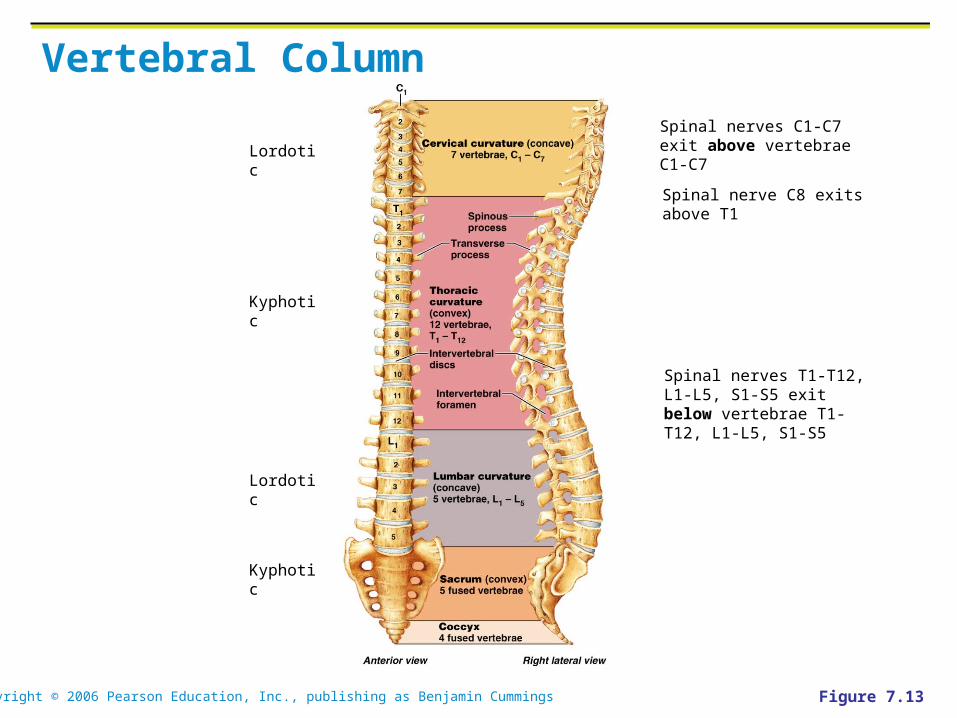
Vertebral Column
Figure 7.13
Spinal nerves C1-C7 exit above vertebrae C1-C7
Spinal nerve C8 exits above T1
Spinal nerves T1-T12, L1-L5, S1-S5 exit below vertebrae T1-T12, L1-L5, S1-S5
Kyphotic
Kyphotic
Lordotic
Lordotic
Copyright © 2006 Pearson Education, Inc., publishing as Benjamin Cummings
Vertebral Column: Curvatures
Kyphosis = posteriorly convex curvature. Thoracic and sacral normally kyphotic (but not too much).
Lordosis = posteriorly concave curvature. Cervical and lumbar normally lordotic (but not too much).
Abnormal spinal curvatures
• Scoliosis (lateral curve)
• Excessive kyphosis (hunchback)
• Excessive lordosis (swayback)
• What happens in pregnancy?
Copyright © 2006 Pearson Education, Inc., publishing as Benjamin Cummings
Vertebral Column: Ligaments
Anterior and posterior longitudinal ligaments – continuous bands down the front and back of the spine from the neck to the sacrum
Short ligaments connect adjoining vertebrae together

Copyright © 2006 Pearson Education, Inc., publishing as Benjamin Cummings
Vertebral Column: Ligaments

Copyright © 2006 Pearson Education, Inc., publishing as Benjamin Cummings
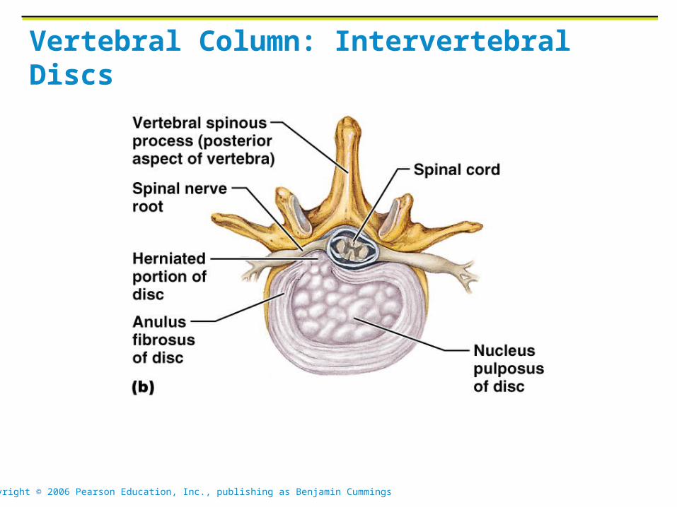
Vertebral Column: Intervertebral Discs
Cushion-like pad composed of two parts
Nucleus pulposus – inner gelatinous nucleus that gives the disc its elasticity and compressibility
Annulus fibrosus – surrounds the nucleus pulposus with a collar composed of collagen and fibrocartilage
Copyright © 2006 Pearson Education, Inc., publishing as Benjamin Cummings
Vertebral Column: Intervertebral Discs
Copyright © 2006 Pearson Education, Inc., publishing as Benjamin Cummings
General Structure of Vertebrae
Body or centrum – disc-shaped, weight-bearing region
Vertebral arch – composed of pedicles and laminae that, along with the centrum, enclose the vertebral foramen
Vertebral foramina – make up the vertebral canal through which the spinal cord passes

Copyright © 2006 Pearson Education, Inc., publishing as Benjamin Cummings
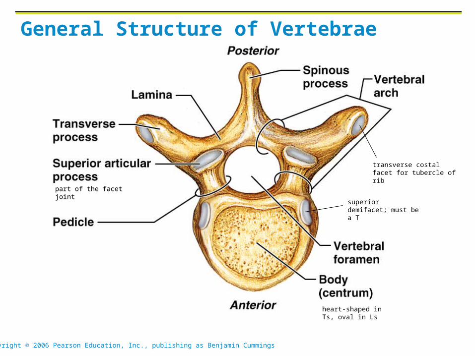
General Structure of Vertebrae
Spinous processes project posteriorly, and transverse processes project laterally
Superior and inferior articular processes – protrude superiorly and inferiorly from the pedicle-lamina junctions
Intervertebral foramina – lateral openings formed from notched areas on the superior and inferior borders of adjacent pedicles
Copyright © 2006 Pearson Education, Inc., publishing as Benjamin Cummings
General Structure of Vertebrae
transverse costal facet for tubercle of rib
superior demifacet; must be a T
heart-shaped in Ts, oval in Ls
part of the facet joint

Copyright © 2006 Pearson Education, Inc., publishing as Benjamin Cummings
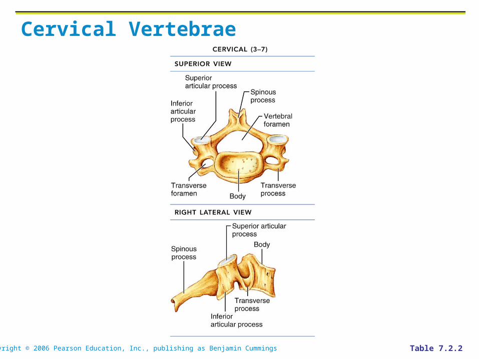
Cervical Vertebrae
Seven vertebrae (C1-C7): smallest, lightest vertebrae
C3-C7: Distinguished by oval bodies, short spinous processes, large, triangular vertebral foramina. Articular facets (sup & inf) form joints with vetrebrae above & below.
Each transverse process contains a transverse foramen. (Only cervical have transverse foramina.)
Copyright © 2006 Pearson Education, Inc., publishing as Benjamin Cummings
Cervical Vertebrae
Table 7.2.2
Copyright © 2006 Pearson Education, Inc., publishing as Benjamin Cummings
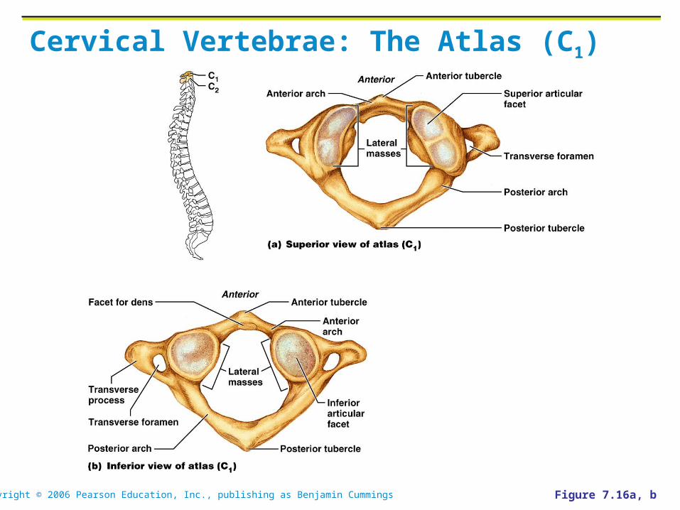
Cervical Vertebrae: Atlas (C1)
No body, no spinous process
Anterior and posterior arches, and two lateral masses
Superior surfaces of lateral masses articulate with occipital condyles
Copyright © 2006 Pearson Education, Inc., publishing as Benjamin Cummings
Cervical Vertebrae: The Atlas (C1)
Figure 7.16a, b

Copyright © 2006 Pearson Education, Inc., publishing as Benjamin Cummings
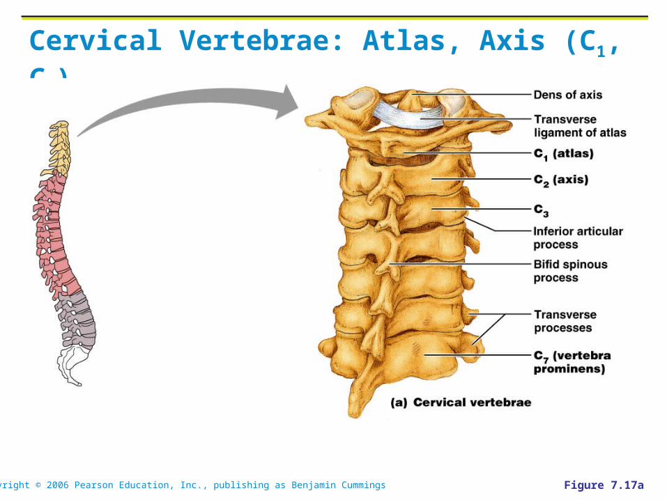
Cervical Vertebrae: Axis (C2)
Body, spine, and vertebral arches, like other cervical vertebrae
Unique feature: dens, or odontoid process, projects superiorly from body, cradled in the anterior arch of the atlas
The dens is a pivot for the rotation of the atlas
The “missing body” of the atlas?

Copyright © 2006 Pearson Education, Inc., publishing as Benjamin Cummings
Cervical Vertebrae: The Axis (C2)
Figure 7.16c
Copyright © 2006 Pearson Education, Inc., publishing as Benjamin Cummings
Cervical Vertebrae: Atlas, Axis (C1, C2)
Figure 7.17a
Copyright © 2006 Pearson Education, Inc., publishing as Benjamin Cummings
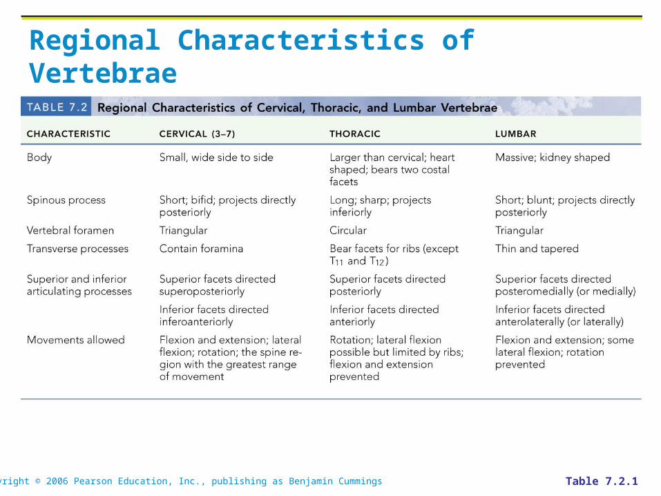
Regional Characteristics of Vertebrae
Table 7.2.1

Copyright © 2006 Pearson Education, Inc., publishing as Benjamin Cummings
Regional Characteristics of Vertebrae
Table 7.2.2
Copyright © 2006 Pearson Education, Inc., publishing as Benjamin Cummings
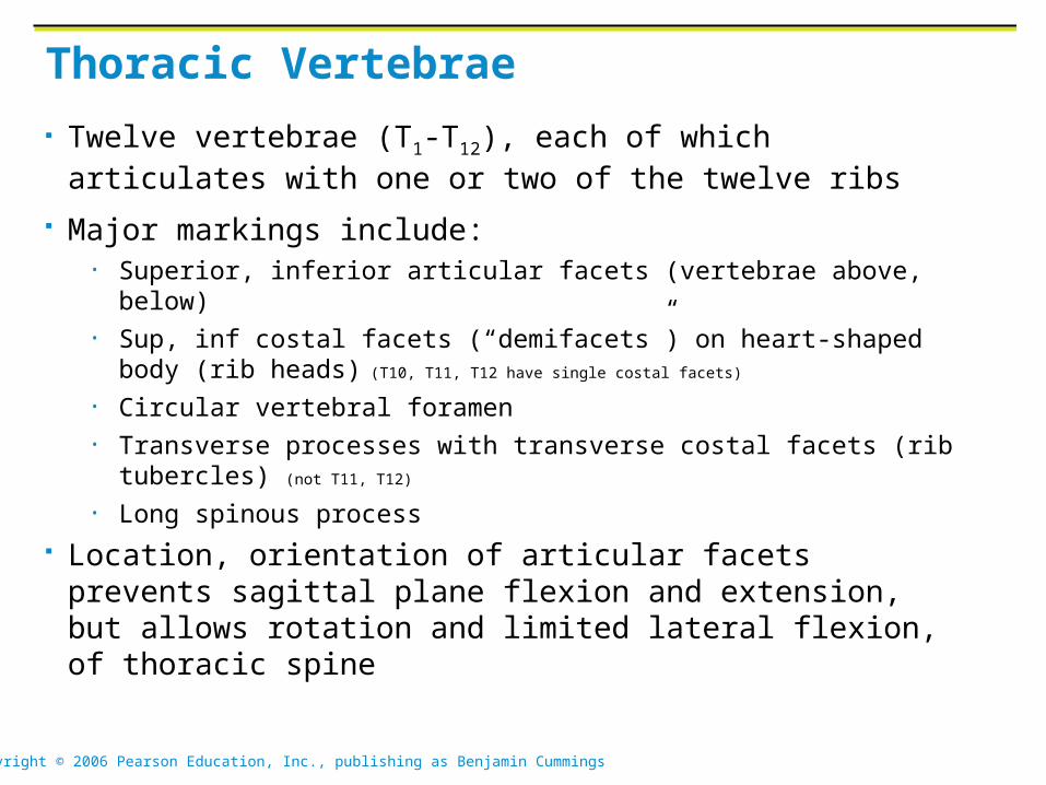
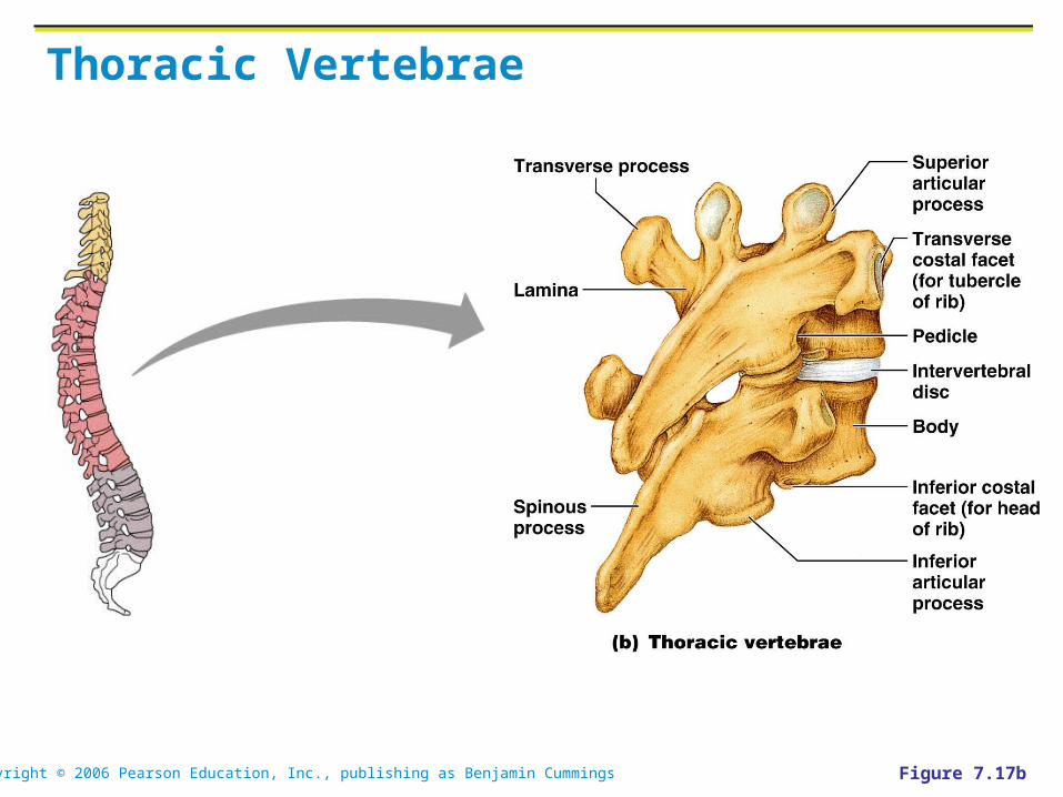
Thoracic Vertebrae
Twelve vertebrae (T1-T12), each of which articulates with one or two of the twelve ribs
Major markings include: • Superior, inferior articular facets (vertebrae above, below)• Sup, inf costal facets (“demifacets”) on heart-shaped body (rib heads)
(T10, T11, T12 have single costal facets)
• Circular vertebral foramen• Transverse processes with transverse costal facets (rib tubercles) (not T11,
T12)
• Long spinous process
Location, orientation of articular facets prevents sagittal plane flexion and extension, but allows rotation and limited lateral flexion, of thoracic spine
Copyright © 2006 Pearson Education, Inc., publishing as Benjamin Cummings
Thoracic Vertebrae
Figure 7.17b

Copyright © 2006 Pearson Education, Inc., publishing as Benjamin Cummings
Lumbar Vertebrae
The five lumbar vertebrae (L1-L5) are located in the small of the back and have an enhanced weight-bearing function
They have short, thick pedicles and laminae, flat hatchet-shaped spinous processes, and a triangular-shaped vertebral foramen
Orientation of articular facets locks the lumbar vertebrae together to provide stability

Copyright © 2006 Pearson Education, Inc., publishing as Benjamin Cummings
Lumbar Vertebrae
Figure 7.17c
Copyright © 2006 Pearson Education, Inc., publishing as Benjamin Cummings
Sacrum
Sacrum
Consists of five fused vertebrae (S1-S5), which shape the posterior wall of the pelvis
It articulates with L5 superiorly, and with the auricular surfaces of the hip bones
Major markings include the sacral promontory, transverse lines, alae, dorsal sacral foramina, sacral canal, and sacral hiatus

Copyright © 2006 Pearson Education, Inc., publishing as Benjamin Cummings
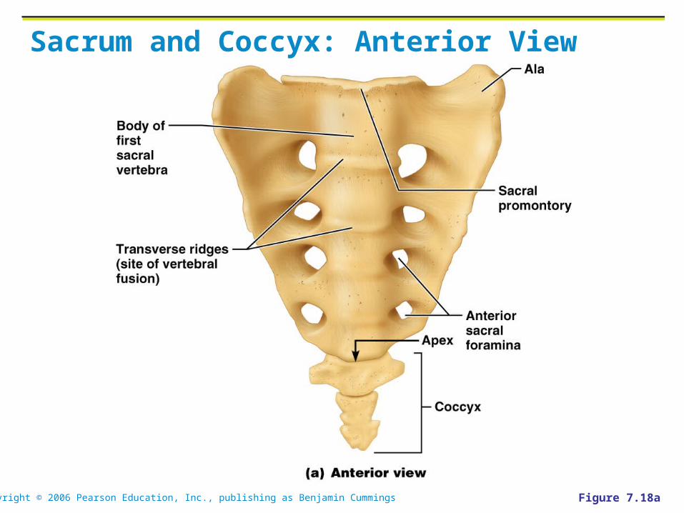
Coccyx
Coccyx (Tailbone)
The coccyx is made up of four (in some cases three to five) fused vertebrae that articulate superiorly with the sacrum
Copyright © 2006 Pearson Education, Inc., publishing as Benjamin Cummings
Sacrum and Coccyx: Anterior View
Figure 7.18a

Copyright © 2006 Pearson Education, Inc., publishing as Benjamin Cummings
Sacrum and Coccyx: Posterior View
Figure 7.18b
Related Documents