UNIVERSIDADE ESTADUAL DE CAMPINAS FACULDADE DE ODONTOLOGIA DE PIRACICABA DOUGLAS RANGEL GOULART Avaliação comparativa da morfologia condilar entre pacientes com hiperplasia condilar unilateral e com deformidade dentofacial Classe III Comparative evaluation of condylar morphology between patients with unilateral condylar hyperplasia and Class III dentofacial deformity PIRACICABA 2015

Welcome message from author
This document is posted to help you gain knowledge. Please leave a comment to let me know what you think about it! Share it to your friends and learn new things together.
Transcript
UNIVERSIDADE ESTADUAL DE CAMPINAS
FACULDADE DE ODONTOLOGIA DE PIRACICABA
DOUGLAS RANGEL GOULART
Avaliação comparativa da morfologia condilar entre
pacientes com hiperplasia condilar unilateral e com
deformidade dentofacial Classe III
Comparative evaluation of condylar morphology
between patients with unilateral condylar hyperplasia and
Class III dentofacial deformity
PIRACICABA
2015
DOUGLAS RANGEL GOULART
Avaliação comparativa da morfologia condilar entre pacientes com
hiperplasia condilar unilateral e com deformidade dentofacial Classe III
Comparative evaluation of condylar morphology between patients with
unilateral condylar hyperplasia and Class III dentofacial deformity
Orientador: Prof. Dr. Marcio de Moraes
PIRACICABA
2015
Tese apresentada à Faculdade de Odontologia de
Piracicaba, da Universidade Estadual de Campinas
para a obtenção do Título de Doutor em Clínica
Odontológica – Área de Concentração em Cirurgia e
Traumatologia Buco-Maxilo-Faciais.
Este exemplar corresponde à versão final da Tese
apresentada à banca pelo aluno Douglas Rangel Goulart,
orientada pelo Prof. Dr. Marcio de Moraes
]
Thesis presented to the Piracicaba Dental School of
the University of Campinas in partial fulfillment of the
requirements for the degree of Doctor in Clinical
Dentistry, in Oral and Maxillofacial Surgery area

DEDICATÓRIA
Dedico este trabalho aos meus pais Hugo César Rodrigues Goulart e
Carmen Silvia Rangel Goulart, por todo amor e suporte dado. Por transformar
minhas conquistas nas deles e tornar meus sonhos possíveis, Obrigado!
AGRADECIMENTOS
À Universidade Estadual de Campinas pela oportunidade de desenvolver o curso
de Pós-graduação na Faculdade de Odontologia de Piracicaba, meus agradecimentos.
À Faculdade de Odontologia de Piracicaba pela oportunidade de enriquecimento
profissional e pessoal.
À Universidad de La Frontera, Temuco- Chile pela oportunidade de realizar esta
pesquisa.
Ao CNPq pela concessão da bolsa de doutorado.
Ao Professor Dr. Márcio de Moraes pela amizade, ensinamentos diários e
oportunidades visando meu crescimento profissional. Um exemplo de profissional e de
dedicação a Universidade.
À Professora Dra. Luciana Asprino, pela amizade, disponibilidade de ensinar e pela
dedicação diária ao curso e aos pacientes.
Ao Professor Dr. José Ricardo de Albergaria Barbosa, excelente ser humano.
Obrigado pela amizade e ensinamentos.
Ao Professor Dr. Alexander Tadeu Sverzut pela amizade, confiança e pelos
ensinamentos diários.
Ao Professor Dr. Renato Mazzonetto (in memorian) pela minha seleção na Pós-
graduação, a oportunidade de entrar nesta instituição.
Ao Professor Dr. Sérgio Olate, grande pesquisador e profissional. Obrigado pela
amizade, orientação e ensinamentos.
Ao Prof. Dr. Rafael Ortega Lopes e Prof. Dr. Cláudio Nóia pela amizade e recepção
na cidade de Piracicaba.
Aos Professores Ana Cláudia Rossi, Matheus Lima de Oliveira e Alexander Tadeu
Sverzut pelo empenho em corrigir e acrescentar conhecimento a este trabalho na etapa de
qualificação.
Aos meus Professores da Universidade de Brasília, base fundamental para
realização dessas etapas. Principalmente aos professores: Profa. Dra. Aline Úrsula Rocha
Fernandes, Profa. Dra. Elizabeth Queiroz, Prof. Dr. Evaldo Arruda de Assis, Prof. Dr. Sérgio
Bruzadelli Macedo e Prof. Dr. André Luís Vieira Cortez.
Aos funcionários da FOP-UNICAMP, pela disponibilidade em ajudar.
Principalmente as funcionárias da Área de Cirurgia: Natália, Edilaine, Angélica, Débora,
Letícia e Patrícia. Obrigado pela confiança, respeito e amizade.
Aos colegas de Pós-graduação Lucas Cavalieri Pereira, Gabriela Mayrink, Castelo
Cidade, Valdir Andrade, Marcelo Breno, Darklilson Santos, Evandro Portella, Joel Mota,
Raquel Correa, Andrezza Lauria, Leandro Pozzer, Clarice Maia, Breno Nogueira, Renato
Ribeiro, Pauline Cardoso, Zarina Tatita, Eder Sigua, Andrés Cáceres, Antônio Lanata, Gustavo
Souza, Rodrigo Chenu e Carolina Ventura. Obrigado pela disponibilidade em ajudar e ensinar.
À minha colega de turma Clarice pela amizade e convívio nestes anos de Pós-
graduação.
Aos meus amigos Éder Alberto Sigua Rodriguez e Natália Alvarez Pinzon, pela
amizade construída, por todo apoio diário e momentos de alegria durante estes anos.
Aos meus amigos Gustavo Souza e Carolina Valadares, pelo convívio e amizade.
Aos colegas de Pós-graduação das demais áreas e alunos de graduação e dos cursos
de atualização e especialização da FOP-Unicamp pelos auxílio e ensinamentos diários.
Aos pacientes pela confiança e por fazer parte do meu processo de aprendizado em
cirurgia bucomaxilofacial e implantodontia.
Aos meus amigos e colegas de graduação pelo incentivo e feliz convívio,
principalmente a Bianca Poncione, Mateus Veppo, Rodrigo Medeiros e Anna Costa.
Aos meus amigos do Colégio Militar Brasília que mesmo longe estavam comigo,
pelo apoio e amizade, obrigado.
Aos meus familiares que sempre torceram pelas minhas conquistas, principalmente
aos tios Flávio e Kátia pela recepção e apoio no estado de São Paulo.
Aos meus pais Hugo e Silvia e irmãos Camila, Rafael e Leonardo, por todo amor e
suporte dado.
À minha namorada Laura Lazzeri Vieira, por compartilhar a vida comigo, pela
amizade e paciência nestes quatro anos de namoro a distância.
À Deus pela oportunidade da vida.

RESUMO
O objetivo deste estudo foi comparar a morfologia condilar de pacientes com hiperplasia
condilar unilateral e pacientes com deformidade dentofacial Classe III por meio de tomografia
computadorizada de feixe cônico (TCFC). Foram realizados dois estudos independentes, o
primeiro com pacientes que procuraram o Departamento de Cirurgia Oral e Maxilofacial da
Universidad de La Frontera - Chile, para tratamento de deformidade e assimetria facial. Foram
selecionados 30 pacientes, 15 com HC e 15 com deformidade dento facial Classe III. Foram
realizadas medidas lineares dos côndilos em uma escala de 1: 1 utilizando o software Ez3D
Viewer Plus (Vatech, Yongin, Coréia). O segundo estudo utilizou 20 TCFC, com o objetivo de
medir o volume condilar. A amostra foi dividida em dois grupos: 10 de pacientes com HC e 10
de pacientes com deformidade dentofacial Classe III. As imagens foram reconstruídas
volumetricamente pelo software Dolphin 3D (Dolphin Imaging & Management Solutions,
Chatsworth, EUA). Este software foi utilizado para medir o volume do côndilo acima do ponto
mais profundo da incisura da mandíbula, o desvio da linha média dentária mandibular e o
trespasse horizontal dos dentes. No estudo I, os pacientes com HC apresentaram diferenças
estatísticas entre o côndilo hiperplásico e côndilo não hiperplásico para o diâmetro
anteroposterior e médio-lateral, comprimento do colo do côndilo e altura do ramo mandibular.
Os pacientes com uma relação esquelética Classe III não apresentaram diferenças
estatisticamente significativas entre os lados direito e esquerdo. A morfologia dos côndilos
destes pacientes foi semelhante aos côndilos com hiperplasia e apresentou diferença
estatisticamente significativa quando comparado com côndilos não hiperplásicos (one-way
ANOVA, p <0,05). No estudo II, o côndilo com hiperplasia mostrou maior volume (1,97 ± 0,52
cm3) e apresentou diferença estatisticamente significativa em relação ao côndilo contralateral
(X2=14,30; p<0,01). Os pacientes com deformidade dentofacial Classe III mostraram simetria
do volume entre os lados direito e esquerdo, e os côndilos apresentaram maior volume em
relação aos côndilos não hiperplásicos no grupo com HC, com uma diferença estatisticamente
significativa (X2= 6,22; p=0,013/ X2=5,50; p=0,019). A morfologia condilar, medidas lineares
e volumétricas, de côndilos hiperplásticos foram semelhantes às dos côndilos de pacientes com
prognatismo mandibular. Estes resultados sugerem um indicativo sobre a possibilidade de
alguns pacientes com deformidade dentofacial Classe III apresentam hiperplasia condilar
bilateral.
Palavras-chave: Assimetria facial; Côndilo mandibular; Hiperplasia condilar, Tomografia
computadorizada de feixe cônico; Tomografia computadorizada de emissão de fóton único.
ABSTRACT
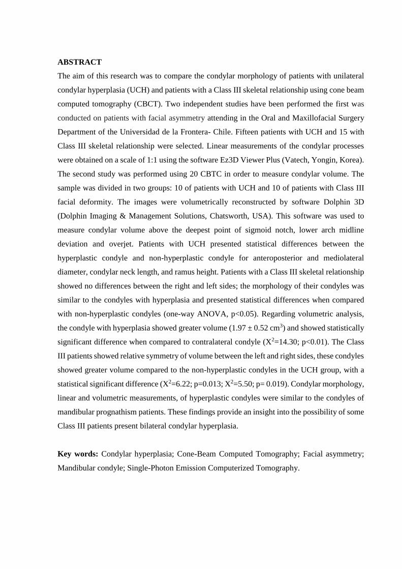
The aim of this research was to compare the condylar morphology of patients with unilateral
condylar hyperplasia (UCH) and patients with a Class III skeletal relationship using cone beam
computed tomography (CBCT). Two independent studies have been performed the first was
conducted on patients with facial asymmetry attending in the Oral and Maxillofacial Surgery
Department of the Universidad de la Frontera- Chile. Fifteen patients with UCH and 15 with
Class III skeletal relationship were selected. Linear measurements of the condylar processes
were obtained on a scale of 1:1 using the software Ez3D Viewer Plus (Vatech, Yongin, Korea).
The second study was performed using 20 CBTC in order to measure condylar volume. The
sample was divided in two groups: 10 of patients with UCH and 10 of patients with Class III
facial deformity. The images were volumetrically reconstructed by software Dolphin 3D
(Dolphin Imaging & Management Solutions, Chatsworth, USA). This software was used to
measure condylar volume above the deepest point of sigmoid notch, lower arch midline
deviation and overjet. Patients with UCH presented statistical differences between the
hyperplastic condyle and non-hyperplastic condyle for anteroposterior and mediolateral
diameter, condylar neck length, and ramus height. Patients with a Class III skeletal relationship
showed no differences between the right and left sides; the morphology of their condyles was
similar to the condyles with hyperplasia and presented statistical differences when compared
with non-hyperplastic condyles (one-way ANOVA, p<0.05). Regarding volumetric analysis,
the condyle with hyperplasia showed greater volume (1.97 ± 0.52 cm3) and showed statistically
significant difference when compared to contralateral condyle (X2=14.30; p<0.01). The Class
III patients showed relative symmetry of volume between the left and right sides, these condyles
showed greater volume compared to the non-hyperplastic condyles in the UCH group, with a
statistical significant difference (X2=6.22; p=0.013; X2=5.50; p= 0.019). Condylar morphology,
linear and volumetric measurements, of hyperplastic condyles were similar to the condyles of
mandibular prognathism patients. These findings provide an insight into the possibility of some
Class III patients present bilateral condylar hyperplasia.
Key words: Condylar hyperplasia; Cone-Beam Computed Tomography; Facial asymmetry;
Mandibular condyle; Single-Photon Emission Computerized Tomography.
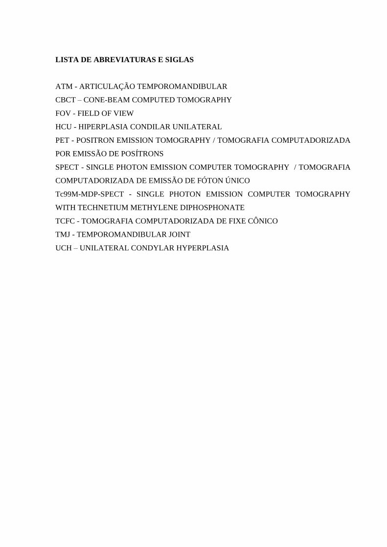
LISTA DE ABREVIATURAS E SIGLAS
ATM - ARTICULAÇÃO TEMPOROMANDIBULAR
CBCT – CONE-BEAM COMPUTED TOMOGRAPHY
FOV - FIELD OF VIEW
HCU - HIPERPLASIA CONDILAR UNILATERAL
PET - POSITRON EMISSION TOMOGRAPHY / TOMOGRAFIA COMPUTADORIZADA
POR EMISSÃO DE POSÍTRONS
SPECT - SINGLE PHOTON EMISSION COMPUTER TOMOGRAPHY / TOMOGRAFIA
COMPUTADORIZADA DE EMISSÃO DE FÓTON ÚNICO
Tc99M-MDP-SPECT - SINGLE PHOTON EMISSION COMPUTER TOMOGRAPHY
WITH TECHNETIUM METHYLENE DIPHOSPHONATE
TCFC - TOMOGRAFIA COMPUTADORIZADA DE FIXE CÔNICO
TMJ - TEMPOROMANDIBULAR JOINT
UCH – UNILATERAL CONDYLAR HYPERPLASIA
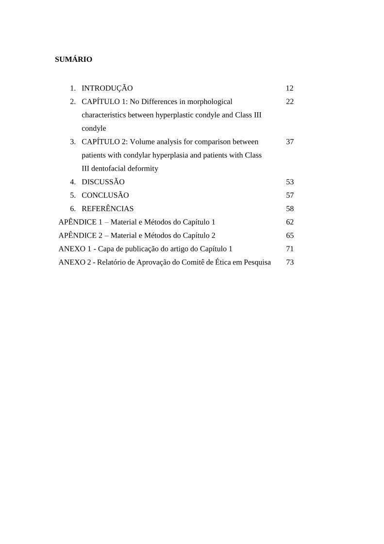
SUMÁRIO
1. INTRODUÇÃO 12
2. CAPÍTULO 1: No Differences in morphological
characteristics between hyperplastic condyle and Class III
condyle
22
3. CAPÍTULO 2: Volume analysis for comparison between
patients with condylar hyperplasia and patients with Class
III dentofacial deformity
37
4. DISCUSSÃO 53
5. CONCLUSÃO 57
6. REFERÊNCIAS 58
APÊNDICE 1 – Material e Métodos do Capítulo 1 62
APÊNDICE 2 – Material e Métodos do Capítulo 2 65
ANEXO 1 - Capa de publicação do artigo do Capítulo 1 71
ANEXO 2 - Relatório de Aprovação do Comitê de Ética em Pesquisa 73
12
1. INTRODUÇÃO
A assimetria facial é frequentemente encontrada na comunidade em geral, porém poucos
casos requerem correção cirúrgica (Jones and Tier, 2012). Nestes casos, a assimetria torna-se
uma condição aflitiva, particularmente quando esta tende a ocorrer na adolescência. Trata-se
geralmente de um problema estético e funcional uma vez que pode estar acompanhado de má-
oclusão. Grande parte das assimetrias da face tem origem no côndilo mandibular (Jonck, 1981).
O côndilo e a função mandibular são dois fatores reguladores que complementam
um ao outro durante o crescimento da face. Acreditava-se que o côndilo exercia um papel
central na regulação do crescimento mandibular, no entanto o advento da Teoria da Matriz
Funcional proporcionou o surgimento de outras teorias relacionadas ao crescimento e
desenvolvimento dessa estrutura óssea (Moss, 1997; Mota et al., 2010). O côndilo é apenas uma
das regiões que contribui localmente para o crescimento total da mandíbula, o processo de
crescimento parece estar associado à função, de forma que fatores extrínsecos regulam o
crescimento no côndilo e na mandíbula (Moss, 1997; Muñoz and Goizueta, 1999). Cerca de
98% do crescimento facial está completo na idade de 15 anos para as mulheres e 17 anos para
os homens. A taxa de crescimento durante a puberdade medida em telerradiografias de perfil
pela distância do condílio ao ponto B é em média de 1,6 mm ao ano para as mulheres e de 2,2
mm para os homens (Wolford et al., 2009).
A hiperplasia condilar é uma patologia rara, em que ocorre o crescimento anormal
do côndilo mandibular que resulta em deformidade e assimetria facial. Esta patologia foi
descrita primeiramente em língua inglesa por Robert Adams em 1836 (Norman and Painter,
1980). A hiperplasia condilar parece ter predileção pelo sexo feminino (Nitzan et al., 2008;
Villanueva-Alcojol et al., 2011) e pode ser encontrada em qualquer idade, porém mais
comumente dos 15 aos 19 anos (Hampf et al., 1985). Na maior parte dos casos não são
observados sintomas, porém podem ser encontrados sinais e sintomas como dor, ruído articular
e restrição dos movimentos mandibulares (Gray et al., 1990).
Nitzan et al. (2008) reportaram uma série de 61 casos de hiperplasia condilar ativa.
Os autores identificaram que a maior parte dos pacientes era do sexo feminino (75%), com
idade acima dos 20 anos (39 pacientes); cerca de 27,5% relatavam dor articular. A maior parte
dos pacientes apresentou assimetria no plano transversal (32), seguido do plano vertical (19) e
ambos (10). Na maioria dos pacientes o formato do côndilo estava normal (73%), porém, em
13
58% da amostra o tamanho do côndilo hiperplásico era evidentemente maior que o lado
contralateral. O pescoço condilar estava alongado em 36 pacientes e mais largo em 6 pacientes.
A etiologia da hiperplasia condilar é controversa e não é bem conhecida, são
sugeridas algumas hipóteses, como uma origem neoplásica, crescimento excessivo reparador
após trauma, resposta a infecção ou ao carregamento anormal da articulação
temporomandibular (Gray et al., 1990). Outras hipóteses incluem distúrbios da circulação
sanguínea por um aumento da vascularização subcondral (Egyedi, 1964), e a influência dos
hormônios sexuais e fatores genéticos e hereditários (Wolford et al., 2009).
A deformidade facial decorrente da hiperplasia condilar pode apresentar diversos
graus de severidade, porém classicamente foram descritos dois quadros clínicos: no primeiro
há um crescimento vertical, com alongamento do pescoço condilar e aumento vertical do ramo
mandibular, associado ao crescimento do corpo mandibular. Nos estágios iniciais esta condição
resulta em mordida aberta na região de molares e pré-molares do lado afetado e não há desvio
do mento. O segundo quadro clínico caracteriza-se pelo desvio acentuado da mandíbula para o
lado não afetado, com deslocamento do mento e da arcada dentária, resultando em mordida
cruzada. O distúrbio de crescimento parece afetar toda a hemi-mandíbula (Jonck, 1981).
Posteriormente, Obwegeser & Makek (1986) consideraram inadequado utilizar a
mesma terminologia para dois quadros clínicos distintos. Dessa forma, propuseram um sistema
de classificação, no qual descreveram duas patologias que poderiam coexistir em alguns
pacientes. A hiperplasia hemimandibular (Hemimandibular hyperplasia) referia-se ao aumento
tridimensional no tamanho da mandíbula (côndilo, pescoço condilar, ramo e corpo). Como este
crescimento geralmente começa antes da puberdade a maxila pode acompanhar o crescimento
mandibular com manutenção da oclusão e alteração do plano oclusal. O crescimento em altura
do corpo é evidenciado pelo aumento da distância entre as raízes dos dentes e o canal
mandibular. Este quadro clínico foi observado bilateralmente em pacientes com acromegalia e
pacientes com mordida profunda associada a hiperplasia de masseter.
O alongamento hemimandibular (Hemimandibular Elongation) caracteriza-se pelo
deslocamento horizontal da mandíbula, enquanto os ramos mandibulares permanecem na
mesma altura. A forma bilateral desta patologia é mais frequente, que gera um aspecto prognata
nos pacientes. Os autores afirmaram que a etiologia destas condições está associada à camada
14
de fibrocartilagem na cabeça condilar, em que pode haver uma hiperatividade e hiperprodução
de fatores de crescimento (Obwegeser and Makek, 1986).
A utilização de tomografias computadorizadas com reconstruções tridimensionais
proporcionou analisar mais precisamente a assimetria facial gerada pela hiperplasia condilar. A
partir de tomografias computadorizadas é possível determinar o grau e a localização exata da
deformidade mandibular (Walters et al., 2013). Nitzan et al. (2008) relataram que o tamanho e
forma condilar variam entre as assimetrias, de modo que a classificação deve ser baseada nos
sinais e sintomas clínicos, principalmente quanto à direção da assimetria: vertical, horizontal
ou ambas. Mutoh et al. (1991) realizaram um estudo para descrever os achados tridimensionais
em tomografias computadorizadas de seis pacientes com hiperplasia condilar. No grupo 1 foram
incluídos 3 pacientes com a combinação de características da hiperplasia e alongamento
hemimandibular, em que foi observado um discreto aumento do côndilo em um caso e um
aumento mais evidente em dois casos; para os três casos foi observado um aumento em altura
do pescoço condilar, ramo e corpo mandibular, além do deslocamento do ângulo mandibular
para baixo. No grupo II foram incluídos três pacientes com alongamento hemimandibular, o
côndilo foi discretamente maior e o pescoço condilar mais longo que o lado não afetado; o
corpo mandibular foi mais longo no lado com hiperplasia condilar. Em ambos os grupos foi
observado um aumento na espessura do osso alveolar em direção lingual (Mutoh et al., 1991).
A assimetria facial resultante da hiperplasia condilar pode ser corrigida após o
término do crescimento anormal ou ter caráter interceptativo com a remoção de parte do côndilo
ainda em crescimento, fato que limita o agravamento da deformidade (Fariña et al., 2015). Na
determinação do plano de tratamento é importante conhecer se o processo patológico está
estável ou progredindo. Para verificar a atividade do crescimento anormal do côndilo diversas
metodologias têm sido propostas, como cintilografia óssea, tomografia por emissão de fóton
único (SPECT) e tomografia por emissão de pósitrons (PET) (Gray et al., 1990; Pripatnanont
et al., 2005; Laverick et al., 2009). Um resultado positivo nestes testes representa que ainda é
esperado um crescimento assimétrico, porém não informa por quanto tempo este permanecerá,
de forma que a espera pelo cessar do crescimento anormal é imprevisível. A comparação de
telerradiografias de perfil e modelos de gesso montados em articulador podem também ser
utilizadas para verificar a atividade da doença (Robinson et al., 1990; Wolford et al., 2014).
A cintilografia óssea é um método baseado na injeção de fosfato marcado com um
radionuclídeo. Este é metabolizado pelas células do tecido ósseo dentro de 2h, e incorporado
15
em área de formação de novo osso ou em processo de remodelação. A quantidade de material
incorporado depende da taxa de atividade metabólica e/ou do fluxo sanguíneo na região, o
resultado é expresso em um filme radiográfico. O radionuclídeo mais utilizado é o tecnécio
(Tc99m) pois emite 140 keV de radiação gama, que é suficiente para detecção e tem uma meia
vida de seis horas. Este método não é específico uma vez que identifica qualquer situação de
aumento metabólico, como tumores malignos e benignos, processos inflamatórios ou de reparo
após trauma (Bittencourt et al., 2005). A hiperplasia condilar geralmente apresenta um
crescimento autolimitante, fato que não pode ser aplicado para todos os pacientes, dessa forma
a cintilografia óssea é importante na determinação da atividade da doença e do plano de
tratamento. Este método começou a ser utilizado em pacientes com hiperplasia condilar na
década de 80, é um método seguro e não invasivo (Matteson et al., 1985).
O SPECT consiste na injeção de um radiofármaco em um paciente. A emissão de
radiação deste será identificada por uma câmara gama que gira em torno do paciente gerando
imagens de diversos ângulos. Para pacientes com hiperplasia condilar inicialmente este método
utilizava como referência a captação do isótopo em outros ossos como o clivus na base do
crânio, porém a comparação direta entre o lado afetado e o lado saudável mostrou-se o método
mais confiável (Saridin et al., 2009).
Outro método de avaliação da hiperplasia condilar é o PET, baseado no
metabolismo tecidual. Um radionuclídeo é administrado por via endovenosa, este será utilizado
pelas células de acordo com o metabolismo. Para diagnostico na região de cabeça e pescoço o
radionuclídeo mais utilizado é o Flúor-18 que marca a fluordesoxiglicose, análoga da glicose.
Laverick et al. (2009) analisaram a utilização do PET em 5 pacientes com suspeita clínica de
hiperplasia condilar, os autores identificaram assimetria na captação dos côndilos normais e
hiperplásicos em todos os pacientes. Este método apresenta melhor resolução espacial que o
SPECT e a cintilografia óssea, ou seja, maior capacidade de representar detalhes. Além disso,
a fração ligada às proteínas é menor o que torna a eliminação mais rápida, com doses de
radiação menores que o SPECT.
A partir dos achados de atividade da hiperplasia condilar existem estudos que
relatam a associação entre a atividade da doença e as características histopatológicas (Gray et
al., 1990). Histologicamente o côndilo é composto por uma camada de fibrocartilagem, uma
camada proliferativa e uma camada de cartilagem hialina, esta última com o avançar da idade
é substituída por fibrocartilagem com poucos condrócitos hipertróficos. A camada de tecido
16
mole é separada do osso medular em indivíduos jovens por uma zona de reabsorção, na qual a
cartilagem é substituída por osso. Com a idade esta zona se torna uma placa óssea que isola o
osso medular da cartilagem (Slootweg and Miiller, 1986). A camada de células mesenquimais
(camada proliferativa) foi encontrada em pacientes com hiperplasia condilar após o período
esperado para seu desaparecimento, fato importante, pois esta permanece com potencial de
crescimento indefinidamente, de forma que mesmo incomum, esta patologia pode manifestar-
se em qualquer idade, como reportaram Gray et al. (1990) em um paciente com 55 anos.
Slootweg & Miller (1986) realizaram um estudo com 22 pacientes que
apresentavam HC para avaliar a existência de diferentes tipos histológicos, e correlacionar as
características histológicas com os achados da cintilografia óssea. Os autores encontraram
quatro variações histológicas. O tipo I foi caracterizado por uma camada proliferativa e de
cartilagem hialina espessa, além disso, foram encontrados remanescentes de cartilagem no osso
medular do pescoço condilar. No tipo II foi observada que a camada proliferativa estava
presente em algumas regiões, sem cartilagem hialina que foi substituída por fibrocartilagem,
foi observada uma placa óssea subcondral contínua e poucos remanescentes de cartilagem no
osso medular do pescoço condilar. No tipo III foi observada a perda da arquitetura das camadas,
presença de uma massa espessa e irregular de cartilagem hialina até a região de osso medular.
E finalmente, o tipo IV, composto por uma placa óssea subcondral contínua coberta por
fibrocatilagem pobre em células, sem a camada proliferativa e de cartilagem hialina. O tipo I
foi relacionado aos pacientes menores de 20 anos, enquanto o tipo II para os maiores,
compatível com o desenvolvimento normal em que aos 20 anos há a substituição da cartilagem
hialina por fibrocartilagem. No tipo III foi observada maior prevalência de dor articular que
poderia estar relacionada com uma origem traumática e não como uma genuína HC. Não foi
encontrada uma correlação entre o tipo histológico e os achados da cintilografia. Os restos de
cartilagem na região de pescoço condilar foram associados a presença de crescimento
ativo.(Slootweg and Miiller, 1986)
Hampf et al. (1985) encontraram correlação positiva entre os achados histológicos
e os resultados da cintilografia. Porém, Slootweeg & Miiller (1986) observaram dois casos de
falso positivo da cintilografia, em que os côndilos apresentavam alterações devido a processo
de artrite. Gray et al. (1990) realizaram um estudo com 20 pacientes com hiperplasia condilar
unilateral de forma a correlacionar achados histológicos com resultados da cintilografia e
observaram uma relação positiva entre a presença e profundidade de ilhas de cartilagem no osso

17
medular e uma maior captação do tecnécio. Assim, não está estabelecido na literatura a
associação direta entre os achados histológicos e a atividade da doença.
O tratamento da hiperplasia condilar é complexo, geralmente necessita de
tratamento ortodôntico para alinhar e descompensar os dentes, seguido de cirurgia ortognática,
utilizando osteotomias na maxila, mandíbula e no mento. Para pacientes com hiperplasia
condilar ativa a condilectomia está indicada, com eliminação de pelo menos 6 mm da parte mais
superior do côndilo (Jones and Tier, 2012). A finalização do tratamento é realizada com a
estabilização e refinamento da oclusão com a ortodontia. A correção estética pode ser realizada
pela remoção de tecido ósseo ou pela colocação de implantes aloplásticos, como de polietileno
poroso de alta densidade na região lateral da mandíbula (Jones and Tier, 2012).
Graziani (1972) reportou um caso em que realizou o tratamento em dois estágios
cirúrgicos, o primeiro por meio de acesso pré-auricular e ressecção do côndilo e o segundo para
correção do contorno mandibular, com correção da hipertrofia do corpo direito e da atrofia do
ramo esquerdo. Norman & Paiter (1980) relataram 12 casos de hiperplasia condilar. Os autores
realizaram condilectomia isolada em 6 casos, condilectomia associada a osteotomia vertical do
ramo mandibular no lado contralateral em 4 casos, condilectomia associada a osteotomia para
redução da borda inferior da mandíbula em um caso e condilectomia associado a osteotomia
sagital do ramo mandibular no lado contralateral em outro caso. No estudo, 10 pacientes
permaneceram com bloqueio maxilomandibular pós-operatório. Foi removido todo o
remanescente condilar que estivesse 5 mm acima da incisura da mandíbula, os autores relataram
bons resultados estéticos e funcionais e não observaram recidiva.
Hampf et al. (1985) em um estudo retrospectivo com 35 pacientes relataram utilizar
13 tipos de tratamento, fato explicado pela diversidade com que a deformidade facial se
manifesta, com variação da severidade da assimetria e da presença ou não do crescimento
compensatório da maxila. O que revela também a falta de um consenso na época sobre o
tratamento mais adequado dentre as opções terapêuticas existentes.
Muñoz et al. (1999) relataram um caso de HC ativa em uma paciente de 13 anos
tratado com condilectomia alta (7mm), este resultou na correção da deformidade e da oclusão.
Os autores afirmaram que a origem desta patologia está na parte mais superior do côndilo e que
pode ser corrigida pela remoção da camada de cartilagem.
18
Wolford et al. (2002) propôs um novo sistema de classificação da hiperplasia
condilar, baseado na frequência da ocorrência, no tipo da deformidade resultante e quanto ao
procedimento cirúrgico necessário. A hiperplasia condilar tipo 1 refere-se a aumento data taxa
de crescimento normal do côndilo mandibular, com manutenção da arquitetura e alongamento
do côndilo, pescoço condilar e ramo mandibular. Neste tipo, predomina o vetor de crescimento
horizontal que causa o crescimento da mandíbula para frente da maxila, expresso por meio da
relação esquelética e oclusal Classe III; o crescimento vertical também pode ocorrer. Este tipo
foi subdividido em duas categorias, o tipo 1A refere-se ao crescimento simétrico bilateral, ou
assimétrico, se a taxa de crescimento de um lado for maior. O prognatismo mandibular
decorrente desta já havia sido proposto por Rowe em 1960 (Rowe, 1960), e teoricamente
discutida por Egyedi (1964) e Obwegeser & Makek (1986), porém sem repercussão ou mais
informações na literatura internacional. O tipo 1B refere-se a forma unilateral, que é menos
comum. O crescimento começa na puberdade e avança até parte da terceira década de vida,
geralmente é autolimitante. A hiperplasia condilar tipo 2 envolve o aumento do côndilo, do
corpo mandibular, da espessura do pescoço condilar e da altura do ramo mandibular. Esta pode
ocorrer em qualquer idade e não é autolimitante, pode ser causada por tumores, como
osteocondroma e osteoma (Wolford et al., 2002).
Wolford et al. (2002) realizaram um estudo com 37 pacientes com hiperplasia
condilar tipo 1, estes foram divididos em dois grupos de acordo com o tratamento empregado.
O grupo 1 composto por 12 pacientes que foram tratados com cirurgia ortognática. O grupo 2
composto por 25 pacientes (12 pacientes com HC tipo 1A e 13 com HC tipo 1B), estes foram
tratados com condilectomia alta, ou seja, remoção de 3 a 5 mm da região mais superior do
côndilo que inclui o polo lateral e medial, associado a cirurgia ortognática. Após um período
médio de 5 anos de acompanhamento foi observada uma taxa maior de crescimento no grupo 1
(3,5 mm) que no grupo 2 (0,8mm). A função mandibular pós-operatória foi semelhante em
ambos os grupos. Todos os pacientes do grupo 1 apresentaram recidiva da Classe III esquelética
e necessitaram de uma nova intervenção cirúrgica. No grupo 2 apenas um paciente apresentou
recidiva transversal da maxila.
Wolford et al. (2009) realizaram um estudo retrospectivo com pacientes com
hiperplasia condilar tipo 1. Os resultados deste estudo são similares ao artigo publicado em
2002 (Wolford et al., 2002), porém foram acrescentados mais pacientes no grupo 2. Os critérios
de inclusão do estudo foram: HC tipo 1 baseado em uma série de avaliações clínicas e de
19
telerradiografias de perfil; condilectomia alta bilateral ou unilateral e reposicionamento do
disco articular para o grupo 2; cirurgia ortognática para correção da deformidade dentofacial;
acompanhamento mínimo de 2 anos; dados clínicos e radiográficos adequados. Os autores
utilizaram os 12 pacientes tratados com cirurgia ortognática do estudo publicado anteriormente
e 42 pacientes tratados com condilectomia alta e cirurgia ortognática. No grupo 2 apenas um
paciente teve que ser submetido a uma nova cirurgia devido a recidiva transversa da maxila. Os
autores concluíram que a condilectomia associada a cirurgia ortognática apresenta resultados
previsíveis a longo prazo (Wolford et al., 2009).
Villanueva-Alcojol et al. (2011) realizaram um estudo retrospectivo com 36
pacientes (25 mulheres e 11 homens) com assimetria facial devido a hiperplasia condilar. Em
24 pacientes o SPECT detectou uma diferença de captação superior a 10% entre o côndilo
hiperplásico e côndilo contralateral, para os demais pacientes a diferença foi qualitativa. Todos
os pacientes foram submetidos a condilectomia, com remoção de 4 a 5 mm da região mais
superior do côndilo, seguido posteriormente por tratamento ortodôntico, e para seis pacientes
cirurgia ortognática. Os pacientes apresentaram confirmação histopatológica do diagnóstico,
foi observada a persistência da camada de células mesenquimais indiferenciadas, uma camada
de cartilagem hipertrófica e ilhas de cartilagem no osso medular. Não foi observada relação
direta entre a captação do isótopo e o tipo histológico.
Não existe um consenso quanto a existência e a forma de tratamento da hiperplasia
condilar bilateral. O diagnóstico diferencial da HC tipo 1 A e B deve ser com: hipoplasia
maxilar, prognatismo mandibular que inicia na infância mantem o crescimento normal,
deslocamento do côndilo anterior a eminência articular, acromegalia, macroglossia, assimetria
facial congênita, outras patologias da articulação temporomandibular como osteoma,
osteocondroma e reabsorção condilar do lado contralateral (Wolford et al., 2009).
O prognatismo mandibular que inicia na infância mantem o crescimento normal ou
deformidade dentofacial Classe III é caracterizado pela posição mandibular mais anterior em
relação à base do crânio e/ou maxila, apresenta prevalência pequena entre os caucasianos e
maior em indivíduos asiáticos. Esta deformidade pode ser devido à retrusão maxilar ou o
prognatismo mandibular, ou pela combinação de ambos (Sugawara & Mitani, 1997). A maior
parte das más-oclusões não é resultado de um processo patológico, e sim da alteração do
desenvolvimento normal. O comprimento total da mandíbula e seu posicionamento anterior
deve ser considerado no diagnóstico diferencial (Chang et al., 2006).
20
A etiologia da deformidade dento-esquelética Classe III não está completamente
elucidada, existem fatores genéticos e hereditários, influenciados por fatores externos como:
posicionamento da língua, postura, hipertrofia das tonsilas e distúrbios endócrinos
(acromegalia, gigantismo e adenoma hipofisário), pois o hormônio do crescimento pode
reativar a região de crescimento na zona subcondilar no período pós-puberal (Chang et al.,
2006).
As alterações morfológicas dos maxilares parecem ocorrer cedo, de forma que a
taxa de crescimento mandibular e maxilar no período dos 7 aos 10 anos de idade é similar para
indivíduos Classe I e III. No período da puberdade (10-15 anos de idade), o comprimento total
da mandíbula e da região do corpo mandibular parece ser maior nos indivíduos Classe III,
porém sem diferenças clinicamente significativas, que sugere que a deformidade foi
estabelecida antes da puberdade. No período pós-puberal não foram observadas diferenças nas
estruturas esqueléticas dos indivíduos Classe I e Classe III (Sugawara and Mitani, 1997).
Aparelhos ortopédicos têm sido utilizados para a correção do prognatismo
mandibular em pacientes em fase de crescimento, mas existem diversos protocolos de
tratamento. No entanto, os resultados parecem mais favoráveis para pacientes com deficiência
de maxila (Chang et al., 2006). Em pacientes adultos com deformidade dentofacial Classe III o
tratamento de escolha é a realização de cirurgia ortognática, com tratamento ortodôntico prévio
visando a correção das compensações dentárias. O tratamento pode incluir recuo de mandíbula
associado ou não ao avanço da maxila. A osteotomia sagital do ramo mandibular e a osteotomia
vertical do ramo mandibular são duas técnicas viáveis (Chang et al., 2006). Este procedimento
é estável para pacientes que não apresentam crescimento condilar e a literatura relata uma
recidiva de 20 a 90%, que segundo Wolford et al. (2009) pode estar relacionada a HC tipo 1
não diagnosticada no pré-operatório. Estes autores propõem a associação entre condilectomia
alta e a cirurgia ortognática para estes casos. Outros fatores relacionados à recidiva são a força
muscular e a utilização ou não de fixação interna estável. Resultados mais estáveis foram
obtidos com a osteotomia sagital do ramo mandibular (Wolford et al., 2009).
Wolford et al. (2014) publicaram uma atualização do sistema de classificação, com
descrição detalhadas das características clínicas, radiográficas e histológicas. A hiperplasia
condilar do tipo 2 refere-se aos casos de osteocondroma, esta foi dividida em tipo 2A refere-se
ao crescimento vertical do côndilo e pescoço condilar, e o tipo 2B em que é observado um vetor
de crescimento horizontal. No tipo 2B o crescimento é tumoral exofítico para fora do côndilo
21
geralmente para medial. A hiperplasia condilar tipo 3 inclui tumores benignos raros, como:
osteoma, condroblastoma, osteoma osteoide, osteoblastoma, cisto ósseo aneurismático e
granuloma central de células gigantes. A hiperplasia condilar do tipo 4 inclui tumores malignos:
condrossarcoma, osteossarcoma e carcinoma metastático (Wolford et al., 2014).
Pode-se observar uma evolução nos métodos de diagnóstico e tratamento da
hiperplasia condilar, de forma que tratamentos sequenciais e padronizados têm sido
estabelecidos (Jones and Tier, 2012). A aparência facial dos pacientes com hiperplasia condilar
depende do grau de crescimento anormal e da idade de início da patologia. Se a HC iniciou
antes de finalizar o crescimento há um desenvolvimento compensatório da maxila, resultando
na inclinação do plano oclusal. Tradicionalmente a correção da assimetria é realizada com
cirurgia ortognática quando não há mais crescimento anormal, e necessita de tratamento
ortodôndico prévio. Em paciente com crescimento condilar ativo a condilectomia está indicada
de forma a minimizar a severidade da deformidade (Fariña et al., 2015).
Apesar de existirem estudos na literatura sobre morfologia e volume condilar de
pacientes com deformidades dentofaciais (Rodrigues et al., 2009; Tecco et al., 2010; Bayram
et al., 2012; Saccucci et al., 2012), não foram encontrados estudos que comparassem côndilos
hiperplásico com de pacientes com deformidade dentofacial Classe III. A hipótese deste estudo
é de que pacientes com deformidade dentofacial Classe III apresentam morfologia condilar
semelhante aos de pacientes com hiperplasia condilar.
O objetivo deste estudo foi comparar a morfologia condilar em tomografias
computadorizadas de feixe cônico de pacientes com hiperplasia condilar e pacientes com
deformidade dentofacial Classe III, por meio de medidas lineares e volumétricas.
22
2. CAPÍTULO 1
No Differences in Morphological Characteristics Between
Hyperplastic Condyle and Class III Condyle
Douglas Rangel Goulart DDS, MS1, Pablo Muñoz DDS2, Sergio Olate DDS, MS,
PhD1,2,3, Márcio de Moraes DDS, MS, PhD1, Rodrigo Fariña DDS4
1. Oral and Maxillofacial Surgery Division, Piracicaba Dental School, State
University of Campinas, Brazil
2. Division of Oral and Maxillofacial Surgery & CEMYQ, Universidad de La
Frontera, Temuco, Chile
3. Center for Biomedical Research, Universidad Autónoma de Chile, Concepcion,
Chile
4. Department of Oral and Maxillofacial Surgery, Universidad de Chile, Santiago,
Chile
International Journal of Oral and Maxillofacial Surgery
Accepted for publication 28 may 2015
23
Abstract
The aim of this research was to compare the condylar morphology of patients with unilateral
condylar hyperplasia (UCH) and patients with a Class III skeletal relationship using cone beam
computed tomography (CBCT). A prospective study was conducted on patients with facial
asymmetry attending in the divison of oral and maxillofacial of the Universidad de la Frontera-
Chile. Fifteen patients with UCH and 15 with Class III skeletal relationship were selected.
Linear measurements of the condylar processes were obtained on a scale of 1:1 using the
software Ez3D Viewer Plus (Vatech, Yongin, Korea). Analysis of variance (ANOVA) and
paired t-test were used, considering p<0.05. Patients with UCH presented statistical differences
between the hyperplastic condyle and non-hyperplastic condyle for anteroposterior and
mediolateral diameter, condylar neck length, and ramus height. Patients with a Class III skeletal
relationship showed no differences between the right and left sides; the morphology of their
condyles was similar to the condyles with hyperplasia and presented statistical differences when
compared with non-hyperplastic condyles (one-way ANOVA, p<0.05). Condylar morphology
of UCH patients could be related to the development of Class III skeletal relationship. These
findings provide an insight into the possibility of some Class III patients present bilateral
condylar hyperplasia.
Key words: Condylar hyperplasia; Facial asymmetry; Mandibular condyle.
Introduction
Unilateral mandibular condylar hyperplasia (UCH) is a complex deformity of the
condyle and the mandible that causes facial asymmetry1. UCH is diagnosed through the clinical
findings of facial asymmetry; occlusal changes should be demonstrated clinically and
radiographically, with an active hyperplasia being confirmed by a bone scan performed at the
initial diagnosis and repeated at least 6 months later 2.
Condylar growth activity has traditionally been assessed by planar scintigraphy
with technetium methylene diphosphonate; however this method lacked anatomical precision.
Tc99m-MDP-SPECT (single photon emission computer tomography with technetium
methylene diphosphonate) has the capability of three-dimensional (3D) reconstruction and
subsequent thin sectioning 1. The information is typically presented as cross-sectional slices
through the patient, but can be freely reformatted or manipulate as required.
24
Classically, condylar hyperplaisa has been described as a unilateral pathology, but
this could be related to the facility of identifying the facial asymmetry produced by differences
in the sizes of the condyles3. Another factor is the difficulty in establishing the abnormal size
when both condyles presented hyperplasia, given that a certain asymmetry is normal to all
human body structures4. Moreover, the diagnostic tools to assess condylar growth are based on
the percentage differences in isotope uptake, which is higher on the condyle with hyperplasia5.
Finally, the comparison between hyperplastic condyle and non-hyperplastic condyle is one the
most important factors for obtaining a final diagnosis of UCH. Thus, the use SPECT and
scintillography has no value to identify the activity of disease in cases of bilateral condylar
hyperplasia6.
In contrast to most studies in the literature, there is a hypothesis that some patients
with a Class III occlusal and skeletal relationship present a bilateral condylar hyperplasia, called
condylar hyperplasia type IA, as the primary cause 6,7. From a biological point of view, it is
possible; the concept of hyperplasia is an abnormal growth of cells, and hypothetically this
could affect both sides.
The purpose of this study was to compare the temporomandibular joint (TMJ)
morphology of patients with UCH and patients with a Class III skeletal relationship using cone
beam computed tomography (CBTC).
Materials and Methods
This prospective study was conducted at the Division of Oral and Maxillofacial
Surgery of the Universidad de La Frontera (Temuco - Chile). A total of 30 patients, 15
consecutive patients with facial asymmetry related to UCH and 15 consecutive patients with a
Class III facial deformity, aged between 15 and 30 years, were assessed between January 2011
and June 2014. These patients had presented for the surgical correction of mandibular of facial
deformity. The study was conducted according to the recommendations of research involving
human beings and was approved by the Ethics Committee in Research of the Universidad de
La Frontera.
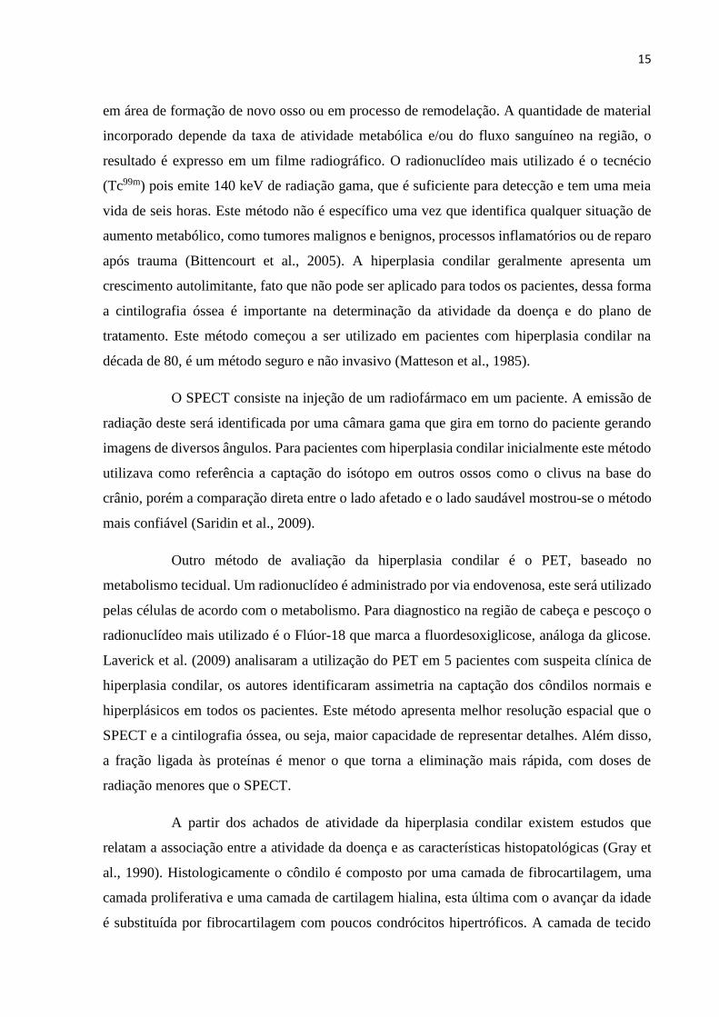
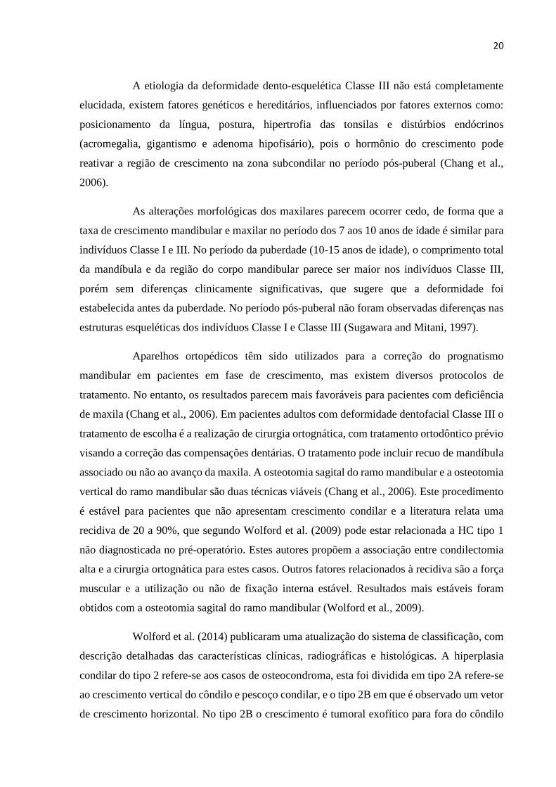
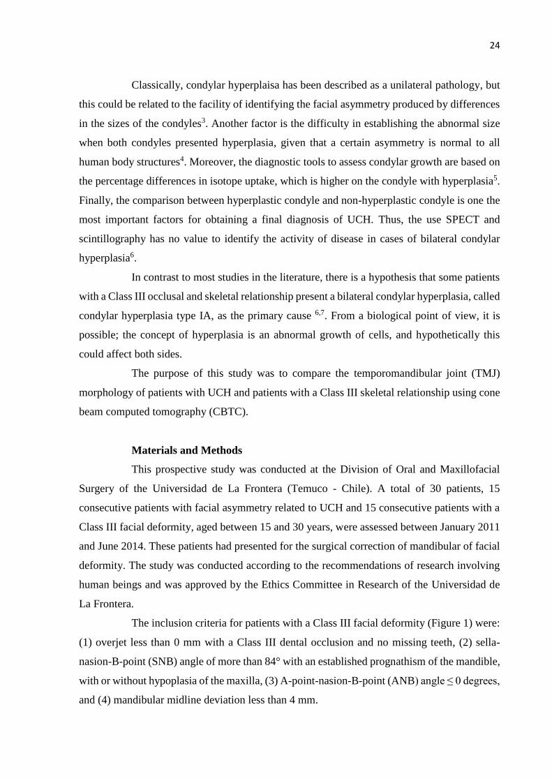
The inclusion criteria for patients with a Class III facial deformity (Figure 1) were:
(1) overjet less than 0 mm with a Class III dental occlusion and no missing teeth, (2) sella-
nasion-B-point (SNB) angle of more than 84° with an established prognathism of the mandible,
with or without hypoplasia of the maxilla, (3) A-point-nasion-B-point (ANB) angle ≤ 0 degrees,
and (4) mandibular midline deviation less than 4 mm.
25
Figure 1 – CBCT image of a Class III patient included in this research showing symmetry
between right and left condyles
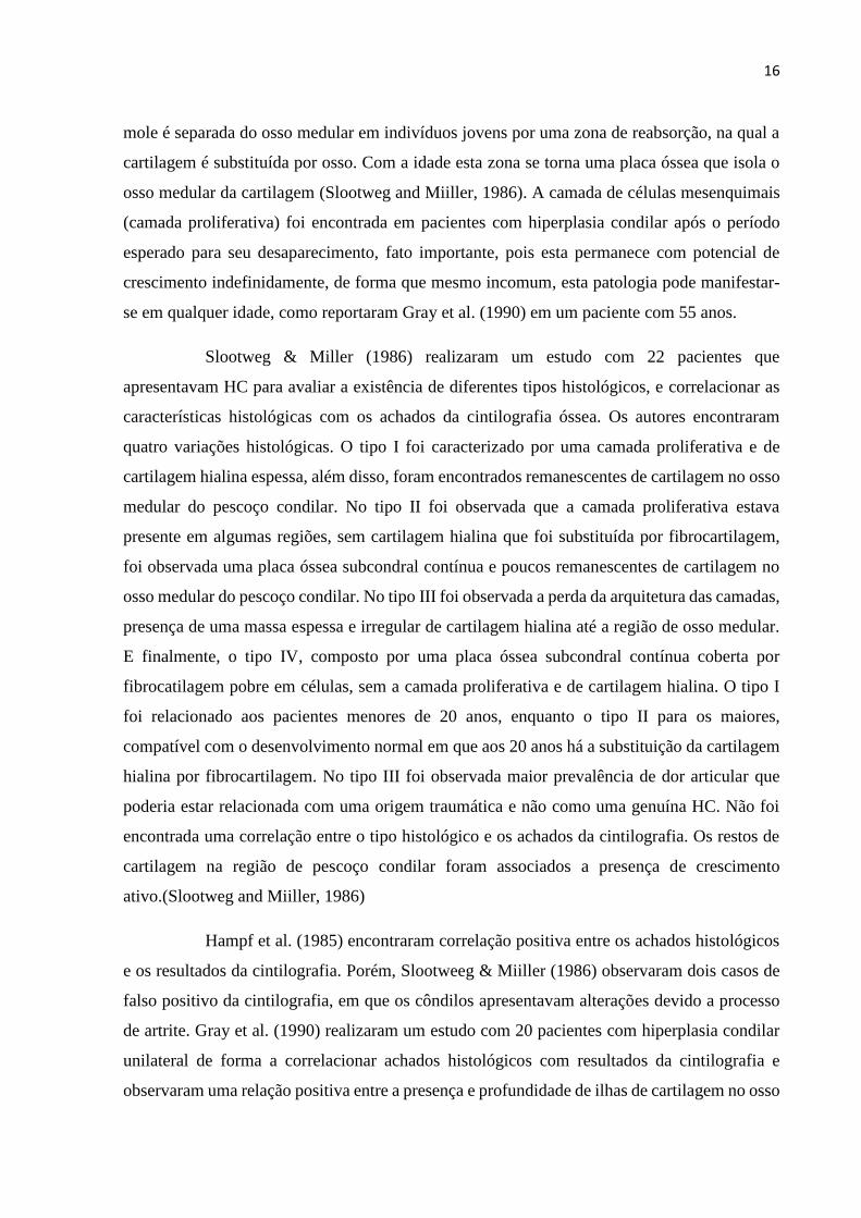
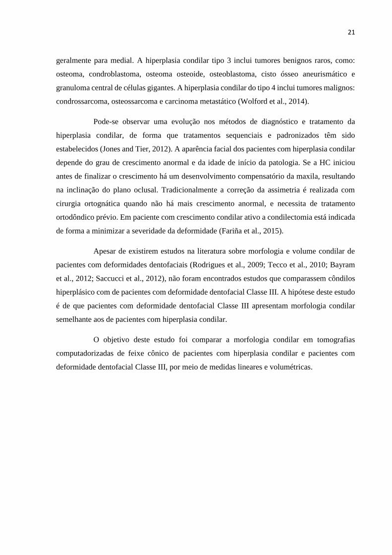
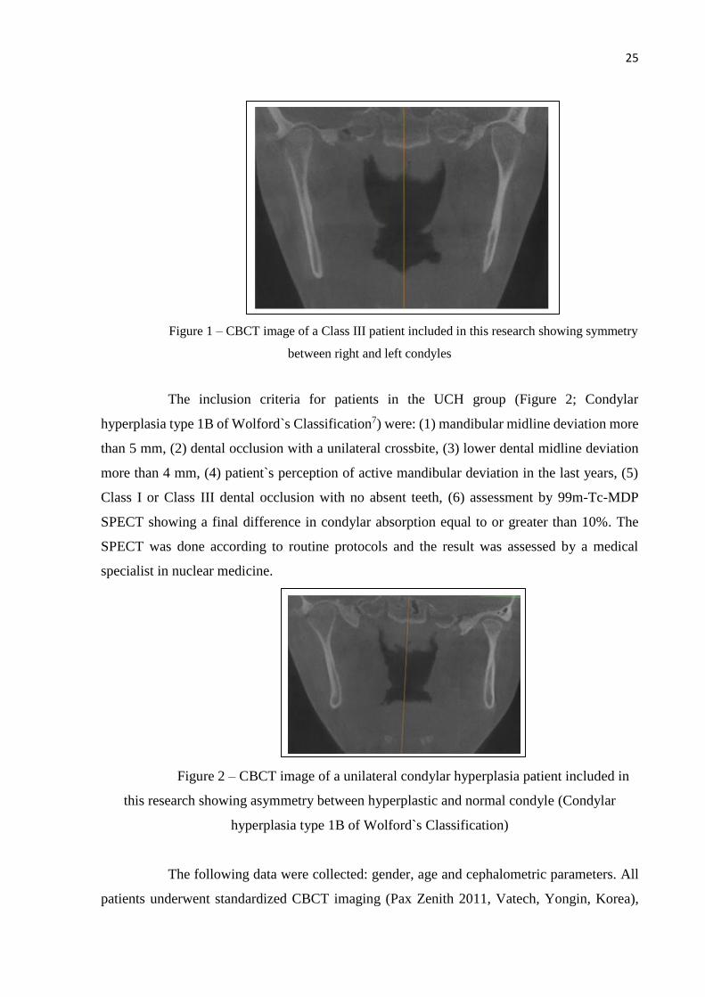
The inclusion criteria for patients in the UCH group (Figure 2; Condylar
hyperplasia type 1B of Wolford`s Classification7) were: (1) mandibular midline deviation more
than 5 mm, (2) dental occlusion with a unilateral crossbite, (3) lower dental midline deviation
more than 4 mm, (4) patient`s perception of active mandibular deviation in the last years, (5)
Class I or Class III dental occlusion with no absent teeth, (6) assessment by 99m-Tc-MDP
SPECT showing a final difference in condylar absorption equal to or greater than 10%. The
SPECT was done according to routine protocols and the result was assessed by a medical
specialist in nuclear medicine.
Figure 2 – CBCT image of a unilateral condylar hyperplasia patient included in
this research showing asymmetry between hyperplastic and normal condyle (Condylar
hyperplasia type 1B of Wolford`s Classification)
The following data were collected: gender, age and cephalometric parameters. All
patients underwent standardized CBCT imaging (Pax Zenith 2011, Vatech, Yongin, Korea),
26
with setting of 90 kV and 12 mA, voxel size 0.12 mm, 1mm cuts in a 190 mm x 240 mm
window, which recorded the condition and morphology of the bilateral TMJ. The computed
tomography images were obtained with the patients in maximum dental intercuspation and the
head positioned with the Frankfort plane parallel to the floor.
Linear measurements of the condylar processes were obtained on a scale of 1:1
using software Ez3D Viewer Plus (Vatech). The measurements using a previously used
method3. The measurements are described in Table 1 and shown in Figures 3-5. Two further
images were obtained for each measurement, immediately before and immediately after (1 mm
cuts) the middle area, calculated in 3D reconstruction (the middle point was obtained in sagittal,
coronal and axial views). A final number was obtained from the average of the three
measurements.
The data were analyzed using descriptive and correlational statistics with the
support of the software SPSS v. 18.0 for Windows (SPSS Inc, Chicago, IL). The paired
Student’s t-tests were used for each measurement, to evaluate the average of the differences
between the sides for each element of the sample. The Shapiro-Wilk test was performed and
the sample was shown to be normally distributed. Levene’s test was applied and showed
homogeneity between the variances. The intraclass correlation coefficient (ICC) was used to
assess method error. Every measurement was performed by one operator and repeated twice at
an interval of one week. The correlation coefficient between the measurements of the first and
second tracings was 0.99 and had a p value of 0.001. One-way analysis of variance (ANOVA)
and Tukey’s post hoc test were used to compare the measurements of patients with condylar
hyperplasia and Class III facial deformity, with the results being significant at P<0.05.
27
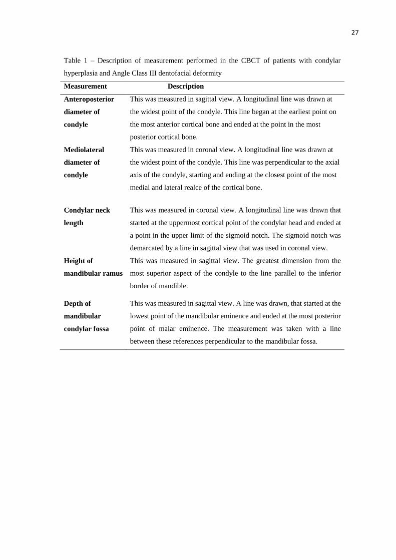
Table 1 – Description of measurement performed in the CBCT of patients with condylar
hyperplasia and Angle Class III dentofacial deformity
Measurement Description
Anteroposterior
diameter of
condyle
This was measured in sagittal view. A longitudinal line was drawn at
the widest point of the condyle. This line began at the earliest point on
the most anterior cortical bone and ended at the point in the most
posterior cortical bone.
Mediolateral
diameter of
condyle
This was measured in coronal view. A longitudinal line was drawn at
the widest point of the condyle. This line was perpendicular to the axial
axis of the condyle, starting and ending at the closest point of the most
medial and lateral realce of the cortical bone.
Condylar neck
length
This was measured in coronal view. A longitudinal line was drawn that
started at the uppermost cortical point of the condylar head and ended at
a point in the upper limit of the sigmoid notch. The sigmoid notch was
demarcated by a line in sagittal view that was used in coronal view.
Height of
mandibular ramus
This was measured in sagittal view. The greatest dimension from the
most superior aspect of the condyle to the line parallel to the inferior
border of mandible.
Depth of
mandibular
condylar fossa
This was measured in sagittal view. A line was drawn, that started at the
lowest point of the mandibular eminence and ended at the most posterior
point of malar eminence. The measurement was taken with a line
between these references perpendicular to the mandibular fossa.
28
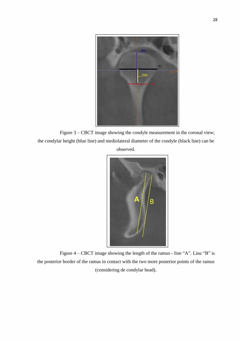
Figure 3 – CBCT image showing the condyle measurement in the coronal view;
the condylar height (blue line) and mediolateral diameter of the condyle (black line) can be
observed.
Figure 4 – CBCT image showing the length of the ramus - line “A”. Line “B” is
the posterior border of the ramus in contact with the two more posterior points of the ramus
(considering de condylar head).
29
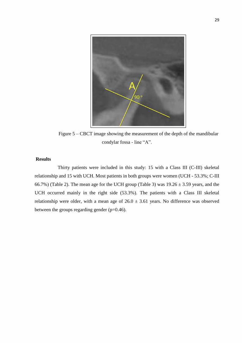
Figure 5 – CBCT image showing the measurement of the depth of the mandibular
condylar fossa - line “A”.
Results
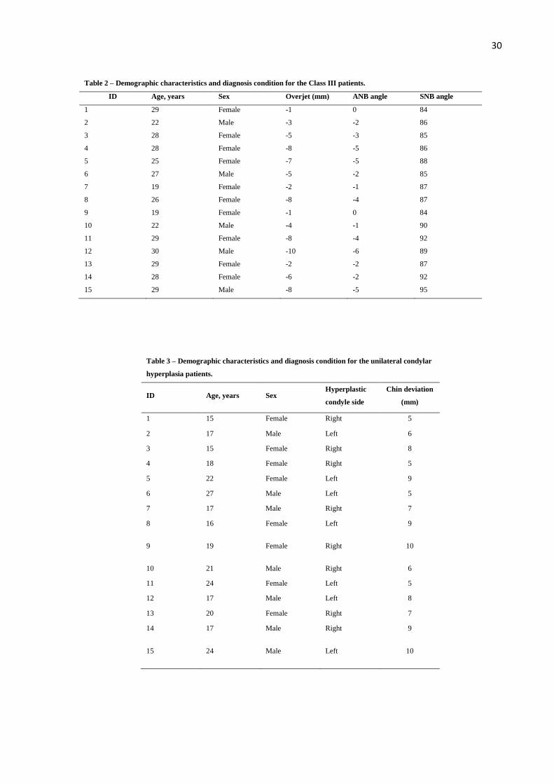
Thirty patients were included in this study: 15 with a Class III (C-III) skeletal
relationship and 15 with UCH. Most patients in both groups were women (UCH - 53.3%; C-III
66.7%) (Table 2). The mean age for the UCH group (Table 3) was 19.26 ± 3.59 years, and the
UCH occurred mainly in the right side (53.3%). The patients with a Class III skeletal
relationship were older, with a mean age of 26.0 ± 3.61 years. No difference was observed
between the groups regarding gender (p=0.46).
30
Table 2 – Demographic characteristics and diagnosis condition for the Class III patients.
ID Age, years Sex Overjet (mm) ANB angle SNB angle
1 29 Female -1 0 84
2 22 Male -3 -2 86
3 28 Female -5 -3 85
4 28 Female -8 -5 86
5 25 Female -7 -5 88
6 27 Male -5 -2 85
7 19 Female -2 -1 87
8 26 Female -8 -4 87
9 19 Female -1 0 84
10 22 Male -4 -1 90
11 29 Female -8 -4 92
12 30 Male -10 -6 89
13 29 Female -2 -2 87
14 28 Female -6 -2 92
15 29 Male -8 -5 95
Table 3 – Demographic characteristics and diagnosis condition for the unilateral condylar
hyperplasia patients.
ID Age, years Sex Hyperplastic
condyle side
Chin deviation
(mm)
1 15 Female Right 5
2 17 Male Left 6
3 15 Female Right 8
4 18 Female Right 5
5 22 Female Left 9
6 27 Male Left 5
7 17 Male Right 7
8 16 Female Left 9
9 19 Female Right 10
10 21 Male Right 6
11 24 Female Left 5
12 17 Male Left 8
13 20 Female Right 7
14 17 Male Right 9
15 24 Male Left 10
31
In the group of patients with UCH, the condyle with hyperplasia showed greater
measurements for all the variables. Statistically significant differences between the condyle
with hyperplasia and contralateral side were observed for anteroposterior diameter (p=0.014),
mediolateral diameter (p<0.001), height of condylar neck (p=0.001) and height of mandibular
ramus (p=0.004). The differences in the depth of the condylar fossa between the right and left
sides was not statistically significant.
The Class III patients showed symmetry between the left and right sides for the
following measurements: anteroposterior diameter of the condyle, mediolateral diameter of the
condyle, condylar neck length and height of mandibular ramus. No statistically significant
difference was observed between the two sides in the depth of the condylar mandibular fossa
(p=0.48).
With respect to the non-hyperplastic condyles in the UCH group, there was a
significant difference in the mediolateral diameter compared to the Class III right condyle
(p=0.009), Class III left condyle (p=0.003), and condyle with hyperplasia (p<0.001).
Additionally, they presented a shorter condylar neck length compared to the Class III condyle
(p=0.02) and condyle with hyperplasia (p=0.01). No statistical difference was observed in the
anteroposterior diameter of the condyle, the height of the mandibular ramus or the depth of the
condylar fossa when compared with the Class III condyle.
The condyle morphology data are presented in Table 4.

32
Table 4 – Results of measurement performed in the CBCT of patients with hyperplasia condylar and dentofacial deformity Class
III of Angle
Condylar Hyperplasia Class III of Angle
Condyle with
Hyperplasia
(Median/ SD)
Normal
Condyle
(Median/
SD)
*p
Right
Condyle
(Median/
SD)
Left
Condyle
(Median/
SD)
**p
One-Way
ANOVA***
Measurement F P
Anteroposterior
diameter of condyle
9.57 ±1.05 8.86 ±0.98 0.014 9.18 ±0.63 9.31±0.62 0.328 1.76 0.165
Mediolateral diameter
of condyle
17.34±1.49 A 13.95±2.43 B 0.000 16.58±2.18A 16.91±2.51 A 0.369 7.29 0.000
Condylar neck length 20.12±2.92 A 16.45 ±3.58 B 0.001 19.98±3.45 A 19,40±3.21 AB 0.147 4.06 0.011
Height of mandibular
ramus
65.15± 6.67 59.85±4.72 0.004 63.08± 6.43 62.46±6.45 0.299 1.89 0.142
Depth of mandibular
condylar fossa
14.26±1.60 13.21±2.04 0.064 13.75±1.53 13.04±1.28 0.480 1.71 0.174
*Paired sample t-test between condyles with and without hyperplasia
** Paired sample t-test between right and left condyles in patients with Class III facial deformity
*** One-way ANOVA and Tukey’s Post Hoc test
Discussion
Condylar hyperplasia includes conditions that create excessive growth and
enlargement of the condyle, which can cause alterations in the bone architecture of the
mandible, malocclusion and dentofacial deformity 5. Obwegeser and Makek (1986) reported
that condylar hyperplasia was always observed to be unilateral; however, they stated that
theoretically a bilateral case could exist8.
The literature indicates that condylar hyperplasia affects the condylar morphology
as well other areas of the mandible as well as anatomical sites adjacent to the articular fossa9.
Classically, condylar hyperplasia is reported as unilateral; however, if both condyles present
hyperplasia, it could represent an overgrowth leading to the development of a mandibular
prognathism and Class III dentofacial deformity10.
The mandible and the TMJ can be loaded differently in people with diverse
dentofacial morphologies, and one could hypothesize that the condyle and the fossa might differ
in shape between people with various malocclusions10. Angle Class II has been associated with
some degree of condylar remodeling11. Another study has shown statistically significant
33
differences in the length of the condylar process between Class II and Class III patients,
revealing this anatomical structure to be longer in Class III cases 12.
The Class III patients in this study showed symmetry between the right and left
condyle. The lack of asymmetry between the measurements is similar to that reported
elsewhere, in which a different methodology was applied for different types of malocclusions
13.
In this research the condyles with hyperplasia were found to be very similar in linear
dimensions when compared with the condyles of adult patients with Class III skeletal
relationship. With these findings, it is possible that patients with a Class III skeletal relationship
are presenting an overgrowth of both condyles. In the literature, this was recently named
condylar hyperplasia type IA7. Wolford et al.6,7 have shown that this pathology usually begins
as a Class I or mild to moderate Class III occlusion and skeletal relationship, which develops
into a worse Class III relationship as the pathological process progress.
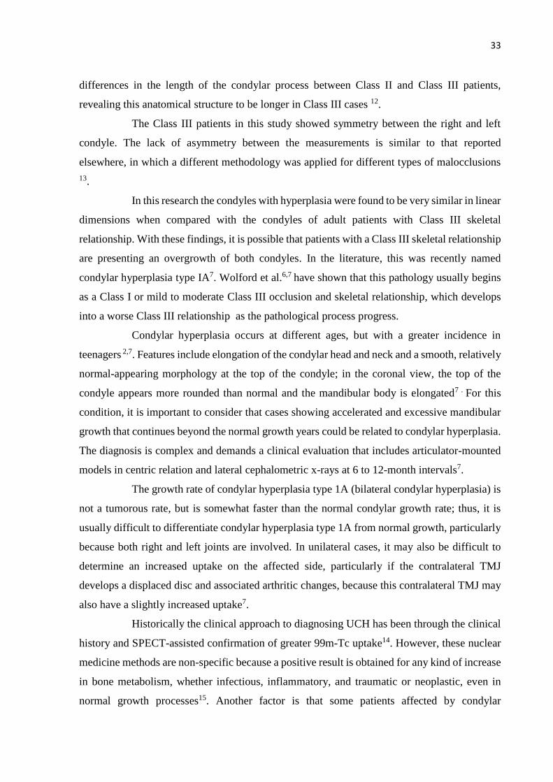
Condylar hyperplasia occurs at different ages, but with a greater incidence in
teenagers 2,7. Features include elongation of the condylar head and neck and a smooth, relatively
normal-appearing morphology at the top of the condyle; in the coronal view, the top of the
condyle appears more rounded than normal and the mandibular body is elongated7 . For this
condition, it is important to consider that cases showing accelerated and excessive mandibular
growth that continues beyond the normal growth years could be related to condylar hyperplasia.
The diagnosis is complex and demands a clinical evaluation that includes articulator-mounted
models in centric relation and lateral cephalometric x-rays at 6 to 12-month intervals7.
The growth rate of condylar hyperplasia type 1A (bilateral condylar hyperplasia) is
not a tumorous rate, but is somewhat faster than the normal condylar growth rate; thus, it is
usually difficult to differentiate condylar hyperplasia type 1A from normal growth, particularly
because both right and left joints are involved. In unilateral cases, it may also be difficult to
determine an increased uptake on the affected side, particularly if the contralateral TMJ
develops a displaced disc and associated arthritic changes, because this contralateral TMJ may
also have a slightly increased uptake7.
Historically the clinical approach to diagnosing UCH has been through the clinical
history and SPECT-assisted confirmation of greater 99m-Tc uptake14. However, these nuclear
medicine methods are non-specific because a positive result is obtained for any kind of increase
in bone metabolism, whether infectious, inflammatory, and traumatic or neoplastic, even in
normal growth processes15. Another factor is that some patients affected by condylar
34
hyperplasia might show positive SPECT scans unrelated to the clinical progression of the
asymmetry5. Additionally, it is difficult to correlate SPECT with other clinical and histological
findings due to its great variability15.
Although bone scintigraphy and SPECT have been used to evaluate the active
growth in UCH, this has no value in cases of bilateral condylar hyperplasia6. Condylar SPECT
may detect active growth when the left condyle is compared to the right condyle, so it is
necessary for one condyle to show an overgrowth of the fibrocartilage layer16.
Facial asymmetry could be related to dimensional changes in the condyle with
hyperplasia; it is hypothesized that morphological and functional changes in both condyles
could determine facial deformities3. The question that remains as a result of these findings is:
If bilateral condylar hyperplasia exists, when should the patients with Class III skeletal
relationship receive surgical treatment? For UCH, surgical treatment is undertaken before
completion of the abnormal growth. If this growth is allowed to proceed, it exacerbates the
facial deformity, asymmetry, and causes dental compensations, affecting the dentoskeletal
development and producing excessive soft tissue development. This may increase the
difficulties in obtaining optimal functional and aesthetic results6.
So when a teenager presents with a mandibular prognathism related to bilateral
condylar hyperplasia, should this situation be treated like UCH? Wolford el al. 6,7 have reported
a protocol related to hyperplastic condyle condition and this could be a response for this matter.
The results of this research provide preliminary information to suggest that some
patients with Class III skeletal relationship could present a morphology similar to UCH. Further
studies are required to provide a better understanding of this biological condition with bilateral
overgrowth of the condyle unit. These results constitute an objective comparison between
condyles involved in UCH and those involved in mandibular prognathism.
References
1. Pripatnanont P, Vittayakittipong P, Markmanee U, Thongmak S, Yipintsoi T.:
The use of SPECT to evaluate growth cessation of the mandible in unilateral condylar
hyperplasia. Int J Oral Maxillofac Surg. 34:364–8, 2005.
2. Nitzan DW, Katsnelson A, Bermanis I, Brin I, Casap N.: The clinical
characteristics of condylar hyperplasia: experience with 61 patients. J Oral Maxillofac Surg.
66:312–8, 2008.

35
3. Olate S, Cantín M, Alisten JP, Uribe F, Navarro P, Olate G, Moraes M. Relacion
Entre el Tamano Condilar y la Asimetria Facial Transversal en Individuos con Hiperplasia
Condilar. Int J Morphol. 31:937–941, 2013.
4. Tecco S, Saccucci M, Nucera R, Polimeni A, Pagnoni M, Cordasco G, Festa F,
Iannetti G.: Condylar volume and surface in Caucasian young adult subjects. BMC Med
Imaging. 10:28, 2010.
5. Saridin CP, Raijmakers PGHM, Shamma S Al, Tuinzing DB, Becking a G.:
Comparison of different analytical methods used for analyzing SPECT scans of patients with
unilateral condylar hyperactivity. Int J Oral Maxillofac Surg. 38:942–6, 2009.
6. Wolford LM, Morales-Ryan C a, García-Morales P, Perez D.: Surgical
management of mandibular condylar hyperplasia type 1. Proc (Bayl Univ Med Cent).22:321–
9, 2009.
7. Wolford LM, Movahed R, Perez DE.: A Classification system for conditions
causing condylar hyperplasia. J Oral Maxillofac Surg.72:567–595, 2014.
8. Obwegeser HL, Makek MS.: Hemimandibular hyperplasia--hemimandibular
elongation. J Maxillofac Surg14:183–208, 1986.
9. Olate S, Almeida A, Alister JP, Navarro P, Netto HD, Moraes M de.: Facial
asymmetry and condylar hyperplasia: considerations for diagnosis in 27 consecutives patients.
Int J Clin Exp Med. 6:937–941, 2013.
10. Katsavrias EG, Halazonetis DJ.: Condyle and fossa shape in Class II and Class
III skeletal patterns: A morphometric tomographic study. Am J Orthod Dentofac Orthop.
128:337–346, 2005.
11. Saccucci M, D’Attilio M, Rodolfino D, Festa F, Polimeni A, Tecco S.: Condylar
volume and condylar area in Class I, Class II and Class III young adult subjects. Head Face
Med. 8:34, 2012.
12. Krisjane Z, Urtane I, Krumina G, Zepa K.: Three-dimensional evaluation of
TMJ parameters in Class II and Class III patients. Stomatologija. 11:32–6, 2009.
13. Rodrigues AF, Fraga MR, Vitral RWF.: Computed tomography evaluation of
the temporomandibular joint in Class II Division 1 and Class III malocclusion patients: condylar
symmetry and condyle-fossa relationship. Am J Orthod Dentofacial Orthop. 136:199–206,
2009.
36
14. Saridin CP, Raijmakers P, Becking AG.: Quantitative analysis of planar bone
scintigraphy in patients with unilateral condylar hyperplasia. Oral Surg Oral Med Oral Pathol
Oral Radiol Endod. 104:259–63, 2007.
15. Fariña R, Becar M, Plaza C, Espinoza I, Franco ME.: Correlation between single
photon emission computed tomography, AgNOR count, and histomorphologic features in
patients with active mandibular condylar hyperplasia. J Oral Maxillofac Surg. 69:356–61,
2011.
16. Robinson PD, Harris K, Coghlan KC, Altman K.: Bone scans and the timing of
treatment for condylar hyperplasia. Int J Oral Maxillofac Surg. 19:243–6, 1990.
37
3. CAPÍTULO 2
Volume analysis for comparison between patients with condylar
hyperplasia and patients with Class III dentofacial deformity
Douglas Rangel Goulart DDS, MS1, Pablo Muñoz DDS2, Márcio de Moraes DDS, MS, PhD1,
Sergio Olate DDS, MS, PhD1,2,3
1. Division of Oral and Maxillofacial Surgery, State University of Campinas, Brazil
2. Division of Oral and Maxillofacial Surgery & CEMYQ, Universidad de La Frontera, Temuco,
Chile
3. Center for Biomedical Research, Universidad de Autónoma de Chile, Temuco, Chile
38
Abstract
Aim: To compare the condylar volume of patients with unilateral condylar hyperplasia (UCH)
and patients with Class III skeletal relationship. Methods: A total of 20 Cone Beam Computed
Tomography (CBCT) images of patients were analyzed. The images were divided into two
groups: 10 from patients with facial asymmetry and 10 from patients with a Class III facial
deformity. Patients were aged between 15 and 30 years. The volumetric data were reconstructed
using Dolphin 3D® software (Dolphin Imaging & Management Solutions, Chatsworth, USA).
This software measured condylar volume above the deepest point of the sigmoid notch, lower
arch midline deviation and overjet. Results: The condyle with hyperplasia showed greater
volume (1.97 ± 0.52 cm3) and showed statistically significant differences compared to the
contralateral condyle (X2=14.30; p<0.01). The Class III patients showed relative symmetry of
volume between the left and right sides; these condyles showed greater volume compared to
the non-hyperplastic condyles in the UCH group with a statistical significant difference
(X2=6.22; p=0.013; X2=5.50; p=0.019). Conclusion: Hyperplastic condyles were similar in
terms of volume to condyles of patients with mandibular prognathism, suggesting that some
patients with Class III skeletal relationship could present bilateral condylar hyperplasia.
Keywords: Cone-Beam Computed Tomography; Facial asymmetry; Mandibular
condyle; Prognathism.
39
Introduction
Unilateral condylar hyperplasia (UCH) is a complex deformity of the condyle and the
mandible that leads to facial asymmetry. It occurs at any age; it does not stop at the end of the
growth period and is more prevalent in females.1 In cases of persistent growth associated with
progressive facial asymmetry, interceptive surgery to remove the hyperplastic condyle by high
condylectomy may help to limit the deformity.2
The first step in managing cases with condylar hyperplasia is assessing condylar
growing activity. Planar scintigraphy with 99 m technetium methylene diphosphonate (Tc-
MDP) has been used to assess growth activity in the condyle but it lacks anatomical precision.
MDP-SPECT (single photon emission computer tomography) has the capability of 3D
reconstruction and subsequent thin sectioning.2 The use of cone beam computed tomography
(CBCT) is widely related in temporomandibular joint (TMJ) studies so that its use combined
with SPECT may reveal the patient’s condition more clearly regarding hard tissue.3
There is a hypothesis that some patients with a Class III occlusal and skeletal
relationship present a bilateral condylar hyperplasia, called Condylar hyperplasia type 1A, as
the primary cause.4 Condylar hyperplasia type 1A is the most frequently occurring form and
involves an accelerated growth rate of the “normal” growth mechanism of the mandibular
condyle with relatively normal architecture of the condyle, but elongation of the condylar head,
neck and mandibular body. This type, with a predominant horizontal growth vector, causes the
mandible to grow forward to the maxilla, creating a Class III occlusal and skeletal relationship.5
A preliminary study suggested that some patients with Class III skeletal relationship could
present condylar morphology similar to hyperplastic condyles.6 Therefore, the purpose of the
present study was to compare the condylar volume of patients with UCH and patients with Class
III skeletal relationship.
40
Materials and methods
This retrospective study was conducted at the Division of Oral and Maxillofacial
Surgery at the Universidad de la Frontera, Temuco, Chile. A total of 20 CBCT images from
patients were analyzed. The patients were attended between January 2011 and June 2015. These
patients had consulted for the surgical correction of dentofacial deformity. The CBCT images
were divided into two groups: 10 from patients with facial asymmetry and 10 from patients
with a Class III facial deformity. The patients in the sample were aged between 15 and 30 years.
The study was conducted according to the recommendations for research involving human
beings and was approved by the local Ethics in Research Committee.
The inclusion criteria for patients with a Class III facial deformity were: (1) overjet less
than 0 mm with a Class III dental occlusion and no missing teeth, (2) sella-nasion-B-point angle
(SNB) equal or more than 84° with an established prognathism of the mandible, with or without
hypoplasia of the maxilla, (3) A-point-nasion-B-point (ANB) angle ≤ 0 degrees, and (4)
mandibular midline deviation less than 4 mm.
The inclusion criteria for patients in the UCH group (Condylar hyperplasia type 1B from
Wolford’s Classification) were: (1) mandibular midline deviation more than 5 mm, (2) dental
occlusion with a unilateral crossbite, (3) lower dental midline deviation more than 4 mm, (4)
patient’s perception of active mandibular deviation in the last years, (5) Class I or Class III
dental occlusion with no absent teeth, (6) assessment by 99m-Tc-MDP SPECT showing a final
difference in condylar absorption equal to or greater than 10%. The SPECT was done according
to routine protocols and the result was assessed by a medical specialist in nuclear medicine.
All patients underwent standardized CBCT imaging (Pax Zenith 2011, Vatech, Yongin,
Korea), with a setting of 90 kVp and 12 mA, voxel size 0.12 mm, 1mm sections in a 190 mm x
240 mm of field of view (FOV), which recorded the condition and morphology of the bilateral
41
TMJ. The computed tomography images were obtained with the patients in maximum dental
intercuspation.
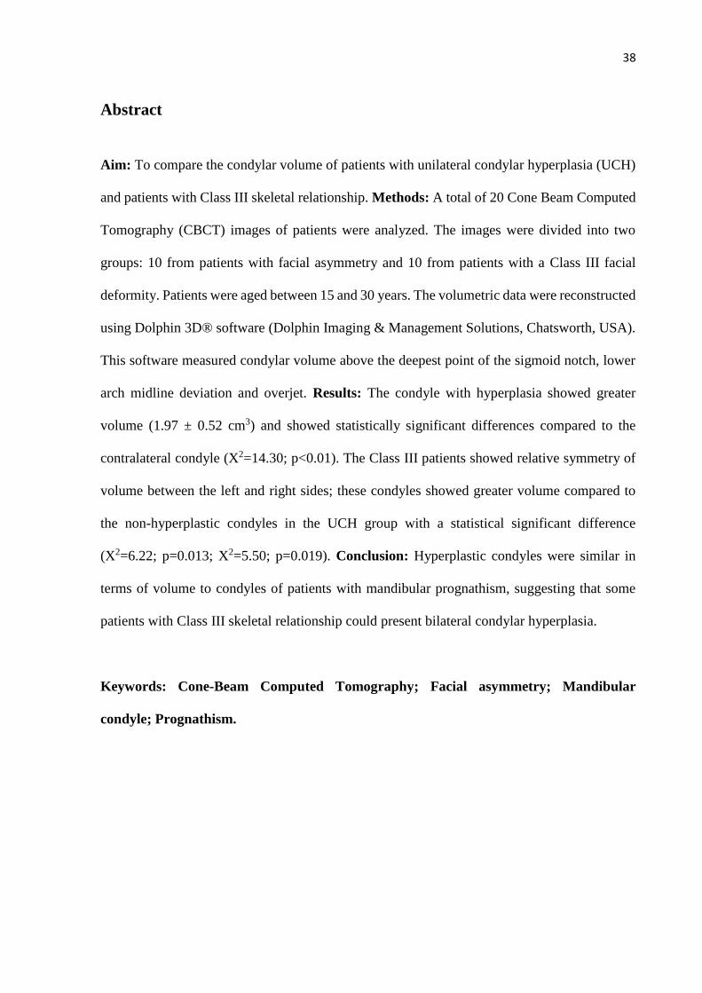
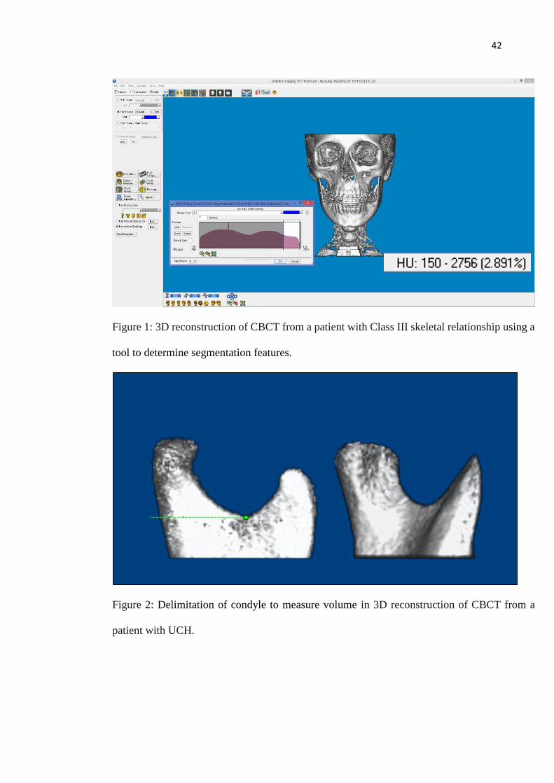
A single observer evaluated 20 CBCT images; the volumetric data were reconstructed
using Dolphin 3D® software (Version 11.5, Dolphin Imaging & Management Solutions,
Chatsworth, USA). The 3D reconstruction of head was positioned with the Frankfort plane
parallel to the horizontal plane. The midsagittal plane was perpendicular to horizontal plane.
The segmentation features was adjusted for gray scale that allowed visualize condyle, between
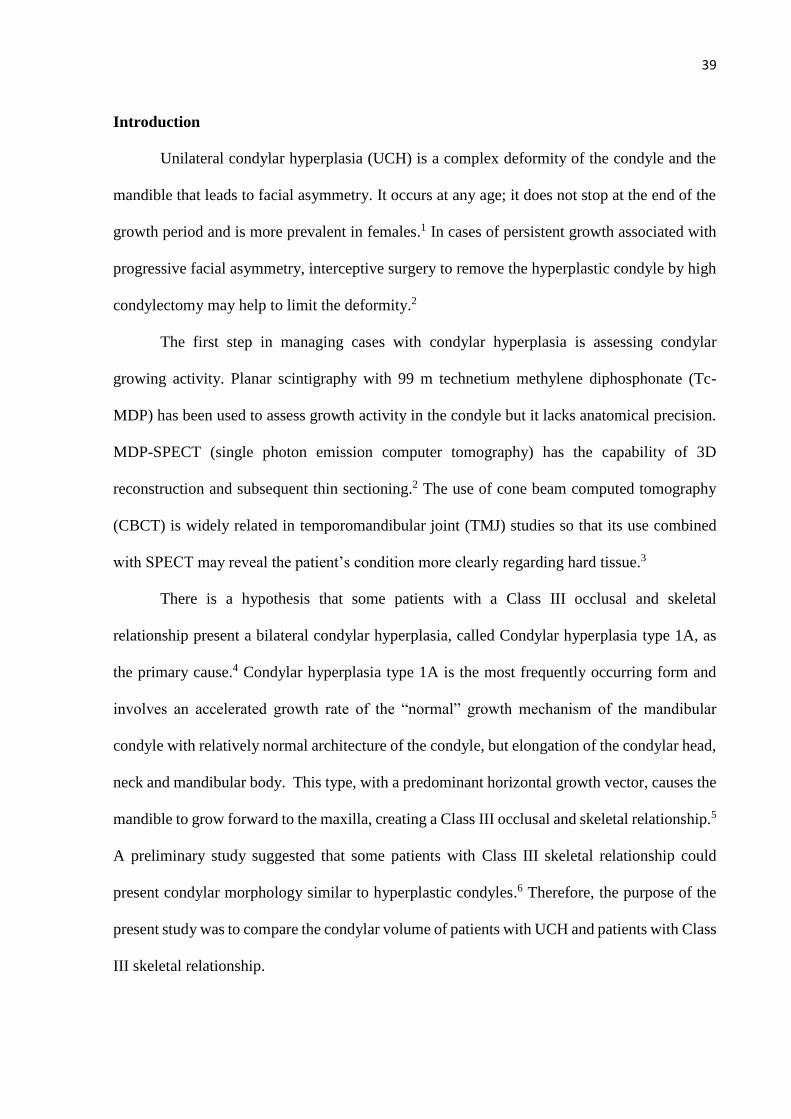
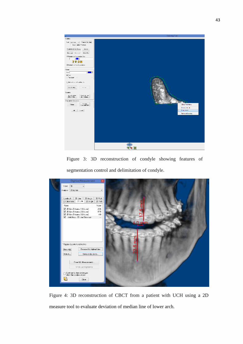
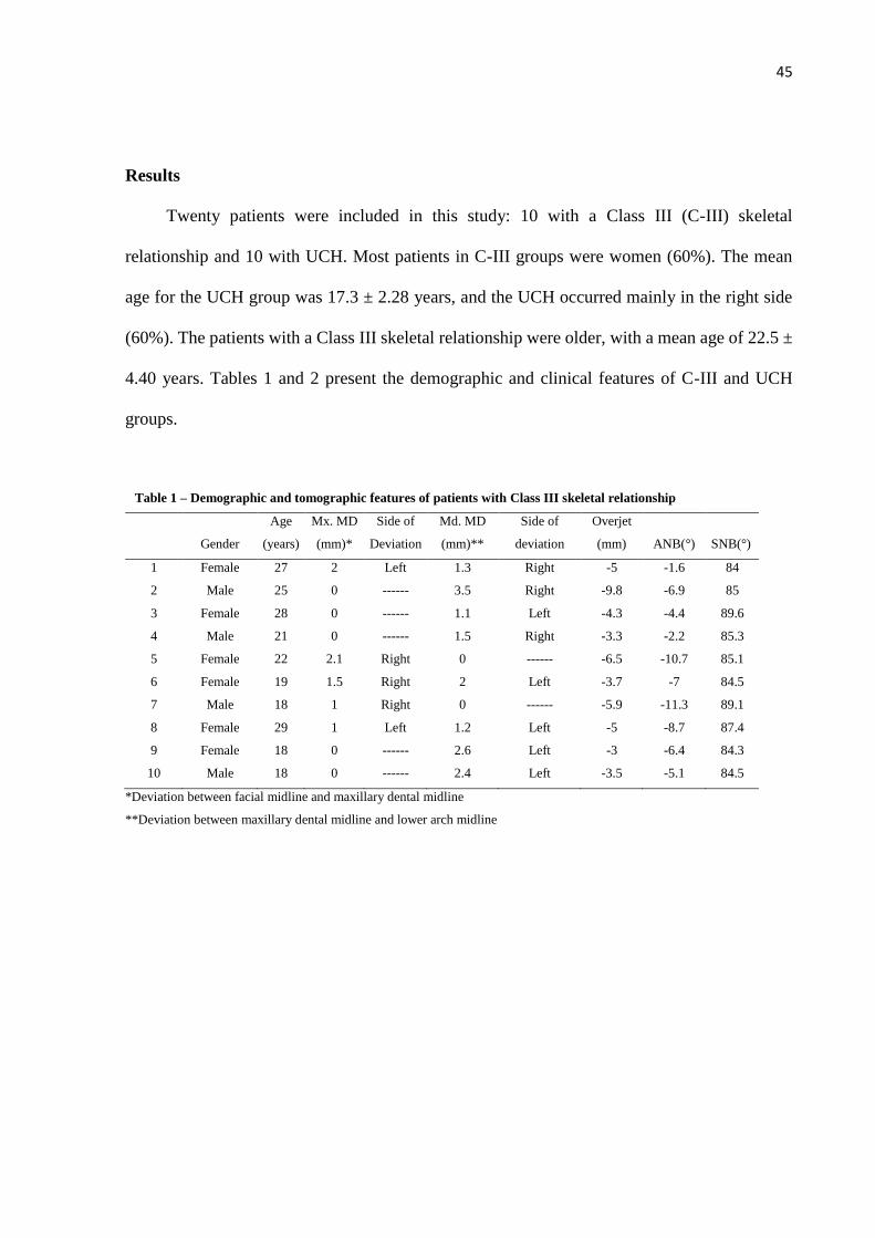
150 to 2756 Hounsfield Unit (Figure 1). Each condyle was isolated using a sculpting tool and
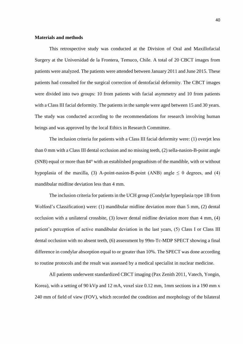
the condyle was delimited to enable the software to measure the volume. To measure the
condylar volume, an imaginary line was created tangent to the deepest point of the sigmoid
notch and parallel to the horizontal plane. The part of the mandible located on this line was
determined as the mandibular condyle for volumetric analysis (Figures 2-3), method similar to
other study (Schlueter et al., 2008). This procedure was performed twice for each condyle with
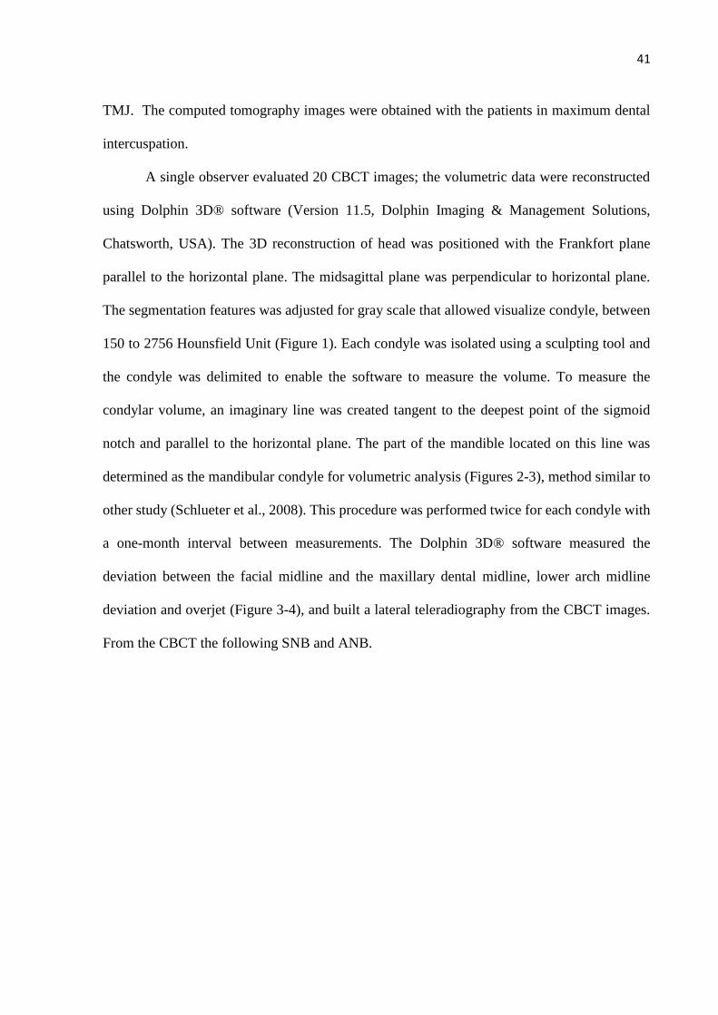
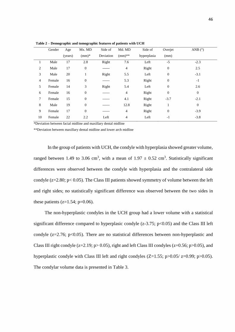
a one-month interval between measurements. The Dolphin 3D® software measured the
deviation between the facial midline and the maxillary dental midline, lower arch midline
deviation and overjet (Figure 3-4), and built a lateral teleradiography from the CBCT images.
From the CBCT the following SNB and ANB.
42
Figure 1: 3D reconstruction of CBCT from a patient with Class III skeletal relationship using a
tool to determine segmentation features.
Figure 2: Delimitation of condyle to measure volume in 3D reconstruction of CBCT from a
patient with UCH.
43
Figure 3: 3D reconstruction of condyle showing features of
segmentation control and delimitation of condyle.
Figure 4: 3D reconstruction of CBCT from a patient with UCH using a 2D
measure tool to evaluate deviation of median line of lower arch.

44
Figure 5: 3D reconstruction of CBCT from a patient with skeletal Class
III relationship using a 2D measure tool to evaluate overjet.
The data were analyzed using descriptive and correlational statistics with the support of
the software SPSS v. 18.0 for Windows (SPSS Inc, Chicago, IL) and Bioestat 5.0 (Instituto
Mamirauá, Brasília, Brazil). The Shapiro-Wilk test was performed and the sample was shown
to be normally distributed. Levene’s test was applied and showed heterogeneity between the
variances (p<0.05). The intraclass correlation coefficient (ICC) was used to assess method
error. The correlation coefficient between the measurements of the first and second tracings
was 0.973 and had a p<0.01. Wilcoxon signed-rank test was performed to compare right and
left condyles in Class III subjects and hyperplasic and non-hyperplasic condyles in condylar
hyperplasia subjects. To compare all measurements we used Kruskal-Wallis tests (for multiple
groups comparison) followed by Dunn's post-hoc test. The level of significance was set at p <
0.05.
45
Results
Twenty patients were included in this study: 10 with a Class III (C-III) skeletal
relationship and 10 with UCH. Most patients in C-III groups were women (60%). The mean
age for the UCH group was 17.3 ± 2.28 years, and the UCH occurred mainly in the right side
(60%). The patients with a Class III skeletal relationship were older, with a mean age of 22.5 ±
4.40 years. Tables 1 and 2 present the demographic and clinical features of C-III and UCH
groups.
Table 1 – Demographic and tomographic features of patients with Class III skeletal relationship
Gender
Age
(years)
Mx. MD
(mm)*
Side of
Deviation
Md. MD
(mm)**
Side of
deviation
Overjet
(mm) ANB(°) SNB(°)
1 Female 27 2 Left 1.3 Right -5 -1.6 84
2 Male 25 0 ------ 3.5 Right -9.8 -6.9 85
3 Female 28 0 ------ 1.1 Left -4.3 -4.4 89.6
4 Male 21 0 ------ 1.5 Right -3.3 -2.2 85.3
5 Female 22 2.1 Right 0 ------ -6.5 -10.7 85.1
6 Female 19 1.5 Right 2 Left -3.7 -7 84.5
7 Male 18 1 Right 0 ------ -5.9 -11.3 89.1
8 Female 29 1 Left 1.2 Left -5 -8.7 87.4
9 Female 18 0 ------ 2.6 Left -3 -6.4 84.3
10 Male 18 0 ------ 2.4 Left -3.5 -5.1 84.5
*Deviation between facial midline and maxillary dental midline
**Deviation between maxillary dental midline and lower arch midline
46
*Deviation between facial midline and maxillary dental midline
**Deviation between maxillary dental midline and lower arch midline
In the group of patients with UCH, the condyle with hyperplasia showed greater volume,
ranged between 1.49 to 3.06 cm3, with a mean of 1.97 ± 0.52 cm3. Statistically significant
differences were observed between the condyle with hyperplasia and the contralateral side
condyle (z=2.80; p< 0.05). The Class III patients showed symmetry of volume between the left
and right sides; no statistically significant difference was observed between the two sides in
these patients (z=1.54; p=0.06).
The non-hyperplastic condyles in the UCH group had a lower volume with a statistical
significant difference compared to hyperplasic condyle (z-3.75; p<0.05) and the Class III left
condyle (z=2.76; p<0.05). There are no statistical differences between non-hyperplastic and
Class III right condyle (z=2.19; p> 0.05), right and left Class III condyles (z=0.56; p>0.05), and
hyperplastic condyle with Class III left and right condyles (Z=1.55; p>0.05/ z=0.99; p>0.05).
The condylar volume data is presented in Table 3.
Table 2 – Demographic and tomographic features of patients with UCH
Gender Age
(years)
Mx. MD
(mm)*
Side of
Deviation
Md. MD
(mm)**
Side of
hyperplasia
Overjet
(mm)
ANB (°)
1 Male 17 2.8 Right 7.6 Left -5 -2.3
2 Male 17 0 ------ 4 Right 0 2.5
3 Male 20 1 Right 5.5 Left 0 -3.1
4 Female 16 0 ------ 5.3 Right 0 -1
5 Female 14 3 Right 5.4 Left 0 2.6
6 Female 16 0 ------ 4 Right 0 0
7 Female 15 0 ------ 4.1 Right -3.7 -2.1
8 Male 19 0 ------ 12.8 Right 1 0
9 Female 17 0 ------ 4 Right 0 -3.9
10 Female 22 2.2 Left 4 Left -1 -3.8
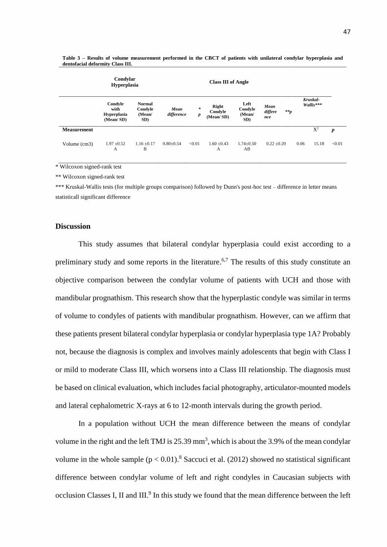
47
Table 3 – Results of volume measurement performed in the CBCT of patients with unilateral condylar hyperplasia and
dentofacial deformity Class III.
Condylar
Hyperplasia Class III of Angle
Condyle
with
Hyperplasia
(Mean/ SD)
Normal
Condyle
(Mean/
SD)
Mean
difference
*
p
Right
Condyle
(Mean/ SD)
Left
Condyle
(Mean/
SD)
Mean
differe
nce
**p
Kruskal-
Wallis***
Measurement X2 p
Volume (cm3) 1.97 ±0.52
A
1.16 ±0.17
B
0.80±0.54
<0.01 1.60 ±0.43
A
1.74±0.50
AB
0.22 ±0.20 0.06 15.18 <0.01
* Wilcoxon signed-rank test
** Wilcoxon signed-rank test
*** Kruskal-Wallis tests (for multiple groups comparison) followed by Dunn's post-hoc test – difference in letter means
statisticall significant difference
Discussion
This study assumes that bilateral condylar hyperplasia could exist according to a
preliminary study and some reports in the literature.6,7 The results of this study constitute an
objective comparison between the condylar volume of patients with UCH and those with
mandibular prognathism. This research show that the hyperplastic condyle was similar in terms
of volume to condyles of patients with mandibular prognathism. However, can we affirm that
these patients present bilateral condylar hyperplasia or condylar hyperplasia type 1A? Probably
not, because the diagnosis is complex and involves mainly adolescents that begin with Class I
or mild to moderate Class III, which worsens into a Class III relationship. The diagnosis must
be based on clinical evaluation, which includes facial photography, articulator-mounted models
and lateral cephalometric X-rays at 6 to 12-month intervals during the growth period.
In a population without UCH the mean difference between the means of condylar
volume in the right and the left TMJ is 25.39 mm3, which is about the 3.9% of the mean condylar
volume in the whole sample (p < 0.01).8 Saccuci et al. (2012) showed no statistical significant
difference between condylar volume of left and right condyles in Caucasian subjects with
occlusion Classes I, II and III.9 In this study we found that the mean difference between the left
48
and right condylar volume in Class III patients was 13.47%; however, the difference is not
statistically significant. The symmetry between right and left condyles has been shown by other
studies using linear measurements in CBCT.6,10 Yet some kind of asymmetry is normal to all
structures of the human body.8 The patients with UCH showed 51.43% of the mean difference
of condylar volume. The difference in condylar volume could be an initial diagnostic tool for
UCH, since CBCT is more affordable than SPECT. However, more studies are required to
determine what the normal variability is and how much the volume difference determines this
pathology.
In the study of condylar volume, there is no consensus as to the point at which the
structure can be isolated to measure the volume. These studies used different software and cut
the condyle at the base of its neck, while the authors believe that the data on the isolated
condylar head can be better associated with the growing mandibular process.8,9 This study used
the sigmoid notch as a reference to isolate the condylar head and neck, given that UCH is a
pathology that may not only affect the condylar head.
Condylar volume may be related to the mandibular final dimension as well as to the
relationship between maxillary and mandibular bases.8 Ratio analysis (mandible/ maxilla
volumes) could be a more accurate way to study the balance of the quantities of both jaws.11
Thus, variability in the shapes and sizes of condyles should be an important factor in diagnosing
TMJ disorders.12 There are several studies in the literature that measure condylar volume related
to facial deformities using CBCT images.8,9,13,14
CBCT is a useful tool to measure and evaluate condylar dimensions.12 The image quality
of CBCT devices, which depends on section thickness, time acquisition of the image (scanning
protocol), voxel dimension and focal spot dimension, may affect volume calculations.15 It is
possible to say that a section thickness of 0.3mm at 0.9mm intervals provides good agreement

49
between estimated and actual condylar volumes.13 These features were considered and used in
the methodology of this investigation.
The treatment of CH includes a high condylectomy (with removal of the top 3 to 5 mm
of the condylar head), together with disc repositioning and orthognathic surgery. Recently a
proportional condylectomy has been proposed as an alternative in cases of active UCH,
reducing the need for secondary surgery.16 Ideally, a virtual treatment planning using CBCT
images and accompanying software should be performed to determine if it is possible to correct
the deformity and malocclusion using only a proportional condylectomy. However, in some
cases there is a maxillary cant due to compensatory maxilla growth. In these cases, virtual
treatment planning is the best choice to assess and correct the facial asymmetry.
This cross-sectional study presents some inherent limitations. Ideally, condylar volume
should be assessed in a group of patients without facial deformity of similar ages. The small
sample size was related to the strict inclusion criteria and the uncommon nature of UCH. The
radiation doses related to CBCT limit the inclusion of healthy individuals and repeated exams.
Others factors should be considered in applying the results of this investigation to clinical
situations. The 3D volumetric depiction depends on the appropriateness of segmentation, the
threshold of bone voxel values and the accurate suppression of the surrounding tissue values to
enhance the structure of interest. The depiction is dependent on the software algorithm, the
spatial and contrast resolution of the scan, the thickness and degree of calcification or
cortication of bone structure and the technical skill of the operator.9
Volumetric depiction depends on appropriate segmentation, in other words the
thresholding of bone pixel values and suppression of surrounding tissue values to enhance
structure of interest. Dolphin 3D provide a semimanual method of segmentation, dependent on
the interaction of the operator with the data produce a visually acceptable 3D rendering.17
However, few studies have used this method to assess condylar volume.17,18 It is difficult to
50
confirm whether the dimensions measured on the images correspond to the real dimensions of
the structures, because the data obtained here cannot be compared with the anatomical truth.8
In this study, it is not possible to determine whether the medullary bone found itself within the
segmentation features, as the program has opaque rendering and it is not possible visualize or
separate the medullary bone.
The similarities between hyperplastic condyles and Class III skeletal relationship
condyles could be a factor that instigates further prospective studies that follows patients during
the growth period in order to identify cases that match CH type 1A. Orthodontists are the most
suitable to evaluate this pathology, as they have opportunity to assess malocclusion at an early
age and follow these patients for a long period. CH type 1A occurs much more frequently than
clinicians realize. Failure to recognize this pathological entity can result in unfavorable
functional and esthetic treatment results following orthodontics and orthognathic surgery.5
The differential diagnosis includes maxillary hypoplasia, mandibular prognathism
without CH (patient starts out as skeletal Class III in early childhood and maintains harmonious
growth between maxilla and mandible with cessation of growth at normal age), dental
interferences of habitual posturing causing anterior positioning of the mandible, acromegaly
and acquired facial asymmetry unrelated to the TMJ.5
The results of this research shows that the hyperplastic condyle is similar in terms of
volume with condyles of patients with mandibular prognathism, suggesting that some patients
with Class III skeletal relationship could present bilateral condylar hyperplasia. The difference
in volume found between condyles in the patients with UCH could be an initial diagnostic tool
in the future with a simple method.
51
References
1. Nitzan DW, Katsnelson A, Bermanis I, Brin I, Casap N: The clinical characteristics of
condylar hyperplasia: experience with 61 patients. J Oral Maxillofac Surg 66: 312, 2008.
2. Pripatnanont P, Vittayakittipong P, Markmanee U, Thongmak S, Yipintsoi T: The use of
SPECT to evaluate growth cessation of the mandible in unilateral condylar hyperplasia.
Int J Oral Maxillofac Surg 34: 364, 2005.
3. Olate S, Almeida A, Alister JP, Navarro P, Netto HD, Moraes M de: Facial asymmetry
and condylar hyperplasia: considerations for diagnosis in 27 consecutives patients. Int J
Clin Exp Med. 6: 937, 2013.
4. Wolford LM, Mehra P, Reiche-Fischel O, Morales-Ryan C, García-Morales P. Efficacy
of high condylectomy for management of condylar hyperplasia. Am J Orthod Dentofac
Orthop. 121(2):136–51, 2002.
5. Wolford LM, Morales-Ryan C A, García-Morales P, Perez D: Surgical management of
mandibular condylar hyperplasia type 1. Proc (Bayl Univ Med Cent) 22: 321, 2009.
6. Goulart DR, Muñoz P, Olate S, Moraes M de, Fariña R: No differences in morphological
characteristics between hyperplastic condyle and Class III condyle. Int J Oral Maxillofac
Surg 44: 1281, 2015.
7. Wolford LM, Movahed R, Perez DE: A Classification system for conditions causing
condylar hyperplasia. J Oral Maxillofac Surg 72: 567, 2014.
8. Tecco S, Saccucci M, Nucera R, Polimeni A, Pagnoni M, Cordasco G, Festa F, Iannetti
G: Condylar volume and surface in Caucasian young adult subjects. BMC Med Imaging
10: 28, 2010.
9. Saccucci M, D’Attilio M, Rodolfino D, Festa F, Polimeni A, Tecco S: Condylar volume
and condylar area in Class I, Class II and Class III young adult subjects. Head Face Med
8: 34, 2012.

52
10. Muñoz G, Olate S, Cantín M, Vásquez B, Sol M. TMJ in facial Class III deformity:
Condylar morphology relations TMJ in facial Class III deformity. Condylar morphology
relations. Int J Clin Exp Med. 2014; 7(9): 3113–3117.
11. Deguchi T, Katashiba S, Inami T, Foong KWC, Huak CY: Morphologic quantification
of the maxilla and the mandible with cone-beam computed tomography. Am J Orthod
Dentofac Orthop 137: 218, 2010.
12. Praveen BN SH: Morphological and Radiological Variations of Mandibular Condyles in
Health and Diseases: A Systematic Review. Dentistry 03: 1, 2013.
13. Bayram M, Kayipmaz S, Sezgin OS, Küçük M: Volumetric analysis of the mandibular
condyle using cone beam computed tomography. Eur J Radiol 81: 1812, 2012.
14. Huntjens E, Kiss G, Wouters C, Carels C: Condylar asymmetry in children with juvenile
idiopathic arthritis assessed by cone-beam computed tomography. Eur J Orthod 30: 545,
2008.
15. Kayipmaz S, Sezgin OS, Saricaoglu ST, Bas O, Sahin B, Küçük M: The estimation of
the volume of sheep mandibular defects using cone-beam computed tomography images
and a stereological method. Dentomaxillofac Radiol 40: 165, 2011.
16. Fariña R, Olate S, Raposo a., Araya I, Alister JP, Uribe F: High condylectomy versus
proportional condylectomy: is secondary orthognathic surgery necessary? Int J Oral
Maxillofac Surg, 2015.
17. Periago DR, Scarfe WC, Moshiri M, Scheetz JP, Silveira AM, Farman AG. Linear
Accuracy and Reliability of Cone Beam CT Derived 3-Dimensional Images Constructed
Using an Orthodontic Volumetric Rendering Program. Angle Orthod. 2008; 78(3): 387–
95.
18. Illipronti-Filho E, Fantini SM, Chilvarquer I: Evaluation of mandibular condyles in
children with unilateral posterior crossbite. Braz Oral Res 29: 1, 2015.
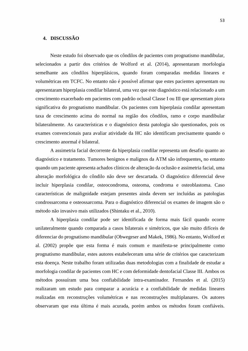
53
4. DISCUSSÃO
Neste estudo foi observado que os côndilos de pacientes com prognatismo mandibular,
selecionados a partir dos critérios de Wolford et al. (2014), apresentaram morfologia
semelhante aos côndilos hiperplásicos, quando foram comparadas medidas lineares e
volumétricas em TCFC. No entanto não é possível afirmar que estes pacientes apresentam ou
apresentaram hiperplasia condilar bilateral, uma vez que este diagnóstico está relacionado a um
crescimento exacerbado em pacientes com padrão oclusal Classe I ou III que apresentam piora
significativa do prognatismo mandibular. Os pacientes com hiperplasia condilar apresentam
taxa de crescimento acima do normal na região dos côndilos, ramo e corpo mandibular
bilateralmente. As características e o diagnóstico desta patologia são questionados, pois os
exames convencionais para avaliar atividade da HC não identificam precisamente quando o
crescimento anormal é bilateral.
A assimetria facial decorrente da hiperplasia condilar representa um desafio quanto ao
diagnóstico e tratamento. Tumores benignos e malignos da ATM são infrequentes, no entanto
quando um paciente apresenta achados clínicos de alteração da oclusão e assimetria facial, uma
alteração morfológica do côndilo não deve ser descartada. O diagnóstico diferencial deve
incluir hiperplasia condilar, osteocondroma, osteoma, condroma e osteoblastoma. Caso
características de malignidade estejam presentes ainda devem ser incluídas as patologias
condrossarcoma e osteossarcoma. Para o diagnóstico diferencial os exames de imagem são o
método não invasivo mais utilizados (Shintaku et al., 2010).
A hiperplasia condilar pode ser identificada de forma mais fácil quando ocorre
unilateralmente quando comparada a casos bilaterais e simétricos, que são muito difíceis de
diferenciar do prognatismo mandibular (Obwegeser and Makek, 1986). No entanto, Wolford et
al. (2002) propõe que esta forma é mais comum e manifesta-se principalmente como
prognatismo mandibular, estes autores estabeleceram uma série de critérios que caracterizam
esta doença. Neste trabalho foram utilizadas duas metodologias com a finalidade de estudar a
morfologia condilar de pacientes com HC e com deformidade dentofacial Classe III. Ambos os
métodos possuíram uma boa confiabilidade intra-examinador. Fernandes et al. (2015)
realizaram um estudo para comparar a acurácia e a confiabilidade de medidas lineares
realizadas em reconstruções volumétricas e nas reconstruções multiplanares. Os autores
observaram que esta última é mais acurada, porém ambos os métodos foram confiáveis.
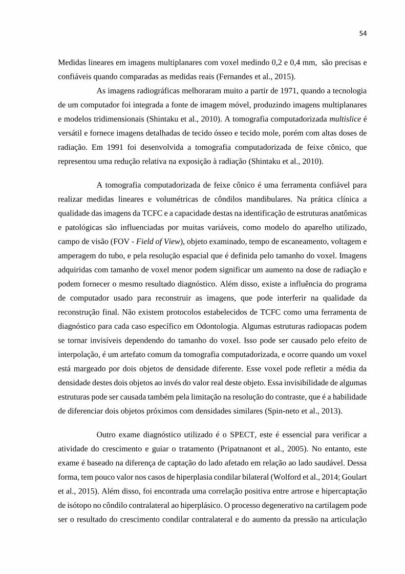
54
Medidas lineares em imagens multiplanares com voxel medindo 0,2 e 0,4 mm, são precisas e
confiáveis quando comparadas as medidas reais (Fernandes et al., 2015).
As imagens radiográficas melhoraram muito a partir de 1971, quando a tecnologia
de um computador foi integrada a fonte de imagem móvel, produzindo imagens multiplanares
e modelos tridimensionais (Shintaku et al., 2010). A tomografia computadorizada multislice é
versátil e fornece imagens detalhadas de tecido ósseo e tecido mole, porém com altas doses de
radiação. Em 1991 foi desenvolvida a tomografia computadorizada de feixe cônico, que
representou uma redução relativa na exposição à radiação (Shintaku et al., 2010).
A tomografia computadorizada de feixe cônico é uma ferramenta confiável para
realizar medidas lineares e volumétricas de côndilos mandibulares. Na prática clínica a
qualidade das imagens da TCFC e a capacidade destas na identificação de estruturas anatômicas
e patológicas são influenciadas por muitas variáveis, como modelo do aparelho utilizado,
campo de visão (FOV - Field of View), objeto examinado, tempo de escaneamento, voltagem e
amperagem do tubo, e pela resolução espacial que é definida pelo tamanho do voxel. Imagens
adquiridas com tamanho de voxel menor podem significar um aumento na dose de radiação e
podem fornecer o mesmo resultado diagnóstico. Além disso, existe a influência do programa
de computador usado para reconstruir as imagens, que pode interferir na qualidade da
reconstrução final. Não existem protocolos estabelecidos de TCFC como uma ferramenta de
diagnóstico para cada caso específico em Odontologia. Algumas estruturas radiopacas podem
se tornar invisíveis dependendo do tamanho do voxel. Isso pode ser causado pelo efeito de
interpolação, é um artefato comum da tomografia computadorizada, e ocorre quando um voxel
está margeado por dois objetos de densidade diferente. Esse voxel pode refletir a média da
densidade destes dois objetos ao invés do valor real deste objeto. Essa invisibilidade de algumas
estruturas pode ser causada também pela limitação na resolução do contraste, que é a habilidade
de diferenciar dois objetos próximos com densidades similares (Spin-neto et al., 2013).
Outro exame diagnóstico utilizado é o SPECT, este é essencial para verificar a
atividade do crescimento e guiar o tratamento (Pripatnanont et al., 2005). No entanto, este
exame é baseado na diferença de captação do lado afetado em relação ao lado saudável. Dessa
forma, tem pouco valor nos casos de hiperplasia condilar bilateral (Wolford et al., 2014; Goulart
et al., 2015). Além disso, foi encontrada uma correlação positiva entre artrose e hipercaptação
de isótopo no côndilo contralateral ao hiperplásico. O processo degenerativo na cartilagem pode
ser o resultado do crescimento condilar contralateral e do aumento da pressão na articulação
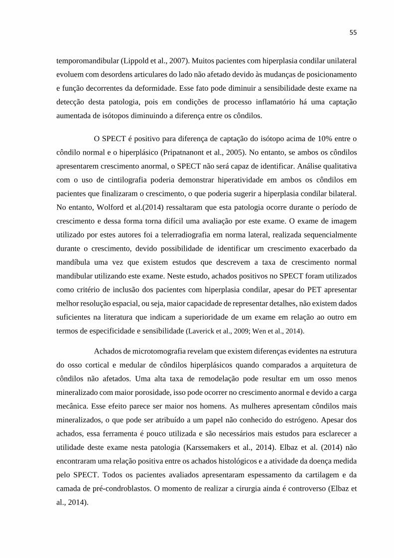
55
temporomandibular (Lippold et al., 2007). Muitos pacientes com hiperplasia condilar unilateral
evoluem com desordens articulares do lado não afetado devido às mudanças de posicionamento
e função decorrentes da deformidade. Esse fato pode diminuir a sensibilidade deste exame na
detecção desta patologia, pois em condições de processo inflamatório há uma captação
aumentada de isótopos diminuindo a diferença entre os côndilos.
O SPECT é positivo para diferença de captação do isótopo acima de 10% entre o
côndilo normal e o hiperplásico (Pripatnanont et al., 2005). No entanto, se ambos os côndilos
apresentarem crescimento anormal, o SPECT não será capaz de identificar. Análise qualitativa
com o uso de cintilografia poderia demonstrar hiperatividade em ambos os côndilos em
pacientes que finalizaram o crescimento, o que poderia sugerir a hiperplasia condilar bilateral.
No entanto, Wolford et al.(2014) ressaltaram que esta patologia ocorre durante o período de
crescimento e dessa forma torna difícil uma avaliação por este exame. O exame de imagem
utilizado por estes autores foi a telerradiografia em norma lateral, realizada sequencialmente
durante o crescimento, devido possibilidade de identificar um crescimento exacerbado da
mandíbula uma vez que existem estudos que descrevem a taxa de crescimento normal
mandibular utilizando este exame. Neste estudo, achados positivos no SPECT foram utilizados
como critério de inclusão dos pacientes com hiperplasia condilar, apesar do PET apresentar
melhor resolução espacial, ou seja, maior capacidade de representar detalhes, não existem dados
suficientes na literatura que indicam a superioridade de um exame em relação ao outro em
termos de especificidade e sensibilidade (Laverick et al., 2009; Wen et al., 2014).
Achados de microtomografia revelam que existem diferenças evidentes na estrutura
do osso cortical e medular de côndilos hiperplásicos quando comparados a arquitetura de
côndilos não afetados. Uma alta taxa de remodelação pode resultar em um osso menos
mineralizado com maior porosidade, isso pode ocorrer no crescimento anormal e devido a carga
mecânica. Esse efeito parece ser maior nos homens. As mulheres apresentam côndilos mais
mineralizados, o que pode ser atribuído a um papel não conhecido do estrógeno. Apesar dos
achados, essa ferramenta é pouco utilizada e são necessários mais estudos para esclarecer a
utilidade deste exame nesta patologia (Karssemakers et al., 2014). Elbaz et al. (2014) não
encontraram uma relação positiva entre os achados histológicos e a atividade da doença medida
pelo SPECT. Todos os pacientes avaliados apresentaram espessamento da cartilagem e da
camada de pré-condroblastos. O momento de realizar a cirurgia ainda é controverso (Elbaz et
al., 2014).
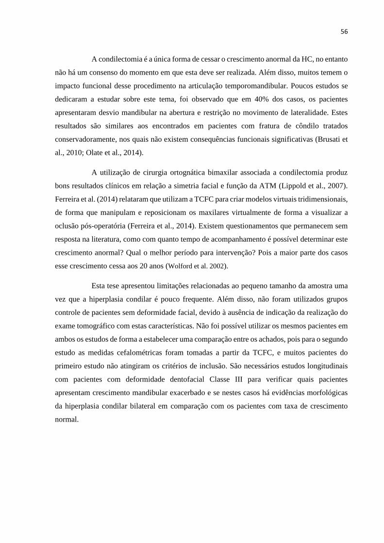
56
A condilectomia é a única forma de cessar o crescimento anormal da HC, no entanto
não há um consenso do momento em que esta deve ser realizada. Além disso, muitos temem o
impacto funcional desse procedimento na articulação temporomandibular. Poucos estudos se
dedicaram a estudar sobre este tema, foi observado que em 40% dos casos, os pacientes
apresentaram desvio mandibular na abertura e restrição no movimento de lateralidade. Estes
resultados são similares aos encontrados em pacientes com fratura de côndilo tratados
conservadoramente, nos quais não existem consequências funcionais significativas (Brusati et
al., 2010; Olate et al., 2014).
A utilização de cirurgia ortognática bimaxilar associada a condilectomia produz
bons resultados clínicos em relação a simetria facial e função da ATM (Lippold et al., 2007).
Ferreira et al. (2014) relataram que utilizam a TCFC para criar modelos virtuais tridimensionais,
de forma que manipulam e reposicionam os maxilares virtualmente de forma a visualizar a
oclusão pós-operatória (Ferreira et al., 2014). Existem questionamentos que permanecem sem
resposta na literatura, como com quanto tempo de acompanhamento é possível determinar este
crescimento anormal? Qual o melhor período para intervenção? Pois a maior parte dos casos
esse crescimento cessa aos 20 anos (Wolford et al. 2002).
Esta tese apresentou limitações relacionadas ao pequeno tamanho da amostra uma
vez que a hiperplasia condilar é pouco frequente. Além disso, não foram utilizados grupos
controle de pacientes sem deformidade facial, devido à ausência de indicação da realização do
exame tomográfico com estas características. Não foi possível utilizar os mesmos pacientes em
ambos os estudos de forma a estabelecer uma comparação entre os achados, pois para o segundo
estudo as medidas cefalométricas foram tomadas a partir da TCFC, e muitos pacientes do
primeiro estudo não atingiram os critérios de inclusão. São necessários estudos longitudinais
com pacientes com deformidade dentofacial Classe III para verificar quais pacientes
apresentam crescimento mandibular exacerbado e se nestes casos há evidências morfológicas
da hiperplasia condilar bilateral em comparação com os pacientes com taxa de crescimento
normal.

57
5. CONCLUSÃO
Na amostra estudada a morfologia condilar de pacientes com prognatismo
mandibular é semelhante à de côndilos hiperplásicos de pacientes com assimetria facial, quando
foram realizadas medidas lineares e volumétricas utilizando imagens de tomografias
computadorizadas de feixe cônico. Esse fato pode ser um indício da ocorrência de hiperplasia
condilar bilateral como fator etiológico da deformidade dentofacial Classe III.
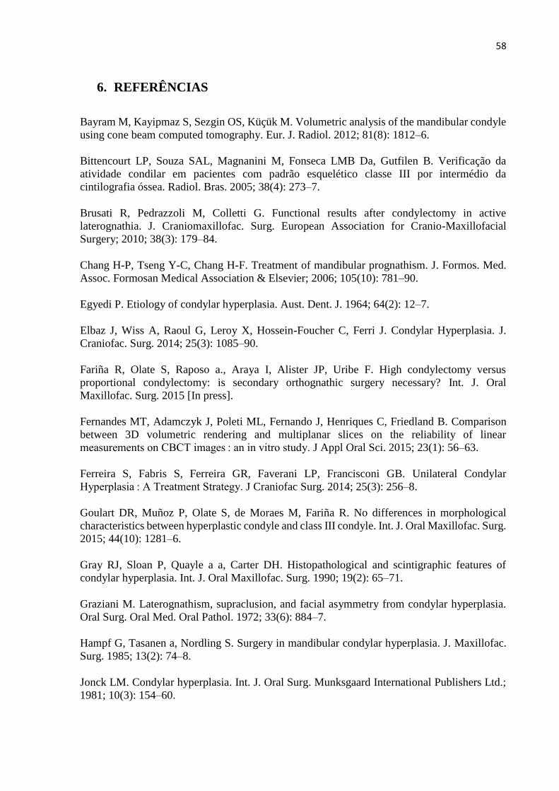
58
6. REFERÊNCIAS
Bayram M, Kayipmaz S, Sezgin OS, Küçük M. Volumetric analysis of the mandibular condyle
using cone beam computed tomography. Eur. J. Radiol. 2012; 81(8): 1812–6.
Bittencourt LP, Souza SAL, Magnanini M, Fonseca LMB Da, Gutfilen B. Verificação da
atividade condilar em pacientes com padrão esquelético classe III por intermédio da
cintilografia óssea. Radiol. Bras. 2005; 38(4): 273–7.
Brusati R, Pedrazzoli M, Colletti G. Functional results after condylectomy in active
laterognathia. J. Craniomaxillofac. Surg. European Association for Cranio-Maxillofacial
Surgery; 2010; 38(3): 179–84.
Chang H-P, Tseng Y-C, Chang H-F. Treatment of mandibular prognathism. J. Formos. Med.
Assoc. Formosan Medical Association & Elsevier; 2006; 105(10): 781–90.
Egyedi P. Etiology of condylar hyperplasia. Aust. Dent. J. 1964; 64(2): 12–7.
Elbaz J, Wiss A, Raoul G, Leroy X, Hossein-Foucher C, Ferri J. Condylar Hyperplasia. J.
Craniofac. Surg. 2014; 25(3): 1085–90.
Fariña R, Olate S, Raposo a., Araya I, Alister JP, Uribe F. High condylectomy versus
proportional condylectomy: is secondary orthognathic surgery necessary? Int. J. Oral
Maxillofac. Surg. 2015 [In press].
Fernandes MT, Adamczyk J, Poleti ML, Fernando J, Henriques C, Friedland B. Comparison
between 3D volumetric rendering and multiplanar slices on the reliability of linear
measurements on CBCT images : an in vitro study. J Appl Oral Sci. 2015; 23(1): 56–63.
Ferreira S, Fabris S, Ferreira GR, Faverani LP, Francisconi GB. Unilateral Condylar
Hyperplasia : A Treatment Strategy. J Craniofac Surg. 2014; 25(3): 256–8.
Goulart DR, Muñoz P, Olate S, de Moraes M, Fariña R. No differences in morphological
characteristics between hyperplastic condyle and class III condyle. Int. J. Oral Maxillofac. Surg.
2015; 44(10): 1281–6.
Gray RJ, Sloan P, Quayle a a, Carter DH. Histopathological and scintigraphic features of
condylar hyperplasia. Int. J. Oral Maxillofac. Surg. 1990; 19(2): 65–71.
Graziani M. Laterognathism, supraclusion, and facial asymmetry from condylar hyperplasia.
Oral Surg. Oral Med. Oral Pathol. 1972; 33(6): 884–7.
Hampf G, Tasanen a, Nordling S. Surgery in mandibular condylar hyperplasia. J. Maxillofac.
Surg. 1985; 13(2): 74–8.
Jonck LM. Condylar hyperplasia. Int. J. Oral Surg. Munksgaard International Publishers Ltd.;
1981; 10(3): 154–60.

59
Jones RHB, Tier G a. Correction of facial asymmetry as a result of unilateral condylar
hyperplasia. J. Oral Maxillofac. Surg. American Association of Oral and Maxillofacial
Surgeons; 2012; 70(6): 1413–25.
Karssemakers LHE, Nolte JW, Tuinzing DB, Langenbach GEJ, Raijmakers PG, Becking a. G.
Microcomputed tomographic analysis of human condyles in unilateral condylar hyperplasia:
increased cortical porosity and trabecular bone volume fraction with reduced mineralisation.
Br. J. Oral Maxillofac. Surg. British Association of Oral and Maxillofacial Surgeons; 2014;
52(10): 940–4.
Laverick S, Bounds G, Wong WL. [18F]-fluoride positron emission tomography for imaging
condylar hyperplasia. Br. J. Oral Maxillofac. Surg. 2009; 47(3): 196–9.
Lippold C, Kruse-Losler B, Danesh G, Joos U, Meyer U. Treatment of hemimandibular
hyperplasia: The biological basis of condylectomy. Br. J. Oral Maxillofac. Surg. 2007; 45(5):
353–60.
Matteson SR, Profit WR, Terry BC, Staab E V, Burkes EJ, Hill C. Bone scanning with 99m
technetium phosphate to assess condylar hyperplasia. Oral Surg. Oral Med. Oral Pathol. 1985;
60(4): 356–67.
Moss ML. The functional matrix hypothesis revisited. Am. J. Orthod. Dentofacial Orthop.
1997; 112(1): 8–11.
Mota RS, Cardoso VAC, Bechara CDS, Reis JGC, Maciel SM. Análise do crescimento das
dimensões da mandíbula em diferentes idades fetais. Dental Press J. Orthod. 2010; 15(2): 113–
21.
Muñoz ME, Monje F, Goizueta C, Rodríguez-Campo F. Active Condylar Hyperplasia Treated
by High Condylectomy: Report of Case. Journal of Oral and Maxillofacial Surgery,1999.
57(12), 1455-1459.
Mutoh Y, Ohashi Y, Uchiyama N, Terada K, Hanada K, Sasaki F. Three-dimensional analysis
of condylar hyperplasia with computed tomography. J. Craniomaxillofac. Surg. 1991; 19(2):
49–55.
Nitzan DW, Katsnelson A, Bermanis I, Brin I, Casap N. The clinical characteristics of condylar
hyperplasia: experience with 61 patients. J. Oral Maxillofac. Surg. 2008; 66(2): 312–8.
Norman JE, Painter DM. Hyperplasia of the mandibular condyle. A historical review of
important early cases with a presentation and analysis of twelve patients. J. Maxillofac. Surg.
1980. p. : 161–75.
Obwegeser HL, Makek MS. Hemimandibular hyperplasia--hemimandibular elongation. J.
Maxillofac. Surg. 1986; 14(4): 183–208.
Olate S, Martinez F, Uribe F, Pozzer L, Cavalieri-pereira L, De M. TMJ function after partial
condylectomy in active mandibular condylar hyperplasia. International journal of clinical and
experimental medicine. 2014; 7(3): 775–9.
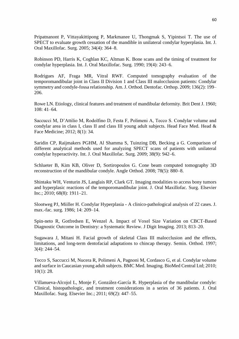
60
Pripatnanont P, Vittayakittipong P, Markmanee U, Thongmak S, Yipintsoi T. The use of
SPECT to evaluate growth cessation of the mandible in unilateral condylar hyperplasia. Int. J.
Oral Maxillofac. Surg. 2005; 34(4): 364–8.
Robinson PD, Harris K, Coghlan KC, Altman K. Bone scans and the timing of treatment for
condylar hyperplasia. Int. J. Oral Maxillofac. Surg. 1990; 19(4): 243–6.
Rodrigues AF, Fraga MR, Vitral RWF. Computed tomography evaluation of the
temporomandibular joint in Class II Division 1 and Class III malocclusion patients: Condylar
symmetry and condyle-fossa relationship. Am. J. Orthod. Dentofac. Orthop. 2009; 136(2): 199–
206.
Rowe LN. Etiology, clinical features and treatment of mandibular deformity. Brit Dent J. 1960;
108: 41–64.
Saccucci M, D’Attilio M, Rodolfino D, Festa F, Polimeni A, Tecco S. Condylar volume and
condylar area in class I, class II and class III young adult subjects. Head Face Med. Head &
Face Medicine; 2012; 8(1): 34.
Saridin CP, Raijmakers PGHM, Al Shamma S, Tuinzing DB, Becking a G. Comparison of
different analytical methods used for analyzing SPECT scans of patients with unilateral
condylar hyperactivity. Int. J. Oral Maxillofac. Surg. 2009; 38(9): 942–6.
Schlueter B, Kim KB, Oliver D, Sortiropoulos G. Cone beam computed tomography 3D
reconstruction of the mandibular condyle. Angle Orthod. 2008; 78(5): 880–8.
Shintaku WH, Venturin JS, Langlais RP, Clark GT. Imaging modalities to access bony tumors
and hyperplasic reactions of the temporomandibular joint. J. Oral Maxillofac. Surg. Elsevier
Inc.; 2010; 68(8): 1911–21.
Slootweg PJ, Miiller H. Condylar Hyperplasia - A clinico-pathological analysis of 22 cases. J.
max.-fac. surg. 1986; 14: 209–14.
Spin-neto R, Gotfredsen E, Wenzel A. Impact of Voxel Size Variation on CBCT-Based
Diagnostic Outcome in Dentistry: a Systematic Review. J Digit Imaging. 2013; 813–20.
Sugawara J, Mitani H. Facial growth of skeletal Class III malocclusion and the effects,
limitations, and long-term dentofacial adaptations to chincap therapy. Semin. Orthod. 1997;
3(4): 244–54.
Tecco S, Saccucci M, Nucera R, Polimeni A, Pagnoni M, Cordasco G, et al. Condylar volume
and surface in Caucasian young adult subjects. BMC Med. Imaging. BioMed Central Ltd; 2010;
10(1): 28.
Villanueva-Alcojol L, Monje F, González-García R. Hyperplasia of the mandibular condyle:
Clinical, histopathologic, and treatment considerations in a series of 36 patients. J. Oral
Maxillofac. Surg. Elsevier Inc.; 2011; 69(2): 447–55.
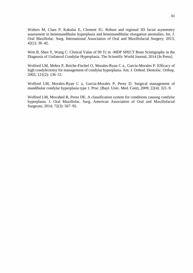
61
Walters M, Claes P, Kakulas E, Clement JG. Robust and regional 3D facial asymmetry
assessment in hemimandibular hyperplasia and hemimandibular elongation anomalies. Int. J.
Oral Maxillofac. Surg. International Association of Oral and Maxillofacial Surgery; 2013;
42(1): 36–42.
Wen B, Shen Y, Wang C. Clinical Value of 99 Tc m -MDP SPECT Bone Scintigraphy in the
Diagnosis of Unilateral Condylar Hyperplasia. The Scientific World Journal, 2014 [In Press].
Wolford LM, Mehra P, Reiche-Fischel O, Morales-Ryan C a., García-Morales P. Efficacy of
high condylectomy for management of condylar hyperplasia. Am. J. Orthod. Dentofac. Orthop.
2002; 121(2): 136–51.
Wolford LM, Morales-Ryan C a, García-Morales P, Perez D. Surgical management of
mandibular condylar hyperplasia type 1. Proc. (Bayl. Univ. Med. Cent), 2009; 22(4): 321–9.
Wolford LM, Movahed R, Perez DE. A classification system for conditions causing condylar
hyperplasia. J. Oral Maxillofac. Surg. American Association of Oral and Maxillofacial
Surgeons; 2014; 72(3): 567–95.

62
APÊNDICE 1
DESCRIÇÃO DAS METODOLOGIAS
Capítulo 1 - Material e Métodos
Foram selecionados 30 pacientes prospectivamente, 15 com deformidade dentofacial Classe
III e 15 pacientes com hiperplasia condilar unilateral. Estes pacientes realizaram tomografia
computadorizada de feixe cônico (TCFC) no aparelho Pax Zenith 2011 (Vatech, Yongin,
Coréia), com as seguintes configurações: 90 kVp, 12 mA, tamanho do voxel de 0,12 mm,
cortes de 1mm de espessura em um FOV (field of view) de 190 mm x 240 mm. Os pacientes
deveriam estar em máxima intercuspidação habitual com o plano de Frankfurt paralelo ao
plano horizontal. As imagens da TCFC foram importadas para o programa Ez3D Viewer Plus
(Vatech, Yongin, Coréia). Neste programa em cada tomografia foi delimitada a região de
interesse (ROI) e o plano sagital médio dos côndilos conforme a Figura 1. A partir desta etapa
foi selecionado o modo de visualização por meio de reconstrução multiplanar. Para cada
plano de visualização (panorâmico-coronal e sagital) foi selecionada a confecção de imagens
com a configuração de cortes de 1 mm de espessura com espaçamento de 1 mm (Figura 2).
Para cada plano de visualização foi escolhido para realizar a mensuração o corte central
seguido de um corte anterior e um corte posterior utilizando a ferramenta Ruler (Figura 3). A
média da medida dos três cortes foi utilizada para a comparação. A Tabela 1 apresenta a
descrição das medidas realizadas. Para cada paciente foram realizadas as mesmas medidas
duas vezes com um intervalo de duas semanas para verificar a confiabilidade dos dados.
Figura 1- Delimitação da região de interesse e do plano sagital médio do côndilo.

63
Figura 2 - Configuração dos cortes com 1 mm de espessura e com espaçamento de 1 mm.
Figura 3 – Utilização da ferramenta Ruler para medir distancia médio-lateral do côndilo.
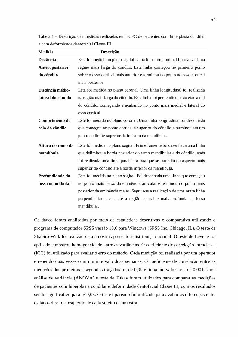
64
Tabela 1 – Descrição das medidas realizadas em TCFC de pacientes com hiperplasia condilar
e com deformidade dentofacial Classe III
Medida Descrição
Distância
Anteroposterior
do côndilo
Esta foi medida no plano sagital. Uma linha longitudinal foi realizada na
região mais larga do côndilo. Esta linha começou no primeiro ponto
sobre o osso cortical mais anterior e terminou no ponto no osso cortical
mais posterior.
Distância médio-
lateral do côndilo
Esta foi medida no plano coronal. Uma linha longitudinal foi realizada
na região mais larga do côndilo. Esta linha foi perpendicular ao eixo axial
do côndilo, começando e acabando no ponto mais medial e lateral do
osso cortical.
Comprimento do
colo do côndilo
Este foi medido no plano coronal. Uma linha longitudinal foi desenhada
que começou no ponto cortical e superior do côndilo e terminou em um
ponto no limite superior da incisura da mandíbula.
Altura do ramo da
mandíbula
Esta foi medida no plano sagital. Primeiramente foi desenhada uma linha
que delimitou a borda posterior do ramo mandibular e do côndilo, após
foi realizada uma linha paralela a esta que se estendia do aspecto mais
superior do côndilo até a borda inferior da mandíbula.
Profundidade da
fossa mandibular
Esta foi medida no plano sagital. Foi desenhada uma linha que começou
no ponto mais baixo da eminência articular e terminou no ponto mais
posterior da eminência malar. Seguiu-se a realização de uma outra linha
perpendicular a esta até a região central e mais profunda da fossa
mandibular.
Os dados foram analisados por meio de estatísticas descritivas e comparativa utilizando o
programa de computador SPSS versão 18.0 para Windows (SPSS Inc, Chicago, IL). O teste de
Shapiro-Wilk foi realizado e a amostra apresentou distribuição normal. O teste de Levene foi
aplicado e mostrou homogeneidade entre as variâncias. O coeficiente de correlação intraclasse
(ICC) foi utilizado para avaliar o erro do método. Cada medição foi realizada por um operador
e repetido duas vezes com um intervalo duas semanas. O coeficiente de correlação entre as
medições dos primeiros e segundos traçados foi de 0,99 e tinha um valor de p de 0,001. Uma
análise de variância (ANOVA) e teste de Tukey foram utilizados para comparar as medições
de pacientes com hiperplasia condilar e deformidade dentofacial Classe III, com os resultados
sendo significativo para p<0,05. O teste t pareado foi utilizado para avaliar as diferenças entre
os lados direito e esquerdo de cada sujeito da amostra.
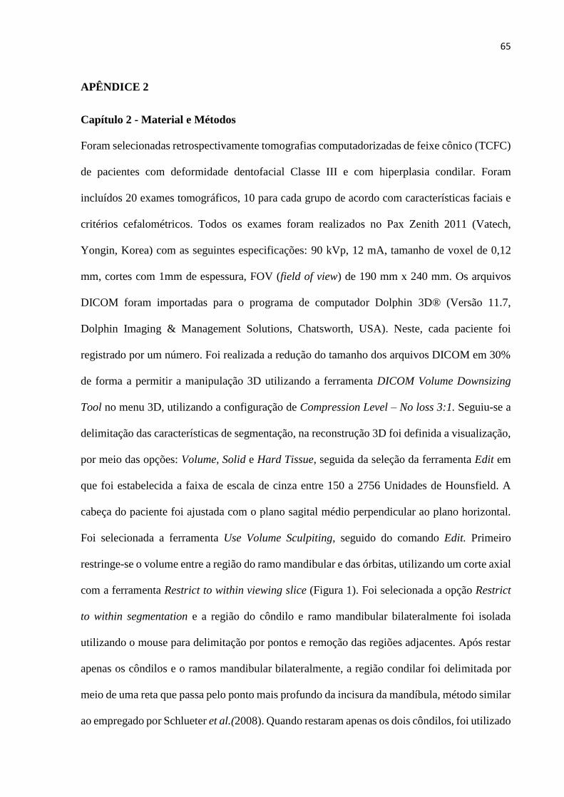
65
APÊNDICE 2
Capítulo 2 - Material e Métodos
Foram selecionadas retrospectivamente tomografias computadorizadas de feixe cônico (TCFC)
de pacientes com deformidade dentofacial Classe III e com hiperplasia condilar. Foram
incluídos 20 exames tomográficos, 10 para cada grupo de acordo com características faciais e
critérios cefalométricos. Todos os exames foram realizados no Pax Zenith 2011 (Vatech,
Yongin, Korea) com as seguintes especificações: 90 kVp, 12 mA, tamanho de voxel de 0,12
mm, cortes com 1mm de espessura, FOV (field of view) de 190 mm x 240 mm. Os arquivos
DICOM foram importadas para o programa de computador Dolphin 3D® (Versão 11.7,
Dolphin Imaging & Management Solutions, Chatsworth, USA). Neste, cada paciente foi
registrado por um número. Foi realizada a redução do tamanho dos arquivos DICOM em 30%
de forma a permitir a manipulação 3D utilizando a ferramenta DICOM Volume Downsizing
Tool no menu 3D, utilizando a configuração de Compression Level – No loss 3:1. Seguiu-se a
delimitação das características de segmentação, na reconstrução 3D foi definida a visualização,
por meio das opções: Volume, Solid e Hard Tissue, seguida da seleção da ferramenta Edit em
que foi estabelecida a faixa de escala de cinza entre 150 a 2756 Unidades de Hounsfield. A
cabeça do paciente foi ajustada com o plano sagital médio perpendicular ao plano horizontal.
Foi selecionada a ferramenta Use Volume Sculpiting, seguido do comando Edit. Primeiro
restringe-se o volume entre a região do ramo mandibular e das órbitas, utilizando um corte axial
com a ferramenta Restrict to within viewing slice (Figura 1). Foi selecionada a opção Restrict
to within segmentation e a região do côndilo e ramo mandibular bilateralmente foi isolada
utilizando o mouse para delimitação por pontos e remoção das regiões adjacentes. Após restar
apenas os côndilos e o ramos mandibular bilateralmente, a região condilar foi delimitada por
meio de uma reta que passa pelo ponto mais profundo da incisura da mandíbula, método similar
ao empregado por Schlueter et al.(2008). Quando restaram apenas os dois côndilos, foi utilizado
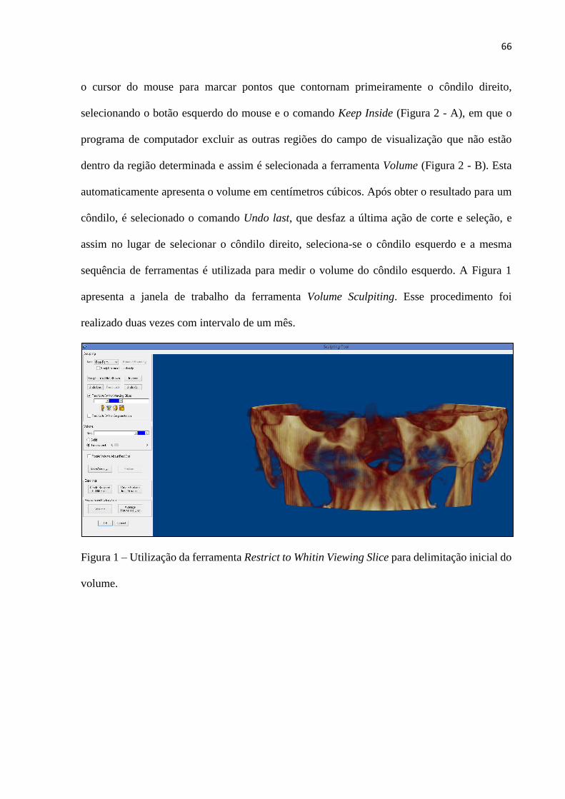
66
o cursor do mouse para marcar pontos que contornam primeiramente o côndilo direito,
selecionando o botão esquerdo do mouse e o comando Keep Inside (Figura 2 - A), em que o
programa de computador excluir as outras regiões do campo de visualização que não estão
dentro da região determinada e assim é selecionada a ferramenta Volume (Figura 2 - B). Esta
automaticamente apresenta o volume em centímetros cúbicos. Após obter o resultado para um
côndilo, é selecionado o comando Undo last, que desfaz a última ação de corte e seleção, e
assim no lugar de selecionar o côndilo direito, seleciona-se o côndilo esquerdo e a mesma
sequência de ferramentas é utilizada para medir o volume do côndilo esquerdo. A Figura 1
apresenta a janela de trabalho da ferramenta Volume Sculpiting. Esse procedimento foi
realizado duas vezes com intervalo de um mês.
Figura 1 – Utilização da ferramenta Restrict to Whitin Viewing Slice para delimitação inicial do
volume.
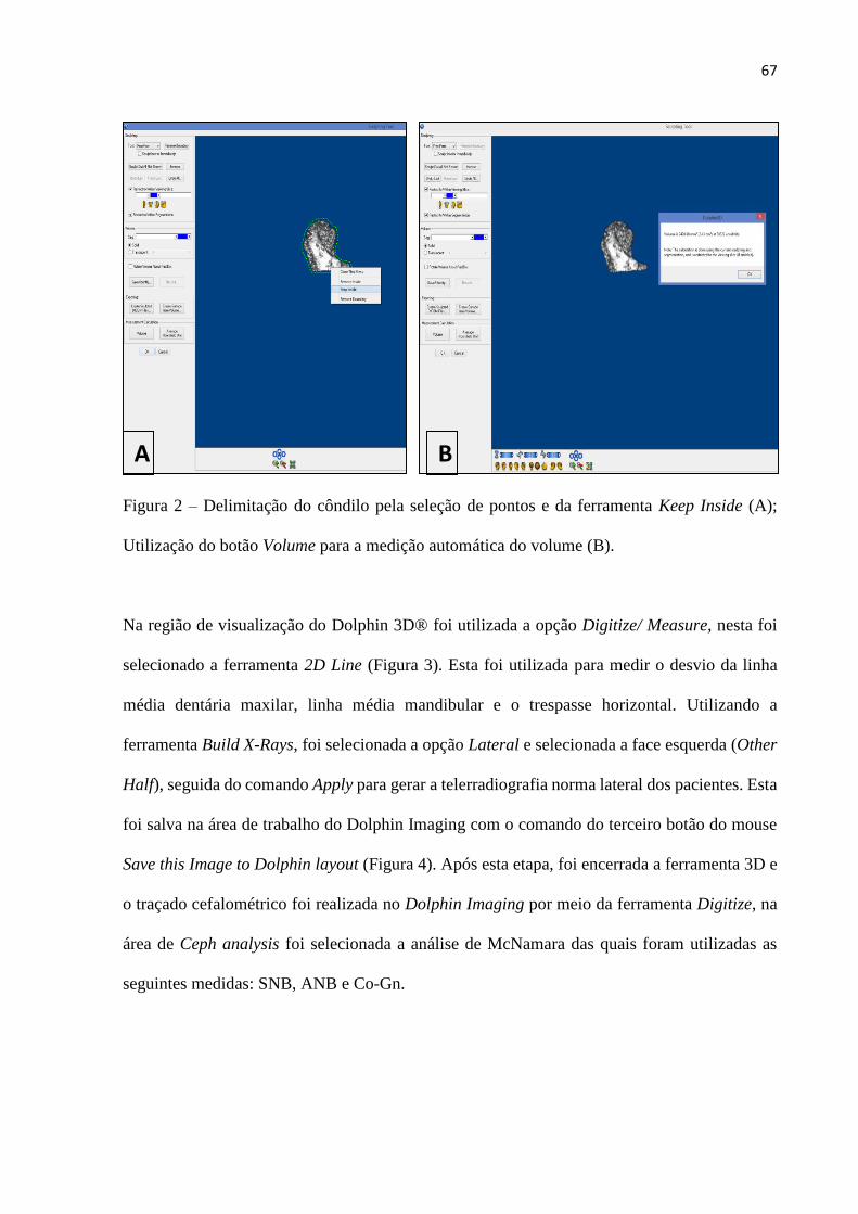
67
Figura 2 – Delimitação do côndilo pela seleção de pontos e da ferramenta Keep Inside (A);
Utilização do botão Volume para a medição automática do volume (B).
Na região de visualização do Dolphin 3D® foi utilizada a opção Digitize/ Measure, nesta foi
selecionado a ferramenta 2D Line (Figura 3). Esta foi utilizada para medir o desvio da linha
média dentária maxilar, linha média mandibular e o trespasse horizontal. Utilizando a
ferramenta Build X-Rays, foi selecionada a opção Lateral e selecionada a face esquerda (Other
Half), seguida do comando Apply para gerar a telerradiografia norma lateral dos pacientes. Esta
foi salva na área de trabalho do Dolphin Imaging com o comando do terceiro botão do mouse
Save this Image to Dolphin layout (Figura 4). Após esta etapa, foi encerrada a ferramenta 3D e
o traçado cefalométrico foi realizada no Dolphin Imaging por meio da ferramenta Digitize, na
área de Ceph analysis foi selecionada a análise de McNamara das quais foram utilizadas as
seguintes medidas: SNB, ANB e Co-Gn.
A B
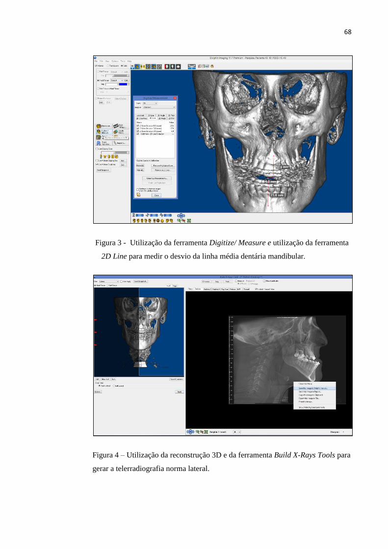
68
Figura 3 - Utilização da ferramenta Digitize/ Measure e utilização da ferramenta
2D Line para medir o desvio da linha média dentária mandibular.
Figura 4 – Utilização da reconstrução 3D e da ferramenta Build X-Rays Tools para
gerar a telerradiografia norma lateral.
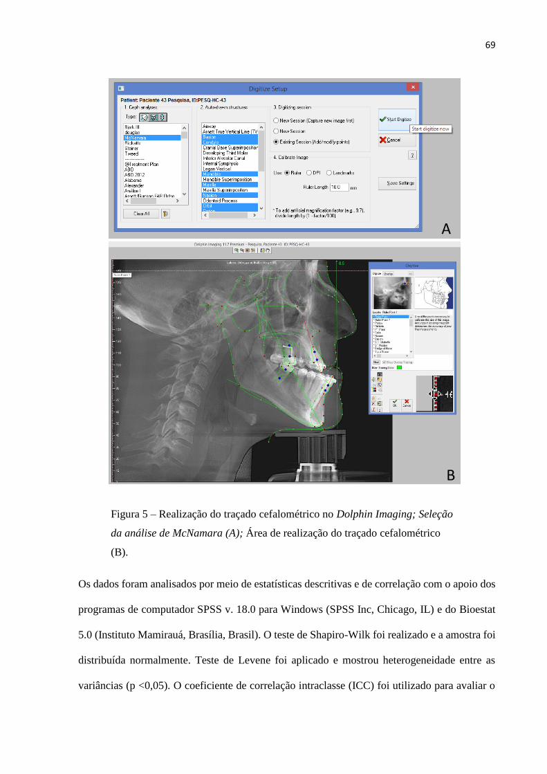
69
Figura 5 – Realização do traçado cefalométrico no Dolphin Imaging; Seleção
da análise de McNamara (A); Área de realização do traçado cefalométrico
(B).
Os dados foram analisados por meio de estatísticas descritivas e de correlação com o apoio dos
programas de computador SPSS v. 18.0 para Windows (SPSS Inc, Chicago, IL) e do Bioestat
5.0 (Instituto Mamirauá, Brasília, Brasil). O teste de Shapiro-Wilk foi realizado e a amostra foi
distribuída normalmente. Teste de Levene foi aplicado e mostrou heterogeneidade entre as
variâncias (p <0,05). O coeficiente de correlação intraclasse (ICC) foi utilizado para avaliar o
A
B

70
erro do método. O coeficiente de correlação entre as medições dos primeiros e segundos
traçados foi 0,973 e tinha um p <0,01. Foi realizado o teste de Wilcoxon para comparar côndilos
direito e esquerdo na Classe III e os côndilos hiperplásicos e não hiperplásicos nos indivíduos
com hiperplasia condilar. Para comparar todas as medições foram utilizados os testes de
Kruskal-Wallis (por comparação de vários grupos), seguida pelo teste post-hoc de Dunn. O
nível de significância foi estabelecido em p <0,05.
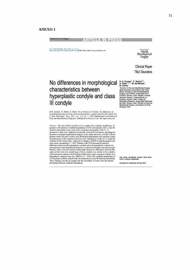
71
ANEXO 1
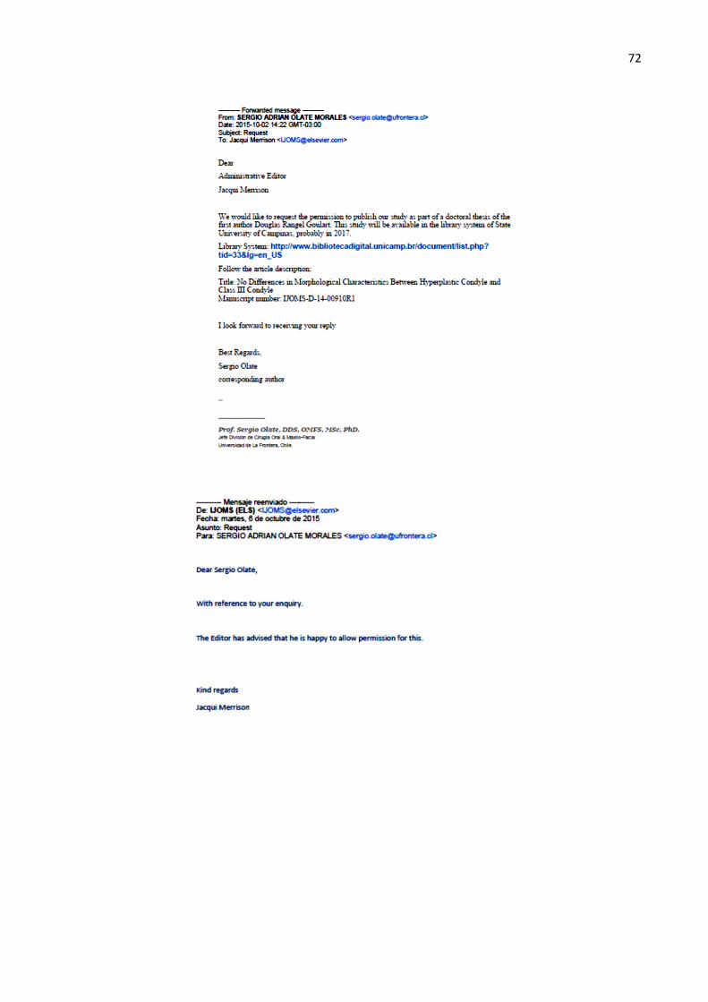
72
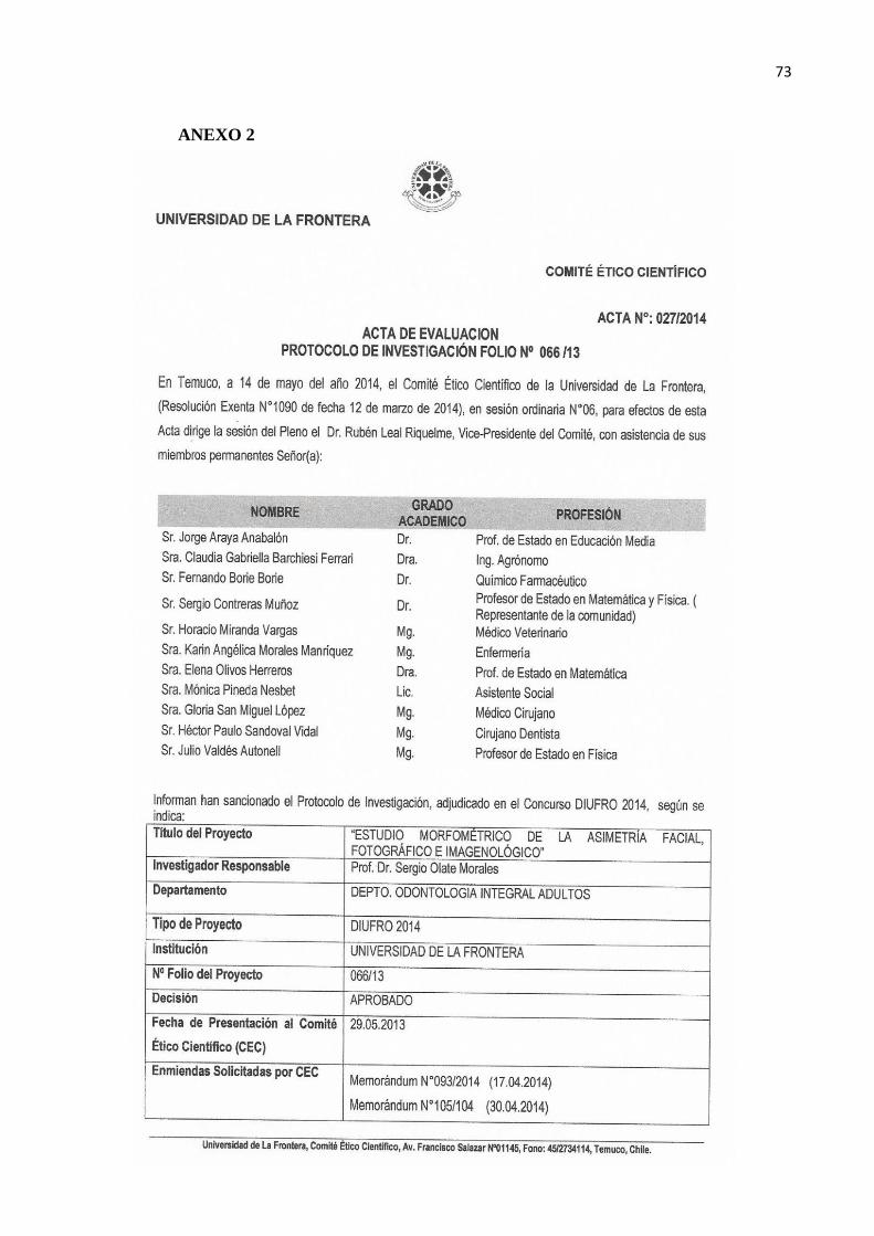
73
ANEXO 2
Related Documents


