i LUCIANA REGINA MOREIRA AVALIAÇÃO DA ANGIOGÊNESE E LINFANGIOGÊNESE NOS CARCINOMAS COLORRETAIS: COMPARAÇÃO ENTRE MÉTODOS DE MENSURAÇÃO E DE DIFERENTES MARCADORES VASCULARES COM INDICADORES ANATOMOPATOLÓGICOS DE PROGNÓSTICO Tese de Doutorado ORIENTADOR: Prof. Dr. JOSÉ VASSALLO Unicamp 2009

Welcome message from author
This document is posted to help you gain knowledge. Please leave a comment to let me know what you think about it! Share it to your friends and learn new things together.
Transcript
i
LUCIANA REGINA MOREIRA
AVALIAÇÃO DA ANGIOGÊNESE E LINFANGIOGÊNESE NOS CARCINOMAS COLORRETAIS: COMPARAÇÃO ENTRE MÉTODOS DE MENSURAÇÃO E DE DIFERENTES MARCADORES VASCULARES
COM INDICADORES ANATOMOPATOLÓGICOS DE PROGNÓSTICO
Tese de Doutorado
ORIENTADOR: Prof. Dr. JOSÉ VASSALLO
Unicamp 2009
ii
LUCIANA REGINA MOREIRA
AVALIAÇÃO DA ANGIOGÊNESE E LINFANGIOGÊNESE NOS CARCINOMAS COLORRETAIS: COMPARAÇÃO ENTRE MÉTODOS DE MENSURAÇÃO E DE DIFERENTES MARCADORES VASCULARES
COM INDICADORES ANATOMOPATOLÓGICOS DE PROGNÓSTICO
Tese de Doutorado apresentada à Pós-Graduação da Faculdade de Ciências Médicas da Universidade Estadual de Campinas para obtenção do Título de Doutor em Ciências Médicas, área de concentração em Anatomia Patológica
ORIENTADOR: Prof. Dr. JOSÉ VASSALLO
Unicamp 2009
FICHA CATALOGRÁFICA ELABORADA PELA
BIBLIOTECA DA FACULDADE DE CIÊNCIAS MÉDICAS UNICAMP
Bibliotecário: Sandra Lúcia Pereira – CRB-8ª / 6044 Título em inglês: Angiogenesis and lymphangiogenesis in colorectal carcinoma: comparison between quantification methods and different immunohistochemical markers with anatomopathologic prognostic factors
Keywords: Angiogensis
Lymphangiogenesis
Colorectal Neoplasms
Immunohistochemistry Titulação: Doutor em Ciências Médicas Área de concentração: Anatomia Patológica Banca examinadora:
Prof. Dr. José Vassallo Prof. Dr. Cláudio Saddy Rodrigues Coy Profª. Drª. Cecília Amélia Fazzio Escanhoela Prof. Dr. Fernando Medina Cunha Profª. Drª. Renata Coudry
Data da defesa: 30-06-2009
Moreira, Luciana Regina M813a Avaliação da angiogênese e linfangiogênese nos
carcinomas colorretais: comparação entre métodos de mensuração e de diferentes marcadores vasculares com indicadores anatomopatológicos de prognóstico / Luciana Regina Moreira. Campinas, SP: [s.n.], 2009.
Orientador: José Vassallo Tese (Doutorado) Universidade Estadual de Campinas.
Faculdade de Ciências Médicas. 1. Angiogênese. 2. Linfangiogênese. 3. Neoplasias
colorretais. 4. Imunoistoquimica. I. Vassallo, José. II. Universidade Estadual de Campinas. Faculdade de Ciências Médicas. III. Título.
iii

iv
Dedico este trabalho...
Aos meus pais, Adilson e Maria do Carmo,
pela dedicação e ensinamentos,
presentes em todos os momentos da minha vida.
Ao Anderson,
pelo amor, carinho, paciência e apoio,
fundamentais em todas as nossas conquistas.
Aos meus amigos,
razões de entusiasmo e fontes de estímulo.
v
Agradecimentos
Ao meu companheiro, Anderson, e meus pais, Adilson e Maria do Carmo, por me
apoiarem, me compreenderem e por suavizarem o meu caminho.
Ao Prof. Dr. José Vassallo pela valiosa amizade, pelos inestimáveis ensinamentos e pela
orientação, essenciais em minha formação. Obrigada pelo apoio e confiança.
Ao Dr. André Almeida Schenka, pela ajuda e pelos ensinamentos imensuráveis neste
processo de aprendizagem.
À Prof. Dra. Miriam Aparecida da Silva Trevisan e à Prof. Dra. Carmen Silvia Passos
Lima pela atenção, dedicação e incentivo.
Ao José Vilton, pela disponibilidade e auxílio na realização das análises estatísticas
deste estudo.
À Dra. Maria Cristina do Amaral Westin e Eliana Borin Lopes Montemor Guarino, pelo
apoio.
Aos amigos de trabalho da Unicamp, em especial Dra. Rita de Cássia Martins, Dra.
Renata Triglia e Dra. Júlia K. Tambascia, pela paciência.
Aos funcionários do Departamento de Anatomia Patológica – FCM/UNICAMP,
sobretudo à Maria do Carmo, pela ajuda indispensável.

vi
O futuro não é um lugar para onde estamos indo, mas um lugar que estamos criando.
O caminho para ele não é encontrado, mas sim construído, e o ato de fazê-lo muda tanto o realizador quanto o destino.
F. Shaar
vii
Estrutura da Tese
Esta tese está sendo apresentada no formato alternativo de disponibilização
de dissertações e teses de mestrado e de doutorado na UNICAMP, de acordo
com o disposto em “Normas, procedimentos e orientações para publicação de
dissertações e teses da Faculdade de Ciências Médicas” (2009).
Trata-se de uma introdução sobre o tema, os objetivos da tese, dois artigos
originais (um aceito e outro submetido às revistas cientifícas) - com a descrição dos
métodos e resultados obtidos – e, por fim, uma discussão geral e os anexos.
Nos anexos foram incluídas planilhas com informações dos casos estudados.
viii
Sumário
1. Introdução .................................................................................................... xvii
1.1. Epidemiologia ......................................................................................... 18
1.2. Patogênese do carcinoma colorrretal ..................................................... 18
1.3. Fatores de prognóstico do carcinoma colorretal ..................................... 20
1.4. Angiogênese, linfangiogênese e carcinoma colorretal ........................... 21
2. Objetivos ....................................................................................................... 25
2.1. Objetivo Geral ......................................................................................... 26
2.2. Objetivos Específicos ............................................................................. 26
3. Publicações................................................................................................... 28
3.1. Artigo 1 ................................................................................................... 29
3.2. Artigo 2 ................................................................................................... 45
4. Discussão ..................................................................................................... 72
5. Conclusões ................................................................................................... 78
6. Referências Bibliográficas ............................................................................ 80
7. Anexos .......................................................................................................... 89

ix
Símbolos, Siglas e Abreviaturas
Símbolos, Siglas e Abreviaturas x
APC Polipose adenomatosa colônica, do inglês adenomatous polyposis coli
CI Intervalo de confiança, do inglês confidence interval
CRC Carcinoma colorretal, do inglês colorectal cancer
DCC Deletado no câncer do cólon, do inglês deleted in colon cancer
DNA Ácido desoxirribonucleico, do inglês deoxyribonucleic acid
F Feminino
H2O2 Peróxido de hidrogênio
HR Razão de risco, do inglês hazard ratio
IQ Imunoistoquímica
INCA Instituto Nacional do Câncer
K-ras do inglês Kirsten retrovirus-associated DNA sequences
M Masculino
mM milimol
MSI Instabilidade de microssatélites, do inglês microsatellite instability
MVD Densidade microvascular, do inglês microvessel density
OMS Organização Mundial da Saúde
OS Sobrevida global, do inglês overall survival
PAF Polipose adenomatosa familiar
pH Potencial hidrogeniônico
SD Desvio padrão, do inglês standard deviation
TVA Área vascular total, do inglês total vascular area
VEGF Fator de crescimento endotelial vascular, do inglês Vascular endothelial growth factor
UNICAMP Universidade Estadual de Campinas
µm Micrômetro

xi
Resumo
Resumo xii
Contexto: A angiogênese e linfangiogênese são processos em que as células
endoteliais se dividem e migram para a formação de novos capilares, dando suporte
para a progressão tumoral. A mensuração destes processos pode discriminar
diferentes extratos prognósticos no câncer. O valor prognóstico da determinação
da angiogênese e linfangiogênese no carcinoma colorretal (CRC) é assunto
controverso na literatura. Isto pode ser devido a variações na forma de análise,
como local de quantificação dentro da amostra tumoral, escolha do marcador
imunoistoquímico e o método de quantificação. No presente trabalho, a angiogênese
e linfangiogênese são estudadas através de programas de análise de imagem,
comparando métodos de quantificação (densidade microvascular - MVD versus
estimativa da área vascular total - TVA), diferentes marcadores imunoistoquímicos
(pan-endoteliais versus vasos neoformados) e áreas de análise (campo central
da lesão versus periferia versus campo de invasão mais profunda no carcinoma).
Objetivos: Comparar esses parâmetros entre adenomas de pacientes sem e
com carcinoma na mucosa adjacente; compará-los nos tecidos colorretais não
neoplásicos, em adenomas e carcinomas, além de encontrar um meio de
quantificação de angiogênese e linfangiogênese que fosse mais fidedigno como
fator prognóstico no CRC. Métodos: 60 CRC esporádicos, 30 adenomas e 10
tecidos colorretais não neoplásicos foram submetidos a estudo imunoistoquímico
para a detecção dos antígenos CD31, CD34, CD105, VEGF-A, VEGF-C e D2-
40. Imagens dos preparados imunoistoquímicos foram capturadas para avaliar
a MVD e a TVA em um programa de análise de imagem. Também foram

Resumo xiii
analisadas a porcentagem e a intensidade de expressão protéica de VEGF-A e
VEGF-C nas células carcinomatosas. Resultados: A imunocoloração da maioria
dos marcadores, bem como a expressão de VEGF-A e VEGF-C, mostraram
aumento estatisticamente significante nos adenomas e carcinomas quando
comparados com tecido não neoplásico para MVD e TVA nos diversos campos
da lesão. Os adenomas de pacientes com carcinoma apresentaram aumento
estatisticamente significante na TVA determinada pelo CD105 (p= 0,019) e na
MVD determinada pelo D2-40 (p= 0,041), quando comparadas com os adenomas
de pacientes sem carcinoma. Dentre os carcinomas, apenas a MVD determinada
pelo marcador CD34 no campo central da lesão diferencia-se estatisticamente com
a recorrência/metástase (p= 0,04) e a sobrevida (p= 0,02). Conclusões: Os
achados apóiam o fato da angiogênese e o aumento da vascularização linfática
ocorrer precocemente, ainda nos adenomas. Também corroboram o fato de que
angiogênese e aumento da contagem de vasos linfáticos ocorram mais em
adenomas de pacientes com carcinomas na mucosa adjacente, possivelmente
influenciados por fatores produzidos pelo tumor. A MVD na área central do
carcinoma determinada pelo marcador imunoistoquímico CD34 adiciona critério
prognóstico, associando-se com recidiva/metástase e sobrevida, enquanto os outros
meios de quantificação vascular e de expressão de fatores de crescimento não
apresentaram resultados estatisticamente significantes. Este método é um fator
prognóstico independente e adicional no CCR.

xiv
Summary
Summary xv
Background: Angiogenesis and lymphangiogenesis play an important role in
the progression of solid tumors, and its quantification may be associated with
prognostic stratification. However, the prognostic value of the assessment of
angiogenesis and lymphangiogenesis in colorectal cancer (CRC) is still controversial.
This may be due to variations in the methods of analysis, as the precise location of
assessment within a tumor sample, the choice of immunohistochemical markers and
the quantification system. In the present study, angiogenesis and lymphangiogenesis
were assessed using image analysis software, comparing two methods of
quantification (microvessel density - MVD versus estimation of the total vascular
area - TVA), different immunohistochemical markers (pan-endothelial versus
neovessel) and areas of analysis (periphery versus inner portions of the lesion
versus the deepest invasion area of carcinoma). Objectives: To compare
angiogenic and lymphangiogenic patterns in adenomatous polyps from patients
without and with sporadic CRC; to compare angiogenesis and lymphangiogenesis in
non neoplasic colorectal tissue, adenomas and cancer, and to search for a
reliable approach to quantify angiogenesis and lymphangiogenesis, which could
be of clinical value as prognostic factor in CRC. Methods: 60 sporadic CRC, 30
colorectal adenomas and 10 non neoplasic colorectal tissues were submitted to
immunohistochemical analysis for CD31, CD34, CD105, VEGF-A, VEGF-C and
D2-40. MVD and TVA were determined by digitalizing the immunohistochemical
reactions and examining them by computer image analysis. Immunostaining for
VEGF-A and VEGF-C was evaluated using a parameter based on the
Summary xvi
percentage of tumor area stained and staining intensity. Results: Staining for
most markers, as well as for VEGF-A and VEGF-C, exhibited significant increase in
adenomas and carcinomas, when assessment of MVD and TVA in different
tumor fields was compared with non neoplastic colorectal tissues. Adenomas from
patients with carcinoma showed significantly higher values of TVA determined
by immunostaining for CD105 (p = 0.019) and of lymphatic MVD determined by
D2-40 (p = 0.041) when compared with adenomas from patients without cancer.
Among patients with CRC, only MVD determined by immunostaining for CD34 in the
central areas of the tumor was significantly correlated with recurrence/metastasis
(p=0.04) and survival rates (p=0.02). Conclusions: Our results support that
angiogenesis and lymphatic vascularization, plays a role in early tumor development
at the stage of adenoma formation. The findings further support the notion that
neoangiogenesis and elevated lymphatic vessel counts occur in colorectal
adenomas from patients with CRC when compared to those without carcinoma,
possible under the influence of factors produced by the carcinoma. Our results
suggest that MVD determined with staining for CD34 in the inner part of the
tumor is more closely related with relapse/metastasis and survival than other means
of vascular quantification. The method is an additional prognostic factor in CCR.

xvii
1. Introdução
Introdução 18
1.1. Epidemiologia
O carcinoma colorretal (CRC) representa 8,5% das neoplasias malignas
no mundo. A Organização Mundial da Saúde (OMS) estima que 940 mil casos
novos ocorram anualmente, com 492 mil mortes. Esta neoplasia tem uma
distribuição mundial, com taxas de mortalidade mais altas na América, Europa
Oriental, Austrália e parte da Ásia. Os fatores ambientais, sobretudo os hábitos
alimentares, são implicados nos contrastes geográficos (1). No Brasil, o CRC
está entre as cinco principais causas de morte por câncer. O Instituto Nacional
do Câncer (INCA) estima que no ano de 2008, 26.990 casos novos dessa
neoplasia incidiram em nosso meio. À exceção dos cânceres não melanocíticos
da pele, entre os cinco tipos de tumor que mais acometeram os brasileiros em
2008, o CRC ficou com o terceiro lugar na população feminina e quarto lugar na
população masculina (2).
1.2. Patogênese do carcinoma colorrretal
O CRC desenvolve-se esporadicamente, como parte de uma síndrome de
câncer hereditário ou num contexto de doença inflamatória intestinal (3). A
forma esporádica corresponde à maioria dos casos, com cerca de 80 a 85%
dos casos (4). As formas hereditárias principais são: a polipose adenomatosa
familiar (PAF) e o CRC hereditário não relacionado à polipose. Na PAF, os
pacientes geralmente desenvolvem mais de cem adenomas colorretais mesmo
Introdução 19
em idades precoces (50% ao redor dos 15 anos e 95% ao redor dos 35 anos).
Nesta forma estão incluídas as variantes Síndrome de Gardner (com cistos
epidérmicos, tumores desmóides ou anormalidades dentárias) e a Síndrome de
Turcot (relacionada a tumores cerebrais, em especial meduloblastoma) (5,6). A
PAF é uma doença autossômica dominante, com 80% dos casos apresentando
mutação do gene supressor tumoral APC (do inglês adenomatous poliposis
coli), quando usados métodos de rotina, e com mais de 95% dos casos com
mutação, quando usada análise de mutação monoalélica. Este gene é localizado
no cromossomo 5q21, sendo responsável pela inibição de transdução de sinais
relativos à proliferação celular (3).
Já a forma do CRC hereditário não relacionado à polipose é uma doença
autossômica dominante, apresenta tumores da porção proximal do cólon, bem
circunscritos e ricos em linfócitos. É relacionada a mutações hereditárias em
qualquer um dos genes de reparo do DNA: hMSH2, hMSH6 hMLH1, hPMS1 e
hPMS2. As mutações destes genes de reparo de DNA são detectadas por
alterações difusas nas repetições de seqüências de nucleotídeos do DNA,
denominado instabilidade de microssatélites - MSI (3,5,6).
Na forma esporádica, dentre os fatores de risco incluem-se: dieta rica em
carboidratos refinados, baixo teor de fibras vegetais, ingestão excessiva de carnes
vermelhas, além do hábito do fumo, ingestão excessiva de álcool e idade
avançada. É proposto que a lentificação do trânsito intestinal exponha o epitélio
a maior quantidade de subprodutos oxidativos (ingestão de ácidos graxos
saturados de carne vermelha e carboidratos refinados). Estes poderiam ser
Introdução 20
convertidos em carcinógenos potenciais pelas bactérias intestinais (7). Além
disso, seria necessário acúmulo de alterações genéticas ao longo do tempo,
tais como: mutação do gene regulador da proliferação celular - APC (presente
em 85% dos casos); mutação do gene transmissor de sinais promotores de
mitose - K-ras (presente em 50% dos casos); mutação do gene supressor
tumoral responsável pela interrupção do ciclo celular frente à lesão de DNA -
gene p53 (70 - 85% dos casos); mutação de genes de reparo de erros do DNA
levando à instabilidade de microssatélites (15% dos casos); deleção do gene
codificador de proteína de adesão celular – gene DCC (em 70-75% dos casos);
anormalidades de metilação do DNA, entre outros (5,7,8).
Três vias principais representam estas alterações genéticas: instabilidade
cromossômica (anormalidades de cariótipo, perda e ganho de cromossomos);
instabilidade de microssatélites (alterações de pequenas seqüências de
nucleotídeos) e alterações epigenéticas (padrões de alterações de expressão
de gene que não afetam diretamente a seqüência primária do DNA, por
exemplo, alteração de metilação) (3,9).
1.3. Fatores de prognóstico do carcinoma colorretal
O estádio baseado na classificação do TNM continua sendo o fator de
prognóstico predominante no CRC (5). Entretanto, sabe-se que estádios
idênticos podem evoluir de maneiras diferentes (10,11). Aspectos morfológicos
como: comprometimento de linfonodal, tipo e graduação tumoral, invasões
linfática e venosa, além de extensão tumoral são, ainda, fatores morfológicos
Introdução 21
importantes para o estabelecimento do prognóstico. Evidências sugerem que a
configuração da borda tumoral, perda da coesão celular na margem invasora, e
linfócitos intratumorais são aspectos morfológicos adicionais, mas ainda não
essencias para o prognóstico (12,13).
Na tentativa de se encontrar fatores prognósticos adicionais no CRC, têm
sido propostas classificações baseadas nos achados moleculares, refletindo os
mecanismos de carcinogênese (14). Este sistema de classificação parece ser
útil na correlação com outros fatores moleculares (por exemplo, o status do
gene p53 parece ter pouco efeito em tumores com altas taxas de metilação e
instabilidade de microssatélites) (3). Quanto à sobrevida dos pacientes com
CRC, vários autores demonstraram associação entre a classificação molecular
e o prognóstico, enquanto outros não confirmaram estes resultados (3,9,12).
Além disto, diversos trabalhos com marcadores biológicos relacionados à
invasão tumoral, ciclo celular, apoptose, proliferação celular, reparo de DNA, fatores
de crescimento, entre outros têm sido realizados. O uso de tais marcadores
como indicadores de valor prognóstico é questionado, apresentando resultados
divergentes na literatura (12).
1.4. Angiogênese, linfangiogênese e carcinoma colorretal
O fato de a progressão tumoral poder ser dependente da angiogênese e
linfangiogênese tem estimulado pesquisas para novos fatores prognósticos e
desenvolvimento de novas estratégias terapêuticas. Isto é bem vindo já que 20-
Introdução 22
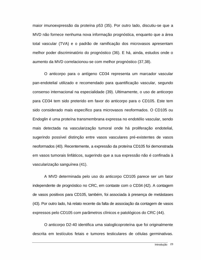
30% dos pacientes com CRC tratado com cirurgia potencialmente curativa irão
recidivar, sugerindo que fatores prognósticos convencionais não são suficientes,
havendo necessidade de fatores adicionais (10,11).
O Fator de crescimento endotelial vascular (VEGF, do inglês vascular
endothelial growth factor) é uma importante glicoproteína estimuladora da
angiogênese. A família VEGF inclui o VEGF-A, VEGF-B, ambos ligantes do receptor
VEGF-R1, mediador da angiogênese, bem como o VEGF-C e VEGF-D, ambos
importantes ligantes do receptor VEGF-R3, envolvido na linfangiogênese. Além do
VEGF-R1, o VEGF-A é transmitido via receptor VEGF-R2, sendo um importante
fator de sinalização para proliferação e migração endotelial vascular (15).
A expressão de VEGF-A no citoplasma das células colorretais pelo estudo
imunoistoquímico (IQ) é maior em adenomas do que na mucosa colorretal normal e
aumenta ainda mais no adenocarcinoma (16-19). O aumento de VEGF-A
também é correlacionado com pior prognóstico na maior parte dos estudos (20-
23). Enquanto outros não conseguiram confirmar esta correlação (24,25). A
expressão da proteína VEGF-C apresenta resultados controversos na literatura
também, com trabalhos associando seu aumento com envolvimento linfático ou
metástases, enquanto outros não obtiveram estes resultados (26-32).
A quantificação vascular demonstra igualmente resultados divergentes
nos CRC. Diversos trabalhos demonstraram pior prognóstico com o aumento de
contagem microvascular - densidade de microvasos (MVD) (33,34,35). Também
foi notado que a MVD está associada com metástases hematogênicas e com a
Introdução 23
maior imunoexpressão da proteína p53 (35). Por outro lado, discutiu-se que a
MVD não fornece nenhuma nova informação prognóstica, enquanto que a área
total vascular (TVA) e o padrão de ramificação dos microvasos apresentam
melhor poder discriminatório do prognóstico (36). E há, ainda, estudos onde o
aumento da MVD correlacionou-se com melhor prognóstico (37,38).
O anticorpo para o antígeno CD34 representa um marcador vascular
pan-endotelial utilizado e recomendado para quantificação vascular, segundo
consenso internacional na especialidade (39). Ultimamente, o uso de anticorpo
para CD34 tem sido preterido em favor do anticorpo para o CD105. Este tem
sido considerado mais específico para microvasos neoformados. O CD105 ou
Endoglin é uma proteína transmembrana expressa no endotélio vascular, sendo
mais detectada na vascularização tumoral onde há proliferação endotelial,
sugerindo possível distinção entre vasos vasculares pré-existentes de vasos
neoformados (40). Recentemente, a expressão da proteína CD105 foi demonstrada
em vasos tumorais linfáticos, sugerindo que a sua expressão não é confinada à
vascularização sanguínea (41).
A MVD determinada pelo uso do anticorpo CD105 parece ser um fator
independente de prognóstico no CRC, em contaste com o CD34 (42). A contagem
de vasos positivos para CD105, também, foi associada à presença de metástases
(43). Por outro lado, há relato recente da falta de associação da contagem de vasos
expressos pelo CD105 com parâmetros clínicos e patológicos do CRC (44).
O anticorpo D2-40 identifica uma sialoglicoproteína que foi originalmente
descrita em testículos fetais e tumores testiculares de células germinativas.
Introdução 24
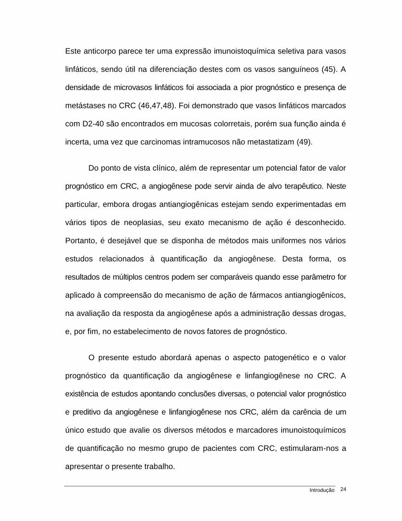
Este anticorpo parece ter uma expressão imunoistoquímica seletiva para vasos
linfáticos, sendo útil na diferenciação destes com os vasos sanguíneos (45). A
densidade de microvasos linfáticos foi associada a pior prognóstico e presença de
metástases no CRC (46,47,48). Foi demonstrado que vasos linfáticos marcados
com D2-40 são encontrados em mucosas colorretais, porém sua função ainda é
incerta, uma vez que carcinomas intramucosos não metastatizam (49).
Do ponto de vista clínico, além de representar um potencial fator de valor
prognóstico em CRC, a angiogênese pode servir ainda de alvo terapêutico. Neste
particular, embora drogas antiangiogênicas estejam sendo experimentadas em
vários tipos de neoplasias, seu exato mecanismo de ação é desconhecido.
Portanto, é desejável que se disponha de métodos mais uniformes nos vários
estudos relacionados à quantificação da angiogênese. Desta forma, os
resultados de múltiplos centros podem ser comparáveis quando esse parâmetro for
aplicado à compreensão do mecanismo de ação de fármacos antiangiogênicos,
na avaliação da resposta da angiogênese após a administração dessas drogas,
e, por fim, no estabelecimento de novos fatores de prognóstico.
O presente estudo abordará apenas o aspecto patogenético e o valor
prognóstico da quantificação da angiogênese e linfangiogênese no CRC. A
existência de estudos apontando conclusões diversas, o potencial valor prognóstico
e preditivo da angiogênese e linfangiogênese nos CRC, além da carência de um
único estudo que avalie os diversos métodos e marcadores imunoistoquímicos
de quantificação no mesmo grupo de pacientes com CRC, estimularam-nos a
apresentar o presente trabalho.

25
2. Objetivos
Objetivos 26
2.1. Objetivo Geral
Analisar o perfil imunoistoquímico de diferentes marcadores relacionados à
angiogênese e linfangiogênese previamente descritos como tendo valor prognóstico
controverso no CRC, utilizando métodos de mensuração: área vascular total
(TVA) e densidade microvascular (MVD), além da porcentagem e a intensidade de
células tumorais positivas para VEGF-A e VEGF-C. Comparar estes parâmetros
nos tecidos colorretais não neoplásicos, adenomas e carcinomas; compará-los entre
adenomas de pacientes sem e com carcinomas e correlacioná-los com fatores
morfológicos e clínicos de utilidade bem estabelecida nos carcinomas colorretais.
2.2. Objetivos Específicos
Artigo 1 – Aceito para publicação no Brazilian Journal of Medical
and Biological Research
Comparar a angiogênese e os vasos linfáticos entre os grupos de
adenomas, um sendo de pacientes sem CRC e o outro de pacientes
com CRC em local distinto na mucosa, usando densidade microvascular
e área total vascular através de análise por programa de imagem em
computador.
Determinar diferenças na angiogênese e os vasos linfáticos entre estes
dois grupos de adenomas e, se é possível inferir que a presença de
carcinoma possa influenciar o adenoma.
Objetivos 27
Artigo 2 – Submetido para publicação na revista Modern Pathology
Determinar o meio de quantificação de angiogênese e linfangiogênese
mais fidedigno como fator prognóstico nos carcinomas colorretais dentre
os diversos marcadores imunoistoquímicos e métodos de quantificação
relacionados a estes processos.
Correlacionar os marcadores e métodos de quantificação relacionados à
angiogênese e linfangiogênese com fatores morfológicos e clínicos
de valor clínico bem estabelecido nos carcinomas colorretais.
Comparar angiogênese e linfangiogênese em tecidos colorretais não
neoplásicos, adenomas e carcinomas.

28
3. Publicações
Publicações 29
3.1. Artigo 1
COMPARISON OF BLOOD NEOANGIOGENESIS AND LYMPHATIC
VASCULARIZATION IN COLORECTAL ADENOMAS FROM PATIENTS WITH
AND WITHOUT CONCOMITANT COLORECTAL CANCER
Authors:
L. R. Moreira1, A. A. Schenka2, P. Latuf Filho3, C. S. P. Lima4, M. A. S. Trevisan5,
J. Vassallo5
Universidade Estadual de Campinas - Unicamp
[1- MD; 2- MD, PhD; 3- PhD; 4- MD, PhD, Professor of Oncology; 5- MD, PhD,
Professor of Pathology]
Correspondence to:
José Vassallo
Laboratório de Patologia Investigativa e Molecular - CIPED; Universidade
Estadual de Campinas
Rua Tessália Viera de Camargo, 126
Caixa Postal 6111. CEP 13083-970 - Campinas – São Paulo, BRASIL
Phone: +55 -19 – 3521.7541 Fax: +55 -19 – 3289.3897
e-mail: [email protected] ; [email protected].
Abbreviated running title: Angiogenesis and lymphatic vessels in colorectal
adenomas
Key words: Angiogenesis; Colorectal adenoma; Colorectal cancer;
Immunohistochemistry; CD105; D2-40
Publicações 30
Blood and lymphatic vessel proliferation is essential for tumor growth and
progression. Most colorectal carcinomas develop from adenomas (adenoma-
carcinoma sequence) in a process due to accumulation of molecular genetic
alterations. About 5% of adenomatous polyps are expected to become
malignant, but data on the differential angiogenic patterns of these lesions in
patients with and without concomitant cancer are missing. The aim of the
present study is to compare the angiogenic and lymphatic patterns of
adenomatous polyps from patients with and without sporadic cancer. Thirty
adenomatous polyps (15 from patients with another principal malignant lesion,
and 15 from patients without cancer) were submitted to immunohistochemical
staining for CD105 (new formed blood vessels marker) and D2-40 (lymphatic
endothelium marker). Microvessel density and total vascular area were
determined by computer image analysis. Image was evaluated using this
program to quantify the stained and total areas and to assess the number of
microvessels. Adenomas from patients with carcinoma showed significantly
higher values of total vascular area determined by immunostaining for CD105
(cut-off value = 4386 µm2; p = 0.019) and of lymphatic microvessel density
determined by immunostaining with D2-40 (cut-off value = 11.5; p = 0.041) when
compared with those from patients without cancer. The present data indicate a
significant increase in blood microvascular area and in lymphatic microvascular
counts in adenomas removed from patients with cancer.
Publicações 31
Introduction
Colorectal carcinoma (CRC) represents an important cause of cancer
mortality in industrialized countries. Most cases (80%) correspond to sporadic
carcinomas and arise from colorectal adenomas (1). The adenoma-carcinoma
sequence involves accumulations of genetic alterations causing progressive
disorders in the cell cycle (2). About 5% adenomatous polyps will probably
become malignant (3).
Angiogenesis plays an important role in tumor progression and metastasis in
most human solid tumors (4-7). This fact has led to new perspectives in the
research of prognostic indicators and of new therapeutic strategies. The fact that
20-30% of patients with CRC treated with potentially curative surgery succumb
from recurrent disease suggests that the conventional prognostic factors are not
totally sufficient (8-11).
Most studies have evaluated angiogenesis as a potential prognostic or
predictive factor in CRC both in early and advanced disease (12,13). Comparison of
literature data has been frequently hindered by variations in patient management,
such as indication of adjuvant therapy, and in the methods for analysis of
angiogenesis (different immunohistochemical markers, quantification methods,
parameters quantified, etc.) (12,14,15).
Endoglin (CD105) is a membrane glycoprotein, part of the TGF-beta receptor
complex, involved in angiogenesis. Markers for this protein identify newly formed
blood vessels, representing a helpful tool in the evaluation of neoangiogenesis
(16). In CRC, CD105 has been correlated to prediction of metastasis (17).
D2-40 is a monoclonal antibody directed against the oncofetal antigen
M2A, present in germ cells, lymphatic endothelium, and some neoplasms such
as mesotheliomas (18,19). Using this antibody, it has been demonstrated that the
colorectal mucosa indeed presents lymphatic vessels in normal, inflammatory and
neoplastic conditions (20,21). However, in contrast to CD105, present in newly
formed blood vessels, the presence of D2-40 does not indicate the degree of
Publicações 32
neolymphangiogenesis. Lymphatic vessel density assessed by D2-40 has been
correlated the prediction of metastasis and with a poor outcome of CRC (22-24).
In spite of the many reports on angiogenesis in cancer, data on the
differential angiogenic patterns of adenomatous lesions in patients with and
without concomitant CRC are not available. The purpose of the present study
was to compare blood angiogenesis and lymphatic vessels between two groups
of adenomatous polyps, one from patients with concomitant CRC at another site
of the mucosa, and the other from patients without carcinoma, using microvessel
counting and total vascular area determination with image analysis software.
Our aim was to determine potential differences between adenomatous polyps
from the two groups of patients, and weather the presence of carcinoma could
influence the vascularization of colorectal adenomas.
Material and Methods
Tissue samples
A retrospective study was performed on 30 low-grade adenomatous
polyps removed by endoscopy or surgery from 15 patients with sporadic CRC
and 15 patients without carcinoma. The latter group did not show evidence of
carcinoma from the time of the procedure throughout a 5-year follow-up.
Hamartomatous and inflammatory polyps were excluded from the study.
The samples were selected from the files of the Department of Pathology,
State University of Campinas, Campinas, SP, Brazil, and included patients
diagnosed from 1987 to 2003. The group of patients with CRC consisted of 8
males and 7 females ranging from 33 to 82 years (median: 61 years); 5 cases
were staged as I, 7 as II, 2 as III, and 1 as IV, according to the TNM pathological
staging system (25). Only low-grade adenomatous polyps which had been
removed from the colorectal mucosa concomitantly to or soon after the
diagnosis of the main malignant lesion, without the effect of neoadjuvant therapy
were included. There were 8 tubular, 6 tubulovillous, and 1 villous adenomas.
Publicações 33
In the group of patients without a diagnosis of CRC 9 were males and 6
were females ranging from 20 to 82 years (median: 56 years). There were 11
tubular and 4 tubulovillous adenomas.
Immunohistochemistry
Tissue specimens had been fixed in 10% formalin and embedded in paraffin
and 3-µm thick sections were placed on silanized slides. Endogenous peroxidase
activity was quenched by incubating the slides with 3% H2O2 for 10 min. Antigen
retrieval was achieved by microwaving tissue sections in 10 mM citrate buffer, pH
6.0, in four cycles of 5 min each. Sections were incubated at room temperature
for 20 min with mouse monoclonal antibodies to CD105 (Endoglin, Clone SN6h,
Dako, USA; diluted 1:15) and to D2-40 (Dako, diluted 1:400). Antigen-antibody
binding was detected using the Advance system (Dako). Internal and external
positive and negative controls were run concomitantly in each reaction batch.
Evaluation of immunohistochemistry
Digital images from two "hot spot" fields stained by each marker were
captured. One area corresponded to the upper/inner portions of the lesions, and
the other to the deeper area of the polyps. The upper/inner areas were grouped
together because there were some small adenomas in which both areas
appeared in the same image. Digitalization was done at 200X magnification, 120
dpi, using a digital camera (Leica DFC360 FX, Leica, Germany) connected to a
bright field microscope (Leica DM5000 B).
The images were examined with image analysis software (Leica QWin
Standard V3, Microsystem Imaging, Leica) set to detect color intensities in a
fixed and constant range. Every image was evaluated using this standardized
program to quantify the proportion between stained and total areas and to
assess the number of microvessels. Immunostained blood and lymphatic
vessels were marked with a circle by the pathologist who analyzed the image to
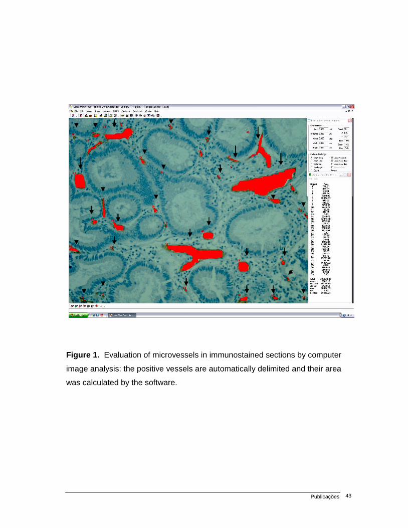
perform automated quantification. An example of the resulting image prepared
for analysis after selection of the immunostained vessels is shown in Figure 1.
Publicações 34
This resulted in the evaluation of two parameters for each marker: microvessel
density (MVD) and total vascular area (TVA).
Statistical analysis
Statistical analysis was performed using the SAS System for Windows software
package (version 9.1.3). For the quantitative parameters, the minimum and maximum
values, mean, standard deviation and median were analyzed. For the qualitative
variables, the absolute and relative frequencies were analyzed. The non-parametric
Mann-Whitney test was used to compare two groups and the Kruskal-Wallis test
was used for three or more groups. The Dunn comparison test was used for
multiple comparisons. The level of significance was set at 5% in all analyses.
Results
CD105 in adenomas from patients with CRC
MVD ranged from 0 to 15 (median 5; mean 5. 53) in "hot spots" of the
upper/inner parts of adenomas and from 0 to 37 (median 1; mean 5.40) in the
deeper areas. TVA ranged from 0 to 219905 µm2 (median 7498; mean 25681.33
µm2) in the "hot spots" of the upper/inner regions and from 0 to 27255 µm2
(median 755; mean 6215 µm2) in the deeper area.
CD105 in adenomas from patients without CRC
MVD ranged from 0 to 10 (median 2; mean 3.20) in "hot spots" of the
upper/inner part of adenomas and from 0 to 10 (median 1; mean 2.53) in the
deeper area. TVA ranged from 0 to 19660 µm2 (median 183; mean 3810.20
µm2), in the "hot spots" of the upper/inner parts and from 0 to 66522 µm2
(median 143; mean 8114.40 µm2) in the deeper area. The results for CD105 are
summarized in Table 1.
Publicações 35
D2-40 in adenomas in patients with CRC
MVD ranged from 4 to 177 (median 12; mean 29.20) in "hot spots" of the
upper/inner parts of the adenoma and TVA ranged from 85 to 40691 µm2
(median 3914; mean 9232 µm2). MVD ranged from 1 to 37 (median 12; mean
14.67) in "hot spots" of the deeper area, and TVA ranged from 676 to 64650
µm2 (median 9563; mean 18473.73 µm2).
D2-40 in adenomas from patients without CRC
MVD ranged from 1 to 18 (median 8; mean 8.40) in "hot spots" of the
upper/inner parts of the adenoma and TVA ranged from 1 to 53112 µm2 (median
5836; mean 11592.47 µm2). MVD ranged from 2 to 14 (median 8; mean 8.27) in
"hot spots" of the deeper area and TVA ranged from 265 to 44491 µm2 (median
8687; mean 14430.73 µm2). The results for D2-40 are summarized in Table 2.
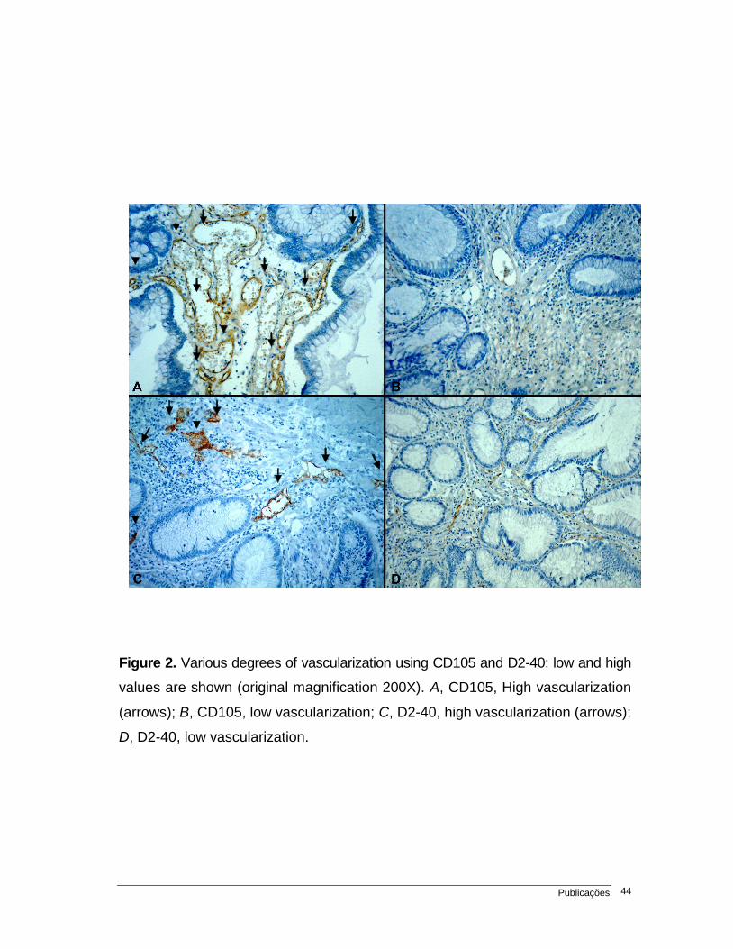
A plate with illustrations of cases with lower and higher vascularization
using both markers is shown in Figure 2.
Statistical analysis of these data showed significantly higher values of TVA
determined by Immunostaining for CD105 (p = 0.019) and of MVD determined
by Immunostaining for D2-40 (p = 0.041) when compared with those from
patients without CRC.
For both markers, there was no significant difference among histological
types of adenoma (tubular, tubulovillous and villous) and MVD or TVA counts, in
the groups of patients with and without CRC.
The cut-off value for TVA determined by CD105 in the upper/inner parts of
the adenomas was 4386, or approximately 4400 µm2 (sensitivity and specificity:
66.7%; predictive positive and predictive negative values: 66.7%; accuracy:
66.7%). The cut-off value of MVD determined by D2-40 in the upper/inner parts of
the adenomas was 11.5 (60.0% sensitivity, 66.7% specificity, 64.3% predictive
positive value, 62.5% predictive negative value, and 63.3% accuracy).
Publicações 36
Discussion
The present data indicate a significant increase in blood microvascular
area and in lymphatic microvascular counts in the upper and inner portions of
adenomas removed from patients with CRC compared to those without carcinoma.
Cancer cells might have an influence on the vascularization of adenomas,
evidence supported by data showing increased levels of angiogenic factors in
colorectal tissues distant from the primary tumor. Hanrahan et al. (26) showed
that vascular endothelial growth factor (VEGF) plays a role early in tumor
development at the stage of adenoma formation. Moreover, increased levels of
VEGF in normal tissue collected from sites distant from the primary tumor have
indicated environmental changes that could help explain our findings, although
this was not directly assessed in our material.
The more significant increase in lymphatic MVD in the upper/inner areas
of the adenomas is in keeping with a previous study reporting a more superficial
location of lymphatic vessels in adenomas. This finding supports the hypothesis
sustained by Fogt et al. (20) that superficial lymphatic vessels may be immature in
normal colonic mucosa and may not communicate with deeper vessels, changing
and maturing through the adenoma-carcinoma process. An equivalent assumption
could be made about the increase in newly formed blood vessels detected by
CD105, which suggests that they may originate superficially on adenomas,
developing and meeting deeper vessels during the progression of malignancy.
The assessment of TVA using immunostaining for CD105 showed
significantly higher values in adenomas from patients with CRC, while assessment
of MVD did not. The opposite was seen in the assessment of lymphatic vessels
using the D2-40 antibody: in contrast to MVD, TVA did not differ significantly
between the two groups of lesions.
These differences might reflect variations in the mechanisms of proliferation
of blood and lymphatic vessels, the former affecting predominantly architectural
scores, and the latter numerical scores. Unlike normal blood vessels, newly
formed blood vessels incorporated during tumor angiogenesis are tortuous and
Publicações 37
dilated, a fact that could explain the higher value of TVA using CD105 in patients
with CRC, an aspect supported by experimental studies (27). The higher MVD
evaluated by D2-40 in patients with CRC could be explained by recent evidence
showing elevated lymphatic vessel counts as an event preceding the increased
number of blood vessels in early gastrointestinal tumors (28). It should be noted
that computer image analysis seems to be more objective and reproducible,
reducing to minimum intraobserver variability from case to case, and increasing
the reliability of information in the study of angiogenesis (29).
The findings reported in the present study support the notion that
neoangiogenesis and elevated lymphatic vessel counts occur in colorectal
adenomas from patients with CRC, allowing us to assume that either angiogenic
factors produced by the carcinoma or constitutional defects of the colorectal
epithelial cells might account for these observations.
Publicações 38
References
1. Ilyas M, Straub J, Tomlinson IP, Bodmer WF. Genetic pathways in colorectal
and other cancers. Eur J Cancer 1999; 35: 335-351.
2. Vogelstein B, Fearon ER, Hamilton SR, Kern SE, Preisinger AC, Leppert M,
et al. Genetic alterations during colorectal-tumor development. N Engl J Med
1988; 319: 525-532.
3. Boyle P, Leon ME. Epidemiology of colorectal cancer. Br Med Bull 2002; 64:
1-25.
4. Vieira SC, Zeferino LC, Da Silva BB, Aparecida PG, Vassallo J, Carasan
GA, et al. Quantification of angiogenesis in cervical cancer: a comparison
among three endothelial cell markers. Gynecol Oncol 2004; 93: 121-124.
5. Offersen BV, Borre M, Sorensen FB, Overgaard J. Comparison of methods
of microvascular staining and quantification in prostate carcinoma: relevance
to prognosis. APMIS 2002; 110: 177-185.
6. Sasano H, Suzuki T. Pathological evaluation of angiogenesis in human
tumor. Biomed Pharmacother 2005; 59 (Suppl 2): S334-S336.
7. Szabo S, Sandor Z. The diagnostic and prognostic value of tumor
angiogenesis. Eur J Surg Suppl 1998; 99-103.
8. Bendardaf R, Lamlum H, Pyrhonen S. Prognostic and predictive molecular
markers in colorectal carcinoma. Anticancer Res 2004; 24: 2519-2530.
9. Ratto C, Sofo L, Ippoliti M, Merico M, Doglietto GB, Crucitti F. Prognostic
factors in colorectal cancer. Literature review for clinical application. Dis
Colon Rectum 1998; 41: 1033-1049.
10. Buyse M, Piedbois P. Should Dukes' B patients receive adjuvant therapy? A
statistical perspective. Semin Oncol 2001; 28: 20-24.
11. Reinmuth N, Parikh AA, Ahmad SA, Liu W, Stoeltzing O, Fan F, et al.
Biology of angiogenesis in tumors of the gastrointestinal tract. Microsc Res
Tech 2003; 60: 199-207.
Publicações 39
12. Tarta C, da Silva V, Teixeira CR, Prolla JC, Meurer L, Neto CC, et al. Digital
image analysis and stereology of angiogenesis in polypoid and nonpolypoid
colorectal adenomas. Anal Quant Cytol Histol 2004; 26: 201-206.
13. Matsuura T, Kuratate I, Teramachi K, Osaki M, Fukuda Y, Ito H. Thymidine
phosphorylase expression is associated with both increase of intratumoral
microvessels and decrease of apoptosis in human colorectal carcinomas.
Cancer Res 1999; 59: 5037-5040.
14. Compton CC. Colorectal carcinoma: diagnostic, prognostic, and molecular
features. Mod Pathol 2003; 16: 376-388.
15. Pavlopoulos PM, Konstantinidou AE, Agapitos E, Kavantzas N,
Nikolopoulou P, Davaris P. A morphometric study of neovascularization in
colorectal carcinoma. Cancer 1998; 83: 2067-2075.
16. Thompson WD, Shiach KJ, Fraser RA, McIntosh LC, Simpson JG. Tumours
acquire their vasculature by vessel incorporation, not vessel ingrowth. J
Pathol 1987; 151: 323-332.
17. Romani AA, Borghetti AF, Del Rio P, Sianesi M, Soliani P. The risk of
developing metastatic disease in colorectal cancer is related to CD105-
positive vessel count. J Surg Oncol 2006; 93: 446-455.
18. Van den Eynden GG, Van der Auwera I, Van Laere SJ, Colpaert CG, van
Dam P, Dirix LY, et al. Distinguishing blood and lymph vessel invasion in
breast cancer: a prospective immunohistochemical study. Br J Cancer 2006;
94: 1643-1649.
19. Ordonez NG. Podoplanin: a novel diagnostic immunohistochemical marker.
Adv Anat Pathol 2006; 13: 83-88.
20. Fogt F, Zimmerman RL, Ross HM, Daly T, Gausas RE. Identification of
lymphatic vessels in malignant, adenomatous and normal colonic mucosa
using the novel immunostain D2-40. Oncol Rep 2004; 11: 47-50.
21. Fogt F, Pascha TL, Zhang PJ, Gausas RE, Rahemtulla A, Zimmerman RL.
Proliferation of D2-40-expressing intestinal lymphatic vessels in the lamina
propria in inflammatory bowel disease. Int J Mol Med 2004; 13: 211-214.
Publicações 40
22. Longatto-Filho A, Pinheiro C, Ferreira L, Scapulatempo C, Alves VA,
Baltazar F, et al. Peritumoural, but not intratumoural, lymphatic vessel
density and invasion correlate with colorectal carcinoma poor-outcome
markers. Virchows Arch 2008; 452: 133-138.
23. Yan G, Zhou XY, Cai SJ, Zhang GH, Peng JJ, Du X. Lymphangiogenic and
angiogenic microvessel density in human primary sporadic colorectal
carcinoma. World J Gastroenterol 2008; 14: 101-107.
24. Matsumoto K, Nakayama Y, Inoue Y, Minagawa N, Katsuki T, Shibao K, et
al. Lymphatic microvessel density is an independent prognostic factor in
colorectal cancer. Dis Colon Rectum 2007; 50: 308-314.
25. International Union against Cancer. TNM classification of malignant
tumours. 5th edn. Geneva: International Union against Cancer
(http://www.uicc.org); 2004.
26. Hanrahan V, Currie MJ, Gunningham SP, Morrin HR, Scott PA, Robinson
BA, et al. The angiogenic switch for vascular endothelial growth factor (VEGF)-
A, VEGF-B, VEGF-C, and VEGF-D in the adenoma-carcinoma sequence during
colorectal cancer progression. J Pathol 2003; 200: 183-194.
27. Vermeulen PB, Gasparini G, Fox SB, Colpaert C, Marson LP, Gion M, et al.
Second international consensus on the methodology and criteria of
evaluation of angiogenesis quantification in solid human tumours. Eur J
Cancer 2002; 38: 1564-1579.
28. Gao Y, Zhong WX, Mu DB, Yuan YP, Zhang YH, Yu JM, et al. Distributions of
angiogenesis and lymphangiogenesis in gastrointestinal intramucosal tumors.
Ann Surg Oncol 2008; 15: 1117-1123.
29. Van der Auwera I, Cao Y, Tille JC, Pepper MS, Jackson DG, Fox SB, et al.
First international consensus on the methodology of lymphangiogenesis
quantification in solid human tumours. Br J Cancer 2006; 95: 1611-1625.
Publicações 41
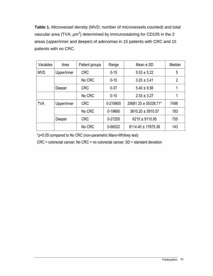
Table 1. Microvessel density (MVD; number of microvessels counted) and total
vascular area (TVA; µm2) determined by immunostaining for CD105 in the 2
areas (upper/inner and deeper) of adenomas in 15 patients with CRC and 15
patients with no CRC.
Variables Area Patient groups Range Mean ± SD Median
MVD Upper/inner CRC 0-15 5.53 ± 5.22 5
No CRC 0-10 3.20 ± 3.41 2
Deeper CRC 0-37 5.40 ± 9.56 1
No CRC 0-10 2.53 ± 3.27 1
TVA Upper/inner CRC 0-219905 25681.33 ± 55338.71* 7498
No CRC 0-19660 3810.20 ± 5910.57 183
Deeper CRC 0-27255 6215 ± 9110.95 755
No CRC 0-66522 8114.40 ± 17675.36 143
*p<0.05 compared to No CRC (non-parametric Mann-Whitney test)
CRC = colorectal cancer; No CRC = no colorectal cancer; SD = standard deviation
Publicações 42
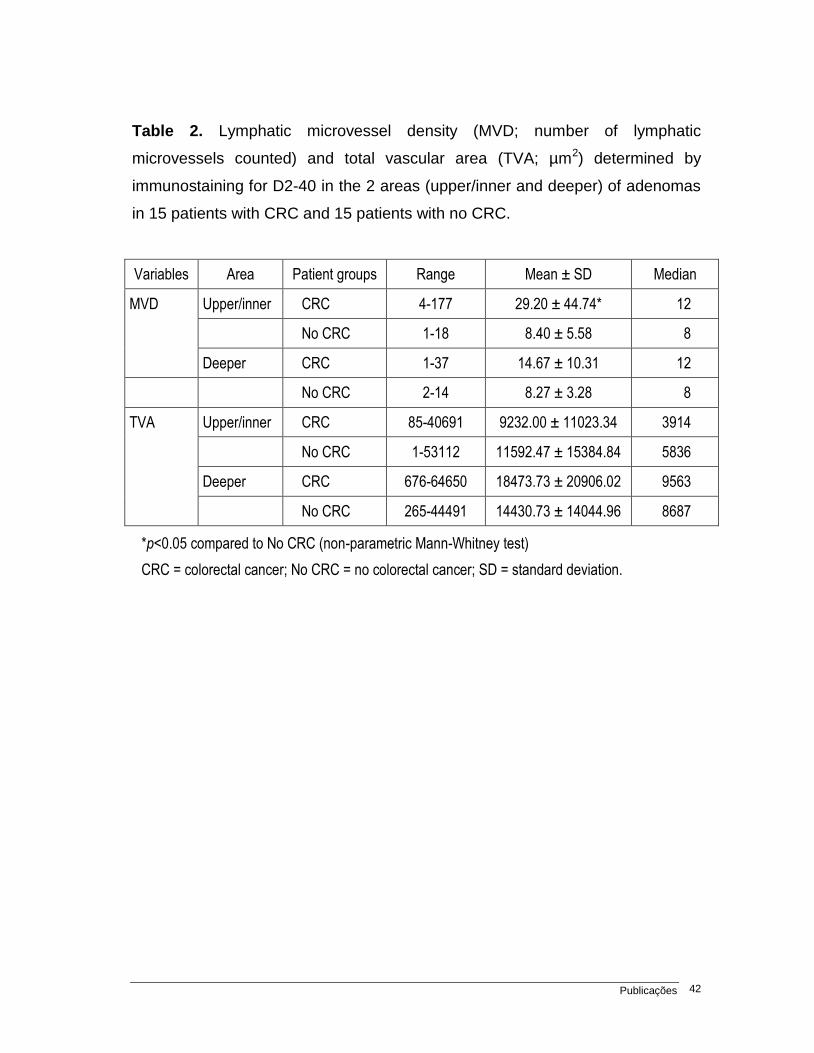
Table 2. Lymphatic microvessel density (MVD; number of lymphatic
microvessels counted) and total vascular area (TVA; µm2) determined by
immunostaining for D2-40 in the 2 areas (upper/inner and deeper) of adenomas
in 15 patients with CRC and 15 patients with no CRC.
Variables Area Patient groups Range Mean ± SD Median
MVD Upper/inner CRC 4-177 29.20 ± 44.74* 12
No CRC 1-18 8.40 ± 5.58 8
Deeper CRC 1-37 14.67 ± 10.31 12
No CRC 2-14 8.27 ± 3.28 8
TVA
Upper/inner CRC 85-40691 9232.00 ± 11023.34 3914
No CRC 1-53112 11592.47 ± 15384.84 5836
Deeper CRC 676-64650 18473.73 ± 20906.02 9563
No CRC 265-44491 14430.73 ± 14044.96 8687
*p<0.05 compared to No CRC (non-parametric Mann-Whitney test)
CRC = colorectal cancer; No CRC = no colorectal cancer; SD = standard deviation.
Publicações 43
Figure 1. Evaluation of microvessels in immunostained sections by computer
image analysis: the positive vessels are automatically delimited and their area
was calculated by the software.
Publicações 44
Figure 2. Various degrees of vascularization using CD105 and D2-40: low and high
values are shown (original magnification 200X). A, CD105, High vascularization
(arrows); B, CD105, low vascularization; C, D2-40, high vascularization (arrows);
D, D2-40, low vascularization.
Publicações 45
3.2. Artigo 2
ANGIOGENESIS AND LYMPHANGIOGENESIS IN COLORECTAL CARCINOMA:
COMPARISON BETWEEN QUANTIFICATION METHODS AND DIFFERENT
IMMUNOHISTOCHEMICAL MARKERS WITH ANATOMOPATHOLOGIC
PROGNOSTIC FACTORS
Luciana R. Moreira1, André A. Schenka2, Paulo Latuf-Filho3, Carmen S. Passos-
Lima4, Miriam A. S. Trevisan5, José Vassallo5
[1- MD; 2- MD, PhD; 3- PhD; 4- MD, PhD, Professor of Oncology; 5- MD, PhD,
Professor of Pathology]
State University of Campinas Medical School, Unicamp
Correspondence to:
José Vassallo
E-mail: [email protected] ; [email protected]
Rua Tessália Viera de Camargo, 126
P.O Box 6111; Zip Code 13083-887 - Campinas – São Paulo, BRAZIL
Phone: +55 -19 – 3521.8958 Fax: +55 -19 – 3289.3897
Support: FAEPEX- State University of Campinas Medical School, São Paulo-
Brazil; Conselho Nacional de Pesquisa Científica (CNPq)
Conflict of interests: The authors declare no conflict of interests.
Authors’ contributions:
LRM and JV participated in all phases of the study, from design to final
manuscript; AAS and PLF participated mainly in the preparation and analysis of
Publicações 46
immunohistochemical reactions; CSPL was responsible for clinical follow-up and
data management; MAST reviewed all histopathological diagnoses. All authors
read and are in accordance with the final manuscript.
Ethical aspects:
The present study was approved by the Committee for Ethics in Medical
Research of our Institution (State University of Campinas Medical School)
Abstract
Background: Blood and lymphatic vessels play an important role in the
progression of solid tumors, and have been considered as potential targets for
therapy. Thus, reliable evaluation of these parameters may have an impact on
patients’ management. Analysis of angiogenesis and lymphangiogenesis in
colorectal cancer (CRC) is controversial in the literature, which may be due to
variations in the methods of analysis: the precise location within the tumor
sample, the choice of immunohistochemical markers and the method of
quantification have been differently evaluated in the literature. Objectives:
Therefore, in the present study it was aimed to search for a reliable approach in
the quantification of angiogenesis and lymphangiogenesis as prognostic factor
in CRC. It was also intended to compare these parameters between non
neoplastic tissue, adenomas and cancer, in order to contribute to the
understanding of angio- and lymphangiogenesis in the progression of these
lesions. Methods: 60 sporadic CRC, 30 colorectal adenomas and 10 colorectal
non neoplastic tissues were submitted to immunohistochemical evaluation of
CD31, CD34, CD105, VEGF-A, VEGF-C and D2-40. Microvessel density (MVD)
and total vascular area (TVA) were determined by computer image analysis for
all markers. Results: The majority of markers showed progressive vessel
counts from non neoplastic tissue to carcinoma, both for MVD and TVA. Only
MVD determined by immunostaining for CD34 in the central areas of the lesion
was significantly correlated with recurrence or metastasis (p=0.04) and survival
Publicações 47
rates (p=0.02) in patients with CRC. Conclusions: Our results corroborate the
increasing in vascularization of carcinoma and suggest that MVD determined
with staining for CD34 in the inner part of the tumor might represent a valuable
parameter to be considered in the management of patients with CRC, as it is
more closely related with relapse/metastasis and survival.
Keywords: angiogenesis; lymphangiogenesis; colorectal cancer;
immunohistochemistry; CD31; CD34; CD105; VEGF-A; VEGF-C; D2-40.
Publicações 48
Introduction
Colorectal carcinoma (CRC) is an important cause of mortality
worldwide.1 The fact that tumor growth is dependent on angiogenesis has
supported recent researches for new prognostic parameters and in the
development of novel therapeutic strategies. This is welcome, in view that 20%-
30% of patients with CRC treated with potentially curative surgery, will succumb
from recurrent disease, suggesting that the conventional prognostic factors may
not be sufficient, and that additional parameters, either morphological or
molecular, are needed for clinical management. 2, 3 In spite of its importance, data
concerning the prognostic value of parameters related to angiogenesis in CRC
remain controversial.
The vascular endothelial growth factor (VEGF) has been identified as an
important family of glycoproteins stimulating vascularity. The VEGF family
includes VEGF-A (or VEGF) and VEGF-B, both ligands for receptor VEGF-R1,
which mediates angiogenesis; VEGF-C and VEGF-D, both important members
binding to the receptor VEGF-R3, which is mainly involved in lymphangiogenesis.
VEGF-A signaling promoting endothelial proliferation, migration and survival is
predominantly transmitted via VEGF-R2.4 A variable proportion of cancer cells
present a cytoplasmic immunostaining for VEGF, which is progressively expressed
in adenomas and in CRC, when compared to colorectal normal tissue. 5, 6
Protein expression of VEGF has been associated with worse prognosis in most
studies7-9, but this has not always been the case, probably due to the different
thresholds of positivity used in the studies.10-12 VEGF-C protein expression has
also involved some controversial data in the literature, with some reports
correlating its expression with lymphatic involvement or the presence of
metastasis, and others not.13-19
Vascular quantification has been the matter of diverging results, some
manuscripts showing correlation of microvessel counting with poorer outcome or
lymph nodes metastases.20 -23 It has been also stated that outcome of patients
with CRC was not correlated with vascular counting, but with vessel ramification
and the total vascular area (TVA).24 In contrast, others have shown that higher
Publicações 49
microvascular counting was correlated with favorable outcome.25, 26
CD105 or Endoglin, a transmembrane protein highly expressed on human
vascular endothelium, is up-regulated in tumor vasculature and proliferating
cells, suggesting the possibility to distinguish newly formed tumor associated
endothelial cells from pre-existing vessels.27, 28 Recently, CD105 was showed in
tumor lymphatics, suggesting that is not confined to the blood vasculature.29
CD105+ microvessels have been preferentially observed in the surface area,
while CD34+ microvessels were evenly distributed in adenomas. In carcinomas,
expression of CD105, but not of CD34, presented significantly higher values in
the adenoma-carcinoma sequence.27 The microvessel counting assessed by
anti-CD105 was shown as independent prognostic parameter for survival in
CRC, in contrast to CD34.28 CD105+ vessel counts have been equally strongly
correlated with the occurrence of metastatic disease.30 On the contrary, others
have shown no significant correlation between CD105+ vessel counts and
clinicopathologic characteristics.31
D2-40 is a monoclonal antibody directed to the M2A antigen, a surface
sialoglycoprotein originally detected in germ cell neoplasia and fetal testicular
gonocytes.32 It has been also demonstrated to selectively immunoreact with the
lymphatic endothelium, but not with blood vascular endothelium. Lymphatic
vessel density was correlated with poor outcome and metastatic disease in
colorectal cancer, 33-35 but the relationship between this parameter and VEGF-C
is unclear. 22 It has been reported that lymphatic vessels labeled with D2-40 are
more superficially located in adenomas and carcinomas than previously
suspected, since intramucosal carcinomas do not metastasize.36
In view of the importance of the evaluation of angiogenesis as a potential
prognostic and predictive parameter, and taking into account the controversial
reports summarized above, it was the purpose of the present study to appraise
the clinical value of angiogenesis in CRC comparing methods of assessment
(microvascular density and total vascular area), and different immunohistochemical
markers to detect angio- and lymphangiogenesis. It was also intended to compare
Publicações 50
angio- and lymphangiogenesis counts between non neoplastic colorectal tissue,
adenomas and carcinomas.
Materials and methods
Patient selection and tissue samples: A retrospective study was performed
on 60 surgically resected sporadic colorectal carcinomas (group 1); 30
adenomatous polyps (group 2, obtained by polypectomy or surgical resection)
and 10 non neoplastic colorectal tissues (group 3, obtained from surgery for
benign conditions). All samples were selected from the files of the Department of
Anatomical Pathology, State University of Campinas Hospital (Unicamp), São
Paulo, Brazil, diagnosed from 1987 to 2003. The study was approved by the
institutional Ethics Committee for Medical Research. Patients from group 1
included 27 (45%) males and 33 (55%) females, with mean age of 60 years
(range 24 to 81 y). Tumors consisted of adenocarcinomas primarily categorized
according to the classification of the World Health Organization; pathological
staging was based on the TNM classification.37 No patient had received chemo-
or radiation therapy before surgery. Eleven cases were staged as I (18.3%), 24
as II (40%), 20 as III (33.3%), and 5 as IV (8.3%). Follow up of patients ranged
from 3 months to 13 years (median 5.34 y); 31 patients deceased. Routinely
stained slides were revised, and, from each case, one tissue sample was
selected, which included the deepest invasive tumor area, avoiding regions with
prominent inflammation and necrosis. The whole section was submitted to
vascular analysis, as recommended elsewhere.38 Group 2 consisted of 17 male
(56.66%) and 13 female (43.33%) patients with mean age of 59 y (range 20 to
82 y). Nineteen cases were classified as tubular adenoma (63.33%), 10 as
tubulovillous (33.33%) and one as villous adenoma (3.33%). Colorectal non
neoplastic tissue (group 3) was obtained from five male and five female
patients, with mean age of 55.5 y (range 32 to 71 y).
Immunohistochemistry: Tissue specimens were fixed in 10% formalin and
Publicações 51
embedded in paraffin. Sections of three µm thick were placed on silanized
slides. Endogenous peroxidase activity was quenched by incubating the slides
in 3% H2O2 for 10 minutes. Antigen retrieval was achieved by microwaving
tissue sections in 10 mM citrate buffer (pH= 6.0), four cycles of five minutes
each. Sections were incubated at room temperature for 30 minutes and then
overnight at 8 – 100 C with mouse monoclonal antibodies to CD31 (clone JC70A,
Dako, Carpenteria, CA, USA, diluted at 1:20); CD34 (clone QBEnd 10, Dako,
diluted at 1:100); CD105 (Endoglin, Clone SN6h, Dako, diluted at 1:15); D2-40
(clone D2-40, Dako, diluted at 1:400); VEGF-A (clone VG1, Dako, diluted at
1:25) and VEGF-C (clone Z-CVC7, Invitrogen, Carlsbad, CA, USA, diluted at
1:100). Antigen-antibody binding was detected using the Advance system
(Dako). Internal and external positive controls included endothelial vascular
cells, endothelial lymphatic cells and cases previously positive for VEGF-A and
C. Negative controls were represented by the same tissue sample used for
positive control, in which primary antibody was omitted.
Evaluation of Immunohistochemistry: Digital images from “hot spot” fields in
different cancer areas (inner, periphery and area of deeper invasion) were
captured at magnification x200, using a digital camera (Leica DFC360 FX,
Solms, Germany) connected to a bright field microscope (Leica DM5000 B).
Digital images from adenomas were captured at inner portions and peripheral
areas. Images from the non neoplastic tissue samples were captured from the
mucosa and the submucosa/muscular. The images were examined by image
analysis software (Leica QWin Standard V3, Microsystem Imaging) setup to
detect color intensities in a fixed and constant range. Every image was
evaluated using this program to quantify the total vascular area (TVA) stained
and to assess microvessel density (MVD). Blood and lymphatic vessel cross-
sections were counted by a semi-automated procedure in the program. An
example is shown in Figure 1.
Staining for VEGF-A and VEGF-C was evaluated using a visual score in
four grades for percentage of positive cells: grade 1, no staining or less than
Publicações 52
25% of the tumor area positive; grade 2, 25-50% of the tumor area stained;
grade 3, 50-75% of the tumor area stained; grade 4, more than 75% of the tumor
area stained. The four grades were used for intensity: grade 1, no staining;
grade 2, weak staining; grade 3, moderate staining; grade 4, strong staining.
Finally, a total score was obtained by adding the two scores. This visual system
was validated by correlating the above scores in 20 cases with values obtained
using the ACIS® Automated Cellular Imaging System (Dako; Figure 2).
Statistical methods: Statistical analysis was performed using the SAS System
for Windows software package (version 9.1.3). For the quantitative parameters,
the minimum and maximum values, mean, standard-deviation (SD) and median
were analyzed. For the qualitative variables, the absolute and relative
frequencies were analyzed. The Pearson coefficient was used to evaluate the
correlation between the visual and computer assessment of immunostaining for
VEGF (see Figure 2). For the other parameters the nonparametric Mann-
Whitney test to compare two groups and the Kruskal-Wallis test for three or
more groups were used. The Dunn's comparison test was used for multiple
comparisons. The Kaplan-Meier method was used to calculate survival curves
and log-tests were performed on the data. The Cox adjusted regression was
used for multivariate analysis. Significance level was set at a minimum of 5%.
Results
Pathologic colorectal tissue (groups 1+2) vs. colorectal non neoplastic
tissues (group 3):
Evaluation of TVA and MVD stained with anti-CD105 in all fields, as well as
the mean value of the three fields, showed significant higher values in pathologic
colorectal tissues than in normal tissues (p< 0.001). The same results were
obtained for MVD in immunostainings with D2-40 (0.001 ≤ p ≤ 0.005), TVA in
immunostainings with anti-CD31 (0.001 ≤ p ≤ 0.02), TVA with anti-CD34 in the inner
Publicações 53
field (p= 0.034) and the VEGF-A, VEGF-C grades (p < 0.008). (Tables 1 and 2).
Cancer (group 1) vs. adenomas (group 2) vs. colorectal non neoplastic
tissues (group 3):
There was significant increase in values between CD105+ TVA and MVD
in all fields (p< 0.001), CD31+ TVA and MVD in all fields (0.001< p < 0.003),
except in peripheral TVA (p = 0.051), CD34+ TVA and MVD in all fields (0.006 ≤
p ≤ 0.045), except in peripheral MVD (p = 0.115). D2-40+ MVD in all fields
(0.001 ≤ p ≤ 0.004) and the VEGF-A, VEGF-C grades (p < 0.001) increase
significantly in the sequence from non neoplastic tissue to adenoma and cancer.
The other parameters did not show significant correlation. (Tables 1 and 2).
Cancer stages I+II vs III+IV:
For practical statistical analysis, we grouped the stages in low (I+II) and
high (III+IV). Tumor staging (I+II vs III+IV) showed significant correlation with
overall survival (OS) (p = 0.0003). There was no significant correlation between
the angiogenic parameters for both stage groups. CD34+ MVD in the inner part
of the tumor presented higher values in more advanced stages of cancer,
although this difference did not reach significance (p= 0.098). (Table 3).
Tumor type:
CD105+ MVD at the periphery of the tumor showed higher values in
poorly differentiated carcinoma than in the mucinous type (p= 0.036). The same
was found for CD31+ TVA at the periphery of the tumor (p= 0.032), CD31+ MVD
in all fields (0.010 ≤ p ≤ 0.043). CD34+ MVD in the deeper area of invasion
showed also greater values in poorly differentiated carcinoma than in the
mucinous type (p= 0.029). (Table 3).
Cancer relapse and metastasis:
CD34+ MVD in the inner part of the tumor showed significantly higher
values in patients with relapse of the tumor or development of metastasis.
Publicações 54
Patients without relapses or metastasis showed median 32.50 µm2, mean
35.75µm2 and SD= 15.63. Patients with relapse or metastasis presented median
42.50 µm2, mean 43.89 µm2 and SD= 16.52 (p= 0.04). Likewise, the MVD mean
values of all three fields stained for CD34 were higher in patients who developed
metastasis or relapse (p= 0.050). The other parameters did not show significant
correlations. (Table 3)
Cancer overall survival:
Analysis preformed by Kaplan-Meier survival rate showed significant
association only with high CD34+ MVD in the inner part of the tumor (p= 0.024).
The median rate of MVD and TVA assessed by CD31, CD34, CD105 and D2-40
in all cancer fields (inner, periphery, deeper invasion and the mean of the three
fields) were chosen as the cutoff point. The median rate of tumor cells
expressing VEGF-A and VEGF-C percentage, intensity and the total score were
also chosen as cutoff.
The median rate of MVD assessed by CD34 in inner field was 37
microvessels (range 12-85) and was the only significant association with overall
survival (p= 0.024).
Adjusted Cox regression analysis showed that CD34+ MVD (hazard ratio
(HR) = 3.36; 95% confidence interval (CI) =1.01-11,20; p= 0.048) and cancer
stage (HR= 6.89; 95% CI = 1.91-24,90; p= 0.003) were significant prognostic
factors for overall survival. (Table 4 and Figure 3)
VEGF-A and VEGF-C did not show significant correlations with
clinicopathological factors in cancer. (Table 5)
Discussion
The data presented herein indicate that, concerning CRC, evaluation of
MVD assessed by immunostaining for CD34 in the inner part of the tumor may
represent an independent prognostic factor. This result is important, since it may
Publicações 55
correspond to an additional parameter for clinicopathological risk evaluation,
especially for those patients which, in spite of the rather favorable indicators at
diagnosis and first therapeutic approach, will present adverse outcome. In
addition, the present study contribute to unravel the controversial data of the
related literature, since the same group of patients was studied with a variety of
quantification methods and immunohistochemical markers for blood and
lymphatic vessels.
While both blood and lymphatic vessels showed increased values in the
progression from non neoplastic tissue to adenoma and cancer, only MVD using
anti-CD34 proved to significantly correlate with the presence of relapse or
metastasis and with overall survival in the group of patients with CRC. Despite
previous studies have shown significant correlation between clinicopathological
features and CD105 or CD31, 23,30 our data are more in accordance with recent
studies in this aspect.31,39 The present results further corroborate the
recommendation to use CD34 as the eligible marker for the evaluation of tumor
angiogenesis.40
That the use of the different vessel markers is not necessarily
interchangeable may reflect variations in the mechanisms of proliferation of
blood vessels in tumors, and in the spectrum of immunoreactivity of each
marker. Unlike normal blood vessels, newly formed blood vessels incorporated
during tumor progression are reported as tortuous and dilated.40 This notion is
corroborated by the significant increase in TVA values assessed with CD105 in
the sequence from normal mucosa to cancer, found herein. Furthermore, it has
already been shown that CD105 was expressed only in CD34-positive vessels,
being unrelated to the expression of CD31 by endothelial cells.41 Thus, although
also considered a pan-endothelial marker, CD31 may not have its use
superimposed to CD34. The latter seems to present a broader spectrum of
reactivity in endothelial cells, supporting once more its eligibility in the evaluation
of tumor angiogenesis.
While some studies emphasize the significance of evaluating angiogenesis
at the front of the lesion, our results clearly demonstrate that the inner portion
Publicações 56
may account for the most relevant changes in vascularization during tumor
progression.8 It was suggested elsewhere that the inner areas of carcinoma
maintain the vasculature through a continuous “remodeling” of existing vessels,
and migration of endothelial cells.40
CD34 is expressed by endothelial cells, including those positive for
CD105, could represent the mechanism of both enhancements of vascular
network (remodeling vessels and new endothelium proliferation). Also MVD
method proved superior over TVA, especially in the inner areas, reflecting
evidences about partitioning of the vessel lumen by insertion of interstitial tissue
during carcinoma progression.40, 42
The ability of tumors to sustain a high vascular net in their inner portions,
in relation to the invading fronts has emerged as an independent prognostic
factor in tumors of the lung, colon and breast.38, 43, 44, 45 A possible explanation
for this finding might be a differential production of vascular survival factors
(inhibitors of endothelial apoptosis) in the central tumor areas. VEGF, for
example, shows properties which are related both to vascular maturation and to
inhibition of endothelial degradation through apoptosis.44
Although there was no statistically significant correlation between VEGF-
A immunostaining and clinicopathological parameters in CRC, microvessel
counts were higher in cases positive for VEGF-A. This is in keeping with the
stimulating role of VEGF-A in angiogenesis. The lack of significance between
expression of VEGF-A and clinicopathological parameters may reflect the fact
that in the present study we have examined only its expression by tumor cells. It
is known that VEGF-A may be also expressed by platelets, granulocytes,
monocytes, mast cells and lymphocytes.40 Besides, previous studies have
shown that VEGF is not the only factor promoting vascular proliferation, and
pointed to the participation of a platelet-derived growth factor in colonic
neovascularization.46, 47 To further address the clinical value of the present
results as predictive factor, evaluation of MVD assessed by CD34
immunostaining in central tumor areas should be used in future studies in which
patients were submitted to additional therapy with angiogenic modulators.
Publicações 57
In accordance with previous studies, our data showed no correlation
between intratumoral lymphatic vessel counts and clinicopathological features.15,
33 This may be due to the fact that, while angiogenesis in intratumoral areas is
essential for tumor development, lymphatic vessels are not, and are even
compressed by proliferating cancer cells. On the other hand, lymph vessels
seem more numerous in peritumoral areas, where they account for drainage of
tissue fluid, and, eventually, metastatic cells.15, 33, 34
In summary, our findings corroborate the clinical value of the assessment
of intratumoral MVD using anti-CD34 as an additional prognostic parameter in
patients with CRC. The data presented herein also substantiate the concept that
the inner areas of the carcinoma maintain the vasculature through a continuous
“remodeling” of the existing vessels and migration of endothelial cells, which are
better evaluated by a “broad spectrum” endothelial marker, as CD34. The ability
of tumors to maintain a high vascular blood density in their inner portions may
represent a promising parameter to evaluate tumor angiogenesis.
Acknowledgements
The authors are grateful to Mrs. Creusa Dalbo and Mr. José Vilton for
assistance in statistical analysis, and to Prof. Dr. Fernando Augusto Soares,
Director of the Department of Pathology of Hospital do Câncer A. C. Camargo,
São Paulo, Brazil, for kindly allowing the use of the ACIS® Automated Cellular
Imaging System (Dako).
Publicações 58
References
1) Benson AB 3rd. Epidemiology, disease progression, and economic burden of
colorectal cancer. J Manag Care Pharm 2007; 13(6 Suppl C):S5-18. Review.
2) Ratto C, Sofo L, Ippoliti M, Merico M, Doglietto GB, Crucitti F. Prognostic
factors in colorectal cancer. Literature review for clinical application. Dis
Colon Rectum 1998 Aug; 41(8):1033-1049.
3) Buyse M, Piedbois P. Should Dukes' B patients receive adjuvant therapy? A
statistical perspective. Semin Oncol 2001 Feb; 28(1 Suppl 1):20-24.
4) Byrne AM, Bouchier-Hayes DJ, Harmey JH. Angiogenic and cell survival
functions of vascular endothelial growth factor (VEGF). J Cell Mol Med 2005
Oct-Dec; 9(4):777-794. Review.
5) Ono T, Miki C. Factors Influencing Tissue Concentration of Vascular
Endothelial Growth Factor in Colorectal Carcinoma. Am J Gastroenterol
2000; 1062-1067.
6) Wong MP, Cheung N, Yuen ST, Leung SY, Chung LP. Vascular Endothelial
Growth Factor is up-Regulated in Early Pre-malignant Stage of Colorectal
Tumour Progression. Int J Cancer 1999; 81: 845-850.
7) Galizia G, Ferraracio F, Lieto E, Orditura M, Castellano P, Imperatore V,
Romano C, et al. Prognostic Value of p27, p53, and Vascular Endothelial
Growth Factor in Dukes A and B Colon Cancer Patients Undergoing
Potentially Curative Surgery. Dis ColonRectum 2004; 47: 1904-1914.
8) Kojima M, Shiokawa A, Nobuyuki O, Yoshiki O, Kato H, Iwaku K, et al.
Clinical Significance of Nuclear Morphometry at the Invasive Front of T1
Colorectal Cancer and Relation to Expression of VEGFA and VEGFC. Oncol
2005; 68: 230-238.
9) Boxer GM, Tsiompanou E, Levine T, Watson R, Begent RH
Immunohistochemical Expression of Vascular Endothelial Growth Factor
Publicações 59
and Microvessel Counting as Prognostic Indicators in Node-negative
Colorectal Cancer. Tumours Biol 2005: 26(1): 1-8.
10) Nanni O, Volpi A, Frassineti GL, De Paola F, Granato AM, Dubini A, Zoli W,
Scarpi E, Turci D, Oliverio G, Gambi A, Amadori D. Role of biological
markers in the clinical outcome of colon cancer. Br J Cancer 2002 Oct 7;
87(8):868-875.
11) Zheng S, Han MY, Xiao ZX, Peng JP, Dong Q. Clinical significance of
vascular endothelial growth factor expression and neovascularization in
colorectal carcinoma. World J Gastroenterol 2003 Jun; 9(6):1227-1230.
12) Khorana AA, Ryan CK, Cox C, Eberly S, Sahasrabudhe DM. Vascular
Endothelial Growth Factor, CD68, and Epidermal Growth Factor Receptor
Expression and Survival in Patients with Stage II and Stage III Colon
Carcinoma: A Role for the Host Response in Prognosis. Cancer 2003; 97:
960-968.
13) Rmali KA, Puntis CA, Jiang WG. Tumor associated angiogenesis in human
colorectal cancer. Colorectal disease 2006, (9):1: 3-14.
14) Kazama S , Kitayama J , Watanabe T , Nagawa H. Expression Pattern of
Vascular Endothelial Growth Factor-C in Human Colorectal Normal Mucosa
and Neoplasic Mucosa. Hepato-Gastroent 2004; 51: 391-395.
15) Duff SE, Jeziorska M, Kumar S, Haboubi N, Sherlock D, O'Dwyer ST,
Jayson GC. Lymphatic vessel density, microvessel density and lymphangiogenic
growth factor expression in colorectal cancer. Colorectal Dis 2007 Nov;
9(9):793-800.
16) Hu WG, Li JW, Feng B, Beveridge M, Yue F, Lu AG, Ma JJ, Wang ML, Guo
Y, Jin XL, Zheng MH. Vascular endothelial growth factors C and D represent
novel prognostic markers in colorectal carcinoma using quantitative image
analysis. Eur Surg Res 2007; 39(4):229-238.
Publicações 60
17) Onogawa S, Kitadai Y, Tanaka S, Kuwai T, Kimura S, Chayama K.
Expression of VEGF-C and VEGF-D at the invasive edge correlates with
lymph node metastasis and prognosis of patients with colorectal carcinoma.
Cancer Sci 2004 Jan; 95(1):32-39.
18) Kaio E, Tanaka S, Kitadai Y, Sumii M, Yoshihara M, Haruma K, Chayama K.
Clinical significance of angiogenic factor expression at the deepest invasive
site of advanced colorectal carcinoma. Oncology 2003; 64(1):61-73.
19) Cao Y. Why and how do tumors stimulate lymphangiogenesis? Lymphat
Res Biol 2008; 6(3-4):145-148. Review.
20) Frank RE, Saclarides TJ, Leurgans S, Speziale NJ, Drab EA, Rubin DB, et
al. Tumor Angiogenesis as a Pretictor of Recurrence and Survival in
Patients with Node-negative Colon Cancer. Ann Surg 1995; 222: 695-699.
21) Bhatavdekar JM, Patel DD, Chikhlikar PR, Shah NG, Vora HH, Ghosh N, et al.
Molecular Markers are Predictors of Recurrence and Survival in Patients with
Dukes C Colorectal Adenocarcinoma. Dis Colon Rectum 2001; 44: 523-533.
22) Gao Y, Zhong WX, Mu DB, Yuan YP, Zhang YH, Yu JM, Sun LP, Wang L, Li
YH, Zhang JB, Zhao Y, Cai SP, Zhou GY. Distributions of angiogenesis and
lymphangiogenesis in gastrointestinal intramucosal tumors. Ann Surg Oncol
2008 Apr; 15(4):1117-1123.
23) Vermeulen PB, Van den Eynden GG, Huget P, Goovaerts G, Weyler J, Lardon
F,et al. Prospective Study of Intramural Microvessel Density, p53 Expression
and Survival in Colorectal Cancer. BR J Cancer 1999; 79(2): 316-322.
24) Pavlopoulos PM, Konstantinidou AE, Agapitos E, Kavantzas N, Nikolopoulou P,
Davaris P. A morphometric study of neovascularization in colorectal carcinoma.
Cancer 1998; 83: 2067-2075.
25) Abdalla SA, Behzad F, Bsharah S, Kumar S, Amini SK, O´Dwyer ST,
Haboubi NY, et al. Prognostic relevance of microvessel density in colorectal
tumours. Oncol Report 1999; 6:839-842.
Publicações 61
26) Prall F, Gringmuth U, Nizze H, Barten M. Microvessel densities and
microvascular architecture in colorectal carcinomas and their liver
metastases: significant correlation of high microvessel densities with better
survival. Histopathology 2003; 42: 482-491.
27) Akagi K, Ikeda Y, Sumiyoshi Y, Kimura Y, Kinoshita J, Miyazaki M, et al.
Estimation of Angiogenesis with Anti-CD105 Immunostaining in the Process
of Colorectal Cancer Development. Surgery 2002; 131: 109-113.
28) Li C, Gardy R, Seon BK, Duff SE, Abdalla S, Renehan A, O'Dwyer ST, Haboubi
N, Kumar S. Both high intratumoral microvessel density determined using
CD105 antibody and elevated plasma levels of CD105 in colorectal cancer
patients correlate with poor prognosis. Br J Cancer. 2003 May 6;
88(9):1424-1431.
29) Clasper S, Royston D, Baban D, Cao Y, Ewers S, Butz S, Vestweber D, Jackson
DG. A novel gene expression profile in lymphatics associated with tumor growth
and nodal metastasis.Cancer Res 2008 Sep 15;68(18):7293-7303.
30) Romani AA, Borghetti AF, Del Rio P, Sianesi M, Soliani P. The risk of
developing metastatic disease in colorectal cancer is related to CD105-
positive vessel count. J Surg Oncol 2006 May 1; 93(6):446-455.
31) Barresi V, Vitarelli E, Tuccari G, Barresi G.Correlative study of microvessel
density and 5-lipoxygenase expression in human sporadic colorectal cancer.
Arch Pathol Lab Med 2008 Nov;132(11):1807-1812.
32) Kahn HJ, Bailey D, Marks A. Monoclonal antibody D2-40, a new marker of
lymphatic endothelium, reacts with Kaposi's sarcoma and a subset of
angiosarcomas. Mod Pathol 2002 Apr; 15(4):434-440.
33) Longatto-Filho A, Pinheiro C, Ferreira L, Scapulatempo C, Alves VA,
Baltazar F, Schmitt F. Peritumoural, but not intratumoural, lymphatic vessel
density and invasion correlate with colorectal carcinoma poor-outcome
markers. Virchows Arch 2008 Feb; 452(2):133-138.
Publicações 62
34) Yan G, Zhou XY, Cai SJ, Zhang GH, Peng JJ, Du X. Lymphangiogenic and
angiogenic microvessel density in human primary sporadic colorectal
carcinoma. World J Gastroenterol 2008 Jan 7; 14(1):101-107.
35) Matsumoto K, Nakayama Y, Inoue Y, Minagawa N, Katsuki T, Shibao K,
Tsurudome Y, Hirata K, Nagata N, Itoh H. Lymphatic microvessel density is
an independent prognostic factor in colorectal cancer. Dis Colon Rectum
2007 Mar; 50(3):308-314.
36) Fogt F, Zimmerman RL, Ross HM, Daly T, Gausas RE. Identification of
lymphatic vessels in malignant, adenomatous and normal colonic mucosa
using the novel immunostain D2-40. Oncol Rep 2004 Jan; 11(1):47-50.
37) International Union against Cancer /TNM Classification of Malignant
Tumours, 5th edition. Geneva, Switzerland. 2004 http://www.uicc.org
38) Giatromanolaki A, Sivridis E, Koukourakis MI. Angiogenesis in colorectal
cancer: prognostic and therapeutic implications. Am J Clin Oncol 2006 Aug;
29(4):408-417. Review
39) Gao J, Knutsen A, Arbman G, Carstensen J, Frånlund B, Sun XF. Clinical
and biological significance of angiogenesis and lymphangiogenesis in
colorectal cancer.Dig Liver Dis 2009 Feb;41(2):116-122.
40) Vermeulen PB, Gasparini G, Fox SB, Colpaert C, Marson LP, Gion M,
Beliën et al.. Second international consensus on the methodology and
criteria of evaluation of angiogenesis quantification in solid human tumours.
Eur J Cancer 2002 Aug; 38(12):1564-1579. Review.
41) Yao X, Qian CN, Zhang ZF, Tan MH, Kort EJ, Yang XJ, Resau JH, Teh BT. Two
distinct types of blood vessels in clear cell renal cell carcinoma have contrasting
prognostic implications. Clin Cancer Res 2007 Jan 1; 13(1):161-169.
42) Patan S, Munn LL, Jain RK. Intussusceptive microvascular growth in a
human colon adenocarcinoma xenograft: a novel mechanism of tumor
angiogenesis. Microvasc Res 1996 Mar; 51(2): 260-272.
Publicações 63
43) Papadopoulos I, Giatromanolaki A, Koukourakis MI, Sivridis E. Tumour
angiogenic activity and vascular survival ability in bladder carcinoma. J Clin
Pathol 2004 Mar;57(3):250-225.
44) Giatromanolaki A, Koukourakis MI, Sivridis E, O'Byrne K, Gatter KC, Harris
AL. Invading edge vs. inner' (edvin) patterns of vascularization: an interplay
between angiogenic and vascular survival factors defines the clinical
behaviour of non-small cell lung cancer. J Pathol 2000 Oct; 192(2):140-149.
45) Giatromanolaki A, Sivridis E, Simopoulos C, Polychronidis A, Gatter KC,
Harris AL, Koukourakis MI. Differential assessment of angiogenic activity
and of vascular survival ability (VSA) in breast cancer. Clin Exp Metastasis
2002; 19(8):673-679.
46) Reinmuth N, Parikh AA, Ahmad SA, Liu W, Stoeltzing O, Fan F, Takeda A,
Akagi M, Ellis LM. Biology of angiogenesis in tumors of the gastrointestinal
tract. Microsc Res Tech 2003 Feb 1; 60(2):199-207. Review.
47) Takahashi Y, Bucana CD, Liu W, Yoneda J, Kitadai Y, Cleary KR, Ellis LM.
Platelet-derived endothelial cell growth factor in human colon cancer
angiogenesis: role of infiltrating cells. J Natl Cancer Inst 1996 Aug 21;
88(16):1146-1151.
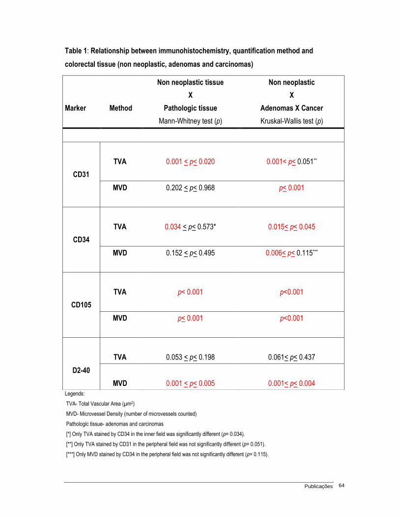
Publicações 64
Table 1: Relationship between immunohistochemistry, quantification method and
colorectal tissue (non neoplastic, adenomas and carcinomas)
Legends:
TVA- Total Vascular Area (µm2)
MVD- Microvessel Density (number of microvessels counted)
Pathologic tissue- adenomas and carcinomas
[*] Only TVA stained by CD34 in the inner field was significantly different (p= 0.034).
[**] Only TVA stained by CD31 in the peripheral field was not significantly different (p= 0.051).
[***] Only MVD stained by CD34 in the peripheral field was not significantly different (p= 0.115).
Marker Method
Non neoplastic tissue
X
Pathologic tissue
Non neoplastic
X
Adenomas X Cancer
Mann-Whitney test (p) Kruskal-Wallis test (p)
TVA 0.001 < p< 0.020 0.001< p< 0.051**
CD31
MVD 0.202 < p< 0.968 p< 0.001
TVA 0.034 < p< 0.573* 0.015< p< 0.045
CD34
MVD 0.152 < p< 0.495 0.006< p< 0.115***
TVA p< 0.001 p<0.001
CD105
MVD p< 0.001 p<0.001
TVA 0.053 < p< 0.198 0.061< p< 0.437
D2-40
MVD 0.001 < p< 0.005 0.001< p< 0.004
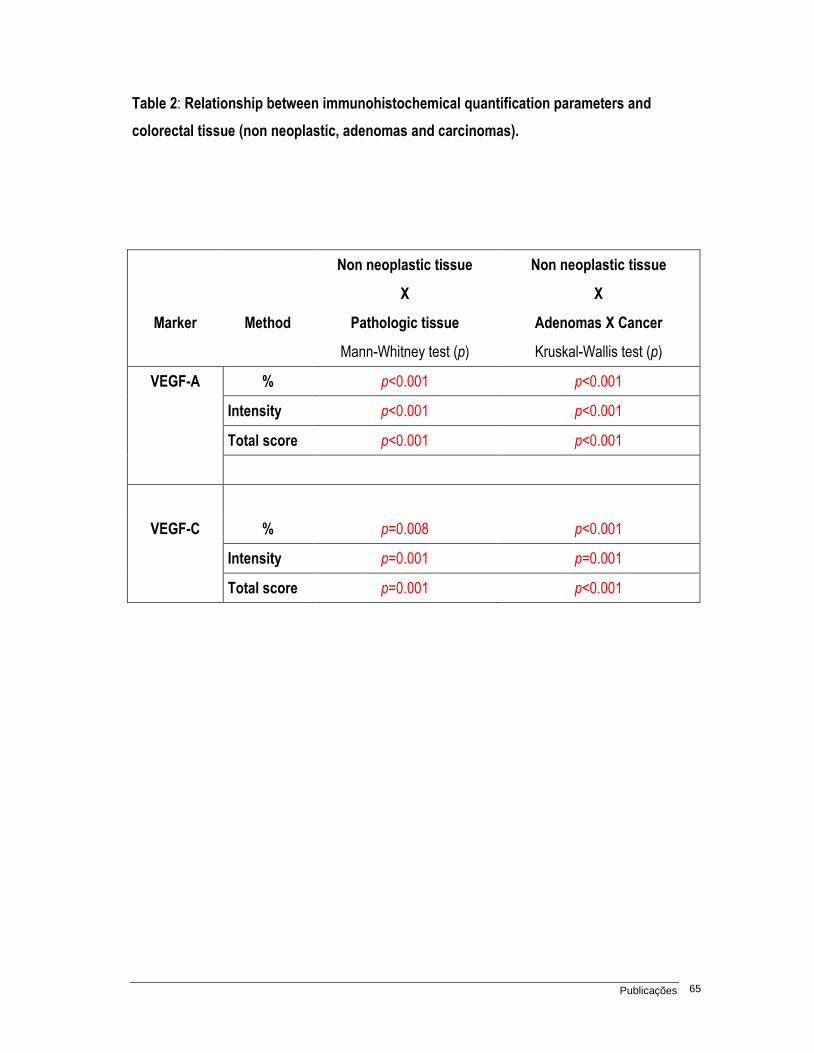
Publicações 65
Table 2: Relationship between immunohistochemical quantification parameters and
colorectal tissue (non neoplastic, adenomas and carcinomas).
Marker Method
Non neoplastic tissue
X
Pathologic tissue
Non neoplastic tissue
X
Adenomas X Cancer
Mann-Whitney test (p) Kruskal-Wallis test (p)
VEGF-A % p<0.001 p<0.001
Intensity p<0.001 p<0.001
Total score p<0.001 p<0.001
VEGF-C % p=0.008 p<0.001
Intensity p=0.001 p=0.001
Total score p=0.001 p<0.001
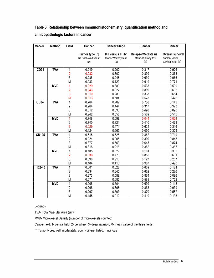
Publicações 66
Table 3: Relationship between immunohistochemistry, quantification method and
clinicopathologic factors in cancer.
Legends:
TVA- Total Vascular Area (µm2)
MVD- Microvessel Density (number of microvessels counted)
Cancer field: 1- central field; 2- periphery; 3- deep invasion; M- mean value of the three fields
[*] Tumor types: well, moderately, poorly differentiated; mucinous
Marker Method Field Cancer Cancer Stage Cancer Cancer
Tumor type [*] I+II versus III+IV Relapse/Metastasis Overall survival
Kruskal-Wallis test (p)
Mann-Whitney test (p)
Mann-Whitney test (p)
Kaplan-Meier survival rate (p)
CD31 TVA 1 0.249 0.202 0.317 0.926 2 0.032 0.300 0.899 0.368 3 0.235 0.248 0.630 0.966 M 0.233 0.129 0.619 0.771
MVD 1 0.029 0.880 0.533 0.599 2 0.043 0.922 0.899 0.602 3 0.010 0.283 0.338 0.664 M 0.013 0.584 0.578 0.476
CD34 TVA 1 0.764 0.787 0.738 0.149 2 0.264 0.444 0.317 0.973 3 0.612 0.833 0.490 0.896 M 0.242 0.558 0.509 0.545
MVD 1 0.748 0.098 0.044 0.024 2 0.740 0.821 0.410 0.478 3 0.029 0.471 0.624 0.316 M 0.124 0.663 0.050 0.309
CD105 TVA 1 0.615 0.528 0.362 0.719 2 0.224 0.908 0.399 0.848 3 0.377 0.563 0.645 0.874 M 0.318 0.216 0.382 0.367
MVD 1 0.105 0.329 0.101 0.302 2 0.036 0.776 0.855 0.631 3 0.590 0.910 0.127 0.257 M 0.184 0.416 0.987 0.490
D2-40 TVA 1 0.601 0.822 0.609 0.124 2 0.834 0.845 0.662 0.276 3 0.273 0.589 0.864 0.096 M 0.671 0.685 0.588 0.752
MVD 1 0.208 0.604 0.699 0.118 2 0.265 0.868 0.858 0.939 3 0.297 0.503 0.870 0.587 M 0.155 0.910 0.410 0.138
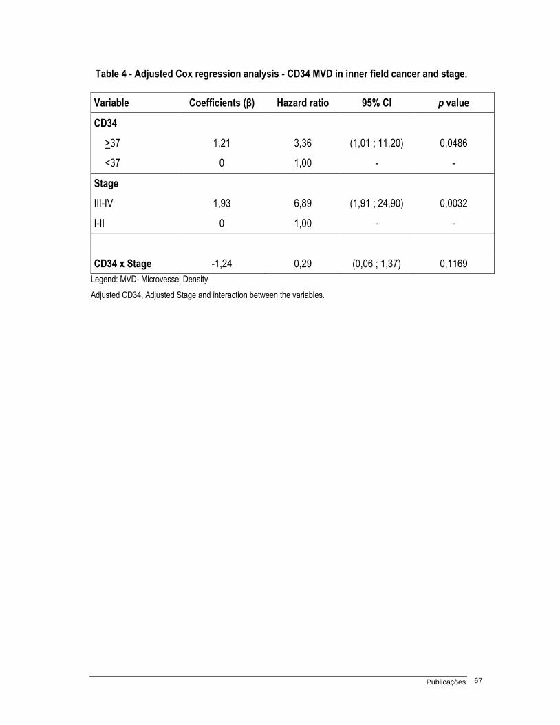
Publicações 67
Table 4 - Adjusted Cox regression analysis - CD34 MVD in inner field cancer and stage.
Variable Coefficients (β) Hazard ratio 95% CI p value
CD34
>37 1,21 3,36 (1,01 ; 11,20) 0,0486
<37 0 1,00 - -
Stage
III-IV 1,93 6,89 (1,91 ; 24,90) 0,0032
I-II 0 1,00 - -
CD34 x Stage -1,24 0,29 (0,06 ; 1,37) 0,1169
Legend: MVD- Microvessel Density
Adjusted CD34, Adjusted Stage and interaction between the variables.
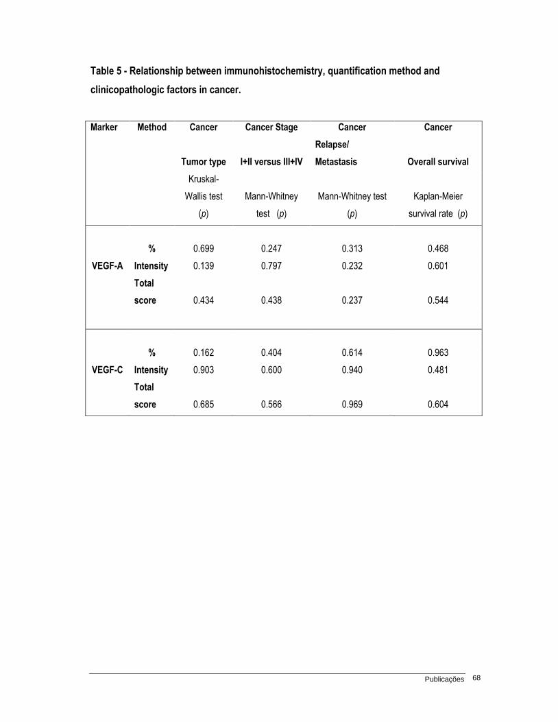
Publicações 68
Table 5 - Relationship between immunohistochemistry, quantification method and
clinicopathologic factors in cancer.
Marker Method Cancer Cancer Stage Cancer Cancer
Tumor type I+II versus III+IV
Relapse/
Metastasis
Overall survival
Kruskal-
Wallis test
(p)
Mann-Whitney
test (p)
Mann-Whitney test
(p)
Kaplan-Meier
survival rate (p)
% 0.699 0.247 0.313 0.468
VEGF-A Intensity 0.139 0.797 0.232 0.601
Total
score 0.434 0.438 0.237 0.544
% 0.162 0.404 0.614 0.963
VEGF-C Intensity 0.903 0.600 0.940 0.481
Total
score 0.685 0.566 0.969 0.604
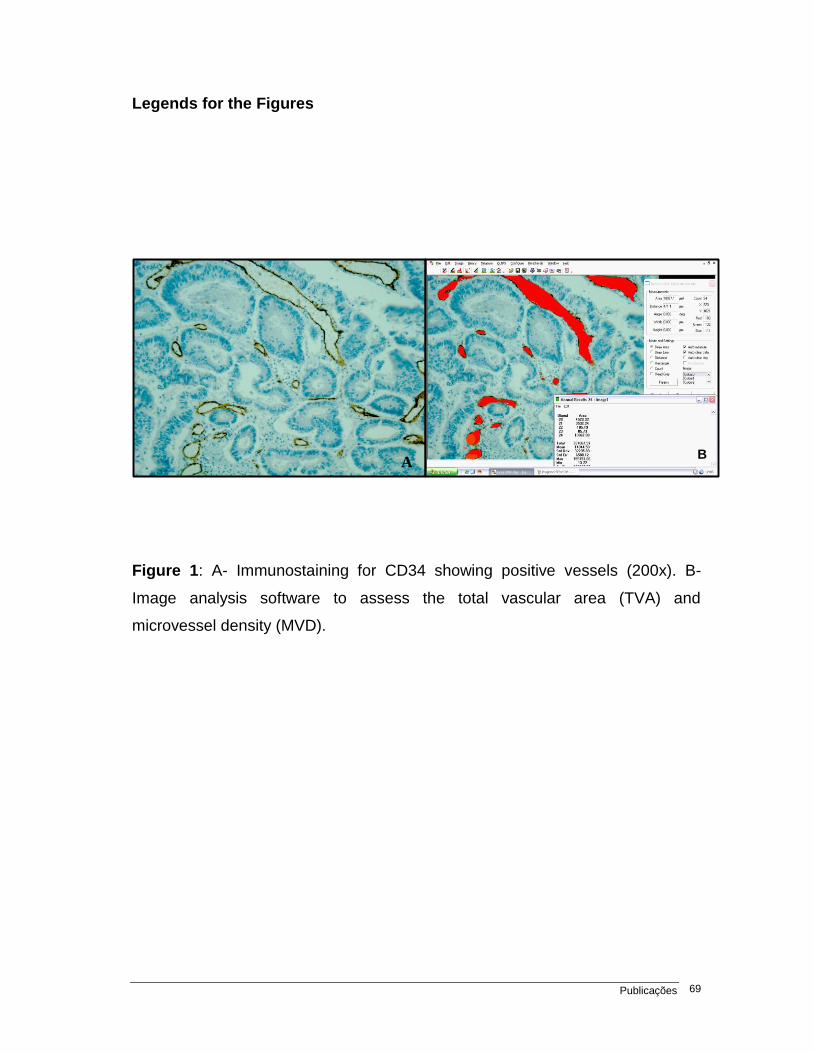
Publicações 69
A
Legends for the Figures
Figure 1: A- Immunostaining for CD34 showing positive vessels (200x). B-
Image analysis software to assess the total vascular area (TVA) and
microvessel density (MVD).
B
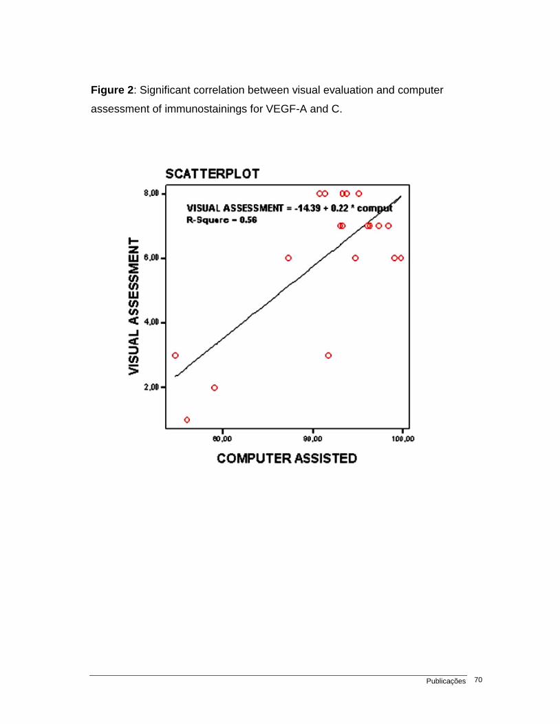
Publicações 70
Figure 2: Significant correlation between visual evaluation and computer
assessment of immunostainings for VEGF-A and C.
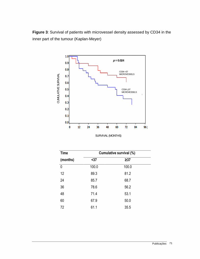
Publicações 71
Figure 3: Survival of patients with microvessel density assessed by CD34 in the
inner part of the tumour (Kaplan-Meyer)
CD34> microvessels
CD34< microvessels
SURVIVAL (MONTHS)
CU
MU
LATIV
E S
UR
VIV
AL
p = 0.024
CD34> microvessels
CD34< microvessels
SURVIVAL (MONTHS)
CU
MU
LATIV
E S
UR
VIV
AL
p = 0.024
CD34 <37
MICROVESSELS
CD34 >37
MICROVESSELSCD34> microvessels
CD34< microvessels
SURVIVAL (MONTHS)
CU
MU
LATIV
E S
UR
VIV
AL
p = 0.024
CD34> microvessels
CD34< microvessels
SURVIVAL (MONTHS)
CU
MU
LATIV
E S
UR
VIV
AL
p = 0.024
CD34 <37
MICROVESSELS
CD34 >37
MICROVESSELS
Time
(months)
Cumulative survival (%)
<37 ≥37
0 100.0 100.0
12 89.3 81.2
24 85.7 68.7
36 78.6 56.2
48 71.4 53.1
60 67.9 50.0
72 61.1 35.5

72
4. Discussão
Discussão 73
O CRC é uma causa importante de mortalidade e incidência entre as
neoplasias (2). Aproximadamente 20-30% dos pacientes com esta neoplasia,
tratados com cirurgia potencialmente curativa, apresentam recidiva, sugerindo que
os fatores prognósticos convencionais não são suficientes e que há necessidade de
fatores adicionais (10,11). O fato de a progressão tumoral poder ser dependente
da angiogênese e linfangiogênese estimulou pesquisas nesta área, entretanto
diversos resultados controversos são encontrados na literatura (50). Os critérios
necessários para quantificação destes processos, tais como: escolha do local no
tumor a ser quantificada a angiogênse, o marcador imunoistoquímico e o método de
quantificação a serem empregados são questões não resolvidas (51).
Nesse estudo analisamos o perfil imunoistoquímico de diferentes marcadores
relacionados à angiogênese e linfangiogênese previamente descritos como
tendo valor prognóstico controverso no carcinoma colorretal, utilizando métodos de
mensuração: área vascular total (TVA) e densidade microvascular (MVD), além da
porcentagem e da intensidade de células tumorais positivas para VEGF-A e VEGF-C.
Ressaltamos que para a determinação da MVD consideramos qualquer célula ou
agrupamento de células endoteliais marcadas e que eram claramente separadas de
outros microvasos adjacentes, por ser o método mais comumente utilizado na
literatura e com menor possibilidade de viés (30,46,52). Esse viés poderia ser
dado pela forte subjetividade embutida na ação de se restringir a contagem apenas
aos vasos sem uma camada muscular, pois, como é sabido, esta pode ser

Discussão 74
descontínua em arteríolas. Também comparamos estes parâmetros nos tecidos
colorretais não neoplásicos, adenomas e carcinomas; nos adenomas de pacientes
sem e com carcinomas, além de correlacioná-los com fatores morfológicos e
clínicos de utilidade bem estabelecida nos CRC.
Os nossos resultados mostraram que a maioria dos marcadores e métodos
exibiu valores progressivamente maiores de angiogênese e linfangiogênese entre
tecido não neoplásico, adenoma e CRC. Estes achados sugerem que o aumento na
angiogênese e linfangiogênese sejam eventos precoces, ocorrendo nas lesões
precursoras e aumentando ainda mais nos carcinomas, corroborando dados
prévios da literatura (53).
Também foi observado que os adenomas provenientes de pacientes
portadores de carcinomas em outro local da mucosa colorretal apresentaram
valores aumentados de TVA determinada pela imunocoloração para CD105 e
de MVD determinada com o uso do D2-40, quando comparados aos adenomas
de pacientes sem carcinomas. Tais achados favorecem proposições existentes na
literatura, de que uma área distinta da mucosa colorretal possa ser influenciada por
fatores produzidos pelo tumor em outro sítio da mucosa (53). Apóiam ainda,
dados de literatura sobre os vasos sanguíneos neoformados (CD105+) serem
tortuosos, justificando a significante superioridade da TVA (39). Por sua vez, os
vasos linfáticos apresentam MVD maior D2-40+, sendo um evento que precede
o aumento de número de vasos sanguíneos em lesão gastrintestinal iniciais,
segundo anteriormente proposto (54).
Discussão 75
Entretanto, quando analisados os carcinomas, apenas a MVD determinada
pelo marcador CD34 no campo central da lesão diferencia-se estatisticamente
segundo a recorrência/ metástase (p= 0,04) e curva de sobrevida (p= 0,02). Estes
dados não apóiam estudos prévios que demonstraram o valor prognóstico do
uso dos demais marcadores imunoistoquímicos (35,43), mas corroboram estudos
mais recentes que recomendam o uso do marcador CD34 (44,52).
O CD31 corresponde ao marcador PECAM-1, sendo também considerado
um marcador pan-endotelial. Participa da adesão leucócito-endotélio na diapedese
e, além disso, é expresso em monócito e células Natural-Killer, tendo relação
com a imunidade tumoral (52). Este fato pode ter influenciado o resultado no
CRC, uma vez que seu espectro de distribuição e função difere do CD34.
Há trabalhos sugerindo que, com o crescimento tumoral, as áreas centrais
dos carcinomas mantêm sua vascularização através de remodelagem contínua
de vasos existentes e de migração de células endoteliais. A habilidade do tumor
em manter esta vascularização central tem sido relatada como um fator de
prognóstico independente em vários tipos de tumor, incluindo pulmão, cólon e
mama (51,55,56,57).
Sugere-se ainda que, com o crescimento tumoral, haja aumento da
pressão intersticial e os vasos sanguíneos sofram remodelagem com rupturas
de seu lúmen e ramificações (58,39).
Estas constatações explicam o fato de a MVD determinada pelo marcador
CD34 diferenciar-se estatisticamente e ser um fator de prognóstico, sobretudo
Discussão 76
na área central dos carcinomas, enquanto que o CD105 (marcador de vasos
neoformados) não conduziu a este resultado.
O fato de não termos obtido resultados estatisticamente significantes entre o
estudo imunoistoquímico dos fatores de crescimento vascular e parâmetros
morfológicos e clínicos no CRC deve refletir a complexidade do controle da
angiogênese, uma vez que foi relatado que o VEGF não é o único fator promotor de
crescimento vascular. Outros componentes, como o Fator de crescimento derivado
de plaquetas, participam desse controle (59,60). Além disto, neste estudo foi
analisada a expressão do VEGF nas células tumorais, sem considerar outras
células que poderiam expressar VEGF, como granulócitos, o que pode ser
responsável pela falta de correlação citada (39).
Nossos dados não mostraram correlação estatisticamente significante dos
vasos linfáticos intratumorais com fatores prognósticos estabelecidos para o CRC,
em acordo com trabalhos anteriores (30,46). Há estudos postulando que enquanto
os vasos sanguíneos são essenciais para o crescimento do tumor, os vasos
linfáticos não o são. Com a progressão tumoral e o aumento da pressão intersticial,
eles seriam mais numerosos nas áreas peritumorais, onde há maior drenagem
de fluidos teciduais e, eventualmente, de células metastáticas (30,46,47).
Os presentes achados apóiam o fato de a angiogênese e o aumento da
vascularização linfática ocorrer precocemente, ainda nos adenomas. Também
corroboram o fato de a angiogênese e o aumento da contagem de vasos linfáticos
ocorrerem mais pronunciadamente em adenomas de pacientes com carcinomas na
Discussão 77
mucosa adjacente, possivelmente influenciados por fatores produzidos pelo tumor. A
MVD na área central do carcinoma determinada pelo marcador imunoistoquímico
CD34 adiciona um critério prognóstico, associando-se com recidiva/ metástase e
sobrevida, enquanto os outros meios de quantificação vascular e de expressão de
fatores de crescimento não apresentaram resultados estatisticamente significantes.
Este representa um fator prognóstico adicional independente no CRC.
Além do valor prognóstico determinado com o uso do marcador CD34 no
CRC, o presente trabalho possibilita um melhor entendimento de eventos
fisiopatológicos relacionados à angiogênese e linfagiogênese e pode contribuir
com linhas de pesquisa futuras no campo da terapêutica anti-angiogênica.

78
5. Conclusões
Conclusões 79
A maioria dos marcadores vasculares e dos métodos de quantificação da
angiogênese e linfangiogênese exibiu valores progressivamente maiores desde o
tecido não neoplásico, ao adenoma e ao CRC, sugerindo que o incremento
da vascularização é um evento precoce na carcinogênese colorretal.
Adenomas de pacientes com carcinoma colorretal mostraram valores
significantemente maiores da área vascular determinados com um marcador de
CD105 e da densidade linfática determinada com o marcador D2-40, quando
comparados com adenomas de pacientes sem carcinomas, sugerindo a influência
de fatores tumorais nos adenomas distantes das lesões carcinomatosas.
Quando analisados os carcinomas, apenas a MVD determinada pelo marcador
de CD34 no campo central da lesão diferencia-se estatisticamente segundo a
recorrência/ metástase e curva de sobrevida. Este método adiciona um fator
de valor prognóstico independente no CRC.

80
6. Referências Bibliográficas
Referências Bibliográficas 81
1. Organização Mundial da Saúde. Cancer database [Acesso em Março de
2009]. Disponível em: www.who.int/research/en/
2. Instituto Nacional do Câncer. Ministério da Saúde. Estimativas 2008:
Incidência de Câncer no Brasil [Acesso em Março de 2009]. Disponível em:
www.inca.gov.br/estimativa/2008.
3. Soreide K, Nedrebo SB, Knapp JC, Glomsaker TB, Soreide JA, Korner H.
Evolving molecular classification by genomic and proteomic biomarkers in
colorectal cancer: Potencial implications for the surgical pathologists. 2009;
31-50.
4. Weitz J, Koch M, Debus J, Höhler T, Galle PR, Büchler M. Colorectal
Cancer. Lancet. 2005; 365: 153-164.
5. Hamilton S R, Aaltonen LA. Pathology and Genetics of Tumors of the
Digestive System. World Health Organization Classification of Tumours.
Lyon: IARC Press; 2000; 105-129.
6. Odze, Robert D. Intestinal tumors. Surgical Pathology of the GI tract, liver,
biliary tract, and pancreas. Philadelphia: Elsevier; 2004; 441- 454.
7. Cotran R, Kumar V, Collins T. Gastrointestinal Pathology. Robbins Pathologic
Basis of Disease. Rio de Janeiro: Guanabara Koogan; 2000; 742- 750.
8. Bodmer WF, Wilding J, Fearnhead NS. Molecular Genetics of colorectal
cancer. Acta Oncologica Brasileira. 2004; 24-30.
Referências Bibliográficas 82
9. Ogino S, Goel A. Molecular Classification and correlates in colorectal
cancer. JMD. 2008;13-26.
10. Ratto C, Sofo L, Ippoliti M, Merico M, Doglietto GB, Crucitti F. Prognostic
factors in colorectal cancer. Literature review for clinical application. Dis
Colon Rectum. 1998; 41(8):1033-1049.
11. Buyse M, Piedbois P. Should Dukes' B patients receive adjuvant therapy? A
statistical perspective. Semin Oncol. 2001; 28(1 Suppl 1): 20-24.
12. Zlobec I, Lugli A. Prognostic and predictive factors in colorectal cancer. J
Clin Pathol. 2008; 561-569.
13. Compton CC. Pathologic prognostic factors in the recurrence of rectal
cancer. Clin Colorectal Cancer. 2002; 2(3):149-60.
14. Jass RJ. Classification of colorectal cancer based on correlation of clinical,
morphological and molecular features. Histopathology. 2007; 50: 113-130.
15. Byrne AM, Bouchier-Hayes DJ, Harmey JH. Angiogenic and cell survival
functions of vascular endothelial growth factor (VEGF). J Cell Mol Med.
2005 Oct-Dec; 9(4): 777-794. Review
16. Ono T, Miki C. Factors Influencing Tissue Concentration of Vascular Endothelial
Growth Factor in Colorectal Carcinoma. Am J Gastroenterol. 2000; 1062-1067.
17. Wong MP, Cheung N, Yuen ST, Leung SY, Chung LP. Vascular Endothelial
Growth Factor is up-Regulated in Early Pre-malignant Stage of Colorectal
Tumour Progression. Int J Cancer. 1999; 81: 845-850.
Referências Bibliográficas 83
18. Aotake T, Lu CD, Chiba Y, Muraoka R, Tanigawa N. Changes of
Angiogenesis and Tumour Cell Apoptosis During Colorectal Carcinogenesis.
Clin Cancer Res. 1999; 5: 135-142.
19. Kazama S , Kitayama J , Watanabe T , Nagawa H. Expression Pattern of
Vascular Endothelial Growth Factor-C in Human Colorectal Normal Mucosa
and Neoplasic Mucosa. Hepato-Gastroent. 2004; 51: 391-395.
20. Cascinu S, Graziano F, Catalano V, Staccioli MP, Barni S, Giordani P, Rossi
MC, Baldelli AM, Muretto P, Valenti A, Catalano G. Differences of vascular
endothelial growth factor (VEGF) expression between liver and abdominal
metastases from colon cancer. Implications for the treatment with VEGF
inhibitors. Clin Exp Metastasis. 2000; 18(8):651-655.
21. Galizia G, Lieto E, Ferraraccio F, Orditura M, De Vita F, Castellano P,
Imperatore V, Romano C, Ciardiello F, Agostini B, Pignatelli C.
Determination of molecular marker expression can predict clinical outcome
in colon carcinomas. Clin Cancer Res. 2004; 10(10):3490-3499.
22. Kojima M, Shiokawa A, Nobuyuki O, Yoshiki O, Kato H, Iwaku K et al.
Clinical Significance of Nuclear Morphometry at the Invasive Front of T1
Colorectal Cancer and Relation to Expression of VEGFA and VEGFC.
Oncol. 2005; 68: 230-238.
23. Boxer GM, Tsiompanou E, Levine T, Watson R, Begent RH
Immunohistochemical Expression of Vascular Endothelial Growth Factor
and Microvessel Counting as Prognostic Indicators in Node-negative
Colorectal Cancer. Tumours Biol. 2005: 26(1): 1-8.
24. Lee JC, Chow NH, Wang ST, Huang SM, Prognostic Value of Vascular
Endothelial Growth Factor Expression in Colorectal Cancer Patients. Eur J
Cancer. 2000; 36: 748-753.
Referências Bibliográficas 84
25. Khorana AA, Ryan CK, Cox C, Eberly S, Sahasrabudhe DM. Vascular
Endothelial Growth Factor, CD68, and Epidermal Growth Factor Receptor
Expression and Survival in Patients with Stage II and Stage III Colon Carcinoma:
A Role for the Host Response in Prognosis. Cancer. 2003; 97: 960-968.
26. Kaio E, Tanaka S, Kitadai Y, Sumii M, Yoshihara M, Haruma K et al. Clinical
significance of angiogenic factor expression at the deepest invasive site of
advanced colorectal carcinoma. Oncology. 2003; 64(1): 61-73.
27. Onogawa S, Kitadai Y, Tanaka S, Kuwai T, Kimura S, Chayama K. Expression
of VEGF-C and VEGF-D at the invasive edge correlates with lymph node
metastasis and prognosis of patients with colorectal carcinoma. Cancer Sci.
2004 Jan; 95(1):32-39.
28. Rmali KA, Puntis CA, Jiang WG. Tumor associated angiogenesis in human
colorectal cancer. Colorectal disease. 2006; (9)1: 3-14.
29. Kazama S , Kitayama J , Watanabe T , Nagawa H. Expression Pattern of
Vascular Endothelial Growth Factor-C in Human Colorectal Normal Mucosa
and Neoplasic Mucosa. Hepato-Gastroent. 2004; 51: 391-395.
30. Duff SE, Jeziorska M, Kumar S, Haboubi N, Sherlock D, O'Dwyer ST et al.
Lymphatic vessel density, microvessel density and lymphangiogenic growth
factor expression in colorectal cancer. Colorectal Dis. 2007 Nov; 9(9):793-800.
31. Hu WG, Li JW, Feng B, Beveridge M, Yue F, Lu AG et al. Vascular endothelial
growth factors C and D represent novel prognostic markers in colorectal
carcinoma using quantitative image analysis. Eur Surg Res. 2007; 39(4):229-238.
32. Cao Y. Why and how do tumors stimulate lymphangiogenesis? Lymphat
Res Biol. 2008; 6(3-4):145-8.
Referências Bibliográficas 85
33. Frank RE, Saclarides TJ, Leurgans S, Speziale NJ, Drab EA, Rubin DB et
al. Tumor Angiogenesis as a Pretictor of Recurrence and Survival in
Patients with Node-negative Colon Cancer. Ann Surg. 1995; 222: 695-699.
34. Bhatavdekar JM, Patel DD, Chikhlikar PR, Shah NG, Vora HH, Ghosh N et al.
Molecular Markers are Predictors of Recurrence and Survival in Patients with
Dukes C Colorectal Adenocarcinoma. Dis Colon Rectum. 2001; 44: 523-533.
35. Vermeulen PB, Van den Eynden GG, Huget P, Goovaerts G, Weyler J, Lardon F
et al. Prospective Study of Intramural Microvessel Density, p53 Expression
and Survival in Colorectal Cancer. Br J Cancer. 1999; 79(2): 316-322.
36. Pavlopoulos PM, Konstantinidou AE, Agapitos E, Kavantzas N, Nikolopoulou P,
Davaris P. A morphometric study of neovascularization in colorectal
carcinoma. Cancer. 1998; 83: 2067-2075.
37. Abdalla SA, Behzad F, Bsharah S, Kumar S, Amini SK, O’Dwyer ST et al.
Prognostic relevance of microvessel density in colorectal tumours. Oncol
Report. 1999; 6: 839-842.
38. Prall F, Gringmuth U, Nizze H, Barten M. Microvessel densities and
microvascular architecture in colorectal carcinomas and their liver
metastases: significant correlation of high microvessel densities with better
survival. Histopathology. 2003; 42: 482-491.
39. Vermeulen PB, Gasparini G, Fox SB, Colpaert C, Marson LP, Gion M et al.
Second international consensus on the methodology and criteria of
evaluation of angiogenesis quantification in solid human tumours. Eur J
Cancer. 2002; 38(12): 1564-1579.
Referências Bibliográficas 86
40. Akagi K, Ikeda Y, Sumiyoshi Y, Kimura Y, Kinoshita J, Miyazaki M et al.
Estimation of Angiogenesis with Anti-CD105 Immunostaining in the Process
of Colorectal Cancer Development. Surgery. 2002; 131: 109-113.
41. Clasper S, Royston D, Baban D, Cao Y, Ewers S, Butz S et al. A novel gene
expression profile in lymphatics associated with tumor growth and nodal
metastasis. Cancer Res. 2008; 68(18):7293-7303.
42. Li C, Gardy R, Seon BK, Duff SE, Abdalla S, Renehan A et al. Both high
intratumoral microvessel density determined using CD105 antibody and
elevated plasma levels of CD105 in colorectal cancer patients correlate with
poor prognosis. Br J Cancer. 2003; 88(9):1424-1431.
43. Romani AA, Borghetti AF, Del Rio P, Sianesi M, Soliani P. The risk of
developing metastatic disease in colorectal cancer is related to CD105-
positive vessel count. J Surg Oncol. 2006; 93(6):446-55.
44. Barresi V, Vitarelli E, Tuccari G, Barresi G. Correlative study of microvessel
density and 5-lipoxygenase expression in human sporadic colorectal cancer.
Arch Pathol Lab Med. 2008; 132(11):1807-1812.
45. Kahn HJ, Bailey D, Marks A. Monoclonal antibody D2-40, a new marker of
lymphatic endothelium, reacts with Kaposi's sarcoma and a subset of
angiosarcomas. Mod Pathol. 2002; 15(4):434-440.
46. Longatto-Filho A, Pinheiro C, Ferreira L, Scapulatempo C, Alves VA,
Baltazar F et al. Peritumoural, but not intratumoural, lymphatic vessel
density and invasion correlate with colorectal carcinoma poor-outcome
markers. Virchows Arch. 2008; 452(2):133-138.
47. Yan G, Zhou XY, Cai SJ, Zhang GH, Peng JJ, Du X. Lymphangiogenic and
angiogenic microvessel density in human primary sporadic colorectal
carcinoma. World J Gastroenterol. 2008; 14(1):101-107.
Referências Bibliográficas 87
48. Matsumoto K, Nakayama Y, Inoue Y, Minagawa N, Katsuki T, Shibao K et
al. Lymphatic microvessel density is an independent prognostic factor in
colorectal cancer. Dis Colon Rectum. 2007; 50(3):308-314.
49. Fogt F, Zimmerman RL, Ross HM, Daly T, Gausas RE. Identification of
lymphatic vessels in malignant, adenomatous and normal colonic mucosa
using the novel immunostain D2-40. Oncol Rep. 2004 Jan; 11(1):47-50.
50. Graziano F, Cascinu S.Prognostic molecular markers for planning adjuvant
chemotherapy trials in Dukes' B colorectal cancer patients: how much
evidence is enough? Ann Oncol. 2003; 14(7):1026-1038.
51. Giatromanolaki A, Koukourakis MI, Sivridis E, O'Byrne K, Gatter KC, Harris
AL. Invading edge vs. inner' (edvin) patterns of vascularization: an interplay
between angiogenic and vascular survival factors defines the clinical
behaviour of non-small cell lung cancer. J Pathol. 2000; 192(2):140-149.
52. Gao J, Knutsen A, Arbman G, Carstensen J, Frånlund B, Sun XF. Clinical
and biological significance of angiogenesis and lymphangiogenesis in
colorectal cancer.Dig Liver Dis. 2009; 41(2):116-122.
53. Hanrahan V, Currie MJ, Gunningham SP, Morrin HR, Scott PA, Robinson BA et
al. The angiogenic switch for vascular endothelial growth factor (VEGF)-A,
VEGF-B, VEGF-C, and VEGF-D in the adenoma-carcinoma sequence during
colorectal cancer progression. J Pathol. 2003; 200: 183-194.
54. Gao Y, Zhong WX, Mu DB, Yuan YP, Zhang YH, Yu JM et al. Distributions
of angiogenesis and lymphangiogenesis in gastrointestinal intramucosal
tumors. Ann Surg Oncol. 2008; 15: 1117-1123.
55. Giatromanolaki A, Sivridis E, Simopoulos C, Polychronidis A, Gatter KC,
Harris AL et al. Differential assessment of angiogenic activity and of vascular
survival ability (VSA) in breast cancer. Clin Exp Metastasis. 2002; 19(8):673-679.
Referências Bibliográficas 88
56. Papadopoulos I, Giatromanolaki A, Koukourakis MI, Sivridis E. Tumour
angiogenic activity and vascular survival ability in bladder carcinoma. J Clin
Pathol. 2004; 57(3):250-255.
57. Giatromanolaki A, Sivridis E, Koukourakis MI. Angiogenesis in colorectal
cancer: prognostic and therapeutic implications. Am J Clin Oncol. 2006;
29(4):408-417.
58. Patan S, Munn LL, Jain RK. Intussusceptive microvascular growth in a
human colon adenocarcinoma xenograft: a novel mechanism of tumor
angiogenesis. Microvasc Res. 1996; 51(2): 260-272.
59. Takahashi Y, Bucana CD, Liu W, Yoneda J, Kitadai Y, Cleary KR et al.
Platelet-derived endothelial cell growth factor in human colon cancer
angiogenesis: role of infiltrating cells. J Natl Cancer Inst. 1996 21;
88(16):1146-1151.
60. Reinmuth N, Parikh AA, Ahmad SA, Liu W, Stoeltzing O, Fan F et al.
Biology of angiogenesis in tumors of the gastrointestinal tract. Microsc Res
Tech. 2003; 60(2):199-207.
89
7. Anexos

Anexos 90
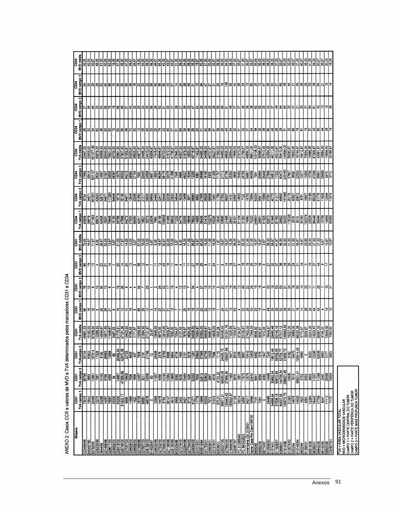
Anexos 91
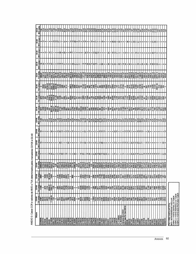
Anexos 92
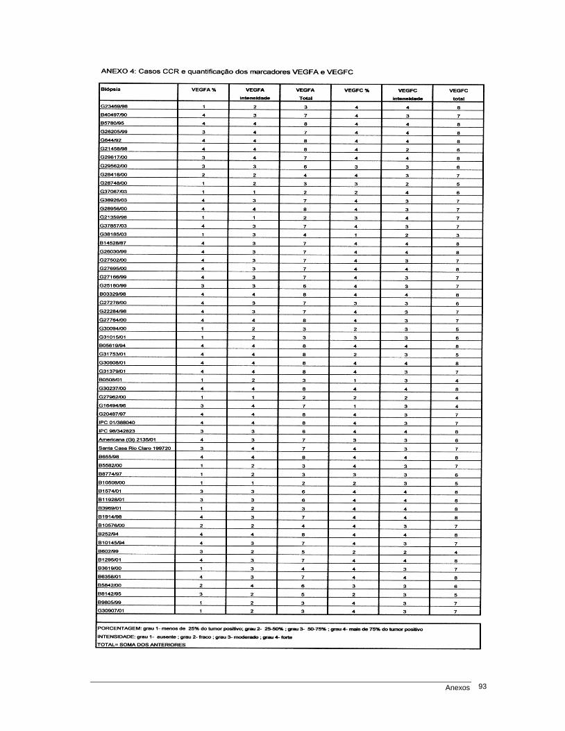
Anexos 93

Anexos 94
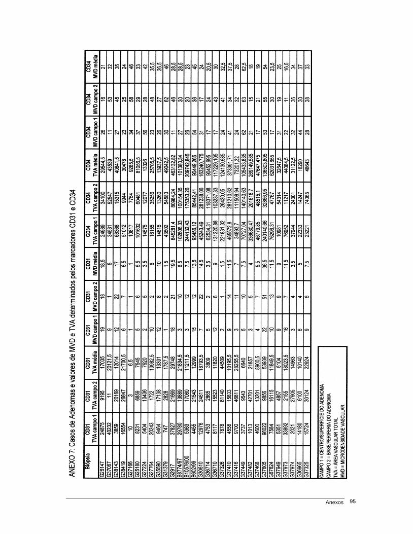
Anexos 95
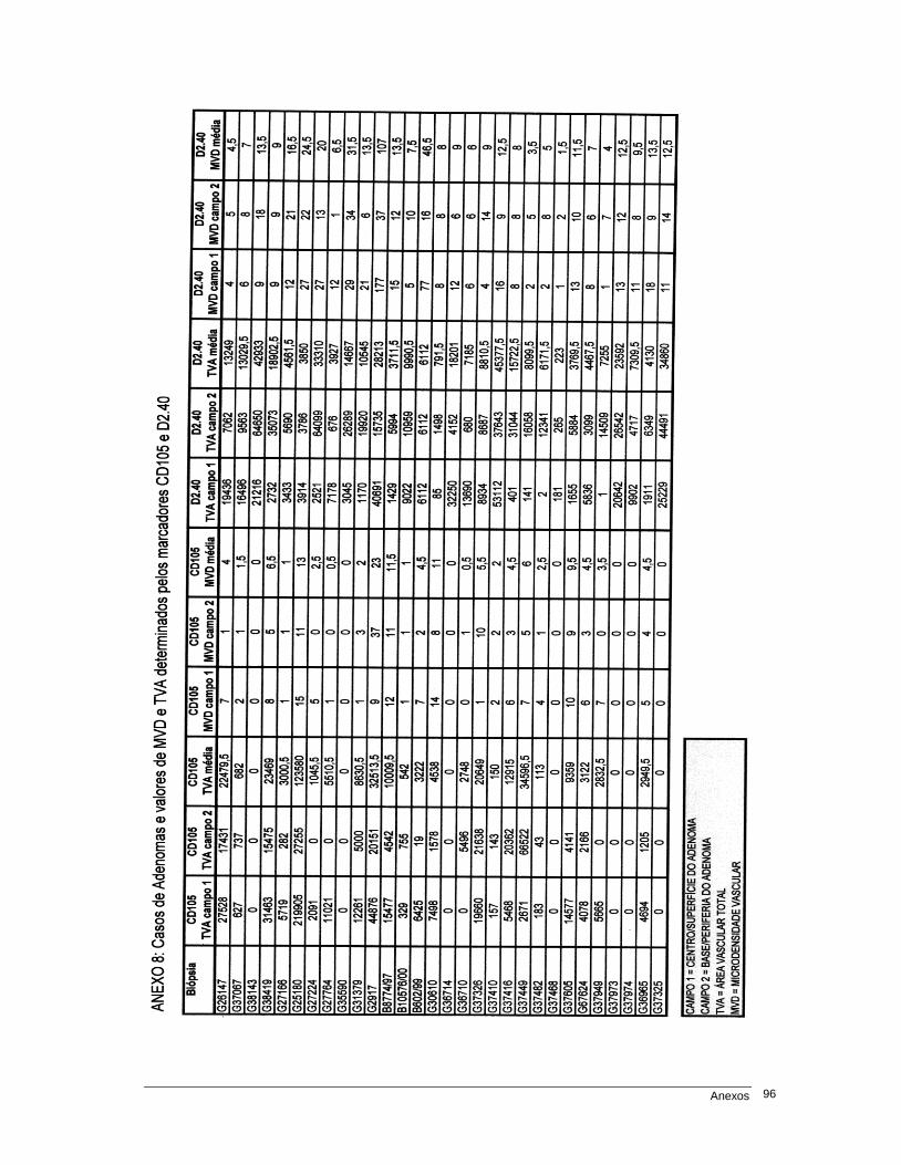
Anexos 96
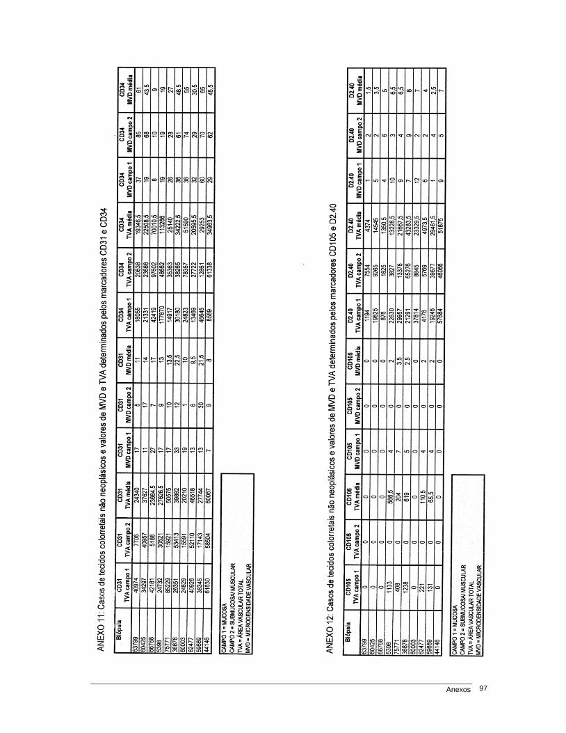
Anexos 97
Anexos 98
ANEXO 13: Permissão do Brazilian Journal of Medical and Biological Research Dear Dr. Luciana Regina Moreira, The Associação Brasileira de Divulgação Científica give you permission to reproduce any part of "Comparison of blood neoangiogenesis and lymphatic vascularization in colorectal adenomas from patients with and without concomitant colorectal cancer" L.R. Moreira, A.A. Schenka, P. Latuf Filho, C.S.P. Lima, M.A.S. Trevisan and J. Vassallo in your Doctoral Thesis to be submitted to Universidade Estadual de Campinas provide that the original citation is cited. Sincerely yours,
Lewis Joel Greene Editor Brazilian Journal of Medical and Biological Research Av. Bandeirantes 3900 14049-900 Ribeirão Preto, SP, Brasil fax 55+16+3633-2119 phone 55+16+3633-1884 email: [email protected] or [email protected] Versão On Line - http://www.bjournal.com.br or www.scielo.br/bjmbr
Related Documents