UNIVERSIDADE NOVE DE JULHO PROGRAMA DE MESTRADO EM CIÊNCIAS DA REABILITAÇÃO FERNANDA CORDEIRO DA SILVA AVALIAÇÃO DA ATIVIDADE ELETROMIOGRÁFICA, FORÇA DE MORDIDA E ASSOCIAÇÃO COM DISFUNÇÃO TEMPOROMANDIBULAR EM PACIENTES HEMIPARÉTICOS PÓS-ACIDENTE VASCULAR ENCEFÁLICO. São Paulo 2014

Welcome message from author
This document is posted to help you gain knowledge. Please leave a comment to let me know what you think about it! Share it to your friends and learn new things together.
Transcript
UNIVERSIDADE NOVE DE JULHO
PROGRAMA DE MESTRADO EM CIÊNCIAS DA REABILITAÇÃO
FERNANDA CORDEIRO DA SILVA
AVALIAÇÃO DA ATIVIDADE ELETROMIOGRÁFICA, FORÇA DE
MORDIDA E ASSOCIAÇÃO COM DISFUNÇÃO
TEMPOROMANDIBULAR EM PACIENTES HEMIPARÉTICOS
PÓS-ACIDENTE VASCULAR ENCEFÁLICO.
São Paulo
2014
FERNANDA CORDEIRO DA SILVA
AVALIAÇÃO DA ATIVIDADE ELETROMIOGRÁFICA, FORÇA DE
MORDIDA E ASSOCIAÇÃO COM DISFUNÇÃO
TEMPOROMANDIBULAR EM PACIENTES HEMIPARÉTICOS
PÓS-ACIDENTE VASCULAR ENCEFÁLICO
Dissertação apresentada à
Universidade Nove de Julho para
obtenção do título de Mestre em
Ciências da Reabilitação.
Orientadora: Profa. Dra. Sandra Kalil Bussadori
São Paulo
2014
Silva, Fernanda Cordeiro da.
Avaliação da atividade eletromiográfica, força de mordida e associação com
disfunção temporomandibular em pacientes hemiparéticos pós-acidente vascular
encefálico. /Fernanda Cordeiro da Silva. 2014.
94 f.
Dissertação (mestrado) – Universidade Nove de Julho - UNINOVE,
São Paulo, 2014.
Orientador (a): Profa Dra Sandra Kalil Bussadori.
1. Acidente vascular encefálico. 2. DTM. 3. Força de mordida. 4.
EMG.
I. Bussadori, Sandra Kalil. II. Titulo
CDU 615.8
DEDICATÓRIA
Dedico este trabalho primeiramente a Deus que é o meu Senhor, meu
Salvador, meu Amigo e a Fonte de toda minha vida.
Dedico ao meu esposo Rafael, meu amor, meu companheiro e maior
incentivador de tudo que eu faço.
“Meu amor, sem você realmente não teria dado certo!!!”
Ao meu querido filho Gustavo, meu menininho inteligente, forte, decidido, o
tempero da minha vida.
“Verdadeiramente ele é a junção do melhor que tinha em nós!!”
A minha família que sempre me amparou em todo tempo, acreditando,
incentivando e curtindo comigo cada “novo curso”.
Amo vocês!!!
AGRADECIMENTOS
Quero agradecer em primeiro lugar a minha querida orientadora Profa Dra
Sandra Kalil Bussadori por ter me ensinado tanto neste tempo. Por ter
acreditado e investido em mim, por me incentivar a caminhar cada vez mais
adiante e a sempre ter foco!
Obrigada por tudo!!!!
Agradeço a cada participante, por ter acreditado nesta pesquisa e doado seu
tempo a este projeto.
Agradeço aos meus companheiros e amigos do mestrado, em especial ao meu
querido amigo Lucas, por estes dois anos incríveis!!
Muito obrigada a todos os professores que, direta ou indiretamente,
participaram, contribuíram e ajudaram a fazer deste estudo uma realidade.
Agradeço a Profa Dra Fernanda Ishida Correa por todas as contribuições
pertinentes e por ter me ensinado tanto na Clínica.
Agradeço a FAPESP pelo apoio financeiro e por ter acreditado nesta pesquisa.
Este apoio foi crucial para o desenvolvimento deste projeto.
E por fim, porém não menos importante, agradeço a UNINOVE por ter me
oferecido toda estrutura necessária para a realização deste sonho.
Muito obrigada!!!
RESUMO
O Acidente Vascular Encefálico é um importante agravo à saúde da população
mundial, que cursa com prejuízos motores que podem comprometer os músculos
mastigatórios. O objetivo deste estudo foi avaliar se a hemiparesia completa,
proveniente de um AVE, pode causar maior comprometimento orofacial,
alterando a atividade eletromiográfica e a força dos músculos mastigatórios
associado à disfunção temporomandibular quando comparada à hemiparesia
incompleta. Foram avaliados indivíduos hemiparéticos pós-ave com idade entre
18 a 75 anos. A amostra foi dividida em dois grupos, sendo: Hemiparéticos
Completos contendo 29 indivíduos e hemiparéticos incompletos contendo 21
indivíduos. Todos os grupos foram submetidos à avaliação para diagnóstico da
DTM através do RDC/TMD, força de mordida através do Dinamômetro da Kratos®
e eletromiografia dos músculos mastigatórios empregando-se um eletromiógrafo
de 4 canais da EMG System®. Os resultados obtidos foram computados e análise
estatística realizada utilizando nível de significância de 95% (p<0,05), onde se
observou diferença estatisticamente significante na DTM entre os hemiparéticos
completos e incompletos (p=0,01). No entanto quanto a FM não foi observada
diferença estatística intergrupos e/ou intragrupos. Quanto à função do membro
superior, avaliada pelo SSQOL, e sua relação com a saúde oral, avaliada pelo
OHIP-14, foi possível observar correlação estatisticamente significativa nos
domínios Dor Física, Limitação Funcional, Desconforto Psicológico, Incapacidade
Física, Incapacidade Social, Desvantagem Social e Incapacidade Psicológica. Em
todos os domínios citados acima, as correlações foram negativas. Em relação à
eletromiografia, foi observada diferença estatisticamente significativa durante o
repouso, na avaliação intergrupos, tanto no lado direito (p=0,005) como no
esquerdo (p=0,005). Verificou-se uma correlação negativa, de moderada
magnitude (-0,643) (p=0,02), entre os sinais eletromiográficos em repouso e os
indivíduos do sexo masculino que não apresentaram DTM no grupo de
hemiparéticos completos. Quanto ao tempo de lesão, também foi observada uma
correlação negativa, de moderada magnitude (-0,663) (p=0,00) entre o tempo de
lesão e o sinal eletromiográfico em indivíduos sem DTM também no grupo dos
hemiparéticos completos, o que nos permitiu concluir que existe relação entre
DTM, tempo de lesão e EMG nos indivíduos hemiparéticos pós-acidente vascular
encefálico, com exceção da FM que não mostrou relação com a hemiparesia. Por
fim, a função orofacial precisa ser melhor compreendida, pois agregará valor
imensurável na reabilitação destes indivíduos.
Palavras-chave – Acidente vascular encefálico, Transtornos da Articulação
Temporomandibular, força de mordida, eletromiografia, músculos
mastigatórios
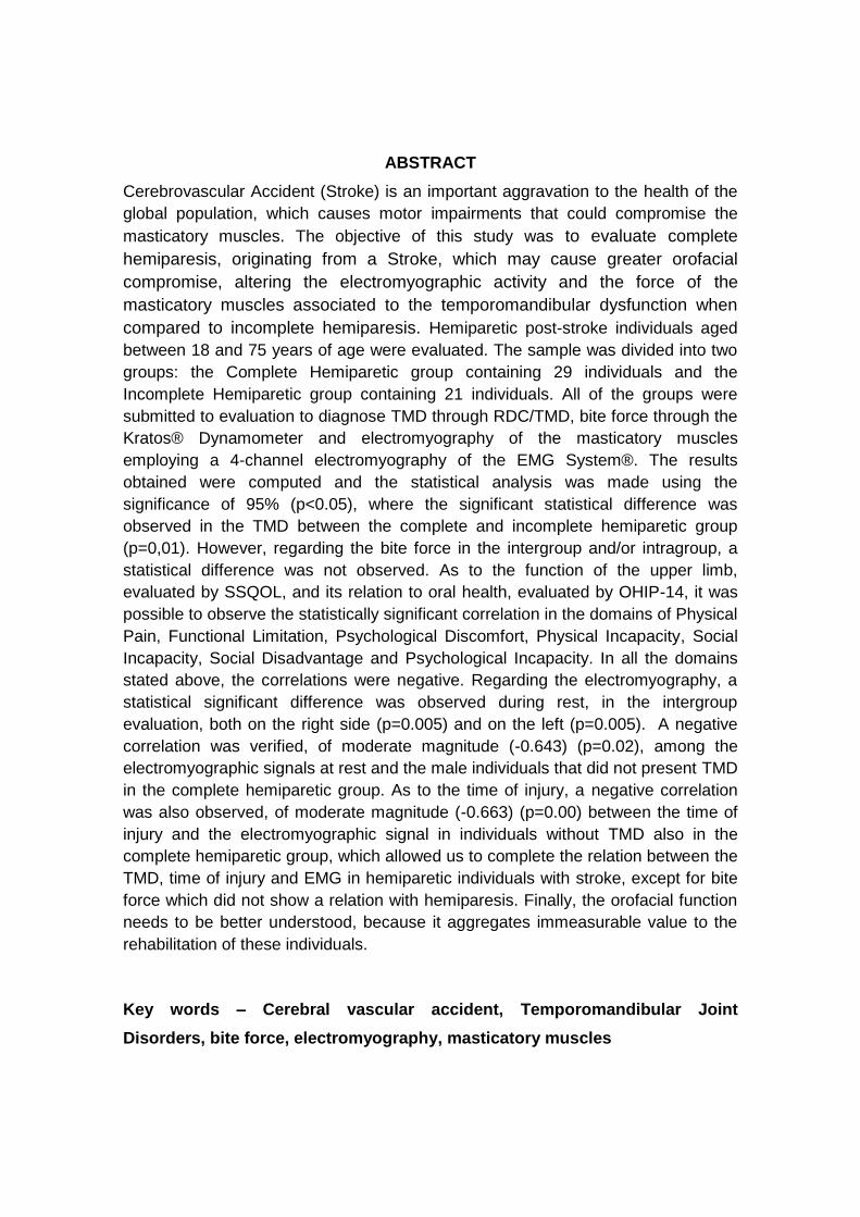
ABSTRACT
Cerebrovascular Accident (Stroke) is an important aggravation to the health of the
global population, which causes motor impairments that could compromise the
masticatory muscles. The objective of this study was to evaluate complete
hemiparesis, originating from a Stroke, which may cause greater orofacial
compromise, altering the electromyographic activity and the force of the
masticatory muscles associated to the temporomandibular dysfunction when
compared to incomplete hemiparesis. Hemiparetic post-stroke individuals aged
between 18 and 75 years of age were evaluated. The sample was divided into two
groups: the Complete Hemiparetic group containing 29 individuals and the
Incomplete Hemiparetic group containing 21 individuals. All of the groups were
submitted to evaluation to diagnose TMD through RDC/TMD, bite force through the
Kratos® Dynamometer and electromyography of the masticatory muscles
employing a 4-channel electromyography of the EMG System®. The results
obtained were computed and the statistical analysis was made using the
significance of 95% (p<0.05), where the significant statistical difference was
observed in the TMD between the complete and incomplete hemiparetic group
(p=0,01). However, regarding the bite force in the intergroup and/or intragroup, a
statistical difference was not observed. As to the function of the upper limb,
evaluated by SSQOL, and its relation to oral health, evaluated by OHIP-14, it was
possible to observe the statistically significant correlation in the domains of Physical
Pain, Functional Limitation, Psychological Discomfort, Physical Incapacity, Social
Incapacity, Social Disadvantage and Psychological Incapacity. In all the domains
stated above, the correlations were negative. Regarding the electromyography, a
statistical significant difference was observed during rest, in the intergroup
evaluation, both on the right side (p=0.005) and on the left (p=0.005). A negative
correlation was verified, of moderate magnitude (-0.643) (p=0.02), among the
electromyographic signals at rest and the male individuals that did not present TMD
in the complete hemiparetic group. As to the time of injury, a negative correlation
was also observed, of moderate magnitude (-0.663) (p=0.00) between the time of
injury and the electromyographic signal in individuals without TMD also in the
complete hemiparetic group, which allowed us to complete the relation between the
TMD, time of injury and EMG in hemiparetic individuals with stroke, except for bite
force which did not show a relation with hemiparesis. Finally, the orofacial function
needs to be better understood, because it aggregates immeasurable value to the
rehabilitation of these individuals.
Key words – Cerebral vascular accident, Temporomandibular Joint
Disorders, bite force, electromyography, masticatory muscles
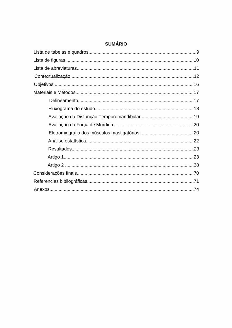
SUMÁRIO
Lista de tabelas e quadros...................................................................................9
Lista de figuras ..................................................................................................10
Lista de abreviaturas..........................................................................................11
Contextualização...............................................................................................12
Objetivos............................................................................................................16
Materiais e Métodos...........................................................................................17
Delineamento.........................................................................................17
Fluxograma do estudo............................................................................18
Avaliação da Disfunção Temporomandibular.........................................19
Avaliação da Força de Mordida..............................................................20
Eletromiografia dos músculos mastigatórios..........................................20
Análise estatística...................................................................................22
Resultados..............................................................................................23
Artigo 1....................................................................................................23
Artigo 2 ...................................................................................................38
Considerações finais..........................................................................................70
Referencias bibliográficas..................................................................................71
Anexos...............................................................................................................74
LISTA DE TABELAS E QUADROS
Artigo 1 – “CORRELATION BETWEEN UPPER LIMB FUNCTION AND ORAL HEALTH
IMPACT IN STROKE SURVIVORS”
Table 1 - Stroke Specific Quality of Life
Table 2 – Correlations between upper extremity function (UEF) subscale of
SSQOL-B and OHIP-14 scales among individuals with hemiparesis stemming
from a stroke
Table 3 – Distribution of impact of oral health conditions per OHIP-14 subscale
among individuals with hemiparesis
Table 4 – Classification of answers to OHIP-14 questionnaire according to
general impact of oral health on quality of life
Artigo 2 – “AVALIAÇÃO ELETROMIOGRÁFICA DOS MÚSCULOS MASTIGATÓRIOS
ASSOCIADOS À DISFUNÇÃO TEMPOROMANDIBULAR EM INDIVÍDUOS HEMIPARÉTICOS
PÓS-ACIDENTE VASCULAR ENCEFÁLICO”
Tabela 1 – Análise descritiva total dos voluntários
Tabela 2 – Prevalência de Disfunção Temporomandibular (DTM) total
Tabela 3 – Análise da DTM (total)
Tabela 4 – Análise da Correlação entre EMG, gênero e tempo de lesão
LISTA DE FIGURAS
Figura 1 – Fluxograma do estudo
Figura 2 – Dinamômetro Kratos®
Artigo 2 – “AVALIAÇÃO ELETROMIOGRÁFICA DOS MÚSCULOS
MASTIGATÓRIOS ASSOCIADO À DISFUNÇÃO
TEMPOROMANDIBULAR EM INDIVÍDUOS HEMIPARÉTICOS PÓS-
ACIDENTE VASCULAR ENCEFÁLICO”
Figura 1 – Sinal eletromiográfico em repouso, MIH e isotonia
Figura 2 – Comparação entre as medianas e intervalos interquartílicos do sinal
EMG (entre os lados direito e esquerdo) nas condições de repouso
Figura 3 - Comparação entre as medianas e intervalos interquartílicos do sinal
EMG (entre os lados direito e esquerdo) nas condições de MIH.
Figura 4 - Comparação entre as medianas e intervalos interquartílicos do sinal
EMG (entre os lados direito e esquerdo) nas condições de isotonia.
LISTA DE ABREVIATURAS
AVE – Acidente Vascular Encefálico
FM – Força de Mordida
DTM – Disfunção temporomandibular
EMG – eletromiografia
TCLE – Termo de consentimento Livre e Esclarecido
MEEM – Mini Exame do Estado Mental
ECT – Escala de Comprometimento do Tronco
CoEP – Comitê de Ética em Pesquisa
RDC/TMD - Research Diagnostic Criteria for Temporomandibular Disorders
SENIAM - Society European Recommendations for Surface Eletromyography
12
1. CONTEXTUALIZAÇÃO
1.1. Acidente Vascular Encefálico
O Acidente Vascular Encefálico (AVE) é um importante agravo à saúde
da população mundial, sendo a principal causa de morte no Brasil e a razão
mais comum de incapacidade neurológica no mundo (PONTES-NETO et al.,
2008). Caracteriza-se por um déficit neurológico focal, repentino e não
convulsivo causado por lesão cerebral consequente de mecanismo vascular
não traumático, por embolia arterial ou venosa, cursando com isquemia ou
hemorragia cerebral (CANNING, 2000).
Dentre as sequelas mais comuns encontradas em pacientes pós-AVE,
destacam-se as alterações sensitivas, cognitivas e motoras, como
espasticidade, padrões anormais de movimento (DOUCET, 2009) e
descondicionamento físico, incluindo fadiga muscular (ALLEN, 2009; KELLY-
HAYES et al., 1998).
Considerando os aspectos motores, a hemiparesia espástica, cursa com
prejuízo da função motora e desvios posturais, diminuição da seletividade do
movimento e inibição recíproca, assim, os músculos agonistas (espásticos) e
antagonistas, contraem-se simultaneamente (OKESON, 1997).
As alterações musculares orofaciais geram comprometimento da função
mastigatória, alteração na preensão dos lábios, diminuição da pressão intra-
oral, alteração da deglutição, ou instabilidade na articulação da fala e da
expressão facial (BRACH et al., 1997).
1.2. Força de Mordida
A mastigação, é uma função aprendida que depende de vias neurais e
conexões sinápticas estabelecidas e comandadas pelo córtex cerebral (ZHAO,
2007).
Acredita-se que a via piramidal é responsável pelo início e controle dos
movimentos mandibulares voluntários. Alguns neurônios corticais controlam os
13
músculos mastigatórios e faciais, possibilitando os movimentos finos e precisos
que são os característicos do sistema estomatognático. O resultado da
integração dos impulsos cerebrais e sensoriais determina o padrão da atividade
mandibular e, portanto, as diferentes posições e movimentações mandibulares
(DOUGLAS, 1994).
Em resumo, o ato mastigatório é um processo complexo, muito
dinâmico, que precisa das aferências nervosas que controlam simultaneamente
a musculatura mastigatória (elevadora e depressora reciprocamente
controlada), a musculatura facial e lingual.
Uma mastigação eficiente não requer apenas dentição (DION, 2007) e
atividade muscular mastigatória adequada (OKIJAMA, 2003), mas depende
também do movimento, força e coordenação de língua, bochecha, lábio e
músculos da mímica facial.
A força de mordida (FM) exercida pelos músculos elevadores da
mandíbula e regulada pelos sistemas nervoso, muscular, esquelético e dentário
(BAKKE, 2006). Alteração neste componente relaciona-se diretamente com a
saúde e integridade do sistema mastigatório (PASSOS, 2010; SHIAU, 1993). E
para que esta função seja harmoniosa são necessárias contínuas modulações
de força (KARKAZIS, 2008) ativadas através dos estímulos mastigatórios
diversos.
A diminuição da FM pode ser provocada por diversos fatores, entre eles
pela diminuição da força muscular dos músculos da mastigação e
adaptabilidade pobre devido à paralisia causada por danos no sistema nervoso
central. A força exercida pelos músculos mastigatórios determina a quantidade
de carga destinada à quebra dos alimentos. (HANSDOTTIR, 2011).
Alterações nas funções orofaciais ficaram evidentes quando foi avaliada
a força de mordida (FM) em sujeitos hemiparéticos crônicos e agudos pós-
AVE, e observaram que a força de mordida máxima do lado contralateral a
lesão estava significativamente correlacionada com as unidades oclusais deste
mesmo lado, em contraste com o lado ipsilateral que não apresentou
correlação com as unidades oclusais (SCHIMMEL et al., 2011).
1.3. Disfunção Temporomandibular
14
Existem vários fatores que influenciam a função mastigatória de forma a
alterá-la. Esses fatores podem envolver alterações estruturais ou funcionais do
sistema estomatognático desencadeando um desequilíbrios considerados como
fatores importantes no desenvolvimento da DTM (NASSIF, 2001).
A DTM caracteriza-se por um grupo de alterações que acometem os
músculos mastigatórios, a articulação temporomandibular (ATM) e estruturas
adjacentes podendo apresentar sintomas como dor, cefaleias, ruídos
articulares e zumbidos (GROSSI, 2004).
1.4. Eletromiografia
A eletromiografia (EMG) foi estabelecida como um importante método de
análise muscular durante comportamento orofacial, pois mede a atividade
elétrica produzida pelos músculos mastigatórios individuais para verificar e
quantificar o equilíbrio muscular entre os músculos de ambos os lados e entre
pares de músculos, e avalia a coordenação muscular em atividades dinâmicas
(MAZZETTO et al., 2014). Estudos eletromiográficos dos músculos faciais são
capazes de fornecer preciosas informações quanto às alterações da fisiologia
do sistema estomatognático de indivíduos com DTM em relação aos indivíduos
sem DTM, procurando esclarecer o relacionamento entre a atividade elétrica e
a resposta mecânica muscular.
A utilização da EMG, associada aos outros métodos clínicos, permite
uma melhor compreensão da participação dos músculos mandibulares no
funcionamento do sistema estomatognático, auxiliando no diagnóstico e no
tratamento de sujeitos com DTM (MARTINS, 2006).
2. JUSTIFICATIVA
O ato mastigatório é um processo complexo, muito dinâmico, que
precisa das aferências nervosas que controlam simultaneamente a musculatura
mastigatória, a musculatura facial e lingual, possibilitando os movimentos finos
e precisos que são os característicos do sistema estomatognático.
15
Diante de relatos da literatura (SCHIMMEL et al., 2011;
CASTROFLORIO et al., 2012; MARTINS, 2006) que sugerem que devido ao
quadro motor, os indivíduos com AVE podem estar sujeitos a apresentarem
comprometimento das funções orofaciais e, como consequência
desenvolverem DTM.
3. OBJETIVOS E HIPÓTESES
3.1 OBJETIVOS
Avaliar se a hemiparesia completa, proveniente de um AVE, pode ter
associação com comprometimento orofacial, alterando a atividade
eletromiográfica e a força dos músculos mastigatórios associado à disfunção
temporomandibular quando comparada à hemiparesia incompleta.
3.2. OBJETIVOS ESPECÍFICOS
Avaliar a atividade eletromiográfica e a força de mordida em pacientes com
hemiparesia completa pós-AVE, e compará-los com hemiparéticos incompletos
com e sem DTM.
3.3 HIPÓTESES
Hipótese nula:
A hemiparesia completa, proveniente de um AVE, não causa maior
comprometimento orofacial, não alterando a atividade eletromiográfica e a força
dos músculos mastigatórios associado à disfunção temporomandibular quando
comparada à hemiparesia incompleta.
Hipótese experimental:
A hemiparesia completa, proveniente de um AVE, pode causar maior
comprometimento orofacial, alterando a atividade eletromiográfica e a força dos
músculos mastigatórios associado à disfunção temporomandibular quando
comparada à hemiparesia incompleta.
16
4. MATERIAL E MÉTODOS
4.1 Aspectos Éticos
O projeto foi aprovado pelo Comitê de Ética em Pesquisa da
Universidade Nove de Julho – UNINOVE (CoEP), sob o número de protocolo
259.239 de 29/04/2013 (anexo I) e foi realizado seguindo as normas que
regulamentam a pesquisa em seres humanos contida na resolução número
466/2012 do Conselho Nacional de Saúde.
Os participantes assinaram o Termo de Consentimento Livre e
Esclarecido (TCLE), após prévia explicação de cada avaliação e esclarecida
toda e qualquer dúvida quanto aos procedimentos, riscos, benefícios.
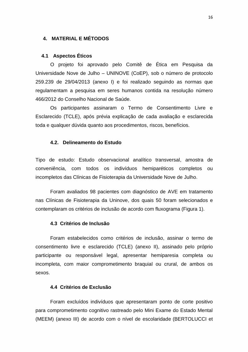
4.2. Delineamento do Estudo
Tipo de estudo: Estudo observacional analítico transversal, amostra de
conveniência, com todos os indivíduos hemiparéticos completos ou
incompletos das Clínicas de Fisioterapia da Universidade Nove de Julho.
Foram avaliados 98 pacientes com diagnóstico de AVE em tratamento
nas Clínicas de Fisioterapia da Uninove, dos quais 50 foram selecionados e
contemplaram os critérios de inclusão de acordo com fluxograma (Figura 1).
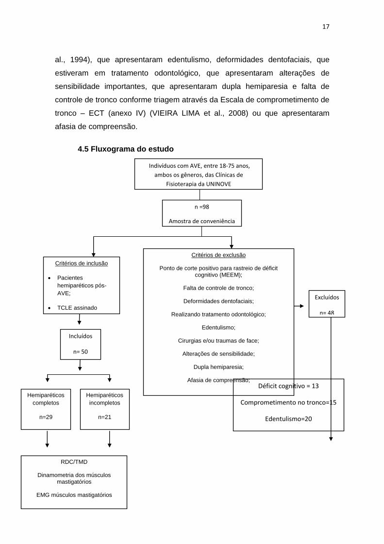
4.3 Critérios de Inclusão
Foram estabelecidos como critérios de inclusão, assinar o termo de
consentimento livre e esclarecido (TCLE) (anexo II), assinado pelo próprio
participante ou responsável legal, apresentar hemiparesia completa ou
incompleta, com maior comprometimento braquial ou crural, de ambos os
sexos.
4.4 Critérios de Exclusão
Foram excluídos indivíduos que apresentaram ponto de corte positivo
para comprometimento cognitivo rastreado pelo Mini Exame do Estado Mental
(MEEM) (anexo III) de acordo com o nível de escolaridade (BERTOLUCCI et
17
al., 1994), que apresentaram edentulismo, deformidades dentofaciais, que
estiveram em tratamento odontológico, que apresentaram alterações de
sensibilidade importantes, que apresentaram dupla hemiparesia e falta de
controle de tronco conforme triagem através da Escala de comprometimento de
tronco – ECT (anexo IV) (VIEIRA LIMA et al., 2008) ou que apresentaram
afasia de compreensão.
4.5 Fluxograma do estudo
Indivíduos com AVE, entre 18-75 anos,
ambos os gêneros, das Clínicas de
Fisioterapia da UNINOVE
Critérios de inclusão
Pacientes
hemiparéticos pós-
AVE;
TCLE assinado
peloparticipante ou
representante legal;
Critérios de exclusão
Ponto de corte positivo para rastreio de déficit cognitivo (MEEM);
Falta de controle de tronco;
Deformidades dentofaciais;
Realizando tratamento odontológico;
Edentulismo;
Cirurgias e/ou traumas de face;
Alterações de sensibilidade;
Dupla hemiparesia;
Afasia de compreensão;
RDC/TMD
Dinamometria dos músculos mastigatórios
EMG músculos mastigatórios
n =98
Amostra de conveniência
Hemiparéticos
completos
n=29
Hemiparéticos
incompletos
n=21
Excluídos
n= 48
Déficit cognitivo = 13
Comprometimento no tronco=15
Edentulismo=20
Incluídos
n= 50
18
Figura 1 – Fluxograma do estudo, conforme STROBE (MALTA et al., 2010)
4.5 Protocolo de avaliação da Disfunção Temporomandibular
A fim de caracterizar a ocorrência de DTM, foi aplicado o Research
Diagnostic Criteria for Temporomandibular Disorders (RDC/ TMD) composto
por dois eixos, sendo que no Eixo II (anexo V) foi avaliado o status psicossocial
do indivíduo por meio de 31 questões. O examinador esteve presente na sala,
porém não auxiliou no preenchimento das respostas para que não houvesse
interferência (DWORKIN, 2009).
Após preenchimento, foram mensurados achados físicos, pelo
examinador treinado, por meio do Eixo I do RDC/ TMD (anexo VI). A avaliação
foi composta por exame clínico intra e extraoral onde foram analisados:
- Padrão de abertura bucal;
- Máxima abertura bucal;
- Ruídos articulares nas excursões, por meio de palpação digital bilateral
e auscultação com estetoscópio;
- Palpação da musculatura mastigatória e região retrocondilar de forma a
avaliar os movimentos condilares e pontos dolorosos nos músculos
mastigatórios.
Para mensuração da extensão de movimentos mandibulares, foi
utilizado paquímetro digital.
Os achados do exame físico foram comparados com os resultados
obtidos no questionário, permitindo caracterizar diagnóstico de DTM. O
diagnóstico não foi hierárquico e permitiu diagnósticos múltiplos para cada
indivíduo. Eles foram divididos em três grupos: musculares (grupo I),
deslocamentos de disco (grupo II) e artralgia, artrite, artrose (grupo III). Foi
realizada tanto avaliação estática quanto a dinâmica. Para esta avaliação, o
participante permaneceu sentado confortavelmente, com quadril a 90 graus de
flexão, pés apoiados no chão, e a mandíbula em uma posição de conforto, com
os dentes levemente em contato.
19
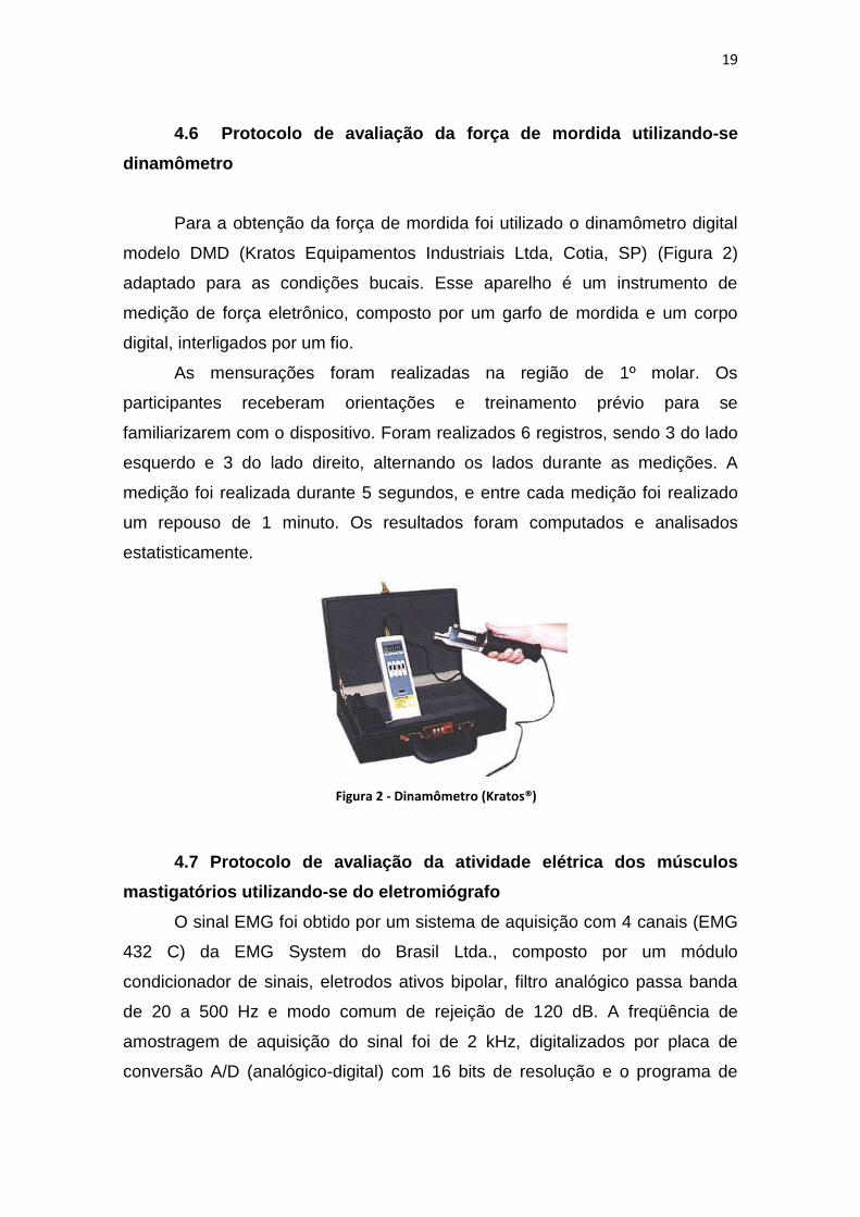
4.6 Protocolo de avaliação da força de mordida utilizando-se
dinamômetro
Para a obtenção da força de mordida foi utilizado o dinamômetro digital
modelo DMD (Kratos Equipamentos Industriais Ltda, Cotia, SP) (Figura 2)
adaptado para as condições bucais. Esse aparelho é um instrumento de
medição de força eletrônico, composto por um garfo de mordida e um corpo
digital, interligados por um fio.
As mensurações foram realizadas na região de 1º molar. Os
participantes receberam orientações e treinamento prévio para se
familiarizarem com o dispositivo. Foram realizados 6 registros, sendo 3 do lado
esquerdo e 3 do lado direito, alternando os lados durante as medições. A
medição foi realizada durante 5 segundos, e entre cada medição foi realizado
um repouso de 1 minuto. Os resultados foram computados e analisados
estatisticamente.
Figura 2 - Dinamômetro (Kratos®)
4.7 Protocolo de avaliação da atividade elétrica dos músculos
mastigatórios utilizando-se do eletromiógrafo
O sinal EMG foi obtido por um sistema de aquisição com 4 canais (EMG
432 C) da EMG System do Brasil Ltda., composto por um módulo
condicionador de sinais, eletrodos ativos bipolar, filtro analógico passa banda
de 20 a 500 Hz e modo comum de rejeição de 120 dB. A freqüência de
amostragem de aquisição do sinal foi de 2 kHz, digitalizados por placa de
conversão A/D (analógico-digital) com 16 bits de resolução e o programa de
20
aquisição foi o EMGLab (EMG System do Brasil Ltda.). Os eletrodos utilizados
foram os bipolares ativos, com pré-amplificação de 20 x.
Para a captação do sinal EMG dos músculos masseter (direito e
esquerdo) e temporal anterior (direito e esquerdo), foram utilizados eletrodos
de superfície descartáveis autoadesivos e do tipo Ag/AgCl (Meditrace infantil),
com diâmetro de 10 mm, fixados no ventre muscular na região que apresentar
maior tônus, após o voluntário realizar moderada intercuspidação dental. Os
eletrodos foram fixados após limpeza com álcool 70%, para diminuir a
impedância entre a pele e os eletrodos. À distância inter-eletrodos foi de 20 mm
entre os centros, como sugerido pela SENIAM (Society European
Recommendations for Surface Eletromyography) (FRERIKS, 2000). Como
referência, foi utilizado um eletrodo no punho esquerdo dos voluntários para
impedir o efeito de interferência de ruídos externos.
A atividade EMG dos músculos masseter e temporal anterior foram
verificadas em três condições: i) repouso; ii) mastigação (isotonia) e iii) máxima
intercuspidação habitual (MIH) (isometria). Os sinais referentes às condições
de repouso e mastigação foram coletados sem material interposto entre os
dentes.
Para a coleta dos dados, os indivíduos permaneceram sentados em uma
cadeira de maneira confortável, com as mãos repousando sobre a coxa.
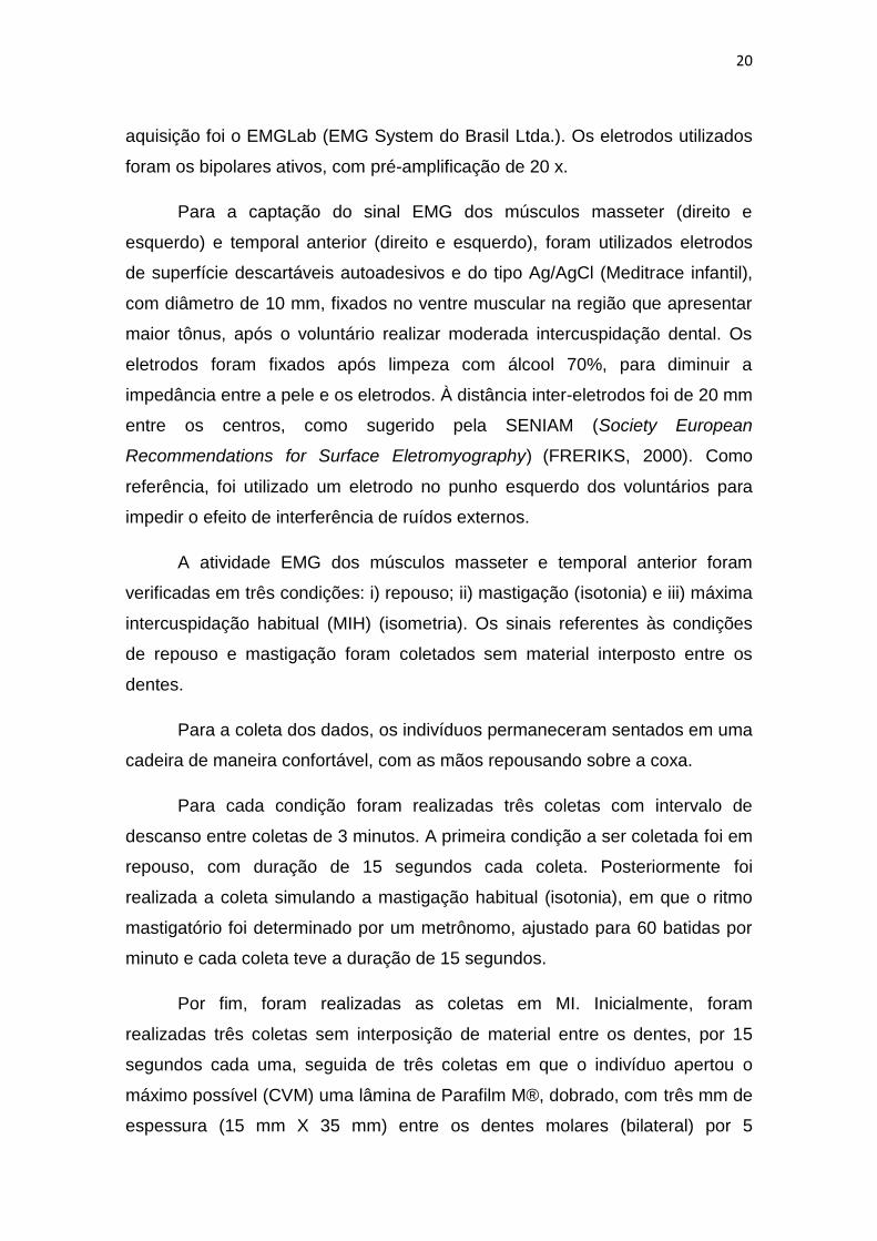
Para cada condição foram realizadas três coletas com intervalo de
descanso entre coletas de 3 minutos. A primeira condição a ser coletada foi em
repouso, com duração de 15 segundos cada coleta. Posteriormente foi
realizada a coleta simulando a mastigação habitual (isotonia), em que o ritmo
mastigatório foi determinado por um metrônomo, ajustado para 60 batidas por
minuto e cada coleta teve a duração de 15 segundos.
Por fim, foram realizadas as coletas em MI. Inicialmente, foram
realizadas três coletas sem interposição de material entre os dentes, por 15
segundos cada uma, seguida de três coletas em que o indivíduo apertou o
máximo possível (CVM) uma lâmina de Parafilm M®, dobrado, com três mm de
espessura (15 mm X 35 mm) entre os dentes molares (bilateral) por 5
21
segundos. Todos os seis períodos mastigatórios foram extraídos com corte de
1 segundo antes e 1 segundo depois do centro do sinal eletromiográfico.
Figura 3 – Protocolo da eletromiografia
4.8 Processamento dos sinais EMG
A atividade EMG dos músculos temporal e masseter nas condições de
repouso mandibular, mastigação isotônica e máxima intercuspidação habitual
foi analisada pela raiz quadrada da média (RMS) da amplitude do sinal EMG
(expressa em µV). Para o repouso mandibular e a mastigação isotônica, o RMS
foi calculado para o total de 15s do sinal coletado.
Para a máxima intercuspidação habitual dos 5 segundos coletados, o
primeiro e o último segundo do sinal EMG bruto foi descartado e o RMS foi
calculado utilizando uma janela de móvel sem sobreposição de 100 ms, sendo
o valor médio das janelas utilizado para a análise.
O RMS de cada condição mastigatória foi normalizado pelo RMS de
maior valor obtido durante as três contrações voluntárias máxima (CVM) (µV /
µV x 100: % CVM) sendo esses expressos como % da CVM.
Os Sinais EMG foram processados por meio de rotinas específicas
desenvolvidas no programa Matlab, versão 7.1 (The MathWorks Inc., Natick ,
22
Massachusetts , EUA).
4.9 Análise Estatística
Para caracterização da amostra e distribuição dos escores obtidos, foi
utilizada estatística descritiva, por meio de medidas de tendência central
(média) e dispersão (desvio-padrão). As variáveis não paramétricas foram
sumarizadas em mediana e intervalo interquartílico. Os resultados foram
submetidos ao teste de Kolmogorov-Smirnov e posteriormente, aos
coeficientes de correlação de Pearson ou Spearman, conforme distribuição dos
dados. A força ou magnitude do relacionamento entre as variáveis foi
classificada como fraca (coeficiente de correlação entre 0,1 e 0,3), moderada
(entre 0,4 e 0,6) e forte (entre 0,7 e 0,9). Para a análise estatística, foi utilizado
o teste qui-quadrado e exato de Fisher para avaliação das variáveis
categóricas, para a comparação de médias teste t-Student e Análise de
Variância (ANOVA), complementada pelo teste LSD e a correlação entre as
variáveis contínuas foi analisada pelo teste de correlação de Pearson,
adotando-se um nível de significância de p< 0,05 (DANCEY, 2006).
4. RESULTADOS
Os resultados dessa pesquisa foram descritos nos dois artigos abaixo.
23
Artigo 1
Artigo 1: Enviado para European Journal of Physical and Rehabilitation
Medicine (A1)
Correlation between upper limb function and oral
health impact in stroke survivors
Fernanda Cordeiro da SILVA, Daniela de Fátima Teixeira da SILVA, Raquel
Agnelli MESQUITA-FERRARI, Kristianne Porta Santos FERNANDES, Sandra
Kalil BUSSADORI
EUROPEAN JOURNAL OF PHYSICAL AND REHABILITATION MEDICINE
Rivista di Medicina Fisica e Riabilitativa
pISSN 1973-9087
Article type: Original Article
Correlation between upper limb function and oral
health impact in stroke survivors
Fernanda C. da Silva, MSc1; Daniela F. T. da Silva, PhD2; Raquel A. Mesquita-
Ferrari, PhD3; Kristianne P. S. Fernandes, PhD3; Sandra K. Bussadori*, PhD3
Affiliations
1. Postgraduate Program in Rehabilitation Sciences, Nove de Julho University
(UNINOVE), São Paulo, SP, Brazil.
24
2. Postgraduate program in Biophotonics Applied to Health Sciences, Nove de
Julho University (UNINOVE) São Paulo, SP, Brazil.
3. Postgraduate Program in Rehabilitation Sciences and Postgraduate Program
in Biophotonics Applied to Health Sciences, Nove de Julho University
(UNINOVE), São Paulo, SP, Brazil.
Congresses: none
Acknowledgements
The authors thank the Fundação de Amparo à Pesquisa do Estado de São
Paulo (FAPESP) grant for fellowships to Master Rehabilitaton student.
Conflict of interest
None declared
Ethical statement
Approved by the local Ethics committee (UNINOVE, Brazil, PP/259.239/13)
*Corresponding author
Sandra Kalil Bussadori, Ph.D.
Nove de Julho University (UNINOVE)
249 Vergueiro Street, Liberdade, São Paulo,SP, Brazil, Zip Code 01504-001
Email: [email protected]
Abstract
Background: Cerebrovascular accident (stroke) is an important health problem
worldwide. It is the most common reason for neurological disability in the world.
25
Aim: To evaluate the relationship between upper limb impairment and oral
health impact in individuals with hemiparesis stemming from a stroke.
Design: cross-sectional study
Setting: The participants were recruited from the Physical Therapy Clinic of
Nove de Julho University (Brazil).
Population: study was conducted with a sample of 27 stroke survivors with
complete or partial hemiparesis with brachial or crural predominance.
Methods: The 14-item short version of the Oral Health Impact Profile (OHIP-14)
was used to evaluate perceptions of oral health. The Brazilian version of the
Stroke Specific Quality of Life Scale (SSQOL-Brazil) was used to evaluate
perceptions regarding quality of life.
Results: A statistically significant association was found between the upper
extremity function subscale of the SSQOL-Brazil and the impact of oral health
evaluated using the OHIP-14, with a strong correlation found for the physical
pain subscale (-0.707), moderate correlations with the functional limitation (-
0.502), psychological discomfort (-0.474), physical disability (-0.461), social
disability (-0.549) and social handicap (-0.555) subscales as well as a weak
correlation with the psychological disability subscale (-0.393). Among the 27
individuals in the present sample, the mean overall OHIP-14 score was 4.63
with a standard deviation (SD) of 6.6 (range: 0.00 to 24.27). Analyzing the
OHIP-14 scores with regard to the impact of oral health on quality of life, the
most frequent classification was weak impact (n = 24; 88.8%), with small rates
of moderate (n = 1; 3.70%) and strong (n = 2; 7.40%) impact.
Conclusions: Compromised upper limb function and self-perceived poor oral
health, whether due to cultural resignation or functional disability, exert a
negative impact on the quality of life of individuals with hemiparesis stemming
from a stroke.
Clinical Rehabilitation Impact: For individuals with hemiparesis, adequate oral
health may mean reintegration into society and a significant improvement in
quality of life. Therefore, the rehabilitation of the paretic upper limb and orofacial
26
function can lead to an improvement in the quality of life of such individuals.
Keywords: quality of life; oral health; stroke
INTRODUCTION
Cerebrovascular accident (stroke) is an important health problem
worldwide. It is the most common reason for neurological disability in the world.1
Stroke is characterized by a sudden, non-convulsive, focal neurological deficit
caused by a brain lesion stemming from a non-traumatic vascular mechanism
due to arterial or venous embolism leading to cerebral ischemia or
hemorrhage.2
The most common manifestations of a stroke are sensory, cognitive and
motor impairments, such as hemiparesis, spasticity, an abnormal movement
pattern3 and physical deconditioning.4,5 According to Saliba et al. (2008),6 upper
limb impairment is one of the most common complaints among stroke survivors
with hemiparesis. It is estimated that 70% of such individuals suffer residual
disability that compromises dexterity during activities of daily living.7 Individuals
with hemiparesis experience slow movements during activities that involve the
upper limbs, such as reaching and grasping, due to the limited range of motion,
segmented movements and a lack of coordination among the joints4,8.
Normal upper limb function involves the capacity for directed reach,
grasping and manipulation of objects, which make up the motor skills required
for the performance of activities of daily living6 that allow an individual to lead an
independent life with self-esteem.7 The execution of proper oral hygiene, for
example, requires adequate motor control of the upper limbs.8 Compromised
upper limb function exerts an impact on the degree of disability experienced by
stroke survivors, with a significant influence on functional performance and
negative consequences regarding personal, familial and social relationships as
well as quality of life9.
27
Despite the gradual return of motor function resulting from a combination
of spontaneous recovery and physical therapy, the use of the paretic limb is
often less than its normal potential in daily living.10 Depending on the degree of
upper limb impairment, the maintenance of adequate oral health among stroke
survivors can be hindered.3,7 Moreover, inadequate oral hygiene can
compromise both oral health and quality of life.
Considering the high prevalence rates of functional limitations to the
paretic arm,6 the aim of the present study was to evaluate the relationship
between upper limb impairment and oral health impact in individuals with
hemiparesis stemming from a stroke.
METHODS
This study received approval from the local human research ethics
committee under process number 259.239. All participants received
clarifications regarding the study and authorized participation by signing a
statement of informed consent in compliance with Resolution 466/2012 of the
Brazilian National Board of Health. The participants were recruited from the
Physical Therapy Clinic of Nove de Julho University (Brazil).
A descriptive, cross-sectional study was conducted with a sample of 27
stroke survivors with complete or partial hemiparesis with brachial or crural
predominance. Control of the trunk was an additional inclusion criterion.
Individuals with cognitive deficit, those with dentofacial deformities, those in
dental treatment and those with sensitivity abnormalities or quadriparesis were
excluded from the study.
The 14-item short version of the Oral Health Impact Profile (OHIP-14)11
was used to evaluate perceptions of oral health12. This questionnaire has seven
subscales, each with two questions: functional limitation, physical pain,
psychological discomfort, physical disability, psychological disability, social
disability and social handicap. The OHIP-14 was developed as a self-
administered questionnaire. However, to eliminate limitations related to the
28
function of the paretic upper limb, the questionnaire was administered in
interview form (the researchers read the questions aloud and marked the
respondents’ answers).
The Brazilian version of the Stroke Specific Quality of Life Scale
(SSQOL-Brazil)13,14 was used to evaluate perceptions regarding quality of life.
This scale has 49 items distributed among 12 subscales (energy, family roles,
language, mobility, mood, personality, self care, social roles, reasoning, upper
extremity function, vision and work/productivity). Each item has five response
options referring to function in the previous week. The score of each item
ranges from 1 to 5 points and the total ranges from 49 (worst perception of
quality of life) to 245 (best perception). In the present study, only the upper
extremity function subscale was employed to analyze its association with oral
health impact. Although upper extremity function on this questionnaire is
evaluated based on actions such as fastening a button and opening/closing a
zipper, SSQOL-Brazil was chosen for use in the present study due to the lack of
specific questionnaires in the literature for the evaluation of upper limb function
in relation to oral self-care and even the function of feeding oneself.
Statistical analysis was performed using the SPSS program version 20.0.
The Shapiro-Wilk test was used to determine whether or not the data had
normal distribution. Parametric data were expressed as mean and standard
deviation (SD). Nonparametric data were expressed as median and interquartile
range. Pearson’s correlation coefficients were calculated to determine the
magnitude, direction and significance of associations between variables related
to upper limb function and oral health impact. The strength of the associations
was classified as weak (correlation coefficient: 0.1 to 0.3), moderate (correlation
coefficient: 0.4 to 0.6) or strong (correlation coefficient: 0.7 to 1). The level of
significance was set to 5% (p < 0.05) for all analyses.
29
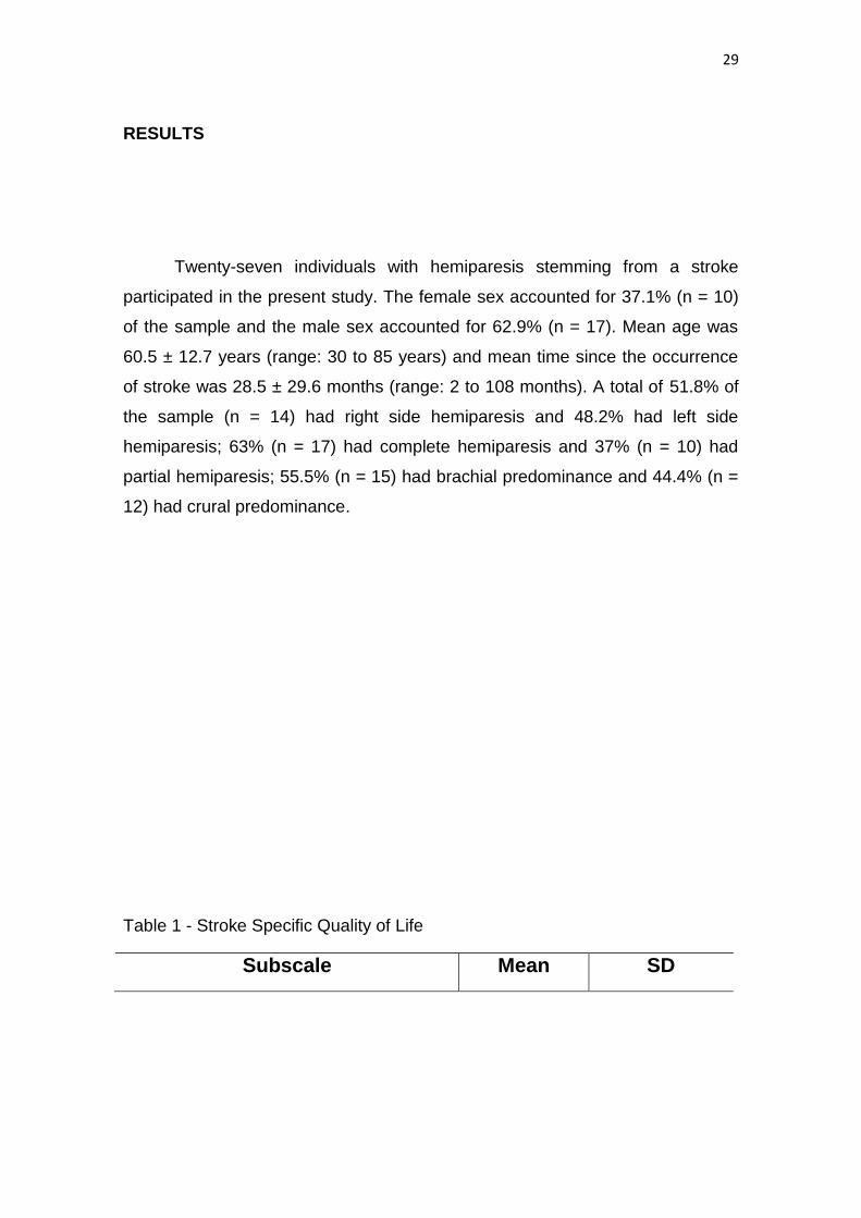
RESULTS
Twenty-seven individuals with hemiparesis stemming from a stroke
participated in the present study. The female sex accounted for 37.1% (n = 10)
of the sample and the male sex accounted for 62.9% (n = 17). Mean age was
60.5 ± 12.7 years (range: 30 to 85 years) and mean time since the occurrence
of stroke was 28.5 ± 29.6 months (range: 2 to 108 months). A total of 51.8% of
the sample (n = 14) had right side hemiparesis and 48.2% had left side
hemiparesis; 63% (n = 17) had complete hemiparesis and 37% (n = 10) had
partial hemiparesis; 55.5% (n = 15) had brachial predominance and 44.4% (n =
12) had crural predominance.
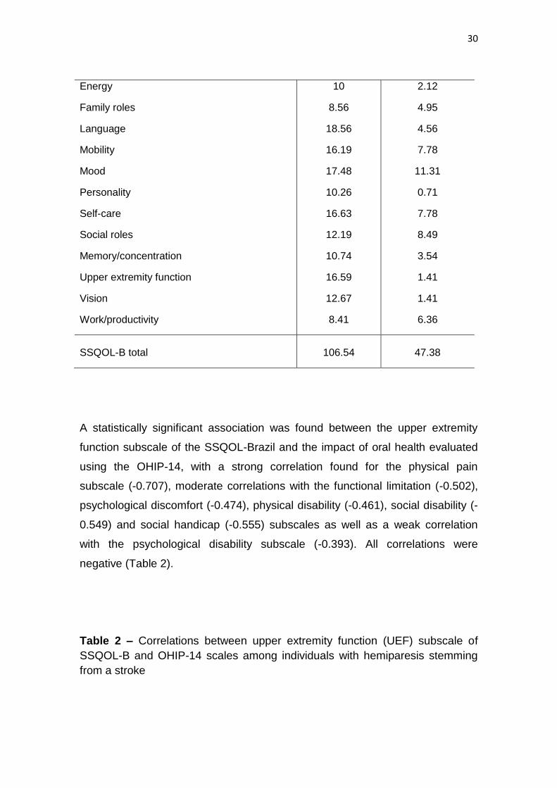
Table 1 - Stroke Specific Quality of Life
Subscale Mean SD
30
Energy
Family roles
Language
Mobility
Mood
Personality
Self-care
Social roles
Memory/concentration
Upper extremity function
Vision
Work/productivity
10
8.56
18.56
16.19
17.48
10.26
16.63
12.19
10.74
16.59
12.67
8.41
2.12
4.95
4.56
7.78
11.31
0.71
7.78
8.49
3.54
1.41
1.41
6.36
SSQOL-B total 106.54 47.38
A statistically significant association was found between the upper extremity
function subscale of the SSQOL-Brazil and the impact of oral health evaluated
using the OHIP-14, with a strong correlation found for the physical pain
subscale (-0.707), moderate correlations with the functional limitation (-0.502),
psychological discomfort (-0.474), physical disability (-0.461), social disability (-
0.549) and social handicap (-0.555) subscales as well as a weak correlation
with the psychological disability subscale (-0.393). All correlations were
negative (Table 2).
Table 2 – Correlations between upper extremity function (UEF) subscale of
SSQOL-B and OHIP-14 scales among individuals with hemiparesis stemming
from a stroke
31
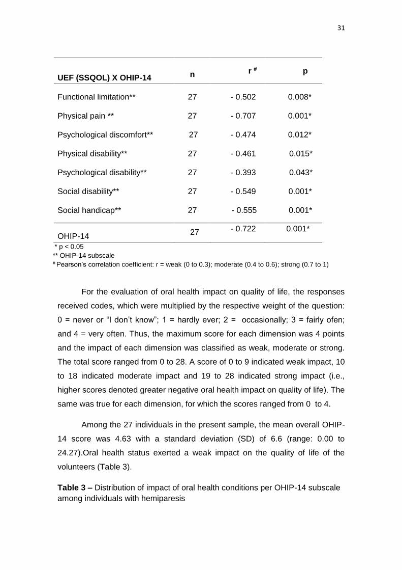
UEF (SSQOL) X OHIP-14 n r # p
Functional limitation**
Physical pain **
Psychological discomfort**
Physical disability**
Psychological disability**
Social disability**
Social handicap**
27
27
27
27
27
27
27
- 0.502
- 0.707
- 0.474
- 0.461
- 0.393
- 0.549
- 0.555
0.008*
0.001*
0.012*
0.015*
0.043*
0.001*
0.001*
OHIP-14 27 - 0.722 0.001*
* p < 0.05
** OHIP-14 subscale # Pearson’s correlation coefficient: r = weak (0 to 0.3); moderate (0.4 to 0.6); strong (0.7 to 1)
For the evaluation of oral health impact on quality of life, the responses
received codes, which were multiplied by the respective weight of the question:
0 = never or “I don’t know”; 1 = hardly ever; 2 = occasionally; 3 = fairly ofen;
and 4 = very often. Thus, the maximum score for each dimension was 4 points
and the impact of each dimension was classified as weak, moderate or strong.
The total score ranged from 0 to 28. A score of 0 to 9 indicated weak impact, 10
to 18 indicated moderate impact and 19 to 28 indicated strong impact (i.e.,
higher scores denoted greater negative oral health impact on quality of life). The
same was true for each dimension, for which the scores ranged from 0 to 4.
Among the 27 individuals in the present sample, the mean overall OHIP-
14 score was 4.63 with a standard deviation (SD) of 6.6 (range: 0.00 to
24.27).Oral health status exerted a weak impact on the quality of life of the
volunteers (Table 3).
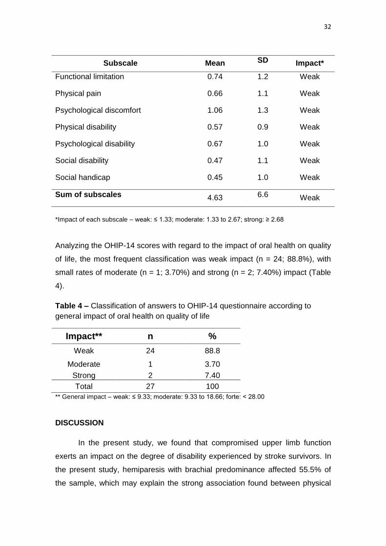
Table 3 – Distribution of impact of oral health conditions per OHIP-14 subscale
among individuals with hemiparesis
32
Subscale Mean SD Impact*
Functional limitation
Physical pain
Psychological discomfort
Physical disability
Psychological disability
Social disability
Social handicap
0.74
0.66
1.06
0.57
0.67
0.47
0.45
1.2
1.1
1.3
0.9
1.0
1.1
1.0
Weak
Weak
Weak
Weak
Weak
Weak
Weak
Sum of subscales 4.63 6.6 Weak
*Impact of each subscale – weak: ≤ 1.33; moderate: 1.33 to 2.67; strong: ≥ 2.68
Analyzing the OHIP-14 scores with regard to the impact of oral health on quality
of life, the most frequent classification was weak impact (n = 24; 88.8%), with
small rates of moderate (n = 1; 3.70%) and strong (n = 2; 7.40%) impact (Table
4).
Table 4 – Classification of answers to OHIP-14 questionnaire according to
general impact of oral health on quality of life
Impact** n %
Weak 24 88.8
Moderate 1 3.70
Strong 2 7.40
Total 27 100
** General impact – weak: ≤ 9.33; moderate: 9.33 to 18.66; forte: < 28.00
DISCUSSION
In the present study, we found that compromised upper limb function
exerts an impact on the degree of disability experienced by stroke survivors. In
the present study, hemiparesis with brachial predominance affected 55.5% of
the sample, which may explain the strong association found between physical
33
pain and upper limb function. Hemiparesis leads to instability in movements of
the trunk and limbs, thereby compromising one’s performance on activities of
daily living, such as oral hygiene, which depends on adequate motor control of
the upper limbs15-19.
Age is another factor that may have influenced the ability to perform self-
care in the present sample, as mean age was 60.5 years. Silvestre et al.20
report that approximately 40% of individuals aged 60 years or older need some
type of assistance to perform at least one instrumental activity of daily living and
10% need assistance to perform basic tasks, such as bathing, dressing and
other aspects of self-care. According to Hunt et al.21 and Slade et al.11, older
adults experience greater functional limitation and psychological discomfort.
The combination of the negative consequences of ageing and stroke leads to a
substantial reduction in quality of life.
The total OHIP-14 score in the present study was low (4.6 points). In
contrast, REED et al.22 found that 137 older adults at an extended care facility
had a poor perception of their oral health status. Many older adults seem not to
be bothered by poor oral health, which demonstrates a certain cultural
resignation.23 Indeed, oral problems are often minimized in comparison to other
adverse health conditions24.Thus, when evaluating quality of life, older adults
often perceive poor oral health as normal or acceptable for someone at an
advanced age25,26.Despite this resigned attitude, the association between oral
health and quality of life indicates that oral problems exert a negative impact on
the emotional wellbeing of this population. In the present study, a negative
correlation was found between social disability (OHIP-14) and quality of life
(SSQOL-B), as individuals with higher social disability scores stemming from
oral problems had lower quality of life scores. These findings are in agreement
with data reported by Marinõ et al.27 and Tatematsu et al. 28, who state that oral
pain and problems eating, chewing, smiling and speaking tend to affect an
individual’s wellbeing substantially.
For individuals with hemiparesis, adequate oral health may mean
reintegration into society and a significant improvement in quality of life.29
34
Therefore, the rehabilitation of the paretic upper limb and orofacial function can
lead to an improvement in the quality of life of such individuals.
CONCLUSION
Compromised upper limb function and self-perceived poor oral health,
whether due to cultural resignation or functional disability, exert a negative
impact on the quality of life of individuals with hemiparesis stemming from a
stroke.
References
1. Pontes-Neto OM, Silva GS, Feitosa MR, de Figueiredo NL, Fiorot JA Jr,
Rocha TN. Stroke awareness in Brazil: alarming results in a community-based
study.Stroke 2008;39:292–6.
2. Canning CG, Ada L, O’Dwyer NJ. Abnormal muscle activation characteristics
associated with loss of dexterity after stroke. Neurological Sci 2000;176:45-56.
3. Doucet BM, Griffin L. Variable stimulation patterns for poststroke hemiplegia.
Muscle Nerve 2009;39(1):54-62.
4. Allen DG. Fatigue in working muscles. J Appl Physiol 2009;106(2):378-84.
5. Kelly-Hayes M, Robertson JT, Broderick JP, et al. The American Heart
Association Stroke Outcome Classification. Stroke 1998;29(6):1274-80.
6. Saliba VA, Júnior IPC, Faria CDC, Salmela LFT. Propriedades Psicométricas
da Motor Activity Log: uma revisão sistemática da literatura. Revista Fisioter
Mov 2008;21(3):59-67.
7. Harris JE, Eng JJ. Paretic Upper-Limb Strength Best Explains Arm Activity in
People With Stroke. Physical Therapy 2007;87(1):88-97.
35
8. Zackoski KM, Dromerick AW, Sahrmann SA, Thach WT, Bastian AJ. How do
strength, sensation, spasticty and joint individuation relate to the reaching
deficits of people with chronic hemiparesis?. Brain 2004;127:1035-46.
9. Shumway-Cook A, Woollacott MH. Controle Motor – Teorias e aplicações
práticas.
10. Uswatte G, Taub E, Morris DM, Vignolo M, McCulloch K. Reliability and
Validity of the Upper-Extremity Motor Activity Log-14 for Measuring Real-World
Arm Use. Stroke 2005; 36:2493-6.
11. Slade GD, Spencer AJ. Development and evaluation of the Oral Health
Impact Profile. Community Dental Health 1994;11(1):3-11.
12. Lai SM, Perera S, Duncan PW, Bode R. Physical and social functioning
after stroke: comparison of the Stroke Impact Scale and Short Form-36. Stroke
2003; 34(2):488-93.
14. Geyh S, Cieza A, Kollerits B, Grimby G, Stucki G. Content comparison of
healthrelated quality of life measures used in stroke based on the international
classification of functioning, disability and health (ICF): a systematic review.
Qual Life Res 2007;16:833-51.
15. Lockette KF, Keyes M. Conditioning with physical disabilities. Chicago:
Rehabilitation Institute of Chicago; 1994.p.433.
16. Chagas EF, Tavares MCGCF. Simetria e transferência de peso do
hemiplégico: relação dessa condição com o desempenho de suas atividades
funcionais. Ver Fisioter Univ São Paulo 2001; 8:40-50.
17. Nakayama H, Jorgensen HS, Raaschou HO, Olsen TS. Compensation in
recovery of upper extremity function after stroke: the Copenhagen Stroke Study.
Arch. Phys. Med. Rehabily 1994;75(8):852-7.
18. Hunter S, Crome P. Hand function and stroke. Rev. Clin. Gerontol
2002;12(1):68-81.
36
19. Terroni LMN, Leite CC, Tinone G, Fraguas JR R. Depressão pós-AVC:
fatores de risco e terapêutica antidepressiva. Rev.Assoc. Med. Bras.
2003;49(4):450-9.
20. Silva SRC, Castellanos FRA. Autopercepção das condições de saúde bucal
por idosos. Rev Saúde Pública 2001;35(4):349-55.
21. Hunt RJ, Slade GD, Strauss RP. Differences between racial groups in the
impact of oral disorders among older adults in North Carolina. J PubUc Health
Dent 1995;55(4):205-9.
22. Reed R, Broder HL, Jenkins G, Spivack E, Janal MN. Oral health promotion
among older persons and their care providers in a nursing home facility.
Gerodontology 2006;23:73-8.
23. Haikal DS, Paula AMB, Martins, AMEBL, Moreira, NA, Ferreira EF.
Autopercepção da saúde bucal e impacto na qualidade de vida do idoso: Uma
abordagem quantiqualitativa. Ciência & Saúde Coletiva 2011;16(7):3317-29.
24. Reisine S, Muler JA. Longitudinal study of work loss related to dental
diseases. Soc Sei Med 1995; 21(12):1309-14.
25. Dahl KE, Wang NJ, Holst D, Ohrn K. Oral health-related quality of life
among adults 68-77 years old in Nord-Trøndelag, Norway. International Journal
of Dental Hygiene 2011;9(1):87-92.
26. Mariño R, Schofield M, Wright C, Calache H, Minichiello V. Self-reported
and clinically determined oral health status predictors for quality of life in
dentate older migrant adults. Community Dentistry and Oral Epidemiology
2008;36:85.
27. Tatematsu M, Mori T, Kawaguchi T, et al. Masticatory performance in 80-
yearold individuals. Gerontology 2004;21:112-9.
37
Artigo 2:
Electromyographic evaluation masticatory
muscles in individuals with hemiparesis and
temporomandibular disorder
Fernanda Cordeiro da Silva1¶,#a, Fabiano Politti1&,#a, Gabriela Regina de Lima2¶,
Daniela de Fátima Teixeira da Silva3¶,#b, Raquel Agnelli Mesquita-Ferrrari1&,
Kristianne Porta Santos Fernandes4¶,#b, Daniela Aparecida Biasotto-Gonzalez1&,
Alessandro Melo Deana3¶,#b, Sandra Kalil Bussadori4*,#a
1. Postgraduate Program in Rehabilitation Sciences, Nove de Julho University
(UNINOVE), São Paulo, SP, Brazil.
2. Graduate physiotherapy, Nove de Julho University (UNINOVE), São Paulo,
SP, Brazil.
3. Postgraduate Program in Biophotonics Applied to Health Sciences, Nove de
Julho University (UNINOVE), São Paulo, SP, Brazil.
4. Postgraduate Program in Rehabilitation Sciences and Postgraduate Program
in Biophotonics Applied to Health Sciences, Nove de Julho University
(UNINOVE), São Paulo, SP, Brazil.
#a Current Address: Department in Postgraduate Program in Rehabilitation
Sciences, Nove de Julho University/UNINOVE, Liberdade, São Paulo, SP,
Brazil.
#b Current Address: Department in Postgraduate Program in Biophotonics
Applied to Health Sciences, Nove de Julho University/UNINOVE, Liberdade,
São Paulo, SP, Brazil.
* Corresponding author
E-mail: [email protected] (SKB)
¶ These authors contributed equally to this work.
&These authors also contributed equally to this work.
38
Abstract
Background: Cerebrovascular diseases, including stroke, are the third major
cause of death and the first major cause of physical and mental disability
throughout the world. Due to the consequent hemiparesis, stroke survivors
often experience residual musculoskeletal impairment as well as sensory and
cognitive alterations. Such muscle imbalances can lead to orofacial alterations,
with possible consequences to the temporomandibular joint. Any derangement
in this joint can lead to temporomandibular disorder.
Methods and Findings: A descriptive, cross-sectional study was conducted
involving 50 individuals. The sample was divided into two groups. Group 1
comprised individuals with complete hemiparesis and Group 2 comprised
individuals with incomplete hemiparesis. The Research Diagnostic Criteria for
Temporomandibular Disorders was used for the diagnosis of TMD. To capture
the EMG signal of the right and left masseter and anterior temporal muscles
was used a four-channel acquisition system. The overall prevalence rate of
TMD was 60%. There was a statistically significant difference between the right
and left side was found in the EMG signal to the jaw at rest, both in the group
with complete hemiparesis and the group with incomplete hemiparesis. No
statistically significant differences in the EMG signal were found between the
right and left sides during isometric contraction or isotonic contraction.
Conclusions: The hemiparesis alters electromyographic signals in the
masticatory muscles with the mandible at rest. Negative correlations were found
between the EMG signal and both the male gender and time elapsed since the
stroke, demonstrating less electrical activity with a longer time since the
neurological event, especially among men.
39
Introduction Cerebrovascular diseases, including stroke, are the third major cause of
death and the first major cause of physical and mental disability throughout the
world(1). Stroke is characterized by a sudden, non-convulsive, focal,
neurological deficit caused by a brain injury stemming from a non-traumatic
vascular mechanism (arterial or venous embolism) that results in ischemia or
hemorrhage(2).
Due to the consequent hemiparesis, stroke survivors often experience
residual musculoskeletal impairment as well as sensory and cognitive
alterations, which cause limitations in the performance of activities of daily
living(3). Such muscle imbalances can lead to orofacial alterations, with
possible consequences to the temporomandibular joint(4), which is an important
element of the stomatognathic system that participates in complex jaw
movements, such as chewing and speaking(5). Any derangement in this joint
can lead to temporomandibular disorder (TMD)(6), which is characterized by a
broad gamut of alterations in the masticatory muscles, temporomandibular joint
and adjacent structures, causing symptoms such as pain, headache, joint
sounds, ringing in the ears and limited jaw movements(7).
Changes in orofacial musculoskeletal structures can be evaluated using
electromyography (EMG), which is a noninvasive method that is easy to
administer(8). The use of EMG in studies on the function of the masticatory
muscles can provide important physiological information on the functioning of
the stomatognathic system and allows the analysis of muscle activity under
different conditions (at rest, during maximum clenching and during chewing)
and assists in the diagnosis and monitoring of TMD(9,10,11,12).
Considering the need for the early identification of problems that
40
predispose individuals with hemiparesis to abnormalities of the stomatognathic
system, EMG can contribute to a better understanding of masticatory muscle
activity in stroke survivors as well as responses to specific treatment modalities.
The aims of the present study were to monitor EMG activity in the
masticatory muscles of individuals with both hemiparesis and TMD and
determine possible correlations with gender and time elapsed since the stroke
event.
Material and Methods This study received approval from the local Human Research Ethics
Committee under process number 259.239, consent was written. All prospective
participants and legal guardians received clarifications regarding the objectives
and procedures. Those who agreed to voluntary participation signed a
statement of informed consent in compliance with Resolution 466/2012 of the
Brazilian National Board of Health. Convenience sampling was performed.
Individuals with hemiparesis stemming from a stroke were recruited from the
Physical Therapy Clinic of University Nove de Julho (São Paulo, Brazil).
A descriptive, cross-sectional study was conducted involving 98
individuals with complete or incomplete hemiparesis with brachial or crural
predominance. Individuals with cognitive impairment, dentofacial deformities,
complete or partial edentulism, quadriparesis and those currently in dental
treatment were excluded. Forty-eight individuals did not meet the eligibility
criteria (15 had trunk impairment, 20 had complete or partial endentulism and
13 had cognitive impairment). Thus, 50 individuals were included in the study.
The sample was divided into two groups. Group 1 comprised individuals
with complete hemiparesis (face, upper limb and lower limb affected on one
side of the body). Group 2 comprised individuals with incomplete hemiparesis
(only the upper and lower limb affected) and was considered the control group.
Inter-group and intra-group comparisons were performed for bite force, TMD,
gender, age, time elapsed since stroke, predominance and type of impairment.
41
Diagnosis of temporomandibular disorder
The Research Diagnostic Criteria for Temporomandibular Disorders
(RDC/TMD)(13) was used for the diagnosis of TMD. This measure is composed
of two axes. Axis I was used for the physical assessment, which involved
intraoral and extraoral clinical exams for the analysis of mandibular movements,
including maximum vertical mandibular movement, joint sounds during right and
left jaw excursion using bilateral digital palpation and a stethoscope for
auscultation, palpation of the masticatory muscles and retrocondylar region for
the evaluation of condyle movements and trigger points in the masticatory
muscles. The findings of the physical exam were compared to the descriptions
on the questionnaire for the diagnosis of the specific group of TMD: muscle
involvement (Group I), disk displacement (Group II) and joint involvement –
arthralgia, arthritis and arthrosis (Group III). The diagnosis was non-hierarchical
and a given participant could have more than one diagnosis. Both static and
dynamic analyses were performed. For such, the volunteer remained seated
comfortably with hips flexed at 90 degrees, feet supported on the floor and the
mandible in a comfortable position with the teeth lightly in contact. Axis II
contains 31 questions for the evaluation of psychosocial aspects of TMD.
Electromyography
The EMG signals were captured using a four-channel acquisition system
(EMG 432 C, EMG System do Brasil Ltda.) composed of a signal conditioning
module, bipolar active electrodes, analog band pass filter of 20 to 500 Hz and a
common mode rejection rate of 120 dB. The sampling frequency was 2 kHz,
digitized using an analog-digital converter with 16 bits of resolution and the
acquisition program was EMGLab (EMG System do Brasil Ltda.). The
electrodes had a pre-amplification of 20 x.
To capture the EMG signal of the right and left masseter and anterior
temporal muscles, self-adhesive, disposable, Ag/AgCl surface electrodes
(Meditrace) measuring 10 mm in diameter were attached to the belly of the
muscle in the region of greatest tone, determined during moderate
42
clenching(14). After cleaning the skin with 70% alcohol to diminish impedance,
the electrodes were spaced at a distance of 20 mm center to center. An
additional electrode was attached to the left wrist to impede interference from
external noise.
EMG of the masticatory muscles was determined under three conditions:
I) at rest; II) during mastication (isotonic contraction); and III) during maximum
voluntary clenching (MVC – isometric contraction). For such, the volunteer was
seated comfortably on a chair with hands resting on the thighs. Three readings
were made under each condition with a three-minute rest period between
readings. In the rest position, each reading lasted 15 seconds. The volunteer
then performed three five-second readings during MVC, clenching on folded
Parafilm M® measuring 3 mm in thickness (15 X 35 mm) placed bilaterally
between the molars. After a rest period, the volunteer was instructed to perform
habitual chewing (isotonic contraction) at a rhythm determined using a
metronome adjusted to 60 beats per minute; each reading lasted 15 seconds.
Finally, MVC was performed for five seconds without the use of Parafilm M®.
Processing of electromyographic signals
EMG activity of the masseter and temporal muscles at rest, during
isometric contraction and during isotonic contraction was analyzed based on the
root mean square (RMS) of the amplitude of the signal (expressed in µV). The
RMS was calculated for the entire 15-second reading with the muscles at rest
and during isotonic contraction. During MVC, the first and last seconds of the
crude EMG signal were discarded and the RMS was calculated using a 100-ms
moving window with no overlap; the mean value of the windows was used for
analysis. The RMS under each condition was normalized by the highest MVC
value (µV/µV x 100: % MVC) and expressed as % of MVC. The EMG signals
were process using specific routines developed for the Matlab program, version
7.1 (The MathWorks Inc., Natick, Massachusetts, USA).
Results
43
Fifty individuals were analyzed (mean age: 60.8 years). The female
gender accounted for 38% of the sample (n = 19) and the male gender
accounted for 62% (n = 31). Mean time elapsed since the stroke was 24.3
months. A total of 52% (n = 26) had right-side hemiparesis, 48% (n = 24) had
left-side hemiparesis, 58% (n = 29) had complete hemiparesis, 42% (n = 21)
had incomplete hemiparesis, 52% (n = 26) had brachial predominance and 48%
(n = 24) had crural predominance.
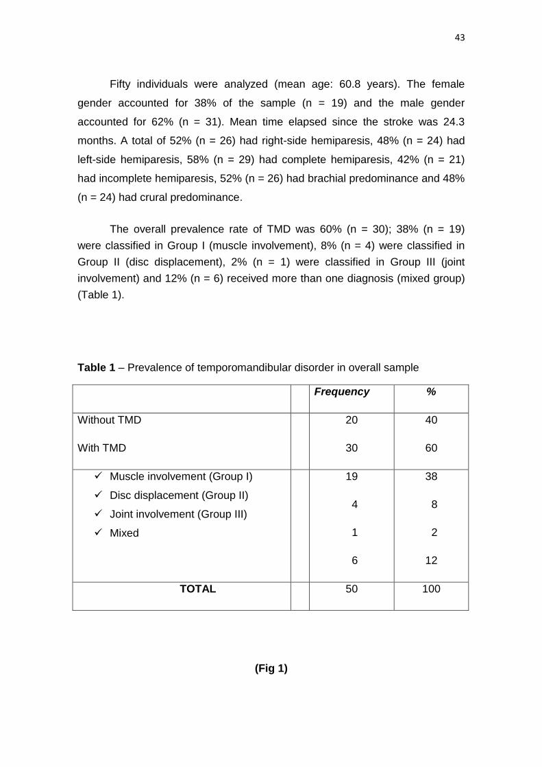
The overall prevalence rate of TMD was 60% (n = 30); 38% (n = 19)
were classified in Group I (muscle involvement), 8% (n = 4) were classified in
Group II (disc displacement), 2% (n = 1) were classified in Group III (joint
involvement) and 12% (n = 6) received more than one diagnosis (mixed group)
(Table 1).
Table 1 – Prevalence of temporomandibular disorder in overall sample
Frequency %
Without TMD
With TMD
20
30
40
60
Muscle involvement (Group I)
Disc displacement (Group II)
Joint involvement (Group III)
Mixed
19
4
1
6
38
8
2
12
TOTAL 50 100
(Fig 1)
44
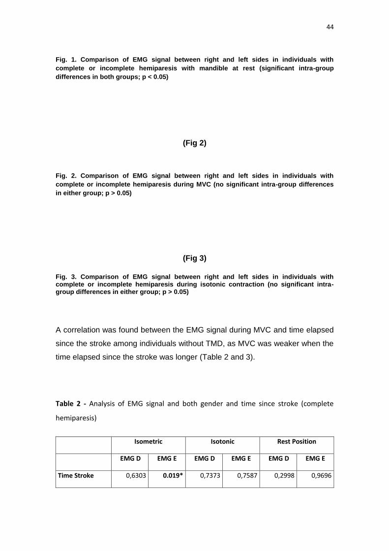
Fig. 1. Comparison of EMG signal between right and left sides in individuals with
complete or incomplete hemiparesis with mandible at rest (significant intra-group
differences in both groups; p < 0.05)
(Fig 2)
Fig. 2. Comparison of EMG signal between right and left sides in individuals with
complete or incomplete hemiparesis during MVC (no significant intra-group differences
in either group; p > 0.05)
(Fig 3)
Fig. 3. Comparison of EMG signal between right and left sides in individuals with complete or incomplete hemiparesis during isotonic contraction (no significant intra-group differences in either group; p > 0.05)
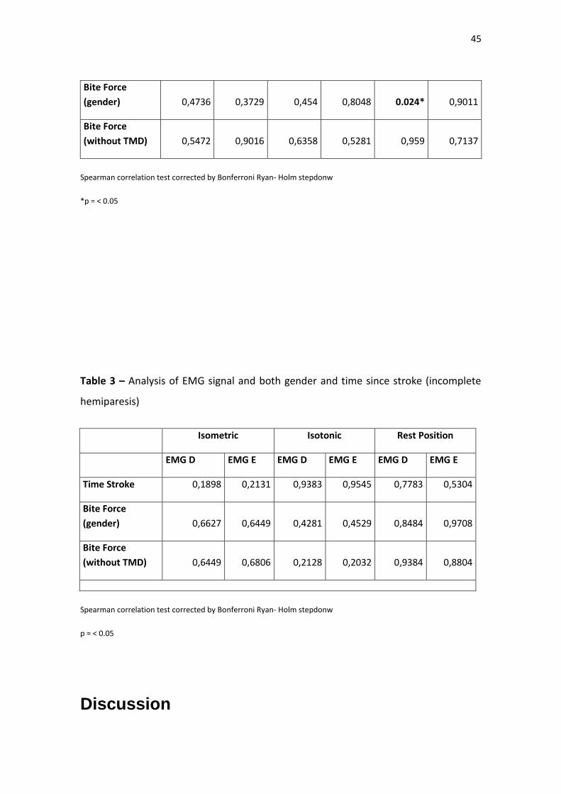
A correlation was found between the EMG signal during MVC and time elapsed
since the stroke among individuals without TMD, as MVC was weaker when the
time elapsed since the stroke was longer (Table 2 and 3).
Table 2 - Analysis of EMG signal and both gender and time since stroke (complete
hemiparesis)
Isometric Isotonic Rest Position
EMG D EMG E EMG D EMG E EMG D EMG E
Time Stroke 0,6303 0.019* 0,7373 0,7587 0,2998 0,9696
45
Bite Force
(gender) 0,4736 0,3729 0,454 0,8048 0.024* 0,9011
Bite Force
(without TMD) 0,5472 0,9016 0,6358 0,5281 0,959 0,7137
Spearman correlation test corrected by Bonferroni Ryan- Holm stepdonw
*p = < 0.05
Table 3 – Analysis of EMG signal and both gender and time since stroke (incomplete
hemiparesis)
Isometric Isotonic Rest Position
EMG D EMG E EMG D EMG E EMG D EMG E
Time Stroke 0,1898 0,2131 0,9383 0,9545 0,7783 0,5304
Bite Force
(gender) 0,6627 0,6449 0,4281 0,4529 0,8484 0,9708
Bite Force
(without TMD) 0,6449 0,6806 0,2128 0,2032 0,9384 0,8804
Spearman correlation test corrected by Bonferroni Ryan- Holm stepdonw
p = < 0.05
Discussion
46
EMG has proven to be an efficient means for evaluating the effects of
drugs, surgical procedures and other forms of therapy, allowing the
measurement of the electric potential in spastic muscles(15). EMG is employed
for the analysis and interpretation of the bioelectrical potential produced in
muscles and nerves during voluntary movements, electrically induced activity or
even electrical activity with the muscle at rest through the capturing of the
electrical potential of base muscle activity(16). Moreover, EMG can indicate
both the beginning and end of muscle activity as well as provide information on
the number of active motor units and the frequency at which the potentials
fire(17).
The determination of the electrical activity in the masticatory muscles is
of fundamental importance to comparisons within and between individuals. In
the present study, a statistically significant difference between the right and left
side was found in the EMG signal with the mandible at rest in both the group
with complete hemiparesis (p=0.002) and the group with incomplete
hemiparesis (p = 0.009), as shown in Fig. 1. This finding may be attributed to a
possible change in the tone of the muscles studied due to hypertonia caused by
a change in motor control, which is commonly found in individuals with injuries
to the central nervous system(18). The same has been found to occur during
reflex contractions, which is reported to be greater in individuals without
paresis(19).
However, the physiological basis of the mandible in the rest position is
one of the more controversial aspects of oral physiology, as the muscles may
be contracted and demonstrate slight electrical activity. In a recent study, the
authors evaluated EMG activity in the massetter and temporal muscles in
women with and without TMD and concluded that those with TMD exhibited
greater asymmetry between muscle pairs as well as hyperactivity of the
masseter muscles(20,21,22).
In the present study, no statistically significant differences in the EMG
signal were found between the right and left sides during isometric contraction
(MVC) or isotonic contraction (mastication), as demonstrated in Figs. 2 and 3.
47
These findings may be explained by the ability of the central nervous system to
program itself in a process of neuronal and biomechanical organization,
recruiting the adjacent muscles as a compensatory response to execute the
functions of biting and chewing(19,23,24).
A correlation was found between the EMG signal during MVC and time
elapsed since the stroke among individuals without TMD, as MVC was weaker
when the time elapsed since the stroke was longer, as shown in Table 2 and 3.
The literature describes hyperreflexia in stroke survivors, which is due to the
loss of inhibition of the peripheral ring, thereby enhancing the stretching reflex
response when a stimulus is given(25). This finding may be explained by the
fact that motor impairment in patients with hemiparesis is not only caused by
the primary neural injury, but also by the secondary deterioration of the
contractile properties of the muscles, likely resulting in muscle fiber atrophy(26).
TMD does not necessarily affect electrical activity in the masticatory
muscles. Changes in EMG signals may be considered either physiological or
functional. However, Turcio et al.(27) found that TMD causes a change in
muscle activity, with a reduction in endurance, especially on the side not used
during mastication.
Investigations involving EMG for patients with different neurological
disorders have furnished additional information on such conditions(28).
However, it is necessary to take the due precautions. If a strict, standardized
protocol is followed, EMG is an effective method for analyzing the
stomatognathic system with good reliability that offers an additional reference
during the clinical evaluation(29,30,31,32,33,34).
Conclusion
Based on the present findings, hemiparesis alters electromyographic signals in
the masticatory muscles with the mandible at rest, which may be explained by
the typical increase in muscle tone in patients with brain lesions. Moreover,
negative correlations were found between the EMG signal and both the male
48
gender and time elapsed since the stroke, demonstrating less electrical activity
with a longer time since the neurological event, especially among men.
References
1 American Heart Association. Heart disease and stroke statistics update;
2005.
2 Pontes-Neto OM, Silva GS, Feitosa MR, et al. Stroke Awareness in Brazil:
Alarming Results in a Community-Based Study. Stroke. 2008;39:292-296.
3. Ayo NE, Wood-Dauphinee S, Ahmed S, Gordon C, Higgins J, McEwen S, et
al. Disablement following stroke. Disabil Rehabil. 1999;21(5-6):258-268.
4. Hunter S, Crome P. Hand function and stroke. Rev Clin Gerontol.
2002;12(1):68-81.
5 Klasser GD, Okeson JP. The clinical usefulness of surface electromyography
in the diagnosis and treatment of temporomandibular disorders. J Am Dent
Assoc.2006;137(6) 763-771.
6. Ahlberg JP, Kovero OA, Hurmerinta KA, Zepa I, Nissinen MJ, Kononen MH.
Maximal bite force and its association with signs and symptoms of TMD,
occlusion, and body mass index in a cohort of young adults. Cranio.
2003;21(4):248-252.
7. Grossi DB, Chaves TC. Physiotherapeutic treatment for temporomandibular
disorders (TMD). Braz J Oral Sci. 2004; 3(10): 492-497.
8. Amorim CF, Giannasi LC, Ferreira LM, Magini M, Oliveira CS, de Oliveira LV,
Hirata T, Politti F. Behavior analysis of electromyographic activity of the
masseter muscle in sleep bruxers. J Bodyw Mov Ther. 2010, 14(3):234–238.
9. Ferrario VF, Serrao G, Dellavia C, Caruso E, Sforza C. Relationship between
the number of occlusal contacts and masticatory muscle activity in healthy
young adults. Cranio. 2002; 20(2):91–98.
10. Serrao G, Sforza C, Dellavia C, Antinori M, Ferrario VF. Relation between
vertical facial morphology and jaw muscle activity in healthy young men. Prog
Orthod. 2003; 4:45–51.
49
11. Kanayama T, Minowa K, Inoue N, Yamaguchi T, Yoshida S, Kawasaki T.
Comparison of phosphocreatine concentration in the human masseter and
medial pterygoid muscles by 31P-CSI. Journal of Oral Rehabilitation.
2001;28:1075-1079.
12. Wang K, Arima T, Arendt-Nielsen L, Svensson P. EMG-force relationship are
influenced by experimental jaw-muscle pain. J Oral Rehabilitation. 2000;27:394-
402.
13. Dworkin SF, LeResche L. Research Diagnostic Criteria for
Temporomandibular Disorders. 2009.
14. Freriks B, Hermens H. European Recommendations for Surface
ElectroMyoGraphy, Results of the SENIAM Project (CD-ROM). The
Netherlands: Roessingh Research and Development. 2000.
15. Basmajian JV. Muscle tone, fatigue and neural influences. In: Basmajian, J.
V. (ed.), Muscles alive. 4. ed. Williams & Wilkins, Baltimore;1998. pp. 1-11.
16. Amadio, A. C. Fundamentos biomecânicos para análise do movimento.
Laboratório de Biomecânica EEFE-USP, São Paulo; 1996.pp. 9-58.
17. Suvinen TI, Kemppainen P. Review of clinical EMG studies related to
muscle and occlusal factors in healthy and TMD subjects. J Oral Rehabil.
2007;34(9):631-644.
18. Sgobbi De Faria CRS, Berzin F. Electromyographic study of the
temporal,masseter and suprahyoid muscles in the mandibular rest position. J
Oral Rehabil. 1998; 25(10):776–780.
19. Mercer, Vicki Stemmons and Shirley A. Sahrmann. Postural Synergies
Associated With a Stepping Task. Physical Therapy. 1999;79(12): 1142-1152.
20. Wessberg GA, Epker BN, Elliott AC. Comparison of mandibular
restpositions induced by phonetics, transcutaneous electrical stimulation, and
masticatory electromyography. J Prosthet Dent. 1993; 49(1):100–105.
21. Miles TS. Postural control of the human mandible. Arch Oral Biol. 2007;
52(4):347–352.
50
22. Van der Glas HW, Van der Bilt A, Abbink JH, Mason AG, Cadden SW.
Functional role of oral reflexes in chewing and biting: phase-, task- and site-
dependent reflex sensitivity. Arch Oral Biol. 2007; 52(4):365–369.
23. Karst Tyler AE, Karst GM. Timing of muscle activity during reaching while
standing: systematic changes with target distance. Gait and Posture.2004;20
(2): 126-133.
24. McGill SM, Cholewicki J. Biomechanical basis for stability: an explanation
to enhance clinical utility. Jounal Orthopaedics and Sports Physical Therapy.
2001;31(2): 96-100.
25. Zupan B, Stokic DS, Bohanec M, Priebe MM & Sherwood AM. Relating
clinical and neurophysiological assessment of spasticity by machine learnig.
lntemational Joumal of Medicallnformatics. 1998; 49,243-251.
26. Tartaglia GM, Lodetti G, Paiva G, De Felicio CM, Sforza C. Surface
electromyographic assessment of patients with long lasting temporomandibular
joint disorder pain. J Electromyogr Kinesiol. 2011; 21(4):659–664.
27. Turcio KHL, Garcia AR, Derogis AR, Zuim PRJ. Electromyographic and
electrovibratographic evaluation before and after TMJ treatment. Pós Grad Rev
Odontol. 2002;5(2):36–43.
28. Toft E, Sinkjjer T, Espersen GT. Quantitation of the strech reflex: tecnical
procedures and clinical application. Acta Neural Scd. 1999; 79,384-390.
29. Ferrario VF, Sforza C, Tartaglia GM, et al. Immediate effect of a
stabilization splint on masticatory muscle activity in temporomandibular disorder
patients. J Oral Rehabil.2002;29(9):810-815.
30. Tartaglia GM, Moreira Rodrigues da Silva MA, Bottini S, et al. Masticatory
muscle activity during maximum voluntary clench in different research
diagnostic criteria for temporomandibular disorders (RDC/TMD) groups. Man
Ther.2008;13(5):434-440.
31. Bodéré C, Téa SH, Giroux-Metges MA, et al. Activity of masticatory muscles
in subjects with different orofacial pain conditions. Pain.2005;116(1):33-41.
51
32. Bevilaqua-Grosso D, Monteiro-Pedro V, Guirro RR, et al. A
physiotherapeutic approach to craniomandibular disorders: a case report. J Oral
Rehabil.2002;29(3):268-273.
33. Landulpho AB, E Silva WA, E Silva FA, et al. Electromyographic evaluation
of masseter and anterior temporalis muscles in patients with
temporomandibular disorders following interocclusal appliance treatment. J Oral
Rehabil. 2004;31(2):95-98.
34. Ceneviz C, Mehta NR, Forgione A, et al. The immediate effect of changing
mandibular position on the EMG activity of the masseter, temporalis,
sternocleidomastoid, and trapezius muscles. Cranio. 2006;24(4):23
5. LIMITAÇÕES DO ESTUDO
Observou-se que o edentulismo foi um dos maiores limitadores do nosso
estudo, pois os indivíduos, em sua maioria idosos, além de apresentarem o
edentulismo não demonstraram interesse em serem protetizados, evidenciando
uma atitude resignada frente a esta realidade.
5.1 CONSIDERAÇÕES FINAIS
Com esta pesquisa foi possível observar que em relação à força de
mordida, não foi observada diferença estatística entre os grupos, ou seja, a
hemiparesia não alterou a força de mordida nos indivíduos. Resultado este
diferente quanto a DTM, ou seja, foi observada diferença estatística entre os
grupos, o que pode confirmar que a hemiparesia pode ser um dos fatores para
o surgimento da Disfunção Temporomandibular.
Foi observado também que o comprometimento do membro superior,
devido à hemiparesia, pode ser um dos fatores percebidos pelos próprios
indivíduos como agravante para as alterações na saúde oral.
52
Quanto à eletromiografia de superfície dos músculos mastigatórios, foi
observada diferença estatisticamente significativa quanto à postura mandibular
em repouso, podendo ter sido causado pelo tônus aumentado, típico nas
lesões do Sistema Nervoso Central, facilitando o surgimento das disfunções
temporomandibulares, pelo excesso de tensão muscular.
Resumindo, são necessários mais estudos para ser melhor entendido os
possíveis agravantes da hemiparesia na função orofacial, uma vez que o
Acidente Vascular Encefálico é uma das doenças mais incapacitantes da
atualidade, sendo responsável por um grande número de co-morbidades e até
mesmo um número expressivo de óbitos. Entender seus mecanismos frente à
Disfunção Temporomandibular nos permitirá traçar condutas mais eficazes
para uma reabilitação mais completa possível.
6. REFERENCIAS BIBLIOGRÁFICAS
1. Pontes-Neto OM, Silva GS, Feitosa MR, et al. Stroke Awareness in Brazil:
Alarming Results in a Community-Based Study. Stroke. 2008;39:292-6.
2. Canning CG, Ada L, O’Dwyer NJ. Abnormal muscle
activation characteristics associated with loss of dexterity after stroke.
Neurological Sci. 2000;176:45-56.
3. Doucet BM, Griffin L. Variable stimulation patterns
for poststroke hemiplegia. Muscle Nerve. 2009;39(1):54-62.
4. Allen DG. Fatigue in working muscles. J Appl Physiol. 2009;106(2):378-84
5. Kelly-Hayes M, Robertson JT, Broderick JP, et al. The
American Heart Association Stroke Outcome Classification. Stroke.
1998;29(6):1274-80.
6. Okeson, JP. Current terminology and diagnostic classification schemes. Oral
Surg Oral Med Oral Pathol Oral Radiol Endod., Jackson (MS), jan.
1997 ;83(1) : 61-4.
53
7. Brach JS, Van Sweringen J, Delitto A, et al. Impairment and disability in
patients with facial neuromuscular dysfunction. Otolariyngol Head Neck Surg.
1997;117(4):315-21.
8. Zhao L, Monahan R. Functional Assessment of the stomatognathic system.
Clin Plastic Surg. 2007 ;34(3) :1-9.
9. Douglas CR. Patofisologia Oral: Fisiologia Normal e Aplicada a Odontologia
e Fonoaudiologia – Editora Pancast – São Paulo, 1994. 179 p.
10. Schimmel M, Leemann B, Herrmann FR, et al. Masticatory Function and Bite
Force in Stroke Patients. J Dent Res. 2011:90(2):230-4.
11. Kogawa EM, Calderon PS, Lauris JRP, Araujo CRP, Conti PCR. Evaluation
of maximal bite force in temporomandibular disorders patients. J Oral Rehabil
2006;33:559-65.
12. Dion N, Cotart JL, Rabilloud M. Correction of nutrition test errors for more
accurate quantification of the link between dental health and malnutrition.
Nutrition. 2007;23:301-7.
13. Okijama S, Ikebe K, Nokubi T. Association between masticatory performance
an maximal occlusal force in young men. J Oral Rehabil. 2003;30:278-82.
14. Nassif NJ, Talic YF. Classic symptoms in temporomandibular disorder
patients: a comparative study. Cranio. 2001:19(1) :33-41.
15. Grossi DB, Chaves TC. Physiotherapeutic treatment for temporomandibular
disorders (TMD). Braz J Oral Sci. 2004: 3(10): 492-7.
16. Castroflorio T, Falla D, Tartaglia GM, et al. Myoelectric manifestations of jaw
elevator muscle fatigue and recovery in healthy and TMD subjects. J Oral
Rehab. 2012:39 (9): 648–658.
17. Rahal A, Goffi-Gomez MVS. Avaliação eletromiográfica do músculo
masseter em pessoas com paralisia facial periférica de longa duração. Rev
CEFAC. 2007; 9(2): 207-12.
54
18. Mazzetto MO, Rodrigues CA, Magri LC, Melchior MO, Paiva G. Severity of
TMD Related to Age, Sexvand Electromyographic Analysis. Braz Dent J
2014, 25(1): 54-8.
19. Bakke M. Bite force and occlusion. Semin Orthod. 2006;12:120-6
20. Passos DCB, Conti PCR, Berretin-Felix G. Temporomandibular Disorder
and maximum bite force in patients with dentofacial deformities : a pilot
study. 2010
21. Shiau YY, Wang JS. The effects of dental condition on hand strength and
maximum bite force. Cranio. 1993;11: 48-54.
22. Karkazis HC, Kossioni AE. Surface EMG activity of the masseter muscle in
denture wearers chewing of hard and soft food. J Oral Rehabil. 2008; 25(1):
8-14.
23. Hansdottir R, Bakke M. Joint tenderness, jaw opening, chewing velocity, and
bite force in patients with temporomandibular joint pain and matched healthy
control subjects. J Orofac Pain.2011 ;18(2) :108-13.
24. Martins T, Ribeiro JP, Garrett C. Disability and quality of life of
stroke survivors: evaluation nine months after discharge. Rev Neurol.
2006;42(11):655-9.
25. Bertolucci PHF, Brucky SMD, Campacci SR, et al. O Mini-Exame do Estado
Mental em uma população geral: Impacto da Escolaridade. Arq
Neuropsiquiatr.1994, 52(1):1-7.
26. Vieira Lima NMF, Rodrigues SY, Fillipo TM, et al. Versão brasileira da Escala
de comprometimento do Tronco: um estudo de validade em sujeitos pós-
acidente vascular encefálico. Fisioter e Pesq. 2008:15 (3):248-53.
27. Dworkin SF, LeResche L. Research Diagnostic Criteria for
Temporomandibular Disorders. 2009.
55
28. Freriks B, Hermens H. European Recommendations for Surface
ElectroMyoGraphy, Results of the SENIAM Project (CD-ROM). The
Netherlands: Roessingh Research and Development; 2000.
29. Dancey C, Reidy J. Estatística sem matemática para psicologia: usando
SPSS para Windows. Porto Alegre, Artmed, 2006.
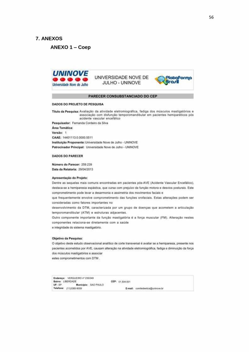
56
7. ANEXOS
ANEXO 1 – Coep
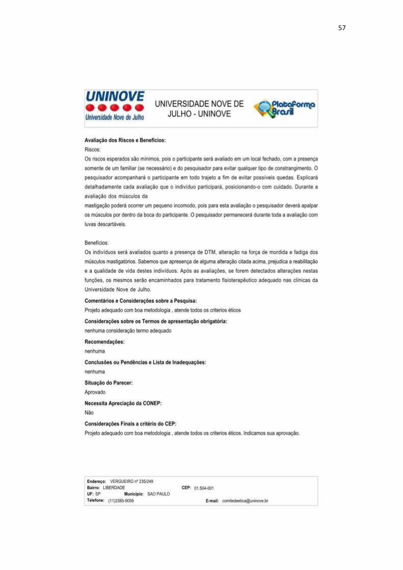
57
58
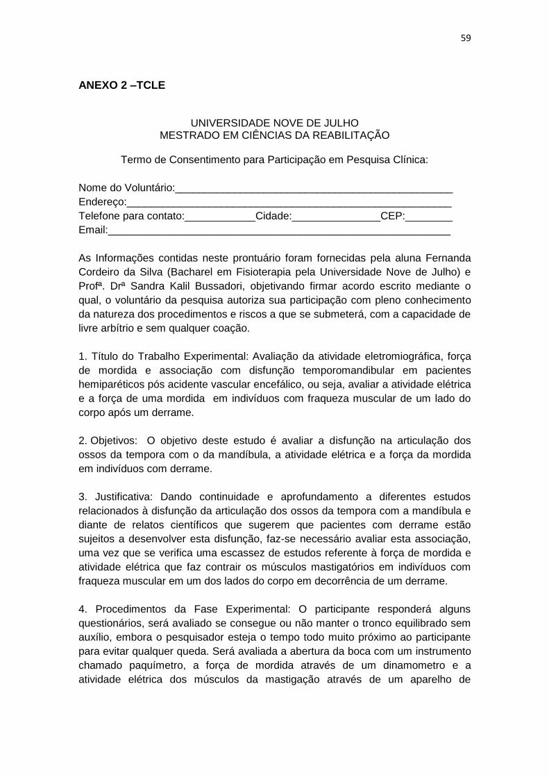
59
ANEXO 2 –TCLE
UNIVERSIDADE NOVE DE JULHO MESTRADO EM CIÊNCIAS DA REABILITAÇÃO
Termo de Consentimento para Participação em Pesquisa Clínica:
Nome do Voluntário:_______________________________________________
Endereço:_______________________________________________________
Telefone para contato:____________Cidade:_______________CEP:________
Email:__________________________________________________________
As Informações contidas neste prontuário foram fornecidas pela aluna Fernanda
Cordeiro da Silva (Bacharel em Fisioterapia pela Universidade Nove de Julho) e
Profª. Drª Sandra Kalil Bussadori, objetivando firmar acordo escrito mediante o
qual, o voluntário da pesquisa autoriza sua participação com pleno conhecimento
da natureza dos procedimentos e riscos a que se submeterá, com a capacidade de
livre arbítrio e sem qualquer coação.
1. Título do Trabalho Experimental: Avaliação da atividade eletromiográfica, força
de mordida e associação com disfunção temporomandibular em pacientes
hemiparéticos pós acidente vascular encefálico, ou seja, avaliar a atividade elétrica
e a força de uma mordida em indivíduos com fraqueza muscular de um lado do
corpo após um derrame.
2. Objetivos: O objetivo deste estudo é avaliar a disfunção na articulação dos
ossos da tempora com o da mandíbula, a atividade elétrica e a força da mordida
em indivíduos com derrame.
3. Justificativa: Dando continuidade e aprofundamento a diferentes estudos
relacionados à disfunção da articulação dos ossos da tempora com a mandíbula e
diante de relatos científicos que sugerem que pacientes com derrame estão
sujeitos a desenvolver esta disfunção, faz-se necessário avaliar esta associação,
uma vez que se verifica uma escassez de estudos referente à força de mordida e
atividade elétrica que faz contrair os músculos mastigatórios em indivíduos com
fraqueza muscular em um dos lados do corpo em decorrência de um derrame.
4. Procedimentos da Fase Experimental: O participante responderá alguns
questionários, será avaliado se consegue ou não manter o tronco equilibrado sem
auxílio, embora o pesquisador esteja o tempo todo muito próximo ao participante
para evitar qualquer queda. Será avaliada a abertura da boca com um instrumento
chamado paquímetro, a força de mordida através de um dinamometro e a
atividade elétrica dos músculos da mastigação através de um aparelho de
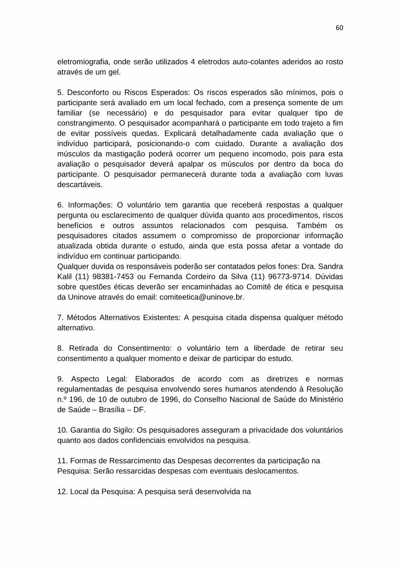
60
eletromiografia, onde serão utilizados 4 eletrodos auto-colantes aderidos ao rosto
através de um gel.
5. Desconforto ou Riscos Esperados: Os riscos esperados são mínimos, pois o
participante será avaliado em um local fechado, com a presença somente de um
familiar (se necessário) e do pesquisador para evitar qualquer tipo de
constrangimento. O pesquisador acompanhará o participante em todo trajeto a fim
de evitar possíveis quedas. Explicará detalhadamente cada avaliação que o
indivíduo participará, posicionando-o com cuidado. Durante a avaliação dos
músculos da mastigação poderá ocorrer um pequeno incomodo, pois para esta
avaliação o pesquisador deverá apalpar os músculos por dentro da boca do
participante. O pesquisador permanecerá durante toda a avaliação com luvas
descartáveis.
6. Informações: O voluntário tem garantia que receberá respostas a qualquer
pergunta ou esclarecimento de qualquer dúvida quanto aos procedimentos, riscos
benefícios e outros assuntos relacionados com pesquisa. Também os
pesquisadores citados assumem o compromisso de proporcionar informação
atualizada obtida durante o estudo, ainda que esta possa afetar a vontade do
indivíduo em continuar participando.
Qualquer duvida os responsáveis poderão ser contatados pelos fones: Dra. Sandra
Kalil (11) 98381-7453 ou Fernanda Cordeiro da Silva (11) 96773-9714. Dúvidas
sobre questões éticas deverão ser encaminhadas ao Comitê de ética e pesquisa
da Uninove através do email: [email protected].
7. Métodos Alternativos Existentes: A pesquisa citada dispensa qualquer método
alternativo.
8. Retirada do Consentimento: o voluntário tem a liberdade de retirar seu
consentimento a qualquer momento e deixar de participar do estudo.
9. Aspecto Legal: Elaborados de acordo com as diretrizes e normas
regulamentadas de pesquisa envolvendo seres humanos atendendo à Resolução
n.º 196, de 10 de outubro de 1996, do Conselho Nacional de Saúde do Ministério
de Saúde – Brasília – DF.
10. Garantia do Sigilo: Os pesquisadores asseguram a privacidade dos voluntários
quanto aos dados confidenciais envolvidos na pesquisa.
11. Formas de Ressarcimento das Despesas decorrentes da participação na
Pesquisa: Serão ressarcidas despesas com eventuais deslocamentos.
12. Local da Pesquisa: A pesquisa será desenvolvida na
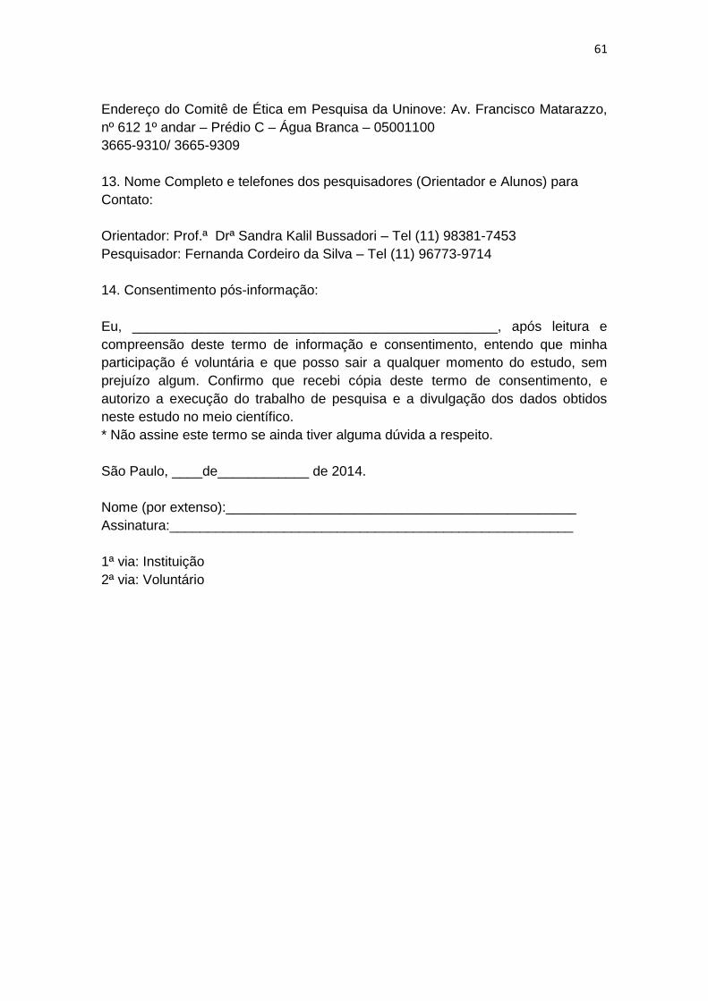
61
Endereço do Comitê de Ética em Pesquisa da Uninove: Av. Francisco Matarazzo,
nº 612 1º andar – Prédio C – Água Branca – 05001100
3665-9310/ 3665-9309
13. Nome Completo e telefones dos pesquisadores (Orientador e Alunos) para
Contato:
Orientador: Prof.ª Drª Sandra Kalil Bussadori – Tel (11) 98381-7453
Pesquisador: Fernanda Cordeiro da Silva – Tel (11) 96773-9714
14. Consentimento pós-informação:
Eu, ________________________________________________, após leitura e
compreensão deste termo de informação e consentimento, entendo que minha
participação é voluntária e que posso sair a qualquer momento do estudo, sem
prejuízo algum. Confirmo que recebi cópia deste termo de consentimento, e
autorizo a execução do trabalho de pesquisa e a divulgação dos dados obtidos
neste estudo no meio científico.
* Não assine este termo se ainda tiver alguma dúvida a respeito.
São Paulo, ____de____________ de 2014.
Nome (por extenso):______________________________________________
Assinatura:_____________________________________________________
1ª via: Instituição
2ª via: Voluntário
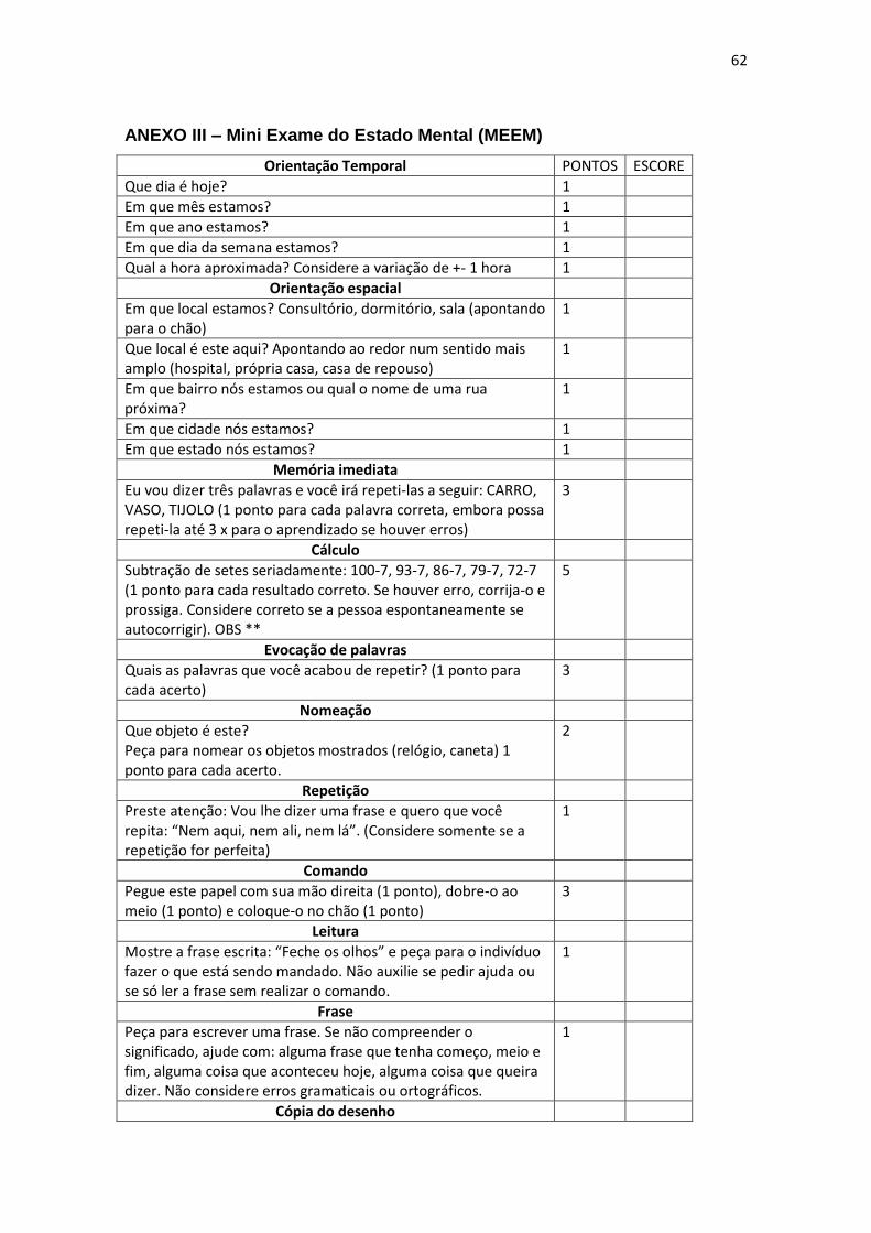
62
ANEXO III – Mini Exame do Estado Mental (MEEM)
Orientação Temporal PONTOS ESCORE
Que dia é hoje? 1
Em que mês estamos? 1
Em que ano estamos? 1
Em que dia da semana estamos? 1
Qual a hora aproximada? Considere a variação de +- 1 hora 1
Orientação espacial
Em que local estamos? Consultório, dormitório, sala (apontando para o chão)
1
Que local é este aqui? Apontando ao redor num sentido mais amplo (hospital, própria casa, casa de repouso)
1
Em que bairro nós estamos ou qual o nome de uma rua próxima?
1
Em que cidade nós estamos? 1
Em que estado nós estamos? 1
Memória imediata
Eu vou dizer três palavras e você irá repeti-las a seguir: CARRO, VASO, TIJOLO (1 ponto para cada palavra correta, embora possa repeti-la até 3 x para o aprendizado se houver erros)
3
Cálculo
Subtração de setes seriadamente: 100-7, 93-7, 86-7, 79-7, 72-7 (1 ponto para cada resultado correto. Se houver erro, corrija-o e prossiga. Considere correto se a pessoa espontaneamente se autocorrigir). OBS **
5
Evocação de palavras
Quais as palavras que você acabou de repetir? (1 ponto para cada acerto)
3
Nomeação
Que objeto é este? Peça para nomear os objetos mostrados (relógio, caneta) 1 ponto para cada acerto.
2
Repetição
Preste atenção: Vou lhe dizer uma frase e quero que você repita: “Nem aqui, nem ali, nem lá”. (Considere somente se a repetição for perfeita)
1
Comando
Pegue este papel com sua mão direita (1 ponto), dobre-o ao meio (1 ponto) e coloque-o no chão (1 ponto)
3
Leitura
Mostre a frase escrita: “Feche os olhos” e peça para o indivíduo fazer o que está sendo mandado. Não auxilie se pedir ajuda ou se só ler a frase sem realizar o comando.
1
Frase
Peça para escrever uma frase. Se não compreender o significado, ajude com: alguma frase que tenha começo, meio e fim, alguma coisa que aconteceu hoje, alguma coisa que queira dizer. Não considere erros gramaticais ou ortográficos.
1
Cópia do desenho
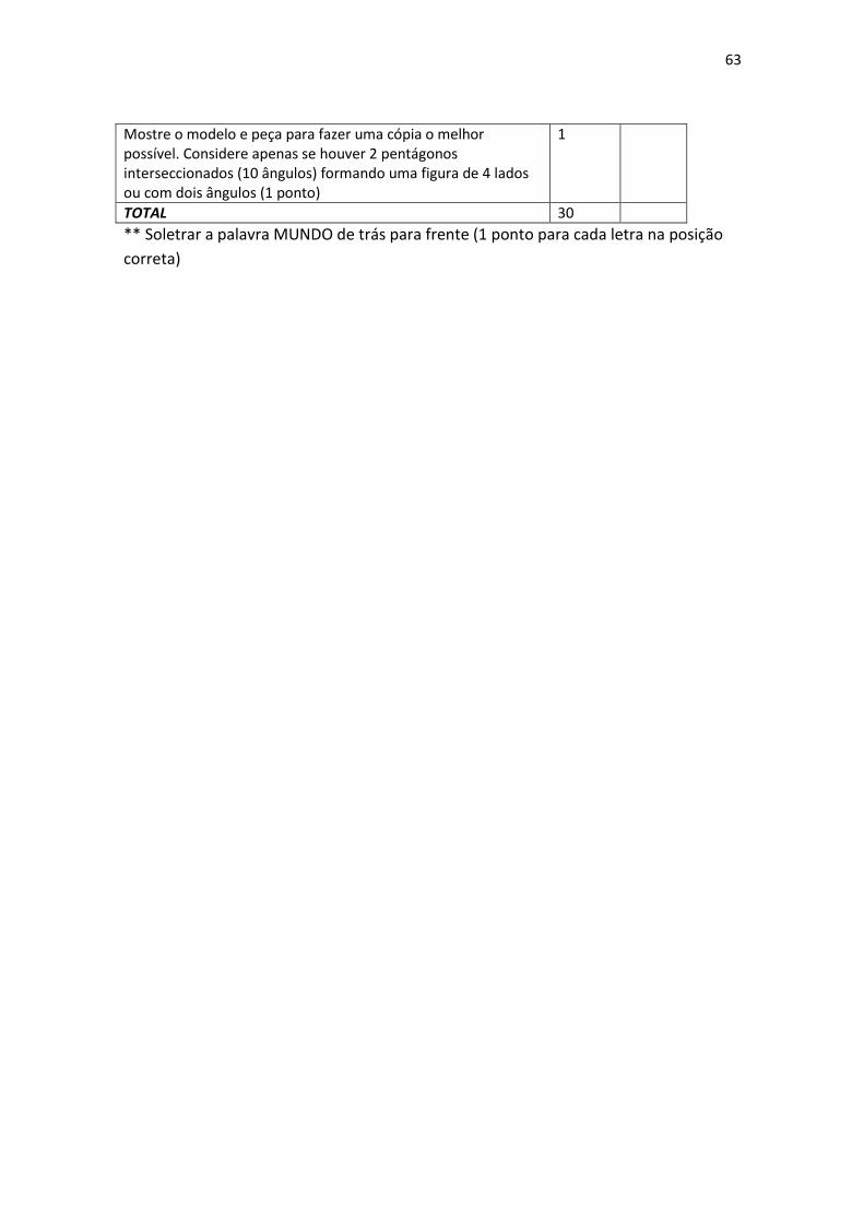
63
Mostre o modelo e peça para fazer uma cópia o melhor possível. Considere apenas se houver 2 pentágonos interseccionados (10 ângulos) formando uma figura de 4 lados ou com dois ângulos (1 ponto)
1
TOTAL 30
** Soletrar a palavra MUNDO de trás para frente (1 ponto para cada letra na posição
correta)
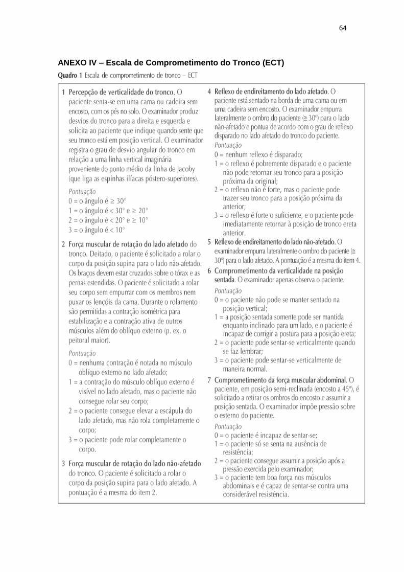
64
ANEXO IV – Escala de Comprometimento do Tronco (ECT)
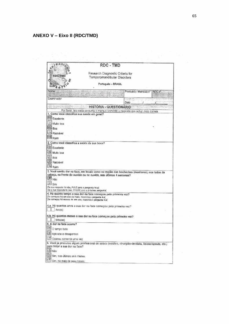
65
ANEXO V – Eixo II (RDC/TMD)
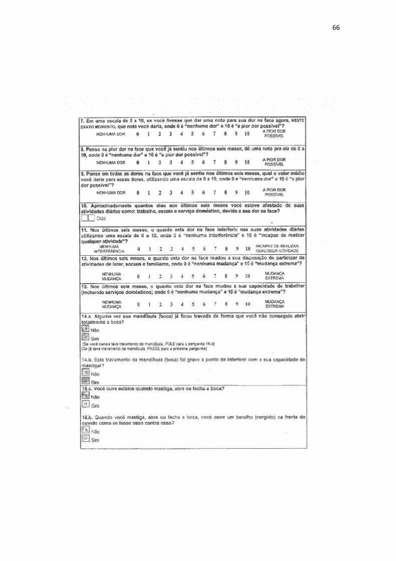
66
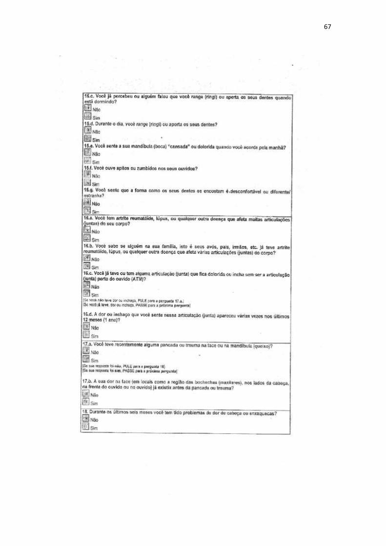
67
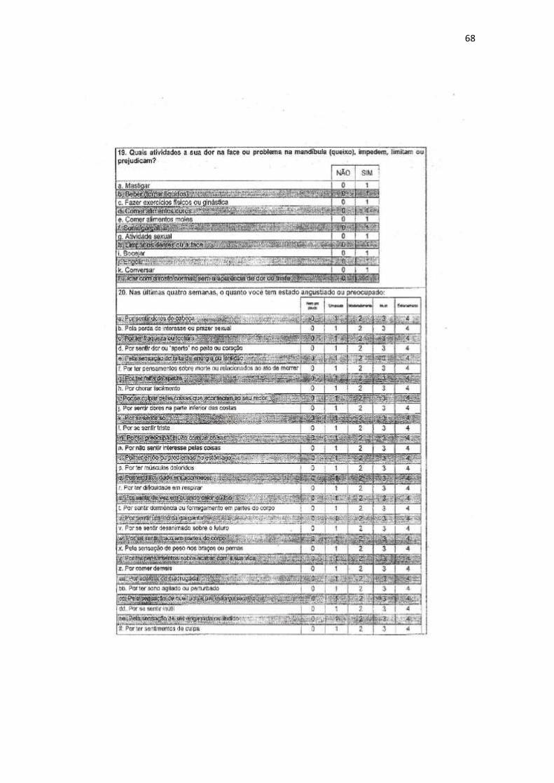
68
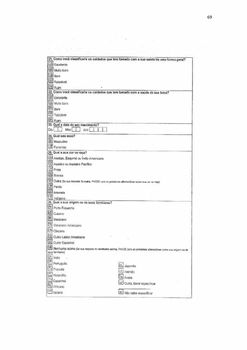
69
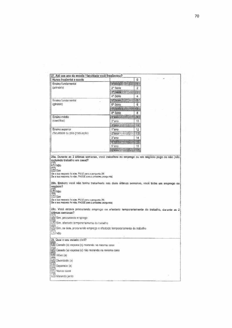
70
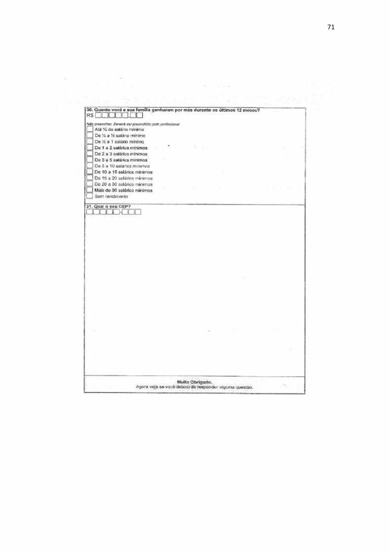
71
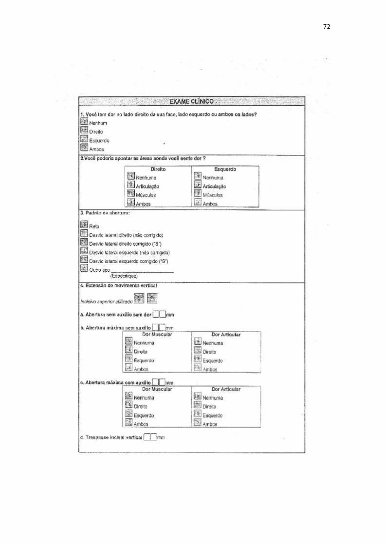
72
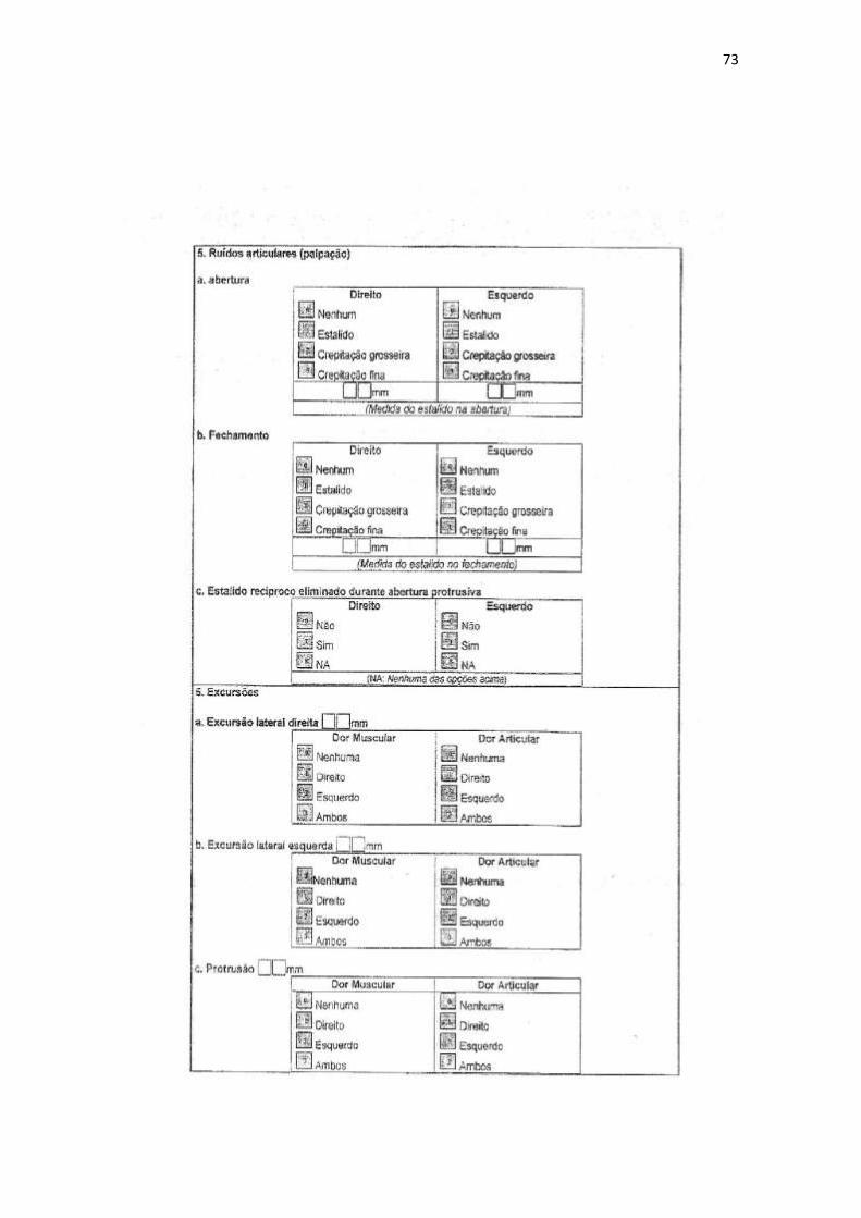
73
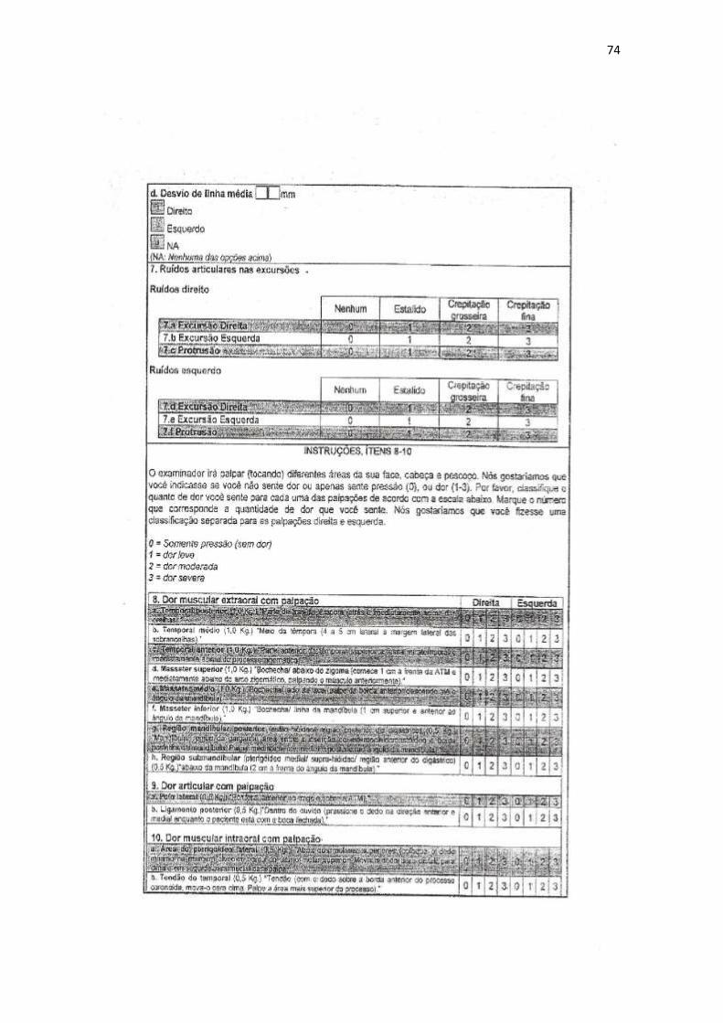
74
75
ANEXO VII – Comprovante de Submissão do Artigo
Related Documents