ORIGINAL ARTICLE Autoregulatory lentiviral vectors allow multiple cycles of doxycycline-inducible gene expression in human hematopoietic cells in vivo M Centlivre 1 , X Zhou 1 , SM Pouw 2,4 , K Weijer 2,4 , W Kleibeuker 1 , AT Das 1 , B Blom 2 , J Seppen 3 , B Berkhout 1 and N Legrand 2,4 1 Laboratory of Experimental Virology, Department of Medical Microbiology, Center for Infection and Immunity Amsterdam (CINIMA), Academic Medical Center of the University of Amsterdam (AMC-UvA), Amsterdam, The Netherlands; 2 Department of Cell Biology and Histology, Center for Immunology of Amsterdam (CIA), Academic Medical Center of the University of Amsterdam (AMC-UvA), Amsterdam, The Netherlands and 3 Department of Experimental Hepatology, Liver Center, Academic Medical Center of the University of Amsterdam (AMC-UvA), Amsterdam, The Netherlands The efficient control of gene expression in vivo from lentiviral vectors remains technically challenging. To analyze inducible gene expression in a human setting, we generated ‘human immune system’ (HIS) mice by transplanting newborn BALB/c Rag2 / IL-2Rg c / immunodeficient mice with human hematopoietic stem cells transduced with a doxycycline- inducible lentiviral vector. We compared several methods of doxycycline delivery to mice, and could accurately measure doxycycline in vivo using a new sensitive detection assay. Two different lentiviral vector designs with constitutive (TRECMV-V14) or autoregulatory (TREAuto-V14) expres- sion of an optimized reverse tetracycline transactivator were used to transduce human hematopoietic stem cells. After transplantation into immunodeficient mice, we analyzed the expression of the green fluorescent protein (GFP) reporter gene in the human hematopoiesis-derived cells that develop and accumulate in the generated HIS mice. We show efficient inducible GFP expression in adult HIS mice containing TREAuto-V14-transduced human cells, whereas GFP expres- sion is poor with the TRECMV-V14 vector. Multiple cycles of doxycycline exposure in the TREAuto-V14 group result in repeated cycles of GFP expression with no loss of intensity. These findings are of major interest for gene therapy and basic research settings that require inducible gene expression. Gene Therapy (2010) 17, 14–25; doi:10.1038/gt.2009.109; published online 3 September 2009 Keywords: humanized mouse model; inducible gene expression; lentiviral vector; hematopoietic stem cell; Tet-On system Introduction Experimental systems in which gene expression can be modulated ‘at will’ are important for many areas of basic research and gene therapy. Among the available approaches, the tetracycline (Tc)-inducible gene regula- tion system has been proven efficient, both in vitro and in vivo. 1,2 The prokaryotic Tc-inducible gene regulation system is based on two elements of the Tn10 Tc resistance operon of Escherichia coli: the repressor protein (TetR) and its target DNA sequence, the tet operator (tetO). 3,4 In the absence of Tc, TetR dimerizes and binds to tetO, blocking transcription of this operon. When Tc is present, its binding to TetR induces a conformational change in TetR, which dissociates from tetO, alleviating the transcrip- tional blockade. By modification of four amino acids in TetR, a reverse TetR mutant was developed that requires doxycycline (dox), a potent analog of Tc, to bind to tetO. 5 In mammalian cells, the dox-regulated system is mainly used for the induction of a target gene under transcriptional control of a Tc-responsive element (TRE) promoter that contains tetO-binding sites fused to a DNA sequence, providing basal promoter activity. Creation of tetO-binding transcriptional transactivator (TA) molecules for mammalian systems was facilitated by fusing TetR or reverse TetR mutant to the VP16 activation domain of herpes simplex virus, giving rise to tTA and rtTA, respectively. 5,6 The tTA protein activates tetO-mediated transcription in the absence of dox (Tet- Off system), whereas rtTA induces transcription in the presence of dox (Tet-On system). Several features, for example, a high induction capacity and rapid respon- siveness, make the Tet-On system particularly attractive as an in vivo inducible system. 1,7 The in vivo conse- quences of long-term exposure to antibiotics—such as Tc and dox—also have to be taken into account. Using the Tet-On approach, the extent of exposure to dox is rather limited when expression of the target gene is required only for a short period of time. The induction of gene expression in restricted time frames may be required in some gene therapy settings, for example, to avoid toxicity resulting from the gene product itself. Received 22 April 2009; revised 4 June 2009; accepted 6 June 2009; published online 3 September 2009 Correspondence: Dr N Legrand, Department of Cell Biology and Histology, Academic Medical Center of the University of Amster- dam (AMC-UvA), Room L3-117, Meibergdreef 15, Amsterdam 1105 AZ, The Netherlands. E-mail: [email protected] 4 Part of the Human Vaccine Consortium, ‘Grand Challenge in Global Health #4: devise reliable testing systems for new vaccines’, http://www.hv-consortium.org Gene Therapy (2010) 17, 14–25 & 2010 Macmillan Publishers Limited All rights reserved 0969-7128/10 $32.00 www.nature.com/gt

Welcome message from author
This document is posted to help you gain knowledge. Please leave a comment to let me know what you think about it! Share it to your friends and learn new things together.
Transcript
ORIGINAL ARTICLE
Autoregulatory lentiviral vectors allow multiple cyclesof doxycycline-inducible gene expression in humanhematopoietic cells in vivo
M Centlivre1, X Zhou1, SM Pouw2,4, K Weijer2,4, W Kleibeuker1, AT Das1, B Blom2, J Seppen3,
B Berkhout1 and N Legrand2,4
1Laboratory of Experimental Virology, Department of Medical Microbiology, Center for Infection and Immunity Amsterdam (CINIMA),Academic Medical Center of the University of Amsterdam (AMC-UvA), Amsterdam, The Netherlands; 2Department of Cell Biologyand Histology, Center for Immunology of Amsterdam (CIA), Academic Medical Center of the University of Amsterdam (AMC-UvA),Amsterdam, The Netherlands and 3Department of Experimental Hepatology, Liver Center, Academic Medical Center of the University ofAmsterdam (AMC-UvA), Amsterdam, The Netherlands
The efficient control of gene expression in vivo from lentiviralvectors remains technically challenging. To analyze induciblegene expression in a human setting, we generated ‘humanimmune system’ (HIS) mice by transplanting newbornBALB/c Rag2�/�IL-2Rgc
�/� immunodeficient mice with humanhematopoietic stem cells transduced with a doxycycline-inducible lentiviral vector. We compared several methods ofdoxycycline delivery to mice, and could accurately measuredoxycycline in vivo using a new sensitive detection assay.Two different lentiviral vector designs with constitutive(TRECMV-V14) or autoregulatory (TREAuto-V14) expres-sion of an optimized reverse tetracycline transactivator wereused to transduce human hematopoietic stem cells. After
transplantation into immunodeficient mice, we analyzed theexpression of the green fluorescent protein (GFP) reportergene in the human hematopoiesis-derived cells that developand accumulate in the generated HIS mice. We show efficientinducible GFP expression in adult HIS mice containingTREAuto-V14-transduced human cells, whereas GFP expres-sion is poor with the TRECMV-V14 vector. Multiple cycles ofdoxycycline exposure in the TREAuto-V14 group result inrepeated cycles of GFP expression with no loss of intensity.These findings are of major interest for gene therapy and basicresearch settings that require inducible gene expression.Gene Therapy (2010) 17, 14–25; doi:10.1038/gt.2009.109;published online 3 September 2009
Keywords: humanized mouse model; inducible gene expression; lentiviral vector; hematopoietic stem cell; Tet-On system
Introduction
Experimental systems in which gene expression can bemodulated ‘at will’ are important for many areas of basicresearch and gene therapy. Among the availableapproaches, the tetracycline (Tc)-inducible gene regula-tion system has been proven efficient, both in vitro andin vivo.1,2 The prokaryotic Tc-inducible gene regulationsystem is based on two elements of the Tn10 Tc resistanceoperon of Escherichia coli: the repressor protein (TetR) andits target DNA sequence, the tet operator (tetO).3,4 In theabsence of Tc, TetR dimerizes and binds to tetO, blockingtranscription of this operon. When Tc is present, itsbinding to TetR induces a conformational change in TetR,which dissociates from tetO, alleviating the transcrip-tional blockade. By modification of four amino acids
in TetR, a reverse TetR mutant was developed thatrequires doxycycline (dox), a potent analog of Tc, tobind to tetO.5
In mammalian cells, the dox-regulated system ismainly used for the induction of a target gene undertranscriptional control of a Tc-responsive element (TRE)promoter that contains tetO-binding sites fused to aDNA sequence, providing basal promoter activity.Creation of tetO-binding transcriptional transactivator(TA) molecules for mammalian systems was facilitatedby fusing TetR or reverse TetR mutant to the VP16activation domain of herpes simplex virus, giving rise totTA and rtTA, respectively.5,6 The tTA protein activatestetO-mediated transcription in the absence of dox (Tet-Off system), whereas rtTA induces transcription in thepresence of dox (Tet-On system). Several features, forexample, a high induction capacity and rapid respon-siveness, make the Tet-On system particularly attractiveas an in vivo inducible system.1,7 The in vivo conse-quences of long-term exposure to antibiotics—such as Tcand dox—also have to be taken into account. Using theTet-On approach, the extent of exposure to dox is ratherlimited when expression of the target gene is requiredonly for a short period of time. The induction of geneexpression in restricted time frames may be requiredin some gene therapy settings, for example, to avoidtoxicity resulting from the gene product itself.
Received 22 April 2009; revised 4 June 2009; accepted 6 June 2009;published online 3 September 2009
Correspondence: Dr N Legrand, Department of Cell Biology andHistology, Academic Medical Center of the University of Amster-dam (AMC-UvA), Room L3-117, Meibergdreef 15, Amsterdam 1105AZ, The Netherlands.E-mail: [email protected] of the Human Vaccine Consortium, ‘Grand Challenge inGlobal Health #4: devise reliable testing systems for new vaccines’,http://www.hv-consortium.org
Gene Therapy (2010) 17, 14–25& 2010 Macmillan Publishers Limited All rights reserved 0969-7128/10 $32.00
www.nature.com/gt
A successful dox-dependent gene expression strategyrelies on two components: the in vivo dox levels and thevector used for gene delivery. Over the past 10 years,improvements in the dox-dependent gene expres-sion system have been obtained. A variety of rtTAproteins have been developed with reduced basalactivity, increased induction capacity and increased doxsensitivity. For example, the recently developed variants,rtTA-V14, -V15 and -V16, require only 10 ng ml�1 dox tobe as active as the rtTA2S-S2 variant at 1000 ng ml�1
dox.8–10 This difference in dox sensitivity may result indifferential gene expression. The determination ofdox levels reached in vivo is thus critical for optimaldox-dependent gene activation with a given rtTA and forreduction of the delivered dose of dox. The vector usedfor delivery of the inducible transgene is also important.Lentiviral vectors were recently developed in which theTRE-regulated transgene as well as the rtTA protein arecombined, allowing the transfer of the complete Tet-Onsystem in one single transduction.11,12 In these vectors,rtTA can be expressed either from a constitutivepromoter or through an autoregulatory loop. Theautoregulatory expression of rtTA was shown to besuperior in vitro as compared with constitutive expres-sion, resulting in lower basal expression levels, higherviral titers, improved induction kinetics and increasedgene expression levels.11 However, these differences havenot yet been analyzed in an in vivo setting, notably in thecontext of human cells.
Several humanized mouse models have been devel-oped and optimized over the last two decades for theexperimental study of human immune system (HIS)development and function.13–16 Along with others, wehave recently established BALB/c Rag-2�/�IL-2Rgc
�/�
newborn mice as a new xenograft model for humanhematopoietic stem cell (HSC) transplantation.17,18
In contrast with other immunodeficient mouse strainshumanized for the immune system (for example, C.B-17SCID mice or NOD/SCID mice),14 the resulting HIS(BALB-Rag/g) mice show all major human myeloid andlymphoid cellular compartments, giving access to in vivoand ex vivo experimentation on human immune cells.14–16
Dox-dependent gene expression strategies have beenapplied to humanized NOD/SCID mice,19 but the morerobust human reconstitution obtained in the HIS (BALB-Rag/g) mice renders this model more attractive forprospective study of gene function in vivo during humanhematopoiesis. As previously mentioned, selection of thertTA variant and the dox delivery method have to becarefully evaluated to obtain optimal gene inductioneffectiveness in this in vivo Tet-On setting.
In this study we evaluated the dox concentrationsreached in the plasma of the HIS (BALB-Rag/g) miceafter a series of dox administration/withdrawal, usingdifferent routes of dox administration, either in thedrinking water or in the diet. Furthermore, we comparedthe performance of two different lentiviral vectors,expressing rtTA either constitutively (TRECMV-V14) orthrough an autoregulatory loop (TREAuto-V14), for theircapacity to induce gene expression in vivo upon doxadministration. The HIS (BALB-Rag/g) mice weretransplanted with human HSC-enriched CD34+CD38�cells transduced with dox-inducible lentiviral vectors,allowing direct monitoring of dox diffusion in the mouseorgans through green fluorescent protein (GFP) expres-
sion. Our results show that the use of the induciblelentiviral vector with an autoregulatory loop ensures amore robust expression of the reporter gene, with no lossof expression after repeated cycles of dox exposure.
Results
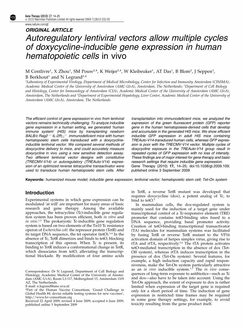
Generation of humanized mice with lentivirus-modifiedhematopoietic stem cellsWe have constructed two lentiviral vectors, TRECMV-V14 and TREAuto-V14, deriving from the TRECMVR2and TREAutoR3 vectors, respectively.11 These two con-structs belong to the third generation of lentiviral self-inactivating vectors,21,22 with the central polypurine tractfor enhanced transduction efficiency and the hepatitis Bvirus post-transcriptional regulatory element for en-hanced gene expression.23 In these two vectors, thertTA2S-S2 (TRECMVR2) and rtTA3 (TREAutoR3) werereplaced by the improved rtTA-V14 variant that showsincreased dox sensitivity and contributes to better viralreplication in the HIV-rtTA backbone, as compared withrtTA2S-S2.10 However, the two elements of the Tet-Onsystem (TRE and rtTA) are combined in a differentmanner in these vectors. In TRECMV-V14, expression ofGFP is under the control of the TRE promoter, consistingof seven copies of tetO fused to the minimal promoterderived from the human CMV immediate early gene 1promoter, whereas rtTA expression is driven by theconstitutive CMV promoter (Figure 1a). In TREAuto-V14,rtTA is placed in an autoregulatory loop where the TREpromoter expresses a bicistronic mRNA that encodesboth GFP and rtTA through an internal ribosomal entrysite (Figure 1b).
The injection of human HSC into immunodeficient(BALB/c RAG2�/�IL-2Rgc
�/�) newborn mice results inthe development of a fully differentiated multilineagehuman immune system in vivo. Within 8 weeks, all majorhuman lymphoid- and myeloid-cell compartments,including B cells, abT cells, gdT cells, natural killer cells,monocytes/macrophages, and conventional and plasma-cytoid dendritic cells, are generated and detected inthe lymphoid organs of such HIS (BALB-Rag/g)mice.14,17,18,29,31 To compare the dox-dependent geneexpression driven by the two different lentiviral vectorsin vivo, we transduced human HSC (CD34+CD38� cells)with TRECMV-V14 or TREAuto-V14 vectors beforexenograft transplantation (Figure 1c). The transducedhuman CD34+CD38� cells were transplanted intoimmunodeficient mice, and an aliquot was culturedin vitro in the presence of dox for 3.5 days for determiningthe transduction efficiency. Using flow cytometry deter-mination of GFP expression as a readout, this initialtransduction efficiency was compared with the recoveryof transduced human cells in the HIS (BALB-Rag/g)mice 8 to 12 weeks after hematopoietic reconstitution,that is, when human cell colonization of the lymphoidorgans is optimal (Figure 1c).
Kinetics of dox accumulation and clearance in theplasma of humanized miceOptimal induction of the Tet-On system criticallydepends on the dox sensitivity of the rtTA variant used,and dox delivery has to be calibrated to reach aneffective, non-toxic concentration in vivo. We compared
Inducible gene delivery in ‘human immune system’ miceM Centlivre et al
15
Gene Therapy

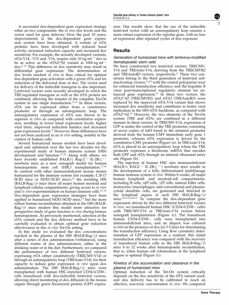
several dox delivery procedures with adult (8–12 weeksafter transplantation) HIS (BALB-Rag/g) mice, eitherin the drinking water (1 mg ml�1) or the food (2 g kgand 6 g kg�1 dox-diets). Blood was harvested fromthe animals at several time points to collect serialplasma samples. We determined the dox concentrationreached in the HIS (BALB-Rag/g) plasma samples usinga new biological assay, referred to as the HeLaDOX
assay, that allows dox measurement in small biologicalsamples.20
In the first set of experiments, HIS (BALB-Rag/g) micecontinuously received dox for up to 3 weeks. Highplasma dox concentrations (41.000 ng ml�1) wereobtained in all the HIS (Rag/g) mice, irrespective of thedelivery method (Figure 2a). This high plateau level wasreached as early as 3 days after onset of the treatment
Figure 1 Generation of HIS (BALB-Rag/g) mice with lentiviral-mediated gene transfer. Vectors were based on the third generation oflentiviral self-inactivating vectors, containing the central polypurine tract (cPPT) and the hepatitis B virus post-transcriptional regulatoryelement (PRE). (a) In the TRECMV-V14 lentiviral vector, GFP is expressed from the TRE promoter and rtTA expression is driven by aconstitutive cytomegalovirus (CMV) promoter. (b) In the TREAuto-V14, the TRE promoter expresses a bicistronic mRNA that encodes bothGFP and rtTA through an IRES. (c) Cell suspensions enriched for human hematopoietic stem cells (HSCs) are prepared from fetal liver tissue.Live nucleated CD34+ cells are magnetically sorted and further enriched for the HSC (CD34+CD38� fraction) using fluorescence activatedcell sorting. Lentiviral supernatants are produced on human embryonic kidney (HEK) 293T cells. Lentivirus-mediated gene transductionof purified HSC is performed immediately after cell sorting. Sublethally irradiated newborn BALB/c Rag-2�/�IL-2Rgc�/� mice are injectedintrahepatically with transduced HSCs. The transduction efficiency is evaluated based on GFP expression after 3.5 days in culture in thepresence of dox. The HIS (BALB-Rag/g) mice are tested after 8 weeks for the presence of human cells in the blood and further analyzedafter onset of dox treatment, starting 8–12 weeks after transplantation. GFP, green fluorescent protein; IRES, internal ribosome entry site;LTR, long terminal repeat; rtTA, reverse Tet transcriptional transactivator; TRE, Tet responsive element.
Inducible gene delivery in ‘human immune system’ miceM Centlivre et al
16
Gene Therapy

and was stably maintained for the 3 weeks ofdox treatment. The animals on the 6 g kg�1 dox-diet regimen showed the highest plasma dox concentra-tion (8.700±2.300 ng ml�1). No obvious signs of animal
discomfort were noticed in all experimental groups. Wenext analyzed the kinetics of dox accumulation andclearance in the plasma of dox-fed HIS (BALB-Rag/g)mice (6 g kg�1 dox-diet) (Figure 2b). The dox levelmeasured 2 days after dox withdrawal dropped to thepre-dox value, with a 485% decrease of the dox concen-tration only 1 day after withdrawal. A second dox-foodadministration showed rapid on-kinetics, with 50% ofthe dox level plateau reached after 1 to 2 days andmaximal dox concentration after 1 week. Similar resultswere obtained with dox supplementation in the drinkingwater (1 mg ml�1; data not shown). Overall, our resultsshow that the half-life of dox in the blood is in the rangeof 1 day, allowing quick on/off dox cycles, indepen-dently of the route of administration.
Doxycycline is a member of the tetracycline familywith a high capacity of diffusion through the blood-brainbarrier.32 The dox-inducible system is thus commonlyused for gene expression studies in the brain, but the doxlevel that can be obtained in situ has never carefullybeen addressed. We therefore harvested the CSF ofthe HIS (BALB-Rag/g) mice that received dox for 3weeks in drinking water (1 mg ml�1) or food (2 g kg�1
and 6 g kg�1 dox-diet), and we determined the doxconcentration (Figure 2c). In parallel, we measured theplasma dox concentration at the time of CSF harvest(Figure 2d). The CSF dox concentrations in the HIS(BALB-Rag/g) mice were approximately 30-fold lowerthan the corresponding plasma levels. With 6 g kg�1 dox-diet, we measured on average 400 ng ml�1 dox in theCSF, and this value dropped threefold for the 2 g kg�1
dox-diet. Such values are compatible with the use of themore sensitive rtTA variants for gene induction in thebrain. With dox-drinking water, we were unable to detectdox in the CSF above the background level measured innon-treated animals. Our results highlight that high doseof dox-delivery in food is highly desirable for propergene induction in the brain.
GFP induction in human cells in the blood of HIS miceWe followed the kinetics of GFP expression when doxwas added and subsequently withdrawn from the water(1 mg ml�1) and food regimen (6 g kg�1 dox-diet) of HIS(BALB-Rag/g) mice. We compared the in vivo capacity ofdox-induced GFP expression between TRECMV-V14 andTREAuto-V14 lentiviral vectors. Similar transductionefficiencies were scored for the two vectors (Figure 3a).In the adult HIS (BALB-Rag/g) mice (8–12 weeks aftertransplantation), the GFP expression by human CD45+blood cells was close to undetectable in the absence ofdox for both vectors, whereas dox treatment inducedGFP expression in a fraction of human CD45+ blood cells(Figure 3b). When compared with the original transduc-tion efficiency, the extent of GFP induction by dox waslargely lost for the TRECMV-V14 but not for theTREAuto-V14 (Figure 3b).
To stipulate the in vivo durability of dox-inducible GFPexpression, we calculated the ratio of the in vivo (GFPinduction in CD45+ cells of HIS mice) and in vitro (initialtransduction efficiency) percentage of GFP+ cells overtime. The recovery of GFP expression after dox treatmentof the animals was only 10–15% of the initial transduc-tion efficiency with the TRECMV-V14 (Figure 3c),whereas almost 100% of GFP expression recovery wasobserved with the TREAuto-V14-transduced human
Figure 2 Determination of dox level reached in the HIS(BALB-Rag/g mice during dox delivery/withdrawal cycle. Thedox levels were determined in the plasma of HIS (BALB-Rag/g)mice before (pre-dox) and during dox treatment. (a) The HIS(BALB-Rag/g) mice received dox for a maximum of 3 weeks, eitherin the water (1 mg ml�1; crosses, n¼ 14–22 depending on the timepoint) or in the food (2 g kg�1 dox-diet, open circles, n¼ 4–7;6 g kg�1 dox-diet, closed triangles, n¼ 22–38). (b) The dox levelsin the plasma of dox-fed HIS (BALB-Rag/g) mice (6 g kg�1
dox-diet) were determined during rounds of dox administration(closed triangles, n¼ 17–33 in first round, n¼ 3–6 in the secondround) and dox withdrawal (open triangle, n¼ 4–10). The doxlevels were also determined in the CSF (c) and the plasma (d)of the same HIS (BALB-Rag/g) mice that either received no dox(open diamonds, n¼ 5), dox in the water (1 mg ml�1; crosses, n¼ 5)or dox in the food (2 g kg�1 dox-diet, open circles, n¼ 5; 6 g kg�1
dox-diet, closed triangles, n¼ 7) for 3 weeks. The results are pooledfrom two to four independent experiments, and each dot representsone individual animal.
Inducible gene delivery in ‘human immune system’ miceM Centlivre et al
17
Gene Therapy

cells (Figure 3d). The results from HIS (BALB-Rag/g)mice receiving dox in drinking water (1 mg ml�1) ordiet (6 g kg�1) were pooled, as no differences in GFPexpression (frequency of GFP+ cells; mean fluorescenceintensity) were observed between the two deliverymethods (not shown). The expression of GFP reached aplateau value as early as day 3 after onset of doxtreatment with both vectors. For TREAuto-V14, areduction in the percentage of GFP+ cells was observed
between 1.5 and 3 weeks of dox treatment. Uponwithdrawal of dox, the frequency of human GFP+ cellsdecreased to pre-dox value in a slow manner, with theGFP expression still being detectable at 3 days off-doxwith both vectors (Figures 3c and d). Considering thatboth vectors express a destabilized GFP (half-life of2 h),33 the slow disappearance of GFP was likely dueto the high dox-sensitivity of rtTA-V14. Overall, themean fluorescence intensity of GFP after 3 days of dox
Figure 3 The kinetics of induction of GFP reporter gene expression in HIS mouse blood. (a) The flow cytometry dot plots show thetransduction efficiency obtained in vitro with human hematopoietic stem cells (HSCs) transduced with TRECMV-V14 (left) and TREAuto-V14(right) vectors, after 3.5 days of culture in the presence of 1000 ng ml�1 dox. The values in the gates indicate the fraction of GFP+ cells(SSC, side scatter). (b) The in vivo GFP induction in the blood of the corresponding HIS (BALB-Rag/g) mice is shown in the bottom plots(+ dox, T+3 weeks), as compared with the pre-dox situation (top plots,�dox). GFP is only expressed in the human hematopoiesis-derivedcells (hCD45+). The values in the dot plots indicate the percentage of GFP� and GFP+ cells among the human (hCD45+) cells. (c) Thekinetic of GFP expression in the presence and absence of dox treatment was monitored in the blood of the TRECMV-V14-transduced HIS(BALB-Rag/g) mice (d: days; wk: weeks). The number of animals (n) is indicated under each time point. The results are expressed as theratio between the frequency of GFP+ human cells measured in the animals (in vivo GFP induction) and the frequency of GFP+ humanCD34+CD38� cells injected in the newborn mice (in vitro HSC transduction efficiency). (d) Similar analysis was performed with theTREAuto-V14-transduced group. (e) The GFP mean fluorescence intensity (MFI) of the hCD45+GFP+ population was measured with thetwo vectors (mean±s.e.m.; n¼ 27–33 at 3 days and 1.5 week; n¼ 17–20 at 3 weeks). The results are expressed as a ratio between theMFI of GFP+ cells and the MFI of GFP� cells (background MFI). (c–e) Data were generated and pooled from two (TREAuto-V14) to four(TRECMV-V14) independent HSC transductions, and each dot represents one individual animal. Results from HIS (BALB-Rag/g) micereceiving dox in drinking water (1 mg ml�1) or diet (6 g kg�1) were pooled, as no differences in GFP expression (frequency of GFP+ cells; MFI)were observed between the two delivery methods. *Po0.05; **Po0.01.
Inducible gene delivery in ‘human immune system’ miceM Centlivre et al
18
Gene Therapy

delivery was significantly higher when expression wasdriven by the TREAuto-V14 vector (fluorescence inten-sity ±110-fold the background value), as compared withthe TRECMV-V14 vector (±65-fold), but this differencewas not maintained after 1.5 week or 3 weeks oftreatment (Figure 3e).
Induction of GFP expression in lymphoid organs andhuman immune cell subsetsWe next evaluated the diffusion of dox in the HIS(BALB-Rag/g) mice organs, using GFP expression as aread-out. The animals were analyzed after 3 weeks ofdox treatment for the frequency of GFP+ cells amonghuman (hCD45+) cells, as well as the frequency of GFP+cells among human B cells (CD19+), T cells (CD3+),plasmacytoid dendritic cells (BDCA2+HLA-DR+)and conventional dendritic cells (BDCA2�HLA-DR+)(Figure 4a). In the spleen, no human GFP+ cells wereobserved in the absence of dox treatment for both vectors(Figure 4b). In the presence of dox, human GFP+ cellsaccumulated with both lentiviral vectors. In accordancewith our observations in blood, the recovery of GFPexpression was significantly lower for TRECMV-V14 ascompared with TREAuto-V14 (Figure 4b). All humanimmune cell subsets expressed GFP, although thefrequency of GFP+ cells was higher in B cell andconventional dendritic cell subsets for both vectors(Figures 4a and c). The GFP mean fluorescence intensitywas lower in GFP+ B cells (±65-fold the backgroundvalue) and T cells (±45-fold), as compared withplasmacytoid- and conventional-dendritic cells subsets(±100-fold) (not shown). Identical results were obtainedin the bone marrow, the thymus and the liver of theanalyzed HIS (BALB-Rag/g) mice (Figures 4d–f and notshown). Induction of GFP expression in TREAuto-V14transduced human cells was significantly (Po0.05) lowerin the thymus (around 40% recovery) than spleen, bonemarrow and liver (65–80% recovery). It should be notedthat the human reconstitution in the brain of HIS (BALB-Rag/g) is virtually absent, and we were consequentlyunable to evaluate the extent of GFP induction in thislocation.
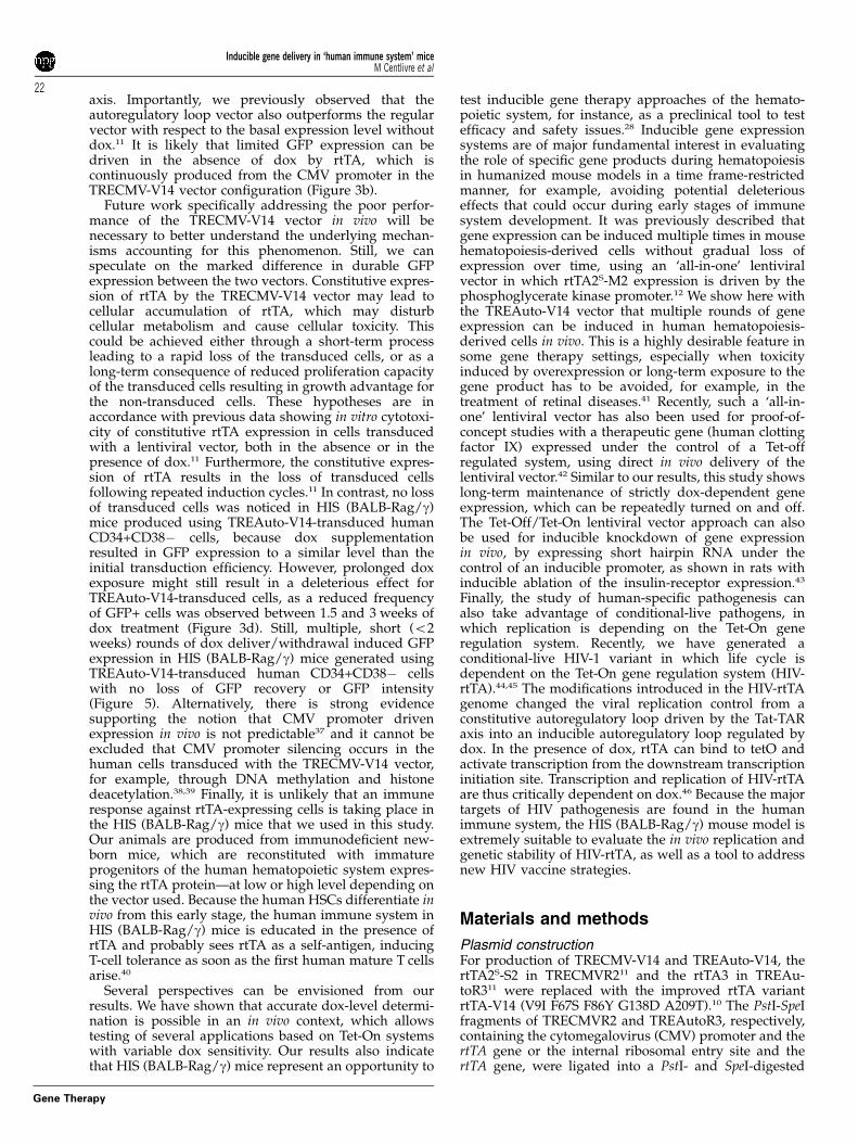
GFP expression after several on/off cyclesof dox deliveryOne desirable feature of an inducible vector is the abilityto support repeated induction cycles of gene expression.We therefore monitored the recovery of human GFP+cells after multiple on/off cycles. The mice that hadreceived dox diet for 3 weeks were subsequently givenfood without dox for 1.5 week (1.5 week off), with doxfor 1 week (1 week on), without dox for 1.5 week (1.5week off) and finally with dox for 1 week (1 week on).Because the TRECMV-V14-transduced cells behavedpoorly, we focused on the HIS (BALB-Rag/g) micegenerated with TREAuto-V14-transduced human HSCto ensure optimal measurement of GFP induction.We measured the frequency of human GFP+ cells inthe blood. We observed that GFP expression was inducedto nearly 100% of the pre-engraftment level, andsubsequently switched-off during dox withdrawal. Thispattern could be repeated in subsequent cycles of doxdelivery (Figure 5a). Importantly, the GFP recovery aftereach cycle of dox delivery was maintained to a similar
level of at least 70% and strict on/off control of transgeneexpression was preserved. A similar level of GFP meanfluorescence intensity was also obtained after each cycleof dox delivery (Figure 5b). We also assessed GFPexpression in multiple organs after the first and the lastcycles of dox treatment. Bone marrow, thymus, spleenand liver of the mice were harvested and screened forGFP expression by the CD45+ human hematopoiesis-derived cells (Figures 5c–f). As observed in the blood,GFP expression was still efficiently induced after thethird dox delivery, and recovery of human GFP+ cellswas similar between the first and the third cycles. Asbefore, the lowest overall induction level was observedfor thymus-derived cells.
Discussion
In this study we analyzed the parameters that are criticalfor successful transgene control in vivo in a humancontext, using the widely used Tet-On system. The proofof concept for inducible gene delivery in a humanizedmouse system was performed by transplanting engi-neered human HSC-enriched CD34+CD38� cells intoNOD/SCID immunodeficient mice,19 but residual mouseT-cell development, B-cell development and naturalkiller cell activity in this genetic background intrinsicallylimit the extent of human reconstitution.14 To circum-vent this potential limitation, we have reconstitutedimmunodeficient BALB/c Rag2�/�IL-2Rgc
�/� newbornmice17,18 with human CD34+CD38� cells that were firstgenetically engineered by ex vivo lentiviral transduction.In contrast with C.B-17 SCID or NOD/SCID mousestrains, BALB/c Rag2�/�IL-2Rgc
�/� mice are fully defec-tive for T-, B- and natural killer cell development andconsequently show an improved susceptibility to xeno-graft transplantation.14 All major lineages of the HIS aregenerated in the resulting HIS (BALB-Rag/g) mice,giving access to a prospective in vivo study of Tet-Onapproaches in the human hematopoietic system. Wetested different methods of dox delivery, the dox levelsobtained in plasma and CSF, different lentiviral vectordesigns, and the induction of transgene expression indifferent tissues and immune cell subsets.
Optimal induction of gene expression in vivo dependson the dox level reached in situ, in relationship with thedox sensitivity of the rtTA used in the transduced vector.There are obvious in vivo technical constrains that areeasily circumvented in vitro, such as fine manipulation ofdox concentration and very rapid supplementation orwithdrawal of dox. The in vivo situation is more complex,due to, for instance, the differential dox penetration intovarious organs and cellular compartments, or thekinetics of dox accumulation and clearance. We com-pared two non-invasive approaches of delivering dox tothe humanized mice: in drinking water (1 mg dox per mlof water, that is, 1 g kg�1 of water) or the food (2 g kg�1
or 6 g kg�1 diet). Considering that the daily intakeof water and food by normal adult mice is B5 g ofwater (that is, B5 ml) and B4 g food,34 exposure todox is expected to follow the hierarchy of 6 g kg�1
food 42 g kg�1 food 4 water 1 mg ml�1. This is whatwe observed when we measured plasma dox levels(Figure 2). The dox concentration reached in all treatedanimals was above the level required for rtTA activation,
Inducible gene delivery in ‘human immune system’ miceM Centlivre et al
19
Gene Therapy

both for the highly dox-sensitive rtTA-V1410 and thertTA2S-M2 and rtTA2S-S2 variants.8
It should be noted that dox delivery in drinking waterimposes several technical limitations, as well as sideeffects. For instance, the bitter taste of dox in water—aswell as sucrose addition to mask it—influences the daily
water intake and limits the maximal concentration ofdox.34 Signs of mouse discomfort—such as weight lossand dehydration—may be observed after only 2 days ofdox treatment.35 Furthermore, dox is known to be lightsensitive and relatively instable in water, and it isadvised to change the supply every other day.36
Figure 4 Induction of GFP expression in lymphoid organs. (a) GFP expression by several human immune cell subsets was measured in thespleen of reconstituted human immune system (HIS; BALB-Rag/g) mice. These subsets were defined as shown in the left FACS plots: B cells(CD3�CD19+), T cells (CD3+CD19�), plasmacytoid dendritic cells (pDC; CD3�CD19�BDCA2+HLA-DR+) and conventional dendritic cells(cDC; CD3�CD19�BDCA2�HLA-DR+). Representative patterns of GFP expression in these subsets are shown for one TRECMV-V14 and oneTREAuto-V14 lentivirus-transduced HIS (BALB-Rag/g) mouse (initial transduction efficiencies, respectively, 20% GFP+ and 13.2% GFP+).(b) The graph shows the recovery of GFP expression in total human cells of the spleen of TRECMV-V14 (closed circles) and TREAuto-V14(gray squares) lentivirus-transduced HIS (BALB-Rag/g) mice, before (n¼ 5 vs 4, respectively) and after 3 weeks (n¼ 16 vs 18) of doxtreatment. The results are expressed as indicated in the legend of Figures 3c and d. (c) The frequency of GFP+ cells within human B cells,T cells, pDC and cDC of TRECMV-V14 (white bars; mean±s.e.m.) and TREAuto-V14 (black bars; mean±s.e.m.) lentivirus-transducedHIS (BALB-Rag/g) mice is detailed. (d–f) The graphs show the recovery of GFP expression in total human cells of the bone marrow (d),the thymus (e) and the liver (f), as before. Data were generated and pooled from two (TREAuto-V14) to four (TRECMV-V14) independentHSC transductions, and each dot in the graphs represents one individual animal. Results from HIS (BALB-Rag/g) mice receiving doxin drinking water (1 mg ml�1) or diet (6 g kg�1) were pooled, as no differences in GFP expression were observed between the twodelivery methods.
Inducible gene delivery in ‘human immune system’ miceM Centlivre et al
20
Gene Therapy

In comparison, the dox-food delivery presents severaladvantages, for example, protection from light, im-proved dox stability and less frequent supply replace-ments (once a week). Most importantly, higher doxconcentrations can be delivered in the food without anysigns of animal discomfort, which allows reachinghigh dox levels in organs showing restricted diffusionof the antibiotic. For instance, we found in the brain onlyB3% of the corresponding plasma dox concentration(Figures 2c and d). The delivery of high levels of dox inthe food (for example, 6 g kg�1) therefore ensuresoptimal CSF dox levels (400 ng ml�1 on average) forproper function of the improved Tet-On systems.10
To determine the efficiency of dox-regulated lentiviralvectors, we generated HIS (BALB-Rag/g) mice with
transduced human CD34+CD38� cells for inducible GFPexpression. We compared two dox-sensitive lentiviralvectors expressing rtTA-V14 either constitutively or in anautoregulatory loop manner. The two vectors showeda similar ex vivo transduction efficiency on humanCD34+CD38� cells, as determined by GFP expressionafter 3.5 days of culture in the presence of saturating doxconcentration. In contrast, only the use of the autoregu-latory loop ensures a robust expression of the reportergene in HIS (BALB-Rag/g) mice, in all the lymphoidorgans and human immune cell subsets analyzed(Figures 3 and 4). As the two vectors make use of thesame rtTA-V14 variant, Tet-responsive element anddestabilized GFP33 (half-life o2 h), we can exclude adifferential effect of dox saturating levels on the Tet-On
Figure 5 Multiple in vivo cycles of induction of GFP expression with TREAuto-V14. (a) The kinetics of GFP expression were followed inthe blood of the human immune system (HIS; BALB-Rag/g) mice generated with TREAuto-V14-transduced human hematopoietic stem cells(HSCs). Three rounds of dox administration in diet (2 g kg�1; closed squares) and dox withdrawal (gray circles) were studied. (b) The GFPmean fluorescence intensity (MFI) of the hCD45+GFP+ population was measured (mean±s.e.m.) after the first (ON-1, 3 weeks, n¼ 17), thesecond (ON-2, 1 week, n¼ 10) and the third (ON-3, 1 week, n¼ 9) round of dox delivery. The results are expressed as indicated in the legendof Figure 3e. (c–f) The ratio between the in vivo GFP induction and the HSC transduction efficiency was determined in the TREAuto-V14groups of mice before dox treatment (n¼ 4), after the first (ON-1; n¼ 18) and the third round (ON-3; n¼ 9) of dox delivery in the bone marrow(c), the thymus (d), the spleen (e) and the liver (f) of the treated mice. Data were generated and pooled from two (TREAuto-V14) independentHSC transductions, and each dot represents one individual animal.
Inducible gene delivery in ‘human immune system’ miceM Centlivre et al
21
Gene Therapy
axis. Importantly, we previously observed that theautoregulatory loop vector also outperforms the regularvector with respect to the basal expression level withoutdox.11 It is likely that limited GFP expression can bedriven in the absence of dox by rtTA, which iscontinuously produced from the CMV promoter in theTRECMV-V14 vector configuration (Figure 3b).
Future work specifically addressing the poor perfor-mance of the TRECMV-V14 vector in vivo will benecessary to better understand the underlying mechan-isms accounting for this phenomenon. Still, we canspeculate on the marked difference in durable GFPexpression between the two vectors. Constitutive expres-sion of rtTA by the TRECMV-V14 vector may lead tocellular accumulation of rtTA, which may disturbcellular metabolism and cause cellular toxicity. Thiscould be achieved either through a short-term processleading to a rapid loss of the transduced cells, or as along-term consequence of reduced proliferation capacityof the transduced cells resulting in growth advantage forthe non-transduced cells. These hypotheses are inaccordance with previous data showing in vitro cytotoxi-city of constitutive rtTA expression in cells transducedwith a lentiviral vector, both in the absence or in thepresence of dox.11 Furthermore, the constitutive expres-sion of rtTA results in the loss of transduced cellsfollowing repeated induction cycles.11 In contrast, no lossof transduced cells was noticed in HIS (BALB-Rag/g)mice produced using TREAuto-V14-transduced humanCD34+CD38� cells, because dox supplementationresulted in GFP expression to a similar level than theinitial transduction efficiency. However, prolonged doxexposure might still result in a deleterious effect forTREAuto-V14-transduced cells, as a reduced frequencyof GFP+ cells was observed between 1.5 and 3 weeks ofdox treatment (Figure 3d). Still, multiple, short (o2weeks) rounds of dox deliver/withdrawal induced GFPexpression in HIS (BALB-Rag/g) mice generated usingTREAuto-V14-transduced human CD34+CD38� cellswith no loss of GFP recovery or GFP intensity(Figure 5). Alternatively, there is strong evidencesupporting the notion that CMV promoter drivenexpression in vivo is not predictable37 and it cannot beexcluded that CMV promoter silencing occurs in thehuman cells transduced with the TRECMV-V14 vector,for example, through DNA methylation and histonedeacetylation.38,39 Finally, it is unlikely that an immuneresponse against rtTA-expressing cells is taking place inthe HIS (BALB-Rag/g) mice that we used in this study.Our animals are produced from immunodeficient new-born mice, which are reconstituted with immatureprogenitors of the human hematopoietic system expres-sing the rtTA protein—at low or high level depending onthe vector used. Because the human HSCs differentiate invivo from this early stage, the human immune system inHIS (BALB-Rag/g) mice is educated in the presence ofrtTA and probably sees rtTA as a self-antigen, inducingT-cell tolerance as soon as the first human mature T cellsarise.40
Several perspectives can be envisioned from ourresults. We have shown that accurate dox-level determi-nation is possible in an in vivo context, which allowstesting of several applications based on Tet-On systemswith variable dox sensitivity. Our results also indicatethat HIS (BALB-Rag/g) mice represent an opportunity to
test inducible gene therapy approaches of the hemato-poietic system, for instance, as a preclinical tool to testefficacy and safety issues.28 Inducible gene expressionsystems are of major fundamental interest in evaluatingthe role of specific gene products during hematopoiesisin humanized mouse models in a time frame-restrictedmanner, for example, avoiding potential deleteriouseffects that could occur during early stages of immunesystem development. It was previously described thatgene expression can be induced multiple times in mousehematopoiesis-derived cells without gradual loss ofexpression over time, using an ‘all-in-one’ lentiviralvector in which rtTA2S-M2 expression is driven by thephosphoglycerate kinase promoter.12 We show here withthe TREAuto-V14 vector that multiple rounds of geneexpression can be induced in human hematopoiesis-derived cells in vivo. This is a highly desirable feature insome gene therapy settings, especially when toxicityinduced by overexpression or long-term exposure to thegene product has to be avoided, for example, in thetreatment of retinal diseases.41 Recently, such a ‘all-in-one’ lentiviral vector has also been used for proof-of-concept studies with a therapeutic gene (human clottingfactor IX) expressed under the control of a Tet-offregulated system, using direct in vivo delivery of thelentiviral vector.42 Similar to our results, this study showslong-term maintenance of strictly dox-dependent geneexpression, which can be repeatedly turned on and off.The Tet-Off/Tet-On lentiviral vector approach can alsobe used for inducible knockdown of gene expressionin vivo, by expressing short hairpin RNA under thecontrol of an inducible promoter, as shown in rats withinducible ablation of the insulin-receptor expression.43
Finally, the study of human-specific pathogenesis canalso take advantage of conditional-live pathogens, inwhich replication is depending on the Tet-On generegulation system. Recently, we have generated aconditional-live HIV-1 variant in which life cycle isdependent on the Tet-On gene regulation system (HIV-rtTA).44,45 The modifications introduced in the HIV-rtTAgenome changed the viral replication control from aconstitutive autoregulatory loop driven by the Tat-TARaxis into an inducible autoregulatory loop regulated bydox. In the presence of dox, rtTA can bind to tetO andactivate transcription from the downstream transcriptioninitiation site. Transcription and replication of HIV-rtTAare thus critically dependent on dox.46 Because the majortargets of HIV pathogenesis are found in the humanimmune system, the HIS (BALB-Rag/g) mouse model isextremely suitable to evaluate the in vivo replication andgenetic stability of HIV-rtTA, as well as a tool to addressnew HIV vaccine strategies.
Materials and methods
Plasmid constructionFor production of TRECMV-V14 and TREAuto-V14, thertTA2S-S2 in TRECMVR211 and the rtTA3 in TREAu-toR311 were replaced with the improved rtTA variantrtTA-V14 (V9I F67S F86Y G138D A209T).10 The PstI-SpeIfragments of TRECMVR2 and TREAutoR3, respectively,containing the cytomegalovirus (CMV) promoter and thertTA gene or the internal ribosomal entry site and thertTA gene, were ligated into a PstI- and SpeI-digested
Inducible gene delivery in ‘human immune system’ miceM Centlivre et al
22
Gene Therapy

pBluescript SK+ plasmid. The resulting plasmids wereused as shuttle plasmids for the cloning of rtTA-V14. ThepCMV-rtTA-V1410 was digested with XcmI and XmaI andthe corresponding fragment was ligated into the XcmI-and XmaI-digested shuttle plasmids, resulting in rtTA-V14-shuttle plasmids. The rtTA-V14-shuttle plasmidswere digested by PstI and SpeI and the correspondingfragments were ligated into TRECMVR2 and TREAu-toR3, resulting in the TRECMV-V14 and the TREAuto-V14 lentiviral vectors.
Cell cultureThe human embryonic kidney 293T cells and HeLaDOXcells20 were grown at 37 1C and in 5% CO2 in Dulbecco’smodified Eagle’s medium (Gibco BRL), and the SupT1cells were grown in RPMI 1640 (Gibco BRL, Breda, TheNetherlands). Both culture media were supplementedusing 10% fetal calf serum, minimal essential medium,nonessential amino acids, penicillin (100 U ml�1) andstreptomycin (100 mg ml�1).
Lentiviral vector preparation and virus titerdeterminationLentiviral vectors21,22 were produced by co-transfection ofvector constructs and packaging constructs pMDLg/pRRE, RSV-rev and pVSVg in 293T cells with lipofecta-mine-2000 (Invitrogen, Carlsbad, CA, USA), as previouslydescribed.23,24 The virus stocks were concentrated usingAmicon Ultra concentrator, MWCO 100 000 (MilliporeCorporation, Bedford, MA, USA), and aliquots of 0.4 mLwere stored at �80 1C. Virus production was determinedby CA-p24 enzyme-linked immunosorbent assay.25 Virustiters were determined by serial dilution of virus onSupT1 cells that were transduced for 6 h. Doxycycline(1000 ng ml�1) was added to the cells for 72 h, cells weresubsequently harvested and GFP expression was mea-sured using flow cytometry.
Generation of human immune system(HIS; BALB-Rag/g) mice and lentiviral transductionsThe BALB/c (H-2d) Rag2�/�IL-2Rgc
�/� mice26 were bredand maintained in isolators, and were fed autoclavedfood and water. Human fetal liver was obtained fromelective abortions, with gestational age ranging from 14to 18 weeks. The use of this tissue was approved by themedical ethical committee of the AMC-UvA and wascontingent on informed consent. Mice with HIS (BALB-Rag/g) were generated with minor modifications ascompared with previous reports.17,18,27 In brief, magne-tically enriched human CD34+ fetal liver cells (498%pure; indirect CD34 human progenitor cell isolationkit, Miltenyi Biotech, Bergisch Gladbach, Germany)were further purified by sorting the HSC-enrichedCD34+CD38� cell fraction, using an ARIA sorter(BD Bioscience, Franklin Lakes NJ, USA). The newborn(o1-week old) Rag2�/�IL-2Rgc�/� mice received sublethal(3.5 Gy) total-body irradiation with a 137Cs source, andwere injected intrahepatically with 5–10� 105 sortedHSC. All manipulations of HIS (BALB-Rag/g) mice wereperformed under laminar flow.
Human CD34+CD38� cells were transduced withTRECMV-V14 or TREAuto-V14 lentiviral vector beforeintrahepatic inoculation, as previously described.28,29
In brief, the sorted CD34+CD38� cells were cultured
overnight in Iscove’s modified Dulbecco’s medium(Invitrogen, Carlsbad CA, USA) supplemented withYssel’s medium,30 5% normal human serum and20 ng ml�1 human stem cell factor, 20 ng ml�1 humanthrombopoietin and 20 ng ml�1 human interleukin-7 (allfrom PeproTech, Rocky Hill, NJ, USA). In the followingday, cells were incubated for 6–8 h with virus super-natant in fibronectin-coated plates (30 mg ml�1; TakaraBiomedicals, Otsu, Shiga, Japan). The cell bulk was nextinoculated into the newborn recipients.
Administration and measurement of doxycyclineTo induce TRE-dependent gene expression, dox wasadministrated to the HIS (BALB-Rag/g) mice (8- to 12-week old) using different routes, either in the drinkingwater or in the food diet. In the drinking water, doxwas administered at a concentration of 1 mg ml�1,supplemented with 1% (wt:vol) sucrose to mask thebitter taste of dox. The dox water was kept in aluminum-wrapped bottles to prevent light-induced degradationand was replaced every 2–3 days. Alternatively, theanimals were fed dox-food (2 g kg�1 or 6 g kg�1, Bioserv,Frenchtown, Canada), which was refreshed once a week.
The dox level in plasma and cerebrospinal fluid (CSF)was determined as previously described.20 In brief,HeLaDOX cells were plated at a density of 104 cells perwell in a 96-well plate in a total volume of 180 ml. After24 h, 20 ml of diluted plasma or CSF samples was addedto the cells. As a standard, cells were treated with 20 mlculture medium containing 0–100 ng ml�1 dox. After48 h, cells were washed with phosphate buffered salineand lysed in passive lysis buffer (Promega, Leiden, TheNetherlands). Firefly luciferase activity was determinedusing luciferase reporter assay according to the manu-facturer’s protocol (Promega). The dox concentration inthe experimental samples was determined by extrapola-tion from the standard curve established in parallel.
Flow cytometry analysis for cell surface markersCell suspensions of the HIS (BALB-Rag/g) mouse organswere prepared in RPMI medium supplemented with 2%fetal calf serum. Cell suspensions were labeled with anti-human mAb targeting the following cell surface markers:CD45 (2D1), CD3 (SK7), CD19 (SJ25C1), HLA-DR (G46–6) from BD Biosciences, and BDCA2 (AC144) fromMiltenyi Biotech, to stain, respectively, for all humancells, T cells, B cell, professional antigen-presenting cellsand plasmacytoid dendritic cells. All washings andreagent dilutions were carried out with phosphatebuffered saline containing 2% fetal calf serum and0.02% sodium azide. All data acquisitions were per-formed using LSR-II (BD Bioscience) flow cytometerinterfaced to FACS-Diva software system. Cellulardebris and dead cells were excluded by their light-scattering characteristics and on the basis of incorpora-tion of 40,6-diamidino-2-phenylindole (Sigma, Zwijndrecht,The Netherlands).
Statistical analysesData were subjected to two-tail paired or unpairedStudent’s t-test analysis where indicated in the figurelegends. The obtained P-values were consideredsignificant when Po0.05.
Inducible gene delivery in ‘human immune system’ miceM Centlivre et al
23
Gene Therapy

Conflict of interest
The authors declare no conflict of interest.
Acknowledgements
This research was sponsored by the Dutch AIDSFoundation (AIDS Fonds, Grant 2005022), the Interna-tional AIDS Vaccine Initiative (IAVI), the TechnologyFoundation STW (the applied science division of NWOand the technology program of the Ministry of EconomicAffairs, Utrecht, The Netherlands) and the NIH/NIAD(No. R21-AI073136). M Centlivre is supported by MarieCurie Intra-European fellowship (MEIF-CT-2007-039689).N Legrand is supported by the Bill and Melinda GatesFoundation (Grand Challenges in Global Health pro-gram—GC4 ‘Human Vaccine Consortium’). We thank thestaff of the ABSL-3 unit of the Animal Research InstituteAmsterdam (ARIA) of the AMC-UvA for excellent careof the animals. We also thank Joost Dalhuizen fortechnical support, Berend Hooibrink for expertise in cellsorting and maintenance of the flow cytometry facilityand Marc Douaisi and Christel Uittenbogaart for discus-sions. Finally, we are grateful to the Bloemenhove Clinic(Heemstede, The Netherlands) for providing fetal tis-sues. MC designed and performed the research, col-lected, analyzed and interpreted the data, and wrote thepaper. XZ contributed new reagents, and analyzed andinterpreted the data. SP and KW performed the research.WK and ATD contributed new reagents. BBl, JS and BBeanalyzed and interpreted the data. NL designed andperformed the research; collected, analyzed and inter-preted the data, and wrote the paper. All authors readand edited the paper.
References
1 Corbel SY, Rossi FM. Latest developments and in vivo use of theTet system: ex vivo and in vivo delivery of tetracycline-regulatedgenes. Curr Opin Biotechnol 2002; 13: 448–452.
2 Toniatti C, Bujard H, Cortese R, Ciliberto G. Gene therapyprogress and prospects: transcription regulatory systems. GeneTherapy 2004; 11: 649–657.
3 Hillen W, Berens C. Mechanisms underlying expression of Tn10encoded tetracycline resistance. Annu Rev Microbiol 1994; 48: 345–369.
4 Hillen W, Gatz C, Altschmied L, Schollmeier K, Meier I. Controlof expression of the Tn10-encoded tetracycline resistance genes.Equilibrium and kinetic investigation of the regulatory reactions.J Mol Biol 1983; 169: 707–721.
5 Gossen M, Freundlieb S, Bender G, Muller G, Hillen W, BujardH. Transcriptional activation by tetracyclines in mammaliancells. Science 1995; 268: 1766–1769.
6 Gossen M, Bujard H. Tight control of gene expression inmammalian cells by tetracycline-responsive promoters. ProcNatl Acad Sci USA 1992; 89: 5547–5551.
7 Mohammadi S, Alvarez-Vallina L, Ashworth LJ, Hawkins RE.Delay in resumption of the activity of tetracycline-regulatablepromoter following removal of tetracycline analogues. GeneTherapy 1997; 4: 993–997.
8 Urlinger S, Baron U, Thellmann M, Hasan MT, Bujard H, HillenW. Exploring the sequence space for tetracycline dependenttranscriptional activators: novel mutations yield expanded rangeand sensitivity. Proc Natl Acad Sci USA 2000; 97: 7963–7968.
9 Das AT, Zhou X, Vink M, Klaver B, Verhoef K, Marzio G et al.Viral evolution as a tool to improve the tetracycline-regulatedgene expression system. J Biol Chem 2004; 279: 18776–18782.
10 Zhou X, Vink M, Klaver B, Berkhout B, Das AT. Optimization ofthe Tet-On system for regulated gene expression through viralevolution. Gene Therapy 2006; 13: 1382–1390.
11 Markusic D, Oude-Elferink R, Das AT, Berkhout B, Seppen J.Comparison of single regulated lentiviral vectors with rtTAexpression driven by an autoregulatory loop or a constitutivepromoter. Nucleic Acids Res 2005; 33: e63.
12 Barde I, Zanta-Boussif MA, Paisant S, Leboeuf M, Rameau P,Delenda C et al. Efficient control of gene expression in thehematopoietic system using a single Tet-on inducible lentiviralvector. Mol Ther 2006; 13: 382–390.
13 Macchiarini F, Manz MG, Palucka AK, Shultz LD. Humanizedmice: are we there yet? J Exp Med 2005; 202: 1307–1311.
14 Legrand N, Weijer K, Spits H. Experimental models to studydevelopment and function of the human immune system in vivo.J Immunol 2006; 176: 2053–2058.
15 Manz MG. Human-hemato-lymphoid-system mice: opportu-nities and challenges. Immunity 2007; 26: 537–541.
16 Shultz LD, Ishikawa F, Greiner DL. Humanized mice in transla-tional biomedical research. Nat Rev Immunol 2007; 7: 118–130.
17 Gimeno R, Weijer K, Voordouw A, Uittenbogaart CH, LegrandN, Alves NL et al. Monitoring the effect of gene silencing byRNA interference in human CD34+ cells injected into newbornRAG2�/� gammac�/� mice: functional inactivation of p53 indeveloping T cells. Blood 2004; 104: 3886–3893.
18 Traggiai E, Chicha L, Mazzucchelli L, Bronz L, Piffaretti JC,Lanzavecchia A et al. Development of a human adaptiveimmune system in cord blood cell-transplanted mice. Science2004; 304: 104–107.
19 Vigna E, Cavalieri S, Ailles L, Geuna M, Loew R, Bujard H et al.Robust and efficient regulation of transgene expression in vivoby improved tetracycline-dependent lentiviral vectors. Mol Ther2002; 5: 252–261.
20 Kleibeuker W, Zhou X, Centlivre M, Legrand N, Page M,Almond N et al. A sensitive cell-based assay to measure thedoxycycline concentration in biological samples. Hum Gene Ther2009, (e-pub ahead of print; doi:10.1089/hum.2008.182).
21 Dull T, Zufferey R, Kelly M, Mandel RJ, Nguyen M, Trono Det al. A third-generation lentivirus vector with a conditionalpackaging system. J Virol 1998; 72: 8463–8471.
22 Zufferey R, Dull T, Mandel RJ, Bukovsky A, Quiroz D, Naldini Let al. Self-inactivating lentivirus vector for safe and efficientin vivo gene delivery. J Virol 1998; 72: 9873–9880.
23 Seppen J, Rijnberg M, Cooreman MP, Oude Elferink RP.Lentiviral vectors for efficient transduction of isolated primaryquiescent hepatocytes. J Hepatol 2002; 36: 459–465.
24 Ter Brake O, Berkhout B. Lentiviral vectors that carryanti-HIV shRNAs: problems and solutions. J Gene Med 2007; 9:743–750.
25 Back NKT, Nijhuis M, Keulen W, Boucher CAB, Oude Essink BB,van Kuilenburg ABP et al. Reduced replication of 3TC-resistantHIV-1 variants in primary cells due to a processivity defect of thereverse transcriptase enzyme. EMBO J 1996; 15: 4040–4049.
26 Weijer K, Uittenbogaart CH, Voordouw A, Couwenberg F,Seppen J, Blom B et al. Intrathymic and extrathymic develop-ment of human plasmacytoid dendritic cell precursors in vivo.Blood 2002; 99: 2752–2759.
27 Legrand N, Weijer K, Spits H. Experimental model for the studyof the human immune system: production and monitoring of‘‘human immune system’’ Rag2�/�gamma c�/�mice. MethodsMol Biol 2008; 415: 65–82.
28 Ter Brake O, Legrand N, von Eije KJ, Centlivre M, Spits H, WeijerK et al. Evaluation of safety and efficacy of RNAi against HIV-1in the human immune system Rag-2(�/�) gamma-c(�/�)mouse model. Gene Therapy 2009; 16: 148–153.
Inducible gene delivery in ‘human immune system’ miceM Centlivre et al
24
Gene Therapy

29 Huntington ND, Legrand N, Alves NL, Jaron B, Weijer K,Plet A et al. IL-15 trans-presentation promotes human NKcell development and differentiation in vivo. J Exp Med 2009; 206:25–34.
30 Yssel H, De Vries JE, Koken M, Van Blitterswijk W, Spits H.Serum-free medium for generation and propagation of func-tional human cytotoxic and helper T cell clones. J ImmunolMethods 1984; 72: 219–227.
31 Legrand N, Cupedo T, van Lent AU, Ebeli MJ, Weijer K, Hanke Tet al. Transient accumulation of human mature thymocytes andregulatory T cells with CD28 superagonist in ‘‘human immunesystem’’ Rag2(�/�)gammac(�/�) mice. Blood 2006; 108: 238–245.
32 Barza M, Brown RB, Shanks C, Gamble C, Weinstein L. Relationbetween lipophilicity and pharmacological behavior of minocy-cline, doxycycline, tetracycline, and oxytetracycline in dogs.Antimicrob Agents Chemother 1975; 8: 713–720.
33 Li X, Zhao X, Fang Y, Jiang X, Duong T, Fan C et al. Generation ofdestabilized green fluorescent protein as a transcription reporter.J Biol Chem 1998; 273: 34970–34975.
34 Hojman P, Eriksen J, Gehl J. Tet-On induction with doxycyclineafter gene transfer in mice: sweetening of drinking water is not agood idea. Anim Biotechnol 2007; 18: 183–188.
35 Cawthorne C, Swindell R, Stratford IJ, Dive C, Welman A.Comparison of doxycycline delivery methods for Tet-induciblegene expression in a subcutaneous xenograft model. J BiomolTech 2007; 18: 120–123.
36 Honnorat-Benabbou VC, Lebugle AA, Sallek B, Duffaut-Lagar-rigue D. Stability study of tetracyclines with respect to their usein slow release systems. J Mater Sci Mater Med 2001; 12: 107–110.
37 Fitzsimons HL, Bland RJ, During MJ. Promoters and regulatoryelements that improve adeno-associated virus transgene expres-sion in the brain. Methods 2002; 28: 227–236.
38 Prosch S, Stein J, Staak K, Liebenthal C, Volk HD, Kruger DH.Inactivation of the very strong HCMV immediate early promoterby DNA CpG methylation in vitro. Biol Chem Hoppe Seyler 1996;377: 195–201.
39 Mehta AK, Majumdar SS, Alam P, Gulati NBV. Epigeneticregulation of cytomegalovirus major immediate-early promoteractivity in transgenic mice. Gene 2009; 428: 20–24.
40 Hogquist KA, Baldwin TA, Jameson SC. Central tolerance:learning self-control in the thymus. Nat Rev Immunol 2005; 5:772–782.
41 Stieger K, Mendes-Madeira A, Meur GL, Weber M,Deschamps JY, Nivard D et al. Oral administration of doxycy-cline allows tight control of transgene expression: a key steptowards gene therapy of retinal diseases. Gene Therapy 2007; 14:1668–1673.
42 Vigna E, Amendola M, Benedicenti F, Simmons AD, Follenzi A,Naldini L. Efficient Tet-dependent expression of human factor IXin vivo by a new self-regulating lentiviral vector. Mol Ther 2005;11: 763–775.
43 Herold MJ, van den BJ, Seibler J, Reichardt HM. Inducible andreversible gene silencing by stable integration of an shRNA-encoding lentivirus in transgenic rats. Proc Natl Acad Sci USA2008; 105: 18507–18512.
44 Marzio G, Verhoef K, Vink M, Berkhout B. In vitro evolution of ahighly replicating, doxycycline-dependent HIV for applicationsin vaccine studies. Proc Natl Acad Sci USA 2001; 98: 6342–6347.
45 Verhoef K, Marzio G, Hillen W, Bujard H, Berkhout B. Strictcontrol of human immunodeficiency virus type 1 replication bya genetic switch: Tet for Tat. J Virol 2001; 75: 979–987.
46 Das AT, Verhoef K, Berkhout B. A conditionally replicating virusas a novel approach toward an HIV vaccine. Methods Enzymol2004; 388: 359–379.
Inducible gene delivery in ‘human immune system’ miceM Centlivre et al
25
Gene Therapy
Related Documents