1 Automatic buffy coat extraction: methodology, feasibility, optimization and validation study Conny Mathay 1 , Wim Ammerlaan 1 and Fay Betsou 1 1 Integrated Biobank of Luxembourg, 6 rue Nicolas Ernest Barblé, 1210 Luxembourg, Luxembourg Buffy coat isolation from whole blood has typically been a long, manual process. We tested and evaluated the feasibility, efficiency and reproducibility of extracting buffy coat by an automated process with the Tecan pipetting robot Freedom Evo200. The Buffy coat (BC) is defined as the layer of leukocytes and platelets that forms between the red blood cells and the plasma when unclotted blood is centrifuged or allowed to stand. The automation of BC extraction is challenging because not only do the blood volumes in the individual blood collection tubes vary but also the quantity, viscosity and texture of the BC layer itself can present important inter-individual variations (e.g. leukocyte numbers increase upon infection). Compared to manual BC extraction, automation guarantees high sample throughput, electronically controlled speed and volume of BC aspiration, no operator-due variability, tracking of sample identifications and recording of all instrument-specific parameters of BC extraction. Methodology and Feasibility Essential platform components for the automation of the BC extraction are the software, the pipetting arm and the Tube Inspection Unit (TIU), which detects differently colored layers of centrifuged blood by laser. For platform specifications please refer to Figure 1. K 2 -EDTA, 10 ml blood collection tubes were centrifuged at 2000g for 10 minutes at room temperature so that the blood separated into plasma, red blood cells and the intermediate thin, white colored layer: the buffy coat (Figure 2). The pipetting robot Freedom Evo200 used a customized human BC script where the blood tubes are first barcode-identified then the software converts the location of the BC location into X, Y, Z vectors, which guide the tips of the pipetting arm to dip precisely into the BC layer and to slowly aspirate the BC. Finally the extracted BC (approximately 1ml) is ejected into a separate tube. One BC extraction run for 1-8 samples takes 5 min 20 sec, our configuration of the platform permits to process 24 samples in one run. Leukocyte content of automatically extracted BCs: Verification The leukocyte subpopulation content of automatically extracted buffy coats from three adult donors was analysed by flow cytometry (Table 1). Compared to reference ranges of leukocyte subpopulations in blood of healthy adults (1, 2), normal values for leukocyte subpopulations (lymphocytes (B-cells and T-cells), monocytes and granulocytes) were detected when BCs were extracted on the Freedom Evo200 platform (natural killer cells were not investigated). Optimization and efficiency

Welcome message from author
This document is posted to help you gain knowledge. Please leave a comment to let me know what you think about it! Share it to your friends and learn new things together.
Transcript
1
Automatic buffy coat extraction: methodology, feasibility, optimization and
validation study
Conny Mathay1, Wim Ammerlaan1 and Fay Betsou1
1Integrated Biobank of Luxembourg, 6 rue Nicolas Ernest Barblé, 1210 Luxembourg, Luxembourg
Buffy coat isolation from whole blood has typically been a long, manual process. We tested and
evaluated the feasibility, efficiency and reproducibility of extracting buffy coat by an automated
process with the Tecan pipetting robot Freedom Evo200.
The Buffy coat (BC) is defined as the layer of leukocytes and platelets that forms between the red
blood cells and the plasma when unclotted blood is centrifuged or allowed to stand. The automation
of BC extraction is challenging because not only do the blood volumes in the individual blood
collection tubes vary but also the quantity, viscosity and texture of the BC layer itself can present
important inter-individual variations (e.g. leukocyte numbers increase upon infection).
Compared to manual BC extraction, automation guarantees high sample throughput, electronically
controlled speed and volume of BC aspiration, no operator-due variability, tracking of sample
identifications and recording of all instrument-specific parameters of BC extraction.
Methodology and Feasibility
Essential platform components for the automation of the BC extraction are the software, the
pipetting arm and the Tube Inspection Unit (TIU), which detects differently colored layers of
centrifuged blood by laser. For platform specifications please refer to Figure 1.
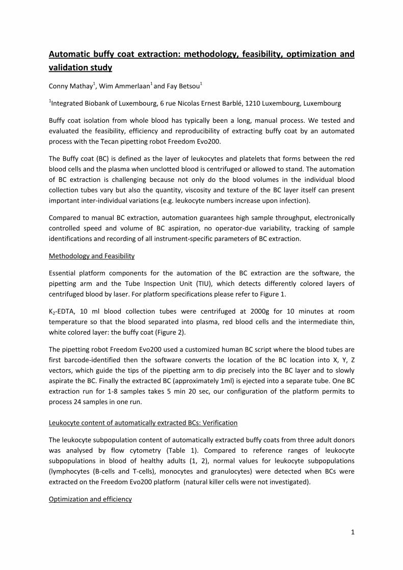
K2-EDTA, 10 ml blood collection tubes were centrifuged at 2000g for 10 minutes at room
temperature so that the blood separated into plasma, red blood cells and the intermediate thin,
white colored layer: the buffy coat (Figure 2).
The pipetting robot Freedom Evo200 used a customized human BC script where the blood tubes are
first barcode-identified then the software converts the location of the BC location into X, Y, Z
vectors, which guide the tips of the pipetting arm to dip precisely into the BC layer and to slowly
aspirate the BC. Finally the extracted BC (approximately 1ml) is ejected into a separate tube. One BC
extraction run for 1-8 samples takes 5 min 20 sec, our configuration of the platform permits to
process 24 samples in one run.
Leukocyte content of automatically extracted BCs: Verification
The leukocyte subpopulation content of automatically extracted buffy coats from three adult donors
was analysed by flow cytometry (Table 1). Compared to reference ranges of leukocyte
subpopulations in blood of healthy adults (1, 2), normal values for leukocyte subpopulations
(lymphocytes (B-cells and T-cells), monocytes and granulocytes) were detected when BCs were
extracted on the Freedom Evo200 platform (natural killer cells were not investigated).
Optimization and efficiency

2
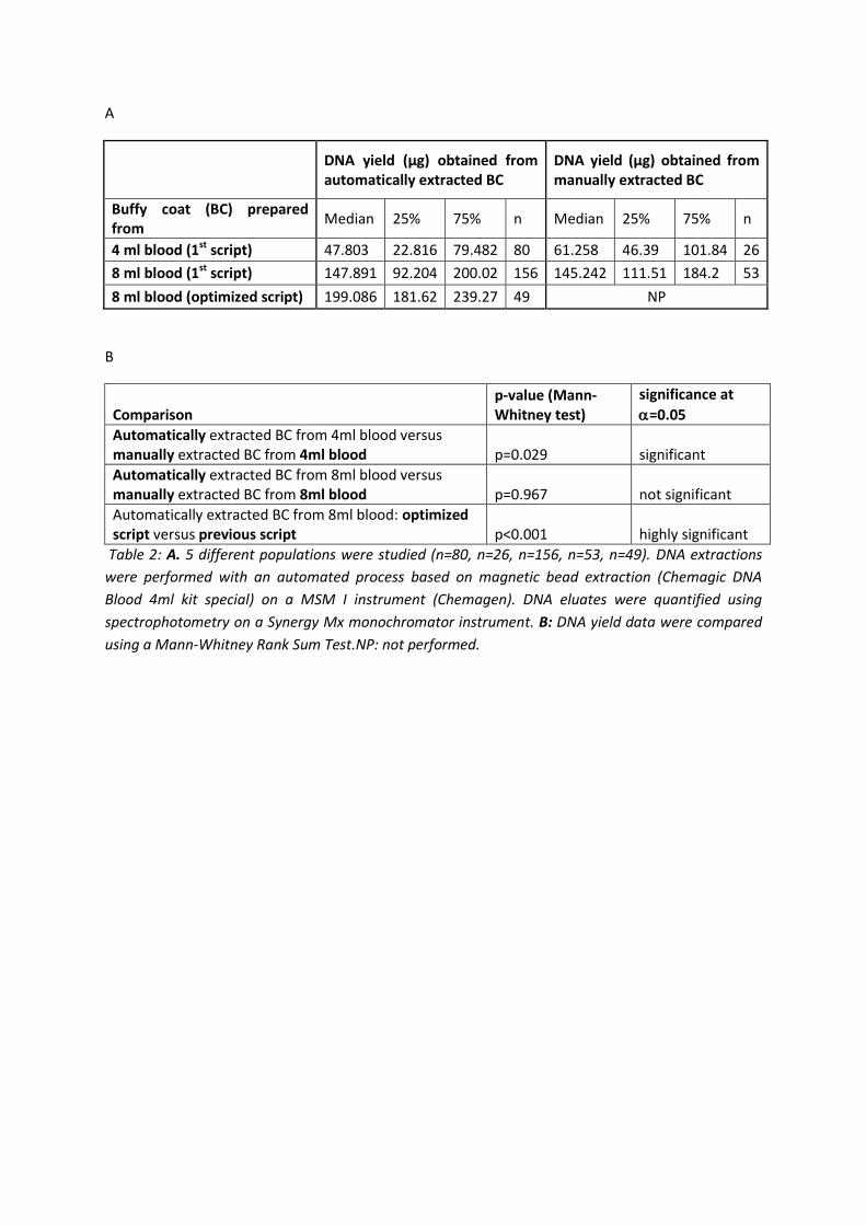
BC is often used for DNA extraction. Quantification of the extracted DNA, from either automatically
or manually prepared BCs, permits to assess the efficiency of the BC automation process. Data
presented in Table 2 were obtained from 364 healthy volunteer donors over a 6-month timespan
during which the BC automation was optimized. The same DNA extraction and quantification
protocols were used for all specimens.
BCs were prepared from 8ml blood either automatically or manually, and half of the BC volume was
used for DNA extraction, corresponding to an initial 4ml blood volume. The automated BC extraction
resulted in a significantly lower DNA yield (47.8 µg) compared to manual BC preparation (61.3 µg).
When the whole BC prepared by the Freedom Evo200 was used for DNA extraction, corresponding
to 8ml of whole blood, a median DNA yield of 147.9 µg was obtained. Manual BC extraction from
8ml blood resulted in a similar median DNA yield (145.2 µg). Optimization of the BC script for
automation resulted in a significantly higher DNA yield (199 µg) when compared to the yields
obtained with the previous script. The optimization took into account some specific mechanical
parameters of the used tube type. Additionally the settings were optimized to allow a complete
aspiration of the BC. As a result, the efficiency of optimized automated BC preparation in terms of
DNA yield is higher than in manually prepared BCs.
Validation
To compare the physical content of extracted BCs to whole blood, cell counting was performed on
blood before BC extraction and on automatically (optimized script) and manually extracted BCs from
the same three donors (Table 3). The automatically extracted BCs contained significantly more
leukocytes and platelets than the manually prepared BCs. Compared to whole blood, 95% of the
blood leukocytes could be recovered in the automatically prepared BCs, whereas in the manually
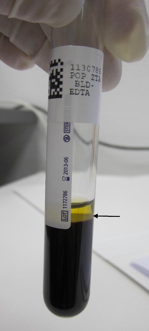
extracted BCs the leukocyte recovery percentage was 56%. Confirmation of these data came from a
visual inspection of the collection tubes 30 minutes after the buffy coat extraction, where less buffy
coat left-over in the automated tubes was clearly observed (Figure 3).
Red blood cell (RBC) contamination in manually or automatically prepared BCs was not significantly
different, however in two of the three examined cases, RBC contamination was higher in the
manually extracted buffy coats. Haemoglobin values were low both in manually and automatically
prepared BCs showing that in general no RBC lysis happened during the BC extraction. The median
haemoglobin value was lower in the automatically extracted BCs (in two of the three cases).
In conclusion, the automatically extracted BCs were of high quality presenting high leukocyte and
platelets numbers and low red blood cell contamination compared to manually prepared BCs.
In a second part of this experiment the DNA content of the automatically or manually prepared BCs
was analysed from the same 3 donors (Table 4). The mean DNA yield of 204 g obtained from BC
from 8 ml blood (very similar to yields shown in Table 2), was 1.5 times higher in the automatically
extracted BCs compared to the manually extracted BCs, however the difference was not significantly
different due to low numbers of donors. The ratios A260/A280 were high and very stable for both
manually and automatically extracted BCs, thus indicating highly pure DNA samples.

3
Discussion
We demonstrate the feasibility of the BC extraction automation on a Tecan Freedom Evo200
platform. Optimization of the script and validation of the method were based on BC cell content
analysis (Blood cell counting and FACS) and on DNA extraction yields. Median DNA yield obtained
with the optimized BC script (199 ug DNA from 8ml blood) is significantly higher than the one
obtained with the first BC script, highlighting that script optimization was a critical factor for best
DNA yield outcomes. The optimized script generated DNA extraction yields which were even
significantly higher than the ones from manually prepared BCs (Table 2 and 4). In conclusion,
automation of buffy coat extraction permits to isolate all leukocyte subpopulations and to extract
high DNA quantities, it eliminates inter-operator variability and is time-saving, since up to 24
samples can be processed at the same time. Besides the Tecan system, Hamilton platforms have also
been reported by the manufacturer as being capable of offering automation of buffy coat extraction.
In our system, the automated BC extraction can be combined, on one hand, with upstream
automated plasma aliquoting in the same run, and downstream automated DNA extraction from BCs
on the other hand.
Acknowledgements
We are grateful to Hans-Peter Sattler and Matthias Greuter from Tecan for continuous assistance
with the platform and Katy Beaumont, Estelle Henry and Audur Thorlaksdottir for excellent technical
help.
Figure legends
Figure 1: Illustration of the Tecan Freedom Evo200 platform at IBBL. The platform is composed of a
pick and place arm (PnP), a liquid handling arm (LiHa) with eight adapters for disposable tips (DiTi)
and a robotic arm with centric gripper. A Tecan Tube Inspection Unit (TIU) is installed to detect
blood separation levels. Tube barcodes are identified by a Datalogic Matrix200 scanner (scanner), 2D
barcodes of individual destination tubes are identified by a FluidX flatbed barcode scanner. FluidX
decappers (XSD-96Pro and XSD-48Pro for 96 respectively 48 individual tubes in racks), a laminar flow
hood and a reagent-cooling unit complete the instruments of the platform. The system is operated
by the software EVOware Standard V2.4.
Figure 2: Typical appearance of a centrifuged blood collection tube after plasma withdrawal. The
buffy coat (arrow) is located between the plasma and the dark red erythrocytes.
Figure 3: Six blood collection tubes from 3 donors after withdrawal of equivalent volume of buffy
coat by manual process (Man, donors 1-3) or automated process (Aut, donors 1-3).
References
1 Herklotz R, Luthi U, Ottiger C, Huber AR. [Metaanalysis of reference values in hematology]. Therapeutische Umschau Revue therapeutique. 2006 Jan;63(1):5-24. 2 Peter HH. Labor und Diagnose: TH-Books Verlagsgesellschaft Frankfurt/Main (2000).

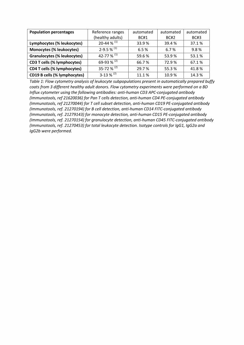
Population percentages Reference ranges (healthy adults)
automated BC#1
automated BC#2
automated BC#3
Lymphocytes (% leukocytes) 20-44 % (1) 33.9 % 39.4 % 37.1 %
Monocytes (% leukocytes) 2-9.5 % (1) 6.5 % 6.7 % 9.8 %
Granulocytes (% leukocytes) 42-77 % (1) 59.6 % 53.9 % 53.1 %
CD3 T cells (% lymphocytes) 69-93 % (2) 66.7 % 72.9 % 67.1 %
CD4 T cells (% lymphocytes) 35-72 % (2) 29.7 % 55.3 % 41.8 %
CD19 B cells (% lymphocytes) 3-13 % (2) 11.1 % 10.9 % 14.3 %
Table 1: Flow cytometry analysis of leukocyte subpopulations present in automatically prepared buffy coats from 3 different healthy adult donors. Flow cytometry experiments were performed on a BD Influx cytometer using the following antibodies: anti-human CD3 APC-conjugated antibody (Immunotools, ref 21620036) for Pan T cells detection, anti-human CD4 PE-conjugated antibody (Immunotools, ref 21270044) for T cell subset detection, anti-human CD19 PE-conjugated antibody (Immunotools, ref. 21270194) for B cell detection, anti-human CD14 FITC-conjugated antibody (Immunotools, ref. 21279143) for monocyte detection, anti-human CD15 PE-conjugated antibody (Immunotools, ref. 21270154) for granulocyte detection, anti-human CD45 FITC-conjugated antibody (Immunotools, ref. 21270453) for total leukocyte detection. Isotype controls for IgG1, IgG2a and IgG2b were performed.
A
DNA yield (µg) obtained from automatically extracted BC
DNA yield (µg) obtained from manually extracted BC
Buffy coat (BC) prepared from
Median 25% 75% n Median 25% 75% n
4 ml blood (1st script) 47.803 22.816 79.482 80 61.258 46.39 101.84 26
8 ml blood (1st script) 147.891 92.204 200.02 156 145.242 111.51 184.2 53
8 ml blood (optimized script) 199.086 181.62 239.27 49 NP
B
Comparison p-value (Mann-Whitney test)
significance at
=0.05
Automatically extracted BC from 4ml blood versus manually extracted BC from 4ml blood p=0.029 significant
Automatically extracted BC from 8ml blood versus manually extracted BC from 8ml blood p=0.967 not significant
Automatically extracted BC from 8ml blood: optimized script versus previous script p<0.001 highly significant
Table 2: A. 5 different populations were studied (n=80, n=26, n=156, n=53, n=49). DNA extractions
were performed with an automated process based on magnetic bead extraction (Chemagic DNA
Blood 4ml kit special) on a MSM I instrument (Chemagen). DNA eluates were quantified using
spectrophotometry on a Synergy Mx monochromator instrument. B: DNA yield data were compared
using a Mann-Whitney Rank Sum Test.NP: not performed.

donor 1 (male, 25 years)
whole blood automated BC manual BC
measured parameter
tot cell nb or tot quantity
tot cell nb or tot quantity
recovery (%) vs whole blood
tot cell nb or tot quantity
recovery (%) vs whole blood
WBC (10⁶) 60.00 61.32 102% 43.75 73%
RBC (10⁶) 39760 8852 22% 11048 28%
HGB (g) 1.168 0.260 22% 0.323 28%
PLT (10⁶) 1576 1157 73% 736 47%
donor 2 (male, 60 years)
whole blood automated BC manual BC
measured parameter
tot cell nb or tot quantity
tot cell nb or tot quantity
recovery (%) vs whole blood
tot cell nb or tot quantity
recovery (%) vs whole blood
WBC (10⁶) 40.00 38.71 97% 17.12 43%
RBC (10⁶) 36400 6800 19% 9903 27%
HGB (g) 1.096 0.211 19% 0.301 27%
PLT (10⁶) 1928 1500 78% 588 30%
donor 3 (female, 44 years)
whole blood automated BC manual BC
measured parameter
tot cell nb or tot quantity
tot cell nb or tot quantity
recovery (%) vs whole blood
tot cell nb or tot quantity
recovery (%) vs whole blood
WBC (10⁶) 41.60 35.02 84% 18.80 45%
RBC (10⁶) 32160 6562 20% 6110 19%
HGB (g) 1.000 0.204 20% 0.190 19%
PLT (10⁶) 2288 1358 59% 705 31%
whole blood automated BC manual BC Comparison
measured parameter
tot cell nb or tot quantity
tot cell nb or tot quantity
recovery (%) vs whole blood
tot cell nb or tot quantity
recovery (%) vs whole blood
automated BC vs manual BC
mean StDev mean StDev mean StDev
WBC (10⁶) 47.20 11.11 45.02 14.24 95% 26.56 14.91 56% p=0.008
RBC (10⁶) 36107 3808 7405 1259 21% 9020 2584 25% NS
HGB (g) 1.088 0.084 0.225 0.030 21% 0.271 0.071 25% NS
PLT (10⁶) 1931 356 1339 172 69% 676 78 35% p=0.043
Table 3: Complete blood cell count data for whole blood, automated buffy coat (optimized script) and
manual buffy coat prepared from the 3 same healthy donors (WBC: white blood cells, RBC: red blood
cells, HGB: haemoglobin, PLT: platelets). Blood cell counting was performed on a Horiba ABX Micros
CRP 200 instrument. Statistical t-test analysis on mean values of the three donors for WBC, RBC, HGB
and PLT (NS=non significant).
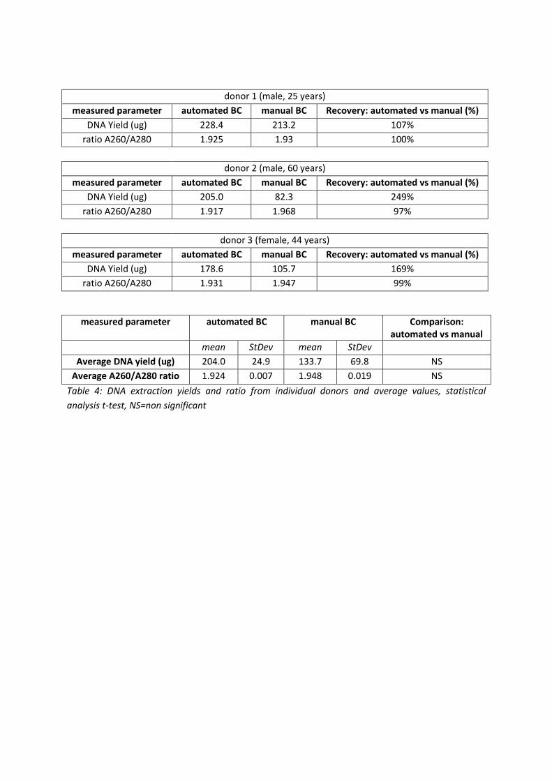
donor 1 (male, 25 years)
measured parameter automated BC manual BC Recovery: automated vs manual (%)
DNA Yield (ug) 228.4 213.2 107%
ratio A260/A280 1.925 1.93 100%
donor 2 (male, 60 years)
measured parameter automated BC manual BC Recovery: automated vs manual (%)
DNA Yield (ug) 205.0 82.3 249%
ratio A260/A280 1.917 1.968 97%
donor 3 (female, 44 years)
measured parameter automated BC manual BC Recovery: automated vs manual (%)
DNA Yield (ug) 178.6 105.7 169%
ratio A260/A280 1.931 1.947 99%
measured parameter automated BC manual BC Comparison: automated vs manual
mean StDev mean StDev
Average DNA yield (ug) 204.0 24.9 133.7 69.8 NS
Average A260/A280 ratio 1.924 0.007 1.948 0.019 NS
Table 4: DNA extraction yields and ratio from individual donors and average values, statistical
analysis t-test, NS=non significant
Related Documents