Article Autologous iPSC-Based Vaccines Elicit Anti-tumor Responses In Vivo Graphical Abstract Highlights d Irradiated iPSCs prevent tumor growth in murine models of breast, lung, and skin cancer d iPSC vaccines target shared antigens between iPSCs and cancer cells d iPSC vaccines promote a humoral and cell-mediated anti- tumor immune profile d As an adjuvant cancer therapy, iPSC vaccination can reactivate the immune system Authors Nigel G. Kooreman, Youngkyun Kim, Patricia E. de Almeida, ..., Ronald Levy, Mark M. Davis, Joseph C. Wu Correspondence [email protected] (R.L.), [email protected] (M.M.D.), [email protected] (J.C.W.) In Brief Wu and colleagues show that cancer immunity against multiple types of cancer can be achieved using an easily generable iPSC-based cancer vaccine. This immunity is based on overlapping epitopes between iPSCs and cancer cells and can also be achieved by reactivating the immune system as an adjuvant immunotherapy. Kooreman et al., 2018, Cell Stem Cell 22, 1–13 April 5, 2018 ª 2018 Elsevier Inc. https://doi.org/10.1016/j.stem.2018.01.016

Welcome message from author
This document is posted to help you gain knowledge. Please leave a comment to let me know what you think about it! Share it to your friends and learn new things together.
Transcript

Article
Autologous iPSC-Based Vaccines Elicit Anti-tumorResponses In Vivo
Graphical Abstract
Highlightsd Irradiated iPSCs prevent tumor growth in murine models of
breast, lung, and skin cancer
d iPSC vaccines target shared antigens between iPSCs and
cancer cells
d iPSC vaccines promote a humoral and cell-mediated anti-
tumor immune profile
d As an adjuvant cancer therapy, iPSC vaccination can
reactivate the immune system
Authors
Nigel G. Kooreman, Youngkyun Kim,
Patricia E. de Almeida, ..., Ronald Levy,
Mark M. Davis, Joseph C. Wu
[email protected] (R.L.),[email protected] (M.M.D.),[email protected] (J.C.W.)
In BriefWu and colleagues show that cancer
immunity against multiple types of cancer
can be achieved using an easily
generable iPSC-based cancer vaccine.
This immunity is based on overlapping
epitopes between iPSCs and cancer cells
and can also be achieved by reactivating
the immune system as an adjuvant
immunotherapy.
Kooreman et al., 2018, Cell Stem Cell 22, 1–13April 5, 2018 ª 2018 Elsevier Inc.https://doi.org/10.1016/j.stem.2018.01.016

Cell Stem Cell
Article
Autologous iPSC-Based VaccinesElicit Anti-tumor Responses In VivoNigel G. Kooreman,1,2,3,4,12 Youngkyun Kim,1,2,3,7,12 Patricia E. de Almeida,1,2,3 Vittavat Termglinchan,1,2,3
Sebastian Diecke,8,9,10 Ning-Yi Shao,1,2,3 Tzu-Tang Wei,1,2,3 Hyoju Yi,1,2,3,7 Devaveena Dey,1,2,3 Raman Nelakanti,1,2,3
Thomas P. Brouwer,1,2,3,4 David T. Paik,1,2,3 Idit Barfi,5 Arnold Han,6 Paul H.A. Quax,4 Jaap F. Hamming,4 Ronald Levy,5,*Mark M. Davis,6,* and Joseph C. Wu1,2,3,11,*1Institute for Stem Cell Biology and Regenerative Medicine, Stanford University School of Medicine, Stanford, CA 94305, USA2Stanford Cardiovascular Institute of Medicine, Stanford University School of Medicine, Stanford, CA 94305, USA3Department of Radiology, Stanford University School of Medicine, Stanford, CA 94305, USA4Department of Surgery, Leiden University Medical Center, Leiden 2333 ZA, the Netherlands5Stanford Cancer Institute, Stanford University School of Medicine, Stanford, CA 94305, USA6Institute for Immunity, Transplantation, and Infection, Stanford University School of Medicine, Stanford, CA 94305, USA7Convergent Research Consortium for Immunologic Disease, Seoul St. Mary’s Hospital, Catholic University of Korea, Seoul 06591, Korea8Max Delbr€uck Center for Molecular Medicine in the Helmholtz Association (MDC), Robert-Rossle Strasse 10, 13125 Berlin, Germany9DZHK (German Centre for Cardiovascular Research), Partner Site, Berlin, Germany10Berlin Institute of Health (BIH), Berlin, Germany11Lead Contact12These authors contributed equally*Correspondence: [email protected] (R.L.), [email protected] (M.M.D.), [email protected] (J.C.W.)https://doi.org/10.1016/j.stem.2018.01.016
SUMMARY
Cancer cells and embryonic tissues share a numberof cellular and molecular properties, suggestingthat induced pluripotent stem cells (iPSCs) may beharnessed to elicit anti-tumor responses in cancervaccines. RNA sequencing revealed that humanand murine iPSCs express tumor-associated anti-gens, and we show here a proof of principle for usingirradiated iPSCs in autologous anti-tumor vaccines.In a prophylactic setting, iPSC vaccines preventtumor growth in syngeneic murine breast cancer,mesothelioma, and melanoma models. As an adju-vant, the iPSC vaccine inhibited melanoma recur-rence at the resection site and reduced metastatictumor load, which was associated with fewer Th17cells and increased CD11b+GR1hi myeloid cells.Adoptive transfer of T cells isolated from vaccine-treated tumor-bearing mice inhibited tumor growthin unvaccinated recipients, indicating that the iPSCvaccine promotes an antigen-specific anti-tumorT cell response. Our data suggest a generalizablestrategy for multiple types of cancer that could provehighly valuable in clinical immunotherapy.
INTRODUCTION
Nearly a century ago, researchers observed that immunizationwith embryonic materials led to the rejection of transplantedtumors (Brewer et al., 2009). More recently, studies identifiedshared transcriptome profiles and antigens on various tumorcells and embryonic cells (Ben-Porath et al., 2008; Ghosh
et al., 2011). This has led to the hypothesis that embryonicstem cells (ESCs) could be used as immunization agents topromote an anti-tumor response. A major advantage of whole-cell vaccination over traditional vaccines, which consist ofinactivated organisms or protein products, is that a broad rangeof antigens can be presented to T cells, including unknown anti-gens (Palena et al., 2006; Yaddanapudi et al., 2012). However,the use of fetal and embryonic materials as vaccines toinduce anti-tumor immunity has not yet advanced beyond animalmodels, owing largely to ethical challenges surrounding thesetherapies.Since the discovery of induced pluripotent stem cells (iPSCs)
(Takahashi et al., 2007; Takahashi and Yamanaka, 2006), plurip-otent cells from a patient’s own tissues can be created that sharenearly identical gene expression and surface markers profileswith ESCs (Bock et al., 2011; Mallon et al., 2013, 2014; Soldneret al., 2009), circumventing a major ethical roadblock. Addition-ally, the tumorigenic (Kooreman and Wu, 2010; Lee et al., 2013)and immunogenic (de Almeida et al., 2014; Zhao et al., 2011)properties of iPSCs with autologous transplantation suggestpotential efficacy in cancer vaccination. Importantly, autologousiPSCs may provide a more accurate and representative panel ofpatient’s tumor immunogens than non-autologously derivedESCs. Here, we test the hypothesis that iPSCs may work as awhole-cell-based vaccine that presents T cells with a broadheterogeneity in cancer-related epitopes.
RESULTS
Human and Murine iPSCs Express Tumor-Specific andTumor-Associated AntigensWe first performed RNA sequencing on 11 different humaniPSC clones to compare expression profiles from a selectedcancer-related gene list to human ESCs (hESCs), cancertissues, and healthy tissues (Figure S1A). Based on this gene
Cell Stem Cell 22, 1–13, April 5, 2018 ª 2018 Elsevier Inc. 1
STEM 2354
Please cite this article in press as: Kooreman et al., Autologous iPSC-Based Vaccines Elicit Anti-tumor Responses In Vivo, Cell Stem Cell (2018),https://doi.org/10.1016/j.stem.2018.01.016

A C
B
D
E
F
Figure 1. Assessing the Optimal Vaccination Schedule, followed by Successful Prophylactic Treatment of Breast Cancer and Melanomain Mice(A) Optimal vaccination was set to C+I vaccination for 4 weeks, as assessed by percent IgG binding to DB7, without a significant increase in non-specific mouse
embryonic feeder (MEF) binding (n = 3 control animals, n = 4 iPSC primed animals, n = 4 C+I primed 2 week, and n = 4 C+I primed 4 week animals, mean ± SEM,
ANOVA with Tukey’s multiple comparison test).
(B) Representative FACS plot of serum IgG binding of PBS 4-week, iPSC 4-week, C+I 2-week, or C+I 4-week vaccinated mice to embryonic fibroblasts, iPSCs,
and DB7 cancer cells. As a control sample for differentiated cells, a partly differentiated cell culture was included in the analysis. This is shown by IgG-positive and
negative cells, indicating that the IgG binding is specific to the undifferentiated portion of the analyzed cells. C+I 4-week-vaccinated mice showed the best IgG
binding to DB7 breast cancer cells.
(legend continued on next page)
STEM 2354
2 Cell Stem Cell 22, 1–13, April 5, 2018
Please cite this article in press as: Kooreman et al., Autologous iPSC-Based Vaccines Elicit Anti-tumor Responses In Vivo, Cell Stem Cell (2018),https://doi.org/10.1016/j.stem.2018.01.016
list, we found human iPSCs cluster with hESCs and the cancertissues, revealing important gene expression overlap in cancergenes between different cancer types and iPSCs. The upregu-lation of a subset of these genes was then also validated inmurine iPSCs and ESCs (Figure S1B). These findings suggestthe possibility of using iPSCs in different species to primethe host in developing immunity against known and, perhaps,unknown tumor-specific antigens (TSAs) and tumor-associatedantigens (TAAs).
iPSC-Vaccine-Primed Mice Mount Strong B and T CellResponses against Breast Cancer In Vitro and In VivoUsing FVB strain iPSCs (Figures S2A and S2D) and theadjuvant CpG, proven to be successful in tumor vaccination(Gilkeson et al., 1998; Goldstein et al., 2011; Mor et al.,1997; Mukherjee et al., 2007), we observed an effectiveimmune response to a murine breast cancer (DB7) with aCpG and iPSCs (C+I) combination. In brief, we first estab-lished the effect of CpG and an optimal vaccination schedule.We primed FVB mice with iPSCs or C+I for 2 weeks or 4 weeksand found the strongest in vitro T cell responses to DB7tumor lysate in the C+I 4-week group (Figures S2E and S2F).In addition, a vaccination schedule of 4 weeks with the C+Icombination resulted in the highest immunoglobulin G (IgG)binding (80.0% ± 3.4%) to DB7 and was therefore used forsubsequent vaccination rounds (Figures 1A and 1B). Afteroptimizing the schedule (Figure 1C), we proceeded with thevaccination of 40 FVB mice divided into four groups:(1) PBS, (2) CpG only, (3) iPSCs only, and (4) C+I. After fouronce-weekly vaccinations, 5 3 104 DB7 cancer cells wereinjected subcutaneously, and tumor size was monitored usingcaliper measurement. After 1 week, all mice presented with asimilar lesion at the injection site that regressed in 7 out of 10C+I-treated mice and progressed to larger tumors in the othergroups (Figures 1D, S3A, and S3B). Four weeks after tumorinoculation, five mice per group were sacrificed to analyzethe immune profiles in blood, spleen, and draining lymphnodes (dLNs). The other five mice per group were used forlong-term survival studies for up to 1 year. Most were sacri-ficed in the first 2 weeks after the end of the experimentwhen their tumor exceeded 1 cm3. However, two mice in theC+I treatment group survived 1 year and had antibody titersagainst iPSCs and DB7 similar to the start of the experimentand were able to fully reject 5 3 104 cancer cells upon reintro-duction (Figures S3C and S3D). The control mice in this exper-iment, primed with iPSC-derived endothelial cells, wereunable to mount IgG responses to the DB7 cell line, therebyruling out the possibility that the culturing conditions withFBS-containing media could be responsible for the cross-reactivity or endogenous murine leukemia viral antigens.
C+I Vaccination Provides Breast Cancer and MelanomaImmunity by Upregulating Antigen Presentation andT-Helper/Cytotoxic T Cell ActivityTo test the effectiveness of our vaccine in targeting multiplecancer types, an additional experiment was performed usingthe melanoma cell line B16F0, which is syngeneic to theC57BL/6 mouse strain. C57BL/6 iPSCs were generated (FiguresS2B and S2D), and 40 mice were again divided into PBS, CpG,iPSCs, and C+I groups and treated for 4 weeks. Following this,5 3 104 B16F0 cells were subcutaneously injected in thelower back. Tumor growth assessment by caliper measurementshowed significantly lower tumor progression by week 2 in theC+I group (Figures 1E, 1F, S3E, and S3F). Due to large tumorsizes in the control groups, themicewere sacrificed 2weeks aftertumor injection. Afterward, the immune cell profiles in blood,dLNs, and spleenswere analyzed using flowcytometry. Cytomet-ric analysis showed a significant decrease in CD4+CD25+FoxP3+
regulatory T cells (T-regs) in blood and an increase in effector/memory helper T cells in dLNs 2 weeks after tumor injections inC57BL/6 mice (Figures 2A and 2B), as well as increased percent-ages of mature antigen-presenting cells (APCs) (Figure 2C).At 4 weeks, FVB mice in the C+I-vaccinated group had signif-
icant increases in the effector/memory cytotoxic T cells in thespleen and dLNs (Figures 2D and 2F). The tumor specificity ofthese cytotoxic T cells was further confirmed by increasedsecretion of interferon-g (IFN-g) by splenocytes isolated fromC+I-vaccinated mice in response to DB7 tumor lysate (Figures3A, 3B, S4A, and S4B). As with the C57BL/6 mice, upregulationof mature APCs and helper T cells was also seen in dLNs of FVBmice (Figures 2E and 2F). Both mouse strains remained healthythroughout the study and showed no signs of autoimmuneresponses due to the vaccine in serum and in tissues (FiguresS4C–S4F). Lastly, the effectiveness of the C+I vaccine was as-sessed in the more clinically relevant orthotopic model of breastcancer. Significant tumor size differences were seen as early as1week after orthotopic transfer of cancer cells in C+I-vaccinatedmice compared to vehicle control, followed by further tumorreduction over the course of 3 weeks (Figures 3C and 3E). Usingan additional group of orthotopic breast cancer mice, in vivotumor specificity was tested by adoptively transferring spleno-cytes from C+I vaccinated or vehicle (PBS+CpG) vaccinatedmice into these tumor-bearing mice (Figure 3D). This resultedin a significant reduction of tumor sizes in the C+I-vaccinatedgroup compared to the vehicle-vaccinated group (Figure 3F).
Tumor Immunity in C+I-Vaccinated Mice Is the Result ofShared Epitopes between iPSCs and Cancer CellsTo test whether the C+I vaccine provides immunityagainst shared epitopes between iPSCs and cancer cells, weperformed additional experiments to assess two-way immunity
(C) Schema showing vaccine preparation consists of sorting murine iPSCs for pluripotency, irradiation, resuspension in adjuvant solution, and subcutaneous
injection in the flank (sites 1–4).
(D) Vaccination of FVB mice with C+I resulted in a complete rejection of the cancer cells in 7 out of 10 mice by 4 weeks and overall reductions in DB7 tumor size
(n = 10 per group; representative images; left). Quantification of the data presented (right).
(E) Vaccination of C57BL/6mice with C+I resulted in significant reduction of melanoma sizes initiated by the aggressive B16F0melanoma cell line by week 2 (n = 8
PBS, n = 9 iPSC primed, n = 10 CpG primed, and n = 9 C+I primed).
(F) Quantification of the tumor size data presented in (E).
Data in (D) and (F) represent mean ± SEM (ANOVA with Tukey’s multiple comparison test). *p < 0.05, **p < 0.001, ***p < 0.001, ****p < 0.0001.
STEM 2354
Cell Stem Cell 22, 1–13, April 5, 2018 3
Please cite this article in press as: Kooreman et al., Autologous iPSC-Based Vaccines Elicit Anti-tumor Responses In Vivo, Cell Stem Cell (2018),https://doi.org/10.1016/j.stem.2018.01.016

by demonstrating (1) cancer immunity by C+I primed T cells and(2) iPSC immunity by tumor-experienced lymphocytes (TELs).For the first experiment, isolated T cells from C+I-vaccinatedor vehicle (PBS+CpG)-vaccinated mice were adoptively trans-
ferred to a group of tumor-bearing orthotopic breast cancermice (n = 7 per group), and tumor growth was measured overthe course of 4 weeks (Figure 4A). This resulted in a significantreduction of tumor sizes in the C+I-vaccinated group compared
A
B C
D E
F
Figure 2. Prophylactic Vaccination Leads to Increased Antigen Presentation in dLNs and Subsequent Effector/Memory T Cell Responsesin dLNs and Spleen(A) 2 weeks after B16F0 introduction, iPSC- and C+I-vaccinated mice showed a significant reduction in percentages of regulatory T cells (CD4+CD25+FoxP3+)
and an increase in effector/memory helper T cells (CD4+CD44+) in the peripheral blood of C+I-vaccinated mice. At that point, only limited upregulation of effector/
memory cytotoxic T cells (CD8+CD44+) was seen.
(B and C) The dLNs in the C+I group had significantly higher percentages of effector/memory helper T cells (B) and increased antigen presentation by mature
antigen-presenting cells (APCs) such as macrophages (CD11b+F4/80+MHC-II+CD86+) and dendritic cells (CD11c+MHC-II+CD86+) (C).
(D) C+I-vaccinated FVB mice showed increased percentages of activated cytotoxic T cells (CD8+granzyme-B+) in spleens 4 weeks after DB7 introduction.
(E and F) dLNs of these mice revealed an increased frequency of mature antigen-presenting macrophages (E) as well as effector/memory helper T cells and
cytotoxic T cells (F).
Data represent mean ± SEM (n = 5 per group; ANOVA with Tukey’s multiple comparison test). *p < 0.05, **p < 0.001, ***p < 0.001, ****p < 0.0001.
STEM 2354
4 Cell Stem Cell 22, 1–13, April 5, 2018
Please cite this article in press as: Kooreman et al., Autologous iPSC-Based Vaccines Elicit Anti-tumor Responses In Vivo, Cell Stem Cell (2018),https://doi.org/10.1016/j.stem.2018.01.016
A B
C D
E F
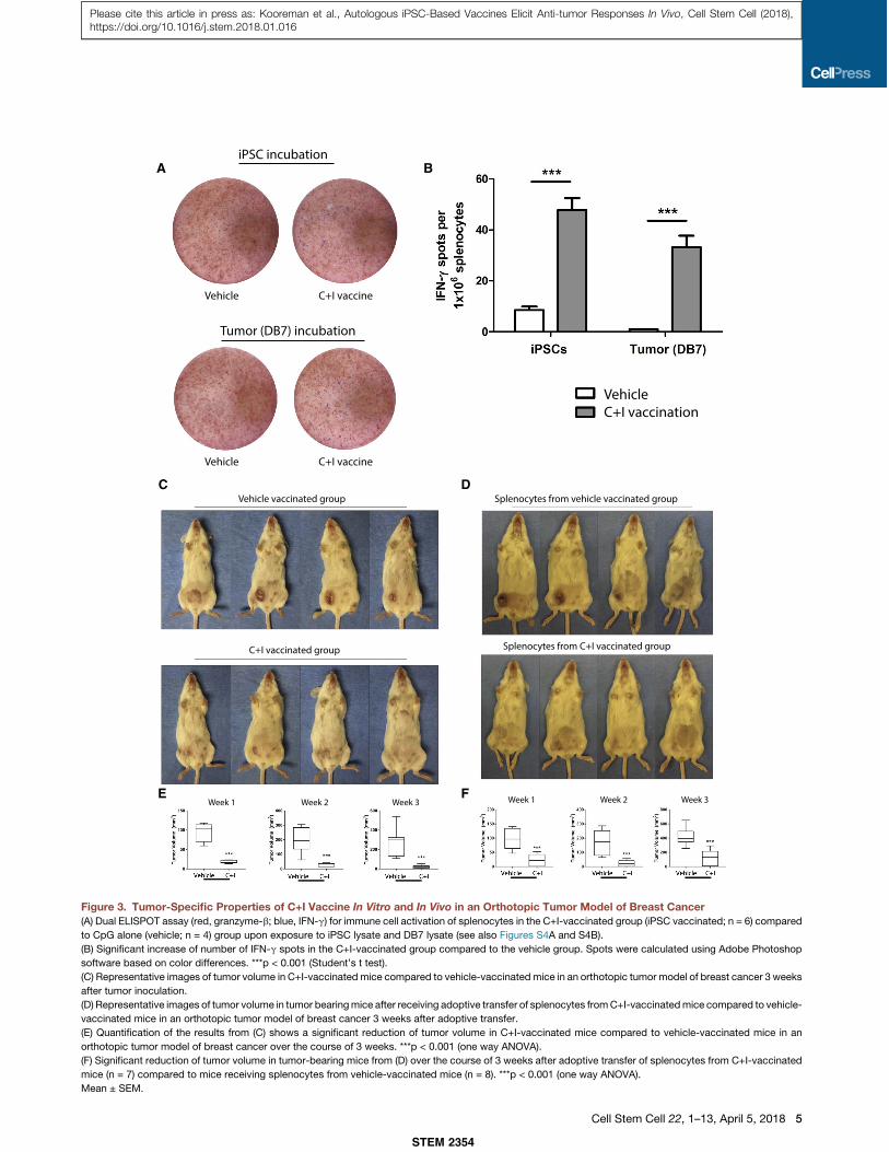
Figure 3. Tumor-Specific Properties of C+I Vaccine In Vitro and In Vivo in an Orthotopic Tumor Model of Breast Cancer(A) Dual ELISPOT assay (red, granzyme-b; blue, IFN-g) for immune cell activation of splenocytes in the C+I-vaccinated group (iPSC vaccinated; n = 6) compared
to CpG alone (vehicle; n = 4) group upon exposure to iPSC lysate and DB7 lysate (see also Figures S4A and S4B).
(B) Significant increase of number of IFN-g spots in the C+I-vaccinated group compared to the vehicle group. Spots were calculated using Adobe Photoshop
software based on color differences. ***p < 0.001 (Student’s t test).
(C) Representative images of tumor volume in C+I-vaccinated mice compared to vehicle-vaccinated mice in an orthotopic tumor model of breast cancer 3 weeks
after tumor inoculation.
(D) Representative images of tumor volume in tumor bearingmice after receiving adoptive transfer of splenocytes fromC+I-vaccinatedmice compared to vehicle-
vaccinated mice in an orthotopic tumor model of breast cancer 3 weeks after adoptive transfer.
(E) Quantification of the results from (C) shows a significant reduction of tumor volume in C+I-vaccinated mice compared to vehicle-vaccinated mice in an
orthotopic tumor model of breast cancer over the course of 3 weeks. ***p < 0.001 (one way ANOVA).
(F) Significant reduction of tumor volume in tumor-bearing mice from (D) over the course of 3 weeks after adoptive transfer of splenocytes from C+I-vaccinated
mice (n = 7) compared to mice receiving splenocytes from vehicle-vaccinated mice (n = 8). ***p < 0.001 (one way ANOVA).
Mean ± SEM.
STEM 2354
Cell Stem Cell 22, 1–13, April 5, 2018 5
Please cite this article in press as: Kooreman et al., Autologous iPSC-Based Vaccines Elicit Anti-tumor Responses In Vivo, Cell Stem Cell (2018),https://doi.org/10.1016/j.stem.2018.01.016
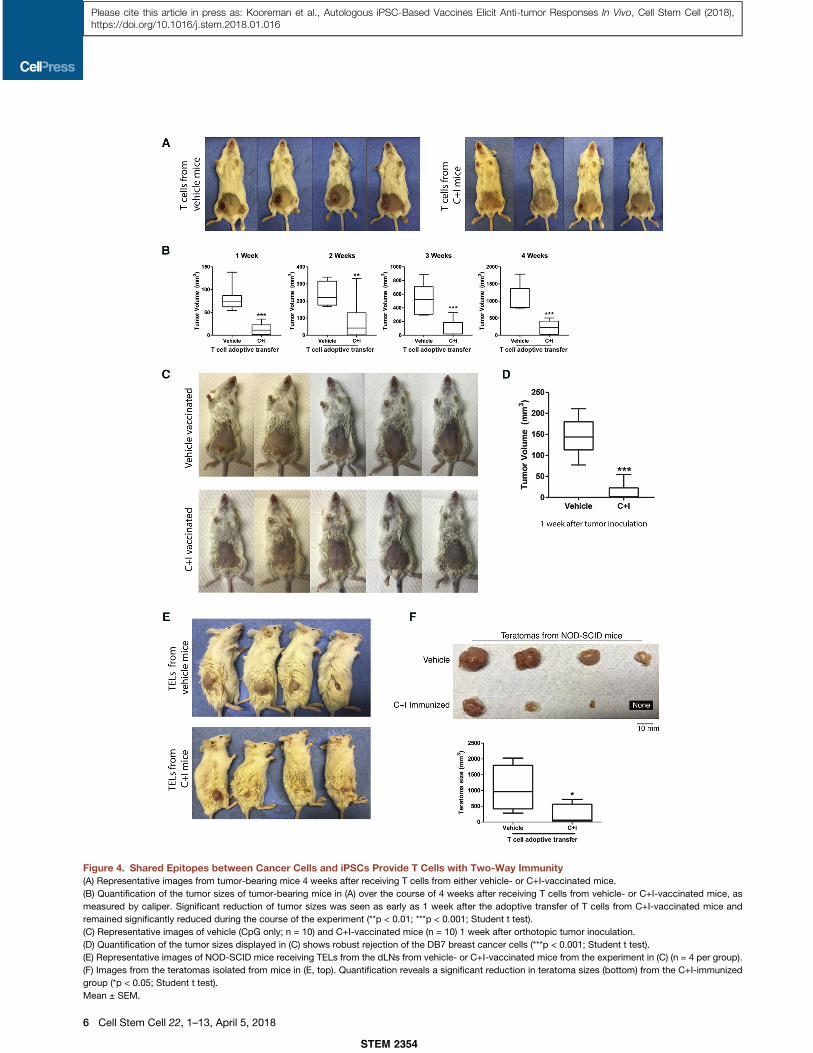
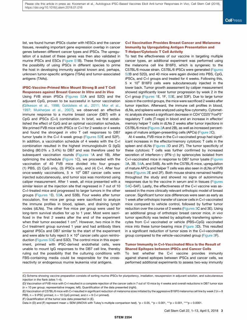
Figure 4. Shared Epitopes between Cancer Cells and iPSCs Provide T Cells with Two-Way Immunity(A) Representative images from tumor-bearing mice 4 weeks after receiving T cells from either vehicle- or C+I-vaccinated mice.
(B) Quantification of the tumor sizes of tumor-bearing mice in (A) over the course of 4 weeks after receiving T cells from vehicle- or C+I-vaccinated mice, as
measured by caliper. Significant reduction of tumor sizes was seen as early as 1 week after the adoptive transfer of T cells from C+I-vaccinated mice and
remained significantly reduced during the course of the experiment (**p < 0.01; ***p < 0.001; Student t test).
(C) Representative images of vehicle (CpG only; n = 10) and C+I-vaccinated mice (n = 10) 1 week after orthotopic tumor inoculation.
(D) Quantification of the tumor sizes displayed in (C) shows robust rejection of the DB7 breast cancer cells (***p < 0.001; Student t test).
(E) Representative images of NOD-SCID mice receiving TELs from the dLNs from vehicle- or C+I-vaccinated mice from the experiment in (C) (n = 4 per group).
(F) Images from the teratomas isolated from mice in (E, top). Quantification reveals a significant reduction in teratoma sizes (bottom) from the C+I-immunized
group (*p < 0.05; Student t test).
Mean ± SEM.
STEM 2354
6 Cell Stem Cell 22, 1–13, April 5, 2018
Please cite this article in press as: Kooreman et al., Autologous iPSC-Based Vaccines Elicit Anti-tumor Responses In Vivo, Cell Stem Cell (2018),https://doi.org/10.1016/j.stem.2018.01.016

to the vehicle-vaccinated group as early as 1 week after theadoptive transfer (Figure 4B). For the second experiment,another batch of mice vaccinated with C+I (n = 10) or vehicle(n = 10) were inoculated with breast cancer cells, and tumorgrowth was measured at 1 week (Figures 4C and 4D). Afterward,we extracted TELs from the dLNs near the tumor site (Torcellanet al., 2017). These TELs were then adoptively transferred toiPSC-inoculated non-obese diabetic severe combined immuno-deficiency (NOD-SCID) mice (53 106 TELs per mouse; n = 4 pergroup), and teratoma development was measured for 4 weeks.Significant reduction in teratoma sizes was seen at 4 weeks inthe NOD-SCID mice receiving TELs from C+I animals thatwere able to reject the DB7 tumor cells, whereas mice receivingTELs from vehicle-vaccinated animals developed large tera-tomas (Figure 4E and 4F).
C+I Vaccination in a Mesothelioma Model Elicits a Pro-inflammatory Profile for Tumor-Infiltrating LymphocytesAs an alternative model for prophylactic treatment, we selectedthe mesothelioma cell line AC29, syngeneic to CBA/J mice.Again, CBA/J iPSCs were created (Figures S2C and S2D), andmice were vaccinated for 4 weeks with PBS (P), CpG and iPSCs(C+I), or CpG with irradiated AC29 cancer cells (C+A) as a posi-tive control. Afterward, 23 106 AC29 cells (A) or 23 106 iPSCs (I)were injected subcutaneously, and after 1 week, the TILs wereanalyzed for their immune profile and T cell receptor (TCR)sequences. Immune profiling was performed with cytometryby time-of-flight (CyTOF) analysis using a phenotype andintracellular staining kit, which revealed an increased presenceof effector/memory CD4+ (24.0%) and CD8+ T cells (22.4%),with a reduction in T-regs in the C+I/A group (1.9%) comparedto P/A control (21.1%, 14.2%, and 3.0%, respectively) (Fig-ure 5A). Using Citrus (cluster identification, characterization,and regression) analysis (Bruggner et al., 2014), B cells andT cells expressing interleukin-2 (IL-2), IL-4, and IL-5 were foundto be predictive of tumor regression in C+I-vaccinated micecompared to the PBS control group (Figures 5B, S5A, S5B,and S5D). Interestingly, systemic cytokine levels were signifi-cantly lower in the vaccinated group and were found tocorrelate with the positive control mice showing tumor rejection(C+I/iPSC; C+A/AC29) (Figures 6A, S6A, and S6B). TCRsequencing in the PBS control group revealed an overlap inT cell clones that are commonly present in thymus and spleen(Figure S6C). In contrast, the TCRs in the C+I group were morediverse among different mice. In addition, there was a generallylower frequency of the clones in the thymus and more similarfrequencies in the spleen, likely because of mouse-specificresponses to the C+I vaccine (Figures 6B and S6D). Interestingly,there was one TCR clone that was shared by four of five mice inthe C+I group but was not present in any of the other groups; thisclone was also extremely rare in naive mice.
C+I Adjuvant Therapy after Tumor Resection Leads toDecreased Tumor Load in Resection Areas and dLNsTo assess the effectiveness of the vaccine as an adjuvanttherapy after tumor resection, we next injected 5 3 104 B16F0tumor cells subcutaneously in the lower back of C57BL/6 miceand R2- or R1-resected mice after 2 weeks. R2-resected micehad no visible recurrence of melanoma in the resection area
(RA) after receiving two adjuvant rounds of C+I vaccine, whereasPBS-control-vaccinated mice had visible tumors within the RAs(Figure S7A). R1-resected mice were vaccinated for 4 weekswith the C+I vaccine (n = 10), CpG (n = 10), and PBS (n = 8)(Figure 7A), after which dLNs and RAs were analyzed using atumor-specific primer designed to detect and quantify theB16F0 melanoma line (Figures S7B–S7G). Tumor load in thedLNs was reduced in both CpG-only and the C+I-vaccinegroups, indicating that CpG acted as a potent adjuvant to inducetumor degradation upon near-tumor injection (Figure S7H). Inter-estingly, in areasmore distant from the vaccination sites, only theC+I-vaccinated group had significantly lower tumor recurrencein the RA (Figure 7B). Systemically, this is explained by reactiva-tion of the immune system (Chung et al., 2013; Dolcetti et al.,2010; Numasaki et al., 2003), as well as a reduction of B16melanoma-promoting Th17 cells (He et al., 2010) compared tothe control groups (Figures 7C, S5C, and S5E).
DISCUSSION
Tumor establishment and progression involve highly proliferativehypoimmunogenic cells that evade the surveillance of the im-mune system. Therefore, new avenues within the field of cancertreatment are being pursued to target cancer by reactivating theimmune system. One way researchers are trying to achieve thisis by using chimeric antigen receptors (CARs), with promisingresults (Lee et al., 2015; Maude et al., 2014; Maus et al., 2014).The idea behind this therapy is to create a cancer-specificantigen receptor and couple this to an effector cell (e.g., T cell),with newer generations of CARs that might even incorporatethe co-stimulatory pathways. However, thus far, results havebeen mixed, with some patients relapsing, possibly due to lossof expression of the targeted antigen (Grupp et al., 2013; Maudeet al., 2014). One way to circumvent this would be to identify newtumor-specific antigens, but large numbers of tumor antigensare still unknown.Pluripotent cells and tissues share known and likely also
unknown TSAs and TAAs with cancer cells and therefore couldbe a potential agent to prime an immune system to target cancer.This modified cell would then function as a surrogate cell typethat resembles the targeted cancer type. A few groups have pur-sued the use of embryonic cells for priming the immune systemin targeting cancer but thus far have not shown efficacy andsafety for the treatment of various types of cancer (Li et al.,2009; Yaddanapudi et al., 2012). In addition, they still rely onthe use of ethically concerning ESCs and a genetically modifiedcell line as an adjuvant (Yaddanapudi et al., 2012), making thesetreatments less suitable for personalized clinical translation.In this study, we showed that prophylactic immunization of
several mouse strains with an iPSC-based vaccine producesan effective immune response to multiple cancer types byupregulation of mature APCs in the dLNs with a subsequentincrease in helper T cells and cytotoxic T cells locally and, lateron, systemically. Interestingly, this led to a systemically favorableT-effector/T-reg ratio, which has been found to reduce tolerizingconditions (Zou, 2005). With our adoptive transfer data on trans-plantation of C+I-primed splenocytes into tumor-bearing mice,we demonstrated the tumor specificity of our iPSC vaccine,which, based on our in vitro data, was likely the result of
STEM 2354
Cell Stem Cell 22, 1–13, April 5, 2018 7
Please cite this article in press as: Kooreman et al., Autologous iPSC-Based Vaccines Elicit Anti-tumor Responses In Vivo, Cell Stem Cell (2018),https://doi.org/10.1016/j.stem.2018.01.016
A
B
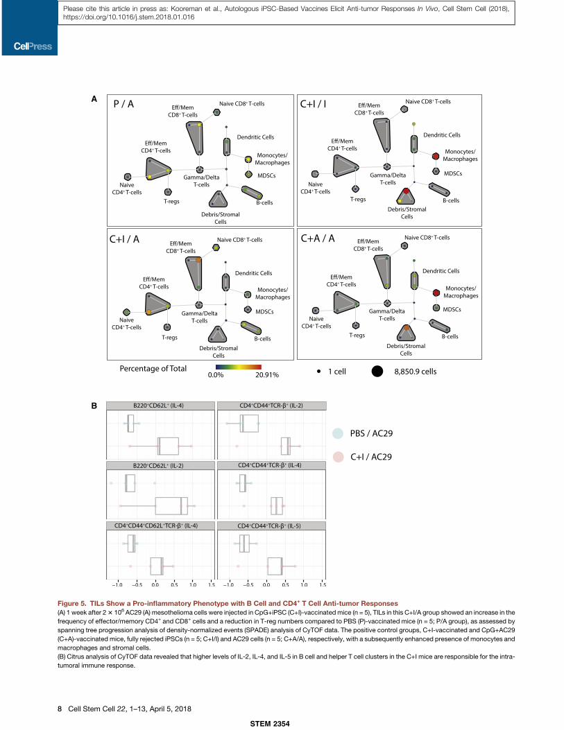
Figure 5. TILs Show a Pro-inflammatory Phenotype with B Cell and CD4+ T Cell Anti-tumor Responses(A) 1 week after 23 106 AC29 (A) mesothelioma cells were injected in CpG+iPSC (C+I)-vaccinatedmice (n = 5), TILs in this C+I/A group showed an increase in the
frequency of effector/memory CD4+ and CD8+ cells and a reduction in T-reg numbers compared to PBS (P)-vaccinated mice (n = 5; P/A group), as assessed by
spanning tree progression analysis of density-normalized events (SPADE) analysis of CyTOF data. The positive control groups, C+I-vaccinated and CpG+AC29
(C+A)-vaccinated mice, fully rejected iPSCs (n = 5; C+I/I) and AC29 cells (n = 5; C+A/A), respectively, with a subsequently enhanced presence of monocytes and
macrophages and stromal cells.
(B) Citrus analysis of CyTOF data revealed that higher levels of IL-2, IL-4, and IL-5 in B cell and helper T cell clusters in the C+I mice are responsible for the intra-
tumoral immune response.
STEM 2354
8 Cell Stem Cell 22, 1–13, April 5, 2018
Please cite this article in press as: Kooreman et al., Autologous iPSC-Based Vaccines Elicit Anti-tumor Responses In Vivo, Cell Stem Cell (2018),https://doi.org/10.1016/j.stem.2018.01.016

IFN-g+ effector T cells. The lifespan of these IFN-g+ effectorT cells (8–10 days) would also explain why there was tumorregression after the adoptive transfer of C+I-primed splenocytesin the orthotopic model of breast cancer for the first 2 weeks,after which a small increase in tumor size was seen (Doomsand Abbas, 2002). To test whether the immunity created by thevaccine is the result of shared epitopes between iPSCs and can-cer cells, we performed adoptive transfer of C+I-primed T cellsto breast-cancer-bearing mice and adoptive transfer of TELsto iPSC-inoculated NOD-SCID mice. With these experiments,
A
B
Fluo
resc
ence
Inte
nsit
y
Fluo
resc
ence
Inte
nsit
y
Figure 6. C+I Vaccination Leads to a Sys-temic Immune Profile Similar to PositiveControl Groups of Tumor Rejection andUpregulation of Vaccine-Specific T CellClones(A) Luminex analysis of serum from the different
treatment groups 1 week after tumor cell introduc-
tion reveals a significantly lower presence of sys-
temic cytokines in the positive control mice (C+I/
iPSC, C+A/AC29) compared to PBS control mice
(PBS/AC29). The C+I/AC29 group follows a similar
trend as the positive control samples (C+I/iPSC
and C+A/AC29; ANOVA with Tukey’s multiple
comparison test; *p<0.05, **p <0.001, ***p<0.001).
(B) Among C+I-vaccinated mice (C+I1 through
C+I5/AC29), there was greater unique vaccine-
associated variance within the TILs, whereas
PBS-vaccinated mice (PBS1 through 5/AC29)
demonstrated a higher uniformity among T cells
that are commonly present in lymphoid organs
(Figures S6C and S6D).
Mean ± SEM.
we were able show that C+I-primedT cells rejected the DB7 breast cancercells and that the primed TELs wereable to reduce teratoma size or stop tera-toma formation altogether. This ‘‘two-wayimmunity’’ demonstrates shared epitopesbetween iPSCs and cancer cells.Looking into the early intra-tumor
immune response, we found mainly Bcells and T cells expressing IL-2, IL-4,and IL-5 with a switch from commonT cell clones to rarer vaccine-associatedT cell clones. Most of these high-frequency clones vary between thevaccinated mice, suggesting that eachmouse mounts a cross-reactive immuneresponse based on different epitopesfrom the iPSCs. This provides furtherevidence that iPSCs share a largerrepertoire of cancer-related epitopes,indicating that this surrogate cell typecould be a potential candidate to limitthe chances of immune evasion bythe cancer cells as has occasionallyreported in CAR therapy (Grupp et al.,2013; Maude et al., 2014).Another issue with CAR therapy is
organ toxicity from cytokine storms upon transfusion of CART cells (Morgan et al., 2010). As we showed in our CBA/J mousedata using the Luminex assay, systemic cytokine levels are low;instead, there is a localized immune response within the tumorsimilar to the positive control group of tumor rejection. In addi-tion, tissue analysis of our mice at different time points aftervaccination did not show any increases in immune cells withinheart and kidney tissues compared to negative control groups,nor were elevated levels of anti-nuclear antigen (ANA) IgG seenin serum from C+I-vaccinated mice.
STEM 2354
Cell Stem Cell 22, 1–13, April 5, 2018 9
Please cite this article in press as: Kooreman et al., Autologous iPSC-Based Vaccines Elicit Anti-tumor Responses In Vivo, Cell Stem Cell (2018),https://doi.org/10.1016/j.stem.2018.01.016
A
B
C
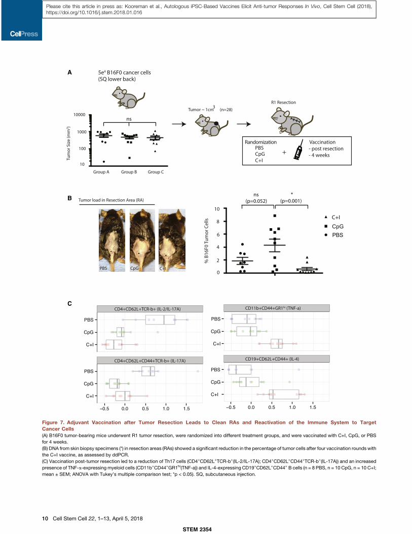
Figure 7. Adjuvant Vaccination after Tumor Resection Leads to Clean RAs and Reactivation of the Immune System to TargetCancer Cells(A) B16F0 tumor-bearing mice underwent R1 tumor resection, were randomized into different treatment groups, and were vaccinated with C+I, CpG, or PBS
for 4 weeks.
(B) DNA from skin biopsy specimens (*) in resection areas (RAs) showed a significant reduction in the percentage of tumor cells after four vaccination rounds with
the C+I vaccine, as assessed by ddPCR.
(C) Vaccination post-tumor resection led to a reduction of Th17 cells (CD4+CD62L+TCR-b+(IL-2/IL-17A); CD4+CD62L+CD44+TCR-b+(IL-17A)) and an increased
presence of TNF-a-expressing myeloid cells (CD11b+CD44+GR1hi(TNF-a)) and IL-4-expressing CD19+CD62L+CD44+ B cells (n = 8 PBS, n = 10 CpG, n = 10 C+I;
mean ± SEM; ANOVA with Tukey’s multiple comparison test; *p < 0.05). SQ, subcutaneous injection.
STEM 2354
10 Cell Stem Cell 22, 1–13, April 5, 2018
Please cite this article in press as: Kooreman et al., Autologous iPSC-Based Vaccines Elicit Anti-tumor Responses In Vivo, Cell Stem Cell (2018),https://doi.org/10.1016/j.stem.2018.01.016

As a therapy for established melanomas, the C+I vaccine wasnot effective in reducing tumor growth, which is likely due to anestablished immunosuppressive tumor microenvironment thatcould potentially be remedied by combining the C+I vaccinewith checkpoint blockade treatment (Le et al., 2015). However,as an adjuvant therapy after R1 resection ofmelanoma, we foundthat the C+I vaccine reactivated the immune system in rejectingremnant melanoma cells by the systemic upregulation of IL-4-expressing B cells and TNF-a-expressing CD11b+GR1hi myeloidcells, as well as a reduction of tumor-promoting Th17 cells. Inthis setting, the cancer epitope heterogeneity of iPSCs, com-bined with the ease of their generation, may make this therapyreadily available as adjuvant immunotherapy for multiple cancertypes within weeks after diagnosis.This last point is crucial for immunotherapy, because it is
commonly known that that the tumor microenvironment couldlimit effectiveness of tumor immunity by suppressive immunecells residing within the tumor. After debulking of thetumor and disrupting the tumor microenvironment to create an‘‘inflamed’’ tumor site, immunotherapy should be more effective(Gajewski et al., 2013). This is demonstrated in our R1 resectedmelanoma model, which again emphasizes the need for a multi-TSA- and TAA-based vaccine to be readily available at time oftumor resection. Having a surrogate whole-cell vaccine withmultiple known (and likely unknown) TSAs and TAAs availableat such a short time after diagnosis would allow the priming ofthe immune system to target large numbers of cancer-specificantigens at a time when cancer cells are most vulnerable.Even though an overlap was seen in murine and human TAA
genes, it is important to note the differences in murine andhuman immunology before extrapolating the above-mentioneddata to humans (Mestas and Hughes, 2004). Further testing ofthe C+I vaccine on human samples ex vivo should therefore beperformed to show efficacy in humans.Taken together, our data show the feasibility of creating broad
tumor immunity against multiple cancer types using an iPSC-based vaccine that presents the immune systemwith large quan-tities of tumor antigens. Compared to current immunotherapystrategies, our iPSC vaccine is capable of reactivating theimmune system to target established cancers without therapy-associatedadverse effects and canbecreatedwithin a fewweeksafter diagnosis. These beneficial properties make this iPSC vac-cine a potential option for personalized adjuvant immunotherapyshortly after conventional primary treatment of cancer.
STAR+METHODS
Detailed methods are provided in the online version of this paperand include the following:
d KEY RESOURCES TABLEd CONTACT FOR REAGENTS AND RESOURCE SHARINGd EXPERIMENTAL MODEL AND SUBJECT DETAILS
B Animal modelsB Generation of murine iPSCs from fibroblastsB Cancer cell lines and implantation
d METHOD DETAILSB CpG + iPSC vaccine preparation and immunizationB Mixed lymphocyte reaction (MLR)
B IgG binding assayB Histopathology of explanted organsB Isolation of inflammatory cells and serum from blood,
spleen, tumor, and dLNsB Staining of inflammatory cells for FACS analysisB Teratoma formationB Generation of tumor lysateB Luminex multiplex cytokine assayB ELISPOT assayB Adoptive transfer of splenocytes and T cellsB Orthotopic tumor modelB Isolation of TELs from draining lymph nodes.B Anti-nuclear antibody (ANA) ELISAB Cytometry by Time of Flight (CyTOF)B PCR detection of the large genomic deletion in Cdkn2aB T cell Receptor (TCR) sequencing
d QUANTIFICATION AND STATISTICAL ANALYSESB Cluster identification, characterization and regression
(Citrus)B Quantification of tumor load for melanoma by digital
droplet PCR (ddPCR)B Analysis of RNA-sequencing dataB Analysis of murine RNA sequencing data
d DATA AND SOFTWARE AVAILABILITY
SUPPLEMENTAL INFORMATION
Supplemental Information includes seven figures and can be found with this
article online at https://doi.org/10.1016/j.stem.2018.01.016.
ACKNOWLEDGMENTS
We thank Dr. A. Connolly for the histopathology analysis of the hearts and
kidneys, G. Busque for assistance with the caliper measurements of the
tumors, and S. Carree and S. Limb for assistance with the graphic design of
the cartoons. We also thank J. Churko for providing the RNA-sequencing
data from the different human iPSC clones. This work was supported in part
by Korean R&D grant HI14C3417 (Y.K.), California Institute of Regenerative
Medicine (CIRM) grants DR2A-05394 and RT3-07798, and NIH grants
R24 HL117756, R01 HL113006, R01 HL133272 (J.C.W.), and U19
AI057229 (M.M.D.).
AUTHOR CONTRIBUTIONS
N.G.K. developed the experimental design, performed and interpreted
experiments, and wrote the manuscript. Y.K. performed and interpreted
experiments. P.E.d.A. performed preliminary experiments and assisted
with the experimental design. V.T. developed the digital droplet PCR (ddPCR)
primers and performed the ddPCR. S.D. assisted with iPSC generation.
N.-Y.S. performed biostatistical analyses on the RNA-sequencing data.
T.-T.W. established the orthotopic breast cancer model and performed the
adoptive transfer of splenocytes. H.Y. assisted with iPSC culture. D.D. assis-
ted with the preliminary in vitro experiments. R.N. and T.P.B. assisted with the
vaccinations, tumor measurements, harvest, and processing of tissues for
downstream analyses. D.T.P assisted with splenocyte isolation for adoptive
transfer and analyzing the RNA-sequencing data. I.B. assisted with the optimi-
zation of the vaccination schedule. A.H. provided experimental advice with the
T cell receptor sequencing. P.H.A.Q., J.F.H., R.L., andM.M.D. provided exper-
imental advice and manuscript writing. J.C.W. provided experimental advice
and design, manuscript writing, and funding support.
DECLARATION OF INTERESTS
The authors declare no competing interests.
STEM 2354
Cell Stem Cell 22, 1–13, April 5, 2018 11
Please cite this article in press as: Kooreman et al., Autologous iPSC-Based Vaccines Elicit Anti-tumor Responses In Vivo, Cell Stem Cell (2018),https://doi.org/10.1016/j.stem.2018.01.016
Received: September 8, 2016
Revised: August 15, 2017
Accepted: January 19, 2018
Published: February 15, 2018
REFERENCES
Anders, S., and Huber,W. (2010). Differential expression analysis for sequence
count data. Genome Biol. 11, R106.
Anders, S., McCarthy, D.J., Chen, Y., Okoniewski, M., Smyth, G.K., Huber, W.,
and Robinson, M.D. (2013). Count-based differential expression analysis of
RNA sequencing data using R and Bioconductor. Nat. Protoc. 8, 1765–1786.
Anders, S., Pyl, P.T., and Huber, W. (2015). HTSeq—a Python framework to
work with high-throughput sequencing data. Bioinformatics 31, 166–169.
Ben-Porath, I., Thomson, M.W., Carey, V.J., Ge, R., Bell, G.W., Regev, A., and
Weinberg, R.A. (2008). An embryonic stem cell-like gene expression signature
in poorly differentiated aggressive human tumors. Nat. Genet. 40, 499–507.
Bock, C., Kiskinis, E., Verstappen, G., Gu, H., Boulting, G., Smith, Z.D., Ziller,
M., Croft, G.F., Amoroso, M.W., Oakley, D.H., et al. (2011). Reference Maps of
human ES and iPS cell variation enable high-throughput characterization of
pluripotent cell lines. Cell 144, 439–452.
Brewer, B.G., Mitchell, R.A., Harandi, A., and Eaton, J.W. (2009). Embryonic
vaccines against cancer: an early history. Exp. Mol. Pathol. 86, 192–197.
Bruggner, R.V., Bodenmiller, B., Dill, D.L., Tibshirani, R.J., and Nolan, G.P.
(2014). Automated identification of stratifying signatures in cellular subpopula-
tions. Proc. Natl. Acad. Sci. USA 111, E2770–E2777.
Chang, G., Gao, S., Hou, X., Xu, Z., Liu, Y., Kang, L., Tao, Y., Liu, W., Huang, B.,
Kou, X., et al. (2014). High-throughput sequencing reveals the disruption of
methylation of imprinted gene in induced pluripotent stem cells. Cell Res.
24, 293–306.
Chen, J.Y., Tang, Y.A., Huang, S.M., Juan, H.F., Wu, L.W., Sun, Y.C., Wang,
S.C., Wu, K.W., Balraj, G., Chang, T.T., et al. (2011). A novel sialyltransferase
inhibitor suppresses FAK/paxillin signaling and cancer angiogenesis and
metastasis pathways. Cancer Res. 71, 473–483.
Chung, A.S., Wu, X., Zhuang, G., Ngu, H., Kasman, I., Zhang, J., Vernes, J.M.,
Jiang, Z., Meng, Y.G., Peale, F.V., et al. (2013). An interleukin-17-mediated
paracrine network promotes tumor resistance to anti-angiogenic therapy.
Nat. Med. 19, 1114–1123.
Churko, J., Lee, J., Ameen, M., Gu, M., Venkaasubramanian, M., Diecke, S.,
Sallam, K., Im, H., Wang, G., Gold, J.D., et al. (2017). Transcriptomic and
epigenomic differences in human induced pluripotent stem cells generated
from six reprogramming methods. Nat. Biomed. Eng. 1, 826–837.
Consortium, E.P.; ENCODE Project Consortium (2011). A user’s guide to the
encyclopedia of DNA elements (ENCODE). PLoS Biol. 9, e1001046.
de Almeida, P.E., Meyer, E.H., Kooreman, N.G., Diecke, S., Dey, D., Sanchez-
Freire, V., Hu, S., Ebert, A., Odegaard, J., Mordwinkin, N.M., et al. (2014).
Transplanted terminally differentiated induced pluripotent stem cells are
accepted by immune mechanisms similar to self-tolerance. Nat. Commun.
5, 3903.
Diecke, S., Lu, J., Lee, J., Termglinchan, V., Kooreman, N.G., Burridge, P.W.,
Ebert, A.D., Churko, J.M., Sharma, A., Kay, M.A., and Wu, J.C. (2015).
Novel codon-optimized mini-intronic plasmid for efficient, inexpensive, and
xeno-free induction of pluripotency. Sci. Rep. 5, 8081.
Dolcetti, L., Peranzoni, E., Ugel, S., Marigo, I., Fernandez Gomez, A., Mesa, C.,
Geilich, M., Winkels, G., Traggiai, E., Casati, A., et al. (2010). Hierarchy of
immunosuppressive strength among myeloid-derived suppressor cell subsets
is determined by GM-CSF. Eur. J. Immunol. 40, 22–35.
Dooms, H., and Abbas, A.K. (2002). Life and death in effector T cells. Nat.
Immunol. 3, 797–798.
Evans, M.S., Chaurette, J.P., Adams, S.T., Jr., Reddy, G.R., Paley, M.A.,
Aronin, N., Prescher, J.A., and Miller, S.C. (2014). A synthetic luciferin
improves bioluminescence imaging in live mice. Nat. Methods 11, 393–395.
Gajewski, T.F., Schreiber, H., and Fu, Y.X. (2013). Innate and adaptive immune
cells in the tumor microenvironment. Nat. Immunol. 14, 1014–1022.
Galvan, D.L., O’Neil, R.T., Foster, A.E., Huye, L., Bear, A., Rooney, C.M., and
Wilson, M.H. (2015). Anti-tumor effects after adoptive transfer of IL-12
transposon-modified murine splenocytes in the OT-I-melanoma mouse
Model. PLoS ONE 10, e0140744.
Ghosh, Z., Huang, M., Hu, S., Wilson, K.D., Dey, D., and Wu, J.C. (2011).
Dissecting the oncogenic and tumorigenic potential of differentiated human
induced pluripotent stem cells and human embryonic stem cells. Cancer
Res. 71, 5030–5039.
Gilkeson, G.S., Conover, J., Halpern, M., Pisetsky, D.S., Feagin, A., and
Klinman, D.M. (1998). Effects of bacterial DNA on cytokine production by
(NZB/NZW)F1 mice. J. Immunol. 161, 3890–3895.
Goldstein, M.J., Varghese, B., Brody, J.D., Rajapaksa, R., Kohrt, H.,
Czerwinski, D.K., Levy, S., and Levy, R. (2011). A CpG-loaded tumor cell
vaccine induces antitumor CD4+ T cells that are effective in adoptive therapy
for large and established tumors. Blood 117, 118–127.
Grupp, S.A., Kalos, M., Barrett, D., Aplenc, R., Porter, D.L., Rheingold, S.R.,
Teachey, D.T., Chew, A., Hauck, B., Wright, J.F., et al. (2013). Chimeric antigen
receptor-modified T cells for acute lymphoid leukemia. N. Engl. J. Med. 368,
1509–1518.
Harrow, J., Frankish, A., Gonzalez, J.M., Tapanari, E., Diekhans, M.,
Kokocinski, F., Aken, B.L., Barrell, D., Zadissa, A., Searle, S., et al. (2012).
GENCODE: the reference human genome annotation for The ENCODE
Project. Genome Res. 22, 1760–1774.
He, D., Li, H., Yusuf, N., Elmets, C.A., Li, J., Mountz, J.D., and Xu, H. (2010).
IL-17 promotes tumor development through the induction of tumor promoting
microenvironments at tumor sites and myeloid-derived suppressor cells.
J. Immunol. 184, 2281–2288.
Kim, D., Langmead, B., and Salzberg, S.L. (2015). HISAT: a fast spliced aligner
with low memory requirements. Nat. Methods 12, 357–360.
Kocat€urk, B., and Versteeg, H.H. (2015). Orthotopic injection of breast cancer
cells into the mammary fat pad of mice to study tumor growth. J. Vis. Exp.
96, 51967.
Kooreman, N.G., andWu, J.C. (2010). Tumorigenicity of pluripotent stem cells:
biological insights from molecular imaging. J. R. Soc. Interface 7 (Suppl 6 ),
S753–S763.
Le, D.T., Uram, J.N., Wang, H., Bartlett, B.R., Kemberling, H., Eyring, A.D.,
Skora, A.D., Luber, B.S., Azad, N.S., Laheru, D., et al. (2015). PD-1 blockade
in tumors with mismatch-repair deficiency. N. Engl. J. Med. 372, 2509–2520.
Lee, A.S., Tang, C., Rao, M.S., Weissman, I.L., and Wu, J.C. (2013).
Tumorigenicity as a clinical hurdle for pluripotent stem cell therapies.
Nat. Med. 19, 998–1004.
Lee, D.W., Kochenderfer, J.N., Stetler-Stevenson, M., Cui, Y.K., Delbrook, C.,
Feldman, S.A., Fry, T.J., Orentas, R., Sabatino, M., Shah, N.N., et al. (2015).
T cells expressing CD19 chimeric antigen receptors for acute lymphoblastic
leukaemia in children and young adults: a phase 1 dose-escalation trial.
Lancet 385, 517–528.
Li, Y., Zeng, H., Xu, R.H., Liu, B., and Li, Z. (2009). Vaccination with human
pluripotent stem cells generates a broad spectrum of immunological and clin-
ical responses against colon cancer. Stem Cells 27, 3103–3111.
Mallon, B.S., Chenoweth, J.G., Johnson, K.R., Hamilton, R.S., Tesar, P.J.,
Yavatkar, A.S., Tyson, L.J., Park, K., Chen, K.G., Fann, Y.C., and McKay,
R.D. (2013). StemCellDB: the human pluripotent stem cell database at the
National Institutes of Health. Stem Cell Res. (Amst.) 10, 57–66.
Mallon, B.S., Hamilton, R.S., Kozhich, O.A., Johnson, K.R., Fann, Y.C., Rao,
M.S., and Robey, P.G. (2014). Comparison of the molecular profiles of human
embryonic and induced pluripotent stem cells of isogenic origin. Stem Cell
Res. (Amst.) 12, 376–386.
Maude, S.L., Frey, N., Shaw, P.A., Aplenc, R., Barrett, D.M., Bunin, N.J., Chew,
A., Gonzalez, V.E., Zheng, Z., Lacey, S.F., et al. (2014). Chimeric antigen
receptor T cells for sustained remissions in leukemia. N. Engl. J. Med. 371,
1507–1517.
Maus, M.V., Grupp, S.A., Porter, D.L., and June, C.H. (2014). Antibody-
modified T cells: CARs take the front seat for hematologic malignancies.
Blood 123, 2625–2635.
STEM 2354
12 Cell Stem Cell 22, 1–13, April 5, 2018
Please cite this article in press as: Kooreman et al., Autologous iPSC-Based Vaccines Elicit Anti-tumor Responses In Vivo, Cell Stem Cell (2018),https://doi.org/10.1016/j.stem.2018.01.016
Mestas, J., and Hughes, C.C. (2004). Of mice and not men: differences
between mouse and human immunology. J. Immunol. 172, 2731–2738.
Mor, G., Singla, M., Steinberg, A.D., Hoffman, S.L., Okuda, K., and Klinman,
D.M. (1997). Do DNA vaccines induce autoimmune disease? Hum. Gene
Ther. 8, 293–300.
Morgan, R.A., Yang, J.C., Kitano, M., Dudley, M.E., Laurencot, C.M., and
Rosenberg, S.A. (2010). Case report of a serious adverse event following
the administration of T cells transduced with a chimeric antigen receptor
recognizing ERBB2. Mol. Ther. 18, 843–851.
Mukherjee, P., Pathangey, L.B., Bradley, J.B., Tinder, T.L., Basu, G.D.,
Akporiaye, E.T., and Gendler, S.J. (2007). MUC1-specific immune therapy
generates a strong anti-tumor response in a MUC1-tolerant colon cancer
model. Vaccine 25, 1607–1618.
Naas, T., Ghorbani, M., Soare, C., Scherling, N., Muller, R., Ghorbani, P., and
Diaz-Mitoma, F. (2010). Adoptive transfer of splenocytes to study cell-
mediated immune responses in hepatitis C infection using HCV transgenic
mice. Comp. Hepatol. 9, 7.
Nelakanti, R.V., Kooreman, N.G., and Wu, J.C. (2015). Teratoma formation: a
tool for monitoring pluripotency in stem cell research. Curr. Protoc. Stem Cell
Biol. 32, 4A.8.1–4A.8.17.
Numasaki, M., Fukushi, J., Ono, M., Narula, S.K., Zavodny, P.J., Kudo, T.,
Robbins, P.D., Tahara, H., and Lotze, M.T. (2003). Interleukin-17 promotes
angiogenesis and tumor growth. Blood 101, 2620–2627.
Palena, C., Abrams, S.I., Schlom, J., and Hodge, J.W. (2006). Cancer vaccines:
preclinical studies and novel strategies. Adv. Cancer Res. 95, 115–145.
Qiu, P., Simonds, E.F., Bendall, S.C., Gibbs, K.D., Jr., Bruggner, R.V.,
Linderman, M.D., Sachs, K., Nolan, G.P., and Plevritis, S.K. (2011).
Extracting a cellular hierarchy from high-dimensional cytometry data with
SPADE. Nat. Biotechnol. 29, 886–891.
Sodhi, A., Tandon, P., and Sarna, S. (1985). Adoptive transfer of immunity
against solid fibrosarcoma in mice with splenocytes and peritoneal
exudate cells obtained after in vitro sensitization and in vivo immunization
with cis-dichlorodiamine platinum(II) treated fibrosarcoma cells. Arch.
Geschwulstforsch. 55, 47–61.
Soldner, F., Hockemeyer, D., Beard,C., Gao,Q., Bell, G.W.,Cook, E.G., Hargus,
G., Blak, A., Cooper, O., Mitalipova, M., et al. (2009). Parkinson’s disease
patient-derived induced pluripotent stem cells free of viral reprogramming
factors. Cell 136, 964–977.
Takahashi, K., and Yamanaka, S. (2006). Induction of pluripotent stem cells
from mouse embryonic and adult fibroblast cultures by defined factors. Cell
126, 663–676.
Takahashi, K., Tanabe, K., Ohnuki, M., Narita, M., Ichisaka, T., Tomoda, K.,
and Yamanaka, S. (2007). Induction of pluripotent stem cells from adult human
fibroblasts by defined factors. Cell 131, 861–872.
Torcellan, T., Hampton, H.R., Bailey, J., Tomura, M., Brink, R., and Chtanova,
T. (2017). In vivo photolabeling of tumor-infiltrating cells reveals highly regu-
lated egress of T-cell subsets from tumors. Proc. Natl. Acad. Sci. USA 114,
5677–5682.
Yaddanapudi, K., Mitchell, R.A., Putty, K., Willer, S., Sharma, R.K., Yan, J.,
Bodduluri, H., and Eaton, J.W. (2012). Vaccination with embryonic stem cells
protects against lung cancer: is a broad-spectrum prophylactic vaccine
against cancer possible? PLoS ONE 7, e42289.
Zhao, T., Zhang, Z.N., Rong, Z., and Xu, Y. (2011). Immunogenicity of induced
pluripotent stem cells. Nature 474, 212–215.
Zou, W. (2005). Immunosuppressive networks in the tumour environment and
their therapeutic relevance. Nat. Rev. Cancer 5, 263–274.
STEM 2354
Cell Stem Cell 22, 1–13, April 5, 2018 13
Please cite this article in press as: Kooreman et al., Autologous iPSC-Based Vaccines Elicit Anti-tumor Responses In Vivo, Cell Stem Cell (2018),https://doi.org/10.1016/j.stem.2018.01.016

STAR+METHODS
KEY RESOURCES TABLE
REAGENT or RESOURCE SOURCE IDENTIFIER
Antibodies
PE/Cy7 anti-mouse CD3 eBioscience 25-0031-82; RRID: AB_469572
APC anti-mouse CD4 BD Bioscience 553051
PE anti-mouse CD4 eBioscience 12-0042-83; RRID: AB_465510
PerCP/Cy5.5 anti-mouse CD8 Biolegend 100734; RRID: AB_2075238
PE/Cy7 anti-mouse CD11b Biolegend 101216; RRID: AB_312799
Pacific Blue anti-mouse/human CD44 Biolegend 103020; RRID: AB_493683
APC anti-mouse CD11c Biolegend 117310; RRID: AB_313779
FITC anti-mouse CD86 BD Bioscience 553691
eFluor 450 anti-mouse F4/80 eBioscience 48-4801-82; RRID: AB_1548747
Alexa Fluor 488 anti-mouse FoxP3 Biolegend 126406; RRID: AB_1089113
PE anti-mouse Granzyme-B eBioscience 12-8898-82; RRID: AB_10870787
Brilliant Violet 510 anti-mouse CD45 eBioscience 103137; RRID: AB_2561392
Alexa Fluor 700 anti-mouse CD25 eBioscience 56-0251-82; RRID: AB_891422
eFluor 780 Fixable Viability Dye eBioscience 65-0865-14
Alexa Fluor 700 anti-mouse GR-1 Ly6 Biolegend 108422; RRID: AB_2137487
PerCP/Cy5.5 anti-mouse NK1.1 Biolegend 108728; RRID: AB_2132705
PerCP/Cy5.5 anti-mouse DX-5 Biolegend 108916; RRID: AB_2129358
PE anti-mouse MHC-II eBioscience 12-5321-81; RRID: AB_465928
Alexa Fluor 488 anti-mouse IgG Thermo Fisher Scientific A-11001; RRID: AB_2534069
Anti-rabbit IgG Cross-adsorbed Thermo Fisher Scientific 31213; RRID: AB_228376
Pacific Blue Rat IgG2b k isotype control Biolegend 400627
Alexa Fluor 488 Rat IgG2b k isotype control Biolegend 400625
PE Rat IgG2b k isotype control Thermo-Fisher Scientific 12-4031-82; RRID: AB_470042
FITC Rat IgG2a k isotype control BD Biosciences 553929
PE Rat IgG2a k isotype control eBioscience 12-4321-80; RRID: AB_1834380
Alexa Fluor 700 Rat IgG1 k isotype control eBioscience 56-4301-80; RRID: AB_494017
Anti SSEA-1 microbeads Miltenyi Biotec 130-094-530
Oct 3/4 anti-mouse Santa Cruz Biotechnology sc-5279
c-Myc anti-mouse EMD Millipore 06-340
SSEA-1 anti-mouse EMD Millipore MAB4301
Nanog anti-mouse Santa Cruz Biotechnology sc-33760
Sox2 anti-mouse Santa Cruz Biotechnology sc-365823
Mouse on Mouse (M.O.M.) Basic Kit Vector Laboratories BMK-2202
Fc Block anti-mouse BD Biosciences 553141
Chemicals, Peptides, and Recombinant Proteins
DMEM, high glucose, GlutaMAX GIBCO 10569-010
Fetal Bovine Serum Life Technologies 26140079
DPBS, no calcium, no magnesium GIBCO 14190250
MEM Non-Essential Amino Acids GIBCO 11140050
TrypLE Express GIBCO 12605-036
CpG ODN 1826 Invivogen tlrl-1826-1
Murine Leukemia Inhibitory Factor (mLIF) EMD Millipore ESG1106
Gelatin Sigma Aldrich G1393-100ML
Isothesia Henry Schein 029405
(Continued on next page)
STEM 2354
e1 Cell Stem Cell 22, 1–13.e1–e7, April 5, 2018
Please cite this article in press as: Kooreman et al., Autologous iPSC-Based Vaccines Elicit Anti-tumor Responses In Vivo, Cell Stem Cell (2018),https://doi.org/10.1016/j.stem.2018.01.016

Continued
REAGENT or RESOURCE SOURCE IDENTIFIER
Matrigel matrix growth factor reduced BD Biosciences 356231
RPMI medium 1640 Life Technologies 11875-119
ACK lysis buffer Quality Biological 118-156-101
Collagenase D Roche 11088882001
Deoxyribonuclease I from bovine pancreas Sigma Aldrich D4263-5VL
Trypsin Inhibitor Sigma Aldrich T9003-1G
HEPES 1M Sigma Aldrich H3662
Percoll GE Density Gradient Media VWR 89428-524
UltraPure 0.5M EDTA Life Technologies 15575-020
PrimeStar GXL DNA Polymerase Clontech R050A
ddPCR Supermix for Probes Bio-Rad 1863024
Critical Commercial Assays
Neon Transfection System 100 ml Kit Thermo Fisher Scientific MPK10025
MycoAlert Detection Kit Lonza LT07-318
MycoAlert Assay Control Set Lonza LT07-518
CFSE Cell Trace staining Thermo Fisher Scientific C34554
Mouse Th1/Th2/Th17 Cytokine Multi-Analyte ELISArray QIAGEN MEM-003A
MaxPar Mouse Spleen/Lymph Node Phenotyping kit Fluidigm 201306
MaxPar Mouse Intracellular Cytokine I Panel kit Fluidigm 201310
Cisplatin viability dye Fluidigm 201062
Cytofix/Cytoperm Permeabilization Solution kit BD Biosciences BDB554714
Pierce BCA Protein Assay Kit Thermo Fisher Scientific 23225
Mouse IFN-gamma/Granzyme B Dual-Color ELISpot Kit R&D Systems ELD5819
Pan T cell Isolation Kit II, mouse Miltenyi Biotec 130-095-130
Anti-nuclear Antigen (IgG) mouse ELISA Kit Antibodies-Online ABIN366290
DNeasy Blood & Tissue kit QIAGEN 69504
Deposited Data
TCRb sequencing tumor-infiltrating lymphocytes This paper immunoACCESS: DOI: 10.21417/B7B648
RNA-seq from human somatic/cancer cell lines ENCODE project https://www.genome.gov/encode/
RNA-seq from human iPS cells Churko et al., 2017 N/A
RNA-seq form murine somatic cell lines ENCODE project http://mouseencode.org/
RNA-seq from murine iPS cells Chang et al., 2014 N/A
Experimental Models: Cell Lines
EmbryoMax Primary Mouse Embryo Fibroblasts EMD Millipore PMEF-N
DB7 breast cancer cells The University of Utah;
Dr. Joe Smith
N/A
B16F0 melanoma cells ATCC CRL-6322
AC29 mesothelioma cells Sigma Aldrich 10092308
Experimental Models: Organisms/Strains
Mouse: FVB/NJ The Jackson Laboratory 001800
Mouse: C57BL/6J The Jackson Laboratory 000664
Mouse: CBA/J The Jackson Laboratory 000656
NOD-SCID IL2Rgammanull (NSG) The Jackson Laboratory 005557
Oligonucleotides
TaqMan Copy Number TFRC probe (Mm00000692_cn) Thermo Fisher Scientific 4400291
50ACTAGCCAGAGGATCTTAAAGACT30 This paper N/A
50GCCATCACTGGAAAGAGAGGC30 This paper N/A
50(HEX)CCTGCCCACCCACTCCCCCTTTTT(Blackhole Quencher)30
This paper N/A
(Continued on next page)
STEM 2354
Cell Stem Cell 22, 1–13.e1–e7, April 5, 2018 e2
Please cite this article in press as: Kooreman et al., Autologous iPSC-Based Vaccines Elicit Anti-tumor Responses In Vivo, Cell Stem Cell (2018),https://doi.org/10.1016/j.stem.2018.01.016

CONTACT FOR REAGENTS AND RESOURCE SHARING
Further information and requests for resources and reagents should be directed to andwill be fulfilled by the Lead Contact, JosephC.Wu ([email protected]).
EXPERIMENTAL MODEL AND SUBJECT DETAILS
Animal modelsYoung adult female FVB, C57BL/6J, and CBA/J mice (6-8 weeks old) were used. Animals were randomly assigned to the differenttreatment groups. Tumor-bearing mice were excluded from the experiment if their physical condition required euthanasia beforethe experimental deadline, due to criteria such as tumor sizes exceeding 1 cm3, visible distress, pain, or illness. All experimentswere approved by the Stanford University Administrative Panel of Laboratory Animal Care (APLAC).
Generation of murine iPSCs from fibroblastsFibroblasts from FVB, C57BL/6J, and CBA/J mice (The Jackson Laboratory, Bar Harbor, Maine) were grown in DMEM Glutamax(ThermoFisher Scientific, Waltham, MA, USA) with 20% fetal bovine serum (FBS) and 1x NEAA (ThermoFisher Scientific). Fibroblastswere dissociated using TrypLE Express (ThermoFisher Scientific) and 1x10⁶ fibroblasts were resuspended in electroporation buffer(Neon system, ThermoFisher Scientific). Cells were transfected with a codon-optimized mini-intronic plasmid (coMIP) containing thefour reprogramming factors Oct-4, Sox-2, c-Myc, and Klf4 (Diecke et al., 2015). After transfection, cells were plated on irradiatedmouse embryonic feeder (MEF) cells and cultured in DMEMwith 15% FBS, 1x NEAA, and 10 ng/ml murine leukemia inhibiting factor(mLIF; EMD Millipore, MA, USA). After iPSC colonies started to appear, they were manually picked and transferred to a fresh feederlayer. The iPSC colonies were grown for a few passages and then transferred to 0.2% gelatin-coated plates to be sorted for SSEA-1usingmagnetic bead sorting (Miltenyi, Germany) to keep a pure undifferentiated population. For characterization, iPSCswere stainedfor Oct4, Nanog, Sox2 (Santa Cruz, CA, USA), SSEA1, and c-Myc (EMD Millipore) to assess pluripotency. In addition, a teratomaassay was performed on all iPSC lines by transplantation of 1x10⁶ iPSCs in the hindlimb of NOD-SCID mice (The Jackson Labora-tory). All cell lines were tested for mycoplasma contamination and found to be negative.
Cancer cell lines and implantationThe breast cancer line DB7 was a gift from Dr. Joe Smith (University of Utah, USA). It was derived from FVB mice and is a non-metastatic cell line. The B16F0 melanoma cell line was purchased from ATCC (Manassas, VA, USA) and is syngeneic to C57BL/6mice. It has low-grade lymphoid metastatic potential to the lungs. The AC29 mesothelioma cancer line was purchased fromSigma-Aldrich (St. Louis, MO, USA). The cancer lines were grown in DMEM, 10% FBS under normal culture conditions. For theC57BL/6 and FVB mice, 5x10⁴ cancer cells were resuspended in 100 mL PBS and injected subcutaneously in the lower back ofthe mice. The CBA/J mice were injected with 2x10⁶ cancer cells. Tumor growth was assessed weekly by caliper measurement.At the end of the study, tumors were explanted and gross examination of draining lymph nodes and lung tissue was performedfor any metastases.
Continued
REAGENT or RESOURCE SOURCE IDENTIFIER
Recombinant DNA
Codon-optimized mini-intronic plasmid (coMIP) Diecke et al., 2015 N/A
Software and Algorithms
ImmunoSeq Analyzer Adaptive Biotechnologies http://clients.adaptivebiotech.com/
Prism GraphPad 7 GraphPad Software https://www.graphpad.com/scientific-
software/prism/
Cytobank Cytobank https://stanford.cytobank.org/cytobank/login
Citrus 0.8 Nolan lab; Stanford, USA. https://github.com/nolanlab/citrus/wiki/
Installing-Citrus
R; version 3.0.3 The R Project https://www.r-project.org/
Adobe Photoshop CS6 Adobe http://www.adobe.com/nl/products/
photoshop.html
Other
29 Gauge x +½’’ needle 3/10cc Terumo Medical SS30M2913
Multiplex-Luminex platform (LabMap200 System) HIMC; Stanford
University, USA
N/A
STEM 2354
e3 Cell Stem Cell 22, 1–13.e1–e7, April 5, 2018
Please cite this article in press as: Kooreman et al., Autologous iPSC-Based Vaccines Elicit Anti-tumor Responses In Vivo, Cell Stem Cell (2018),https://doi.org/10.1016/j.stem.2018.01.016

METHOD DETAILS
CpG + iPSC vaccine preparation and immunizationFor each mouse, 2x10⁶ SSEA-1-sorted syngeneic murine iPSCs were irradiated at 6,000 rads prior to injection. Cells weresuspended in 100 mL of 5 mM CpG (Invivogen, San Diego, USA), dissolved in PBS, and loaded into 1/4 cc insulin syringes(Terumo). Mice were placed in an induction chamber and anesthetized with 2% isoflurane (Isothesia, Butler Schein) in 100%oxygen with a delivery rate of 2cl/min until the loss of righting reflex, as per APLAC guidelines at Stanford University. Immunizationwas performed by subcutaneous injection of the vaccine in the flanks of the mice, with the injection site changing every week.Mice were monitored weekly for early signs of auto-reactivity to the vaccine by weight measurements and gross examinationof overall appearance. Vaccination preparation and dosage were the same for the prophylactic and adjuvant treatment experi-ments. The prophylactic vaccination studies were replicated several times in the same mouse strain, different mouse strainsand by different investigators. The investigator analyzing the tumor sizes and data from the adjuvant treatment experiment wasblinded for the different treatment groups.
Mixed lymphocyte reaction (MLR)Spleens were isolated, minced, and filtered through a 70 mm strainer. After multiple washes with glucose-containing RPMI, the pelletwas resuspended in ACK lysis buffer for removal of red blood cells. CFSE-labeled (ThermoFisher Scientific) splenocytes from C+Ivaccinated mice were then plated at a density of 1x10⁵ cells per 100 ul in a 96-well plate and incubated for 72 hr with another100 mL solution of DB7 tumor lysate, ranging from 1-10 mg. After 72 hr, the plate was spun down and the supernatant isolatedfor cytokine analysis using the mouse Th1/Th2/Th17 Cytokines Multi-Analyte ELISArray kit (QIAGEN, Hilden, Germany), and thecell pellet was analyzed with the LSR-II Flow Cytometer to assess T cell proliferation.
IgG binding assayCells were washed multiple times with PBS and resuspended in 100 mL FACS buffer with the addition of 2 mL of serum from thevaccinated mice and incubated for 30 minutes on 4!C. Following this, cells were washed multiple times and incubated with ananti-IgG FITC secondary antibody (ThermoFisher Scientific) for another 20 min on 4!C. As an isotype control an IgG antibody,pre-adsorbed for murine IgG and IgM, was included. The cells were then analyzed using the LSR-II Flow Cytometer.
Histopathology of explanted organsAt time of sacrifice, the heart and kidneys were explanted from vaccinatedmice and processed for histopathology. Briefly, the organswere fixed overnight in 4% paraformaldehyde and transferred to 70% ethanol for 24 hr. Fixed samples were embedded in paraffinand 5 mm sections were cut and stained with hematoxylin and eosin (H&E) for histological analysis by a pathologist.
Isolation of inflammatory cells and serum from blood, spleen, tumor, and dLNsFVB, C57BL/6, and CBA/J experimental mice were sacrificed at 4, 2, and 1 week(s), respectively, after tumor inoculation. Tissueswere isolated from themice and placed in a digestion buffer containing RPMI, FBS, collagenase, DNase, trypsin inhibitor and HEPES,then minced and placed in a shaker at 37!C for 45 min. Samples were than filtered through a 70 mm strainer, spun down, andresuspended in ACK lysis buffer to remove any red blood cells. After lysis, the cell suspension was washed with PBS and usedfor subsequent analyses. Additionally, dissociated tumors were passed through a Percoll gradient to remove non-immune cellsand isolate tumor-infiltrating leukocytes (TILs). Blood was collected in two separate tubes per mouse for PBMC (EDTA containingtube) and serum isolation (uncoated tube).
Staining of inflammatory cells for FACS analysisInflammatory cells isolated from blood and tissues were resuspended in 200 mL FACS buffer (DPBS, 2% FBS and 200 mM EDTA),blocked with a FcR-blocking Reagent (BD PharMingen, San Diego, CA, USA), and divided into 2 tubes. One tube was stainedwith a surface marker panel, containing CD3, CD4, CD25 (eBioscience), CD8a, CD44, CD45 (Biolegend), and the intracellularmarkers Granzyme-B (eBioscience) and FoxP3 (Biolegend). The second tube was stained for F4/80, MHC-II (eBioscience), CD86(BD Biosciences), CD11b, CD11c, NK1.1, Ly6-G, and CD45 (Biolegend). A rat IgG2b k isotype control was included for CD44,FoxP3 (Biolegend), and MHC-II (eBioscience). A rat IgG2a k isotype was included for the Granzyme-B (eBioscience) and CD86(BD Biosciences) staining. For CD25 staining, the IgG1 k isotype (eBioscience) was included. In both panels, the fixable viabilitydye 780 (Invitrogen) was added to exclude dead cells from the analysis. Extracellular staining was performed prior to fixing andpermeabilizing the samples for staining with intracellular markers. Samples were analyzed on the LSR-II Flow Cytometer analyzerin the Beckmann FACS facility (Stanford University).
Teratoma formationTeratoma formation was performed as previously described (Nelakanti et al., 2015), with the exception of site of injection. For thismanuscript, a flank injection was preferred over a hindlimb injection to ensure easier access for over-time measurements ofteratoma size. In brief, 1x10⁶ iPSCs were resuspended in growth factor reduced Matrigel (50 mL per injection) and injected in
STEM 2354
Cell Stem Cell 22, 1–13.e1–e7, April 5, 2018 e4
Please cite this article in press as: Kooreman et al., Autologous iPSC-Based Vaccines Elicit Anti-tumor Responses In Vivo, Cell Stem Cell (2018),https://doi.org/10.1016/j.stem.2018.01.016

the flank of immunodeficient mice (NOD-SCID IL2Rgammanull;NSG). Teratoma sizes at site of injection were measured over timeusing a caliper. After four weeks, mice were sacrificed and the teratomas harvested for final measurements.
Generation of tumor lysate1x107 tumor cells were used from in vitro culture and resuspended in 1 mL of PBS. The cell suspension was frozen to "80!C for45 min and then thawed on 37!C for 30 min. This process was repeated for a total of three times. Afterward, the suspension wasspun down and the supernatant, containing tumor lysate, was isolated for protein concentration measurements using the PierceBCA Protein Assay Kit (ThermoFisher Scientific).
Luminex multiplex cytokine assayProduction of various cytokines was measured in cell culture supernatant and serum samples using a multiplex-Luminex platform(LabMap200 System; Luminex) in conjunction with Panomics antibodies at the Human Immune Monitoring Center at StanfordUniversity.
ELISPOT assaySplenocytes (5x10⁵) were isolated as described above and co-cultured with either iPSC or DB7 lysate (35 mg) for the duration of 37 hr,after which the secretion of granzyme-b and IFN-g was measured by Enzyme-Linked ImmunoSpot (ELISPOT) according tothe manufacturer’s instructions (cat# ELD5819, R&D Systems, Diaclone). Adobe Photoshop CS6 software was used for thecalculation of size and number of IFN-g positive spots.
Adoptive transfer of splenocytes and T cellsC+I vaccinated and vehicle vaccinated mice were sacrificed and their splenocytes isolated, as previously described (Galvan et al.,2015; Naas et al., 2010; Sodhi et al., 1985). In brief, the spleens were digested and passed through a 70 mm strainer. Afterwardred blood cells were lysed with ACK lysis buffer (cat# 118-156-101, Quality biology, INC.) and the remaining splenocytes washedwith PBS. The splenocytes were then dissolved in 200 mL PBS solution and intravenously injected in an orthotopic model ofbreast cancer by tail vein injection. For the adoptive transfer of T cells, the procedure is as described above, with the additionof a magnetic bead sorting using the Pan T cell isolation kit to acquire CD3+ T cells (#130-095-130, Miltenyi, Germany) afterthe final washing step.
Orthotopic tumor modelFVB mice were injected with 2x10⁶ DB7 tumor cells directly into the mammary fat pad tissue, as previously described (Kocat€urk andVersteeg, 2015). The range of cancer cell number was based on previous reports (Chen et al., 2011; Evans et al., 2014) and was set at2x106 DB7 cancer cells after validating the model and achieving a tumor incidence of 100%.
Isolation of TELs from draining lymph nodes.After sacrificing the C+I and vehicle-vaccinated mice, their dLNs were isolated, minced and passed through a 70 mm strainer.After washing the cells with PBS, the T cell portion of the TELs were isolated using the Pan T cell isolation kit to acquire CD3+
T cells (#130-095-130, Miltenyi, Germany).
Anti-nuclear antibody (ANA) ELISAMurine blood was collected from PBS, CpG only, or CpG-iPSCs vaccinated mice and the plasma was separated from the blood viacentrifugation for 15min at 1000 g. The plasma sampleswere diluted at 1:200with sample dilution. The concentrations of anti-nuclearantibodies (IgG) were determined using an ELISA kit, according to the manufacturer’s instructions (Antibodies-Online;antibodies-online.com). For this experiment, four biological replicates per group were used and for each biological replicate threetechnical replicates were included.
Cytometry by Time of Flight (CyTOF)Immune cells were isolated from explanted tissues according to aforementioned methods. Cells were stained with the MouseSpleen/Lymph Node Phenotyping kit, the Mouse Intracellular Cytokine I Panel kit, and the viability dye Cisplatin (Fluidigm, SouthSan Francisco, CA). Cells were resuspended in MaxPar water at a concentration of 1x10⁵-1x10⁸ cells per ml with the addition ofnormalization beads and ran on a CyTOF2 (Fluidigm) machine. Following this, the data were normalized using the normalizationbeads. The data were analyzed using the Cytobank online software for spanning tree progression analysis of density-normalizedevents (SPADE) (Qiu et al., 2011).
PCR detection of the large genomic deletion in Cdkn2aPrimers were designed to detect the junction of the large deletion in Cdkn2a of the B16 melanoma cell line (Figure S7B). Each 25 mlPCR reaction solution contained 1.25 units of PrimeSTAR! GXL DNA Polymerase (Clontech) and 50-100 ng of genomicDNA extracted by DNeasy Blood & Tissue Kit (QIAGEN) (Figure S7C). PCR products were then analyzed by Sanger sequencingand aligned with the gene database in NCBI (Figure S7D).
STEM 2354
e5 Cell Stem Cell 22, 1–13.e1–e7, April 5, 2018
Please cite this article in press as: Kooreman et al., Autologous iPSC-Based Vaccines Elicit Anti-tumor Responses In Vivo, Cell Stem Cell (2018),https://doi.org/10.1016/j.stem.2018.01.016
T cell Receptor (TCR) sequencingThe DNA from the TILs infiltrating the AC29 tumors was isolated using the DNeasy Blood & Tissue kit (QIAGEN). Samples weresubmitted to Adaptive Biotechnologies (Seattle, WA) for a survey level TCR sequencing. The minimum DNA content from thesubmitted samples was 150 ng per sample with DNA quality A260/280 between 1.8 to 2.0. Data analysis as well as assessmentof TCR clonality between samples were performed in collaboration with Adaptive Biotechnologies. In brief, a list of TCR cloneswithin each sample and their frequencies within the DNA sample were provided. For the T cell overlap search, theamino acid sequences of the clones appearing in 4 or 5 of the samples in the two sample groups were compared. Data fromthe CI treatment group and the PBS control group were ruled comparable with similar average productive unique values(PBS: 3582.2, CI: 3005.4).
QUANTIFICATION AND STATISTICAL ANALYSES
All values are expressed as meanc ± cs.d. or meanc ± cs.e.m. as indicated. Intergroup differences were appropriately assessed byeither unpaired two-tailed Student’s t test or one-way/two-way analysis of variance (ANOVA) with Tukey’s posthoc test using PRISMGraphPad software. * Pc < c0.05, **Pc < c0.01, ***Pc < c0.001, ****Pc < c0.0001.
Cluster identification, characterization and regression (Citrus)In brief, based on hierarchical clustering and a regularized regression model, Citrus generates a list of stratifying clusters andbehaviors frommultidimensional data. In addition, it can describe their features (e.g., intracellular cytokines) and provide a predictivemodel for newly acquired data or validation samples. The stratifying features from these clusters were plotted as median expressionon the x axis (Figures 5B, 7C, and S5B). CyTOF data were analyzed using Cytobank and gated for viable single cells, after which theFCS files were uploaded in the GUI from Citrus 0.8 and the script was run in R (version 3.0.3). For the analysis of the splenocytesexposed to B16F0 tumor lysate, Citrus analysis was performed with 10,000 sampling events with 0.2% (567 events) minimumclustering. For the TILs, Citrus analysis was based on 1,000 sampling events with 500 eventsminimum clustering. Clustering featureswere found to be of interest with a cv.min and cv.fdr.constrained of less than 25.
Quantification of tumor load for melanoma by digital droplet PCR (ddPCR)Primers and probe were designed to detect 3 SNPs (colored in red) that are specific to the B16 melanoma cell line. DNA wasextracted from the tumor resection area and dLNs of C57BL/6 mice four weeks after R1 tumor resection using the DNeasyBlood & Tissue Kit (QIAGEN). Each ddPCR reaction solution was reconstituted to a final volume of 20cmL using 40 to 50 ng ofDNA template and ddPCR Supermix for Probes, without dUTP (BioRad). Each sample was quantified by using 2 probes: MT probeto assess the tumor load, and TaqMan!Copy Number TFRC probe (Mm00000692_cn, ThermoFisher) to assess the cell amount(Figure S7E, F). The final primer and probe concentrations were 900cnM and 250cnM, respectively. Droplet formation was carriedout using a QX100 droplet generator with 20cmL of PCR reaction solution. A rubber gasket was placed over the cartridge and loadedinto the droplet generator. The emulsion (?35cml in volume) was then slowly transferred using a multichannel pipette to a 96-Welltwin.tec PCR Plates (Eppendorf). The plate was then heat-sealed with foil and the emulsion was cycled to end point per the manu-facturer’s protocol with annealing temperature at 62.5!C. The samples were then read using a BioRad QX100 reader. The standardcurve was created for different amounts of tumor load, including 0%, 1%, 5%, 10%, 25%, 50%, 75%, 90%, 95%, 99%, and 100%,and linear regression equation was utilized to quantify the tumor load for each DNA sample (Figure S7G). Following are the sequencesof the primers and probes for detecting tumor load:
Forward primer, 50ACTAGCCAGAGGATCTTAAAGACT30;Reverse primer, 50GCCATCACTGGAAAGAGAGGC30;Mutant Probe, 50(HEX)CCTGCCCACCCACTCCCCCTTTTT (Blackhole Quencher)30; (red indicating mutant-specific alleles).
Analysis of RNA-sequencing dataThe pair-end human RNA-seq data from normal and cancer cell lines were downloaded from the GEO repository of ENCODEproject (https://www.genome.gov/encode/) (Consortium, 2011). The RNA-seq of iPSCs reprogrammed by different methodswere generated from previous publication (Churko et al., 2017). The fastq files of the sequencing were aligned to the humangenome (hg19) by Hisat (https://ccb.jhu.edu/software/hisat/index.shtml) (Kim et al., 2015). The aligned reads were assigned byHT-seq (http://www-huber.embl.de/users/anders/HTSeq) (Anders et al., 2015) to the gene annotation (version 19) providedby the GenCode project (http://www.gencodegenes.org/) (Harrow et al., 2012). The gene expression levels were estimatedand normalized by DESeq (https://bioconductor.org/packages/release/bioc/html/DESeq.html) (Anders and Huber, 2010;Anders et al., 2013). The gene expression levels were extracted from a curated selection of cancer-related genes described inseven datasets (allOnco; Bushman Lab, University of Pennsylvania) after which a heatmap was generated using GENE-E(https://www.broadinstitute.org/cancer/software/GENE-E).
STEM 2354
Cell Stem Cell 22, 1–13.e1–e7, April 5, 2018 e6
Please cite this article in press as: Kooreman et al., Autologous iPSC-Based Vaccines Elicit Anti-tumor Responses In Vivo, Cell Stem Cell (2018),https://doi.org/10.1016/j.stem.2018.01.016

Analysis of murine RNA sequencing dataThe pair-end/single-end RNA-seq data from tissues were downloaded from the mouse ENCODE project (http://mouseencode.org/).The mouse iPSC data were downloaded from GSE36294 (Chang et al., 2014). The fastq files of the sequencing were aligned tothe mouse genome (mm10) by Hisat (https://ccb.jhu.edu/software/hisat/index.shtml). The aligned reads were assigned byHT-seq (http://www-huber.embl.de/users/anders/HTSeq) to the gene annotation (version M3) provided by the GenCode project(http://www.gencodegenes.org/). The gene expression levels were estimated and normalized by DESeq (https://bioconductor.org/packages/release/bioc/html/DESeq.html). The heatmap was generated using GENE-E (https://www.broadinstitute.org/cancer/software/GENE-E).
DATA AND SOFTWARE AVAILABILITY
T cell receptor sequencing data have been deposited in the immuneACCESS platform under the following accession number:https://doi.org/10.21417/B7B648.
STEM 2354
e7 Cell Stem Cell 22, 1–13.e1–e7, April 5, 2018
Please cite this article in press as: Kooreman et al., Autologous iPSC-Based Vaccines Elicit Anti-tumor Responses In Vivo, Cell Stem Cell (2018),https://doi.org/10.1016/j.stem.2018.01.016
Related Documents