Autoimmunity, hypogammaglobulinemia, lymphoproliferation and mycobacterial disease in patients with dominant activating mutations in STAT3 Emma M. Haapaniemi, 1 Meri Kaustio, 2 Hanna L.M. Rajala, 3 Arjan J. van Adrichem, 2 Leena Kainulainen, 4 Virpi Glumoff, 5 Rainer Doffinger, 6 Heikki Kuusanmäki, 2 Tarja Heiskanen-Kosma, 7 Luca Trotta, 2 Samuel Chiang, 8 Petri Kulmala, 5,9 Samuli Eldfors, 2 Riku Katainen, 10 Sanna Siitonen, 11 Marja-Liisa Karjalainen-Lindsberg, 11 Panu E. Kovanen, 12 Timo Otonkoski, 13 Kimmo Porkka, 3 Kaarina Heiskanen, 14 Arno Hänninen, 15 Yenan T. Bryceson, 8 Raija Uusitalo-Seppälä, 16 Janna Saarela, 2 Mikko Seppänen, 17 Satu Mustjoki, 3 and Juha Kere 1,18 1 Folkhälsan Institute of Genetics and Research Programs Unit, Molecular Neurology, University of Helsinki, Helsinki, Finland; 2 Institute for Molecular Medicine Finland, University of Helsinki, Helsinki, Finland; 3 Hematology Research Unit Helsinki, Department of Hematology, University of Helsinki and Helsinki University Central Hospital Cancer Center, Helsinki, Finland; 4 Department of Pediatrics and Department of Medicine, Turku University Hospital, Turku, Finland 5 Department of Medical Microbiology and Immunology, Medical Research Center Oulu, Oulu University Hospital and University of Oulu, Oulu, Finland; 6 Department of Clinical Biochemistry and Immunology, Addenbrooke’s Hospital and National Institute for Health Research (NIHR), Cambridge Biomedical Research Center, Cambridge, United Kingdom 7 Department of Pediatrics, Kuopio University Hospital, Kuopio, Finland; 8 Center for Infectious Medicine, Department of Medicine, Karolinska Institutet, Stockholm, Sweden; 9 Department of Pediatrics, Medical Research Center Oulu, Oulu University Hospital and University of Oulu, Oulu, Finland 10 Department of Medical Genetics, Genome-Scale Biology Research Program, Institute of Biomedicine, University of Helsinki, Helsinki, Finland; 11 Laboratory Services (HUSLAB), Helsinki University Central Hospital, Helsinki, Finland; 12 Department of Pathology, Helsinki University Central Hospital, Helsinki, Finland; 13 Children’s Hospital, Helsinki University Central Hospital, Helsinki, Finland, and Research Programs Unit, Molecular Neurology, University of Helsinki, Helsinki, Finland; 14 Children’s Hospital, Helsinki University Central Hospital, Helsinki, Finland, 15 Department of Medical Microbiology and Immunology, University of Turku, Turku, Finland 16 Department of Infectious Diseases, Satakunta Central Hospital, Pori, Finland 17 Immunodeficiency Unit, Department of Medicine, Helsinki University Central Hospital, Helsinki, Finland; 18 Department of Biosciences and Nutrition, and Center for Innovative Medicine, Karolinska Institutet, Stockholm, Sweden Running title: Germline STAT3 mutations in IPEX-like syndrome Blood First Edition Paper, prepublished online October 27, 2014; DOI 10.1182/blood-2014-04-570101 Copyright © 2014 American Society of Hematology For personal use only. on April 16, 2016. by guest www.bloodjournal.org From

Welcome message from author
This document is posted to help you gain knowledge. Please leave a comment to let me know what you think about it! Share it to your friends and learn new things together.
Transcript
Autoimmunity, hypogammaglobulinemia, lymphoproliferation and mycobacterial disease in patients with dominant activating mutations in STAT3 Emma M. Haapaniemi,1 Meri Kaustio,2 Hanna L.M. Rajala,3 Arjan J. van Adrichem,2 Leena Kainulainen,4 Virpi Glumoff,5 Rainer Doffinger,6 Heikki Kuusanmäki,2 Tarja Heiskanen-Kosma,7 Luca Trotta,2 Samuel Chiang,8 Petri Kulmala,5,9 Samuli Eldfors,2 Riku Katainen,10 Sanna Siitonen,11 Marja-Liisa Karjalainen-Lindsberg,11 Panu E. Kovanen,12 Timo Otonkoski,13 Kimmo Porkka,3 Kaarina Heiskanen,14 Arno Hänninen,15 Yenan T. Bryceson,8 Raija Uusitalo-Seppälä,16 Janna Saarela,2 Mikko Seppänen,17 Satu Mustjoki,3 and Juha Kere1,18 1Folkhälsan Institute of Genetics and Research Programs Unit, Molecular Neurology, University of Helsinki, Helsinki, Finland; 2Institute for Molecular Medicine Finland, University of Helsinki, Helsinki, Finland; 3Hematology Research Unit Helsinki, Department of Hematology, University of Helsinki and Helsinki University Central Hospital Cancer Center, Helsinki, Finland; 4Department of Pediatrics and Department of Medicine, Turku University Hospital, Turku, Finland 5Department of Medical Microbiology and Immunology, Medical Research Center Oulu, Oulu University Hospital and University of Oulu, Oulu, Finland; 6Department of Clinical Biochemistry and Immunology, Addenbrooke’s Hospital and National Institute for Health Research (NIHR), Cambridge Biomedical Research Center, Cambridge, United Kingdom 7Department of Pediatrics, Kuopio University Hospital, Kuopio, Finland; 8Center for Infectious Medicine, Department of Medicine, Karolinska Institutet, Stockholm, Sweden; 9Department of Pediatrics, Medical Research Center Oulu, Oulu University Hospital and University of Oulu, Oulu, Finland 10Department of Medical Genetics, Genome-Scale Biology Research Program, Institute of Biomedicine, University of Helsinki, Helsinki, Finland; 11Laboratory Services (HUSLAB), Helsinki University Central Hospital, Helsinki, Finland; 12Department of Pathology, Helsinki University Central Hospital, Helsinki, Finland;
13 Children’s Hospital, Helsinki University Central Hospital, Helsinki, Finland, and Research Programs Unit, Molecular Neurology, University of Helsinki, Helsinki, Finland; 14 Children’s Hospital, Helsinki University Central Hospital, Helsinki, Finland, 15Department of Medical Microbiology and Immunology, University of Turku, Turku, Finland 16Department of Infectious Diseases, Satakunta Central Hospital, Pori, Finland 17Immunodeficiency Unit, Department of Medicine, Helsinki University Central Hospital, Helsinki, Finland; 18Department of Biosciences and Nutrition, and Center for Innovative Medicine, Karolinska Institutet, Stockholm, Sweden Running title: Germline STAT3 mutations in IPEX-like syndrome
Blood First Edition Paper, prepublished online October 27, 2014; DOI 10.1182/blood-2014-04-570101
Copyright © 2014 American Society of Hematology
For personal use only.on April 16, 2016. by guest www.bloodjournal.orgFrom
HAAPANIEMI et al STAT3 MUTATIONS IN IPEX-LIKE SYNDROME
2
Corresponding authors Satu Mustjoki, M.D. Mikko Seppänen, M.D. Hematology Research Unit Helsinki Immunodeficiency Unit Helsinki University Central Hospital Division of Infectious Diseases Haartmaninkatu 8 Helsinki University Central Hospital P.O. Box 700 P.O.Box 348 FIN-00290 Helsinki, Finland FI-00290 Helsinki, Finland e-mail: [email protected] e-mail: [email protected] Tel +358 9 471 71898 Tel +358 9 471 75923 Fax +358 9 471 71897 Fax +358 9 471 5945 Janna Saarela, M.D. Institute for Molecular Medicine Finland University of Helsinki Tukholmankatu 8 P.O. Box 20 FIN-00290 Helsinki, Finland e-mail: [email protected] Tel +358 9 191 25755 Word count (text): 3954 Word count (abstract): 148 Figure count: 3 Table count: 3 Reference count: 29 IMMUNOBIOLOGY Submitted: 4/15/2014 2nd revised version submitted: 3/10/2014
For personal use only.on April 16, 2016. by guest www.bloodjournal.orgFrom
HAAPANIEMI et al STAT3 MUTATIONS IN IPEX-LIKE SYNDROME
3
Key points
• Germline activating STAT3 mutations were detected in three patients with autoimmunity,
hypogammaglobulinemia and mycobacterial disease.
• T cell lymphoproliferation, deficiency of regulatory and Th17 T cells, NK cells, dendritic
cells and eosinophils were common.
Abstract
The signal transducer and activator of transcription (STAT) family of transcription factors
orchestrate hematopoietic cell differentiation. Recently, mutations in STAT1, STAT5B, and STAT3
have been linked to development of IPEX-like syndrome. Here, we immunologically characterized
three patients with de novo activating mutations in the DNA binding or dimerization domains of
STAT3 (p.K392R, p.M394T and p.K658N, respectively). The patients displayed multi-organ
autoimmunity, lymphoproliferation, and delayed-onset mycobacterial disease. Immunologically, we
noted hypogammaglobulinemia with terminal B cell maturation arrest, dendritic cell deficiency,
peripheral eosinopenia, increased double-negative (CD4-CD8-) T cells, and decreased NK, Th17,
and regulatory T cell numbers. Notably, the patient harboring the K392R mutation developed T cell
LGL leukemia at age 14. Our results broaden the spectrum of phenotypes caused by activating
STAT3 mutations, highlight the role of STAT3 in the development and differentiation of multiple
immune cell lineages, and strengthen the link between the STAT family of transcription factors and
autoimmunity.
Keywords
Signal transducer and activator of transcription 3; immune dysregulation–polyendocrinopathy–
enteropathy–X-linked; Mendelian susceptibility to mycobacterial disease; Large Granular
Lymphocyte leukemia; regulatory T cell; Dendritic cell deficiency; hypogammaglobulinemia;
autoinflammation
For personal use only.on April 16, 2016. by guest www.bloodjournal.orgFrom
HAAPANIEMI et al STAT3 MUTATIONS IN IPEX-LIKE SYNDROME
4
Introduction
Primary immunodeficiency (PID) syndromes are a heterogeneous group of diseases with variable
manifestations, including autoimmunity. The most characteristic early-onset autoimmunity
syndrome is Immunodysregulation–Polyendocrinopathy–Enteropathy–X-linked (IPEX) syndrome,
which leads to fatal autoimmunity unless treated with stem cell transplantation. IPEX is associated
with recessive mutations in FOXP3, encoding a transcription factor essential for regulatory T cell
(Treg) development.1 Other genetic causes include mutations in CD25, STAT1, STAT5B, and ITCH. 2-
4
The signal transducer and activator of transcription (STAT) transcription factors are widely
expressed in hematological and other cell types, and mutations causing either gain or loss of STAT
activity have been associated with PID syndromes.2,5-8 The cytokine-receptor–Janus kinase (JAK)–
STAT pathway has an important role in the regulation of the immune system, and different STAT
family members have been ascribed specific roles in determining T cell differentiation in response
to certain cytokines. Generally, Th1 cell differentiation is mediated by the IFN-γ–STAT1 and IL-
12–STAT4 axis, Th2 differentiation by the IL-4–STAT6 axis, Th17 by the IL-6–STAT3 axis, and
commitment to Treg pathway by the IL-2–STAT5 axis.9,10 Consequently, mutations in STAT genes
lead to variable clinical presentations, ranging from susceptibility to viral infections and
mycobacterial disease to multi-organ autoimmunity.2,5-8 As an example, dominant-negative germline
mutations in STAT3 cause Hyper-IgE-syndrome (HIES),5,6 whereas recently discovered somatic
activating STAT3 mutations have been found in 40-70% cases of large granular lymphocytic (LGL)
leukemia, a neoplastic disease accompanied by autoimmune manifestations such as rheumatoid
arthritis and autoimmune cytopenias.11-13
We evaluated three patients who carried germline heterozygous activating STAT3 mutations, two of
which were recently published as part of a larger cohort featuring five STAT3-gain-of-function
patients.14 The two patients presented with aggressive multi-organ autoimmunity and
lymphoproliferation, including pediatric LGL leukemia. The third patient first described here had
late-onset autoimmune manifestations and developed disseminated mycobacterial disease in late
adolescence. Immunologically, we noted hypogammaglobulinemia with terminal B cell maturation
arrest, dendritic cell deficiency, peripheral eosinopenia, increased double-negative (CD4-CD8-) T
cells, and low NK, Th17, and regulatory T cell counts.
For personal use only.on April 16, 2016. by guest www.bloodjournal.orgFrom
HAAPANIEMI et al STAT3 MUTATIONS IN IPEX-LIKE SYNDROME
5
Methods
Study patients
We evaluated two patients characterized by early-onset autoimmunity and growth failure previously
published as part of a larger autoimmunity cohort14 and one with delayed-onset disseminated non-
tuberculous mycobacteriosis (Table 1 and Figure 1, detailed case descriptions are in Supplementary
appendix). Patient 1 is a 17-year old female born full term without complications. She was first
brought to medical attention at 12 months of age for diarrhea and abdominal pain caused by
autoimmune enteropathy. At the age of two, she developed generalized, livedo-like exfoliating
dermatitis (Figure 1). At age six, marked and progressive lymphadenopathy and splenomegaly were
noted, with lymph node biopsy showing polyclonal CD4+ T cell expansion. At age 10, she suffered
from sicca and was diagnosed with bilateral posterior uveitis with cystic macular edema that has
since led to severe visual impairment. She also experienced recurrent autoinflammatory episodes
with high fever, sterile pleuritis, and serositis with concomitant rise in inflammatory markers. Her
growth was retarded and alternated between -2 SD to -4 SD. Due to recurrent upper respiratory tract
infections since birth, multiple tympanostomies and functional endoscopic sinus surgery were
performed at age 11. From early school age, the patient has suffered from reversible
bronchoconstriction and at age 12, high-resolution computer tomography showed moderate
bronchiectasis. Immunoglobulin replacement therapy was then introduced to treat mild unspecific
hypogammaglobulinemia with positive response in her rate of infections. Recently, the patient
developed rapidly worsening cryptogenic organizing pneumonia requiring invasive ventilation and
high-dose steroids. At the time of sampling, she was using systemic tacrolimus and corticosteroid
medication and was on intravenous immunoglobulin replacement therapy.
Patient 2 is a 15-year-old female who was born small for gestational age at week 34 (1380 g/40.5
cm/30.5 cm, -5 SD). At birth, she was diagnosed with neonatal diabetes mellitus with extremely
high insulin (IAA), glutamate decarboxylase (GADA) and islet cell (ICA) autoantibodies.15 The
patient suffered from multiple early-onset allergies. Despite initial height catch-up, worsening
idiopathic growth failure with gradual deterioration to -7 SD was noted. At 12 months, she was
diagnosed with coeliac disease. The pancreas was rudimentary in the abdominal magnetic
resonance imaging. She developed desquamative interstitial pneumonitis in infancy that later
progressed to pulmonary fibrosis. At school age, she suffered from recurrent pneumonias.
Gradually worsening and severe unspecific hypogammaglobulinemia was noted, leading to
immunoglobulin replacement therapy at age 12. At age 14, the patient developed megaloblastic
For personal use only.on April 16, 2016. by guest www.bloodjournal.orgFrom

HAAPANIEMI et al STAT3 MUTATIONS IN IPEX-LIKE SYNDROME
6
anemia (Mean corpuscular volume 101, Hemoglobin 6.0 g/L) with clonal T cell large granular
lymphocyte (LGL) proliferation and was subsequently diagnosed with T cell LGL leukemia.
Recently, she developed relapsing thrombosis of right internal carotid artery and suspected
vasculopathy. She is currently dependent on weekly red blood cell transfusions. At the time of
sampling she was using systemic tacrolimus, high-dose steroid and mycophenolate mofetil and was
on intravenous immunoglobulin replacement therapy.
Patient 3 is a 22-year-old female with normal growth and development. Reactions to vaccinations,
including the BCG vaccination, were normal. In early childhood she had several ear infections
leading to tympanostomy and adenotomy. At the age of 17 the patient presented with prolonged
diarrhea and abdominal pain caused by lymphocytic colitis, which was successfully treated with
peroral budesonide and loperamide. She also experienced episodes of marked immune
thrombocytopenia and has reported swelling and stiffness in small joints. At 19, the patient
developed persistent fever due to Mycobacterium avium pneumonia and was also diagnosed with
antibody deficiency. The patient received immunoglobulin replacement therapy and standard
treatment for mycobacterial infection with good response. At age 21, the patient developed
fistulating cervical lymphadenitis with concomitant mediastinal and axillar lymphadenopathy. M.
avium was found in lymph node biopsy, bone marrow and feces. The patient is currently being
treated with a combination of clarithromycin, ethambutol and levofloxacin as well as intravenous
immunoglobulin. At the time of sampling, no immunomodulatory drugs were used.
The study was conducted in accordance to the principles of the Helsinki Declaration and was
approved by the Helsinki University Central Hospital Ethics Committee. Written informed consent
was obtained from all patients and healthy controls.
DNA and RNA extraction and selection of γδ T cells
Genomic DNA was extracted from freshly sorted T cell fractions, EDTA blood samples or salivary
samples using Qiagen FlexiGene DNA kit (Qiagen), Gentran puregene kit (Qiagen) or OraGene
DNA Self-Collection Kit (OGR-250, DNA Genonek). RNA was extracted from heparin blood
samples with the Qiagen miRNeasy kit (Qiagen). The CD3+γδ
+ cell fraction (patient 2) was sorted
from fresh PMNCs by flow cytometry using antibodies against CD3, CD8, CD3, TCR-αβ, and
TCRB-γδ (BD Biosciences).
For personal use only.on April 16, 2016. by guest www.bloodjournal.orgFrom
HAAPANIEMI et al STAT3 MUTATIONS IN IPEX-LIKE SYNDROME
7
Exome sequencing from whole blood, saliva and γδ T cell fractions and validation of
candidate mutations
Whole exome sequencing was performed in Institute for Molecular Medicine Finland (FIMM)
sequencing core facility, Science for Life laboratory Stockholm, and University of Exeter according
to established laboratory protocols. The read mapping, variant calling and filtering steps for somatic
and germ-line variants were performed as described previously.12,16 The candidate mutations were
verified by capillary sequencing from blood and salivary DNA samples. The primers are listed in
Supplementary table S1.
STAT3 luciferase reporter assay and analysis of Y705-pSTAT3 in transiently transfected cells
The K658N, K392R, and M394T mutations were introduced into wild type (WT) STAT3 sequence
in pDEST40 vector using Phusion Site Directed Mutagenesis Kit (Thermo Scientific) (primer
sequences are in Supplementary table S1). The STAT3 luciferase reporter assay and pSTAT3Y705
western blotting were performed as previously described.12 Briefly, HEK293 cells stably expressing
a STAT3-responsive firefly luciferase reporter were plated onto 96 well plates at 15,000 cells/well
and 6 h after plating, transfected with empty, WT or mutant STAT3 plasmids. The following day,
the cells were starved for 3 h and subsequently mock treated or stimulated with IL-6 for 3 h. The
luciferase activity was measured with One-Glo Luciferase Assay System (Promega) according to
manufacturer’s recommendations. Equal plasmid transfection and STAT3 phosphorylation were
assessed by western blotting using parallel-derived whole cell lysates. Mouse anti-STAT3 (9139,
Cell Signaling Technology 1:1000), polyclonal rabbit anti-human pSTAT3Y705 (9131, Cell
Signaling Technology 1:1000), and mouse anti-α-tubulin (T902, Sigma-Aldrich 1:1000) were used
as primary antibodies. Secondary antibodies were goat anti-rabbit IRDye 800 (Li-cor Odyssey 926-
32211 - 1:1:15.000) and goat anti-mouse IRDye 680 (Li-cor Odyssey 926-32220 - 1:1:15.000).
Statistical significance was calculated using 2-way ANOVA.
Immunophenotyping of T, B, and NK cell subsets and peripheral blood Y705-pSTAT3
analysis
Fresh EDTA-blood samples or PBMNCs were used for B and T lymphocyte immunophenotyping
using 4- or 6-color flow cytometry panel with mAbs against the surface antigens IgM, IgD, CD3,
CD4, CD8, CD16⁄56, CD19, CD21, CD27, CD33, CD34, CD38, CD45, CD56, CD57, CD133,
HLA-DR, CD62L, CD45RA, CD45RO and Ki-67 (BD Biosciences).17 The memory status of T
For personal use only.on April 16, 2016. by guest www.bloodjournal.orgFrom
HAAPANIEMI et al STAT3 MUTATIONS IN IPEX-LIKE SYNDROME
8
cells was studied with the antibody panel including anti-CD45 (clone 2D1), -CD3 (SK7), -CD4
(SK3), -CD45RA (GB11) and -CCR7 (150503) (R&D Systems).17 Phosphorylated STAT3
(pSTAT3Y705) expression was assessed using Y705-pSTAT3-PeCF594 (cat. 562673, BD
Biosciences). For Treg analysis, anti-CD4-PerCP (BD345770), -CD25-APC (BD555434) and -
CD127-PE (BD557938) mAbs (BD Biosciences) were used for surface staining and FOXP3 Alexa
fluor 488 mAb (320112, BioLegend) for intracellular staining (eBioscience).
For phenotyping of IL-17 positive Th17 cells, fresh PBMNCs were stimulated for 16 h with anti-
CD3/anti-CD28 beads (Life Technologies) in the presence of Brefeldin A (Sigma-Aldrich).
Thereafter, the cells were fixed, permeabilized and stained with anti-CD4 (Alexa Fluor 488
BD557695), CD69-APC (BD555533), and IL-17A-PE (BD560486) (BD Biosciences). Samples
from patients 2 and 3 were additionally stained with CD161-APC-Cy7 (BD557756) (BD
Biosciences). Samples were analyzed with FACSAria II or FACSCanto II flow cytometer and
FACSDiva (BD Biosciences) or FlowJo softwares (TreeStar Inc).
Evaluation of Treg suppressor capacity and NK and CD3+CD8--mediated cell cytotoxicity
CD4+CD25+CD127- Treg cells were sorted from whole blood using Human CD4+ T Cell
Enrichment Cocktail (Stemcell Technologies) and fluorescence-activated cell sorting with mAbs
against CD4-PerCP (BD345770), CD25-APC (BD555434) and CD127-PE (BD557938) (BD
Biosciences). The cells were incubated for 6 days with CFSE-labelled autologous responder T cells
in ratios 1:0.5, 1:1 and 1:2 for patient 1 and in a ratio of 1:2 for patients 2 and 3. Anti-CD3/anti-
CD28 beads (Life Technologies) were used as stimulus. CD4+ cells were analyzed using FACSAria
II flow cytometer (BD Biosciences). The suppression percentage was calculated with the following
formula: 100- [(% proliferation in presence of Treg/ % proliferation in absence of Treg)x100].18
Evaluation of T and NK cell responses is described in detail elsewhere.17,19 For the assessment of T
cell activation and degranulation, fresh mononuclear cells (MNCs) were stimulated for 6 h with
anti-CD3, anti-CD28 and anti-CD49d (BD Biosciences). For NK cell degranulation, cytokine and
cytotoxicity assays, fresh MNCs or FACS-sorted CD3-CD16/56+ NK cells were stimulated with
K562 target cells for 6 h. The cells were analyzed using 4- or 6-color flow cytometry panel with
mAbs against the antigens CD45, CD3, CD4, CD8, CD16, CD56, CD45, CD45RA, TCRγ, CCR7,
IFN-γ and TNF. Additionally, standard 4 h chromium 51 (51Cr)-release assays were performed
according to established protocols for clinical samples using magnetic bead–separated CD3+CD8+ T
cell or CD3−CD56+ NK cell subsets.19,20
For personal use only.on April 16, 2016. by guest www.bloodjournal.orgFrom
HAAPANIEMI et al STAT3 MUTATIONS IN IPEX-LIKE SYNDROME
9
Cytokine production
Whole blood was diluted 1:5 with Rosewell Park Memorial Institute medium (RPMI) into 96-well
plates and activated by single stimulation or co-stimulations as indicated with IL-12 (20 ng/ml;
R&D Systems; Abingdon), Phytohemagglutinin (PHA; 10 μg/ml; Sigma-Aldrich), LPS (1 μg/ml)
List Biochemicals, IFN-y (2x10exp IU/ml, Immukin, Boehringer Ingelheim), IL-18 (20 ng/ml;
R&D Systems; Abingdon), BCG (SSI; 3.4x10exp4/well), PMA (10ng/ml, Sigma), Ionomycin
(1μg/ml; Sigma). Supernatants were taken at 24h. Cytokines were measured using standard ELISA
according to the manufacturer’s recommendations (IFN-y, Pelikine, Sanquin, NL), or multiplexed
particle based flow cytometry (TNFa, IL-12, IL-10, IL-6, IL-17; R+D Systems Fluorokinemap) on a
Luminex analyser (Bio-Plex, Bio-rad, UK)
For evaluation of IFN-γ signaling in monocytes, PBMNCs were plated in flat-bottomed 96-well
plates (Costar Corning #3596) at 0.5x10⁶ cells / well and stimulated with IFN-γ (0.01 ng/ml – 150
ng/ml; Immunotools) for 60 min. PBMNCs were thereafter fixed, permeabilised, and stained with
FITC- anti-CD14 (11-0149) and PE-anti-pSTAT1 (12-9008) antibodies according to manufacturer’s
protocol (eBioScience). STAT1 phosphorylation was determined in CD14+ monocytes using flow
cytometry. To assess TLR-signaling in monocytes, PBMNCs were stimulated with 100 ng/ml of
LPS (Sigma Aldrich) or left unstimulated for 60 min. L-selectin shedding was determined from
CD14+ monocytes by flow cytometry with antibodies againstanti PE-anti-CD62L (12-9008) and
FITC-anti-CD14 (11-0149). Flow cytometry was performed with Accuri cytometer and
manufacturer’s software (Becton Dickinson).
Anti-cytokine serology was performed by multiplexed particle-based flow cytometry as previously
described.21 Serum IgG antibodies to the following cytokines were investigated: IFN-γ, TNF, IL-12,
IL-23, IFN-α, IFN-ω, IL-6, IL17A, IL17F, IL-22 and GM-CSF.
Immunohistochemical staining of phospho-STAT3 and cleaved caspase-3
Immunohistochemisty (IHC) of bone marrow (BM) biopsy paraffin sections was performed
according to standard techniques using pSTAT3Y705 mAb (9145S, Cell Signaling Technology)
1:100 and cleaved caspase-3 mAB (Cell Signaling Technology) 1:300. BM biopsy slides from 3
healthy individuals were used as controls.
For personal use only.on April 16, 2016. by guest www.bloodjournal.orgFrom
HAAPANIEMI et al STAT3 MUTATIONS IN IPEX-LIKE SYNDROME
10
Results
Gain-of function STAT3 mutations are associated with multisystemic autoimmunity and
mycobacterial disease
Patients 1 and 2 were recently shown to carry heterozygous, activating mutations in STAT3.14 The
mutations (p.K658N at chr17:40474427 C>G and p.K392R at chr17:40481630 T>C) localized to
the STAT3 Src-like homolog 2 (SH2) and DNA binding domains (Figure 2A-B). Exome sequencing
was used to identify a novel de novo missense STAT3 mutation at position chr17:40481624 A>G
resulting in methionine-to-threonine substitution at position 394 (M394T) in the STAT3 DNA
binding domain in patient 3. To compare the functional effect of these mutations, we transiently
transfected HEK293 cell line stably expressing luciferase under a STAT3-specific site-promoter
with constructs encoding WT or mutated STAT3. For K392R and M394T mutations, we observed
STAT3 transcriptional activation under basal conditions suggesting that these mutants are
constitutively active (Figure 2C). In case of K658N mutant there was no transcriptional activity
under basal conditions, but the mutant showed higher STAT3 transcriptional activation to low IL-6
concentrations than WT STAT3, the effect saturating in higher concentrations (Figure 2D).
Effects of STAT3 mutations on STAT3 phosphorylation status
The phosphorylation of tyrosine residue 705 (pY705) of STAT3 is essential for the dimerization
and activation of WT STAT3.22 To evaluate whether the observed STAT3 hyperactivity was
dependent on increased STAT3 phosphorylation, we used parallel-derived whole cell lysates of the
transiently transfected HEK293 cells to determine the level of pSTAT3Y705 protein by western
blotting both at baseline and after IL-6 stimulation. Expression of mutant pSTAT3Y705 was similar
to WT (Figure 2E). Additionally, we assessed the expression of pSTAT3Y705 from fresh whole
blood samples by FACS-based phosphoflow method (Figure 2F). The proportion of pSTAT3Y705-
positive lymphocytes ranged between upper normal to slightly increased in K392R and K658N
mutated patients.
Additionally, bone marrow biopsies from patients 1 (K658N) and 2 (K392R) were stained for
pSTAT3Y705 IHC. In both cases we observed increased number of pSTAT3Y705-positive cells
(Figure 3A-C). Morphologically the pSTAT3Y705-positive BM-infiltrating cells were classified as
LGLs. The number of pSTAT3Y705-positive lymphocytes was higher in the patient 2 carrying the
For personal use only.on April 16, 2016. by guest www.bloodjournal.orgFrom
HAAPANIEMI et al STAT3 MUTATIONS IN IPEX-LIKE SYNDROME
11
K392R mutation, which could be related to the recently made T-LGL leukemia diagnosis (Figure
3A-B).
STAT3 hyperactivity is associated with peripheral eosinopenia, hypogammaglobulinemia and
deficiency of Treg, NK and dendritic cells
The effects of the STAT3 mutations K392R, K658N and M394T on the properties, phenotype, and
functionality of hematopoietic cells were analyzed in detail using IHC and flow cytometry (Table 2,
Figure 1C). In the myeloid lineage of patients 1 (K658N) and 2 (K392R), we observed marked
peripheral eosinopenia with modest BM eosinophilia, suggesting an eosinophil mobilization defect.
The BM biopsies from both patients were stained with cleaved caspase-3 antibody to detect
increased eosinophil apoptosis, but the results were comparable to healthy controls (data not
shown). We also noted plasmacytoid dendritic cell (DC) deficiency in all patients. The other cells of
the myeloid lineage showed normal maturation in the BM and normal peripheral blood counts.
The results of the lymphoid lineage analysis are presented in Table 2. The patients had normal
overall CD3+CD4+ and CD3+CD8+ T cell and CD19+ B cell counts but low relative CD3–
CD16+CD56+ NK cell counts. In the more detailed analyses of cytotoxic lymphocyte subsets, the
frequencies of early differentiated CD56bright and late differentiated CD57+ NK cells were normal.
The patients’ NK cells expressed normal levels of cytotoxic granule constituents perforin, granzyme
A, and granzyme B (data not shown). Moreover, NK cell and cytotoxic CD3+CD8+CD57+ T cell
degranulation and target cell killing were also within normal range, as was IFN-γ and TNF
production in response to engagement of ITAM-coupled activating receptors (data not shown). NK
cell killing of K562 target cells was also assessed and found to be within normal range (data not
shown).
Over time, all patients developed unspecific hypogammaglobulinemia or antibody deficiency
(Table 2). In B cell subset analyses, the relative numbers of activated CD19+CD38lowCD21low B
cells and CD19+CD21+ mature B cells were increased. Additionally, a rise in marginal zone-like
CD19+CD27+IgD+IgM+ B cells with a corresponding decrease in CD19+CD27+IgD-IgM- switched
memory B cells was observed. The patients were screened for autoantibodies against endocrine and
exocrine organs as well as intracellular proteins (for a detailed account see Supplementary table
S2). Patient 1 had positive anti-thyroid peroxidase (TPO) antibodies without clinical thyroid
disease. Patient 2 had high titer diabetes autoantibodies. All other autoantibody titers were negative.
For personal use only.on April 16, 2016. by guest www.bloodjournal.orgFrom
HAAPANIEMI et al STAT3 MUTATIONS IN IPEX-LIKE SYNDROME
12
In the T cell compartment, we noticed a deficiency of CD4+CD25+FOXP3+ Tregs in the IPEX-like
patients 1 and 2 (Table 2). Also, the suppressive capacity of Treg cells was reduced (Supplementary
Figure 2). In patient 3, Treg cell counts and suppressive capacity were comparable to the controls.
Surprisingly, the proportions of IL-17 producing CD4+CD69+ Th17 cells were also decreased in all
patients. To confirm the finding, the production of IL-17 upon PHA-stimulation was assessed by
multiplexed particle based flow cytometry in patient 3. This showed minimal response
(Supplementary Figure 5).
Intact cytokine production in M394T-mutated patient with mycobacterial disease
Patient 3 with the STAT3 M394T mutation developed disseminated mycobacterial disease in late
adolescence. Since Mendelian susceptibility to mycobacterial disease generally involves defects in
IL-12/IFN-γ feedback loop,23 the pathway was extensively tested but found normal. IFN-γ-
receptor–STAT1 signaling was intact, since STAT1 phosphorylation and upregulation of HLA-DR
expression followed normal dose-response curves after in vitro stimulation with IFN-γ (data not
shown). There was normal LPS-induced shedding of L-selectin (CD62L) suggesting normal TLR
signaling (data not shown).
Release of IL-12, TNF and IFN-γ was normal after stimulation of PBMNC with T-cell specific
antigens. Notably, upon stimulation of PBMNC with IL-12 plus LPS or IL-18, IFN-γ production
was very low (Supplementary figure 5). These results suggested a defect in NK cell-mediated
release of IFN-γ. However, flow cytometric assessment of intracellular IFN-γ production revealed
normal production of IFN-γ on a per cell basis (data not shown). Therefore, the reduced release of
IFN-γ likely reflected the overall low frequency of NK cells among PBMNC rather than a defect in
NK cell function per se. The patient was also tested negative for autoantibodies against various
cytokines including IFN-γ, TNF, IL-12, IL-23, IFN-α, IFN-ω, IL-6, IL17A, IL17F, IL-22 and GM-
CSF (data not shown).
K392R-mutated patient developed T cell LGL leukemia
Patient 2 with the STAT3 K392R mutation developed aberrant LGL proliferation, which was
associated with megaloblastic anemia. In the detailed T cell subset analysis, the phenotype of the
abnormal cells was CD3+TCR-γδ+, and they accounted for 57% of all CD3+ T cells (Figure 3D-F).
However, the CD3+TCR-γδ+ population was not homogenous: 45 % of the cells were CD8+
For personal use only.on April 16, 2016. by guest www.bloodjournal.orgFrom
HAAPANIEMI et al STAT3 MUTATIONS IN IPEX-LIKE SYNDROME
13
whereas the rest had TCR-γδ+CD4-CD8- immunophenotype (Figure 3D-F). The clonality of the
LGL proliferation was confirmed by the positive result of a routine clinical TCR-γδ receptor PCR
analysis. Since the LGL proliferation mainly consisted of CD4-CD8- cells, we reviewed the
patients’ earlier CD4-CD8- counts. All patients’ proportions of CD3+CD4-CD8- T cells were above
median (Table 2), but only in the K392R-mutated patient they were predominantly γδ T cells.
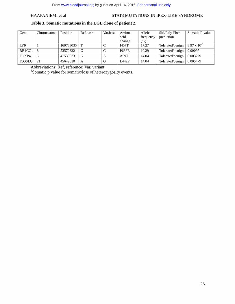
No cytogenetic alterations were found in the LGL-subset in routine clinical investigations. To
elucidate potential oncogenic single nucleotide variants (SNVs) driving the LGL expansion, the
CD3+TCR γδ cells were exome sequenced in parallel with the germline DNA extracted from saliva
sample. Four novel somatic mutations were called in the following genes: LY9, RB1CC1, FOXP4
and ICOSLG (Table 3). The variant allele frequency varied between 10-17%, suggesting that the
mutations were located in a subpopulation of TCR-γδ+ cells. No loss of heterozygosity for the
germline STAT3 K392R mutation was observed, and no genomic rearrangements were detected.
For personal use only.on April 16, 2016. by guest www.bloodjournal.orgFrom

HAAPANIEMI et al STAT3 MUTATIONS IN IPEX-LIKE SYNDROME
14
Discussion
In this study, we identified activating germline STAT3 mutations K658N,14 K392R14 and M394T in
three patients with autoimmunity, hypogammaglobulinemia, lymphoproliferation and mycobacterial
disease. Autoimmunity and hypogammaglobulinemia were seen in all cases, and the displayed
autoimmune phenomena are distinctly rare in children (desquamative interstitial pneumonitis,
posterior uveitis). Lymphoproliferation (lymphadenopathy, splenomegaly or pediatric T-LGL
leukemia) was present in two cases. One patient developed disseminated mycobacterial disease in
late adolescence. The patients presented with somewhat high proportions of CD3+CD4-CD8- T cells
with decreased counts of dendritic, Treg, Th17 and NK cells as well as deficiency of switched
memory B cells.
Heterozygous loss-of-function STAT3 mutations have been associated with autosomal dominant
HIES, which is characterized by high serum IgE, eosinophilia, eczema and immunodeficiency.5,6
Our first patient developed eczema that differed from the typical hyper-IgE eczema clinically and
histopathologically (data not shown). All patients were susceptible to respiratory infections, partly
due to their hypogammaglobulinemia. No other features of HIES were noted. The mutations in
HIES localize to the DNA-binding and SH2 domains of STAT3, whereas the observed activating
STAT3 mutations scatter throughout the protein.5,6,14 Mutations in the DNA binding domain caused
constitutive activation of STAT3, whereas the K658N mutation in the dimerization domain only
conferred hypersensitivity to interleukins. The difference in action however does not correlate with
the phenotype. It is possible that under physiologic conditions, hypersensitivity to low levels of
interleukins is sufficient for persistent activation of STAT3 signaling.
Autoimmunity is commonly seen in patients with germline STAT mutations, sometimes with
concomitant Treg deficiency.2,3 (For comparison between IPEX-like syndromes caused by STAT1,
STAT3, and STAT5B mutations see supplementary table S5). STAT3 promotes the activation and
expansion of autoimmunity-associated Th17 cells, whereas STAT5 drives the immunosuppressive
Treg fate. STAT3 and STAT5b bind to multiple sites of the IL-17 locus, with STAT3 binding
promoting IL-17 transcription, and STAT5b binding conversely repressing IL-17 transcription.24,25
Th17 deficiency is seen in loss-of-function STAT3 mutations and HIES.26,27 Curiously, our patients
with activating STAT3 mutations presented also with a reduced number of Th17 cells and decreased
IL-17 production.
For personal use only.on April 16, 2016. by guest www.bloodjournal.orgFrom
HAAPANIEMI et al STAT3 MUTATIONS IN IPEX-LIKE SYNDROME
15
A notable feature of the STAT3 hyperactivity patients was lymphoproliferation, which has not been
described in other IPEX-like syndromes.3 The somewhat elevated CD4-CD8- T cell counts observed
in our patients may suggest a defect in the lymphocyte apoptosis.28 Notably, patient 2 (K392R)
developed T-LGL leukemia at age 14. LGL leukemia is mainly diagnosed in the elderly and is often
accompanied by autoimmune processes such as rheumatoid arthritis and autoimmune cytopenias.
Somatic STAT3 gain-of-function mutations have been identified in 40-70% of T cell LGL leukemia
cases.11-13 The occurrence of pediatric LGL leukemia in patient 1 and the presence of LGL-like cells
in the bone marrow of patient 2 suggest STAT3 is to be a central oncogene in LGL leukemia
pathogenesis.
Patient 3 (M394T) presented only mild autoimmunity but developed disseminated mycobacterial
disease in late adolescence. In contrast to most known mycobacterial susceptibility syndromes,23 IL-
12-IFN-γ signaling was not impaired. Dendritic cell deficiencies cause mycobacterial disease,29 and
the observed lack of plasmacytoid dendritic cells may partly explain her condition. Why our IPEX-
like patients have not developed mycobacterial infections is unknown. Since dendritic cell
deficiency -associated mycobacterial disease is often late-onset, their young age might provide an
explanation.
In conclusion, activating germline STAT3 mutations lead to broad range of immune disturbances
including multi-organ autoimmunity, lymphoproliferation, hypogammaglobulinemia, and delayed
-onset mycobacterial disease. Emerging STAT3 inhibitors, some of which are in clinical trials, may
benefit such patients. Our results provide insights into the role of STAT3 in the pathogenesis of
autoimmune diseases and highlight the oncogenic nature of STAT3 in LGL leukemia development.
For personal use only.on April 16, 2016. by guest www.bloodjournal.orgFrom
HAAPANIEMI et al STAT3 MUTATIONS IN IPEX-LIKE SYNDROME
16
Acknowledgements
We thank Andrew Hattersley and his team in the University of Exeter for exciting collaboration in
the initial discovery of the K392R mutation and the early functional studies. Outi Vaarala and Jarno
Honkanen are acknowledged at performing accessory Th1 cytokine immunophenotyping. Personnel
at the Hematology Research Unit Helsinki, Institute for Molecular Medicine Finland (FIMM) and
Science for Life laboratory Stockholm are acknowledged for their expert clinical and technical
assistance. This work was supported by the Academy of Finland, Sigrid Juselius Foundation, Emil
Aaltonen Foundation, Finnish Medical Foundation, Finnish Cancer Organizations, Instrumentarium
Science Foundation, Jane and Aatos Erkko Foundation, Alma and K.A. Snellman Foundation, and
Foundation for Pediatric Research.
Author contributions
E.H. designed the study, coordinated the project, analyzed the data and wrote the paper. M.K. and
H.L.M.R. contributed to writing of the paper and performed laboratory analysis. S.M., M.S., J.S.
and J.K. designed and supervised the study, reviewed the data and contributed to writing of the
paper. Y.T.B., S.C., V.G., P.K., S.S., H.K., A.A., R.D. and A.H. designed and performed laboratory
analysis. S.E., L.T. and R.K. designed and performed bioinformatics analysis. M-L.K-L. and P.E.K.
reviewed the immunopathology. T.H-K., T.O., M.S., K.P., R.U-S., L.K. and K.H. provided clinical
care for the patients. All authors read and approved the final manuscript.
Disclosure of Conflicts of Interest
K.P. has received research funding and honoraria from Novartis and Bristol-Myers Squibb. S.M. has
received honoraria from Novartis and Bristol-Myers Squibb. M.S. has received honoraria from
Octapharma and Sanquin.
For personal use only.on April 16, 2016. by guest www.bloodjournal.orgFrom

HAAPANIEMI et al STAT3 MUTATIONS IN IPEX-LIKE SYNDROME
17
References
1. Bennett CL, Christie J, Ramsdell F, et al. The immune dysregulation, polyendocrinopathy, enteropathy, X-linked syndrome (IPEX) is caused by mutations of FOXP3. Nat Genet. 2001;27(1):20-21. 2. Uzel G, Sampaio EP, Lawrence MG, et al. Dominant gain-of-function STAT1 mutations in FOXP3 wild-type immune dysregulation-polyendocrinopathy-enteropathy-X-linked-like syndrome. J Allergy Clin Immunol. 2013;131(6):1611-1623. 3. Verbsky JW, Chatila TA. Immune dysregulation, polyendocrinopathy, enteropathy, X-linked (IPEX) and IPEX-related disorders: an evolving web of heritable autoimmune diseases. Curr Opin Pediatr. 2013;25(6):708-714. 4. Lohr NJ, Molleston JP, Strauss KA, et al. Human ITCH E3 ubiquitin ligase deficiency causes syndromic multisystem autoimmune disease. Am J Hum Genet. 2010;86(3):447-453. 5. Holland SM, DeLeo FR, Elloumi HZ, et al. STAT3 mutations in the hyper-IgE syndrome. N Engl J Med. 2007;357(16):1608-1619. 6. Minegishi Y, Saito M, Tsuchiya S, et al. Dominant-negative mutations in the DNA-binding domain of STAT3 cause hyper-IgE syndrome. Nature. 2007;448(7157):1058-1062. 7. Dupuis S, Jouanguy E, Al-Hajjar S, et al. Impaired response to interferon-alpha/beta and lethal viral disease in human STAT1 deficiency. Nat Genet. 2003;33(3):388-391. 8. Liu L, Okada S, Kong XF, et al. Gain-of-function human STAT1 mutations impair IL-17 immunity and underlie chronic mucocutaneous candidiasis. J Exp Med. 2011;208(8):1635-1648. 9. Knosp CA, Johnston JA. Regulation of CD4+ T-cell polarization by suppressor of cytokine signalling proteins. Immunology. 2012;135(2):101-111. 10. O'Shea JJ, Plenge R. JAK and STAT signaling molecules in immunoregulation and immune-mediated disease. Immunity. 2012;36(4):542-550. 11. Jerez A, Clemente MJ, Makishima H, et al. STAT3 mutations unify the pathogenesis of chronic lymphoproliferative disorders of NK cells and T-cell large granular lymphocyte leukemia. Blood. 2012;120(15):3048-3057. 12. Koskela HL, Eldfors S, Ellonen P, et al. Somatic STAT3 mutations in large granular lymphocytic leukemia. N Engl J Med. 2012;366(20):1905-1913. 13. Fasan A, Kern W, Grossmann V, Haferlach C, Haferlach T, Schnittger S. STAT3 mutations are highly specific for large granular lymphocytic leukemia. Leukemia. 2013;27(7):1598-1600. 14. Flanagan SE, Haapaniemi E, Russell MA, et al. Activating germline mutations in STAT3 cause early-onset multi-organ autoimmune disease. Nat Genet. 2014;46(8):812-814. 15. Otonkoski T, Roivainen M, Vaarala O, et al. Neonatal Type I diabetes associated with maternal echovirus 6 infection: a case report. Diabetologia. 2000;43(10):1235-1238. 16. Sulonen AM, Ellonen P, Almusa H, et al. Comparison of solution-based exome capture methods for next generation sequencing. Genome Biol. 2011;12(9):R94. 17. Ilander M, Kreutzman A, Rohon P, et al. Enlarged Memory T-Cell Pool and Enhanced Th1-Type Responses in Chronic Myeloid Leukemia Patients Who Have Successfully Discontinued IFN-alpha Monotherapy. PLoS One. 2014;9(1):e87794. 18. Ruitenberg JJ, Boyce C, Hingorani R, Putnam A, Ghanekar SA. Rapid assessment of in vitro expanded human regulatory T cell function. J Immunol Methods. 2011;372(1-2):95-106. 19. Chiang SC, Theorell J, Entesarian M, et al. Comparison of primary human cytotoxic T-cell and natural killer cell responses reveal similar molecular requirements for lytic granule exocytosis but differences in cytokine production. Blood. 2013;121(8):1345-1356. 20. Schneider EM, Lorenz I, Muller-Rosenberger M, Steinbach G, Kron M, Janka-Schaub GE. Hemophagocytic lymphohistiocytosis is associated with deficiencies of cellular cytolysis but normal expression of transcripts relevant to killer-cell-induced apoptosis. Blood. 2002;100(8):2891-2898. 21. Puel A, Doffinger R, Natividad A, et al. Autoantibodies against IL-17A, IL-17F, and IL-22
For personal use only.on April 16, 2016. by guest www.bloodjournal.orgFrom

HAAPANIEMI et al STAT3 MUTATIONS IN IPEX-LIKE SYNDROME
18
in patients with chronic mucocutaneous candidiasis and autoimmune polyendocrine syndrome type I. J Exp Med. 2010;207(2):291-297. 22. Bromberg JF, Wrzeszczynska MH, Devgan G, et al. Stat3 as an oncogene. Cell. 1999;98(3):295-303. 23. Cottle LE. Mendelian susceptibility to mycobacterial disease. Clin Genet. 2011;79(1):17-22. 24. Yang XO, Panopoulos AD, Nurieva R, et al. STAT3 regulates cytokine-mediated generation of inflammatory helper T cells. J Biol Chem. 2007;282(13):9358-9363. 25. Yang XP, Ghoreschi K, Steward-Tharp SM, et al. Opposing regulation of the locus encoding IL-17 through direct, reciprocal actions of STAT3 and STAT5. Nat Immunol. 2011;12(3):247-254. 26. Minegishi Y, Saito M, Nagasawa M, et al. Molecular explanation for the contradiction between systemic Th17 defect and localized bacterial infection in hyper-IgE syndrome. J Exp Med. 2009;206(6):1291-1301. 27. de Beaucoudrey L, Puel A, Filipe-Santos O, et al. Mutations in STAT3 and IL12RB1 impair the development of human IL-17-producing T cells. J Exp Med. 2008;205(7):1543-1550. 28. Magerus-Chatinet A, Stolzenberg MC, Loffredo MS, et al. FAS-L, IL-10, and double-negative CD4- CD8- TCR alpha/beta+ T cells are reliable markers of autoimmune lymphoproliferative syndrome (ALPS) associated with FAS loss of function. Blood. 2009;113(13):3027-3030. 29. Collin M, Bigley V, Haniffa M, Hambleton S. Human dendritic cell deficiency: the missing ID? Nat Rev Immunol. 2011;11(9):575-583.
For personal use only.on April 16, 2016. by guest www.bloodjournal.orgFrom
HAAPANIEMI et al STAT3 MUTATIONS IN IPEX-LIKE SYNDROME
19
Tables
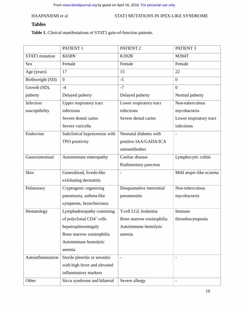
Table 1. Clinical manifestations of STAT3 gain-of-function patients.
PATIENT 1 PATIENT 2 PATIENT 3
STAT3 mutation K658N K392R M394T
Sex Female Female Female
Age (years) 17 15 22
Birthweight (SD) 0 -5 0
Growth (SD),
puberty
-4
Delayed puberty
-7
Delayed puberty
0
Normal puberty
Infection
susceptibility
Upper respiratory tract
infections
Severe dental caries
Severe varicella
Lower respiratory tract
infections
Severe dental caries
Non-tuberculous
mycobacteria
Lower respiratory tract
infections
Endocrine Subclinical hypotyreosis with
TPO positivity
Neonatal diabetes with
positive IAA/GADA/ICA
autoantibodies
-
Gastrointestinal Autoimmune enteropathy Coeliac disease
Rudimentary pancreas
Lymphocytic colitis
Skin Generalized, livedo-like
exfoliating dermatitis
- Mild atopic-like eczema
Pulmonary Cryptogenic organising
pneumonia, asthma-like
symptoms, bronchiectasis
Desquamative interstitial
pneumonitis
Non-tuberculous
mycobacteria
Hematology Lymphadenopathy consisting
of polyclonal CD4+ cells
hepatosplenomegaly
Bone marrow eosinophilia
Autoimmune hemolytic
anemia
T-cell LGL leukemia
Bone marrow eosinophilia
Autoimmune hemolytic
anemia
Immune
thrombocytopenia
Autoinflammation Sterile pleuritis or serositis
with high fever and elevated
inflammatory markers
- -
Other Sicca syndrome and bilateral Severe allergy -
For personal use only.on April 16, 2016. by guest www.bloodjournal.orgFrom

HAAPANIEMI et al STAT3 MUTATIONS IN IPEX-LIKE SYNDROME
20
posterior uveitis with cystic
macular edema
Abbreviations: SD, Standard Deviation; IAA, Insulin autoantibodies; GADA, Glutamic acid decarboxylase autoantibodies; ICA, Islet cell autoantibodies
For personal use only.on April 16, 2016. by guest www.bloodjournal.orgFrom

HAAPANIEMI et al STAT3 MUTATIONS IN IPEX-LIKE SYNDROME
21
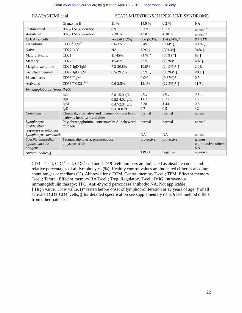
Table 2. Immunologic features of patients with STAT3 gain-of-function mutations.
Normal range / Healthy control median value
Patient 1 (K658N)
Patient 2 (K392R)
Patient 3 (M394T)
Leukocytes 3400-8200 7100 6900 (7500)* 5900 Lymphocytes 1000-4500 1700 4278 (1270)* 820 ↓ Monocytes 200-800 600 690 (370)* 570 Neutrophils 1500-7500 4800 1750 (5830)* 4130 Basophils 0-100 0 182 (0)* 80 Eosinophils 100-400 0 ↓ 10 (0)*↓ 280 platelets 150000-360000 267000 290000 (266000)* 271000 NK-cells (CD3-CD16+/56+) 90-600 (12%) 12 (0.7%) ↓ 128 (3%) (38,
0.5%)* ↓ 40-90(4-13%) ↓
NK cell function and maturation normal normal normal Dendritic cells Plasmacytoid lin-HLA-DR+CD123+CD11c- 0.1-0.3% 0.06 % ↓ <0.01% ↓ 0.01% ↓ Monocytoid lin-HLA-DR+CD123-CD11c+ 0.1-0.3% 0.2 % <0.01% ↓ 0.73% ↑ CD3+ T cells
700-2100 (71%) 1462 (86%) 3507 (82%) (1092, 86%)*
474 (74%)
TCRαβ+ 94% 94% 45% ↓ (93%)* 97%
TCRγδ + 6% 6% 55% ↑ (7%)* 3%
CD57+ 21% 8.4% 65% ↑ 65% ↑
CD4-CD8- 4.2% 3.4% 30% (2.3%)* ↑ 6 %
CD4-CD8- TCRαβ+ <3.4% 1.6 % 1 % 5%↑ CD4-CD8- TCRγδ + NA 1.8 % 29 %↑ 1% CD3+CD4+ T cells
458-1406 (60%) 789 (54%) 666 (19%)↓ (380, 34.8%)*
307 (48%) ↓
CD45RO+ 51 % 46 % 70% ↑ NA
CD45RA+ 47 % 51 % 26% ↓ 18% ↓
Ki-67+ 2 % 2,50 % 1.80 % NA
HLA-DR+ 3 % 29 % 1.90 % NA
TCM CCR7+CD45RA- 38 % 44 % 54% ↑ 56% ↑
Naive CCR7+CD45RA+ 45 % 50 % 24% ↓ 20% ↓
TEM CCR7-CD45RA- 11 % 5 % 22% ↑ 23% ↑
Temra CCR7-CD45RA+ 4 % 1 % 1 % 2%
Granzyme B+ 1 % 0.2 % 1.9 % NA
unstimulated IFNγ/TNFα secretion 0 % 0.2 % 0.1 % normal§
stimulated IFNγ/TNFα secretion 5 % 7.3 % 21.5% ↑ normal§ Treg FOXP3+CD25+ 2.3-7.8% 1.45% ↓ (0.67%)* ↓ 4.9%
Treg suppressive capacity low low normal
Th17 † CD69+IL17+ 0.47-1.59% 0.13% ↓ (0.35%)* ↓ 0.22 ↓
CD3+CD8+ T-cells 200-1200 (51%) 570 (39%) 1790 (51%) (339, 31%)*
134 (21%)
CD45RO+ 41 % 20 % 5% ↓ NA
CD45RA+ 72 % 76 % 93% ↑ 68%
Ki-67+ 1 % 2,70 % 16% ↑ NA
HLA-DR+ 3 % 29 % 56% ↑ NA
TCM CCR7+CD45RA- 8 % 11 % 3 % 13%
Naive CCR7+CD45RA+ 35 % 62% ↑ 8% ↓ 45% ↑
TEM CCR7-CD45RA- 27 % 8% ↓ 4% ↓ 22%
Temra CCR7-CD45RA+ 33 % 19 % 86% ↑ 21%
For personal use only.on April 16, 2016. by guest www.bloodjournal.orgFrom
HAAPANIEMI et al STAT3 MUTATIONS IN IPEX-LIKE SYNDROME
22
Granzyme B+ 11 % 14,9 % 0.2 % NA
unstimulated IFNγ/TNFα secretion 0 % 0,1 % 0.1 % normal§ stimulated IFNγ/TNFα secretion 7.20 % 4.50 % 4.30 % normal§ CD19+ B-cells 70-230 (12%) 660 (9.3%) 174 (14%)* 80 (11%) Transitional CD38hiIgMhi 0.6-3.5% 3.4% (0%)* ↓ 0.4% ↓
Naive CD27-IgD+ NA 70% ↑ (68%)*↑ 94% ↑
Mature B-cells CD21+ 11-45% 84 % ↑ (70%)* ↑ 88 ↑
Memory CD27+ 15-45% 23 % (26 %)* 4% ↓
Marginal zone-like CD27+IgD+IgM+ 7.2-30.8% 19.5% ↑ (24.9%)* ↑ 2.9%
Switched memory CD27+IgD-IgM- 6.5-29.2% 0.5% ↓ (0.5%)* ↓ <0.1 ↓
Plasmablasts CD38++IgM- - 0.0% (0.37%)* 0.5
Activated CD38lowCD21low 0.6-3.5% 12.1% ↑ (22.5%)* ↑ 11.7↑
Immunoglobulins (prior IVIG) IgG 6.8-15.0 g/L 5.8↓ 2.8↓ 0.16↓ IgA 0.52-4.02 g/L 1.67 0.21 1.7 IgM 0.47-2.84 g/L 3.38 1.44 0.6 IgE 0-110 IU/L 0.7 0.5 <2 Complement Classical., alternative and mannan-binding lectin
pathway hemolytic activities normal normal normal
Lymphocyte proliferative responses to mitogens
Phytohemagglutinin, concanavalin A, pokeweed mitogen
normal normal normal
Lymphocyte chemotaxis NA NA normal Specific antibodies against vaccine antigens
Tetanus, diphtheria, pneumococcal polysaccharide
protective protective tetanus unprotective, others NA
Autoantibodies ‡ TPO + negative negative
CD3+ T-cell, CD4+ cell, CD8+ cell and CD19+ cell numbers are indicated as absolute counts and relative percentages of all lymphocytes (%). Healthy control values are indicated either as absolute count ranges or medians (%). Abbreviations: TCM, Central memory T-cell; TEM, Effector memory T-cell; Temra, Effector memory RA T-cell; Treg, Regulatory T-cell; IVIG, intravenous immunoglobulin therapy; TPO, Anti-thyroid peroxidase antibody; NA, Not applicable, ↑ High value, ↓ low value, ()* tested before onset of lymphoproliferation at 12 years of age, † of all activated CD3+CD4+ cells, ‡ for detailed specification see supplementary data, § test method differs from other patients
For personal use only.on April 16, 2016. by guest www.bloodjournal.orgFrom
HAAPANIEMI et al STAT3 MUTATIONS IN IPEX-LIKE SYNDROME
23
Table 3. Somatic mutations in the LGL clone of patient 2.
Gene Chromosome Position Ref.base Var.base Amino acid change
Allele frequency (%)
Sift/Poly-Phen prediction
Somatic P-value1
LY9 1 160788035 T C I457T 17.27 Tolerated/benign 8.97 x 10⁻⁶
RB1CC1 8 53570332 G C P686R 10.29 Tolerated/benign 0.00097
FOXP4 6 41533673 G A A59T 14.04 Tolerated/benign 0.003229
ICOSLG 21 45649510 A G L442P 14.04 Tolerated/benign 0.005479
Abbreviations: Ref, reference; Var, variant. 1Somatic p value for somatic/loss of heterozygosity events.
For personal use only.on April 16, 2016. by guest www.bloodjournal.orgFrom

HAAPANIEMI et al STAT3 MUTATIONS IN IPEX-LIKE SYNDROME
24
Figure legends
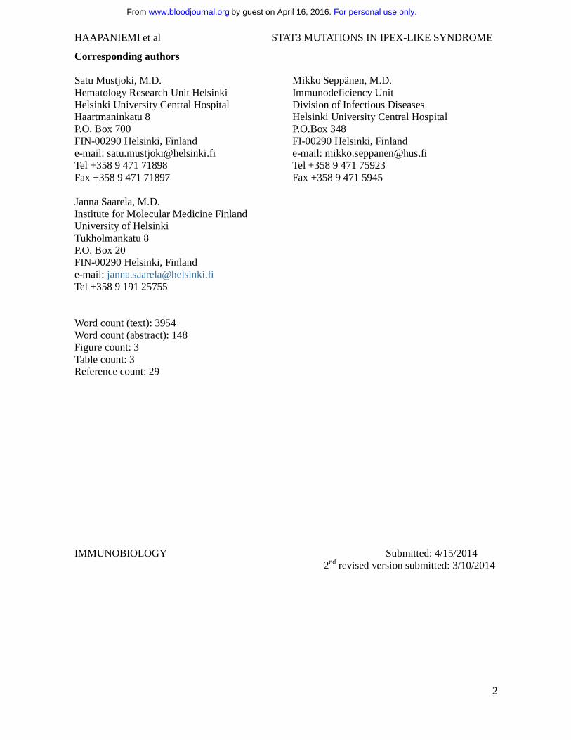
Figure 1. Clinical characteristics of patients. A. Livedo-like generalized exfoliating dermatitis in
patient 1. The rash culminates in limb extensor areas. B. High-resolution computerized tomography
of patient 2 showing ground-glass opacity, bronchoalveolar thickening and increased nodularity. C.
Bone marrow biopsy from patient 1 showing modest bone marrow eosinophilia despite observed
peripheral eosinopenia (yellow asterisks). HE stain, 40x magnification.
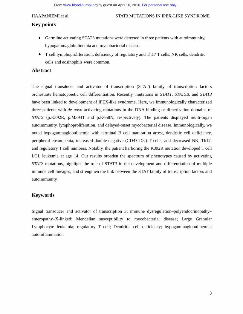
Figure 2. STAT3 mutations K658N, K392R, and M394T in studied patients. A. Schematic
representation of STAT3 protein domains with the observed mutations marked as black lines. Germ-
line and somatic mutation hotspots for Hyper-IgE syndrome5,6 and LGL leukemia11-13 are indicated
as green and blue bars above, respectively. B. Crystallographic structure of STAT3 dimer (the
RCSB Protein Data Bank code 1BG1). K658N, K392R, and M394T mutations are indicated as red
dots. C-D. HEK293 cells containing STAT3-responsive luciferase were transfected with empty, wild
type and mutant STAT3 overexpression plasmids with or without IL-6 stimulation. The K392R and
M394T significantly increased STAT3 transcriptional activity in basal and stimulated conditions.
Error bars represent SEM (n=6, panel C). The K658N mutant showed hypersensitivity to IL-6
stimulation in low concentrations. Error bars represent SEM (n=3, panel D). 2 way ANOVA, * =
p<0.05, ** = p<0.01 and *** = P<0.001. E. No significant increase in pSTAT3Y705 phosphorylation
was observed when HEK293 cells were transfected with mutant STAT3 overexpression constructs.
Equal amounts of parallel-derived whole cell lysates were loaded per condition. α-tubulin and
STAT3 were used as loading and expression controls, respectively. + and – signs indicate presence
and absence of IL-6 stimulation. F. In peripheral blood, no significant increase in STAT3
phosphorylation was noted in studied patients. Color change indicates relative pSTAT3Y705
expression. Forward panel: K392R, middle panel, K658N, back panel: healthy control (n=3, value
range presented in brackets).
Figure 3. Abnormal lymphocyte populations detected in STAT3-mutated patients. Figures A-
C: Bone marrow biopsy shows abnormally high number of phospho-STAT3 positive lymphocytes
both in patient 2 (p.K392R) (A) but also, to a lesser extent, in patient 1 (p.K658N) (B). Patient 3
(M394T) was not available for study. In healthy bone marrow, no phospho-STAT3 cells are present
(C). Figures 3D-F: Flow cytometry results from patient 2 (p. K392R). Majority of lymphocytes
were CD3+ (A) with 57% of the population expressing TCR-γδ (B). The TCR-γδ+ population
consisted of CD4-CD8- and CD4-CD8+ T-cells. The expression of TCR-γδ was considerably lower
For personal use only.on April 16, 2016. by guest www.bloodjournal.orgFrom

HAAPANIEMI et al STAT3 MUTATIONS IN IPEX-LIKE SYNDROME
25
in CD4-CD8- cells than in CD4-CD8+ T-cells and therefore two populations are seen in the scatter
plot. (C). In healthy individuals, TCR-γδ-expressing T-cells account less than 6% of all CD3+ T-
cells and the TCR-γδ expression is normally uniform. 40x magnification, HE stain.
For personal use only.on April 16, 2016. by guest www.bloodjournal.orgFrom

*
*
A B
C
Figure 1
For personal use only.on April 16, 2016. by guest www.bloodjournal.orgFrom

Coiled coil ND DNA binding Linker SH2 TAD
1 585465130 320 770A
K658NK392R
688
M394TB
K392RM394T
K658N
- 0.5 ng/well 5 ng/well0
200
400
600
800
Lum
ines
cenc
e (R
LU) EP
WtK658N
05
10
500
1000
1500
2000
Lum
ines
cenc
e (R
LU) empty vector
wild typeK392RM394TK658N
IL-6 - 10 ng/well
IL-6
Lymphocytes CD3+CD4+CD8- CD3+CD4-CD8- CD3+CD4-CD8+
pSTAT3 pSTAT3 pSTAT3 pSTAT3
Healthy control
Patient 2 Patient 1
Healthy control mean (n=3) 1,0 (0,7-1,2) 1,0 (0,6-1,5) 1,0 (0,8-1,1) 1,0 (0,7-1,3)
Pa�ent 1 (n=2) 1,3 (1,2-1,3) 1,3 (1,2-1,4) 1,2 (1,0-1,5) 1,2 (1,0-1,3) Pa�ent 2 (n=2) 1,3 (1,2-1,5) 1,5 (1,4-1,6) 1,3 (1,1-1,5) 1,3 (1,1-1,5)
0.90 1.45 2.00
D
C
F
******
***
**
empty vector***
IL-6 - + - + - + - + - +pSTAT3STAT3
pSTA
T3/S
TAT3
mea
n in
tens
itypSTAT3
α-tubulinSTAT3
5 ng/well0.5 ng/well
E
Empty ve
ctor
Wild ty
pe
K392R
K658N
M394T
K392RWild typeEmpty vector
M394TK658N
K658NWild typeEmpty vector
0
2
4
6
8 MockIL-6
For personal use only.on April 16, 2016. by guest www.bloodjournal.orgFrom

A B C
CD3 TCR CD8
SS
C
CD3+ 81%of lymphocytesCD3neg
of CD3+
of CD3+negCD8+negCD8neg
50
1
00
150
2
00
250
-188
0
102
10
3
10
4
10
5
-487
0
10
3
10
4
10
5
TC
R
102 103 104 105-296 0 103 104 105
-569 0 103 104 105
D E F
Figure 3
For personal use only.on April 16, 2016. by guest www.bloodjournal.orgFrom
doi:10.1182/blood-2014-04-570101Prepublished online October 27, 2014;
Uusitalo-Seppälä, Janna Saarela, Mikko Seppänen, Satu Mustjoki and Juha KereRaijaKovanen, Timo Otonkoski, Kimmo Porkka, Kaarina Heiskanen, Arno Hänninen, Yenan T. Bryceson,
Petri Kulmala, Samuli Eldfors, Riku Katainen, Sanna Siitonen, Marja-Liisa Karjalainen-Lindsberg, Panu E.Glumoff, Rainer Doffinger, Heikki Kuusanmäki, Tarja Heiskanen-Kosma, Luca Trotta, Samuel Chiang, Emma M. Haapaniemi, Meri Kaustio, Hanna L.M. Rajala, Arjan J. van Adrichem, Leena Kainulainen, Virpi STAT3mycobacterial disease in patients with dominant activating mutations in Autoimmunity, hypogammaglobulinemia, lymphoproliferation and
http://www.bloodjournal.org/site/misc/rights.xhtml#repub_requestsInformation about reproducing this article in parts or in its entirety may be found online at:
http://www.bloodjournal.org/site/misc/rights.xhtml#reprintsInformation about ordering reprints may be found online at:
http://www.bloodjournal.org/site/subscriptions/index.xhtmlInformation about subscriptions and ASH membership may be found online at:
digital object identifier (DOIs) and date of initial publication. indexed by PubMed from initial publication. Citations to Advance online articles must include final publication). Advance online articles are citable and establish publication priority; they areappeared in the paper journal (edited, typeset versions may be posted when available prior to Advance online articles have been peer reviewed and accepted for publication but have not yet
Copyright 2011 by The American Society of Hematology; all rights reserved.Hematology, 2021 L St, NW, Suite 900, Washington DC 20036.Blood (print ISSN 0006-4971, online ISSN 1528-0020), is published weekly by the American Society of
For personal use only.on April 16, 2016. by guest www.bloodjournal.orgFrom
Related Documents