AUTOIMMUNITY AND IMMUNODEFICIENCY Topic Editors Luigi D. Notarangelo and Rosa Bacchetta IMMUNOLOGY

Welcome message from author
This document is posted to help you gain knowledge. Please leave a comment to let me know what you think about it! Share it to your friends and learn new things together.
Transcript

AUTOIMMUNITY AND IMMUNODEFICIENCY
Topic EditorsLuigi D. Notarangelo and Rosa Bacchetta
IMMUNOLOGY
Frontiers in Immunology August 2013 | Autoimmunity and Immunodeficiency | 1
ABOUT FRONTIERSFrontiers is more than just an open-access publisher of scholarly articles: it is a pioneering approach to the world of academia, radically improving the way scholarly research is managed. The grand vision of Frontiers is a world where all people have an equal opportunity to seek, share and generate knowledge. Frontiers provides immediate and permanent online open access to all its publications, but this alone is not enough to realize our grand goals.
FRONTIERS JOURNAL SERIESThe Frontiers Journal Series is a multi-tier and interdisciplinary set of open-access, online journals, promising a paradigm shift from the current review, selection and dissemination processes in academic publishing. All Frontiers journals are driven by researchers for researchers; therefore, they constitute a service to the scholarly community. At the same time, the Frontiers Journal Series operates on a revo-lutionary invention, the tiered publishing system, initially addressing specific communities of scholars, and gradually climbing up to broader public understanding, thus serving the interests of the lay society, too.
DEDICATION TO QUALITYEach Frontiers article is a landmark of the highest quality, thanks to genuinely collaborative interac-tions between authors and review editors, who include some of the world’s best academicians. Research must be certified by peers before entering a stream of knowledge that may eventually reach the public - and shape society; therefore, Frontiers only applies the most rigorous and unbiased reviews.Frontiers revolutionizes research publishing by freely delivering the most outstanding research, evaluated with no bias from both the academic and social point of view.By applying the most advanced information technologies, Frontiers is catapulting scholarly publishing into a new generation.
WHAT ARE FRONTIERS RESEARCH TOPICS?Frontiers Research Topics are very popular trademarks of the Frontiers Journals Series: they are collections of at least ten articles, all centered on a particular subject. With their unique mix of varied contributions from Original Research to Review Articles, Frontiers Research Topics unify the most influential researchers, the latest key findings and historical advances in a hot research area! Find out more on how to host your own Frontiers Research Topic or contribute to one as an author by contacting the Frontiers Editorial Office: [email protected]
FRONTIERS COPYRIGHT STATEMENT© Copyright 2007-2013 Frontiers Media SA. All rights reserved.
All content included on this site, such as text, graphics, logos, button icons, images, video/audio clips, downloads, data compilations and software, is the property of or is licensed to Frontiers Media SA (“Frontiers”) or its licensees and/or subcontractors. The copyright in the text of individual articles is the property of their respective authors, subject to a license granted to Frontiers.
The compilation of articles constituting this e-book, as well as all content on this site is the exclusive property of Frontiers. Images and graphics not forming part of user-contributed materials may not be downloaded or copied without permission.
Articles and other user-contributed materials may be downloaded and reproduced subject to any copyright or other notices. No financial payment or reward may be given for any such reproduction except to the author(s) of the article concerned.
As author or other contributor you grant permission to others to reproduce your articles, including any graphics and third-party materials supplied by you, in accordance with the Conditions for Website Use and subject to any copyright notices which you include in connection with your articles and materials.
All copyright, and all rights therein, are protected by national and international copyright laws.
The above represents a summary only. For the full conditions see the Conditions for Authors and the Conditions for Website Use.
ISSN 1664-8714ISBN 978-2-88919-164-2DOI 10.3389/978-2-88919-164-2

Frontiers in Immunology August 2013 | Autoimmunity and Immunodeficiency | 2
Topic Editors:Luigi D. Notarangelo, Harvard Medical School, USARosa Bacchetta, Fondazione Centro San Raffaele Del Monte Tabor, Italy
Immune regulation results from a finely tuned network of distinct mechanisms operating throughout life and balancing the need to clear infections and prevent self-aggression. Primary Immunodeficiencies (PIDs) are “experiments of nature” where the ability to fight against pathogens is deeply impaired. The study of patients with PIDs has been instrumental to identify and characterize key components and mechanisms that govern development and function of the human immune system. Recently, it
has become clear that in congenital monogenic diseases the ability of the immune system to build and maintain active tolerance to self can be specifically altered, so that autoimmune symptoms may easily prevail over infections in these pathologies. In addition, increasing observations have brought the attention to the fact that hypomorphic mutations in genes that control T and/or B cell development are often associated with clinical and laboratory features of immune dysregulation, thus expanding the spectrum of PID phenotypes. For example, mutations in genes driving T cell development can lead to defective lymphostromal cross-talk in the thymus and impinge of negative selection of self-reactive T cells and/or Treg function. Similarly, disorders of B cell development may associate with defects of receptor editing and/or with abnormalities of peripheral B cell homeostasis. On the other hand, autoantibodies can provoke defective immune responses by targeting cytokines and/or immune cells.
AUTOIMMUNITY AND IMMUNODEFICIENCY
Image created by Aisha Sauer and Luigi D. Notarangelo using Servier Medical Art.

Frontiers in Immunology August 2013 | Autoimmunity and Immunodeficiency | 3
This Research Topic will focus on i) summarizing updated clinical and immunological features of diseases characterized by immune dysregulation of known and still undefined origin and ii) gathering new insights into the mechanisms of T and B cell development, function and interaction, in order to broader the comprehension of the pathogenesis of autoimmunity and to ultimately advance the definition of novel therapeutic strategies.

Frontiers in Immunology August 2013 | Autoimmunity and Immunodeficiency | 4
Table of Contents
05 Immunodeficiency with Autoimmunity: Beyond the ParadoxR. Bacchetta and L. D. Notarangelo
06 Immune Dysregulation, Polyendocrinopathy, Enteropathy, X-Linked Syndrome: A Paradigm of Immunodeficiency with AutoimmunityFederica Barzaghi, Laura Passerini and Rosa Bacchetta
31 Autoimmunity in Wiskott–Aldrich Syndrome: An Unsolved EnigmaMarco Catucci, Maria Carmina Castiello, Francesca Pala, Marita Bosticardo and Anna Villa
45 Autoimmune Dysregulation and Purine Metabolism in Adenosine Deaminase DeficiencyAisha Vanessa Sauer, Immacolata Brigida, Nicola Carriglio and Alessandro Aiuti
64 The STAT5b Pathway Defect and AutoimmunityTakahiro Kanai, Jennifer Jenks and Kari Christine Nadeau
72 APECED: Is This a Model for Failure of T Cell and B Cell Tolerance?Nicolas Kluger, Annamari Ranki and Kai Krohn
84 Pathogenesis of Autoimmunity in Common Variable ImmunodeficiencyKlaus Warnatz and Reinhard E. Voll
90 Autoimmune Cytopenias In Common Variable ImmunodeficiencyJenna C. Podjasek and Roshini S. Abraham
97 TH17 Cells in Autoimmunity and Immunodeficiency: Protective or Pathogenic?Ashish K. Marwaha, Nicole J. Leung, Alicia N. McMurchy and Megan K. Levings
105 Dendritic Cells a Double-Edge Sword in Autoimmune ResponsesGiada Amodio and Silvia Gregori

Immunodeficiency with autoimmunity: beyond the paradox
R. Bacchetta1* and L. D. Notarangelo 2*1 San Raffaele Telethon Institute for Gene Therapy (HSR-TIGET), Division of Regenerative Medicine Stem Cells and Gene Therapy, San Raffaele Scientific Institute, Milan, Italy2 Division of Immunology and the Manton Center for Orphan Disease Research, Children’s Hospital Boston, Harvard Medical School, Boston, MA, USA*Correspondence: [email protected]; [email protected]
Edited by:Eric Meffre, Yale University School of Medicine, USA
Reviewed by:Eric Meffre, Yale University School of Medicine, USA
The association of immunodeficiency and autoimmunity may represent a paradox, yet it has been described in an increasing num-ber of conditions. Use of unbiased genomic approach to identify novel forms of primary immunodeficiencies (PIDs), along with in-depth functional studies in biological samples from affected indi-viduals continue to unravel novel mechanisms underlying immune dysregulation in patients with altered ability of fighting pathogens. In particular, it has been clearly established that genetic defects that affect T and B cell development compromise not just the ability to generate a diversified repertoire of lymphocytes capable of rec-ognizing multiple pathogens, but also impinge on mechanisms of central and peripheral tolerance, hence favoring autoimmune and inflammatory manifestations.
Yet, the diagnosis of autoimmune symptoms in the context of PIDs is troublesome, the prognosis unclear, and the treatment chal-lenging. In the present collection of manuscripts, several experts in the field provide an overview of the spectrum of different forms of monogenic defects of the immune system manifesting also with autoimmunity, and discuss established and novel mechanisms involved in immune dysregulation.
Studies on patients with Immunedysregulation-Polyendo-crinopathy-Enteropathy-X-linked (IPEX) Syndrome, have paved the way to understand the phenotype arising from impaired peripheral tolerance due to dysfunctional regulatory T cells (Treg) expressing mutated FOXP3. However, this important T cell subpopulation can also be affected in other forms of PID, such as Wiskott–Aldrich syndrome (WAS) and adenosine deami-nase (ADA) deficiency. In these disorders, the underlying genetic defect affects multiple cell types, resulting in impaired immune defense, but also poor Treg function. Similarly, STAT5B muta-tions disrupt an essential intracellular transcriptional activa-tor for Treg cells, causing reduction of Treg number in affected individuals.
Autoimmune polyendocrinopathy-candidiasis-ectodermal dystro-phy (APECED) is an autosomal recessive condition due to mutation of the Autoimmune regulator (AIRE) gene. Patients with APECED present with predominant organ specific autoimmunity and autoanti-bodies with multiple specificities. AIRE has been shown to play a criti-cal role in allowing expression of self-antigens in the thymus, thereby permitting deletion of self-reactive T lymphocytes or their diversion to Treg cells. Thus APECED stands as the prototypic monogenic dis-order of central T cell tolerance. While it is still questionable whether deficiency of AIRE also affects peripheral tolerance, recent data indicate that the autoimmune-associated tissue damage may not be primarily due to autoantibodies, but rather to autoreactive CD8+ T cells.
Moreover, recent studies in patients affected with Common Variable Immunodeficiency, a condition in which proper specific antibody production is deficient in favor of pathogenic autoanti-body secretion, have highlighted the importance of mechanisms that control B cell development and receptor editing in maintaining immune homeostasis.
Finally, two manuscripts call the attention to the dual role of cer-tain cell types and their ability to acquire different immunological functions depending on the environment in which they differenti-ate, as described for Th17 cells and dendritic cells, at the end of the Topic. Possibly, the future of medicine should aim to implement physiological plasticity and to empower epigenetics modifications in order to recover from inborn errors of Nature.
Received: 19 February 2013; accepted: 09 March 2013; published online: 12 April 2013.Citation: Bacchetta R and Notarangelo LD (2013) Immunodeficiency with autoimmunity: beyond the paradox. Front. Immunol. 4:77. doi: 10.3389/fimmu.2013.00077This article was submitted to Frontiers in Primary Immunodeficiencies, a specialty of Frontiers in Immunology.Copyright © 2013 Bacchetta and Notarangelo. This is an open-access article distributed under the terms of the Creative Commons Attribution License, which permits use, dis-tribution and reproduction in other forums, provided the original authors and source are credited and subject to any copyright notices concerning any third-party graphics etc.
www.frontiersin.org April 2013 | Volume 4 | Article 77 |
Editorialpublished: 12 April 2013
doi: 10.3389/fimmu.2013.00077
5

REVIEW ARTICLEpublished: 31 July 2012
doi: 10.3389/fimmu.2012.00211
Immune dysregulation, polyendocrinopathy, enteropathy,X-linked syndrome: a paradigm of immunodeficiencywith autoimmunityFederica Barzaghi 1,2, Laura Passerini 1 and Rosa Bacchetta1*1 Division of Regenerative Medicine, Stem Cells and Gene Therapy, San Raffaele Telethon Institute for Gene Therapy, San Raffaele Scientific Institute, Milan, Italy2 Vita Salute San Raffaele University, Milan, Italy
Edited by:Luigi Daniele Notarangelo, HarvardMedical School, USA
Reviewed by:Andrew Gennery, NewcastleUniversity, UKNancy Bunin, Children’s Hospital ofPhiladelphia, USA
*Correspondence:Rosa Bacchetta, San RaffaeleTelethon Institute for Gene Therapy,San Raffaele Scientific Insitute, ViaOlgettina 58, 20132 Milano, Italy.e-mail: [email protected]
Immune dysregulation, polyendocrinopathy, enteropathy, X-linked (IPEX) syndrome is a raremonogenic primary immunodeficiency (PID) due to mutations of FOXP3, a key transcrip-tion factor for naturally occurring (n) regulatoryT (Treg) cells.The dysfunction ofTreg cells isthe main pathogenic event leading to the multi-organ autoimmunity that characterizes IPEXsyndrome, a paradigm of genetically determined PID with autoimmunity. IPEX has a severeearly onset and can become rapidly fatal within the first year of life regardless of the typeand site of the mutation. The initial presenting symptoms are severe enteritis and/or type-1 diabetes mellitus, alone or in combination with eczema and elevated serum IgE. Otherautoimmune symptoms, such as hypothyroidism, cytopenia, hepatitis, nephropathy, arthri-tis, and alopecia can develop in patients who survive the initial acute phase. The currenttherapeutic options for IPEX patients are limited. Supportive and replacement therapiescombined with pharmacological immunosuppression are required to control symptoms atonset. However, these procedures can allow only a reduction of the clinical manifesta-tions without a permanent control of the disease. The only known effective cure for IPEXsyndrome is hematopoietic stem cell transplantation, but it is always limited by the avail-ability of a suitable donor and the lack of specific guidelines for bone marrow transplantin the context of this disease. This review aims to summarize the clinical histories andgenomic mutations of the IPEX patients described in the literature to date. We will focuson the clinical and immunological features that allow differential diagnosis of IPEX syn-drome and distinguish it from other PID with autoimmunity. The efficacy of the currenttherapies will be reviewed, and possible innovative approaches, based on the latest high-lights of the pathogenesis to treat this severe primary autoimmune disease of childhood,will be discussed.
Keywords: IPEX, FOXP3,Treg, autoimmune enteropathy, neonatal diabetes, neonatal eczema, HSCT
INTRODUCTIONImmune dysregulation, polyendocrinopathy, enteropathy, X-linked (IPEX) syndrome is a rare monogenic primary immun-odeficiency (PID), characterized by multi-organ autoimmunity. Itis caused by mutations in the transcription factor forkhead box p3(FOXP3), the master gene of T regulatory (Treg) cells. The dis-ease shows an X-linked hereditary pattern: only males are affected,whereas the carrier mothers are healthy.
Although IPEX syndrome is a rare disease, the recent increasein the number of patients referred for diagnosis suggests thatthe occurrence of the disease has been underestimated so far. Atpresent, 63 FOXP3 mutations have been published, for an overallnumber of 136 patients described, and of these about half havebeen diagnosed in the last 3 years. This also indicates that theawareness of the disease has been growing with a better under-standing of the role of FOXP3 and Treg cells in maintainingperipheral tolerance.
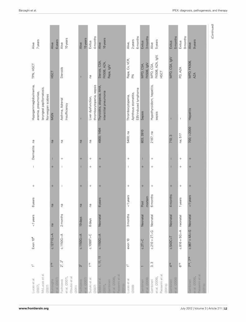
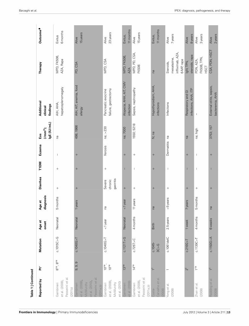
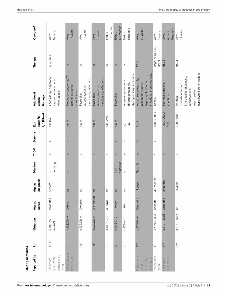
Overall, the analysis of cases reported so far (Table 1) confirmsthe relevance of the three main clinical manifestations and their
early onset while highlighting the occurrence of unusual symp-toms. The genetic analysis is always required for accurate diagnosis,although other tests such as tissue biopsy and/or autoantibodydetection are important, as complementary tools, in the diagnosticprocess and follow-up.
IPEX syndrome can be fatal in early infancy if not recognized,therefore a timely diagnosis is essential to start appropriate treat-ment. Treating IPEX patients poses a threefold challenge: autoim-munity, infections supported by the autoimmune damage, and theseverity of the overall picture. Both novel and existing therapeuticapproaches will be discussed with an emphasis on the central roleof Treg cell impairment in the pathogenesis of IPEX syndrome.
GENETICS OF IPEX SYNDROMEImmune dysregulation, polyendocrinopathy, enteropathy, X-linked syndrome was described for the first time in 1982 in a largefamily with 19 affected males across five generations,as an X-linkedsyndrome with diarrhea that was lethal in most male infants bythe first months or years of life (Powell et al., 1982). Only 20 years
www.frontiersin.org July 2012 | Volume 3 | Article 211 | 6

Barzaghi et al. IPEX: diagnosis, pathogenesis, and therapy
Tab
le1
|Clin
ical
feat
ure
s,th
erap
yan
do
utc
om
ein
rep
ort
edIP
EX
pat
ien
ts.
Rep
ort
edby
Pt◦
Mu
tati
on
Age
at
on
set
Age
at
dia
gn
osi
s
Dia
rrh
eaT
1DM
Ecz
ema
Eo
s
(/m
m3);
IgE
(IU
/mL)
Ad
dit
ion
al
clin
ical
fin
din
gs
Th
erap
yO
utc
om
e�
Peak
eet
al.
(199
6),W
ildin
etal
.(20
01)
V6f ,
2fc.
1290
_130
9
del_
insT
GG
6w
eeks
Post
mor
tem
++
+ Peel
ing
skin
3170
;999
9–
40,0
00µ
g/L
Ane
mia
,
lym
phad
enop
athy
, sep
sis
TPN
,pla
sma
IVE
xitu
s
10m
onth
s
Ferg
uson
etal
.
(200
0),
Ben
nett
etal
.
(200
1b),
McG
inne
ss
etal
.(20
06)
F1-V
1f ,
V2f ,
1f
c.11
50G
>A
1m
onth
9ye
ars
+−
+23
0–90
0;
755–
3492
Pem
phig
oid
nodu
laris
;
bullo
uspe
mph
igoi
d;
infe
ctio
ns,a
sthm
a
PD
,CSA
,
daps
one,
IgIV
,
ritux
imab
Aliv
e
14ye
ars
Ferg
uson
etal
.
(200
0),
Ben
nett
etal
.
(200
1b)
F1-IV
10f
c.11
50G
>A
#2
mon
ths
–− (V
omiti
ng)
−+
936;
250
Hyp
othy
roid
ism
;
infe
ctio
n
naE
xitu
s
10m
onth
s
F1-V
4fc.
1150
G>
A#
2w
eeks
–+
++
620;
30H
ypog
amm
aglo
bulin
emia
,
infe
ctio
ns,s
epsi
s
IgIV
Exi
tus
2ye
ars
Ferg
uson
etal
.
(200
0),
Ben
nett
etal
.
(200
1b)
F1-V
5f ,
V7f
c.11
50G
>A
3w
eeks
Post
mor
tem
−+
+50
0;22
Sep
sis
IgIV
,CSA
Exi
tus
12w
eeks
Cha
tila
etal
.
(200
0)
3-F1
;F2f
−14
,15,
27,2
8
c.10
44
+4A
>G
;
c.75
0_75
2
delG
GA
3w
eeks
−3
mon
ths
na5/
55/
54/
5na
;4/5
hype
rIg
E
Aut
oim
mun
ecy
tope
nia
3/5,
food
alle
rgy5
/5
naE
xitu
s
1/5,
aliv
e
4/5
Lev y
-Lah
ad
and
Wild
in
(200
1),W
ildin
etal
.(20
01)
1f ,F1
fc.
1189
C>
T#B
irth
–− (A
toni
cgu
t)
−+
naH
ypot
onia
,
hypo
thyr
oidi
sm,
thro
mbo
cyto
peni
a,
perit
oniti
s,ch
olan
gitis
–E
xitu
s
19da
ys
2f ,F1
fc.
1189
C>
TB
irth
Post
mor
tem
− (Ileo
)
+−
naC
ache
xia,
hypo
toni
a,
thro
mbo
cyto
peni
a,
perit
oniti
s
–E
xitu
s
5w
eeks
3f ,F1
fc.
1189
C>
T#B
irth
–+
+−
naIn
fect
ions
,sep
sis
Dex
amet
haso
ne,
CSA
Wild
inet
al.
(200
1)
3nac.
1113
T>G
nana
++
+na
Ane
mia
HS
CT
Aliv
e,
age
na
Frontiers in Immunology | Primary Immunodeficiencies July 2012 | Volume 3 | Article 211 | 7

Barzaghi et al. IPEX: diagnosis, pathogenesis, and therapy
4nac.
1150
G>
Ana
na+
++
naH
ypot
hyro
idis
m,
thro
mbo
cyto
peni
a,se
psis
naE
xitu
s
4m
onth
s
Ben
nett
etal
.
(200
1b),
Koba
yash
i
etal
.(20
11)
F2,5
c.12
93_1
294
delC
T
nana
+−
−na
–H
SC
TE
xitu
s
age
na
Koba
y ash
i
etal
.(20
01,
2011
),
Fuch
izaw
a
etal
.(20
07),
Ots
ubo
etal
.
(201
1)
1f ,1f ,
3,
1f
c.22
7del
T15
days
na+
−−
na;+
Thyr
oidi
tis,A
HA
,
tubu
lone
phro
path
y*
FK50
6,
beta
met
haso
ne
Aliv
e
19ye
ars
Koba
yash
i
etal
.(20
01,
2011
)
2f ,4f
c.10
87A
>G
nana
++
−na
;+Th
yroi
ditis
,
tubu
lone
phro
path
y*,
infe
ctio
ns,s
epsi
s
naE
xitu
s
3ye
ars
Bau
det
al.
(200
1)
1fc.
1113
T>G
#na
–+
+Ic
hthy
osis
naIT
Pna
Exi
tus
4.5
mon
ths
2fc.
1 113
T>G
1m
onth
4m
onth
s+
+Ic
hthy
osis
na;1
750
ITP,
AH
A,a
utoi
mm
une
neut
rope
nia,
chol
esta
tic
hepa
titis
MP
D,F
K50
6,
HS
CT
Exi
tus
2ye
ars
7m
onth
s
Wild
inet
al.
(200
2),
McM
urch
y
etal
.(20
10)
1fc.
1040
G>
A3
mon
ths
13ye
ars
++
−na
Infe
ctio
n(s
epsi
s)P
D,C
SA,F
K50
6,
HS
CT
Exi
tus
14ye
ars
Wild
inet
al.
(200
2)
2c.
1044+
459A
>G
<1
mon
thna
++
+na
Lym
phad
enop
athy
,
hepa
tosp
leno
meg
aly
ecze
ma,
hypo
thyr
oidi
sm,
AH
A,i
mm
une
neut
rope
nia,
infe
ctio
ns
Ste
roid
s,
ritux
imab
,IgI
V
Aliv
e
5ye
ars
3fc.
748_
750
delA
AG
,
c.54
3C>
T
2m
onth
s9
year
s+
+−
naA
rthr
itis,
ITP ,
hepa
tom
egal
y,m
ild
hepa
titis
,pro
gres
sive
rena
lins
uffic
ienc
y
Ste
roid
s,C
SA,
FK50
6,ro
feco
xib,
MTX
,ritu
xim
ab,
IgIV
,HS
CT
Exi
tus
10ye
ars (C
ontin
ued)
www.frontiersin.org July 2012 | Volume 3 | Article 211 | 8

Barzaghi et al. IPEX: diagnosis, pathogenesis, and therapy
Tab
le1
|Co
nti
nu
ed
Rep
ort
edby
Pt◦
Mu
tati
on
Age
at
on
set
Age
at
dia
gn
osi
s
Dia
rrh
eaT
1DM
Ecz
ema
Eo
s
(/m
m3);
IgE
(IU
/mL)
Ad
dit
ion
al
clin
ical
fin
din
gs
Th
erap
yO
utc
om
e�
Ow
enet
al.
(200
3)
A-1
fc.
227d
elT#
2w
eeks
–+
−+
naLy
mph
oid
infil
trat
ion
of
the
panc
reas
naE
xitu
s
6w
eeks
A-2
fc.
227d
elT
3w
eeks
na+
−+
naH
epat
itis
PD
N,A
ZA,C
SAA
live
10ye
ars
Nie
ves
etal
.
(200
4)
1nac.
1150
G>
A7
mon
ths
9ye
ars
++
++
;33
Alo
peci
a,lo
ngitu
dina
l
ridgi
ngna
ils,a
utoi
mm
une
neut
rope
nia,
seve
re
anem
ia,s
ubcl
inic
al
thyr
oidi
tis
PD
,CSA
,IgI
V,
G-C
SF
Aliv
e
11ye
ars
Tana
kaet
al.
(200
5),
Fuch
izaw
a
etal
.(20
07),
Koba
yash
i
etal
.(20
11),
Ots
ubo
etal
.
(201
1)
1,4,
1,3
c.11
1 7T>
G2
mon
ths
4m
onth
s+
−−
na;
2895
–727
5
–C
SA,P
D,I
gIV,
HS
CT
Aliv
e
7y e
ars
Bin
dlet
al.
(200
5)
1nac.
968-
20A
>C
7ye
ars
10ye
ars
+−
Der
mat
itis
na;1
7,37
0C
SAin
duce
dch
roni
c
inte
rstit
ialn
ephr
itis
Ste
roid
s,P
D,
AZA
,CSA
,MTX
,
rapa
Aliv
e
15ye
ars
2f+
<2
mon
ths
+−
+na
;300
0–
Ste
roid
s,FK
506,
AZA
,rap
a
Aliv
eag
e
na
3f+
<2
mon
ths
+−
+na
;200
0–
Ste
roid
s,FK
506,
AZA
,rap
a
Aliv
eag
e
na
Maz
zola
riet
al.
(200
5)
1napr
omot
er
regi
on
4m
onth
s<
1ye
ar+
−+
na;7
63se
psis
MP
D,C
SA,
HS
CT
Aliv
e
2ye
ars
4m
onth
s
Bac
chet
ta
etal
.(20
06),
Gam
bine
ri
etal
.(20
08),
McM
urch
y
etal
.(20
10),
Pass
erin
iet
al.
(201
1b)
1,12
,
12,1
2
c.11
17-
1118
TT>
GC
neon
atal
3m
onth
s+
++
768;
8423
–M
MF,
HS
CT
Aliv
e
9ye
ars
Frontiers in Immunology | Primary Immunodeficiencies July 2012 | Volume 3 | Article 211 | 9

Barzaghi et al. IPEX: diagnosis, pathogenesis, and therapy
2f ,
5f ,
6f ,5f ,
5f
c.54
3C>
T,
c.97
0T>
C
neon
atal
+−
+27
80;3
74A
llerg
icas
thm
aN
one
Aliv
e
7y e
ars
3,2,
1,2
c.3G
>A
Neo
nata
l<
1ye
ars
++
+55
2;28
,800
Hyp
othy
roid
ism
,
lym
phad
enop
athy
,
hepa
tosp
leno
meg
aly
MP
D,C
SA,
HS
CT
Aliv
e
10ye
ars
De
Ben
edet
ti
etal
.(20
06)
1fc.
454+
4A>
G18
days
22ye
ars
+−
+N
;200
Rec
urre
ntar
thrit
is,
psor
iasi
form
derm
atiti
s,
hepa
tom
egal
y
PD
,MP
D,C
SA,
FK50
6,in
flixi
mab
Aliv
e
22ye
ars
2c.
323C
>T
14m
onth
s7
year
s+
−−
N;7
4S
tero
id-r
espo
nsiv
e
pneu
mon
iaan
d
peric
ardi
tis,r
ecur
rent
arth
ritis
PD
N,P
D,A
ZAal
ive
7ye
ars
Mye
rset
al.
(200
6)
1nac.
1-7G
>T
1da
yPo
st
mor
tem
+§
−na
Hyp
othy
roid
ism
,
Infe
ctio
ns
naE
xitu
s
54da
ys
2nac.
1169
G>
A4
days
Post
mor
tem
++
+na
Infe
ctio
nsna
Exi
tus
<2
year
s
Gav
inet
al.
(200
6)
1nac.
210_
210
+1G
G>
AC
nana
++
+na
;hig
hA
HA
,ITP
FK50
6,st
eroi
ds,
TPN
Aliv
e
5m
onth
s
2-p1
fc.
751_
753
delG
AG
nana
++
+na
;hig
hTh
yroi
ditis
,AH
AIn
term
itten
t
ster
oids
Aliv
e
6ye
ars
2-p2
fc.
751_
753
delG
AG
nana
++
+na
;hig
hTh
yroi
ditis
FK50
6A
live
9ye
ars
3fg.
-624
7_-
4859
del
nana
+-
+na
;hig
hfo
odal
lerg
ies
FK50
6A
live
4ye
ars
Mou
dgil
etal
.
(200
7),R
ao
etal
.(20
07)
1,2
c.30
3_30
4
delT
T
4m
onth
s6
mon
ths
++
+–;
1564
Alo
peci
a,A
HA
,
lym
phad
enop
athy
,
hypo
thyr
oidi
sm,M
GN
,
food
alle
rgie
s,in
fect
ions
TPN
,CSA
,PD
,
ritux
imab
,HS
CT
Aliv
e
4ye
ars
(Con
tinue
d)
www.frontiersin.org July 2012 | Volume 3 | Article 211 | 10

Barzaghi et al. IPEX: diagnosis, pathogenesis, and therapy
Tab
le1
|Co
nti
nu
ed
Rep
ort
edby
Pt◦
Mu
tati
on
Age
at
on
set
Age
at
dia
gn
osi
s
Dia
rrh
eaT
1DM
Ecz
ema
Eo
s
(/m
m3);
IgE
(IU
/mL)
Ad
dit
ion
al
clin
ical
fin
din
gs
Th
erap
yO
utc
om
e�
Hel
tzer
etal
.
(200
7)
1nac.
817-
1G>
Abi
rth
Post
mor
tem
+§
Rus
hna
;532
0–
FK50
6E
xitu
s
79da
ys
2nac.
1061
delC
<2
mon
ths
2ye
ars
+−
+na
;134
–N
GT,
infli
xim
ab,
illeo
stom
y,
mer
capt
opur
ine,
ster
oids
Aliv
e
4ye
ars
3nac.
210G
>T
2m
onth
sna
+−
+na
;6R
ecur
rent
airw
ay
infe
ctio
ns,I
TP,m
otor
dela
y,hy
pogl
ycem
ic
seiz
ures
,ane
mia
of
chro
nic
dise
ases
,
oste
open
ia,
hypo
gam
mag
lobu
linem
ia
TPN
,NG
T,R
apa,
IgIV
Aliv
e
8ye
ars
Rao
etal
.
(200
7)
1naS
plic
e
junc
tion
Intr
on9
nana
+ Col
itis
na+
naFo
odal
lerg
ies,
reac
tive
airw
ays
dise
ase,
AH
A,
infe
ctio
ns
Imur
an,C
SA,P
D,
HS
CT
Aliv
e
9ye
ars
3nac.
1271
G>
Ana
na+ C
oliti
s
na+
nafo
odal
lerg
ies,
AH
A,M
GN
,
infe
ctio
ns
FK50
6,M
MF,
PD
,
HS
CT
Aliv
e
5ye
ars
4nac.
1226
A>
Gna
na+ C
oliti
s
na−
naA
HA
TPN
,FK
506,
ritux
imab
,PD
,
alem
tuzu
mab
,
HS
CT
Aliv
e
1ye
ars
Torg
erso
n
etal
.(20
07),
Hal
abi-T
awil
etal
.(20
09),
Pate
y-M
aria
ud
deS
erre
etal
.
(200
9),M
oes
etal
.(20
10)
IV.1
f ,6f ,
8f ,2na
g.-6
247_
-
4859
del
3w
eeks
na+
−+
950;
>30
00Fo
odal
lerg
ies,
chei
litis
,
onyc
hody
stro
phy,
recu
rren
tin
fect
ions
,
seps
is,H
pga
strit
is
TPN
,FK
506,
Rap
a
Aliv
e
6ye
ars
IV.2
f ,7f ,
7f ,1na
g.-6
247_
-
4859
del
5w
eeks
na+
−+
2400
;
365–
>20
00
Food
alle
rgie
s,ch
eilit
is,
recu
rren
tin
fect
ions
,
seps
is
TPN
,ste
roid
s,
FK50
6,A
ZA,
Rap
a
Aliv
e
9ye
ars
Frontiers in Immunology | Primary Immunodeficiencies July 2012 | Volume 3 | Article 211 | 11
Barzaghi et al. IPEX: diagnosis, pathogenesis, and therapy
Luca
set
al.
(200
7),
McL
ucas
etal
.
(200
7)
1fE
xon
10#
<1
year
s6
year
s+
−D
erm
atiti
sna
Hyp
ogam
mag
lobu
linem
ia,
anem
ia,p
neum
onia
s,
lary
ngea
lpap
illom
atos
is,
Nor
weg
ian
scab
ies
TPN
,HS
CT
Aliv
e
7ye
ars
Bur
roug
hs
etal
.(20
07)
1nac.
1271
G>
Ana
na+
+−
naM
GN
HS
CT
Aliv
e
6ye
ars
Fuch
izaw
a
etal
.(20
07),
Ots
ubo
etal
.
(201
1)
2f ,2f
c.11
50G
>A
2m
onth
sna
−−
+na
Ast
hma,
Adr
enal
Insu
ffici
ency
Ste
roid
sA
live
10ye
ars
Fuch
izaw
a
etal
.(20
07)
3fc.
1150
G>
A19
days
na+
−+
na–
–A
live
15ye
ars
Suz
ukie
tal
.
(200
7)
1nac.
1099
T>C
8da
ysna
++
+na
Live
rdy
sfun
ctio
n,
thro
mbo
cyto
peni
a,se
psis
naE
xitu
s
4m
onth
s
Tadd
ioet
al.
(200
7),
Gam
bine
ri
etal
.(20
08),
Pass
erin
iet
al.
(201
1b)
1,1 1
,11
c.11
50G
>A
Neo
nata
l6
y ear
s+
++
4900
; 149
4Th
yroi
ditis
,alo
peci
a,A
HA
,
inte
rstit
ialp
neum
onia
Ste
rois
,CS A
,
FK50
6,A
ZA,
Rap
a,Ig
IV
Aliv
e
1 6ye
ars
Luca
set
al.
(200
8)
1fex
on10
3m
onth
s<
1ye
ars
+−
+54
00;n
aTh
rom
bocy
tope
nia,
Aph
thou
sst
omat
itis,
EB
V-in
duce
dly
mph
oma
Rap
a,C
x,VC
R,
PN
Aliv
e,
2ye
ars
6m
onth
s
Gam
bine
ri
etal
.(20
08)
1c.
2T>
CN
eona
tal
Post
mor
tem
++
−80
3;39
10S
epsi
sM
PD
,CSA
,
FK50
6,Ig
IV
Exi
tus
3m
onth
s
Gam
bine
ri
etal
.(20
08),
Pass
erin
iet
al.
(201
1b)
3,3
c.21
0+
2T>
GN
eona
tal
6m
onth
s+
++
2187
;na
Hyp
othy
roid
ism
,hep
atiti
s,
seps
is
MP
D,C
SA,
FK50
6,A
ZA,I
gIV,
HS
CT
Aliv
e
5ye
ars
Gam
bine
ri
etal
.(20
08)
4nac.
543C
>T
Neo
nata
l4
mon
ths
+−
−71
0;3
–M
PD
,CSA
,IgI
VE
xitu
s
5m
onth
s
6nac.
816+
5G>
Ane
onat
al1
year
s+
++
na;5
17–
PD
,AZA
Exi
tus
9m
onth
s
Gam
bine
ri
etal
. (20
08),
Pass
erin
iet
al.
(201
1b)
7na,7
nac.
967+
4A>
GN
eona
tal
<1
year
s+
++
700;
>20
00H
epat
itis
MP
D,F
K50
6,
AZA
Aliv
e
9ye
ars
(Con
tinue
d)
www.frontiersin.org July 2012 | Volume 3 | Article 211 | 12
Barzaghi et al. IPEX: diagnosis, pathogenesis, and therapy
Tab
le1
|Co
nti
nu
ed
Rep
ort
edby
Pt◦
Mu
tati
on
Age
at
on
set
Age
at
dia
gn
osi
s
Dia
rrh
eaT
1DM
Ecz
ema
Eo
s
(/m
m3);
IgE
(IU
/mL)
Ad
dit
ion
al
clin
ical
fin
din
gs
Th
erap
yO
utc
om
e�
Gam
bine
ri
etal
.(20
08),
Pass
erin
iet
al.
(201
1b)
8na,8
nac.
1015
C>
GN
eona
tal
5m
onth
s+
+−
naA
IH,A
HA
,
hepa
tosp
leno
meg
aly
MP
D,F
K50
6,
AZA
,Rap
a
Exi
tus
6m
onth
s
Gam
bine
ri
etal
.(20
08),
McM
urch
y
etal
.(20
10),
Pass
erin
iet
al.
(201
1a,b
)
9,9,
9c.
1040
G>
TN
eona
tal
1ye
ars
++
+49
8;19
66A
IH,A
IT,a
nem
ia,f
ood
alle
rgy
PD
,CSA
Aliv
e
15ye
ars
Gam
bine
ri
etal
.(20
08),
McM
urch
y
etal
.(20
10)
10na
,
10na
c.10
40G
>T
<1
year
naS
ever
e
chro
nic
gast
ritis
+X
eros
isna
;>23
0Pa
ncre
atic
exoc
rine
failu
re,g
astr
ecto
my
MP
D,C
SAA
live
23ye
ars
Gam
bine
ri
etal
.(20
08)
13na
c.11
21T>
GN
eona
tal
<1
year
+−
+na
;700
0A
lope
cia,
AH
A,A
IT,C
MV
infe
ctio
n
MP
D,F
K50
6,
AZA
Exi
tus,
11m
onth
s
Gam
bine
ri
etal
.(20
08),
Pass
erin
iet
al.
(201
1a,b
)
14na
c.72
5T>
C4
mon
ths
11ye
ars
+−
+15
50;5
218
Sep
sis,
neph
ropa
thy
MP
D,P
D,C
SA,
FK50
6
Aliv
e
15ye
ars
Cos
ta-
Car
valh
oet
al.
(200
8)
1fc.
1045
-
3C>
G
Birt
hna
++
+N
;na
Hyp
othy
roid
ism
,AH
A,
infe
ctio
ns
naE
xitu
s,
11m
onth
s
Yong
etal
.
(200
8)
1c.
1061
delC
2.5
year
s<
5ye
ars
+−
Der
mat
itis
naIn
fect
ions
Ste
roid
s,
mes
alaz
ine,
infli
xim
ab,A
ZA,
6-M
P,ra
pa
Aliv
e
7ye
ars
2fc.
210G
>T
1w
eek
7ye
ars
+−
+na
Res
pira
tory
and
GI
infe
ctio
ns,A
HA
,ITP
IgIV
,TP
N,
ster
oids
, rap
a
Aliv
e
8y e
ars
Zhan
etal
.
(200
8)
1nac.
1139
C>
T4
mon
ths
5m
onth
s+
−−
na;h
igh
–P
DN
,AZA
,
FK50
6,TP
N,
HS
CT
Aliv
e
3ye
ars
Red
ding
etal
.
(200
9)
1fc.
1150
G>
A6
wee
ksna
+−
+37
53;1
57E
xter
nalo
titis
,sep
sis,
bact
erem
ia,A
HA
CSA
,PD
N,H
SC
TA
live
2ye
ars
Frontiers in Immunology | Primary Immunodeficiencies July 2012 | Volume 3 | Article 211 | 13

Barzaghi et al. IPEX: diagnosis, pathogenesis, and therapy
Hal
abi-T
awil
etal
.(20
09)
1nac.
1113
T>G
nana
+e
+ Ery
thro
-
derm
naC
onge
nita
lich
thyo
sis,
HA
,
recu
rren
tin
fect
ions
,
seps
is
nana
2nac.
736-
1G>
Ana
na+
e+ E
ryth
ro-
derm
naC
heili
tis,H
A,M
GN
,
recu
r ren
tin
fect
ions
,
seps
is
nana
3nac.
110 1
C>
Gna
na+
e+
naR
ecur
rent
infe
ctio
ns,
seps
is
nana
4nac.
560C
>T
nana
+e
+P
sori-
asifo
rm
rash
naC
heili
tis,o
nych
odys
trop
hy,
HA
,rec
urre
ntin
fect
ions
nana
5nac.
1121
T >G
nana
+e
+P
sori-
asifo
rm
rash
naH
A,M
GN
,rec
urre
nt
infe
ctio
ns,s
epsi
s
nana
8nac.
751_
753
delG
AG
nana
+e
−na
HA
,rec
urre
ntin
fect
ions
nana
9nac.
751_
753
delG
AG
nana
+e
−na
HA
, rec
urre
ntin
fect
ions
,
seps
is
nana
D’H
enne
zel
etal
.(20
09)
1c.
1150
G>
Abi
rth
<7
wee
ks+
+E
xfol
iativ
e
der -
mat
itis
naH
ypot
hyro
idis
m,
Res
pira
tor y
Dis
tres
s,
Sei
zure
s,R
enal
Failu
re,
Panc
ytop
enia
TPN
,rap
aE
xitu
s
7w
eeks
Pate
y-M
aria
ud
deS
erre
etal
.
(200
9)
1naTr
unca
ted
Prot
ein
1.5
mon
ths
na+
−D
erm
atiti
sna
;NA
ITna
na
2natr
unca
ted
prot
ein
6.5
year
sna
+−
Der
mat
itis
na;N
Alle
rgic
Ast
hma
nana
3nac.
1100
T>G
1ye
arna
++
−na
;Ntu
bulo
inte
rstit
ialn
ephr
itis
nana
4nap.
E25
1del
4m
onth
sna
++
−na
;hig
hA
HA
,AIN
nana
5nac.
1121
T>G
2m
onth
sna
++
Der
mat
itis
na;h
igh
AIT
,AIN
nana
6nac.
111 3
T>G
4m
onth
sna
+−
Der
mat
itis
na;N
AH
A,A
ITna
na
9nac.
560C
>T
11m
onth
sna
+−
Der
mat
itis
na;h
igh
AIT
,foo
dal
lerg
yna
na
10na
p.E
251d
el7
mon
ths
na+
+−
na;h
igh
AIT
, AIN
,tub
uloi
nter
stiti
al
neph
ritis
nana
11na
Trun
cate
d
prot
ein
6m
onth
sna
++
Der
mat
itis
na;N
AH
A,A
IT,M
GN
nana
12na
p.E
251d
el1
year
na+
−−
na;N
–na
na
(Con
tinue
d)
www.frontiersin.org July 2012 | Volume 3 | Article 211 |14
Barzaghi et al. IPEX: diagnosis, pathogenesis, and therapy
Tab
le1
|Co
nti
nu
ed
Rep
ort
edby
Pt◦
Mu
tati
on
Age
at
on
set
Age
at
dia
gn
osi
s
Dia
rrh
eaT
1DM
Ecz
ema
Eo
s
(/m
m3);
IgE
(IU
/mL)
Ad
dit
ion
al
clin
ical
fin
din
gs
Th
erap
yO
utc
om
e�
Has
him
ura
etal
.(20
09),
Ots
ubo
etal
.
(201
1)
1f ,4f
c.74
8_75
0
delA
AG
2m
onth
s5
year
s− Vo
miti
ng
++
na;1
141
Food
alle
rgy,
neph
rotic
synd
rom
e,in
fect
ions
,
AH
A,s
epsi
s
CSA
,MP
DA
live
5ye
ars
Rub
io-C
abez
as
etal
. (20
09)
Ic.
1222
G>
A2
day s
na−
+−
na,N
Nep
hrot
icsy
ndro
me,
TIA
,
c hro
nic
diab
etes
com
plic
atio
ns
naA
live
15ye
ars
IIaf
c.12
22G
>A
3w
eeks
na+
+−
na;N
Thyr
oidi
tis,
muc
ocut
aneo
us
cand
idia
sis,
infe
ctio
ns
naA
live
12ye
ars
IIbf
c.12
22G
>A
3.5
mon
ths
na+
+−
na;N
Thyr
oidi
tis,
muc
ocut
aneo
us
cand
idia
sis,
infe
ctio
ns
naA
live
12ye
ars
IIIc.
1010
G>
A30
days
na+
+−
na;2
266
–na
Exi
tus,
13m
onth
s
IVc.
101 5
C>
G1
wee
kna
Mal
dige
stio
n
++
na;N
Thyr
oidi
tisna
Exi
tus,
5.5
mon
ths
Vc.
227d
elT
1da
yna
++
−−
;
132
Ane
mia
,neu
trop
enia
,
thro
mbo
cyto
peni
a,
dyst
hyro
idis
m,i
nfec
tions
naE
xitu
s
8m
onth
s
Sca
illon
etal
.
(200
9),
McM
urch
y
etal
.(20
10)
1nac.
1040
G>
A8
mon
ths
19y e
ars
Gas
triti
s+
−N
;NA
utoi
mm
une
gast
ritis
,
panc
reat
icat
roph
y,
hypo
-γ-g
lobu
linem
ia,
infe
ctio
ns,b
ronc
hiec
tasi
s,
PD
NA
live
22y e
ars
Dor
sey
etal
.
(200
9)
1fc.
***8
78A
>G
neon
atal
4.5
mon
ths+
§+
850;
>50
00S
epsi
sR
apa,
MTX
,PD
,
HS
CT
Aliv
e
1ye
ars
Bur
roug
hs
etal
. (20
10)
1nac.
210+
2del
T2
mon
ths
2m
onth
s+
+−
1000
–200
0;
183
Hem
olyt
ican
emia
,
infe
ctio
ns
HS
CT
Aliv
e,
4ye
ars
9m
onth
s
2nac.
816+
7G>
Cna
11ye
ars
++
−20
00;8
42A
nem
ia,
ster
oid-
depe
nden
t
inte
rstit
iall
ung
dise
ase,
mem
bran
ous
neph
ropa
thy,
hypo
thyr
oidi
sm,i
nfec
tions
HS
CT
Aliv
e
17ye
ars
Frontiers in Immunology | Primary Immunodeficiencies July 2012 | Volume 3 | Article 211 | 15

Barzaghi et al. IPEX: diagnosis, pathogenesis, and therapy
Har
buz
etal
.
(20 1
0)
F1f
–II3
,
II4,I
V4,
IV5,
3,4,
5,6
c.81
6+
4A>
G#
na–
6/6
nana
nase
psis
PN
Exi
tus
<5
year
s
6/6
F2f
–1
c.81
6+
4A>
G2
mon
ths
Post
mor
tem
+ Vom
iting
−+
na;>
4200
Sep
sis
Ste
roid
s,TP
NE
xitu
s
3ye
ars
Moe
set
al.
(20 1
0)
3nag.
560C
>T
Birt
hna
+−
Ski
n
path
ol,
Hig
h;55
00Th
rom
bocy
tope
nia,
Bas
edow
hype
rthy
roid
ism
,Hp
gast
ritis
,alle
rgy
FK50
6,H
SC
TE
xitu
s
8ye
ars
4nac.
1121
T>G
Birt
hna
+−
+H
igh;
8500
Hem
olyt
ican
emia
,
thro
mbo
cyto
peni
a,al
lerg
y
FK50
6,R
apa
Exi
tus,
14m
onth
s
5nac.
751_
753
delG
AG
6w
eeks
na+
−+
Hig
h;
12,5
00
Hyp
othy
roid
ism
,
inte
rstit
ialn
ephr
itis,
hem
olyt
ican
emia
,
FK50
6,R
apa,
HS
CT
Exi
tus
10ye
ars
6nac.
751_
753
delG
AG
4w
eeks
na+
+S
kin
path
ol
na;2
150
AIH
,hem
olyt
ican
emia
,
agra
nulo
cyto
sis
FK50
6E
xitu
s
8m
onth
s
7nac.
1015
C>
G7
days
na+
+S
kin
path
ol,
no ecze
ma
na;6
50H
emol
ytic
anem
iaFK
506
Exi
tus
7m
onth
s
Tsud
aet
al.
(201
0)
1c.
210+
1G>
Ana
na+
++
na;3
700
Thyr
oidi
tis,h
epat
itis,
neph
ropa
thy
HS
CT
na
2c.
210+
1G>
Ana
na−
−+
na;3
210
neph
ropa
thy
nana
3c.
543C
>T
nana
+−
−na
;1–
nana
4c.
816+
7G>
Cna
na+
++
na;8
42Th
yroi
ditis
, nep
hrop
athy
,
recu
rren
tin
fect
ions
nana
5c.
817G
>T
nana
+−
+na
;364
Thyr
oidi
tisna
na
8c.
1150
G>
Ana
na+
−+
na;2
444
–na
na
9c.
1157
G>
Ana
na+
−−
na–
nana
10c.
1169
G>
Ana
na+
++
na;2
950
Rec
urre
ntin
fect
ions
nana
11c.
1 190
G>
Ana
na+
++
na;6
57–
nana
12c.
***8
76A
>G
nana
+−
+na
–na
na
Wan
get
al.
(201
0)
1naIn
tron
12.
5m
onth
s2.
5m
onth
s+
++
na;+
Thro
mbo
cyto
peni
a,
hepa
titis
,hyp
othy
roid
ism
,
infe
ctio
ns
naE
xitu
s
4.5
mon
ths
An
etal
.(20
11)
1fc.
1080
_108
1
insA
20da
y sPo
st
mor
tem
++
+99
10;7
5Pr
otei
nuria
,Sep
sis
Sup
port
ive
trea
tmen
t
Exi
tus
1m
onth (C
ontin
ued)
www.frontiersin.org July 2012 | Volume 3 | Article 211 | 16

Barzaghi et al. IPEX: diagnosis, pathogenesis, and therapy
Tab
le1
|Co
nti
nu
ed
Rep
ort
edby
Pt◦
Mu
tati
on
Age
at
on
set
Age
at
dia
gn
osi
s
Dia
rrh
eaT
1DM
Ecz
ema
Eo
s
(/m
m3);
IgE
(IU
/mL)
Ad
dit
ion
al
clin
ical
fin
din
gs
Th
erap
yO
utc
om
e�
2fc.
1110
G>
A14
days
Post
mor
tem
+−
+22
;681
Nep
hrot
icsy
ndro
me,
lym
phad
enop
athy
,
sple
nom
egal
y,pn
eum
onia
Sup
port
ive
trea
tmen
t
Exi
tus
11m
onth
s
3fc.
970T
>C
26da
y sPo
st
mor
tem
++
−34
50;3
Pne
umon
iaS
uppo
rtiv
e
trea
tmen
t
Exi
tus
5m
onth
s
Bae
etal
.
(201
1)
1c.
210+
1G>
A11
mon
ths
11ye
ars
++
−N
;na
PR
CA
,MG
N,i
nfec
tions
PD
Aliv
e
13ye
ars
Koba
yash
i
etal
.(20
11)
2c.
1-23
G>
Tna
na+
+−
naN
ephr
otic
synd
rom
eC
SA,C
SA
live,
age
na
Ots
ubo
etal
.
(201
1)
5fc.
210+
1G>
T6
mon
ths
na+
+−
na;n
aN
ephr
otic
synd
rom
eC
SA,s
tero
ids
Aliv
e
26ye
ars
Kas
owet
al.
(201
1)
1fc.
1150
G>
A1.
5m
onth
s<
7m
onth
s+
−+
+;1
57–1
000
AH
A,i
nfec
tions
Ritu
xim
ab,C
S A,
PD
,HS
CT
Aliv
e
3ye
ars
7m
onth
s
Lope
zet
al.
(201
1)
1c.
748_
750
delA
AG
2m
onth
sna
++
++
;45
AIH
PD
,CSA
,AZA
,
HS
CT
Aliv
e
6ye
ars
Pass
erin
iet
al.
(201
1b)
17c.
1037
T>C
Neo
nata
l<
4m
onth
s+
−S
ebor
rhoe
ic
derm
atiti
s
467;
1278
Infe
ctio
ns,s
epsi
sM
PD
,FK
506,
HS
CT
Aliv
e
3ye
ars
18c.
***8
76A
>G
Neo
nata
lna
+−
Seb
orrh
oeic
derm
atiti
s
2300
;
>20
00
Hyp
oton
iaTP
N,s
tero
ids,
CSA
,HS
CT
Aliv
e
8ye
ars
Pass
erin
iet
al.
(201
1a)
20c.
816+
2del
T5
mon
ths
27ye
ars
+−
+20
;424
AIT
,ost
eom
yelit
is,
arth
ritis
,S.a
ureu
sse
psis
,
bron
chiti
s
CSA
,MP
D,R
apa
Aliv
e
28ye
ars
Pt,
patie
nt;E
os,e
osin
ophi
ls;n
a,no
tava
ilabl
e;N
,with
inno
rmal
rang
es;e
,uns
peci
fied
endo
crin
opat
hy;I
TP,i
diop
athi
cth
rom
bocy
tope
nic
purp
ura;
AIT
,aut
oim
mun
eth
rom
bocy
tope
nia;
AIN
,aut
oim
mun
ene
utro
peni
a;
PR
CA
,pur
ere
dce
llsap
lasi
a;M
GN
,mem
bran
ous
glom
erul
onep
hriti
s;A
HA
,aut
oim
mun
ehe
mol
ytic
anem
ia;H
A,h
emat
olog
ical
abno
rmal
ities
(cyt
open
ias,
hepa
tosp
leno
meg
aly,
orly
mph
aden
opat
hy);
AIH
,aut
oim
-
mun
ehe
patit
is;T
IA,t
rans
ient
isch
emic
atta
ck;M
SSA
,Met
hici
llin-
sens
itive
Sta
phyl
ococ
cus
aure
us;P
N,p
aren
tera
lnut
ritio
n;TP
N,t
otal
pare
nter
alnu
triti
on;N
GT,
naso
gast
rictu
be;P
D,p
redn
ison
e;P
DN
,pre
dnis
olon
e;
CSA
,cy
clos
porin
e;FK
506,
tacr
olim
us,
MTX
,m
etho
trex
ate;
AZA
,az
athi
oprin
e;C
x,cy
clop
hosp
ham
ide;
VCR
,vi
ncris
tine;
HS
CT,
hem
atop
oiet
icst
emce
llstr
ansp
lant
atio
n;FU
follo
w-u
p;TN
DM
,tr
ansi
ent
neon
atal
diab
etes
;GI,
gast
roin
test
inal
.
*In
this
case
,tub
ulon
ephr
opat
hyco
uld
bedu
ebo
thto
the
unde
rlyin
gdi
seas
eor
tota
crol
imus
.◦
Patie
ntID
refe
rsto
the
enum
erat
ion
ofpa
tient
sas
repo
rted
inth
eor
igin
alpu
blic
atio
nslis
ted
inco
lum
n1.
§H
ypo
orhy
perg
lyce
mia
.f P
ositi
vefa
mili
alhi
stor
y.# T
hem
utat
ion
has
not
been
stud
ied
inth
ispa
tient
but
inot
her
rela
tives
with
anIP
EX
phen
otyp
ebe
long
ing
toth
esa
me
gend
er.
�Th
eag
ew
ritte
nin
the
outc
ome
colu
mn
refe
rsto
the
age
ofth
epa
tient
sat
the
late
stfo
llow
-up
from
each
publ
icat
ion.
Frontiers in Immunology | Primary Immunodeficiencies July 2012 | Volume 3 | Article 211 | 17

Barzaghi et al. IPEX: diagnosis, pathogenesis, and therapy
later, in two unrelated kindred with IPEX phenotype, Chatila et al.(2000) identified mutations in JM2 (later called FOXP3) in thecentromeric region of the X chromosome (Xq11.3-q13.3). Shortlyafter, Bennett et al. (2001b) and Wildin et al. (2001) confirmedthat IPEX syndrome is the human equivalent of the scurfy mouse,the natural mouse model of the disease, and identified mutationsin the FOXP3 gene in additional IPEX patients. Of note, in thefirst family described in 1982, the disease mapped to the pericen-tromeric region of the X chromosome (Bennett et al., 2000), butno identifiable mutation on FOXP3 was found, so that it was sus-pected to have a non-coding mutation that affects transcriptionalregulation or RNA splicing (Bennett et al., 2001b).
The highly conserved FOXP3 gene is composed of 12 exonsencoding a protein of 431 amino acids in humans. Amongthe 63 mutations reported thus far (Figure 1), the majority ofthem (27/63) alter the C-terminal forkhead (FKH) DNA-bindingdomain of the protein, while the remaining of the mutationsoccur outside the FKH domain. The latter include mutationsaffecting the N-terminal proline-rich (PRR) domain (14/63), theleucine-zipper (LZ) domain (5/63), the LZ-FKH loop (9/63), theregion upstream the initial ATG (3/63), and the C-terminal (3/63;Figure 1).
Moreover, mutations of the polyadenylation site of the gene(2/63) have been described, which lead to the expression of anunstable FOXP3 mRNA and usually result in severe, early onsetdisease (Bennett et al., 2001a; Dorsey et al., 2009; Tsuda et al.,2010; Passerini et al., 2011b). Patients with mutations that abro-gate expression of functional FOXP3 protein (i.e., missense orframeshift mutations or splicing defects resulting in a prematurestop codon) tend to have severe presentation as well (Gavin et al.,2006; Gambineri et al., 2008; Burroughs et al., 2010; An et al.,2011). Nonetheless, the severity of the disease is not always depen-dent on the absence of protein expression. The majority of affectedindividuals have missense mutations (usually point mutations)resulting in a normal or reduced level of expression of mutant
protein. Such mutations lead to an impaired transcriptional regu-latory activity by altering the binding sites to DNA, the interactionwith other molecules (e.g., NFAT,AP1, RORα), or the dimerizationof FOXP3 (Figure 1).
Independently from the type or site of the FOXP3 mutation,all patients described but five (Ferguson et al., 2000; Fuchizawaet al., 2007; Rubio-Cabezas et al., 2009; Scaillon et al., 2009; Tsudaet al., 2010; Otsubo et al., 2011) developed gastrointestinal symp-toms (mainly diarrhea).The exact nature of genotype-phenotypecorrelation has been difficult to pinpoint, especially consideringthe age at onset and the disease outcome. For example, in 13patients presenting with the same mutation (c.1150G>A), theonset ranged from birth to 7 months (Table 1). In addition theoutcome was influenced by other factors such as timing of the ther-apeutic intervention, concomitant infections, and each individualpatient’s response to therapy.
The histopathological lesions also differ among the patientscarrying the same mutation, further suggesting that the genotypedoes not strictly correlate with phenotypical changes of the tar-get organs (Patey-Mariaud de Serre et al., 2009). This inconsistentcorrelation between genotype and phenotype may reflect the com-plex intracellular interactions of FOXP3 (Allan et al., 2005) andalso strongly suggests the role of environmental or epigenetic fac-tors that might participate in determining the clinical picture andoutcome (Gambineri et al., 2008).
CLINICAL MANIFESTATIONSMost IPEX patients are born at term after an uneventful preg-nancy from unrelated parents. A careful family history may revealthe presence of male subjects in the maternal lineage with similarclinical phenotype, early death, or multiple spontaneous abor-tions. Notably, these patients may have other affected brothers,but females belonging to the same lineage are usually healthy.
At birth, they may have a normal weight and length withoutpathological findings. The onset of IPEX syndrome usually occurs
FIGURE 1 | Schematic representation of the FOXP3 gene reporting all themutations published so far. Annotations refer to both coding sequence andprotein, when applicable (www.ncbi.nlm.nih.gov/CCDS, accession number
CCDS14323.1). *c543C>T is a polymorphism. E, exon; Color code: orange,N-terminal domain; green, zinc finger domain; blue, leucin-zipper domain; red,forkhead domain.
www.frontiersin.org July 2012 | Volume 3 | Article 211 | 18

Barzaghi et al. IPEX: diagnosis, pathogenesis, and therapy
in males within their first months of life, but in some cases evenafter few days or weeks, and can be rapidly fatal if not diagnosedand treated. The most severe cases are characterized by the earlyonset of a triad of clinical manifestations: intractable diarrhea,type-1 diabetes mellitus (T1DM), and eczema.
Autoimmune enteropathy is a hallmark of IPEX syndrome.Patients present with neonatal, watery, and sometimes mucoid orbloody acute diarrhea. This acute severe enteropathy often beginsin the first days of life or during breast-feeding, thus showing tobe independent from cow milk or gluten introduction in the diet.However, it could be worsen by switching from breast-feeding toregular formula. It typically persists despite dietary exclusions andbowel rest. Since it results in severe malabsorption and significantfailure to thrive, parenteral nutrition is often required. In additionto diarrhea, other gastrointestinal manifestations can present, suchas vomiting (Ferguson et al., 2000; Hashimura et al., 2009; Har-buz et al., 2010; Otsubo et al., 2011), gastritis (Nieves et al., 2004;Gambineri et al., 2008; Scaillon et al., 2009), ileus (Levy-Lahad andWildin, 2001), and colitis (Lucas et al., 2007; Otsubo et al., 2011;Table 1).
Type-1 diabetes mellitus can precede or follow enteritis. T1DMis present in the majority of patients including newborns, andis usually difficult to control (Peake et al., 1996; Baud et al.,2001; Gambineri et al., 2008). There have been rare cases (6/136)presenting with diabetes mellitus without auto-antibodies (Rubio-Cabezas et al., 2009; Scaillon et al., 2009). Imaging studies orautopsy and histological examination often reveal destruction ofthe pancreas and intense lymphocytic infiltrate, suggesting thatan immune mediated damage of this organ may have a role inthe pathogenesis (Wildin et al., 2002; Costa-Carvalho et al., 2008;Rubio-Cabezas et al., 2009).
Cutaneous manifestations appear in the first months of life.Similar to diarrhea and diabetes, cutaneous manifestations arevery common (95/136) and can be the first sign of the disease(Table 1).
Dermatitis can be eczematiform (mainly atopic dermatitis)(Wildin et al., 2002; Owen et al., 2003; Ruemmele et al., 2008),ichthyosiform (Baud et al., 2001; Rao et al., 2007), psoriasiform(Nieves et al., 2004; De Benedetti et al., 2006), or any combina-tion of the above (e.g., atopic dermatitis and psoriasis coexistingon different areas of the skin) (Halabi-Tawil et al., 2009). Skininvolvement is severe and diffuse, characterized by erythematousexudative plaques that could evolve into more lichenfied plaques(Halabi-Tawil et al., 2009). Pruritus can be a major complainin these patients since it is intense and difficult to control withanti-histamine drugs. Cutaneous lesions often show resistance toclassic treatments such as topical steroids or tacrolimus and canbe complicated by bacterial infections (most commonly Staphy-lococcus aureus and epidermidis) with potential development ofsepsis (Halabi-Tawil et al., 2009). Other manifestations affectingthe integumentary system include: painful and fissurary cheilitis(Halabi-Tawil et al., 2009), onychodystrophy (Halabi-Tawil et al.,2009), and alopecia (Nieves et al., 2004; Moudgil et al., 2007;Gambineri et al., 2008).
Two patients presented with severe allergies to food or otherallergens causing asthma, skin rashes, and gastrointestinal symp-toms in the absence of endocrinopathies. These patients were
initially diagnosed and treated as severely allergic individuals(Torgerson et al., 2007). Given this, severe allergic conditions inassociation with other autoimmune symptoms should raise thesuspicion of IPEX syndrome.
The clinical picture can be complicated by the presence of otherautoimmune symptoms (Table 1): thyroiditis (27/136) with eitherhyperthyroidism or, more commonly, hypothyroidism (Kobayashiet al., 2001;Wildin et al., 2001, 2002; Nieves et al., 2004; Myers et al.,2006; Moudgil et al., 2007; Costa-Carvalho et al., 2008; Gambineriet al., 2008; Halabi-Tawil et al., 2009; Rubio-Cabezas et al., 2009;Wang et al., 2010; Otsubo et al., 2011) cytopenias (42/136) suchas hemolytic anemia, thrombocytopenia, and neutropenia, andhepatitis (8/136) that may be autoimmune with positive auto-antibodies (Table 1). Renal disease can be related either to autoim-munity or to prolonged administration of nephrotoxic drugs. Theyare generally described as tubulonephropathy (Kobayashi et al.,2001; Otsubo et al., 2011) and nephrotic syndrome (Gambineriet al., 2008; Rubio-Cabezas et al., 2009; An et al., 2011; Otsuboet al., 2011), although interstitial nephritis (Bindl et al., 2005;Patey-Mariaud de Serre et al., 2009; Moes et al., 2010) and mem-branous glomerulonephritis (Moudgil et al., 2007; Halabi-Tawilet al., 2009; Burroughs et al., 2010; Bae et al., 2011) have alsobeen found in some patients’ histopathological examinations. Arare manifestation associated with the milder forms of IPEX withdelayed diagnosis is arthritis involving one or more joints (Wildinet al., 2002; De Benedetti et al., 2006). Splenomegaly and lym-phadenopathy may progress as a result of an ongoing autoimmunelymphoproliferation, as evidenced by the extensive lymphocyticinfiltrates in secondary lymphoid organs found in several patientsduring autopsy (Wildin et al., 2002; Ochs and Torgerson, 2007;Costa-Carvalho et al., 2008). Despite multiple and early autoim-mune manifestations typical of IPEX syndrome, it is important tounderline that their number may increase with age. IPEX patients’presentation typically begins early with some of these autoimmunesymptoms, and progresses with new manifestations over years.
The clinical spectrum can be worsened by infections, althoughthey are less frequent than the more prominent signs describedabove. The onset of IPEX syndrome is often associated with infec-tions, however a clear causative role of pathogens in the onset ofautoimmunity has not been demonstrated and infections can oftenbe the consequence of multiple immunosuppressive (IS) therapyand poor clinical conditions.
The most frequent infections are pneumonia, airway infections,gastrointestinal, and skin super-infections that may lead to life-threatening sepsis from Enterococcus spp. and Staphylococcus spp.(Halabi-Tawil et al., 2009). Other common pathogens are Clostrid-ium difficile, Candida albicans, Pneumocystis jiroveci, CMV, andEBV.
LABORATORY FINDINGSLaboratory tests can be normal at onset. There are no specific diag-nostic findings in IPEX syndrome although the laboratory abnor-malities consistent with T1DM and severe enteropathy are com-mon. Moreover, other alterations may suggest ongoing autoim-mune manifestations in other target organs, such as hypothy-roidism, cytopenias, hepatitis, or nephropathy. Markedly elevatedIgE levels and eosinophil counts are observed in the majority
Frontiers in Immunology | Primary Immunodeficiencies July 2012 | Volume 3 | Article 211 | 19

Barzaghi et al. IPEX: diagnosis, pathogenesis, and therapy
of patients as an early hallmark of the disease (Table 1). SerumIgA, IgG, and IgM levels are generally normal or low due to theprotein-losing enteropathy.
Patients in the acute phase of the disease, prior to IS therapy,can have normal or elevated white blood cell counts. Leukocytosis,if present, is due to an increase in lymphocytes but the percent-age of the different lymphocyte subpopulations (CD3, CD4, CD8,CD16, CD19) remains unchanged despite immune dysregulation.The CD4/CD8 ratio is maintained or increased and the T cellrepertoire is polyclonal. The percentages of naive and memory Tcells are mostly comparable to their age-matched controls. TheCD4+CD25+FOXP3+ Treg cells are present (Gavin et al., 2006;Gambineri et al., 2008), but FOXP3 expression can be reduced ifFOXP3 mutation prevents the expression of the protein (Bacchettaet al., 2006) or if the patient is exposed to IS therapy (Gam-bineri et al., 2008). In addition, in vitro proliferative responsesto mitogens are normal unless the patient is treated with IS drugs(Bacchetta et al., 2006). The in vitro cytokine production shows adecrease in Th1 cytokines and an increase in Th2 (Chatila et al.,2000; Nieves et al., 2004; Bacchetta et al., 2006). The karyotype isnormal.
A variety of auto-antibodies are detected in most patients andtheir presence usually correlates with signs of pathology in the tar-get organs, but their production may also be a sign of immunedysregulation without an obvious pathological linkage (Tsudaet al., 2010).
There is increasing evidence that anti-enterocyte antibodiesare characteristic of IPEX patients, although not all patientshave been tested because the assay is not widely accessible. Theautoimmune enteropathy-related 75 kDa antigen (AIE-75), pre-dominantly expressed in brush border of the small intestine andproximal tubules of the kidney, has been identified as a specific tar-get of the auto-antibodies present in IPEX patients sera (Kobayashiet al., 1998, 1999, 2011; Gambineri et al., 2003; Patey-Mariaud deSerre et al., 2009; Moes et al., 2010).
In addition, a recent study of Kobayashi et al. identified villin,a 95-kDa actin-binding protein, as another brush border anti-gen aberrantly targeted in IPEX syndrome. Like AIE-75, villinis also expressed both in the microvilli of the small intestineand in the proximal renal tubules. In this study, five out of fiveIPEX patients showed anti-AIE-75 antibodies and four out of fivedisplayed anti-villin antibodies. None of the control sera fromhealthy subjects or patients affected by non-IPEX pathologies (e.g.,autoimmune enteropathies of different origin, enterocolitis, andcolon cancer) were positive for anti-AIE-75 antibodies and only afew were weakly positive for anti-villin antibodies. High levels ofanti-villin auto-antibodies have been found only in children withIPEX syndrome (Kobayashi et al., 2011). These findings confirmthe specificity of both anti-AIE-75 and anti-villin antibodies forIPEX syndrome. Their link to the tissue damage, the correlationto the progression of the disease, and their predictive value haveto be clarified.
Early presence of detectable auto-antibodies against insulin,pancreatic islet cells, or anti-glutamate decarboxylase correlateswith occurrence of neonatal T1DM. Moreover, anti-thyroglobulinand anti-microsome peroxidase antibodies are detected inautoimmune thyroiditis even in the absence of functional
impairment; Coombs antibodies, anti-platelets antibodies, andanti-neutrophils antibodies are often present in autoimmunecytopenias; anti-smooth muscle (ASMA) and anti-liver-kidney-muscle (anti-LKM) antibodies are positive in autoimmune hepati-tis. Recently, Huter et al. (2010) reported that sera from IPEXpatients react against keratins, especially keratin 14, suggestingthis molecule as a target for autoreactive lymphocytes in the skinof IPEX patients.
Although there is no pathognomonic finding specific to IPEX,biopsies of the affected organs can help in excluding other etiolo-gies. Main histological findings in the gastrointestinal tracts aretotal or subtotal villous atrophy with mucosal lymphocytic andeosinophil infiltration, but they are not specific for the disease. Ina recent work,Patey-Mariaud de Serre and colleagues described theintestinal morphological changes of twelve IPEX patients (Patey-Mariaud de Serre et al., 2009). Three different kinds of lesionswere found in the gastrointestinal tract: (1) the graft-versus-hostdisease-like pattern was the most frequent form observed; (2) theceliac disease-like pattern, found in two patients; (3) depletion ofthe intestinal goblet cells along with the presence of anti-gobletcell auto-antibodies, reported in one child. Hence, one of thesehistopathological patterns in the proper clinical context and anassociation with circulating anti-AIE-75 auto-antibodies wouldsuggest the diagnosis of IPEX syndrome.
In addition, one case reported the autoimmune destructionof pancreatic exocrine cells contributing to the diarrheal disease(Heltzer et al., 2007).
The histopathological changes at the skin biopsies are usuallynon-specific for IPEX syndrome since there is a wide range ofpossible dermatological pictures. The clinical and histopathologi-cal features of skin pathology of 10 IPEX patients were describedby Halabi-Tawil et al. (2009). Either subacute /chronic spongioticdermatitis or psoriasiform changes, also consistent with a chroniclichenified eczema, have been shown. One out of the four biopsiesshowed a slight perivascular lymphocytic infiltrate in the upperdermis, while the others showed a moderate to intense superfi-cial dermal infiltrate with the simultaneous presence of eosinophiland lymphocyte infiltrates. Although the majority of skin alter-ations were compatible with atopic or psoriasiform dermatitis,IPEX patients may present with uncommon allergic (Nieves et al.,2004), autoimmune (Ferguson et al., 2000; McGinness et al., 2006),or infectious (McLucas et al., 2007) dermatological complications.
DIFFERENTIAL DIAGNOSISA neonate presenting a single severe manifestation of IPEX syn-drome such as enteropathy, diabetes, or newborn erythrodermamay pose a diagnostic challenge for the physician. For each ofthem, the suspicion of IPEX syndrome should be raised once othermore common diseases have been excluded.
In a neonate presenting with isolated diarrhea, an autoimmunepathogenesis of the enteropathy is a rare event. Table 2 providesa summary of the possible causes of enteropathy in newbornsand infants. IPEX enteropathy, like other diarrheal diseases, mayhave either an aggressive or insidious onset. When the onset ofthe diarrhea is acute, microbial origins need to be excluded first.When the diarrhea persists, a wide range of differential diagnosishas to be considered (Murch, 2001). The most common cause is
www.frontiersin.org July 2012 | Volume 3 | Article 211 | 20

Barzaghi et al. IPEX: diagnosis, pathogenesis, and therapy
Table 2 | Differential diagnosis of early onset persistent diarrhea.
Infectious and post-enteritis diarrhea
FOOD-SENSITIVE ENTEROPATHY OR ENTEROCOLITIS
Cow’s milk sensitive enteropathy (most frequent)
Celiac disease
Non-celiac gluten sensitivity
Food protein induced enterocolitis
Eosinophilic gastroenteropathy
ANATOMICAL DEFECTS AND DYSMOTILITY DISORDERS
Hirschsprung disease
Intestinal lymphangiectasia
Short bowel syndrome (post surgery)
Stagnant loop syndrome (post surgery)
Chronic intestinal pseudo-obstruction
TRANSPORT DEFECTS
Chloride-bicarbonate exchanger defect (chloride-losing diarrhea)
Sodium hydrogen exchanger (congenital sodium diarrhea)
Ileal bile acid receptor defect
Sodium-glucose cotransporter defect (glucose-galactose malabsorption)
Abetalipoproteinemia
Hypolipoproteinemia
Acrodermatitis enteropathica (zinc deficiency)
ENZYMATIC DEFECTS
Enterokinase deficiency
Disaccharidase congenital defect (lactase, sucrase-isomaltase)
PANCREATIC MALABSORPTION
Cystic fibrosis
Shwachman syndrome
PRIMARY EPITHELIAL CAUSES OF INTRACTABLE DIARRHEA
Microvillous inclusion disease
Tufting enteropathy
Heparan sulfate deficiency
IMMUNODEFICIENCIES (USUALLY UNMASKED BY A PATHOGEN)
Severe combined immunodeficiency (SCID)
Thymic hypoplasia
Class II major histocompatibility (MHCII) deficiency
CD40 ligand deficiency
Neutrophilic specific granule defect
Acquired immunodeficiency syndrome (AIDS)
IBD (very rare in infancy, to be considered as a part of a PID)
METABOLIC DISEASES
Mitochondrial myopathy
Wolman disease
AUTOIMMUNE ENTEROPATHY
food-sensitive enteropathy, so appropriate exclusion diets shouldbe initiated for an adequate period. Anatomical abnormalitiessuch as malrotation and pseudo-obstruction may cause bacterialovergrowth with chronic diarrhea and malabsorption. If chronicdiarrhea is associated to protein-losing enteropathy, lymphangec-tasia should also be considered. Transport or enzyme disordersinduce selective malabsorption of glucose-galactose, lipids, fat-soluble vitamins, amino acids, electrolytes, and zinc (Murch, 2001,
2006). In some of these cases,diarrhea would be abrogated by with-drawing oral feeding. Moreover, malabsorption could be in somecases related to pancreatic disease rather than to an intestinal trans-port or enzymatic alteration. Nevertheless, the intestinal biopsiesin both cases show a normal architecture with intact villous-crypt axis, unlike in IPEX. On the contrary, primary epithelialenteropathies, such as microvillous inclusion disease and tuftingenteropathy, are characterized by blunting villi at the intestinalbiopsy and usually appear in the first days after birth. They shouldbe excluded if diarrhea is prolonged and continues during totalparenteral nutrition (Sherman et al., 2004). Immunodeficiencies,such as severe combined immunodeficiency (SCID) or interme-diate forms of combined immunodeficiency (CID), may presentfirst with gastrointestinal symptoms, often fatal in early childhoodif untreated (Geha et al., 2007). In the latter cases, diarrhea maybe due to a prolonged impairment to clear enteric pathogens orto a primary concomitant autoimmunity. Even metabolic diseasesor endocrinopathies could manifest with chronic diarrhea. Fur-ther metabolic and hormonal assessment should be consideredin such cases. Autoimmune enteropathy is usually a diagnosis ofexclusion. Once the aforementioned diseases have been excludedby appropriate clinical or laboratory evaluations, the presence ofthe following clinical and histological findings indicative of theautoimmune pathogenesis, should be considered: an unrespon-siveness to dietary restriction and total parenteral nutrition, anassociation with other autoimmune conditions (Unsworth andWalker-Smith, 1985), small intestinal villous atrophy with hyper-plastic crypt, mononuclear cells infiltrate within the intestinalmucosa (Murch, 1997). Autoimmune enteropathy can be also oneof the symptoms of complex forms of immune dysregulation, butother clinical or laboratory features usually help to distinguishthem from IPEX syndrome (Table 4).
The onset of permanent diabetes mellitus in the neonatal ageis described as a rare event (Rubio-Cabezas et al., 2010). Althoughautoimmune T1DM is diagnosed in over 95% of children present-ing with diabetes after 6 months of age (Porter and Barrett, 2004),alternative etiologies should be considered in newborns and younginfants presenting with diabetes before 6 months of age (Hatter-sley et al., 2009). Most of these patients have a monogenic formof disease, even if the responsible gene remains unknown in up to40% of patients (Edghill et al., 2008). The main monogenic causesof early onset diabetes are mutations in Kir6.2 gene (the inwardrectifier subunit of the ATP-sensitive potassium channel of the β
cells), in SUR1 gene (the regulatory subunit of the KATP channelin pancreatic β cells) and in the preproinsulin gene. Mutations ofchromosome 6q24 and mutations of the insulin gene may also beconsidered (Valamparampil et al., 2009; Greeley et al., 2010). Thepresence of auto-antibodies specific for pancreatic antigens before6 months of age should however pose the question of FOXP3mutation (Greeley et al., 2010). A recent study reported that 4%of male patients with permanent neonatal diabetes were found tohave FOXP3 mutations (Rubio-Cabezas et al., 2009). The diag-nosis of IPEX becomes more obvious when diabetes is precededor followed by other symptoms related to immune dysregulation,such as enteropathy and eczema.
Skin pathology is a common finding in infants diagnosed withIPEX syndrome. The absence of other clinical signs may delay
Frontiers in Immunology | Primary Immunodeficiencies July 2012 | Volume 3 | Article 211 | 21

Barzaghi et al. IPEX: diagnosis, pathogenesis, and therapy
Table 3 | Differential diagnosis of erythroderma presenting in the
neonatal period.
INFECTIONS
Staphylococcal scalded skin syndrome (SSSS)
Congenital cutaneous candidiasis
IMMUNODEFICIENCY
Graft-versus-host disease (GvHD) with underlying SCID
Omenn’s syndrome
ICHTHYOSES
Non-syndromic ichthyoses (non-bollous ichthyoses, bollous ichthyoses)
Syndromic ichthyoses (Netherton’s syndrome, Conradi–Hünermann
syndrome)
METABOLIC DISEASES
Multiple carboxylase deficiencies
Essential fatty acid deficiency
DRUGS
Ceftriaxone
Vancomycin
OTHER SKIN PATHOLOGIES
Infantile seborrheic dermatitis
Atopic dermatitis
Psoriasis
Cutaneous mastocytosis
the diagnosis, especially in neonates and infants (Nieves et al.,2004). The presentation ranges from mild eczema to severe gen-eralized erythroderma or other unusual skin manifestations withpoor response to steroids (Halabi-Tawil et al., 2009; Redding et al.,2009). Focusing on the neonates and infants presenting erythro-derma as single diffuse manifestation of IPEX syndrome at onset,Table 3 summarizes the possible clinical pictures that should beconsidered for differential diagnosis (Hoeger and Harper, 1998;Fraitag and Bodemer, 2010). Erythroderma is an inflammatoryskin disorder affecting the majority of the body surface, with sub-acute or chronic evolution accompanied by scaling skin. In theneonatal period, it can also be the primary manifestation of severalconditions. Perinatal or neonatal infections such as Staphylococcalscalded skin syndrome (SSSS) and congenital cutaneous candidi-asis may result in diffuse skin involvement. Skin swab and/or skinbiopsy is usually diagnostic.
Immunodeficiencies may present with extended skin alter-ations as a result of the immune aggression sustained by autoreac-tive newborn’s lymphocytes (as in Omenn’s syndrome) or mater-nal lymphocytes expanding after birth in the immunodeficienthost (graft-versus-host disease with underlying SCID). Immuno-logical assessment confirms the diagnosis of PID in these cases(Table 4). If ichthyoses is suspected, skin biopsy is diagnostic.Metabolic disorders can be associated with erythroderma, but usu-ally it is not the only complain and other systemic signs can supportthe diagnosis. Ceftriaxone or Vancomycin, if recently adminis-tered, should be stopped immediately to rule out drug-inducedskin reactions. Other common skin pathology of infancy, e.g.,atopic eczema and psoriasis, may evolve into erythroderma, butthe early presentation, the persistency of the lesions, and the lim-ited response to topical treatment may increase the suspicion of
IPEX syndrome. As recently pointed out by Leclerc-Mercier et al.(2010),early skin biopsy has a central role in excluding the majorityof these pathological conditions.
The clinical characteristics that are common in PID withautoimmunity and unique to IPEX are summarized in Table 4.The differential diagnoses with primary immunodeficiencies asso-ciated with immune dysregulation and subsequent autoimmunephenomena, such as CD25 deficiency, STAT5b deficiency, Omenn’ssyndrome, Wiskott–Aldrich syndrome, Hyper IgE syndrome,autoimmune lymphoproliferative syndrome, autoimmune poly-endocrinopathy candidiasis ectodermal dystrophy, should alwaysbe considered.
FOXP3 DYSFUNCTION AND DISEASE PATHOGENESISForkhead box p3 is a transcription factor, master regulator forthe function of thymic-derived regulatory T (nTreg) cells (Wildinet al., 2001; Fontenot et al., 2003; Bacchetta et al., 2007; Gambineriet al., 2008). These cells are among the main subsets of CD4+ Tcells appointed to maintain peripheral self-tolerance.
CD4+CD25+FOXP3+ T cells can be present in normal per-centage in the peripheral blood of the IPEX patients. This wasdemonstrated not only by immunophenotype, but also by analy-sis of the Treg-cell-specific-demethylated-region (TSDR; Passeriniet al., 2011b; Barzaghi et al., 2012), whose demethylation ensurescell-specific stable expression of FOXP3 (Baron et al., 2007; Wiec-zorek et al., 2009).Therefore, in IPEX patients FOXP3mut Tregcells are physically present but functionally impaired, and this isconsidered the primary direct cause of autoimmunity in IPEX(Bacchetta et al., 2006; D’Hennezel et al., 2009; Moes et al., 2010).In this respect, IPEX syndrome is the best example of monogenicautoimmune disease due to Treg deficiency. However, autoimmu-nity in other immunodeficiencies, such as ADA-SCID and WAS,has been recently associated with altered function of Treg cells,regardless of FOXP3 expression (Marangoni et al., 2007; Saueret al., 2012).
Despite the general consensus on the fact that FOXP3 is funda-mental for acquisition and maintenance of suppressive functionby nTreg cells (Gavin et al., 2007; Wan and Flavell, 2007; Williamsand Rudensky, 2007), it is unclear how the different mutationsaffect their function. Functional in vitro studies on Treg cells ofIPEX patients revealed that the degree of functional impairment ofthe suppressive activity varies among the patients, with completeabrogation of suppressive function in patients with null mutations(Bacchetta et al., 2006). Similarly, mutations in the FKH DNA-binding domain of FOXP3 that caused severe IPEX (p.R347H andp.F373A) were only partially blocked in their ability to reprogramconventional T cells into Treg cells (McMurchy et al., 2010). It maytherefore be hypothesized that some mutated forms of the pro-tein retain residual protein activities, thus only partially impairingFOXP3 functions. The molecular mechanisms of Treg-mediatedsuppression remain controversial, hence our understanding of theimpact of different FOXP3 mutations on Treg cell function isincomplete.
In addition to the well-accepted loss of suppressive function,we recently described that FOXP3 mutations cause high insta-bility of the Treg cell compartment, with a marked shift to theTh17 cell phenotype of bona fide nTreg cells expressing a mutated
www.frontiersin.org July 2012 | Volume 3 | Article 211 | 22

Barzaghi et al. IPEX: diagnosis, pathogenesis, and therapy
Tab
le4
|Diff
eren
tial
dia
gn
osi
so
fp
rim
ary
imm
un
od
efici
enci
es(P
ID)
pre
sen
tin
gw
ith
auto
imm
un
ity.
IPE
XC
D25
def
STA
T5b
def
OM
EN
N’S
WA
SH
IES
ALP
SA
PE
CE
D
Ons
etN
eona
tal,
1ye
ar<
1ye
ar,e
arly
infa
ncy
<1
year
,
infa
ncy
Neo
nata
l,1
year
1ye
ar,e
arly
infa
ncy
Neo
nata
l,
1ye
ar
Neo
nata
l,1
year
Infa
ncy,
youn
g
adul
thoo
d
Ent
erop
athy
Alw
ays
Freq
uent
Freq
uent
Freq
uent
Poss
ible
Not
pres
ent
Not
freq
uent
Not
freq
uent
End
ocrin
opat
hyT1
DM±
thyr
oidi
tisTh
yroi
ditis
GH
unre
spon
-
sive
ness
Abs
ent
Abs
ent
Abs
ent
Abs
ent
Hyp
opar
athy
roid
ism
,
adre
nali
nsuf
fi-
cien
cy±
T1D
M,
thyr
oidi
tis
Ski
nle
sion
sE
czem
a,
eryt
hrod
erm
a
Ecz
ema,
eryt
hrod
erm
a
Ecz
ema
Ery
thro
derm
a,
alop
ecia
Ecz
ema
New
born
rash
,
ecze
ma
Urt
icar
ialr
ash
Alo
peci
a,vi
tilig
o
Infe
ctio
nsR
are/
seco
ndar
yto
ISR
ecur
rent
/
pers
iste
nt
(par
ticul
arly
CM
V)
Rec
urre
nt
(vira
l),se
vere
varic
ella
Sev
ere
Freq
uent
Rec
urre
nt
pulm
onar
y,
cuta
neou
s
(S.a
ureu
s)
Not
freq
uent
Can
didi
asis
Ane
mia
Poss
ible
Poss
ible
Rar
eFr
eque
ntPo
ssib
leA
bsen
tFr
eque
ntR
are
Thro
mbo
cyto
peni
aPo
ssib
lePo
ssib
leR
are
Poss
ible
Alw
ays
Abs
ent
Freq
uent
Rar
e
Neu
trop
enia
Poss
ible
Poss
ible
Rar
eR
are
Rar
eR
are
Freq
uent
Rar
e
Oth
ers
Failu
reto
thriv
e,
hepa
tosp
leno
meg
aly,
lym
phoa
deno
path
y,
othe
rau
toim
mun
e
man
ifest
atio
ns
Failu
reto
thriv
e,
hepa
tosp
leno
meg
aly,
lym
phoa
deno
path
y
Gro
wth
failu
re,
chro
nic
lung
dise
ase,
inte
rstit
ial
pneu
mon
ia
Failu
reto
thriv
e,
hepa
tosp
leno
meg
aly,
lym
phoa
deno
path
y,
infla
mm
ator
y
pneu
mon
itis
and
ente
ritis
Failu
reto
thriv
e,
hem
orra
gies
,
othe
r
auto
imm
une
man
ifest
atio
ns,
tum
ors
Cha
ract
eris
tic
face
,cat
hedr
al
pala
te,b
one
frac
ture
s
Hep
atos
plen
omeg
aly,
lym
phad
enop
athy
,
othe
rau
toim
mun
e
man
ifest
atio
ns
Ova
rian
orte
stic
ular
failu
re,g
astr
itis,
hepa
titis
,
kera
toco
njun
ctiv
itis
Eos
inop
hilia
/hyp
er
IgE
Pres
ent
Pres
ent
Pres
ent
Pres
ent
Pres
ent
Pres
ent
Abs
ent
Abs
ent
Her
edita
rypa
tter
nX
-link
edA
RA
RA
R/u
nkno
wn
X-li
nked
AD
/AR
/
unkn
own
AD
/unk
now
nA
R
Gen
eFO
XP
3IL
-2R
A(C
D25
)ST
AT5b
RA
G1/
2(9
0%),
Art
emis
/IL7R
A/,
AD
A/D
NA
ligas
e
IV/γ
c/un
know
n
WA
SP
STAT
3/TY
K2/
DO
CK
8/
unkn
own
FAS
/FA
SL/
CA
SP
8/
CA
SP
10/u
nkno
wn
AIR
E
WA
S,W
isko
tt–A
ldric
hsy
ndro
me;
HIE
S,hy
per
IgE
synd
rom
e;A
LPS,
auto
imm
une
lym
phop
rolif
erat
ive
synd
rom
e;A
PE
CE
D,
auto
imm
une
poly
endo
crin
opat
hyca
ndid
iasi
sec
tode
rmal
dyst
roph
y;de
f,de
ficie
ncy;
IS,
imm
unos
uppr
essi
on;T
1DM
,typ
e-1
diab
etes
mel
litus
;AR
,aut
osom
alre
cess
ive;
AD
,aut
osom
aldo
min
ant.
Frontiers in Immunology | Primary Immunodeficiencies July 2012 | Volume 3 | Article 211 | 23
Barzaghi et al. IPEX: diagnosis, pathogenesis, and therapy
form of FOXP3 (Passerini et al., 2011b). Indeed, the plasticitybetween different CD4+ T cell subsets is a new and dynamic con-cept, particularly pronounced between the Th17 and Treg cellcompartments (Lee et al., 2009), although the in vivo relevanceof such phenomenon is controversial (Zhou et al., 2009; Rubtsovet al., 2010). Thus, in addition to the loss of suppressive function,FOXP3 mutations are associated with inflammation-driven con-version from a regulatory to an effector (i.e., IL-17-producing)phenotype of mutated Treg cells, which may directly contribute tothe autoimmune damage in the target organs.
While the necessity of FOXP3 for suppressive function of Tregcells is undisputed, it is unclear whether functional FOXP3 isessential for thymic development of Treg cells in humans. Datafrom murine models of FOXP3 deficiency indicate that FOXP3is dispensable for thymic development of Treg cells, but ratheressential for their maintenance in the periphery, as demonstratedin Foxp3gfpko female mice (Gavin et al., 2007) and in FILIG mice,which display reduced Foxp3 expression in Treg cells (Wan andFlavell, 2007). On the other hand, data from healthy carriersof FOXP3 mutations and transplanted IPEX patients with lowperipheral donor chimerism clearly indicate that only Treg cellsexpressing a wild type FOXP3 survive long term in the periphery,although leave it unclear whether the selective advantage is alreadyactive during thymic differentiation or occurs later on in life (DiNunzio et al., 2009; Seidel et al., 2009). Our recent observation thatbona fide Treg cells can be detected by TSDR demethylation analy-sis in the peripheral blood of IPEX patients both at the onset of thedisease and several years after IS treatment, regardless of FOXP3expression, demonstrates that functional FOXP3 is not necessaryfor thymic differentiation of Treg cells in humans, as previouslydemonstrated for murine Treg cells (Gavin et al., 2007), and thatFOXP3mut Treg cells can survive and be detected long term, in theperipheral blood of patients with IPEX syndrome (Passerini et al.,2011b; Barzaghi et al., 2012).
Evidences from studies on human and murine models showthat Type-1 regulatory T (Tr1) cells can contribute to suppress-ing the development of autoimmunity in addition to nTreg cells(Roncarolo et al., 2006; Sakaguchi, 2006). We recently demon-strated that Tr1 cells can develop in IPEX patients regardless ofFOXP3 expression (Passerini et al., 2011a). This observation sug-gests that FOXP3-independent immune regulation can potentiallycontribute to controlling the disease, although Tr1 cells alone donot seem adequate to suppress the initial acute phase of the dis-ease. Thus, it is tempting to conclude that FOXP3 is not necessaryfor function and development of adaptive Treg cells, the IL-10producing Tr1 cells.
In humans, FOXP3 is also expressed transiently upon activa-tion, in conventional Teff cells (Allan et al., 2007; Tran et al., 2007;Passerini et al., 2008), in which a still unknown function has beenpostulated (Ziegler, 2006; McMurchy et al., 2010). This impliesthat FOXP3 mutations may also impinge on Teff cell functionand suggests that FOXP3-dependent Teff impaired function maydirectly contribute to the pathogenic mechanism underlying thedisease. In support of this hypothesis are the data demonstratingan impaired Th1 cytokine production from IPEX T cells, with rel-ative increase of Th2 cytokines (Chatila et al., 2000; Nieves et al.,2004; Bacchetta et al., 2006). In addition, we observed an increased
proportion of IL-17 producing cells in the patients’ PBMC, whichcould be derived in part from converted Treg, as mentioned above,or in part from Teff cells.
Overall, our current view of the pathogenesis of IPEX syndromeis that, even if impairment of Treg function is the major step, otherfactors such as inflammation and Th17 elevation can cooperate inmaintaining and perpetuating the immune-dysfunction.
THERAPYDue to the limited and sporadic number of cases reported inliterature, it has been difficult up to now to compare differenttherapeutic strategies and relative outcomes. Therefore, the ther-apeutic approaches for the treatment of IPEX patients are stillbased on the experiences in single patients. Moreover, given theunclear genotype-phenotype correlation, the clinical course of thedisease and the response to therapy can be variable and not alwayssatisfying. Therapy is therefore targeted to the clinical manifesta-tions and severity of the individual patient. The current treatmentsavailable for IPEX syndrome include replacement and supportivetherapy, IS therapy, and hematopoietic stem cell transplantation(HSCT). Nutritional support and IS therapy should be promptlystarted to counteract the initial acute manifestations. A wastingsyndrome can acutely affect the outcome of these patients, callingfor a collaborative multi-disciplinary effort among clinicians fromdifferent specialties such as gastroenterology, infectious disease,and immunohematology.
REPLACEMENT AND SUPPORT THERAPYAt onset, the patient should be hospitalized and receive a broad-spectrum supportive care (fluids, TPN, albumin) with replace-ment therapy for endocrine disorders (e.g., insulin and/or thyroidhormones), autoimmune cytopenias (e.g., hemocomponents), orhypogammaglobulinemia (e.g., intravenous immunoglobulins).Prophylactic antibiotics should be used considering the multi-ple potential sources of infection such as skin lesion, damagedgastrointestinal lining, and central venous catheter. Infectiousepisodes can drastically exacerbate or complicate the existingclinical symptoms, endangering the patient’s life.
IMMUNOSUPPRESSIVE THERAPYMonotherapy or combination immunosuppression reported so farhas shown to be only partially effective in controlling the autoim-mune manifestations. Multiple IS therapies are often required tocontrol symptoms (Gambineri et al., 2008).
Glucocorticoids (prednisone and methylprednisolone) areused as the first line therapy to limit progression of organ dam-age (Gambineri et al., 2008). If the response to prednisone isinadequate, betamethasone (the equivalent oral dose) could havesignificantly better efficacy (Kobayashi et al., 2001; Taddio et al.,2007). Then other IS drugs can be added onto the steroids regimen.Cyclosporine and/or tacrolimus have been most commonly usedin conjunction with steroids (Baud et al., 2001; Wildin et al., 2002;Mazzolari et al., 2005; Taddio et al., 2007; Gambineri et al., 2008).Azathioprine also has been used with steroids and/or tacrolimuswith partial control of the disease (Bindl et al., 2005). The idealdose of medication should be determined to maximize clinicalbenefit of the individual patient while minimizing side effects.
www.frontiersin.org July 2012 | Volume 3 | Article 211 | 24

Barzaghi et al. IPEX: diagnosis, pathogenesis, and therapy
Thanks to a better understand the disease pathogenesis, clini-cians nowadays tend to choose more specific IS drugs, based onthe medication’s mechanism of action. Calcineurin inhibitors havepartial efficacy with high toxicity and simultaneously suppress Teffcells, expression of FOXP3, and Treg cell function. On the contrary,rapamycin selectively target Teff cells and do not interfere with thefunction of Treg cells, which are insensitive to mTOR inhibitors(Battaglia et al., 2006; Allan et al., 2008). Even if it is not clear ifFOXP3mut Treg cells respond to rapamycin in the same way asFOXP3wt, the use of rapamycin (alone or in combination withazathioprine or steroids) has given promising clinical results infour IPEX cases (Bindl et al., 2005; Gambineri et al., 2008; Yonget al., 2008). In these reports, rapamycin was used not as a first linetherapy, but as a second choice when calcineurin inhibitor failed.The dosage used (approximately 0.15 mg/kg/day) was adjusted tomaintain serum levels between 8 and 12 ng/mL. In three patientswith IPEX syndrome, the combination of rapamycin, methotrex-ate, and steroid (in one case) and rapamycin, steroid, and azathio-prine (in the other two) allowed to obtain clinical remission in allcases and maintain it over time (follow-up of 5 years, 6 months,and 1.5 years, respectively; Bindl et al., 2005). The same positiveeffect was achieved in one patient with rapamycin and steroid,and with rapamycin monotherapy in another. Both showed clin-ical remission with a follow-up of 21 and 15 months, respectively(Yong et al., 2008). Based on these positive responses to rapamycin,its use as the first line IS drug in conjunction with steroid might beconsidered instead of calcineurin inhibitors. Of note, administra-tion of rapamycin should be accompanied by frequent monitoringof serum drug level with appropriate dose adjustment, since theenteropathy may affect the drug intestinal absorption.
In IPEX patients who survived the first years of life, immuno-suppression may stabilize the existing symptoms, but flares ofthe disease may occur and new symptoms may arise despite thetherapy.
HEMATOPOIETIC STEM CELL TRANSPLANTATIONCurrently, the only cure for IPEX syndrome is allogeneic HSCT. Asummary of the published data regarding HSCT in IPEX patientsis provided in Table 5. Early HSCT leads to the best outcome, as theorgans are yet to be damaged from autoimmunity and the adverseeffects of therapy. For this reason it is fundamental to ensure anearly diagnosis. Twenty-eight cases reported received HSCT, 6 outof these 28 patients died despite HSCT or during conditioning(Table 1).
Among the 15 cases of transplanted IPEX patients reportedin detail (Table 5), half of them (8/15) received the transplantbefore 1 year of age, one of whom died. Among the other half,two patients who received the transplant at 9 and 13 years of agedied of infections shortly after. More recently a 16-year-old patientunderwent HSCT and a 1-year follow-up was reported. Despite theunfortunate outcome in some patients, the HSCT should be alwaysrecommended as the therapy of choice.
Both myeloablative and non-myeloablative conditioning regi-mens were used in order to limit complications associated withtransplantation. The non-myeloablative regimens may enablereduction of both the post-transplant infectious complicationsand the toxicity of high dose chemotherapy. IPEX patients are
very susceptible to the side effects of chemotherapy because oftheir poor clinical conditions. The use of a non-myeloablativeconditioning can more easily result in a partial chimerism.
Both related and unrelated matched donors were used success-fully. Only one patient received HSC from cord blood (Lucas et al.,2007) and three from mobilized peripheral blood (Zhan et al.,2008; Seidel et al., 2009; Burroughs et al., 2010), otherwise bonemarrow was used as source of HSC (Baud et al., 2001; Wildin et al.,2002; Mazzolari et al., 2005; Rao et al., 2007; Dorsey et al., 2009).
The longest follow-up reported is approximately 8 years post-HSCT for three patients, including one patient transplanted atour Institute (unpublished observations: E. Mazzolari; M. Sei-del; R. Bacchetta). Only one of these patients reached full-donorchimerism, however other cases with favorable outcome despitepartial chimerism have been described. Therefore, complete donorengraftment in all hematopoietic lineages may not be necessary,but the preferential engraftment of donor Treg cells does indicatethat at least the replacement of this cell subset is essential to curethe disease (Seidel et al., 2009). In light of this observation, thechoice of drugs for GvHD prophylaxis should aim for the survivalof donor Treg cells.
Since wild type Treg cells seem to be sufficient to control thedisease, future cell/gene therapy approaches designed to selectivelyrestore the repertoire of Treg cells represent a promising oppor-tunity. Constitutive lenti-viral mediated overexpression of FOXP3into CD4+ T cells can convert Teff into Treg cells both in healthysubject (Allan et al., 2008) and in IPEX patients with differentmutations (Passerini, in preparation). When a HLA compatibledonor is not available, treatment with engineered T cells couldbe envisaged. Whether these cells would survive long enough toprovide a stable life-long immune regulation without generalizedimmunosuppression remains to be clarified.
CONCLUSIONImmune dysregulation, polyendocrinopathy, enteropathy, X-linked syndrome can be suspected on the basis of clinical andlaboratory features, and the timely recognition of the diseaseleads to significant therapeutic benefits. A multicentre collab-orative effort is desirable to implement studies in a widercohort of patients, in order to achieve a complete knowledgeof the disease, to better understand the factors that influencethe outcome, and to identify new therapeutic targets. Func-tional impairment of Treg cells has been recognized as the pri-mary defect at the basis of the immunodeficiency leading toautoimmunity in IPEX syndrome. However, there is evidencethat FOXP3 mutations can contribute to a complex immune-dysfunction, also involving Teff cells, and possibly other cell sub-sets. Immunological studies on IPEX syndrome have been instru-mental in other PID to identify Treg dysfunctions, independentfrom FOXP3 mutations, as cause of autoimmunity and willmost likely advance the knowledge and the therapeutic perspec-tives of other diseases with immune dysregulation of differentorigin.
ACKNOWLEDGMENTSThe authors thank the members of the Italian Study Group ofIPEX (www.ipexconsortium.org). Our work is supported by the
Frontiers in Immunology | Primary Immunodeficiencies July 2012 | Volume 3 | Article 211 | 25

Barzaghi et al. IPEX: diagnosis, pathogenesis, and therapy
Tab
le5
|Hem
ato
po
ieti
cst
emce
lltr
ansp
lan
tati
on
inIP
EX
pat
ien
ts.
Bau
d
etal
.
(200
1)
Wild
inet
al.(
2002
)M
azzo
lari
etal
.
(200
5)
Luca
s
etal
.
(200
7)
Rao
etal
.(20
07)
Zh
an
etal
.
(200
8)
Sei
del
etal
.
(200
9)
Do
rsey
etal
.
(200
9)
Bu
rro
ug
hs
etal
.(20
10)
Kas
ow
etal
.(20
11)
Age
at
onse
t
1m
onth
3m
onth
s2
mon
ths
4m
onth
s<
1ye
arna
2m
onth
s§na
na4
mon
ths
neon
atal
neon
atal
2m
onth
sna
1.5
mon
ths
Age
atB
MT
6m
onth
s13
year
s9
year
s9
mon
ths
6ye
ars
7ye
ars
1ye
ars
5m
onth
s
4ye
ars
6m
onth
s5
mon
ths
11m
onth
s7
mon
ths
9m
onth
s16
year
s7
mon
ths
Mut
atio
nc.
1113
T>G
c.10
40G
>A
c.74
8_75
0
delA
AG
Prom
oter
regi
on
Exo
n10
Intr
on9
c.30
3_30
4
delT
T
c.12
71
G>
A
c.12
26
A>
G
c.11
39
C>
T
c.3G
>A
c.**
*878
A>
G
c.21
0+
2
delT
c.81
6+
7
G>
C
c.11
50
G>
A
Con
ditio
ning
ATG
,Bu,
Cx
Cx,
TBI,
ATG
Flu,
Bu,
Cx,
ATG
Flu,
Bu,
ATG
ALM
,Flu
,Mel
phA
LM,F
lu,
Cx
ALM
,Flu
,
Mel
ph
ALM
,
Flu,
Mel
ph
Flu,
TBI
ALM
,Flu
,
Thio
,Mel
ph
CD
34+
sour
ce
BM
BM
BM
BM
CB
BM
BM
BM
BM
mP
Bm
PB
BM
mP
BB
MB
M
Don
orM
RD
MR
DM
UD
MR
DM
UD
,
5/6˚
MU
D,
8/8˚
MU
D,
7/8˚
MS
D,
8/8˚
MU
D,
8/8˚
MU
DM
UD
,
10/1
0˚
MU
D,
10/1
0˚
MU
DM
RD
MU
D**
*,
10/1
0˚
Chi
mer
ism
(%)
100→
3010
0→
5010
0→
7070
(Tre
g
100)
98na
nana
na10
090
Treg
,10
WB
C
100
100
100
100→
30
Rem
issi
onYe
s*Ye
sYe
s**
Yes
Yes
Yes
Yes
Yes
Yes
Yes
Yes
Yes
Yes
Yes
Yes
GvH
Dna
No
No
No
Gut
2˚G
ut2˚
No
No
No
Ski
n-gu
t,
2–3˚
Ski
n2˚
Ski
n2˚
Ski
n2˚
Gut
2˚N
o
Out
com
eE
xitu
sE
xitu
sE
xitu
sA
live
Aliv
eA
live
Aliv
eA
live
Aliv
eA
live
Aliv
eA
live
Aliv
eA
live
Aliv
e
na,n
otav
aila
ble;
ATG
,ant
i-thy
mog
lobu
lin;B
u,bu
sulfa
n;C
x,cy
clop
hosp
ham
ide;
TBI,
tota
lbod
yirr
adia
tion;
Flu,
fluda
rabi
ne;A
LM,a
lem
tuzu
mab
;Mel
ph,m
elph
alan
;Thi
o,th
iote
pa;B
M,b
one
mar
row
;CB
,cor
dbl
ood;
mP
B,m
obili
zed
perip
hera
lblo
od;M
RD
,mat
ched
rela
ted
dono
r;M
UD
,mat
ched
unre
late
ddo
nor;
GvH
D,g
raft
-ver
sus-
host
dise
ase.
§ The
sam
eca
seis
repo
rted
also
inM
oudg
ilet
al.(
2007
);˚d
egre
eof
HLA
-mat
ch;*
tran
sien
tre
mis
sion
;**p
artia
lrem
issi
on;*
**T
and
Bde
plet
ed.
www.frontiersin.org July 2012 | Volume 3 | Article 211 | 26

Barzaghi et al. IPEX: diagnosis, pathogenesis, and therapy
Telethon Foundation (Tele 10A4 to Rosa Bacchetta), the ItalianMinistry of Health (Grant RF-2009-1485896 to Rosa Bacchetta),and the Seventh Framework project (FP7) of the European Com-munity (Cell-PID to Rosa Bacchetta).The authors also thankMinso Kim, a medical student at New York University, for helpin discussing and editing the present manuscript during her
international stay at HSR-TIGET; Massimiliano Cecconi, HumanGenetic Laboratory at Galliera Hospital, for helping in revision ofthe reported mutations, and Dr. Graziano Barera for useful dis-cussion on neonatal enteropathies. Lastly, we are grateful to thepatients and their families for their trust and participation in ourstudies.
REFERENCESAllan, S. E., Alstad, A. N., Merindol,
N., Crellin, N. K., Amendola, M.,Bacchetta, R., Naldini, L., Roncarolo,M. G., Soudeyns, H., and Levings,M. K. (2008). Generation of potentand stable human CD4+ T regula-tory cells by activation-independentexpression of FOXP3. Mol. Ther. 16,194–202.
Allan, S. E., Crome, S. Q., Crellin,N. K., Passerini, L., Steiner, T.S., Bacchetta, R., Roncarolo, M.G., and Levings, M. K. (2007).Activation-induced FOXP3 inhuman T effector cells does notsuppress proliferation or cytokineproduction. Int. Immunol. 19,345–354.
Allan, S. E., Passerini, L., Bacchetta, R.,Crellin, N., Dai, M., Orban, P. C.,Ziegler, S. F., Roncarolo, M. G., andLevings, M. K. (2005). The role of2 FOXP3 isoforms in the generationof human CD4+ Tregs. J. Clin. Invest.115, 3276–3284.
An, Y. F., Xu, F., Wang, M., Zhang, Z.Y., and Zhao, X. D. (2011). Clin-ical and molecular characteristicsof immunodysregulation, polyen-docrinopathy, enteropathy, X-linkedsyndrome in China. Scand. J.Immunol. 74, 304–309.
Bacchetta, R., Gambineri, E., andRoncarolo, M. G. (2007). Role ofregulatory T cells and FOXP3 inhuman diseases. J. Allergy Clin.Immunol. 120, 227–235; quiz236–227.
Bacchetta, R., Passerini, L., Gambineri,E., Dai, M., Allan, S. E., Perroni,L., Dagna-Bricarelli, F., Sartirana, C.,Matthes-Martin, S., Lawitschka, A.,Azzari, C., Ziegler, S. F., Levings, M.K., and Roncarolo, M. G. (2006).Defective regulatory and effectorT cell functions in patients withFOXP3 mutations. J. Clin. Invest.116, 1713–1722.
Bae, K. W., Kim, B. E., Choi, J. H.,Lee, J. H., Park, Y. S., Kim, G. H.,Yoo, H. W., and Seo, J. J. (2011). Anovel mutation and unusual clinicalfeatures in a patient with immunedysregulation, polyendocrinopathy,enteropathy, X-linked (IPEX)syndrome. Eur. J. Pediatr. 170,1611–1615.
Baron, U., Floess, S., Wieczorek, G.,Baumann, K., Grutzkau, A., Dong,
J., Thiel, A., Boeld, T. J., Hoff-mann, P., Edinger, M., Turbachova,I., Hamann, A., Olek, S., and Huehn,J. (2007). DNA demethylation in thehuman FOXP3 locus discriminatesregulatory T cells from activatedFOXP3(+) conventional T cells. Eur.J. Immunol. 37, 2378–2389.
Barzaghi, F., Passerini, L., Gambineri,E., Ciullini Mannurita, S., Cornu,T., Kang, E. S., Choe, Y. H., Can-crini, C., Corrente, S., Ciccocioppo,R., Cecconi, M., Zuin, G., Discepolo,V., Sartirana, C., Schmidtko, J., Ikin-ciogullari, A., Ambrosi, A., Roncar-olo, M. G., Olek, S., and Bacchetta,R. (2012). Demethylation analysis ofthe FOXP3 locus shows quantita-tive defects of regulatory T cells inIPEX-like syndrome. J. Autoimmun.38, 49–58.
Battaglia, M., Stabilini, A., Migliavacca,B.,Horejs-Hoeck, J.,Kaupper,T., andRoncarolo, M. G. (2006). Rapamycinpromotes expansion of functionalCD4+CD25+FOXP3+ regulatory Tcells of both healthy subjects andtype 1 diabetic patients. J. Immunol.177, 8338–8347.
Baud, O., Goulet, O., Canioni, D.,Le Deist, F., Radford, I., Rieu, D.,Dupuis-Girod, S., Cerf-Bensussan,N., Cavazzana-Calvo, M., Brousse,N., Fischer, A., and Casanova, J. L.(2001). Treatment of the immunedysregulation, polyendocrinopathy,enteropathy, X-linked syndrome(IPEX) by allogeneic bone marrowtransplantation. N. Engl. J. Med. 344,1758–1762.
Bennett, C. L., Brunkow, M. E., Rams-dell, F., O’Briant, K. C., Zhu, Q.,Fuleihan, R. L., Shigeoka, A. O.,Ochs, H. D., and Chance, P. F.(2001a). A rare polyadenylation sig-nal mutation of the FOXP3 gene(AAUAAA→AAUGAA) leads tothe IPEX syndrome. Immunogenetics53, 435–439.
Bennett, C. L., Christie, J., Ramsdell,F., Brunkow, M. E., Ferguson, P. J.,Whitesell, L., Kelly, T. E., Saulsbury,F. T., Chance, P. F., and Ochs, H.D. (2001b). The immune dysregula-tion, polyendocrinopathy, enteropa-thy, X-linked syndrome (IPEX) iscaused by mutations of FOXP3. Nat.Genet. 27, 20–21.
Bennett, C. L., Yoshioka, R., Kiyo-sawa, H., Barker, D. F., Fain, P. R.,
Shigeoka, A. O., and Chance, P.F. (2000). X-Linked syndrome ofpolyendocrinopathy, immune dys-function, and diarrhea maps toXp11.23-Xq13.3. Am. J. Hum. Genet.66, 461–468.
Bindl, L., Torgerson, T., Perroni,L., Youssef, N., Ochs, H. D.,Goulet, O., and Ruemmele, F. M.(2005). Successful use of the newimmune-suppressor sirolimus inIPEX (immune dysregulation, poly-endocrinopathy, enteropathy, X-linked syndrome). J. Pediatr. 147,256–259.
Burroughs, L. M., Storb, R., Leisenring,W. M., Pulsipher, M. A., Loken,M. R., Torgerson, T. R., Ochs, H.D., and Woolfrey, A. E. (2007).Intensive postgrafting immunesuppression combined with non-myeloablative conditioning fortransplantation of HLA-identicalhematopoietic cell grafts: resultsof a pilot study for treatment ofprimary immunodeficiency disor-ders. Bone Marrow Transplant. 40,633–642.
Burroughs, L. M., Torgerson, T. R.,Storb, R., Carpenter, P. A., Rawlings,D. J., Sanders, J., Scharenberg, A. M.,Skoda-Smith, S., Englund, J., Ochs,H. D., and Woolfrey, A. E. (2010).Stable hematopoietic cell engraft-ment after low-intensity nonmye-loablative conditioning in patientswith immune dysregulation, polyen-docrinopathy, enteropathy, X-linkedsyndrome. J. Allergy Clin. Immunol.126, 1000–1005.
Chatila, T. A., Blaeser, F., Ho, N., Led-erman, H. M., Voulgaropoulos, C.,Helms, C., and Bowcock, A. M.(2000). JM2, encoding a fork head-related protein, is mutated in X-linked autoimmunity-allergic dis-regulation syndrome. J. Clin. Invest.106, R75–R81.
Costa-Carvalho, B. T., De Moraes-Pinto,M. I., De Almeida, L. C., De SeixasAlves, M. T., Maia, R. P., De Souza, R.L., Barreto, M., Lourenco, L.,Vicente,A. M., Coutinho, A., and Carneiro-Sampaio, M. (2008). A remark-able depletion of both naive CD4+and CD8+ with high proportion ofmemory T cells in an IPEX infantwith a FOXP3 mutation in the fork-head domain. Scand. J. Immunol. 68,85–91.
De Benedetti, F., Insalaco, A., Dia-manti, A., Cortis, E., Muratori, F.,Lamioni, A., Carsetti, R., Cusano,R., De Vito, R., Perroni, L., Gam-barara, M., Castro, M., Bottazzo, G.F., and Ugazio, A. G. (2006). Mech-anistic associations of a mild phe-notype of immunodysregulation,polyendocrinopathy, enteropathy, x-linked syndrome. Clin. Gastroen-terol. Hepatol. 4, 653–659.
D’Hennezel, E., Ben-Shoshan, M., Ochs,H. D., Torgerson, T. R., Russell, L.J., Lejtenyi, C., Noya, F. J., Jabado,N., Mazer, B., and Piccirillo, C. A.(2009). FOXP3 forkhead domainmutation and regulatory T cells inthe IPEX syndrome. N. Engl. J. Med.361, 1710–1713.
Di Nunzio, S., Cecconi, M., Passerini,L., Mcmurchy, A. N., Baron, U., Tur-bachova, I., Vignola, S., Valencic, E.,Tommasini, A., Junker, A., Cazzola,G., Olek, S., Levings, M. K., Perroni,L., Roncarolo, M. G., and Bacchetta,R. (2009). Wild-type FOXP3 is selec-tively active in CD4+CD25(hi) reg-ulatory T cells of healthy female car-riers of different FOXP3 mutations.Blood 114, 4138–4141.
Dorsey, M. J., Petrovic, A., Morrow, M.R., Dishaw, L. J., and Sleasman, J. W.(2009). FOXP3 expression follow-ing bone marrow transplantationfor IPEX syndrome after reduced-intensity conditioning. Immunol.Res. 44, 179–184.
Edghill, E. L., Flanagan, S. E., Patch, A.M., Boustred, C., Parrish, A., Shields,B., Shepherd, M. H., Hussain, K.,Kapoor, R. R., Malecki, M., Mac-donald, M. J., Stoy, J., Steiner, D.F., Philipson, L. H., Bell, G. I., Hat-tersley, A. T., and Ellard, S. (2008).Insulin mutation screening in 1,044patients with diabetes: mutationsin the INS gene are a commoncause of neonatal diabetes but a rarecause of diabetes diagnosed in child-hood or adulthood. Diabetes 57,1034–1042.
Ferguson, P. J., Blanton, S. H., Saulsbury,F. T., Mcduffie, M. J., Lemahieu,V., Gastier, J. M., Francke, U.,Borowitz, S. M., Sutphen, J. L., andKelly, T. E. (2000). Manifestationsand linkage analysis in X-linkedautoimmunity-immunodeficiencysyndrome. Am. J. Med. Genet. 90,390–397.
Frontiers in Immunology | Primary Immunodeficiencies July 2012 | Volume 3 | Article 211 | 27
Barzaghi et al. IPEX: diagnosis, pathogenesis, and therapy
Fontenot, J. D., Gavin, M. A., andRudensky, A. Y. (2003). Foxp3 pro-grams the development and func-tion of CD4+CD25+ regulatory Tcells. Nat. Immunol. 4, 330–336.
Fraitag, S., and Bodemer, C. (2010).Neonatal erythroderma. Curr. Opin.Pediatr. 22, 438–444.
Fuchizawa, T., Adachi, Y., Ito, Y.,Higashiyama, H., Kanegane, H.,Futatani, T., Kobayashi, I., Kamachi,Y., Sakamoto, T., Tsuge, I., Tanaka,H., Banham, A. H., Ochs, H. D., andMiyawaki, T. (2007). Developmen-tal changes of FOXP3-expressingCD4+CD25+ regulatory T cellsand their impairment in patientswith FOXP3 gene mutations. Clin.Immunol. 125, 237–246.
Gambineri, E., Perroni, L., Passerini, L.,Bianchi, L., Doglioni, C., Meschi, F.,Bonfanti, R., Sznajer, Y., Tommasini,A., Lawitschka, A., Junker, A., Dun-stheimer, D., Heidemann, P. H., Caz-zola, G., Cipolli, M., Friedrich, W.,Janic, D., Azzi, N., Richmond, E.,Vignola, S., Barabino, A., Chiumello,G., Azzari, C., Roncarolo, M. G., andBacchetta, R. (2008). Clinical andmolecular profile of a new seriesof patients with immune dysregula-tion, polyendocrinopathy, enteropa-thy, X-linked syndrome: inconsis-tent correlation between forkheadbox protein 3 expression and diseaseseverity. J. Allergy Clin. Immunol.122, 1105–1112.e1101.
Gambineri, E., Torgerson, T. R., andOchs, H. D. (2003). Immunedysregulation, polyendocrinopathy,enteropathy, and X-linked inheri-tance (IPEX), a syndrome of sys-temic autoimmunity caused bymutations of FOXP3, a criti-cal regulator of T-cell homeosta-sis. Curr. Opin. Rheumatol. 15,430–435.
Gavin, M. A., Rasmussen, J. P., Fontenot,J. D., Vasta, V., Manganiello, V.C., Beavo, J. A., and Rudensky, A.Y. (2007). Foxp3-dependent pro-gramme of regulatory T-cell differ-entiation. Nature 445, 771–775.
Gavin, M. A., Torgerson, T. R., Hous-ton, E., Deroos, P., Ho, W. Y., Stray-Pedersen, A., Ocheltree, E. L., Green-berg, P. D., Ochs, H. D., and Ruden-sky, A. Y. (2006). Single-cell analy-sis of normal and FOXP3-mutanthuman T cells: FOXP3 expressionwithout regulatory T cell develop-ment. Proc. Natl. Acad. Sci. U.S.A.103, 6659–6664.
Geha, R. S., Notarangelo, L. D.,Casanova, J. L., Chapel, H., Conley,M. E., Fischer, A., Hammarstrom,L., Nonoyama, S., Ochs, H. D.,Puck, J. M., Roifman, C., Seger,
R., and Wedgwood, J. (2007). Pri-mary immunodeficiency diseases: anupdate from the International Unionof Immunological Societies PrimaryImmunodeficiency Diseases Classi-fication Committee. J. Allergy Clin.Immunol. 120, 776–794.
Greeley, S. A., Tucker, S. E., Worrell,H. I., Skowron, K. B., Bell, G. I.,and Philipson, L. H. (2010). Updatein neonatal diabetes. Curr. Opin.Endocrinol. Diabetes Obes. 17, 13–19.
Halabi-Tawil, M., Ruemmele, F. M.,Fraitag, S., Rieux-Laucat, F., Neven,B., Brousse, N., De Prost, Y., Fis-cher, A., Goulet, O., and Bodemer,C. (2009). Cutaneous manifestationsof immune dysregulation, polyen-docrinopathy, enteropathy, X-linked(IPEX) syndrome. Br. J. Dermatol.160, 645–651.
Harbuz, R., Lespinasse, J., Boulet,S., Francannet, C., Creveaux, I.,Benkhelifa, M., Jouk, P. S., Lunardi,J., and Ray,P. F. (2010). Identificationof new FOXP3 mutations and pre-natal diagnosis of IPEX syndrome.Prenat. Diagn. 30, 1072–1078.
Hashimura, Y., Nozu, K., Kanegane,H., Miyawaki, T., Hayakawa, A.,Yoshikawa, N., Nakanishi, K., Take-moto, M., Iijima, K., and Matsuo, M.(2009). Minimal change nephroticsyndrome associated with immunedysregulation, polyendocrinopathy,enteropathy, X-linked syndrome.Pediatr. Nephrol. 24, 1181–1186.
Hattersley, A., Bruining, J., Shield, J.,Njolstad, P., and Donaghue, K. C.(2009). The diagnosis and manage-ment of monogenic diabetes in chil-dren and adolescents. Pediatr. Dia-betes 10(Suppl. 12), 33–42.
Heltzer, M. L., Choi, J. K., Ochs, H.D., Sullivan, K. E., Torgerson, T. R.,and Ernst, L. M. (2007). A potentialscreening tool for IPEX syndrome.Pediatr. Dev. Pathol. 10, 98–105.
Hoeger, P. H., and Harper, J. I. (1998).Neonatal erythroderma: differentialdiagnosis and management of the“red baby.” Arch. Dis. Child. 79,186–191.
Huter, E. N., Natarajan, K., Torgerson,T. R., Glass, D. D., and Shevach, E.M. (2010). Autoantibodies in scurfymice and IPEX patients recognizekeratin 14. J. Invest. Dermatol. 130,1391–1399.
Kasow, K. A., Morales-Tirado, V. M.,Wichlan, D., Shurtleff, S. A., Abra-ham, A., Persons, D. A., and Riberdy,J. M. (2011). Therapeutic in vivoselection of thymic-derived naturalT regulatory cells following non-myeloablative hematopoietic stemcell transplant for IPEX. Clin.Immunol. 141, 169–176.
Kobayashi, I., Imamura, K., Kubota, M.,Ishikawa, S., Yamada, M., Tonoki,H., Okano, M., Storch, W. B.,Moriuchi, T., Sakiyama, Y., andKobayashi, K. (1999). Identifica-tion of an autoimmune enteropathy-related 75-kilodalton antigen. Gas-troenterology 117, 823–830.
Kobayashi, I., Imamura, K., Yamada,M., Okano, M., Yara, A., Ikema,S., and Ishikawa, N. (1998). A 75-kD autoantigen recognized by serafrom patients with X-linked autoim-mune enteropathy associated withnephropathy. Clin. Exp. Immunol.111, 527–531.
Kobayashi, I., Kubota, M., Yamada, M.,Tanaka, H., Itoh, S., Sasahara, Y.,Whitesell, L., and Ariga, T. (2011).Autoantibodies to villin occur fre-quently in IPEX, a severe immunedysregulation, syndrome caused bymutation of FOXP3. Clin. Immunol.141, 83–89.
Kobayashi, I., Shiari, R., Yamada, M.,Kawamura, N., Okano, M., Yara,A., Iguchi, A., Ishikawa, N., Ariga,T., Sakiyama, Y., Ochs, H. D., andKobayashi, K. (2001). Novel muta-tions of FOXP3 in two Japanesepatients with immune dysregula-tion, polyendocrinopathy, enteropa-thy, X linked syndrome (IPEX). J.Med. Genet. 38, 874–876.
Leclerc-Mercier, S., Bodemer, C.,Bourdon-Lanoy, E., Larousserie,F., Hovnanian, A., Brousse, N.,and Fraitag, S. (2010). Early skinbiopsy is helpful for the diagnosisand management of neonatal andinfantile erythrodermas. J. Cutan.Pathol. 37, 249–255.
Lee,Y. K., Mukasa, R., Hatton, R. D., andWeaver, C. T. (2009). Developmen-tal plasticity of Th17 and Treg cells.Curr. Opin. Immunol. 21, 274–280.
Levy-Lahad, E., and Wildin, R. S.(2001). Neonatal diabetes mellitus,enteropathy, thrombocytopenia, andendocrinopathy: further evidencefor an X-linked lethal syndrome. J.Pediatr. 138, 577–580.
Lopez, S. I., Ciocca, M., Oleastro, M.,Cuarterolo, M. L., Rocca, A., DeDavila, M. T., Roy, A., Fernandez, M.C., Nievas, E., Bosaleh, A., Torger-son, T. R., and Ruiz, J. A. (2011).Autoimmune hepatitis type 2 in achild with IPEX syndrome. J. Pediatr.Gastroenterol. Nutr. 53, 690–693.
Lucas, K. G., Ungar, D., Comito, M.,Bayerl, M., and Groh, B. (2007). Sub-myeloablative cord blood transplan-tation corrects clinical defects seenin IPEX syndrome. Bone MarrowTransplant. 39, 55–56.
Lucas, K. G., Ungar, D., Comito, M.,and Groh, B. (2008). Epstein Barr
virus induced lymphoma in a childwith IPEX syndrome. Pediatr BloodCancer 50, 1056–1057.
Marangoni, F., Trifari, S., Scara-muzza, S., Panaroni, C., Martino, S.,Notarangelo, L. D., Baz, Z., Metin,A., Cattaneo, F., Villa, A., Aiuti, A.,Battaglia, M., Roncarolo, M. G., andDupre, L. (2007). WASP regulatessuppressor activity of human andmurine CD4(+)CD25(+)FOXP3(+)natural regulatory T cells. J. Exp.Med. 204, 369–380.
Mazzolari, E., Forino, C., Fontana, M.,D’Ippolito, C., Lanfranchi, A., Gam-bineri, E., Ochs, H., Badolato, R., andNotarangelo, L. D. (2005). A newcase of IPEX receiving bone mar-row transplantation. Bone MarrowTransplant. 35, 1033–1034.
McGinness, J. L., Bivens, M. M., Greer,K. E., Patterson, J. W., and Saulsbury,F. T. (2006). Immune dysregula-tion, polyendocrinopathy, enteropa-thy, X-linked syndrome (IPEX) asso-ciated with pemphigoid nodularis:a case report and review of the lit-erature. J. Am. Acad. Dermatol. 55,143–148.
McLucas, P., Fulchiero, G. J. Jr., Fernan-dez, E., Miller, J. J., and Zaenglein, A.L. (2007). Norwegian scabies mim-icking onychomycosis and scalp der-matitis in a child with IPEX syn-drome. J. Am. Acad. Dermatol. 56,S48–S49.
McMurchy, A. N., Gillies, J., Allan,S. E., Passerini, L., Gambineri,E., Roncarolo, M. G., Bacchetta,R., and Levings, M. K. (2010).Point mutants of forkhead boxP3 that cause immune dysregula-tion, polyendocrinopathy, enteropa-thy, X-linked have diverse abilities toreprogram T cells into regulatory Tcells. J. Allergy Clin. Immunol. 126,1242–1251.
Moes, N., Rieux-Laucat, F., Begue, B.,Verdier, J., Neven, B., Patey, N., Torg-erson, T. T., Picard, C., Stolzenberg,M. C., Ruemmele, C., Rings, E. H.,Casanova, J. L., Piloquet, H., Biver,A., Breton, A., Ochs, H. D., Her-mine, O., Fischer, A., Goulet, O.,Cerf-Bensussan, N., and Ruemmele,F. M. (2010). Reduced expression ofFOXP3 and regulatory T-cell func-tion in severe forms of early-onsetautoimmune enteropathy. Gastroen-terology 139, 770–778.
Moudgil, A., Perriello, P., Loechelt, B.,Przygodzki, R., Fitzerald, W., andKamani, N. (2007). Immunodys-regulation, polyendocrinopathy,enteropathy, X-linked (IPEX)syndrome: an unusual cause ofproteinuria in infancy. Pediatr.Nephrol. 22, 1799–1802.
www.frontiersin.org July 2012 | Volume 3 | Article 211 | 28

Barzaghi et al. IPEX: diagnosis, pathogenesis, and therapy
Murch, S. H. (1997). The molecu-lar basis of intractable diarrhoeaof infancy. Baillieres Clin. Gastroen-terol. 11, 413–440.
Murch, S. H. (2001). Unusualenteropathies. Gastrointest. Endosc.Clin. N. Am. 11, 741–766, vii.
Murch, S. H. (2006). “Protracted diar-rhea,” in Pediatric Gastrointestinaland Liver Disease, 3rd Edn, eds J. S.Hyams and R. Wylley (Amsterdam:Elsevier), 279–287.
Myers, A. K., Perroni, L., Costigan, C.,and Reardon, W. (2006). Clinicaland molecular findings in IPEX syn-drome. Arch. Dis. Child. 91, 63–64.
Nieves, D. S., Phipps, R. P., Pollock, S.J., Ochs, H. D., Zhu, Q., Scott, G.A., Ryan, C. K., Kobayashi, I., Rossi,T. M., and Goldsmith, L. A. (2004).Dermatologic and immunologicfindings in the immune dysregula-tion, polyendocrinopathy, enteropa-thy, X-linked syndrome. Arch Der-matol 140, 466–472.
Ochs, H. D., and Torgerson, T.R. (2007). Immune dysregula-tion, polyendocrinopathy, enteropa-thy, X-linked inheritance: model forautoaggression. Adv. Exp. Med. Biol.601, 27–36.
Otsubo, K., Kanegane, H., Kamachi, Y.,Kobayashi, I., Tsuge, I., Imaizumi,M., Sasahara, Y., Hayakawa, A.,Nozu, K., Iijima, K., Ito, S.,Horikawa, R., Nagai, Y., Takatsu,K., Mori, H., Ochs, H. D., andMiyawaki, T. (2011). Identificationof FOXP3-negative regulatory T-like(CD4(+)CD25(+)CD127(low))cells in patients with immunedysregulation, polyendocrinopathy,enteropathy, X-linked syndrome.Clin. Immunol. 141, 111–120.
Owen, C. J., Jennings, C. E., Imrie,H., Lachaux, A., Bridges, N. A.,Cheetham, T. D., and Pearce,S. H. (2003). Mutational analy-sis of the FOXP3 gene and evi-dence for genetic heterogeneityin the immunodysregulation, poly-endocrinopathy, enteropathy syn-drome. J. Clin. Endocrinol. Metab.88, 6034–6039.
Passerini, L., Allan, S. E., Battaglia, M.,Di Nunzio, S., Alstad, A. N., Levings,M. K., Roncarolo, M. G., and Bac-chetta, R. (2008). STAT5-signalingcytokines regulate the expression ofFOXP3 in CD4+CD25+ regulatoryT cells and CD4+CD25- effector Tcells. Int. Immunol. 20, 421–431.
Passerini, L., Di Nunzio, S., Gregori,S., Gambineri, E., Cecconi, M., Sei-del, M. G., Cazzola, G., Perroni, L.,Tommasini, A., Vignola, S., Guidi,L., Roncarolo, M. G., and Bac-chetta, R. (2011a). Functional type 1
regulatory T cells develop regardlessof FOXP3 mutations in patients withIPEX syndrome. Eur. J. Immunol. 41,1120–1131.
Passerini, L., Olek, S., Di Nunzio, S.,Barzaghi, F., Hambleton, S., Abi-nun, M., Tommasini, A., Vignola, S.,Cipolli, M., Amendola, M., Naldini,L., Guidi, L., Cecconi, M., Roncarolo,M. G., and Bacchetta, R. (2011b).Forkhead box protein 3 (FOXP3)mutations lead to increased TH17cell numbers and regulatory T-cellinstability. J. Allergy Clin. Immunol.128, 1376–1379.e1371.
Patey-Mariaud de Serre, N., Can-ioni, D., Ganousse, S., Rieux-Laucat, F., Goulet, O., Ruemmele,F., and Brousse, N. (2009). Diges-tive histopathological presentationof IPEX syndrome. Mod. Pathol. 22,95–102.
Peake, J. E., Mccrossin, R. B., Byrne, G.,and Shepherd, R. (1996). X-linkedimmune dysregulation, neonatalinsulin dependent diabetes, andintractable diarrhoea. Arch. Dis.Child. Fetal Neonatal Ed. 74, F195–F199.
Porter, J. R., and Barrett, T. G. (2004).Acquired non-type 1 diabetes inchildhood: subtypes, diagnosis, andmanagement. Arch. Dis. Child. 89,1138–1144.
Powell, B. R., Buist, N. R., and Stenzel,P. (1982). An X-linked syndrome ofdiarrhea, polyendocrinopathy, andfatal infection in infancy. J. Pediatr.100, 731–737.
Rao, A., Kamani, N., Filipovich, A.,Lee, S. M., Davies, S. M., Dalal,J., and Shenoy, S. (2007). Success-ful bone marrow transplantationfor IPEX syndrome after reduced-intensity conditioning. Blood 109,383–385.
Redding, A. R., Lew, D. B., Conley, M.E., and Pivnick, E. K. (2009). Aninfant with erythroderma, skin scal-ing, chronic emesis, and intractablediarrhea. Clin. Pediatr. (Phila.) 48,978–980.
Roncarolo, M. G., Gregori, S., Battaglia,M., Bacchetta, R., Fleischhauer,K., and Levings, M. K. (2006).Interleukin-10-secreting type 1 reg-ulatory T cells in rodents andhumans. Immunol. Rev. 212, 28–50.
Rubio-Cabezas, O., Klupa, T., andMalecki, M. T. (2010). Permanentneonatal diabetes mellitus – theimportance of diabetes differentialdiagnosis in neonates and infants.Eur. J. Clin. Invest. 41, 323–333.
Rubio-Cabezas, O., Minton, J. A.,Caswell, R., Shield, J. P., Deiss,D., Sumnik, Z., Cayssials, A., Herr,M., Loew, A., Lewis, V., Ellard, S.,
and Hattersley, A. T. (2009). Clin-ical heterogeneity in patients withFOXP3 mutations presenting withpermanent neonatal diabetes. Dia-betes Care 32, 111–116.
Rubtsov, Y. P., Niec, R. E., Josefowicz, S.,Li, L., Darce, J., Mathis, D., Benoist,C., and Rudensky, A. Y. (2010). Sta-bility of the regulatory T cell lineagein vivo. Science 329, 1667–1671.
Ruemmele, F. M., Moes, N., DeSerre, N. P., Rieux-Laucat, F., andGoulet, O. (2008). Clinical andmolecular aspects of autoimmuneenteropathy and immune dysregu-lation, polyendocrinopathy autoim-mune enteropathy X-linked syn-drome. Curr. Opin. Gastroenterol. 24,742–748.
Sakaguchi, S. (2006). Regulatory T cells.Springer Semin. Immunopathol. 28,1–2.
Sauer, A. V., Brigida, I., Carriglio, N.,Hernandez, R. J., Scaramuzza, S.,Clavenna, D., Sanvito, F., Poliani, P.L., Gagliani, N., Carlucci, F., Tabuc-chi, A., Roncarolo, M. G., Trag-giai, E., Villa, A., and Aiuti, A.(2012). Alterations in the adeno-sine metabolism and CD39/CD73adenosinergic machinery cause lossof Treg cell function and autoimmu-nity in ADA-deficient SCID. Blood119, 1428–1439.
Scaillon, M., Van Biervliet, S., Bon-tems, P., Dorchy, H., Hanssens, L.,Ferster, A., Segers, V., and Cad-ranel, S. (2009). Severe gastritis inan insulin-dependent child with anIPEX syndrome. J. Pediatr. Gastroen-terol. Nutr. 49, 368–370.
Seidel, M. G., Fritsch, G., Lion, T.,Jurgens, B., Heitger, A., Bacchetta,R., Lawitschka, A., Peters, C., Gad-ner, H., and Matthes-Martin, S.(2009). Selective engraftment ofdonor CD4+25high FOXP3-positiveT cells in IPEX syndrome after non-myeloablative hematopoietic stemcell transplantation. Blood 113,5689–5691.
Sherman, P. M., Mitchell, D. J., and Cutz,E. (2004). Neonatal enteropathies:defining the causes of protracteddiarrhea of infancy. J. Pediatr. Gas-troenterol. Nutr. 38, 16–26.
Suzuki, S., Makita, Y., Mukai, T., Mat-suo, K., Ueda, O., and Fujieda, K.(2007). Molecular basis of neonataldiabetes in Japanese patients. J. Clin.Endocrinol. Metab. 92, 3979–3985.
Taddio, A., Faleschini, E., Valencic, E.,Granzotto, M., Tommasini, A., Lep-ore, L., Andolina, M., Barbi, E.,and Ventura, A. (2007). Medium-term survival without haematopoi-etic stem cell transplantation ina case of IPEX: insights into
nutritional and immunosuppres-sive therapy. Eur. J. Pediatr. 166,1195–1197.
Tanaka, H., Tsugawa, K., Kudo, M., Sug-imoto, K., Kobayashi, I., and Ito, E.(2005). Low-dose cyclosporine A ina patient with X-linked immune dys-regulation, polyendocrinopathy andenteropathy. Eur. J. Pediatr. 164,779–780.
Torgerson, T. R., Linane, A., Moes,N., Anover, S., Mateo, V., Rieux-Laucat, F., Hermine, O., Vijay, S.,Gambineri, E., Cerf-Bensussan, N.,Fischer, A., Ochs, H. D., Goulet,O., and Ruemmele, F. M. (2007).Severe food allergy as a variant ofIPEX syndrome caused by a dele-tion in a noncoding region of theFOXP3 gene. Gastroenterology 132,1705–1717.
Tran, D. Q., Ramsey, H., and She-vach, E. M. (2007). Induction ofFOXP3 expression in naive humanCD4+FOXP3 T cells by T-cellreceptor stimulation is transform-ing growth factor-beta dependentbut does not confer a regulatoryphenotype. Blood 110, 2983–2990.
Tsuda, M., Torgerson, T. R., Selmi, C.,Gambineri, E., Carneiro-Sampaio,M., Mannurita, S. C., Leung, P.S., Norman, G. L., and Gersh-win, M. E. (2010). The spec-trum of autoantibodies in IPEXsyndrome is broad and includesanti-mitochondrial autoantibodies.J. Autoimmun. 35, 265–268.
Unsworth, D. J., and Walker-Smith, J. A.(1985). Autoimmunity in diarrhoealdisease. J. Pediatr. Gastroenterol.Nutr. 4, 375–380.
Valamparampil, J. J., Chirakkarot, S.,Savida, P., and Omana, S. (2009).Clinical profile and etiology of dia-betes mellitus with onset at less than6 months of age. Kaohsiung J. Med.Sci. 25, 656–662.
Wan, Y. Y., and Flavell, R. A. (2007).Regulatory T-cell functions are sub-verted and converted owing to atten-uated Foxp3 expression. Nature 445,766–770.
Wang, J., Li,X., Jia,Z.,Tian,Y.,Yu, J., Bao,L.,Wu,Y., and Ni, B. (2010). ReducedFOXP3 expression causes IPEX syn-drome onset: an implication froman IPEX patient and his disease-freetwin brother. Clin. Immunol. 137,178–180.
Wieczorek, G., Asemissen, A., Model,F., Turbachova, I., Floess, S., Lieben-berg, V., Baron, U., Stauch, D.,Kotsch, K., Pratschke, J., Hamann,A., Loddenkemper, C., Stein,H., Volk, H. D., Hoffmuller, U.,Grutzkau, A., Mustea, A., Huehn,J., Scheibenbogen, C., and Olek, S.
Frontiers in Immunology | Primary Immunodeficiencies July 2012 | Volume 3 | Article 211 | 29

Barzaghi et al. IPEX: diagnosis, pathogenesis, and therapy
(2009). Quantitative DNA methy-lation analysis of FOXP3 as a newmethod for counting regulatory Tcells in peripheral blood and solidtissue. Cancer Res. 69, 599–608.
Wildin, R. S., Ramsdell, F., Peake, J.,Faravelli, F., Casanova, J. L., Buist,N., Levy-Lahad, E., Mazzella, M.,Goulet, O., Perroni, L., Bricarelli,F. D., Byrne, G., Mceuen, M.,Proll, S., Appleby, M., and Brunkow,M. E. (2001). X-linked neonataldiabetes mellitus, enteropathy andendocrinopathy syndrome is thehuman equivalent of mouse scurfy.Nat. Genet. 27, 18–20.
Wildin, R. S., Smyk-Pearson, S.,and Filipovich, A. H. (2002).Clinical and molecular featuresof the immunodysregulation,
polyendocrinopathy, enteropathy,X linked (IPEX) syndrome. J. Med.Genet. 39, 537–545.
Williams, L. M., and Rudensky, A. Y.(2007). Maintenance of the Foxp3-dependent developmental programin mature regulatory T cells requirescontinued expression of Foxp3. Nat.Immunol. 8, 277–284.
Yong, P. L., Russo, P., and Sullivan, K. E.(2008). Use of sirolimus in IPEX andIPEX-like children. J. Clin. Immunol.28, 581–587.
Zhan, H., Sinclair, J., Adams, S., Cale,C. M., Murch, S., Perroni, L.,Davies, G., Amrolia, P., and Qasim,W. (2008). Immune reconstitutionand recovery of FOXP3 (forkheadbox P3)-expressing T cells aftertransplantation for IPEX (immune
dysregulation, polyendocrinopathy,enteropathy, X-linked) syndrome.Pediatrics 121, e998–e1002.
Zhou, L., Chong, M. M., and Littman,D. R. (2009). Plasticity of CD4+ Tcell lineage differentiation. Immu-nity 30, 646–655.
Ziegler, S. F. (2006). FOXP3: of miceand men. Annu. Rev. Immunol. 24,209–226.
Conflict of Interest Statement: Theauthors declare that the research wasconducted in the absence of any com-mercial or financial relationships thatcould be construed as a potential con-flict of interest.
Received: 03 May 2012; accepted: 01 July2012; published online: 31 July 2012.
Citation: Barzaghi F, Passerini L andBacchetta R (2012) Immune dysreg-ulation, polyendocrinopathy, enteropa-thy, X-linked syndrome: a paradigmof immunodeficiency with autoim-munity. Front. Immun. 3:211. doi:10.3389/fimmu.2012.00211This article was submitted to Frontiers inPrimary Immunodeficiencies, a specialtyof Frontiers in Immunology.Copyright © 2012 Barzaghi, Passeriniand Bacchetta. This is an open-accessarticle distributed under the termsof the Creative Commons AttributionLicense, which permits use, distributionand reproduction in other forums, pro-vided the original authors and sourceare credited and subject to any copy-right notices concerning any third-partygraphics etc.
www.frontiersin.org July 2012 | Volume 3 | Article 211 | 30

REVIEW ARTICLEpublished: 18 July 2012
doi: 10.3389/fimmu.2012.00209
Autoimmunity in Wiskott–Aldrich syndrome: anunsolved enigmaMarco Catucci 1, Maria Carmina Castiello1,2, Francesca Pala1, Marita Bosticardo1 and Anna Villa1,3*1 San Raffaele Telethon Institute for Gene Therapy (HSR-TIGET), Milan, Italy2 Vita-Salute San Raffaele University, Milan, Italy3 Milan Unit, Istituto di Ricerca Genetica e Biomedica, Consiglio Nazionale delle Ricerche, Milan, Italy
Edited by:Luigi Daniele Notarangelo, HarvardMedical School, USA
Reviewed by:Gerben Bouma, UCL Institute of ChildHealth, UKMike Recher, University HospitalBasel, SwitzerlandStefano Volpi, Children’s HospitalBoston, Harvard Medical School, USA
*Correspondence:Anna Villa, San Raffaele TelethonInstitute for Gene Therapy(HSR-TIGET), Via Olgettina 58, 20132,Milano, Italy.e-mail: [email protected]
Wiskott–Aldrich Syndrome (WAS) is a severe X-linked Primary Immunodeficiency thataffects 1–10 out of 1 million male individuals. WAS is caused by mutations in the WASProtein (WASP) expressing gene that leads to the absent or reduced expression of theprotein. WASP is a cytoplasmic protein that regulates the formation of actin filaments inhematopoietic cells. WASP deficiency causes many immune cell defects both in humansand in the WAS murine model, the Was−/− mouse. Both cellular and humoral immunedefects in WAS patients contribute to the onset of severe clinical manifestations, in par-ticular microthrombocytopenia, eczema, recurrent infections, and a high susceptibility todevelop autoimmunity and malignancies. Autoimmune diseases affect from 22 to 72%of WAS patients and the most common manifestation is autoimmune hemolytic ane-mia, followed by vasculitis, arthritis, neutropenia, inflammatory bowel disease, and IgAnephropathy. Many groups have widely explored immune cell functionality in WAS partiallyexplaining how cellular defects may lead to pathology. However, the mechanisms under-lying the occurrence of autoimmune manifestations have not been clearly described yet.In the present review, we report the most recent progresses in the study of immune cellfunction in WAS that have started to unveil the mechanisms contributing to autoimmunecomplications in WAS patients.
Keywords:Wiskott–Aldrich syndrome, autoimmunity, primary immunodeficiency,T lymphocytes, B lymphocytes
WISKOTT–ALDRICH SYNDROME: CELLULAR DEFECTS ANDCLINICAL MANIFESTATIONSWiskott–Aldrich Syndrome (WAS) is a rare X-linked PrimaryImmunodeficiency (PID) that affects 1–10 out of a million maleindividuals (Ochs and Thrasher, 2006), whose life expectancy isabout 15 years in severe cases (Imai et al., 2004). Affected patientsdemonstrate both cellular and humoral immunodeficiency, highsusceptibility to infections, eczema, microthrombocytopenia, andincreased risk of autoimmune disorders and lymphomas (Bosti-cardo et al., 2009). WAS is caused by defective expression ofWAS Protein (WASP), a key regulator of cytoskeletal organiza-tion in hematopoietic cells (Figure 1). The WAS gene is locatedon the X chromosome and encodes a 502 amino acid protein(Derry et al., 1994), which is constitutively expressed in thecytoplasm of hematopoietic cells (Kim et al., 2000). WASP ispresent in an auto-inhibited conformation and its activation ismainly induced by the binding with GTPase Cell division Cycle42 (CDC42; Abdul-Manan et al., 1999). Other factors, such as theNon-Catalytic region of tyrosine Kinase adaptor protein (NCK;Tomasevic et al., 2007), and the phosphorylation of WASP tyro-sine residue 291 (Y291) can activate WASP independently ofCDC42 (Cory et al., 2002; Badour et al., 2004). The binding ofPhosphatidylinositol-4,5-bisphosphate (PIP2) is also an impor-tant regulator of WASP activation by inducing a stable acting form(Imai et al., 1999). WASP, in the active form, binds the Actin-Related Protein (ARP)2/3 complex, which gives rise to nucleation
of actin filaments at the side of pre-existing filaments, thus creatinga branching network of actin at the plasma membrane (Symonset al., 1996; Machesky and Insall, 1998; Miki et al., 1998; Macheskyand Gould, 1999; Blanchoin et al., 2000; Pantaloni et al., 2000). Theactivity of the ARP2/3 complex was shown to contribute to a vari-ety of cellular functions, including change of cell shape, motility,endocytosis, and phagocytosis (Welch and Mullins, 2002).
The severity of disease, measured on the basis of the classifica-tion proposed by Zhu et al. (1997) and subsequently modified(Ochs and Thrasher, 2006; Ochs et al., 2009), is schematicallyreported in Table 1.
A score from one to two identifies patients affected from amilder form of the disease, named X-Linked Thrombocytopenia(XLT;Villa et al., 1995), and characterized by reduced expression offull-length mutated protein and microthrombocytopenia. Local-ized eczema and occasional respiratory infections, in addition tomicrothrombocytopenia, identify score 2 of the disease. Patientswho develop microthrombocytopenia, associated with persistentbut therapy-responsive eczema or infections receive a score of 3,whereas a score of 4 is given if eczema or infections do not respondto treatments. Finally, score 5 is assigned to patients developingautoimmunity or tumors.
Wiskott–Aldrich Syndrome gene mutations are scatteredthroughout the entire length of the WAS gene, although somehot spots have been identified (Ochs and Thrasher, 2006). Muta-tions that abolish WASP expression are mainly associated with a
www.frontiersin.org July 2012 | Volume 3 | Article 209 | 31

Catucci et al. Autoimmunity in Wiskott–Aldrich syndrome
FIGURE 1 | Wiskott–Aldrich syndrome structure and interactingproteins. TCR, BCR, chemokine receptors, TLRs, integrins, and the Fcreceptor γ-chain can promote the release of GDP from Rho familyGTPases, allowing GTP to bind. In immune cells, the major Rho GTPaseis the Cell Division Cycle 42 (CDC42). The WASP-Homology 1 (WH1)domain mediates the binding to WASP-Interacting Protein (WIP; Rameshet al., 1997). The Phosphatidylinositol-4,5-bisphosphate (PIP2) links to theBasic (B) domain and stabilizes WASP active form. The binding of theGTPase-Binding Domain (GBD) with CDC42 induces WASP activation
(Kolluri et al., 1996; Symons et al., 1996; Miki et al., 1998). Theproline-rich region (PPP) provides binding sites for theVasodilator-Stimulated Phosphoprotein (VASP), and also for SRC familytyrosine kinases and SRC Homology 3 (SH3) domain-containing proteinssuch as the adaptor proteins GRB2, FYN, PI3K, and NCK. TheVerprolin-homology (V) domain binds to actin monomers, and theCofilin-homology (C) and Acidic (A) domains bind to the Actin-RelatedProtein (ARP)2/3 complex. The V/C/A region functions as the platform toinitiate actin polymerization (Park et al., 2010).
severe clinical phenotype (full blown WAS) and a life expectancybelow 20 years of age (Jin et al., 2004). On the contrary, missensemutations, which result in residual expression of a full-lengthpoint-mutated WASP, are often associated with XLT (Villa et al.,1995; Notarangelo et al., 2002; Albert et al., 2010), correspond-ing to a disease score of 0.5–2 and a longer life expectancy (Imaiet al., 2004). All patients harboring mutations in the WAS gene
are micro-thrombocytopenic, although intermittent X-LinkedThrombocytopenia (iXLT) is observed in some patients with sub-stantial protein expression (Notarangelo et al., 2002). Importantly,up to 11% of patients can present somatic mosaicism due to spon-taneous in vivo reversion of the original mutation or second-sitecompensatory mutations that restore production of the WAS geneproduct (Stewart et al., 2007). The revertant mutation can occur
Frontiers in Immunology | Primary Immunodeficiencies July 2012 | Volume 3 | Article 209 | 32

Catucci et al. Autoimmunity in Wiskott–Aldrich syndrome
Table 1 | WAS scoring system according to Zhu et al. (1997), with subsequent refinements (Ochs andThrasher, 2006; Ochs et al., 2009).
Clinical scores iXLT XLT WAS
<1 1 2 3 4 5
Thrombocytopenia −/+ + + + + +
Small platelets + + + + + +
Eczema − − (+) + ++ (+)/+/++
Immunodeficiency − −/(+) (+) + + (+)/+
Infections − − (+) + +/++ (+)/+/++
Autoimmunity or malignancy − − − − − +
Scoring system:−, absent; (+), mild;+, present;++, present and severe. iXLT, intermittent X-linked thrombocytopenia; WAS, Wiskott–Aldrich syndrome; XLT, X-linked
thrombocytopenia.
at various stages of hematopoietic differentiation thus conferringhigh selective advantage to revertant cells over mutated cell popu-lations not expressing WASP. Although many reports describe theoccurrence of this phenomenon, it is still not clear whether thepresence of somatic mosaicism might correlate with a better clin-ical course of the disease (Davis and Candotti, 2009; Trifari et al.,2010).
Absence or residual WASP expression causes functional defectsin all immune cells (Figure 2).
The formation of the Immunological Synapse (IS) in T cells andT Cell Receptor (TCR)-dependent activation (Dupre et al., 2002;Trifari et al., 2006; Nikolov et al., 2010), the cytotoxic activity ofCD8+ T cells and Natural Killer (NK) cells (Orange et al., 2002;de Meester et al., 2010) and the suppressor activity of Naturallyoccurring Regulatory T (nTreg) cells (Adriani et al., 2007, 2011;Humblet-Baron et al., 2007; Maillard et al., 2007; Marangoni et al.,2007) are all impaired in WASP-deficient cells. Motility, adhesionand migration of B cells are also defective (Westerberg et al., 2005;Meyer-Bahlburg et al., 2008). Additionally, the lack of WASP affectspodosome formation (Burns et al., 2001; Calle et al., 2004), motil-ity (Binks et al., 1998; de Noronha et al., 2005) and T cell primingin Dendritic Cells (DCs; Pulecio et al., 2008; Bouma et al., 2011), aswell as podosome and phagocytic cup formation in macrophages(Linder et al., 1999; Tsuboi and Meerloo, 2007). Invariant NaturalKiller T (iNKT) cell functionality (Astrakhan et al., 2009; Locciet al., 2009), adhesion, and migration of neutrophils (Zhang et al.,2006) are also altered in the absence of WASP. Moreover, WASP isalso involved in signal transduction (Figure 3). In particular, TCR-dependent nuclear recruitment of Nuclear Factor of Activated Tcells (NFAT)-1 in CD4+ T cells and both NFAT-1 and NFAT-2in CD8+ T cells are reduced in WAS patients and correlate withdefective Th1 cytokine production (Cianferoni et al., 2005; Trifariet al., 2006). Additionally, WASP is involved in B Cell Receptor(BCR) signaling by binding to the Src homology three domainsof several tyrosine kinases, such as the Bruton’s Tyrosine Kinase(BTK; Cory et al., 1996; Sharma et al., 2009).
The most common finding in WAS patients is microthrombo-cytopenia which causes frequent hemorrhages in more than 80%of patients (Ochs, 2002; Imai et al., 2004) and severe bleedingepisodes that lead to death in 4–10% of patients (Sullivan et al.,1994; Imai et al., 2004). The mechanism underlying thrombocy-topenia is not completely understood. One possible explanation
could be an abnormal platelet clearance induced by an increasedexposure of phosphatidylserine on the outer plasma membrane ofWASP-deficient platelets (Shcherbina et al., 2009). Another mech-anism of platelet reduction that needs to be investigated morein detail, is the elimination mediated by autoimmune reaction.In fact, the presence of platelet-associated antibodies in Was−/−
mice (Marathe et al., 2009) and in some patients has been reported(Corash et al., 1985; Semple et al., 1997). The second most com-mon manifestation in WAS patients is the eczema. It is observed in80% of patients and its severity inversely correlates with the expres-sion of WASP. Indeed, it has been shown that WAS patients withresidual WASP expression develop moderate or transient formof the disease, whereas most of WASP-negative patients developsevere, treatment-resistant eczema (Imai et al., 2004). High IgElevels (more than 1000 IU/mL) were observed in 62% of WASP-negative patients and in 25% of WASP-positive. Although higherIgE levels may represent a possible cause of eczema, the correlationbetween increased IgE levels and eczema has not yet been demon-strated. WAS patients are highly susceptible to infections by bacte-ria, viruses, and fungi (Imai et al., 2004). Of note, WASP-negativepatients are more frequently affected by bacterial infections (otitismedia, skin abscess, pneumonia, enterocolitis, meningitis, sepsis,urinary tract infection, and others), viral infections (Herpes sim-plex and Cytomegalovirus) and fungal infections (Candida spp.,Aspergillus spp., and Pneumocystis carinii) as compared to WASP-positive patients (Imai et al., 2004). Patients with clinically mostsevere WAS develop malignancies and/or autoimmune manifes-tations. Malignancies can affect adolescent and young adult WASpatients more than infants (Sullivan et al., 1994; Imai et al., 2004).Epstein–Barr virus (EBV)-positive B cell lymphoma is most fre-quently reported, but also myelodysplasia can be observed insome patients (Imai et al., 2004). Autoimmune complicationsare frequently observed in WAS, affecting 22–72% of patients(Dupuis-Girod et al., 2003; Imai et al., 2004). WAS patients withautoimmune diseases constitute a high-risk group with poor prog-nosis. Moreover, autoimmunity is associated with a higher risk ofa later development of tumors and an increased risk of mortality(Sullivan et al., 1994). A better understanding of the mechanismsunderlying autoimmunity in WAS would be crucial for the devel-opment of more effective therapies for the management of thesemanifestations in WAS and could also provide new insights in thepathogenesis of autoimmunity in PIDs.
www.frontiersin.org July 2012 | Volume 3 | Article 209 | 33

Catucci et al. Autoimmunity in Wiskott–Aldrich syndrome
FIGURE 2 | Schematic view of cellular defects described in WASP-deficient cells. MΦ, Macrophage; Neut., Neutrophil.
AUTOIMMUNE MANIFESTATIONS IN WAS PATIENTS ANDCURRENT TREATMENTSThe most common autoimmune manifestation in WAS ishemolytic anemia (36%), followed by vasculitis (includingcerebral vasculitis; 29%), arthritis (29%), neutropenia (25%),
inflammatory bowel disease (9%), and IgA nephropathy (3%).Henoch–Schönlein-like purpura, dermatomyositis, recurrentangioedema, and uveitis have also been reported in some patients(Dupuis-Girod et al., 2003; Imai et al., 2004). Moreover, multipleautoimmune manifestations can be observed. In most cases, and
Frontiers in Immunology | Primary Immunodeficiencies July 2012 | Volume 3 | Article 209 | 34

Catucci et al. Autoimmunity in Wiskott–Aldrich syndrome
FIGURE 3 | Role of WASP inTCR and BCR signaling pathways. Themain signaling molecules (blue and green ovals) acting downstream ofTCR (A), BCR, and TLRs (B) are depicted. The red boxes indicate the mainpathways induced downstream of these receptors. The role of WASP in
these pathways is indicated by stars close to interacting molecules: redstars show the interactions that are demonstrated, whereas the blue starswith question marks show hypothetical involvement of WASP in TLRsignaling pathways.
in all cases of hemolytic anemia, the onset of autoimmune compli-cations occurs early in life (0–5 years; Dupuis-Girod et al., 2003).Interestingly, it has been recently found that Was−/− mice developproliferative glomerulonephritis with increased IgA in the serumand IgA production by splenic B cells (Nikolov et al., 2010; Shimizuet al., 2012). Moreover old Was−/−mice showed aberrant glycosy-lation of IgA (Shimizu et al., 2012), feature that has been associatedto the development of nephropathy-like glomerular lesions withIgA deposition (Nishie et al., 2007). Although these studies havebeen performed on WAS mouse model, they clearly suggest a pos-sible mechanism for the pathogenesis of glomerulonephritis inWAS patients.
Clinical management of WAS patients is a significant chal-lenge since, with the exception of Bone Marrow Transplantation(BMT), most available therapies are not curative. Intravenousimmunoglobulins (IVIG) and antibiotic prophylaxis are oftenused to reduce the risk of infections in WAS patients, but it isnot clear whether these treatments effectively reduce the incidenceof life-threatening infections (Conley et al., 2003). Splenectomysignificantly increases and often normalizes the platelet counts(Corash et al., 1985; Mullen et al., 1993). However, it does not fullyovercome the risk of bleeding and further predisposes to sepsis,obliging the patients to life-long antibiotic prophylaxis (Mullenet al., 1993). Relapse of thrombocytopenia has been describedin a fraction of splenectomized WAS patients (Corash et al.,1985; Dupuis-Girod et al., 2003). Of note, in some cases, throm-bocytopenia was found to be autoantibody-mediated and alsoassociated with hemolytic anemia or cerebral vasculitis (Dupuis-Girod et al., 2003). Therefore, splenectomy is indicated only insevere cases, for which there is no prospect for other curativeinterventions. Treatment with human recombinant Interleukin
2 (hrIL-2) appeared to be effective in reducing herpes virusinfections and improving dermatitis in a WAS patient (Azumaet al., 1993). Administration of hrIL-2 ameliorated proliferationof cultured T cells from one patient (Azuma et al., 2000) andrestored cytotoxicity and actin accumulation at the IS in NKcells from another treated patient (Orange et al., 2011). SinceWAS T cells are less efficient in producing IL-2, NK cells donot receive sufficient IL-2, thus resulting in reduced NK acti-vation and failure to respond effectively to infections. A clinicaltrial with hrIL-2 is currently ongoing in WAS (ClinicalTrials.govidentifier NCT00774358). The treatment of choice for autoim-mune manifestations in WAS patients consists of steroids, alone,or in association with cyclosporine (Dupuis-Girod et al., 2003).Steroids are the first-line treatment for all patients with hemolyticanemia and efficiently induce remission in 10% of cases, are par-tially effective in 60% of cases, while are ineffective in 30% ofcases. Cyclophosphamide and azathioprine are also used in somecases and are effective in a small percentage of cases. Patientswith severe autoimmune thrombocytopenia after splenectomy areusually treated with IVIG, high-dose steroids, azathioprine, andcyclophosphamide. Other autoimmune or inflammatory compli-cations are generally treated with steroids, in association withcyclosporine, and are effective in the majority of skin vasculi-tis, arthritis, bowel inflammatory disease and renal disease cases(Dupuis-Girod et al., 2003). Anti-CD20 monoclonal antibodytherapy has been also performed for the treatment of autoim-mune hemolytic anemia in some patients. This treatment resultseffective in correcting the anemia, but it may need repeated coursesdue to relapse of the disease (Ship et al., 2002; Kim et al., 2007).
Currently, the only resolutive therapeutic option for WASpatients is BMT. When a Related HLA-Identical Donor (RID) is
www.frontiersin.org July 2012 | Volume 3 | Article 209 | 35

Catucci et al. Autoimmunity in Wiskott–Aldrich syndrome
available, BMT leads to 73–100% survival (Mullen et al., 1993;Ozsahin et al., 1996, 2008; Filipovich et al., 2001; Antoine et al.,2003; Kobayashi et al., 2006; Pai et al., 2006; Moratto et al., 2011).On the other hand, transplantation using the bone marrow of aMismatched Related Donor (MMRD) results in a poor survivalranging from 29 to 52% (Mullen et al., 1993; Filipovich et al.,2001; Kobayashi et al., 2006; Ozsahin et al., 2008). In addition, thistype of transplant is associated with an elevated risk of developinglife-threatening EBV+ lymphoproliferative syndrome, infections,autoimmunity, and graft-versus-host disease (GVHD; Filipovichet al., 2001), therefore it is not recommended except in case ofemergency. When a suitable related donor is missing, transplan-tation using the bone marrow or cord blood from a MatchedUnrelated Donor (MUD) is a valid therapeutic option, leading to71–81% survival (Filipovich et al., 2001; Kobayashi et al., 2006; Paiet al., 2006). Two recent retrospective studies have analyzed long-term outcome and donor cell engraftment in WAS patients whohave been treated by Hematopoietic Stem Cell Transplantation(HSCT; Ozsahin et al., 2008; Moratto et al., 2011). They observedthat 20% of patients developed autoimmune manifestations afterHSCT independently of chronic GVHD (Ozsahin et al., 2008)and some patients had more than one manifestation. Autoim-mune manifestations appeared at a median of 1.5 years after HSCT(range: 4 months to 10 years). The median duration of autoimmu-nity was 4 years (range: 1–20 years). Autoimmune manifestationswere more frequent in recipients of MUD (28%) and MMRD(26%) than RID HSCT (11%). Ozsahin and colleagues investi-gated whether patients developing autoimmunity after HSCT hadautoimmune manifestations also before treatment. Overall, 17patients had autoimmune manifestations before transplantationthat persisted thereafter in seven of them. Conversely, autoimmu-nity occurred de novo in 11–23% of transplanted patients. A veryinteresting observation in both retrospective studies was the strongcorrelation between autoimmunity occurrence and the chimerismpattern. Overall, incomplete reconstitution of lymphocyte countsand incidence of autoimmunity were higher in patients with alower degree of chimerism in both lymphoid and myeloid com-partments as compared to patients with full chimerism (Ozsahinet al., 2008; Moratto et al., 2011).
A very promising alternative to HSCT, when a matched donor ismissing, is the infusion of gene corrected autologous Hematopoi-etic Stem Cells (HSCs). Two different Gene Therapy (GT) clinicaltrials have been approved: a Retroviral Vector (RV)-mediated genetransfer (Boztug et al., 2010) and a LentiviralVector (LV)-mediatedGT approach, developed by our and other groups (Dupre et al.,2006; Galy et al., 2008). In the RV-mediated clinical trial, sustainedexpression of WASP in HSCs, lymphoid and myeloid cells, andplatelets was shown in two treated patients 3 years after GT (Boztuget al., 2010). T and B cells, NK cells, and monocytes were also func-tionally corrected resulting in improved clinical conditions. Signsand symptoms of autoimmunity disappeared in both patientswithin the first year after GT. In one of the two reported patients,severe autoimmune hemolytic anemia, autoimmune thrombo-cytopenia, and autoimmune neutropenia disappeared; whereassevere eczema resolved in the second patient. However, in thistrial, leukemia occurred in one out of ten GT patients, prob-ably due to insertional mutagenesis caused by RV integration
(Press Release, Hannover Medical School, http://www.asgct.org/UserFiles/file/Genetherapy_WAS_final_english.pdf). This adverseevent gives rise to some concerns on the safety of RV-mediated GTfor WAS. A multicenter clinical trial using a third generation LVcarrying WAS gene driven by the endogenous promoter is on goingin Milan, Paris, and London. Preclinical data in the murine modelindicate that the LV-mediated GT approach is effective in restoringimmune cell functionality (Blundell et al., 2008; Marangoni et al.,2009; Bosticardo et al., 2011; Catucci et al., 2011). GT treatedWas−/− mice did not show any adverse events or tumors evenin long-term follow up studies (Marangoni et al., 2009). Finally,we and others demonstrated the efficacy of LV-mediated GT inCD34+ cells obtained from WAS patients (Charrier et al., 2007;Scaramuzza et al., 2012). Nevertheless, data from the clinical studyare needed to provide definitive evidence of the efficacy and safetyof this novel therapeutic approach.
REGULATION OF T CELL TOLERANCE IN WAST cells are significantly reduced in peripheral blood of WASpatients and show a defective proliferation in response to TCRstimulation by CD3-specific antibody, although this defect ispresent only at low doses of agonistic antibody (Molina et al.,1992, 1993). TCR-dependent activation in WASP-deficient T cellsresults in a reduced IL-2 production (Molina et al., 1993), that isassociated with delayed NFAT-1 nuclear translocation and defec-tive T-bet induction (Cianferoni et al., 2005; Trifari et al., 2006;Taylor et al., 2010). T cell activation is regulated by the formationof the IS, a polarized cluster of TCR, costimulatory molecules, sig-naling molecules, and integrins at the T cell:antigen presenting cell(APC) interface. To promote their lateral movement on the plasmamembrane, the molecules being recruited to the IS are associ-ated with specific cholesterol-enriched membrane microdomains,called lipid rafts. In the absence of WASP, IS can be formed onlyafter strong TCR stimulation (Cannon and Burkhardt, 2004). Inparticular, WASP-deficient T cells fail to upregulate GM1 on thecell surface, cluster GM1 in the lipid rafts during IS formation(Dupre et al., 2002) and maintain IS stability after migration (Simset al., 2007).
It is commonly assumed that autoimmunity is a consequence ofthe breakdown of central or peripheral tolerance to self-antigens.nTreg cells are fundamental to maintain tolerance to self-antigensand suppress excessive immune responses. nTreg cell develop-ment and function depend on TCR signaling, together with CD28recruitment, FOXP3 expression, and presence of IL-2 (Sakaguchiet al., 2008). Several groups, including ours, have described thedefects of nTreg cells in WAS patients and Was−/− mice in local-izing and suppressing T effector cell response (Adriani et al.,2007; Maillard et al., 2007; Marangoni et al., 2007), although theirnumber in blood of WAS patients is comparable with healthydonors (Marangoni et al., 2007). It is not clear whether a defec-tive thymic development of Was−/− nTreg cells could accountfor their impaired in vivo suppressive function, since one grouphas shown reduced nTreg cell percentage in the thymus (Maillardet al., 2007), while three other groups observed normal frequencywhile showed a reduced function in vivo (Adriani et al., 2007;Humblet-Baron et al., 2007; Marangoni et al., 2007), but all showeda reduced in vivo suppression. Was−/− nTreg cell failure to control
Frontiers in Immunology | Primary Immunodeficiencies July 2012 | Volume 3 | Article 209 | 36

Catucci et al. Autoimmunity in Wiskott–Aldrich syndrome
aberrant T cell activation has been also demonstrated in vivo ina mouse model of autoimmunity (Humblet-Baron et al., 2007).Moreover, selective advantage of WASP-expressing nTreg cells wasshown in a WAS patient with revertant mutation, demonstratingthat WASP has a role in nTreg cell fitness (Humblet-Baron et al.,2007). Although the requirement of WASP for nTreg cell func-tionality has been demonstrated, the role of WASP in these cellsis still unclear. Indeed, differently from effector T cells, WASP isnot recruited to the IS (Marangoni et al., 2007), thus suggestinga possible role of WASP in TCR signaling of nTreg cells. More-over, WASP-deficient nTreg cells are also defective in suppressingB cell activation. In fact, it has been shown in in vitro studies thatnTreg cells from Was−/−mice are less efficient in turning off B cellproliferation and this defect is associated with a reduced killingof B cells and significantly decreased secretion of granzyme B bynTreg cells (Adriani et al., 2011). Susceptibility of WAS patients todevelop autoimmune diseases can be at least in part explained bynTreg cell dysfunction.
Recent findings have demonstrated that also T effector cells areimplicated in tolerance breakdown in WAS. Indeed, in responseto restimulation through the TCR, activated T cells can undergoapoptosis, and this event is called restimulation-induced cell death(RICD; Lenardo, 1991; Siegel et al., 2000). RICD process con-tributes to the maintenance of peripheral immune tolerance byeliminating T cells responding to prolonged presence of antigens,such as self-antigens and persistent pathogen antigens (Critchfieldet al., 1994; Ettinger et al., 1995; Weant et al., 2008). In CD4+ Tcells, RICD is induced by the Tumor Necrosis Factor (TNF) familymember Fas ligand (FasL) that is released and binds its recep-tor Fas in an autocrine fashion (Critchfield et al., 1994; Ettingeret al., 1995; Siegel et al., 2000; Green et al., 2003; Weant et al., 2008).Nikolov and colleagues have shown that WASP is required for T cellapoptosis by RICD. In the absence of WASP, the release of FasL byCD4+ T cells is reduced and this is associated to a decreased TCR-mediated apoptosis (Nikolov et al., 2010). Together with nTreg celldefects, these recent findings highlight the role played by effectorT cells in the maintenance of T cell tolerance in WAS.
REGULATION OF B CELL TOLERANCE IN WASIn the last years, many studies have assessed the role of B cellsin driving autoimmune diseases such as Rheumatoid Arthritis(RA), Multiple Sclerosis (MS), and Systemic Lupus Erythematosus(SLE; Townsend et al., 2010). These data revealed the complex roleof B cells that work independently or synergistically with othercomponents of the innate and adaptive immune system to driveautoimmune pathogenesis.
For many years, the functionality of B cells in WAS patientswas poorly investigated. The presence of a skewed distribution ofserum Ig isotypes (reduced IgM, normal IgG, and elevated IgEand IgA levels) and a reduced or absent antibody production topolysaccharides and other T cell-independent antigens (Goldinget al., 1984; Ochs and Thrasher, 2006) represent the first evi-dences of a defective B cell effector function in WAS patients. Thisprompted many researchers to investigate more in detail the Bcell compartment in WAS, mainly taking advantage of the murinemodel of the disease (Was−/− mice). In the last decade, it hasbeen clearly defined that the lack of WASP causes defects in the
cytoskeletal functions of B cells, including adhesion, migration,and homing (Westerberg et al., 2005, 2008; Meyer-Bahlburg et al.,2008). These defects may compromise the capacity of B cells to beproperly activated and reach the site of infection contributing tothe inability of the immunodeficient host to completely eradicateinfectious agents. In this respect, it has been accepted, in particu-lar for PIDs, that chronic immune response due to an incompletepathogen clearance may favor breakdown of peripheral tolerance.Of note, the complement receptors CD21 (CR2) and CD35 (CR1)are expressed at lower levels on B cells of patients with WAS (Parket al., 2005) contributing to a suboptimal B cell capacity to captureand present opsonized antigens. Additionally, given the criticalrole of CD21 and CD35 in the negative selection of self-reactiveB cells (Prodeus et al., 1998), the altered expression or functionof these receptors may affect the maintenance of B cell tolerancein Immune Complex (IC)-mediated autoimmune diseases such asSLE and RA (Erdei et al., 2009).
The fate of self-reactive B cells within the bone marrow andperipheral lymphoid compartment is largely determined by thestrength of signal mediated by BCR in response to antigen cross-linking (Nemazee and Burki, 1989; Erikson et al., 1991; Goodnow,1996). To this regard, reports of a defective BCR activation arecontroversial. Activation of WASP-deficient B cells was found tobe defective after BCR engagement in terms of calcium mobi-lization in primary B cells isolated from WAS patients and alsoin WASP-deficient EBV-transformed B cell lines (Simon et al.,1992). However, this defect was not confirmed by Henriquez et al.(1994). More recently, studies performed on Was−/−mice showeda normal proliferative response of B cells after stimulation withanti-IgM, LPS, or anti-CD40 (Snapper et al., 1998; Zhang et al.,1999) and a normal or increased class switch (Westerberg et al.,2005). However, the presence of circulating autoantibodies inWAS patients (Dupuis-Girod et al., 2003; Schurman and Candotti,2003) and in Was−/− mice (Humblet-Baron et al., 2007; Nikolovet al., 2010; Becker-Herman et al., 2011; Bosticardo et al., 2011)represents the first evidence of a perturbed B cell tolerance. Veryrecently, two studies have shown the contribution of B cell intrin-sic defects to the pathogenesis of autoimmunity in two differentmurine models. Indeed, Becker-Herman et al. (2011) observedthat female Was+/− mice generate anti-nuclear antibodies at ratesand titers equivalent to Was−/− mice even though heterozygousanimals have a normal nTreg cell compartment. Based on this evi-dence, they demonstrated in mixed BM chimeras, in which only Bcells lacked WASP expression, that the selective defect in B cells issufficient for the generation of autoantibodies. Additionally, theysuggested that BCR/Toll-Like Receptor (TLR) co-engagement inWas−/− B cells from chimeras could mediate tolerance breakdown,since the loss of Myeloid Differentiation primary response gene88 (MyD88) signaling abolished the production of anti-dsDNAantibodies, germinal center formation, and development of sys-temic autoimmune disease (Becker-Herman et al., 2011). Morerecently, by conditional WAS gene deletion in B cells (B/WcKOmice), Recher et al. (2012) observed that WASP deficiency limitedto B cells is sufficient to promote autoantibody production andkidney tissue damage in B/WcKO mice.
Overall, these findings highlight the contribution of B cells tothe pathogenesis of autoimmunity in WAS and suggest that the B
www.frontiersin.org July 2012 | Volume 3 | Article 209 | 37
Catucci et al. Autoimmunity in Wiskott–Aldrich syndrome
cell autonomous defect could represent a sufficient factor to breaktolerance in WAS. However, in addition to the supposed role forTLR signaling in the autoreactivity of Was−/− B cells, other mech-anisms are potentially involved and need to be further investigatedboth in mice and humans.
FUTURE DIRECTIONSWAS is characterized by a very complex spectrum of cellulardefects, many of which can predispose patients to the developmentof autoimmunity (Figure 4).
As described above, defective control of the strength of immuneresponse by nTreg cells, the presence of autoantibodies and poten-tially autoreactive B cells have been demonstrated in WAS (Bosti-cardo et al., 2009). However several mechanisms shown to beinvolved in the pathogenesis of autoimmune diseases still needto be investigated in WAS. iNKT cells have been shown to preventautoimmune disease in a mouse model of experimental Autoim-mune Encephalomyelitis (EAE; Miyamoto et al., 2001; Singh et al.,
2001) and uveitis (Oh et al., 2011). Although the mechanismshave not been fully understood, it has been shown that iNKTactivation reduces autoimmune symptoms by limiting the devel-opment of Th17 cells in a cell contact- and cytokine-dependentmanner (Mars et al., 2009). Moreover, in mice, iNKT cells sup-press anti-DNA antibody production and reduce autoreactive Bcells (Yang et al., 2011), whereas iNKT reduction leads to increasedautoreactive B cell activation (Wermeling et al., 2010). Since iNKTcells are reduced in WAS patients and functionally defective inWas−/− mice (Astrakhan et al., 2009; Locci et al., 2009) it canbe hypothesized that such impairment may also contribute toautoimmunity.
A mechanism contributing to the tolerance breakdown in PIDsis related to the inability of innate immune cells, in particular DCs,to properly activate adaptive immune response (Arkwright et al.,2002). Since DCs have a role in the induction of nTreg cells (Mani-cassamy and Pulendran,2011) and DCs lacking WASP are defectivein T cell priming (Bouma et al., 2007; Pulecio et al., 2008), it is
FIGURE 4 | Schematic view of immunodeficiency andautoimmunity in WAS. The impairment of both innate and adaptiveimmune systems is responsible of immunodeficiency (blue box) andautoimmunity (red box) in WAS. Immune cell defects described inWASP-deficient cells are shown in red. The incomplete pathogenclearance is sustained by cytoskeleton and functional defects ofmacrophages, DCs, T cells, B cells, and their defective interactions. Thereduced expression of CD21 and CD35, two complement receptorsinvolved in antigen uptake and presentation and also in negativeselection of self-reactive B cells, places B cells at the interface
between immunodeficiency and autoimmunity. Defective suppressionof WASP-deficient nTreg cells toward both T and B cells contributes tothe tolerance breakdown in WAS. Defect in RICD process, resulting indefective effectorT cell apoptotic death afterTCR restimulation, concursin the persistence of T cell response to pathogens or self-antigens.Additionally, intrinsic B cell defects contribute to autoimmunity in WAS,probably via a TLR-mediated mechanism. Dashed lines representhypothetical mechanisms involved in WAS-related autoimmunity. MHC,Major Histocompatibility Complex; RICD, Restimulation-Induced CellDeath; autoAbs, autoantibodies; Ag, Antigen.
Frontiers in Immunology | Primary Immunodeficiencies July 2012 | Volume 3 | Article 209 | 38

Catucci et al. Autoimmunity in Wiskott–Aldrich syndrome
possible to hypothesize that a defect in nTreg cell induction by DCsmight occur in the absence of WASP. Immunodysregulation can bealso sustained by overload of pathogen antigens or apoptotic mate-rial due to defective clearance by innate immune cells. Antigenoverload in fact results in a prolonged immune response, whichpromotes expansion of Th17 cell subset, playing a central role inmany autoimmune diseases, such as MS, RA, and Crohn’s disease(Langrish et al., 2005; Fouser et al., 2008; Isaksson et al., 2009;Sharma et al., 2009). Furthermore, reduced clearance of apoptoticmaterial has been associated to the accumulation of autoantibod-ies in SLE (Gaipl et al., 2005; Fransen et al., 2012). WASP-deficientDCs are impaired in antigen uptake and migration to secondarylymphoid tissues (Westerberg et al., 2003; de Noronha et al., 2005)suggesting an inefficient pathogen clearance, process that needs tobe investigated in in vivo models of infection. Moreover, WASP-deficient macrophages are less efficient in uptaking apoptotic cellsboth in vitro and in vivo (Leverrier et al., 2001). All together, thesefindings suggest that dysregulation of Th17 cell activation mightcontribute to autoimmunity induction in WAS patients, althoughno evidence has been provided so far to sustain such hypothesis.
Recent studies have demonstrated the key role played by a spe-cific subset of DCs, namely plasmacytoid DCs (pDCs), in thepathogenesis of systemic autoimmune diseases. In particular, IFN-α produced by pDCs upon recognition of foreign nucleic acids viaTLR7 and TLR9 contributes to tolerance breakdown in severalautoimmune diseases, such as SLE, SS, and psoriasis (Ronnblom,2011). In these clinical settings, self-nucleic acid-containing ICstrigger TLR7 or TLR9 leading to an uncontrolled pDCs activation.In PIDs, an increased susceptibility to viral infection, in combina-tion with a defective clearance of pathogens, could be the triggeringfactor of the over-activation of type I IFN pathway. Moreover, celldeath induced by viral infection leads to the release and accumula-tion of self-antigens in the extracellular matrix. Since PID patients,including WAS patients, are highly susceptible to infections andfail to completely eradicate the pathogens, high levels of ICs andactivation of the type I IFN system can be expected. Furthermore,increasing evidences highlight the role played by neutrophils inSLE in the induction of type I IFN production. Mature neutrophilsare primed in vivo by type I IFN and die upon exposure to anti-ribonucleoprotein antibodies, releasing neutrophil extracellulartraps (NETs) which in turn activate pDCs to produce high lev-els of type I IFN (Garcia-Romo et al., 2011). Overall, these studieshave demonstrated an important role of neutrophils and pDCs inpromoting autoimmune diseases and it can be envisaged that thesemechanisms may act in the complexity of WAS autoimmunity.
Triggering of autoreactive B cells by self-nucleic acid-containing ICs can be another possible mechanism underlyingthe production of autoantibodies in WAS. In fact, it is knownthat self-nucleic acid-containing ICs can activate B cells throughsynergistic engagement of BCR and TLR7 or TLR9 (Leadbetteret al., 2002; Lau et al., 2005; Chaturvedi et al., 2008), and the lossof MyD88 signaling in Was−/− mice abolish the production ofanti-dsDNA antibodies (Becker-Herman et al., 2011). The recentfindings of B cell intrinsic defect (Becker-Herman et al., 2011;Recher et al., 2012) open a new scenario in tolerance breakdownin WAS although the underlying mechanisms are still unclear. Itis known that B cell tolerance is established through central and
peripheral checkpoints during B cell maturation which requireproper BCR and TLR signaling together with extrinsic factors(Meffre, 2011). The cytoskeleton controls the distribution of theBCR and shapes its signaling (Batista et al., 2010). In particular, thedensity of actin network inversely correlates with the rate of BCRdiffusion and the restriction of BCR diffusion limits signaling.Since WASP is required for actin polymerization and cytoskeletalorganization in B cells (Facchetti et al., 1998; Westerberg et al.,2005), it is reasonable to speculate that the threshold of activationmight be altered in WASP-deficient B cells. In the bone marrow,receptor editing is the major mechanism aimed at eliminating self-reactive B cells during differentiation (Monroe and Dorshkind,2007; von Boehmer and Melchers, 2010) by editing autoreactivereceptors through secondary rearrangements in light chain loci(Halverson et al., 2004). Abnormal receptor editing is involved inthe loss of central B cell tolerance (Ng et al., 2004). Interestingly,alterations in the regulation of secondary recombination eventshave been reported in BTK-, Interleukin-1 Receptor-associatedKinase 4 (IRAK4)-, and MyD88-deficient patients and in a groupof Common Variable Immunodeficiency (CVID) patients withexpanded autoreactive CD21−/low B cells (Ng et al., 2004; Isnardiet al., 2008; Meffre, 2011). Given the interaction of WASP with BTK(Cory et al., 1996; Sharma et al., 2009), the involvement of MyD88signaling in B cell tolerance (Becker-Herman et al., 2011) and theincreased frequency of CD21− B cells in WAS patients (Park et al.,2005), it would be worth to investigate whether receptor editing isdefective also in the absence of WASP. Furthermore, in the periph-ery, survival of autoreactive B cells is supported by high levels ofBAFF and APRIL, members of the TNF superfamily, found to beincreased in several autoimmune diseases (Townsend et al., 2010)and lymphopenic conditions (Cassani et al., 2010). This representsan important mechanism involved in the regulation of peripheralhuman B cell tolerance that would be interesting to investigate inWAS. Finally, a new function as regulator of immune response hasbeen described for B cells and is mainly mediated by the secretionof IL-10 (Matsushita et al., 2008; Yanaba et al., 2009; Watanabeet al., 2010). Although the origin of regulatory B cells is unclear,MZ B cells (Lenert et al., 2005; Evans et al., 2007) seem to have reg-ulatory functions. Thus, considering the reduction of MZ B cellsin Was−/−mice (Westerberg et al., 2008; Bosticardo et al., 2011), itwould be interesting to investigate whether a defect in regulatoryB cell function is a factor contributing to autoimmunity.
In conclusion, together with the defects already described in theliterature, these future lines of enquiry underline the greater thanexpected extent to which the WASP deficiency affects the immunesystem. Further research is necessary to define the underlying mol-ecular and cellular mechanisms leading to autoimmunity, whichrepresents the main collateral damage caused by WASP deficiency.
ACKNOWLEDGMENTSThis work was supported by grants from Italian Telethon Founda-tion (to AnnaVilla), and Ministero della Salute RF 2007-2008-2009Giovani Ricercatori Grant (to Marita Bosticardo). Maria CarminaCastiello conducted this study as partial fulfillment of her PhD. inMolecular Medicine, Program in Basic and Applied Immunology,San Raffaele University, Milan, Italy. Figures were produced usingServier Medical Art (www.servier.com).
www.frontiersin.org July 2012 | Volume 3 | Article 209 | 39

Catucci et al. Autoimmunity in Wiskott–Aldrich syndrome
REFERENCESAbdul-Manan, N., Aghazadeh, B., Liu,
G. A., Majumdar, A., Ouerfelli, O.,Siminovitch, K. A., and Rosen, M.K. (1999). Structure of Cdc42 incomplex with the GTPase-bindingdomain of the ‘Wiskott-Aldrichsyndrome’ protein. Nature 399,379–383.
Adriani, M., Aoki, J., Horai, R., Thorn-ton, A. M., Konno, A., Kirby, M.,Anderson, S. M., Siegel, R. M., Can-dotti, F., and Schwartzberg, P. L.(2007). Impaired in vitro regula-tory T cell function associated withWiskott-Aldrich syndrome. Clin.Immunol. 124, 41–48.
Adriani, M., Jones, K. A., Uchiyama, T.,Kirby, M. R., Silvin, C., Anderson, S.M., and Candotti, F. (2011). Defec-tive inhibition of B-cell prolifera-tion by Wiskott-Aldrich syndromeprotein-deficient regulatory T cells.Blood 117, 6608–6611.
Albert, M. H., Bittner, T. C., Nonoyama,S., Notarangelo, L. D., Burns, S.,Imai, K., Espanol, T., Fasth, A., Pel-lier, I., Strauss, G., Morio, T., Gath-mann, B., Noordzij, J. G., Fillat, C.,Hoenig, M., Nathrath, M., Meindl,A., Pagel, P., Wintergerst, U., Fis-cher, A., Thrasher, A. J., Belohrad-sky, B. H., and Ochs, H. D. (2010).X-linked thrombocytopenia (XLT)due to WAS mutations: clinicalcharacteristics, long-term outcome,and treatment options. Blood 115,3231–3238.
Antoine, C., Muller, S., Cant, A.,Cavazzana-Calvo, M., Veys, P.,Vossen, J., Fasth, A., Heilmann, C.,Wulffraat, N., Seger, R., Blanche,S., Friedrich, W., Abinun, M.,Davies, G., Bredius, R., Schulz, A.,Landais, P., and Fischer, A. (2003).Long-term survival and transplan-tation of haemopoietic stem cellsfor immunodeficiencies: report ofthe European experience 1968-99.Lancet 361, 553–560.
Arkwright, P. D., Abinun, M., andCant, A. J. (2002). Autoimmunity inhuman primary immunodeficiencydiseases. Blood 99, 2694–2702.
Astrakhan, A., Ochs, H. D., and Rawl-ings, D. J. (2009). Wiskott-Aldrichsyndrome protein is required forhomeostasis and function of invari-ant NKT cells. J. Immunol. 182,7370–7380.
Azuma, H., Oshima, M., Ito, K., Okuno,A., Kawabata, I., Banba, K., Mura-hashi, H., Sekine, T., Kato, Y., Ike-buchi, K., and Ikeda, H. (2000).Impaired interleukin-2 productionin T-cells from a patient withWiskott-Aldrich syndrome: basisof clinical effect of interleukin-2
replacement therapy. Eur. J. Pediatr.159, 633–634.
Azuma, H., Sakata, H., Saijyou, M., andOkuno, A. (1993). Effect of inter-leukin 2 on intractable herpes virusinfection and chronic eczematoiddermatitis in a patient with Wiskott-Aldrich syndrome. Eur. J. Pediatr.152, 998–1000.
Badour, K., Zhang, J., Shi, F., Leng,Y., Collins, M., and Siminovitch,K. A. (2004). Fyn and PTP-PEST-mediated regulation of Wiskott-Aldrich syndrome protein (WASp)tyrosine phosphorylation is requiredfor coupling T cell antigen receptorengagement to WASp effector func-tion and T cell activation. J. Exp.Med. 199, 99–112.
Batista, F. D., Treanor, B., and Harwood,N. E. (2010). Visualizing a role forthe actin cytoskeleton in the regu-lation of B-cell activation. Immunol.Rev. 237, 191–204.
Becker-Herman, S., Meyer-Bahlburg,A., Schwartz, M. A., Jackson, S. W.,Hudkins, K. L., Liu, C., Sather, B. D.,Khim, S., Liggitt, D., Song,W., Silver-man, G. J., Alpers, C. E., and Rawl-ings, D. J. (2011). WASp-deficient Bcells play a critical, cell-intrinsic rolein triggering autoimmunity. J. Exp.Med. 208, 2033–2042.
Binks, M., Jones, G. E., Brickell, P.M., Kinnon, C., Katz, D. R., andThrasher, A. J. (1998). Intrinsic den-dritic cell abnormalities in Wiskott-Aldrich syndrome. Eur. J. Immunol.28, 3259–3267.
Blanchoin, L., Amann, K. J., Higgs, H.N., Marchand, J. B., Kaiser, D. A.,and Pollard, T. D. (2000). Directobservation of dendritic actin fila-ment networks nucleated by Arp2/3complex and WASP/Scar proteins.Nature 404, 1007–1011.
Blundell, M. P., Bouma, G., Calle,Y., Jones, G. E., Kinnon, C., andThrasher, A. J. (2008). Improvementof migratory defects in a murinemodel of Wiskott-Aldrich syn-drome gene therapy. Mol. Ther. 16,836–844.
Bosticardo, M., Draghici, E., Schena, F.,Sauer, A. V., Fontana, E., Castiello,M. C., Catucci, M., Locci, M., Nal-dini, L., Aiuti, A., Roncarolo, M.G., Poliani, P. L., Traggiai, E., andVilla, A. (2011). Lentiviral-mediatedgene therapy leads to improve-ment of B-cell functionality in amurine model of Wiskott-Aldrichsyndrome. J. Allergy Clin. Immunol.127, 1376–1384 e1375.
Bosticardo, M., Marangoni, F., Aiuti,A., Villa, A., and Grazia Roncar-olo, M. (2009). Recent advances inunderstanding the pathophysiology
of Wiskott-Aldrich syndrome. Blood113, 6288–6295.
Bouma, G., Burns, S., and Thrasher, A.J. (2007). Impaired T-cell primingin vivo resulting from dysfunctionof WASp-deficient dendritic cells.Blood 110, 4278–4284.
Bouma,G.,Mendoza-Naranjo,A.,Blun-dell, M. P., de Falco, E., Parsley,K. L., Burns, S. O., and Thrasher,A. J. (2011). Cytoskeletal remodel-ing mediated by WASp in dendriticcells is necessary for normal immunesynapse formation and T-cell prim-ing. Blood 118, 2492–2501.
Boztug, K., Schmidt, M., Schwarzer, A.,Banerjee, P. P., Diez, I. A., Dewey,R. A., Bohm, M., Nowrouzi, A.,Ball, C. R., Glimm, H., Naundorf,S., Kuhlcke, K., Blasczyk, R., Kon-dratenko, I., Marodi, L., Orange,J. S., von Kalle, C., and Klein, C.(2010). Stem-cell gene therapy forthe Wiskott-Aldrich syndrome. N.Engl. J. Med. 363, 1918–1927.
Burns, S., Thrasher, A. J., Blundell,M. P., Machesky, L., and Jones, G.E. (2001). Configuration of humandendritic cell cytoskeleton by RhoGTPases, the WAS protein, and dif-ferentiation. Blood 98, 1142–1149.
Calle, Y., Chou, H. C., Thrasher, A. J.,and Jones, G. E. (2004). Wiskott-Aldrich syndrome protein and thecytoskeletal dynamics of dendriticcells. J. Pathol. 204, 460–469.
Cannon, J. L., and Burkhardt, J.K. (2004). Differential roles forWiskott-Aldrich syndrome proteinin immune synapse formation andIL-2 production. J. Immunol. 173,1658–1662.
Cassani, B., Poliani, P. L., Marrella, V.,Schena, F., Sauer, A. V., Ravanini,M., Strina, D., Busse, C. E., Rege-nass, S., Wardemann, H., Martini,A., Facchetti, F., van der Burg, M.,Rolink, A. G., Vezzoni, P., Grassi,F., Traggiai, E., and Villa, A. (2010).Homeostatic expansion of autoreac-tive immunoglobulin-secreting cellsin the Rag2 mouse model ofOmenn syndrome. J. Exp. Med. 207,1525–1540.
Catucci, M., Prete, F., Bosticardo, M.,Castiello, M. C., Draghici, E., Locci,M., Roncarolo, M. G., Aiuti, A., Ben-venuti, F., and Villa, A. (2011). Den-dritic cell functional improvementin a preclinical model of lentiviral-mediated gene therapy for Wiskott-Aldrich syndrome. Gene Ther. doi:10.1038/gt.2011.202. [Epub aheadof print].
Charrier, S., Dupre, L., Scaramuzza,S., Jeanson-Leh, L., Blundell, M.P., Danos, O., Cattaneo, F., Aiuti,A., Eckenberg, R., Thrasher, A. J.,
Roncarolo, M. G., and Galy, A.(2007). Lentiviral vectors targetingWASp expression to hematopoieticcells, efficiently transduce and cor-rect cells from WAS patients. GeneTher. 14, 415–428.
Chaturvedi,A., Dorward, D., and Pierce,S. K. (2008). The B cell receptorgoverns the subcellular location ofToll-like receptor 9 leading to hyper-responses to DNA-containing anti-gens. Immunity 28, 799–809.
Cianferoni, A., Massaad, M., Feske,S., de la Fuente, M. A., Gallego,L., Ramesh, N., and Geha, R. S.(2005). Defective nuclear translo-cation of nuclear factor of acti-vated T cells and extracellular signal-regulated kinase underlies deficientIL-2 gene expression in Wiskott-Aldrich syndrome. J. Allergy Clin.Immunol. 116, 1364–1371.
Conley, M. E., Saragoussi, D.,Notarangelo, L., Etzioni, A.,and Casanova, J. L. (2003). Aninternational study examining ther-apeutic options used in treatmentof Wiskott-Aldrich syndrome. Clin.Immunol. 109, 272–277.
Corash, L., Shafer, B., and Blaese,R. M. (1985). Platelet-associatedimmunoglobulin, platelet size, andthe effect of splenectomy in theWiskott-Aldrich syndrome. Blood65, 1439–1443.
Cory, G. O., Garg, R., Cramer, R., andRidley, A. J. (2002). Phosphorylationof tyrosine 291 enhances the abilityof WASp to stimulate actin polymer-ization and filopodium formation.Wiskott-Aldrich syndrome protein.J. Biol. Chem. 277, 45115–45121.
Cory, G. O., Maccarthy-Morrogh, L.,Banin, S., Gout, I., Brickell, P. M.,Levinsky, R. J., Kinnon, C., andLovering, R. C. (1996). Evidence thatthe Wiskott-Aldrich syndrome pro-tein may be involved in lymphoidcell signaling pathways. J. Immunol.157, 3791–3795.
Critchfield, J. M., Racke, M. K.,Zuniga-Pflucker, J. C., Cannella, B.,Raine, C. S., Goverman, J., andLenardo, M. J. (1994). T cell dele-tion in high antigen dose therapyof autoimmune encephalomyelitis.Science 263, 1139–1143.
Davis, B. R., and Candotti, F. (2009).Revertant somatic mosaicism inthe Wiskott-Aldrich syndrome.Immunol. Res. 44, 127–131.
de Meester, J., Calvez, R., Valitutti, S.,and Dupre, L. (2010). The Wiskott-Aldrich syndrome protein regulatesCTL cytotoxicity and is requiredfor efficient killing of B cell lym-phoma targets. J. Leukoc. Biol. 88,1031–1040.
Frontiers in Immunology | Primary Immunodeficiencies July 2012 | Volume 3 | Article 209 | 40

Catucci et al. Autoimmunity in Wiskott–Aldrich syndrome
de Noronha, S., Hardy, S., Sinclair, J.,Blundell, M. P., Strid, J., Schulz,O., Zwirner, J., Jones, G. E., Katz,D. R., Kinnon, C., and Thrasher,A. J. (2005). Impaired dendritic-cellhoming in vivo in the absence ofWiskott-Aldrich syndrome protein.Blood 105, 1590–1597.
Derry, J. M., Ochs, H. D., and Francke,U. (1994). Isolation of a novel genemutated in Wiskott-Aldrich syn-drome. Cell 79, 922.
Dupre, L., Aiuti, A., Trifari, S., Martino,S., Saracco, P., Bordignon, C., andRoncarolo, M. G. (2002). Wiskott-Aldrich syndrome protein regulateslipid raft dynamics during immuno-logical synapse formation. Immunity17, 157–166.
Dupre, L., Marangoni, F., Scaramuzza,S., Trifari, S., Hernandez, R. J., Aiuti,A., Naldini, L., and Roncarolo, M.G. (2006). Efficacy of gene ther-apy for Wiskott-Aldrich syndromeusing a WAS promoter/cDNA-containing lentiviral vector and non-lethal irradiation. Hum. Gene Ther.17, 303–313.
Dupuis-Girod, S., Medioni, J., Haddad,E., Quartier, P., Cavazzana-Calvo,M., Le Deist, F., de Saint Basile, G.,Delaunay, J., Schwarz, K., Casanova,J. L., Blanche, S., and Fischer, A.(2003). Autoimmunity in Wiskott-Aldrich syndrome: risk factors, clini-cal features, and outcome in a single-center cohort of 55 patients. Pedi-atrics 111, e622–e627.
Erdei, A., Isaak, A., Torok, K., Sandor,N., Kremlitzka, M., Prechl, J., andBajtay, Z. (2009). Expression androle of CR1 and CR2 on B andT lymphocytes under physiologicaland autoimmune conditions. Mol.Immunol. 46, 2767–2773.
Erikson, J., Radic, M. Z., Camper, S.A., Hardy, R. R., Carmack, C., andWeigert, M. (1991). Expression ofanti-DNA immunoglobulin trans-genes in non-autoimmune mice.Nature 349, 331–334.
Ettinger, R., Panka, D. J., Wang, J. K.,Stanger, B. Z., Ju, S. T., and Marshak-Rothstein, A. (1995). Fas ligand-mediated cytotoxicity is directlyresponsible for apoptosis of normalCD4+ T cells responding to a bac-terial superantigen. J. Immunol. 154,4302–4308.
Evans, J. G., Chavez-Rueda, K. A.,Eddaoudi, A., Meyer-Bahlburg, A.,Rawlings, D. J., Ehrenstein, M. R.,and Mauri, C. (2007). Novel sup-pressive function of transitional 2B cells in experimental arthritis. J.Immunol. 178, 7868–7878.
Facchetti, F., Blanzuoli, L., Vermi,W., Notarangelo, L. D., Giliani,
S., Fiorini, M., Fasth, A., Stew-art, D. M., and Nelson, D. L.(1998). Defective actin polymeriza-tion in EBV-transformed B-cell linesfrom patients with the Wiskott-Aldrich syndrome. J. Pathol. 185,99–107.
Filipovich, A. H., Stone, J. V., Tomany,S. C., Ireland, M., Kollman, C., Pelz,C. J., Casper, J. T., Cowan, M. J.,Edwards, J. R., Fasth, A., Gale, R. P.,Junker, A., Kamani, N. R., Loechelt,B. J., Pietryga, D. W., Ringden, O.,Vowels, M., Hegland, J., Williams,A. V., Klein, J. P., Sobocinski, K. A.,Rowlings, P. A., and Horowitz, M.M. (2001). Impact of donor typeon outcome of bone marrow trans-plantation for Wiskott-Aldrich syn-drome: collaborative study of theInternational Bone Marrow Trans-plant Registry and the NationalMarrow Donor Program. Blood 97,1598–1603.
Fouser, L. A., Wright, J. F., Dunussi-Joannopoulos, K., and Collins, M.(2008). Th17 cytokines and theiremerging roles in inflammation andautoimmunity. Immunol. Rev. 226,87–102.
Fransen, J. H., Berden, J. H., Koeter, C.M., Adema, G. J., Van Der Vlag, J.,and Hilbrands, L. B. (2012). Effect ofadministration of apoptotic blebs ondisease development in lupus mice.Autoimmunity 45, 290–297.
Gaipl, U. S., Voll, R. E., Sheriff, A.,Franz, S., Kalden, J. R., and Her-rmann, M. (2005). Impaired clear-ance of dying cells in systemic lupuserythematosus. Autoimmun. Rev. 4,189–194.
Galy, A., Roncarolo, M. G., andThrasher, A. J. (2008). Developmentof lentiviral gene therapy for WiskottAldrich syndrome. Expert Opin. Biol.Ther. 8, 181–190.
Garcia-Romo, G. S., Caielli, S., Vega,B., Connolly, J., Allantaz, F., Xu,Z., Punaro, M., Baisch, J., Guiducci,C., Coffman, R. L., Barrat, F.J., Banchereau, J., and Pascual,V. (2011). Netting neutrophils aremajor inducers of type I IFN pro-duction in pediatric systemic lupuserythematosus. Sci. Transl. Med. 3,73ra20.
Golding, B., Muchmore, A. V., andBlaese, R. M. (1984). Newbornand Wiskott-Aldrich patient B cellscan be activated by TNP-Brucellaabortus: evidence that TNP-Brucellaabortus behaves as a T-independenttype 1 antigen in humans. J.Immunol. 133, 2966–2971.
Goodnow, C. C. (1996). Balancingimmunity and tolerance: deletingand tuning lymphocyte repertoires.
Proc. Natl. Acad. Sci. U.S.A. 93,2264–2271.
Green, D. R., Droin, N., and Pinkoski,M. (2003). Activation-induced celldeath in T cells. Immunol. Rev. 193,70–81.
Halverson, R., Torres, R. M., andPelanda, R. (2004). Receptor editingis the main mechanism of B cell tol-erance toward membrane antigens.Nat. Immunol. 5, 645–650.
Henriquez, N. V., Rijkers, G. T.,and Zegers, B. J. (1994). Antigenreceptor-mediated transmembranesignaling in Wiskott-Aldrich syn-drome. J. Immunol. 153, 395–399.
Humblet-Baron, S., Sather, B., Anover,S., Becker-Herman, S., Kasprowicz,D. J., Khim, S., Nguyen, T., Hudkins-Loya, K., Alpers, C. E., Ziegler, S.F., Ochs, H., Torgerson, T., Camp-bell, D. J., and Rawlings, D. J. (2007).Wiskott-Aldrich syndrome proteinis required for regulatory T cellhomeostasis. J. Clin. Invest. 117,407–418.
Imai, K., Morio, T., Zhu, Y., Jin, Y., Itoh,S., Kajiwara, M., Yata, J., Mizutani,S., Ochs, H. D., and Nonoyama, S.(2004). Clinical course of patientswith WASP gene mutations. Blood103, 456–464.
Imai, K., Nonoyama, S., Miki, H.,Morio, T., Fukami, K., Zhu, Q.,Aruffo, A., Ochs, H. D., Yata, J., andTakenawa, T. (1999). The pleckstrinhomology domain of the Wiskott-Aldrich syndrome protein isinvolved in the organization of actincytoskeleton. Clin. Immunol. 92,128–137.
Isaksson, M., Ardesjo, B., Ronnblom, L.,Kampe, O., Lassmann, H., Eloranta,M. L., and Lobell, A. (2009). Plas-macytoid DC promote priming ofautoimmune Th17 cells and EAE.Eur. J. Immunol. 39, 2925–2935.
Isnardi, I., Ng, Y. S., Srdanovic, I.,Motaghedi, R., Rudchenko, S., VonBernuth, H., Zhang, S. Y., Puel,A., Jouanguy, E., Picard, C., Garty,B. Z., Camcioglu, Y., Doffinger,R., Kumararatne, D., Davies, G.,Gallin, J. I., Haraguchi, S., Day,N. K., Casanova, J. L., and Mef-fre, E. (2008). IRAK-4- and MyD88-dependent pathways are essential forthe removal of developing autoreac-tive B cells in humans. Immunity 29,746–757.
Jin, Y., Mazza, C., Christie, J. R., Giliani,S., Fiorini, M., Mella, P., Gan-dellini, F., Stewart, D. M., Zhu, Q.,Nelson, D. L., Notarangelo, L. D.,and Ochs, H. D. (2004). Mutationsof the Wiskott-Aldrich syndromeprotein (WASP): hotspots, effecton transcription, and translation
and phenotype/genotype correla-tion. Blood 104, 4010–4019.
Kim, A. S., Kakalis, L. T., Abdul-Manan,N., Liu, G. A., and Rosen, M. K.(2000). Autoinhibition and activa-tion mechanisms of the Wiskott-Aldrich syndrome protein. Nature404, 151–158.
Kim, J. J., Thrasher, A. J., Jones, A.M., Davies, E. G., and Cale, C. M.(2007). Rituximab for the treatmentof autoimmune cytopenias in chil-dren with immune deficiency. Br. J.Haematol. 138, 94–96.
Kobayashi, R., Ariga, T., Nonoyama, S.,Kanegane, H., Tsuchiya, S., Morio,T., Yabe, H., Nagatoshi, Y., Kawa, K.,Tabuchi, K., Tsuchida, M., Miyawaki,T., and Kato, S. (2006). Outcome inpatients with Wiskott-Aldrich syn-drome following stem cell transplan-tation: an analysis of 57 patients inJapan. Br. J. Haematol. 135, 362–366.
Kolluri, R., Tolias, K. F., Carpenter, C.L., Rosen, F. S., and Kirchhausen,T. (1996). Direct interaction of theWiskott-Aldrich syndrome proteinwith the GTPase Cdc42. Proc. Natl.Acad. Sci. U.S.A. 93, 5615–5618.
Langrish, C. L., Chen, Y., Blumenschein,W. M., Mattson, J., Basham, B., Sedg-wick, J. D., Mcclanahan, T., Kastelein,R. A., and Cua, D. J. (2005). IL-23drives a pathogenic T cell populationthat induces autoimmune inflam-mation. J. Exp. Med. 201, 233–240.
Lau, C. M., Broughton, C., Tabor, A.S., Akira, S., Flavell, R. A., Mamula,M. J., Christensen, S. R., Shlomchik,M. J., Viglianti, G. A., Rifkin, I. R.,and Marshak-Rothstein, A. (2005).RNA-associated autoantigens acti-vate B cells by combined B cellantigen receptor/Toll-like receptor7 engagement. J. Exp. Med. 202,1171–1177.
Leadbetter, E. A., Rifkin, I. R.,Hohlbaum, A. M., Beaudette,B. C., Shlomchik, M. J., andMarshak-Rothstein, A. (2002).Chromatin-IgG complexes activateB cells by dual engagement of IgMand Toll-like receptors. Nature 416,603–607.
Lenardo, M. J. (1991). Interleukin-2programs mouse alpha beta T lym-phocytes for apoptosis. Nature 353,858–861.
Lenert, P., Brummel, R., Field, E.H., and Ashman, R. F. (2005).TLR-9 activation of marginal zoneB cells in lupus mice regulatesimmunity through increased IL-10production. J. Clin. Immunol. 25,29–40.
Leverrier, Y., Lorenzi, R., Blundell, M.P., Brickell, P., Kinnon, C., Ridley,A. J., and Thrasher, A. J. (2001).
www.frontiersin.org July 2012 | Volume 3 | Article 209 | 41

Catucci et al. Autoimmunity in Wiskott–Aldrich syndrome
Cutting edge: the Wiskott-Aldrichsyndrome protein is required forefficient phagocytosis of apoptoticcells. J. Immunol. 166, 4831–4834.
Linder, S., Nelson, D., Weiss, M., andAepfelbacher, M. (1999). Wiskott-Aldrich syndrome protein regu-lates podosomes in primary humanmacrophages. Proc. Natl. Acad. Sci.U.S.A. 96, 9648–9653.
Locci, M., Draghici, E., Marangoni, F.,Bosticardo,M.,Catucci,M.,Aiuti,A.,Cancrini, C., Marodi, L., Espanol, T.,Bredius, R. G., Thrasher,A. J., Schulz,A., Litzman, J., Roncarolo, M. G.,Casorati, G., Dellabona, P., and Villa,A. (2009). The Wiskott-Aldrich syn-drome protein is required for iNKTcell maturation and function. J. Exp.Med. 206, 735–742.
Machesky, L. M., and Gould, K. L.(1999). The Arp2/3 complex:a multifunctional actin orga-nizer. Curr. Opin. Cell Biol. 11,117–121.
Machesky, L. M., and Insall, R.H. (1998). Scar1 and the relatedWiskott-Aldrich syndrome protein,WASP, regulate the actin cytoskele-ton through the Arp2/3 complex.Curr. Biol. 8, 1347–1356.
Maillard, M. H., Cotta-de-Almeida,V., Takeshima, F., Nguyen, D. D.,Michetti, P., Nagler, C., Bhan, A.K., and Snapper, S. B. (2007). TheWiskott-Aldrich syndrome proteinis required for the function ofCD4(+)CD25(+)Foxp3(+) regula-tory T cells. J. Exp. Med. 204,381–391.
Manicassamy, S., and Pulendran, B.(2011). Dendritic cell control oftolerogenic responses. Immunol.Rev. 241, 206–227.
Marangoni, F., Bosticardo, M., Charrier,S., Draghici, E., Locci, M., Scara-muzza, S., Panaroni, C., Ponzoni, M.,Sanvito, F., Doglioni, C., Liabeuf, M.,Gjata, B., Montus, M., Siminovitch,K., Aiuti, A., Naldini, L., Dupre, L.,Roncarolo, M. G., Galy, A., and Villa,A. (2009). Evidence for long-termefficacy and safety of gene ther-apy for Wiskott-Aldrich syndromein preclinical models. Mol. Ther. 17,1073–1082.
Marangoni, F., Trifari, S., Scara-muzza, S., Panaroni, C., Martino,S., Notarangelo, L. D., Baz, Z.,Metin, A., Cattaneo, F., Villa,A., Aiuti, A., Battaglia, M., Ron-carolo, M. G., and Dupre, L.(2007). WASP regulates suppressoractivity of human and murineCD4(+)CD25(+)FOXP3(+) nat-ural regulatory T cells. J. Exp. Med.204, 369–380.
Marathe, B. M., Prislovsky, A.,Astrakhan, A., Rawlings, D. J.,Wan, J. Y., and Strom, T. S. (2009).Antiplatelet antibodies in WASP(-)mice correlate with evidence ofincreased in vivo platelet con-sumption. Exp. Hematol. 37,1353–1363.
Mars, L. T., Araujo, L., Kerschen, P.,Diem, S., Bourgeois, E., Van, L. P.,Carrie, N., Dy, M., Liblau, R. S.,and Herbelin, A. (2009). InvariantNKT cells inhibit development of theTh17 lineage. Proc. Natl. Acad. Sci.U.S.A. 106, 6238–6243.
Matsushita, T., Yanaba, K., Bouaziz, J.D., Fujimoto, M., and Tedder, T. F.(2008). Regulatory B cells inhibitEAE initiation in mice while otherB cells promote disease progression.J. Clin. Invest. 118, 3420–3430.
Meffre, E. (2011). The establishment ofearly B cell tolerance in humans:lessons from primary immunodefi-ciency diseases. Ann. N. Y. Acad. Sci.1246, 1–10.
Meyer-Bahlburg, A., Becker-Herman,S., Humblet-Baron, S., Khim, S.,Weber, M., Bouma, G., Thrasher, A.J., Batista, F. D., and Rawlings, D.J. (2008). Wiskott-Aldrich syndromeprotein deficiency in B cells resultsin impaired peripheral homeostasis.Blood 112, 4158–4169.
Miki, H., Suetsugu, S., and Takenawa, T.(1998). WAVE, a novel WASP-familyprotein involved in actin reorgani-zation induced by Rac. EMBO J. 17,6932–6941.
Miyamoto, K., Miyake, S., and Yama-mura, T. (2001). A syntheticglycolipid prevents autoimmuneencephalomyelitis by inducing TH2bias of natural killer T cells. Nature413, 531–534.
Molina, I. J., Kenney, D. M., Rosen, F. S.,and Remold-O’Donnell, E. (1992).T cell lines characterize events inthe pathogenesis of the Wiskott-Aldrich syndrome. J. Exp. Med. 176,867–874.
Molina, I. J., Sancho, J., Terhorst, C.,Rosen, F. S., and Remold-O’Donnell,E. (1993). T cells of patients withthe Wiskott-Aldrich syndrome havea restricted defect in prolifera-tive responses. J. Immunol. 151,4383–4390.
Monroe, J. G., and Dorshkind, K.(2007). Fate decisions regulat-ing bone marrow and peripheralB lymphocyte development. Adv.Immunol. 95, 1–50.
Moratto, D., Giliani, S., Bonfim, C.,Mazzolari, E., Fischer, A., Ochs, H.D., Cant,A. J., Thrasher,A. J., Cowan,M. J., Albert, M. H., Small, T., Pai,
S. Y., Haddad, E., Lisa, A., Hamble-ton, S., Slatter, M., Cavazzana-Calvo,M., Mahlaoui, N., Picard, C., Torger-son, T. R., Burroughs, L., Koliski, A.,Neto, J. Z., Porta, F., Qasim, W., Veys,P., Kavanau, K., Honig, M., Schulz,A.,Friedrich,W.,and Notarangelo,L.D. (2011). Long-term outcome andlineage-specific chimerism in 194patients with Wiskott-Aldrich syn-drome treated by hematopoietic celltransplantation in the period 1980-2009: an international collaborativestudy. Blood 118, 1675–1684.
Mullen, C. A., Anderson, K. D., andBlaese, R. M. (1993). Splenectomyand/or bone marrow transplanta-tion in the management of theWiskott-Aldrich syndrome: long-term follow-up of 62 cases. Blood 82,2961–2966.
Nemazee, D. A., and Burki, K. (1989).Clonal deletion of B lymphocytesin a transgenic mouse bearing anti-MHC class I antibody genes. Nature337, 562–566.
Ng, Y. S., Wardemann, H., Chelnis,J., Cunningham-Rundles, C., andMeffre, E. (2004). Bruton’s tyro-sine kinase is essential for humanB cell tolerance. J. Exp. Med. 200,927–934.
Nikolov, N. P., Shimizu, M., Cleland,S., Bailey, D., Aoki, J., Strom, T.,Schwartzberg, P. L., Candotti, F.,and Siegel, R. M. (2010). Systemicautoimmunity and defective Fas lig-and secretion in the absence of theWiskott-Aldrich syndrome protein.Blood 116, 740–747.
Nishie, T., Miyaishi, O., Azuma,H., Kameyama, A., Naruse, C.,Hashimoto, N., Yokoyama, H.,Narimatsu, H., Wada, T., andAsano, M. (2007). Develop-ment of immunoglobulin Anephropathy- like disease in beta-1,4-galactosyltransferase-I-deficientmice. Am. J. Pathol. 170, 447–456.
Notarangelo, L. D., Mazza, C., Giliani,S., D’Aria, C., Gandellini, F., Rav-elli, C., Locatelli, M. G., Nelson, D.L., Ochs, H. D., and Notarangelo,L. D. (2002). Missense mutations ofthe WASP gene cause intermittent X-linked thrombocytopenia. Blood 99,2268–2269.
Ochs, H. D. (2002). The Wiskott-Aldrich syndrome. Isr. Med. Assoc. J.4, 379–384.
Ochs, H. D., Filipovich, A. H., Veys,P., Cowan, M. J., and Kapoor, N.(2009). Wiskott-Aldrich syndrome:diagnosis, clinical and laboratorymanifestations, and treatment.Biol. Blood Marrow Transplant. 15,84–90.
Ochs, H. D., and Thrasher, A. J. (2006).The Wiskott-Aldrich syndrome. J.Allergy Clin. Immunol. 117, 725–738;quiz 739.
Oh, K., Byoun, O. J., Ham, D. I., Kim,Y. S., and Lee, D. S. (2011). Invari-ant NKT cells regulate experimentalautoimmune uveitis through inhibi-tion of Th17 differentiation. Eur. J.Immunol. 41, 392–402.
Orange, J. S., Ramesh, N., Remold-O’Donnell, E., Sasahara, Y., Koop-man, L., Byrne, M., Bonilla, F. A.,Rosen, F. S., Geha, R. S., and Stro-minger, J. L. (2002). Wiskott-Aldrichsyndrome protein is required for NKcell cytotoxicity and colocalizes withactin to NK cell-activating immuno-logic synapses. Proc. Natl. Acad. Sci.U.S.A. 99, 11351–11356.
Orange, J. S., Roy-Ghanta, S., Mace, E.M., Maru, S., Rak, G. D., Sanborn,K. B., Fasth, A., Saltzman, R., Pais-ley, A., Monaco-Shawver, L., Baner-jee, P. P., and Pandey, R. (2011). IL-2induces a WAVE2-dependent path-way for actin reorganization thatenables WASp-independent humanNK cell function. J. Clin. Invest. 121,1535–1548.
Ozsahin, H., Cavazzana-Calvo, M.,Notarangelo, L. D., Schulz, A.,Thrasher, A. J., Mazzolari, E., Slat-ter, M. A., Le Deist, F., Blanche,S., Veys, P., Fasth, A., Bredius, R.,Sedlacek, P., Wulffraat, N., Ortega,J., Heilmann, C., O’Meara, A.,Wachowiak, J., Kalwak, K., Matthes-Martin,S.,Gungor,T., Ikinciogullari,A., Landais, P., Cant, A. J., Friedrich,W., and Fischer, A. (2008). Long-term outcome following hematopoi-etic stem-cell transplantation inWiskott-Aldrich syndrome: collabo-rative study of the European Societyfor Immunodeficiencies and Euro-pean Group for Blood and Mar-row Transplantation. Blood 111,439–445.
Ozsahin, H., Le Deist, F., Benkerrou,M., Cavazzana-Calvo, M., Gomez, L.,Griscelli, C., Blanche, S., and Fischer,A. (1996). Bone marrow transplan-tation in 26 patients with Wiskott-Aldrich syndrome from a single cen-ter. J. Pediatr. 129, 238–244.
Pai, S. Y., Demartiis, D., Forino,C., Cavagnini, S., Lanfranchi, A.,Giliani, S., Moratto, D., Mazza, C.,Porta, F., Imberti, L., Notarangelo,L. D., and Mazzolari, E. (2006).Stem cell transplantation for theWiskott-Aldrich syndrome: a single-center experience confirms efficacyof matched unrelated donor trans-plantation. Bone Marrow Transplant.38, 671–679.
Frontiers in Immunology | Primary Immunodeficiencies July 2012 | Volume 3 | Article 209 | 42
Catucci et al. Autoimmunity in Wiskott–Aldrich syndrome
Pantaloni, D., Boujemaa, R., Didry,D., Gounon, P., and Carlier, M.F. (2000). The Arp2/3 complexbranches filament barbed ends:functional antagonism with cappingproteins. Nat. Cell Biol. 2, 385–391.
Park, H., Chan, M. M., and Iritani, B. M.(2010). Hem-1: putting the “WAVE”into actin polymerization during animmune response. FEBS Lett. 584,4923–4932.
Park, J. Y., Shcherbina, A., Rosen, F.S., Prodeus, A. P., and Remold-O’Donnell, E. (2005). Phenotypicperturbation of B cells in theWiskott-Aldrich syndrome. Clin.Exp. Immunol. 139, 297–305.
Prodeus, A. P., Goerg, S., Shen, L. M.,Pozdnyakova, O. O., Chu, L., Alicot,E. M., Goodnow, C. C., and Car-roll, M. C. (1998). A critical rolefor complement in maintenance ofself-tolerance. Immunity 9, 721–731.
Pulecio, J., Tagliani, E., Scholer, A., Prete,F., Fetler, L., Burrone, O. R., andBenvenuti, F. (2008). Expression ofWiskott-Aldrich syndrome proteinin dendritic cells regulates synapseformation and activation of naiveCD8+ T cells. J. Immunol. 181,1135–1142.
Ramesh, N., Anton, I. M., Hartwig, J.H., and Geha, R. S. (1997). WIP,a protein associated with wiskott-aldrich syndrome protein, inducesactin polymerization and redistrib-ution in lymphoid cells. Proc. Natl.Acad. Sci. U.S.A. 94, 14671–14676.
Recher, M., Burns, S. O., de la Fuente,M. A., Volpi, S., Dahlberg, C., Wal-ter, J. E., Moffitt, K., Mathew, D.,Honke, N., Lang, P. A., Patrizi, L.,Falet, H., Keszei, M., Mizui, M., Csiz-madia, E., Candotti, F., Nadeau, K.,Bouma, G., Delmonte, O. M., Fru-goni, F., Fomin, A. B., Buchbinder,D., Lundequist, E. M., Massaad, M.J., Tsokos, G. C., Hartwig, J., Manis,J., Terhorst, C., Geha, R. S., Snapper,S.,Lang,K. S.,Malley,R.,Westerberg,L., Thrasher, A. J., and Notarangelo,L. D. (2012). B cell-intrinsic defi-ciency of the Wiskott-Aldrich syn-drome protein (WASp) causes severeabnormalities of the peripheral B-cell compartment in mice. Blood119, 2819–2828.
Ronnblom, L. (2011). The type I inter-feron system in the etiopathogene-sis of autoimmune diseases. Ups. J.Med. Sci. 116, 227–237.
Sakaguchi, S., Yamaguchi, T., Nomura,T., and Ono, M. (2008). RegulatoryT cells and immune tolerance. Cell133, 775–787.
Scaramuzza, S., Biasco, L., Ripa-monti, A., Castiello, M. C., Loper-fido, M., Draghici, E., Hernandez,
R. J., Benedicenti, F., Radrizzani,M., Salomoni, M., Ranzani, M.,Bartholomae, C. C., Vicenzi, E.,Finocchi, A., Bredius, R., Bosticardo,M., Schmidt, M.,Von Kalle, C., Mon-tini, E., Biffi, A., Roncarolo, M. G.,Naldini, L., Villa, A., and Aiuti, A.(2012). Preclinical safety and efficacyof human CD34(+) cells transducedwith lentiviral vector for the treat-ment of Wiskott-Aldrich syndrome.Mol. Ther. doi: 10.1038/mt.2012.23.[Epub ahead of print].
Schurman, S. H., and Candotti, F.(2003). Autoimmunity in Wiskott-Aldrich syndrome. Curr. Opin.Rheumatol. 15, 446–453.
Semple, J. W., Siminovitch, K. A., Mody,M., Milev, Y., Lazarus, A. H., Wright,J. F., and Freedman, J. (1997).Flow cytometric analysis of plateletsfrom children with the Wiskott-Aldrich syndrome reveals defectsin platelet development, activationand structure. Br. J. Haematol. 97,747–754.
Sharma, M. D., Hou, D. Y., Liu, Y., Koni,P. A., Metz, R., Chandler, P., Mellor,A. L., He,Y., and Munn, D. H. (2009).Indoleamine 2,3-dioxygenase con-trols conversion of Foxp3+ Tregs toTH17-like cells in tumor-draininglymph nodes. Blood 113, 6102–6111.
Shcherbina, A., Cooley, J., Lutskiy, M.I., Benarafa, C., Gilbert, G. E.,and Remold-O’Donnell, E. (2009).WASP plays a novel role in regu-lating platelet responses dependenton alphaIIbbeta3 integrin outside-in signalling. Br. J. Haematol. 148,416–427.
Shimizu, M., Nikolov, N. P., Ueno,K., Ohta, K., Siegel, R. M., Yachie,A., and Candotti, F. (2012). Devel-opment of IgA nephropathy-likeglomerulonephritis associated withWiskott-Aldrich syndrome proteindeficiency. Clin. Immunol. 142,160–166.
Ship, A., May, W., and Lucas, K. (2002).Anti-CD20 monoclonal antibodytherapy for autoimmune hemolyticanemia following T cell-depleted,haplo-identical stem cell transplan-tation. Bone Marrow Transplant. 29,365–366.
Siegel, R. M., Chan, F. K., Chun, H.J., and Lenardo, M. J. (2000). Themultifaceted role of Fas signalingin immune cell homeostasis andautoimmunity. Nat. Immunol. 1,469–474.
Simon, H. U., Mills, G. B., Hashimoto,S., and Siminovitch, K. A. (1992).Evidence for defective transmem-brane signaling in B cells frompatients with Wiskott-Aldrich syn-drome. J. Clin. Invest. 90, 1396–1405.
Sims, T. N., Soos, T. J., Xenias, H.S., Dubin-Thaler, B., Hofman, J.M., Waite, J. C., Cameron, T. O.,Thomas, V. K., Varma, R., Wiggins,C. H., Sheetz, M. P., Littman, D. R.,and Dustin, M. L. (2007). Oppos-ing effects of PKCtheta and WASpon symmetry breaking and reloca-tion of the immunological synapse.Cell 129, 773–785.
Singh, A. K., Wilson, M. T., Hong,S., Olivares-Villagomez, D., Du, C.,Stanic, A. K., Joyce, S., Sriram, S.,Koezuka, Y., and Van Kaer, L. (2001).Natural killer T cell activationprotects mice against experimen-tal autoimmune encephalomyelitis.J. Exp. Med. 194, 1801–1811.
Snapper, S. B., Rosen, F. S., Mizoguchi,E., Cohen, P., Khan, W., Liu, C. H.,Hagemann, T. L., Kwan, S. P., Ferrini,R., Davidson, L., Bhan,A. K., and Alt,F. W. (1998). Wiskott-Aldrich syn-drome protein-deficient mice reveala role for WASP in T but not B cellactivation. Immunity 9, 81–91.
Stewart, D. M., Candotti, F., and Nel-son, D. L. (2007). The phenomenonof spontaneous genetic reversionsin the Wiskott-Aldrich syndrome:a report of the workshop of theESID Genetics Working Party at theXIIth Meeting of the European Soci-ety for Immunodeficiencies (ESID).Budapest, Hungary October 4–7,2006. J. Clin. Immunol. 27, 634–639.
Sullivan, K. E., Mullen, C. A., Blaese, R.M., and Winkelstein, J. A. (1994).A multiinstitutional survey of theWiskott-Aldrich syndrome. J. Pedi-atr. 125, 876–885.
Symons, M., Derry, J. M., Karlak, B.,Jiang, S., Lemahieu, V., Mccormick,F., Francke, U., and Abo, A.(1996). Wiskott-Aldrich syndromeprotein, a novel effector for theGTPase CDC42Hs, is implicatedin actin polymerization. Cell 84,723–734.
Taylor, M. D., Sadhukhan, S., Kottan-gada, P., Ramgopal, A., Sarkar, K.,D’Silva, S., Selvakumar, A., Candotti,F., and Vyas, Y. M. (2010). Nuclearrole of WASp in the pathogenesisof dysregulated TH1 immunity inhuman Wiskott-Aldrich syndrome.Sci. Transl. Med. 2, 37ra44.
Tomasevic, N., Jia, Z., Russell, A.,Fujii, T., Hartman, J. J., Clancy, S.,Wang, M., Beraud, C., Wood, K.W., and Sakowicz, R. (2007). Dif-ferential regulation of WASP andN-WASP by Cdc42, Rac1, Nck,and PI(4,5)P2. Biochemistry 46,3494–3502.
Townsend, M. J., Monroe, J. G., andChan, A. C. (2010). B-cell targetedtherapies in human autoimmune
diseases: an updated perspective.Immunol. Rev. 237, 264–283.
Trifari, S., Scaramuzza, S., Catucci, M.,Ponzoni, M., Mollica, L., Chiesa,R., Cattaneo, F., Lafouresse, F.,Calvez, R., Vermi, W., Medicina,D., Castiello, M. C., Marangoni,F., Bosticardo, M., Doglioni, C.,Caniglia, M., Aiuti, A., Villa, A.,Roncarolo, M. G., and Dupre, L.(2010). Revertant T lymphocytes ina patient with Wiskott-Aldrich syn-drome: analysis of function and dis-tribution in lymphoid organs. J.Allergy Clin. Immunol. 125, 439–448e438.
Trifari, S., Sitia, G., Aiuti, A., Scara-muzza, S., Marangoni, F., Guidotti,L. G., Martino, S., Saracco, P.,Notarangelo, L. D., Roncarolo, M.G., and Dupre, L. (2006). Defec-tive Th1 cytokine gene transcriptionin CD4+ and CD8+ T cells fromWiskott-Aldrich syndrome patients.J. Immunol. 177, 7451–7461.
Tsuboi, S., and Meerloo, J. (2007).Wiskott-Aldrich syndrome pro-tein is a key regulator of thephagocytic cup formation inmacrophages. J. Biol. Chem. 282,34194–34203.
Villa, A., Notarangelo, L., Macchi,P., Mantuano, E., Cavagni, G.,Brugnoni, D., Strina, D., Patrosso,M. C., Ramenghi, U., Sacco, M.G., Ugazio, A., and Vezzoni, P.(1995). X-linked thrombocytopeniaand Wiskott-Aldrich syndrome areallelic diseases with mutations in theWASP gene. Nat. Genet. 9, 414–417.
von Boehmer, H., and Melchers, F.(2010). Checkpoints in lymphocytedevelopment and autoimmune dis-ease. Nat. Immunol. 11, 14–20.
Watanabe, R., Ishiura, N., Nakashima,H., Kuwano, Y., Okochi, H., Tamaki,K., Sato, S., Tedder, T. F., andFujimoto, M. (2010). Regulatory Bcells (B10 cells) have a suppressiverole in murine lupus: CD19 andB10 cell deficiency exacerbates sys-temic autoimmunity. J. Immunol.184, 4801–4809.
Weant,A. E.,Michalek,R. D.,Khan, I. U.,Holbrook, B. C., Willingham, M. C.,and Grayson, J. M. (2008). Apopto-sis regulators Bim and Fas functionconcurrently to control autoimmu-nity and CD8+ T cell contraction.Immunity 28, 218–230.
Welch, M. D., and Mullins, R. D.(2002). Cellular control of actinnucleation. Annu. Rev. Cell Dev. Biol.18, 247–288.
Wermeling, F., Lind, S. M., Jordo,E. D., Cardell, S. L., and Karls-son, M. C. (2010). Invariant NKTcells limit activation of autoreactive
www.frontiersin.org July 2012 | Volume 3 | Article 209 | 43
Catucci et al. Autoimmunity in Wiskott–Aldrich syndrome
CD1d-positive B cells. J. Exp. Med.207, 943–952.
Westerberg, L., Larsson, M., Hardy, S.J., Fernandez, C., Thrasher, A. J.,and Severinson, E. (2005). Wiskott-Aldrich syndrome protein deficiencyleads to reduced B-cell adhesion,migration, and homing, and adelayed humoral immune response.Blood 105, 1144–1152.
Westerberg, L., Wallin, R. P., Greicius,G., Ljunggren, H. G., and Sev-erinson, E. (2003). Efficient anti-gen presentation of soluble, butnot particulate, antigen in theabsence of Wiskott-Aldrich syn-drome protein. Immunology 109,384–391.
Westerberg, L. S., de la Fuente, M.A., Wermeling, F., Ochs, H. D.,Karlsson, M. C., Snapper, S. B.,and Notarangelo, L. D. (2008).WASP confers selective advantagefor specific hematopoietic cell pop-ulations and serves a unique role
in marginal zone B-cell home-ostasis and function. Blood 112,4139–4147.
Yanaba, K., Bouaziz, J. D., Mat-sushita, T., Tsubata, T., and Ted-der, T. F. (2009). The develop-ment and function of regulatory Bcells expressing IL-10 (B10 cells)requires antigen receptor diversityand TLR signals. J. Immunol. 182,7459–7472.
Yang, J. Q., Wen, X., Kim, P. J.,and Singh, R. R. (2011). Invari-ant NKT cells inhibit autoreactiveB cells in a contact- and CD1d-dependent manner. J. Immunol. 186,1512–1520.
Zhang, H., Schaff, U. Y., Green, C. E.,Chen, H., Sarantos, M. R., Hu, Y.,Wara, D., Simon, S. I., and Low-ell, C. A. (2006). Impaired integrin-dependent function in Wiskott-Aldrich syndrome protein-deficientmurine and human neutrophils.Immunity 25, 285–295.
Zhang, J., Shehabeldin, A., da Cruz,L. A., Butler, J., Somani, A. K.,Mcgavin, M., Kozieradzki, I., DosSantos, A. O., Nagy, A., Grinstein, S.,Penninger, J. M., and Siminovitch,K. A. (1999). Antigen receptor-induced activation and cytoskele-tal rearrangement are impaired inWiskott-Aldrich syndrome protein-deficient lymphocytes. J. Exp. Med.190, 1329–1342.
Zhu, Q., Watanabe, C., Liu, T.,Hollenbaugh, D., Blaese, R. M.,Kanner, S. B., Aruffo, A., andOchs, H. D. (1997). Wiskott-Aldrich syndrome/X-linkedthrombocytopenia: WASP genemutations, protein expres-sion, and phenotype. Blood 90,2680–2689.
Conflict of Interest Statement: Theauthors declare that the research wasconducted in the absence of any com-mercial or financial relationships that
could be construed as a potential con-flict of interest.
Received: 23 April 2012; paper pendingpublished: 06 May 2012; accepted: 01 July2012; published online: 18 July 2012.Citation: Catucci M, CastielloMC, Pala F, Bosticardo M andVilla A (2012) Autoimmunityin Wiskott–Aldrich syndrome: anunsolved enigma. Front. Immun. 3:209.doi: 10.3389/fimmu.2012.00209This article was submitted to Frontiers inPrimary Immunodeficiencies, a specialtyof Frontiers in Immunology.Copyright © 2012 Catucci, Castiello,Pala, Bosticardo and Villa. This is anopen-access article distributed under theterms of the Creative Commons Attribu-tion License, which permits use, distrib-ution and reproduction in other forums,provided the original authors and sourceare credited and subject to any copy-right notices concerning any third-partygraphics etc.
Frontiers in Immunology | Primary Immunodeficiencies July 2012 | Volume 3 | Article 209 | 44
REVIEW ARTICLEpublished: 27 August 2012
doi: 10.3389/fimmu.2012.00265
Autoimmune dysregulation and purine metabolism inadenosine deaminase deficiencyAisha Vanessa Sauer 1, Immacolata Brigida1, Nicola Carriglio1,2 and Alessandro Aiuti 1,2*1 San Raffaele Telethon Institute for Gene Therapy, Milan, Italy2 Università degli Studi di Roma Tor Vergata, Rome, Italy
Edited by:Rosa Bacchetta, San RaffaeleScientific Institute, Italy
Reviewed by:Rosa Bacchetta, San RaffaeleScientific Institute, ItalyMirjam van Der Burg, Erasmus MC,Netherlands
*Correspondence:Alessandro Aiuti , San RaffaeleTelethon Institute for Gene Therapy,Via Olgettina 58, Dibit 2A2, Milan20132, Italy.e-mail: [email protected]
Genetic defects in the adenosine deaminase (ADA) gene are among the most commoncauses for severe combined immunodeficiency (SCID). ADA-SCID patients suffer fromlymphopenia, severely impaired cellular and humoral immunity, failure to thrive, and recur-rent infections. Currently available therapeutic options for this otherwise fatal disorderinclude bone marrow transplantation (BMT), enzyme replacement therapy with bovineADA (PEG-ADA), or hematopoietic stem cell gene therapy (HSC-GT). Although varyingdegrees of immune reconstitution can be achieved by these treatments, breakdown oftolerance is a major concern in ADA-SCID. Immune dysregulation such as autoimmunehypothyroidism, diabetes mellitus, hemolytic anemia, and immune thrombocytopenia arefrequently observed in milder forms of the disease. However, several reports documentsimilar complications also in patients on long-term PEG-ADA and after BMT or GT treat-ment. A skewed repertoire and decreased immune functions have been implicated inautoimmunity observed in certain B-cell and/or T-cell immunodeficiencies, but it remainsunclear to what extent specific mechanisms of tolerance are affected in ADA deficiency.Herein we provide an overview about ADA-SCID and the autoimmune manifestationsreported in these patients before and after treatment. We also assess the value of theADA-deficient mouse model as a useful tool to study both immune and metabolic dis-ease mechanisms. With focus on regulatory T- and B-cells we discuss the lymphocytesubpopulations particularly prone to contribute to the loss of self-tolerance and onset ofautoimmunity in ADA deficiency. Moreover we address which aspects of immune dysregu-lation are specifically related to alterations in purine metabolism caused by the lack of ADAand the subsequent accumulation of metabolites with immunomodulatory properties.
Keywords: adenosine deaminase, severe combined immunodeficiency, ADA-SCID, autoimmunity, gene therapy
THE ADA METABOLISMTHE ADA ENZYMEAs an enzyme of the purine salvage pathway, adenosine deam-inase (ADA) catalyzes the deamination of adenosine and 2′-deoxyadenosine, as well as several naturally occurring methylatedadenosine compounds (Hirschhorn and Ratech, 1980; Ratechet al., 1989). The deamination of adenosine and 2′-deoxyadenosinegives rise to inosine and deoxyinosine, respectively (Hirschhornand Candotti, 2006). Further conversion of these deaminatednucleosides leads to hypoxanthine, which can be either trans-formed irreversibly into uric acid or salvaged into mononucle-osides (Figure 1).
Although ADA is present in all cell types, its enzyme activ-ity differs considerably among tissues. The highest amounts inhumans are found in lymphoid tissues, particularly the thymus,
Abbreviations: ADA, adenosine deaminase; Adora, adenosine receptor(s); ANA,anti-nuclear antibody; BCR, B cell receptor; BMT, bone marrow transplanta-tion; HLA, human leukocyte antigen; HSC, hematopoietic stem cell; HSC-GT,hematopoietic stem cell gene therapy; PEG-ADA, pegylated bovine ADA; SCID,severe combined immunodeficiency; TCR, T-cell receptor; TLR, Toll-like receptor;Tregs, naturally occurring regulatory T cells.
the brain, and gastrointestinal tract. The ADA enzyme is ubiqui-tously expressed both intracellularly and on the cell surface whereit complexes with two molecules of CD26 as a combined protein(Kameoka et al., 1993).
THE ADA SUBSTRATES ADENOSINE AND 2′-DEOXYADENOSINE2′-Deoxyadenosine is a component of DNA and primarily derivesfrom its breakdown. Therefore, 2′-deoxyadenosine concentrationis expected to be highest at sites of cell death, such as the bonemarrow and thymus, where lymphocytes undergo apoptotic deathduring differentiation and selection. 2′-Deoxyadenosine behavesas a cytotoxic metabolite and is generally considered the pri-mary cause of lymphotoxicity in ADA-severe combined immun-odeficiency (SCID; Hirschhorn and Candotti, 2006). The moststriking metabolic alteration in ADA deficiency is the accumu-lation of massive amounts of dATP in erythrocytes and lym-phocytes (Hirschhorn et al., 1992). This results from uptakeof increasing 2′-deoxyadenosine present in surrounding bodyfluids with subsequent intracellular phosphorylation and trap-ping.
Adenosine on the other hand is a component of adeninenucleotides including ATP and RNA (Hirschhorn and Candotti,
www.frontiersin.org August 2012 | Volume 3 | Article 265 | 45

Sauer et al. Autoimmunity in ADA-SCID
FIGURE 1 |The adenosine deaminase (ADA) metabolism. ADA isan enzyme of the purine salvage pathway, which catalyzes theirreversible deamination of adenosine and 2′-deoxyadenosine intoinosine and 2′-deoxyinosine, respectively. Most adenosine derivesfrom endogenous breakdown of ATP and degradation of RNA, or istaken up exogenously by ubiquitously expressed nucleosidetransporters. Unlike adenosine, 2′-deoxyadenosine is formed by DNAdegradation is predominantly catabolized by ADA. Further conversionof inosine nucleoside leads to hypoxanthine, which can either enter a
non-reversible pathway to uric acid or salvaged back into othermononucleosides. In the absence of ADA, the presence of thesealternative “bypass” pathways results in normal concentrations of thecatabolic products of the enzyme reaction in patients with ADA-SCID.Conversely, the levels of ADA substrates, adenosine and2′-deoxyadenosine, are not only found in increased amounts inextracellular body fluids, but they also “spill over” into additionalpathways normally only minimally utilized, thus contributing to thepathogenic mechanisms of the disease.
2006). Elevated adenosine levels, as occurring in ADA deficiencycontribute to apoptosis and block in the differentiation of thy-mocytes, causing severe T lymphopenia in mice and humans(Apasov et al., 2001; Gaspar et al., 2009; Poliani et al., 2009). More-over adenosine, acting through cell surface G protein-coupledreceptors, functions as an extracellular signal transducer in avariety of physiological processes (Olah and Stiles, 1995). Apartfrom T-cell receptor signaling (Huang et al., 1997), adenosineis involved in the control of heart rate and blood pressure(Fukunaga et al., 1982; Belardinelli et al., 1989), renal function(Churchill and Bidani, 1982), inflammatory responses (Black-burn,2003),and in neurotransmission (Fredholm and Dunwiddie,1988).
ADA-SCIDAdenosine deaminase deficiency is the second-most prevalentform (approximately 20%) of SCID. The overall incidence inEurope is estimated to range between 1:375,000 and 1:660,000 livebirths. ADA-deficient patients suffer from lymphopenia, severelyimpaired cellular and humoral immune function, failure to thrive,and a rapidly fatal course due to infection (Hirschhorn andCandotti, 2006). Moreover, autoimmune manifestations are com-monly observed in milder forms of the disease. Currently availabletherapeutic options include bone marrow transplantation (BMT),enzyme replacement therapy with bovine ADA (PEG-ADA), orhematopoietic stem cell gene therapy (HSC-GT).
IMMUNE DEFECTSLymphopenia and attrition of immune function over time are thetwo findings common to all presentations of ADA deficiency. Itis associated with thymic hypoplasia and a severe depletion ofall three major categories of lymphocytes, T-, B-, and NK-cells(Buckley et al., 1997). Absence of cellular and humoral immu-nity and a rapidly fatal course due to infections with fungal, viral,and opportunistic agents are characteristic of early onset formsof ADA deficiency (Giblett et al., 1972; Buckley et al., 1997).Total immunoglobulin levels may be only slightly depressed atbirth due to the maternal contribution of IgG, whereas bothIgM and IgA, which ordinarily do not cross the placental bar-rier, are often absent. However, once IgG levels decline as maternalantibodies are cleared, a pronounced hypogammaglobulinemiasignals the absence of humoral immunity (Morgan et al., 1987;Hirschhorn and Candotti, 2006). About 20% of ADA-SCID casesoccur later in childhood (delayed) or beyond (late/adult onset).Delayed or late-onset patients have significant immunodeficiency,but variable clinical manifestations (Ozsahin et al., 1997). Theseforms show progressive immunological and clinical deteriora-tion, often associated with autoimmune manifestations, includ-ing hemolytic anemia, and immune thrombocytopenia (Parkmanet al., 1975; Aiuti et al., 2003). Serum immunoglobulin levelsare altered in late-onset patients, with IgG2 levels being highlyreduced or absent. IgE levels are elevated and often associated toeczema and asthma. An inability to produce antibodies against
Frontiers in Immunology | Primary Immunodeficiencies August 2012 | Volume 3 | Article 265 | 46

Sauer et al. Autoimmunity in ADA-SCID
polysaccharide and pneumococcal antigens was frequently foundin ADA-SCID patients with milder forms of the disease (Levy et al.,1988).
NON-IMMUNE DEFECTSThe initial and most devastating presentation of ADA-SCID isdue to the immune defects (Gaspar et al., 2009). Nonetheless,several non-immune abnormalities have been described in ADAdeficiency, indicating that this disease should be considered asystemic metabolic disorder (Aiuti et al., 2003; Hirschhorn andCandotti, 2006). ADA is ubiquitously expressed in all cell types;when absent, the systemic metabolic toxicity is frequently asso-ciated with organ damage (Sauer and Aiuti, 2009). These includehepatic and renal disease (Bollinger et al., 1996), skeletal alterations(Sauer et al., 2009), neurological abnormalities (Honig et al., 2007;Titman et al., 2008), and behavioral impairments (Rogers et al.,2001). Because complications from infections usually predomi-nate in the clinical presentation of infants with ADA deficiency,the full spectrum of non-immunologic manifestations and theirnatural course may be obscured (Honig et al., 2007). It is impor-tant to note, that several abnormalities have been described in fewpatients only, and might reflect effects of infectious agents ratherthan primary defects due to ADA deficiency: i.e., renal and adrenalabnormalities, phyloric stenosis, and hepatic disease (Hirschhornand Candotti, 2006).
THERAPIES FOR ADA DEFICIENCYBone marrow transplantation with allogeneic HSC has long beenconsidered the mainstay of ADA-SCID treatment. However, unlikeother SCID forms, two other treatment options are available forADA-SCID: enzyme replacement therapy with pegylated bovineADA (PEG-ADA) and autologous HSC-GT (Hershfield et al., 1987;Aiuti et al., 2009). The availability of different treatment modal-ities presents an opportunity for improved patient care but alsodifficulties in deciding upon the specific choice of treatment forindividual patients (Figure 2). Making the correct choice is fur-ther complicated by the fact that ADA deficiency is not purely animmune defect, and that the systemic manifestations, which canbe of major clinical consequence, must also be managed (Gasparet al., 2009).
HEMATOPOIETIC STEM CELL TRANSPLANTATIONHematopoietic stem cell-transplantation (BMT) from allogeneichuman leukocyte antigen (HLA)-compatible sibling donorsresulting in long-term survival and effective immune reconsti-tution is the treatment of choice for patients with ADA-SCID andother severe variants of primary immunodeficiencies. Since lessthan 20% of ADA-SCID patients have access to HLA-matchedfamily donors, transplants are often performed from mismatchedfamily or matched unrelated donors (Antoine et al., 2003; Gasparet al., 2009; Ferrua et al., 2010). A recent retrospective analysison the specific outcome of transplants for ADA-SCID collecteddata from several multicenter studies and analyzed the survivalof 106 patients who received a total of 119 transplants (Hassanet al., 2012). BMT from matched sibling and family donors hada significantly better overall survival (86 and 81%) in compari-son to BMT from matched unrelated (66%) and haploidentical
donors (43%). Indicating that despite recent progress in trans-plantation, the use of alternative donors is still associated witha reduced overall survival (Gaspar et al., 2009). This is furthercomplicated by the fact that ADA-SCID patients are more difficultto transplant especially from unrelated and haploidentical donorspossibly due to their need for conditioning and the underlyingmetabolic nature of the disease (Gaspar et al., 2009; Sauer et al.,2009). While superior survival was seen in patients who receivedunconditioned transplants in comparison to myeloablative proce-dures (81 and 54%), non-engraftment was a major problem afterunconditioned haploidentical transplants (Hassan et al., 2012).
Long-term immune recovery showed that regardless of trans-plant type, overall T-cell numbers were similar although a fasterrate of T-cell recovery was observed following matched siblingor matched unrelated BMT. Humoral immunity and donor Bcell engraftment was achieved in nearly all evaluable survivingpatients and most patients were able to discontinue immunoglob-ulin replacement, suggesting that immune recovery is relativelycomplete (Hassan et al., 2012). According to the available data,the immunological and metabolic recovery after transplant is wellmaintained even after 10 years or longer in some patients (Gasparet al., 2009).
Nevertheless delayed or suboptimal immune reconstitution asa result of poor early engraftment or gradual decline in immunefunctions is observed in a significant fraction of surviving patients(Gaspar et al., 2009). Complications such as graft-versus-host dis-ease, autoimmune and inflammatory manifestations, persistentinfections, and disease-related issues have been described (Honiget al., 2007; Titman et al., 2008; Mazzolari et al., 2009).
In summary, the results obtained with transplantation fromHLA-identical siblings or family donors indicate superiordonor/host compatibility and outcome both in terms of survivaland sustained immune recovery. Whereas the current evidencesuggests that haploidentical donor transplants (performed with orwithout conditioning) have a poor chance of success and are there-fore only undertaken if no other treatment options are available(Gaspar et al., 2009).
ENZYME REPLACEMENT THERAPY WITH PEG-ADAEnzyme replacement therapy with PEG-ADA was developed aslifesaving, not curative treatment for patients lacking an HLA-compatible donor. Attachment of PEG through lysine residuesconfers several therapeutically beneficial properties to ADA (Abu-chowski et al., 1977; Davis et al., 1981). This chemical modificationof the bovine enzyme reduces its immunogenicity and prevents itsdegradation by plasmatic proteases as well as the binding of neu-tralizing antibodies (Abuchowski et al., 1977; Davis et al., 1981).Thereby the circulating life of the compound is prolonged fromminutes to days as clearance from the circulation is inhibited(Booth and Gaspar,2009). Cellular uptake of PEG-ADA is insignif-icant and its distribution is limited to the plasma. Enzymaticallyactive ADA continuously circulates and eliminates accumulatingadenosine and 2′-deoxyadenosine metabolites (Chan et al., 2005).The principle of exogenous PEG-ADA administration is basedon the direct conversion of accumulating ADA substrates in theplasma and the indirect reduction of intracellular toxic metabolitesby diffusion.
www.frontiersin.org August 2012 | Volume 3 | Article 265 | 47

Sauer et al. Autoimmunity in ADA-SCID
FIGURE 2 | Current therapeutic options in ADA-SCID and reportedautoimmune manifestations after treatment. Immune reconstitutionin ADA deficiency can be achieved by bone marrow transplantation,enzyme replacement, or gene therapy, nonetheless recovery of immunefunctions may vary depending on the applied treatment and patient’scharacteristics. Treatment of choice remains bone marrowtransplantation from an HLA-identical sibling donor, while transplantsfrom alternative donors are associated with high morbidity and mortality.Enzyme replacement therapy using pegylated bovine ADA is anon-curative treatment requiring weekly intramuscular injections withPEG-ADA. ADA-SCID has been the pioneer disease for the development
of human gene therapy. It is based on the reinfusion of autologous HSCtransduced with a retroviral vector containing the ADA cDNA. Variabledegrees of immune reconstitution can be achieved by these treatments,but onset of autoimmunity is of concern in post-treatment ADA-SCIDpatients. Reported autoimmune manifestations include: autoimmunehypothyroidism, diabetes mellitus, thrombocytopenia, hemolytic anemia,and development of anti-ADA antibodies. HLA, human leukocyte antigen;BMT, bone marrow transplantation; MUD, matched unrelated donor;PEG-ADA, pegylated bovine ADA; HSC, hematopoietic stem cell; PSC,pluripotent stem cell; CLP, committed lymphocyte precursor; NK, naturalkiller cell.
To date more than 150 patients worldwide have received thistreatment (Booth and Gaspar, 2009; Gaspar et al., 2009). PEG-ADA is usually administered weekly or bi-weekly by intramuscularinjections throughout life. In general, PEG-ADA treatment seemsto be well tolerated, with clinical benefits appreciable after the firstmonth of therapy (Figure 3). Studies have shown that upon theinitiation of PEG-ADA therapy, the absolute numbers of circulat-ing T- and B-lymphocytes and NK-cells increase and protectiveimmune function develops (Weinberg et al., 1993). Although onlylimited information is available, some analysis indicated that abouthalf of PEG-ADA treated patients discontinued IVIg (Gasparet al., 2009), whereas long-term follow-up suggests that immunerecovery is often incomplete (Booth and Gaspar, 2009). Tworetrospective studies showed that despite initial improvements, the
lymphocyte counts of all PEG-ADA treated patients were below thenormal range at all times. A gradual decline of mitogenic prolifer-ative responses occurred after a few years of treatment and normalantigenic responses occurred less than expected (Kohn, 2008;Serana et al., 2010). No toxic or hypersensitivity reactions havebeen reported with PEG-ADA administration. However, severalother side effects have been reported including manifestations ofimmune dysregulations including autoimmunity (type I diabetes,hypothyroidism, immune thrombocytopenia, hemolytic anemia)and allergic manifestations (Notarangelo et al., 1992; Ozsahinet al., 1997). An additional concern with PEG-ADA beyond about8–10 years is the emergence of serious complications, includinglymphoid and hepatic malignancies, and progression of chronicpulmonary insufficiency (Gaspar et al., 2009).
Frontiers in Immunology | Primary Immunodeficiencies August 2012 | Volume 3 | Article 265 | 48

Sauer et al. Autoimmunity in ADA-SCID
FIGURE 3 | Immune reconstitution and development ofautoimmunity after PEG-ADA treatment. Enzyme replacement therapywith pegylated bovine ADA is a lifesaving but non-curative treatment forADA-SCID patients. It provides metabolic detoxification and protectiveimmune function with patients remaining clinically well, but immunereconstitution is often suboptimal and may not be long-lived. Shortly afterinitiation of PEG-ADA treatment, patients show recovery of B-cell counts,
followed by a gradual increase in T-cell numbers and reconstitution ofimmune cell functions. However, the long-term consequences ofPEG-ADA treatment are unknown. Immune recovery in B and T- cells isbelow normal levels. Major concerns are the susceptibility toopportunistic infections and the development of autoimmunity due tolymphopenia with gradual decline of immune functions and perturbationof T- and B-cell tolerance.
The main side effect associated with the use of PEG-ADAis the development of anti-ADA antibody. The development ofspecific IgG antibody to bovine peptide epitopes of PEG-ADAhas been reported by several groups and often coincides with animprovement in humoral immunity (Chaffee et al., 1992; Lainkaet al., 2005; Booth et al., 2007). In about 10% of treated patients,inhibitory antibodies lead to the enhanced clearance of PEG-ADAwith subsequent decline in metabolic parameters and immunefunction (Chaffee et al., 1992; Hershfield, 1995; Lainka et al., 2005).
GENE THERAPYHematopoietic stem cell gene therapy is a promising therapeuticoption for genetic disorders of the immune system (Bordignonand Roncarolo, 2002; Fischer et al., 2005). ADA-deficient SCIDhas been under intensive preclinical and clinical investigation andnowadays represents a paradigmatic model of gene therapy forinherited disorders (Aiuti et al., 2003, 2007). The strong rationalefor somatic gene therapy and the need for alternative treatmentsled to the design of clinical trials based on retroviral-mediated genetransfer of the normal ADA gene into autologous HSCs (Aiuti,2004). Replication-deficient, recombinant retroviruses derivedfrom the backbone of Moloney murine leukemia virus (MLV)were selected for these trials because of the available long-termexperience and their ability to efficiently insert the therapeuticgene into the genome of dividing hematopoietic cells.
Since 2000, 37 patients have been treated in Italy, UK, and USA,achieving substantial clinical benefit in the majority of them. Allpatients received reduced intensity conditioning and PEG-ADAwas discontinued to exploit the selective growth advantage forgene corrected over defective cells. At present, all patients arealive and in 26 patients PEG-ADA is no longer required (Aiutiet al., 2009; Gaspar et al., 2011; Montiel-Equihua et al., 2012).Gene therapy resulted in sustained engraftment of transducedcells, increased lymphocyte counts, improvement of cellular andhumoral responses, and effective metabolic detoxification (Aiutiet al., 2009; Gaspar et al., 2011). Gene corrected cells were detectedin myeloid and lymphoid subsets, the latter being more repre-sented due to their survival advantage (Aiuti et al., 2009; Gasparet al., 2011). In the HSR-TIGET study, all children maintainedstable engraftment of vector ADA-transduced CD34+ cells withsustained systemic detoxification (Aiuti et al., 2009). At present, 15of the 18 treated children do not require enzyme replacement ther-apy, with the longest follow-up at 11 years after treatment (Aiutiet al., 2009; Ferrua et al., 2010). These findings demonstrated theclinical efficacy of ADA gene transfer in restoring normal immunefunction and metabolic functions of ADA-SCID patients.
Unlike trials with gammaretroviral vectors in other diseases likeX-linked SCID (Hacein-Bey-Abina et al., 2008; Howe et al., 2008),chronic granulomatous disease (Ott et al., 2006) and Wiskott–Aldrich Syndrome (Trobridge, 2011), the cumulative experience
www.frontiersin.org August 2012 | Volume 3 | Article 265 |49

Sauer et al. Autoimmunity in ADA-SCID
of these studies for ADA-SCID (Aiuti et al., 2009; Ferrua et al.,2010; Montiel-Equihua et al., 2012) did not reveal leukemic oroncogenic events, indicating that ADA-SCID gene therapy has afavorable risk/benefit profile. Unique risk factors may have con-tributed to the differential outcome of the other trials, such asvector constructs or promoters, inappropriate expression of trans-genes involved in cell signaling (Kohn, 2008), cooperation betweentransgene and cellular oncogenes (Dave et al., 2009), or the diseasebackground itself (Shou et al., 2006).
AUTOIMMUNITY IN ADA-SCIDImmunodeficiency and autoimmune phenomena may occur con-comitantly in the same individual (Etzioni, 2003). Immune dysreg-ulation, which often manifests as multiple forms of autoimmunity,can affect both the adaptive and innate immune system, indicatingthat all these immune components are required for the appropri-ate development of tolerance in humans (Cunningham-Rundles,2011). Since varying degrees of immune reconstitution can beachieved by the available treatment options for ADA-SCID, break-down of tolerance and development of autoimmunity can repre-sent a major concern. Autoimmune dysregulation are frequentlyobserved in patients with milder forms of the disease or late-onsetpatients. They may manifest as autoimmune hypothyroidism, dia-betes mellitus, hemolytic anemia, and immune thrombocytopenia(Notarangelo et al., 1992; Ozsahin et al., 1997; Figure 2).
Similar complications, such as autoimmune hemolytic anemiaand autoimmune thyroiditis, have also been reported in at leastnine patients after long-term PEG-ADA treatment (Ratech et al.,1989; Notarangelo et al., 1992; Ozsahin et al., 1997; Gaspar et al.,2009; Serana et al., 2010). Refractory hemolytic anemia was fatal inthree patients (Gaspar et al., 2009). Two additional studies assesseddefects in the lymphoid compartments of ADA-SCID patients fol-lowing PEG-ADA. Different degrees of abnormalities in the B-cellcompartment and inability to respond to vaccines, despite thepresence of normal serum-Ig or hypogammaglobulinemia werereported (Malacarne et al., 2005). Moreover, a retrospective lon-gitudinal analysis in ADA-SCID patients treated with PEG-ADAshowed that decreased levels of newly produced B cells underliethe progressive and significant decrease in circulating B cells inthese patients (Serana et al., 2010). Since long-term PEG-ADAtreatment is associated with abnormalities in B cell subsets, butoften also with a decrease in T-cell functions (Malacarne et al.,2005), a limited B or T-cell repertoire combined with alterationsin peripheral tolerance could further favor breakdown of tolerance(Figure 3).
No specific reports on immune dysregulation or autoimmu-nity in BMT-treated ADA-SCID patients are available in liter-ature (Serana et al., 2010). Nevertheless, autoimmune manifes-tations have been reported in larger single-center studies onBMT-treated patients with various kinds of immunodeficiencies,including ADA deficiency (Mazzolari et al., 2009; Neven et al.,2009). The major immune dysregulations observed in both studiesincluded thyroid autoimmunity, autoimmune hemolytic anemia,and glomerulonephritis (Mazzolari et al., 2009; Neven et al., 2009).
Most recently autoimmune manifestations have also beendescribed in patients treated with HSC-GT (Aiuti et al., 2009).Four ADA-SCID patients, including one patient that already
showed immune dysregulation while on PEG-ADA, developedsigns of autoimmunity, such as hemolytic anemia, thrombo-cytopenia, autoimmune hepatitis, and autoimmune thyroiditis(Aiuti et al., 2009 and unpublished observation).
ADA-DEFICIENT MOUSE MODELThe availability of a genetic animal model for ADA deficiencyallowed a wide range of biochemical and immunological exper-iments that are not feasible in humans. The first attempts togenerate ADA-deficient mice lead to their perinatal death due tosevere liver damage (Blackburn et al., 1998). Subsequent studiessuggested that ADA expression in trophoblast cells of the placentais critical for fetal development in the mouse. Thus, ADA-deficientmice were successfully generated by specifically targeting expres-sion of an ADA minigene to the trophoblast lineage of ADA+/−mice and by inter-crossing these mice. This gave rise to litters thatcontained mice expressing the ADA minigene in their placentathat were also homozygous for the ADA null allele (ADA−/−;Blackburn et al., 1998).
UNTREATED ADA−/−MICEThe ADA−/− mouse reproduces not only the biochemical butalso the immunological abnormalities of the human disease phe-notype. They manifest both combined immunodeficiency as wellas metabolic abnormalities and are therefore commonly used toassess the effect of ADA deficiency not only on the lymphoidorgans and peripheral blood, but also its systemic organ toxic-ity. ADA−/− deficient mice die at approximately 3 weeks of agefrom severe respiratory distress (Blackburn et al., 1998).
Initial examinations of the thymus and spleen revealed a sub-stantial decrease in organ size. The cellular proportion from thethymus of ADA−/−mice showed a significant increase in the per-centage double-negative immature thymocytes, accompanied bya decrease in the percentage of CD4+ or CD8+ single-positivethymocytes. T-cell apoptosis was abundant in the ADA-deficientthymi (Blackburn et al., 1998). ADA−/− splenic B lymphocytesshowed defects in proliferation and activation with high propen-sity to undergo B cell receptor-mediated apoptosis. As a result,profound loss of germinal center architecture was noted, whichmay be responsible for impaired B cell development (Aldrich et al.,2003). Lymphopenia was also seen in the peripheral circulation,confirming that this model of ADA deficiency exhibits a SCIDphenotype.
At death, the severe immune deficiency and organ alterationsare the most prominent features, whereas no apparent autoim-mune manifestations can be observed. The almost completeabsence of effector T- and B-cell populations in these mice andthe high levels of anti-inflammatory adenosine might prevent theirdevelopment in the first 3 weeks of life. Reconstitution of effectorT- and B-cells as well as metabolic detoxification after treatmentmight therefore be requirements for the onset of autoimmunity(Sauer et al., 2012a).
MODEL FOR AUTOIMMUNITY IN ADA-DEFICIENT MICESimilarly to ADA-SCID patients, ADA−/− mice can be treatedwith PEG-ADA, HSC-GT with transduced BM ADA−/− cells, orBMT with wild type donor cells (Mortellaro et al., 2006; Sauer
Frontiers in Immunology | Primary Immunodeficiencies August 2012 | Volume 3 | Article 265 | 50

Sauer et al. Autoimmunity in ADA-SCID
et al., 2009). A dose of 1000 U/kg/week of PEG-ADA starting frompostnatal day 10 provides rescue and metabolic detoxification inADA−/− mice (Blackburn et al., 2000). HSC-GT is performedusing a SIN-lentiviral vector driving ADA expression from thephosphoglycerate kinase (PGK) promoter (Mortellaro et al., 2006),instead of the gammaretroviral vector used in the clinical trial. Along-term comparative approach between these three treatmentoptions revealed important new information on their efficacy andestablished a model for autoimmunity in the context of long-termPEG-ADA treatment (Sauer et al., 2012a).
The long-term survival of PEG-ADA, HSC-GT, and BMT-treated mice was comparable between the three groups (60–70%with respect to wildtype). This outcome was the result of an earlymortality in the BMT and HSC-GT treated groups, while PEG-ADA treated mice had a less stable long-term survival. As expectedfrom the fact that PEG-ADA remains in circulation without enter-ing in cells, ADA activity in PEG-ADA treated mice was exclusivelydetectable in the plasma. Reconstitution of enzymatic activity inRBC, BM, spleen, and thymus from BMT-treated mice was com-parable to wildtype, while only slightly lower in HSC-GT treatedmice (Sauer et al., 2012a).
Strikingly, ADA−/− mice treated with PEG-ADA developedmultiple autoantibodies and hypothyroidism in contrast to micetreated with HSC-GT or BMT. Proliferation of various lymphocytesubpopulations, including B cells and highly abnormal antibodyproduction affecting all types of antibody subclasses was observedin PEG-ADA treated mice. Moreover, autoantibodies that reactedto ADA, platelets, the thyroid, and the gastrointestinal tract weredetected in the sera from PEG-ADA treated mice. Focal atresia withnon-secreting follicles, an increase in apoptotic cells in affectedtissue areas and significantly elevated levels of thyroid-stimulatinghormone (TSH) represent signs of autoimmune hypothyroidism.The role of autoantibodies against the stomach and intestine devel-oping in PEG-ADA treated mice, without causing gross patholog-ical alterations, remained unclear. However, it was hypothesizedthat the occurrence of antibody responses to GI tissues not onlyinterferes with nutrient uptake, but also reflect alterations ingastrointestinal immunity (Sauer et al., 2012a). The establishedmouse model for autoimmunity after PEG-ADA treatment repre-sents a valuable model for future studies on the in vivo effects ofPEG-ADA on immune cell function and inflammatory responses.
Interestingly, PEG-ADA treated mice produced antibodies toADA, platelets, the thyroid, and gastrointestinal tract, but notother organs such as the pancreas or endocrine glands. The strongoverlap of autoimmune manifestations observed in this modelof autoimmunity in ADA−/− mice with those reported in ADA-deficient patients suggests that a component of autoimmune sus-ceptibility may map to the target tissue. In both humans and inmouse models, single genetic loci have been linked with suscep-tibility to multiple autoimmune diseases. The genes underlyingsuch loci, including AIRE, FoxP3, CTLA-4, and PTPN22, are likelyto confer a general predisposition to the failure of immune tol-erance and development of an auto-aggressive immune response(Hill et al., 2007). However, other loci are clearly disease specific,and presumably modify a generalized predisposition to conferorgan/disease specificity. Interestingly, recent studies have impli-cated ADA polymorphisms in the development of type1 diabetes
and rheumatoid arthritis (Sebastiani et al., 2006; Saccucci et al.,2009).
ROLE OF ADA METABOLITES IN IMMUNE CELLDEVELOPMENT AND FUNCTIONAlthough autoimmunity is frequently observed in certain immun-odeficiencies, there is accumulating evidence that ADA deficiencypredisposes to this phenomenon not only through general mech-anisms of immune dysregulation but also through specific alter-ations caused by the accumulating ADA metabolites. Main fea-ture of ADA deficiency is the gradual accumulation of adenosineand 2′-deoxyadenosine nucleosides. In the absence of ADA, thesenucleosides are metabolized differently into AXP or dAXP, respec-tively, and exert distinct biochemical action (AXP: AMP, ADP,or ATP; dAXP: dAMP, dADP, or dATP). Several pathophysio-logical mechanisms have been proposed to describe the role ofADA substrates in cytotoxicity as well as their immunomodu-latory properties in patients and in the ADA-deficient mousemodel (Hirschhorn and Candotti, 2006). The major effects ofadenosine, 2′-deoxyadenosine and their nucleotide byproducts aresummarized in Table 1.
CYTOTOXICITY OF 2′-DEOXYADENOSINE AND dATPBased upon in vivo and in vitro findings, several mechanisms arebelieved to account for the block of lymphocyte development inADA-SCID (Hirschhorn and Candotti, 2006). The biochemicalhallmarks of ADA deficiency consist of the general belief, that2′-deoxyadenosine is the primary cause of lymphotoxicity in ADA-SCID, which exerts its effects at the nucleoside level or after conver-sion to dATP. Although 2′-deoxyadenosine is a weak substrate foradenosine kinase and deoxycytidine kinase, in the absence of ADAthese enzymes can phosphorylate 2′-deoxyadenosine. In turn, theresulting dATP pool expansion may interfere with a number ofcritical metabolic pathways.
These ADA substrate accumulations inhibit methyl-transferreactions by suicide inactivation of S-adenosylhomocysteine(SAH) hydrolase (Hershfield et al., 1979). dATP is known to bea feedback inhibitor of ribonucleotide reductase. Its inhibitioncauses an imbalance of deoxynucleotides (dNTP), leading to animpairment of DNA synthesis, which is critical for the expan-sion of lymphocytes in response to antigenic challenge (Benvenisteet al., 1995).
ROLE OF ADENOSINE AS ANTI-INFLAMMATORY MEDIATORBy binding to G-coupled adenosine receptors present on thesurface of target cells, adenosine acts as an extracellular signaltransducer to exert suppressive functions (Sitkovsky et al., 2004).Physiologically, adenosine-mediated triggering of these receptorscan promote a fine-tuning of the inflammatory responses. In thecontext of defective ADA metabolizing enzyme, where the extra-cellular levels of adenosine are increased, this regulation may beexaggerated and cause immune dysfunction. This process is mostlyregulated by the aberrant engagement of adenosine 2a receptor(Adora2a)-mediated signaling.
Functional studies on T cells from ADA-deficient mice andpatients showed an increased susceptibility to apoptosis as well asaltered intra- and extracellular signaling leading to impaired T-cell
www.frontiersin.org August 2012 | Volume 3 | Article 265 | 51

Sauer et al. Autoimmunity in ADA-SCID
Table 1 | Effects of ADA metabolites on lymphocyte development and function.
ADA metabolite Cell typeMouse/
humanMode of action Reference
2′-Deoxyadenosine Lymphocytes Human Inhibition of SAHH activity results in accumulation
of S-adenosylhomocysteine and inhibition of
transmethylation reactions
Hershfield et al. (1979) and Benveniste et al.
(1995)
Fibroblasts Mouse SAHH acts as a physiological modulator of
Fas-mediated cell death
Ratter et al. (1996)
T cells Human Inhibition of T-cell activation by aberrant Adora2a
signaling and PKA hyperactivation
Cassani et al. (2008)
dATPLympocytes
and RBCHuman Intracellular ATP depletion Siaw et al. (1980), Simmonds et al. (1982), Koller
et al. (1984), and Simmonds et al. (1984)
T cells Human Inhibition of ribonucleotide reductase causes an
imbalance of dNTPs and an impairment of DNA
synthesis
Waddell and Ullman (1983), Benveniste et al.
(1995)
T cells Human Accumulation of DNA single strand breaks Cohen and Thompson (1986) and
Gangi-Peterson et al. (1999)
T cells Mouse Inhibition of thymocyte development past the
CD4−/CD8− double-negative stage
Van de Wiele et al. (2002) and Van de Wiele
et al. (2006)
B cells Human Nucleotide pool imbalance affects TdT activity
during V(D)J recombination in the bone marrow
Gangi-Peterson et al. (1999)
Adenosine T cells Human Compromised TCR/CD28-driven proliferation and
cytokine production, defective activation of NF-κB
transcriptional events
Cassani et al. (2008)
Resting T cells Human Upregulation of CD152, CTLA-4, normally involved
in the termination of immune responses
Vendetti et al. (2002)
T cells Mouse Decreased TCR-triggered activation and
upregulation of activation markers
Apasov and Sitkovsky (1999), Apasov et al.
(2001)
Activated T cells Mouse Inhibition of IL-2, TNFα, and INFγ secretion Erdmann et al. (2005) and Lappas et al. (2005)
B cells Human Adora2a signaling interferes with BCR- and
TLR-function, inhibition of B-cell activation after
stimulation
Sauer et al. (2012b)
B cells Mouse Profound loss of GC, susceptibility to apoptosis,
defects in B-cell proliferation and activation, block
in Ag-dependent B-cell maturation
Aldrich et al. (2003)
B cells Mouse Increase of intracellular cAMP suppresses the
activation of NF-κB after BCR and TLR-stimulation
Minguet et al. (2005)
TregsMouse/
humanAdora signaling causes alterations in the
CD39/CD73 adenosinergic machinery, upregulation
of FoxP3
Sauer et al. (2012a)
Adaptive Tregs Mouse Adora2a signaling inhibits the generation of
adaptive effector T cells and promotes the
induction of adaptive Tregs, upregulation of FoxP3
Zarek et al. (2008)
ATP T cells Human Purinergic stimulation through P2X receptors
prolongs TCR-initiated activation and IL-2 secretion
Yip et al. (2009)
T cells Mouse Antagonism of P2X blunts TCR-mediated activation
and results in unresponsiveness to subsequent
stimulation
Schenk et al. (2008)
Tregs Mouse Activation of P2X7 inhibits the suppressive
potential and stability of Tregs
Schenk et al. (2011)
Adora2a, adenosine 2a receptor; ATP, adenosine-5′-triphosphate; BCR, B cell receptor; cAMP, 3′,5′-cyclic adenosine monophosphate; dATP, 2′-deoxyadenosine
5′-triphosphate; dNTPs, deoxynucleotides; GC, germinal center; INFγ, interferon gamma; NF-κB, nuclear factor kappa B; P2X/P2X7, purinergic receptors; PKA, cAMP-
dependent protein kinase A; RBC, red blood cells; SAHH, S-adenosylhomocysteine hydrolase; TCR, T-cell receptor; TdT, terminal deoxynucleotidyl transferase; TNFα,
tumor necrosis factor alpha; Tregs, naturally occurring regulatory T cells.
Frontiers in Immunology | Primary Immunodeficiencies August 2012 | Volume 3 | Article 265 | 52
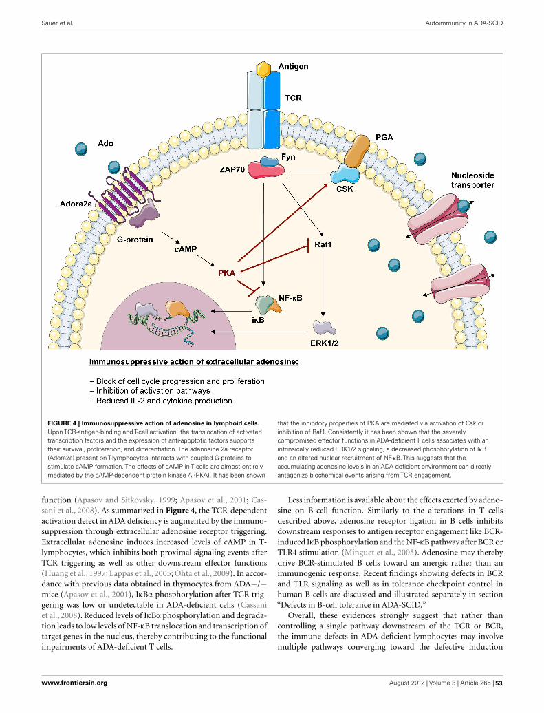
Sauer et al. Autoimmunity in ADA-SCID
FIGURE 4 | Immunosuppressive action of adenosine in lymphoid cells.Upon TCR-antigen-binding and T-cell activation, the translocation of activatedtranscription factors and the expression of anti-apoptotic factors supportstheir survival, proliferation, and differentiation. The adenosine 2a receptor(Adora2a) present on T-lymphocytes interacts with coupled G-proteins tostimulate cAMP formation. The effects of cAMP in T cells are almost entirelymediated by the cAMP-dependent protein kinase A (PKA). It has been shown
that the inhibitory properties of PKA are mediated via activation of Csk orinhibition of Raf1. Consistently it has been shown that the severelycompromised effector functions in ADA-deficient T cells associates with anintrinsically reduced ERK1/2 signaling, a decreased phosphorylation of IκBand an altered nuclear recruitment of NF-κB. This suggests that theaccumulating adenosine levels in an ADA-deficient environment can directlyantagonize biochemical events arising from TCR engagement.
function (Apasov and Sitkovsky, 1999; Apasov et al., 2001; Cas-sani et al., 2008). As summarized in Figure 4, the TCR-dependentactivation defect in ADA deficiency is augmented by the immuno-suppression through extracellular adenosine receptor triggering.Extracellular adenosine induces increased levels of cAMP in T-lymphocytes, which inhibits both proximal signaling events afterTCR triggering as well as other downstream effector functions(Huang et al., 1997; Lappas et al., 2005; Ohta et al., 2009). In accor-dance with previous data obtained in thymocytes from ADA−/−mice (Apasov et al., 2001), IκBα phosphorylation after TCR trig-gering was low or undetectable in ADA-deficient cells (Cassaniet al., 2008). Reduced levels of IκBα phosphorylation and degrada-tion leads to low levels of NF-κB translocation and transcription oftarget genes in the nucleus, thereby contributing to the functionalimpairments of ADA-deficient T cells.
Less information is available about the effects exerted by adeno-sine on B-cell function. Similarly to the alterations in T cellsdescribed above, adenosine receptor ligation in B cells inhibitsdownstream responses to antigen receptor engagement like BCR-induced IκB phosphorylation and the NF-κB pathway after BCR orTLR4 stimulation (Minguet et al., 2005). Adenosine may therebydrive BCR-stimulated B cells toward an anergic rather than animmunogenic response. Recent findings showing defects in BCRand TLR signaling as well as in tolerance checkpoint control inhuman B cells are discussed and illustrated separately in section“Defects in B-cell tolerance in ADA-SCID.”
Overall, these evidences strongly suggest that rather thancontrolling a single pathway downstream of the TCR or BCR,the immune defects in ADA-deficient lymphocytes may involvemultiple pathways converging toward the defective induction
www.frontiersin.org August 2012 | Volume 3 | Article 265 | 53

Sauer et al. Autoimmunity in ADA-SCID
of lymphocyte activation. They also illustrate how extracellu-lar adenosine levels can interfere with the downstream signal-ing transduction upon activation, thereby exerting its immuno-suppressive activity on the transcriptional machinery. Becauseof cell-specific expression and regulation, aberrant adenosinereceptor-mediated signaling might also contribute to the occur-rence of autoimmune manifestations observed in some ADA-SCIDpatients (Kohn, 1996; Ozsahin et al., 1997).
ROLE OF ATP AND OTHER PURINERGIC RECEPTORSStimulation of almost all mammalian cell types leads to the releaseof cellular ATP and autocrine feedback through a diverse array ofpurinergic receptors (Junger, 2011). ATP binds to two classes ofpurinergic P2 receptors in the plasma membrane of eukaryoticcells: P2X receptors, which are ligand-gated ion channels, and het-erotrimeric G protein-coupled P2Y receptors (Schenk et al., 2011).Depending on the types of purinergic receptors that are involved,autocrine signaling can promote or inhibit immune cell activationand fine-tune adaptive immune responses (Junger, 2011; Schenket al., 2011).
In addition to the autocrine feedback mechanisms that reg-ulate the function of healthy immune cells, purinergic receptorsallow immune cells to recognize ATP that is released from dam-aged or stressed host cells. Thus, the purinergic signaling systemsof immune cells serve an important function in the recognition ofdanger signals. ATP that is released by stressed cells guides phago-cytes to inflammatory sites and promotes clearance of damagedand apoptotic cells (Elliott et al., 2009; Junger, 2011).
To date, little information is available on alterations in theATP-induced regulation of immune cells in ADA deficiency. Itis reported that dATP accumulation in the absence of ADA leadsto a cellular depletion of ATP (Siaw et al., 1980; Simmonds et al.,1982, 1984; Koller et al., 1984). The pool of extracellular ATP onthe other hand might well be augmented in ADA-deficient lym-phoid organs, due to the increased percentage of cells undergoingapoptosis. It can therefore be hypothesized that alterations in ATPconcentrations in ADA deficiency also influence T-cell responseson the level of TCR induced activation and in response to stimulifrom an inflammatory microenvironment.
BREAK OF TOLERANCE AND CONTRIBUTION OFLYMPHOCYTES TO AUTOIMMUNITY IN ADA DEFICIENCYAdaptive immunity requires sophisticated regulatory mechanismsto ensure protection to a variety of pathogenic microbes whilemaintaining immune self-tolerance and preventing autoimmunity(Sakaguchi et al., 2008). The main mechanisms for the inductionand maintenance of a self-tolerant repertoire, which is diverse inantigen recognition, are central and peripheral tolerance. Centraltolerance is the mechanism able to eliminate newly developingT cells and B cells that have high affinity to self (Mathis andBenoist, 2004). Central tolerance is distinct from peripheral tol-erance in that it occurs while cells are still present in the primarylymphoid organs, whereas emigrant cells are controlled throughperipheral tolerance mechanisms, after they reach the periphery(Wardemann and Nussenzweig, 2007; Klein et al., 2009). Theseinclude suppression of autoreactive cells by regulatory T cells andthe generation of hyporesponsiveness (anergy) in lymphocytes,
which encounter antigen in the absence of the co-stimulatorysignals that accompany inflammation (Meffre and Wardemann,2008).
Numerous mechanisms have been proposed to explain thebreak of tolerance and development of autoimmune manifes-tations, such as defective negative selection of autoreactive T-lymphocytes in the thymus, alterations in the number and/orfunction of regulatory T cells, defects of the central and peripheralB-cell tolerance checkpoints, impaired apoptosis of autoreactivelymphocytes, break of tolerance due to increased or decreasedclearance of apoptotic cells and pathogens, or increased homeosta-tic lymphoid proliferation and cytokine secretion associated withlymphopenia (Carneiro-Sampaio and Coutinho, 2007; Westerberget al., 2008; Notarangelo, 2009; Meffre, 2012).
T-CELL TOLERANCECentral T-cell tolerance mechanisms are based on the eliminationor negative selection of the majority of T cells recognizing selfwith high affinity for negative selection in the thymus. Nonethe-less thymic selection is not a tight process and T cells expressinglow-avidity TCR on their cell surface are frequently released in theperiphery, where they are potentially dangerous to the host as theycan be effectively recruited into an autoimmune response (Parishand Heath, 2008).
A major cause of tolerance breakdown is associated with lym-phopenia (Daikeler and Tyndall, 2007). This typical state of pri-mary immunodeficiencies may contribute to the induction ofspontaneous homeostatic proliferation of residual T cells allowingperipheral expansion of autoreactive cells with a skewed reper-toire. Particularly, after conditioning or transplantation these cellsmay persist, since insufficient thymic reconstitution may affect thecontrol of self-reactivity due to defective negative selection in thethymus and/or reduced regulatory T-cell development and func-tion (Hauri-Hohl et al., 2007). In the case of ADA deficiency, it hasbeen hypothesized that the structure and functions of the thymicmicroenvironment might be altered, either directly, by toxicityof purine metabolites, or indirectly, by failure of T cells arrestedin their development to deliver supportive signals to the thymicstroma (Apasov et al., 2001).
Peripheral tolerance depends on the balance between immuneresponses to invading pathogens and immune tolerance to self-antigens. In the context of tissue damage and frequently occurringinfections in primary immunodeficient patients, apoptotic cellsrepresent a major source of autoantigen. Since apoptosis plays amajor role in the deletion of autoreactive lymphocytes and theremoval of virus-infected cells, defects in cell death have beenimplicated in the development of autoimmune diseases and per-sistent viral infection (Utz et al., 2000). The release of self-antigeninto the intracellular space and their presentation mediated bydendritic cells or other antigen-presenting cells may prime naiveautoreactive T cells, which were not eliminated by depletion oranergy (Waldner et al., 2004). Several mechanisms exist, includ-ing a spectrum of CD4+ regulatory T cells (Tregs), to suppressself-reactive T cells that escape thymic clonal deletion and atten-uate anti-pathogen effector mechanisms from inducing immunepathology (Piccirillo and Thornton, 2004). There is ample evi-dence that Tregs actively mediate suppression to control immune
Frontiers in Immunology | Primary Immunodeficiencies August 2012 | Volume 3 | Article 265 | 54
Sauer et al. Autoimmunity in ADA-SCID
responses to self- and non-self-antigens and the onset of autoim-munity (Bach,2003; Sakaguchi,2005). Lessons from other primaryimmunodeficiencies have provided unequivocal evidence for theessential role of Tregs in suppressing autoreactive T cells in theperiphery (Westerberg et al., 2008). Rising of autoimmunity maynot only be linked to a reduction in Treg numbers but also to atten-uation of their suppressive activity (Sakaguchi et al., 2008). Whilethis is principally mediated by cell–cell contact, recent findingsrevealed additional mechanisms of Treg-mediated suppression,including secretion of immunosuppressive cytokines, functionalmodification or killing of APC, and metabolic disruption (Vig-nali et al., 2008). Moreover, extracellular adenosine produced byTregs, has been identified as one of the mechanisms mediatingtheir suppressive activity (Sitkovsky et al., 2008; Mandapathil et al.,2010). Treg cells possess a unique biochemical signature amongstT cells in that they generate and sustain high adenosine con-centrations. Since Tregs primarily mediate peripheral control ofautoreactive T cells, it is conceivable that this compartment mightbe specifically affected in ADA-SCID (see also Defective RegulatoryT Cell Function in ADA Deficiency). Consequently the autoim-mune manifestations associated with ADA deficiency might bethe result of an altered purine metabolism interfering with normalregulatory T-cell function (Sauer et al., 2012a).
B-CELL TOLERANCEA variety of mechanisms ranging from clonal deletion to func-tional inactivation by anergy of autoreactive B cells serve to shapethe peripheral B-cell repertoire. Nevertheless, dysregulation of Bcell development and autoantibody production is a characteris-tic of most autoimmune diseases including rheumatoid arthri-tis, systemic lupus erythematosus, and type 1 diabetes, but alsoimmunodeficiencies such as CVID, Wiskott–Aldrich Syndrome,and X-linked agammaglobulinemia (Park et al., 2005; Cuss et al.,2006; Westerberg et al., 2008). In ADA deficiency, some of theobserved immune dysregulation were hypothesized to be associ-ated with a more restricted B-cell repertoire due to abnormalitiesin central B-cell generation or to a dysregulated expansion of thesecells in the periphery.
Autoantibodies appear in the serum many years before theonset of clinical disease suggesting an early break in B-celltolerance (Wardemann et al., 2003). Some of B-cell mediatedautoimmune diseases, such as myasthenia gravis (MG), idiopathicautoimmune thrombocytopenic purpura (AITP), and Graves’ dis-ease are characterized by auto-Abs production that destroy targettissues (Barsalou et al., 2011; Cunningham-Rundles, 2011). Aremarkably high proportion of autoantibodies associated with sys-temic autoimmune diseases binds DNA, RNA, or macromolecularcomplexes that contain DNA or RNA. It has been hypothesized,that under certain circumstances these intracellular autoantigensbecome visible to the immune system when they accumulate dur-ing apoptosis. In fact the impaired clearance of apoptotic celldebris and dsDNA by macrophages might induce TLR signal-ing and differentiation of autoreactive B cells (Gaipl et al., 2006).Response to nucleic acid-containing immunecomplexes relies onthe coengagement of endosomal members of the TLR family, TLR9and TLR7 (Marshak-Rothstein, 2006). Therefore, self-antigensthat can effectively engage both the BCR and either TLR7 or TLR9
might stimulate autoreactive B cells that are normally quiescent,through inherent adjuvant activity and trigger the development ofsystemic autoimmune disease (Marshak-Rothstein, 2006). In ADAdeficiency, the metabolic basis underlying immune cell deficiencyis the cytotoxic effect impact of the ADA substrates deoxyadeno-sine and dATP, leading to apoptosis of lymphocytes. It is thereforeconceivable that developing B lymphocytes in affected lymphoidorgans encounter massive amounts of nucleic acid. Nucleic acid-sensing TLRs might therefore represent Achilles’heel in susceptibleADA-deficient patients by which relative tolerance for nucleic acid-containing antigens is breached and autoimmunity occurs (Konoet al., 2009).
NEW INSIGHTS INTO IMMUNE CELL DYSFUNCTION ANDONSET OF AUTOIMMUNITY IN ADA DEFICIENCYRecent in-depth studies have revealed specific defects in ADAdeficiency that may contribute to the onset of autoimmunity inthese patients. Herein we discuss alterations in the adenosiner-gic machinery of ADA-deficient regulatory T cells and in B-celltolerance in the absence of functional ADA.
DEFECTIVE REGULATORY T-CELL FUNCTION IN ADA DEFICIENCYAlthough autoimmune manifestations are frequent findingsin ADA-deficient patients with milder forms or in patientsunder PEG-ADA, mechanisms causing the loss of peripheraltolerance and onset of autoimmunity have remained elusive.CD4+CD25+FoxP3+ Tregs actively suppress pathological andphysiological immune responses in order to maintain periph-eral immune self-tolerance and prevent autoimmunity (Sakaguchiet al., 2008; Sitkovsky et al., 2008). Extracellular adenosine pro-duced by Tregs has been described as one of the mechanismsmediating their suppressive activity (Figure 5A). Concordantexpression of the ectoenzymes CD39 and CD73 has been reportedboth for murine and human Tregs (Borsellino et al., 2007; Deaglioet al., 2007; Mandapathil et al., 2010). The CD39 ectoenzyme pro-duces AMP from ATP or ADP, which is subsequently convertedinto extracellular adenosine by the CD73 ectoenzyme (Hasko et al.,2008). Treg function requires the coordinated expression of theAdora2a on activated T effector cells to enable adenosine-mediatedimmunosuppression (Sitkovsky et al., 2008). Moreover, Tregs havebeen shown to express low levels of ADA, whereas T effector cellsare enriched in ADA but express low levels of CD39 and CD73(Mandapathil et al., 2010; Sauer et al., 2012a). This molecular pro-file of Tregs (CD39+CD73+ADAlow) has functional importance,as it not only confers Tregs the capability to produce extracellularadenosine but also to sustain relatively high concentrations due tolow ADA expression (Mandapathil et al., 2010).
Figure 5B summarizes recently described defects and func-tional alterations of the adenosinergic pathway in Tregs from ADA-deficient mice and patients (Sauer et al., 2012a). ADA−/− Tregsshowed significantly higher expression of CD39, while expressingsignificantly less CD73. ADA−/− Tregs are sensitive to extra-cellular adenosine concentrations and the expression of CD73is regulated by this metabolite. With adenosine accumulating inADA−/− mice, possibly to avoid a further increase of extracellu-lar concentrations, CD73 is reduced and ADA−/− Tregs display a
www.frontiersin.org August 2012 | Volume 3 | Article 265 | 55

Sauer et al. Autoimmunity in ADA-SCID
FIGURE 5 | Loss of regulatoryT-cell function in ADA-SCID. (A) Byconcomitant expression of CD39 and CD73, Tregs have the enzymaticmachinery to generate and maintain high levels of extracellular adenosine.Contrarily to T effector cells, Tregs express low levels of ADA and CD26.Extracellular ATP or ADP is converted by the ectonucleotidase CD39 intoAMP, which is further converted into adenosine by the CD73 ectoenzyme.The produced adenosine binds to the Adora2A receptor expressed onactivated effector T cells, which are enriched in ADA and the surface-boundglycoprotein CD26. The coordinated expression of CD39 and CD73 on Tregsand Adora2a on T effector cells enables adenosine-mediatedimmunosuppression. (B) In the absence of ADA, deficient Tregs expresshigh levels of CD39, increasing their capacity for ATP hydrolysis, butreduced levels of CD73 on their surface. Although both CD39 and CD73 arerate limiting for extracellular adenosine generation, CD73 is the lastcomponent of the ectoenzymatic chain. With accumulating adenosine
levels, possibly to avoid a further increase of extracellular concentrations,CD73 is reduced and ADA−/−Tregs display a decreased suppressiveactivity toward T effector cells. Nevertheless no apparent autoimmunemanifestations can be observed at onset, likely due to the severely reducedeffector T- and B-cell populations. Moreover, the accumulating extracellularadenosine is likely to maintain an anti-inflammatory environment in thesemice. (C) After PEG-ADA treatment, the adenosinergic machinery of CD39and CD73 are upregulated, indicating an increased requirement for ATPhydrolysis and enhanced adenosine production. Despite the initial rescue ofsuppressive activity by upregulation of CD73 for elevated adenosineproduction, long-term PEG-ADA treatment interferes with Treg function byaugmenting adenosine turnover. PEG-ADA present in the extracellular spaceeliminates adenosine produced by the ectoenzymatic chain and hindersadenosine-mediated suppression by interfering between adenosine andAdora2a expressed on T effector cells.
Frontiers in Immunology | Primary Immunodeficiencies August 2012 | Volume 3 | Article 265 | 56

Sauer et al. Autoimmunity in ADA-SCID
decreased suppressive activity toward T effector cells. The under-lying mechanism accounting for increased CD39 expression inADA−/− Tregs remains to be elucidated. However, intracellularcAMP levels, which are elevated in the absence of ADA, have beenreported to increase CD39 expression (Liao et al., 2010).
In order to dissect the cellular mechanisms leading to lossof peripheral tolerance, ADA−/− mice were studied after treat-ment with PEG-ADA, HSC-GT, and BMT. Although short-termPEG-ADA treatment initially rescued Treg-mediated suppressionin comparison to untreated ADA−/− mice, their functional-ity became exhausted by long-term PEG-ADA treatment. Tregsfrom PEG-ADA treated animals maintained increased expres-sion of CD39 and upregulated CD73 expression in comparisonto age-matched wildtype controls. Consistently, CD39 activitymeasured by ATP consumption and AMP formation, as wellas adenosine production by CD73 were significantly increasedin comparison with wildtype Tregs. These results were con-firmed in a cohort of patients including 7 PEG-ADA treatedand 11 retroviral HSC-GT treated patients. The percentageof CD4+CD25+FOXP3+CD127−/low Tregs was significantlyreduced in PEG-ADA treated patients and their expression ofCD39 and CD73 ectonucleotidase were significantly increased.Unlike Tregs from HSC-GT treated patients and HD, Tregs iso-lated from PEG-ADA treated patients were unable to suppress theproliferation of effector cells (Sauer et al., 2012a).
The obtained results revealed an elevated adenosine catab-olism in the presence of PEG-ADA, characterized by alter-ations in the adenosinergic machinery producing high levels ofadenosine and a significantly increased turnover by the enzy-matic activity of PEG-ADA. Upregulation of CD73 in treatedADA−/− mice and patients can therefore be interpreted as acompensatory mechanism representing a higher requirement forATP/ADP to adenosine conversion in the presence of extracel-lular PEG-ADA. Despite the initial rescue of suppressive activ-ity by upregulation of CD73 for elevated adenosine production,long-term PEG-ADA treatment interfered with Treg function byaugmenting adenosine turnover. These findings fit the hypoth-esis that PEG-ADA present in the extracellular space eliminatesadenosine produced by this ectoenzymatic chain and hindersadenosine-mediated suppression by interfering between adeno-sine and Adora2a expressed on T effector cells (Sauer et al., 2012a;Figure 5C).
DEFECTS IN B-CELL TOLERANCE IN ADA-SCIDAlthough PEG-ADA induces metabolic detoxification, BMT andHSC-GT provide superior restoration of purine metabolismand immune functions. However, it had remained unclear howpatient’s B cells contribute to autoimmune complications and ifB-cell tolerance is established properly in ADA-deficient patientsbefore and after treatment.
Random V(D)J recombination produces large numbers of anti-bodies displaying self-reactive specificities and during normalB-cell development the majority of these antibodies are removedat two distinct checkpoints in the bone marrow and periphery(Wardemann et al., 2003). Large numbers of self-reactive antibod-ies are removed from the B cell repertoire during the immatureB cell stage in the bone marrow, where BCR-mediated selection
plays a crucial role in controlling B-cell survival based on exces-sively strong or weak BCR signals that identify autoreactive orfunctionally unfit B cells (Goodnow, 1996; Nemazee et al., 2000;Cancro, 2009; Figure 6). Alterations of BCR signaling thresh-olds result in a defective central B-cell tolerance checkpoint andinterfere with the removal of developing autoreactive B cells inhumans (Ng et al., 2004; Menard et al., 2011). In addition to theirBCRs, B cells also express TLRs that were originally described tobind microbial components but that are also able to recognizeself-antigens (Marshak-Rothstein, 2006) and are involved in theremoval of developing anti-nuclear antibody (ANA)-expressingB cells (Isnardi et al., 2008). Both BCR- and TLR-mediated B-cellresponses have been reported to be modulated by adenosine recep-tor signaling and intracellular cyclic adenosine monophospate(cAMP), which are increased in ADA deficiency (Apasov et al.,2001; Hershfield, 2005; Minguet et al., 2005; Power Coombs et al.,2011).
B-cell tolerance checkpoints in ADA-SCID patients wereassessed by cloning antibodies expressed by single B cells beforeand after successful HSC-GT (Sauer et al., 2012b). New emi-grant/transitional and mature naïve B cells from ADA-deficientpatients before HSC-GT contained high frequencies of autore-active and ANA-expressing clones compared to healthy donors,revealing defective central and peripheral B-cell tolerance check-points in the absence of functional ADA.
The receptor candidates for the removal of ANA-expressingclones are nucleic acid-sensing endosomal members of the TLRfamily, TLR7 and TLR9 (Marshak-Rothstein, 2006), thereby sug-gesting that ADA impinges not only on BCR but more importantlyTLR signaling. A similar mechanism has also been hypothesizedto contribute to B-cell dysfunctions, defective B-cell proliferation,and activation observed in ADA-deficient mice (Aldrich et al.,2003; Hershfield, 2005). The accumulating adenosine blocks NF-κB activation in murine B cells stimulated through BCRs or TLR4by LPS (Minguet et al., 2005; Power Coombs et al., 2011). In linewith this hypothesis, we found that stimulation through TLR7and TLR9 were significantly dependent on proper ADA enzymaticactivity and adenosine receptor engagement, further underlin-ing the inability of these receptors to function in the absence offunctional ADA (Figure 6).
Strikingly, ADA-deficient patients treated with HSC-GT dis-played quasi-normal early B-cell tolerance checkpoints as evi-denced by restored efficient removal of developing autoreactiveand anti-nuclear B cells. Hence, ADA plays an essential role in theestablishment of early B-cell tolerance and the removal of devel-oping autoreactive B cells in humans (Luning Prak, 2012; Saueret al., 2012b).
CONCLUDING REMARKS ON THE OCCURRENCE OFAUTOIMMUNITY AFTER ADA-SCID TREATMENTIn summary, the available literature provides supporting evidencefor a predisposition to autoimmunity in ADA deficiency. Alter-ations in both central and peripheral tolerance in T- and B-cellshave been described to contribute to the pathogenesis of autoim-munity. Moreover, it is becoming increasingly clear that tolerancemechanisms and immune responses are specifically altered by thelack of ADA and the accumulation of its substrates.
www.frontiersin.org August 2012 | Volume 3 | Article 265 | 57
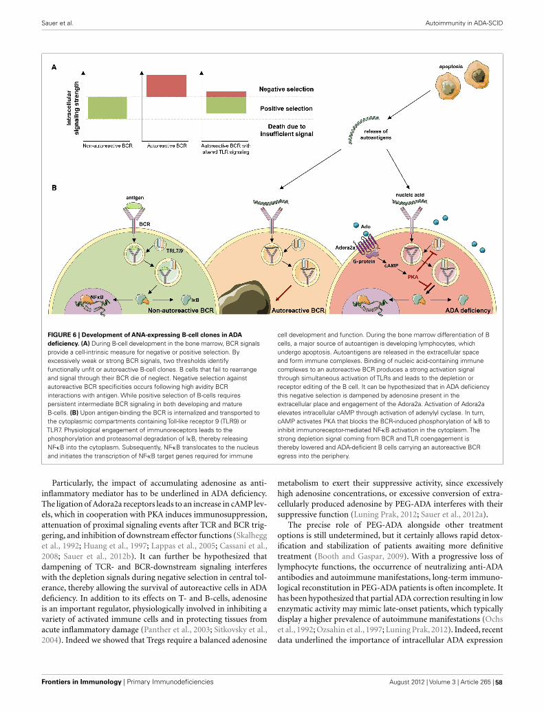
Sauer et al. Autoimmunity in ADA-SCID
FIGURE 6 | Development of ANA-expressing B-cell clones in ADAdeficiency. (A) During B-cell development in the bone marrow, BCR signalsprovide a cell-intrinsic measure for negative or positive selection. Byexcessively weak or strong BCR signals, two thresholds identifyfunctionally unfit or autoreactive B-cell clones. B cells that fail to rearrangeand signal through their BCR die of neglect. Negative selection againstautoreactive BCR specificities occurs following high avidity BCRinteractions with antigen. While positive selection of B-cells requirespersistent intermediate BCR signaling in both developing and matureB-cells. (B) Upon antigen-binding the BCR is internalized and transported tothe cytoplasmic compartments containing Toll-like receptor 9 (TLR9) orTLR7. Physiological engagement of immunoreceptors leads to thephosphorylation and proteasomal degradation of IκB, thereby releasingNF-κB into the cytoplasm. Subsequently, NF-κB translocates to the nucleusand initiates the transcription of NF-κB target genes required for immune
cell development and function. During the bone marrow differentiation of Bcells, a major source of autoantigen is developing lymphocytes, whichundergo apoptosis. Autoantigens are released in the extracellular spaceand form immune complexes. Binding of nucleic acid-containing immunecomplexes to an autoreactive BCR produces a strong activation signalthrough simultaneous activation of TLRs and leads to the depletion orreceptor editing of the B cell. It can be hypothesized that in ADA deficiencythis negative selection is dampened by adenosine present in theextracellular place and engagement of the Adora2a. Activation of Adora2aelevates intracellular cAMP through activation of adenylyl cyclase. In turn,cAMP activates PKA that blocks the BCR-induced phosphorylation of IκB toinhibit immunoreceptor-mediated NF-κB activation in the cytoplasm. Thestrong depletion signal coming from BCR and TLR coengagement isthereby lowered and ADA-deficient B cells carrying an autoreactive BCRegress into the periphery.
Particularly, the impact of accumulating adenosine as anti-inflammatory mediator has to be underlined in ADA deficiency.The ligation of Adora2a receptors leads to an increase in cAMP lev-els, which in cooperation with PKA induces immunosuppression,attenuation of proximal signaling events after TCR and BCR trig-gering, and inhibition of downstream effector functions (Skalhegget al., 1992; Huang et al., 1997; Lappas et al., 2005; Cassani et al.,2008; Sauer et al., 2012b). It can further be hypothesized thatdampening of TCR- and BCR-downstream signaling interfereswith the depletion signals during negative selection in central tol-erance, thereby allowing the survival of autoreactive cells in ADAdeficiency. In addition to its effects on T- and B-cells, adenosineis an important regulator, physiologically involved in inhibiting avariety of activated immune cells and in protecting tissues fromacute inflammatory damage (Panther et al., 2003; Sitkovsky et al.,2004). Indeed we showed that Tregs require a balanced adenosine
metabolism to exert their suppressive activity, since excessivelyhigh adenosine concentrations, or excessive conversion of extra-cellularly produced adenosine by PEG-ADA interferes with theirsuppressive function (Luning Prak, 2012; Sauer et al., 2012a).
The precise role of PEG-ADA alongside other treatmentoptions is still undetermined, but it certainly allows rapid detox-ification and stabilization of patients awaiting more definitivetreatment (Booth and Gaspar, 2009). With a progressive loss oflymphocyte functions, the occurrence of neutralizing anti-ADAantibodies and autoimmune manifestations, long-term immuno-logical reconstitution in PEG-ADA patients is often incomplete. Ithas been hypothesized that partial ADA correction resulting in lowenzymatic activity may mimic late-onset patients, which typicallydisplay a higher prevalence of autoimmune manifestations (Ochset al., 1992; Ozsahin et al., 1997; Luning Prak, 2012). Indeed, recentdata underlined the importance of intracellular ADA expression
Frontiers in Immunology | Primary Immunodeficiencies August 2012 | Volume 3 | Article 265 | 58

Sauer et al. Autoimmunity in ADA-SCID
and superior efficacy of gene therapy over PEG-ADA treatment forthe development of functional T- and B-cell tolerance, includingTregs (Sauer et al., 2012a,b).
Both BMT and HSC-GT provide efficient reconstitution ofthe immune system through endogenous enzymatic ADA activ-ity. BMT from an HLA-identical sibling donor remains thetreatment of choice, while transplants from alternative donorsare associated with high morbidity and mortality. The occur-rence of mixed chimerism in transplanted patients with otherprimary immunodeficiencies is associated with a higher inci-dence of autoimmune manifestations (Moratto et al., 2011) andmight well play a role also in ADA deficiency (Cancrini et al.,2010). Moreover, transplantation-induced lymphopenia is a pos-sible cause for the homeostatic expansion of autoreactive T-and/or B-cells with subsequent loss of self-tolerance (Daikelerand Tyndall, 2007). Phenomena of immune dysregulation asoccurring in the context of pre-transplant conditioning andBMT might further trigger the onset of autoimmunity (Etzioni,2003).
In accordance with the current guidelines of the Europeangroup for Blood and Marrow Transplantation (EBMT) and giventhe long-term experience in gene therapy (Aiuti et al., 2009), thistreatment can now be considered for all ADA-SCID patients lack-ing an HLA-identical sibling donor (Gaspar et al., 2009). AfterHSC-GT, high levels (50–90% on average) of gene correction weredetected in T- and B- and NK-cells (Aiuti et al., 2009), leadingto an efficient systemic detoxification and recovery of immune
cell functions. However, as suggested by the cloning of singleB-cell receptors, non-gene corrected cells may carry autoreactivespecificities (Sauer et al., 2012b). It can be hypothesized that theco-existence of non-corrected autoreactive T- or B-cells and therestored functional T cell help could allow the development ofautoimmune manifestations in ADA-SCID patients after HSC-GT (Aiuti et al., 2009). Modification of preparatory regimen orincreased gene transfer efficiency by more robust approaches suchas lentiviral vectors may further improve HSC-GT outcome forADA deficiency (Mortellaro et al., 2006).
Adenosine deaminase-SCID remains a challenging conditionto treat (Gaspar et al., 2009). With large-scale outcome studies stilllacking, the choice between lifelong PEG-ADA, unrelated BMTand HSC-GT is currently based on the risk/benefit ratio, the avail-ability, and costs of the three different treatment options (Gasparet al., 2009). Due to the rarity of the disease and the small cohortnumbers, accurate survey and long-term follow-up will be essen-tial to determine the outcome following different treatments andtheir efficacy in restoring immune tolerance.
ACKNOWLEDGMENTSSupported by grants from the Italian Telethon Foundation:TIGET, core grant A1; the European Commission: Advanced Cell-based Therapies for the treatment of Primary ImmunoDeficiency(Cell-PID), HEALTH F5-2010-261387; and the Ministero dellaSalute: Ricerca Finalizzata, 005/RF-2009-1485896. Figures 2–6were produced using Servier Medical Art.
REFERENCESAbuchowski, A., van Es, T., Palczuk, N.
C., and Davis, F. F. (1977). Alter-ation of immunological propertiesof bovine serum albumin by cova-lent attachment of polyethylene gly-col. J. Biol. Chem. 252, 3578–3581.
Aiuti, A. (2004). Gene therapy foradenosine-deaminase-deficientsevere combined immunode-ficiency. Best Pract. Res. Clin.Haematol. 17, 505–516.
Aiuti, A., Cassani, B., Andolfi, G.,Mirolo, M., Biasco, L., Recchia, A.,Urbinati, F.,Valacca, C., Scaramuzza,S., Aker, M., Slavin, S., Cazzola, M.,Sartori, D., Ambrosi, A., Di Serio,C., Roncarolo, M. G., Mavilio, F.,and Bordignon, C. (2007). Multilin-eage hematopoietic reconstitutionwithout clonal selection in ADA-SCID patients treated with stem cellgene therapy. J. Clin. Invest. 117,2233–2240.
Aiuti, A., Cattaneo, F., Galimberti, F.,Benninghoff, U., Cassani, B., Cal-legaro, L., Scaramuzza, S., Andolfi,G., Mirolo, M., Brigida, I., Tabuc-chi, A., Carlucci, F., Eibl, M., Aker,M., Slavin, S., Al-Mousa, H., AlGhonaium, A., Ferster, A., Dup-penthaler, A., Notarangelo, L., Win-tergerst, U., Buckley, R., Bregni, M.,Valsecchi, M., Rossi, P., Ciceri, F.,Miniero, R., Bordignon, C., andRoncarolo, M. (2009). Long-term
safety and efficacy of gene therapyfor adenosine deaminase (ADA)-deficient severe combined immun-odeficiency. N. Engl. J. Med. 360,447–458.
Aiuti, A., Ficara, F., Cattaneo, F., Bor-dignon, C., and Roncarolo, M. G.(2003). Gene therapy for adeno-sine deaminase deficiency. Curr.Opin. Allergy Clin. Immunol. 3,461–466.
Aldrich, M. B., Chen, W., Blackburn,M. R., Martinez-Valdez, H., Datta,S. K., and Kellems, R. E. (2003).Impaired germinal center matura-tion in adenosine deaminase defi-ciency. J. Immunol. 171, 5562–5570.
Antoine, C., Muller, S., Cant, A.,Cavazzana-Calvo, M., Veys, P.,Vossen, J., Fasth, A., Heilmann, C.,Wulffraat, N., Seger, R., Blanche, S.,Friedrich, W., Abinun, M., Davies,G., Bredius, R., Schulz, A., Landais,P., and Fischer, A. (2003). Long-term survival and transplantationof haemopoietic stem cells forimmunodeficiencies: report of theEuropean experience 1968–1999.Lancet 361, 553–560.
Apasov, S. G., Blackburn, M. R., Kellems,R. E., Smith, P. T., and Sitkovsky,M. V. (2001). Adenosine deaminasedeficiency increases thymic apopto-sis and causes defective T cell recep-tor signaling. J. Clin. Invest. 108,131–141.
Apasov, S. G., and Sitkovsky, M.V. (1999). The extracellular ver-sus intracellular mechanisms ofinhibition of TCR-triggered acti-vation in thymocytes by adeno-sine under conditions of inhibitedadenosine deaminase. Int. Immunol.11, 179–189.
Bach, J. F. (2003). Regulatory T cellsunder scrutiny. Nat. Rev. Immunol.3, 189–198.
Barsalou, J., Saint-Cyr, C., Drouin, E.,Le Deist, F., and Haddad, E. (2011).High prevalence of primary immunedeficiencies in children with autoim-mune disorders. Clin. Exp. Rheuma-tol. 29, 125–130.
Belardinelli, L., Linden, J., and Berne,R. M. (1989). The cardiac effects ofadenosine. Prog. Cardiovasc. Dis. 32,73–97.
Benveniste, P., Zhu, W., and Cohen, A.(1995). Interference with thymocytedifferentiation by an inhibitor of S-adenosylhomocysteine hydrolase. J.Immunol. 155, 536–544.
Blackburn, M. R. (2003). Too much ofa good thing: adenosine overloadin adenosine-deaminase-deficientmice. Trends Pharmacol. Sci. 24,66–70.
Blackburn, M. R., Aldrich, M., Volmer,J. B., Chen, W., Zhong, H., Kelly,S., Hershfield, M. S., Datta, S.K., and Kellems, R. E. (2000).The use of enzyme therapy to
regulate the metabolic and phe-notypic consequences of adenosinedeaminase deficiency in mice. Dif-ferential impact on pulmonary andimmunologic abnormalities. J. Biol.Chem. 275, 32114–32121.
Blackburn, M. R., Datta, S. K., andKellems, R. E. (1998). Adenosinedeaminase-deficient mice generatedusing a two-stage genetic engineer-ing strategy exhibit a combinedimmunodeficiency. J. Biol. Chem.273, 5093–5100.
Bollinger, M. E., Arredondo-Vega, F. X.,Santisteban, I., Schwarz, K., Hersh-field, M. S., and Lederman, H. M.(1996). Brief report: hepatic dys-function as a complication of adeno-sine deaminase deficiency. N. Engl. J.Med. 334, 1367–1371.
Booth, C., and Gaspar, H. B. (2009).Pegademase bovine (PEG-ADA) forthe treatment of infants and chil-dren with severe combined immun-odeficiency (SCID). Biologics 3,349–358.
Booth, C., Hershfield, M., Notarangelo,L., Buckley, R., Hoenig, M.,Mahlaoui, N., Cavazzana-Calvo,M., Aiuti, A., and Gaspar, H.B. (2007). Management optionsfor adenosine deaminase defi-ciency; proceedings of the EBMTsatellite workshop (Hamburg,March 2006). Clin. Immunol. 123,139–147.
www.frontiersin.org August 2012 | Volume 3 | Article 265 | 59
Sauer et al. Autoimmunity in ADA-SCID
Bordignon, C., and Roncarolo, M.G. (2002). Therapeutic applica-tions for hematopoietic stem cellgene transfer. Nat. Immunol. 3,318–321.
Borsellino, G., Kleinewietfeld, M., DiMitri, D., Sternjak, A., Diamantini,A., Giometto, R., Hopner, S., Cen-tonze, D., Bernardi, G., Dell’acqua,M. L., Rossini, P. M., Battistini,L., Rotzschke, O., and Falk, K.(2007). Expression of ectonucleoti-dase CD39 by Foxp3+ Treg cells:hydrolysis of extracellular ATP andimmune suppression. Blood 110,1225–1232.
Buckley, R. H., Schiff, R. I., Schiff,S. E., Markert, M. L., Williams,L. W., Harville, T. O., Roberts,J. L., and Puck, J. M. (1997).Human severe combined immunod-eficiency: genetic, phenotypic, andfunctional diversity in one hun-dred eight infants. J. Pediatr. 130,378–387.
Cancrini, C., Ferrua, F., Scarselli, A.,Brigida, I., Romiti, M. L., Bar-era, G., Finocchi, A., Roncarolo,M. G., Caniglia, M., and Aiuti, A.(2010). Role of reduced intensityconditioning in T-cell and B-cellimmune reconstitution after HLA-identical bone marrow transplanta-tion in ADA-SCID. Haematologica95, 1778–1782.
Cancro, M. P. (2009). Signallingcrosstalk in B cells: managing worthand need. Nat. Rev. Immunol. 9,657–661.
Carneiro-Sampaio, M., and Coutinho,A. (2007). Tolerance and autoim-munity: lessons at the bedside ofprimary immunodeficiencies. Adv.Immunol. 95, 51–82.
Cassani, B., Mirolo, M., Cattaneo, F.,Benninghoff, U., Hershfield, M.,Carlucci, F., Tabucchi, A., Bor-dignon, C., Roncarolo, M. G., andAiuti, A. (2008). Altered intra-cellular and extracellular signalingleads to impaired T-cell functionsin ADA-SCID patients. Blood 111,4209–4219.
Chaffee, S., Mary, A., Stiehm, E. R.,Girault, D., Fischer, A., and Hersh-field, M. S. (1992). IgG antibodyresponse to polyethylene glycol-modified adenosine deaminase inpatients with adenosine deami-nase deficiency. J. Clin. Invest. 89,1643–1651.
Chan, B., Wara, D., Bastian, J., Her-shfield, M. S., Bohnsack, J., Azen,C. G., Parkman, R., Weinberg, K.,and Kohn, D. B. (2005). Long-term efficacy of enzyme replacementtherapy for adenosine deaminase(ADA)-deficient severe combined
immunodeficiency (SCID). Clin.Immunol. 117, 133–143.
Churchill, P. C., and Bidani, A.K. (1982). Hypothesis: adenosinemediates hemodynamic changes inrenal failure. Med. Hypotheses 8,275–285.
Cohen, A., and Thompson, E. (1986).DNA repair in nondividinghuman lymphocytes: inhibition bydeoxyadenosine. Cancer Res. 46,1585–1588.
Cunningham-Rundles, C. (2011).Autoimmunity in primary immunedeficiency: taking lessons fromour patients. Clin. Exp. Immunol.164(Suppl. 2), 6–11.
Cuss, A. K., Avery, D. T., Cannons, J. L.,Yu,L. J.,Nichols,K. E.,Shaw,P. J., andTangye, S. G. (2006). Expansion offunctionally immature transitionalB cells is associated with human-immunodeficient states character-ized by impaired humoral immu-nity. J. Immunol. 176, 1506–1516.
Daikeler, T., and Tyndall, A.(2007). Autoimmunity follow-ing haematopoietic stem-celltransplantation. Best Pract. Res.Clin. Haematol. 20, 349–360.
Dave, U. P.,Akagi, K., Tripathi, R., Cleve-land, S. M., Thompson, M. A.,Yi, M.,Stephens, R., Downing, J. R., Jenkins,N. A., and Copeland, N. G. (2009).Murine leukemias with retroviralinsertions at Lmo2 are predictiveof the leukemias induced in SCID-X1 patients following retroviral genetherapy. PLoS Genet. 5, e1000491.doi:10.1371/journal.pgen.1000491
Davis, S., Abuchowski, A., Park, Y. K.,and Davis, F. F. (1981). Alterationof the circulating life and anti-genic properties of bovine adeno-sine deaminase in mice by attach-ment of polyethylene glycol. Clin.Exp. Immunol. 46, 649–652.
Deaglio, S., Dwyer, K. M., Gao, W.,Friedman, D., Usheva, A., Erat, A.,Chen, J. F., Enjyoji, K., Linden, J.,Oukka, M., Kuchroo, V. K., Strom, T.B., and Robson, S. C. (2007). Adeno-sine generation catalyzed by CD39and CD73 expressed on regulatory Tcells mediates immune suppression.J. Exp. Med. 204, 1257–1265.
Elliott, M. R., Chekeni, F. B., Tram-pont, P. C., Lazarowski, E. R., Kadl,A., Walk, S. F., Park, D., Woodson,R. I., Ostankovich, M., Sharma, P.,Lysiak, J. J., Harden, T. K., Leitinger,N., and Ravichandran, K. S. (2009).Nucleotides released by apoptoticcells act as a find-me signal to pro-mote phagocytic clearance. Nature461, 282–286.
Erdmann, A. A., Gao, Z. G., Jung, U.,Foley, J., Borenstein, T., Jacobson, K.
A., and Fowler, D. H. (2005). Acti-vation of Th1 and Tc1 cell adeno-sine A2A receptors directly inhibitsIL-2 secretion in vitro and IL-2-driven expansion in vivo. Blood 105,4707–4714.
Etzioni, A. (2003). Immune defi-ciency and autoimmunity. Autoim-mun. Rev. 2, 364–369.
Ferrua, F., Brigida, I., and Aiuti, A.(2010). Update on gene therapyfor adenosine deaminase-deficientsevere combined immunodefi-ciency. Curr. Opin. Allergy Clin.Immunol. 10, 551–556.
Fischer, A., Le Deist, F., Hacein-Bey-Abina, S., Andre-Schmutz, I., BasileGde, S., De Villartay, J. P., andCavazzana-Calvo, M. (2005). Severecombined immunodeficiency.A model disease for molecularimmunology and therapy. Immunol.Rev. 203, 98–109.
Fredholm, B. B., and Dunwiddie, T. V.(1988). How does adenosine inhibittransmitter release? Trends Pharma-col. Sci. 9, 130–134.
Fukunaga,A. F., Flacke,W. E., and Bloor,B. C. (1982). Hypotensive effects ofadenosine and adenosine triphos-phate compared with sodium nitro-prusside. Anesth. Analg. 61, 273–278.
Gaipl, U. S., Sheriff,A., Franz, S., Munoz,L. E., Voll, R. E., Kalden, J. R.,and Herrmann, M. (2006). Ineffi-cient clearance of dying cells andautoreactivity. Curr. Top. Microbiol.Immunol. 305, 161–176.
Gangi-Peterson, L., Sorscher, D. H.,Reynolds, J. W., Kepler, T. B., andMitchell, B. S. (1999). Nucleotidepool imbalance and adenosinedeaminase deficiency induce alter-ations of N-region insertions duringV(D)J recombination. J. Clin. Invest.103, 833–841.
Gaspar, H. B., Aiuti, A., Porta, F., Can-dotti, F., Hershfield, M. S., andNotarangelo, L. D. (2009). How Itreat ADA deficiency. Blood 114,3524–3532.
Gaspar, H. B., Cooray, S., Gilmour, K.C., Parsley, K. L., Zhang, F., Adams,S., Bjorkegren, E., Bayford, J., Brown,L., Davies, E. G., Veys, P., Fair-banks, L., Bordon, V., Petropolou,T., Kinnon, C., and Thrasher,A. J. (2011). Hematopoietic stemcell gene therapy for adenosinedeaminase-deficient severe com-bined immunodeficiency leads tolong-term immunological recov-ery and metabolic correction. Sci.Transl. Med. 3, 97ra80.
Giblett, E. R., Anderson, J. E., Cohen,F., Pollara, B., and Meuwissen, H. J.(1972). Adenosine-deaminase defi-ciency in two patients with severely
impaired cellular immunity. Lancet2, 1067–1069.
Goodnow, C. C. (1996). Balancingimmunity and tolerance: deletingand tuning lymphocyte repertoires.Proc. Natl. Acad. Sci. U.S.A. 93,2264–2271.
Hacein-Bey-Abina, S., Garrigue, A.,Wang, G. P., Soulier, J., Lim, A.,Morillon, E., Clappier, E., Caccavelli,L., Delabesse, E., Beldjord, K.,Asnafi,V., Macintyre, E., Dal Cortivo, L.,Radford, I., Brousse, N., Sigaux, F.,Moshous, D., Hauer, J., Borkhardt,A., Belohradsky, B. H., Winterg-erst, U., Velez, M. C., Leiva, L.,Sorensen, R., Wulffraat, N., Blanche,S., Bushman, F. D., Fischer, A., andCavazzana-Calvo, M. (2008). Inser-tional oncogenesis in 4 patientsafter retrovirus-mediated gene ther-apy of SCID-X1. J. Clin. Invest. 118,3132–3142.
Hasko, G., Linden, J., Cronstein, B., andPacher, P. (2008). Adenosine recep-tors: therapeutic aspects for inflam-matory and immune diseases. Nat.Rev. Drug Discov. 7, 759–770.
Hassan, A., Booth, C., Brightwell, A.,Allwood, Z., Veys, P., Rao, K.,Honig, M., Friedrich, W., Gennery,A., Slatter, M., Bredius, R., Finoc-chi, A., Cancrini, C., Aiuti, A.,Porta, F., Lanfranchi, A., Ridella,M., Steward, C., Filipovich, A.,Marsh, R., Bordon, V., Al-Muhsen,S., Al-Mousa, H., Alsum, Z., Al-Dhekri, H., Al Ghonaium, A., Speck-mann, C., Fischer, A., Mahlaoui, N.,Nichols, K. E., Grunebaum, E., AlZahrani, D., Roifman, C. M., Boe-lens, J., Davies, E. G., Cavazzana-Calvo, M., Notarangelo, L., and Gas-par, H. B. (2012). Outcome ofhematopoietic stem cell transplanta-tion for adenosine deaminase defi-cient severe combined immunode-ficiency. Blood. doi:10.1182/blood-2011-12-396879. [Epub ahead ofprint].
Hauri-Hohl, M. M., Keller, M. P., Gill,J., Hafen, K., Pachlatko, E., Boulay,T., Peter, A., Hollander, G. A., andKrenger, W. (2007). Donor T-cellalloreactivity against host thymicepithelium limits T-cell develop-ment after bone marrow transplan-tation. Blood 109, 4080–4088.
Hershfield, M. (2005). New insightsinto adenosine-receptor-mediatedimmunosuppression and the role ofadenosine in causing the immun-odeficiency associated with adeno-sine deaminase deficiency. Eur. J.Immunol. 35, 25–30.
Hershfield, M. S. (1995). PEG-ADAreplacement therapy for adenosinedeaminase deficiency: an update
Frontiers in Immunology | Primary Immunodeficiencies August 2012 | Volume 3 | Article 265 | 60

Sauer et al. Autoimmunity in ADA-SCID
after 8.5 years. Clin. Immunol.Immunopathol. 76, S228–S232.
Hershfield, M. S., Buckley, R. H., Green-berg, M. L., Melton, A. L., Schiff, R.,Hatem, C., Kurtzberg, J., Markert,M. L., Kobayashi, R. H., Kobayashi,A. L., and Abuchowski, A. (1987).Treatment of adenosine deaminasedeficiency with polyethylene glycol-modified adenosine deaminase. N.Engl. J. Med. 316, 589–596.
Hershfield, M. S., Kredich, N. M.,Ownby, D. R., Ownby, H., andBuckley, R. (1979). In vivoinactivation of erythrocyte S-adenosylhomocysteine hydrolaseby 2′-deoxyadenosine in adenosinedeaminase-deficient patients. J.Clin. Invest. 63, 807–811.
Hill, N. J., Hultcrantz, M., Sarvet-nick,N.,and Flodstrom-Tullberg,M.(2007). The target tissue in autoim-munity – an influential niche. Eur. J.Immunol. 37, 589–597.
Hirschhorn, R., and Candotti, F. (2006).“Immunodeficiency due to defectsof purine metabolism,” in PrimaryImmunodeficiency Diseases, eds H. D.Ochs, C. I. E. Smith, and J. M. Puck(Oxford: Oxford University Press),169–196.
Hirschhorn, R., Nicknam, M. N., Eng,F., Yang, D. R., and Borkowsky,W. (1992). Novel deletion and anew missense mutation (Glu 217Lys) at the catalytic site in twoadenosine deaminase alleles of apatient with neonatal onset adeno-sine deaminase – severe combinedimmunodeficiency. J. Immunol. 149,3107–3112.
Hirschhorn, R., and Ratech, H. (1980).Isozymes of adenosine deaminase.Isozymes Curr. Top. Biol. Med. Res.4, 131–157.
Honig, M., Albert, M. H., Schulz,A., Sparber-Sauer, M., Schutz, C.,Belohradsky, B., Gungor, T., Rojew-ski, M. T., Bode, H., Pannicke, U.,Lippold, D., Schwarz, K., Debatin, K.M., Hershfield, M. S., and Friedrich,W. (2007). Patients with adenosinedeaminase deficiency surviving afterhematopoietic stem cell transplanta-tion are at high risk of CNS compli-cations. Blood 109, 3595–3602.
Howe, S. J., Mansour, M. R.,Schwarzwaelder, K., Bartholo-mae, C., Hubank, M., Kempski,H., Brugman, M. H., Pike-Overzet,K., Chatters, S. J., de Ridder, D.,Gilmour, K. C., Adams, S., Thorn-hill, S. I., Parsley, K. L., Staal,F. J., Gale, R. E., Linch, D. C.,Bayford, J., Brown, L., Quaye, M.,Kinnon, C., Ancliff, P., Webb, D. K.,Schmidt, M., Von Kalle, C., Gaspar,H. B., and Thrasher, A. J. (2008).
Insertional mutagenesis combinedwith acquired somatic mutationscauses leukemogenesis followinggene therapy of SCID-X1 patients.J. Clin. Invest. 118, 3143–3150.
Huang, S., Apasov, S., Koshiba, M., andSitkovsky, M. (1997). Role of A2aextracellular adenosine receptor-mediated signaling in adenosine-mediated inhibition of T-cell acti-vation and expansion. Blood 90,1600–1610.
Isnardi, I., Ng, Y. S., Srdanovic, I.,Motaghedi, R., Rudchenko, S., VonBernuth, H., Zhang, S. Y., Puel,A., Jouanguy, E., Picard, C., Garty,B. Z., Camcioglu, Y., Doffinger,R., Kumararatne, D., Davies, G.,Gallin, J. I., Haraguchi, S., Day,N. K., Casanova, J. L., and Mef-fre, E. (2008). IRAK-4- and MyD88-dependent pathways are essential forthe removal of developing autoreac-tive B cells in humans. Immunity 29,746–757.
Junger, W. G. (2011). Immune cellregulation by autocrine purinergicsignalling. Nat. Rev. Immunol. 11,201–212.
Kameoka, J., Tanaka, T., Nojima, Y.,Schlossman, S. F., and Morimoto, C.(1993). Direct association of adeno-sine deaminase with a T cell acti-vation antigen, CD26. Science 261,466–469.
Klein,L.,Hinterberger,M.,Wirnsberger,G., and Kyewski, B. (2009). Anti-gen presentation in the thymus forpositive selection and central toler-ance induction. Nat. Rev. Immunol.9, 833–844.
Kohn, D. B. (1996). Gene therapyfor hematopoietic and immune dis-orders. Bone Marrow Transplant.18(Suppl. 3), S55–S58.
Kohn, D. B. (2008). Gene therapyfor childhood immunological dis-eases. Bone Marrow Transplant. 41,199–205.
Koller, C. A., Orringer, E. P., Berkowitz,L. R., and Mulhern, A. T. (1984).Role of glycolysis in deoxyadenosineinduced ATP depletion and dATPaccumulation in red cells. Prog. Clin.Biol. Res. 165, 227–239.
Kono, D. H., Haraldsson, M. K., Law-son, B. R., Pollard, K. M., Koh,Y. T., Du, X., Arnold, C. N., Bac-cala, R., Silverman, G. J., Beutler,B. A., and Theofilopoulos, A. N.(2009). Endosomal TLR signaling isrequired for anti-nucleic acid andrheumatoid factor autoantibodies inlupus. Proc. Natl. Acad. Sci. U.S.A.106, 12061–12066.
Lainka, E., Hershfield, M. S., San-tisteban, I., Bali, P., Seibt, A.,Neubert, J., Friedrich, W., and
Niehues, T. (2005). polyethyleneglycol-conjugated adenosinedeaminase (ADA) therapy providestemporary immune reconstitu-tion to a child with delayed-onsetADA deficiency. Clin. Diagn. Lab.Immunol. 12, 861–866.
Lappas, C. M., Rieger, J. M., and Linden,J. (2005). A2A adenosine receptorinduction inhibits IFN-gamma pro-duction in murine CD4+ T cells. J.Immunol. 174, 1073–1080.
Levy, Y., Hershfield, M. S., Fernandez-Mejia, C., Polmar, S. H., Scudiery,D., Berger, M., and Sorensen, R. U.(1988). Adenosine deaminase defi-ciency with late onset of recurrentinfections: response to treatmentwith polyethylene glycol-modifiedadenosine deaminase. J. Pediatr. 113,312–317.
Liao, H., Hyman, M. C., Baek, A. E.,Fukase, K., and Pinsky, D. J. (2010).cAMP/CREB-mediated transcrip-tional regulation of ectonucleosidetriphosphate diphosphohydrolase 1(CD39) expression. J. Biol. Chem.285, 14791–14805.
Luning Prak, E. T. (2012). Restoring bal-ance to B cells in ADA deficiency. J.Clin. Invest. 122, 1960–1962.
Malacarne, F., Benicchi, T., Notarangelo,L. D., Mori, L., Parolini, S., Caimi,L., Hershfield, M., and Imberti,L. (2005). Reduced thymic out-put, increased spontaneous apopto-sis and oligoclonal B cells in polyeth-ylene glycol-adenosine deaminase-treated patients. Eur. J. Immunol. 35,3376–3386.
Mandapathil, M., Hilldorfer, B.,Szczepanski, M. J., Czystowska,M., Szajnik, M., Ren, J., Lang, S.,Jackson, E. K., Gorelik, E., andWhiteside, T. L. (2010). Generationand accumulation of immuno-suppressive adenosine by humanCD4+CD25highFOXP3+ regula-tory T cells(TREG). J. Biol. Chem.285, 7176–7186.
Marshak-Rothstein, A. (2006). Toll-like receptors in systemic autoim-mune disease. Nat. Rev. Immunol. 6,823–835.
Mathis, D., and Benoist, C. (2004). Backto central tolerance. Immunity 20,509–516.
Mazzolari, E., de Martiis, D., Forino, C.,Lanfranchi, A., Giliani, S., Marzollo,R., Airo, P., Imberti, L., Porta, F., andNotarangelo, L. D. (2009). Single-center analysis of long-term out-come after hematopoietic cell trans-plantation in children with congen-ital severe T cell immunodeficiency.Immunol. Res. 44, 4–17.
Meffre, E. (2012). The establish-ment of early B cell tolerance
in humans: lessons from primaryimmunodeficiency diseases. Ann. N.Y. Acad. Sci. 1246, 1–10.
Meffre, E., and Wardemann, H. (2008).B-cell tolerance checkpoints inhealth and autoimmunity. Curr.Opin. Immunol. 20, 632–638.
Menard, L., Saadoun, D., Isnardi, I., Ng,Y. S., Meyers, G., Massad, C., Price,C., Abraham, C., Motaghedi, R.,Buckner, J. H., Gregersen, P. K., andMeffre, E. (2011). The PTPN22 alleleencoding an R620W variant inter-feres with the removal of develop-ing autoreactive B cells in humans. J.Clin. Invest. 121, 3635–3644.
Minguet, S., Huber, M., Rosenkranz,L., Schamel, W. W., Reth, M., andBrummer, T. (2005). Adenosine andcAMP are potent inhibitors of theNF-kappa B pathway downstream ofimmunoreceptors. Eur. J. Immunol.35, 31–41.
Montiel-Equihua, C. A., Thrasher, A. J.,and Gaspar, H. B. (2012). Gene ther-apy for severe combined immunod-eficiency due to adenosine deami-nase deficiency. Curr. Gene Ther. 12,57–65.
Moratto, D., Giliani, S., Bonfim, C.,Mazzolari, E., Fischer, A., Ochs, H.D., Cant,A. J., Thrasher,A. J., Cowan,M. J., Albert, M. H., Small, T., Pai,S. Y., Haddad, E., Lisa, A., Hamble-ton, S., Slatter, M., Cavazzana-Calvo,M., Mahlaoui, N., Picard, C., Torger-son, T. R., Burroughs, L., Koliski, A.,Neto, J. Z., Porta, F., Qasim, W., Veys,P., Kavanau, K., Honig, M., Schulz,A., Friedrich, W., and Notarangelo,L. D. (2011). Long-term outcomeand lineage-specific chimerism in194 patients with Wiskott–Aldrichsyndrome treated by hematopoieticcell transplantation in the period1980–2009: an international collab-orative study. Blood 118, 1675–1684.
Morgan, G., Levinsky, R. J., Hugh-Jones, K., Fairbanks, L. D., Morris,G. S., and Simmonds, H. A. (1987).Heterogeneity of biochemical, clini-cal and immunological parametersin severe combined immunodefi-ciency due to adenosine deaminasedeficiency. Clin. Exp. Immunol. 70,491–499.
Mortellaro, A., Jofra Hernandez, R.,Guerrini, M. M., Carlucci, F., Tabuc-chi, A., Ponzoni, M., Sanvito, F.,Doglioni, C., Di Serio, C., Biasco,L., Follenzi, A., Naldini, L., Bor-dignon, C., Roncarolo, M. G., andAiuti, A. (2006). Ex vivo genetherapy with lentiviral vectors res-cues adenosine deaminase (ADA)-deficient mice and corrects theirimmune and metabolic defects.Blood 108, 2979–2988.
www.frontiersin.org August 2012 | Volume 3 | Article 265 | 61

Sauer et al. Autoimmunity in ADA-SCID
Nemazee, D., Kouskoff, V., Hertz, M.,Lang, J., Melamed, D., Pape, K., andRetter, M. (2000). B-cell-receptor-dependent positive and negativeselection in immature B cells. Curr.Top. Microbiol. Immunol. 245, 57–71.
Neven, B., Leroy, S., Decaluwe, H., LeDeist, F., Picard, C., Moshous, D.,Mahlaoui, N., Debre, M., Casanova,J. L., Dal Cortivo, L., Madec,Y., Hacein-Bey-Abina, S., De SaintBasile, G., De Villartay, J. P., Blanche,S., Cavazzana-Calvo, M., and Fis-cher, A. (2009). Long-term outcomeafter haematopoietic stem cell trans-plantation of a single-centre cohortof 90 patients with severe combinedimmunodeficiency: long-term out-come of HSCT in SCID. Blood 113,4114–4124.
Ng, Y. S., Wardemann, H., Chelnis,J., Cunningham-Rundles, C., andMeffre, E. (2004). Bruton’s tyrosinekinase is essential for human B celltolerance. J. Exp. Med. 200, 927–934.
Notarangelo, L. D. (2009). Primaryimmunodeficiencies (PIDs) present-ing with cytopenias. HematologyAm. Soc. Hematol. Educ. Program2009, 139–143.
Notarangelo, L. D., Stoppoloni, G.,Toraldo, R., Mazzolari, E., Coletta,A., Airo, P., Bordignon, C., andUgazio, A. G. (1992). Insulin-dependent diabetes mellitus andsevere atopic dermatitis in a childwith adenosine deaminase defi-ciency. Eur. J. Pediatr. 151, 811–814.
Ochs, H. D., Buckley, R. H., Kobayashi,R. H., Kobayashi, A. L., Sorensen, R.U., Douglas, S. D., Hamilton, B. L.,and Hershfield, M. S. (1992). Anti-body responses to bacteriophagephi X174 in patients with adeno-sine deaminase deficiency. Blood 80,1163–1171.
Ohta, A., Madasu, M., Kini, R., Subra-manian, M., Goel, N., and Sitkovsky,M. (2009). A2A adenosine receptormay allow expansion of T cells lack-ing effector functions in extracel-lular adenosine-rich microenviron-ments. J. Immunol. 183, 5487–5493.
Olah, M. E., and Stiles, G. L. (1995).Adenosine receptor subtypes: char-acterization and therapeutic regula-tion. Annu. Rev. Pharmacol. Toxicol.35, 581–606.
Ott, M. G., Schmidt, M.,Schwarzwaelder, K., Stein, S.,Siler, U., Koehl, U., Glimm, H.,Kuhlcke, K., Schilz, A., Kunkel,H., Naundorf, S., Brinkmann, A.,Deichmann, A., Fischer, M., Ball, C.,Pilz, I., Dunbar, C., Du, Y., Jenkins,N. A., Copeland, N. G., Luthi, U.,Hassan, M., Thrasher, A. J., Hoelzer,D., Von Kalle, C., Seger, R., and Grez,
M. (2006). Correction of X-linkedchronic granulomatous disease bygene therapy, augmented by inser-tional activation of MDS1-EVI1,PRDM16 or SETBP1. Nat. Med. 12,401–409.
Ozsahin, H., Arredondo-Vega, F. X.,Santisteban, I., Fuhrer, H., Tuch-schmid, P., Jochum, W., Aguzzi, A.,Lederman, H. M., Fleischman, A.,Winkelstein, J. A., Seger, R. A., andHershfield, M. S. (1997). Adeno-sine deaminase deficiency in adults.Blood 89, 2849–2855.
Panther, E., Corinti, S., Idzko, M.,Herouy, Y., Napp, M., La Sala,A., Girolomoni, G., and Nor-gauer, J. (2003). Adenosine affectsexpression of membrane molecules,cytokine and chemokine release, andthe T-cell stimulatory capacity ofhuman dendritic cells. Blood 101,3985–3990.
Parish, I. A., and Heath, W. R. (2008).Too dangerous to ignore: self-tolerance and the control of ignorantautoreactive T cells. Immunol. CellBiol. 86, 146–152.
Park, J. Y., Shcherbina, A., Rosen, F.S., Prodeus, A. P., and Remold-O’Donnell, E. (2005). Phenotypicperturbation of B cells in theWiskott–Aldrich syndrome. Clin.Exp. Immunol. 139, 297–305.
Parkman, R., Gelfand, E. W., Rosen, F.S., Sanderson, A., and Hirschhorn,R. (1975). Severe combined immun-odeficiency and adenosine deami-nase deficiency. N. Engl. J. Med. 292,714–719.
Piccirillo, C. A., and Thornton, A.M. (2004). Cornerstone of periph-eral tolerance: naturally occurringCD4+CD25+ regulatory T cells.Trends Immunol. 25, 374–380.
Poliani, P. L.,Vermi, W., and Facchetti, F.(2009). Thymus microenvironmentin human primary immunodefi-ciency diseases. Curr. Opin. AllergyClin. Immunol. 9, 489–495.
Power Coombs, M. R., Belderbos, M.E., Gallington, L. C., Bont, L.,and Levy, O. (2011). Adenosinemodulates Toll-like receptor func-tion: basic mechanisms and trans-lational opportunities. Expert Rev.Anti. Infect. Ther. 9, 261–269.
Ratech, H., Hirschhorn, R., andGreco, M. A. (1989). Pathologicfindings in adenosine deaminasedeficient-severe combined immun-odeficiency. II. Thymus, spleen,lymph node, and gastrointestinaltract lymphoid tissue alterations.Am. J. Pathol. 135, 1145–1156.
Ratter, F., Germer, M., Fischbach,T., Schulze-Osthoff, K., Peter,M. E., Droge, W., Krammer, P.
H., and Lehmann, V. (1996).S-adenosylhomocysteine as aphysiological modulator of Apo-1-mediated apoptosis. Int. Immunol.8, 1139–1147.
Rogers, M. H., Lwin, R., Fairbanks,L., Gerritsen, B., and Gaspar, H.B. (2001). Cognitive and behav-ioral abnormalities in adenosinedeaminase deficient severe com-bined immunodeficiency. J. Pediatr.139, 44–50.
Saccucci, P., Manca Bitti, M. L., Bot-tini, N., Rapini, N., Piccinini, S.,D’Annibale, F., Chiarelli, F., Verrotti,A., Bottini, E., and Gloria-Bottini,F. (2009). Type 1 diabetes: evidenceof interaction between ACP1 andADA1 gene polymorphisms. Med.Sci. Monit. 15, CR511–C517.
Sakaguchi, S. (2005). Naturally aris-ing Foxp3-expressing CD25+CD4+regulatory T cells in immunologicaltolerance to self and non-self. Nat.Immunol. 6, 345–352.
Sakaguchi, S., Yamaguchi, T., Nomura,T., and Ono, M. (2008). RegulatoryT cells and immune tolerance. Cell133, 775–787.
Sauer, A. V., and Aiuti, A. (2009).New insights into the pathogene-sis of adenosine deaminase-severecombined immunodeficiency andprogress in gene therapy. Curr.Opin. Allergy Clin. Immunol. 9,496–502.
Sauer, A. V., Brigida, I., Carriglio, N.,Jofra Hernandez, R., Scaramuzza, S.,Clavenna, D., Sanvito, F., Poliani, P.L., Gagliani, N., Carlucci, F., Tabuc-chi, A., Roncarolo, M. G., Trag-giai, E., Villa, A., and Aiuti, A.(2012a). Alterations in the adeno-sine metabolism and CD39/CD73adenosinergic machinery cause lossof Treg cell function and autoimmu-nity in ADA-deficient SCID. Blood119, 1428–1439.
Sauer, A. V., Morbach, H., Brigida, I.,Ng, Y. S., Aiuti, A., and Meffre, E.(2012b). Defective B cell tolerancein adenosine deaminase deficiencyis corrected by gene therapy. J. Clin.Invest. 122, 2141–2152.
Sauer, A. V., Mrak, E., Jofra Hernan-dez, R., Zacchi, E., Cavani, F., Casir-aghi, M., Grunebaum, E., Roifman,C. M., Cervi, M. C., Ambrosi, A.,Carlucci, F., Roncarolo, M. G., Villa,A., Rubinacci, A., and Aiuti, A.(2009). ADA-deficient SCID is asso-ciated with a specific microenviron-ment and bone phenotype charac-terized by RANKL/OPG imbalanceand osteoblast insufficiency. Blood114, 3216–3226.
Schenk, U., Frascoli, M., Proietti, M.,Geffers, R., Traggiai, E., Buer, J.,
Ricordi, C., Westendorf, A. M., andGrassi, F. (2011). ATP inhibits thegeneration and function of regula-tory T cells through the activation ofpurinergic P2X receptors. Sci. Signal.4, ra12.
Schenk, U., Westendorf, A. M., Radaelli,E., Casati, A., Ferro, M., Fumagalli,M., Verderio, C., Buer, J., Scanziani,E., and Grassi, F. (2008). Puriner-gic control of T cell activation byATP released through pannexin-1hemichannels. Sci. Signal. 1, ra6.
Sebastiani, G. D., Bottini, N., Greco,E., Saccucci, P., Canu, G., Lucarelli,P., Gloria-Bottini, F., and Fontana,L. (2006). A study of Adenosine-Deaminase genetic polymorphismin rheumatoid arthritis. Int. J.Immunopathol. Pharmacol. 23,791–795.
Serana, F., Sottini, A., Chiarini, M.,Zanotti, C., Ghidini, C., Lan-franchi, A., Notarangelo, L. D.,Caimi, L., and Imberti, L. (2010).The different extent of B and Tcell immune reconstitution afterhematopoietic stem cell transplanta-tion and enzyme replacement thera-pies in SCID patients with adenosinedeaminase deficiency. J. Immunol.185, 7713–7722.
Shou, Y., Ma, Z., Lu, T., and Sor-rentino, B. P. (2006). Unique riskfactors for insertional mutagenesisin a mouse model of XSCID genetherapy. Proc. Natl. Acad. Sci. U.S.A.103, 11730–11735.
Siaw, M. F., Mitchell, B. S., Koller, C.A., Coleman, M. S., and Hutton, J.J. (1980). ATP depletion as a con-sequence of adenosine deaminaseinhibition in man. Proc. Natl. Acad.Sci. U.S.A. 77, 6157–6161.
Simmonds, H. A., Goday, A., Morris,G. S., Fairbanks, L. D., and Levin-sky, R. J. (1984). dATP accumulationand ATP depletion in platelets inadenosine deaminase deficiency: sig-nificance for the immune response?Biosci. Rep. 4, 809–818.
Simmonds, H. A., Levinsky, R. J., Perrett,D., and Webster, D. R. (1982). Recip-rocal relationship between erythro-cyte ATP and deoxy-ATP levels ininherited ADA deficiency. Biochem.Pharmacol. 31, 947–951.
Sitkovsky, M., Lukashev, D., Deaglio, S.,Dwyer, K., Robson, S. C., and Ohta,A. (2008). Adenosine A2A receptorantagonists: blockade of adenosiner-gic effects and T regulatory cells. Br.J. Pharmacol. 153(Suppl. 1), S457–S464.
Sitkovsky, M. V., Lukashev, D., Apasov,S., Kojima, H., Koshiba, M., Cald-well, C., Ohta, A., and Thiel, M.(2004). Physiological control of
Frontiers in Immunology | Primary Immunodeficiencies August 2012 | Volume 3 | Article 265 | 62
Sauer et al. Autoimmunity in ADA-SCID
immune response and inflamma-tory tissue damage by hypoxia-inducible factors and adenosine A2Areceptors. Annu. Rev. Immunol. 22,657–682.
Skalhegg, B. S., Landmark, B. F., Doske-land, S. O., Hansson, V., Lea, T.,and Jahnsen, T. (1992). Cyclic AMP-dependent protein kinase type Imediates the inhibitory effects of3′,5′-cyclic adenosine monophos-phate on cell replication in humanT lymphocytes. J. Biol. Chem. 267,15707–15714.
Titman, P., Pink, E., Skucek, E.,O’Hanlon, K., Cole, T. J., Gaspar, J.,Xu-Bayford, J., Jones, A., Thrasher,A. J., Davies, E. G., Veys, P. A.,and Gaspar, H. B. (2008). Cogni-tive and behavioral abnormalities inchildren after hematopoietic stemcell transplantation for severe con-genital immunodeficiencies. Blood112, 3907–3913.
Trobridge, G. D. (2011). Genotoxicityof retroviral hematopoietic stem cellgene therapy. Expert Opin. Biol. Ther.11, 581–593.
Utz, P. J., Gensler, T. J., and Anderson, P.(2000). Death, autoantigen modifi-cations, and tolerance. Arthritis Res.2, 101–114.
Van de Wiele, C. J., Joachims, M. L.,Fesler, A. M., Vaughn, J. G., Black-burn, M. R., Mcgee, S. T., andThompson, L. F. (2006). Further
differentiation of murine double-positive thymocytes is inhibitedin adenosine deaminase-deficientmurine fetal thymic organ culture.J. Immunol. 176, 5925–5933.
Van de Wiele, C. J., Vaughn, J. G., Black-burn, M. R., Ledent, C. A., Jacob-son, M., Jiang, H., and Thompson,L. F. (2002). Adenosine kinase inhi-bition promotes survival of fetaladenosine deaminase-deficient thy-mocytes by blocking dATP accu-mulation. J. Clin. Invest. 110,395–402.
Vendetti, S., Riccomi, A., Sacchi, A.,Gatta, L., Pioli, C., and De Magistris,M. T. (2002). Cyclic adenosine 5′-monophosphate and calcium induceCD152 (CTLA-4) up-regulation inresting CD4+ T lymphocytes. J.Immunol. 169, 6231–6235.
Vignali, D. A. A., Collison, L. W., andWorkman, C. J. (2008). How regula-tory T cells work. Nat. Rev. Immunol.8, 523–532.
Waddell, D., and Ullman, B. (1983).Characterization of a culturedhuman T-cell line with geneticallyaltered ribonucleotide reductaseactivity. Model for immunod-eficiency. J. Biol. Chem. 258,4226–4231.
Waldner, H., Collins, M., and Kuchroo,V. K. (2004). Activation of antigen-presenting cells by microbialproducts breaks self tolerance and
induces autoimmune disease. J.Clin. Invest. 113, 990–997.
Wardemann, H., and Nussenzweig, M.C. (2007). B-cell self-tolerance inhumans. Adv. Immunol. 95, 83–110.
Wardemann, H., Yurasov, S., Schaefer,A., Young, J. W., Meffre, E., andNussenzweig, M. C. (2003). Pre-dominant autoantibody productionby early human B cell precursors.Science 301, 1374–1377.
Weinberg, K., Hershfield, M. S., Bas-tian, J., Kohn, D., Sender, L.,Parkman, R., and Lenarsky, C.(1993). T lymphocyte ontogenyin adenosine deaminase-deficientsevere combined immune deficiencyafter treatment with polyethyleneglycol-modified adenosine deami-nase. J. Clin. Invest. 92, 596–602.
Westerberg, L. S., Klein, C., and Snap-per, S. B. (2008). Breakdown ofT cell tolerance and autoimmu-nity in primary immunodeficiency –lessons learned from monogenic dis-orders in mice and men. Curr. Opin.Immunol. 20, 646–654.
Yip, L., Woehrle, T., Corriden, R.,Hirsh, M., Chen, Y., Inoue, Y., Fer-rari, V., Insel, P. A., and Junger,W. G. (2009). Autocrine regulationof T-cell activation by ATP releaseand P2×7 receptors. FASEB J. 23,1685–1693.
Zarek, P. E., Huang, C. T., Lutz, E. R.,Kowalski, J., Horton, M. R., Linden,
J., Drake, C. G., and Powell, J. D.(2008). A2A receptor signaling pro-motes peripheral tolerance by induc-ing T-cell anergy and the generationof adaptive regulatory T cells. Blood111, 251–259.
Conflict of Interest Statement: Theauthors declare that the research wasconducted in the absence of any com-mercial or financial relationships thatcould be construed as a potential con-flict of interest.
Received: 08 May 2012; accepted: 02August 2012; published online: 27 August2012.Citation: Sauer AV, Brigida I, Car-riglio N and Aiuti A (2012) Autoim-mune dysregulation and purine metab-olism in adenosine deaminase defi-ciency. Front. Immun. 3:265. doi:10.3389/fimmu.2012.00265This article was submitted to Frontiers inPrimary Immunodeficiencies, a specialtyof Frontiers in Immunology.Copyright © 2012 Sauer, Brigida, Car-riglio and Aiuti. This is an open-accessarticle distributed under the terms of theCreative Commons Attribution License,which permits use, distribution andreproduction in other forums, providedthe original authors and source are cred-ited and subject to any copyright noticesconcerning any third-party graphics etc.
www.frontiersin.org August 2012 | Volume 3 | Article 265 | 63
REVIEW ARTICLEpublished: 14 August 2012
doi: 10.3389/fimmu.2012.00234
The STAT5b pathway defect and autoimmunity
Takahiro Kanai †, Jennifer Jenks† and Kari Christine Nadeau*
Division of Immunology and Allergy, Department of Pediatrics, School of Medicine, Stanford University, Stanford, CA, USA
Edited by:Rosa Bacchetta, Fondazione CentroSan Raffaele Del Monte Tabor, Italy
Reviewed by:Michael Jordan, Cincinnati Children’sHospital/University of Cincinnati, USARupali Das, Children’s Hospital ofPhiladelphia, USA
*Correspondence:Kari Christine Nadeau, Division ofImmunology and Allergy, Departmentof Pediatrics, School of Medicine,Stanford University, Stanford, CA94305, USA.e-mail: [email protected]†Takahiro Kanai and Jennifer Jenkscontributed equally to this review.
The signal transducer and activator of transcription (STAT) 5b is a universal transcription fac-tor that plays key biological roles in allergic diseases, immunodeficiencies, autoimmunities,cancers, hematological diseases, growth disorders, and lung diseases. The identificationof distinct pathological manifestations of STAT5b deficiency in humans has highlighted thecritical role of the STAT5b pathway. Proper gene transcription at IL-2Rα, FOXP3, Bcl-2, andgrowth hormone (GH) associated loci are thought to be associated with normal STAT5b tran-scriptional activity.These genes are thought to play important roles in allergy/autoimmunity,immunodeficiency, cancer/anemia, and growth, respectively. The STAT5A and STAT5Bgenes are collocated on 17q11. Although these two monomeric proteins exhibit peptidesequence similarities of >90%, it is known through observations of STAT5b deficient sub-jects that STAT5a and STAT5b are not fully redundant in humans. Patients with STAT5bdeficiency have decreased numbers of regulatory CD4+CD25high T cell (Treg) despite theirSTAT5a levels being normal. Prior studies on STAT5b deficient subjects have revealedimmunological aberrations associated with the following disease phenotype: modest lym-phopenia and decreased populations of Treg, γ-δ T cells, and natural killer (NK) cells. Mostsubjects with STAT5b deficiency show severe eczema, and autoimmune disease (juve-nile idiopathic arthritis, autoimmune thyroiditis, idiopathic thrombocytic purpura) which arethought to be associated withTreg dysfunction.We will review the likely pathophysiologicalmechanisms associated with STAT5b deficiency.
Keywords: allergy, autoimmunity, IL-2, immunodeficiency, STAT5b, CD25, Foxp3, Bcl-2
INTRODUCTIONThe signal transducer and activator of transcription (STAT) 5bis a universal transcription factor that plays key biological rolesin allergic disease, immunodeficiencies, autoimmunities, cancers,hematological disease,growth disorders, and lung disease (Bugginsand Pepper, 2010; Nadeau et al., 2011).
There are several differences between human and mouse inthe roles of STAT5b (Nadeau et al., 2011). The identification ofSTAT5b deficiency in humans, and the distinct and destructivepathology associated with this deficiency has highlighted the criti-cal role the STAT5b pathway. Research on the immunologic func-tion of STAT5b has demonstrated its importance for the in vivoaccumulation of regulatory CD4+CD25high T cells (Treg) withimmunoregulatory function (Cohen et al., 2006; Nadeau et al.,2011). The specific role that STAT5b plays in the pathogenesis ofthe aforementioned diseases has led to suggestions that the tran-scription factor might have potential as a novel diagnostic and/ortherapeutic target in some disease settings.
In this review, we summarize recent advances in our under-standing of the STAT5b pathway in human mainly as well as theautoimmune manifestations induced by the defects within it.
THE STAT5b PATHWAYSTAT5b GENE AND PROTEIN, AND NON-REDUNDANCY BETWEENSTAT5a AND STAT5bThe STAT5B gene is collocated on 17q11.2 approximately 12 kbapart from STAT5A (Figure 1). Both genes are regulated by a Sp-1cis-element (Crispi et al., 2004).
Although STAT5a and STAT5b show peptide sequence similar-ities of >90%, they differ by six amino acid in the DNA bindingdomain and 20 amino acids in their carboxy termini (Boucheronet al., 1998; Grimley et al., 1999; Soldaini et al., 2000; Wei et al.,2008). Additional reports of a common disease phenotype specif-ically associated with STAT5b deficiency in humans (but no suchphenotype associated with STAT5a deficiency) indicates that, atleast in humans, the roles of STAT5a and STAT5b are not fullyredundant (Nadeau et al., 2011).
Structural dissimilarities between the STAT5a and the STAT5bon transactivation domains or subtle differences in the DNA bind-ing affinities of STAT5 dimer pairs could influence gene regulation,but cell-dependent asymmetries in the availability of phosphory-lated STAT5a or STAT5b could also another factor. Signal atten-uation by phosphatase action or classic feedback inhibition, ortruncated forms of STAT5b lacking in transactivation capacity,may compete upstream for activation and diminish access of fulllength molecules to DNA binding sites (Grimley et al., 1999). Thus,both STAT5 proteins could bind to the same targets, and any dif-ferences between STAT5a and STAT5b may arise from differentialexpression or difference in kinetics of DNA binding (Grimley et al.,1999).
UPSTREAM OF STAT5b: CYTOKINES AND THEIR RECEPTORSSignal transducer and activator of transcription 5b is a commondownstream effector of the IL-2, -4, -7, -9, -13, -15, -21, growthhormone (GH; Liu et al., 1997), erythropoietin, thrombopoietin,and granulocyte colony-stimulating factor signaling molecules
www.frontiersin.org August 2012 | Volume 3 | Article 234 | 64

Kanai et al. The STAT5b pathway defects and autoimmunity
FIGURE 1 |The STAT5B gene is collocated on 17q11.2 approximately12 kb apart from the STAT5A gene. The STAT5B gene is on the negativestrand, and the STAT5a gene is on the positive strand. The genomic size ofSTAT5b is 58,700–77,229. The genomic size of STAT5a is approximately24,000.
(Nadeau et al., 2011). Each cytokine has associated receptors, andeach receptor has associated Janus kinases (JAK). For example, theIL-2 receptor is composed of an α chain (CD25), β chain (CD122),and γ chain (CD132; Lin and Leonard, 2000). The β chain is asso-ciated with JAK1 and JAK3 (Zhu et al., 1998) and the γ chain isassociated with JAK3 (Figure 2; Russell et al., 1995). The growthhormone receptor (GHR) is associated with JAK2 (Hwa et al.,2011).
The CD25 plays an important role as an integral component ofthe high affinity IL-2 receptor. Its ligand, IL-2, is a cytokine knownfor the role it plays in lymphocytic function, especially with rela-tion to T cell biology. There are two functional receptors for IL-2:one is a heterodimeric complex formed by the β and γ chains, whilethe other is a trimeric membrane-spanning complex composed ofthe α, β, and γ subunits. The latter receptor has a higher affinity forIL-2 than the former (Lin and Leonard, 2000). Additionally, defectsin STAT5b expression and function have been shown to result inreduced expression of IL-2Rα, thereby potentially limiting cellularresponse to IL-2 signaling (Cohen et al., 2006).
The engagement between cytokines and their cell surface recep-tors results in subsequent activation of receptor-associated JAKtyrosine kinase activity. Activated JAKs phosphorylate specifictyrosine resides in the cytoplasmic domain of their associatedreceptor,and these newly phosphorylated residues serve as dockingsites for STAT proteins (Figure 2; Grimley et al., 1999).
PHOSPHORYLATION OF STAT5b BY JAKs (MAINLY JAK1 AND 3)Intracellular signal transduction pathways are essential for trans-forming extracellular cytokine signaling into appropriate cellularresponses. The phosphorylation of STAT molecules is a key com-ponent in the JAK/STAT signal transduction pathway (Xu and Qu,2008).
Cytokine engagement of membrane-associated receptorsbrings receptor subunits into proximal relationships necessaryfor JAK autophosphorylation (Figure 2). Cytoplasmic STATmonomers are subsequently able to bind the phosphotyrosineresidues on engaged cytokine receptors through the highly con-served SH2 domain located on all proteins of the STAT family(Figure 3).
As a result of this docking, JAK and STAT molecules are broughtinto close enough proximity to allow for JAK phosphorylation,and therefore activation, of STAT molecules. In the case of STAT5,phosphorylated STAT5a and/or STAT5b then homo- or hetero-dimerize (sometime tetramaerization; John et al., 1999; Soldainiet al., 2000; Mandal et al., 2011) by each SH2 domain, leavethe receptor, and translocate to the nucleus where they act as atranscriptional activator for each target gene (Levy and Darnell,2002).
DOWNSTREAM OF THE STAT5b PATHWAYSignal transducer and activator of transcription 5b dimers translo-cate into the nucleus and bind to specific regions thought tobe associated with transcription of FOXP3, CD25, Bcl-2, IGF-1(Nadeau et al., 2011). Reports indicate that STAT5b may preferen-tially interact with different DNA binding sites depending on thecell type considered.
Fork-head box P3 (FOXP3): a key transcription factor essential forTreg cell development and functionThe transcription factor FOXP3 is critical for the thymic devel-opment of Tregs (Sakaguchi et al., 2008). In mice, CD4+CD25+
peripheral T cells and CD4+CD25+CD8− thymocytes expressFoxp3 and are considered to be immunoregulatory, whereas otherthymocytes/T cells, either in a resting or activated state, do not(Fontenot et al., 2003; Hori et al., 2003; Khattri et al., 2003;Sakaguchi et al., 2008).
Studies investigating the effects of FOXP3 suppression reportcomplications associated with Treg dysfunction to be a mainpathological consequence. Mutations of the FOXP3 gene werefound to be the cause of an IPEX (immune dysregulation, polyen-docrinopathy, enteropathy, X-linked syndrome), which is charac-terized by autoimmune disease in multiple endocrine organs (as intype I diabetes and thyroiditis), inflammatory bowel disease, andsevere allergy (Chatila et al., 2000; Bennett et al., 2001; Wildin et al.,2001). Deletion or dysfunction of FOXP3 causes impaired func-tion and/or homeostasis of Tregs, and has been implicated in thedevelopment of several common autoimmune and inflammatorydiseases (Campbell and Koch, 2011).
The essential role of CD25 in Treg development and functionHigh expression of CD25 is considered to be a marker of Tregs(Sakaguchi et al., 1995) and studies have elaborated on this con-cept, demonstrating that the IL-2Rα serves not only as a markerfor natural Treg, but also, as a protein essential for its developmentand function (Sakaguchi et al., 2008). The importance of CD25in the development of a normal immune response is emphasizedby the finding that a truncation mutant of CD25 results in animmunodeficiency in humans characterized by an increased sus-ceptibility to viral, bacterial, and fungal infection (Sharfe et al.,1997). In addition, gene targeting analysis also reveals that CD25deficient mice exhibit autoimmunity (Willerford et al., 1995).
While CD25 contributes to IL-2 binding affinity and not to therecruitment of signaling molecules (Lin and Leonard, 2000) its roleas a component of the high affinity IL-2 receptor makes it indis-pensable for the activation of cell signaling pathways associatedwith IL-2 signal transduction (Sakaguchi et al., 2008).
Frontiers in Immunology | Primary Immunodeficiencies August 2012 | Volume 3 | Article 234 | 65
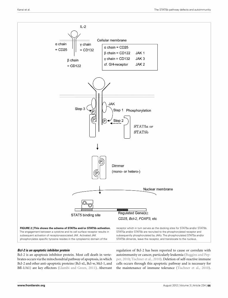
Kanai et al. The STAT5b pathway defects and autoimmunity
FIGURE 2 |This shows the schema of STAT5a and/or STAT5b activation.The engagement between a cytokine and its cell surface receptor results insubsequent activation of receptor-associated JAK. Activated JAKphosphorylates specific tyrosine resides in the cytoplasmic domain of the
receptor which in turn serves as the docking sites for STAT5a and/or STAT5b.STAT5a and/or STAT5b are recruited to the phosphorylated receptor andsubsequently phosphorylated by JAKs. The phosphorylated STAT5a and/orSTAT5b dimerize, leave the receptor, and translocate to the nucleus.
Bcl-2 is an apoptotic inhibitor proteinBcl-2 is an apoptosis inhibitor protein. Most cell death in verte-brates occurs via the mitochondrial pathway of apoptosis, in whichBcl-2 and other anti-apoptotic proteins (Bcl-xL, Bcl-w, Mcl-1, andBfl-1/A1) are key effectors (Llambi and Green, 2011). Aberrant
regulation of Bcl-2 has been reported to cause or correlate withautoimmunity or cancer, particularly leukemia (Buggins and Pep-per, 2010; Tischner et al., 2010). Deletion of self-reactive immunecells occurs through this apoptotic pathway and is necessary forthe maintenance of immune tolerance (Tischner et al., 2010).
www.frontiersin.org August 2012 | Volume 3 | Article 234 | 66
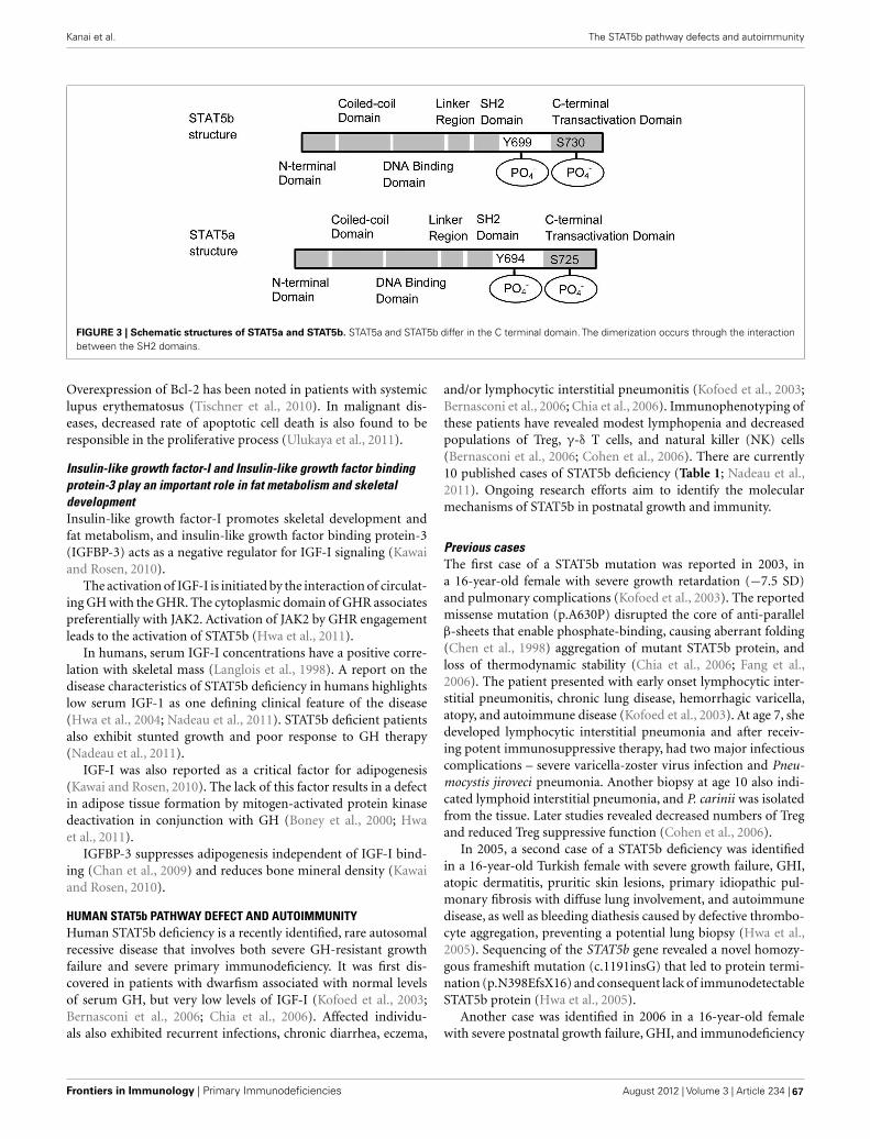
Kanai et al. The STAT5b pathway defects and autoimmunity
FIGURE 3 | Schematic structures of STAT5a and STAT5b. STAT5a and STAT5b differ in the C terminal domain. The dimerization occurs through the interactionbetween the SH2 domains.
Overexpression of Bcl-2 has been noted in patients with systemiclupus erythematosus (Tischner et al., 2010). In malignant dis-eases, decreased rate of apoptotic cell death is also found to beresponsible in the proliferative process (Ulukaya et al., 2011).
Insulin-like growth factor-I and Insulin-like growth factor bindingprotein-3 play an important role in fat metabolism and skeletaldevelopmentInsulin-like growth factor-I promotes skeletal development andfat metabolism, and insulin-like growth factor binding protein-3(IGFBP-3) acts as a negative regulator for IGF-I signaling (Kawaiand Rosen, 2010).
The activation of IGF-I is initiated by the interaction of circulat-ing GH with the GHR. The cytoplasmic domain of GHR associatespreferentially with JAK2. Activation of JAK2 by GHR engagementleads to the activation of STAT5b (Hwa et al., 2011).
In humans, serum IGF-I concentrations have a positive corre-lation with skeletal mass (Langlois et al., 1998). A report on thedisease characteristics of STAT5b deficiency in humans highlightslow serum IGF-1 as one defining clinical feature of the disease(Hwa et al., 2004; Nadeau et al., 2011). STAT5b deficient patientsalso exhibit stunted growth and poor response to GH therapy(Nadeau et al., 2011).
IGF-I was also reported as a critical factor for adipogenesis(Kawai and Rosen, 2010). The lack of this factor results in a defectin adipose tissue formation by mitogen-activated protein kinasedeactivation in conjunction with GH (Boney et al., 2000; Hwaet al., 2011).
IGFBP-3 suppresses adipogenesis independent of IGF-I bind-ing (Chan et al., 2009) and reduces bone mineral density (Kawaiand Rosen, 2010).
HUMAN STAT5b PATHWAY DEFECT AND AUTOIMMUNITYHuman STAT5b deficiency is a recently identified, rare autosomalrecessive disease that involves both severe GH-resistant growthfailure and severe primary immunodeficiency. It was first dis-covered in patients with dwarfism associated with normal levelsof serum GH, but very low levels of IGF-I (Kofoed et al., 2003;Bernasconi et al., 2006; Chia et al., 2006). Affected individu-als also exhibited recurrent infections, chronic diarrhea, eczema,
and/or lymphocytic interstitial pneumonitis (Kofoed et al., 2003;Bernasconi et al., 2006; Chia et al., 2006). Immunophenotyping ofthese patients have revealed modest lymphopenia and decreasedpopulations of Treg, γ-δ T cells, and natural killer (NK) cells(Bernasconi et al., 2006; Cohen et al., 2006). There are currently10 published cases of STAT5b deficiency (Table 1; Nadeau et al.,2011). Ongoing research efforts aim to identify the molecularmechanisms of STAT5b in postnatal growth and immunity.
Previous casesThe first case of a STAT5b mutation was reported in 2003, ina 16-year-old female with severe growth retardation (−7.5 SD)and pulmonary complications (Kofoed et al., 2003). The reportedmissense mutation (p.A630P) disrupted the core of anti-parallelβ-sheets that enable phosphate-binding, causing aberrant folding(Chen et al., 1998) aggregation of mutant STAT5b protein, andloss of thermodynamic stability (Chia et al., 2006; Fang et al.,2006). The patient presented with early onset lymphocytic inter-stitial pneumonitis, chronic lung disease, hemorrhagic varicella,atopy, and autoimmune disease (Kofoed et al., 2003). At age 7, shedeveloped lymphocytic interstitial pneumonia and after receiv-ing potent immunosuppressive therapy, had two major infectiouscomplications – severe varicella-zoster virus infection and Pneu-mocystis jiroveci pneumonia. Another biopsy at age 10 also indi-cated lymphoid interstitial pneumonia, and P. carinii was isolatedfrom the tissue. Later studies revealed decreased numbers of Tregand reduced Treg suppressive function (Cohen et al., 2006).
In 2005, a second case of a STAT5b deficiency was identifiedin a 16-year-old Turkish female with severe growth failure, GHI,atopic dermatitis, pruritic skin lesions, primary idiopathic pul-monary fibrosis with diffuse lung involvement, and autoimmunedisease, as well as bleeding diathesis caused by defective thrombo-cyte aggregation, preventing a potential lung biopsy (Hwa et al.,2005). Sequencing of the STAT5b gene revealed a novel homozy-gous frameshift mutation (c.1191insG) that led to protein termi-nation (p.N398EfsX16) and consequent lack of immunodetectableSTAT5b protein (Hwa et al., 2005).
Another case was identified in 2006 in a 16-year-old femalewith severe postnatal growth failure, GHI, and immunodeficiency
Frontiers in Immunology | Primary Immunodeficiencies August 2012 | Volume 3 | Article 234 | 67

Kanai et al. The STAT5b pathway defects and autoimmunity
Tab
le1
|Dem
og
rap
hic
so
fp
ub
lish
edca
ses
wit
hS
TAT
5bd
efici
ency
.
#1#2
#3#4
#5#6
#7#8
#9#1
0R
efer
ence
valu
es
STAT
5bM
utat
ion
A63
0P11
91in
sGR
152X
R15
2X11
03in
sC16
80de
lG16
80de
lG42
4_42
7del
424_
427d
elF6
46S
−/−
−/−
−/−
−/−
−/−
−/−
−/−
−/−
−/−
−/−
Pla
ceof
orig
inA
rgen
tina
Turk
eyA
rgen
tina
Arg
entin
aC
arib
bean
Kuw
ait
Kuw
ait
Bra
zil
Bra
zil
Arg
entin
a
Sex
FF
FF
MF
FM
MF
Hei
ght,
SD
S−
7.5
−7.
8−
9.9
−5.
3−
5.9
−5.
8−
5.6
−5.
6−
3-5
.90
Age
16.5
16.4
15.3
1231
24
62
18
Ons
etof
chro
nic
pulm
onar
y
dise
ase
7ye
ars
7ye
ars
1ye
ar6
mon
ths
No
6ye
ars
6ye
ars
Yes
Yes
No
Ski
npa
thol
ogy
Ecz
ema
Ecz
ema
Ecz
ema
Ecz
ema
Ichy
tosi
sN
oN
oA
topi
cA
topi
cS
ebor
rhei
c
derm
atiti
s
Aut
oim
mun
em
anife
stat
ions
+A
bsto
BE
CIT
P+
Abs
to
plat
elet
s
No
AIT
with+
Abs
to
thym
oglo
bulin
Ichy
tosi
s+
Abs
topl
atel
ets
SJI
AN
oN
oN
oA
IT
Pare
ntal
cons
angu
inity
Yes
Yes
NA
Ado
pted
NA
Yes
Yes
ND
ND
Ado
pted
Pate
rnal
heig
ht,S
DS
−0.
3−
0.9
−2.
2N
A−
0.8
−1.
28−
1.28
−1.
6−
1.6
NA
Mot
her
heig
ht,S
DS
−1.
2−
0.6
−3.
3N
A−
2.8
−0.
6−
0.6
−1.
3−
1.3
NA
Birt
hw
eigh
t/le
ngth
(cm
)14
00/N
D23
50/4
925
00/N
D16
50/N
D32
70/5
024
00/N
D36
00/N
D16
50/3
924
00/4
922
50/4
4
Pube
rty
Del
ayed
Del
ayed
Del
ayed
NA
Del
ayed
NA
NA
NA
NA
NA
BIO
CH
EM
IST
RY
GH
basa
l(ng
/mL)
9.4
14.2
6.6
1.8
0.13
17.7
5.7
1.7
11.
7
GH
stim
ulat
ed(n
g/m
L)53
.8N
AN
A12
.514
.2N
AN
A20
.614
27.1
>6.
0
IGF-
Istim
ulat
ed(n
g/m
L)38
7.0
<10
0.8
14<
5<
534
<25
1611
9–48
3
IGFB
P-3
(ng/
mL)
874
543
NA
500
180
700
800
520
750
840
210-
740
ALS
(aci
dla
bile
subu
nit;
mg/
L)2.
91.
2N
A0.
70.
70.
40.
852
075
0N
A
Ref
eren
ces
for
publ
ishe
dca
seC
ohen
etal
.
(200
6),K
ofoe
d
etal
.(20
03)
Hw
a
etal
.
(200
5)
Ber
nasc
oni
etal
.
(200
6)
Ber
nasc
oni
etal
.
(200
6)
Vida
rsdo
ttir
etal
.
(200
6)
Hw
a
etal
.
(200
7)
Hw
a
etal
.
(200
7)
Pugl
iese
-
Pire
set
al.
(201
0)
Pugl
iese
-
Pire
set
al.
(201
0)
Sca
glia
etal
.
(201
2)
ITP,
idio
path
icth
rom
bocy
tope
nic
purp
ura;
AIT
,aut
oim
mun
eth
yroi
ditis
;Abs
,ant
ibod
ies;
BE
C,b
ronc
hial
epith
elia
lcel
ls;S
JIA
,sys
tem
icju
veni
leid
iopa
thic
arth
ritis
;ND
,not
diag
nose
d;N
A,n
otcu
rren
tlyav
aila
ble;
PC
,
pers
onal
com
mun
icat
ion.
The
data
of#6
and
#7in
adja
cent
colu
mns
repr
esen
tda
tafr
omsi
blin
gs.
Het
eroz
ygot
eST
AT5b
-defi
cien
tsub
ject
sw
hoar
ere
lativ
esof
hom
ozyg
ote
STAT
5b-d
efici
ents
ubje
cts
curr
ently
have
notb
een
iden
tified
toha
vean
yab
norm
alcl
inic
alph
enot
ypes
,alth
ough
asp
ecifi
cde
taile
dre
view
ofth
eir
clin
ical
dem
ogra
phic
san
dla
bora
tory
stud
ies
has
not
been
done
yet.
www.frontiersin.org August 2012 | Volume 3 | Article 234 | 68

Kanai et al. The STAT5b pathway defects and autoimmunity
(Bernasconi et al., 2006). Pulmonary-function tests showed mixed,restrictive, and obstructive moderate ventilative insufficiency, butno lung biopsy was performed. Notably, this case was the firstto identify a role for STAT5b not only in the human GH signal-ing cascade, but also in the cytokine-mediated immune response.The STAT5b deficient patient had moderate T cell lymphopenia,normal CD4/CD8 ratios, and very low numbers of NK cells andγ-δ T cells, and the T cells presented a chronically hyperactivatedphenotype (Bernasconi et al., 2006).
Since 2012, five other mutations have been published on atotal of seven additional subjects (Table 1). Lung pathology hasbeen common among these patients (8 of 10), but of theseremaining seven subjects, only a few have received lung biop-sies. A STAT5b deficient male with the mutation 424_427delreceived a biopsy at 6 years of age that indicated severe lympho-cytic interstitial pneumonitis. Considered together, these studieshave firmly established a correlation between STAT5b deficiencyand immune dysfunction, in addition to GHI and severe growthproblems.
Clinical manifestations and diagnosisSignal transducer and activator of transcription 5b deficiencyshould be considered in the differential diagnosis of a patientwho has normal gestational growth and birth size but acquiressignificantly short stature and recurrent infections. This patternof growth is typical of patients with GHI. Height may range from−3.0 to−9.9 SD in girls and boys, respectively (Table 1).
Regarding hormone evaluations, all described patients havehad normal levels of GH at baseline, but after stimulation, GHconcentrations were often elevated (Table 1). In contrast, serumIGF-I, IGFBP-3, and acid labile subunit concentrations were low,and even upon administration of GH, remained low. Elevatedprolactin levels were also observed in patients with recordedconcentrations.
Most patients have displayed evidence of immune dysfunc-tion, including atopic disease, chronic lung disease, viral infections,and/or autoimmune diatheses. Often present in childhood, severepulmonary disease is of particular concern, as it has affected 8 ofthe 10 known STAT5b deficient patients and two patients have diedof respiratory failure. For all cases of lung pathology except for thatof Patient #5, an axial chest CT scan has shown increased inter-stitial patterns and ground-glass appearance. These pulmonarylesions are T cell predominant, despite peripheral lymphopenia.In most cases, severe eczema, thrombocytopenic purpura, and/orautoimmune disease, such as juvenile idiopathic arthritis, werepresent in addition to severe lung disease. However, it should benoted that 1 of the 10 subjects to date has less severe immune
dysfunction. Congenital ichthyosis was diagnosed at birth, andthe patient had hemorrhagic varicella at 16 years of age but had nohistory of pulmonary of immunological problems (Vidarsdottiret al., 2006).
Previous immunological studies have established the impor-tance of STAT5b proteins in the development, homeostasis, andproliferation of different lymphocyte populations. Immune reper-toires of STAT5b deficient patients have shown moderate lym-phopenia, with very low numbers of NK and T cells, as well as Tregdysfunction. Furthermore, B cell populations and immunoglobu-lin G levels in at least patient are normal to elevated, as consistentwith autoimmune disease symptoms (Cohen et al., 2006).
Disease managementIn order to improve clinical outcomes for patients with STAT5bdeficiency, optimizing early diagnosis in these patients is critical.To date, overall management of STAT5b deficiency is still unclear.GH therapy is ineffective due to the patients’ GHI. It is presumedthat IGF-I therapy may be an effective treatment, unless the pres-ence of chronic infection limits the growth response. However, todate, no clinical trials of IGF-I therapy have been performed inthese patients.
Patients should be closely monitored for signs and symptomsof immunodeficiency. Infections such as severe varicella or recur-rent pneumonias should be aggressively treated with appropriateantimicrobial therapies. Patients with autoimmune conditions,atopic diseases, or pulmonary fibrosis may also require antipro-liferatives or immunosuppressants, such as steroids, to addressoveractive effector T cell responses. Because severe chronic lungdisease in this patient population often leads to high morbid-ity and mortality, patients should be carefully monitored withpulmonary-function tests and physical examinations, which mayimprove treatment options to decrease the lung disease severity.
Although current management of STAT5b deficiency is pri-marily dictated by specific end-organ pathology, current researchis addressing the possibility of enhancing STAT5b and/or STAT5apathways (Zeiser et al., 2008; Strauss et al., 2009). Future therapymay be expected to prevent and reflect rationally based drug designto enhance certain drug targets in the STAT5b and/or STAT5apathways.
CONCLUSIONIn this review, we focused on the STAT5b pathway and the mech-anisms by which defects in protein structure and or expressionmight result in autoimmunity. A better understanding of STAT5band its distinct biological functions is necessary for the develop-ment of new diagnostic and therapeutic approaches for treatingpatients suffering from its deficiency.
REFERENCESBennett, C. L., Christie, J., Ramsdell,
F., Brunkow, M. E., Ferguson, P. J.,Whitesell, L., Kelly, T. E., Saulsbury,F. T., Chance, P. F., and Ochs, H.D. (2001). The immune dysregula-tion, polyendocrinopathy, enteropa-thy, X-linked syndrome (IPEX) iscaused by mutations of FOXP3. Nat.Genet. 27, 20–21.
Bernasconi, A., Marino, R., Ribas, A.,Rossi, J., Ciaccio, M., Oleastro, M.,Ornani, A., Paz, R., Rivarola, M.A., Zelazko, M., and Belgorosky,A. (2006). Characterization ofimmunodeficiency in a patientwith growth hormone insensitiv-ity secondary to a novel STAT5bgene mutation. Pediatrics 118,e1584–e1592.
Boney, C. M., Gruppuso, P. A., Faris, R.A., and Frackelton, A. R. Jr. (2000).The critical role of Shc in insulin-likegrowth factor-I-mediated mitogen-esis and differentiation in 3T3-L1preadipocytes. Mol. Endocrinol. 14,805–813.
Boucheron, C., Dumon, S., Santos, S.C., Moriggl, R., Hennighausen, L.,Gisselbrecht, S., and Gouilleux, F.
(1998). A single amino acid in theDNA binding regions of STAT5Aand STAT5B confers distinct DNAbinding specificities. J. Biol. Chem.273, 33936–33941.
Buggins, A. G., and Pepper, C. J.(2010). The role of Bcl-2 fam-ily proteins in chronic lympho-cytic leukaemia. Leuk. Res. 34,837–842.
Frontiers in Immunology | Primary Immunodeficiencies August 2012 | Volume 3 | Article 234 | 69

Kanai et al. The STAT5b pathway defects and autoimmunity
Campbell, D. J., and Koch, M. A.(2011). Phenotypical and functionalspecialization of FOXP3+ regula-tory T cells. Nat. Rev. Immunol. 11,119–130.
Chan, S. S., Schedlich, L. J., Twigg, S.M., and Baxter, R. C. (2009). Inhibi-tion of adipocyte differentiation byinsulin-like growth factor-bindingprotein-3. Am. J. Physiol. Endocrinol.Metab. 296, E654–E663.
Chatila, T. A., Blaeser, F., Ho, N., Led-erman, H. M., Voulgaropoulos, C.,Helms, C., and Bowcock, A. M.(2000). JM2, encoding a fork head-related protein, is mutated in X-linked autoimmunity-allergic dys-regulation syndrome. J. Clin. Invest.106, R75–R81.
Chen, X., Vinkemeier, U., Zhao, Y.,Jeruzalmi, D., Darnell, J. E. Jr., andKuriyan, J. (1998). Crystal structureof a tyrosine phosphorylated STAT-1 dimer bound to DNA. Cell 93,827–839.
Chia, D. J., Subbian, E., Buck, T. M.,Hwa, V., Rosenfeld, R. G., Skach,W. R., Shinde, U., and Rotwein,P. (2006). Aberrant folding of amutant Stat5b causes growth hor-mone insensitivity and proteaso-mal dysfunction. J. Biol. Chem. 281,6552–6558.
Cohen, A. C., Nadeau, K. C., Tu, W.,Hwa, V., Dionis, K., Bezrodnik, L.,Teper, A., Gaillard, M., Heinrich,J., Krensky, A. M., Rosenfeldand,R. G., and Lewis, D. B. (2006).Cutting edge: decreased accumu-lation and regulatory function ofCD4+CD25(high) T cells in humanSTAT5b deficiency. J. Immunol. 177,2770–2774.
Crispi, S., Sanzari, E., Monfregola, J.,De Felice, N., Fimiani, G., Ambro-sio, R., D’Urso, M., and Ursini, M.V. (2004). Characterization of thehuman STAT5A and STAT5B pro-moters: evidence of a positive andnegative mechanism of transcrip-tional regulation. FEBS Lett. 562,27–34.
Fang, P., Kofoed, E. M., Little, B. M.,Wang, X., Ross, R. J., Frank, S. J.,Hwa,V., and Rosenfeld, R. G. (2006).A mutant signal transducer and acti-vator of transcription 5b, associatedwith growth hormone insensitivityand insulin-like growth factor-I defi-ciency, cannot function as a sig-nal transducer or transcription fac-tor. J. Clin. Endocrinol. Metab. 91,1526–1534.
Fontenot, J. D., Gavin, M. A., andRudensky, A. Y. (2003). Foxp3 pro-grams the development and func-tion of CD4+ CD25+ regulatory Tcells. Nat. Immunol. 4, 330–336.
Grimley, P. M., Dong, F., and Rui, H.(1999). Stat5a and Stat5b: frater-nal twins of signal transduction andtranscriptional activation. CytokineGrowth Factor Rev. 10, 131–157.
Hori, S., Nomura, T., and Sakaguchi,S. (2003). Control of regulatoryT cell development by the tran-scription factor Foxp3. Science 299,1057–1061.
Hwa, V., Camacho-Hübner, C., Little,B. M., David, A., Metherell, L. A.,El-Khatib, N., Savageb, M. O., andRosenfeld,R. G. (2007). Growth hor-mone insensitivity and severe shortstature in siblings: a novel mutationat the exon 13-intron 13 junctionof the STAT5b gene. Horm. Res. 68,218–224.
Hwa, V., Little, B., Adiyaman, P., Kofoed,E. M., Pratt, K. L., Ocal, G.,Berberoglu, M., and Rosenfeld, R.G. (2005). Severe growth hormoneinsensitivity resulting from totalabsence of signal transducer andactivator of transcription 5b. J. Clin.Endocrinol. Metab. 90, 4260–4266.
Hwa, V., Little, B., Kofoed, E. M.,and Rosenfeld, R. G. (2004). Tran-scriptional regulation of insulin-like growth factor-I by interferon-gamma requires STAT-5b. J. Biol.Chem. 279, 2728–2736.
Hwa, V., Nadeau, K., Wit, J. M., andRosenfeld, R. G. (2011). STAT5bdeficiency: lessons from STAT5bgene mutations. Best Pract. Res. Clin.Endocrinol. Metab. 25, 61–75.
John, S., Vinkemeier, U., Soldaini, E.,Darnell, J. E. Jr., and Leonard, W. J.(1999). The significance of tetramer-ization in promoter recruitment byStat5. Mol. Cell. Biol. 19, 1910–1918.
Kawai, M., and Rosen, C. J. (2010).The IGF-I regulatory system andits impact on skeletal and energyhomeostasis. J. Cell. Biochem. 111,14–19.
Khattri, R., Cox, T., Yasayko, S. A., andRamsdell, F. (2003). An essentialrole for Scurfin in CD4+ CD25+T regulatory cells. Nat. Immunol. 4,337–342.
Kofoed, E. M., Hwa,V., Little, B., Woods,K. A., Buckway, C. K., Tsubaki, J.,Pratt, K. L., Bezrodnik, L., Jaspe,H., Teppe, A., Heinrich, J. J., andRosenfeld,R. G. (2003). Growth hor-mone insensitivity associated with aSTAT5b mutation. N. Engl. J. Med.349, 1139–1147.
Langlois, J. A., Rosen, C. J., Visser,M., Hannan, M. T., Harris, T.,Wilson, P. W. F., and Kiel, D. P.(1998). Association between insulin-like growth factor I and bonemineral density in older womenand men: the Framingham Heart
Study. J. Clin. Endocrinol. Metab. 83,4257–4262.
Levy, D. E., and Darnell, J. E. Jr. (2002).Stats: transcriptional control andbiological impact. Nat. Rev. Mol.Cell. Biol. 3, 651–662.
Lin, J. X., and Leonard, W. J. (2000). Therole of Stat5a and Stat5b in signalingby IL-2 family cytokines. Oncogene19, 2566–2576.
Liu, X., Robinson, G. W., Wagner, K.U., Garrett, L., Wynshaw-Boris, A.,and Hennighausen, L. (1997). Stat5ais mandatory for adult mammarygland development and lactogenesis.Genes Dev. 11, 179–186.
Llambi, F., and Green, D. R. (2011).Apoptosis and oncogenesis: give andtake in the BCL-2 family. Curr. Opin.Genet. Dev. 21, 12–20.
Mandal, M., Powers, S. E., Maienschein-Cline, M., Bartom, E. T., Hamel,K. M., Kee, B. L., Dinner, A. R.,and Clark, M. R. (2011). Epige-netic repression of the Igk locus bySTAT5-mediated recruitment of thehistone methyltransferase Ezh2. Nat.Immunol. 12, 1212–1220.
Nadeau, K., Hwa, V., and Rosenfeld,R. G. (2011). STAT5b deficiency: anunsuspected cause of growth fail-ure, immunodeficiency, and severepulmonary disease. J. Pediatr. 158,701–708.
Pugliese-Pires, P. N., Tonelli, C. A.,Dora, J. M., Silva, P. C., Czepielewski,M., Simoni, G., Arnhold, I. J. P.,and Jorge, A. A. L. (2010). Anovel STAT5B mutation causingGH insensitivity syndrome associ-ated with hyperprolactinemia andimmune dysfunction in two malesiblings. Eur. J. Endocrinol. 163,349–355.
Russell, S. M., Tayebi, N., Nakajima,H., Riedy, M. C., Roberts, J. L., andAman,M. J. (1995). Mutation of Jak3in a patient with SCID: essential roleof Jak3 in lymphoid development.Science 270, 797–800.
Sakaguchi, S., Sakaguchi, N., Asano,M., Itoh, M., and Toda, M. (1995).Immunologic self-tolerance main-tained by activated T cells expressingIL-2 receptor alpha-chains (CD25).Breakdown of a single mecha-nism of self-tolerance causes variousautoimmune diseases. J. Immunol.155, 1151–1164.
Sakaguchi, S., Yamaguchi, T., Nomura,T., and Ono, M. (2008). RegulatoryT cells and immune tolerance. Cell133, 775–787.
Scaglia, P. A., Martínez, A. S., Feigerlová,E., Bezrodnik, L., Gaillard, M. I.,Di Giovanni, D., Ballerini, M. G.,Jasper, H. G., Heinrich, J. J., Fang,P., Domené, H. M., Rosenfeld,
R. G., and Hwa, V. (2012). Anovel missense mutation in theSH2 domain of the STAT5B generesults in a transcriptionally inac-tive STAT5b associated with severeIGF-I deficiency, immune dysfunc-tion, and lack of pulmonary dis-ease. J. Clin. Endocrinol. Metab. 97,E830–E839.
Sharfe, N., Dadi, H. K., Shahar,M., and Roifman, C. M. (1997).Human immune disorder aris-ing from mutation of the alphachain of the interleukin-2 recep-tor. Proc. Natl. Acad. Sci. U.S.A. 94,3168–3171.
Soldaini, E., John, S., Moro, S., Bol-lenbacher, J., Schindler, U., and,Leonard, W. J. (2000). DNA bind-ing site selection of dimeric andtetrameric Stat5 proteins revealsa large repertoire of divergenttetrameric Stat5a binding sites. Mol.Cell. Biol. 20, 389–401.
Strauss, L., Czystowska, M., Szajnik,M., Mandapathil, M., and White-side, T. L. (2009). Differentialresponses of human regulatory Tcells (Treg) and effector T cellsto rapamycin. PloS ONE 4, e5994.doi:10.1371/journal.pone.0005994
Tischner, D., Woess, C., Ottina, E., andVillunger, A. (2010). Bcl-2-regulatedcell death signalling in the preven-tion of autoimmunity. Cell DeathDis. 1, e48.
Ulukaya, E., Acilan, C., and Yil-maz, Y. (2011). Apoptosis: whyand how does it occur in biol-ogy? Cell Biochem. Funct. 29,468–480.
Vidarsdottir, S., Walenkamp, M. J.,Pereira, A. M., Karperien, M., vanDoorn, J., van Duyvenvoorde, H. A.,White, S., Breuning, M. H., Roelf-sema, F., Kruithof, M. F., van Dissel,J., Janssen, R.,Wit, J. M., and Romijn,J. A. (2006). Clinical and biochemi-cal characteristics of a male patientwith a novel homozygous STAT5bmutation. J. Clin. Endocrinol. Metab.91, 3482–3485.
Wei, L., Laurence, A., and O’Shea, J.J. (2008). New insights into theroles of Stat5a/b and Stat3 in Tcell development and differentia-tion. Semin. Cell Dev. Biol. 19,394–400.
Wildin, R. S., Ramsdell, F., Peake,J., Faravelli, F., Casanova, J.L., Buist, N., Levy-Lahad, M.,Mazzella, E. M., Goulet, O., Perroni,L., Bricarelli, F. D., Byrne, G.,McEuen, M., Proll, S., Appleby,M., and Brunkow, M. E. (2001).X-linked neonatal diabetes mellitus,enteropathy and endocrinopathysyndrome is the human equivalent
www.frontiersin.org August 2012 | Volume 3 | Article 234 | 70

Kanai et al. The STAT5b pathway defects and autoimmunity
of mouse scurfy. Nat. Genet. 27,18–20.
Willerford, D. M., Chen, J., Ferry, J.A., Davidson, L., Ma, A., and Alt,F. W. (1995). Interleukin-2 recep-tor alpha chain regulates the sizeand content of the peripheral lym-phoid compartment. Immunity 3,521–530.
Xu, D., and Qu, C. K. (2008). Pro-tein tyrosine phosphatases in theJAK/STAT pathway. Front. Biosci.13:4925–4932.
Zeiser, R., Leveson-Gower, D. B.,Zambricki, E. A., Kambham,
N., Beilhack, A., Loh, J., Hou1,J.-Z., and Negrin, R. S. (2008).Differential impact of mammaliantarget of rapamycin inhibition onCD4+ CD25+ Foxp3+ regulatoryT cells compared with conven-tional CD4+ T cells. Blood 111,453–462.
Zhu, M. H., Berry, J. A., Rus-sell, S. M., and Leonard, W. J.(1998). Delineation of the regionsof interleukin-2 (IL-2) receptor betachain important for association ofJak1 and Jak3. Jak1-independentfunctional recruitment of Jak3 to
Il-2Rbeta. J. Biol. Chem. 273,10719–10725.
Conflict of Interest Statement: Theauthors declare that the research wasconducted in the absence of any com-mercial or financial relationships thatcould be construed as a potential con-flict of interest.
Received: 04 May 2012; paper pendingpublished: 27 May 2012; accepted: 15 July2012; published online: 14 August 2012.Citation: Kanai T, Jenks J and NadeauKC (2012) The STAT5b pathway defect
and autoimmunity. Front. Immun.3:234. doi: 10.3389/fimmu.2012.00234This article was submitted to Frontiers inPrimary Immunodeficiencies, a specialtyof Frontiers in Immunology.Copyright © 2012 Kanai, Jenks andNadeau. This is an open-access arti-cle distributed under the terms of theCreative Commons Attribution License,which permits use, distribution andreproduction in other forums, providedthe original authors and source arecredited and subject to any copyrightnotices concerning any third-party graph-ics etc.
Frontiers in Immunology | Primary Immunodeficiencies August 2012 | Volume 3 | Article 234 | 71

“fimmu-03-00232” — 2012/7/31 — 17:51 — page 1 — #1
REVIEW ARTICLEpublished: 02 August 2012
doi: 10.3389/fimmu.2012.00232
APECED: is this a model for failure of T celland B cell tolerance?Nicolas Kluger1, Annamari Ranki1 and Kai Krohn2*
1 Department of Dermatology, Allergology and Venereology, Institute of Clinical Medicine, Skin and Allergy Hospital,Helsinki University Central Hospital, University of Helsinki, Helsinki, Finland
2 Clinical Research Institute HUCH Ltd, Helsinki, Finland
Edited by:
Rosa Bacchetta, Fondazione CentroSan Raffaele Del Monte Tabor, Italy
Reviewed by:
Rosa Bacchetta, Fondazione CentroSan Raffaele Del Monte Tabor, ItalyMario Abinun, Newcastle upon TyneHospitals NHS Foundation Trust, UK
*Correspondence:
Kai Krohn, Clinical Research InstituteHUCH Ltd, Salmentaantie 751,36450 Salmentaka, Finland.e-mail: [email protected]
In APECED, the key abnormality is in the T cell defect that may lead to tissue destruc-tion chiefly in endocrine organs. Besides, APECED is characterized by high-titer antibodiesagainst a wide variety of cytokines that could partly be responsible for the clinical symp-toms during APECED, mainly chronic mucocutaneous candidiasis, and linked to antibodiesagainst Th17 cells effector molecules, IL-17 and IL-22. On the other hand, the same anti-bodies, together with antibodies against type I interferons may prevent the patients fromother immunological diseases, such as psoriasis and systemic lupus erythematous. Thesame effectorTh17 cells, present in the lymphocytic infiltrate of target organs of APECED,could be responsible for the tissue destruction. Here again, the antibodies against the cor-responding effector molecules, anti-IL-17 and anti-IL-22 could be protective.The occurrenceof several effector mechanisms (CD4+ Th17 cell and CD8+ CTL and the effector cytokinesIL-17 and IL-22), and simultaneous existence of regulatory mechanisms (CD4+Treg and anti-bodies neutralizing the effect of the effector cytokines) may explain the polymorphism ofAPECED. Almost all the patients develop the characteristic manifestations of the complex,but temporal course and severity of the symptoms vary considerably, even among siblings.The autoantibody profile does not correlate with the clinical picture. One could speculatethat a secondary homeostatic balance between the harmful effector mechanisms, and thefavorable regulatory mechanisms, finally define both the extent and severity of the clini-cal condition in the AIRE defective individuals. The proposed hypothesis that in APECED,in addition to strong tissue destructive mechanisms, a controlling regulatory mechanismdoes exist, allow us to conclude that APECED could be treated, and even cured, withimmunological manipulation.
Keywords: AIRE, APECED, endocrine disorders, interleukin 17, interleukin 22, IPEX,T regulatory cells
INTRODUCTIONAutoimmune polyendocrinopathy syndrome type 1 (APS-1) orautoimmune polyendocrinopathy–candidiasis–ectodermal dys-trophy syndrome (APECED; OMIM 240300) is a rare recessivelyinherited disorder (Perheentupa, 2002; Betterle and Zanchetta,2003; Perheentupa, 2006; Husebye et al., 2009). It is caused bymutations in the autoimmune regulator (AIRE) gene locatedon locus 21q22.3 (Bjorses et al., 1996; Nagamine et al., 1997;The Finnish–German APECED Consortium, 1997). APECEDdisplays a worldwide distribution, but specific clusters of highprevalence of the disease are observed among Finns (1:25,000;
Abbreviations: AADC, aromatic L-amino acid decarboxylase; AD, Addison’s disease;AE, autoimmune enteropathy; APS-1, autoimmune polyendocrinopathy syn-drome type 1; APECED, autoimmune polyendocrinopathy–candidiasis–ectodermaldystrophy; CMC, chronic mucocutaneous candidiasis; EECs, enteroendocrinecells; GI, gastro-intestinal; HP, hypoparathyroidism; IPEX, immune dysregula-tion, polyendocrinopathy, enteropathy and X-linked; IF, intrinsic factor; IL-1,interleukin-1; IL-17, interleukin-17; IL-22, interleukin-22; mTECs, thymicmedullary epithelial cells; NALP-5, NACHT leucine-rich-repeat protein 5; PE,promiscuous expression; PTH, parathormone; SLE, systemic lupus erythemato-sus; TH, tyrosine hydroxylase; TPH, tryptophan hydroxylase; TRIMs, tripartitemotif-containing proteins; TSA, tissue-specific antigens.
Ahonen et al., 1990) and Sardinians 1 (1:14,500; Rosatelli et al.,1998; Meloni et al., 2012). It is characterized by the variable asso-ciation of autoimmune endocrine [hypoparathyroidism (HP),Addison’s disease (AD), hypothyroidism, gonadal insufficiency,insulin-dependent diabetes mellitus, atrophic gastritis, and Bier-mer’s disease] and non-endocrine disorders (keratitis, malabsorp-tion, vitiligo, and alopecia areata) and a specific predisposition tochronic mucocutaneous candidiasis (CMC). A definite diagnosisof APECED is made upon one of the following criteria: (i) thepresence of at least two of three major clinical features: CMC, HP,and AD, or (ii) one disease component if a sibling has already adefinite diagnosis, or (iii) disease-causing mutations in both alle-les of the AIRE gene. However, APECED being highly variablein its presentation, the classical triad may be complete only afteryears of evolution and diagnose may be therefore missed. Besides,APECED may appear during adolescence or in the young adult(Husebye et al., 2009). Therefore, criteria for a probable APECEDhave been defined as follows: (i) presence of one of CMC, HP,AD (before 30 years of age) and at least one of the minor compo-nents chronic diarrhea, keratitis, periodic rash with fever, severeconstipation, autoimmune hepatitis, vitiligo, alopecia, enamel
www.frontiersin.org August 2012 | Volume 3 | Article 232 | 72

“fimmu-03-00232” — 2012/7/31 — 17:51 — page 2 — #2
Kluger et al. APECED syndrome
hypoplasia, (ii) any component and anti-interferon antibodies, or(iii) any component and antibodies against NACHT leucine-richrepeat protein 5 (NALP5), AADC, tryptophan hydroxylase (TPH),or TH (Husebye et al., 2009).
FROM CIRCULATING AUTOIMMUNE ANTIBODIESTO AIRE, FOXP3, APECED, AND IPEXOur knowledge of the nature of the condition now called APS-1or APECED has increased simultaneously with the general devel-opment of immunology and autoimmunity. Since the conditionwas clearly defined in the end of 1950s and early 1960s, the char-acteristic clinical picture, the immunological abnormalities andthe relationship to other autoimmune endocrine diseases weredefined in late 1960s and early 1970s. Furthermore, the geneticsof APECED, and the fact that the syndrome was caused by a reces-sive gene defect – as opposed to the HLA-linked genetics seen inthe other solitarily occurring endocrine diseases – were character-ized in the 1980s and the target antigens in the organs affected byAPECED were molecularly defined in 1990s. A landmark stage inthe study of APECED was reached in 1997, when the long soughtAPECED gene was cloned by two independent groups (Nagamineet al., 1997; The Finnish–German APECED Consortium, 1997).Finally, a new phase in APECED research occurred during thefirst decennium of 2000, when the autoantibodies toward solublemediators if immune response were characterized (Meager et al.,2006; Kisand et al., 2011).
The notion that several diseases affecting endocrine organs andearlier defined as idiopathic, were in fact caused by an autoim-mune response toward self antigens, became apparent when novelimmunological methods became available in 1950s and early 1960s(Blizzard et al., 1963). The association of the three conditions,candidiasis, HP, and AD that were later judged to be the hall-marks of APECED was clearly stated by the groups of Blizzard andMaclaren (Blizzard et al., 1963; Brun, 1978; Neufeld et al., 1981).These groups also defined two clearly distinct syndromes withseveral associated autoimmune diseases: autoimmune polyglan-dular syndrome type 1 (PGS-1) and polyglandular syndrome type2 (PGS-2). The nomenclature was later changed to APS-1 andAPS-2, and the former further to APECED (Ahonen et al., 1990;Perheentupa, 2002, 2006; Betterle and Zanchetta, 2003).
Pioneering studies in this field were made especially with theuse of immunohistochemistry, demonstrating antibodies react-ing with gastric parietal cells in chronic gastritis (Walder et al.,1963; Irvine et al., 1965) and intrinsic factor (IF) in perniciousanemia (Schwartz, 1961; Jeffries et al., 1962), with thyroid epithe-lial cells in various forms of thyroid diseases (Witebsky et al., 1957;Irvine et al., 1962; Doniach and Roitt, 1964), with the beta cells ofLangerhans islands in diabetes mellitus (Kaldany, 1979; Bottazzoet al., 1980), and adrenal cortical cells in AD (Blizzard et al., 1962).
Blizzard’s group noticed that the two polyglandular syndromes,APS-1 and APS-2, differed in their HLA haplotypes (Neufeld et al.,1981). Further studies on the HLA haplotypes revealed that thegenetic basis of APS-2, but also of the other isolated forms ofendocrine autoimmune diseases found in APS-1, were in the HLAhaplotype of the patients. In contrast, APS-1 was shown not to belinked to HLA, and studies with large patient material, collectedby Perheentupa’s group in Finland, clearly stated that APS-1 was
linked to a recessively inherited gene defect (Ahonen et al., 1990;Perheentupa, 2002, 2006). The autoimmune endocrinopathiescould thus be grouped on the basis of their genetic backgroundin two distinct categories: those linked to HLA variation and theone, APS-1 caused by a single mutated gene (Table 1). At thatstage, however, the responsible gene, the APECED gene, was notyet identified. Once identified, the APECED gene was renamedas AIRE in 1997 (Nagamine et al., 1997; The Finnish–GermanAPECED Consortium, 1997).
Another immunopathy, termed originally as autoimmuneenteropathy (AIE) and later identified as immune dysregula-tion, polyendocrinopathy, enteropathy and X-linked (IPEX), wasdescribed in the 1980s and 1990s (Powell et al., 1982). This dis-order was later shown to be caused by a defect in a single gene,FOXP3 (Bennett et al., 2000). IPEX and APECED are two examplesof immune deficiency diseases disclosing both disturbed toleranceand autoimmune phenomena (Moraes-Vasconcelos et al., 2008).Traditionally, reviews tend to associate both IPEX and APECEDbecause of common features. However, both clinical manifesta-tions and predisposition to infections are rather different whencomparing both diseases (Moraes-Vasconcelos et al., 2008).
AIRE GENE, MUTATIONS, AND MECHANISM OF ACTIONAIRE is expressed in thymus, lymph nodes, and fetal liver, andencodes a protein with two putative zinc fingers and other motifssuggestive of a transcriptional regulator (Nagamine et al., 1997;The Finnish–German APECED Consortium, 1997). The AIREgene, approximately 13 kb in length, contains 14 exons thatencode a polypeptide of 545 amino acids. The AIRE protein func-tions as a transcription factor (Fierabracci, 2011; Gardner et al.,2009). AIRE is expressed in the thymic medullary epithelial cells(mTECs, Figure 1) and in cells of the monocyte/dendritic celllineage (Kogawa et al., 2002). mTECs through the expression ofMHC class II express a wide array of tissue-restricted antigens(TRAs) derived from different organs in the body. TRAs includeself-proteins with patterns of expression restricted to a single orsmall handful of organs. Thymic expression of TRA serves as animportant source of self-antigens to allow the negative selectionof autoreactive T cells. Collectively, mTEC and thymic mono-cyte/dendritic cells play a crucial role in establishing self-toleranceby eliminating autoreactive T cells (negative selection) and/orby producing immunoregulatory FOXP3+ T cells, which preventCD4+ T cell-mediated organ-specific autoimmune diseases. Col-lectively, several studies in mouse and man have shown that AIREregulates thymic expression of several genes of ectopic peripheralproteins including many TRAs. Thus, AIRE dysfunction leads toa decrease in the expression of TRAs in the thymus, and con-sequently, autoreactive T cell clones escape into the periphery(Derbinski et al., 2005; Moraes-Vasconcelos et al., 2008; Gardneret al., 2009; Fierabracci, 2011)
The most common AIRE mutation, the “Finnish mutation,”R257X, affects 82% of Finnish APECED alleles (Nagamine et al.,1997; The Finnish–German APECED Consortium, 1997). Inter-estingly, this mutation occurs also in 70% of the Russian APECEDpatients studied (Orlova et al., 2010). The same mutation, R257Xwas also detected in Swiss patients on a different haplotype withclosely linked polymorphic markers (Nagamine et al., 1997) and in
Frontiers in Immunology | Primary Immunodeficiencies August 2012 | Volume 3 | Article 232 | 73

“fimmu-03-00232” — 2012/7/31 — 17:51 — page 3 — #3
Kluger et al. APECED syndrome
Ta
ble
1|
Key
lab
ora
tory
fin
din
gs
inth
ed
iffe
ren
ta
uto
imm
un
ee
nd
ocri
ne
dis
ea
se
s.
Dia
gn
osis
Cli
nic
al
fin
din
gs
Au
toa
nti
bo
die
sH
LA
Ge
ne
de
fect
Ce
llu
lar
imm
un
ere
sp
on
se
AP
EC
ED
(AP
S-1
)C
andi
dias
isan
dm
ultip
lefa
ilure
ofm
ost
endo
crin
eor
gans
and
non-
endo
crin
e
auto
imm
unity
Aga
inst
alla
ffect
edor
gans
No
asso
ciat
ion
(?)
Clo
seto
100
mut
atio
ns
desc
ribed
inth
eA
IRE
gene
CTL
agai
nsta
ffect
edor
gans
?
Failu
rein
Treg
popu
latio
n
AP
S-2
Add
ison
’sdi
seas
ew
ithin
sulin
-dep
enda
nt
type
Idia
bete
sor
thyr
oid
dise
ases
Aga
inst
adre
nalc
orte
x,pa
ncre
atic
beta
cells
,thy
roid
Ris
kha
plot
ypes
HLA
DR
3:
DR
B1*
0301
,DQ
A1*
0501
,
DQ
B1*
0201
DR
4
DR
1,D
R7,
DR
13,a
nd
DR
14:p
rote
ctiv
e(B
ette
rle
and
Zanc
hett
a,20
03)
No
sing
lege
nede
fect
CTL
agai
nsta
ffect
edor
gans
?
Add
ison
’sdi
seas
eLo
wle
vels
ofgl
uco-
and
min
eral
ocor
ticoi
d
Hig
hA
CTH
,low
cort
isol
,hig
hre
nin,
low
aldo
ster
one,
subn
orm
alco
rtis
ol
resp
onse
toA
CTH
test
:hyp
onat
rem
ia,
hype
rkal
emia
Aga
inst
P45
0c21
,P45
0scc
HLA
-DR
B1-
DQ
A1-
DQ
B1
HLA
-DR
3
No
sing
lege
nede
fect
CTL
agai
nsta
ffect
edor
gans
?
IPE
XE
nter
opat
hy,d
iabe
tes
skin
dise
ase
(mai
nly
ecze
ma)
,fai
lure
toth
rive,
thyr
oidi
tis,r
ecur
rent
infe
ctio
ns
Aga
inst
ente
rocy
tes
(aut
oim
mun
e
ente
ropa
thy-
rela
ted
75-k
Da
antig
en)
panc
reat
ic-is
let
cells
,ins
ulin
,and
glut
amic
acid
deca
rbox
ilase
(GA
D),
and
thyr
oid
(ant
ithyr
oid
mic
roso
mal
antib
odie
s)
No
asso
ciat
ion
Def
ectiv
eFO
XP
3ge
neIm
paire
dfu
nctio
nof
regu
la-
tory
Tce
lls,
defe
ctiv
eIL
-2,
IFN
-γ,a
ndTN
F-α
prod
uctio
n.
Incr
ease
dpr
oduc
tion
ofIL
-17
www.frontiersin.org August 2012 | Volume 3 | Article 232 | 74
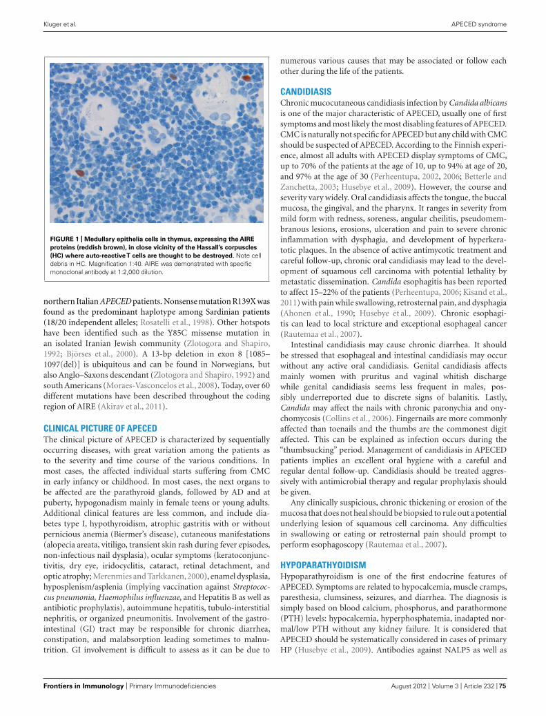
“fimmu-03-00232” — 2012/7/31 — 17:51 — page 4 — #4
Kluger et al. APECED syndrome
FIGURE 1 | Medullary epithelia cells in thymus, expressing the AIRE
proteins (reddish brown), in close vicinity of the Hassall’s corpuscles
(HC) where auto-reactiveT cells are thought to be destroyed. Note celldebris in HC. Magnification 1:40. AIRE was demonstrated with specificmonoclonal antibody at 1:2,000 dilution.
northern Italian APECED patients. Nonsense mutation R139X wasfound as the predominant haplotype among Sardinian patients(18/20 independent alleles; Rosatelli et al., 1998). Other hotspotshave been identified such as the Y85C missense mutation inan isolated Iranian Jewish community (Zlotogora and Shapiro,1992; Björses et al., 2000). A 13-bp deletion in exon 8 [1085–1097(del)] is ubiquitous and can be found in Norwegians, butalso Anglo–Saxons descendant (Zlotogora and Shapiro, 1992) andsouth Americans (Moraes-Vasconcelos et al., 2008). Today, over 60different mutations have been described throughout the codingregion of AIRE (Akirav et al., 2011).
CLINICAL PICTURE OF APECEDThe clinical picture of APECED is characterized by sequentiallyoccurring diseases, with great variation among the patients asto the severity and time course of the various conditions. Inmost cases, the affected individual starts suffering from CMCin early infancy or childhood. In most cases, the next organs tobe affected are the parathyroid glands, followed by AD and atpuberty, hypogonadism mainly in female teens or young adults.Additional clinical features are less common, and include dia-betes type I, hypothyroidism, atrophic gastritis with or withoutpernicious anemia (Biermer’s disease), cutaneous manifestations(alopecia areata, vitiligo, transient skin rash during fever episodes,non-infectious nail dysplasia), ocular symptoms (keratoconjunc-tivitis, dry eye, iridocyclitis, cataract, retinal detachment, andoptic atrophy; Merenmies and Tarkkanen,2000), enamel dysplasia,hyposplenism/asplenia (implying vaccination against Streptococ-cus pneumonia, Haemophilus influenzae, and Hepatitis B as well asantibiotic prophylaxis), autoimmune hepatitis, tubulo-interstitialnephritis, or organized pneumonitis. Involvement of the gastro-intestinal (GI) tract may be responsible for chronic diarrhea,constipation, and malabsorption leading sometimes to malnu-trition. GI involvement is difficult to assess as it can be due to
numerous various causes that may be associated or follow eachother during the life of the patients.
CANDIDIASISChronic mucocutaneous candidiasis infection by Candida albicansis one of the major characteristic of APECED, usually one of firstsymptoms and most likely the most disabling features of APECED.CMC is naturally not specific for APECED but any child with CMCshould be suspected of APECED. According to the Finnish experi-ence, almost all adults with APECED display symptoms of CMC,up to 70% of the patients at the age of 10, up to 94% at age of 20,and 97% at the age of 30 (Perheentupa, 2002, 2006; Betterle andZanchetta, 2003; Husebye et al., 2009). However, the course andseverity vary widely. Oral candidiasis affects the tongue, the buccalmucosa, the gingival, and the pharynx. It ranges in severity frommild form with redness, soreness, angular cheilitis, pseudomem-branous lesions, erosions, ulceration and pain to severe chronicinflammation with dysphagia, and development of hyperkera-totic plaques. In the absence of active antimycotic treatment andcareful follow-up, chronic oral candidiasis may lead to the devel-opment of squamous cell carcinoma with potential lethality bymetastatic dissemination. Candida esophagitis has been reportedto affect 15–22% of the patients (Perheentupa, 2006; Kisand et al.,2011) with pain while swallowing, retrosternal pain, and dysphagia(Ahonen et al., 1990; Husebye et al., 2009). Chronic esophagi-tis can lead to local stricture and exceptional esophageal cancer(Rautemaa et al., 2007).
Intestinal candidiasis may cause chronic diarrhea. It shouldbe stressed that esophageal and intestinal candidiasis may occurwithout any active oral candidiasis. Genital candidiasis affectsmainly women with pruritus and vaginal whitish dischargewhile genital candidiasis seems less frequent in males, pos-sibly underreported due to discrete signs of balanitis. Lastly,Candida may affect the nails with chronic paronychia and ony-chomycosis (Collins et al., 2006). Fingernails are more commonlyaffected than toenails and the thumbs are the commonest digitaffected. This can be explained as infection occurs during the“thumbsucking” period. Management of candidiasis in APECEDpatients implies an excellent oral hygiene with a careful andregular dental follow-up. Candidiasis should be treated aggres-sively with antimicrobial therapy and regular prophylaxis shouldbe given.
Any clinically suspicious, chronic thickening or erosion of themucosa that does not heal should be biopsied to rule out a potentialunderlying lesion of squamous cell carcinoma. Any difficultiesin swallowing or eating or retrosternal pain should prompt toperform esophagoscopy (Rautemaa et al., 2007).
HYPOPARATHYOIDISMHypoparathyroidism is one of the first endocrine features ofAPECED. Symptoms are related to hypocalcemia, muscle cramps,paresthesia, clumsiness, seizures, and diarrhea. The diagnosis issimply based on blood calcium, phosphorus, and parathormone(PTH) levels: hypocalcemia, hyperphosphatemia, inadapted nor-mal/low PTH without any kidney failure. It is considered thatAPECED should be systematically considered in cases of primaryHP (Husebye et al., 2009). Antibodies against NALP5 as well as
Frontiers in Immunology | Primary Immunodeficiencies August 2012 | Volume 3 | Article 232 | 75
“fimmu-03-00232” — 2012/7/31 — 17:51 — page 5 — #5
Kluger et al. APECED syndrome
against the calcium-sensing receptor of parathyroid epithelial cellhave been identified in APECED patients (Gavalas et al., 2007;Kemp et al., 2009, 2010). Patients who are free of HP need anannual monitoring of blood calcium and phosphorus levels. Man-agement of HP relies on daily oral supplementation of vitamin Dderivatives and calcium.
GASTRITIS AND PERNICIOUS ANEMIAChronic gastritis, with or without concomitant pernicious anemiabelongs to the APECED complex but is found only in a fraction ofcases. In non-APECED population, two types of chronic gastritisoccur, divided by Strickland into type A and B gastritis. Type Agastritis was known to be caused by autoimmunity while the Bgastritis was suspected to be the results of environmental factors.In early 1980s, it was shown by Warren and Marshall (1984) thatthe major environmental factor was in fact a chronic infection withHelicobacterium pylori.
The type A chronic gastritis, with and without pernicious ane-mia that occur in non-APECED individuals, is clearly linkedto certain HLA risk haplotypes, in analogy to isolated AD. InAPECED patients, the chronic gastritis differs from the abovealso in time of occurrence and the speed of the progression. Innon-APECED patients, the time needed for progression from theearly stage of gastritis, the superficial form to diffuse gastritis, toatrophic gastric and to full gastric atrophy is a slow process, takingup several decennia. Also, the process usually starts in the adultlife. In contrast, an APECED-associated gastritic process is muchfaster and can start in the first decennium of life. Thus, one ofthe authors of this review was able to follow such a gastric processin two 8-year-old girls with sequential gastric biopsies and couldsee how, within the time period of only 2 months, the superficialprocess lead to complete gastric atrophy of the fundus and corpus(K. Krohn, personal experience).
The target molecule for the parietal cell antibodies were shownto be the sodium-potassium channel molecule of the parietal cellson corpus and fundal part of the stomach (Karlsson et al., 1988).In antral gastritis, the antigen are the gastrin-producing cells(Uibo and Krohn, 1984).
Pernicious anemia is the end stage of the gastric immunologicaldestruction, caused partly by the lack of IF, that in addition to thehydrochloric acid is the main product of parietal cells, but also bythe autoantibodies recognizing this vitamin B12-binding protein.There are two types of antibodies to IF: one blocking vitaminB12 binding to IF and another type, binding to the IF moleculewithout interfering with vitamin B12 binding (Toh et al., 1997).Both antibody types prevent the binding of IF to its receptor onthe ileal mucosa and subsequent translocation of the vitamin B12from ileum to circulation.
ADDISON’S DISEASEAdrenocortical failure or AD, described by Thomas Addison inthe ninetieth century, is considered one of the three hallmarksof APECED, but it occurs also as a solitary disease, or as part ofthe APS-2 complex. Today, in western word, most cases of ADare caused by autoimmunity, but adrenal cortical destruction andsubsequent cortical failure can be caused by several other factors,notably by secondary tuberculosis or other chronic infections. In
retrospect, the cases described by Thomas Addison were mostlikely caused by tuberculosis.
The clinical signs and symptoms of AD are mostly similar inAPECED and in solitary AD as well as in APS-2 complex. Theseinclude decreased levels of gluco- and mineralocorticoids and ele-vated ACTH concentrations. The most severe consequence of ADis the life-threatening Addisonian crisis, characterized by generalfatigue, dizziness, diarrhea, and death, if the patient is not quicklysubstituted with corticosteroids, mainly hydrocortisol.
Autoantibodies to adrenal cortex are the characteristicimmunological feature in AD, be it part of APECED or APS-2or the solitary form. These antibodies can be easily demonstratedby immunofluorescence. However, in APECED, but not in theother forms of AD, the autoantibodies are precipitating, and thisphenomenon can be demonstrated by Ouchterlony’s immunod-iffusion (Andrada et al., 1968; Krohn et al., 1974; Heinonen et al.,1976). In immunodiffusion with APECED serum against adrenalhomogenate three precipitating lines were observed, and one ofthese were shown to represent a mitochondrial antigen while thetwo others were microsomal.
The nature of the adrenal cortical autoantigens were revealedin early 1990s and shown to be the three main steroidogenicenzymes, P450c17, P450c21, and P450scc (Krohn et al., 1974;Winqvist et al., 1993; Uibo et al., 1994a,b). These three enzymeswere also shown to be the ones that could be precipitatedby immunodiffusion. The autoantibodies against these threesteroidogenic enzymes clearly distinguish the three clinical con-ditions with adrenal failure: antibodies to all three can be foundonly in APECED, while in solitary AD and in APS-1, only anti-bodies to P450c21 are seen. Furthermore, in non-autoimmuneAD, caused by tumors or chronic infections, such antibodiesdo not occur.
GONADAL FUNCTIONSAutoimmune oophoritis is responsible for an ovarian insufficiencythat may be dramatic for female patients as insufficiency startingin teenagers and young adults. Patients may have either a pri-mary amenorrhea with no or arrested puberty. Other patientsdevelop premature menopause. The diagnosis is confirmed by sex-ual hormones status; elevated plasma levels of follicle stimulatinghormone (FSH) and luteinizing hormone (LH) and low estrogenlevels. Autoantibodies against side-chain cleavage enzyme havebeen related to ovarian insufficiency (Soderbergh et al., 2004) andalso steroidogenic enzymes antibodies against cytochrome p45021-hydroxylase (CYP21A2), cytochrome p450 17α-hydroxylase(CYP17), and cytochrome p450 side-chain cleavage enzyme(CYP11A1).
In female patients, hormonal substitution by estrogen needsto be initiated during puberty. It is strongly advised not to delaypregnancy. In case of hypogonadism, embryo donation has beentried with success.
In males, testicular failure is less common and occurs later.The prevalence of hypogonadism in males is three times lower(8–28%) than in females (35–70%; Perheentupa, 2006). It leads toclinical hypogonadism or isolated azoospermia (Husebye et al.,2009). It has been hypothesized that the blood–testis barrierprotects the Leydig cells from an autoimmune attack. However,
www.frontiersin.org August 2012 | Volume 3 | Article 232 | 76
“fimmu-03-00232” — 2012/7/31 — 17:51 — page 6 — #6
Kluger et al. APECED syndrome
the physiopathogenic link between circulating autoantibodies andhypogonadism is far from being clear. The two steroidogenicenzymes, p450scc and p450c17, are the main antigens in gonadalfailure linked to APECED, but other potential antigenic targetshave been identified such as testis-expressed protein TSGA10(Reimand et al., 2008). However, despite autoantibodies directedagainst TSGA10 in 7% of the APECED patients, no correlationcould be found with gonadal failure (Reimand et al., 2008). Oneshould not forget that the origin of gonadal dysfunction maybe related to an authentic-specific autoimmune attack but alsobe related to other hormonal dysfunction such as AD, pituitaryinsufficiency, dysthyroidism, or diabetes for instance. Besides,Schaller et al. (2008) suggested that lack of AIRE might affect fer-tility by disrupting scheduled apoptosis of testicular germ cells.In this respect, the recent hypothesis presented by Matsumoto(2011) that the function of AIRE in thymus would not be inthe regulation of transcription but rather in the developmentand differentiation of the medullary epithelial cells is of primaryinterest
OTHER ENDOCRINE DISORDERSVarious other endocrine disorders have been described such dia-betes type I mellitus, hypothyroidism, and pituitary failure, thelatter diagnosed by a growth hormone deficiency. The diagno-sis and management of these conditions does not differ fromthe standard guidelines for each disorder separately (Perheentupa,2002; Betterle and Zanchetta, 2003; Husebye et al., 2009).
OTHER NON-ENDOCRINE DISORDERSEnamel hypoplasia affect mainly permanent teeth (Perheentupa,2006), but also deciduous teeth (Pavlic and Waltimo-Sirén, 2009).Pavlic and Waltimo-Sirén (2009) recently suggested that an inad-equate process of enamel formation might affect all ameloblastsin phase. Ameloblasts have an epithelial origin with parenchy-mal cells of endocrine origin. It is speculated that ameloblastsor secreted protein in the extracellular matrix may be the tar-get of autoantibodies leading to hypoplasia. Thereby, APECEDwould be the first model of dental hard tissue autoimmune disease(Pavlic and Waltimo-Sirén, 2009).
Ocular manifestations affect 25% of the patients and includemainly keratitis that can lead to blindness. It is assumed that theorigin of keratitis is the result of autoimmunity against cornealepithelium (Merenmies and Tarkkanen, 2000; Perheentupa, 2006).However, to our knowledge no specific antibodies have been iden-tified in APECED patients. Only antibodies against OBP1 havebeen found in the AIRE mouse model against lacrimal glands(DeVoss et al., 2010).
Hyposplenism or asplenia is often diagnosed upon the develop-ment of thrombocytosis, circulating Howell–Jolly bodies, abdomi-nal ultrasound imaging or in case of severe S. pneumoniae infection(Pollak et al., 2009). Destruction of the spleen in APECED has beenrelated to an autoimmune attack against the spleen (Perheentupa,2002, 2006; Betterle and Zanchetta, 2003; Husebye et al., 2009)although the exact mechanism remains obscure. Again, the mecha-nisms proposed by Schaller et al. (2008) and by Matsumoto (2011)are of interest, as AIRE expression has also been described in lym-phoid tissue and skin. A speculative hypothesis to the evolution of
splenic atrophy could thus be disturbance of differentiation, dueto lack of AIRE expression.
Various types of GI manifestations are common in APECEDpatients. These include chronic diarrhea that can be related to HP,severe constipation. Intestinal infection by candida and giardiaespecially, pancreatic insufficiency and autoimmune enteropa-thy. Several autoreactive circulating antibodies directed towardintestinal components have been described. Ekwall et al. (1998)identified TPH as an intestinal autoantigen in APECED patients.TPH is expressed in serotonin-producing cells in the central ner-vous system and in the intestine. In their series of 80 patients, theywere able to relate “GI symptoms” to the presence of circulatingTPH antibodies and also to the total absence of enterochromaf-fin cells in the mucosa of small bowel. These enteroendocrinecells (EECs) are scattered through the intestinal mucosa, from thegastric body and antrum to the rectum. They play a key role ingrowth of the gut, blood flow, motility, secretion of pancreaticenzymes, bile, and bicarbonate-rich fluid (Posovszky et al., 2012).TPH antibodies were found in 89% of the APECED patients withGI symptoms and in 34% of those without (Ekwall et al., 1998).Antibodies can precede clinical symptoms (Ekwall et al., 1998).Conversely, TPH autoantibodies are absent in other inflamma-tory or autoimmune intestinal diseases. Additionally, Sköldberget al. (2003) identified also autoantibodies against histidine decar-boxylase expressed by EEC – like cells in the gastric mucosa. Itis noteworthy, that it is not a routine procedure to perform EECsstaining on intestinal biopsies in case of diarrhea or malabsorption,as stressed by Ohsie et al. (2009). Besides, several studies showedrepeatedly that EECs were lacking in the intestinal mucosa andwere related to chronic diarrhea (Padeh et al., 1997; Ward et al.,1999; Oliva-Hemker et al., 2006; Posovszky et al., 2012).
Tubulo-interstitial nephritis, life-threatening autoimmunebronchiolitis and other rare manifestations have also beenreported in APECED (Perheentupa, 2002, 2006; Betterle andZanchetta, 2003; Husebye et al., 2009). The main identifiedautoantibodies are summarized in Table 2.
TREATMENTManagement of APECED relies in education of the patients toknow his disease, education of the local physician, and theknowledge that new components of the disease may developduring life. Psychological support is strongly recommended asthis disease impairs greatly the quality of life of the patients(Perheentupa, 2006). Except candidiasis treatment that has beenexplained previously, treatment of APECED relies mostly on hor-mone replacement therapy according to affected organs (thyroid,parathyroid, pancreas, etc.). In some rare and potentially lethalsituations, however, patients may require corticosteroid treatmentin association with immunosuppressive therapies. These rare sit-uations include autoimmune hepatitis, especially its fulminantform, which may be lethal and therefore prompt immunosup-pressive therapy is needed (Obermayer-Straub et al., 2001). Thesame is true for interstitial nephritis and bronchiolitis in associ-ation to APECED. Immunosuppressive therapies have been alsoproposed in case of severe intestinal malabsorption with efficacy(Padeh et al., 1997; Ward et al., 1999). Very recently, Rituximab,a chimeric monoclonal antibody targeting B cell lymphocytes
Frontiers in Immunology | Primary Immunodeficiencies August 2012 | Volume 3 | Article 232 | 77

“fimmu-03-00232” — 2012/7/31 — 17:51 — page 7 — #7
Kluger et al. APECED syndrome
Table 2 | Main identified target of autoimmune antibodies in APECED
patients.
Diagnosis Main identified circulating autoantibodies
Addison’s disease 21 hydroxylase, 17α hydroxylase
Side-chain cleavage enzyme antibodies
(or steroid cell antibodies)
Hypoparathyroidism NALP5, Ca2+ sensing receptor
Hypothyroidism Thyroperoxydase
Thyroglobuline
Hypogonadism 17α hydroxylase
Side-chain cleavage enzyme antibodies
(or steroid cell antibodies)
Diabetes type I Glutamic acid decarboxylase 65-kDa isoform
(GAD65)
Insulin
Tyrosine phosphatase (IA2)
Pituitary insufficiency Tudor Domain containing protein 6 (TDRD6)
Atrophic gastritis/
Biermer’s disease
Intrinsic factor, gastric parietal cell
Intestine Glutamic acid decarboxylase 65-kDa isoform
(GAD65)
Histidine decarboxylase
Tryptophan hydroxylase
Autoimmune hepatitis Aromatic L-amino acid decarboxylase (AADC)
Cytochrome P450 1A2
Cytochrome P450 2A6
Cytochrome P450 1A1
Cytochrome P450 2B6
Vitiligo Transcription factors: SOX 9, SOX 10, aromatic
L-amino acid decarboxylase (AADC)
Alopecia areata Tyrosine hydroxylase
Nephropathy Antibody against tubular basement membrane
(Hannigan et al., 1996)
Pulmonary disease Potassium channel regulatory protein (KCNRG)
Eye OBP1*
Non-tissue specific** IFN-α, IFN-β, IFN-ω, IL-22, IL-17F, IL-17A
*Identified in a mouse model AIRE−/−.**Main non-tissue-specific antibodies according to Kisand et al. (2011).
expressive CD20 has been successfully used in a young patient withbronchiolitis (Popler et al., 2012). The rationale for Rituximab usein APECED is supported by the presence of B cell infiltrates in theaffected organs (Gavanescu et al., 2008).
AUTOANTIBODIES TOWARD INTERFERONS AND CYTOKINESAt the beginning of this millennium, the antibody responses tothe main target organs affected in APECED, and the responsi-ble target antigens were fairly well characterized. A new periodin APECED studies started along the publication by Meager
et al. (2006), describing high-titer antibodies to several type Iinterferons in practically all APECED patients studied. This anti-interferon response was exceptionally strong, since serum titersup to 1:1,000,000, and clearly exceeding the titers seen againstorgan-specific antigens, were found.
Furthermore, high-titer antibodies were seen against thetwo main mediators secreted by Th17 cells, interleukin-17 andinterleukin-22 (IL-17 and IL-22). Responses with lower titers wereoccasionally seen against other interleukins, too. In our own asyet unpublished observations we have detected occasional high-titer responses against several other interleukins and chemokines,as well, but in contrast to the aforementioned responses, theseresponses are not characteristic to all APECED patients but ratheroccur occasionally in only a few patients.
The significance of these novel findings are still unclear, butsome information concerning the role of IL-17/IL-22 antibod-ies in the chronic candida infections, characteristic for APECED,has been obtained. Th17 cells secrete IL-17 and IL-22, whichare cytokines with potent antifungal properties (Engelhardt andGrimbacher, 2012) and the occurrence of autoantibodies againstIL-17/IL-22 were reported to closely correlate to the presence ofcandida infection (Kisand et al., 2011; Engelhardt and Grimbacher,2012). However, recent evidence points to a new interactionbetween AIRE and dectin-1, a pattern-recognition receptor thatis important in antifungal innate immunity. Pedroza et al. (2012)recently showed that AIRE participates in the dectin-1 signalingpathway, and thus, missing AIRE activity could contribute to fun-gal susceptibility through this pathway. Dectin-1 is expressed onphagocytes and was recently shown to induce a non-canonicalcaspase-8 inflammasome in response to fungal and mycobac-terial infection (Gringhuis et al., 2012). The activation of thedectin signaling pathway also leads to expression of IL-17 and22 and defensins, however. Besides, other mechanisms suchDominant-negative mutations in STAT3, gain of mutation ofSTAT1, mutations in IL-17F and IL-17R may be alternate causesof CMC (Engelhardt and Grimbacher, 2012).
The significance of the antibody response toward interferonsand other cytokines is presently also unclear. One could speculatethat some of these antibodies against type I interferons as well asreacting with IL-17 and IL-22 might have a protective function.As pointed out by Waterfield and Anderson (2011), antibodies totype I interferons do not seem to lead to increased susceptibilityto viral infections. This resistance might be due to redundancyand it has to be seen whether this anti-interferon response isdirected only toward certain members of the interferon family.While Th17 cell response and the release of soluble IL-17 andIL-22 are evidently necessary for the defense against mucocuta-neous candida infection, the same cytokines have a role in thedevelopment of psoriasis. Similarly, interferons are known to beinvolved in the pathogenesis of several conditions, and one suchchronic ailment is the autoimmune diseases belonging to the sys-temic lupus erythematosus (SLE) complex. Anti-interferon alphaantibodies are currently being tested as a therapeutic mean againstSLE (Merrill et al., 2011). In order to be able to find out if theantibodies against interferons and other cytokines could have aprotective role in APECED, large APECED patient cohorts have tobe studied in order to find out whether, e.g., psoriasis and SLE are
www.frontiersin.org August 2012 | Volume 3 | Article 232 | 78

“fimmu-03-00232” — 2012/7/31 — 17:51 — page 8 — #8
Kluger et al. APECED syndrome
significantly less common in APECED patients than in the generalpopulation.
The reason for the antibody response toward soluble immunemediators is still unclear, and we do not yet know what exactlyelicits them and thus, only speculative scenarios can be presented.It is conceivable to hypothesize, however, that the tissue destruc-tion preceding the failure of the endocrine organs may have arole. Tissue destruction, be it caused by trauma, viral infec-tion or autoimmune attack, would probably lead not only tothe release of potential tissue-specific autoantigens and thus, toautoantibody formation against these proteins, but could alsolead to an inflammatory response and production of severalmediators of inflammation. One key group of molecules in thisrespect is the acute phase proteins, notably those belonging to theIL-1 group.
It is generally believed that the destruction of the endocrineorgans in APECED is caused by the autoreactive CD8+ cyto-toxic T cells, although definitive evidence for this mechanism isstill lacking (Betterle and Zanchetta, 2003; Moraes-Vasconceloset al., 2008). This hypothesis is reinforced by the examination ofmicroscopic examinations of samples, sometimes obtained post-mortem. Indeed, parathyroid, adrenal glands, or ovaries pathologydisclosed also atrophy and lymphocytic infiltration that suggestlymphocytic aggression of the organs leading to atrophy anddysfunction (Betterle and Zanchetta, 2003). This is also stressed,indirectly, by the analysis of the AIRE-deficient mouse model, whodevelop also a lymphocytic infiltration in some inner organs alongwith atrophy (Ramsey et al., 2002).
However, cell destruction caused by an immune responseagainst the endocrine organ would in fact lead to a similar sit-uation that is thought to happen in viral infections. In fact, severalautoimmune diseases, such as diabetes type I or chronic autoim-mune liver diseases are thought to be a consequence of precedingviral infection: enterovirus infection in the case of diabetes typeI and hepatitis B in the case of chronic active hepatitis. In viralinfections, a specific group of intracellular regulatory molecules,TRIMs (tripartite motif-containing proteins), have been shown tohave a key role in eliciting an autoimmune or auto inflammatoryconsequence (Jefferies et al., 2011).
The TRIM protein family is a form of RING domain containingE3 ligases and they exert a variety of biological functions, relatedto immunity and inflammation (Jefferies et al., 2011). Specifically,of the more than 20 different TRIM proteins, some seem to up-regulate the expression of type I interferons and proinflammatorycytokines, notable interleukin-1beta (IL-1beta). Furthermore, thesame mediators of immune response and inflammation are insome cases known to up-regulate the expression of TRIMs. Thus,a vicious circle can theoretically occur and this in turn couldlead to autoimmunity. So far, overexpression of TRIMs, or anautoimmune response toward them, has been shown to be linkedto autoimmune and autoinflammatory processes in Sjögren’ssyndrome or rheumatoid arthritis (Jefferies et al., 2011).
Presently, we have no information how the occurrence ofautoantibodies toward the interferons and other mediators ofimmune response might affect the aforementioned vicious circle,but it is conceivable to speculate that such an antibody responsecould have an balancing effect. One could thus form a hypothesis,
that in APECED, the primary defect outside thymus, where theautoreactive T cells are not destroyed, would be the cell destructionof the endocrine organs by cell-mediated immune response, fol-lowed by release of cellular components taken up by professionalantigen presenting cells and further stimulating the activation ofCD4+ Th-cells and finally resulting in an autoantibody responseto these organ-specific antigens. However, simultaneous overex-pression of TRIMs and subsequent up-regulation of a variety ofsoluble mediators of immune response and inflammation, such asinterferons and members of the IL-1 family would lead to autoan-tibody formation also against these cytokines. Lastly, one reasonfor the break of tolerance to immune mediators, and subsequentproduction of autoantibodies could be related the fact that AIREexpression seems to occur, in addition to thymic epithelial cells alsooutside thymus, notably in dendritic cells, that normally expressalso such mediators (Heath and Carbone, 2009)
The consequences of such cytokine-directed antibody responseare still an open question. In case of the Th17 type interleukins(IL-17 and IL-22) there is convincing evidence that such antibod-ies are linked to the CMC. However, at least in some cases, theantibodies may have a balancing, down-regulating effect on theexpression of the corresponding biologically active molecules butalso, by regulating the immune response to target organs. Thus,it is possible to presume, that especially the antibodies to type Iinterferons might have a protective effect, as some chronic immunediseases, such as psoriasis and SLE, are rare or non-existing amongAPECED patients.
CELL-MEDIATED IMMUNE RESPONSESAlthough it is now a generally accepted view that the consequenceof the AIRE defect in APECED will lead to the escape of the poten-tially autoreactive T cells, there is in fact rather little direct evidenceto show that the tissue destruction in the endocrine organs affectedin APECED is caused by cytotoxic CD8+ T cells. Furthermore,most studies describing the phenotype of the lymphocytes infil-trating affected organs is not from APECED patients directly, butfrom patients suffering of solitary lesions that are similar to theones seen in APECED, such as solitary AD or diabetes. However,the solitary endocrine diseases, such as isolated thyroid diseaseor AD are remarkably similar in their clinical picture as well asimmunological findings as those of APECED. Thus, in solitaryAD and in APECED with adrenocortical failure, autoantibodiesrecognize the p450c21 steroidogenic enzyme. Interestingly, in thisdisease complex CD8+ T cells that reach against specific T cellepitopes in p450c21 has been demonstrated (Bratland et al., 2009;Rottembourg et al., 2010) Likewise, in thyroid diseases, thyroglob-ulin and thyroid peroxidase are recognized by the autoantibodies,irrespective if the condition is occurring alone, in association ofAPS-2 or as part of the APECED complex. The similar syner-gism in terms of the nature of autoantigens occurs in chronicimmunological liver diseases, too.
In chronic aggressive hepatitis the lymphocytic infiltrating cellpopulation has been shown to be of the CD8+ lineage (Si et al.,1984). In a murine model of Graves’ disease, the CD8+ cellpopulation contains also the recently identified CD8+CD122+T cells that are functionally similar to the CD4+CD25+ regulatoryT cells (Ryan et al., 2005). Furthermore, studies in thyroid and
Frontiers in Immunology | Primary Immunodeficiencies August 2012 | Volume 3 | Article 232 | 79
“fimmu-03-00232” — 2012/7/31 — 17:51 — page 9 — #9
Kluger et al. APECED syndrome
other affected organs show that one of the main cell populationin the lymphocytic infiltrate are in fact the CD4+ Th17 cells thatsecrete as effector molecules, the cytokines IL-17 and IL-22. Inexperimental autoimmune diseases, the balance between the Th17effector cells and the two regulatory T cells, CD8+CD122+ andCD4+CD25+, seems to regulate both the occurrence and severityof tissue destruction and functional failure.
There could thus be two distinct mechanisms operating in thepathogenesis of autoimmunity in the endocrinopathies: one medi-ated by soluble effector molecules, such as IL-17 and IL-22 aswell as type I interferons, and an other one mediated by effectorT cells, which are either of the CD8+ CTL cell or of the Th17 effec-tor cell lineage. To counteract these, again two distinct biologicalprocesses would occur: the production of autoantibodies and sec-ondly, the emergency of the regulatory T cells. As to the regulatoryT cell response, it is to note that one key immunological failure inAPECED, is the dysregulation of the Treg cell maturation (Ryanet al., 2005; Kekäläinen et al., 2007; Saitoh et al., 2007; Laakso et al.,2010, 2011; Wolff et al., 2010).
In normal thymus, Treg maturation follows a prepro-grammed scheme, and the immature CD8+CD4+FOXP3+seems to be prone to apoptosis, whereas the more matureform CD4+CD8−FOXP3+ cells form the active Treg popula-tion (Lehtoviita et al., 2009). According to Endharti et al. (2011)the CD8+CXCR3+ Tregs in humans are functionally similar tomurine CD8+CD122+ Tregs. Furthermore, in APECED patientsthe recent thymic emigrant (RTE) pool of Treg cells shift to the acti-vated pool and the RTE reservoir is depleted. Most importantly,in APECED patients these cells express less FOXP3 than in thehealthy controls (Laakso et al., 2010). Thus, in APECED the newlyformed Treg cells have a developmental defect and their function is
therefore impaired. Data concerning the CD8+ regulatory T cellsin APECED patients is missing, however.
The finding that the regulatory T cell population in APECED isfunctionally defective and that the expression of the key moleculefor Treg function, the FOXP3 is impaired, is consistent with clini-cal findings in IPEX syndrome, caused by a defect in the functionof the FOXP3 gene. However, it should be noted that the effect ofFOXP3 mutations in Treg population also in the IPEX patients ishighly variable. Also, in contrast to APECED there seems to be agenotype–phenotype correlation in IPEX, as different mutationsare associated in variable clinical picture, that show differencesin severity as well in the types of clinical components that arepresent (Torgerson et al., 2007; d’Hennezel et al., 2009) A consis-tent finding in IPEX is however the inability of the CD4+CD25high Tregs to suppress the function of autologous effector T cells(Bacchetta et al., 2006). There are, thus, several differences in theclinical picture of APECED and IPEX, but both conditions showclear immune destruction of at least some endocrine organs. Bothconditions also share some similarities in the GI symptoms.
The proposed hypothesis that in APECED both tissue destruc-tive mechanisms and controlling regulatory mechanisms existraises a question whether APECED could be treated or evencured by immunological manipulations. To find an answer forthis question is one of the further challenges for APECED research
ACKNOWLEDGMENTSThe study was financially supported by the European ScienceFoundation (ESF) and the Sigrid Juselius foundation (to NicolasKluger). We thank Professors Jaakko Perheentupa, Adrian Hayday,and Pärt Peterson, as well as Drs Kai Kisand, Annalisa Macagno,and Edward Stuart for stimulating discussions.
REFERENCESAhonen, P., Myllarniemi, S., Sipila, I.,
and Perheentupa, J. (1990). Clinicalvariation of autoimmune polyendo-crinopathy–candidiasis–ectodermaldystrophy (APECED) in a series of68 patients. N. Engl. J. Med. 322,1829–1836.
Akirav, E. M., Ruddle, N. H., andHerold, K. C. (2011). The role ofAIRE in human autoimmune disease.Nat. Rev. Endocrinol. 7, 25–33.
Andrada, J. A., Bigazzi, P. L., Andrada,E., Milgrom, F., and Witebsky,E. (1968). Serological investigationson Addison’s disease. JAMA 206,1535–1541.
Bacchetta, R., Passerini, L., Gambineri,E., Dai, M., Allan, S. E., Perroni,L., Dagna-Bricarelli, F., Sartirana, C.,Matthes-Martin, S., Lawitschka, A.,Azzari, C., Ziegler, S. F., Levings, M.K., and Roncarolo, M. G. (2006).Defective regulatory and effectorT cell functions in patients withFOXP3 mutations. J. Clin. Invest. 116,1713–1722.
Bennett, C. L., Yoshioka, R., Kiyo-sawa, H., Barker, D. F., Fain, P.R., Shigeoka, A. O., and Chance,
P. F. (2000). X-Linked syndrome ofpolyendocrinopathy, immune dys-function, and diarrhea maps toXp11.23–Xq13.3. Am. J. Hum. Genet.66, 461–468.
Betterle, C., and Zanchetta, R. (2003).Update on autoimmune polyen-docrine syndromes (APS). ActaBiomed. 74, 9–33.
Bjorses, P., Alltonen, J., Vikman, A.,Perheentupa, J., Ben-Zion, G., Chi-umello, G., Dahl, N., Heidemen, P.,Hoorweg-Nijman, J. J., Mathivon, L.,Mullis, P. E., Pohl, M., Ritzen, M.,Romeo, G., Shapiro, M. S., Smith, C.S., Solyom, J., Zlotogora, J., and Pelto-nen, L. (1996). Genetic homogeneityof autoimmune polyglandular dis-ease type I. Am. J. Hum. Genet. 59,879–886.
Björses, P., Halonen, M., Palvimo, J. J.,Kolmer, M., Aaltonen, J., Ellonen,P., Perheentupa, J., Ulmanen, I., andPeltonen, L. (2000). Mutations inthe AIRE gene: effects on subcellularlocation and transactivation func-tion of the autoimmune polyendo-crinopathy-candidiasis-ectodermaldystrophy protein. Am. J. Hum.Genet. 66, 378–392.
Blizzard, R. M., Tomasi, T. B., andChristy, N. P. (1963). Autoanti-bodies against thyroid and adrenaltissue in a patient with mutiple, pri-mary endocrine deficiencies. J. Clin.Endocrinol. Metab. 23, 1179–1180.
Blizzard, R. M., Chandler, R. W.,Kyle, M. A., and Hung, W. (1962).Adrenal antibodies in Addison’s dis-ease. Lancet 2, 901–903.
Bottazzo, G. F., Dean, B. M., Gorsuch, A.N., Cudworth, A. G., and Doniach, D.(1980). Complement-fixing islet-cellantibodies in type-I diabetes: possiblemonitors of active beta-cell damage.Lancet 1, 668–672.
Bratland, E., Skinningsrud, B., Undlien,D. E., Mozes, E., and Huse-bye, E. S. (2009). T cell responsesto steroid cytochrome P450 21-hydroxylase in patients with autoim-mune primary adrenal insufficiency.J. Clin. Endocrinol. Metab. 12,5117–5124.
Brun, J. M. (1978). Auto-immune juve-nile polyendocrinopathy (author’stransl). Ann. Endocrinol. (Paris) 39,463–481.
Collins, S. M., Dominguez, M., Ilmari-nen, T., Costigan, C., and Irvine, A.
D. (2006). Dermatological manifes-tations of autoimmune polyendo-crinopathy-candidiasis-ectodermaldystrophy syndrome. Br. J. Dermatol.154, 1088–1093.
Derbinski, J., Gäbler, J., Brors, B.,Tierling, S., Jonnakuty, S., Hergen-hahn, M., Peltonen, L., Walter, J.,and Kyewski, B. (2005). Promiscuousgene expression in thymic epithelialcells is regulated at multiple levels. J.Exp. Med. 202, 33–45.
DeVoss, J. J., LeClair, N. P., Hou, Y.,Grewal, N. K., Johannes, K. P., Lu,W., Yang, T., Meagher, C., Fong, L.,Strauss, E. C., and Anderson, M. S.(2010). An autoimmune response toodorant binding protein 1a is associ-ated with dry eye in the Aire-deficientmouse. J. Immunol. 184, 4236–4246.
d’Hennezel, E., Ben-Shoshan, M., Ochs,H. D., Torgerson, T. R., Russell, L.J., Lejtenyi, C., Noya, F. J., Jabado, N.,Mazer, B., and Piccirillo, C. A. (2009).FOXP3 forkhead domain mutationand regulatory T cells in the IPEXsyndrome. N. Engl. J. Med. 361,1710–1713.
Doniach, D., and Roitt, I. M.(1964). An evaluation of gastric
www.frontiersin.org August 2012 | Volume 3 | Article 232 | 80
“fimmu-03-00232” — 2012/7/31 — 17:51 — page 10 — #10
Kluger et al. APECED syndrome
and thyroid auto-immunity in relata-tion to hematologic auto-immunity.Semin. Hematol. 1, 313–343.
Ekwall, O., Hedstrand, H., Grimelius, L.,Haavik, J., Perheentupa, J., Gustafs-son, J., Husebye, E., Kämpe, O.,and Rorsman, F. (1998). Identifica-tion of tryptophan hydroxylase as anintestinal autoantigen. Lancet 352,279–283.
Endharti, A. T., Okuno, Y., Shi, Z.,Misawa, N., Toyokuni, S., Ito, M.,Isobe, K., and Suzuki, H. (2011).CD8+CD122+ regulatory T cells(Tregs) and CD4+ Tregs coopera-tively prevent and cure CD4+ cell-induced colitis. J. Immunol. 186,41–52.
Engelhardt, K. R., and Grimbacher,B. (2012). Mendelian traits caus-ing susceptibility to mucocutaneousfungal infections in human sub-jects. J. Allergy Clin. Immunol. 129,294–305.
Fierabracci, A. (2011). Recent insightsinto the role and molecular mech-anisms of the autoimmune regula-tor (AIRE) gene in autoimmunity.Autoimmu. Rev. 10, 137–143.
Gardner, J. M., Fletcher, A. L., Anderson,M. S., and Turley, S. J. (2009). AIRE inthe thymus and beyond. Curr. Opin.Immunol. 21, 582–589.
Gavalas, N. G., Kemp, E. H., Krohn, K.J., Brown, E. M., Watson, P. F., andWeetman, A. P. (2007). The calcium-sensing receptor is a target of autoan-tibodies in patients with autoimmunepolyendocrine syndrome type 1. J.Clin. Endocrinol. Metab. 92, 2107–2114.
Gavanescu, I., Benoist, C., and Mathis,D. (2008). B cells are required forAire-deficient mice to develop multi-organ autoinflammation: a therapeu-tic approach for APECED patients.Proc. Natl. Acad. Sci. U.S.A. 105,13009–13014.
Gringhuis, S. I., Kaptein, T. M., Wev-ers, B. A., Theelen, B., van derVlist, M., Boekhout, T., and Gei-jtenbeek, T. B. (2012). Dectin-1is an extracellular pathogen sensorfor the induction and processing ofIL-1β via a noncanonical caspase-8inflammasome. Nat. Immunol. 13,246–254.
Hannigan, N. R., Jabs, K., Perez-Atayde,A. R., and Rosen, S. (1996). Autoim-mune interstitial nephritis and hep-atitis in polyglandular autoimmunesyndrome. Pediatr. Nephrol. 10,511–514.
Heath, W. R., and Carbone, F. R.(2009). Dendritic cell subsets in pri-mary and secondary T cell responsesat body surfaces. Nat. Immunol. 10,1237–1244.
Heinonen, E., Krohn, K., Perheen-tupa, J., Aro, A., and Pelkonen, R.(1976). Association of precipitatinganti-adrenal anti-adrenal antibodieswith moniliasis-polyendocrinopathysyndrome. Ann. Clin. Res. 8,262–265.
Husebye, E. S., Perheentupa, J.,Rautemaa, R., and Kämpe, O. (2009).Clinical manifestations and manage-ment of patients with autoimmunepolyendocrine syndrome type I. J.Intern. Med. 265, 514–529.
Irvine, W. J., Davies, S. H., Teitelbaum,S., Delamore, I. W., and Williams, A.W. (1965). The clinical and patholog-ical significance of gastric parietal cellantibody. Ann. N. Y. Acad. Sci. 124,657–691.
Irvine, W. J., MacGregor, A. G., andStuart, A. E. (1962). The prognos-tic significance of thyroid antibodiesin the management of thyrotoxicosis.Lancet 2, 843–847.
Jefferies, C., Wynne, C., and Higgs, R.(2011). Antiviral TRIMs: friend orfoe in autoimmune and autoinflam-matory disease? Nat. Rev. Immunol.11, 617–625.
Jeffries, G. H., Hoskins, D. W., andSleisenger, M. H. (1962). Antibodyto intrinsic factor in serum frompatients with pernicious anemia. J.Clin. Invest. 41, 1106–1115.
Kaldany, A. (1979). Autoantibodies toislet cells in diabetes mellitus. Dia-betes 28, 102–105.
Karlsson, F. A., Burman, P., Lööf,L., and Mårdh, S. (1988). Majorparietal cell antigen in autoim-mune gastritis with pernicious ane-mia is the acid-producing H+,K+-adenosine triphosphatase of thestomach. J. Clin. Invest. 81,475–479.
Kekäläinen, E., Tuovinen, H., Joensuu,J., Gylling, M., Franssila, R., Pönty-nen, N., Talvensaari, K., Perheentupa,J., Miettinen, A., and Arstila, T. P.(2007). A defect of regulatory Tcells in patients with autoimmunepolyendocrinopathy-candidiasis-ectodermal dystrophy. J. Immunol.178, 1208–1215.
Kemp, E. H., Gavalas, N. G., Akhtar, S.,Krohn, K. J., Pallais, J. C., Brown, E.M., Watson, P. F., and Weetman, A. P.(2010). Mapping of human autoan-tibody binding sites on the calcium-sensing receptor. J. Bone Miner. Res.25, 132–140.
Kemp, E. H., Gavalas, N. G., Krohn,K. J., Brown, E. M., Watson, P.F., and Weetman, A. P. (2009).Activating autoantibodies against thecalcium-sensing receptor detectedin two patients with autoimmunepolyendocrine syndrome type 1. J.
Clin. Endocrinol. Metab. 94, 4749–4756.
Kisand, K., Lilic, D., Casanova, J. L.,Peterson, P., Meager, A., and Will-cox, N. (2011). Mucocutaneous can-didiasis and autoimmunity againstcytokines in APECED and thymomapatients: clinical and pathogeneticimplications. Eur. J. Immunol. 41,1517–1527.
Kogawa, K., Nagafuchi, S., Katsuta,H., Kudoh, J., Tamiya, S., Sakai, Y.,Shimizu, N., and Harada, M. (2002).Expression of AIRE gene in periph-eral monocyte/dendritic cell lineage.Immunol. Lett. 80, 195–198.
Krohn, K., Perheentupa, J., andHeinonen, E. (1974). Precipi-tating anti-adrenal antibodies inAddison’s disease. Clin. Immunol.Immunopathol. 3, 59–68.
Laakso, S. M., Laurinolli, T. T., Rossi,L. H., Lehtoviita, A., Sairanen, H.,Perheentupa, J., Kekäläinen, E., andArstila, T. P. (2010). Regulatory T celldefect in APECED patients is asso-ciated with loss of naive FOXP3(+)precursors and impaired activatedpopulation. J. Autoimmun. 35,351–357.
Laakso, S. M., Kekäläinen, E., Rossi, L.H., Laurinolli, T. T., Mannerström,H., Heikkilä, N., Lehtoviita, A., Per-heentupa, J., Jarva, H., and Arstila,T. P. (2011). IL-7 dysregulation andloss of CD8+ T cell homeostasisin the monogenic human diseaseautoimmune polyendocrinopathy-candidiasis-ectodermal dystrophy. J.Immunol. 187, 2023–2030.
Lehtoviita, A., Rossi, L. H., Kekäläi-nen, E., Sairanen, H., and Arstila, T.P. (2009). The CD4(+)CD8(+) andCD4(+) subsets of FOXP3(+) thy-mocytes differ in their response togrowth factor deprivation or stim-ulation. Scand. J. Immunol. 70,377–383.
Matsumoto, M. (2011). Contrastingmodels for the roles of Aire in thedifferentiation program of epithelialcells in the thymic medulla. Eur. J.Immunol. 41, 12–17.
Meager, A., Visvalingam, K., Peter-son, P., Möll, K., Murumägi, A.,Krohn, K., Eskelin, P., Perheen-tupa, J., Husebye, E., Kadota,Y., and Willcox, N. (2006). Anti-interferon autoantibodies in autoim-mune polyendocrinopathy syndrometype 1. PLoS Med. 3, e289. doi:10.1371/journal.pmed.0030289
Meloni, A., Willcox, N., Meager, A.,Atzeni, M., Wolff, A. S., Husebye, E.S., Furcas, M., Rosatelli, M. C., Cao,A., and Congia, M. (2012). Autoim-mune polyendocrine syndrometype 1: an extensive longitudinal
study in Sardinian patients. J.Clin. Endocrinol. Metab. 97,1114–1124.
Merenmies, L., and Tarkkanen, A.(2000). Chronic bilateral keratitis inautoimmune polyendocrinopathy-candidiadis-ectodermal dystrophy(APECED). A long-term follow-up and visual prognosis. ActaOphthalmol. Scand. 78, 532–535.
Merrill, J. T., Wallace, D. J., Petri, M.,Kirou, K. A., Yao, Y., White, W. I.,Robbie, G., Levin, R., Berney, S.M., Chindalore, V., Olsen, N., Rich-man, L,. Le, C., Jallal, B., and White,B.; Lupus Interferon Skin Activity(LISA) Study Investigators. (2011).Safety profile and clinical activity ofsifalimumab, a fully human anti-interferon α monoclonal antibody,in systemic lupus erythematosus: aphase I, multicentre, double-blindrandomised study. Ann. Rheum. Dis.70, 1905–1913.
Moraes-Vasconcelos, D., Costa-Carvalho, B. T., Torgerson, T. R.,and Ochs, H. D. (2008). Primaryimmune deficiency disorders pre-senting as autoimmune diseases:IPEX and APECED. J. Clin. Immunol.28, S11–S19.
Nagamine, K., Peterson, P., Scott, H. S.,Kudoh, J., Minoshima, S., Heino, M.,Krohn, K. J., Lalioti, M. D., Mullis,P. E., Antonarakis, S. E., Kawasaki,K., Asakawa, S., Ito, F., and Shimizu,N. (1997). Positional cloning ofthe APECED gene. Nat. Genet. 17,393–398.
Neufeld, M., Maclaren, N. K.,and Blizzard, R. M. (1981). Twotypes of autoimmune Addison’s dis-ease associated with different polyg-landular autoimmune (PGA) syn-dromes. Medicine (Baltimore) 60,355–362.
Obermayer-Straub, P., Perheentupa, J.,Braun, S., Kayser, A., Barut, A., Loges,S., Harms, A., Dalekos, G., Strass-burg, C. P., and Manns, M. P. (2001).Hepatic autoantigens in patients withautoimmune polyendocrinopathy-candidiasis-ectodermal dystrophy.Gastroenterology 121, 668–677.
Ohsie, S., Gerney, G., Gui, D., Kahana,D., Martín, M. G., and Cortina,G. (2009). A paucity of colonicenteroendocrine and/or enterochro-maffin cells characterizes a subsetof patients with chronic unex-plained diarrhea/malabsorption.Hum. Pathol. 40, 1006–1014.
Oliva-Hemker, M., Berkenblit, G. V.,Anhalt, G. J., and Yardley, J. H. (2006).Pernicious anemia and widespreadabsence of gastrointestinal endocrinecells in a patient with autoimmunepolyglandular syndrome type I and
Frontiers in Immunology | Primary Immunodeficiencies August 2012 | Volume 3 | Article 232 | 81
“fimmu-03-00232” — 2012/7/31 — 17:51 — page 11 — #11
Kluger et al. APECED syndrome
malabsorption. J. Clin. Endocrinol.Metab. 91, 2833–2838.
Orlova, E. M., Bukina, A. M.,Kuznetsova, E. S., Kareva, M. A.,Zakharova, E. U., Peterkova, V. A.,and Dedov, I. I. (2010). Autoim-mune polyglandular syndrome type 1in Russian patients: clinical variantsand autoimmune regulator muta-tions. Horm. Res. Paediatr. 73,449–457.
Padeh, S., Theodor, R., Jonas, A., andPasswell, J. H. (1997). Severe malab-sorption in autoimmune polyendo-crinopathy-candidosis-ectodermaldystrophy syndrome successfullytreated with immunosuppression.Arch. Dis. Child. 76, 532–534.
Pavlic, A., and Waltimo-Sirén, J. (2009).Clinical and microstructural aber-rations of enamel of deciduous andpermanent teeth in patients withautoimmune polyendocrinopathy-candidiasis-ectodermal dystrophy.Arch. Oral Biol. 54, 658–665.
Pedroza, L. A., Kumar, V., Sanborn,K. B., Mace, E. M., Niinikoski,H., Nadeau, K., Vasconcelos Dde,M., Perez, E., Jyonouchi, S.,Jyonouchi, H., Banerjee, P. P.,Ruuskanen, O., Condino-Neto, A.,and Orange, J. S. (2012). Autoim-mune regulator (AIRE) contributesto Dectin-1-induced TNF-α pro-duction and complexes with cas-pase recruitment domain-containingprotein 9 (CARD9), spleen tyro-sine kinase (Syk), and Dectin-1. J.Allergy Clin. Immunol. 129, 464–472,472.e1–3.
Perheentupa, J. (2002). APS-I/APECED: the clinical diseaseand therapy. Endocrinol. Metab. Clin.North Am. 31, 295–320, vi.
Perheentupa, J. (2006). Autoimmunepolyendocrinopathy-candidiasis-ectodermal dystrophy. J. Clin.Endocrinol. Metab. 91, 2843–2850.
Pollak, U., Bar-Sever, Z., Hoffer, V., Mar-cus, N., Scheuerman, O., and Garty,B. Z. (2009). Asplenia and functionalhyposplenism in autoimmune polyg-landular syndrome type 1. Eur. J.Pediatr. 168, 233–235.
Popler, J., Alimohammadi, M., Kämpe,O., Dalin, F., Dishop, M. K., Barker, J.M., Moriarty-Kelsey, M., Soep, J. B.,and Deterding, R. R. (2012). Autoim-mune polyendocrine syndrome typeUtility of KCNRG autoantibodies asa marker of active pulmonary dis-ease and successful treatment withrituximab. Pediatr. Pulmonol. 47,84–87.
Posovszky, C., Lahr, G., von Schnurbein,J., Buderus, S., Findeisen, A.,Schröder, C., Schütz, C., Schulz,A, Debatin, K. M., Wabitsch, M., and
Barth, T. F. (2012). Loss of enteroen-docrine cells in autoimmune-poly-endocrine-candidiasis-ectodermal-dystrophy (APECED) syndromewith gastrointestinal dysfunction.J. Clin. Endocrinol. Metab. 97,E292–E300.
Powell, B. R., Buist, N. R., and Stenzel, P.(1982). A X-linked syndrome of diar-rhea, polyendocrinopathy, and fatalinfection in infancy. J. Pediatr. 100,731–737.
Ramsey, C., Winqvist, O., Puhakka,L., Halonen, M., Moro, A., Kämpe,O., Eskelin, P., Pelto-Huikko, M.,and Peltonen, L. (2002). Aire defi-cient mice develop multiple featuresof APECED phenotype and showaltered immune response. Hum. Mol.Genet. 11, 397–409.
Rautemaa, R., Hietanen, J., Niissalo,S., Pirinen, S., and Perheentupa,J. (2007). Oral and oesophagealsquamous cell carcinoma – a compli-cation or component of autoimmunepolyendocrinopathy-candidiasis-ectodermal dystrophy (APECED,APS-I). Oral Oncol. 43, 607–613.
Reimand, K., Perheentupa, J., Link, M.,Krohn, K., Peterson, P., and Uibo,R. (2008). Testis-expressed proteinTSGA10 an auto-antigen in autoim-mune polyendocrine syndrome typeI. Int. Immunol. 20, 39–44.
Rosatelli, M. C., Meloni, A., Meloni,A., Devoto, M., Cao, A., Scott, H. S.,Peterson, P., Heino, M., Krohn, K. J.,Nagamine, K., Kudoh, J., Shimizu,N., and Antonarakis, S. E. (1998).A common mutation in Sardinianautoimmune polyendocrinopathy-candidiasis-ectodermal dystrophypatients. Hum. Genet. 103,428–434.
Rottembourg, D., Deal, C., Lambert, M.,Mallone, R., Carel, J. C., Lacroix, A.,Caillat-Zucman, S., and le Deist, F.(2010). 21-Hydroxylase epitopes aretargeted by CD8 T cells in autoim-mune Addison’s disease. J. Autoim-mun. 35, 309–315.
Ryan, K. R., Lawson, C. A., Lorenzi,A. R., Arkwright, P. D., Isaacs, J. D.,and Lilic, D. (2005) CD4+CD25+T-regulatory cells are decreased inpatients with autoimmune polyen-docrinopathy candidiasis ectodermaldystrophy. J. Allergy Clin. Immunol.116, 1158–1159.
Saitoh, O., Abiru, N., Nakahara, M., andNagayama, Y. (2007). CD8+CD122+T cells, a newly identified regulatoryT subset, negatively regulate Graves’hyperthyroidism in a murine model.Endocrinology 148, 6040–6046.
Schaller, C. E., Wang, C. L., Beck-Engeser, G., Goss, L., Scott, H.S., Anderson, M. S., and Wabl,
M. (2008). Expression of Aire andthe early wave of apoptosis inspermatogenesis. J. Immunol. 180,1338–1343.
Schwartz, M. (1961). Antibodies tointrinsic factor. Acta Allergol. 16,263–266.
Si, L., Whiteside, T. L., Van Thiel,D. H., and Rabin, B. S. (1984).Lymphocyte subpopulations at thesite of “piecemeal” necrosis in endstage chronic liver diseases and reject-ing liver allografts in cyclosporine-treated patients. Lab. Invest. 50,341–347.
Sköldberg, F., Portela-Gomes, G. M.,Grimelius, L., Nilsson, G., Per-heentupa, J., Betterle, C., Husebye,E. S., Gustafsson, J., Rönnblom,A., Rorsman, F., and Kämpe,O. (2003). Histidine decarboxylase,a pyridoxal phosphate-dependentenzyme, is an autoantigen of gas-tric enterochromaffin-like cells. J.Clin. Endocrinol. Metab. 88, 1445–1452.
Soderbergh, A., Myhre, A. G., Ekwall,O., Gebre-Medhin, G., Hedstrand,H., Landgren, E., Miettinen, A.,Eskelin, P., Halonen, M., Tuomi,T., Gustafsson, J., Husebye, E.S., Perheentupa, J., Gylling, M.,Manns, M. P., Rorsman, F., Kämpe,O., and Nilsson, T. (2004). Preva-lence and clinical associations of 10defined autoantibodies in autoim-mune polyendocrine syndrome typeI. J. Clin. Endocrinol. Metab. 89,557–562.
The Finnish–German APECED Con-sortium. (1997). An autoimmunedisease, APECED, caused by muta-tions in a novel gene featuring twoPHD-type zinc-finger domains. Nat.Genet. 17, 399–403.
Toh, B. H., van Driel, I. R., and Gleeson,P. A. (1997). Pernicious anemia. N.Engl. J. Med. 337, 1441–1448.
Torgerson, T. R., Linane, A., Moes,N., Anover, S., Mateo, V., Rieux-Laucat, F., Hermine, O., Vijay, S.,Gambineri, E., Cerf-Bensussan, N.,Fischer, A., Ochs, H. D., Goulet,O., and Ruemmele, F. M. (2007).Severe food allergy as a variant ofIPEX syndrome caused by a dele-tion in a noncoding region of theFOXP3 gene. Gastroenterology 132,1705–1717.
Uibo, R., Aavik, E., Peterson, P., Per-heentupa, J., Aranko, S., Pelkonen, R.,and Krohn, K. J. (1994a). Autoanti-bodies to cytochrome P450 enzymesP450scc, P450c17, and P450c21 inautoimmune polyglandular diseasetypes I and II and in isolated Addi-son’s disease. J. Clin. Endocrinol.Metab. 78, 323–328.
Uibo, R., Perheentupa, J., Ovod, V.,and Krohn, K. J. (1994b). Char-acterization of adrenal autoantigensrecognized by sera from patientswith autoimmune polyglandular syn-drome (APS) type I. J. Autoimmun. 7,399–411.
Uibo, R. M., and Krohn, K. J.(1984). Demonstration of gastrincell autoantibodies in antral gastritiswith avidin-biotin complex antibodytechnique. Clin. Exp. Immunol. 58,341–347.
Walder, A. I., Lunseth, J. B., and Peter,E. T. (1963). Antibodies to the gas-tric parietal cell. Surg. Forum 14,327–328.
Ward, L., Paquette, J., Seidman, E.,Huot, C., Alvarez, F., Crock, P.,Delvin, E., Kämpe, O., and Deal, C.(1999). Severe autoimmune poly-endocrinopathy-candidiasis-ecto-dermal dystrophy in an adolescentgirl with a novel AIRE mutation:response to immunosuppressivetherapy. J. Clin. Endocrinol. Metab.84, 844–852.
Warren, J. R., and Marshall, B. J.(1984). Unidentified curved bacilli inthe stomach of patients with gastri-tis and peptic ulceration. Lancet 1,1311–1315.
Waterfield, M., and Anderson, M.S. (2011). Autoimmunity’s collateraldamage: Immunodeficiency hints atautoreactivity to cytokines. Nat. Med.17, 1054–1055.
Winqvist, O., Gustafsson, J., Rorsman,F., Karlsson, F. A., and Kämpe, O.(1993). Two different cytochromeP450 enzymes are the adrenal anti-gens in autoimmune polyendocrinesyndrome type I and Addison’s dis-ease. J. Clin. Invest. 92, 2377–2385.
Witebsky, E., Rose, N. R., Paine, J. R.,and Egan, R. W. (1957). Thyroid-specific autoantibodies. Ann. N. Y.Acad. Sci. 69, 669–677.
Wolff, A. S., Oftedal, B. E., Kisand,K., Ersvaer, E., Lima, K., and Huse-bye, E. S. (2010). Flow cytometrystudy of blood cell subtypes reflectsautoimmune and inflammatory pro-cesses in autoimmune polyendocrinesyndrome type I. Scand. J. Immunol.71, 459–467.
Zlotogora, J., and Shapiro, M. S.(1992). Polyglandular autoimmunesyndrome type I among Iranian Jews.J. Med. Genet. 29, 824–826.
Conflict of Interest Statement: Theauthors declare that the research wasconducted in the absence of any com-mercial or financial relationships thatcould be construed as a potential con-flict of interest.
www.frontiersin.org August 2012 | Volume 3 | Article 232 | 82
“fimmu-03-00232” — 2012/7/31 — 17:51 — page 12 — #12
Kluger et al. APECED syndrome
Received: 24 April 2012; accepted: 15 July2012; published online: 02 August 2012.Citation: Kluger N, Ranki A and Krohn K(2012) APECED: is this a model for fail-ure of T cell and B cell tolerance? Front.
Immun. 3:232. doi: 10.3389/fimmu.2012.00232This article was submitted to Frontiers inPrimary Immunodeficiencies, a specialtyof Frontiers in Immunology.
Copyright © 2012 Kluger, Ranki andKrohn. This is an open-access arti-cle distributed under the terms of theCreative Commons Attribution License,which permits use, distribution and
reproduction in other forums, pro-vided the original authors and sourceare credited and subject to any copy-right notices concerning any third-partygraphics etc.
Frontiers in Immunology | Primary Immunodeficiencies August 2012 | Volume 3 | Article 232 | 83

“fimmu-03-00210” — 2012/7/17 — 20:14 — page 1 — #1
PERSPECTIVE ARTICLEpublished: 18 July 2012
doi: 10.3389/fimmu.2012.00210
Pathogenesis of autoimmunity in common variableimmunodeficiencyKlaus Warnatz1,2* and Reinhard E. Voll 1,2
1 Centre of Chronic Immunodeficiency, University Medical Center Freiburg, University of Freiburg, Freiburg, Germany2 Division of Rheumatology and Clinical Immunology, University Medical Center Freiburg, Freiburg, Germany
Edited by:
Luigi Daniele Notarangelo, HarvardMedical School, USA
Reviewed by:
Mario Abinun, Newcastle upon TyneHospitals NHS Foundation Trust, UKRupali Das, Children’s Hospital ofPhiladelphia, USA
*Correspondence:
Klaus Warnatz, Centre of ChronicImmunodeficiency, UniversityMedical Center Freiburg, Universityof Freiburg, Breisacher Str. 117,79106 Freiburg, Germany.e-mail: [email protected]
Common variable immunodeficiency (CVID) presents in up to 25% of patients with autoim-mune (AI) manifestations. Given the frequency and early onset in some patients with CVID,AI dysregulation seems to be an integral part of the immunodeficiency. Antibody-mediatedAI cytopenias, most often affecting erythrocytes and platelets make up over 50% of thesepatients.This seems to be distinct from mainly cell-mediated organ-specific autoimmunity.Some patients present like patients with AI lymphoproliferative syndrome. Interestingly, inthe majority of patients with AI cytopenias the immunological examination reveals a dys-regulated B andT cell homeostasis.These phenotypic changes are associated with alteredsignaling through the antigen receptor which may well be a potential risk factor for disturbedimmune tolerance as has been seen in STIM1 deficiency. In addition, elevated B cell-activating factor serum levels in CVID patients may contribute to survival of autoreactiveB cells. Of all genetic defects associated with CVID certain alterations in TACI, CD19, andCD81 deficiency have most often been associated with AI manifestations. In conclusion,autoimmunity in CVID offers opportunities to gain insights into general mechanisms ofhuman autoimmunity.
Keywords: autoimmune cytopenia, autoimmunity, CD21low B cells, common variable immunodeficiency,
hypogammaglobulinemia
Autoimmunity is an integral part of immune dysregulation ina quarter of patients with common variable immunodeficiency(CVID), often presenting as the first manifestation of the dis-ease (Agarwal and Cunningham-Rundles, 2009). In recent yearsanalyses of the immune disturbances have revealed complex dys-regulations of the immune system. In parallel, progress in ourcomprehension of the pathogenesis of connective tissue disorderslike systemic lupus erythematosus (SLE) allows for comparison ofcommon roots of human autoimmune (AI) disorders.
This perspective article is an attempt to summarize the factorswhich contribute to autoimmunity in CVID.
Autoimmune cytopenias are the most common AI manifesta-tions in CVID and the focus of this article. In the context of distinctassociated alterations of the cellular immune system AI cytope-nias appear to be a separate manifestation from organ-specificautoimmunity in CVID (Boileau et al., 2011; Cheng and Anderson,2012). The presentation of AI-CVID patients resembles patientswith autoimmune lymphoproliferative syndrome (ALPS) with thecoincidence of lymphoproliferation and AI cytopenias (Seve et al.,2008; Wehr et al., 2008; Boileau et al., 2011). While none of the cel-lular markers, such as increased double negative T cells or reducedswitched memory B cells, helped to distinguish AI-CVID fromFAS-ALPS, increased serum levels of soluble Fas ligand, interleukin(IL) 10, and vitamin B12 allowed a distinction between FAS-ALPSpatients and AI-CVID to be made (Rensing-Ehl et al., 2010). Noneof the tested CVID patients carried a genomic or somatic muta-tion in FAS, rendering FAS-ALPS a differential diagnosis. Thus,the reason that lymphoproliferation and autoimmunity are seen
together in most of the CVID patients remains obscure. Othercauses of ALPS and ALPS-related disorders have not been excludedsystematically in AI-CVID.
Other immunodeficiencies strongly associated with AI mani-festations comprise immune dysregulation, polyendocrinopathy,enteropathy X-linked (IPEX) syndrome, autoimmune polyen-docrine syndrome type 1, combined immunodeficiencies (CID)including hypomorphic severe (S)CID variants (Liston et al.,2008), both calcium channelopathies, Wiskott–Aldrich syndrome(WAS), DiGeorge syndrome, Good syndrome, activation-induceddeaminase (AID) deficiency, CD25 deficiency, Stat5b deficiency,and cartilage hair dysplasia (Al-Herz et al., 2011).
Most of these immunodeficiencies are associated with (i) dis-turbed T cell homeostasis, (ii) altered antigen receptor, or (iii)altered cytokine signaling. Aspects relevant in patients with CVIDshall be discussed in the following sections.
DISTURBED T CELL HOMEOSTASIS IN AI-CVIDDisturbed T cell homeostasis is a common contributing fac-tor to the development of autoimmunity in different forms ofmonogenic primary immunodeficiency disorders (PIDs). Severalfeatures of disturbed cell homeostasis are also present in CVID.Lymphopenia affects mostly CD4 T cells and especially naïve CD4T cells, while CD8 T cells become relatively expanded (Giovannettiet al., 2007). Both CD4 and CD8 T cells are activated as determinedby the expression of activation markers and Ki67. Thymic out-put was decreased, but Ki67 expression was particularly strongin naïve and central memory T cells, suggesting homeostatic
www.frontiersin.org July 2012 | Volume 3 | Article 210 | 84
“fimmu-03-00210” — 2012/7/17 — 20:14 — page 2 — #2
Warnatz and Voll Autoimmunity in CVID
proliferation as described for other immunodeficiency models(Cassani et al., 2010). In addition, the Vβ repertoire of CD4 T cellshad contracted. These changes are well known to be associatedwith an increased risk of autoimmunity as previously demon-strated in murine models and human AI disease (Datta andSarvetnick, 2009).
The severe reduction in naïve CD4 T cells in CVID has been sug-gested as a criterion for the diagnosis of late-onset CID (LOCID;Malphettes et al., 2009) for resembling the immunological andclinical phenotype of patients with hypomorphic SCID muta-tions (Liston et al., 2008; Cassani et al., 2010; De Ravin et al.,2010). Interestingly, the association of CD4 lymphopenia in pri-mary immunodeficiency seems to be stronger with granulomatousinflammatory disease than AI cytopenias (Schuetz et al., 2008;Mouillot et al., 2010). IL-7, which has a key role in the expansionof autoreactive T cell clones in the lymphopenic host, was alsofound to be elevated in a subgroup of CVID patients (Holm et al.,2005). Though increased IL-7 levels were not associated with T celllymphopenia, they nevertheless correlated with a more frequentincidence of autoimmunity. The regular feedback mechanism ofIL-7 regulation seemed to fail in the small group of AI-CVIDpatients examined. The production of several other cytokinesincluding IL-2, interferon (IFN)-γ, IL-4, and TNFα is altered insome CVID patients, but none of the reported alterations havebeen examined for their role in eliciting autoimmunity (Fischeret al., 1994; Fritsch et al., 1994; Mullighan et al., 1997). Testing therole of specific cytokines in this setting will be of great interest asit is likely to reveal potential therapeutic targets.
Skewing of CD8 T cells is often more prominent than thatof CD4 T cells (Giovannetti et al., 2007). Cytomegalovirus (CMV)causes immunosenescence associated with terminal differentiationof CD8 effector T cells which results in a skewing of the repertoire.In CVID this phenomenon was exaggerated (Kuntz et al., 2011).A chronic viral infection is therefore a potential trigger for theclinical manifestation of AI disease in a disturbed immune system(Marashi et al., 2011).
Selection, activation, and differentiation of T cells in CVIDmay also be affected by an impaired response of the T cell recep-tor after stimulation (Fischer et al., 1994; Boncristiano et al., 2000;Paccani et al., 2005). However, to date, the published investiga-tions neither report an underlying genetic defect nor a correlationbetween altered T cell receptor signaling and a higher preva-lence of autoimmunity. Currently, the only intrinsic T cell defectwhich causes CVID was found in a total of 11 patients with defi-ciency of the inducible costimulator (ICOS; Warnatz et al., 2006;Takahashi et al., 2009). Only one of the original nine Europeanpatients presented with AI neutropenia, whereas AI manifestationswere more prominent in the two Japanese patients presenting with(rheumatoid) arthritis, inflammatory bowel disease, interstitialpneumonitis, and psoriasis.
Finally, many reports have described reduced numbers of cir-culating regulatory T cells in CVID, especially affecting FreiburgIa patients with reduced switched memory B cells and expan-sion of CD21low B cells (see below; Fevang et al., 2007; Genreet al., 2009; Horn et al., 2009; Melo et al., 2009; Yu et al., 2009;Arumugakani et al., 2010; Mouillot et al., 2010). Several ofthe factors mentioned above, such as a CID-like phenotype
with or without a disturbed TCR signal (Picard et al., 2009;Sauer et al., 2012), cytokine disturbance (Setoguchi et al., 2005),and even persistent CD4 lymphopenia itself (Matsuoka et al.,2010) might contribute to the reduction in regulatory T cells.Interestingly, even ICOS deficiency disturbs maintenance andfunction of regulatory T cells (Kornete et al., 2012), thus poten-tially rendering regulatory T cell deficiency a crucial element inAI dysregulation which is also common to different forms ofimmunodeficiency.
DISTURBED B CELL HOMEOSTASIS IN AI-CVIDB cell homeostasis is also disturbed in CVID patients. Therefore,reduced switched memory B cell development and the expansionof activated CD21low B cells are associated with the manifestationof AI-CVID (Warnatz et al., 2002; Sanchez-Ramon et al., 2008;Isnardi et al., 2010; Boileau et al., 2011). CD21low B cells con-tain a high proportion of autoreactive clones (Rakhmanov et al.,2009; Isnardi et al., 2010) suggesting a disturbed selection of theB cell repertoire. This may involve defects in central selectionfor some (Isnardi et al., 2010), but not all patients (Rakhmanovet al., 2010). Several factors have been identified as interferingwith B cell selection. Firstly, the signal strength of the BCR itselfdetermines the outcome during selection (Khan, 2009). Severalmouse models have demonstrated that alterations in the signal-ing machinery (Cornall and Goodnow, 1998; Wang and Clark,2003) and the balance between co-stimulatory (Tedder et al., 1997)and inhibitory co-receptors (Cornall et al., 1998) determine thecounter-selection of AI B cell clones. In CVID patients dis-turbed antigen receptor signaling was described and is discussedbelow.
Given the negative feedback loop of immune complexes onB cells and plasma cells via the inhibitory receptors (Seite et al.,2010; Baerenwaldt et al., 2011) it is intriguing to speculate as towhether low serum IgG by itself may contribute to antibody-mediated AI cytopenias as one of the first manifestations inAI-CVID. Signaling by FcγRIIB inhibits B cell activation andcan even induce apoptosis in plasma cells (Xiang et al., 2007).Additionally, a lack of inhibition of monocytes/macrophagesby FcγRIIB may foster overwhelming inflammatory responsesand granuloma formation, a serious clinical problem seen ina subset of AI-CVID patients. Lupus-like disease in FcγRIIB-deficient C57BL/6 mice (Bolland and Ravetch, 2000) as well asthe increased risk of SLE in homozygous carriers of the dysfunc-tional FcγRIIB I232T variant (Floto et al., 2005) clearly indicatea crucial role for this inhibitory receptor in the maintenanceof humoral tolerance. This hypothesis is supported by the factthat in most CVID patients the initiation of immunoglobulinreplacement leads to an amelioration of the bouts of AI-mediatedcytopenias.
The other major factors, which contribute to B cell-mediatedautoimmunity, are related to survival signals during selection(Cancro, 2004). For B cells, overexpression of B cell-activatingfactor (BAFF) causes increased survival of autoreactive B cells andovert autoimmunity (Mackay et al., 1999; Thien et al., 2004). It isnoteworthy that most CVID patients present with elevated BAFFlevels (Kreuzaler et al., 2012). Currently it is unknown whetherelevated BAFF levels sustain the expansion of CD21low B cells in
Frontiers in Immunology | Primary Immunodeficiencies July 2012 | Volume 3 | Article 210 | 85
“fimmu-03-00210” — 2012/7/17 — 20:14 — page 3 — #3
Warnatz and Voll Autoimmunity in CVID
CVID. The number of circulating CD21low B cells increases inother AI diseases, such as SLE (Wehr et al., 2004), rheumatoidarthritis (Isnardi et al., 2010), and cryoglobulinemia (Terrier et al.,2011), supporting an association with autoimmunity. In contrastto SLE, where switched memory B cells are relatively expandedand active disease is associated with expansion of circulating plas-mablasts (Dorner and Lipsky, 2004), AI-CVID has a more severereduction in the number of switched memory B cells when com-pared to other CVID patients. This could represent a disturbedperipheral differentiation and selection. Increased autoimmu-nity associated with poor germinal center function has also beenobserved in deficiency of the AID (Hase et al., 2008), but no abnor-malities of AID expression or function have been described inCVID at this point.
Of all the genetic mutations which are associated with CVID,AI manifestations are most common in TACI-deficiency [18/50(36%) vs 112/490 (23%) in wt TACI CVID; Salzer et al., 2009]. Inparticular, heterozygous C104R mutations seem to effect a pre-disposition for autoimmunity (11/20 patients, 55%; Salzer et al.,2009). While partial TACI signals in a heterozygous state may con-tribute to the survival of autoreactive B cells, a formal proof of thishypothesis is still missing. AI manifestations including glomeru-lonephritis and vasculitis (interestingly with deposits of IgA) aswell as AI thrombocytopenia (AI-TP) have also been described forCD19 and CD81 deficiency, and are possibly related to the dis-turbed antigen receptor signal in these patients (see also below;van Zelm et al., 2006, 2010; Vince et al., 2011). The other B cell-intrinsic genetic defects associated with CVID (BAFF-R, CD20,CD21) have not been reported with AI manifestations (Warnatzet al., 2009; Kuijpers et al., 2010; Thiel et al., 2011, but to date onlysingle patients have been described for each defect, thus precludingdefinite conclusions.
In recent years, a B cell population producing IL10 hasbeen described as regulatory B cells (Mauri and Bosma, 2012).Currently, nothing is known about their existence and functionin CVID.
DISTURBED ANTIGEN RECEPTOR SIGNALIN AUTOIMMUNE CVIDSeveral mouse models of increased BCR signals demonstrate anincreased prevalence of AI manifestations (Dorner and Lipsky,2006). On the other hand, models of decreased TCR signaling canalso represent a risk factor for autoimmunity (summarized in Lis-ton et al., 2008). Decreased TCR signals are thought to interferewith negative selection either through a selective or a strongerimpact on tolerogenic signals (Liston et al., 2008) thus poten-tially impairing the generation of regulatory T cells (Liston andRudensky, 2007). In humans, ORAI (Feske et al., 2006) and Stim1deficiency (Picard et al., 2009) need to be mentioned as proto-types of reduced antigen receptor signal strongly associated withthe coincidence of immunodeficiency and autoimmunity in theaffected patients. Also in B cells of the subgroup of CVID patientswith an increased risk of AI manifestations, calcium signaling isreduced compared to other CVID patients and healthy controls(Foerster et al., 2010; van de Ven et al., 2011). The exact mechanismof the signaling defect and its potential interference with selectionare unknown. In WAS, antigen receptor signaling is impaired due
to mutations in the WAS protein (Zhang et al., 1999). Interestingly,WASP deficiency also leads to increased AI disease associated withdecreased CD27+ memory B cells and increased CD21low B cells(Park et al., 2005). Although WASP deficiency affects both T andB cell receptor signaling, B cell-intrinsic defects clearly contributeto autoimmunity in WAS (Recher et al., 2012). As indicated above,previous reports have found disturbed TCR-induced calcium sig-nals (Fischer et al., 1996) in 40–50% of CVID patients but a linkto immune dysregulation in the identified patients has not beenestablished.
ALTERED TYPE I INTERFERON SIGNALIN AUTOIMMUNE CVIDCytokines have been implicated in AI dysregulation. Type I IFNsare thought to be particularly important as (i) AI reactions areinduced in patients after treatment with type I IFNs, (ii) theIFN signature is increased in patients with SLE, and (iii) somechronic viral infections are associated with autoimmunity (Halland Rosen, 2010). The mechanisms are manifold and includeinduction of dendritic cell (DC) maturation and increased BAFFproduction, a positive feed back loop in toll-like-receptors (TLR)7 and 9 signaling leading to class switched antibody production(Hall and Rosen, 2010).
Type I IFNs have not been well examined in CVID patients.There exists only a single report of increased type I IFN productionin CVID patients (Strannegard et al., 1987); others have detectedincreased MxA expression as a marker of IFN exposure in leuko-cytes of only 2/13 CVID patients (Rump et al., 1995). So far noattempt to correlate in CVID IFN expression to AI manifestationshas been made.
Type I IFN expression and the induction of AI reactions isclosely linked to the activation of TLRs on plasmacytoid DCs(pDCs) and B cells (Green and Marshak-Rothstein, 2011). Dif-ferent strains of AI prone mice rendered deficient in TLR7/9 orMyD88 expression produce dramatically fewer autoantibodies anddevelop less severe disease (Green and Marshak-Rothstein, 2011).Surprisingly, however, TLR9 deficiency in the presence of normalTLR7 function reduces only anti double-strain-DNA autoanti-body levels, but not other autoantibodies and is associated witha more severe AI disease, suggesting a regulatory role of TLR9for TLR7-mediated immune disease. In CVID patients, pDC andB cell responses to TLR7 and 9 ligands are impaired (Yu et al.,2012). Subanalysis of the reported data suggests that a subgroupof patients is more seriously affected by reduced TLR signaling.While the authors correlate the reduced function to increasedinfection susceptibility no correlation to autoimmunity ismentioned.
In summary, autoimmunity is a prominent clinical feature inCVID. Associated factors include disturbed B and T cell homeosta-sis and selection, altered antigen receptor signals, increased BAFFlevels, and possibly altered TLR signaling. Pathogenic mechanisms,however, have not been identified yet on a molecular level. Fur-ther research needs to consider established mechanisms in othergenetically defined immunodeficiency disorders to unravel theunderlying immune dysregulation in CVID. Our improved knowl-edge will not only steer potential treatment strategies but also ourconcept of autoimmunity in general.
www.frontiersin.org July 2012 | Volume 3 | Article 210 | 86

“fimmu-03-00210” — 2012/7/17 — 20:14 — page 4 — #4
Warnatz and Voll Autoimmunity in CVID
ACKNOWLEDGMENTSThis work was supported by Deutsche Forschungsgemein-schaft Grant SFB 620 project C1 (to Klaus Warnatz) andFOR 831 project 8 (VO673/31, to Reinhard E. Voll) and
by the German Federal Ministry of Education and Research(BMBF 01 EO0803, to Klaus Warnatz and Reinhard E.Voll). The authors are responsible for the contents of thispublication.
REFERENCESAgarwal, S., and Cunningham-Rundles,
C. (2009). Autoimmunity in com-mon variable immunodeficiency.Curr. Allergy Asthma Rep. 9, 347–352.
Al-Herz, W., Bousfiha, A., Casanova,J.-L., Chapel, H., Conley, M. E.,Cunningham-Rundles, C., Etzioni,A., Fischer, A., Franco, J. L., Geha,R., Hammarstrom, L., Nonoyama,S., Notarangelo, L. D., Ochs, H.D., Puck, J., Roifman, C. M., Seger,R., and Tang, M. (2011). Pri-mary immunodeficiency diseases: anupdate on the classification from theInternational Union of Immunolog-ical Societies Expert Committee forPrimary Immunodeficiency. Front.Immunol. 2:54. doi: 10.3389/fimmu.2011.00054
Arumugakani, G., Wood, P. M., andCarter, C. R. (2010). Frequencyof Treg cells is reduced in CVIDpatients with autoimmunity andsplenomegaly and is associated withexpanded CD21lo B lymphocytes. J.Clin. Immunol. 30, 292–300.
Baerenwaldt, A., Lux, A., Danzer,H., Spriewald, B. M., Ullrich, E.,Heidkamp, G., Dudziak, D., andNimmerjahn, F. (2011). Fcgammareceptor IIB (FcgammaRIIB) main-tains humoral tolerance in the humanimmune system in vivo. Proc. Natl.Acad. Sci. U.S.A. 108, 18772–18777.
Boileau, J., Mouillot, G., Gerard, L.,Carmagnat, M., Rabian, C., Oksen-hendler, E., Pasquali, J. L., andKorganow, A. S. (2011). Autoimmu-nity in common variable immunode-ficiency: correlation with lymphocytephenotype in the French DEFI study.J. Autoimmun. 36, 25–32.
Bolland, S., and Ravetch, J. V.(2000). Spontaneous autoimmunedisease in Fc(gamma)RIIB-deficientmice results from strain-specific epis-tasis. Immunity 13, 277–285.
Boncristiano, M., Majolini, M. B.,D’Elios, M. M., Pacini, S., Valensin, S.,Ulivieri, C., Amedei, A., Falini, B., DelPrete, G., Telford, J. L., and Baldari,C. T. (2000). Defective recruitmentand activation of ZAP-70 in commonvariable immunodeficiency patientswith T cell defects. Eur. J. Immunol.30, 2632–2638.
Cancro, M. P. (2004). Peripheral B-cell maturation: the intersection ofselection and homeostasis. Immunol.Rev. 197, 89–101.
Cassani, B., Poliani, P. L., Marrella, V.,Schena, F., Sauer, A. V., Ravanini,M., Strina, D., Busse, C. E., Rege-nass, S., Wardemann, H., Martini,A., Facchetti, F., van der Burg, M.,Rolink, A. G., Vezzoni, P., Grassi,F., Traggiai, E., and Villa, A. (2010).Homeostatic expansion of autoreac-tive immunoglobulin-secreting cellsin the Rag2 mouse model of Omennsyndrome. J. Exp. Med. 207,1525–1540.
Cheng, M. H., and Anderson, M. S.(2012). Monogenic autoimmunity.Annu. Rev. Immunol. 30, 393–427.
Cornall, R. J., Cyster, J. G., Hibbs, M. L.,Dunn, A. R., Otipoby, K. L., Clark, E.A., and Goodnow, C. C. (1998). Poly-genic autoimmune traits: Lyn, CD22,and SHP-1 are limiting elements of abiochemical pathway regulating BCRsignaling and selection. Immunity 8,497–508.
Cornall, R. J., and Goodnow, C. C.(1998). B cell antigen receptor sig-nalling in the balance of tolerance andimmunity. Novartis Found. Symp.215, 21–30.
Datta, S., and Sarvetnick, N. (2009).Lymphocyte proliferation inimmune-mediated diseases. TrendsImmunol. 30, 430–438.
De Ravin, S. S., Cowen, E. W., Zarem-ber, K. A., Whiting-Theobald, N. L.,Kuhns, D. B., Sandler, N. G., Douek,D. C., Pittaluga, S., Poliani, P. L.,Lee, Y. N., Notarangelo, L. D., Wang,L., Alt, F. W., Kang, E. M., Milner,J. D., Niemela, J. E., Fontana-Penn,M., Sinal, S. H., and Malech, H.L. (2010). Hypomorphic Rag muta-tions can cause destructive midlinegranulomatous disease. Blood 116,1263–1271.
Dorner, T., and Lipsky, P. E. (2004).Correlation of circulating CD27 highplasma cells and disease activity insystemic lupus erythematosus. Lupus13, 283–289.
Dorner, T., and Lipsky, P. E. (2006).Signalling pathways in B cells: impli-cations for autoimmunity. Curr. Top.Microbiol. Immunol. 305, 213–240.
Feske, S., Gwack, Y., Prakriya, M.,Srikanth, S., Puppel, S. H., Tanasa, B.,Hogan, P. G., Lewis, R. S., Daly, M.,and Rao, A. (2006). A mutation inOrai1 causes immune deficiency byabrogating CRAC channel function.Nature 441, 179–185.
Fevang, B., Yndestad, A., Sandberg, W.J., Holm, A. M., Muller, F., Aukrust, P.,
and Froland, S. S. (2007). Low num-bers of regulatory T cells in commonvariable immunodeficiency: associa-tion with chronic inflammation invivo. Clin. Exp. Immunol. 147,521–525.
Fischer, M. B., Hauber, I., Eggenbauer,H., Thon, V., Vogel, E., Schaffer, E.,Lokaj, J., Litzman, J., Wolf, H. M., andMannhalter, J. W. (1994). A defectin the early phase of T-cell receptor-mediated T-cell activation in patientswith common variable immunodefi-ciency. Blood 84, 4234–4241.
Fischer, M. B., Wolf, H. M., Hauber,I., Eggenbauer, H., Thon, V., Sas-gary, M., and Eibl, M. M. (1996).Activation via the antigen receptor isimpaired in T cells, but not in B cellsfrom patients with common variableimmunodeficiency. Eur. J. Immunol.26, 231–237.
Floto, R. A., Clatworthy, M. R., Heil-bronn, K. R., Rosner, D. R., MacAry,P. A., Rankin, A., Lehner, P. J.,Ouwehand, W. H., Allen, J. M.,Watkins, N. A., and Smith, K. G.(2005). Loss of function of a lupus-associated FcgammaRIIb polymor-phism through exclusion from lipidrafts. Nat. Med. 11, 1056–1058.
Foerster, C., Voelxen, N., Rakhmanov,M., Keller, B., Gutenberger, S.,Goldacker, S., Thiel, J., Feske, S.,Peter, H. H., and Warnatz, K. (2010).B cell receptor-mediated calcium sig-naling is impaired in B lymphocytesof type Ia patients with common vari-able immunodeficiency. J. Immunol.184, 7305–7313.
Fritsch, A., Junker, U., Vogelsang,H., and Jager, L. (1994). On inter-leukins 4, 6 and 10 and theirinterrelationship with immunoglob-ulins G and M in common variableimmunodeficiency. Cell Biol. Int. 18,1067–1075.
Genre, J., Errante, P. R., Kokron,C. M., Toledo-Barros, M., Camara,N. O., and Rizzo, L. V. (2009).Reduced frequency of CD4(+)CD25(HIGH)FOXP3(+) cells and dimin-ished FOXP3 expression in patientswith common variable immunodefi-ciency: a link to autoimmunity? Clin.Immunol. 132, 215–221.
Giovannetti, A., Pierdominici, M.,Mazzetta, F., Marziali, M., Renzi, C.,Mileo, A. M., De Felice, M., Mora,B., Esposito, A., Carello, R., Piz-zuti, A., Paggi, M. G., Paganelli, R.,Malorni, W., and Aiuti, F. (2007).
Unravelling the complexity of T cellabnormalities in common variableimmunodeficiency. J. Immunol. 178,3932–3943.
Green, N. M., and Marshak-Rothstein,A. (2011). Toll-like receptor drivenB cell activation in the inductionof systemic autoimmunity. Semin.Immunol. 23, 106–112.
Hall, J. C., and Rosen, A. (2010). TypeI interferons: crucial participantsin disease amplification in autoim-munity. Nat. Rev. Rheumatol. 6,40–49.
Hase, K., Takahashi, D., Ebisawa, M.,Kawano, S., Itoh, K., and Ohno, H.(2008). Activation-induced cytidinedeaminase deficiency causes organ-specific autoimmune disease. PLoSONE 3, e3033. doi: 10.1371/journal.pone.0003033
Holm, A. M., Aukrust, P., Damas, J.K., Muller, F., Halvorsen, B., andFroland, S. S. (2005). Abnormalinterleukin-7 function in commonvariable immunodeficiency. Blood105, 2887–2890.
Horn, J., Manguiat, A., Berglund, L. J.,Knerr, V., Tahami, F., Grimbacher, B.,and Fulcher, D. A. (2009). Decrease inphenotypic regulatory T cells in sub-sets of patients with common vari-able immunodeficiency. Clin. Exp.Immunol. 156, 446–454.
Isnardi, I., Ng, Y. S., Menard, L., Mey-ers, G., Saadoun, D., Srdanovic, I.,Samuels, J., Berman, J., Buckner, J. H.,Cunningham-Rundles, C., and Mef-fre, E. (2010). Complement receptor2/CD21− human naive B cells con-tain mostly autoreactive unrespon-sive clones. Blood 115, 5026–5036.
Khan, W. N. (2009). B cell receptor andBAFF receptor signaling regulation ofB cell homeostasis. J. Immunol. 183,3561–3567.
Kornete, M., Sgouroudis, E., and Piccir-illo, C. A. (2012). ICOS-dependenthomeostasis and function of Foxp3+regulatory T cells in islets of nonobesediabetic mice. J. Immunol. 188,1064–1074.
Kreuzaler, M., Rauch, M., Salzer, U.,Birmelin, J., Rizzi, M., Grimbacher,B., Plebani, A., Lougaris, V., Quinti,I., Thon, V., Litzman, J., Schlesier,M., Warnatz, K., Thiel, J., Rolink,A. G., and Eibel, H. (2012). Sol-uble BAFF levels inversely correlatewith peripheral B cell numbers andthe expression of BAFF receptors. J.Immunol. 188, 497–503.
Frontiers in Immunology | Primary Immunodeficiencies July 2012 | Volume 3 | Article 210 | 87

“fimmu-03-00210” — 2012/7/17 — 20:14 — page 5 — #5
Warnatz and Voll Autoimmunity in CVID
Kuijpers, T. W., Bende, R. J., Baars, P. A.,Grummels, A., Derks, I. A., Dolman,K. M., Beaumont, T., Tedder, T. F., vanNoesel, C. J., Eldering, E., and vanLier, R. A. (2010). CD20 deficiencyin humans results in impaired T cell-independent antibody responses. J.Clin. Invest. 120, 214–222.
Kuntz, M., Goldacker, S., Blum, H. E.,Pircher, H., Stampf, S., Peter, H. H.,Thimme, R., and Warnatz, K. (2011).Analysis of bulk and virus-specificCD8+ T cells reveals advanced differ-entiation of CD8+ T cells in patientswith common variable immunodefi-ciency. Clin. Immunol. 141, 177–186.
Liston, A., Enders, A., and Siggs, O. M.(2008). Unravelling the associationof partial T-cell immunodeficiencyand immune dysregulation. Nat. Rev.Immunol. 8, 545–558.
Liston, A., and Rudensky, A. Y. (2007).Thymic development and peripheralhomeostasis of regulatory T cells.Curr. Opin. Immunol. 19, 176–185.
Mackay, F., Woodcock, S. A., Lawton, P.,Ambrose, C., Baetscher, M., Schnei-der, P., Tschopp, J., and Browning, J.L. (1999). Mice transgenic for BAFFdevelop lymphocytic disorders alongwith autoimmune manifestations. J.Exp. Med. 190, 1697–1710.
Malphettes, M., Gerard, L., Carmagnat,M., Mouillot, G.,Vince, N., Boutboul,D., Berezne, A., Nove-Josserand, R.,Lemoing, V., Tetu, L., Viallard, J. F.,Bonnotte, B., Pavic, M., Haroche,J., Larroche, C., Brouet, J. C., Fer-mand, J. P., Rabian, C., Fieschi, C.,and Oksenhendler, E. (2009). Late-onset combined immune deficiency:a subset of common variable immun-odeficiency with severe T cell defect.Clin. Infect. Dis. 49, 1329–1338.
Marashi, S. M., Raeiszadeh, M., Work-man, S., Rahbar, A., Soderberg-Naucler, C., Klenerman, P., Chee,R., Webster, A. D., Milne, R. S., andEmery, V. C. (2011). Inflammation incommon variable immunodeficiencyis associated with a distinct CD8(+)response to cytomegalovirus. J.Allergy Clin. Immunol. 127, 1385–1393.
Matsuoka, K., Kim, H. T., McDonough,S., Bascug, G., Warshauer, B., Koreth,J., Cutler, C., Ho, V. T., Alyea,E. P., Antin, J. H., Soiffer, R. J.,and Ritz, J. (2010). Altered regula-tory T cell homeostasis in patientswith CD4+ lymphopenia followingallogeneic hematopoietic stem celltransplantation. J. Clin. Invest. 120,1479–1493.
Mauri, C., and Bosma, A. (2012).Immune regulatory function of Bcells. Annu. Rev. Immunol. 30,221–241.
Melo, K. M., Carvalho, K. I., Bruno,F. R., Ndhlovu, L. C., Ballan, W.M., Nixon, D. F., Kallas, E. G.,and Costa-Carvalho, B. T. (2009).A decreased frequency of regulatoryT cells in patients with commonvariable immunodeficiency. PLoSONE 4, e6269. doi: 10.1371/journal.pone.0006269
Mouillot, G., Carmagnat, M., Gerard,L., Garnier, J. L., Fieschi, C., Vince,N., Karlin, L., Viallard, J. F., Jaussaud,R., Boileau, J., Donadieu, J., Gardem-bas, M., Schleinitz, N., Suarez, F.,Hachulla, E., Delavigne, K., Morisset,M., Jacquot, S., Just, N., Galicier, L.,Charron, D., Debre, P., Oksenhendler,E., and Rabian, C. (2010). B-cell andT-cell phenotypes in CVID patientscorrelate with the clinical phenotypeof the disease. J. Clin. Immunol. 30,746–755.
Mullighan, C. G., Fanning, G. C.,Chapel, H. M., and Welsh, K. I.(1997). TNF and lymphotoxin-alphapolymorphisms associated with com-mon variable immunodeficiency:role in the pathogenesis of granu-lomatous disease. J. Immunol. 159,6236–6241.
Paccani, S., Boncristiano, M., Patrussi,L., Ulivieri, C., Wack, A., Valensin,S., Hirst, T. R., Amedei, A., DelPrete, G., Telford, J. L., D’Elios, M.M., and Baldari, C. T. (2005). Defec-tive VAV expression and impairedF-actin reorganization in a subset ofcommon variable immunodeficiencypatients with T-cell defects. Blood106, 626–634.
Park, J. Y., Shcherbina, A., Rosen, F.S., Prodeus, A. P., and Remold-O’Donnell, E. (2005). Phenotypicperturbation of B cells in theWiskott–Aldrich syndrome. Clin.Exp. Immunol. 139, 297–305.
Picard, C., McCarl, C. A., Papolos,A., Khalil, S., Luthy, K., Hivroz, C.,LeDeist, F., Rieux-Laucat, F., Rechavi,G., Rao, A., Fischer, A., and Feske, S.(2009). STIM1 mutation associatedwith a syndrome of immunodefi-ciency and autoimmunity. N. Engl. J.Med. 360, 1971–1980.
Rakhmanov, M., Gutenberger, S., Keller,B., Schlesier, M., Peter, H. H., andWarnatz, K. (2010). CD21low Bcells in common variable immun-odeficiency do not show defects inreceptor editing, but resemble tissue-like memory B cells. Blood 116,3682–3683.
Rakhmanov, M., Keller, B., Gutenberger,S., Foerster, C., Hoenig, M., Driessen,G., van der, B. M., van Dongen, J.J., Wiech, E., Visentini, M., Quinti,I., Prasse, A., Voelxen, N., Salzer, U.,Goldacker, S., Fisch, P., Eibel, H.,
Schwarz, K., Peter, H. H., and War-natz, K. (2009). Circulating CD21lowB cells in common variable immun-odeficiency resemble tissue homing,innate-like B cells. Proc. Natl. Acad.Sci. U.S.A. 106, 13451–13456.
Recher, M., Burns, S. O., de la Fuente,M. A., Volpi, S., Dahlberg, C., Wal-ter, J. E., Moffitt, K., Mathew, D.,Honke, N., Lang, P. A., Patrizi, L.,Falet, H., Keszei, M., Mizui, M., Csiz-madia, E., Candotti, F., Nadeau, K.,Bouma, G., Delmonte, O. M., Fru-goni, F., Fomin, A. B., Buchbinder,D., Lundequist, E. M., Massaad, M.J., Tsokos, G. C., Hartwig, J., Manis,J., Terhorst, C., Geha, R. S., Snapper,S., Lang, K. S., Malley, R., Westerberg,L., Thrasher, A. J., and Notarangelo,L. D. (2012). B cell-intrinsic defi-ciency of the Wiskott–Aldrich syn-drome protein (WASp) causes severeabnormalities of the peripheral B-cell compartment in mice. Blood 119,2819–2828.
Rensing-Ehl, A., Warnatz, K., Fuchs,S., Schlesier, M., Salzer, U., Draeger,R., Bondzio, I., Joos, Y., Janda, A.,Gomes, M., Abinun, M., Hamble-ton, S., Cant, A., Shackley, F., Flood,T., Waruiru, C., Beutel, K., Sieper-mann, K., Dueckers, G., Niehues, T.,Wiesel, T., Schuster, V., Seidel, M.G., Minkov, M., Sirkia, K., Kopp,M. V., Korhonen, M., Schwarz, K.,Ehl, S., and Speckmann, C. (2010).Clinical and immunological over-lap between autoimmune lympho-proliferative syndrome and commonvariable immunodeficiency. Clin.Immunol. 137, 357–365.
Rump, J. A., Jakschiess, D., Walker,U., Schlesier, M., von Wussow, P.,and Peter, H. H. (1995). Commonvariable immunodeficiency (CVID)and MxA-protein expression in bloodleucocytes. Clin. Exp. Immunol. 101,89–93.
Salzer, U., Bacchelli, C., Buckridge, S.,Pan-Hammarstrom, Q., Jennings, S.,Lougaris, V., Bergbreiter, A., Hagena,T., Birmelin, J., Plebani, A., Web-ster, A. D., Peter, H. H., Suez, D.,Chapel, H., Lean-Tooke, A., Spickett,G. P., Anover-Sombke, S., Ochs,H. D., Urschel, S., Belohradsky, B.H., Ugrinovic, S., Kumararatne, D.S., Lawrence, T. C., Holm, A. M.,Franco, J. L., Schulze, I., Schneider,P., Gertz, E. M., Schaffer, A. A.,Hammarstrom, L., Thrasher, A. J.,Gaspar, H. B., and Grimbacher, B.(2009). Relevance of biallelic versusmonoallelic TNFRSF13B mutationsin distinguishing disease-causingfrom risk-increasing TNFRSF13Bvariants in antibody deficiencysyndromes. Blood 113, 1967–1976.
Sanchez-Ramon, S., Radigan, L., Yu,J. E., Bard, S., and Cunningham-Rundles, C. (2008). Memory B cellsin common variable immunodefi-ciency: clinical associations and sexdifferences. Clin. Immunol. 128,314–321.
Sauer, A. V., Brigida, I., Carriglio, N.,Hernandez, R. J., Scaramuzza, S.,Clavenna, D., Sanvito, F., Poliani, P.L., Gagliani, N., Carlucci, F., Tabuc-chi, A., Roncarolo, M. G., Trag-giai, E., Villa, A., and Aiuti, A.(2012). Alterations in the adeno-sine metabolism and CD39/CD73adenosinergic machinery cause lossof Treg cell function and autoimmu-nity in ADA-deficient SCID. Blood119, 1428–1439.
Schuetz, C., Huck, K., Gudowius, S.,Megahed, M., Feyen, O., Hubner, B.,Schneider, D. T., Manfras, B., Pan-nicke, U., Willemze, R., Knuchel,R., Gobel, U., Schulz, A., Borkhardt,A., Friedrich, W., Schwarz, K., andNiehues, T. (2008). An immunode-ficiency disease with RAG mutationsand granulomas. N. Engl. J. Med. 358,2030–2038.
Seite, J. F., Cornec, D., Renaudineau,Y., Youinou, P., Mageed, R. A.,and Hillion, S. (2010). IVIg mod-ulates BCR signaling through CD22and promotes apoptosis in maturehuman B lymphocytes. Blood 116,1698–1704.
Setoguchi, R., Hori, S., Takahashi,T., and Sakaguchi, S. (2005).Homeostatic maintenance of naturalFoxp3(+) CD25(+) CD4(+) regula-tory T cells by interleukin (IL)-2 andinduction of autoimmune disease byIL-2 neutralization. J. Exp. Med. 201,723–735.
Seve, P., Bourdillon, L., Sarrot-Reynauld, F., Ruivard, M., Jaus-saud, R., Bouhour, D., Bonotte,B., Gardembas, M., Poindron, V.,Thiercelin, M. F., Broussolle, C., andOksenhendler, E. (2008). Autoim-mune hemolytic anemia and com-mon variable immunodeficiency: acase–control study of 18 patients.Medicine (Baltimore) 87, 177–184.
Strannegard, O., Bjorkander, J., Hell-strand, K., Pacsa, A., Hermodsson, S.,and Hanson, L. A. (1987). Interferonand beta 2-microglobulin in patientswith common variable immunode-ficiency or selective IgA deficiency.Int. Arch. Allergy Appl. Immunol. 84,217–222.
Takahashi, N., Matsumoto, K., Saito,H., Nanki, T., Miyasaka, N., Kobata,T., Azuma, M., Lee, S. K., Mizu-tani, S., and Morio, T. (2009).Impaired CD4 and CD8 effectorfunction and decreased memory T
www.frontiersin.org July 2012 | Volume 3 | Article 210 | 88

“fimmu-03-00210” — 2012/7/17 — 20:14 — page 6 — #6
Warnatz and Voll Autoimmunity in CVID
cell populations in ICOS-deficientpatients. J. Immunol. 182, 5515–5527.
Tedder, T. F., Inaoki, M., and Sato, S.(1997). The CD19–CD21 complexregulates signal transduction thresh-olds governing humoral immunityand autoimmunity. Immunity 6,107–118.
Terrier, B., Joly, F., Vazquez, T.,Benech, P., Rosenzwajg, M., Car-pentier, W., Garrido, M., Ghillani-Dalbin, P., Klatzmann, D., Cacoub,P., and Saadoun, D. (2011). Expan-sion of functionally anergic CD21-/low marginal zone-like B cell clonesin hepatitis C virus infection-relatedautoimmunity. J. Immunol. 187,6550–6563.
Thiel, J., Kimmig, L., Salzer, U.,Grudzien, M., Lebrecht, D., Hagena,T., Draeger, R., Volxen, N., Bergbre-iter, A., Jennings, S., Gutenberger, S.,Aichem, A., Illges, H., Hannan, J.P., Kienzler, A. K., Rizzi, M., Eibel,H., Peter, H. H., Warnatz, K., Grim-bacher, B., Rump, J. A., and Schlesier,M. (2011). Genetic CD21 deficiencyis associated with hypogammaglobu-linemia. J. Allergy Clin. Immunol. 129,801–810.
Thien, M., Phan, T. G., Gardam, S.,Amesbury, M., Basten, A., Mackay,F., and Brink, R. (2004). ExcessBAFF rescues self-reactive B cellsfrom peripheral deletion and allowsthem to enter forbidden follicular andmarginal zone niches. Immunity 20,785–798.
van de Ven, A. A., Compeer, E. B.,Bloem, A. C., van de, C., van, G. M.,van Montfrans, J. M., and Boes, M.(2011). Defective calcium signalingand disrupted CD20-B-cell receptordissociation in patients with com-mon variable immunodeficiency dis-orders. J. Allergy Clin. Immunol. 129,755–761.
van Zelm, M. C., Reisli, I., van der, B.M., Castano, D., van Noesel, C. J.,
van Tol, M. J., Woellner, C., Grim-bacher, B., Patino, P. J., van Dongen,J. J., and Franco, J. L. (2006). Anantibody-deficiency syndrome due tomutations in the CD19 gene. N. Engl.J. Med. 354, 1901–1912.
van Zelm, M. C., Smet, J., Adams, B.,Mascart, F., Schandene, L., Janssen,F., Ferster, A., Kuo, C. C., Levy,S., van Dongen, J. J., and vander Burg, M. (2010). CD81 genedefect in humans disrupts CD19complex formation and leads to anti-body deficiency. J. Clin. Invest. 120,1265–1274.
Vince, N., Boutboul, D., Mouillot, G.,Just, N., Peralta, M., Casanova, J.L., Conley, M. E., Bories, J. C.,Oksenhendler, E., Malphettes, M.,and Fieschi, C. (2011). Defects inthe CD19 complex predispose toglomerulonephritis, as well as IgG1subclass deficiency. J. Allergy Clin.Immunol. 127, 538–541.
Wang, L. D., and Clark, M. R. (2003).B-cell antigen-receptor signalling inlymphocyte development. Immunol-ogy 110, 411–420.
Warnatz, K., Bossaller, L., Salzer, U.,Skrabl-Baumgartner, A., Schwinger,W., van der, B. M., van Don-gen, J. J., Orlowska-Volk, M.,Knoth, R., Durandy, A., Draeger,R., Schlesier, M., Peter, H. H.,and Grimbacher, B. (2006). HumanICOS deficiency abrogates the ger-minal center reaction and provides amonogenic model for common vari-able immunodeficiency. Blood 107,3045–3052.
Warnatz, K., Salzer, U., Rizzi,M., Fischer, B., Gutenberger, S.,Bohm, J., Kienzler, A. K., Pan-Hammarstrom, Q., Hammarstrom,L., Rakhmanov, M., Schlesier, M.,Grimbacher, B., Peter, H. H., andEibel, H. (2009). B-cell activatingfactor receptor deficiency is associ-ated with an adult-onset antibody
deficiency syndrome in humans.Proc. Natl. Acad. Sci. U.S.A. 106,13945–13950.
Warnatz, K., Wehr, C., Drager, R.,Schmidt, S., Eibel, H., Schlesier, M.,and Peter, H. H. (2002). Expansionof CD19(hi)CD21(lo/neg) B cellsin common variable immunodefi-ciency (CVID) patients with autoim-mune cytopenia. Immunobiology 206,502–513.
Wehr, C., Eibel, H., Masilamani,M., Illges, H., Schlesier, M., Peter,H. H., and Warnatz, K. (2004).A new CD21low B cell populationin the peripheral blood of patientswith SLE. Clin. Immunol. 113,161–171.
Wehr, C., Kivioja, T., Schmitt, C., Ferry,B., Witte, T., Eren, E., Vlkova, M.,Hernandez, M., Detkova, D., Bos, P.R., Poerksen, G., von Bernuth, H.,Baumann, U., Goldacker, S., Guten-berger, S., Schlesier, M., Bergeron-van der, C. F., Le, G. M., Debre, P.,Jacobs, R., Jones, J., Bateman, E.,Litzman, J., van Hagen, P. M., Ple-bani, A., Schmidt, R. E., Thon, V.,Quinti, I., Espanol, T., Webster, A.D., Chapel, H., Vihinen, M., Oksen-hendler, E., Peter, H. H., and War-natz, K. (2008). The EUROclass trial:defining subgroups in common vari-able immunodeficiency. Blood 111,77–85.
Xiang, Z., Cutler, A. J., Brownlie, R.J., Fairfax, K., Lawlor, K. E., Sev-erinson, E., Walker, E. U., Manz,R. A., Tarlinton, D. M., and Smith,K. G. (2007). FcgammaRIIb controlsbone marrow plasma cell persistenceand apoptosis. Nat. Immunol. 8,419–429.
Yu, G. P., Chiang, D., Song, S. J., Hoyte,E. G., Huang, J., Vanishsarn, C.,and Nadeau, K. C. (2009). Regula-tory T cell dysfunction in subjectswith common variable immunodefi-ciency complicated by autoimmune
disease. Clin. Immunol. 131,240–253.
Yu, J. E., Zhang, L., Radigan,L., Sanchez-Ramon, S., andCunningham-Rundles, C. (2012).TLR-mediated B cell defects andIFN-alpha in common variableimmunodeficiency. J. Clin. Immunol.32, 50–60.
Zhang, J., Shehabeldin, A., da Cruz, L.A., Butler, J., Somani, A. K., McGavin,M., Kozieradzki, I., dos Santos, A.O., Nagy, A., Grinstein, S., Pen-ninger, J. M., and Siminovitch, K.A. (1999). Antigen receptor-inducedactivation and cytoskeletal rearrange-ment are impaired in Wiskott–Aldrich syndrome protein-deficientlymphocytes. J. Exp. Med. 190,1329–1342.
Conflict of Interest Statement: Theauthors declare that the research wasconducted in the absence of any com-mercial or financial relationships thatcould be construed as a potential con-flict of interest.
Received: 25 April 2012; accepted: 01July 2012; published online: 18 July2012.Citation: Warnatz K and Voll RE (2012)Pathogenesis of autoimmunity in com-mon variable immunodeficiency. Front.Immun. 3:21. doi: 10.3389/fimmu.2012.00210This article was submitted to Frontiers inPrimary Immunodeficiencies, a specialtyof Frontiers in Immunology.Copyright © 2012 Warnatz and Voll.This is an open-access article distributedunder the terms of the Creative CommonsAttribution License, which permits use,distribution and reproduction in otherforums, provided the original authors andsource are credited and subject to anycopyright notices concerning any third-party graphics etc.
Frontiers in Immunology | Primary Immunodeficiencies July 2012 | Volume 3 | Article 210 | 89
“fimmu-03-00189” — 2012/7/24 — 12:31 — page 1 — #1
MINI REVIEW ARTICLEpublished: 24 July 2012
doi: 10.3389/fimmu.2012.00189
Autoimmune cytopenias in common variableimmunodeficiencyJenna C. Podjasek1 and Roshini S. Abraham2*
1 Division of Allergic Diseases, Department of Medicine, Mayo Clinic, Rochester, MN, USA2 Cellular and Molecular Immunology Laboratory, Division of Clinical Biochemistry and Immunology, Department of Laboratory Medicine
and Pathology, Mayo Clinic, Rochester, MN, USA
Edited by:
Rosa Bacchetta, Fondazione CentroSan Raffaele Del Monte Tabor, Italy
Reviewed by:
Rosa Bacchetta, Fondazione CentroSan Raffaele Del Monte Tabor, ItalyAntonio Condino-Neto, University ofSão Paulo, Brazil
*Correspondence:
Roshini S. Abraham, Cellular andMolecular Immunology Laboratory,Division of Clinical Biochemistry andImmunology, Department ofLaboratory Medicine and Pathology,Mayo Clinic, Hilton 210e, 200 FirstStreet Southwest, Rochester,MN 55905, USA.e-mail: [email protected]
Common variable immunodeficiency (CVID) is a humoral immunodeficiency whose primarydiagnostic features include hypogammaglobulinemia involving two or more immunoglob-ulin isotypes and impaired functional antibody responses in the majority of patients. Whileincreased susceptibility to respiratory and other infections is a common thread that bindsa large cross-section of CVID patients, the presence of autoimmune complications in thisimmunologically and clinically heterogeneous disorder is recognized in up to two-thirdsof patients. Among the autoimmune manifestations reported in CVID (20–50%; Chapelet al., 2008; Cunningham-Rundles, 2008), autoimmune cytopenias are by far the mostcommon occurring variably in 4–20% (Michel et al., 2004; Chapel et al., 2008) of thesepatients who have some form of autoimmunity. Association of autoimmune cytopeniaswith granulomatous disease and splenomegaly has been reported.The spectrum of autoim-mune cytopenias includes thrombocytopenia, anemia, and neutropenia. While it may seemparadoxical “prima facie” that autoimmunity is present in patients with primary immunedeficiencies, in reality, it could be considered two sides of the same coin, each reflectinga different but inter-connected facet of immune dysregulation. The expansion of CD21 lowB cells in CVID patients with autoimmune cytopenias and other autoimmune features hasalso been previously reported. It has been demonstrated that this unique subset of B cellsis enriched for autoreactive germline antibodies. Further, a correlation has been observedbetween various B cell subsets, such as class-switched memory B cells and plasmablasts,and autoimmunity in CVID. This review attempts to explore the most recent concepts andhighlights, along with treatment of autoimmune hematological manifestations of CVID.
Keywords: common variable immunodeficiency (CVID), autoimmune cytopenias, immune thrombocytopenia,
autoimmune hemolytic anemia, autoimmune lymphoproliferative syndrome, Evans syndrome
INTRODUCTIONCommon variable immunodeficiency (CVID) is a highly het-erogeneous immunodeficiency with varying complexity. The keydiagnostic elements include low IgG (2 SD below mean of age)along with low IgA and/or IgM (Park et al., 2008; Resnick et al.,2011). CVID is considered the most commonly encountered andclinically relevant primary immunodeficiency in adults (Chapelet al., 2008; Park et al., 2008) and though the majority of patientsare diagnosed between the age of 20 and 40 years, at least another20% are diagnosed during childhood (>2 years) or adolescence(Cunningham-Rundles, 2010).
While recurrent sinopulmonary infections are one of the hall-marks of this disease, gastrointestinal, viral, and systemic bacterialinfections have also been reported (Park et al., 2008; Resnicket al., 2011). Besides infections, CVID is associated with a vari-ety of non-infectious complications including pulmonary disease,autoimmunity, granulomatous disease, gastrointestinal disease,and malignancy (Chapel et al., 2008; Resnick et al., 2011).
The clinical heterogeneity and complexity of CVID has led torenewed efforts over the past decade to identify causal geneticdefects as well as correlate the “immuno-phenotype” with clinical
phenotype (Warnatz et al., 2002; Piqueras et al., 2003; Wehr et al.,2008; Eibel et al., 2010). In the last 10 years, monogenic defectsassociated with antibody deficiency have been described in a smallsubset of CVID patients or patients with hypogammaglobuline-mia, or single or few families with a history of consanguinity.These genetic defects include disease-causing mutations or poly-morphisms in the TNFRSF13B (TACI), CD19, ICOS, TNFRSF13C(BAFF-R), CD81, CD20, MSH5, and CD21 genes (Grimbacheret al., 2003; Salzer et al., 2004, 2005, 2009; Castigli et al., 2005,2007; Warnatz et al., 2005; van Zelm et al., 2006, 2010; Kane-gane et al., 2007; Pan-Hammarstrom et al., 2007; Schaffer et al.,2007; Sekine et al., 2007; Zhang et al., 2007; Kuijpers et al., 2010;Frank, 2012; Thiel et al., 2012). However, single-gene defects wereidentified in only a relatively small subset of CVID patients rais-ing the possibility that the majority (>75%) of CVID patientshave oligogenic or polygenic defects. This was recently substanti-ated by a genome-wide association study of 363 CVID patients,which revealed that copy number variations (CNV), includinggene duplications and/or deletions were present and this analysisled to the identification of several “novel” genes, which may playan important role in the immune response, and genetic variations
www.frontiersin.org July 2012 | Volume 3 | Article 189 | 90

“fimmu-03-00189” — 2012/7/24 — 12:31 — page 2 — #2
Podjasek and Abraham Cytopenias in CVID
therein could lead to a disease phenotype associated with CVID(Orange et al., 2011).
Paradoxical as it may seem, autoimmune manifestations arenot uncommon in patients with primary immunodeficiencies(PIDDs) and at least 25% of all PIDDs described in the 2011 IUISclassification may have some form of autoimmune phenomenon(Bussone and Mouthon, 2009; Notarangelo, 2009; Al-Herz et al.,2011). The autoimmunity observed in PIDDs may be relatedeither to a direct or indirect genetic effect, and includes defectsin genes that regulate immunological self-tolerance as well asgenetic variations that alter immune regulation. Not surpris-ingly, therefore, autoimmune features are identified relativelyfrequently in CVID patients (Brandt and Gershwin, 2006; Knightand Cunningham-Rundles, 2006; Cunningham-Rundles, 2008).
AUTOIMMUNITY IN CVIDAutoimmune hematological abnormalities, specifically cytope-nias, are the most common of all autoimmune manifestationsin CVID and may present as thrombocytopenia, anemia or neu-tropenia. In the longitudinal study mentioned above, immunethrombocytopenia (ITP) was reported in 14% of patients, whileautoimmune hemolytic anemia (AIHA) and neutropenia was lesscommon with only 7 and <1%, respectively, of the cohort affected(Resnick et al., 2011). It should also be kept in mind that autoim-mune cytopenias may in fact be the presenting symptom for asmall subset of CVID patients, especially in children, where Evanssyndrome (ES) has been reported to precede the clinical andimmunological phenotype of CVID (Savasan et al., 2007). Otherautoimmune presentations reported in CVID include rheumatoidarthritis, anti-IgA antibodies, vitiligo, and alopecia (Horn et al.,2007; Park et al., 2008; Resnick et al., 2011). A very recent longitu-dinal study assessing clinical complications that cause morbidityand mortality in CVID patients identified autoimmune compli-cations in 29% of a cohort of 473 patients studied over 4 decades(Resnick et al., 2011). Interestingly, in the same study, the pres-ence of autoimmunity was not associated with an increase inmortality.
IMMUNOLOGICAL AND PHENOTYPIC MANIFESTATIONSOF AUTOIMMUNE CYTOPENIAS IN CVIDAs alluded to previously, several clinical and immunological clas-sifications have been posited in an attempt to stratify and maybe even simplify the complex and heterogeneous phenotypesseen in CVID (Warnatz et al., 2002; Piqueras et al., 2003; Chapelet al., 2008; Wehr et al., 2008). The relatively more recent EURO-class study attempted to cohesively link the earlier Freiburg andParis classifications by correlating B cell subset immunopheno-types with clinical presentation specifically providing correlationfor autoimmunity, granulomatous disease, and splenomegaly(Warnatz et al., 2002; Piqueras et al., 2003; Wehr et al., 2008).Of particular relevance was the correlation of an expansion ofCD21low/dim B cells with splenomegaly (Wehr et al., 2008). TheCD21low/dim B cells have been previously reported to be a subsetof anergic B cells with defective signaling that has the capac-ity to home to sites of inflammation (Rakhmanov et al., 2009,2010; Foerster et al., 2010; Charles et al., 2011). Additionally, cor-relations were identified between an expansion of transitional B
cells with lymphadenopathy and autoimmune cytopenias withreduced plasmablasts – pre-terminally differentiated plasma cells(Wehr et al., 2008).
Data from Sanchez-Ramon et al. (2008) and Vodjgani et al.(2007) provide independent substantiation of the associationbetween low class-switched memory B cells and clinical featuresof autoimmunity and splenomegaly in CVID patients reportedby the EUROclass and other classification studies (Warnatz et al.,2002; Piqueras et al., 2003; Wehr et al., 2008).
Martinez-Gamboa et al. (2009) showed that there was a numer-ical decrease in memory B cell numbers in ITP patients whounderwent splenectomy and alluded to a potential role for thespleen in maintaining memory B cell homeostasis. However, adifferent study suggests that the age at which splenectomy isperformed is more relevant to maintenance of marginal zone(memory) B cells numbers than consideration of splenectomyin isolation, regardless of age at which the procedure is done(Wasserstrom et al., 2008).
Besides the correlation of B cell subsets, specifically switchedmemory B cells, with autoimmunity, there is evidence frommultiple human and mouse models on the significance and impor-tance of regulatory T cells expressing FOXP3 in suppressingor controlling autoimmunity (Buckner, 2010; Long and Buck-ner, 2011). It has been shown in at least a subset of CVIDpatients, particularly those with autoimmune features, that thereis a substantial decrease in relative frequency (%) but not abso-lute quantitation of FOXP3+ Tregs raising the possibility ofabnormal immune regulation in these patients (Arumugakaniet al., 2010), though the mechanism of immune dysregula-tion in this context may extend beyond numerical changes topossible functional alterations as well (Jang et al., 2011; Long andBuckner, 2011).
Another recent study demonstrated B cell receptor recombi-nation bias in a subset of CVID patients and postulated that thismay predispose to decreased secondary recombination with sub-sequent defective central tolerance leading ultimately to the escapeof autoreactive clones (Romberg et al., 2011). Further, a biomarker(soluble BAFF/BLys) produced by monocytes and dendritic cells(DCs), which is a critical B cell survival and proliferation factor,and known to be abnormally increased in contexts of autoim-munity, especially in rheumatologic diseases (Becker-Merok et al.,2006) was also been shown to be elevated in CVID patientsbut there was no demonstrable correlation with the incidence ofautoimmunity (Knight et al., 2007).
CVID: OVERLAP WITH AUTOIMMUNELYMPHOPROLIFERATIVE SYNDROMEAND EVANS SYNDROMEPublished data have demonstrated a clear immunologic andclinical overlap between CVID, ES, and autoimmune lymphopro-liferative syndrome (ALPS). ES is characterized by the presenceof autoimmune cytopenias in two or more hematopoietic lin-eages. A small study evaluating 12 pediatric patients with ESdetermined that half (6/12) also had elevated αβ TCR+ DNTT cells (CD3+CD4–8–) and defective Fas apoptosis characteris-tic of ALPS patients (Teachey et al., 2005). A subsequent largerstudy of 45 patients with ES substantiated the earlier finding
Frontiers in Immunology | Primary Immunodeficiencies July 2012 | Volume 3 | Article 189 | 91
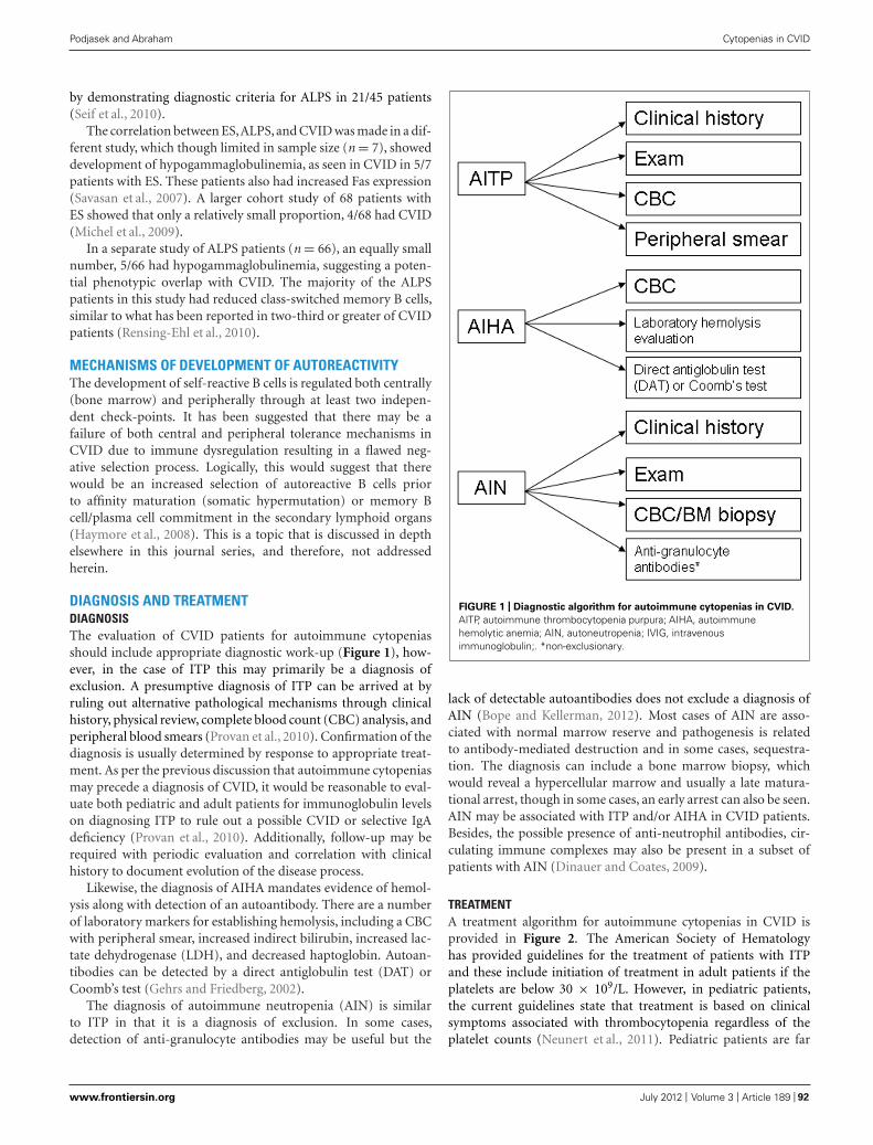
“fimmu-03-00189” — 2012/7/24 — 12:31 — page 3 — #3
Podjasek and Abraham Cytopenias in CVID
by demonstrating diagnostic criteria for ALPS in 21/45 patients(Seif et al., 2010).
The correlation between ES,ALPS, and CVID was made in a dif-ferent study, which though limited in sample size (n = 7), showeddevelopment of hypogammaglobulinemia, as seen in CVID in 5/7patients with ES. These patients also had increased Fas expression(Savasan et al., 2007). A larger cohort study of 68 patients withES showed that only a relatively small proportion, 4/68 had CVID(Michel et al., 2009).
In a separate study of ALPS patients (n = 66), an equally smallnumber, 5/66 had hypogammaglobulinemia, suggesting a poten-tial phenotypic overlap with CVID. The majority of the ALPSpatients in this study had reduced class-switched memory B cells,similar to what has been reported in two-third or greater of CVIDpatients (Rensing-Ehl et al., 2010).
MECHANISMS OF DEVELOPMENT OF AUTOREACTIVITYThe development of self-reactive B cells is regulated both centrally(bone marrow) and peripherally through at least two indepen-dent check-points. It has been suggested that there may be afailure of both central and peripheral tolerance mechanisms inCVID due to immune dysregulation resulting in a flawed neg-ative selection process. Logically, this would suggest that therewould be an increased selection of autoreactive B cells priorto affinity maturation (somatic hypermutation) or memory Bcell/plasma cell commitment in the secondary lymphoid organs(Haymore et al., 2008). This is a topic that is discussed in depthelsewhere in this journal series, and therefore, not addressedherein.
DIAGNOSIS AND TREATMENTDIAGNOSISThe evaluation of CVID patients for autoimmune cytopeniasshould include appropriate diagnostic work-up (Figure 1), how-ever, in the case of ITP this may primarily be a diagnosis ofexclusion. A presumptive diagnosis of ITP can be arrived at byruling out alternative pathological mechanisms through clinicalhistory, physical review, complete blood count (CBC) analysis, andperipheral blood smears (Provan et al., 2010). Confirmation of thediagnosis is usually determined by response to appropriate treat-ment. As per the previous discussion that autoimmune cytopeniasmay precede a diagnosis of CVID, it would be reasonable to eval-uate both pediatric and adult patients for immunoglobulin levelson diagnosing ITP to rule out a possible CVID or selective IgAdeficiency (Provan et al., 2010). Additionally, follow-up may berequired with periodic evaluation and correlation with clinicalhistory to document evolution of the disease process.
Likewise, the diagnosis of AIHA mandates evidence of hemol-ysis along with detection of an autoantibody. There are a numberof laboratory markers for establishing hemolysis, including a CBCwith peripheral smear, increased indirect bilirubin, increased lac-tate dehydrogenase (LDH), and decreased haptoglobin. Autoan-tibodies can be detected by a direct antiglobulin test (DAT) orCoomb’s test (Gehrs and Friedberg, 2002).
The diagnosis of autoimmune neutropenia (AIN) is similarto ITP in that it is a diagnosis of exclusion. In some cases,detection of anti-granulocyte antibodies may be useful but the
FIGURE 1 | Diagnostic algorithm for autoimmune cytopenias in CVID.
AITP, autoimmune thrombocytopenia purpura; AIHA, autoimmunehemolytic anemia; AIN, autoneutropenia; IVIG, intravenousimmunoglobulin;. *non-exclusionary.
lack of detectable autoantibodies does not exclude a diagnosis ofAIN (Bope and Kellerman, 2012). Most cases of AIN are asso-ciated with normal marrow reserve and pathogenesis is relatedto antibody-mediated destruction and in some cases, sequestra-tion. The diagnosis can include a bone marrow biopsy, whichwould reveal a hypercellular marrow and usually a late matura-tional arrest, though in some cases, an early arrest can also be seen.AIN may be associated with ITP and/or AIHA in CVID patients.Besides, the possible presence of anti-neutrophil antibodies, cir-culating immune complexes may also be present in a subset ofpatients with AIN (Dinauer and Coates, 2009).
TREATMENTA treatment algorithm for autoimmune cytopenias in CVID isprovided in Figure 2. The American Society of Hematologyhas provided guidelines for the treatment of patients with ITPand these include initiation of treatment in adult patients if theplatelets are below 30 × 109/L. However, in pediatric patients,the current guidelines state that treatment is based on clinicalsymptoms associated with thrombocytopenia regardless of theplatelet counts (Neunert et al., 2011). Pediatric patients are far
www.frontiersin.org July 2012 | Volume 3 | Article 189 | 92
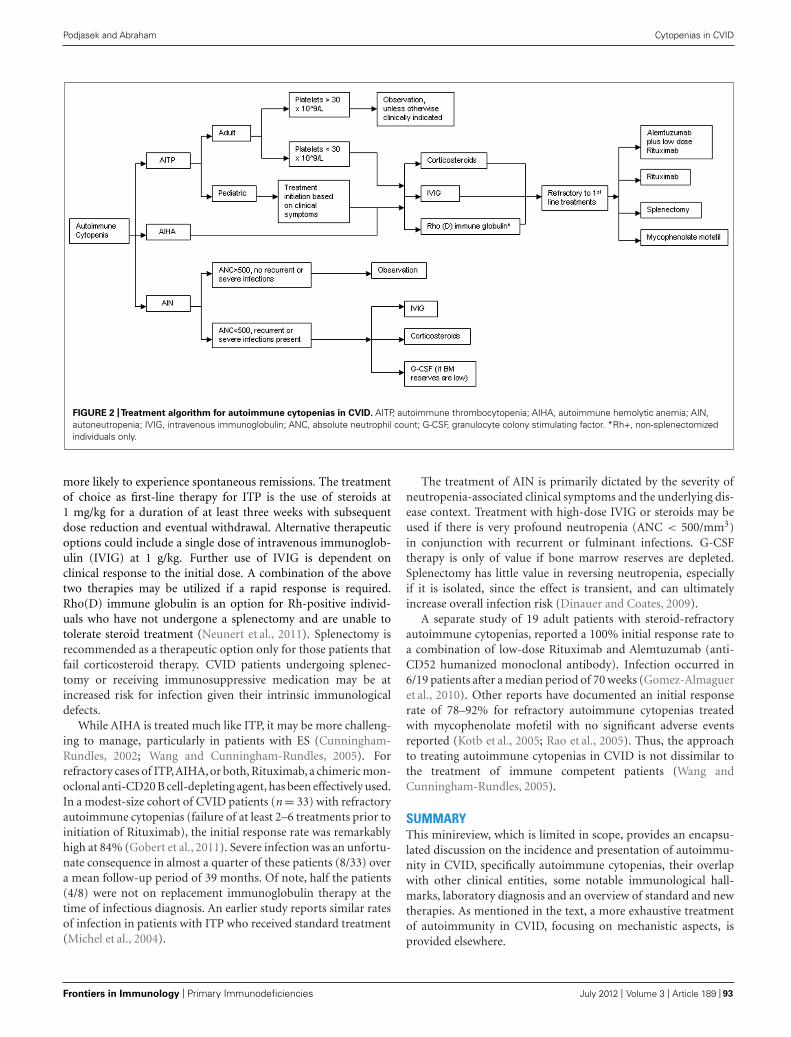
“fimmu-03-00189” — 2012/7/24 — 12:31 — page 4 — #4
Podjasek and Abraham Cytopenias in CVID
FIGURE 2 |Treatment algorithm for autoimmune cytopenias in CVID. AITP, autoimmune thrombocytopenia; AIHA, autoimmune hemolytic anemia; AIN,autoneutropenia; IVIG, intravenous immunoglobulin; ANC, absolute neutrophil count; G-CSF, granulocyte colony stimulating factor. *Rh+, non-splenectomizedindividuals only.
more likely to experience spontaneous remissions. The treatmentof choice as first-line therapy for ITP is the use of steroids at1 mg/kg for a duration of at least three weeks with subsequentdose reduction and eventual withdrawal. Alternative therapeuticoptions could include a single dose of intravenous immunoglob-ulin (IVIG) at 1 g/kg. Further use of IVIG is dependent onclinical response to the initial dose. A combination of the abovetwo therapies may be utilized if a rapid response is required.Rho(D) immune globulin is an option for Rh-positive individ-uals who have not undergone a splenectomy and are unable totolerate steroid treatment (Neunert et al., 2011). Splenectomy isrecommended as a therapeutic option only for those patients thatfail corticosteroid therapy. CVID patients undergoing splenec-tomy or receiving immunosuppressive medication may be atincreased risk for infection given their intrinsic immunologicaldefects.
While AIHA is treated much like ITP, it may be more challeng-ing to manage, particularly in patients with ES (Cunningham-Rundles, 2002; Wang and Cunningham-Rundles, 2005). Forrefractory cases of ITP,AIHA, or both, Rituximab, a chimeric mon-oclonal anti-CD20 B cell-depleting agent, has been effectively used.In a modest-size cohort of CVID patients (n = 33) with refractoryautoimmune cytopenias (failure of at least 2–6 treatments prior toinitiation of Rituximab), the initial response rate was remarkablyhigh at 84% (Gobert et al., 2011). Severe infection was an unfortu-nate consequence in almost a quarter of these patients (8/33) overa mean follow-up period of 39 months. Of note, half the patients(4/8) were not on replacement immunoglobulin therapy at thetime of infectious diagnosis. An earlier study reports similar ratesof infection in patients with ITP who received standard treatment(Michel et al., 2004).
The treatment of AIN is primarily dictated by the severity ofneutropenia-associated clinical symptoms and the underlying dis-ease context. Treatment with high-dose IVIG or steroids may beused if there is very profound neutropenia (ANC < 500/mm3)in conjunction with recurrent or fulminant infections. G-CSFtherapy is only of value if bone marrow reserves are depleted.Splenectomy has little value in reversing neutropenia, especiallyif it is isolated, since the effect is transient, and can ultimatelyincrease overall infection risk (Dinauer and Coates, 2009).
A separate study of 19 adult patients with steroid-refractoryautoimmune cytopenias, reported a 100% initial response rate toa combination of low-dose Rituximab and Alemtuzumab (anti-CD52 humanized monoclonal antibody). Infection occurred in6/19 patients after a median period of 70 weeks (Gomez-Almagueret al., 2010). Other reports have documented an initial responserate of 78–92% for refractory autoimmune cytopenias treatedwith mycophenolate mofetil with no significant adverse eventsreported (Kotb et al., 2005; Rao et al., 2005). Thus, the approachto treating autoimmune cytopenias in CVID is not dissimilar tothe treatment of immune competent patients (Wang andCunningham-Rundles, 2005).
SUMMARYThis minireview, which is limited in scope, provides an encapsu-lated discussion on the incidence and presentation of autoimmu-nity in CVID, specifically autoimmune cytopenias, their overlapwith other clinical entities, some notable immunological hall-marks, laboratory diagnosis and an overview of standard and newtherapies. As mentioned in the text, a more exhaustive treatmentof autoimmunity in CVID, focusing on mechanistic aspects, isprovided elsewhere.
Frontiers in Immunology | Primary Immunodeficiencies July 2012 | Volume 3 | Article 189 | 93
“fimmu-03-00189” — 2012/7/24 — 12:31 — page 5 — #5
Podjasek and Abraham Cytopenias in CVID
REFERENCESAl-Herz, W., Bousfiha, A., Casanova,
J.-L., Chapel, H., Conley, M. E.,Cunningham-Rundles, C., Etzioni,A., Fischer, A., Franco, J. L., Geha,R., Hammarstrom, L., Nonoyama, S.,Notarangelo, L. D., Ochs, H. D., Puck,J., Roifman, C., Seger, R., and Tang,M. L. K. (2011). Primary immun-odeficiency diseases: an update onthe classification from the Interna-tional Union of Immunological Soci-eties Expert Committee for PrimaryImmunodeficiency. Front. Immunol.2:54. doi: 10.3389/fimmu.2011.00054
Arumugakani, G., Wood, P. M.,and Carter, C. R. (2010). Fre-quency of Treg cells is reduced inCVID patients with autoimmunityand splenomegaly and is associ-ated with expanded CD21lo B lym-phocytes. J. Clin. Immunol. 30,292–300.
Becker-Merok, A., Nikolaisen, C.,and Nossent, H. C. (2006). B-lymphocyte activating factor insystemic lupus erythematosus andrheumatoid arthritis in relationto autoantibody levels, diseasemeasures and time. Lupus 15,570–576.
Bope, E. T., and Kellerman, R. D. (2012).Conn’s Current Therapy. Philadel-phia: Elsevier/Saunders.
Brandt, D., and Gershwin, M. E. (2006).Common variable immune defi-ciency and autoimmunity. Autoim-mun. Rev. 5, 465–470.
Buckner, J. H. (2010). Mecha-nisms of impaired regulation byCD4(+)CD25(+)FOXP3(+) regula-tory T cells in human autoimmunediseases. Nat. Rev. Immunol. 10,849–859.
Bussone, G., and Mouthon, L. (2009).Autoimmune manifestations in pri-mary immune deficiencies. Autoim-mun. Rev. 8, 332–336.
Castigli, E., Wilson, S., Garibyan, L.,Rachid, R., Bonilla, F., Schneider, L.,Morra, M., Curran, J., and Geha, R.(2007). Reexamining the role of TACIcoding variants in common vari-able immunodeficiency and selec-tive IgA deficiency. Nat. Genet. 39,430–431.
Castigli, E., Wilson, S. A., Garibyan,L., Rachid, R., Bonilla, F., Schneider,L., and Geha, R. S. (2005). TACI ismutant in common variable immun-odeficiency and IgA deficiency. Nat.Genet. 37, 829–834.
Chapel, H., Lucas, M., Lee, M., Bjorkan-der, J., Webster, D., Grimbacher, B.,Fieschi, C., Thon, V., Abedi, M. R.,and Hammarstrom, L. (2008). Com-mon variable immunodeficiency
disorders: division into distinctclinical phenotypes. Blood 112,277–286.
Charles, E. D., Brunetti, C., Marukian,S., Ritola, K. D., Talal, A. H., Marks,K., Jacobson, I. M., Rice, C. M.,and Dustin, L. B. (2011). ClonalB cells in patients with hepatitisC virus-associated mixed cryoglobu-linemia contain an expanded anergicCD21low B-cell subset. Blood 117,5425–5437.
Cunningham-Rundles, C. (2002).Hematologic complications of pri-mary immune deficiencies. BloodRev. 16, 61–64.
Cunningham-Rundles, C. (2008).Autoimmune manifestations in com-mon variable immunodeficiency.J. Clin. Immunol. 28(Suppl. 1),S42–S45.
Cunningham-Rundles, C. (2010). HowI treat common variable immunedeficiency. Blood 116, 7–15.
Dinauer, M. C., and Coates, T. D.(2009). Disorders of Phagocyte Func-tion and Number, Chap. 50. Phila-delphia, PA: Churchill LivingstoneElsevier.
Eibel, H., Salzer, U., and War-natz, K. (2010). Common vari-able immunodeficiency at the endof a prospering decade: towardsnovel gene defects and beyond. Curr.Opin. Allergy Clin. Immunol. 10,526–533.
Foerster, C., Voelxen, N., Rakhmanov,M., Keller, B., Gutenberger, S.,Goldacker, S., Thiel, J., Feske, S.,Peter, H. H., and Warnatz, K. (2010).B cell receptor-mediated calcium sig-naling is impaired in B lymphocytesof type Ia patients with common vari-able immunodeficiency. J. Immunol.184, 7305–7313.
Frank, M. M. (2012). CD21 deficiency,complement, and the developmentof common variable immunodefi-ciency. J. Allergy Clin. Immunol. 129,811–813.
Gehrs, B. C., and Friedberg, R.C. (2002). Autoimmune hemolyticanemia. Am. J. Hematol. 69,258–271.
Gobert, D., Bussel, J. B., Cunningham-Rundles, C., Galicier, L., Dechartres,A., Berezne, A., Bonnotte, B., Derevel,T., Auzary, C., Jaussaud, R., Lar-roche, C., Lequellec, A., Ruivard,M., Seve, P., Smail, A., Viallard,J. F., Godeau, B., Hermine, O.,and Michel, M. (2011). Efficacyand safety of rituximab in com-mon variable immunodeficiency-associated immune cytopenias: aretrospective multicentre study on33 patients. Br. J. Haematol. 155,498–508.
Gomez-Almaguer, D., Solano-Genesta,M., Tarin-Arzaga, L., Herrera-Garza, J. L., Cantu-Rodriguez,O. G., Gutierrez-Aguirre, C. H.,and Jaime-Perez, J. C. (2010).Low-dose rituximab and alem-tuzumab combination therapy forpatients with steroid-refractoryautoimmune cytopenias. Blood 116,4783–4785.
Grimbacher, B., Hutloff, A., Schlesier,M., Glocker, E., Warnatz, K., Drager,R., Eibel, H., Fischer, B., Schaffer, A.A., Mages, H. W., Kroczek, R. A.,and Peter, H. H. (2003). Homozy-gous loss of ICOS is associatedwith adult-onset common variableimmunodeficiency. Nat. Immunol. 4,261–268.
Haymore, B. R., Mikita, C. P., andTsokos, G. C. (2008). Common vari-able immune deficiency (CVID) pre-senting as an autoimmune disease:role of memory B cells. Autoimmun.Rev. 7, 309–312.
Horn, J., Thon, V., Bartonkova, D.,Salzer, U., Warnatz, K., Schlesier,M., Peter, H. H., and Grimbacher,B. (2007). Anti-IgA antibodies incommon variable immunodeficiency(CVID): diagnostic workup and ther-apeutic strategy. Clin. Immunol. 122,156–162.
Jang, E., Cho, W. S., Cho, M. L., Park, H.J., Oh, H. J., Kang, S. M., Paik, D. J.,and Youn, J. (2011). Foxp3+ regula-tory T cells control humoral autoim-munity by suppressing the develop-ment of long-lived plasma cells. J.Immunol. 186, 1546–1553.
Kanegane, H., Agematsu, K., Futatani,T., Sira, M. M., Suga, K.,Sekiguchi, T., van Zelm, M. C., andMiyawaki, T. (2007). Novel muta-tions in a Japanese patient withCD19 deficiency. Genes Immun. 8,663–670.
Knight, A. K., and Cunningham-Rundles, C. (2006). Inflammatoryand autoimmune complicationsof common variable immunedeficiency. Autoimmun. Rev. 5,156–159.
Knight, A. K., Radigan, L., Mar-ron, T., Langs, A., Zhang, L., andCunningham-Rundles, C. (2007).High serum levels of BAFF, APRIL,and TACI in common variableimmunodeficiency. Clin. Immunol.124, 182–189.
Kotb, R., Pinganaud, C., Trichet, C.,Lambotte, O., Dreyfus, M., Delfraissy,J. F., Tchernia, G., and Goujard,C. (2005). Efficacy of mycopheno-late mofetil in adult refractory auto-immune cytopenias: a single centerpreliminary study. Eur. J. Haematol.75, 60–64.
Kuijpers, T. W., Bende, R. J., Baars,P. A., Grummels, A., Derks, I. A.,Dolman, K. M., Beaumont, T., Ted-der, T. F., Van Noesel, C. J., Elder-ing, E., and Van Lier, R. A. (2010).CD20 deficiency in humans resultsin impaired T cell-independent anti-body responses. J. Clin. Invest. 120,214–222.
Long, S. A., and Buckner, J. H. (2011).CD4+FOXP3+ T regulatory cells inhuman autoimmunity: more thana numbers game. J. Immunol. 187,2061–2066.
Martinez-Gamboa, L., Mei, H., Lod-denkemper, C., Ballmer, B., Hansen,A., Lipsky, P. E., Emmerich, F., Rad-bruch, A., Salama, A., and Dorner, T.(2009). Role of the spleen in periph-eral memory B-cell homeostasis inpatients with autoimmune thrombo-cytopenia purpura. Clin. Immunol.130, 199–212.
Michel, M., Chanet, V., Dechartres, A.,Morin, A. S., Piette, J. C., Cirasino,L., Emilia, G., Zaja, F., Ruggeri, M.,Andres, E., Bierling, P., Godeau, B.,and Rodeghiero, F. (2009). The spec-trum of Evans syndrome in adults:new insight into the disease based onthe analysis of 68 cases. Blood 114,3167–3172.
Michel, M., Chanet, V., Galicier,L., Ruivard, M., Levy, Y., Her-mine, O., Oksenhendler, E., Scha-effer, A., Bierling, P., and Godeau,B. (2004). Autoimmune thrombocy-topenic purpura and common vari-able immunodeficiency: analysis of21 cases and review of the lit-erature. Medicine (Baltimore) 83,254–263.
Neunert, C., Lim, W., Crowther, M.,Cohen, A., Solberg, L. Jr., andCrowther, M. A. (2011). The Amer-ican Society of Hematology 2011evidence-based practice guideline forimmune thrombocytopenia. Blood117, 4190–4207.
Notarangelo, L. D. (2009). Primaryimmunodeficiencies (PIDs) present-ing with cytopenias. HematologyAm. Soc. Hematol. Educ. Program139–143.
Orange, J. S., Glessner, J. T., Resnick, E.,Sullivan, K. E., Lucas, M., Ferry, B.,Kim, C. E., Hou, C., Wang, F., Chi-avacci, R., Kugathasan, S., Sleasman,J. W., Baldassano, R., Perez, E. E.,Chapel, H., Cunningham-Rundles,C., and Hakonarson, H. (2011).Genome-wide association identifiesdiverse causes of common variableimmunodeficiency. J. Allergy Clin.Immunol. 127, 1360–1367, e1366.
Pan-Hammarstrom, Q., Salzer, U., Du,L., Bjorkander, J., Cunningham-Rundles, C., Nelson, D. L., Bacchelli,
www.frontiersin.org July 2012 | Volume 3 | Article 189 | 94

“fimmu-03-00189” — 2012/7/24 — 12:31 — page 6 — #6
Podjasek and Abraham Cytopenias in CVID
C., Gaspar, H. B., Offer, S.,Behrens, T. W., Grimbacher, B.,and Hammarstrom, L. (2007).Reexamining the role of TACIcoding variants in common vari-able immunodeficiency and selec-tive IgA deficiency. Nat. Genet. 39,429–430.
Park, M. A., Li, J. T., Hagan, J. B.,Maddox, D. E., and Abraham, R. S.(2008). Common variable immun-odeficiency: a new look at an olddisease. Lancet 372, 489–502.
Piqueras, B., Lavenu-Bombled, C.,Galicier, L., Bergeron-Van DerCruyssen, F., Mouthon, L., Chevret,S., Debre, P., Schmitt, C., and Oksen-hendler, E. (2003). Common variableimmunodeficiency patient classifica-tion based on impaired B cell mem-ory differentiation correlates withclinical aspects. J. Clin. Immunol. 23,385–400.
Provan, D., Stasi, R., Newland, A. C.,Blanchette, V. S., Bolton-Maggs, P.,Bussel, J. B., Chong, B. H., Cines,D. B., Gernsheimer, T. B., Godeau,B., Grainger, J., Greer, I., Hunt, B.J., Imbach, P. A., Lyons, G., McMil-lan, R., Rodeghiero, F., Sanz, M. A.,Tarantino, M., Watson, S., Young, J.,and Kuter, D. J. (2010). Internationalconsensus report on the investiga-tion and management of primaryimmune thrombocytopenia. Blood115, 168–186.
Rakhmanov, M., Gutenberger, S., Keller,B., Schlesier, M., Peter, H. H., andWarnatz, K. (2010). CD21low Bcells in common variable immun-odeficiency do not show defects inreceptor editing, but resemble tissue-like memory B cells. Blood 116,3682–3683.
Rakhmanov, M., Keller, B., Gutenberger,S., Foerster, C., Hoenig, M., Driessen,G., Van Der Burg, M., Van Don-gen, J. J., Wiech, E., Visentini, M.,Quinti, I., Prasse, A., Voelxen, N.,Salzer, U., Goldacker, S., Fisch, P.,Eibel, H., Schwarz, K., Peter, H. H.,and Warnatz, K. (2009). CirculatingCD21low B cells in common vari-able immunodeficiency resemble tis-sue homing, innate-like B cells. Proc.Natl. Acad. Sci. U.S.A. 106, 13451–13456.
Rao, V. K., Dugan, F., Dale, J. K., Davis,J., Tretler, J., Hurley, J. K., Fleisher,T., Puck, J., and Straus, S. E. (2005).Use of mycophenolate mofetil forchronic, refractory immune cytope-nias in children with autoimmunelymphoproliferative syndrome. Br. J.Haematol. 129, 534–538.
Rensing-Ehl, A., Warnatz, K., Fuchs,S., Schlesier, M., Salzer, U., Draeger,R., Bondzio, I., Joos, Y., Janda, A.,
Gomes, M., Abinun, M., Hamble-ton, S., Cant, A., Shackley, F., Flood,T., Waruiru, C., Beutel, K., Sieper-mann, K., Dueckers, G., Niehues, T.,Wiesel, T., Schuster, V., Seidel, M.G., Minkov, M., Sirkia, K., Kopp,M. V., Korhonen, M., Schwarz, K.,Ehl, S., and Speckmann, C. (2010).Clinical and immunological over-lap between autoimmune lympho-proliferative syndrome and commonvariable immunodeficiency. Clin.Immunol. 137, 357–365.
Resnick, E. S., Moshier, E. L., Godbold,J. H., and Cunningham-Rundles, C.(2011). Morbidity and mortality incommon variable immune deficiencyover 4 decades. Blood 119, 1650–1657.
Romberg, N., Ng, Y.-S., Cunningham-Rundles, C., and Meffre, E.(2011). Common variable immun-odeficiency patients with increasedCD21-/lo B cells suffer from alteredreceptor editing and defective B celltolerance. Blood 118, 5977–5978.
Salzer, U., Bacchelli, C., Buckridge, S.,Pan-Hammarstrom, Q., Jennings,S., Lougaris, V., Bergbreiter, A.,Hagena, T., Birmelin, J., Plebani, A.,Webster, A. D., Peter, H. H., Suez,D., Chapel, H., Mclean-Tooke, A.,Spickett, G. P., Anover-Sombke, S.,Ochs, H. D., Urschel, S., Belohradsky,B. H., Ugrinovic, S., Kumararatne,D. S., Lawrence, T. C., Holm, A. M.,Franco, J. L., Schulze, I., Schneider,P., Gertz, E. M., Schaffer, A. A.,Hammarstrom, L., Thrasher, A. J.,Gaspar, H. B., and Grimbacher, B.(2009). Relevance of biallelic versusmonoallelic TNFRSF13B mutationsin distinguishing disease-causingfrom risk-increasing TNFRSF13Bvariants in antibody deficiencysyndromes. Blood 113, 1967–1976.
Salzer, U., Chapel, H. M., Webster, A.D., Pan-Hammarstrom, Q., Schmitt-Graeff, A., Schlesier, M., Peter, H.H., Rockstroh, J. K., Schneider, P.,Schaffer, A. A., Hammarstrom, L.,and Grimbacher, B. (2005). Muta-tions in TNFRSF13B encoding TACIare associated with common variableimmunodeficiency in humans. Nat.Genet. 37, 820–828.
Salzer, U., Maul-Pavicic, A., Cunnin-gham-Rundles, C., Urschel, S.,Belohradsky, B. H., Litzman, J.,Holm, A., Franco, J. L., Plebani,A., Hammarstrom, L., Skrabl, A.,Schwinger, W., and Grimbacher, B.(2004). ICOS deficiency in patientswith common variable immunodefi-ciency. Clin. Immunol. 113, 234–240.
Sanchez-Ramon, S., Radigan, L., Yu,J. E., Bard, S., and Cunningham-Rundles, C. (2008). Memory B cells
in common variable immunodefi-ciency: clinical associations and sexdifferences. Clin. Immunol. 128,314–321.
Savasan, S., Warrier, I., Buck, S., Kaplan,J., and Ravindranath, Y. (2007).Increased lymphocyte Fas expressionand high incidence of common vari-able immunodeficiency disorder inchildhood Evans’ syndrome. Clin.Immunol. 125, 224–229.
Schaffer, A. A., Salzer, U., Ham-marstrom, L., and Grimbacher,B. (2007). Deconstructing com-mon variable immunodeficiency bygenetic analysis. Curr. Opin. Genet.Dev. 17, 201–212.
Seif, A. E., Manno, C. S., Sheen,C., Grupp, S. A., and Teachey,D. T. (2010). Identifying autoim-mune lymphoproliferative syndromein children with Evans syndrome: amulti-institutional study. Blood 115,2142–2145.
Sekine, H., Ferreira, R. C., Pan-Hammarstrom, Q., Graham, R. R.,Ziemba, B., De Vries, S. S., Liu, J.,Hippen, K., Koeuth, T., Ortmann,W., Iwahori, A., Elliott, M. K., Offer,S., Skon, C., Du, L., Novitzke, J.,Lee, A. T., Zhao, N., Tompkins, J.D., Altshuler, D., Gregersen, P. K.,Cunningham-Rundles, C., Harris, R.S., Her, C., Nelson, D. L., Ham-marstrom, L., Gilkeson, G. S., andBehrens, T. W. (2007). Role for Msh5in the regulation of Ig class switchrecombination. Proc. Natl. Acad. Sci.U.S.A. 104, 7193–7198.
Teachey, D. T., Manno, C. S., Axsom, K.M., Andrews, T., Choi, J. K., Green-baum, B. H., Mcmann, J. M., Sulli-van, K. E., Travis, S. F., and Grupp,S. A. (2005). Unmasking Evanssyndrome: T-cell phenotype andapoptotic response reveal autoim-mune lymphoproliferative syndrome(ALPS). Blood 105, 2443–2448.
Thiel, J., Kimmig, L., Salzer, U.,Grudzien, M., Lebrecht, D., Hagena,T., Draeger, R., Volxen, N., Bergbre-iter, A., Jennings, S., Gutenberger, S.,Aichem, A., Illges, H., Hannan, J.P., Kienzler, A. K., Rizzi, M., Eibel,H., Peter, H. H., Warnatz, K., Grim-bacher, B., Rump, J. A., and Schlesier,M. (2012). Genetic CD21 deficiencyis associated with hypogammaglobu-linemia. J. Allergy Clin. Immunol. 129,801–810 e806.
van Zelm, M. C., Reisli, I., Van Der Burg,M., Castano, D., Van Noesel, C. J.,Van Tol, M. J., Woellner, C., Grim-bacher, B., Patino, P. J., Van Dongen,J. J., and Franco, J. L. (2006). Anantibody-deficiency syndrome due tomutations in the CD19 gene. N. Engl.J. Med. 354, 1901–1912.
van Zelm, M. C., Smet, J., Adams, B.,Mascart, F., Schandene, L., Janssen,F., Ferster, A., Kuo, C. C., Levy, S., VanDongen, J. J., and Van Der Burg, M.(2010). CD81 gene defect in humansdisrupts CD19 complex formationand leads to antibody deficiency. J.Clin. Invest. 120, 1265–1274.
Vodjgani, M., Aghamohammadi, A.,Samadi, M., Moin, M., Hadjati,J., Mirahmadian, M., Parvaneh, N.,Salavati, A., Abdollahzade, S., Rezaei,N., and Srrafnejad, A. (2007). Analy-sis of class-switched memory B cellsin patients with common variableimmunodeficiency and its clinicalimplications. J. Investig. Allergol. Clin.Immunol. 17, 321–328.
Wang, J., and Cunningham-Rundles, C.(2005). Treatment and outcome ofautoimmune hematologic disease incommon variable immunodeficiency(CVID). J. Autoimmun. 25, 57–62.
Warnatz, K., Denz, A., Drager, R.,Braun, M., Groth, C., Wolff-Vorbeck,G., Eibel, H., Schlesier, M., andPeter, H. H. (2002). Severe defi-ciency of switched memory B cells(CD27(+)IgM(−)IgD(−)) in sub-groups of patients with commonvariable immunodeficiency: a newapproach to classify a heterogeneousdisease. Blood 99, 1544–1551.
Warnatz, K., Salzer, U., and Guten-berger, S. (2005). Finally found:human BAFF-R deficiency causeshypogammaglobulinemia. Clin.Immunol. 115, 820.
Wehr, C., Kivioja, T., Schmitt, C., Ferry,B., Witte, T., Eren, E., Vlkova, M.,Hernandez, M., Detkova, D., Bos, P.R., Poerksen, G., Von Bernuth, H.,Baumann, U., Goldacker, S., Guten-berger, S., Schlesier, M., Bergeron-Van Der Cruyssen, F., Le Garff, M.,Debre, P., Jacobs, R., Jones, J., Bate-man, E., Litzman, J., Van Hagen, P.M., Plebani, A., Schmidt, R. E., Thon,V., Quinti, I., Espanol, T., Webster, A.D., Chapel, H., Vihinen, M., Oksen-hendler, E., Peter, H. H., and War-natz, K. (2008). The EUROclass trial:defining subgroups in common vari-able immunodeficiency. Blood 111,77–85.
Wasserstrom, H., Bussel, J., Lim, L.C., and Cunningham-Rundles, C.(2008). Memory B cells and pneumo-coccal antibody after splenectomy. J.Immunol. 181, 3684–3689.
Zhang, L., Radigan, L., Salzer, U.,Behrens, T. W., Grimbacher, B., Diaz,G., Bussel, J., and Cunningham-Rundles, C. (2007). Transmembraneactivator and calcium-modulatingcyclophilin ligand interactormutations in common variableimmunodeficiency: clinical and
Frontiers in Immunology | Primary Immunodeficiencies July 2012 | Volume 3 | Article 189 | 95
“fimmu-03-00189” — 2012/7/24 — 12:31 — page 7 — #7
Podjasek and Abraham Cytopenias in CVID
immunologic outcomes in heterozy-gotes. J. Allergy Clin. Immunol. 120,1178–1185.
Conflict of Interest Statement: Theauthors declare that the research wasconducted in the absence of any
commercial or financial relationshipsthat could be construed as a potentialconflict of interest.
Received: 22 February 2012; accepted: 18June 2012; published online: 24 July 2012.Citation: Podjasek JC and AbrahamRS (2012) Autoimmune cytopenias
in common variable immunodefi-ciency. Front. Immun. 3:189. doi:10.3389/fimmu.2012.00189This article was submitted to Frontiers inPrimary Immunodeficiencies, a specialtyof Frontiers in Immunology.Copyright © 2012 Podjasek and Abra-ham. This is an open-access article
distributed under the terms of theCreative Commons Attribution License,which permits use, distribution andreproduction in other forums, pro-vided the original authors and sourceare credited and subject to any copy-right notices concerning any third-partygraphics etc.
www.frontiersin.org July 2012 | Volume 3 | Article 189 | 96

MINI REVIEW ARTICLEpublished: 04 June 2012
doi: 10.3389/fimmu.2012.00129
TH17 cells in autoimmunity and immunodeficiency:protective or pathogenic?Ashish K. Marwaha1,2, Nicole J. Leung1,2, Alicia N. McMurchy 2,3 and Megan K. Levings2,3*
1 Department of Pathology and Laboratory Medicine, University of British Columbia, Vancouver, BC, Canada2 Child and Family Research Institute, Vancouver, BC, Canada3 Department of Surgery, University of British Columbia, Vancouver, BC, Canada
Edited by:
Rosa Bacchetta, Fondazione CentroSan Raffaele Del Monte Tabor, Italy
Reviewed by:
Rosa Bacchetta, Fondazione CentroSan Raffaele Del Monte Tabor, ItalyFrancesco Annunziato, University ofFlorence, Italy
*Correspondence:
Megan K. Levings, Department ofSurgery, University of BritishColumbia, A4-186, 950 West 28thAvenue, Vancouver, BC, Canada V5Z4H4.e-mail: [email protected]
In 2005 a newly discovered T helper cell subset that secreted interleukin (IL)-17 becamethe center of attention in immunology. Initial studies painted Th17 cells as the culprit fordestruction in many different autoimmune and auto-inflammatory diseases. Subsequently,the discovery of patients with primary immunodeficiencies in the IL-17 pathway taughtus that Th17 cells have a critical role in defense against certain fungal and bacterial infec-tions. Moreover, the paradoxical exacerbation of Crohn’s disease in the clinical trials of aSecukinumab (AIN457), a fully human neutralizing antibody to IL-17A, has cast into doubta universal pro-inflammatory and harmful role for Th17 cells. Evidence now suggests thatdepending on the environmentTh17 cells can alter their differentiation program, ultimatelygiving rise to either protective or pro-inflammatory cells. In this review we will summarizethe evidence from patients with immunodeficiencies, autoimmune, or auto-inflammatorydiseases that teaches us how the pro-inflammatory versus protective function ofTh17 cellsvaries within the context of different human diseases.
Keywords:Th17 cells, autoimmunity,T regulatory cells, immunodeficiency, inflammatory bowel disease, psoriasis,
type 1 diabetes, secukinumab
INTRODUCTIONIn rare cases, a mutation in an essential gene can disrupt immunehomeostasis, leading to clinical immunodeficiency. More com-monly, when individuals with a genetic predisposition are exposedto environmental triggers, a failure of immune homeostasis canlead to autoimmunity. In this review, we will discuss how par-allel studies of immunodeficiencies and autoimmune diseaseshave advanced our knowledge of a CD4+ T cell lineage firstcharacterized by production of IL-17A, Th17 cells.
Th17 cells were identified in 2005 (Harrington et al., 2005;Langrish et al., 2005; Park et al., 2005) and, as for other CD4+T cell lineages, their development, is controlled by a combina-tion of cytokines which initiate a program of transcription fac-tor expression and epigenetic re-modeling (van der Gast et al.,2011). In humans, the cytokines which instruct Th17 cell lin-eage development likely include IL-6, IL-21, IL-23, and IL-1β(Acosta-Rodriguez et al., 2007a; Chen et al., 2007; Evans et al.,2007; Wilson et al., 2007; Liu and Rohowsky-Kochan, 2008), witha potential synergistic role for TGF-β (Manel et al., 2008; Volpeet al., 2008; Yang et al., 2008a) via its ability to suppress Th1 celllineage commitment (Santarlasci et al., 2009). Cytokine-drivenactivation of the signal transducer and activator of transcription(STAT) 3 pathway is an essential step in Th17 cell differentiation(Holland et al., 2007; Yang et al., 2007; Ma et al., 2008), ultimatelyleading to expression of their lineage-defining transcription fac-tor: retinoid orphan receptor (ROR)C2 (Acosta-Rodriguez et al.,2007a; Annunziato et al., 2007; Wilson et al., 2007; Manel et al.,2008; Crome et al., 2009). Although the IL-17 cytokine familyincludes six members (Kolls and Linden, 2004), Th17 cells are
thought to only produce IL-17A and IL-17F, which are 55% iden-tical (Kolls and Linden, 2004). IL-17A can combine with IL-17Fto form a heterodimer and both can form homodimers (Wrightet al., 2007).
Th17 cells have many phenotypic characteristics that distin-guish them from other Th cell lineages. In addition to IL-17A andIL-17F, Th17 cells secrete other signature cytokines including IL-21 and IL-22 (Bending et al., 2011). They have also been reportedto produce IFN-γ (Annunziato et al., 2007), IL-4 (Cosmi et al.,2010), IL-10 (McGeachy et al., 2007), IL-9 (Beriou et al., 2010),IL-26, CXCL8, and CCL20 (Boniface et al., 2008). They are poorproducers of IL-2, which may result in their poor proliferativepotential in vitro (Santarlasci et al., 2012). They constitutively co-express CCR4 and CCR6, but not CXCR3 (Acosta-Rodriguez et al.,2007b), and are derived from CD161+ precursors (Cosmi et al.,2008). The effects of Th17 cells on other cells have recently beenhighlighted in many reviews (Annunziato and Romagnani, 2011;Gaffen, 2011; Gaffen et al., 2011; Ghoreschi et al., 2011; Milner,2011; Pappu et al., 2011; Wilke et al., 2011).
Th17 cells initially developed a reputation as a destructiveelement in several diseases including multiple sclerosis (MS),rheumatoid arthritis (RA), psoriasis, and inflammatory bowel dis-ease (IBD). In animal models this reputation, came from evidencethat the lack of IL-17-producing cells ameliorates experimentalautoimmune encephalitis (EAE) and collagen induced arthri-tis (CIA; Cua et al., 2003). In humans, the reputation was dueto correlative data documenting an increase in IL-17-producingcells, particularly at sites of tissue inflammation (Wilke et al.,2011). However, these original conclusions were over-simplified
www.frontiersin.org June 2012 | Volume 3 | Article 129 | 97

Marwaha et al. Th17 in autoimmunity and immunodeficiency
and as discussed below in some diseases Th17 cells clearly have aprotective role.
THE ROLE OF Th17 CELLS IN PRIMARYIMMUNODEFICIENCIESMuch of what we know about human Th17 cells comes fromthe study of a rare primary immunodeficiency called Hyper-immunoglobulin E (Job’s, syndrome). This disease is caused bymutations in STAT3 (Holland et al., 2007) but the underlying cel-lular basis for the characteristic phenotype of severe pneumonias,mucocutaneous candidiasis, and Staphylococcus aureus abscesses(Buckley et al., 1972; Grimbacher et al., 1999) remained unknownuntil several groups found that these patients lack Th17 cells intheir peripheral blood (de Beaucoudrey et al., 2008; Ma et al.,2008; Milner et al., 2008; Renner et al., 2008). In addition, naïveTh cells from Job’s syndrome patients have low levels of RORC2expression and cannot be differentiated into Th17 cells in vitro(de Beaucoudrey et al., 2008; Ma et al., 2008; Milner et al., 2008;Renner et al., 2008).
One complication when interpreting data from Job’s syndromepatients is that STAT3 is activated downstream of other cytokines,making it difficult to attribute a clinical phenotype to one pathway.Recently, other immunodeficiencies have been described whichinvolve more specific defects in the IL-17 pathway. For example,two patients with chronic mucocutaneous candidiasis (CMC) dis-ease, characterized by chronic or persistent infection with Candidaalbicans and S. aureus, were found to have an IL-17RA autosomalrecessive deficiency or an IL-17F autosomal dominant deficiency(Puel et al., 2011). In addition, patients with a deficiency inthe intracellular adaptor molecule CARD9, which is essential fordectin-1 signaling, also suffer from systemic Candidiasis infection(Glocker et al., 2009) and have low numbers of Th17 cells in theirperipheral blood.
Together, these data have led to the hypothesis that in humansTh17 cells have an essential role in protective immunity the spe-cific pathogens C. albicans and S. aureus. In accordance with thisconclusion, Sallusto et al. have characterized different subsets ofhuman Th17 cells that can be differentiated in vitro with antigenspecific stimulation by C. albicans and S. aureus (Zielinski et al.,2012).
Th17 cells have an intriguing close developmental link withFOXP3+CD4+ regulatory T cells (Tregs). FOXP3 and RORC2 candirectly interact via a DNA-independent mechanism, and dur-ing Th17 cell development FOXP3 is transiently expressed (Zhouet al., 2008). Moreover, upon activation fully differentiated humanTh17 cells preferentially express FOXP3 in comparison to Th1cells (McMurchy and Levings, unpublished data). Indeed there isincreasing evidence for the existence of cells that co-express IL-17and FOXP3 (Ayyoub et al., 2009; Beriou et al., 2009; Miyara et al.,2009; Voo et al., 2009; Kryczek et al., 2011; Ye et al., 2011).
Immune dysregulation, polyendocrinopathy, enteropathy X-linked syndrome (IPEX) is a triad of autoimmune syndromesincluding enteropathy, type 1 diabetes, and hyper-IgE (McMurchyet al., 2010), resulting from mutations in FOXP3. The cellular basisfor this disease has been attributed to Treg dysfunction (Bacchettaet al., 2006; D’Hennezel et al., 2009), but recently Bacchetta et al.investigated whether part of the cellular defect in IPEX patients
may not only relate to Treg dysfunction, but also to changesin Th17 cells. Indeed IPEX patients possessed an increased fre-quency of cells with a surface profile characteristic of Th17 cells(CD4+CCR6+CD161+), expressing RORC2, and producing IL-17 (Passerini et al., 2011). Interestingly, in patients with pointmutations that do not abrogate FOXP3 expression, there was anincreased frequency of FOXP3+ cells within the CD161+ Th17cell gate. These data suggest that dysfunctional Tregs may prefer-entially differentiate into Th17 cells and that an expansion of thissubset may underlie some of the clinical phenotype of IPEX. Analternative interpretation is that the Th17 cells in IPEX patientsare highly activated and that in this case FOXP3 expression isa consequence of T cell activation and not Treg lineage commit-ment (Ziegler, 2006). Regardless, in this immunodeficiency there isstrong correlative evidence that Th17 cells may have a detrimentalpro-inflammatory effect.
EVIDENCE FOR THE PRO-INFLAMMATORY ROLE OF Th17 INHUMAN AUTOIMMUNITYThe first recognition of the importance of Th17 cells came fromstudies of EAE. The notion of EAE as a Th1-mediated disease waschallenged when mice deficient in the p40 subunit of IL-12 werefound to be resistant to EAE whereas mice deficient in the p35 sub-unit were actually more susceptible to disease (Becher et al., 2002).Cua et al. (2003) solved this paradox by using genetically deficientmice to show that IL-23p19 and IL-12p40, but not IL-12p35, wereessential for EAE development. IL-23, which shares the IL-12p40subunit with IL-12, was subsequently found to stabilize Th17 cells,and these cells were found to be the main contributing factor inEAE (Langrish et al., 2005). Subsequently, a correlation betweenTh17 cells and human autoimmunity was sought. Below we dis-cuss the evidence for a pro-inflammatory role in autoimmunityand describe attempts to target this axis therapeutically.
Psoriasis is an auto-inflammatory skin disease characterized byrecurrent demarcated red and scaly skin plaques. These plaquesinclude infiltrating T cells (mainly Th cells) and dendritic cells inthe dermis as well as cytotoxic T cells and neutrophil in the epi-dermis (Lowes et al., 2007). The resulting inflammatory processresults in rapid keratinocyte proliferation, abnormal keratinocytedifferentiation, and angiogenesis (Lowes et al., 2007). Initially,increased levels of IFN-γ, TNF-α, and IL-12 in the serum andlesions of psoriasis patients labeled this as a Th1-mediated disease(Di Cesare et al., 2009). However, RORC, IL-1β, IL-6, and IL-23 arealso increased in psoriatic skin lesions (Di Cesare et al., 2009) lead-ing to the possibility that Th17 and Th1 act in synergy to producepsoriatic inflammation.
Th17 cells are thought to be recruited to the skin by expres-sion of CCL20, the ligand for CCR6, then locally stabilized by IL-1and IL-23. Since both IL-17 and IFN-γ cause keratinocytes andantigen presenting cells (APCs) to produce more IL-1, IL-23, andCCL20, a positive feedback loop causing keratinocyte proliferationis established (Kryczek et al., 2008; Zaba et al., 2009). Several mon-oclonal antibodies targeting TNF-α and the p40 subunit shared byIL-12 and IL-23 (Ustekinumab) have been approved for clinicaluse in psoriasis. Since IL-17 can act synergistically with TNF-α toinduce keratinocytes to express inflammatory proteins (Chiricozziet al., 2011), it is possible that anti-TNF-α acts in part by inhibiting
Frontiers in Immunology | Primary Immunodeficiencies June 2012 | Volume 3 | Article 129 | 98

Marwaha et al. Th17 in autoimmunity and immunodeficiency
Th17 cell-driven inflammation. Targeting IL-17 alone with Secuk-inumab (AIN457) or Ixekizumab, both fully human neutralizingantibodies to IL-17A, is also effective in psoriasis (Hueber et al.,2010; Leonardi et al., 2012), confirming that this is likely a majorpathogenic cytokine in this skin disease. Since Th17 cells are notthe sole producers of IL-17 [other possible sources of this cytokinein psoriatic plaques include γδ T cells (Cai et al., 2011), mast cells(Lin et al., 2011), neutrophils (Lin et al., 2011), and Tregs (Boven-schen et al., 2011)], whether or not Th17 cells are the major sourceof this cytokine in skin remains to be determined.
Another disease with strong links to Th17 cells is RA, a chronicautoimmune disease that leads to joint destruction. T cells infil-trating the synovial fluid of RA patients produce high amountsof IL-17A, IL-1β, and IL-6 (Cascao et al., 2010), especially duringearly disease and pre-treatment (Chabaud et al., 1999; Ziolkowskaet al., 2000; Hwang and Kim, 2005; Leipe et al., 2010). Notably,the levels of IL-17 in the synovium correlate with joint damage,whereas those of IFN-γ correlate with protection (Kirkham et al.,2006). Recent evidence supports a role for IL-17F as well as IL-17Ain RA, with the two related cytokines acting in synergy to induceother pro-inflammatory cytokines and chemokines in synovio-cytes, myeloid cells, and synovial fibroblasts (Lundy et al., 2007;Tran et al., 2007; Hot and Miossec, 2011; van Hamburg et al.,2011). Hence, analogous to the process in psoriasis, a positivepro-inflammatory feedback loop encourages more Th17 differ-entiation and maintenance in the joint (reviewed in Sarkar andFox, 2010). Direct clinical evidence for the role of IL-17 in RAcomes from recent clinical trials which found that Secukinumaband another anti-IL-17A therapeutic known as LY2439821 signifi-cantly benefit these patients (Genovese et al., 2010; Morrison et al.,2011).
Multiple sclerosis is a neurological disease that results fromauto-inflammatory damage to the myelin sheaths surroundingnerves in the brain and spinal cord. This disease has historicallybeen associated with the discovery of Th17 cells since, as discussedabove, they have a major pathogenic role in EAE (Bettelli et al.,2008; Dong, 2008; Dubin and Kolls, 2008; Weaver and Hatton,2009). MS patients have increased IL-17 mRNA in their blood aswell as cerebrospinal fluid (Matusevicius et al., 1999), and expres-sion of miRNA326 in their peripheral blood mononuclear cellspromotes Th17 cell differentiation and correlates with diseaseseverity (Du et al., 2009). Blood-brain barrier endothelial cellslayers are more permeable to in vitro polarized Th17 cells, espe-cially if the monolayer is pre-treated with IL-17 or IL-22 (Kebiret al., 2007). These data led to the hypothesis that in MS Th17cells weaken the blood-brain barrier and enable the migration ofimmune cells into the normally immune privileged sites within thecentral nervous system. If this is the case, then Th17 cells may havemore of a facilitative than directly pathogenic role in the nervoussystem, distinct from their clear role in the positive feedback loopof inflammation in psoriasis and RA. Recruitment has begun for aphase II clinical trials of Secukinumab in patients with relapsing-remitting MS. This trial will provide significant insight into thequestion of whether IL-17 blockade in MS can induce a clinicallyrelevant protective function.
Type 1 diabetes (T1D) is characterized by autoimmune destruc-tion of pancreatic islet cells resulting in the loss of insulinproduction. Murine studies have yielded conflicting results on
the role of Th17 in the NOD mouse model of T1D (Vukkadapuet al., 2005; Jain et al., 2008; Emamaullee et al., 2009; van denBrandt et al., 2010; Lee et al., 2011; Liu et al., 2011; Joseph et al.,2012), including a potential ability to convert into Th1 cells in vivo(Bending et al., 2009; Martin-Orozco et al., 2009). However, morerecent data suggest some of this apparent plasticity could be relatedto the study of in vitro polarized Th17 cells, which are not suf-ficiently stabilized at the epigenetic level (Bending et al., 2011;Cohen et al., 2011). Patients with T1D, have an increase in circulat-ing IL-17-producing cells (Honkanen et al., 2010; Marwaha et al.,2010; Hughson et al., 2011), including FOXP3 expressing Th17cells (Marwaha et al., 2010), monocytes that secrete Th17 polar-izing cytokines (Bradshaw et al., 2009), and islet-antigen specificTh17 cells (Arif et al., 2011). There is also evidence that pancreaticlymph nodes from T1D patients have an increase in Th17 cells(Ferraro et al., 2011) and that islets from T1D patients, who diedclose to diagnosis, express IL-17A, RORC, and IL-22 (Arif et al.,2011). Mechanistically IL-17 enhances IL-1β, IFN-γ, and TNF-α-induced apoptosis in human islets (Arif et al., 2011). We havealso found that significantly elevated levels of CD8+IL-17+ cellsare detectable in the peripheral blood of a large subset of patientswith T1D at disease onset (Marwaha et al., 2010). In the contextof mounting correlative evidence that IL-17-producing cells maybe pathogenic in the early stages of T1D onset, clinical trials totest the effects of therapy with agents such as Secukinumab orUstekinumab are warranted.
A PROTECTIVE ROLE FOR Th17 IN THE GUTThe success of Ustekinumab, a human IL-12/23 monoclonal anti-body, in patients with moderate to severe Crohn’s disease heldpromise for the targeting of the IL-17 pathway to modulate thisdisease (Sandborn et al., 2008). However, the paradoxical exacer-bation of Crohn’s disease in the clinical trial of a Secukinumab,cast into doubt the pro-inflammatory role of Th17 cells in the gut.Whilst IL-17 cell-producing cells are found in high numbers ininflamed mucosa in Crohn’s disease and Ulcerative colitis patients,more recent data demonstrate that characterization on the basisof IL-17 alone is insufficient to classify these cells as pathogenic.As described above, Th17 can co-secrete IFN-γ (Annunziato et al.,2007; Lee et al., 2009; Cosmi et al., 2011; Hirota et al., 2011) orco-express FOXP3 (Ayyoub et al., 2009; Beriou et al., 2009; Miyaraet al., 2009; Voo et al., 2009; Kryczek et al., 2011; Ye et al., 2011),indicating the existence of multiple subsets of Th17 cells withfunctional specialization (Figure 1).
In retrospect, data from mouse models of colitis heeded a warn-ing as to the protective role for Th17 cells in Crohn’s disease. Inthe dextran sulfate sodium (DSS)-induced colitis model, admin-istration of a neutralizing IL-17A antibody (Ogawa et al., 2004),deletion of IL-17A (Yang et al., 2008b), or of IL-22 (Zenewiczet al., 2008) all resulted in a worsening of the colitis. In con-trast, IL-17F-deficient (Yang et al., 2008b) and IL-21-deficientmice (Fina et al., 2008) were protected against DSS inducedcolitis. These data suggest there are non-redundant roles of IL-17A versus F, and that, at least in the gut, IL-21 rather thanIL-17A or IL-22 may be a primary Th17-derived pathogeniccytokine.
How could Th17-derived cytokines exert a protective func-tional role in the intestine? First, IL-17A improves barrier function
www.frontiersin.org June 2012 | Volume 3 | Article 129 | 99
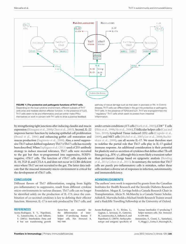
Marwaha et al. Th17 in autoimmunity and immunodeficiency
FIGURE 1 |The protective and pathogenic functions ofTh17 cells.
Depending on the local cytokine environment, different subsets of Th17cells arise and mediate distinct effector function. In the presence of IL-23,Th17 cells seem to be pro-inflammatory and can either make IFN-γthemselves or work in concert with Th1 cells to drive a positive feedback
pathway of tissue damage such as that seen in psoriasis or RA. In Crohn’sdisease, Th17 cells can differentiate in the gut into protective or pathogenicTh17 cells. In the presence of TGF-β and IL-21, Th17 are re-programmed into“regulatory” Th17 cells which seem to protect from intestinalinflammation.
by strengthening tight junctions after inducing claudin and mucinexpression (Kinugasa et al., 2000; Chen et al., 2003). Second, IL-22improves barrier function by inducing epithelial cell proliferation(Brand et al., 2006) and enhancing goblet cell restoration andmucus production (Sugimoto et al., 2008). Also, a novel suppres-sive Th17 subset dubbed regulatory Th17 (rTh17) cells has recentlybeen described. When Esplugues et al. (2011) used a CD3-antibodystrategy to induce mucosal tolerance, Th17 cells were recruitedto the gut but then re-programmed into suppressive, FOXP3-negative, rTh17 cells. The function of rTh17 cells depends onIL-10, TGF-β, and CTLA-4, and does not occur in CCR6-deficientmice where Th17 are not recruited to the gut. The latter data indi-cate that the mucosal immunity micro-environment is critical forthe development of rTh17 cells.
CONCLUSIONDifferent flavors of Th17 differentiation, ranging from highlypro-inflammatory to suppressive, result from different cytokinemicro-environments in various diseases. Th17 cells can no longerbe identified solely on the production of IL-17A since the com-bination of co-secreted cytokines is key to defining their effectorfunction. Moreover, IL-17 is not only produced by Th17 cells, and
under certain conditions γδ T cells (Stark et al., 2005),CD8+ T cells(Shin et al., 1998; He et al., 2006), T follicular helper cells (Cua andTato, 2010), Lymphoid Tissue induced (LTi) cells (Cupedo et al.,2009), and NKT cells (Michel et al., 2007; Lee et al., 2008; Rachit-skaya et al., 2008), can all secrete IL-17. We must therefore startto redefine the partial role that Th17 cells play in IL-17-guidedimmune response. An additional consideration is their potentialfor plasticity and co-secretion of cytokines that define other Th celllineages (e.g., IFN-γ), although this is more likely a transient ratherthan permanent change based on epigenetic analysis (Bendinget al., 2011; Cohen et al., 2011). In summary, the notion that Th17cells are purely pro-inflammatory cells is mistaken, rather thesecells mediate a diverse set of responses in infection, autoimmunity,and immunodeficiency.
ACKNOWLEDGMENTSThe authors’ own work is supported by grants from the CanadianInstitutes for Health Research and the Juvenile Diabetes ResearchFoundation. Megan K. Levings holds a Canada Research Chair inTransplantation. Alicia N. McMurchy is a Canada Vanier Scholar.Ashish K. Marwaha holds a Michael Smith Research Trainee awardand a Radcliffe Travelling Fellowship at the University of Oxford.
REFERENCESAcosta-Rodriguez, E. V., Napolitani,
G., Lanzavecchia, A., and Sallusto,F. (2007a). Interleukins 1beta and6 but not transforming growth
factor-beta are essential forthe differentiation of inter-leukin 17-producing human Thelper cells. Nat. Immunol. 8,942–949.
Acosta-Rodriguez, E. V., Rivino, L.,Geginat, J., Jarrossay, D., Gattorno,M., Lanzavecchia,A., Sallusto, F., andNapolitani, G. (2007b). Surface phe-notype and antigenic specificity of
human interleukin 17-producing Thelper memory cells. Nat. Immunol.8, 639–646.
Annunziato, F., Cosmi, L., Santarlasci,V.,Maggi,L.,Liotta,F.,Mazzinghi,B.,
Frontiers in Immunology | Primary Immunodeficiencies June 2012 | Volume 3 | Article 129 | 100

Marwaha et al. Th17 in autoimmunity and immunodeficiency
Parente, E., Filì, L., Ferri, S., Fros-ali, F., Giudici, F., Romagnani, P.,Parronchi, P., Tonelli, F., Maggi, E.,and Romagnani, S. (2007). Phe-notypic and functional features ofhuman Th17 cells. J. Exp. Med. 204,1849–1861.
Annunziato, F., and Romagnani, S.(2011). Mouse T helper 17 pheno-type: not so different than in manafter all. Cytokine 56, 112–115.
Arif, S., Moore, F., Marks, K.,Bouckenooghe, T., Dayan, C.M., Planas, R., Vives-Pi, M., Powrie,J., Tree, T., Marchetti, P., Huang,G. C., Gurzov, E. N., Pujol-Borrell,R., Eizirik, D. L., and Peakman,M. (2011). Peripheral and isletinterleukin-17 pathway activationcharacterizes human autoimmunediabetes and promotes cytokine-mediated beta-cell death. Diabetes60, 2112–2119.
Ayyoub, M., Deknuydt, F., Raimbaud,I., Dousset, C., Leveque, L., Bioley,G., and Valmori, D. (2009). Humanmemory FOXP3+ Tregs secreteIL-17 ex vivo and constitutivelyexpress the T(H)17 lineage-specifictranscription factor RORgamma t.Proc. Natl. Acad. Sci. U.S.A. 106,8635–8640.
Bacchetta, R., Passerini, L., Gambineri,E., Dai, M., Allan, S. E., Perroni,L., Dagna-Bricarelli, F., Sartirana, C.,Matthes-Martin, S., Lawitschka, A.,Azzari, C., Ziegler, S. F., Levings, M.K., and Roncarolo, M. G. (2006).Defective regulatory and effectorT cell functions in patients withFOXP3 mutations. J. Clin. Invest.116, 1713–1722.
Becher, B., Durell, B. G., and Noelle, R. J.(2002). Experimental autoimmuneencephalitis and inflammation in theabsence of interleukin-12. J. Clin.Invest. 110, 493–497.
Bending, D., De La Pena, H., Veld-hoen, M., Phillips, J. M., Uytten-hove, C., Stockinger, B., and Cooke,A. (2009). Highly purified Th17 cellsfrom BDC2.5NOD mice convertinto Th1-like cells in NOD/SCIDrecipient mice. J. Clin. Invest. 119,565–572.
Bending, D., Newland, S., Krejci,A., Phillips, J. M., Bray, S., andCooke, A. (2011). Epigeneticchanges at Il12rb2 and Tbx21in relation to plasticity behaviorof Th17 cells. J. Immunol. 186,3373–3382.
Beriou, G., Bradshaw, E. M., Lozano,E., Costantino, C. M., Hastings, W.D., Orban, T., Elyaman, W., Khoury,S. J., Kuchroo, V. K., Baecher-Allan,C., and Hafler, D. A. (2010). TGF-beta induces IL-9 production from
human Th17 cells. J. Immunol. 185,46–54.
Beriou, G., Costantino, C. M., Ashley, C.W.,Yang, L., Kuchroo,V. K., Baecher-Allan, C., and Hafler, D. A. (2009).IL-17-producing human peripheralregulatory T cells retain suppressivefunction. Blood 113, 4240–4249.
Bettelli, E., Korn, T., Oukka, M., andKuchroo, V. K. (2008). Inductionand effector functions of T(H)17cells. Nature 453, 1051–1057.
Boniface, K., Blom, B., Liu, Y. J., andDe Waal Malefyt, R. (2008). Frominterleukin-23 to T-helper 17 cells:human T-helper cell differentia-tion revisited. Immunol. Rev. 226,132–146.
Bovenschen, H. J., Van De Kerkhof,P. C., Van Erp, P. E., Woestenenk,R., Joosten, I., and Koenen, H. J.(2011). Foxp3+ regulatory T cellsof psoriasis patients easily differenti-ate into IL-17A-producing cells andare found in lesional skin. J. Invest.Dermatol. 131, 1853–1860.
Bradshaw, E. M., Raddassi, K., Elya-man, W., Orban, T., Gottlieb, P.A., Kent, S. C., and Hafler, D. A.(2009). Monocytes from patientswith type 1 diabetes spontaneouslysecrete proinflammatory cytokinesinducing Th17 cells. J. Immunol. 183,4432–4439.
Brand, S., Beigel, F., Olszak, T., Zitz-mann, K., Eichhorst, S. T., Otte, J.M., Diepolder, H., Marquardt, A.,Jagla, W., Popp, A., Leclair, S., Her-rmann, K., Seiderer, J., Ochsenkuhn,T., Goke, B., Auernhammer, C. J.,and Dambacher, J. (2006). IL-22is increased in active Crohn’s dis-ease and promotes proinflamma-tory gene expression and intestinalepithelial cell migration. Am. J. Phys-iol. Gastrointest. Liver Physiol. 290,G827–G838.
Buckley, R. H., Wray, B. B., and Bel-maker, E. Z. (1972). Extreme hyper-immunoglobulinemia E and unduesusceptibility to infection. Pediatrics49, 59–70.
Cai, Y., Shen, X., Ding, C., Qi, C., Li,K., Li, X., Jala, V. R., Zhang, H.G., Wang, T., Zheng, J., and Yan, J.(2011). Pivotal role of dermal IL-17-producing gammadelta T cellsin skin inflammation. Immunity 35,596–610.
Cascao, R., Moura, R. A., Perpetuo,I., Canhao, H., Vieira-Sousa, E.,Mourao, A. F., Rodrigues, A. M.,Polido-Pereira, J., Queiroz, M. V.,Rosario, H. S., Souto-Carneiro, M.M., Graca, L., and Fonseca, J. E.(2010). Identification of a cytokinenetwork sustaining neutrophil andTh17 activation in untreated early
rheumatoid arthritis. Arthritis Res.Ther. 12, R196.
Chabaud, M., Durand, J., Buchs, N.,Fossiez, F., Page, G., Frappart, L.,and Miossec, P. (1999). Humaninterleukin-17: A T cell-derivedproinflammatory cytokine pro-duced by the rheumatoid synovium.Arthritis Rheum. 42, 963–970.
Chen, Y., Thai, P., Zhao, Y. H.,Ho, Y. S., Desouza, M. M., andWu, R. (2003). Stimulation of air-way mucin gene expression byinterleukin (IL)-17 through IL-6paracrine/autocrine loop. J. Biol.Chem. 278, 17036–17043.
Chen, Z., Tato, C. M., Muul, L., Lau-rence, A., and O’Shea, J. J. (2007).Distinct regulation of interleukin-17 in human T helper lymphocytes.Arthritis Rheum. 56, 2936–2946.
Chiricozzi, A., Guttman-Yassky, E.,Suarez-Farinas, M., Nograles, K. E.,Tian, S., Cardinale, I., Chimenti, S.,and Krueger, J. G. (2011). Integrativeresponses to IL-17 and TNF-alphain human keratinocytes account forkey inflammatory pathogenic cir-cuits in psoriasis. J. Invest. Dermatol.131, 677–687.
Cohen, C. J., Crome, S. Q., Macdonald,K. G., Dai, E. L., Mager, D. L., andLevings, M. K. (2011). Human Th1and Th17 cells exhibit epigenetic sta-bility at signature cytokine and tran-scription factor loci. J. Immunol. 187,5615–5626.
Cosmi, L., Cimaz, R., Maggi, L., San-tarlasci, V., Capone, M., Borriello,F., Frosali, F., Querci, V., Simonini,G., Barra, G., Piccinni, M. P.,Liotta, F., De Palma, R., Maggi, E.,Romagnani, S., and Annunziato, F.(2011). Evidence of the transientnature of the Th17 phenotype ofCD4+CD161+ T cells in the syn-ovial fluid of patients with juvenileidiopathic arthritis. Arthritis Rheum.63, 2504–2515.
Cosmi, L., De Palma, R., Santarlasci,V., Maggi, L., Capone, M., Frosali,F., Rodolico, G., Querci, V., Abbate,G., Angeli, R., Berrino, L., Fambrini,M., Caproni, M., Tonelli, F., Lazzeri,E., Parronchi, P., Liotta, F., Maggi,E., Romagnani, S., and Annunzi-ato, F. (2008). Human interleukin17-producing cells originate from aCD161+CD4+ T cell precursor. J.Exp. Med. 205, 1903–1916.
Cosmi, L., Maggi, L., Santarlasci, V.,Capone, M., Cardilicchia, E., Frosali,F., Querci, V., Angeli, R., Matucci, A.,Fambrini, M., Liotta, F., Parronchi,P., Maggi, E., Romagnani, S., andAnnunziato, F. (2010). Identificationof a novel subset of human circu-lating memory CD4(+) T cells that
produce both IL-17A and IL-4. J.Allergy Clin. Immunol. 125, 222–230.
Crome, S. Q., Wang, A. Y., Kang,C. Y., and Levings, M. K. (2009).The role of retinoic acid-relatedorphan receptor variant 2 and IL-17 in the development and func-tion of human CD4+ T cells. Eur.J. Immunol. 39, 1480–1493.
Cua, D. J., Sherlock, J., Chen,Y., Murphy,C. A., Joyce, B., Seymour, B., Lucian,L., To, W., Kwan, S., Churakova, T.,Zurawski, S., Wiekowski, M., Lira, S.A., Gorman, D., Kastelein, R. A., andSedgwick, J. D. (2003). Interleukin-23 rather than interleukin-12 is thecritical cytokine for autoimmuneinflammation of the brain. Nature421, 744–748.
Cua, D. J., and Tato, C. M. (2010). InnateIL-17-producing cells: the sentinelsof the immune system. Nat. Rev.Immunol. 10, 479–489.
Cupedo, T., Crellin, N. K., Papazian, N.,Rombouts, E. J.,Weijer, K., Grogan, J.L., Fibbe, W. E., Cornelissen, J. J., andSpits, H. (2009). Human fetal lym-phoid tissue-inducer cells are inter-leukin 17-producing precursors toRORC+ CD127+ natural killer-likecells. Nat. Immunol. 10, 66–74.
de Beaucoudrey, L., Puel, A., Filipe-Santos, O., Cobat, A., Ghandil, P.,Chrabieh, M., Feinberg, J., VonBernuth, H., Samarina, A., Jan-niere, L., Fieschi, C., Stephan, J.L., Boileau, C., Lyonnet, S., Jon-deau, G., Cormier-Daire, V., Le Mer-rer, M., Hoarau, C., Lebranchu, Y.,Lortholary, O., Chandesris, M. O.,Tron, F., Gambineri, E., Bianchi, L.,Rodriguez-Gallego, C., Zitnik, S. E.,Vasconcelos, J., Guedes, M., Vitor, A.B., Marodi, L., Chapel, H., Reid, B.,Roifman, C., Nadal, D., Reichenbach,J., Caragol, I., Garty, B. Z., Dogu,F., Camcioglu, Y., Gulle, S., Sanal,O., Fischer, A., Abel, L., Stockinger,B., Picard, C., and Casanova, J. L.(2008). Mutations in STAT3 andIL12RB1 impair the development ofhuman IL-17-producing T cells. J.Exp. Med. 205, 1543–1550.
D’Hennezel, E., Ben-Shoshan, M., Ochs,H. D., Torgerson, T. R., Russell, L.J., Lejtenyi, C., Noya, F. J., Jabado,N., Mazer, B., and Piccirillo, C. A.(2009). FOXP3 forkhead domainmutation and regulatory T cells inthe IPEX syndrome. N. Engl. J. Med.361, 1710–1713.
Di Cesare, A., Di Meglio, P., and Nes-tle, F. O. (2009). The IL-23/Th17axis in the immunopathogenesis ofpsoriasis. J. Invest. Dermatol. 129,1339–1350.
Dong, C. (2008). TH17 cells in devel-opment: an updated view of their
www.frontiersin.org June 2012 | Volume 3 | Article 129 | 101

Marwaha et al. Th17 in autoimmunity and immunodeficiency
molecular identity and genetic pro-gramming. Nat. Rev. Immunol. 8,337–348.
Du, C., Liu, C., Kang, J., Zhao, G., Ye,Z., Huang, S., Li, Z., Wu, Z., and Pei,G. (2009). MicroRNA miR-326 reg-ulates TH-17 differentiation and isassociated with the pathogenesis ofmultiple sclerosis. Nat. Immunol. 10,1252–1259.
Dubin, P. J., and Kolls, J. K. (2008). Th17cytokines and mucosal immunity.Immunol. Rev. 226, 160–171.
Emamaullee, J. A., Davis, J., Merani, S.,Toso, C., Elliott, J. F., Thiesen, A., andShapiro, A. M. (2009). Inhibitionof Th17 cells regulates autoimmunediabetes in NOD mice. Diabetes 58,1302–1311.
Esplugues, E., Huber, S., Gagliani, N.,Hauser, A. E., Town, T., Wan, Y.Y., O’Connor, W. Jr., Rongvaux, A.,Van Rooijen, N., Haberman, A. M.,Iwakura, Y., Kuchroo, V. K., Kolls, J.K., Bluestone, J. A., Herold, K. C.,and Flavell, R. A. (2011). Controlof TH17 cells occurs in the smallintestine. Nature 475, 514–518.
Evans, H. G., Suddason, T., Jackson, I.,Taams, L. S., and Lord, G. M. (2007).Optimal induction of T helper 17cells in humans requires T cell recep-tor ligation in the context of Toll-like receptor-activated monocytes.Proc. Natl. Acad. Sci. U.S.A. 104,17034–17039.
Ferraro, A., Socci, C., Stabilini, A., Valle,A., Monti, P., Piemonti, L., Nano,R., Olek, S., Maffi, P., Scavini, M.,Secchi, A., Staudacher, C., Bonifa-cio, E., and Battaglia, M. (2011).Expansion of Th17 cells and func-tional defects in T regulatory cellsare key features of the pancreaticlymph nodes in patients with type1 diabetes. Diabetes 60, 2903–2913.
Fina, D., Sarra, M., Fantini, M. C., Rizzo,A., Caruso, R., Caprioli, F., Stolfi,C., Cardolini, I., Dottori, M., Boiri-vant, M., Pallone, F., Macdonald,T. T., and Monteleone, G. (2008).Regulation of gut inflammation andth17 cell response by interleukin-21.Gastroenterology 134, 1038–1048.
Gaffen, S. L. (2011). Recent advancesin the IL-17 cytokine fam-ily. Curr. Opin. Immunol. 23,613–619.
Gaffen, S. L., Hernandez-Santos, N., andPeterson, A. C. (2011). IL-17 signal-ing in host defense against Candidaalbicans. Immunol. Res. 50, 181–187.
Genovese, M. C., Van Den Bosch, F.,Roberson, S. A., Bojin, S., Biagini,I. M., Ryan, P., and Sloan-Lancaster,J. (2010). LY2439821, a human-ized anti-interleukin-17 monoclonalantibody, in the treatment of
patients with rheumatoid arthri-tis: a phase I randomized, double-blind, placebo-controlled, proof-of-concept study. Arthritis Rheum. 62,929–939.
Ghoreschi, K., Laurence, A., Yang, X.P., Hirahara, K., and O’Shea, J. J.(2011). T helper 17 cell heterogene-ity and pathogenicity in autoim-mune disease. Trends Immunol. 32,395–401.
Glocker, E. O., Hennigs, A., Nabavi,M., Schaffer, A. A., Woellner, C.,Salzer, U., Pfeifer, D., Veelken, H.,Warnatz, K., Tahami, F., Jamal, S.,Manguiat,A., Rezaei, N.,Amirzargar,A. A., Plebani, A., Hannesschlager,N., Gross, O., Ruland, J., and Grim-bacher, B. (2009). A homozygousCARD9 mutation in a family withsusceptibility to fungal infections. N.Engl. J. Med. 361, 1727–1735.
Grimbacher, B., Holland, S. M., Gallin, J.I., Greenberg, F., Hill, S. C., Malech,H. L., Miller, J. A., O’Connell, A.C., and Puck, J. M. (1999). Hyper-IgE syndrome with recurrent infec-tions – an autosomal dominant mul-tisystem disorder. N. Engl. J. Med.340, 692–702.
Harrington, L. E., Hatton, R. D., Man-gan, P. R., Turner, H., Murphy, T.L., Murphy, K. M., and Weaver, C.T. (2005). Interleukin 17-producingCD4+ effector T cells develop viaa lineage distinct from the T helpertype 1 and 2 lineages. Nat. Immunol.6, 1123–1132.
He, D., Wu, L., Kim, H. K., Li, H.,Elmets, C. A., and Xu, H. (2006).CD8+ IL-17-producing T cells areimportant in effector functions forthe elicitation of contact hypersen-sitivity responses. J. Immunol. 177,6852–6858.
Hirota, K., Duarte, J. H., Veldhoen, M.,Hornsby, E., Li,Y., Cua, D. J., Ahlfors,H., Wilhelm, C., Tolaini, M., Menzel,U., Garefalaki,A., Potocnik,A. J., andStockinger, B. (2011). Fate mappingof IL-17-producing T cells in inflam-matory responses. Nat. Immunol. 12,255–263.
Holland, S. M., Deleo, F. R., Elloumi,H. Z., Hsu, A. P., Uzel, G., Brod-sky, N., Freeman, A. F., Demidowich,A., Davis, J., Turner, M. L., Ander-son, V. L., Darnell, D. N., Welch,P. A., Kuhns, D. B., Frucht, D. M.,Malech, H. L., Gallin, J. I., Kobayashi,S. D., Whitney, A. R., Voyich, J. M.,Musser, J. M., Woellner, C., Schaffer,A. A., Puck, J. M., and Grimbacher,B. (2007). STAT3 mutations in thehyper-IgE syndrome. N. Engl. J. Med.357, 1608–1619.
Honkanen, J., Nieminen, J. K., Gao, R.,Luopajarvi, K., Salo, H. M., Ilonen,
J., Knip, M., Otonkoski, T., and Vaar-ala, O. (2010). IL-17 immunity inhuman type 1 diabetes. J. Immunol.185, 1959–1967.
Hot, A., and Miossec, P. A. (2011).Effects of interleukin (IL)-17A andIL-17F in human rheumatoid arthri-tis synoviocytes. Ann. Rheum. Dis.70, 727–732.
Hueber, W., Patel, D. D., Dryja, T.,Wright, A. M., Koroleva, I., Bruin,G., Antoni, C., Draelos, Z., Gold, M.H., Psoriasis Study, G., Durez, P., Tak,P. P., Gomez-Reino, J. J., Rheuma-toid Arthritis Study Group, Foster,C. S., Kim, R. Y., Samson, C. M.,Falk, N. S., Chu, D. S., Callanan, D.,Nguyen, Q. D., Uveitis Study Group,Rose, K., Haider, A., and Di Padova,F. (2010). Effects of AIN457, a fullyhuman antibody to interleukin-17A,on psoriasis, rheumatoid arthritis,and uveitis. Sci. Transl. Med. 2,52ra72.
Hughson, A., Bromberg, I., Johnson, B.,Quataert, S., Jospe, N., and Fowell, D.J. (2011). Uncoupling of prolifera-tion and cytokines from suppressionwithin the CD4+CD25+Foxp3+ T-cell compartment in the 1st year ofhuman type 1 diabetes. Diabetes 60,2125–2133.
Hwang, S. Y., and Kim, A. H. (2005).Expression of IL-17 homologs andtheir receptors in the synovial cells ofrheumatoid arthritis patients. Mol.Cells 19, 180–184.
Jain, R., Tartar, D. M., Gregg, R. K.,Divekar, R. D., Bell, J. J., Lee,H. H., Yu, P., Ellis, J. S., Hoe-man, C. M., Franklin, C. L., andZaghouani, H. (2008). InnocuousIFNgamma induced by adjuvant-free antigen restores normoglycemiain NOD mice through inhibition ofIL-17 production. J. Exp. Med. 205,207–218.
Joseph, J., Bittner, S., Kaiser, F. M.,Wiendl, H., and Kissler, S. (2012).IL-17 silencing does not pro-tect nonobese diabetic mice fromautoimmune diabetes. J. Immunol.188, 216–221.
Kebir, H., Kreymborg, K., Ifergan, I.,Dodelet-Devillers, A., Cayrol, R.,Bernard, M., Giuliani, F., Arbour,N., Becher, B., and Prat, A. (2007).Human TH17 lymphocytes promoteblood-brain barrier disruption andcentral nervous system inflamma-tion. Nat. Med. 13, 1173–1175.
Kinugasa, T., Sakaguchi, T., Gu, X., andReinecker, H. C. (2000). Claudinsregulate the intestinal barrier inresponse to immune mediators. Gas-troenterology 118, 1001–1011.
Kirkham, B. W., Lassere, M. N.,Edmonds, J. P., Juhasz, K. M., Bird,
P. A., Lee, C. S., Shnier, R., andPortek, I. J. (2006). Synovial mem-brane cytokine expression is pre-dictive of joint damage progressionin rheumatoid arthritis: a two-yearprospective study (the DAMAGEstudy cohort). Arthritis Rheum. 54,1122–1131.
Kolls, J. K., and Linden, A. (2004).Interleukin-17 family membersand inflammation. Immunity 21,467–476.
Kryczek, I., Bruce, A. T., Gudjonsson, J.E., Johnston, A., Aphale, A., Vatan,L., Szeliga, W., Wang, Y., Liu, Y.,Welling, T. H., Elder, J. T., andZou, W. (2008). Induction of IL-17+ T cell trafficking and devel-opment by IFN-gamma: mechanismand pathological relevance in psori-asis. J. Immunol. 181, 4733–4741.
Kryczek, I., Wu, K., Zhao, E., Wei, S.,Vatan, L., Szeliga, W., Huang, E.,Greenson, J., Chang, A., Rolinski, J.,Radwan, P., Fang, J., Wang, G., andZou, W. (2011). IL-17+ regulatoryT cells in the microenvironments ofchronic inflammation and cancer. J.Immunol. 186, 4388–4395.
Langrish, C. L., Chen, Y., Blumenschein,W. M., Mattson, J., Basham, B., Sedg-wick, J. D., Mcclanahan, T., Kastelein,R. A., and Cua, D. J. (2005). IL-23 drives a pathogenic T cell pop-ulation that induces autoimmuneinflammation. J. Exp. Med. 201,233–240.
Lee, K. A., Kang, M. H., Lee, Y. S., Kim,Y. J., Kim, D. H., Ko, H. J., and Kang,C. Y. (2008). A distinct subset ofnatural killer T cells produces IL-17, contributing to airway infiltra-tion of neutrophils but not to airwayhyperreactivity. Cell. Immunol. 251,50–55.
Lee, S. M., Yang, H., Tartar, D. M., Gao,B., Luo, X., Ye, S. Q., Zaghouani, H.,and Fang, D. (2011). Prevention andtreatment of diabetes with resvera-trol in a non-obese mouse modelof type 1 diabetes. Diabetologia 54,1136–1146.
Lee, Y. K., Turner, H., Maynard, C. L.,Oliver, J. R., Chen, D., Elson, C. O.,and Weaver, C. T. (2009). Late devel-opmental plasticity in the T helper17 lineage. Immunity 30, 92–107.
Leipe, J., Grunke, M., Dechant, C.,Reindl, C., Kerzendorf, U., Schulze-Koops, H., and Skapenko, A. (2010).Role of Th17 cells in human autoim-mune arthritis. Arthritis Rheum. 62,2876–2885.
Leonardi, C., Matheson, R., Zachariae,C., Cameron, G., Li, L., Edson-Heredia, E., Braun, D., and Baner-jee, S. (2012). Anti-interleukin-17monoclonal antibody ixekizumab in
Frontiers in Immunology | Primary Immunodeficiencies June 2012 | Volume 3 | Article 129 | 102
Marwaha et al. Th17 in autoimmunity and immunodeficiency
chronic plaque psoriasis. N. Engl. J.Med. 366, 1190–1199.
Lin, A. M., Rubin, C. J., Khandpur, R.,Wang, J. Y., Riblett, M., Yalavarthi, S.,Villanueva, E. C., Shah, P., Kaplan,M. J., and Bruce, A. T. (2011). Mastcells and neutrophils release IL-17through extracellular trap forma-tion in psoriasis. J. Immunol. 187,490–500.
Liu, H., and Rohowsky-Kochan, C.(2008). Regulation of IL-17 inhuman CCR6+ effector memory Tcells. J. Immunol. 180, 7948–7957.
Liu, S. M., Lee, D. H., Sullivan, J. M.,Chung, D., Jager, A., Shum, B. O.,Sarvetnick, N. E., Anderson, A. C.,and Kuchroo, V. K. (2011). Differ-ential IL-21 signaling in APCs leadsto disparate Th17 differentiationin diabetes-susceptible NOD anddiabetes-resistant NOD.Idd3 mice. J.Clin. Invest. 121, 4303–4310.
Lowes, M. A., Bowcock, A. M., andKrueger, J. G. (2007). Pathogenesisand therapy of psoriasis. Nature 445,866–873.
Lundy, S. K., Sarkar, S., Tesmer, L. A.,and Fox, D. A. (2007). Cells of thesynovium in rheumatoid arthritis. Tlymphocytes. Arthritis Res. Ther. 9,202.
Ma, C. S., Chew, G. Y., Simpson, N.,Priyadarshi, A., Wong, M., Grim-bacher, B., Fulcher, D. A., Tangye, S.G., and Cook, M. C. (2008). Defi-ciency of Th17 cells in hyper IgE syn-drome due to mutations in STAT3. J.Exp. Med. 205, 1551–1557.
Manel, N., Unutmaz, D., and Littman,D. R. (2008). The differentiation ofhuman T(H)-17 cells requires trans-forming growth factor-beta andinduction of the nuclear recep-tor RORgammat. Nat. Immunol. 9,641–649.
Martin-Orozco, N., Chung, Y., Chang,S. H., Wang, Y. H., and Dong, C.(2009). Th17 cells promote pancre-atic inflammation but only inducediabetes efficiently in lymphopenichosts after conversion into Th1 cells.Eur. J. Immunol. 39, 216–224.
Marwaha, A. K., Crome, S. Q., Pana-giotopoulos, C., Berg, K. B., Qin, H.,Ouyang, Q., Xu, L., Priatel, J. J., Lev-ings, M. K., and Tan, R. (2010). Cut-ting edge: increased IL-17-secretingT cells in children with new-onsettype 1 diabetes. J. Immunol. 185,3814–3818.
Matusevicius, D., Kivisäkk, P., He, B.,Kostulas, N., Ozenci, V., Fredrikson,S., and Link, H. (1999). Interleukin-17 mRNA expression in blood andCSF mononuclear cells is augmentedin multiple sclerosis. Mult. Scler. 5,101–104.
McGeachy, M. J., Bak-Jensen, K. S.,Chen, Y., Tato, C. M., Blumenschein,W., Mcclanahan, T., and Cua, D. J.(2007). TGF-beta and IL-6 drive theproduction of IL-17 and IL-10 byT cells and restrain T(H)-17 cell-mediated pathology. Nat. Immunol.8, 1390–1397.
McMurchy, A. N., Gillies, J., Allan, S. E.,Passerini, L., Gambineri, E., Roncar-olo, M. G., Bacchetta, R., and Lev-ings, M. K. (2010). Point mutants offorkhead box P3 that cause immunedysregulation, polyendocrinopathy,enteropathy, X-linked have diverseabilities to reprogram T cells intoregulatory T cells. J. Allergy Clin.Immunol. 126, 1242–1251.
Michel, M. L., Keller, A. C., Paget, C.,Fujio, M., Trottein, F., Savage, P. B.,Wong, C. H., Schneider, E., Dy, M.,and Leite-De-Moraes, M. C. (2007).Identification of an IL-17-producingNK1.1(neg) iNKT cell populationinvolved in airway neutrophilia. J.Exp. Med. 204, 995–1001.
Milner, J. D. (2011). IL-17 producingcells in host defense and atopy. Curr.Opin. Immunol. 23, 784–788.
Milner, J. D., Brenchley, J. M., Laurence,A., Freeman, A. F., Hill, B. J., Elias, K.M., Kanno, Y., Spalding, C., Elloumi,H. Z., Paulson, M. L., Davis, J., Hsu,A., Asher, A. I., O’Shea, J., Holland,S. M., Paul, W. E., and Douek, D.C. (2008). Impaired T(H)17 cell dif-ferentiation in subjects with autoso-mal dominant hyper-IgE syndrome.Nature 452, 773–776.
Miyara, M., Yoshioka, Y., Kitoh, A.,Shima, T., Wing, K., Niwa, A., Pari-zot, C., Taflin, C., Heike, T., Valeyre,D., Mathian, A., Nakahata, T., Yam-aguchi, T., Nomura, T., Ono, M.,Amoura, Z., Gorochov, G., and Sak-aguchi, S. (2009). Functional delin-eation and differentiation dynamicsof human CD4+ T cells express-ing the FoxP3 transcription factor.Immunity 30, 899–911.
Morrison, P. J., Ballantyne, S. J., andKullberg, M. C. (2011). Interleukin-23 and T helper 17-type responsesin intestinal inflammation: fromcytokines to T-cell plasticity.Immunology 133, 397–408.
Ogawa, A., Andoh, A., Araki, Y., Bamba,T., and Fujiyama, Y. (2004). Neutral-ization of interleukin-17 aggravatesdextran sulfate sodium-induced col-itis in mice. Clin. Immunol. 110,55–62.
Pappu, R., Ramirez-Carrozzi, V.,and Sambandam, A. (2011). Theinterleukin-17 cytokine family:critical players in host defence andinflammatory diseases. Immunology134, 8–16.
Park, H., Li, Z., Yang, X. O., Chang, S.H., Nurieva, R., Wang, Y. H., Wang,Y., Hood, L., Zhu, Z., Tian, Q., andDong,C. (2005). A distinct lineage ofCD4 T cells regulates tissue inflam-mation by producing interleukin 17.Nat. Immunol. 6, 1133–1141.
Passerini, L., Olek, S., Di Nunzio, S.,Barzaghi, F., Hambleton, S., Abi-nun, M., Tommasini, A., Vignola, S.,Cipolli, M., Amendola, M., Naldini,L., Guidi, L., Cecconi, M., Roncar-olo, M. G., and Bacchetta, R. (2011).Forkhead box protein 3 (FOXP3)mutations lead to increased TH17cell numbers and regulatory T-cellinstability. J. Allergy Clin. Immunol.128, 1376–1379.
Puel, A., Cypowyj, S., Bustamante, J.,Wright, J. F., Liu, L., Lim, H. K.,Migaud, M., Israel, L., Chrabieh, M.,Audry, M., Gumbleton, M., Toulon,A., Bodemer, C., El-Baghdadi, J.,Whitters, M., Paradis, T., Brooks,J., Collins, M., Wolfman, N. M.,Al-Muhsen, S., Galicchio, M., Abel,L., Picard, C., and Casanova, J.L. (2011). Chronic mucocutaneouscandidiasis in humans with inbornerrors of interleukin-17 immunity.Science 332, 65–68.
Rachitskaya,A. V., Hansen,A. M., Horai,R., Li, Z., Villasmil, R., Luger, D.,Nussenblatt, R. B., and Caspi, R.R. (2008). Cutting edge: NKT cellsconstitutively express IL-23 receptorand RORgammat and rapidly pro-duce IL-17 upon receptor ligationin an IL-6-independent fashion. J.Immunol. 180, 5167–5171.
Renner, E. D., Rylaarsdam, S., Anover-Sombke,S.,Rack,A. L.,Reichenbach,J., Carey, J. C., Zhu, Q., Jansson, A. F.,Barboza, J., Schimke, L. F., Leppert,M. F., Getz, M. M., Seger, R. A., Hill,H. R., Belohradsky, B. H., Torgerson,T. R., and Ochs, H. D. (2008). Novelsignal transducer and activator oftranscription 3 (STAT3) mutations,reduced T(H)17 cell numbers, andvariably defective STAT3 phospho-rylation in hyper-IgE syndrome. J.Allergy Clin. Immunol. 122, 181–187.
Sandborn, W. J., Feagan, B. G., Fedo-rak, R. N., Scherl, E., Fleisher, M. R.,Katz, S., Johanns, J., Blank, M., Rut-geerts, P., and Ustekinumab Crohn’sDisease Study Group. (2008). Arandomized trial of Ustekinumab,a human interleukin-12/23 mon-oclonal antibody, in patients withmoderate-to-severe Crohn’s disease.Gastroenterology 135, 1130–1141.
Santarlasci, V., Maggi, L., Capone, M.,Frosali, F., Querci, V., De Palma,R., Liotta, F., Cosmi, L., Maggi, E.,Romagnani, S., and Annunziato, F.(2009). TGF-beta indirectly favors
the development of human Th17cells by inhibiting Th1 cells. Eur. J.Immunol. 39, 207–215.
Santarlasci, V., Maggi, L., Capone,M., Querci, V., Beltrame, L., Cav-alieri, D., D’Aiuto, E., Cimaz,R., Nebbioso, A., Liotta, F., DePalma, R., Maggi, E., Cosmi, L.,Romagnani, S., and Annunziato, F.(2012). Rarity of human T helper17 cells is due to retinoic acidorphan receptor-dependent mech-anisms that limit their expansion.Immunity 36, 201–214.
Sarkar, S., and Fox, D. A. (2010). Target-ing IL-17 and Th17 cells in rheuma-toid arthritis. Rheum. Dis. Clin.North Am. 36, 345–366.
Shin, H. C., Benbernou, N., Fekkar,H., Esnault, S., and Guenounou,M. (1998). Regulation of IL-17,IFN-gamma and IL-10 in humanCD8(+) T cells by cyclic AMP-dependent signal transduction path-way. Cytokine 10, 841–850.
Stark, M. A., Huo, Y., Burcin, T. L., Mor-ris, M. A., Olson, T. S., and Ley,K. (2005). Phagocytosis of apoptoticneutrophils regulates granulopoiesisvia IL-23 and IL-17. Immunity 22,285–294.
Sugimoto, K., Ogawa, A., Mizoguchi, E.,Shimomura, Y., Andoh, A., Bhan, A.K., Blumberg, R. S., Xavier, R. J., andMizoguchi, A. (2008). IL-22 ame-liorates intestinal inflammation in amouse model of ulcerative colitis. J.Clin. Invest. 118, 534–544.
Tran, C. N., Lundy, S. K., White, P.T., Endres, J. L., Motyl, C. D.,Gupta, R., Wilke, C. M., Shelden,E. A., Chung, K. C., Urquhart, A.G., and Fox, D. A. (2007). Molecu-lar interactions between T cells andfibroblast-like synoviocytes: role ofmembrane tumor necrosis factor-alpha on cytokine-activated T cells.Am. J. Pathol. 171, 1588–1598.
van den Brandt, J., Fischer, H. J., Wal-ter, L., Hunig, T., Kloting, I., andReichardt, H. M. (2010). Type 1 dia-betes in biobreeding rats is criti-cally linked to an imbalance betweenTh17 and regulatory T cells and analtered TCR repertoire. J. Immunol.185, 2285–2294.
van der Gast, C. J., Gosling, P., Tiwari, B.,and Bending, G. D. (2011). Spatialscaling of arbuscular mycorrhizalfungal diversity is affected by farm-ing practice. Environ. Microbiol. 13,241–249.
van Hamburg, J. P., Asmawidjaja, P.S., Davelaar, N., Mus, A. M., Colin,E. M., Hazes, J. M., Dolhain, R.J., and Lubberts, E. (2011). Th17cells, but not Th1 cells, from patientswith early rheumatoid arthritis are
www.frontiersin.org June 2012 | Volume 3 | Article 129 |103
Marwaha et al. Th17 in autoimmunity and immunodeficiency
potent inducers of matrix met-alloproteinases and proinflamma-tory cytokines upon synovial fibrob-last interaction, including autocrineinterleukin-17A production. Arthri-tis Rheum. 63, 73–83.
Volpe, E., Servant, N., Zollinger, R.,Bogiatzi, S. I., Hupe, P., Baril-lot, E., and Soumelis, V. (2008).A critical function for transform-ing growth factor-beta, interleukin23 and proinflammatory cytokinesin driving and modulating humanT(H)-17 responses. Nat. Immunol. 9,650–657.
Voo, K. S., Wang, Y. H., Santori, F. R.,Boggiano, C., Arima, K., Bover, L.,Hanabuchi, S., Khalili, J., Marinova,E., Zheng, B., Littman, D. R., andLiu, Y. J. (2009). Identification of IL-17-producing FOXP3+ regulatory Tcells in humans. Proc. Natl. Acad. Sci.U.S.A. 106, 4793–4798.
Vukkadapu, S. S., Belli, J. M., Ishii, K.,Jegga, A. G., Hutton, J. J., Aronow, B.J., and Katz, J. D. (2005). Dynamicinteraction between T cell-mediatedbeta-cell damage and beta-cell repairin the run up to autoimmune dia-betes of the NOD mouse. Physiol.Genomics 21, 201–211.
Weaver, C. T., and Hatton, R. D.(2009). Interplay between the TH17and TReg cell lineages: a (co-)evolutionary perspective. Nat. Rev.Immunol. 9, 883–839.
Wilke, C. M., Bishop, K., Fox, D., andZou, W. (2011). Deciphering therole of Th17 cells in human disease.Trends Immunol. 32, 603–611.
Wilson, N. J., Boniface, K., Chan, J.R., Mckenzie, B. S., Blumenschein,
W. M., Mattson, J. D., Basham,B., Smith, K., Chen, T., Morel,F., Lecron, J. C., Kastelein, R.A., Cua, D. J., Mcclanahan, T.K., Bowman, E. P., and De WaalMalefyt, R. (2007). Development,cytokine profile and function ofhuman interleukin 17-producinghelper T cells. Nat. Immunol. 8,950–957.
Wright, J. F., Guo, Y., Quazi, A., Luxen-berg, D. P., Bennett, F., Ross, J. F., Qiu,Y., Whitters, M. J., Tomkinson, K. N.,Dunussi-Joannopoulos, K., Carreno,B. M., Collins, M., and Wolfman, N.M. (2007). Identification of an inter-leukin 17F/17A heterodimer in acti-vated human CD4+ T cells. J. Biol.Chem. 282, 13447–13455.
Yang, L., Anderson, D., Baecher-Allan,C., Hastings, W. D., Bettelli, E.,Oukka, M., Kuchroo, V. K., andHafler, D. A. (2008a). IL-21 andTGF-β are required for differentia-tion of human Th17 cells. Nature454, 350–352.
Yang, X. O., Chang, S. H., Park, H.,Nurieva, R., Shah, B., Acero, L.,Wang, Y. H., Schluns, K. S., Broad-dus, R. R., Zhu, Z., and Dong, C.(2008b). Regulation of inflamma-tory responses by IL-17F. J. Exp. Med.205, 1063–1075.
Yang, X. O., Panopoulos, A. D.,Nurieva, R., Chang, S. H., Wang,D., Watowich, S. S., and Dong, C.(2007). STAT3 regulates cytokine-mediated generation of inflamma-tory helper T cells. J. Biol. Chem. 282,9358–9363.
Ye, J., Su, X., Hsueh, E. C., Zhang,Y., Koenig, J. M., Hoft, D. F.,
and Peng, G. (2011). Humantumor-infiltrating Th17 cells havethe capacity to differentiate intoIFN-gamma+ and FOXP3+ Tcells with potent suppressivefunction. Eur. J. Immunol. 41,936–951.
Zaba, L. C., Fuentes-Duculan, J.,Eungdamrong, N. J., Abello, M.V., Novitskaya, I., Pierson, K. C.,Gonzalez, J., Krueger, J. G., andLowes, M. A. (2009). Psoriasis ischaracterized by accumulation ofimmunostimulatory and Th1/Th17cell-polarizing myeloid dendriticcells. J. Invest. Dermatol. 129,79–88.
Zenewicz, L. A., Yancopoulos, G. D.,Valenzuela, D. M., Murphy, A. J.,Stevens, S., and Flavell, R. A. (2008).Innate and adaptive interleukin-22 protects mice from inflamma-tory bowel disease. Immunity 29,947–957.
Zhou, L., Lopes, J. E., Chong, M. M.,Ivanov, I. I., Min, R., Victora, G.D., Shen, Y., Du, J., Rubtsov, Y.P., Rudensky, A. Y., Ziegler, S. F.,and Littman, D. R. (2008). TGF-beta-induced Foxp3 inhibits T(H)17cell differentiation by antagonizingRORgammat function. Nature 453,236–240.
Ziegler, S. F. (2006). FOXP3: of miceand men. Annu. Rev. Immunol. 24,209–226.
Zielinski, C. E., Mele, F., Aschenbren-ner, D., Jarrossay, D., Ronchi, F.,Gattorno, M., Monticelli, S., Lanza-vecchia, A., and Sallusto, F. (2012).Pathogen-induced human T(H)17cells produce IFN-gamma or IL-10
and are regulated by IL-1beta.Nature 484, 514–518.
Ziolkowska, M., Koc, A.,Luszczykiewicz, G., Ksiezopolska-Pietrzak, K., Klimczak, E.,Chwalinska-Sadowska, H., andMaslinski, W. (2000). High levelsof IL-17 in rheumatoid arthritispatients: IL-15 triggers in vitroIL-17 production via cyclosporinA-sensitive mechanism. J. Immunol.164, 2832–2838.
Conflict of Interest Statement: Theauthors declare that the research wasconducted in the absence of any com-mercial or financial relationships thatcould be construed as a potential con-flict of interest.
Received: 10 April 2012; paper pend-ing published: 21 April 2012; accepted:04 May 2012; published online: 04 June2012.Citation: Marwaha AK, Leung NJ,McMurchy AN and Levings MK (2012)TH17 cells in autoimmunity andimmunodeficiency: protective or path-ogenic? Front. Immun. 3:129. doi:10.3389/fimmu.2012.00129This article was submitted to Frontiers inPrimary Immunodeficiencies, a specialtyof Frontiers in Immunology.Copyright © 2012 Marwaha, Leung ,McMurchy and Levings. This is an open-access article distributed under the termsof the Creative Commons AttributionNon Commercial License, which per-mits non-commercial use, distribution,and reproduction in other forums, pro-vided the original authors and source arecredited.
Frontiers in Immunology | Primary Immunodeficiencies June 2012 | Volume 3 | Article 129 | 104

“fimmu-03-00233” — 2012/8/2 — 11:09 — page 1 — #1
REVIEW ARTICLEpublished: 02 August 2012
doi: 10.3389/fimmu.2012.00233
Dendritic cells: a double-edge sword in autoimmuneresponsesGiada Amodio and Silvia Gregori*
San Raffaele Telethon Institute for Gene Therapy (OSR-TIGET), Division of Regenerative Medicine, Stem Cells and Gene Therapy, San Raffaele ScientificInstitute, Milan, Italy
Edited by:
Rosa Bacchetta, Fondazione SanRaffaele, Italy
Reviewed by:
Rosa Bacchetta, Fondazione SanRaffaele, ItalyElena Perez, University of SouthFlorida, USA
*Correspondence:
Silvia Gregori, San Raffaele TelethonInstitute for Gene Therapy, Division ofRegenerative Medicine, Stem Cellsand Gene Therapy, San RaffaeleScientific Institute, Via Olgettina 58,20132 Milan, Italy.e-mail: [email protected]
Dendritic cells (DC) are antigen-presenting cells that play a pivotal role in regulating innateand adaptive immune responses. In autoimmunity, DC act as a double-edged sword sinceon one hand they initiate adaptive self-reactive responses and on the other they play apivotal role in promoting and maintaining tolerance. Thus, DC are the most importantcells in either triggering self-specific responses or in negatively regulating auto-reactiveresponses.The latter function is mediated by DC in the steady-state or specialized subsetsof DC, named tolerogenic DC. Clinical and experimental evidence indicate that prolongedpresentation of self-antigens by DC is crucial for the development of destructive autoim-mune diseases, and defects in tolerogenic DC functions contribute to eradication ofself-tolerance. In recent years, DC have emerged as therapeutic targets for limiting theirimmunogenicity against self-antigens, while tolerogenic DC have been conceived as ther-apeutic tools to restore tolerance. The purpose of this review is to give a general overviewof the current knowledge on the pathogenic role of DC in patients affected by autoimmunediseases. In addition, the protective role of tolerogenic DC will be addressed.The currentlyapplied strategies to block immune activation or to exploit the tolerogenic potential of DCwill be discussed.
Keywords: dendritic cells, autoimmune diseases, tolerance
INTRODUCTIONDendritic cells (DC) are professional antigen-presenting cells(APC) specialized in capturing and processing antigens (Ags) topresent to T cells. DC constitute a front-line defense against patho-gens, are located throughout the body, and form complex networksthat allow them to communicate with different cells. Therefore,DC are critically involved in the initiation of adaptive immuneresponses and, as such, are defined immunogenic DC. These DCmight be implicated in the induction of autoimmune responsesvia the activation of auto-reactive T cells and the consequenteradication of self-tolerance. Conversely, DC in the steady-state,or specialized subsets of DC, termed tolerogenic DC, promoteand maintain tolerance through several non-overlapping mech-anisms. Tolerogenic DC can induce apoptosis of effector T cells,skew T cell phenotype, and promote anergy and/or regulatory Tcells (Tregs; Morelli and Thomson, 2007; Gregori, 2011). Thus,defects in the activities of tolerogenic DC may also contribute tobreak self-tolerance and to induce autoimmune responses.
An optimal balance between immunogenic and tolerogenicDC is therefore fundamental to prevent self-reactive immuneresponses and to maintain immune self-specific homeostasis. Inthis review, we will give an overview of the different role of bothimmunogenic and tolerogenic DC in promoting autoimmunedisease onset and/or progression, focusing primarily on humanpathological conditions.
HUMAN DENDRITIC CELL SUBSETSDendritic cells are present in all tissues and they function asan important bridge between innate and adaptive immunity, by
cellular interactions or through secretion of pro-inflammatoryand immuno-regulatory cytokines (Banchereau and Steinman,1998; Larregina and Falo, 2005; Merad et al., 2008; Rescignoand Di Sabatino, 2009; Lambrecht and Hammad, 2010;Thomson, 2010).
In the bloodstream, DC circulate as immature cells charac-terized by a low expression of human leukocyte antigen (HLA)class II and co-stimulatory molecules, high endocytic activity, andlow T cell activation potential. Circulating DC constantly patrolthe surrounding environment for pathogens, such as viruses andbacteria. Upon Ag encounter, DC undergo a complex process ofmaturation meanwhile they travel to the lymph nodes, where theyactivate helper and cytotoxic T cells as well as B cells. Imma-ture DC in the steady-state migrate at low ratio to the lymphnodes without undergoing activation, can present Ags to T cellsin the absence of co-stimulation and induce clonal T cell anergy(Schwartz et al., 1989), deletion of auto-reactive T cells (Hawigeret al., 2001; Steinman and Nussenzweig, 2002), and promote Tregs(Dhodapkar et al., 2001). Tolerogenic DC, both circulating andtissue resident, contribute to the induction and maintenance ofself-specific tolerance.
In humans, two major and intrinsically different subpopula-tions of DC have been described: myeloid DC (myDC), called alsoconventional DC, and plasmacytoid DC (pDC), which differ intheir transcriptional program, development, phenotypic markers,and immunological functions (Belz and Nutt, 2012). myDC pickup Ags in the periphery and move to T cell areas of peripheral lym-phoid organs to initiate immunity through a number of differentevents including maturation and cytokine secretion, all of which
www.frontiersin.org August 2012 | Volume 3 | Article 233 | 105

“fimmu-03-00233” — 2012/8/2 — 11:09 — page 2 — #2
Amodio and Gregori DC in autoimmunity
are regulated by recognition of pathogens via Toll-like receptors(TLR; Watts et al., 2010).
Myeloid DC are present in the peripheral blood and in severaltissues where they acquire specialized functions. In the blood-stream, several subpopulations of immunogenic myDC, all ofthem expressing CD11c, and the myeloid markers CD13 andCD33, are present (Table 1). These cells include CD16+ (theyare also characterized by the expression of M-DC8; Schakel et al.,1999), BDCA-1+, and BDCA-3+ (Dzionek et al., 2001) that havedifferent ability to stimulate allogeneic T cells (MacDonald et al.,2002). Distinct phenotypical and functional characteristics are dis-played by myDC resident in peripheral tissues. These myDC can bedistinguished according to the expression of specific markers: lan-gerin (CD207) expressing cells (Geissmann et al., 2002; Larreginaand Falo, 2005) are Langerhans cells (LC) and interstitial dermalDC localized in the skin; CD103+ DC reside in the lamina propria(LP) of the small intestine (Jaensson et al., 2008; Rescigno and DiSabatino, 2009); C-type lectin+ (DC-SIGN) DC are present in thedecidua (Laskarin et al., 2007); BDCA-1+ and BDCA-3+ DC havebeen described in the lung (Demedts et al., 2005; Table 1).
In addition to immunogenic myDC, other subsets of myDCwith tolerogenic properties have been described such as DCexpressing the scavenger receptor CD163 and immunoglobulin-like transcript 3 (ILT3; Maniecki et al., 2006). We recentlyidentified DC-10, which are tolerogenic DC characterized by theexpression of CD11c+, CD14+, CD16+, CD83+, and the tolero-genic molecules HLA-G and ILT4 (Gregori et al., 2010). DC-10display a mature phenotype since they express both HLA classII and co-stimulatory molecules. They have a unique cytokinesecretion profile consisting of high levels of IL-10 in the absenceof IL-12 (Gregori et al., 2010). Specialized subsets of tolerogenicDC have been described in each tissue where they maintain tissuehomeostasis and tolerance (reviewed in Gregori, 2011).
Plasmacytoid DC are component of the innate immune systemand are specialized in producing interferon-α (IFN-α) upon acti-vation via TLR7- and TLR9-mediated recognition of nucleic acids,and participate in T cell immunity (reviewed by Colonna et al.,
2004). Similar to myDC, immature pDC as well as alternativelyactivated pDC are involved in promoting tolerance (Hanabuchiet al., 2010; Martin-Gayo et al., 2010). pDC are characterized bythe expression of BDCA-2, BDCA-4 (Dzionek et al., 2001), IL-3R(CD123; Jahnsen et al., 2000), and ILT7 (Cao and Bover, 2010).pDC are found in the peripheral blood, lymph nodes, and thethymus, and they are recruited to sites of inflammation underpathological conditions (Swiecki and Colonna, 2010).
DENDRITIC CELLS IN CENTRAL AND PERIPHERALTOLERANCETo avoid autoimmune reactions, self-reactive lymphocytes have tobe deleted or rendered tolerant. Several mechanisms are operatingin the central and peripheral compartments to induce and main-tain tolerance. Defects in these mechanisms are associated with theactivation of immune responses against self-Ags (Goodnow et al.,2005). Central tolerance occurs in the thymus and leads to thedeletion of self-reactive T cells through the positive and negativeselection (Hogquist et al., 2005). The role of DC in central toler-ance has become evident in the last decades. Thymic myDC arevery efficient in mediating negative selection of developing thy-mocytes (Brocker et al., 1997; Ohnmacht et al., 2009). In addition,peripheral myDC can migrate to the thymus and contribute tonegative selection (Bonasio et al., 2006; Proietto et al., 2008). Boththymic myDC and pDC play an important role in promoting posi-tive selection of Tregs (Proietto et al., 2008; Hanabuchi et al., 2010;Martin-Gayo et al., 2010). Thus, myDC and pDC cooperate in thethymus to promote on one hand negative selection of self-reactiveT cells, and on the other positive selection of Tregs.
To control immune responses to self-Ags that are not expressedin the thymus or may escape negative selection, different mech-anisms of tolerance are operational in the periphery during theentire lifespan. Mechanisms of peripheral tolerance include celldeath with consequent clonal deletion, development of a stateof T cell unresponsiveness, and active suppression mediatedby Tregs. DC, via the production of the immuno-modulatorycytokines IL-10 and TGF-β or the expression of the tolerogenic
Table 1 | Different subsets of human dendritic cells.
Tissue distribution Markers Reference
Myeloid DC Immunogenic
BDCA-1 Blood/tissues CD11c, BDCA-1 MacDonald et al. (2002), Dzionek et al. (2000, 2001)
BDCA-3 Blood/tissues CD11c, BDCA-3 MacDonald et al. (2002), Dzionek et al. (2000, 2001)
M-DC8 Blood/tissues M-DC8, CD16 Schakel et al. (1999)
DC-SIGN Blood/decidua CD11c, DC-SIGN Laskarin et al. (2007)
Langerhans cells Skin CD207, CD1a Larregina and Falo (2005), Geissmann et al. (2002)
Myeloid DC tolerogenic
CD163 Blood CD11c, ILT3 Maniecki et al. (2006)
DC-10 Blood/tissues CD11c, CD14, CD16, CD83, HLA-G, ILT4 Gregori et al. (2010)
CD103 Lamina propria CD11c, CD103 Jaensson et al. (2008), Rescigno and Di Sabatino (2009)
Plasmacytoid DC
pDC Blood/tissues BDCA-2, BDCA-4, CD123, ILT7 Dzionek et al. (2000, 2001), Jahnsen et al. (2000), Cao and Bover (2010)
Frontiers in Immunology | Primary Immunodeficiencies August 2012 | Volume 3 | Article 233 | 106

“fimmu-03-00233” — 2012/8/2 — 11:09 — page 3 — #3
Amodio and Gregori DC in autoimmunity
molecules indoleamine 2,3-dioxygenase (IDO) or ILTs (Morelliand Thomson, 2007; Gregori, 2011), can regulate several of theseprocesses.
ROLE OF DENDRITIC CELLS IN PRIMING AND SUSTAININGSELF-REACTIVE IMMUNE RESPONSESIn genetically susceptible individuals, autoimmune diseases maydevelop as a result of alterations in the expression of self-Ags byDC, or access to immune privileged sites, or modification of theactivation state of DC that became potent activators/inducers ofself-reactive effector T cells. Multiple evidences from pre-clinicalmodels of autoimmune diseases indicate that DC loaded withself-Ags acquired an activated phenotype and are able to trig-ger autoimmune responses via the induction of T helper 1 (Th1)and Th17 responses (Torres-Aguilar et al., 2010). Priming of self-reactive T cells by activated DC that have taken up apoptotic celldebris may also lead to break-down self-tolerance and can resultin autoimmunity (Lleo et al., 2008). The pro-inflammatory envi-ronment generally observed in organs target of autoimmunitycan modify several tolerogenic DC functions, shifts the balancebetween tolerogenic and immunogenic DC toward the latter, andcontributes to the development of autoimmune diseases.
Several factors in autoimmune patients indicate that the dys-regulation in the immunogenic and tolerogenic DC is associatedwith excessive self-reactive responses and inflammation.
ABERRANT ACTIVATION OF IMMUNOGENIC DENDRITIC CELLSIN HUMAN AUTOIMMUNE DISEASESIn the last decades, studies of DC in patients indicate that aberrantDC activation or functions are associated with different autoim-mune diseases as including Rheumatoid Arthritis (RA), MultipleSclerosis (MS), Systemic Lupus Erythematosus (SLE), Psoriasis,and Inflammatory Bowel Disease (IBD; Table 2).
Rheumatoid ArthritisIn the peripheral blood of RA patients, but also in synovial fluidsand tissues, increased numbers of myDC and pDC are present(Lebre and Tak, 2009). Studies on myDC from synovial fluid ofRA patients show that these cells display an activated phenotype asthey express high levels of HLA-DR and co-stimulatory molecules.Interestingly, myDC in inflamed tissues are associated with T cellsin structures similar to germinal centers where they stimulate self-reactive T cells (Santiago-Schwarz, 2004). These myDC are alsoinvolved in promoting synovial inflammation due to their abil-ity to secrete pro-inflammatory cytokines (Jongbloed et al., 2006;Lebre et al., 2008).
The role of pDC in the RA pathogenesis is dual: on one handin synovial tissues pDC via the secretion of type I IFNs contributeto local inflammation, although at lower extend as compared tomyDC (Pettit et al., 2000; Takakubo et al., 2008); on the otherhand, pDC could play a role in activating B cells via the expressionof B cell-activating factor (Lebre et al., 2008), leading to antibodyproduction, which sustain tissue damage.
Multiple SclerosisThe active participation of DC in the MS pathology is supported bytheir presence and activation in the central nervous system (CNS)of MS patients (Pashenkov et al., 2002). Increased frequency of
myDC in the CNS at early stages of the disease and their pres-ence within the demyelinating lesions indicate that myDC play arole in re-activating T cell responses to myelin upon entry intothe CNS (Wu and Laufer, 2007). In addition to their identifi-cation in the CNS during the disease, analyses of myDC in theperipheral blood of MS patients revealed their ability to secretepro-inflammatory cytokines at higher levels than DC from nor-mal donors (Karni et al., 2006). These activated myDC, polarizeCD4+ T cells toward IFN-γ-producing effector cells (Karni et al.,2006; Vaknin-Dembinsky et al., 2006). Thus, myDC in MS patientsare highly immunogenic and contribute to disease induction andprogression.
The role of pDC in the MS pathogenesis is less clear. No dif-ferences in the absolute number of pDC have been found in theperipheral blood of MS patients. However, a reduced stimula-tory activity of pDC and a limited expression of co-stimulatorymolecules upon in vitro activation were described, suggesting animpairment in the maturation and an altered regulatory functionsof pDC in MS patients (Stasiolek et al., 2006).
Systemic Lupus ErythematosusThe induction of SLE and disease severity is associated with adefect in clearance of apoptotic cells by macrophages (Herrmannet al., 1998). This results in hyper-activation of DC and leads tothe chronic inflammation observed in SLE (Seitz and Matsushima,2010). When apoptotic cells are not rapidly removed, they releaseblebs, in which SLE auto-Ags are clustered, and induce maturationof DC. These DC can stimulate the production of IL-2, IFN-γ, and,in particular, IL-17 by T cells that sustain autoimmune responses(Fransen et al., 2009). Although myDC are reduced in the periph-eral blood of SLE patients (Robak et al., 2004; Migita et al., 2005),they contribute to effector T cell activation because of their acti-vated phenotype (Ding et al., 2006; Gerl et al., 2010). In line withthis notion, monocytes from SLE patients undergo an accelerateddifferentiation in vitro and express high levels of co-stimulatorymolecules (Ding et al., 2006; Gerl et al., 2010).
Plasmacytoid DC are also reduced in the peripheral blood ofSLE patients (Robak et al., 2004; Migita et al., 2005), but they accu-mulate in inflamed skin lesions (Mori et al., 1994) where theyare selectively attracted by ChemR23, the chemokine receptor forchemerin (Vermi et al., 2005). Moreover, circulating pDC fromSLE patients migrate in response to CCL19 (Gerl et al., 2010). It hasbeen proposed that the increased responsiveness to CCL19 mightlead to pDC accumulation in T cell area of lymph nodes wherethey increase the priming of self-reactive T cells and contribute toSLE pathogenesis (Gerl et al., 2010).
PsoriasisIn psoriatic lesions, the frequency of myDC is 30-fold increasedwith respect to normal skin (Zaba et al., 2007). The large propor-tion of these cells secretes TNF-α, IL-12, IL-23, and the induciblenitric oxide synthase (iNOS; Lowes et al., 2005). These cytokinesactivate keratinocytes and fibroblasts to secrete pro-inflammatorycytokines (IL-6 and IL-1) that induce effector Th1 and Th17cells, contributing to dermal inflammation and epidermalhyperplasia characteristic of psoriasis (Zheng et al., 2007;Pene et al., 2008).
www.frontiersin.org August 2012 | Volume 3 | Article 233 | 107

“fimmu-03-00233” — 2012/8/2 — 11:09 — page 4 — #4
Amodio and Gregori DC in autoimmunity
Table 2 | Role of dendritic cell subsets in human autoimmune diseases.
Disease DC subsets Localization Function Reference
Rheumatoid Arthritis myDC Blood/synovial tissues ↑ Effector T cell priming
↑ Pro-inflammatory cytokines
↑ DC activation
Santiago-Schwarz (2004),
Takakubo et al. (2008)
pDC Synovial tissues ↑ IFN-α production
↑ B cell activation
Takakubo et al. (2008),
Lebre et al. (2008)
Multiple Sclerosis myDC Cerebrospinal fluid ↑ T cell activation
↑ Pro-inflammatory cytokines
Pashenkov et al. (2002),
Wu and Laufer (2007),
Karni et al. (2006)
pDC Blood Impairment in maturation
↓ Ability to prime FOXP3+ Tregs
Stasiolek et al. (2006)
Systemic Lupus Erythematosus myDC Blood ↑ DC activation
↑ Effector Th1/Th17 cell priming
Seitz and Matsushima (2010),
Fransen et al. (2009)
pDC Inflamed lesions/LN ↑ Effector T cell priming Robak et al. (2004),
Migita et al. (2005),
Gerl et al. (2010)
Psoriasis myDC Psoriatic lesions ↑ Pro-inflammatory cytokine
↑ Effector T cell priming
Lowes et al. (2005),
Zheng et al. (2007)
pDC Psoriatic lesions ↑ IFN-α production Nestle et al. (2005)
Inflammatory Bowel Disease myDC Inflamed lesions ↑ DC activation
↑ Pro-inflammatory cytokines
↑ T cell activation
te Velde et al. (2003),
Hart et al. (2005)
pDC Lamina propria ↑ DC activation
↑ IFN-α production
↓ TNF-α production
Baumgart et al. (2011)
The frequency of IFN-α-secreting pDC is also increased inpsoriatic lesions and participate to local inflammation (Nestleet al., 2005).
Inflammatory Bowel DiseaseSeveral studies in Crohn’s disease (CD) and ulcerative colitis (UC)patients have demonstrated an abnormal intestinal accumulationof DC expressing BDCA-1, which contribute to excessive T cellactivation (de Baey et al., 2003; te Velde et al., 2003; Silva et al.,2004). DC from CD patients have an altered cytokine productionprofile since they produce higher levels of IL-12 and IL-6 than DCfrom healthy donors (Hart et al., 2005). Thus, myDC accumulatein the intestine of IBD patients where they activate pathogenicT cells.
It has been recently reported that pDC might participate toinflammation in the mucosa of CD and UC patients. Indeed highfrequency of pDC was found in inflamed mucosa of CD and UCpatients. Studies on pDC from the peripheral blood of flaring CDand UC patients demonstrated that they express higher levels ofCD40 and CD86, and they secrete higher amounts of TNF-α than
pDC from healthy subjects. However, these pDC were impaired intheir ability to secrete IFN-α (Baumgart et al., 2011). Thus, theseresults suggest that aberrant activation of pDC or alteration in theirregulatory functions could play a role in the pathogenesis of IBD.
These examples clearly indicate that hyper-activation of myDCis one of the key factors in promoting self-reactive T cell immunity.Moreover, an aberrant pDC distribution and function contributeto the local inflammation in target organs of autoimmunity. In thisscenario, activated DC are recruited to the inflamed tissues wherethey secrete pro-inflammatory cytokines (i.e., IL-1, TNF-α, IFN-α, and IL-6) or express high levels of co-stimulatory moleculesthat induce an immune-stimulatory loop causing re-activation ofself-reactive T cells and recruitment and/or the activation of otherimmune cells, including additional DC.
ALTERATION OF TOLEROGENIC DENDRITIC CELL FUNCTIONSAND AUTOIMMUNITYIn homeostatic and resting conditions (in the absence of inflam-mation) DC preserve an immature or semi-mature phenotype,
Frontiers in Immunology | Primary Immunodeficiencies August 2012 | Volume 3 | Article 233 | 108
“fimmu-03-00233” — 2012/8/2 — 11:09 — page 5 — #5
Amodio and Gregori DC in autoimmunity
and actively participate in the maintenance of tolerance towardself-Ags. In these conditions, tissue resident tolerogenic DCcontrol self-reactive T cell responses by preventing excessive localinflammation and autoimmune-mediated tissue damages. Thepresence of high levels of pro-inflammatory mediators observedin chronic inflamed tissues decreases the regulatory activity oftolerogenic DC.
One of the most important features of tolerogenic DC istheir ability to secrete immuno-regulatory cytokines, such asIL-10 and TGF-β. IL-10 directly suppresses T cell responses byinhibiting the secretion of IL-2 and IFN-γ (Vieira et al., 1991)and by preventing T cell proliferation (Taga and Tosato, 1992).Similarly, TGF-β potently inhibits T cell responses (Gorelik andFlavell, 2002). IL-10 controls a number of different cells impli-cated in inflammatory responses, including APC (Mosser andZhang, 2008). The expression of HLA class II, co-stimulatorymolecules (de Waal Malefyt et al., 1991) and pro-inflammatorycytokines (Fiorentino et al., 1991a,b) is down-regulated by IL-10.On the other hand, IL-10 up-regulates the expression of tolero-genic molecules such as ILT3 and ILT4 (as reviewed in Suciu-Focaet al., 2005), and HLA-G (Moreau et al., 1999) on APC, render-ing them capable of dampening immune responses and inducingTregs (Carosella et al., 2011). In the steady-state, DC secrete highlevels of IL-10, can modulate the activation of neighboring myDC,and promote the de novo induction of tolerogenic DC. In vitrostudies demonstrated that maturation of monocytes derived DCin the presence of exogenous IL-10 is inhibited, and resultingDC become able to induce anergic/suppressive T cells (Steinbrinket al., 1997, 2002). Moreover, differentiation of monocytes derivedDC in the presence of IL-10 results in a population of maturemyDC, called DC-10, which secrete high levels of IL-10 and arepotent inducers of Ag-specific IL-10-producing type 1 regulatory(Tr1) cells in vitro (Gregori et al., 2010; Pacciani et al., 2010). Inaddition to their ability to secrete high levels of IL-10, DC-10strongly express ILT4 and HLA-G, which are necessary for effi-cient Tr1 cell induction. In inflamed tissues, high amounts ofpro-inflammatory cytokines lead to the down-regulation of IL-10production that could impair the modulation of already differen-tiated DC, and the de novo induction of tolerogenic DC, includingDC-10.
It has been reported that mutations in IL-10 or in its receptorlead to the loss of IL-10 function and cause severe intractable infantand adult enterocolitis (Glocker et al., 2009, 2010), demonstrat-ing the critical role of IL-10 in maintaining intestinal tolerance.More recently, it has been shown that DC generated from periph-eral monocytes of IBD children carrying a mutation in IL-10Rsecrete significantly higher amounts of TNF-α, IL-12, and IL-23than DC from healthy controls (Begue et al., 2011). These dataindicate that impairment in the ability of DC to produce IL-10 and to respond to it is critically involved in the pathogenesisof IBD.
In addition to soluble factors, tolerogenic DC can expressimmuno-regulatory enzymes such as IDO and heme oxygenase-1(HO-1), which suppress T cell responses and promote immunetolerance. IDO inhibits effector T cell proliferation by reducingtryptophan that is necessary for cell division (Mellor and Munn,2004). HO-1 is the rate-limiting enzyme in heme catabolism
and it acts as an anti-inflammatory molecules, controlling apop-tosis, T cell proliferation and activation (Otterbein et al., 2000;Pae et al., 2004). In non-pathological conditions, Foxp3+ Tregspromote IDO expression in myDC through the interaction of cyto-toxic T-lymphocyte antigen 4 (CTLA-4) with CD80 and CD86(Fallarino et al., 2002, 2003; Grohmann et al., 2002). ResultingmyDC acquire the ability to generate Foxp3+ Tregs (Mellor andMunn, 2004). During inflammation, chronically activated myDC,although expressing high levels of CD80 and CD86, become refrac-tory to the inhibitory signal induced by Foxp3+ Tregs and unableto express IDO.
Indoleamine 2,3-dioxygenase can also be expressed by pDCalternatively activated with anti-CD40L and IL-3 (Martin-Gayoet al., 2010) or with thymic stromal lymphopoietin (TSLP;Hanabuchi et al., 2010). These IDO expressing pDC have beenshown to promote the induction of Foxp3+ Tregs. In the synovialfluid of RA patients, IDO expressing pDC have been identified(Takakubo et al., 2008), but their limited number and the presenceof an increased frequency of activated myDC impair their ability tocounteract self-reactive effector T cell responses by the inductionof Tregs.
Immune cells and non-immune cells can play an importantrole in driving the development of tolerogenic DC. It has beenshown that human intestinal epithelial cells (IECs) through thesecretion of TSLP, TGF-β, and retinoic acid drive the developmentof CD103+ tolerogenic DC (Iliev et al., 2009). CD103+ DC pro-mote the de novo induction of Foxp3+ Tregs and inhibit Th1 andTh17 responses (Iliev et al., 2009). In CD patients, IECs do notexpress TSLP and fail to control DC-mediated pro-inflammatoryresponses, resulting in abnormal release of IL-12 (Rimoldi et al.,2005) and reduced ability to induce CD103+ DC (Rescigno andDi Sabatino, 2009). This perturbation in the cross-talk betweenIECs and DC disrupts the intestinal immune-homeostasis andpromotes gut inflammation.
In conclusion, chronic inflammation and the presence of highlevels of pro-inflammatory cytokines in target organs of autoim-munity and in the periphery alters the regulatory activity oftolerogenic DC and generate an imbalance between tolerogenicand immunogenic DC, which sustains constant activation ofself-reactive T cells leading to tissue damage.
STRATEGIES TO PROMOTE TOLERANCE BYTARGETING DENDRITIC CELLSAutoimmune diseases are the result of a potent and de-regulatedimmuno-responses toward self-Ags mediated by a variety ofimmune cells, including B and T lymphocytes, and APC. Thecritical role of DC in the initiation and in the progression ofautoimmune diseases indicates that DC targeting therapies couldrepresent a good alternative to current immuno-modulatory ther-apies already approved for the treatment of autoimmune diseases.Two alternatives approaches can be foreseen to modulate DC:(i) therapies targeting immunogenic DC to lower their activation,(ii) therapies targeting tolerogenic DC to improve their functionand induction.
Treatment with monoclonal antibodies (mAb) against pro-inflammatory cytokines or their receptors aiming to reduce the DCimmunogenicity are currently under clinical investigation for the
www.frontiersin.org August 2012 | Volume 3 | Article 233 | 109

“fimmu-03-00233” — 2012/8/2 — 11:09 — page 6 — #6
Amodio and Gregori DC in autoimmunity
treatment of autoimmune diseases. Administration of Anakinra, arecombinant version of IL-1Rα, in combination with methotrex-ate (MTX), or of Tocilizumab, a humanized mAb that competeswith IL-6 for receptor binding, provided good clinical benefitin RA patients (Smolen et al., 2008; Niu et al., 2011). Positiveresults were obtained also in patients with RA, CD, and psoria-sis treated with anti-TNF-α mAb (Infliximab; Present et al., 1999;Cohen et al., 2000; Ricart et al., 2001). Two recent phase II clini-cal trials, proved the efficacy and safety of a two different mAbsagainst IL-17 (Ixekizumab; Leonardi et al., 2012) or its receptor(Brodalumab; Papp et al., 2012) for the treatment of Psoriasis.Despite these encouraging results, additional studies are neededto evaluate the safety of long-term treatment with these mAbsand to define the optimal schedule for their efficacy. Notably,to obtain stable clinical benefit, chronic administration of thesemAbs is required since clinical symptoms return after treatmentwithdrawal.
An alternative approach to block DC immuno-stimulatoryactivity is the inhibition of co-stimulatory molecules (CD80 andCD86). In pre-clinical models of autoimmune diseases the efficacyof CD28/B7 blockade by CTLA-4Ig has been shown (Salomon andBluestone, 2001). Interestingly, while the efficacy and tolerabil-ity of CTLA-4Ig (Abatacept) have been reported across multipleinternational, randomized, double blind, placebo control trialsin patients with active RA (Massarotti, 2008), its effect in otherautoimmune diseases, such as Psoriasis and MS, is still not clear(Sakthivel, 2009) and additional investigations are required.
Results from these clinical trials indicate that therapies withmAb aim at inhibiting pro-inflammatory cytokines or co-stimulatory signaling pathways are efficacious; however, theyrequired long-term administration with consequent long-termdetrimental effects for patients.
Another alternative strategy to restore tolerance in autoim-munity is to improve the induction and function of tolerogenicDC. The majority of the efforts have been focused on gener-ating tolerogenic DC in vitro to be subsequently administeredin vivo as cell therapy, rather than in promoting in vivo the expan-sion of tolerogenic DC. Different immune-modulatory agentshave been used in order to modify the phenotype, cytokine pro-files and activity of DC. Encouraging results have been obtainedby treating DC with biological agents such as dexamethasone(Piemonti et al., 1999) or vitamin D3 (Penna and Adorini, 2000)or cytokines such as TNF-α (van Duivenvoorde et al., 2004, 2007)or IL-10 (Steinbrink et al., 1997, 2002; Sato et al., 2003; Gregoriet al., 2010). In pre-clinical models of arthritis (van Duivenvo-orde et al., 2004, 2007), EAE (Menges et al., 2002), and type 1diabetes (T1D; Feili-Hariri et al., 2002) the efficacy of in vitroinduced tolerogenic DC-based cell therapy has been demon-strated. In addition, repetitive injection of immature DC has beenshown to protect mice from collagen-induced arthritis (Charbon-nier et al., 2006). To date, in the field of autoimmune diseases,no data have been published using immuno-modulatory pDC astherapeutic tools.
Despite the fact that in vitro generated human tolerogenic DChave been studied in research settings, the described methodshave not been translated into clinical grade protocols. Recentlya comparative analysis of good manufacturing practice protocols
to generate human tolerogenic DC using IL-10, TGF-β, vitaminD3, dexamethasone or rapamycin has been performed (Boks et al.,2012). Results from this study demonstrated that DC activatedin the presence of IL-10 (IL-10 DC) showed the most powerfultolerogenic characteristics with high IL-10 production and lowT cell activation. Based on these results the authors suggestedthat IL-10 DC are the best suitable subset of tolerogenic DC fortolerance inducing therapies. We developed a protocol to gener-ate human tolerogenic DC by differentiating monocyte derivedDC in the presence of exogenous IL-10. Resulting cells, calledDC-10, represent a powerful subset of tolerogenic DC. DC-10are phenotypically and functionally stable and upon activationthey maintain their cytokine production profile (high IL-10/IL-12 ratio) and their ability to differentiate adaptive Ag-specificTr1 cells (S. Gregori, personal communication). In alternativeto IL-10, a method to generate clinical grade tolerogenic DCfrom patients with RA using vitamin D3 and dexamethasonehas been also developed (Harry et al., 2010) and a clinical trialfor treating RA patients will be initiated soon (Moreau et al.,2009). Results from this first proof of principle clinical trialwill provide informations on the safety and efficacy of tolero-genic DC-based cell therapy to restore tolerance in autoimmunesettings.
CONCLUSIONS AND PERSPECTIVESOver the past years significant progresses have been achievedin understanding the pathological role of DC in autoimmunediseases and how tolerogenic DC regulate and maintain tol-erance toward self-Ags. Although a number of questions stillremain to be addressed, inhibition of the immunogenic branchof DC function or induction of the tolerogenic one has becomea feasible approach to restore tolerance in autoimmune diseases.Current approaches based on the administration of mAb againstimmunogenic proteins have been successful, however the lack ofinformation regarding long-term safety and the chronic infusionlimited their broaden application. Alternatively, in vitro differenti-ated tolerogenic DC are of great potential interest as cell therapy forthere-establishment of immunological tolerance in autoimmunediseases. Nevertheless, the optimal type of tolerogenic DC stillremains to be defined. It has to be taken into account that tolero-genic DC should be resistant to maturation either induced by invivo transfer or by inflammatory mediators. Moreover, the routeand dose of administration as well as the need of in vivo pharma-cological treatments for maintaining their tolerogenic functionshave to be still determined. Further studies in humanized mousemodel as well as in large animals will elucidate these aspectsand will allow the establishment of protocols with tolerogenicDC-based cell therapy for clinical application in autoimmunediseases.
ACKNOWLEDGMENTSThis work was supported by Telethon Foundation “ComitatoTelethon Fondazione Onlus” Core Grant OSR-TIGET project E2(Rome) and by the Italian Ministry of Health. Dr. Giada Amodioconducted this study as partial fulfillment of her PhD in Molec-ular Medicine, Program in Basic and Applied Immunology, SanRaffaele University, Milan, Italy.
Frontiers in Immunology | Primary Immunodeficiencies August 2012 | Volume 3 | Article 233 | 110
“fimmu-03-00233” — 2012/8/2 — 11:09 — page 7 — #7
Amodio and Gregori DC in autoimmunity
REFERENCESBanchereau, J., and Steinman, R.
M. (1998). Dendritic cells and thecontrol of immunity. Nature 392,245–252.
Baumgart, D. C., Metzke, D., Guckel-berger, O., Pascher, A., Grotzinger, C.,Przesdzing, I., Dorffel, Y., Schmitz,J., and Thomas, S. (2011). Aber-rant plasmacytoid dendritic cell dis-tribution and function in patientswith Crohn’s disease and ulcerativecolitis. Clin. Exp. Immunol. 166,46–54.
Begue, B., Verdier, J., Rieux-Laucat, F.,Goulet, O., Morali, A., Canioni, D.,Hugot, J. P., Daussy, C., Verkarre, V.,Pigneur, B., Fischer, A., Klein, C.,Cerf-Bensussan, N., and Ruemmele,F. M. (2011). Defective IL10 signalingdefining a subgroup of patients withinflammatory bowel disease. Am. J.Gastroenterol. 106, 1544–1555.
Belz, G. T., and Nutt, S. L. (2012).Transcriptional programming of thedendritic cell network. Nat. Rev.Immunol. 12, 101–113.
Boks, M. A., Kager-Groenland, J. R.,Haasjes, M. S., Zwaginga, J. J., VanHam, S. M., and Ten Brinke, A.(2012). IL-10-generated tolerogenicdendritic cells are optimal for func-tional regulatory T cell induction – acomparative study of human clinical-applicable DC. Clin. Immunol. 142,332–342.
Bonasio, R., Scimone, M. L., Schaerli,P., Grabie, N., Lichtman, A. H., andVon Andrian, U. H. (2006). Clonaldeletion of thymocytes by circulatingdendritic cells homing to the thymus.Nat. Immunol. 7, 1092–1100.
Brocker, T., Riedinger, M., and Kar-jalainen, K. (1997). Targeted expres-sion of major histocompatibilitycomplex (MHC) class II moleculesdemonstrates that dendritic cells caninduce negative but not positiveselection of thymocytes in vivo. J.Exp. Med. 185, 541–550.
Cao, W., and Bover, L. (2010). Signal-ing and ligand interaction of ILT7:receptor-mediated regulatory mech-anisms for plasmacytoid dendriticcells. Immunol. Rev. 234, 163–176.
Carosella, E. D., Gregori, S., andLemaoult, J. (2011). The tolerogenicinterplay(s) among HLA-G, myeloidAPCs, and regulatory cells. Blood 118,6499–6505.
Charbonnier, L. M., van Duivenvo-orde, L. M., Apparailly, F., Cantos,C., Han, W. G., Noel, D., Duper-ray, C., Huizinga, T. W., Toes, R.E., Jorgensen, C., and Louis-Plence,P. (2006). Immature dendritic cellssuppress collagen-induced arthritisby in vivo expansion of CD49b+
regulatory T cells. J. Immunol. 177,3806–3813.
Cohen, R. D., Tsang, J. F., andHanauer, S. B. (2000). Infliximabin Crohn’s disease: first anniversaryclinical experience. Am. J. Gastroen-terol. 95, 3469–3477.
Colonna, M., Trinchieri, G., and Liu,Y. J. (2004). Plasmacytoid dendriticcells in immunity. Nat. Immunol. 5,1219–1226.
de Baey, A., Mende, I., Baretton, G.,Greiner, A., Hartl, W. H., Baeuerle,P. A., and Diepolder, H. M. (2003). Asubset of human dendritic cells in theT cell area of mucosa-associated lym-phoid tissue with a high potential toproduce TNF-alpha. J. Immunol. 170,5089–5094.
Demedts, I. K., Brusselle, G. G., Ver-maelen, K. Y., and Pauwels, R. A.(2005). Identification and character-ization of human pulmonary den-dritic cells. Am. J. Respir. Cell Mol.Biol. 32, 177–184.
de Waal Malefyt, R., Haanen, J., Spits,H., Roncarolo, M. G., te Velde, A.,Figdor, C., Johnson, K., Kastelein, R.,Yssel, H., and De Vries, J. E. (1991).Interleukin 10 (IL-10) and viral IL-10 strongly reduce antigen-specifichuman T cell proliferation by dimin-ishing the antigen-presenting capac-ity of monocytes via downregulationof class II major histocompatibilitycomplex expression. J. Exp. Med. 174,915–924.
Dhodapkar, M. V., Steinman, R. M.,Krasovsky, J., Munz, C., and Bhard-waj, N. (2001). Antigen-specific inhi-bition of effector T cell function inhumans after injection of immaturedendritic cells. J. Exp. Med. 193,233–238.
Ding, D., Mehta, H., Mccune, W. J.,and Kaplan, M. J. (2006). Aberrantphenotype and function of myeloiddendritic cells in systemic lupuserythematosus. J. Immunol. 177,5878–5889.
Dzionek, A., Fuchs, A., Schmidt,P., Cremer, S., Zysk, M., Mil-tenyi, S., Buck, D. W., and Schmitz,J. (2000). BDCA-2, BDCA-3, andBDCA-4: three markers for distinctsubsets of dendritic cells in humanperipheral blood. J. Immunol. 165,6037–6046.
Dzionek, A., Sohma, Y., Nagafune, J.,Cella, M., Colonna, M., Facchetti,F., Gunther, G., Johnston, I., Lanza-vecchia, A., Nagasaka, T., Okada, T.,Vermi, W., Winkels, G., Yamamoto,T., Zysk, M., Yamaguchi, Y., andSchmitz, J. (2001). BDCA-2, a novelplasmacytoid dendritic cell-specifictype II C-type lectin, mediates anti-gen capture and is a potent inhibitor
of interferon alpha/beta induction. J.Exp. Med. 194, 1823–1834.
Fallarino, F., Grohmann, U., Hwang, K.W., Orabona, C., Vacca, C., Bianchi,R., Belladonna, M. L., Fioretti, M.C., Alegre, M. L., and Puccetti, P.(2003). Modulation of tryptophancatabolism by regulatory T cells. Nat.Immunol. 4, 1206–1212.
Fallarino, F., Vacca, C., Orabona, C.,Belladonna, M. L., Bianchi, R., Mar-shall, B., Keskin, D. B., Mellor, A.L., Fioretti, M. C., Grohmann, U.,and Puccetti, P. (2002). Functionalexpression of indoleamine 2,3-dioxy-genase by murine CD8 alpha(+) den-dritic cells. Int. Immunol. 14, 65–68.
Feili-Hariri, M., Falkner, D. H., andMorel, P. A. (2002). Regulatory Th2response induced following adoptivetransfer of dendritic cells in predia-betic NOD mice. Eur. J. Immunol. 32,2021–2030.
Fiorentino, D. F., Zlotnik, A., Mosmann,T. R., Howard, M., and O’Garra, A.(1991a). IL-10 inhibits cytokine pro-duction by activated macrophages. J.Immunol. 147, 3815–3822.
Fiorentino, D. F., Zlotnik, A., Vieira, P.,Mosmann, T. R., Howard, M., Moore,K. W., and O’Garra, A. (1991b). IL-10acts on the antigen-presenting cell toinhibit cytokine production by Th1cells. J. Immunol. 146, 3444–3451.
Fransen, J. H., Hilbrands, L. B., Ruben,J., Stoffels, M., Adema, G. J., Van DerVlag, J., and Berden, J. H. (2009).Mouse dendritic cells matured byingestion of apoptotic blebs induceT cells to produce interleukin-17.Arthritis Rheum. 60, 2304–2313.
Geissmann, F., Dieu-Nosjean, M. C.,Dezutter, C., Valladeau, J., Kayal, S.,Leborgne, M., Brousse, N., Saeland,S., and Davoust, J. (2002). Accumula-tion of immature Langerhans cells inhuman lymph nodes draining chron-ically inflamed skin. J. Exp. Med. 196,417–430.
Gerl, V., Lischka, A., Panne, D., Gross-mann, P., Berthold, R., Hoyer, B.F., Biesen, R., Bruns, A., Alexan-der, T., Jacobi, A., Dorner, T.,Burmester, G. R., Radbruch, A., andHiepe, F. (2010). Blood dendriticcells in systemic lupus erythemato-sus exhibit altered activation state andchemokine receptor function. Ann.Rheum. Dis. 69, 1370–1377.
Glocker, E. O., Frede, N., Perro, M.,Sebire, N., Elawad, M., Shah, N., andGrimbacher, B. (2010). Infant coli-tis – it’s in the genes. Lancet 376,1272.
Glocker, E. O., Kotlarz, D., Boztug, K.,Gertz, E. M., Schaffer, A. A., Noyan,F., Perro, M., Diestelhorst, J., All-roth, A., Murugan, D., Hatscher, N.,
Pfeifer, D., Sykora, K. W., Sauer, M.,Kreipe, H., Lacher, M., Nustede, R.,Woellner, C., Baumann, U., Salzer,U., Koletzko, S., Shah, N., Segal,A. W., Sauerbrey, A., Buderus, S.,Snapper, S. B., Grimbacher, B., andKlein, C. (2009). Inflammatory boweldisease and mutations affecting theinterleukin-10 receptor. N. Engl. J.Med. 361, 2033–2045.
Goodnow, C. C., Sprent, J., FazekasDe St Groth, B., and Vinuesa, C. G.(2005). Cellular and genetic mecha-nisms of self tolerance and autoim-munity. Nature 435, 590–597.
Gorelik, L., and Flavell, R. A. (2002).Transforming growth factor-beta inT-cell biology. Nat. Rev. Immunol. 2,46–53.
Gregori, S. (2011). Dendritic cells innetworks of immunological toler-ance. Tissue Antigens 77, 89–99.
Gregori, S., Tomasoni, D., Pacciani,V., Scirpoli, M., Battaglia, M., Mag-nani, C. F., Hauben, E., and Ron-carolo, M. G. (2010). Differentiationof type 1 T regulatory cells (Tr1) bytolerogenic DC-10 requires the IL-10-dependent ILT4/HLA-G pathway.Blood 116, 935–944.
Grohmann, U., Orabona, C., Fallarino,F., Vacca, C., Calcinaro, F., Falorni,A., Candeloro, P., Belladonna, M. L.,Bianchi, R., Fioretti, M. C., and Puc-cetti, P. (2002). CTLA-4-Ig regulatestryptophan catabolism in vivo. Nat.Immunol. 3, 1097–1101.
Hanabuchi, S., Ito, T., Park, W. R.,Watanabe, N., Shaw, J. L., Roman, E.,Arima, K., Wang, Y. H., Voo, K. S.,Cao, W., and Liu, Y. J. (2010). Thymicstromal lymphopoietin-activatedplasmacytoid dendritic cells inducethe generation of FOXP3+ regu-latory T cells in human thymus. J.Immunol. 184, 2999–3007.
Harry, R. A., Anderson, A. E., Isaacs, J.D., and Hilkens, C. M. (2010). Gen-eration and characterisation of ther-apeutic tolerogenic dendritic cells forrheumatoid arthritis. Ann. Rheum.Dis. 69, 2042–2050.
Hart, A. L., Al-Hassi, H. O., Rigby, R. J.,Bell, S. J., Emmanuel, A. V., Knight,S. C., Kamm, M. A., and Stagg, A.J. (2005). Characteristics of intesti-nal dendritic cells in inflammatorybowel diseases. Gastroenterology 129,50–65.
Hawiger, D., Inaba, K., Dorsett, Y., Guo,M., Mahnke, K., Rivera, M., Ravetch,J. V., Steinman, R. M., and Nussen-zweig, M. C. (2001). Dendritic cellsinduce peripheral T cell unrespon-siveness under steady state conditionsin vivo. J. Exp. Med. 194, 769–779.
Herrmann, M., Voll, R. E., Zoller, O. M.,Hagenhofer, M., Ponner, B. B., and
www.frontiersin.org August 2012 | Volume 3 | Article 233 | 111
“fimmu-03-00233” — 2012/8/2 — 11:09 — page 8 — #8
Amodio and Gregori DC in autoimmunity
Kalden, J. R. (1998). Impaired phago-cytosis of apoptotic cell materialby monocyte-derived macrophagesfrom patients with systemic lupuserythematosus. Arthritis Rheum. 41,1241–1250.
Hogquist, K. A., Baldwin, T. A., andJameson, S. C. (2005). Central tol-erance: learning self-control in thethymus. Nat. Rev. Immunol. 5,772–782.
Iliev, I. D., Mileti, E., Matteoli, G.,Chieppa, M., and Rescigno, M.(2009). Intestinal epithelial cells pro-mote colitis-protective regulatory T-cell differentiation through dendriticcell conditioning. Mucosal Immunol.2, 340–350.
Jaensson, E., Uronen-Hansson, H.,Pabst, O., Eksteen, B., Tian, J.,Coombes, J. L., Berg, P. L., Davidsson,T., Powrie, F., Johansson-Lindbom,B., and Agace, W. W. (2008). Smallintestinal CD103+ dendritic cells dis-play unique functional propertiesthat are conserved between mice andhumans. J. Exp. Med. 205, 2139–2149.
Jahnsen, F. L., Lund-Johansen, F.,Dunne, J. F., Farkas, L., Haye, R., andBrandtzaeg, P. (2000). Experimen-tally induced recruitment of plasma-cytoid (CD123high) dendritic cells inhuman nasal allergy. J. Immunol. 165,4062–4068.
Jongbloed, S. L., Lebre, M. C., Fraser,A. R., Gracie, J. A., Sturrock, R. D.,Tak, P. P., and Mcinnes, I. B. (2006).Enumeration and phenotypical anal-ysis of distinct dendritic cell subsetsin psoriatic arthritis and rheuma-toid arthritis. Arthritis Res. Ther.8, R15.
Karni, A., Abraham, M., Mon-sonego, A., Cai, G., Freeman, G.J., Hafler, D., Khoury, S. J., andWeiner, H. L. (2006). Innate immu-nity in multiple sclerosis: myeloiddendritic cells in secondary pro-gressive multiple sclerosis are acti-vated and drive a proinflammatoryimmune response. J. Immunol. 177,4196–4202.
Lambrecht, B. N., and Hammad, H.(2010). The role of dendritic andepithelial cells as master regulators ofallergic airway inflammation. Lancet376, 835–843.
Larregina, A. T., and Falo, L. D. Jr.(2005). Changing paradigms in cuta-neous immunology: adapting withdendritic cells. J. Invest. Dermatol.124, 1–12.
Laskarin, G., Kammerer, U., Rukav-ina, D., Thomson, A. W., Fer-nandez, N., and Blois, S. M.(2007). Antigen-presenting cells andmaterno-fetal tolerance: an emerging
role for dendritic cells. Am. J. Reprod.Immunol. 58, 255–267.
Lebre, M. C., Jongbloed, S. L., Tas,S. W., Smeets, T. J., Mcinnes, I. B.,and Tak, P. P. (2008). Rheumatoidarthritis synovium contains two sub-sets of CD83-DC-LAMP- dendriticcells with distinct cytokine profiles.Am. J. Pathol. 172, 940–950.
Lebre, M. C., and Tak, P. P. (2009). Den-dritic cells in rheumatoid arthritis:which subset should be used as a toolto induce tolerance? Hum. Immunol.70, 321–324.
Leonardi, C., Matheson, R., Zachariae,C., Cameron, G., Li, L., Edson-Heredia, E., Braun, D., and Baner-jee, S. (2012). Anti-interleukin-17monoclonal antibody ixekizumab inchronic plaque psoriasis. N. Engl. J.Med. 366, 1190–1199.
Lleo, A., Selmi, C., Invernizzi, P., Podda,M., and Gershwin, M. E. (2008).The consequences of apoptosis inautoimmunity. J. Autoimmun. 31,257–262.
Lowes, M. A., Chamian, F., Abello, M.V., Fuentes-Duculan, J., Lin, S. L.,Nussbaum, R., Novitskaya, I., Car-bonaro, H., Cardinale, I., Kikuchi,T., Gilleaudeau, P., Sullivan-Whalen,M., Wittkowski, K. M., Papp, K.,Garovoy, M., Dummer, W., Stein-man, R. M., and Krueger, J. G. (2005).Increase in TNF-alpha and induciblenitric oxide synthase-expressing den-dritic cells in psoriasis and reduc-tion with efalizumab (anti-CD11a).Proc. Natl. Acad. Sci. U.S.A. 102,19057–19062.
MacDonald, K. P., Munster, D. J., Clark,G. J., Dzionek, A., Schmitz, J., andHart, D. N. (2002). Characteriza-tion of human blood dendritic cellsubsets. Blood 100, 4512–4520.
Maniecki, M. B., Moller, H. J., Moestrup,S. K., and Moller, B. K. (2006). CD163positive subsets of blood dendriticcells: the scavenging macrophagereceptors CD163 and CD91 are coex-pressed on human dendritic cellsand monocytes. Immunobiology 211,407–417.
Martin-Gayo, E., Sierra-Filardi, E.,Corbi, A. L., and Toribio, M. L.(2010). Plasmacytoid dendritic cellsresident in human thymus drive nat-ural Treg cell development. Blood115, 5366–5375.
Massarotti, E. M. (2008). Clinical andpatient-reported outcomes in clinicaltrials of abatacept in the treatment ofrheumatoid arthritis. Clin. Ther. 30,429–442.
Mellor, A. L., and Munn, D. H. (2004).IDO expression by dendritic cells: tol-erance and tryptophan catabolism.Nat. Rev. Immunol. 4, 762–774.
Menges, M., Rossner, S., Voigtlander,C., Schindler, H., Kukutsch, N. A.,Bogdan, C., Erb, K., Schuler, G., andLutz, M. B. (2002). Repetitive injec-tions of dendritic cells matured withtumor necrosis factor alpha induceantigen-specific protection of micefrom autoimmunity. J. Exp. Med. 195,15–21.
Merad, M., Ginhoux, F., and Collin,M. (2008). Origin, homeostasisand function of Langerhans cellsand other langerin-expressing den-dritic cells. Nat. Rev. Immunol. 8,935–947.
Migita, K., Miyashita, T., Maeda, Y.,Kimura, H., Nakamura, M., Yat-suhashi, H., Ishibashi, H., and Eguchi,K. (2005). Reduced blood BDCA-2+(lymphoid) and CD11c+ (myeloid)dendritic cells in systemic lupus ery-thematosus. Clin. Exp. Immunol. 142,84–91.
Moreau, A., Hill, M., Thebault, P.,Deschamps, J. Y., Chiffoleau, E.,Chauveau, C., Moullier, P., Ane-gon, I., Alliot-Licht, B., and Cuturi,M. C. (2009). Tolerogenic dendriticcells actively inhibit T cells throughheme oxygenase-1 in rodents and innonhuman primates. FASEB J. 23,3070–3077.
Moreau, P., Adrian-Cabestre, F., Menier,C., Guiard, V., Gourand, L., Dausset,J., Carosella, E. D., and Paul, P. (1999).IL-10 selectively induces HLA-Gexpression in human trophoblastsand monocytes. Int. Immunol. 11,803–811.
Morelli, A. E., and Thomson, A. W.(2007). Tolerogenic dendritic cellsand the quest for transplant toler-ance. Nat. Rev. Immunol. 7, 610–621.
Mori, M., Pimpinelli, N., Romagnoli,P., Bernacchi, E., Fabbri, P., andGiannotti, B. (1994). Dendritic cellsin cutaneous lupus erythematosus: aclue to the pathogenesis of lesions.Histopathology 24, 311–321.
Mosser, D. M., and Zhang, X. (2008).Interleukin-10: new perspectives onan old cytokine. Immunol. Rev. 226,205–218.
Nestle, F. O., Conrad, C., Tun-Kyi, A.,Homey, B., Gombert, M., Boyman,O., Burg, G., Liu, Y. J., and Gilliet,M. (2005). Plasmacytoid preden-dritic cells initiate psoriasis throughinterferon-alpha production. J. Exp.Med. 202, 135–143.
Niu, X., He, D., Deng, S., Li, W., Xi, Y.,Xie, C., Jiang, T., Zhang, J. Z., Dong,C., and Chen, G. (2011). Regulatoryimmune responses induced by IL-1 receptor antagonist in rheumatoidarthritis. Mol. Immunol. 49, 290–296.
Ohnmacht, C., Pullner, A., King, S. B.,Drexler, I., Meier, S., Brocker, T., and
Voehringer, D. (2009). Constitutiveablation of dendritic cells breaks self-tolerance of CD4 T cells and resultsin spontaneous fatal autoimmunity.J. Exp. Med. 206, 549–559.
Otterbein, L. E., Bach, F. H., Alam, J.,Soares, M., Tao Lu, H., Wysk, M.,Davis, R. J., Flavell, R. A., and Choi,A. M. (2000). Carbon monoxide hasanti-inflammatory effects involvingthe mitogen-activated protein kinasepathway. Nat. Med. 6, 422–428.
Pacciani, V., Gregori, S., Chini, L., Cor-rente, S., Chianca, M., Moschese,V., Rossi, P., Roncarolo, M. G., andAngelini, F. (2010). Induction ofanergic allergen-specific suppressor Tcells using tolerogenic dendritic cellsderived from children with allergiesto house dust mites. J. Allergy Clin.Immunol. 125, 727–736.
Pae, H. O., Oh, G. S., Choi, B. M.,Chae, S. C., Kim, Y. M., Chung, K.R., and Chung, H. T. (2004). Car-bon monoxide produced by hemeoxygenase-1 suppresses T cell prolif-eration via inhibition of IL-2 produc-tion. J. Immunol. 172, 4744–4751.
Papp, K. A., Leonardi, C., Menter,A., Ortonne, J. P., Krueger, J. G.,Kricorian, G., Aras, G., Li, J., Rus-sell, C. B., Thompson, E. H., andBaumgartner, S. (2012). Brodalumab,an anti-interleukin-17-receptor anti-body for psoriasis. N. Engl. J. Med.366, 1181–1189.
Pashenkov, M., Teleshova, N., Kouwen-hoven, M., Kostulas, V., Huang,Y. M., Soderstrom, M., and Link,H. (2002). Elevated expression ofCCR5 by myeloid (CD11c+) blooddendritic cells in multiple sclerosisand acute optic neuritis. Clin. Exp.Immunol. 127, 519–526.
Pene, J., Chevalier, S., Preisser, L.,Venereau, E., Guilleux, M. H., Ghan-nam, S., Moles, J. P., Danger, Y.,Ravon, E., Lesaux, S., Yssel, H.,and Gascan, H. (2008). Chroni-cally inflamed human tissues areinfiltrated by highly differentiatedTh17 lymphocytes. J. Immunol. 180,7423–7430.
Penna, G., and Adorini, L. (2000).1 Alpha, 25-dihydroxyvitamin D3inhibits differentiation, maturation,activation, and survival of dendriticcells leading to impaired alloreactiveT cell activation. J. Immunol. 164,2405–2411.
Pettit, A. R., MacDonald, K. P.,O’Sullivan, B., and Thomas, R.(2000). Differentiated dendritic cellsexpressing nuclear RelB are predom-inantly located in rheumatoid syn-ovial tissue perivascular mononu-clear cell aggregates. Arthritis Rheum.43, 791–800.
Frontiers in Immunology | Primary Immunodeficiencies August 2012 | Volume 3 | Article 233 | 112

“fimmu-03-00233” — 2012/8/2 — 11:09 — page 9 — #9
Amodio and Gregori DC in autoimmunity
Piemonti, L., Monti, P., Allavena, P.,Sironi, M., Soldini, L., Leone, B. E.,Socci, C., and Di Carlo, V. (1999).Glucocorticoids affect human den-dritic cell differentiation and matu-ration. J. Immunol. 162, 6473–6481.
Present, D. H., Rutgeerts, P., Targan,S., Hanauer, S. B., Mayer, L., VanHogezand, R. A., Podolsky, D. K.,Sands, B. E., Braakman, T., Dewoody,K. L., Schaible, T. F., and Van Deven-ter, S. J. (1999). Infliximab for thetreatment of fistulas in patients withCrohn’s disease. N. Engl. J. Med. 340,1398–1405.
Proietto, A. I., Van Dommelen, S., Zhou,P., Rizzitelli, A., D’Amico, A., Step-toe, R. J., Naik, S. H., Lahoud, M.H., Liu, Y., Zheng, P., Shortman,K., and Wu, L. (2008). Dendriticcells in the thymus contribute to T-regulatory cell induction. Proc. Natl.Acad. Sci. U.S.A. 105, 19869–19874.
Rescigno, M., and Di Sabatino, A.(2009). Dendritic cells in intesti-nal homeostasis and disease. J. Clin.Invest. 119, 2441–2450.
Ricart, E., Panaccione, R., Loftus, E. V.,Tremaine, W. J., and Sandborn, W.J. (2001). Infliximab for Crohn’s dis-ease in clinical practice at the MayoClinic: the first 100 patients. Am. J.Gastroenterol. 96, 722–729.
Rimoldi, M., Chieppa, M., Salucci, V.,Avogadri, F., Sonzogni, A., Sampietro,G. M., Nespoli, A., Viale, G., Allavena,P., and Rescigno, M. (2005). Intestinalimmune homeostasis is regulated bythe crosstalk between epithelial cellsand dendritic cells. Nat. Immunol. 6,507–514.
Robak, E., Smolewski, P., Wozniacka,A., Sysa-Jedrzejowska, A., Stepien, H.,and Robak, T. (2004). Relationshipbetween peripheral blood dendriticcells and cytokines involved in thepathogenesis of systemic lupus ery-thematosus. Eur. Cytokine Netw. 15,222–230.
Sakthivel, P. (2009). Bench to bed-side of CTLA-4: a novel immuno-therapeutic agent for inflammatorydisorders. Recent Pat. Inflamm.Allergy Drug Discov. 3, 84–95.
Salomon, B., and Bluestone, J. A. (2001).Complexities of CD28/B7: CTLA-4costimulatory pathways in autoim-munity and transplantation. Annu.Rev. Immunol. 19, 225–252.
Santiago-Schwarz, F. (2004). Dendriticcells: friend or foe in autoimmunity?Rheum. Dis. Clin. North Am. 30,115–134.
Sato, K., Yamashita, N., Baba, M.,and Matsuyama, T. (2003). Modifiedmyeloid dendritic cells act as regula-tory dendritic cells to induce anergic
and regulatory T cells. Blood 101,3581–3589.
Schakel, K., Poppe, C., Mayer, E., Fed-erle, C., Riethmuller, G., and Rieber,E. P. (1999). M-DC8+ leukocytes –a novel human dendritic cell popula-tion. Pathobiology 67, 287–290.
Schwartz, R. H., Mueller, D. L., Jenkins,M. K., and Quill, H. (1989). T-cell clonal anergy. Cold Spring Harb.Symp. Quant. Biol. 54(Pt 2), 605–610.
Seitz, H. M., and Matsushima, G.K. (2010). Dendritic cells in sys-temic lupus erythematosus. Int. Rev.Immunol. 29, 184–209.
Silva, M. A., Lopez, C. B., Riverin, F.,Oligny, L., Menezes, J., and Seidman,E. G. (2004). Characterization anddistribution of colonic dendritic cellsin Crohn’s disease. Inflamm. BowelDis. 10, 504–512.
Smolen, J. S., Beaulieu, A., Rubbert-Roth, A., Ramos-Remus, C., Roven-sky, J., Alecock, E., Woodworth,T., and Alten, R. (2008). Effectof interleukin-6 receptor inhibi-tion with tocilizumab in patientswith rheumatoid arthritis (OPTIONstudy): a double-blind, placebo-controlled, randomised trial. Lancet371, 987–997.
Stasiolek, M., Bayas, A., Kruse, N.,Wieczarkowiecz, A., Toyka, K. V.,Gold, R., and Selmaj, K. (2006).Impaired maturation and altered reg-ulatory function of plasmacytoiddendritic cells in multiple sclerosis.Brain 129, 1293–1305.
Steinbrink, K., Graulich, E., Kub-sch, S., Knop, J., and Enk, A. H.(2002). CD4(+) and CD8(+) anergicT cells induced by interleukin-10-treated human dendritic cells displayantigen-specific suppressor activity.Blood 99, 2468–2476.
Steinbrink, K., Wolfl, M., Jonuleit, H.,Knop, J., and Enk, A. H. (1997).Induction of tolerance by IL-10-treated dendritic cells. J. Immunol.159, 4772–4780.
Steinman, R. M., and Nussenzweig,M. C. (2002). Avoiding horror auto-toxicus: the importance of dendriticcells in peripheral T cell tolerance.Proc. Natl. Acad. Sci. U.S.A. 99,351–358.
Suciu-Foca, N., Manavalan, J. S., Scotto,L., Kim-Schulze, S., Galluzzo, S.,Naiyer, A. J., Fan, J., Vlad, G., andCortesini, R. (2005). Molecular char-acterization of allospecific T suppres-sor and tolerogenic dendritic cells:review. Int. Immunopharmacol. 5,7–11.
Swiecki, M., and Colonna, M. (2010).Accumulation of plasmacytoid DC:roles in disease pathogenesis and
targets for immunotherapy. Eur. J.Immunol. 40, 2094–2098.
Taga, K., and Tosato, G. (1992). IL-10inhibits human T cell proliferationand IL-2 production. J. Immunol.148, 1143–1148.
Takakubo, Y., Takagi, M., Maeda, K.,Tamaki, Y., Sasaki, A., Asano, T.,Fukushima, S., Kiyoshige, Y., Orui,H., Ogino, T., and Yamakawa, M.(2008). Distribution of myeloid den-dritic cells and plasmacytoid den-dritic cells in the synovial tissues ofrheumatoid arthritis. J. Rheumatol.35, 1919–1931.
te Velde, A. A., Van Kooyk, Y., Braat,H., Hommes, D. W., Dellemijn,T. A., Slors, J. F., Van Deven-ter, S. J., and Vyth-Dreese, F. A.(2003). Increased expression of DC-SIGN + IL-12 + IL-18+ and CD83+IL-12-IL-18- dendritic cell popu-lations in the colonic mucosa ofpatients with Crohn’s disease. Eur. J.Immunol. 33, 143–151.
Thomson, A. W. (2010). Tolerogenicdendritic cells: all present and cor-rect? Am. J. Transplant. 10, 214–219.
Torres-Aguilar, H., Blank, M., Jara, L.J., and Shoenfeld, Y. (2010). Tolero-genic dendritic cells in autoimmunediseases: crucial players in induc-tion and prevention of autoimmu-nity. Autoimmun. Rev. 10, 8–17.
Vaknin-Dembinsky, A., Balashov, K.,and Weiner, H. L. (2006). IL-23 isincreased in dendritic cells in mul-tiple sclerosis and down-regulationof IL-23 by antisense oligos increasesdendritic cell IL-10 production. J.Immunol. 176, 7768–7774.
van Duivenvoorde, L. M., Han, W.G., Bakker, A. M., Louis-Plence,P., Charbonnier, L. M., Apparailly,F., Van Der Voort, E. I., Jorgensen,C., Huizinga, T. W., and Toes, R.E. (2007). Immunomodulatory den-dritic cells inhibit Th1 responses andarthritis via different mechanisms. J.Immunol. 179, 1506–1515.
van Duivenvoorde, L. M., Louis-Plence,P., Apparailly, F., Van Der Voort, E. I.,Huizinga, T. W., Jorgensen, C., andToes, R. E. (2004). Antigen-specificimmunomodulation of collagen-induced arthritis with tumornecrosis factor-stimulated dendriticcells. Arthritis Rheum. 50, 3354–3364.
Vermi, W., Riboldi, E., Wittamer, V.,Gentili, F., Luini, W., Marrelli, S., Vec-chi, A., Franssen, J. D., Communi, D.,Massardi, L., Sironi, M., Mantovani,A., Parmentier, M., Facchetti, F., andSozzani, S. (2005). Role of ChemR23in directing the migration of myeloidand plasmacytoid dendritic cells to
lymphoid organs and inflamed skin.J. Exp. Med. 201, 509–515.
Vieira, P., De Waal-Malefyt, R., Dang,M. N., Johnson, K. E., Kastelein, R.,Fiorentino, D. F., Devries, J. E., Ron-carolo, M. G., Mosmann, T. R., andMoore, K. W. (1991). Isolation andexpression of human cytokine syn-thesis inhibitory factor cDNA clones:homology to Epstein-Barr virus openreading frame BCRFI. Proc. Natl.Acad. Sci. U.S.A. 88, 1172–1176.
Watts, C., West, M. A., and Zaru, R.(2010). TLR signalling regulated anti-gen presentation in dendritic cells.Curr. Opin. Immunol. 22, 124–130.
Wu, G. F., and Laufer, T. M. (2007). Therole of dendritic cells in multiple scle-rosis. Curr. Neurol. Neurosci. Rep. 7,245–252.
Zaba, L. C., Cardinale, I., Gilleaudeau,P., Sullivan-Whalen, M., Suarez-Farinas, M., Fuentes-Duculan, J.,Novitskaya, I., Khatcherian, A., Bluth,M. J., Lowes, M. A., and Krueger,J. G. (2007). Amelioration of epi-dermal hyperplasia by TNF inhibi-tion is associated with reduced Th17responses. J. Exp. Med. 204, 3183–3194.
Zheng, Y., Danilenko, D. M., Valdez,P., Kasman, I., Eastham-Anderson,J., Wu, J., and Ouyang, W.(2007). Interleukin-22, a T(H)17cytokine, mediates IL-23-induceddermal inflammation and acanthosis.Nature 445, 648–651.
Conflict of Interest Statement: Theauthors declare that the research wasconducted in the absence of any com-mercial or financial relationships thatcould be construed as a potential con-flict of interest.
Received: 20 April 2012; paper pendingpublished: 27 May 2012; accepted: 15July 2012; published online: 02 August2012.Citation: Amodio G and Gregori S (2012)Dendritic cells: a double-edge sword inautoimmune responses. Front. Immun.3:233. doi: 10.3389/fimmu.2012.00233This article was submitted to Frontiers inPrimary Immunodeficiencies, a specialtyof Frontiers in Immunology.Copyright © 2012 Amodio and Gre-gori. This is an open-access article dis-tributed under the terms of the CreativeCommons Attribution License, whichpermits use, distribution and reproduc-tion in other forums, provided the origi-nal authors and source are credited andsubject to any copyright notices concern-ing any third-party graphics etc.
www.frontiersin.org August 2012 | Volume 3 | Article 233 | 113
Related Documents