PDF hosted at the Radboud Repository of the Radboud University Nijmegen The following full text is a publisher's version. For additional information about this publication click this link. http://hdl.handle.net/2066/18869 Please be advised that this information was generated on 2022-03-19 and may be subject to change.

Welcome message from author
This document is posted to help you gain knowledge. Please leave a comment to let me know what you think about it! Share it to your friends and learn new things together.
Transcript

PDF hosted at the Radboud Repository of the Radboud University
Nijmegen
The following full text is a publisher's version.
For additional information about this publication click this link.
http://hdl.handle.net/2066/18869
Please be advised that this information was generated on 2022-03-19 and may be subject to
change.

AUTOGENOUS BONE AND BOVINE BONE MINERAL AS GRAFTING MATERIALS IN
MAXILLOFACIAL SURGERY
ISBN: 90-9013821-8 Printed: Benda BV, Nijmegen M.A.W. Merkx All rights reserved.
1

2

AUTOGENOUS BONE AND BOVINE BONE MINERAL AS GRAFTING MATERIALS IN
MAXILLOFACIAL SURGERY
Een wetenschappelijke proeve op het gebied van de Medische Wetenschappen
PROEFSCHRIFT
ter verkrijging van de graad van doctor aan de Katholieke Universiteit Nijmegen,
volgens besluit van het College van Decanen in het openbaar te verdedigen
op vrijdag 30 juni 2000 des namiddags om 1.30 uur precies
door
Matthias Adrianus Wilhelmus Merkx geboren 30 mei 1959
te Nijmegen
3

Promotor:Prof.dr. H.P.M. Freihofer
Co-promotor:Dr. J.C. Maltha
Manuscriptcommissie:Dr. P. BumaProf.dr. E.H. Burger (Vrije Universiteit Amsterdam) Prof.dr. J. JansenProf.dr. A.M. Kuijpers-Jagtman (voorzitter)Prof.dr. P.J.W. Stoelinga
Paranimfen:Dr. H.A.M. Kerstjens L.J.E.E. Scheijmans
The study presented in this thesis was conducted at the Department o f Oral and Maxillofacial Surgery (Head: Prof.dr. P.J.W. Stoelinga (since 01-05-1999), former Head: Prof.dr. H.P.M. Freihofer), University Medical Centre St. Radboud and the Department o f Orthodontics and Oral Biology (Head: Prof.dr. A.M. Kuijpers- Jagtman), College of Dental Science, University o f Nijmegen, The Netherlands.
This research and its publication was supported by Curasan Benelux b.v., Dent-Med Materials, Fonds Wetenschap Beoefening afdeling Mond- en Kaakchirurgie UMC St. Radboud, Geistlich Biomaterials, Hes Assurantiën b.v., Orthomed b.v., Rhône Poullenc, Straumann b.v., Strijker Leibinger and TTL Laverman.
4

VOOR LIESBETH
EMILIE, COEN EN JOB
5

ó

CONTENTS
Chapter 1 General Introduction 91.1 Introduction 111.2 Mechanisms of Bone Graft Healing 111.3 Types o f Graft Material 131.4 Modes of Application 201.5 Microstructure o f the Graft 211.6 Experimental Set up and Aim of the Study 22
Chapter 2 Incorporation of Three Types o f Bone Block Grafts in 33the Facial Skeleton (Biomaterials 1999; 20: 639-45)
Chapter 3 Incorporation of Particulate Bone Grafts in the Facial 47Skeleton(Biomaterials 1999; 20:2029-35)
Chapter 4 Incorporation of Composite Bone Grafts in the Facial 61Skeleton(Clin Oral Impl Res; in press)
Chapter 5 Tooth Eruption through Autogenous and Xenogenous 73Bone Transplants: A Histologic and Radiographic Evaluation in Beagle Dogs (J Cranio Maxillofac Surg 1997; 25: 212-9)
Chapter 6 Clinical Results o f the Use of Anorganic Bone Minerals 89in Oral and Maxillofacial Surgery: a Literature Review.(submitted for publication)
Chapter 7 Discussion, Conclusions and Suggestions for Future 107 Research
Chapter 8 Summary 115Samenvatting 121
Ten Slotte 125Curriculum Vitae 127
A ppendix 128
7

List of AbbreviationsABG Autogenous Bone GraftBBM Bovine Bone Mineral(rh)BMP (Recombinant Human) Bone Morphogenetic ProteinDFDBA Demineralized Freeze Dried Bone AllograftFDA Food and Drug AdministrationHA HydroxyapatiteOMF Oral and Maxillo-FacialOP Osteogenic ProteinPDGF Platelet Derived Growth FactorPRP Platelet Rich PlasmaTCP Tri-Calcium PhosphateTGF Transforming Growth Factor
8

CHAPTER 1
GENERAL INTRODUCTION
9

10

General Introduction
1.1 INTRODUCTIONThe history of autogenous bone grafting goes back to the nineteenth century. In 1821 VON WALTER was probably one of the first surgeons who used autogenous corticocancellous bone grafts to reconstruct lost parts o f the maxillofacial skeleton (cited in HABAL and REDDI 1987, DE BOER 1988). Until the present day the use of autogenous bone grafts is still considered as the standard in reconstructive oral and maxillofacial (OMF) surgery (BURCHARDT 1983, CUTTING et al. 1984, HABAL 1992a). These grafts can be used to augment the severely atrophic edentulous alveolar ridges, to reconstruct alveolar defects in cleft palate patients, or to bridge defects that are the result o f ablative surgery or trauma. Autogenous bone grafts, when adequately fixed, usually adapt to the recipient site and incorporate well into the defects. A layer o f connective tissue will mostly cover the grafts while from the interface between the recipient bone and the graft revascularisation takes place ultimately leading to the formation of new bone (URIST 1980). Fresh autografts contain surviving cells and inductive proteins, which can stimulate osteogenesis. From a biological point o f view, it is the best material available since it is non- immunogenic and it partially retains its viability immediately after transplantation. Alloplastic bone substitutes on the other hand, which are also commonly used as graft material, provide at best a scaffold for bone formation (ISAKSSON 1992).
The limited availability, the sometimes unpredictable resorption, the need for a second operation site, the time consuming procedure, donor site morbidity, the quantity o f bone available and the increased costs o f hospitalisation, has led to a search for alternatives. It is, therefore, the aim of the current study to gain insight into the healing capacity o f three types o f grafts with the idea to ultimately produce a graft that would alleviate or minimize the need for autogenous bone grafts.
1.2 MECHANISMS OF BONE GRAFT HEALINGA prerequisite for a bone graft to serve its purpose is healing. Many factors influence this healing process, which may also be considered as a kind of bone regeneration (GLOWACKI et al. 1981, MUTHUKUMARAN and REDDI 1985, SALIBIAN et al. 1987, CONNOLLY et al. 1989, WEILAND et al. 1990, HOLLINGER 1993). The healing process is complicated and involves factors like bone metabolism, changes in hormonal balance and external influences that are present for a prolonged time (HABAL and REDDI 1987). Revascularisation is mandatory for graft healing as newly formed blood vessels enable nutrients and humoral substances to be brought into the graft (LOZANO et al. 1976, LEE and LANGER 1983). If revascularisation is successful new bone is formed around the transformed osteocytes at the margins of
11

Chapter 1
Table 1.1 Mechanisms o f bone healing (GLOWACKI 1992)
Type Physiologic principle Examples of materials
Osteogenesis Transplantation of viable osteoblasts Cancellous bone, marrow, periosteumand preosteoblasts and vascularized grafts
Osteoconduction Ingrowth of bone from the margins of Cortical segments; banked allogenicdefect with gradual resorption of or xenogenic bone; resorbableimplant materials
Osteoinduction Phenotypic conversion of mesenchymal cells into bone cells
Demineralized bone and dentin
the graft and the old bone is slowly replaced. This process is called ‘creeping substitution’. Cancellous bone allows the easy penetration whereas cortical bone is more difficult to penetrate for capillaries (BURCHARDT and Enneking 1978, E itel et al. 1980, KUSIAK et al. 1985, A rden and BURGIO 1992, HABAL 1992a). On the other hand, the volumetric maintenance of cortical bone grafts is superior to that of cancellous bone (Sm ith and A bram son 1974, Z ins and W hitaker 1983, W ilkes et al. 1985, DADO and IZQUIERDO 1989, PHILIPS and RAHN 1990). Corticocancellous bone usually produces the best results because it has characteristics o f both. It enables adequate revascularisation and thus enhances incorporation of the graft into the surrounding structures and it also possesses mechanical strength. Differences in healing between several cortico-cancellous donor sites are related to the three dimensional osseous architecture (the cortical/cancellous ratio) rather than to the embryonic origin per se (membranous versus endochondral bone) (HARDESTY and MARSH 1990).
Osseous healing is achieved by direct osteogenesis, via osteoinduction or via osteoconduction (GLOWACKI 1992) (table 1.1). These mechanisms are not separate and distinct, but overlapping (MISCH and D ietsh 1993). Understanding these processes enables the surgeon to make the most appropriate selection for a particular application. Osteoinductive materials stimulate the differentiation of osteogenic cells from mesenchymal cells. Osteoconductive materials provide a permanent or resorbable scaffold for the ingrowth of bone from the margins o f the defect.
It is possible to determine which healing mechanism(s) is (are) stimulated by a material by examining its effects in osseous and ectopic, non-osseous, sites (GLOWACKI 1992) (table 1.2). If implanted subcutaneously, osteoinductive materials elicit enchondral osteogenesis, whereas osteoconductive materials appear
12

General Introduction
Table 1.2 Differences between osteoinductive and osteoconductive materials(GLOWACKI1992)
Osteoinductive Osteoconductive
In ectopic implants,
Is bone formed? Yes No
In intraosseous implants,What is geometry of healing? Field From margins
Is healing enchondral? Yes No
In onlay implants,What is geometry of healing? Field From underlying bone
Is healing enchondral? Yes No
Examples Demineralized bone Porous particulate ceramics, collagen matrix
to be inert. The latter only provoke mild inflammatory reactions. Osteoconductive grafts allow ossification from the margins o f the defect combined with the resorption of the non-vital matrix. This process is similar to the formation of periosteal collarbone in fracture repair.
1.3 TYPES OF GRAFT MATERIALPer definition there are four types o f grafts, i.e. autografts, allografts, alloplasts and xenografts. The term autograft refers to tissue transplanted from one site to another within the same individual. Allografts are obtained from cadavers or living individuals from the same species. In human medicine they can be obtained from tissue banks (KÜBLER 1997). Alloplasts are synthetic materials consisting of biological inert substances. Xenografts are composed of tissue taken from another species (i.e. from an animal source, usually bovine). In case the organic material is removed from xenogenic bone, it may be considered as an alloplast (Ga rg 1999). The term ‘composite graft’ refers to grafts that are composed of materials from different origins, usually autogenous bone mixed with other materials (HABAL 1991).
1.3.1 Autogenous bone graftsAutogenous bone grafts may consist o f cortical or cancellous bone but usually a combination of both is used, called cortico-cancellous grafts. This type of graft is probably the graft most often used in reconstructive OMF surgery. The easier the
13

Chapter 1
penetration of blood vessels into the graft to revascularise it, the less mechanical stress the graft can take.
1.3.1.1 Cortical bone graftsPure cortical grafts have limited application. They may be used as onlay grafts in the maxillofacial skeleton. In order to serve their purpose the graft needs to be completely remodelled by creeping substitution for which adequate revascularisation is required. This in turn demands rigid fixation of the graft to the recipient bone because any micromovement may jeopardise this process. Typical applications include onlay grafts to the maxilla to improve infra-orbital or paranasal contour (HABAL 1992a). The most often used donor site is the cranial vault (especially in older patients) first described by TESSIER (1982). There is hardly any morbidity associated with this donor site even if the dura is visualised or exposed (HABAL 1992b).
1.3.1.2 Cancellous bone graftsPure cancellous bone is commonly used in OMF surgery to fill bony defects such as alveolar clefts, for non healing fractures, around dental implant defects and other small osseous defects. Cancellous bone lacks the mechanical strength, desired for reconstruction of larger defects except when rigid appliances are used. Cancellous bone is used because of the ease o f its application and its ability to improve healing. The porous structure o f these grafts allows for rapid revascularisation, with cellular ingrowth and differentiation and subsequent remodelling and substitution. The most common donor sites for autogenous cancellous grafts are the anterior or posterior iliac crest or the tibial plateau.
1.3.1.3 Corticocancellous bone graftsCortico-cancellous bone grafts have a wide application in reconstructive OMF surgery. The cortical layer strengthens the graft, which implies that the graft can be contoured to fit the defect that needs to be filled or bridged. As a free graft it can be used to bridge a defect up to 5 cm provided adequate blood supply is assured from the enveloping soft tissues. Much larger cortico-cancellous bone grafts can be used when microvascular anastomoses guarantee the primary survival o f the graft. In most cases these grafts have also to be fixed rigidly to the surrounding bone to allow proper healing, while the patient can still function with a more or less intact jaw. Typical donor sites are rib, iliac crest, fibula or radius, whereas oral sites include chin, retromolar area and mandibular body.
14

General Introduction
1.3.2 AllograftsThe prim ary forms o f allografts are frozen, freeze-dried (lyophilised), dem ineralized freeze-dried and irradiated bone. Because allografts are not osteogenic, bone form ation takes longer and results in less volum e com pared to autogenous grafts (MISCH and DIETSH 1993). They revascularize quickly but the risk o f an antigen transm ission, especially in a high vascular area as the facial skeleton, increases the risk o f rejection o f the graft. In the OM F area dem ineralized bone grafts can be used after irradiation.
The m ost com m only used allograft is dem ineralized and freeze-dried bone. The latter is used for m inim ising the antigenicity (BLOCK and POSER 1995), resulting in a dem ineralized bone m atrix (DFDBA). In 1965, URIST (1965) reported
that dem ineralized bone and dentin provide postem bryonic osteoinductive stimuli and that this m aterial is capable o f inducing ectopic bone form ation w hen im planted subcutaneously or into m uscle pouches in rodents. REDDI and HUGGINS (1972) found that im plantation o f particles o f dem ineralized bone resulted in the induction o f enchondral bone. This enchondral osteogenesis is sim ilar to the process occurring in em bryonic skeletogenesis, grow th o f long bones and healing o f fractures (REDDI and HUGGINS 1972, URIST 1973, REDDI et al. 1987, HAMMONDS et al. 1991, SASANO et al. 1993). Successful experim ents in anim al studies led to the clinical application o f DFDBA, w ith satisfactory results particularly in small defects (G lo w a ck i et al. 1981, M u llik en et al. 1981, KABAN et al. 1982, SONIS et al. 1983, Glo w a ck i and M u llik en 1985, M a x so n et al. 1990). In larger defects the
results are reproducible in children, but not in adults (HABAL 1992b). In hum an sinus floor elevation procedures, allogenic D FD BA increases m ineralised tissue volum e m ainly by osteoconduction, supported by rem ineralisation but not osteoinduction (GROENEVELD et al. 1999a).
1.3.3 Alloplasts and xenoplastsA m ajor category o f grafting m aterial consists o f an anorganic m atrix either obtained by sintering calcium phosphates such as tricalcium phosphate (TCP) or hydroxyapatite (HA), or consisting o f deproteinized bovine bone m ineral (BBM ) or derived from the natural exoskeleton o f reef-building sea coral. A norganic alloplastic bone m inerals, i f used alone, are osteoconductive and consequently do not have the capacity to bridge critical size m axillofacial defects (BOYNE 1991). Nevertheless, they have been used for a variety o f indications such as periodontal defects (BOWEN et al. 1989), alveolar clefts (El D eeb et al. 1989), facial contour corrections (SALYER and HALL 1989, ROSEN and MCFARLAND 1990) and alveolar
15

Chapter 1
ridge augmentations (HOLMES and HAGLER 1988, MERCIER et al. 1992). Numerous problems have been reported such as fracture, infection and exfoliation of the implanted material (Hupp and KENNA 1988, ROONEY et al. 1988, PIECUCH et al.1990, SCHLIEPHAKE and NEUKAM 1991). These complications have been attributed to the fact that the grafts are devoid of living tissue and that they can only be used in uncompromised recipient beds (Siebert et al. 1986). It has been shown that the rate of bone ingrowth can be increased by using a resorbable membrane to allow for guided bone formation inside subperiosteally implanted HA blocks (SCHLIEPHAKE et al. 1994).
1.3.3.1 Tricalciumphosphate and HydroxyapatiteSintered calcium phosphate ceramics, particularly a-TCP and HA, are the anorganic graft materials most often used in OMF surgery. The rationale for their use is found in their biochemical and structural similarity to the mineral phase of bone. Both materials are available as solid or porous block, chips, or granules. Each has markedly different physical and degradative properties, strongly dependent on the structure (i.e. solid versus porous), its micro structure and the presence of minor impurities. In general, a-TCP is degraded within days or weeks after implantation, whereas high-density crystalline HA is irresorbable or its degradation may take years (HOLMES et al. 1984, SHIMAZAKI and MOONEY 1985, BUCHOLZ et al. 1989, NAGAHARA et al. 1992).
TCP is sintered into polycrystalline porous ceramics. Histological studies of a-TCP ceramic implanted in human osseous defects showed excellent tissue compatibility but no new bone formation (BALDOCK et al. 1985). However, a histological study of biopsies of soft tissues removed from defects filled with a- TCP revealed osteoid forming alongside and within many of the ceramic particles (FRANK et al. 1985). Viable mature bone fragments were found separate from the ceramic material (BOWERS et al. 1977, NAAMAN-BOU-ABBOUD et al. 1994). One of the drawbacks of a-TCP, however, is that it resorbs faster than it can be replaced by newly formed bone (REIDA et al. 1977, Nery et al. 1978). Probably this is the reason why only studies with a-TCP in small defects have been published, such as periodontal lesions (HASHIMOTO et al. 1995). Changes in the chemical structure of the material (P-TCP form) that are recently introduced, reduce the resorption rate and intend to provide a physical matrix suitable for deposition of new bone (MISCH and DIETSH 1993).
HA is, similar to a-TCP, synthesised by a procedure that results in a crystalline spatial structure close to that of cortical bone matrix (DESJARDIN 1985).
16

General Introduction
Its degradation does not start before 9 m onths after im plantation (K ent et al. 1983, BUCHOLZ et al. 1987, BRAYE et al. 1996). Various authors studied the use o f HA. They considered its slow resorption relative to the characteristic tim e span for bone regeneration, to be either an asset (H e r r et al. 1993) or a liability (D oll et al. 1990). In biopsies taken one year after m andibular ridge augm entation w ith a
com posite o f HA particles in a m atrix o f purified fibrillar collagen, the HA particles appeared to be surrounded by dense fibrous connective tissue and trabeculae o f w oven and lam ellar bone (M eh lisc h et al. 1990). This com bination was found to
be biocom patible w ith hum an tissue and receptive o f direct bone apposition on the HA particles. A n argum ent against HA is that as long as it m aintains its m echanical strength, the bone that is form ed w ithin the porous netw ork o f the HA scaffold is stress shielded and, therefore, does not undergo m echanical loading that acts as a trigger for rem odelling. Long-term persistence o f rem odelling bone w ould be more
likely to occur i f the initial scaffold w ould gradually degrade allowing the bone to be loaded. A n argum ent in favour o f HA is that its structure resem bles natural hydroxyapatite m ore closely than TCP does and, therefore, it m ight represent a better scaffold for bone ingrow th (HAERS et al. 1991, YASZEMSKI et al. 1996).
1.3.3.2 Bovine Bone Mineral (BBM)BBM is derived from bovine bone. The structure consists o f a w ide interconnective pore system that can easily be invaded by blood vessels resulting in osteoblastic migration. BBM contains pores o f different sizes: m acropores (300-1500 ^m ), m icropores (H aversian and vascular m arrow canals) and intracrystalline spaces (326 nm ) (PEETZ 1997). This results in an overall porosity o f 70% to 75%, reaching a
surface area o f almost 100 m /g. N o other current com m ercially available syntheticm aterial shows this high porosity (HÄMMERLE et al. 1997): e.g. synthetic HA
2 2 reaches a surface area o f approxim ately 1 - 10 m /g and natural coralline 3.1 m /g(W h ite and Shors 1986, Peetz 1997).
BBM is therm ally and chem ically treated in order to extract organic constituents and thereby elim inating its antigenicity and potential inflam m atory response by the recipient (H islo p et al. 1993, COHEN et al. 1994). Some authors, however, reported that BBM still contains collagen type I fibres, w hich m ight cause antigenic reactions (L in sem a y er 1981). Antibodies against bovine collagen are rare and, therefore, are unlikely to be responsible for graft failure (De LUSTRO et al.1990). On the other hand, collagen I m ight stimulate the prim ary attachm ent o f osteoblasts by an interaction w ith the ß-1 integrin subunit o f the pre-osteoblast resulting in a faster bone deposition as com pared to HA (Ba sle et al. 1998).
17

Chapter 1
BBM is prone to resorption by osteoclasts or m ultinucleated giant cells (PINHOLT et al. 1991, DERSOT et al. 1995) although other studies refute this
(MANDELKOW et al. 1990, STASSEN et al. 1994, SKOGLUND et al. 1997, YOUNG et al. 1999). Contrary to HA, w hich is not replaced by bone, because it does not resorb, the resorption o f BBM finally leads to substitution and rem odelling.
1.3.3.3 CorallineA nother hydroxyapatite structure is derived from coral (Porites spec.). The calcium carbonate skeleton is converted for 90% into HA, 3% rem ain calcium carbonate and the rem ainder consists alm ost entirely o f TCP (HOLMES et al. 1987, MARTIN et al. 1989). It has fully interconnected pores o f 100 - 500 ^m in diam eter and a surface to volum e ratio o f 3.1 m /g providing an good m atrix for bone ingrow th by osteoconduction (W hite and SHORS 1986). It has been used in bone regeneration
studies w ith varied success (HOLMES et al. 1987, DOLL et al. 1990). The resorbability o f this m aterial is questionable (MARTIN et al. 1993, PAPACHARALAMBOUS and A n a sta so ff 1993, POLLICK et al. 1995, BRAYE et al. 1996).
BUCHOLZ et al. (1997) dem onstrated bone healing in 46 cases o f nonhealing
fractures o f m etaphyseal and diaphyseal bones treated w ith coralline. One o f the m ajor disadvantages o f coralline as an im plant m aterial is its initial weakness (55% o f cancellous bone strength) (BUCHOLZ et al. 1987, MURPHY et al. 1992) and the difficulty o f handeling it (A sh m a n 1992). This excludes its use in cases in w hich the initial m echanical load has to be borne by the graft alone (SARTORIS et al. 1987). A fter bone ingrowth, the m echanical properties o f coralline im plants are dram atically improved. Six m onths after im plantation its strength is 1,5 - 3 times that o f a cancellous autograft (BUCHOLZ et al. 1987).
1.3.3.4 Active glass ceramicsBioactive glass ceram ics (Bioglass, Perioglass, Biogran) are com posed o f calcium salts and phosphates in sim ilar proportions as found in bone and teeth, as w ell as sodium salts and silicates w hich are essential for bone m ineralisation. PerioGlas is the particulate form o f Bioglass. B iogran is a soluble, hydrophilic and slightly hem ostatic pow der m ade o f bioactive glass granules. The ceramic is available as an am orphous m aterial that shows m ore favourable properties than the crystalline form, w hich is believed to degrade faster under the influence o f tissue fluids by physical-chem ical reactions. Because it is not porous, release o f silicates and ingrow th o f blood vessels and other tissue com ponents into the m aterial is not
18

General Introduction
possible. WILSON and NOLETTI (1990) mentioned two properties contributing to its usefulness: (1) the relatively fast host cells reaction and (2) the ability to bind to connective tissue collagen. The bioactivity of Bioglass may stimulate the reparative process (Fetner et al. 1994) which give rise to a relative short time needed for full bony ingrowth as compared to HA (OONISHI et al. 1997).
These materials are mainly applied in periodontal defects (ZAMET et al. 1997, Lovelace et al. 1998, Froum et al. 1998), pre-implant mandibular osseous defects (Schepers et al. 1998), and peri-implant defects (HALL et al. 1999). There is only one study comparing bioactive glass particles with autogenous bone grafts in patients with skeletal deformities of the face after trauma or tumour surgery. Clinical and radiographical (without histological) examination showed no ‘relapse’ of the aesthetic result in both groups after one year follow-up (Suominen and KINNUNEN 1996).
1.3.4 Composite grafts with autogenous bone
Per defenition a composite graft is composed of materials from different origin. Several attempts have been made to accelerate their healing process, by adding autogenous cancellous bone (COBB et al. 1990) or using electrical stimulation (Lew and MARINO 1991) but also by adding growth factors (BMP) (KAWAMURA et al. 1987, RIPAMONTI 1991) or platelet rich plasma (PRP) (MARX et al. 1998). LANG and Mertens (1990) even tried to implant cultured homologous osteoblast-like cells in alloplastic scaffolds with success. All these attempts were ment to reduce the volume of autogenous bone needed and to accelerate the bone healing. It also may improve the efficiency and may even prevent resorption, possibly resulting in a dense new bone formation (MISCH and Dietsh 1993, SATOW et al. 1997).
Osteoconductive and osteoinductive materials may also be used in combination with autogenous bone. In case of an alloplastic material this is ment to be osteoconductive as to allow migration of capillaries and cells into the pores resulting in deposition of new bone (HAERS et al. 1991, SATOW et al. 1997). HA particles in conjunction with autogenous cancellous bone appeared to be excellent grafting material because it enhances the osteoconduction and fastens the solidification (VANASSCHE et al. 1988) while HA alone does not (WANGERIN et al. 1985). HABAL (1991) used composites of HA, autogenous corticocancellous bone, temporalis fascia slivers, fibrillar collagen, blood and antibiotics in cleft alveolus surgery in children between the ages of 6-21 years. His results showed no changes or interferences with growth of the facial skeleton of these children. The results,
19

Chapter 1
however, w ere som ewhat com prom ised by the sim ultaneous use o f orthodontic appliances during the healing phase.
A t present, no studies have been reported on com posite grafts consisting o f autogenous bone and BBM.
R ecom binant hum an bone m orphogenetic proteins (rhBM P), osteogenic proteins (OP), or autogenous platelet derived grow th factors (PDGF) can be added to com posite grafts. It m ight be a reasonable alternative to autogenous bone grafting in osseous reconstructive procedures, circum venting the lim itations o f donor bone supply and the risk o f complications. M any osteoinductive factors have been added to com posite grafts in several experiments. The results indicate that the com bination o f any com posite graft w ith a BM P has the greatest potential for regeneration o f experim ental bony defects (DAMIEN et al. 1990, DOLL et al. 1990, RIPAMONTI et al. 1996). A t present, the A m erican Food and Drug A dm inistration (FDA) has not registered rhBM P and, therefore, the experience is still in a phase II stadium. The results appear to be som ewhat unpredictable (GROENEVELD et al. 1999b). Inform ation concerning the efficacy and safety o f rhBM P or OP, however, is essential for the design o f future hum an applications. The use o f dem ineralized allogenic or xenogenous bone m ay contain the risk o f host pathogen transm ission. The construction o f collagenous m atrices in w hich osteoinductive proteins are em bedded w ould result in an inconsistent perform ance, a lim ited supply and tenuous inductive ability (GROENEVELD et al. 1999a). The use o f autogenous platelet-rich plasm a (PRP), w hich contains PDGF and transform ing grow th factor (TGF-^1 and -^2) seems more prom ising (MARX et al. 1998).
1.4 MODES OF APPLICATION.Grafts can be used in large pieces called ‘b locks’ or m ay be m anufactured or cut into small particles. Both forms can be obtained in a dense and a porous form referring to the m icrostructure o f the graft. A ll types o f graft m aterial can be delivered in blocks. Blocks are usually shaped before application to precisely fit the defect and to bear lim ited m echanical stress. They are com m only used to restore continuity for exam ple in m andibular reconstructive surgery. W hen autogenous grafts are used for this purpose the supply is som ew hat limited. Particulate grafts are com posed o f small irregular chips easily obtained by cutting block grafts into chips by milling (ISAKSSON 1992). This type o f graft is usually applied in areas where little m echanical strength is needed. It can be used to fill small defects, to correct m inim al contour irregularities and for packing around larger grafts. The chips are usually o f different shape and size and m ay facilitate and promote bone
2 0

General Introduction
regeneration (HABAL 1992a). Since the introduction o f im proved rigid fixation
techniques, chips can also be used for larger defects. Trays offer an excellent means o f holding particulate, autogenous grafts in the desired contour (B o y n e 1983, A lbert et al. 1986, L ea k e et al. 1992, Ch e u n g et al. 1994, T id em a n et al. 1998).
As a consequence, the patient can have near-norm al function during the healing phase, w hich m ay take as long as 2 years.
1.5 MICROSTRUCTURE OF THE GRAFTGrafts, but particularly alloplasts, exhibiting large inner surface areas, are better recipient sites for bone ingrow th than m aterials w ith a com paratively small surface area (Fra m e et al. 1987, HOLMES and ROSER 1987, KASPERK et al. 1988). The
penetration o f host bone into the inner part o f the graft m aterial is related to the porosity o f the m aterial (HOLMES and ROSER 1987, KASPERK et al. 1988, DACULSI and PASSUTI 1990, VAN EEDEN and RIPAMONTI 1994, HÄMMERLE et al. 1997).
Cortical bone is com posed o f a dense osteoid m atrix and, therefore, difficult to penetrate by new blood vessels. Even years after application only the interface is the area that is revascularised, w hile the rem ainder o f the cortical com ponent m ay stay non-vital. In cancellous grafts, however, large open areas enable revascularisation very well, bringing cellular regeneration, resulting in a
rem odelling and substitution, w ith new bone form ing as old bone is resorbed. The m ore dense the bone graft, the harder it is to be revascularised, to becom e incorporated, and to rem ain viable or to becom e replaced by viable bone by creeping substitution (HARDESTY and MARSH 1990, SULLIVAN and SZWAJKUN1991, Philips and Ra h n 1992).
Ceramic calcium phosphate m aterials can be delivered in dense and porous forms o f blocks and particles. A general disadvantage o f porous ceram ic blocks is that their strength decreases exponentially w ith the increase in porosity. Blocks have a high com pressive strength but are brittle; thus they are not considered suitable for load bearing (GARG 1999). Hydroxyapatite (HA) particles, instead o f blocks HA, are less brittle and circumvent this problem. Porous HA blocks have been used as an alternative to HA particles (Fra m e 1987). BOYNE (1991) noted a decrease in the clinical use o f non- resorbable, nonporous HA for alveolar ridge augmentation and an increase in the use o f porous HA because the non-resorbable form tends to lack cohesive strength, w hilst the particles migrate under stress during the healing process. The use o f porous HA blocks, however, m ight result in m echanical fragile areas i f used to fill defects in the cortex o f long bones or thin m andibles (YAMAGUCHI et al. 1995).
2 1

Chapter 1
1.6 EXPERIMENTAL SET-UP AND AIM OF THE STUDY
The experim ental m odel chosen to test bone grafts or bone substitutes should preferably have sim ilar physiologic properties as the clinical site where it w ill be used. M ost o f the tests on grafting m aterials are perform ed in anim al calvaria. This has been shown to be a reproducible m odel (SIROLA 1960, GLOWACKI et al. 1981, Reid et al. 1981, TAKAGI and URIST 1982, PROLO et al. 1982, ISAKSSON 1992, KLINGE et al. 1992). The calvarial m odel has m any sim ilarities to the m axillofacial
region. Both the calvaria and the facial bones develop by m em branous bone deposition and show m orphological and em bryological sim ilarities (SCHMITZ and HOLLINGER 1986). In calvarial bone m orphogenesis and healing, however, the dura plays a role because o f its osteoinductive capacity m aking it an osteogenic site (HOBAR et al. 1993, Gu zel et al. 1995, HOBAR et al. 1996, Yu et al. 1997). In the
facial skeleton respiratory epithelium is involved w hich is not osteoinductive and thus represents a non-osteogenic site. This respiratory epithelium in the growing individual acts as a resorptive endosteum , leading to an increase in overall size o f the (para)nasal sinuses (ZINS et al. 1984). It is, therefore, questionable w hether the calvaria is a suitable research m odel for OM F surgery, as bone grafts are often used to bridge gaps overlying a paranasal sinus w ith respiratory epithelium as a lining. In
this thesis the frontal bone (over the frontal sinus) o f young adult fem ale goats as experim ental site is chosen. This sinus has a large volum e lying betw een the orbits. The m ucosal lining is consists o f a ciliar epithelium , sim ilar to the m axillary sinus.
Four full thickness standardized defects can be m ade under general anaesthesia w ith a trephine. In a pilot study defects w ith a diam eter o f 14 m m showed to be o f critical size, w hich m eans they w ill not heal spontaneously. The selected site enables the study different grafting m aterials in one animal.
In the present experim ental study BBM w as chosen as an alternative for autogenous bone because this m aterial has norm al osseous porosity and undergoes progressive resorption w hen used as a bone substitute (BOYNE 1992). It also gives rise to the deposition o f lam ellated bone because o f its osteoconductive capacity and it eventually w ill be resorbed. There are, however, no studies available on the behaviour o f a com posite graft consisting o f autogenous bone and BBM. This study was designed to gain further insight in graft healing in general in the m axillofacial area w ith an emphasis on the mode o f application (block graft versus particulate grafts). The prim ary aim w as to study the healing o f autogenous cancellous and cortical bone grafts alone and in com posite grafts using BBM in a new, standardised anim al model.
2 2

General Introduction
W hen considering the use o f grafts in the m axillofacial area, alveolar cleft defects im m ediately come into mind. These sites require grafts that allow teeth to erupt (clefts). To study the eruption o f teeth through bone grafts, an experim ental m odel has to fulfil several requirem ents. The experim ental animals m ust have preferable two generations o f teeth, a deciduous and a perm anent one. The developm ent o f the teeth has to start in the postnatal period and it has to be slow enough to allow accurate docum entation by m eans o f large series o f radiographs. The animals have to be large enough to m ake standardized radiographs o f the developing teeth and the m andibles possible w ithout superposition o f other structures. The experim ental animals m ust have a know n background, and the norm al grow th and developm ent o f the anim als has to be w ell documented. Finally they m ust be easy to handle. Dogs, and especially beagles fulfil m ost o f these requirem ents. The use o f fem ale dogs only is based on the data reported by MALTHA (1982). It is shown that sex differences in the pattern o f em ergence exist and that the variation in the age o f em ergence o f different teeth in fem ales is sm aller than in m ales (KREMENAK 1967).
A nother w ell know n reason for bone grafting in the OM F area is ‘sinus
floor augm entation’ . These sites form an adequate environm ent for endosteal implants, to create a fixation for a dental prosthesis. The final aim o f the study was to review the current clinical literature w ith regard to various anorganic m aterials and alloplasts used for sinus floor grafting.
1.7 LITERATURE• ALBERT TW, Smith JD, Everts EC, Cook TA. Dacron mesh tray and cancellous bone
in reconstruction of mandibular defects. Arch Otolaryngol Head Neck Surg 1986; 112 : 53-9
• ALPER G, BERNICK S, Yazdi M, NIMNI ME. Osteogenesis in bone defects in rats: the effects of hydroxyapatite and demineralized bone matrix. Am J Med Sci 1989; 298: 371-6
• ARDEN RL, BURGIO DL. Bone autografting of the craniofacial skeleton: clinical and biological considerations. Am J Otolaryngol 1992; 13: 328-41
• ASHMAN A. The use of synthetic bone materials in dentistry. Compendium 1992; 13: 1020-6
• BALDOCK WT, HUTCHENS LH JR, MCFALL WT JR; SIMPSON DM. An evaluation of tricalcium phosphate implants in human periodontal osseous defects of two patients. J Periodontol 1985; 56: 1-7
• BANWART JC, ASHER MA, Hassanein RS. Iliac crest bone graft harvest donor site morbidity. A statistical evaluation. Spine 1995; 20: 1055-60
• BASLE MF, Grizon F, Pascaretti C, Lesourd M, Chappard D. Shape and orientation of osteoblast-like cells (Saos-2) are influenced by collagen fibers in xenogenic bone biomaterial. J Biomed Mater Res 1998; 40: 350-7
• BLOCK JE, POSER J. Does xenogeneic demineralized bone matrix have clinical utility as a bone graft substitute? Med Hypotheses 1995; 45: 27-32
23

Chapter 1
• BOWEN JA, Mellonig JT, Gray JL, Towle HT. Comparison of decalcified freeze-dried bone allograft and porous particulate hydroxyapatite in human periodontal osseous defects. J Periodontol 1989; 60: 647-54
• BOWERS g m , Vargo JW, LEVY B, Emerson JR, Bergquist JJ. Histologic observations following the placement of tricalcium phosphate implants in human intrabony defects. J Periodontol 1986; 57: 286-7
• BOYNE PJ. The use of marrow particulate grafts and titanium implants in the reconstruction of resected mandibles. In: Jacobs JJ. (ed.) Maxillofacial trauma: An international perspective. Preager Publisher, Westport, CT 1983: 212-8
• BOYNE PJ. Advances in preprosthetic surgery and implantation. Curr Opin Dent 1991; 1: 277-8
• BOYNE PJ. Maxillofacial surgery. In: HABAL MB, REDDI HD (eds.) Bone grafts and bone substitutes. Saunders, Philadelphia 1992; 291-8
• Braye F, Irigaray JL, Jallot E, Oudadesse H, Weber g , Deschamps N, Deschamps C, Frayssinet P, Tourenne P, Tixier H, Terver S, Lefaivre J, Amirabadi A. Resorption kinetics of osseous substitute: natural coral and synthetic hydroxyapatite. Biomaterials 1996; 17: 1345-50
• BUCHOLZ RW, CARLTON A, Holmes RE. Hydroxyapatite and tricalcium phosphate bone graft substitutes. Orthop Clin North Am 1987; 18: 323-34
• BUCHOLZ RW, CARLTON A, Holmes RE. Interporous hydroxyapatite as a bone graft substitute in tibial plateau fractures. Clin Orthop 1989; 240: 53-62
• BURCHARDT H. The biology of bone graft repair. Clin Orthop 1983; 174: 28-42• BURCHARDT H, ENNEKING WF. Transplantation of bone. Surg Clin North Am 1978; 58:
403-27• CHEUNG LK, Samman N, Tong AC, TiDEMAN H. Mandibular reconstruction with the
Dacron urethane tray: a radiologic assessment of bone remodeling. J Oral Maxillofac Surg 1994; 52: 373-80
• Cobb c m , Eick JD, BARKER BF, Mosby EL, HIATT WR. Restoration of mandibular continuity defects using combinations of hydroxylapatite and autogenous bone: microscopic observations. J Oral Maxillofac Surg 1990; 48: 268-75
• Cohen RE, Mullarky RH, Noble B, Comeau RL, Neiders m e . Phenotypic characterisation of mononuclear cells following anorganic bovine bone implantation in rats. J Periodontol 1994; 65; 1008-15
• CONNOLLY J, Guse R, Lippiello L, Dehne R. Development of an osteogenic bone- marrow preparation. J Bone Joint Surg Am 1989; 71: 684-91
• CUTTING CB, McCarthy JG, Berenstein A. Blood supply of the upper craniofacial skeleton: the search for composite calvarial bone flaps. Plast Reconstr Surg 1984; 74: 603-10
• DACULSI G, PASSUTI N. Effect of the macroporosity for osseous substitution of calcium phosphate ceramics. Biomaterials 1990; 11: 86-7
• DADO DV, Izquierdo R. Absorption of onlay bone grafts in immature rabbits: membranous versus enchondral bone and bone struts versus paste. Ann Plast Surg 1989; 23: 39-48
• DAMIEN CJ, Parsons JR, BENEDICT JJ, WeISMAN DS. Investigation of a hydroxyapatite and calcium sulfate composite supplemented with an osteoinductive factor. J Biomed Mater Res 1990; 24: 639-54
• DE BOER HH. The history of bone grafts. Clin Orthop 1988; 226: 292-8• DE LUSTRO F, DASCH J, KEEFE J, ELLINGSWORTH L. Immune responses to allogeneic
and xenogeneic implants of collagen and collagen derivatives. Clin Orthop 1990; 260: 263-79
24

General Introduction
• Dersot JM, Colombier ML, Lafont J, Baroukh B, Septier D, Saffar JL. Multinucleated giant cells elicited around hydroxyapatite particles implanted in craniotomy defects are not osteoclasts. Anat Rec 1995; 242: 166-76
• DESJARDINS RP. Hydroxyapatite for alveolar ridge augmentation: indications and problems. J Prosthet Dent 1985; 54: 374-83
• Doll BA, TOWLE HJ, Hollinger JO, Reddi AH, Mellonig JT. The osteogenic potential of two composite graft systems using osteogenin. J Periodontol 1990; 61: 74550
• EITEL F, Schenk RK, Schweiberer L. Corticale Revitalisierung nach Marknagelung an der Hundetibia. Unfallheilk 1980; 83: 202-7
• EL Deeb ME, Bevis RR, GOMEZ-MARIN O. Canines erupted through grafted alveolar cleft defects in patients with alveolar clefts: a pulp testing evaluation study. Cleft Palate J 1989; 26:100-4
• FETNER AE, HaRTIGAN MS, LOW SB. Periodontal repair using PerioGlas in nonhuman primates: Clinical and histological observations. Compend Contin Educ Dent 1994; 15: 932-8
• FRAME JW. Hydroxyapatite as a biomaterial for alveolar ridge augmentation. Int J Oral Maxillofac Surg 1987; 16: 642-55
• FRAME JW, ROUT PG, BROWNE RM. Ridge augmentation using solid and porous hydroxylapatite particles with and without autogenous bone or plaster. J Oral Maxillofac Surg 1987; 45: 771-8
• FRANK RM, KLEWANSKY P, HEMMERLE J, TENENBAUM H. Ultrastructural demonstration of the importance of crystal size of bioceramic powders implanted into human periodontal lesions. J Clin Periodontol 1991; 18: 669-80
• FROUM SJ, WEINBERG MA, Tarnow d . Comparison of bioactive glass synthetic bone graft particles and open debridement in the treatment of human periodontal defects. A clinical study. J Periodontol 1998; 69: 698-709
• GARG AK. Grafting materials in repair and restoration. In: Lynch SE, Genco RJ, Marx RE. (eds.) Tissue engineering: applications in maxillofacial surgery and periodontics. Quintessence, Inc., Carol Stream, Il, 1999; 83-101
• GLOWACKI J. Tissue response to bone derived implants. In Habal MB, Reddi AH. (eds.) Bone grafts and bone substitutes. Saunders, Philadelphia, 1992; 84-91
• GLOWACKI J, Altobelli d , Mulliken JB. Fate of mineralizated and demineralized osseous implants in cranial defects. Calcif Tissue Int 1981; 33: 71-6
• GLOWACKI J, MULLIKEN JB. Demineralized bone implants. Clin Plast Surg 1985; 12: 233-41
• GROENEVELD EH, VAN DEN BERGH JP, HOLZMANN P, TEN BRUGGENKATE CM, TUINZING DB, BURGER EH. Mineralisation processes in demineralized bone matrix grafts in human maxillary sinus floor elevations. J Biomed Mater Res 1999a; 48: 393-402
• GROENEVELD EH, VAN DEN BERGH JP, HOLZMANN P, TEN BRUGGENKATE CM, TUINZING DB, BURGER EH. Histomorphometrical analysis of bone formed in human maxillary sinus floor elevations grafted with OP-1 device, demineralized bone matrix or autogenous bone. Clin Oral Impl Res 1999b; 10: 499-509
• GÜZEL MZ, YILDIRIM AM, YÜCEL A, SERADJMIR M, DERVISOGLU s . Osteogenic potential of infant dural grafts in different recipient beds. J Craniofac Surg 1995; 6: 48993
• HABAL MB. Bone-ceramic composite for enhancement of bone graft regeneration. J Craniofac Surg 1991; 2: 27-32
• HABAL MB. Different forms of bone grafts. In: Habal MB, Reddi HD (eds.) Bone grafts and bone substitutes. Saunders, Philadelphia 1992a; 6-8
25
Chapter 1
• HABAL MB. Craniofacial Surgery. In: Habal MB, Reddi HD (eds.) Bone grafts and bone substitutes. Saunders, Philadelphia 1992b; 316-65
• HABAL MB, REDDI AH. An update on bone grafting and bone substituting. Adv Plast Reconstr Surg 1987; 3: 147-210
• HAERS PE, VAN STRAATEN W, STOELINGA PJ, DE KOOMEN HA, BLIJDORP PA. Reconstruction of the severely resorbed mandible prior to vestibuloplasty or placement of endosseous implants. A 2 to 5 year follow-up. Int J Oral Maxillofac Surg. 1991; 20: 14954
• Hall EE, Meffert RM, Hermann JS, Mellonig JT, Cochran DL. Comparison of bioactive glass to demineralized freeze-dried bone allograft in the treatment of intrabony defects around implants in the canine mandible. J Periodontol 1999; 70: 526-35
• HÄMMERLE CH, OLAH AJ, SCHMID J, FLUCKIGER L, GOGOLEWSKI S, WINKLER JR, LANG NP. The biological effect of natural bone mineral on bone neoformation on the rabbit skull. Clin Oral Implants Res 1997; 8: 198-207
• Hammonds RG Jr, Schwall R, Dudley A, Berkemeier L, Lai C, Lee J, CUNNINGHAM N, Reddi AH, WOOD WI, Mason AJ. Bone-inducing activity of mature BMP-2b produced from a hybrid BMP-2a/2b precursor. Mol Endocrinol 1991; 5: 149-55
• HARDESTY RA, Marsh JL. Craniofacial onlay bone grafting: a prospective evaluation of graft morphology, orientation, and embryonic origin. Plast Reconstr Surg 1990; 85: 5-14
• HASHIMOTO-UOSHIMA M, ISHIKAWA I, KINOSHITA A, WENG HT, ODA S. Clinical and histologic observation of replacement of biphasic calcium phosphate by bone tissue in monkeys. Int J Periodontics Restorative Dent 1995; 15: 205-13
• HERR G, WAHL D, KUSSWETTER W. Osteogenic activity of bone morphogenetic protein and hydroxyapatite composite implants. Ann Chir Gynaecol Suppl 1993; 207: 99-107
• HISLOP WS, FINLAY PM, MOOS KP. A preliminary study into the use of anorganic bone in oral and maxillofacial surgery. Br J Oral Maxillofac Surg 1993; 31: 149-53
• HOBAR PG, MASSON JA, Wilson R, ZeRWEKH J. The importance of the dura in craniofacial surgery. Plast Reconstr Surg 1996; 98: 217-225
• Hobar PG, Schreiber JS, McCarthy JG, Thomas PA. The role of the dura in cranial bone regeneration in the immature animal. Plast Reconstr Surg 1993; 92: 405-10
• HOLLINGER JO. Factors for osseous repair and delivery. Part I. J Craniofac Surg 1993; 4: 115-9
• HOLMES RE, Roser SM. Porous hydroxyapatite as a bone graft substitute in alveolar ridge augmentation: a histometric study. Int J Oral Maxillofac Surg 1987; 16: 718-28
• HOLMES RE, Ha GLER H. Porous hydroxyapatite as a bone graft substitute in maxillary augmentation. An histometric study. J Craniomaxillofac Surg 1988; 16: 199-205
• HOLMES RE, MOONEY V, BUCHOLZ RW, TENCER A. A coralline hydroxyapatite bone graft substitute. Preliminary report. Clin Orthop 1984; 188: 252-62
• HOLMES RE, BUCHOLZ RW, MOONEY V. Porous hydroxyapatite as a bone graft substitute in diaphyseal defects: a histometric study. J Orthop Res 1987; 5: 114-21
• HUPP JR, McKenna SJ. Use of porous hydroxylapatite blocks for augmentation of atrophic mandibles. J Oral Maxillofac Surg 1988; 46: 53S-45
• ISAKSSON S. Aspects of bone healing and bone substitute incorporation: an experimental study in rabbit skull bone defects. Swed Dent J Suppl 1992; 84: 3-46
• KABAN LB, MULLIKEN JB, Glowacki J. Treatment of jaw defects with demineralized bone implants. J Oral Maxillofac Surg 1982; 40: 623-6
• KASPERK C, Ewers R, Simons B, KASPERK R. Algae-derived (phycogene) hydroxylapatite. A comparative histological study. Int J Oral Maxillofac Surg 1988; 17: 319-24
• KAWAMURA M, IWATA H, SATO K, MIURA T. Chondro-osteogenetic response to crude bone matrix proteins bound to hydroxyapatite. Clin Orthop 1987; 217: 281-92
26

General Introduction
• KENT JN, Quinn JH, Zide MF, Guerra LP, Boyne PJ. Alveolar ridge augmentation using resorbable hydroxyapatite with or without autogenous cancellous bone. J Oral Maxillofac Surg 1983; 41: 629-42
• KLINGE b , Alberius p , Isaksson S, Johnson J. Osseous response to implanted natural bone mineral and synthetic hydroxylapatite ceramic in the repair of experimental skull defects. J Oral Maxillofac Surg 1992; 50: 241-49
• Kremenak CR. Dental exfoliation and eruption chronology in beagles. J Dent Res 1967; 46: 686-93
• KÜBLER NR. Osteoinduktion und -reparation. Mund Kiefer Gesichtschir 1997; 1: 2-25• KUSIAK JF, ZINS JE, WHITAKER LA. The early revascularisation of membranous bone.
Plast Reconstr Surg 1985; 76: 510-6• LANG H, MERTENS T. The use of cultures of human osteoblastlike cells as an in vitro test
system for dental materials. J Oral Maxillofac Surg 1990; 48: 606-11• LEAKE D, BOUCHOUCHA S, HABAL MB. Computer assisted device fabrication for bone
grafting. In HABAL MB, REDDI AH. (eds.) Bone grafts and bone substitutes, Saunders, Philadelphia, 1992; 299-315
• LEE A, LANGER R. Shark cartilage contains inhibitors of tumor angiogenesis. Science 1983; 221(4616): 1185-7
• LEW D, MARINO A. The effect of electrical stimulation on bone formation around hydroxyapatite implants placed on the rabbit mandible. J Oral Maxillofac Surg 1991; 49: 735-9
• LINSEMAYER TF. Collagen. In: HAY ED. (ed.) Cell biology of the extracellular matrix. Plenum Press NY, 1981; 5-38
• Lovelace TB, Mellonig JT, Meffert RM, Jones AA, Nummikoski PV, Cochran DL. Clinical evaluation of bioactive glass in the treatment of periodontal osseous defects in humans. J Periodontol 1998; 69: 1027-35
• LOZANO AJ, CESTERO HJ Jr, SALYER KE. The early vascularisation of onlay bone grafts. Plast Reconstr Surg 1976; 58: 302-5
• MALTHA JC. The process of tooth eruption in beagle dogs. Nijmegen, Thesis 1982• MANDELKOW HK, HALLFELDT KK, KESSLER SB, GAYK M, SIEBECK M, SCHWEIBERER
L. Knochenneubildung nach Implantation verschiedener Hydroxylapatitkeramiken. Tierexperimentelle Studie am Bohrlochmodell der Schaftstibia. Unfallchirurg 1990; 93: 376-9
• Marx RE, Carlson ER, Eichstaedt RM, Schimmele SR, Strauss JE, Georgeff KR. Platelet-rich plasma: Growth factor enhancement for bone grafts. Oral Surg Oral Med Oral Pathol Oral Radiol Endod 1998; 85: 638-46
• Martin RB, Chapman MW, Holmes RE, Sartoris DJ, Shors EC, Gordon JE, HEITTER DO, SHARKEY NA, Zissimos AG. Effects of bone ingrowth on the strength and non-invasive assessment of a coralline hydroxyapatite material. Biomaterials 1989; 10: 481-8
• Martin RB, Chapman MW, Sharkey NA, Zissimos SL, Bay B, Shors EC. Bone ingrowth and mechanical properties of coralline hydroxyapatite 1 year after implantation. Biomaterials 1993; 14: 341-8
• MAXSON BB, BAXTER SD, ViG KW, Fonseca RJ. Allogeneic bone for secondary alveolar cleft osteoplasty. J Oral Maxillofac Surg 1990; 48: 933-41
• MEHLISCH DR, LEIDER AS, ROBERTS WE. Histologic evaluation of the bone/graft interface after mandibular augmentation with hydroxylapatite/purified fibrillar collagen composite implants. Oral Surg Oral Med Oral Pathol 1990; 70: 685-92
• MERCIER P, Huang h , Cholewa J, Djokovic S. A comparative study of the efficacy and morbidity of five techniques for ridge augmentation of the mandible. J Oral Maxillofac Surg 1992; 50: 210-7
27

Chapter 1
• MISCH CE, DIETSH F. Bone-grafting materials in implant dentistry. Implant Dent 1993; 2: 158-67
• MULLIKEN JB, GLOWACKI J, KABAN LB, FOLKMAN J, MURRAY JE. Use of demineralized allogeneic bone implants for the correction of maxillocraniofacial deformities. Ann Surg 1981; 194: 366-72
• MURPHY MD, Sartoris DJ, Bramble JM. Radiographic assessment of bone grafts. In: HABAL MB, REDDI AH. (eds.) Bone grafts and bone substitutes. Saunders, Philadelphia 1992; 9-36
• MUTHUKUMARAN N, REDDI AH. Bone matrix-induced local bone induction. Clin Orthop 1985; 200:159-64
• Naaman-Bou-Abboud N, Patat JL, Guillemin G, Issahakian S, Forest N, OUHAYOUN JP. Evaluation of the osteogenic potential of biomaterials implanted in the palatal connective tissue of miniature pigs using undecalcified sections. Biomaterials 1994; 15: 201-7
• NAGAHARA K, ISOGAI M, SHIBATA K, MEENAGHAN MA. Osteogenesis of hydroxyapatite and tricalcium phosphate used as a bone substitute. Int J Oral Maxillofac Implants 1992; 7: 72-9
• NERY EB, LYNCH KL, Rooney GE. Alveolar ridge augmentation with tricalcium phosphate ceramic. J Prosthet Dent 1978; 40: 668-75
• OONISHI H, KUSHITANI S, YASUKAWA E, IWAKI H, HENCH LL, WILSON J, TSUJI E, SUGIHARA T. Particulate bioglass compared with hydroxyapatite as a bone graft substitute. Clin Orthop 1997; 334: 316-25
• PAPACHARALAMBOUS SK, ANASTASOFF KI. Natural coral skeleton used as onlay graft for contour augmentation of the face. A preliminary report. Int J Oral Maxillofac Surg 1993; 22: 260-4
• PEETZ M. Characterisation of xenogeneic bone material. In: BOYNE PJ (ed.) Osseous reconstruction of the maxilla and mandible: surgical techniques using titanium mesh and bone mineral. Quintessence, Carol Stream, Il, 1997; 87-100
• PHILIPS JH, RAHN BA. Fixation effects on membranous and endochondral onlay bone- graft resorption. Plast Reconstr Surg 1990; 82: 872-7
• PHILIPS JH, RAHN BA. Fixation effects on membranous and endochondral onlay bone graft revascularisation and bone deposition. Plast Reconstr Surg 1992; 85: 891-7
• PIECUCH JF, PONICHTERA A, NIKOUKARI H. Long-term evaluation of porous hydroxyapatite blocks for alveolar ridge augmentation. Int J Oral Maxillofac Surg 1990; 19: 147-50
• PINHOLT EM, BANG G, HAANAES HR. Alveolar ridge augmentation in rats by Bio-Oss. Scand J Dent Res 1991; 99: 154-61
• POLLICK S, SHORS EC, HOLMES RE, KRAUT RA. Bone formation and implant degradation of coralline porous ceramics placed in bone and ectopic sites. J Oral Maxillofac Surg 1995; 53: 915-22
• PROLO DJ, PEDROTTI PW, BURRES KP, OKLUND S. Superior osteogenesis in transplanted allogenic canine skull following chemical sterilisation. Clin Orthop 1982; 168: 230-42
• REDDI AH, HUGGINS C. Biochemical sequences in the transformation of normal fibroblasts in adolescent rats. Proc Nat Acad Sci USA 1972; 69: 1601-5
• REDDI AH, WIENTROUB S, MUTHUKUMARAN N. Biologic principles of bone induction. Orthop Clin North Am 1987; 18: 207-12
• REID CA, McCarthy JG, Kolber AB. A study of regeneration in parietal bone defects in rabbits. Plast Reconstr Surg 1981; 67: 591-6
• REJDA BV, PEELEN JG, DE GROOT K. Tri-calcium phosphate as a bone substitute. J Bioeng 1977; 1: 93-7
28

General Introduction
• RIPAMONTI U. The induction of bone in osteogenic composites of bone matrix and porous hydroxyapatite replicas: an experimental study on the baboon (Papio ursinus). J Oral Maxillofac Surg 1991; 49: 817-30
• Ripamonti U, Van Den Heever B, Sampath TK, Tucker MM, Rueger DC, Reddi AH. Complete regeneration of bone in the baboon by recombinant human osteogenic protein-1 (hOP-1, bone morphogenetic protein-7). Growth Factors 1996; 13: 273-89
• ROONEY T, Berman S, InDRESANO AT. Evaluation of porous block hydroxylapatite for augmentation of alveolar ridges. J Oral Maxillofac Surg 1988; 46: 15-8
• ROSEN h m , McFarland MM. The biologic behavior of hydroxyapatite implanted into the maxillofacial skeleton. Plast Reconstr Surg 1990; 85: 718-23
• SALIBIAN AH, ANZEL SH, SALYER WA. Transfer of vascularized grafts of iliac bone to the extremities. J Bone Joint Surg Am 1987; 69: 1319-27
• SALYER KE, Hall CD. Porous hydroxyapatite as an onlay bone-graft substitute for maxillofacial surgery. Plast Reconstr Surg 1989; 84: 236-44
• Sartoris DJ, Holmes RE, Bucholz RW, Mooney V, Resnick D. Coralline hydroxyapatite bone-graft substitutes in a canine diaphyseal defect model. Radiographic- histometric correlation. Invest Radiol 1987; 22: 590-6
• Sasano Y, Ohtani E, Narita K, Kagayama M, Murata M, Saito T, Shigenobu K, TAKITA H, MIZUNO M, KUBOKI Y. BMPs induce direct bone formation in ectopic sites independent of the enchondral ossification in vivo. Anatomical Record 1993; 236: 373-80
• Satow S, Slagter AP, Stoelinga PJ, Habets LL. Interposed bone grafts to accommodate endosteal implants for retaining mandibular overdentures. A 1-7 year follow-up study. Int J Oral Maxillofac Surg 1997; 26: 358-64
• SCHEPERS E, Barbier L, Ducheyne P. Implant placement enhanced by bioactive glass particles of narrow size range. Int J Oral Maxillofac Implants 1998; 13: 655-65
• Schliephake H, Neukam FW, Hutmacher D, Becker J. Enhancement of bone ingrowth into a porous hydroxylapatite-matrix using a resorbable polylactic membrane: an experimental pilot study. J Oral Maxillofac Surg 1994; 52: 57-63
• SCHLIEPHAKE H, Neukam FW. Bone replacement with porous hydroxyapatite blocks and titanium screw implants: an experimental study. J Oral Maxillofac Surg 1991; 49: 151-6
• SCHMITZ JP, Hollinger JO. The critical size defect as an experimental model for craniomandibulofacial nonunions. Clin Orthop 1986; 205: 299-308
• SHIMAZAKI K, MOONEY V. Comparative study of porous hydroxyapatite and tricalcium phosphate as bone substitute. J Orthop Res 1985; 3: 301-10
• SIEBERT HR, WAGNER K, Rueger JM. Biologische Wertigkeit verschiedener Knochenersatzmittel in der Behandlung von Knochendefekten. Tierexperimentelle Untersuchungen. Unfallchir 1986; 12: 98-100
• SIROLA K. Regeneration of defects in the calvaria: an experimental study. Ann Med Exp Biol Fenn 1960; 38: suppl 2
• Skoglund A, HISING P, YOUNG C. A clinical and histologic examination in humans of the osseous response to implanted natural bone mineral. Int J Oral Maxillofac Implants 1997; 12: 194-9
• SMITH JD, Abramson M. Membranous versus endochondral bone autografts. Arch Otolaryngol 1974; 99: 203-5
• SONIS ST, KABAN LB, GLOWACKI J. Clinical trial of demineralized bone powder in the treatment of periodontal defects. J Oral Med 1983; 38: 117-22
• STASSEN LF, HISLOP WS, STILL d m , Moos KF. Use of anorganic bone in periapical defects following apical surgery: a prospective trial. Br J Oral Maxillofac Surg 1994; 32: 83-5
29

Chapter 1
• SULLIVAN WG, SZWAJKUN PR. Revascularisation of cranial versus iliac crest bone grafts in the rabbit. Plast Reconstr Surg 1991; 87: 1105-9
• SUOMINEN E, KINNUNEN J. Bioactive glass granules and plates in the reconstruction of defects of the facial bones. Scand J Plast Reconstr Surg Hand Surg 1996; 30: 281-9
• TAKAGI K, URIST MR. The reaction of the dura to bone morphogenetic protein (BMP) in repair of skull defects. Ann Surg 1982; 196: 100-9
• TESSIER P. Autogenous bone grafts taken from the calvarium for facial and cranial applications. Clin Plast Surg 1982; 9: 531-8
• TIDEMAN H, SAMMAN N, CHEUNG LK. Functional reconstruction of the mandible: a modified titanium mesh system. Int J Oral Maxillofac Surg 1998; 27: 339-45
• URIST MR. Bone formation by osteoinduction. Science 1965; 150: 893-9• URIST MR. A bone morphogenetic system in residues of bone matrix in the mouse. Clin
Orthop 1973; 91: 210-20• URIST MR. Bone transplants and implants. In: URIST MR (ed.). Fundamental and clinical
physiology of bone. Philadelphia 1980, 331-68• Urist MR, Silverman BF, Buring K, Dubuc FL, Rosenberg JM. The bone induction
principle. Clin Orthop 1967; 53: 243-83• VANASSCHE BJ, STOELINGA PJ, DE KOOMEN HA, Blijdorp PA, Schoenaers JH.
Reconstruction of the severely resorbed mandible with interposed bone grafts and hydroxylapatite. A 2-3 year follow-up. Int J Oral Maxillofac Surg 1988; 17: 157-60
• VAN EEDEN SP, RiPAMONTI U. Bone differentiation in porous hydroxyapatite in baboons is regulated by the geometry of the substratum: implications for reconstructive craniofacial surgery. Plast Reconstr Surg 1994; 93: 959-66
• WANGERIN K, EWERS R, KESTEL M. Interaction of bone resorption and bone synthesis using an autoclaved bone graft. Prog Clin Biol Res 1985; 187: 343-51
• WEILAND AJ, WEISS AP, Moore JR, TOLO VT. Vascularized fibular grafts in the treatment of congenital pseudarthrosis of the tibia. J Bone Joint Surg Am 1990; 72: 65462
• WHITE E, Shors EC. Biomaterial aspects of Interpore-200 porous hydroxyapatite. Dent Clin North Am 1986; 30: 49-67
• WILKES GH, KERNAHAN DA, CHRISTENSON M. The long-term survival of onlay bone grafts: a comparative study in mature and immature animals. Ann Plast Surg 1985; 15: 374-8
• WILSON J, NOLLETTI D. Bonding of soft tissues to Bioglass. In: Yamamuro T, Hench LL, Wilson J (eds.) Handbook of bioactive ceramics, vol 1. CRC Press, Boca Raton, FL. 1990; 282-302
• YAMAGUCHI K, HIRANO T, YOSHIDA G, IWASAKI K. Degradation-resistant character of synthetic hydroxyapatite blocks filled in bone defects. Biomaterials 1995; 16: 983-5
• Yaszemski MJ, Payne RG, Hayes WC, Langer R, MIKOS AG. Evolution of bone transplantation: molecular, cellular and tissue strategies to engineer human bone. Biomaterials 1996; 17: 175-85
• YOUNG C, SANDSTEDT P, SKOGLUND A. A comparative study of anorganic xenogenic bone and autogenous bone implants for bone regeneration in rabbits. Int J Oral Maxillofac Implants 1999; 14: 72-6
• Yu JC, McClintock JS, Gannon F, Gao XX, Mobasser JP, Sharawy M. Regional differences of dura osteoinduction: squamous dura induces osteogenesis, sutural dura induces chondrogenesis and osteogenesis. Plast Reconstr Surg 1997; 100: 23-31
• Zamet J S, Darbar UR, Griffiths GS, Bulman J S, Bragger U, Burgin W, Newman HN. Particulate bioglass as a grafting material in the treatment of periodontal intrabony defects. J Clin Periodontol 1997; 24: 410-8
30

General Introduction
ZINS JE, KUSIAK JF, WHITAKER LA, ENLOW DH. The influence of the recipient site on bone grafts to the face. Plast Reconstr Surg 1984; 73: 371-81ZINS JE, WHITAKER LA. Membranous versus endochondral bone: implications for craniofacial reconstruction. Plast Reconstr Surg 1983; 72: 778-8
31

CHAPTER 2
INCORPORATION OF THREE TYPES OF BONE BLOCK GRAFTS IN THE FACIAL SKELETON
M atthias A.W . M erkx Jaap C. M altha
H ans-Peter M. Freihofer Anne M arie K uijpers-Jagtm an
Biom aterials 1999; 20: 639-45
33

Chapter 2
ABSTRACTThe regenerative response on autogenous cancellous and cortical bone grafts, and on a com m ercially available xenogenous bovine bone m ineral (BBM ) (Bio-Oss®, Geistlich-Pharm a, W olhusen, Switzerland) was com pared in standardized bony defects related to a paranasal sinus.On 15 skeletally m ature goats four critically size full thickness bone defects were trephined in the frontal bone. These defects were filled at random w ith a cortical bone plug, a cancellous bone plug, a plug o f spongeous BBM cut into shape or left empty. Fluorochrom e bone m arkers were injected subcutaneously 1 and 5 weeks after transplantation and one w eek before the animals were sacrificed. This took place 3, 6, 12 and 24 weeks after surgery.
H istological evaluation showed that autogenous bone grafts were all accepted and incorporated in a sim ilar w ay as in calvarial defects. BBM w as only osteoconductive. N ew bone was form ed at the m argins o f the defects, and only little o f the BBM was incorporated. M ost o f the BBM was gradually resorbed by m ultinucleated osteoclast-like cells.
34

Block Grafts in the Facial Skeleton
2.1 INTRODUCTIONSeveral forms of autogenous bone transplants have been used in attempts to achieve rapid and proper bone healing in facial reconstruction (SALYER and TAYLOR 1987). Allografts and xenografts (e.g. deproteinized bone) or other bone substitutes (e.g. glass tubes, ceramics and plastics) which also are commonly used as graft material, only may provide a scaffold for bone formation (URIST 1980, ISAKSSON 1992).
In oral and maxillofacial surgery (OMS), gap bridging over a paranasal sinus with autogenous bone grafts is the 'accepted standard' (HABAL 1992). Two different types of autogenous bone grafts are most often used: cortical and cancellous. Each type has advantages and disadvantages based on differences in architecture of the graft itself (Smith and ABRAMSON 1974, BURCHARDT and ENNEKING 1978, Eitel et al. 1980, SULLIVAN and SZWAJKUN 1982, ZINS and WHITHAKER 1983, Kusiak et al. 1985, WILKES et al. 1985, DADO and IZQUIERDO1989, HARDESTY and MARSH 1990, PHILIPS and RAHN 1990, 1992, ARDEN and BURGIO 1992).
Various allogenous and alloplastic materials have been developed to avoid the problems related to harvesting autogenous bone grafts. These materials are supposed to enhance bony ingrowth in non-weight-bearing gap and revision models, although they do not have bone inductive capacities.
Most of the tests on bone grafting materials are performed in animal calvaria. This has shown to be an accurate and reproducible mode (SIROLA 1960, Glowacki et al. 1981, Reid et al. 1981, PROLO et al. 1982, TAKAGI and URIST 1982, ISAKSSON 1992, KLINGE et al. 1992). The calvarial model has many similarities to the maxillofacial region as acceptor site. Both the calvaria and the facial bones develop by membranous bone deposition and show morphological and embryological similarities (SCHMITZ and HOLLINGER 1986). In calvarial morphogenesis and bone healing the dura plays a central role (ISAKSSON 1992, HOBAR et al. 1993, Guzel et al. 1995, Yu et al. 1997). However, in the morphogenesis of the facial skeleton not dura but respiratory epithelium is involved. Bone grafts in OMS are usually used to bridge gaps overlying a paranasal sinus with respiratory epithelium as underlining. This respiratory epithelium normally acts as a resorptive endosteum, leading to an increase in overall size of the (para)nasal sinuses (ZINS et al. 1984).
Therefore the purpose of this study was to evaluate bone healing in the facial skeleton overlying a paranasal sinus in response to autogenous cancellous and cortical bone grafts, and a commercially available xenogenous bovine bone mineral
35

Chapter 2
Fig 2.1 Schematic drawing o f the (prepared) skull o f a goa t with trephine holes over the frontal sinus (CHA TELAIN1987)
(BBM) (Bio-Oss@, Geistlich-Pharma, Wolhusen, Switzerland).
2.2 MATERIAL AND METHODSThe study was performed on 15 skeletally mature female Dutch milk goats (Capra Hircus Sana) of about 2 years of age and weighing about 60 kg. Under general anaesthesia a cancellous bone plug was harvested from the anterior iliac crest with a trephine (inner 0 14 mm). In a similar way a cortical bone plug was removed from the caudal mandibular margin, without avulsing the mandibular nerve. Both bone plugs were stored in sterile 0.9% NaCl solution. The third type of material used in this research was Bio-Oss@ (Geistlich-Pharma, Wolhusen, Switzerland), delivered as a spongiosa block measuring 1x1x2 cm. It was cut into a circular plug ( 0 14 mm, 0.5 cm thick) and stored in sterile 0.9% NaCl solution as well.
A longitudinal midsagittal incision was made over the snout. Four full thickness bone defects were made in the frontal bone with a trephine (outer 0 14 mm) overlying the frontal sinus leaving the mucosal lining and its fibrous attachment to the inner table around the defect intact (fig 2.1). In each animal the
36

Block Grafts in the Facial Skeleton
Fig 2.2 Radiograph of a frontal bone specimen, 3 weeks after transplantation, with tantalum implants around the defects. 1 = Control, 2 = Cancellous bone graft, 3 = BBM sponge, 4 = Cortical bone graft
defects were filled at random w ith one o f the im plant m aterials or left empty. Prior to the im plant procedure m etal bone m arkers were inserted by the m ethod o f BJÖRK (1955), one adjacent to each defect to facilitate orientation at sectioning o f histological specim ens and to im prove radiographic evaluation. The periosteum was repositioned and the incision closed w ith resorbable sutures.
For the histological study, fluorochrom e bone m arkers w ere injected subcutaneously 1 w eek (Tetracycline 20 mg/kg), 5 w eeks (Xylenol Orange 90 m g/kg) after transplantation, and one w eek before the animals w ere given up
(Calceine 7 mg/kg).
Groups o f 3 anim als were sacrificed at 3, 6, or 12 weeks and 6 animals at 24 weeks after surgery w ith a lethal dose o f pentobarbitone after norm al anaesthetic induction procedures. B lock resection o f the frontal bone w as perform ed. A fter stripping all soft tissue they were stored in 4% 0,1 M phosphate-buffered form aldehyde for fixation. Contact radiographs were obtained by placing the frontal bone specim en paranasal side down on a 5 x 7 cm intraoral film (Kodak Ektaspeed)
37

Chapter 2
using a radiographic apparatus (Philips Oralix), set at 70 kV, and an exposure time o f 0.7 seconds.
A fter fixation, the explants were divided into sm aller specim ens in such a w ay that each experim ental site was split into two halves. One h a lf was decalcified in 20% form ic acid and 5% sodium citrate, dehydrated and em bedded in Paraplast® (Sherwood M edical, St. Louis, U .S.A .) (WIJDEVELD et al. 1991). Serial sections, 7 ^m thick, were cut transversally in the m idpart o f the lesion and stained w ith haem atoxylin and eosin. The other h a lf o f each defect was em bedded in PM M A w ithout decalcification and 30 ^m sections were obtained using a Leitz 1600
rotating w ater-cooled diam ond saw. These sections rem ained unstained. Two observers evaluated all sections.
2.3 RESULTS
2.3.1 Macroscopical and morphological assessment
H ealing w as uneventful in all instances w ithout any signs o f infection or wound dehiscences. Palpation o f the defects in sacrificed animals revealed that after 3 weeks trephine rim s o f the em pty defects were still marked, and after 6 weeks they were m ore rounded. A fter 12 and 24 weeks a tight sheet crossed the defect. The autogenous cortical and cancellous bone plugs were rigidly fixed in all animals. The BBM grafts also seem ed to be fixed in the trephine defect at 3 and 6 weeks. A fter 12 and 24 weeks, however, hardly any BBM could be detected in the centre o f the defect.
2.3.2 Radiographic analyses
The radiographs did not reveal any difference betw een the two types o f autogenous grafts at any time. The original cortical or cancellous radiologic structure was still detectable after 3 w eeks (fig 2.2). A ll im plants seem ed w ell am algam ated to the surrounding bony margins. The control defects showed no healing after 3 weeks but at the edges o f some defects new bone m ineralization was observed at later times. A fter 3 and 6 weeks the defects treated w ith BBM were nearly com pletely filled w ith m ineralised tissue. A lthough BBM was less radiopaque after 6 w eeks than after 3 weeks, the BBM structure was still identifiable. A fter 12 weeks tissue w ithin the defects was not com pletely m ineralised anymore and after 24 w eeks the am ount o f m ineralised tissue w ithin the defect had decreased further, suggesting resorption o f the implant.
38

Block Grafts in the Facial Skeleton
Fig 2.3a BBM 6 weeks after implantation. Margin of a defect: osteoconduction in BBM sponge (x40). BBM = Incorporated particle of Bovine Bone Mineral, Arrows = Margin of the defect.
Fig 2.3b BBM 6 weeks after implantation Centre of the same defect as 3a: osteoclasts around BBM (x40). BBM = Bovine Bone Mineral, Arrows = Active osteoclasts around non-incorporated BBM particle.
Fig 2.4 Undecalcified section of cancellous bone graft 1 2 weeks after transplantation. Labelling reflecting a high remodelling activity. The original spongeous structure can be recognized (x25).
Fig 2.5 Undecalcified section of cortical bone graft 1 2 weeks after transplantation. More regular labelling pattern with concentric circles over the whole graft reflecting a slow remodelling process. Mark the difference with fig. 2.4 (x25).
/ • ■ . . . •
S f e r - ; ; k P -V '* *1 r-p <jJ y’ a
ci;-.— • ^
■h e p' B
N 500 i~imi--------------1
Fig 2.6 BBM 12 weeks after implantation. Extensive osteoclastic activity around remnants of non-integrated, partly degraded BBM in the centre of the defect (x25). BBM = Bovine Bone Mineral, Arrows = Osteoclasts, B = Bone
Fig 2.7 Control defect 24 weeks after operation. The non-bridged defect showed variable amounts of bone deposition, extending to a considerable distance of the margins. Remaining osteoblasts or lining cells on the bone surface indicated a stable phase (x5). N = Nasal side, P = Periosteal side, B = Bony margin of the defect
39

Chapter 2
2.3.3 Histologic analyses2.3.3.1 General featuresIn general no inflam m atory activity, granulation tissue or encapsulation was
observed in any o f the specimens. The approxim ated periosteal flap and sinus epithelium seem ed thicker over the defects. Pronounced subperiosteal bone deposition was observed close to the lesions. These phenom ena could not be seen at the side o f the sinus.
2.3.3.2 Three weeks after transplantationC ontro l: Bone form ation was seen at the edge o f the defect and m any osteoblasts covered the bone, suggesting continuing bone deposition. Thick bands o f
tetracycline labelled bone indicated an active bone ingrow th in the first period. C ancellous an d co rtica l g rafts: For the first 3 weeks following transplantation, cancellous and cortical grafts underw ent com parable repair processes and showed com parable revascularisation o f the grafts. N ecrotic tissue in the m arrow spaces in the centre o f the transplants seem ed to be rem oved by m acrophages. Local osteoclastic activity was seen. Tetracycline was deposited at the edge o f the grafts, in sm aller amounts in the cortical than in cancellous grafts. BBM : W oven bone was deposited at the edge o f the defect growing centripetally into the porosity o f the BBM. Spaces w ithout ingrowing bone were infiltrated by loose im m ature fibrous connective tissue.
2.3.3.3 Six weeks after transplantationC ontro l: C entripetal bony ingrow th labelled w ith thick bands o f tetracycline and thinner bands o f calcein indicate continuing but slower bone deposition in the w eek
prior to death. C ancellous: M em branous bone form ation is found at the periosteal side o f the bone graft while at the side o f the sinus bone deposition and resorptive fields could be seen. Bone rem odelling took place throughout the whole graft. No tetracycline labelling and a thick area o f calcein labelling was seen. C ortical: subperiosteal and some subm ucosal bone form ation over the donor-acceptor transition confirm ed the acceptation o f the bone graft. The graft itse lf appeared to be vital at the m argins and rem odelling took place by osteoclastic resorption o f the bone and bone deposition w ithin the central canals o f osteons. There was no distinct tetracycline or calcein labelling in the centre o f the graft. BBM : Fan-wise centripetal cancellous bone ingrow th in the BBM sponge em anated m ostly from the m argins o f the defect (fig 2.3 a) and in the centre o f the defect an extensive activity o f m ultinucleated osteoclast-like cells was seen around the BBM (fig 2.3b). A
40

Block Grafts in the Facial Skeleton
Fig 2.8 BBM 24 weeks after implantation.Integrated BBM near the margin of a defect. No remodelling activity is seen around these particles.(x25). Arrows = Bovine Bone Mineral
sheath o f fibrous tissue separated the BBM from the new bone in m ost cases. A few BBM particles w ere incorporated in new ly form ed bone and had becom e part o f the rem odelling process.
2.3.3.4 Twelve weeks after transplantationC ontro l: Osteogenesis in the control defects was confined to their margins. The periosteum and the underlining paranasal m ucosa w as fused and included some isolated islands o f young bone. The m argins o f the new bone were rounded and covered w ith fibrous tissue and some osteoblastic cells. The centripetal bone ingrow th was labelled w ith xylenol and calcein. The tetracycline labelled bone was already rem odelled. C ancellous: H igh osteoblastic activity and some osteoclasts revealed activation o f bone deposition on the graft. In fluorescence m icroscopy all 3 labels cou ld be seen in an irreg u la r p a tte rn (fig 2 .4). C o r tic a l: L am ella r bone
deposition was seen at the periosteal surface w ith a m arked transition o f donor/acceptor bone. The whole graft was involved in the rem odelling process. Osteoblasts deposited osteoid w ithin the central canals o f osteons and areas o f necrotic graft m atrix were tunnelled by new osteons. A ll three labels were recognized over the whole graft, often in concentric circles (fig 2.5). BBM : In contrast to the control defects, the BBM prevented fusion o f the periosteum and the paranasal mucosa. It created an area filled w ith loose connective tissue and rem nants o f BBM. Periosteum , paranasal m ucosa and the m argins o f the defect delineated this area. A t the margins, BBM was integrated in bone, w hich was lined w ith resting osteoblasts and some osteoclasts. A ll three labels were found in the bone. In the central area vascular connective tissue appeared m ore m ature and an extensive bone resorption was found around rem nants o f non-integrated, partly degraded BBM (fig 2.6).
41

Chapter 2
2.3.3.4 Twenty-four weeks after transplantationC ontro l: The non-bridged defects showed variable am ounts o f bone deposition, often extending to a considerable distance o f the margins. Resting osteoblasts or lining cells on the bone surface indicated a stable phase in w hich only internal rem odelling took place as was also indicated by the calcein labelling (fig 2.7).
C ancellous: The cancellous grafts had a m ore cortical aspect than the original spongiosa but not the same as the acceptor bone. The grafts were com pletely integrated w ith norm al rem odelling o f the cancellous bone and cortical bone deposition. N o tetracycline, some rem nants o f xylenol and an equal distribution o f calceine labelling could be detected over the w hole graft. C ortica l: Cortical grafts had returned to their original structure showing com pact bone. N o areas o f tetracycline labelling could be detected. Xylenol and calcein labelling, however, were evident. BBM : Integrated BBM particles could only be detected near the original m argins o f the defect (fig 2.8). The m ajority o f the particles in the centre appeared progressively to be resorbed and they w ere replaced by fibrous tissue. Only few rem nants were still present. Thin xylenol and calcein bands and no tetracycline labelling were found in the new ly form ed bone around the BBM scaffold.
2.4 DISCUSSIONThe purpose o f this study was to investigate the feasibility o f autogenous bone grafts and grafts o f xenogenous anorganic resorbable bone m ineral in osseous defects in the m axillofacial area overlying a (para)nasal mucosa. The rationale for this approach was that calvarial defects m ay show other responses than m axillofacial defects (MAYER et al. 1996).
Three weeks after im plantation the only difference betw een cancellous and cortical grafts was an initial faster revascularisation and bone deposition in the former. A fter 6 weeks, the first deposited bone in cancellous grafts is already rem odelled, w hile in cortical grafts the first deposited bone is still present. Also after 12 weeks rem odelling in the cancellous grafts is more active than in the cortical grafts. A fter 24 weeks, the cortical grafts still showed slow rem odelling w hile the cancellous grafts had a m ore stable aspect. These differences confirm the faster revascularisation and rem odelling o f the cancellous grafts (BURCHARDT and En n e k in g 1978, A r d en and Bu rg io 1992).
In case o f a shortage o f autogenous bone, it is interesting to know w hether anorganic xenogenous BBM can be used as an alternative. This question has still to be answered. A t the one hand, reports indicate structural sim ilarity to cancellous
42

Block Grafts in the Facial Skeleton
bone and successful and efficient applications in oral and maxillofacial surgery (Boyne 1990, Hislop et al. 1993, Wetzel et al. 1995), but at the other hand also failures are reported (HISLOP et al. 1993, STASSEN et al. 1994). Bio-Oss® is a BBM which closely parallels the structure of human bone enabling an early and effective bone apposition (SCHENK 1991, KLINGE et al. 1992) by osteoconduction (FUKUTA et al. 1992, KATTHAGEN 1993, JENSSEN et al. 1996). KLINGE et al. concluded from their comparison of Bio-Oss® with two synthetic dense non-resorbable hydroxyapatite granules in calvarial defects of rabbits, that no tendency of bone induction by the BBM was detectable, although the resorbable implant seemed to promote initial bone healing more extensively (KLINGE et al. 1992).
In this study centripetal ingrowth of new woven bone in the BBM was seen after 3 weeks and the situation was stabilised after 6 weeks. Slow resorption of integrated BBM is in accordance with the results of JENSEN et al. (1996). Fibroblasts which had migrated to the centre of BBM implanted defects may inhibit osteogenesis (NYMAN et al. 1982, GOTTLOW et al. 1984, DAHLIN et al. 1994, Petit and RIPAMONTI 1994) by producing factors that hamper differentiation of osteoprogenitor cell populations (OGISO et al. 1989).
In a rabbit calvarial wound model THALLER et al. (1993) did not see osteoclastic activity 12 weeks after implantation of BBM but they found a continuing reactive bone formation. KLINGE et al. (1992), also working in rabbit calvaria, found osteoclasts in moderate amounts only close to the BBM implant tissue interface indicating an active remodelling but not around the non-integrated isolated BBM. This study shows active, ongoing resorption of non-integrated, nonfunctional BBM by multinucleated osteoclastlike cells. The same is reported by GLOWACKI et al. (1988), who consider this a normal physiological response. These results indicate that for answering questions on osseous wound repair in maxillofacial regions in relation to (para)nasal mucosa an experimental model as used in the present study is more favourable than a model using calvarial defects.
2.5 CONCLUSION
Osseous defects over (para)nasal sinuses filled with autogenous bone grafts, show acceptance and incorporation of the graft and heal equivalent to calvarial defects. Bio-Oss®, as a BBM, is only osteoconductive, and new bone is formed only at the margins of the defect like in non-filled control sites. The bone in the BBM defects is only partly in direct contact with the implant surface. Multinucleated osteoclastlike cells has gradually resorbed the non-functional, central part of the BBM.
43

Chapter 2
2.6 LITERATURE• ARDEN RL, Burgio DL. Bone autografts of the craniofacial skeleton: clinical and
biological considerations Am J Otolaryngol 1992; 13: 328-41• BJÖRK A. Facial growth in man, studied with the aid of metallic implants. Acta Odont
Scan 1955; 13: 9-34• BOYNE PJ. Comparison of Bio-Oss® and other implant materials in the maintenance of
the alveolar ridge of the mandible in man. Abstract Int Symp Modern Trends Bone Substitutes 1990; Switserland
• BURCHARDT H, ENNEKING WF. Transplantation of bone. Surg Clin North Am 1978; 58: 403-27
• CHATELAIN E. Atlas d'anatomie de la chèvre, Capra Hircus L., Institut de la Recherche Argonomique, Paris, 1987
• DADO DV, IZQUIERDO R. Absorption of onlay bone grafts in immature rabbits: membranous versus enchondral bone and bone struts versus paste. Ann Plast Surg 1989; 23: 39-48
• DAHLIN C, Sandberg E, Alberius P, Linde A. Restoration of mandibular nonunion bone defects: an experimental study in rats using an osteopromotive membrane method. Int J Oral Maxillofac Surg 1994; 23: 237-42
• Eitel F, Schweiberer L, Saur K, Dambe LT, Klapp f . Theoretische Grundlagen der Knochentransplantation: Osteogenese und Revascularisation als Leitung des Wirtslagers. In Hierholzer G, Zilch H. (eds.) Transplantatlager und Implantatlager bei verschiedene Operationsverfahren 1980; 1-12
• Fukuta K, Har Shai Y, Collares MV, Lichten JB, Jackson IT. Comparison of inorganic mineral particles with porous hydroxyapatite granules and cranial bone dust in the reconstruction of full-thickness skull defect. J Craniofac Surg 1992; 3: 25-9
• GLOWACKI J, ALTOBELLI D, MULLIKEN JB. Fate of mineralizated and demineralized osseous implants in cranial defects. Calcif Tissue Int 1981; 33: 71-6
• GLOWACKI J, REY C, Cox KA, Lian J. The role of osteocalcin in osteoclast differentation. Calcif Tissue Int 1988; 42: A31
• GOTTLOW J, NYMAN S, Karring T, Lindhe J. New attachment formation as the result of controled tissue regeneration. J Clin Periodont 1984; 11: 494-503
• GÜZEL MZ, YILDIRIM AM, YÜCEL A, SERADJMIR M, DERVISOGLU S. Osteogenic potential of infant dural grafts in different recipient beds. J Craniofac Surg 1995; 6: 48993
• HABAL MB. Craniofacial Surgery. In: HABAL MB, REDDI HD (eds.) Bone grafts and bone substitutes. WB Saunders Company, Philadelphia 1992, 316-65
• HARDESTY RA, Marsh JL. Craniofacial onlay bone grafting: a prospective evaluation of graft morphology, orientation, and embryologic origin. Plast Reconstr Surg 1990; 85: 514
• HISLOP WS, FINLAY PM, MOOS KF. A prelimenary study into the use of anorganic bone in oral and maxillofacial surgery. Br J Oral Maxillofac Surg 1993; 31: 149-53
• HOBAR PG, MASSON JA, WILSON R, ZeRWEKH J. The importance of the dura in craniofacial surgery. Plast Reconstr Surg 1996; 98: 217-25
• Hobar PG, Schreiber JS, McCarthy JG, Thomas PA. The role of the dura in cranial bone regeneration in the immature animal. Plast Reconstr Surg 1993; 92: 405-10
• ISAKSSON S. Aspects of bone healing and bone substitute incorporation: an experimental study in rabbit skull bone defects. Swed Dent J Suppl 1992; 84: 3-46
44

Block Grafts in the Facial Skeleton
• Jensen SS, Pinholt EM, Hjorting Hansen E, Melsen F, Ruyter IE. Tissue reaction and material characteristics of four bone substitutes. Int J Oral Maxillofac Impl 1996; 11: 55-66
• KATTHAGEN BD. Bedeutung der Knochentransplantation in der orthopädischen Chirurgie. In PESCH HJ, StöSS H, KUMMER B. (eds.) Osteologie aktuell Bd VII. Springer Verlag, Berlin, Heidelberg, New York 1993; 151-3
• KLINGE B, Alberius P, Isaksson S, Johnson j . Osseous response to implanted natural bone mineral and synthetic hydroxylapatite ceramic in the repair of experimental skull defects. J Oral Maxillofac Surg 1992; 50: 241-9
• KUSIAK JF, ZINS JE, WHITAKER LA. The early revascularisation of membranous bone. Plast Reconstr Surg 1985; 76: 510-4
• MAYER M, HoLLINGER J, RON E, WOZNEY J. Maxillary cleft repair in dogs using recombinant human bone morphogenetic protein-2 and a polymer carrier. Plast Reconstr Surg 1996; 98: 247-59
• NYMAN S, GOTTLOW J, Karring T, LINDHE J. The regenerative potential of the periodontal ligament: an experimental study in the monkey. J Clin Periodont 1982; 9: 257-65
• Ogiso B, Hughes FJ, McCulloch CAG, Melcher AH. Proliferation of osteoprogenitor cells is inhibited by fibroblast-conditioned medium. J Dent Res 1989; 68: 983
• PETIT JC, RIPAMONTI U. Tissue seggregation enhances calvarial osteogenesis in adult primates. J Craniofac Surg 1994; 5: 34-43
• PHILIPS JH, rahn BA. Fixation effects on membranous and endochondral onlay bone graft revascularisation and bone deposition. Plast Reconstr Surg 1992; 85: 891-7
• PHILIPS JH, rahn BA. Fixation effects on membranous and endochondral onlay bone- graft resorption. Plast Reconstr Surg 1990; 82: 872-7
• PROLO DJ, PEDROTTI PW, BURRES KP, Oklund S. Superior osteogenesis in transplanted allogenic canine skull following chemical sterilisation. Clin Orthop 1982; 168: 230-42
• REID CA, McCarthy JG, Kober AB. A study of regeneration in parietal bone defects in rabbits. Plast Reconstr Surg 1981; 67: 591-6
• SALYER KE, Taylor DP. Bone grafts in craniofacial surgery. Clin Plast Surg 1987; 14: 27-35
• SCHENK RK. Zur Problematik der Knochenersatzstoffe: Histophysiologie des Knochenumbaus und der Substitution von Knochenersatzstoffen. Hefte Unfallheilkd 1991; 216: 23-5
• SCHMITZ JP, Hollinger JO. The critical size defect as an experimental model for craniomandibulofacial non unions. Clin Orthop 1986; 205: 299-308
• SIROLA K. Regeneration of defects in the calvaria: an experimental study. Ann Med Exp Biol Fenn 1960; 38: suppl 2
• SMITH JD, Abramson M. Membranous versus endochondral bone autografts. Arch Otolaryngol 1974; 99: 203-5
• STASSEN LFA, HISLOP WS, STILL DM, MOOS KF. Use of anorganic bone in periapical defects following apical surgery: a prospective trial. Brit J Oral Max Fac Surg 1994; 32: 83-5
• SULLIVAN WG, Szwajkun PR. Revascularisation of cranial versus iliac crest bone grafts in the rabbit. Plast. Reconstr. Surg 1991; 87: 1105-9
45

Chapter 2
• TAKAGI K, URIST MR. The reaction of the dura to bone morphogenetic protein (BMP) in repair of skull defects. Ann Surg 1982; 196: 100-9
• Thaller SR, Hoyt J, BORJESON K, Dart A, TESLUK H. Reconstruction of cranial defects with anorganic bone mineral (Bio-Oss®) in a rabbit model. J Craniofac Surg 1993; 4: 79-84
• URIST MR. Bone transplants and implants. In Urist MR (eds.) Fundamental and clinical physiology of bone. Philadelphia 1980, 331-68
• WETZEL AC, Stich H, Caffesse RG. Bone apposition onto oral implants in the sinus area filled with different grafting materials. Clin Oral Impl Res 1995; 6: 155-63
• WIJDEVELD MGMM, MALTHA JC, GRUPPING EM, JONGE J DE, KUIJPERS-JAGTMAN AM. Tissue response to palatal surgery at different ages on Beagles dogs. Archs Oral Biol 1991; 36: 837-43
• WILKES GH, KERNAHAN DA, Christenson M. The long-term survival of onlay bone grafts. A comparison study in mature and immature animals. Ann Plast Surg 1985; 15: 374-8
• Yu JC, McClintock JS, Gannon F, Goa XX, Mobasser JP, Sharawy M. Regional differences of dura osteoinduction: squamous dura induces osteogenesis, sutural dura induces chondrogenesis and osteogenesis. Plast Reconstr Surg 1997; 100: 23-31
• ZINS JE, KUSIAK JF, WHITAKER LA, ENLOW DH. The influence of the recipient site on bone grafts to the face. Plast Reconstr Surg 1984; 73: 371-81
• ZINS JE, WHITAKER LA. Membranous versus enchondral bone: implications for craniofacial reconstruction. Plast Reconstr Surg 1983; 72: 778-84
46

CHAPTER 3
INCORPORATION OF PARTICULATE BONE GRAFTS IN THE FACIAL SKELETON
M atthias A.W. M erkx Jaap C. M altha
Hans-Peter M. Freihofer
Anne Marie Kuijpers-Jagtm an
Biomaterials 1999; 20: 2029-35

Chapter 3
ABSTRACTThe purpose o f this study was to compare the regenerative response o f autogenous cortical and cancellous bone chips and a natural particulate bovine bone mineral (BBM) (Bio-Oss®, Geistlich-Pharma, W olhusen, Switzerland) in standard bony defects related to a paranasal sinus together and to bone blocks.On 13 skeletally m ature fem ale goats four standard critically size full thickness bone defects were m ade in the frontal bone overlying the frontal sinus. These defects were filled at random with cortical bone chips, cancellous bone chips, spongiosa granules o f a BBM or left empty. Fluorochrom e bone m arkers were injected subcutaneously 1 and 5 weeks after transplantation and one w eek before the animals were sacrificed. The animals were put dow n 3, 6, 12 and 24 weeks after surgery.
Particulate autogenous cancellous bone is the m aterial o f choice for bridging a bony defect in the maxillofacial area w hen there is no need for mechanical strength. It heals in the same w ay as a cancellous bone block will do. Particulate cortical bone is not reliable enough to be used as a solitary bone grafting material under these conditions. A cortical block as a solitary im plant gives better results. Particulate BBM as a solitary im plant in a critical size defect does not stimulate osteoconduction but gives rise to an extensive osteoclastic activity stimulated by the mutual loose relation. A solid block o f BBM is, in a similar case, more reliable.
48

Particulate Bone Grafts in the Facial Skeleton
3.1 INTRODUCTIONThe problem o f reconstruction o f bony defects in the maxillofacial region as a result o f malformation, infection, trauma, or oncologic resection has not yet been satisfactory solved (REDONDO et al. 1995). The survival o f bone grafts is determined partly by the degree and rapidity o f the revascularisation (FONSECA et al. 1980). The revascularisation in turn is influenced by the architecture o f the graft itself. Particulate bone grafts present few er barriers to the diffusion o f tissue fluids as well as to the subsequent ingrowth o f vascular tissue o f the host.
Bone chips or particulate bone are easily and conveniently obtained as surplus m aterial w hen producing m ajor grafts or perform ing osteotomies (ISAKSSON
1992). It can be used to fill small defects, to correct m inim al contour irregularities and for packing around larger grafts. Particulate bone im plants m ay facilitate and promote bony regeneration (HABAL 1992b).
W hen there is no surplus material o f autogenous bone, a second surgical side has to be created and the operation time is increased. Therefore alloplastic materials have been developed and tested as possible substitutes (REDONDO et al. 1995). In this respect deproteinized bovine bone m ineral (BBM ) is supposed to offer excellent biocom patibility and physicochem ical characteristics corresponding to the mineral component o f the original bone (JARCHO 1981). Bio-Oss® (Geistlich-Pharma, W olhusen, Switzerland) is such a natural material consisting o f anorganic bovine bone containing carbonate apatite. Investigations have shown Bio-Oss® to have a natural morphologic structure, a chemical composition identical to that o f bone, a large inner surface and porosity comparable to that o f bone, a crystalline structure identical to that o f bone tissue, and a composition that is purely anorganic (THALLER et al. 1993, PEETZ 1997).
A suitable bone substitute is o f interest to reconstructive surgeons: it would obviate the need for harvesting autogenous bone with the concomitant risks and costs associated w ith a second operative site (DERSOT et al. 1995). Before such materials can be used clinically, they have to be tested in animal models. M any o f these tests are perform ed in animal calvaria (GLOWACKI et al. 1981, R eid et al. 1981, TAKAGI and URIST 1982, ISAKSSON 1992, KLINGE et al. 1992). Calvarial defect models,
however, m ay show other responses to therapeutic interventions than maxillofacial surgical models (MAYER et al. 1996, M erkx et al. 1999). It is well know n that the dura and periosteum have an osteogenic potential in young hum ans and animals (R eid et al. 1981, Guzel et al. 1995). In osseous maxillofacial defects how ever the (para)nasal respiratory epithelium is involved w hich has an osteoclastic potential. In calvarial m orphogenesis and bone healing the dura plays a central role (HOBAR et al.
49

Chapter 3
1993, Guzel et al. 1995, HOBAR et al. 1996, Yu et al. 1997). In the facial skeleton, however, not dura but respiratory epithelium is involved in the morphogenesis. In maxillofacial surgery bone grafts are usually used to bridge gaps overlying a paranasal sinus with respiratory epithelium as underlining. This respiratory epithelium normally acts as a resorptive endosteum, leading to an increase in overall size of the (para)nasal sinuses (ZINS et al. 1994).
We have previously evaluated the incorporation of three types of block implants (autogenic cancellous, autogenic cortical and a commercially available xenogenic BBM (Bio-Oss®)) in osseous maxillofacial defects overlying a paranasal sinus in a goat model (Merkx et al. 1999). That animal model appeared to be a valuable tool for answering questions on osseous wound repair in maxillofacial regions in relation to (para)nasal mucosa and bone block grafting.
The purpose of this study was to compare the healing response induced by particulate cortical and spongeous bone and BBM granules in this test model to one another and to bone blocks. From a clinical point of view it is important to know whether a graft is reliable in terms of stability and bone quality (ISAKSSON 1992).
3.2 MATERIAL AND METHODS
The study was performed on 13 skeletally mature female Dutch milk goats (Capra Hircus Sana) of about 2 years of age and weighing about 60 kg. Briefly, under general anaesthesia, with a trephine (inner 0 14 mm), a full thickness corticocancellous bone plug was harvested from the anterior iliac crest. In a similar way a cortical bone plug was removed without avulsing the mandibular nerve. Both bone plugs were cut into chips of 10-15 mm by Tessier bone-mill (Leibinger, GmbH, Tuttlingen, Germany) and stored in sterile 0.9% NaCl solution. The third type of material used in this study was BBM (Bio-Oss®, Geistlich-Pharma, Wolhusen, Switzerland), delivered as spongiosa granules of 1 - 2 mm stored in sterile 0.9% NaCl solution as well.
Four full thickness bone defects were made in the frontal bone with a trephine (outer 0 14 mm) overlying the frontal sinus, leaving the mucosal lining and its fibrous attachment to the inner table around the defect intact (fig 3.1). The defects were filled at random with one of the implant materials or left empty. Prior to the implant procedure metal bone markers were inserted by the method of BJORK (1955), one adjacent to each defect to facilitate orientation at sectioning of histological specimens and to improve radiographic evaluation.
50

Particulate Bone Grafts in the Facial Skeleton
F ig 3.1 Schematic drawing o f the (prepared) skull o f a goa t with trephine holes over the frontal sinus (CHA TELAIN1987)
For the histological study, fluorochrome bone markers were injected subcutaneously one w eek (Tetracycline 20 mg/kg), five weeks (Xylenol Orange 90 mg/kg) after implantation, and one week before the animals were put down (Calceine 7 mg/kg).
Groups o f three animals were surrendered 3, 6, or 12 weeks and 4 animals 24 weeks after surgery. Block resection o f the frontal bone was perform ed and all soft tissue was stripped. The blocks including the transplantation sites were stored in 4% 0,1 M phosphate-buffered formaldehyde for fixation. Contact radiographs were obtained by placing the frontal bone specimens paranasal side down on a 5 x 7 cm intraoral film (Kodak Ektaspeed).
A fter fixation, the explants were divided into smaller specimens in such a way that each experimental site was split into two halves. One ha lf was decalcified in
20% formic acid and 5% sodium citrate, dehydrated and em bedded in Paraplast® (Sherwood M edical, St. Louis, U .S.A.) (WIJDEVELD 1991). From these specimens serial sections, 7 ^m thick, were cut transversally in the m idpart o f the lesion and
51

Chapter 3
stained w ith haem atoxylin and eosin. Two observers evaluated all sections. The healing pattern was analyzed with particular reference to fibrous tissue involvement, new bone formation, graft incorporation, bone m arrow developm ent and bone maturation. The other h a lf o f each defect was em bedded in PM M A without decalcification and 30 ^m sections were obtained using a Leitz 1600 rotating water-
cooled diamond saw. These sections rem ained unstained and were evaluated by fluorescence microscopy.
3.3 RESULTS3.3.1 Macroscopical and morphological assessmentAll goats tolerated the surgical procedure well, w ithout anesthetic complications or sepsis. Healing was uneventful in all instances w ithout any sign o f local infection or wound dehiscences. Palpation o f the defects in sacrificed animals revealed that at 3 weeks p.o. trephine rims o f the em pty defects were still marked, and at 6 weeks p.o. they were m ore rounded. A t 12 and 24 weeks p.o. a tight sheet closed the trephine defect and the edges were hardly palpable. The defects filled w ith autogenous cancellous bone chips were difficult to detect after 24 weeks p.o. and bridged w ith a rigidly fixed layer o f bone in all animals. The autogenous cortical chips inlays were still detectable after 24 weeks p.o. because they were pierced at some places by connective tissue strains. A t 3 and 6 weeks the BBM grafted areas were bridged with a sheet o f fibrous tissue. A t the edges some new bone was form ed and granules o f BBM were centrally located. A t 12 and 24 weeks p.o. how ever in the centre o f the defect, BBM could hardly be detected in the fibrous tissue.
3.3.2 Radiographic analysisThe original cortical or cancellous radiological structure was still detectable at all moments. The control defects showed no healing after 3 weeks but at the edges o f some defects new m ineralized bone was observed at later instances. A fter 3 weeks (fig 3.2) the defects treated w ith BBM were more radiopaque than at 6 weeks. A t 12 weeks tissue w ithin the defects filled with BBM was not m ineralized completely anymore (fig 3.3) and at 24 weeks the am ount o f mineralized tissue w ithin the defect had decreased further, suggesting resorption o f the implant.
3.3.3 Histologic analysis3.3.3.1 General aspectsNo generalized inflam m atory activity, granular tissue or encapsulation was observed in any o f the specimens. The approximated periosteal flap and sinusepithelium over
52

Particulate Bone Grafts in the Facial Skeleton
de control and BBM defects seemed thicker than in the areas grafted with autogenous bone. Pronounced subperiosteal bone deposition was observed close to the lesions. These phenom ena could not be seen at the site o f the sinus.
3.3.3.2 Three weeks after transplantationC ontrol: Bone form ation was seen at the edge o f the defect and superior to the acceptor cortex probably as an effect o f lifting o f the periosteum. Finger-shape ingrowth spicules were lined with active osteoblasts, suggesting a continuing bone deposition. Thick bands o f tetracycline labelled bone near the edge o f the defect indicated an active bony growth in the first period. Cancellous: A t the acceptor as well as the im plant site new bone was seen throughout the whole defect, produced by a layer o f active osteoblasts around the grafted particles (fig 3.4). M ultinucleated osteoclast-like cells were seen occasionally at the site o f the sinus. M ost o f the lacunae in the particles were empty. The tetracycline labelling was vague and could hardly be recognized. The calceine labelling, however, could be seen over the whole grafted area in the new bone as well as in the form er chips. C ortical: More osteoclast-like cells around the cortical particles were seen than around the cancellous particles, m ainly located at the site o f the sinus. The tetracycline and calcein labelling was evident at the acceptor site and around the chips but were less clear than in the cancellous grafted areas. The chips themselves were not stained. BBM : In some places at the edge o f the defect new bone was deposited at the BBM particles by osteoblasts m ostly independent o f the im plant material. In those areas the BBM particles seemed to be pushed forward as by a snowshovel. N o signs o f osteoconduction could be recognized. N o labelling was found in or around the BBM granules but a clear calceine labelling was present in the new ly form ed bone originating from the edges o f the defects.
3.3.3.3 Six weeks after transplantationC ontrol: Centripetal bony ingrowth labelled with thick bands o f tetracycline and thinner bands o f calcein indicated continuing but slower bone deposition in the last
week. The structure o f the new bone adapted to the original acceptor bone w ith a large central defect filled w ith fibrous tissue. Cancellous: The cancellous chips were still recognizable with fibrous connective tissue in between. M ost lacunae in the chips were empty, but a few showed viable osteocytes. Osteoclastic but m ainly osteoblastic activity around the chips caused a net bone deposition over the whole grafted area. In the undecalcified sections tetracycline and calceine labelled the acceptor site more extensively than the new bone in the grafted area. The chips themselves were vaguely
53

Chapter 3
stained w ith calcein. C ortical: The cortical chips had a necrotic aspect w ith em pty lacunae em bedded in vital new bone coming from the acceptor bone as well as from new foci in the centre. This new bone had a cancellous aspect. In some places osteoclasts were seen on the paranasal side. One o f the three defects was bridged with a small layer o f new bone on the periosteal side. The other two defects were not bridged. A tetracycline and calcein labelling could only be detected at the margins o f the defect while a solitary calcein labelling was seen in the new bone around the chips without staining the chips themselves. BBM : Centripetal deposition o f new bone was found w hich was in contact w ith bone-BBM in some places. Osteoclastic activity was seen around the non-integrated BBM particles at the periosteal as well as paranasal side. The interstitial space between the im planted particles was infiltrated w ith immature fibrous connective tissue.
3.3.3.4 Twelve weeks after transplantationC ontrol: Osteogenesis in the control defects was confined to the margins o f the defects. In the central area a fusion o f the periosteum w ith the underlining paranasal m ucosa was found w ith some isolated islands o f young bone in between. The margins o f the new bone were rounded and covered with fibrous tissue and some osteoblastic cells. The centripetal bone ingrowth was labeled w ith xylenol and calcein. The tetracycline labelled bone had already been remodelled. C ancellous: N ew bone was deposited from the edges o f the defect as well as around the chips. A t the periosteal side m ore bone form ation was seen than at the paranasal side. The defects were totally filled with chips em bedded in new bone. The structure o f the chips was still recognizable in the irregular trabeculae o f the new bone (fig 3.5). Only occasional osteoclasts and osteoblasts were present. Hardly a difference could be seen between the acceptor and donor tissue with no tetracycline, a vague xylenol and a clear calceine labelling in and around the chips which becam e part o f the bony structure. C ortical: The irregular trabecular structures o f the chips could be recognized with empty lacunae em bedded in new w oven bone arising from the acceptor and im plant sites. The defects were not totally bridged but areas o f new bone/chips alternated w ith islands o f connective tissue. x y len o l and calcein labels were seen over the whole grafted area but not w ithin the chips themselves. BBM : A t the edge o f the defect incorporated particles o f BBM were involved in the physiologic remodelling process (fig 3.6). The space between periosteum and m ucosa was filled w ith BBM particles em bedded in a stroma o f fibrous tissue. N o bone deposition was found in that area. A bundant osteoclastic activity at the paranasal side around these non-integrated particles decreased the am ount o f im plant material.
54

Particulate Bone Grafts in the Facial Skeleton
Fig 3.2 Radiograph of a frontal bone specimen,3 weeks after transplantation, with tantalum implants around the defects. 1 = Control, 2 = Cancellous bone graft, 3 = BBM sponge, 4 = Cortical bone graft
Fig 3.3 Radiograph of a frontal bone specimen, 1 2 weeks after transplantation, with tantalum implants around the defects. 1 = Cortical bone graft, 2 = Cancellous bone graft, 3 = Control,4 = BBM sponge
Fig 3.4 Cancellous particulate bone, 3 weeks after implantation, embedded in young bone delineated by osteoblasts, (orig. magn. x25)
Fig 3.5 Cancellous bone chips, 12 weeks after implantation. The structure of the chips is still recognizable in the irregular trabeculae of new bone (orig. magn. x25). Arrows = former chips with empty lacunae
Fig 3.6 BBM 12 weeks after implantation. Incorporated particles of BBM are involved in the physiologic remodelling process orig. Magn.
55

Chapter 3
3.3.3.5 Twenty four weeks after transplantationC ontro l: The non-bridged defects showed variable amounts o f bone deposition, often extending a considerable distance from the margins. Resting osteoblasts on the bonesurface indicated a stable phase in which only internal remodelling took place as indicated by calcein labelling. Cancellous: A uniform remodelling process over the
whole defect could be seen. The form er spongeous structure o f the chips could hardly be recognized. In fluorescence m icroscopy more bone form ation at the periosteal than at the paranasal side was seen. Some remnants o f tetracycline were found at the edge o f the original defect. In the centre m ainly calceine was equally deposited. C ortical: Small non-union areas filled w ith connective tissue were still detectable without osteoblastic activity. Collagen fibres piarced the bone in a direction perpendicular to the bone surface, and the margins o f these defects showed rounded edges. In the undecalcified slides tetracycline labelled bone surrounded the chip-like structure in the centre while xylenol and calcein labelling were found throughout the whole defect. BBM : Some isolated BBM particles in the centre o f the defect were left and em bedded in fibrous tissue w ith osteoclastic activity at the paranasal side. In comparison with 6 and 12 weeks the fibrous tissue was more m ature and the amount and size o f granules had decreased.
3.4 DISCUSSIONParticulate bone is com posed o f small chips and is applied in areas where no mechanical strength is required (DODD et al. 1988, HABAL 1992a). Clinically, chips are easier to put in a bony defect than block implants: the defect should be stented well w ithout fitting exactly. Healing takes place as the bony chips become incorporated. Rapid revascularisation o f the grafted tissue is followed by solidification w hich can w ithstand m echanical forces and weight bearing. In humans, this process m ay take m ore than a year to be com pleted (MURPHY et al. 1992, M erkx et al. 1997, Peetz 1997, M erk x et al. 1999). Autogenous cortical bone particles o f ‘interm ediate’ size have the optimal potential for regeneration o f bone. Small particle grafts (8mm ) were quicker revascularized, showed more osteoclastic activity and therefore resorbed m uch quicker and m ore complete than the large particle sized grafts (50mm3) (FONSECA et al. 1980). The average particle size o f 10-15 m m 3 used in this study can be considered as 'intermediate'. FONSECA (1980) found initial edema and inflam m ation only in the large particulate grafts. This study shows similarities w ith the latter. On the other hand, 12 weeks postoperatively the structure o f the cancellous particles was hardly distinguishable, which is in agreement with the small particulate areas (FONSECA et al. 1980).
56

Particulate Bone Grafts in the Facial Skeleton
In this research project the defects filled with autogenous cancellous bone chips healed uneventfully. After six weeks the defects were nearly bridged, but the trabecular structure of the chips could still be detected. In the defects filled with cortical chips, osteoblasts were trying to bridge the spaces between the particles. Fibroblasts between the cortical chips produced connective tissue strains perpendicular to the defects with an inhibitory effect on bone formation: they hampered the osteoblasts in bridging the remaining gaps. The chips themselves had a necrotic aspect and were not stained by the labels as abundantly as the cancellous chips. In cortical block grafts, at that time, a continuum of centripetal labelling of tetracycline and calcein was seen although not equally distributed over the whole graft (Merkx et al. 1999). So after 6 weeks particulate cortical bone seems not to be such a reliable grafting material as the cortical bone blocks are. The cancellous graft areas were bridged and accepted in the physiological remodelling process. This is similar to the results of bone-block implantation. The cortical graft areas, however, were pierced by bundles of collagen: they were not smoothly bridged. This indicates that a steady state had been reached: the defect will not heal any further by itself. This was also seen by HOLLINGER (1993) who compared autogenous cortical and cancellous particulate allergen antigen free bone.
In case of a shortage of autogenous bone, anorganic xenogenous BBM might be a valuable alternative (BOYNE 1990, HISLOP et al. 1993, Peetz 1997). However, some failures have been reported as well (STASSEN et al. 1994, Wetzel et al. 1995). Bio-Oss® is such a natural material consisting of anorganic bovine bone matrix containing carbonate apatite.
Histologically, the BBM-implanted sides underwent initial fibroblastic response, with formation of immature connective tissue around the granules. The centripetal ingrowth of new woven bone in BBM, seen in a previous study with bone block implants (Merkx et al. 1999), could not be seen in the particulate BBM 3 weeks after implantation. The new bone pushed the granules forward as by a 'snowshovel' and no signs of osteoconduction were seen. This 'snowshovel' phenomenon was also observed in BBM implanted sides overlying erupting premolars in beagle dogs (Merkx et al. 1997). The assumption that more time for incorporation for the BBM granules may improve the results, cannot be supported. To prevent this 'snowshovel' effect, it seems better to implant an exact fitting block instead of particulate BBM. It is noteworthy that the present results are in agreement with previous findings showing that granular hydroxyapatite failed to initiate bone differentiation in heterotopic and orthotopic sites (RIPAMONTI and Reddi 1992, VAN EEDEN and RIPAMONTI 1994).
57

Chapter 3
The local mechanical environm ent has great influence on bone regeneration
and healing. In areas w ith physiological m echanical loading, bone is rem odelled very slowly (SALAMA 1983, KLEIN-NULEND et al. 1993), while in areas w ith more
mechanical loading bone w ill resorb, bone grafts w ill not incorporate (e.g. over erupting teeth (MERKX et al. 1997)) or bone fractures will not heal resulting in a
pseudoarthrosis (MURPHY et al. 1992). The m ultinucleated giant cells/osteoclasts observed around the granules 6 and 12 weeks p.o. is also reported in the block study but not that excessively (M erk x et al. 1999). The mutual loose relation o f the granules m ay stimulate these cells m ore and in an earlier stage. The presence o f m ultinucleated giant cells around the granules 6 weeks p.o. is also reported by other investigators who exam ined the tissue responses to non-resorbable hydroxyapatite (HA) granules (DERSOT et al. 1995). They stated that the giant cells generally flattened and assumed a non-foreign-body type o f configuration. In this experiment, after 24 weeks, osteoclasts/multinuclear giant cells are still observed around the nonincorporated particles suggesting a continuing resorption. It is reported that m acrophages with intracytoplasm ic granular particles can be detected 1 year after an augmentation procedure o f the alveolar ridge in which non-porous HA had been used (DONATH 1988).
3.5 CONCLUSION
In conclusion it m ay be stated that autogenous particulate cancellous bone is the choice material for bridging a bony defect in the maxillofacial area w hen mechanical strength is not needed. They heal in the same w ay as cancellous bone blocks do. Cortical bone chips are not reliable enough to be used as solitary bone grafting material under these conditions. A cortical block as a solitary im plant gives better results. BBM granules as solitary im plant in a critical size defect do not stimulate osteoconduction but give rise to an extensive osteoclastic activity, probably stimulated by the mutual loose relation or the continuous m echanical changes. A solid block o f BBM is in a similar case is m ore reliable but not as good as a cortical block.
3.6 LITERATURE• BJORK A. Facial growth in man, studied with the aid of metallic implants. Acta Odont Scan
1955; 13:9-34• BOYNE PJ. Comparison of Bio-OssR and other implant materials in the maintenance of the
alveolar ridge of the mandible in man. Abstract Int Symp Modern Trends Bone Substitutes 1990; Switserland.
58

Particulate Bone Grafts in the Facial Skeleton
• CHATELAIN E. In: Atlas d'anatomie de la chèvre, Capra Hircus L., Institut de la Recherche Argonomique, Paris 1987
• Dersot JM, Colombier ML, Lafont L, Baroukh B, Septier D, Saffar JL. Multinucleated giant cells elicited around hydroxyapatite particles implanted in craniotomy defects are not osteoclasts. Anat Rec 1995; 242: 166-76
• Dodd CA, FeRGUSSON CM, FREEDMAN L. Allograft versus autograft in scoliosis surgery. J Bone Joint Surg 1988; 70: 431-4
• DONATH K. The implantation of bone replacement materials in the jaw. Dtsch Zahnärztl Z 1988; 43: 16-21
• EEDEN SP VAN, RiPAMONTI U. Bone differentiation in porous hydroxyapatite in baboons is regulated by geometry of the substratum: implications for reconstructive craniofacial surgery. Plast Reconstr Surg 1994; 93: 959-66
• Fonseca RJ, Clark PJ, Jefferson-Burkes E, Baker RD. Revascularisation and healing of onlay particulate autologous bone grafts in primates. J Oral Surg 1980; 38: 572-7
• Glowacki J, Altobelli D, Mulliken JB. Fate of mineralized and demineralized osseous implants in cranial defects. Calcif Tissue Int 1981; 33: 71-6
• GÜZEL MZ, Yildirim AM, YÜCEL A, Seradjmir M, Dervisoglu S. Osteogenic potential of infant dural grafts in different recipient beds. J Craniofac Surg 1995; 6: 489-93
• HaBAL MB. Craniofacial Surgery. In: HABAL MB, ReDDI HD. (eds.) Bone grafts and bone substitutes. WB Saunders Company, Philadelphia 1992a; 316-65
• HABAL MB. Different forms of bone grafts. In: HABAL MB, ReDDI HD. (eds.) Bone grafts and bone substitutes. WB Saunders Company, Philadelphia 1992b; 6-8
• HiSLOP WS, FINLAY PM, Moos KF. A preliminary study into the use of anorganic bone in oral and maxillofacial surgery. Br J Oral Maxillofac Surg 1993; 31: 149-53
• HOBAR PG, MaSSON JA, Wilson R, ZeRWEKH J. The importance of the dura in craniofacial surgery. Plast Reconstr Surg 1996; 98: 217-25
• Hobar PG, Schreiber JS, McCarthy JG, Thomas PA. The role of the dura in cranial bone regeneration in the immature animal. Plast Reconstr Surg 1993; 92: 405-10
• HOLLINGER J. Factors for osseous repair and delivery: Part 2. J Craniofac Surg 1993; 4: 135-41
• Isaksson S. Aspects of bone healing and bone substitute incorporation: an experimental study in rabbit skull bone defects. Swed Dent J Suppl 1992; 84: 3-46
• Jarcho M. Calcium phosphate ceramics as a hard tissue prosthetics. Clin Orthop 1981; 157:259-78
• Klein-Nulend J, Semains CM, Veldhuijzen JP, Burger EH. Effect on mechanical stimulation on the production of soluble bone factors in cultured fetal mouse calvariae. Cell Tissue Res 1993; 271: 513-7
• KLINGE b , Alberius P, Isaksson s , Johnson J. Osseous response to implanted natural bone mineral and synthetic hydroxylapatite ceramic in the repair of experimental skull defects. J Oral Maxillofac Surg 1992; 50: 241-9
• Kraut RA. Composite graft for mandibular alveolar ridge augmentation: a preliminary report. J Oral Maxillofac Surg 1985; 43: 856-9
• MAYER M, HOLLINGER J, Ron E, WOZNEY J. Maxillary cleft repair in dogs using recombinant human bone morphogenetic protein-2 and a polymer carrier. Plast Reconstr Surg 1996; 98: 247-59
• MeRKX MAW, Maltha JC, Freihofer HPM, KUIJPERS-JaGTMAN AM. Incorporation of three types of bone block implants in the facial skeleton. Biomaterials 1999; 20: 639-45
59

Chapter 3
• Merkx MAW, Maltha JC, Hoff M van 't, Kuijpers-Jagtman AM, Freihofer HPM. Tooth eruption through autogenous and xenogenous bone transplants: A histologic and radiographic evaluation in beagle dogs. J Cranio Maxillofac Surg 1997; 25: 212-9
• Murphey MD, Sartoris DJ, BRAMBLE JM. Radiographic assessment of bone grafts. In: HABAL MB, REDDI HD. (eds.) Bone grafts and bone substitutes. WB Saunders Company, Philadelphia 1992; 9-36
• PEETZ M. Characterisation of xenogenic bone material. In: BOYNE PJ. (ed.) Osseous reconstruction of the maxilla and the mandible: surgical techniques using titanium mesh and bone mineral. Quintessence Publishing Co, Carol Stream Il., 1997; 87-100.
• REDONDO l m , CANTERA JMG, HERNANDEZ AV, PUERTA CV. Effect of particulate porous hydroxyapatite on osteoinduction of demineralized bone autografts in experimental reconstruction of the rat mandible. Int J Oral Maxillofac Surg 1995; 24: 445-8
• REID CA, McCarthy JG, Kober AB. A study of regeneration in parietal bone defects in rabbits. Plast Reconstr Surg 1981; 67: 591-6
• RIPAMONTI U, REDDI AH. The critical role of geometry of porous hydroxyapatite delivery system in induction of bone by osteogenin, a bone morphogenetic protein. Matrix 1992; 12: 202-12
• Salama R. Xenogenic bone grafting in humans. Clin Ortop 1983; 174: 113-21• SANDERS B, Cox R. Inferior-border rib grafting for augmentation of the atrophic
edentulous mandible. J Oral Surg 1976; 34: 897-900• Sarnat BG. Differential growth and healing of bone and teeth. Clin Orthop 1984; 183:
219-37• Stassen LFA, Hislop WS, STILL d m , Moos KF. Use of anorganic bone in periapical
defects following apical surgery: a prospective trial. Brit J Oral Maxillofac Surg 1994; 32: 83-5
• TaKAGI K, URIST MR. The reaction of the dura to bone morphogenetic protein (BMP) in repair of skull defects. Ann Surg 1982; 196: 100-9
• THALLER SR, Hoyt J, Borjeson K, Dart A, Tesluk H. Reconstruction of cranial defects with anorganic bone mineral (Bio-OssR) in a rabbit model. J Craniofac Surg 1993; 4: 79-84
• WETZEL AC, stich H, Caffesse RG. Bone apposition onto oral implants in the sinus area filled with different grafting materials. Clin Oral Impl Res 1995; 6: 155-63
• WIJDEVELD MGMM, MALTHA JC, GRUPPING EM, JONGE J DE, KUIJPERS-JAGTMAN AM. Tissue response to palatal surgery at different ages on Beagles dogs. Archs Oral Biol 1991; 36: 837-43
• YU JC, McCLINTOCK JS, GANNON F, Goa XX, MOBASSER JP, SHARAWY m . Regional differences of dura osteoinduction: squamous dura induces osteogenesis, sutural dura induces chondrogenesis and osteogenesis. Plast Reconstr Surg 1997; 100: 23-31
• ZINS JE, KuSIAK JF, WHITAKER LA, EnLOW DH. The influence of the recipient site on bone grafts to the face. Plast Reconstr Surg 1984; 73: 371-81
60

CHAPTER 4
INCORPORATION OF COMPOSITE BONE GRAFTS IN THE FACIAL SKELETON
M atthias A.W . M erkx Jaap C. M altha
H ans-Peter M. Freihofer
Clin Oral Impl Res 2000; in press
61

Chapter 4
ABSTRACTThe purpose o f this study was to evaluate the capacity o f com posite grafts consisting o f either particulate cancellous or particulate cortical bone and anorganic bovine bone m ineral (BBM ) (Bio-Oss®) to induce regeneration in standardized bony defects overlying the frontal sinus. Four full thickness bone defects o f critical size were m ade in the frontal bone in each o f 8 skeletally m ature fem ale goats. These defects were filled at random w ith com posite cancellous bone/BBM grafts or com posite cortical bone/BBM grafts. Control defects were not included but could be evaluated using data from a previous study in w hich the same experim ental setting was used19. Fluorochrom e bone m arkers w ere injected subcutaneously 1 and 5 weeks after im plantation, and 1 w eek before the animals w ere sacrificed. Two animals w ere put down at 3, 6, 12 and 24 weeks after surgery respectively. The results were evaluated by histological means including fluorescence m icroscopy. In conclusion, com posite grafts consisting o f autogenous cancellous bone/BBM yielded good results, com bining the advantages o f each m aterial alone and reducing the disadvantages o f each w hen used separately. Critical size defects in the m axillofacial area, overlying a paranasal sinus, filled w ith this m aterial heal uneventfully w ithin 12 weeks. Com posite grafts consisting o f cortical bone and BBM show less favourable results. These grafts induce osteoclasts, probably by the presence o f non-functional BBM , resulting in resorption o f the cortical bone chips.
62

Composite Bone Grafts in the Facial Skeleton
4.1 INTRODUCTIONA ugm entation o f the atrophied m axilla by sinuslifting or reconstruction o f osseous defects in the m axillofacial skeleton in contact w ith a paranasal sinus is still a great challenge. The use o f autogenous bone grafts has been the ‘golden standard’ so far (HABAL 1992). D isadvantages o f this procedure are the long operation tim e, the creation o f a donor site and the subsequent m orbidity. Furtherm ore, there is often a shortage o f donor bone. Some o f these disadvantages can be overcom e or decreased by the use o f bone substitutes.
Bone substitutes m ay be divided into tw o m ajor categories. One consists o f an organic m atrix m ade by either dem ineralizing xenogenous bone or by the in vitro
construction o f collagenous lattices in w hich osteoinductive proteins are embedded. D em ineralized bone m ay have the risk o f host pathogen transm ission, inconsistent perform ance, lim ited supply and tenuous inductive ability. The use o f recom binant hum an osteoinductive proteins em bedded in collagenous m atrices are prom ising but till now not registered by the A m erican Food and Drug Adm inistration.
The other m ajor category consists o f an inorganic m atrix m ade by either sintering calcium phosphates such as hydroxyapatite (HA) or by deproteinizing xenogenous bone. Inorganic bone m inerals, i f used alone, do not show the capacity to bridge critical size m axillofacial defects (BOYNE 1991, Merkx et al. 1999a and 1999b). Com posite bone grafts, consisting o f HA and autogenous corticocancellous bone, however, seem to be m ore prom ising according to studies in cleft alveolus cases (HABAL 1991), in preprosthetic surgery (HAERS et al. 1991, CAWOOD et al.1994, TOLMAN 1995) and in post-oncologic surgical reconstructions (COBB et al.1990, BOYNE 1992). Com posite grafts w ould com bine the advantages o f their com ponents and m inim ise their disadvantages (HABAL 1991). The prim ary role o f anorganic additives w ould be the enhancem ent o f osteoconduction, during the second phase o f bone healing (KENT et al. 1983).
Reports on com posite grafts in w hich anorganic bovine bone m ineral (BBM ) such as Bio-Oss® (G eistlich-Pharm a, W olhusen, Switzerland) is com bined with particulate autogenous cortical or cancellous bone are lacking up to now. The purpose o f this study was to evaluate the possibilities o f a BBM in com bination w ith both kinds o f autogenous particulate bone in bridging critical size osseous defects in an experim ental setting.
4.2 MATERIAL AND METHODSThe study was perform ed on 8 skeletally m ature fem ale D utch m ilk goats (Capra Hircus Sana) o f about 2 years o f age and weighing about 60 kg. The surgical
63

Chapter 4
procedures have been described extensively elsew here19. Briefly, under general anaesthesia a cancellous bone plug w as harvested from the anterior iliac crest w ith a trephine. In a sim ilar w ay a cortical bone plug was rem oved from the m andible w ithout avulsing the m andibular nerve. Both bone plugs were cut into chips o f approxim ately 10-15 m m by Tessier bone-m ill (Leibinger, Tuttlingen, Germany)
(CHIAPASCO and RHONCHI 1994) and m ixed (1:1) w ith a xenogenous anorganic bone m ineral (Bio-Oss®, G eistlich-Pharm a, W olhusen, Switzerland), delivered as spongiosa granules o f 1-2 m m 3.
Four full thickness bone defects were m ade in the frontal bone w ith a trephine (outer diam eter 14 m m ) overlying the frontal sinus, leaving the m ucosal
lining and its fibrous attachm ent to the inner table around the defect intact (fig 4.1). In each anim al the defects w ere filled at random w ith one o f the two im plant materials. Prior to the im plant procedure m etal bone m arkers were inserted (B j ^ rk 1955), one adjacent to each defect to facilitate orientation at sectioning o f histological specim ens and to im prove radiographic evaluation. The periosteum was repositioned and the incision closed w ith resorbable sutures. Control defects were not included but data from previous studies w ith the same experim ental setting were used (M e rk x et al. 1999a and 1999b).
For the histological study, fluorochrom e bone m arkers w ere injected subcutaneously 1 w eek (Tetracycline 20 mg/kg), 5 weeks (Xylenol Orange 90 m g/kg) after im plantation, and 1 w eek before the anim als w ere killed (Calceine 7 mg/kg).
Groups o f 2 animals were put dow n 3, 6, 12 and 24 weeks after surgery. N orm al anaesthetic induction procedures w ere perform ed after w hich a lethal dose o f pentobarbitone was administered. B lock resection o f the frontal bone was perform ed and all soft tissue was stripped. The blocks including the transplantation sites were stored in 4% 0.1 M phosphate-buffered form aldehyde for fixation. Contact radiographs were obtained after placing the frontal bone specim en w ith the paranasal side down on a 5x7 cm intraoral film (Kodak Ektaspeed).
A fter fixation, the explants were divided into sm aller specim ens in such a w ay that each experim ental site was split into two halves. One h a lf was decalcified in 20% form ic acid and 5% sodium citrate, dehydrated and em bedded in Paraplast® (Sherwood M edical, St Louis, M O, USA) (WIJDEVELD et al. 1991). From these specimens serial sections, 7 ^m thick, were cut transversally in the m idpart o f the lesion and stained w ith haem atoxylin and eosin. Two observers evaluated all sections. The healing pattern was analysed w ith particular reference to fibrous tissue involvem ent, new bone formation, graft incorporation, bone m arrow developm ent
64

Composite Bone Grafts in the Facial Skeleton
F ig 4.1 Schematic drawing o f the (prepared) skull o f a goa t with trephine holes over the frontal sinus (CHA TELAIN1987).
and bone m aturation. The other h a lf o f each specim en was em bedded in PM M A w ithout decalcification and 30 ^m sections were obtained. These sections rem ained unstained and were evaluated by fluorescence m icroscopy.
4.3 RESULTS
4.3.1 Macroscopical and morphological assessment
All goats tolerated the surgical procedure well, w ithout anaesthetic com plications or
sepsis. Healing was uneventful in all instances w ithout any sign o f local infection or w ound dehiscences. The defects filled w ith com posite cancellous bone/BBM grafts w ere difficult to detect clinically after 12 and 24 weeks postoperatively and they w ere bridged w ith a rigidly fixed layer o f bone in all animals. A t 24 weeks postoperatively the com posite cortical bone/BBM grafts w ere still m anually
palpable because o f a rough surface at some places interrupted by persisting perforations.
65

Chapter 4
4.3.2 Radiographic analysisThe radiographs did not reveal major differences between the two types of composites after 3 weeks. The original chip structure of the cortical/BBM grafts, however, was better recognizable than in the cancellous/BBM grafts. After 6 weeks the solitary cortical chips could be recognized while the cancellous/BBM grafts were more equally amalgamated. After 12 and finally after 24 weeks the cancellous/BBM grafted areas could hardly be recognized anymore. The cortical/BBM grafted areas were not totally amalgamated but had some radiolucencies over the former defects.
4.3.3 Histologic analysisGeneral aspects. No generalised inflammatory activity, granular tissue or encapsulation was observed in any of the specimens during the experimental period. Pronounced subperiosteal bone deposition was observed close to the lesions. This phenomenon could not be seen at the paranasal side.
Three weeks after transplantation. Cancellous/BBM grafts: An active bone formation was observed at the edges of the defects and from individual cancellous bone chips (fig 4.2). New bone was deposited along and around a few solitary BBM particles. It is self-evident that this 'integrated/accepted' BBM itself had a non-vital necrotic aspect with empty lacunae. Most of the BBM particles showed no adjacent bone formation. Multinucleated giant cells were resorbing the BBM. Vital staining showed calceine labelling at the edges of the BBM granules suggesting some active calcium uptake in the BBM. The calceine labelling of the cancellous bone chips was more intense. Cortical/BBM grafts: Bone sprouted mainly from the edges of the defect. Osteoclastic activity was found around individual cortical and BBM chips (fig 4.3). In cortical chips most of the lacunae were empty. No fluorochrome markers were found in these chips whereas the new bone at the edges was labelled with calceine.
Six weeks after transplantation. Cancellous/BBM grafts: The defects were almost bridged with integrated BBM surrounded by 'multinucleated' cells and resorption lacunae throughout the whole defect (fig 4.4). Some BBM particles were integrated. They were mainly located under the periosteum or the paranasal mucosa, embedded in fibrous tissue and surrounded by multinucleated cells. In vital staining, the former cancellous bone chips could be recognized by a lower staining intensity in their centre surrounded by a zone of intense calceine labelling covering almost the whole defect. The BBM granules were equally vaguely stained by calceine. Cortical/BBM grafts: At the edges some integrated BBM granules and necrotic
6 6

Composite Bone Grafts in the Facial Skeleton
Fig 4.2 Cancellous/BBM graft 3 weeks after implantation. An active bone formation sprouted from solitaire cancellous bone chips. The BBM particles are surrounded by multinucleated cells (orig. magn. x25), B = Bovine bone mineral; C = Cancellous bone chip surrounded by new bone; arrow = active bone formation.
Fig 4.3 Cortical/BBM graft 3 weeks after implantation. Osteoclastic activity was found around individual cortical and BBM chips (orig. magn. x25). No new bone formation was seen around the original cortical chips. B = Bovine bone mineral; C = cortical bone chip; arrow = multinucleated cells.
Fig 4.4 Cancellous/BBM graft 6 weeks after implantation. Integrated BBM surrounded by new bone in which the former cancellous and the BBM particles could be recognized (orig. magn. x25). B = Bovine bone mineral; arrow = former cancellous bone chip.
Fig 4.5 Cortical/BBM graft 6 weeks after implantation. In the centre of the defects osteoclast-like cells surrounded the cortical chips and the BBM granules. No bone deposition was seen. (orig. magn. x25). B= Bovine bone mineral; arrow = multinucleated cells.
Fig 4.6 Cancellous/BBM graft 12 weeks after implantation. The BBM particles are integrated in more compact bone than in Fig. 4. The original structure of the cancellous chips could hardly be recognized anymore (orig. Magn. x25). B = Bovine bone mineral.
Fig 4.7 Cortical/BBM graft 12 weeks after implantation. In all cases BBM granules in the central area were embedded in mature fibrous tissue surrounded by multinucleated cells (orig. magn. x25). B = Bovine bone mineral; F = fibrous tissue.
67

Chapter 4
cortical chips were seen in the newly formed bone without osteoclastic activity. Most of the BBM granules were not integrated but embedded in a very dense connective tissue stroma. In the centre of the defects osteoclast-like cells surrounded these granules (Fig 4.5). No tetracycline or calceine labelling of the BBM and the cortical chips was seen over the whole defect whereas at the edges the new bone was intensely labelled.
Twelve weeks after transplantation. Cancellous/BBM grafts: All defects were bridged with bone showing some osteoblastic and osteoclastic activity. The BBM particles were integrated in young bone with a physiological remodelling showing resorption lacunae (Fig 4.6) but the quantity of BBM was reduced compared to the 3 and 6 weeks defects. The vital staining showed a xylenol and calceine labelling throughout the whole defect. Hardly any tetracycline could be recognized. Cortical/BBM grafts: None of the defects was totally bridged. In all cases BBM granules and some cortical chips in the central area were embedded in mature fibrous tissue surrounded by multinucleated cells (Fig. 4.7). The new bone formed from the edges of the defects was stained with tetracycline, xylenol and calceine.
Twenty four weeks after transplantation. Cancellous/BBM grafts: The defects were filled with cancellous bone. The initial edges of the defects could hardly be recognized anymore. Some small pieces of BBM were integrated and bone remodelling was found throughout the former defect. In vital staining no tetracycline, a vague xylenol and normal calceine labelling was seen in which the original structure of cancellous chips had disappeared. Cortical/BBM grafts: None of the defects was bridged. In the centre of the defects some BBM particles and some cortical chips could be seen embedded in mature fibrous connective tissue without osteoclastic activity. In vital staining the newly formed bone at the edges of the defects showed less xylenol and calceine labelling in comparison to the cancellous/BBM grafts. At the edges BBM was faintly stained with xylenol and not with tetracycline or calceine.
4.4 DISCUSSIONAutogenous bone grafts from different locations are mainly used in augmentation procedures of the atrophied maxilla or in osseous reconstructions in the maxillofacial area. Problems with resorption of the autogenous grafts, shortage of donor site bone and morbidity of the donor site have led to the search for alternatives or additives. One of the alternatives to compensate for shortage of donor site bone is the use of alloplastic (synthetic) inorganic materials and
68

Composite Bone Grafts in the Facial Skeleton
autogenous particulate corticocancellous or cancellous bone in composite graft- implant substrates (HABAL 1991, HAERS et al. 1991, CAWOOD et al. 1994, TOLMAN 1995). These grafts can lead to a better bone regeneration by enhancement of the osteoconduction. HA is used as a porous non-biodegradable ceramic resembling the anorganic bone matrix.
Another possibility is the use of a biological anorganic material such as Bio-Oss®, which has a bovine origin. This material has a normal osseous structure and is prone to normal osteoclastic resorption (PINHOLT et al. 1991, M e r k x et al. 1999a and 1999b), although other studies refute this (MANDELKOW et al. 1990, STASSEN et al. 1994, SKOGLUND et al. 1997, YOUNG et al. 1999). It is osteoconductive and becomes involved in normal bone remodelling in which it is replaced by new lamellar bone (BOYNE 1992). It is brittle and lacks the basic elasticity of normal bone. This makes it less suitable for use on its own (M e rk x et al. 1999a and 1999b). However, in combination with autogenous bone grafts, it can provide an additional stimulus for osteoconduction and as an expander (HABAL1991). The advantages of this additive as supporting material for the particulate autogenous bone should be compared with the advantages of the use of this bone graft alone.
A bone/HA ratio varying from 1:1 to 1:2 is recommended, to avoid excessive fibrous encapsulation of the HA granules (COBB et al. 1990, T id w e l l1992). Because there was no information about the ideal ratio for bone/BBM a 1:1 ratio is used in this study.
The maxillofacial defects used in the present study ( 0 14 mm) were of critical size (M e rk x et al. 1999a), which means that they will not heal spontaneously (SCHMITZ and HOLLINGER 1986). This size may resemble the magnitude of an osseous defect, e.g. in preprosthetic surgery, in a cleft alveolus or an osteotomy gap. In the same experimental setting (M e rk x et al. 1999a) the healing pattern of empty defects were described extensively and therefor not used in this experiment as a control.
Cancellous/BBM composite grafted areas in our study showed a stable situation after 12 weeks. All the defects were bridged and integrated BBM particles had to a large extent been replaced by newly formed bone due to physiological remodelling. After 24 weeks the same features were seen, although hardly any BBM particles could be seen anymore.
The involvement of implanted autogenous iliac crest bone in normal physiological remodelling has recently been reported by BLOMQVIST et al. in an osteometric and morphometric analysis of sinus lifting procedures . They described
69

Chapter 4
an active bone rem odelling (deposition and resorption) instead o f only bone deposition as soon as one m onth after transplantation. These findings m ay indicate
that cancellous/BBM com posite bone graft healing is accom plished w ithin 12 weeks. Extension o f the follow-up period w ith another 12 weeks does not seem to result in a better bone quality. W hen cancellous chips are im planted w ithout an additive the same healing response is seen (M e rk x et al. 1999b): after 6 weeks the defects are nearly bridged with the trabecular structure still detectable w hile after 12 weeks in a stable rem odelling phase this structure had disappeared.
Cortical/BBM com posite grafts showed a m ore extensive osteoclastic activity around non-vital cortical bone chips com pared to grafts consisting o f particulate cortical bone alone (M e rk x et al. 1999b). This study showed that both at 12 and 24 weeks after im plantation o f cortical/BBM grafts none o f the defects was bridged, w hile after im plantation o f cortical bone alone in the same setting only small fenestrations w ere found in the form er defects. M ore osteoid tissue w as seen w hen pure cortical chips as a bone grafting m aterial was used (M e rk x et al. 1999b). This difference can be clarified by the presence o f m ore m ultinucleated cells around individual granules and the cortical chips. These cells, directly recruited by the presence o f BBM , are able to resorb the non-vital cortical chips (GLOWACKI and COX 1986).
A nother problem in bone grafting is the m orbidity o f the donor site. Reduction o f the m orbidity can be achieved by using a trephine for harvesting cancellous bone from the iliac crest (KALK et al. 1996, LUNDGREN et al. 1997) or the tibia plateau (VAN DAMME and M e rk x 1996) instead o f harvesting a corticocancellous bone block from the iliac crest (BANWART et al. 1995) due to leaving m ost o f the m uscular attachm ent uncom prom ised. The present study showed that the com bination o f BBM /cortical chips led to a m ore pronounced osteoclastic activity than the com bination o f BBM /cancellous bone. This m eans that the cortical com ponents in particulate cortico-cancellous bone grafts m ay not have a positively additive influence and can be omitted, leading to a reduction o f the donor site morbidity.
4.5 CONCLUSIONIn conclusion, a com posite graft consisting o f autogenous cancellous bone and BBM is reliable, com bining the advantages o f each m aterial alone and m inim ising the disadvantages o f each w hen used separately. Critical size defects in the m axillofacial area, heal uneventfully w ithin 12 w eeks, w hen filled w ith this material. A n additional 12 weeks m ay not yield a better bone quality. Cortical bone
70

Composite Bone Grafts in the Facial Skeleton
in a com posite graft at least has no additive value in bone healing, and probably an opposite effect is reached by the resorption o f the cortical chips by osteoclasts induced by the presence o f non-functional BBM.
4.6 LITERATURE• BANWART JC, ASHER MA, Hassanein RS. Iliac crest bone graft harvest donor site
morbidity. A statistical evaluation. Spine 1995; 20: 1055-60• BJÖRK A. Facial growth in man, studied with the aid of metallic implants. Acta Odontol
Scand 1955; 13: 9-34.• BLOMQVIST JE, Alberius P, ISAKSSON S, Linde A, Obrant K. Importance of bone
graft quality for implant integration after maxillary sinus reconstruction. Oral Surg Oral Med Oral Pathol Oral Radiol Endodont 1998; 86: 268-74
• BOYNE PJ. Vergleich von Bio-Oss und anderen Implantationsmaterialien bei der Erhaltung des Alveolarkammes des Unterkiefers beim Menschen. Unfallheilk 1991; 216: 98-103
• BOYNE PJ. Maxillofacial surgery. In: Habal MB, Reddi HD (eds.) Bone grafts and bone substitutes. Saunders, Philadelphia, 1992; 291-8
• CAWOOD JI, STOELINGA PJ, BROUNS JJ. Reconstruction of the severely resorbed (Class VI) maxilla. A two step procedure. Int J Oral Maxillofac Surg 1994; 23: 219-25
• CHATELAIN E. In: Atlas d'anatomie de la chèvre, Capra Hircus L. Paris: Institut de la Recherche Argonomique. 1987
• CHIAPASCO M, RONCHI P. Sinus lift and endosseous implants - preliminary surgical and prosthetic results. Eur J Prosthodont Restorat Dent 1994; 3: 15-21
• Cobb CM, EICK JD, Barker BF, Mosby EL, HIATT WR. Restoration of mandibular continuity defects using combinations of hydroxyapatite and autogenous bone. J Oral Maxillofac Surg 1990; 48; 268-75
• DAMME PA VAN, MERKX MAW. A modification of the tibial bone graft harvesting technique. Int J Oral Maxillofac Surg 1996; 25: 346-8
• GLOWACKI J, Cox KA. Osteoclastic features of cells that resorb bone implants in rats. Calc Tiss Int 1986; 39: 97-103.
• HABAL MB. Bone-ceramic composite for enhancement of bone graft regeneration. J Craniofac Surg 1991; 1: 27-32.
• HABAL, M.B. Different forms of bone grafts. In: Habal MB, Reddi HD (eds.) Bone grafts and bone substitutes. Saunders, Philadelphia, 1992; 6-8
• Haers PEJ, Van Straaten W, Stoelinga PJW, De Koomen HA, Blijdorp PA. Reconstruction of the severely resorbed mandible prior to vestibuloplasty or placement of endosseous implants. Int J Oral Maxillofac Surg 1991; 20: 149-54
• KALK WW, RAGHOEBAR GM, JANSMA J, BOERING G. Morbidity from iliac crest bone harvesting. J Oral Maxillofac Surg 1996; 54: 1424-9
• KENT JN, Quinn JH, Zide MF, Guerra LP, Boyne PJ. Alveolar ridge augmentation using resorbable hydroxyapatite with or without autogenous cancellous bone. J Oral Maxillofac Surg 1983; 41: 629-42
• LUNDGREN S, NYSTROM E, NILSON H, GÜNNE J, LINDHAGEN O. Bone grafting to the maxillary sinus, nasal floor and anterior maxilla in the atrophic edentulous maxilla. A two-stage technique. Int J Oral Maxillofac Surg 1997; 26: 428-34
• MANDELKOW HK, HALLFELDT KK, KESSLER SB, GAYK M, SIEBECK M. New bone formation following implantation of various hydroxyapatite ceramics. Animal experiment with bore holes in sheep tibia. Unfallchirurgie 1990; 93: 376-81
71

Chapter 4
• MERKX MAW, MALTHA JC, Freihofer HPM, KUIJPERS-JAGTMAN AM. Incorporation of three different types of bone block implants in the facial skeleton. Biomaterials 1999a; 20: 639-45
• MERKX MAW, MALTHA JC, Freihofer HPM, KUIJPERS-JAGTMAN AM. Incorporation of three different types of particulate bone grafts in the facial skeleton. Biomaterials 1999b;20:2029-35
• PINHOLT EM, Bang G, Haanaes HR. Alveolar ridge augmentation in rats by Bio-Oss. Scan J Dent Res 1991; 99: 154-61
• SCHMITZ JP, Hollinger JO. The critical size defect as an experimental model for craniomandibulofacial nonunions. Clin Orthop Rel Res 1986; 205: 299-308
• Skoglund A, HISSING P, Young CA. A clinical and histologic examination in humans of the osseous response to implanted natural bone mineral. Int J Oral Maxillofac Impl 1997; 12: 194-200
• Stassen LF, HISLOP WS, Still DM, Moos KF. Use of anorganic bone in periapical defects following apical surgery: a prospective trial. Brit J Oral Maxillofac Surg 1994; 32: 83-5
• Tidwell JK, Blijdorp PA, Stoelinga PJW, Brouns JB, Hinderks F. Composite grafting of the maxillary sinus for placement of endosteal implants. Int J Oral Maxillofac Surg 1992; 21: 204-9
• TOLMAN DE. Reconstructive procedures with endosseous implants in grafted bone: a review of the literature. Int J Oral Maxillofac Impl 1995; 10: 275-94
• WIJDEVELD MGMM, MALTHA JC, GRUPPING EM, JONGE J DE, KUIJPERS-JAGTMAN AM. Tissue response to palatal surgery at different ages on beagle dogs. Arch Oral Biol 1991; 36: 837-43
• Young C, Sandstedt P, Skoglund A. A comparative study of anorganic bone and autogenous bone implants for bone regeneration in rabbits. Int J Oral Maxillofac Impl 1999; 14: 72-6
72

CHAPTER 5
TOOTH ERUPTION THROUGH AUTOGENOUS AND XENOGENOUS BONE
TRANSPLANTS: A HISTOLOGIC AND RADIOGRAPHIC EVALUATION IN BEAGLE DOGS
M atthias A.W . M erkx Jaap C. M altha
M artin van 't H of H ans-Peter M. Freihofer
Anne M arie K uijpers-Jagtm an
Journal of Cra.nio-Maxi11ofa.cial Surgery 1997; 25: 212-9
73

Chapter 5
ABSTRACTThe effect o f im planting autogenous and anorganic xenogenous bone transplants into m etabolically active sites w ithin Beagle dog m andibles during perm anent prem olar tooth eruption was examined. Ten beagles, 14 weeks old were used. Before the onset o f the radiographic experiments, m etal bone m arkers were placed in the caudal m argin o f the m andible at an age o f 10 weeks. The deciduous first and third m olar w ere extracted and their sockets over the second and fourth prem olar were im planted w ith autogenous particulate enchondral iliac crest bone, autogenous particulate m em branous m andibular body bone, xenogenous anorganic bovine bone m ineral (BBM ) spongiosa granules (1-2 m m 3) (Bio-Oss®, Geistlich Pharma, Switzerland) or left empty. The third prem olar served as control side. Standardized oblique lateral radiographs were m ade once a week. A num ber o f co-ordinates o f defined points and structures were determ ined by m eans o f a co-ordinate digitising system. The animals were put dow n 4, 10, and 16 weeks after bone transplantation for histologic exam ination o f the transplantation sides.
A ll prem olars showed no delay in eruption or disruption o f crown and root developm ent. In histology the BBM particles w ere not resorbed or integrated in the alveolar bone but pushed forward into the gingiva.W e have dem onstrated that there is no difference in eruption curve o f the perm anent prem olars in the 4 groups (A NOVA p > 0.5) and that bone transplantation has no inhibitory effect on eruption (ANOVA p > 0.3) and crown developm ent o f the
underlying perm anent prem olar, but that B io-O ss® has not the same resorbable or integrating capability as autogenous bone grafts.
74

Eruption through Autogenous and Xenogenous Bone Transplants
5.1 INTRODUCTIONSecondary bone grafting of the residual alveolar cleft in patients with cleft lip and palate has become a well established procedure with well-accepted advantages (Freihofer et al. 1993, KALAAJI et al. 1996). One of these advantages is to promote the eruption of the permanent canine into the alveolar defect (WITSENBURG and Remmelink 1993). Therefore the bone grafting has to be performed prior to permanent canine eruption, at the stage in which this tooth has formed about one half to two thirds of its root. Usually, free autogenous bone from the anterior iliac crest, the rib or the chin of the mandible is used (WITSENBURG and Remmelink
1993). The age of the patient by that time varies between 8 and 12 years. The bone graft stabilises the dento-osseous segments, improves alveolar continuity, decreases tooth loss due to periodontal disease, increases alar cartilage support, and allows any unerupted teeth in the area to erupt through osseous tissue (WITSENBURG and Remmelink 1993). These grafts may be autografts, allografts or xenografts. Different sources of autografts for closing palatal defects are described: chin, rib, calvarium and ileum. Chin bone is reported to give the best results (Freihofer et al. 1993). Advantages of the chin as donor site are reduction of operation time, less morbidity, decreased admission time in the hospital and an invisible scar (HOPPENREIJS et al. 1992). Also the way of healing is in favour of the use of chin bone: intramembranous bone i.e. ectomesenchymal bone maintains more of its volume (Smith and ABRAMSON 1974) because of less post-operative resorption (ZINS and WHITHAKER 1983, KOOLE et al. 1991). This is probably due to earlier vascularization of the transplant (KUSIAK et al. 1985) in comparison with enchondral, i.e. mesenchymal, bone transplants.
Bone grafting also has disadvantages, such as the need for surgery at another site, extension of surgery time, increased possibility of infection, and other complications (HOPPENREIJS et al. 1992, SUGIMOTO et al. 1993). A special disadvantage of the chin as donor site is its small volume: if the cusps of the mandibular permanent canines are not yet erupted, the space to take a graft is very small (BfeHR and COULON 1996). Damage of these teeth is an unacceptable complication. So, if there is a chance of damaging the toothbuds, autografts taken from rib, ilium or calvarium are indicated (BORSTLAP et al. 1990, WITSENBURG and Freihofer 1990, Cohen et al. 1991, Kortebein et al. 1991, Koole et al. 1991).
Apart from autografts also xenografts taken from different species are advocated, but they do not have extensive clinical application in palatal surgery. The use of xenografts has the potential to reduce morbidity as harvest of autogenous bone is unnecessary. The antigenic potential of xenografts can be diminished or
75

Chapter 5
elim inated by chem ical treatm ents. Bio-Oss® (Geistlich-Pharm a, W olhusen, Switzerland) is a natural m aterial consisting o f anorganic bovine bone m ineral (BBM ) containing carbonate apatite. Electron m icroscopic exam inations have shown that its m acro- and m icroconfigurations closely parallel the structure o f hum an bone. BBM , which is com m ercially available in either spongiosa or cortical blocks and particles, has been com pletely deproteinized by m eans o f a proprietary extraction process that m akes it free o f antigenicity. This perm its its im plantation as a m atrix that appears to stimulate all norm al physiologic responses closely, m im icking the stages o f bone repair (KLINGE et al. 1992). Previous clinical reports have dem onstrated its efficiency in oral and m axillofacial surgery (BOYNE 1990, H islo p et al. 1993, W etzel et al. 1995).
Before using xenogenous bone transplants in alveolar clefts m ore has to be know n about their effect on tooth eruption. Therefore, their effects on the eruption o f perm anent prem olars were studied in beagle dogs after extraction o f the deciduous predecessors. The extraction wounds were filled w ith different autogenous and xenogenous bone grafts and the subsequent tooth eruption was studied by radiographical and histological techniques.
5.2 MATERIAL AND METHODS
5.2.1 Surgical procedures
The experim ent was perform ed in 10 fem ale beagle dogs o f 14 weeks old. They were housed in light- and tem perature controlled room s and access to food and w ater w as allowed ad libitum. Four experim ental treatm ents (m andibular bone graft, iliac bone graft, BBM , sham) were carried out in the extraction-sockets o f the m andibular m1 and m3, the predecessors o f the P2 and P4. Treatm ent m odalities were in each dog determ ined by at random perm utation. The eruption o f the P3 served as control.
Prior to the surgical procedures, the animals w ere prem edicated w ith 0.5 ml Thalamonal® (fentanyl, 0.05 m g/m l plus droperidol 2.5 m g/m l; Janssen
Pharm aceutica, Beerse, Belgium ) and 0.5 m l atropine (atropine sulphate 0.5 mg/ml). Subsequently, they were anaesthetised w ith an intravenous injection o f 30 mg/kg Narcovet® (sodium pentobarbital 60 m g/m l; Apharm o, Arnhem , The Netherlands). A fter intubation, anaesthesia was m aintained w ith Ethrane® (enflurane 15 mg/ml; Abott, Am stelveen, The Netherlands). The oral m ucosa and the dentition were cleaned w ith chlorhexidinedigluconate 0.1% in water. In addition, approxim ately 6 ml Xylocaine® (lidocaine hydrochloride 0.4 m g/m l; A stra Chem icals, Rijswijk, The
76

Eruption through Autogenous and Xenogenous Bone Transplants
N etherlands) was injected into the vestibular m ucosa to avoid excessive bleeding during surgery.
A fter routine aseptic m easures the anterior iliac crest was exposed by means o f a 2-3 cm incision through the skin, subcutaneous tissue and periosteum . W ith a dental bur a 0,5-1 cm bone graft was harvested. A sim ilar procedure was used to harvest a bone graft from the angle o f the m andible which w as used as a substitute for chin bone. The bone grafts were placed in isotonic saline and finally cut into small chips up to about 10 m m 3 by a bone grinder.
The m1 and m3, the predecessors o f the P2 and P4, o f the right and left m andible w ere extracted. The extraction sockets w ere filled w ith either autogenous iliac crest bone chips, autogenous m andibular bone chips, BBM spongiosa granules (1-2 m m 3) (Bio-Oss®, Geistlich Pharma, Switzerland) or left empty. The m ucosal defects were prim ary closed by a m ucosal flap and sutured w ith a resorbable wire (Vicryl 3-0, Ethicon, Norderstedt, Germany). The m 2 was left in place to evaluate the norm al eruption pattern o f its successor, the P3.
A ll animals were m edicated prior to surgery w ith 1 ml o f A lbipen® 15% (am picilline anhydrate 150 m g/m l; M ycopharm , de Bilt, The N etherlands) and m aintenance doses o f 1 ml Albipen® LA (am picillin anhydrate 100 mg/ml;
M ycopharm , de Bilt, The N etherlands) on days 2 and 4 postoperatively. A ll animals received a norm al diet after surgery.
5.2.2 Radiographic procedures
Before the start o f the study, under general anaesthesia, tantalum im plants (Ole Dich, Hvidovre, Denm ark), m easuring 1.2 mm in length and 0.5 mm in w idth were
inserted as bone m arker in the m argin o f the m andible deep to the m2, the m3 and as far distal as possible (B j ^ r k 1955). This w as done 4 weeks before the extraction o f the m1 and m3 at an age o f 10 weeks (fig 5.1). During that period every w eek standardized oblique-lateral radiographs o f the right and left halves o f the m andible o f each dog w ere m ade under general anaesthesia to follow the norm al eruptive movem ents. To that end, the animals were fixed in an especially constructed cephalostat (MALTHA 1982) w ith two ear rods and w ith a pin in the m idsagittal plane. For the radiographs Kodak® X -ray speed photo (Kodak N ederland BV, Driebergen-Rijsenburg, The N etherlands) was used. Radiographs w ere taken w ith a Philips Practic® x -ra y M achine (Philips N ederland, The Hague, The Netherlands), set at 20 m A and 90 kV, the focus-film distance being 3.0 m, the object-film
distance 7 cm becam e loose, a new one was inserted im m ediately, and the
77

Chapter 5
Fig 5.1 Radiograph o f the mandible o f dog 7, F ig 5.2 Tracing o f the mandible o f dog 7, at at an age o f 10 weeks after insertion o f the an age o f 10 weeks with points digitised with tantalum bone markers. an electronic measuring table.
radiographic procedure was repeated.
5.2.3 Radiographic analysisThe definition of tooth eruption used in this study has been restricted to the vertical movement because only small mesio-distal movements were observed. Therefore the changes in the X-co-ordinates of the tips of the teeth during the eruption were all omitted.
For each of the radiographs the points of the 3 metal bone markers and the tips of the deciduous m1, m2, m3, P2, P3 and P4 were digitised (fig 5.2). The coordinates of these points were digitised with an electronic measuring table (VAN D e r L in d e n et al. 1972) with a accuracy of 0.01 mm. Due to the stable metal bone markers the x-axis was defined as a straight line through the most distant bone markers, and the Y-axis as a straight line perpendicular to the X-axis with the origin situated at a defined distance from the most proximal bone marker. The co-ordinate system was chosen in such way that all the co-ordinates in the mandible were situated in the first quadrant, so they had all positive values (MALTHA 1982). To describe the eruption of the premolars, the Y-co-ordinates of each measurement were plotted against the age of the animals.
5.2.4 Statistical analysisThe developmental stages for the P3 and the P4 are attained almost simultaneously (MALTHA 1982). Therefore the data of the P3 could function as a control for the
78

Eruption through Autogenous and Xenogenous Bone Transplants
eruption of the P4. The eruption of the P2 had no controlled data. The total vertical tooth displacement in the experimental, sham and control groups in the period from 18-23 weeks is compared, using a one-way analysis of variance (ANOVA).
All recordings were performed twice by the first author resulting in a measurement error of 0.007 mm for the bone markers and 0.11 mm for the tips of the teeth. Finally the mean of both recordings was used reducing the measurement error with V2.
5.2.5 Histological evaluationThe animals were sacrificed for the histological study. One animal 4 weeks, 2 animals 10 weeks, and 6 animals 16 weeks after surgery. One animal had died during the surgical intervention at the beginning of the experiment. The animalswere brought under general anaesthesia using 30 mg/kg Narcovet®, after which 0.5 mg/kg heparin (Thromboliquine®; Organon Tetnika, Boxtel, The Netherlands) was administered. After making the final radiographs a lethal dose of Narcovet® was injected intravenously. The thorax of the animals was opened and the vascular system was perfused via the arch of the aorta with physiological saline, followed by 4% neutral formaldehyde as a fixative. After perfusion the mandibles were dissected and immersed in 4% neutral formaldehyde, decalcified in 20% formic acid and 5% sodium citrate, dehydrated and embedded in Paraplast® (Sherwood Medical, St. Louis, U.S.A.) (WIJDEVELD et al. 1991). Serial mesiodistal sections were cut at 7 ^m and stained with haematoxylin and eosin.
5.3 RESULTS5.3.1 RadiologyThe eruption curves of the P2, P3 and P4 in all experimental groups are comparable. In first instance the premolar moves slowly in an occlusal direction. At a certain age the rate of the movements increases, finally reaching a stable plateau after an abrupt slowing down of the vertical movement.
Test sites: Two weeks after transplantation (age 16 weeks), the permanent P3 and P4 started to erupt through the bone grafts and they were seen intra-orally at 21 weeks of age without dysplasia or resorption of the crown (SUGIMOTO et al.1993) and without difference between P3 and P4 (ANOVA, p > 0.3). The mean vertical eruption from 18-23 of age showed no difference between the four experimental groups (ANOVA, p > 0.3) (tab 1). The average vertical eruption in the sham group seemed to start 1 week earlier than in the other groups without any statistical value (fig 5.3).
79
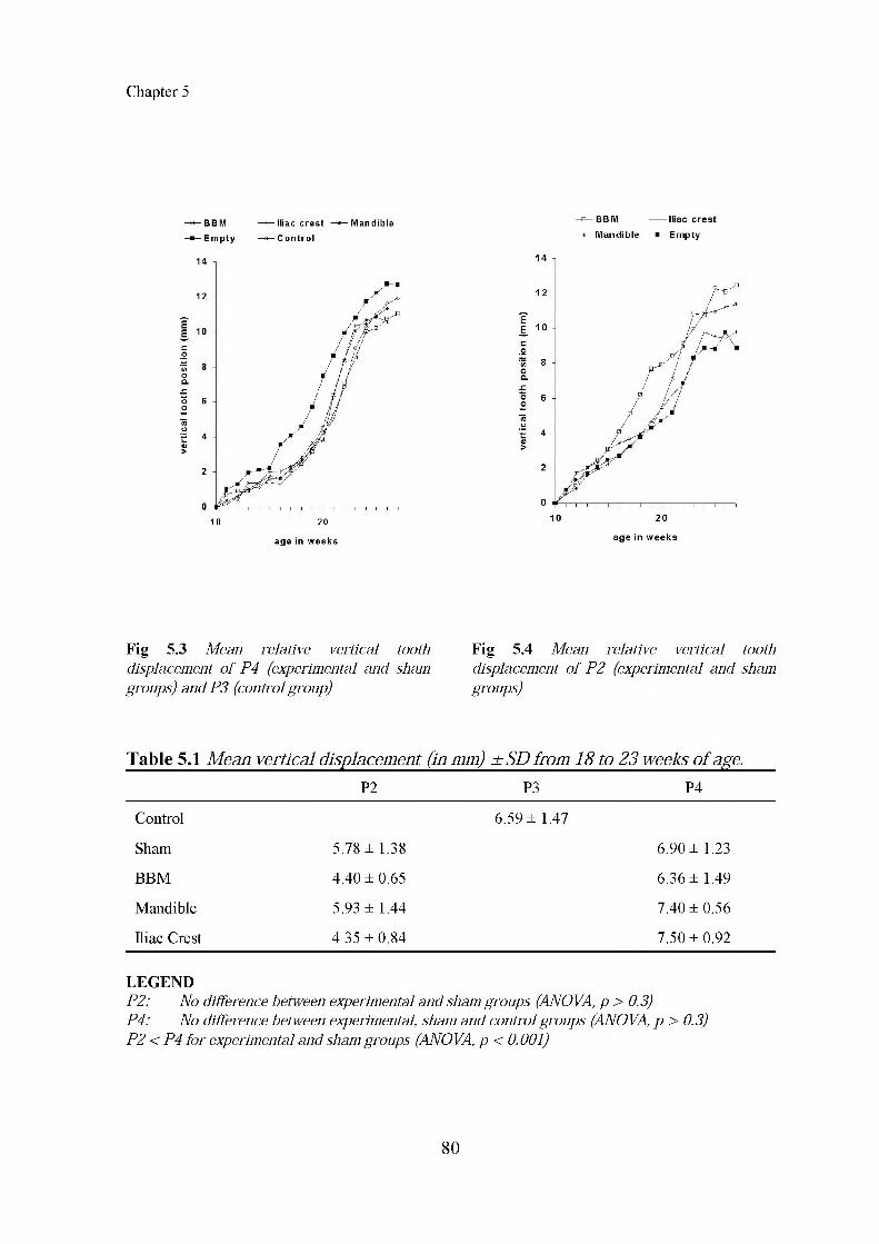
Chapter 5
age in w e e k s ag e in w e e k s
Fig 5.3 Mean relative vertical tooth F ig 5.4 Mean relative vertical tooth displacement o f P4 (experimental and sham displacement o f P2 (experimental and sham groups) and P3 (control group) groups)
Table 5.1 Mean vertical displacement (in mm) ± SD from 18 to 23 weeks of age.P2 P3 P4
Control 6.59 ± 1.47
Sham 5.78 ± 1.38 6.90 ± 1.23
BBM 4.40 ± 0.65 6.36 ± 1.49
Mandible 5.93 ± 1.44 7.40 ± 0.56
Iliac Crest 4.35 ± 0.84 7.50 ± 0.92
L EG EN DP2: No difference between experimental and sham groups (ANOVA, p > 0.3)P4: No difference between experimental, sham and control groups (ANOVA, p > 0.3) P2 < P4 for experimental and sham groups (ANOVA, p < 0.001)
80

Eruption through Autogenous and Xenogenous Bone Transplants
The eruption curve of the experimental P2 is not as uniform as the one of the P4, probably due to mesio-distal and linguo-buccal movements (fig 5.4). Although the vertical movement of the P2 in the period 18-23 weeks is slower than the P3 - P4 (ANOVA, p < 0.001), the rate of eruption shows no significant differences between the four experimental groups (ANOVA, p > 0.3).
Control sites: In these sites resorption of the m2 tooth roots and the eruption of the successive permanent P3 started 4 weeks after the beginning of the experiment. At an age of 21 weeks, the P3 erupted in the oral cavity and its roots were almost completely formed. The eruption lasts to a mean age of 23.1 ± 1.0 weeks. From that point the tip of the P3 remains stable in nearly all animals.
5.3.1 HistologyMandibular bone chips: In eighteen-week old beagle dogs (four weeks after transplantation) the cortical bone chips had a non-vital aspect with only some osteocytes in the lacunae and no signs of inflammation (fig 5.5). The surfaces of the grafts occlusal to the erupting tooth were integrated in the normal physiological process of resorption. The periodontal ligament was intact with a normal structure and orientation of the fibres. At an age of 24 weeks the teeth showed no delay of eruption or disruption with an attachment of the functional epithelium on the enamel-cement border. The implanted bone grafts were incorporated in the developing alveolar bone. After 30 weeks all premolars were erupted to their functional level with normal alveolar bone height, intact periodontal ligament, normal crown and roots of a normal length. No remnants of bone grafts were seen anymore.
Iliac crest bone chips: At an age of 18 weeks (four weeks after transplantation) the cancellous iliac crest bone chips were fully incorporated with osteons among the whole transplant, without signs of inflammation (fig 5.6). The structure and orientation of the parodontal ligament of the erupting tooth was normal. Some cartilage remnants, probably from the iliac crest, were seen. The bone grafts were integrated in the physiological resorption process occlusal to the erupting tooth. At an age of 24 weeks no abnormalities were seen. After 30 weeks the premolars had emerged with intact periodontal ligament, crown and roots. Remnants of the bone chips were integrated in the alveolar bone.
BBM: The number of BBM granules was reduced at 18 weeks (four weeks after transplantation) because connective tissue was lying between particles and they were not compressed anymore (fig 5.7 and 5.8). The granules were not yet incorporated, lying in the empty alveolus as well as occlusal to the alveolar bone
81

Chapter 5
CBC
Fig 5.5 Photomicrograph o f cortical bone chips four weeks after transplantation at an age o f 18 weeks showing the non-vital aspect o f the bone chips with only some osteocytes in the lacunae. (Haematoxylin and Eosin; x40). CBC = cortical bone chips, AB = alveolar bone
r
r ■ V- 1 . ..
T
V. A ' ; ■'
rrj*\ •
— '■ • 'vr-i' • 1 ,
• - ; \ ' ' ; AB• V . ■
250 A/m
i------------ 1
Fig 5.6 Photomicrograph o f spongious bone chips four weeks after transplantation at an age o f 18 weeks after transplantation showing the incorporated bone chips with osteons among them. Arrow indicates remnants o f cartilage (Haematoxylin and Eosin; x40). SBC = spongious bone chips, O = new osteon
Fig 5.7 Photomicrograph o f BBM particles four weeks after transplantation at an age o f 18 weeks showing the connective tissue layer around them without signs o f inflammation. Arrow indicates BBM particle (Haematoxylin and Eosin; x10). T = tooth, AB = alveolar bone
Fig 5.8 Detail o f fig 5.7. Osteoclast-like cells surrounding the particles (Haematoxylin and Eosin; x40)
82

Eruption through Autogenous and Xenogenous Bone Transplants
Fig 5.9 Photomicrograph o f BBM particles 16 weeks after transplantation at an age o f 30 weeks. Arrow indicates BBM particle (Haematoxylin and Eosin; x10). T = tooth, AB = alveolar bone
Fig 5.10 Photomicrograph o f one BBM particle 16 weeks after transplantation at an age o f 30 weeks showing the thin fibrous sheet around the particle lying in connective tissue, not integrated in the alveolar bone. Arrow indicates BBM particle. (Haematoxylin and Eosin; x40)
within connective tissue. Macrophages and osteoclast-like cells surrounded them. The attachment of the fibres of the periodontal ligament of the erupting tooth had a normal height at the enamel-cement border. At 24 weeks foreign body giant cells were seen in the vicinity of the particles without further signs of inflammation. The presence of the granules was restricted to the connective tissue occlusal to the enamel-cement border.
At an age of 30 weeks a thin fibrous sheath without an inflammatory response or osteoclastic activity surrounded most of the BBM particles (fig 5.9 and 5.10). Some of them were incorporated in the bone but most of them sat in connective tissue occlusal to the alveolar bone. The premolar eruption was undisturbed with a normal structure of the periodontal ligament, crown and roots.
Control: The eruption processes in the control group were not disturbed with a normal orientation and structure of the fibres of the periodontal ligament. In the period between extraction of the deciduous molar and emergence of its successor no alveolar bone was deposited in the empty alveolus.
83

Chapter 5
5.4 DISCUSSIONThere are still unanswered questions to the physiology of bone transplantation into the alveolar defect. One of these questions concerns the effect of the bone graft on the eruption of the permanent canine. In our institute chin bone, of ectomesenchymal origin, is the chosen grafting material (FREIHOFER et al. 1993). Membranous bone has several advantages over enchondral bone: it usually is removed with less donor site morbidity and less postoperative pain (TESSIER 1982, ROCHE and S c h w a r tz 1993, B A h r and COULON 1996) and it has been shown that it is less subject to postoperative remodelling and resorption (S m ith and A b ra m s o n 1974, Z in s and W h i th a k e r 1983, F e in b e rg and V i t t 1988, B A h r
and COULON 1996). In this experiment there were no indications of a slower eruption through the mandibular bone graft. Only the eruption through the 'empty' alveolus of the P4 seemed to start earlier than through the grafted alveolus.
Studies of premolar eruption in dogs have shown that it is accomplished primarily by localised resorption of alveolar bone on the coronal side of the tooth ( C a h i l l 1969, M a r k s and C a h i l l 1986, M a r k s et al. 1993), that the osteoclasts are activated on bone surfaces just prior to eruption (MARKS et al. 1983, W ise et al. 1985) and that eruption depends on the dental follicle proper, a thin collagenous investment of the crown of each developing tooth (CAHILL and MARKS 1980, MARKS and C a h i l l 1984, M a r k s et al. 1993). This cell-mediated process results in a local tuning of the erupting tooth and the overlying alveolar bone.
In our experiment the erupting premolars are able to resorb the cancellous iliac crest and cortical mandibular bone grafts without any difference to each other and to the control sites. The number of BBM granules is reduced, although we did not see resorption sites. The presence of osteoclasts on the surface of BBM granules at 4 and 10 weeks after surgery as observed by us and KLINGE et al. (1992) only suggest resorption in a more physiological manner. Most of the BBM granules are pushed forward into the soft tissues or probably discharged into the oral cavity during the eruption. Maybe more time for incorporation is needed in comparison with autologous bone transplants because of the lack of osteoinductive capacity ( T h a l l e r et al. 1994).
The results of our study are in accordance with a study which showed that the effect of four kinds of calcium phosphate ceramic particle insertion (hydroxyapatite (HA) dense and porous, and tricalciumphosphate (TCP) dense and porous) on tooth eruption in 20 3-month old dogs gave no delay of tooth eruption, no dysplasia and no resorption of dental hard tissue (SUGIMOTO et al. 1993). Studies on the effect of calciumphosphate on tooth eruption in forty 3- to 4-month old
84

Eruption through Autogenous and Xenogenous Bone Transplants
kittens indicated that the use of a nonresorbable HA for grafting, however, resulted in impediment of eruption and distortion of crown development. This was in contrast to the TCP, which had a minimal effect on tooth eruption and development (F e in b e r g and V it t 1988, F e in b e r g et al. 1989). So the uniformity of eruption curves in our experimental (and control) groups indicates that BBM may be considered as a resorbable calcium phosphate.
5.5 CONCLUSIONThe results of this study indicate that the use of cortical mandible bone or spongeous iliac crest bone in residual alveolar clefts has no negative influence on the eruption of permanent teeth.
The number of BBM particles decreased during eruption. Probably the time interval between insertion of the particles and the start of the eruption is too short for incorporation into the bone because of the lack of osteoinductive capacity. BBM particles separated from new bone are pushed into peripheral soft tissue or discharged into the oral cavity at the time of eruption. Iliac crest bone, mandibular bone and BBM, when implanted into extraction sockets of deciduous teeth, have no inhibitory effect on eruption and crown development of the underlying permanent mandibular premolars. Before using this xenogene bone mineral in alveolar clefts, more has to be known of the healing process in osseous defects, resembling the volume of alveolar clefts.
5.6 LITERATURE• BAHR W , COULON JP. Limits o f the mandibular symphysis as a donor site for bone grafts
in early secondary cleft palate osteoplasty. Int J Oral M axillofac Surg 1996; 25: 389-93• BJORK A. Facial growth in man, studied with the aid o f metallic implants. A cta Odontol
Scand 1955; 13: 9-34• BORSTLAP W .A, HEIDBUCHEL KL, FREIHOFER HPM, KUIJPERS-JAGTMAN AM. Early
secondary bone grafting o f alveolar cleft defects. A comparison between chin and rib grafts. J Cranio M axillofac Surg 1990; 18: 201-5
• BOYNE PJ. Comparison o f Bio-Oss® and other implant materials in the maintenance o f the alveolar ridge o f the mandible in man. Luzern, Switzerland. International symposium on "Modern Trends in Bone Graft Substitute Materials" M ay 11, 1990
• Cah ill DR, MARKS SC. Tooth eruption: evidence for the central role o f the dental follicle. J Oral Pathol 1980; 9: 189-200
• CAHILL DR. Eruption pathway formation in the presence o f experimental tooth impaction in puppies. Anat Res 1969; 164: 67-78
• Cohen M, Figueroa AA, Haviv Y, S chafer ME, Aduss H. Iliac versus cranial bone for secondary grafting o f residual alveolar clefts. Plast Reconstr Surg 1991; 87: 423-7
• FEINBERG SE, V itt M. Effect o f calcium phosphate ceramic implants on tooth eruption. J Oral M axillofac Surg 1988; 46: 124-7
85

Chapter 5
• FEINBERG SE, WEISBRODE SE, HEINTSCHEL G. Radiographic and histological analysis of tooth eruption through calcium phosphate ceramics in cats. Archs Oral Biol 1989; 34: 975-84
• FREIHOFER HPM, BORSTLAP WA, KUIJPERS-JAGTMAN AM, VOORSMIT RACA, DAMME PA van, HEIDBUCHEL KLWM, BORSTLAP-ENGELS VMF. Timing and transplant materials for closure of alveolar clefts: a clinical comparison of 296 cases. J Cranio Maxillofac Surg 1993; 21: 143-8
• HISLOP WS, FINLAY PM, MOOS KF. A preliminary study into the uses of anorganic bone in oral and maxillofacial surgery. Br J Oral Maxillofac Surg 1993; 31: 149-53
• HOPPENREIJS TJ, NIJDAM ES. FREIHOFER HP. The chin as a donor site in early secondary osteoplasty: a retrospective clinical and radiological evaluation. J Cranio Maxillofac Surg 1992; 20: 119-24
• KALAAJI A, LILJA J, FRIEDE H, ELANDER A. Bone grafting in the mixed and permanent dentition in cleft lip and palate patients: long-term results and the role of the surgeon's experience. J Cranio Maxillofac Surg 1996; 24: 29-35
• KLINGE B, ALBERIUS P, ISAKSSON S, JONSSON J. Osseous response to implanted natural bone mineral and synthetic hydroxyapatite ceramic in the repair of experimental skull bone defects. J Oral Maxillofac Surg 1992; 50: 241-9
• KOOLE R, BOSKER H, DUSSEN FN VAN DER. Late secondary autogenous bone grafting in cleft patients comparing mandibular (ectomesenchymal) and iliac crest (mesenchymal) grafts. J Cranio Maxillofac Surg. 1989; 17 Suppl 1: 28-30
• KOOLE R, VISSER WJ, KLEIN WR, SUIKER AMH. A Comparative investigation on autologous mandibular and iliac crest bone grafts: an experimental study in sheep. J Cranio Maxillofac Surg 1991; 19: 133-43
• KORTEBEIN MJ, NELSON CL, SADOVE AM. Retrospective analysis of 135 secondary alveolar cleft grafts using iliac or calvarial bone. J Oral Maxillofac Surg. 1991; 49: 493-8
• KUSIAK JF, ZINS JE, WHITHAKER LA. The early revascularization of membranous bone. Plast Reconstr Surg 1985; 76: 510-4
• LINDEN FPGM van DER, BOERSMA H, Peters KA, RAABEN J. Three dimensional analyses of dental casts by means of the optocom. J Dent Res 1972; 51: 1100
• MALTHA JC. The process of tooth eruption in beagle dogs. Nijmegen, Thesis 1982.• MARKS SC, GORSKI JP, Wise GE. The mechanisms and mediators of tooth eruption -
Models for developmental biologists. Int J Dev Biol 1993; 39: 223-30• MARKS SC, Cahill DR. The Ultrastructure of alveolar bone during tooth eruption. Am J
Anat 1986; 177: 427-38• MARKS SC, Cahill DR. Experimental study in the dog of the non-active role of the tooth
in the eruptive process. Archs Oral Biol 1984; 29: 311-22• MARKS SC, Cahill DR, Wise GE. The cytology of the dental follicle and adjacent
alveolar bone during tooth eruption in the dog. Am J Anat 1983; 168: 277-89• ROCHE YA, Schwartz HC. The mandibular body bone (MBB) graft: an alternative
source of membranous bone. J Cranio Maxillofac Surg 1993; 21: 199-201• SMITH JD, Abramson M. Membranous versus endochondral bone autografts. Arch
Otolaryngol 1974; 99: 203-5• SUGIMOTO A, OHNO K, MICHI K, KANEGAE H, AIGASE S, TACHIKAWA T. Effect of
calcium phosphate ceramic particle insertion on tooth eruption. Oral Surg Oral Med Oral Pathol 1993; 76: 141-8
• TESSIER P. Autogenous bone grafts taken from the calvarium for facial and cranial applications. Clin Plast Surg 1982; 9: 531
• THALLER SR, Hoyt J, DART A, BORJESON K, TESLUK H. Repair of experimental calvarial defects with Bio-Oss particels and collagen sponges in a rabbit model. J Craniofac Surg 1994; 5: 242-6
86

Eruption through Autogenous and Xenogenous Bone Transplants
WETZEL AC, Stic h h , Ca ffesse RG. Bone apposition onto oral implants in the sinus area filled with different grafting materials. A histological study in beagle dogs. Clin Oral Impl Res 1995; 6: 155-63WIJDEVELD MGMM, MALTHA JC, GRUPPING EM, JONGE J DE, KUIJPERS-JAGTMAN AM. Tissue response to palatal surgery at different ages on Beagles dogs. Archs Oral Biol 1991; 36: 837-43.WISE g e , Mark s s c , Ca h ill DR. Ultrastructural features o f the dental follicle associated with formation o f the tooth eruption pathway in the dog. J Oral Pathol 1985; 14: 15-26W itsen bu rg B., H.J. Rem m elin k . Reconstruction o f residual alveolo-palatal bone defects in cleft patients. A retrospective study. J Cranio M axillofac Surg 1993; 21: 239-44 WITSENBURG B, Fr EIHOFER HPM. Autogenous rib graft for reconstruction o f alveolar bone defects in cleft patients. Long-term follow-up results. J Cranio M axillofac Surg 1990; 18: 55-62Z iNS JE, W hITHAKER LA. M embranous versus enchondral bone: implications for craniofacial reconstruction. Plast Reconstr Surg 1983; 72: 778-84
87

CHAPTER 6
ANORGANIC BONE MINERALS IN ORAL AND MAXILLOFACIAL SURGERY:
A LITERATURE REVIEW
Matthias A.W. Merkx Jaap C. Maltha
Hans Peter M. Freihofer
Submitted
89

Chapter 6
ABSTRACTA variety of anorganic alloplastic grafting materials are used in the oral- and maxillofacial (OMF) area to bridge osseous defects or to create adequate bone- volume for placement of endosseous implants. Because of the variation in literature data about the effectiveness of these materials in reconstructive procedures of the maxilla in comparison with autogenous bone grafts, it is difficult to make protocols and select proper indications. A literature search in ‘Medline’ from 1989-1999 was performed to get a better insight into the pros and cons of the different modalities. Studies in which different graft materials were used in contact with the maxillary sinus with histomorphometric data were selected. Eight studies met the inclusion criteria: in 6 studies bone grafts were used in sinus floor elevation procedures and in 2 studies osteotomy gaps were bridged. It is concluded that autogenous bone gives the highest amount of osteoid volume, independent from the healing interval (1-24 months), application form (particulate versus block) and donor site (iliac crest versus chin). The results of inorganic calcium phosphate materials (bovine bone mineral or hydroxyapatite), solitary or as additive in composite grafts with autogenous particulate bone or demineralized freeze dried bone, shows a large range of values, but in all cases the results are inferior to those of autogenous bone grafts. The data on anorganic calcium phosphate materials cannot be compared mutually, because of differences in healing interval, mixture ratios and histological procedures, but they do indicate that the use of composite grafts is to be preferred over the use of solitary anorganic bone mineral.
90

Anorganic Bone Minerals: a Review
6.1 INTRODUCTIONBone grafting in oral- and maxillofacial (OMF) surgery is a common procedure. Well-known indications for osseous reconstructive surgery are the augmentation of the floor of the maxillary sinus in the severely atrophic posterior maxilla, the reconstruction of the alveolus in cleft palate patients, bridging osteotomy gaps or reconstruction of (post) traumatic osseous defects. Various therapeutic protocols, surgical techniques, and materials are used. The debate still continues about the best graft material and the proper indications and protocols (KENT and BLOCK 1989, C h a n a v a z 1990, P e t t i s et al. 1990, R e ic h e and G a r k 1991, W a g n e r 1991, CRANIN et al. 1993, MOY et al. 1993, VLASSIS et al. 1993, H U r z e le r et al. 1997). Interpretation of literature is hampered by personal preferences of the authors which hinders direct comparison of studies concerning human bone grafting in the OMF field. Nevertheless such a comparison may be worthwhile for the development of a consistent protocol to optimise the success of surgery in the long term. The threedimensional form of the defect, the vitality and quality of the (newly formed) bone and the subsequent success of implantology, orthodontics, and stability and bone continuity dictates the protocol of choice after orthognathic and trauma surgery.
In the past, autografts were the only material of choice for bridging an OMF osseous defect (HABAL 1992). Bone substitutes nowadays are commonly used to minimise donor site morbidity and to diminish the length of hospitalisation. The amount of osteoid in the grafted area is used as an indication of success of the procedure. This can be measured by histomorphometric analyses of core biopsies. Such evaluation is not possible in orthognathic surgery, trauma cases or bridged cleft alveolus, as in these cases a re-entry into the acceptor site has to be avoided and thus standardized biopsies are not taken. In preprosthetic surgery, however, histologic data can be obtained during implant- or abutment placement after several months of graft healing and so information about bone quality can be provided. This could be extrapolated to an indication for the success of bone grafting in orthognathic-, trauma- and cleft palate surgery, e.g. for the stabilisation of fractures and the ability to withstand the stresses induced through mastication.
The majority of the reports on the augmentation of the bony floor of the maxillary sinus in humans, have been in the form of clinical reports on implant survival over a given period with autogenous bone graft (ABG) from the iliac crest (KENT and BLOCK 1989, LOZADA et al. 1993, NYSTROM et al. 1993, RAGHOUBAR et al. 1993, CAWOOD et al. 1994, KELLER et al. 1994, MISCH and DIETSH 1994, GUNNE et al. 1995, BLOMQVIST et al. 1996, 1998a, CHAN et al. 1996, BLOCK and KENT 1997, DAELEMANS et al. 1997, LUNDGREN et al. 1997, 1999, SCHLIEPHAKE et
91

Chapter 6
al. 1997, JENSEN and S e n n e rb y 1998), bone substitutes such as allografts (S m ile r et al. 1992, FUGAZZOTTO 1994, VALENTINI and A b e n s u r 1997, JENSEN and S e n n e rb y 1998), hydroxy-apatite (HA) (W a g n e r 1991, S m ile r et al. 1992, S m a l l et al. 1993, F u g a z z o t t o 1994, H ü r z e l e r et al. 1996, W h e e le r 1996, W h e e le r et al. 1997) and bovine bone mineral (BBM) (S m ile r et al. 1992, HÜRZELER et al. 1996, VALENTINI and ABENSUR 1997) or combinations of different kinds of materials ( T id w e l l et al. 1992, MOY et al. 1993, SMALL et al. 1993, H ü r z e l e r et al. 1996, KAPTEIN et al. 1998).
Evaluations of survival rates only are not indicative for the quality of the bone graft ( W h e e le r et al. 1996, B lo m q v is t et al. 1998b, T o n g et al. 1998). For the evaluation of bone quality histomorphometric analyses of core biopsies are indicated as they enable quantification of bone healing and remodelling. The purpose of this paper is to review the literature of histomorphometric data on different anorganic grafting materials used in the OMF sinus area in humans in comparison to autogenous bone. These data will be compared with data derived from animal experiments because these models are in principle accurate and reproducible although they will not always completely imitate the human situation. A meta-analysis of the literature would be preferable as it offers a structured method to systematically identify, review and analyse published reports. However, due to variations in experimental set-ups and data reporting, this appeared to be impossible.
6.2 LITERATURE SEARCHTwo reviewers (MM, JM) searched Medline from 1989-1999 for literature
on histological evaluation of bone grafts in sinus, facial, orofacial and maxillofacial surgery in human studies in which anorganic matrices such as HA, coral or bovine bone mineral (BBM) were used, in the English literature. The reviewers used the following criteria: Reports about clinical histomorphometric analysed bone cores after sinus floor bone grafting or bridging osteotomy gaps were accepted, despite differences in histological techniques. In some instances older literature was included in order to clarify the background of choices and decisions reported.
‘One area grafted’ case-reports and papers only concerning the osseointegration of dental implants, the quality of the sinus epithelium, or the treatment of periodontal defects were excluded. Evaluations of bone grafts or substitutes in combination with membranes for guided bone regeneration were also excluded. Furthermore clinical studies dealing with the function of osteoinductive proteins (BMP’s) in graft healing were not evaluated because they are not admitted
92

Anorganic Bone Minerals: a Review
for clinical use by the American Food and Drug Administration (FDA) and the research is (therefore) still in a phase II stadium with unpredictable results ( G r o e n e v e ld et al. 1999b).
6.3 CORE PUBLICATIONSEight clinical papers reporting on anorganic or autogenous grafting materials in contact with the maxillary sinus (6 sinus floor grafts and 2 osteotomy gaps), were found in which biopsies, taken before implant or abutment placement, were evaluated histomorphometrically (table 6.2) (MOY et al. 1993, NUNES et al. 1996, W h e e le r et al. 1996, A y e rs et al. 1998, B lo m q v is t et al. 1998b, L o r e n z e t t i et al. 1998, FROUM et al. 1998, HANISCH et al. 1999). A y e rs et al. (1998) and N u n e s et al. (1996), from the same institute, determined the long-term bone ingrowth into and -apposition along HA porous implants placed to bridge osteotomy gaps. BLOMQVIST et al. (1998b) tried to determine the importance of bone graft quality for implant integration after maxillary sinus reconstruction with iliac ABG. FROUM et al. (1998) reported the data of 34 sinus floor elevations that used BBM alone (n=5) or in combination with ABG (n=7) or demineralized freeze-dried bone allograft (DFDBA) (n=8) or a combination of these three materials (n=14) as a graft material. The results of their 79 sinus floor elevations which membranes were used for guided bone regeneration are not discussed. HANISCH et al. (1999) evaluated mineralization stages and processes during bone healing 6, 8, 10 or 12 months after allogenic-xenogenic bone grafting sinus augmentation procedures in 20 patients. LORENZETTI (1998) discussed in a ‘preliminary’ report the bone augmentation of the inferior floor of the maxillary sinus with ABG (chin block (n=3), chin particulate (n=3), iliac crest (n=8)) or a composite (HA/ABG chin) (n=3) bone graft. MOY et al. (1993) used histomorphometric analysis to quantitate the bone composition of a corticocancellous chin bone block (n=1), HA (n=1), a composite of these two materials (1:1) (n=4) and HA mixed with demineralized bone powder (n=1) in maxillary sinus augmentation. The single cases will not be discussed. W h e e le r et al. (1996) evaluated the results of 36 sinusfloor augmentations with HA (n=16), HA/oral ABG (n=12), HA/iliac crest ABG (n=6), BBM (n=1) and iliac crest ABG (n=1). Because case reports were excluded the last two materials are not discussed. Several aspects of these papers will be discussed in relation to other literature.
93

Table 6.1 Histometric evaluated healing intervals in months of grafts implanted in human maxillary sinus.Literature A yers Blo m q v ist FROUM Ha n isc h LORENZETTI M oy Nun es W h e ele r
et al. 1998 et al. 1998 et al. 1998 et al. 1999 et al. 1998 et al. 1993 et al. 1997 et al. 1996
Grafting materialA BG chin block 10
A BG chin particulate 4
A BG iliac bone 1, 2, 4, 6, 12 6
BBM 6
BBM /ABG 6
BBM /ABG/DFDBA 6
BBM /DFDBA 6 6, 8, 10, 12
HA 4 - 3 6 [19] 1 4 - 3 0 [19] 6
HA/ABG chin (1:1) 12 6, 10
HA/ABG iliac crest (1:4) 6
HA/ABG oral (4:1) 6
L EG EN DABG = Autogenous Bone Graft BBM = Bovine Bone MineralDFDBA = Demineralized Freeze-Dried Bone AllograftHA = HydroxyapatiteDBP = Demineralized Bone Powdern = Months

Table 6.3 Osteoid volume in % of total field after sinus floor elevation procedures.Grafting material
Versus timeStandard 1 mns 2 mns 4 mns 6 mns 8 mns 10 mns 12-24 mns n Author
A BG chin block 69.3 3 LORENZETU et al. 1998A BG chin particulate 62.6 3 LORENZETU et al. 1998A BG iliac bone 53.0 8 LORENZETU et al. 1998A BG iliac bone 41 23 38 33 26 4, 5, 6, 4, 2 Blo m q v ist et al. 1998A lveolar ridge residual 32.6 17 Ha n isc h et al. 1999BBM 8.5 5 FROUM et al. 1998BBM /ABG 18.5 7 FROUM et al. 1998BBM /ABG/DFDBA 19 14 FROUM et al. 1998BBM /DFDBA 14 8 FROUM et al. 1998BBM /DFDBA 8.1 9.0 11.8 20.7 5, 5, 5, 5 Ha n isc h et al. 1999HA 16.4 16 W h eeler et al. 1996HA 27 8 NUNES et al. 1997HA 23 17 AYERS et al. 1998HA/ABG chin (1:1) 43.6 3 LORENZETTI et al. 1998HA/ABG chin (1:1) 44.4 4 MOY et al. 1993H A/A BG iliac crest (1:4) 19.3 6 W h eeler et al. 1996HA/ABG oral (4:1) 11.3 12 W h eeler et al. 1996
LEGENDABG = Autogenous Bone Graft BBM = Bovine Bone MineralDFDBA = Demineralized Freeze-Dried Bone AllograftHA = HydroxyapatiteDBP = Demineralized Bone Powdern = number of grafted sinuses in each group

Chapter 6
6.3.1 HistologyEvaluation of the core literature is hampered by the variation in histological techniques used. Histomorphometrical evaluation of bone is performed in most cases by some kind of grid method. In this method a grid is placed over the microscopical image. By counting the number of points or measuring the length of the lines that overlie the item of interest, the volume of that item can be estimated. The value of bone histomorphometry is dependent on impeccable tissue preservation and proper section thickness. The effect of tissue thickness refers to an increase in the visualisation of tissue compartments as section thickness increases (HOLMES and ROSER 1987). In general, as the specimen thickness is increased, the accuracy of the histomorphometric assessment decreases. The optimal thickness of specimen is 4.5 - 5 ^m (JOWSEY 1977). Another method of quantitative analysis used the backscattering scanning electron microscopy. This technique does not depend on sectioning of the tissue (HOLMES and ROSER 1987). Both techniques are used in the core articles (table 6.2). In 3 papers 30-45 ^m histologic sections were used (MOY et al. 1993, W h e e le r et al. 1996, FROUM et al. 1998), while in the other studies 10-20 ^m (HANISCH et al. 1999), 7 ^m (LORENZETTI et al. 1998) or 4 ^m (BLOMQVIST et al. 1998b) section thickness was used. In the studies in which HA bone grafts were placed to bridge osteotomy gaps, 400 ^m sections were ground (N u n es et al. 1997, A y e rs et al. 1998). In spite of these differences and because of the absence of other, more uniform literature, we put these data together knowing the results are only indicative without secure value.
The amount of osteoid from the residual alveolar process, which has been undisturbed during the grafting procedure as measured by Hanisch et al. (1999), was 32.6%. This datum was used as an internal standard. The values in table 6.3 can be related to this standard as far as measurements with the backscatter method are concerned (table 6.2). This can give an indication of the vitality of different grafting materials used in clinical sinus floor elevation procedures. Because of the difference in patient numbers and time intervals between implantation and biopsy, no definite conclusion can be drawn. Most data are available at a six months healing time interval. Histomorphometric analyses of ABG in the core publications gave the highest amount of bone content in the grafted area (23.0% - 69.3%) independent of the interval used for healing. No clear difference could be detected between different donor sites and application forms (chin versus iliac crest or block versus particulate). The histomorphometric values of composite grafts consisting of ABG and an anorganic mineral (HA or BBM) showed no difference in the amount of bone in the grafted area (11.3% - 19%), whereas HA alone (16.4% - 20.3%)
96

Inorganic Bone Materials: a Review
differed slightly from BBM alone (8.5%) (table 6.3). All composite grafts have a lower osteoid content (8.1% - 19.3%) than the residual alveolar ridge (32.6%). The histomorphometric data in the core studies on composites also have a lower osteoid content than ABG. It seems that ABG’s are in favour to composites as they show at least temporarily a higher osteoid content than the residual alveolar ridge.
In animal studies in which grafts consisting of cancellous ABG/BBM (1:1) were used. Good results were obtained, combining the advantages of each material alone and reducing the disadvantages of each when used separately (M e rk x et al. 2000).
Table 6.2 Techniques used for histomorphometric analyses in core papers.A u th o r Y ear Slide T hickness (^.m) H istom orphom etric T echnique
Ayers et al. 1998 400 - grinding Histomorphometry
BLOMQVIST et al. 1998 4 Histomorphometry
FROUM et al. 1998 35-45 Histomorphometry (computerized)
Hanisch et al. 1999 10-20 Backscatter electron microscopy
LORENZETTI et al. 1998 7 Histomorphometry
MOY et al. 1993 30-40 Backscatter electron microscopy
NUNES et al. 1997 400 - grinding Histomorphometry
Wheeler et al. 1996 30-40 Backscatter electron microscopy
6.3.2 Healing periodAnother problem in the evaluation of the core publications is the difference of time interval between bone grafting and biopsy taken between the studies and sometimes even during a research project. For example, LORENZETTI et al. (1998) evaluated the bone quality after sinus elevation procedures of four different grafting materials (iliac crest bone blocks, chin bone blocks, particulate chin bone and composite chin/HA (1:1)). The ‘iliac crest bone’ group was biopted after a 6 months’ healing period “to limit the graft volume resorption that has been reported with iliac crest bone grafts” (S m ith and A b ra m s o n 1974, B u r c h a r d t 1987, N y s t r o m et al. 1995). The ‘chin bone block’ group was biopted after 8-12 months “to achieve sufficient revascularization and viability of this type of graft, essentially consisting of compact bone” (URIST 1980, BURCHARDT 1987). The ‘particulate chin bone’ group was biopted after 4 months, “since the original compact bone structure had been removed by the particulation process and graft volume could be presumed to occur during the first phase of healing” (SHIROTA et al. 1996). The healing for the
97

Chapter 6
fourth group (chin ABG/HA (1:1)) was prolonged to 12 months to “allow adequate new bone formation, as indicated for this type of graft” (MOY et al. 1993).
Only two prospective studies evaluated the mineralization stage with the same grafting material at various time intervals (BLOMQVIST et al. 1998b, HANISCH et al. 1999). HANISCH et al. (1999) used BBM/DFDBA and evaluated it after 6, 8, 10 and 12 months. In their study no statistically significant difference was seen between the amount of newly formed bone in the grafted areas during the different intervals was seen. They concluded that the mineralization process of this composite is already complete after six months, but new bone formation continues at least up to 12 months resulting in a higher osteoid content (table 6.3). At that time, however, the amount of newly formed bone was still lower than the volume of residual bone. BLOMQVIST et al. (1998b) reported a highly increased bone remodelling (deposition and resorption) activity already 2 months after grafting and concluded that an extension of the healing period by an additional 2 or 4 months would not yield any advantage but may rather induce progressive bone resorption.
In other human and animal studies different measures were used for healing capacity and also time scales differed (LUNDGREN et al. 1999). In some human studies it appears that a period of six or even twelve months is too short to allow complete incorporation of the bone graft (NYSTROM et al. 1993; A y e rs et al. 1998). When HA is implanted solitarily, the amount of bone present within the implant increases with implantation time and appears to reach a plateau around the 20 months time frame (A y e rs et al. 1998). In course of time the soft tissue gave way to primarily woven and lamellar bone. If cancellous chips, with or without an anorganic additive, are implanted in a frontal sinus wall defect of goats, bone graft ingrowth is accomplished within 12 weeks (MERKX et al. 1999b, 2000). Extension of the follow-up period with another 12 weeks did not seem to result in a better bone quality. This kind of studies revealed that new bone formation in grafts in contact with the maxillary sinus varies considerably depending on bone graft type and healing time.
6.3.3 MaterialsTwelve different (combinations of) materials are used in the core publications and evaluated at different time intervals (1-24 months) (table 6.1). The only anorganic bone substitutes used in the core studies are BBM and HA. Their action is based on osteoconduction, which means that new bone formation takes place along their surface. In one study also DFDBA is used in composites. DFDBA may result in both osteoinduction and osteoconduction (MISCH and DIETSH 1993), although the
98

Inorganic Bone Materials: a Review
inductive capacity is disputable (GROENEVELD et al. 1999). It is generally believed that BMP’s and other non-collagenous proteins in the matrix are responsible for the osteoinduction of this material (Z h a n g et al. 1997).
Particulate bone material is easily and conveniently obtained as a surplus material when harvesting major grafts or during osteotomies. It may be used to fill small defects, to correct minimal contour irregularities and for packing around larger grafts. Also particulate bone can easily be mixed with anorganic bone additives. LORENZETTI et al. (1998) evaluated the differences in behaviour of blocks versus particulate materials. They suggest that graft architecture, conformation, and composition may influence the mean bone content at the end of the healing. In an animal study it was stated that autogenous cancellous bone chips are the material of choice for bridging a bony defect in the maxillofacial area when there is no need for mechanical strength. They heal in the same way as cancellous bone blocks do and are easily and conveniently obtained as surplus material when producing major grafts or performing osteotomies (MERKX et al. 1999a, b).
The combination of an anorganic bone mineral with autogenous bone grafts, can provide an additional stimulus for osteoconduction and as an expander (HABAL 1991, LINDHOLM et al. 1994). The ideal composite ratio is still unknown, as no research has pointed out this parameter in detail. However, the different ratios of composite mixtures may be of influence on the results. An ABG/HA ratio varying from 1:1 to 1:2 is recommended to avoid excessive fibrous encapsulation of the hydroxyapatite granules (COBB et al. 1990; TIDWELL et al. 1992). In one of the core papers the mixture rates differed: W h e e le r et al. (1996) said there “usually was a mixture of 4:1 ratio of HA to autogenous intraoral bone whereas the reverse was true when using bone harvested from the iliac crest”. An argument for this difference is not given. When the ratio of anorganic bone mineral versus ABG increased, the radiological evaluation showed that resorption of the bone additive decrease exponentially (HORCH and STEEGMANN 1985).
In an animal histological study it is shown that cancellous ABG/BBM (1:1) composite grafted in frontal sinus wall defects showed a stable situation after 12 weeks and that the BBM particles had become replaced to a large extent by newly formed bone due to physiological remodelling. After 24 weeks the same features were seen, but BBM particles could hardly be detected anymore (MERKX et al. 2000).
DFDBA showed lower osteoid contents after 6 months than the autogenous grafts (FROUM et al. 1998, HANISCH et al. 1999). It was observed that formation of
99

Chapter 6
mature lamellar bone in these kinds of grafts took at least twice as much time as in autogenous bone grafts (JENSEN and S e n n e rb y 1998).
Two differences between HA and BBM may be important for the clinician. First, the three-dimensional structure, including the porosity of the grafting material, which has an important effect on bone healing (HOLMES 1987, KASPERK et al. 1988, DACULSI and PASSUTI 1990, VAN EEDEN and RIPAMONTI 1994, HÄMMERLE et al. 1997). Materials exhibiting large surface areas showed better bone-graft contact than materials with comparatively small surface area (HOLMES and ROSER 1987, KASPERK et al. 1988). The surface area of each graft particle of BBM is considerably larger than that of porous bioceramics (e.g. HA) (HÄMMERLE et al. 1997). Therefore it might be a better grafting material. The second reason is that sintered HA particles, used as a space filler, are not substituted with normal bone because of their irresorbability. The use of porous HA blocks might result in mechanical fragile areas if used to fill defects in the cortex of long bones or thin mandibles (YAMAGUCHI et al. 1995). BBM is prone to osteoclastic resorption (PINHOLT et al. 1991, MERKX et al. 1999a, b) although other studies refute this (MANDELKOW et al. 1990, STASSEN et al. 1994, SKOGLUND et al. 1997, YOUNG et al. 1999). The resorption of BBM finally leads to a physiological substitution and remodelling which could be an advantage in favour for BBM. However studies about this specific subject are lacking so far.
6.4 CONCLUSIONAllogenic (ceramic) bone replacing materials as reported in literature, which all consist of calcium phosphate complexes, differ in origin (xenogenic, ceramic or coral), structure (pore size, absolute surface area and interconnections), chemical composition and finally (therefore) in resorbability. All these materials are supposed to provide a scaffold for bone ingrowth by means of osteoconduction which is enabled by the establishment of an environment allowing early migration of osteoblasts or osteoprogenitor cells into the area in which bone regeneration is intended (HÄMMERLE et al. 1997).
The successful regeneration of bone requires cells that will produce osteoid, an appropriate scaffold upon which the new bone is deposited and bioactive molecules to modulate the process. The host site should, of course, provide a vascular supply and nutrients. The transplant has to meet some mechanical requirements in order to approximate the mechanical properties of the bone that is replaced in the reconstructed area. The anatomy, functional loading, and chemical circumstances of a particular reconstruction dictate the exact mechanical
100

Inorganic Bone Materials: a Review
requirements. Technology is now able to apply each of the required bone regeneration components separately (HOLLINGER 1993, YAZEMSKI et al. 1996).
Anorganic bone substitutes as solitary graft material, do not contribute to an early bone ingrowth and do not fulfil the requirements of an early functional loading. If it is necessary to use anorganic bone additives, the material that reestablishes the integrity of the resorbed or injured maxillofacial skeleton should be a temporary material and be mixed with autogenous bone of cancellous origin. This is necessary because only then, the bone that is formed is soon able to bear functional load e.g. of dental implants, chewing or orthodontic forces. Consideration should be given to the intended clinical use of the materials. Prospective clinical studies should start to compare the healing response of different (anorganic) grafting materials at different acceptor sites in the maxillofacial area. This can provide information about the ideal graft composition, the optimal healing time, the value of anorganic additives, the effect of functional loading and, in the future, the impact of the use of osteoinductive proteins.
6.5 LITERATURE• AYERS RA, Simske SJ, Nunes CR, Wolford LM. Long-term bone ingrowth and
residual microhardness of porous block hydroxyapatite implants in humans. J Oral Maxillofac Surg 1998; 56: 1297-301
• BLOCK MS, KENT JN. Sinus augmentation for dental implants: the use of autogenous bone. J Oral Maxillofac Surg 1997; 55: 1281-6
• BLOMQVIST JE, ALBERIUS P, ISAKSSON S. Retrospective analysis of one-stage maxillary sinus augmentation with endosseous implants. Int J Oral Maxillofac Impl 1996; 11: 51221
• BLOMQVIST JE, ALBERIUS P, ISAKSSON S. Two-stage maxillary sinus reconstruction with endosseous implants: a prospective study. Int J Oral Maxillofac Impl 1998a; 13: 758-66
• BLOMQVIST JE, ALBERIUS P, ISAKSSON S, LINDE A, OBRANT K. Importance of bone graft quality for implant integration after maxillary sinus reconstruction. Oral Surg Oral Med Oral Pathol Oral Radiol Endodont 1998b; 86: 268-74
• BURCHARDT H. Biology of bone transplantation. Orthop Clin North Am 1987; 18: 187-96• CAWOOD JI, STOELINGA PJ, BROUNS JJ. Reconstruction of the severely resorbed (Class
VI) maxilla. A two-step procedure. Int J Oral Maxillofac Surg 1994; 23: 219-25• CHANAVAZ M. Maxillary sinus: anatomy, physiology, surgery, and bone grafting related
to implantology--eleven years of surgical experience (1979-1990). J Oral Implantol 1990; 16: 199-209
• CHAN MF, Howell RA, Cawood JI. Prosthetic rehabilitation of the atrophic maxilla using pre-implant surgery and endosseous implants. Brit Dent J 1996; 181: 51-8
• Cobb CM, EICK JD, Barker BF, Mosby EL, HIATT WR. Restoration of mandibular continuity defects using combinations of hydroxyapatite and autogenous bone: microscopic observations. J Oral Maxillofac Surg 1990; 48: 268-75
• CRANIN AN, RUSSELL D, Andrews JP, MEHRALI M. Immediate implantation into the posterior maxilla after antroplasty: the Cranin-Russell Operation. J Oral Implantol 1993; 19: 143-50
101

Chapter 6
• DACULSI G, PASSUTI N. Effect o f the macroporosity for osseous substitution o f calcium phosphate ceramics. Biomaterials 1990; 11: 86-7
• DAELEMANS P, HERMANS M, G odet F, M alevez C. Autologous bone graft to augment the maxillary sinus in conjunction with immediate endosseous implants: a retrospective study up to 5 years. Int J Periodont Restorat Dent 1997; 17: 27-39
• FROUM SJ, Tarnow DP, W allace SS, R ohrer MD, CHO SC. Sinus floor elevation using anorganic bovine bone m atrix (OsteoGraf/N) with and without autogenous bone: a clinical, histologic, radiographic, and histomorphometric analysis--Part 2 o f an ongoing prospective study. Int J Periodont Restorat Dent 1998; 18: 528-43
• FUGAZZOTTO PA. M axillary sinus grafting with and without simultaneous implant placement: technical considerations and case reports. Int J Periodont Restorat Dent 1994; 14: 544-51
• GROENEVELD EHJ, BERGH JPA VAN DEN, HOLZMANN P, BRUGGENKATE CM TEN, TUINZING DB, BURGER EH. M ineralization processes in demineralized bone matrix grafts in hum an maxillary sinus floor elevations. J Biomed M at Res 1999a; 48: 393-402
• GROENEVELD EHJ, BERGH JPA VAN DEN, HOLZMANN P, BRUGGENKATE CM TEN, TUINZING DB, BURGER EH. Histomorphometrical analysis o f bone formed in human m axillary sinus floor elevations grafted with OP-1 device, demineralized bone matrix or autogenous bone. Clin Oral Impl Res 1999b; 10: 499-509
• GUNNE J, NYSTROM E, KAHNBERG KE. Bone grafts and implants in the treatm ent o f the severely resorbed maxillae: a 3-year follow-up o f the prosthetic restoration. Int J Prosthodont 1995; 8: 38-45
• HABAL MB. Bone-ceramic composite for enhancement o f bone graft regeneration. J Craniofac Surg 1991; 2: 27-32
• HABAL MB. Craniofacial Surgery. In: HABAL MB and REDDI HD (Eds.) Bone grafts and bone substitutes, Saunders, Philadelphia 1992; 316-365
• HÄMMERLE CH, OLAH AJ, SCHMID J, FLUCKIGER L, GOGOLEWSKI S, WINKLER JR, LANG NP. The biological effect o f natural bone mineral on bone neoformation on the rabbit skull. Clin Oral Impl Res 1997; 8: 198-207
• HANISCH O, LOZADA JL, HOLMES RE, CALHOUN CJ, KAN JY, SPIEKERMANN H. M axillary sinus augmentation prior to placem ent o f endosseous implants: A histomorphometric analysis. Int J Oral M axillofac Impl 1999; 14: 329-36
• HOLLINGER J. Strategies for regenerating bone o f the craniofacial complex. Bone 1993; 14: 575-80
• HOLMES RE, ROSER SM. Porous hydroxyapatite as a bone graft substitute in alveolar ridge augmentation: a histometric study. Int J Oral M axillofac Surg 1987; 16: 718-28
• HORCH HH, Steegmann B. Erfahrungen m it dem resorbierbaren TCP-Keramikgranulat zur Fullung grosserer Knochendefekte nach Zystektomie im Kieferbereich. Dtsch Zahnärztliche Zeitschr 1985; 40: 672-7
• HÜRZELER MB, KIRSCH A, ACKERMANN KL, QUINONES CR. Reconstruction o f the severely resorbed m axilla with dental implants in the augmented maxillary sinus: a 5-year clinical investigation. Int J Oral M axillofac Impl 1996; 11: 466-75
• HÜRZELER MB, QUINONES CR, KIRSCH A, GLOKER C, SCHUPBACH P, STRUB JR, CAFFESSE RG. M axillary sinus augmentation using different grafting m aterials and dental implants in monkeys. Part I. Evaluation o f anorganic bovine-derived bone matrix. Clin Oral Impl Res 1997; 8: 476-86
• JENSEN OT, Sennerby L. Histologic analysis o f clinically retrieved titanium m icroimplants placed in conjunction with maxillary sinus floor augmentation. Int J Oral M axillofac Impl 1998; 13: 513-21
• JOWSEY J. The bone biopsy, Plenum M edical, New York 1977
102

Inorganic Bone Materials: a Review
• KAPTEIN ML, Putter C de, Lange GL de, BLIJDORP PA. Survival of cylindrical implants in composite grafted maxillary sinuses. J Oral Maxillofac Surg 1998; 56: 137680
• Kasperk C, EWERS R, Simons B, Kasperk r . Knochenersatzmaterial aus Algen. Dtsch Zahnärztliche Zeitschr 1988; 43: 116-9
• KELLER EE, Eckert SE, Tolman d e . Maxillary antral and nasal one-stage inlay composite bone graft: preliminary report on 30 recipient sites. J Oral Maxillofac Surg 1994; 52: 438-47
• KENT JN, Block MS. Simultaneous maxillary sinus floor bone grafting and placement of hydroxylapatite-coated implants. J Oral Maxillofac Surg 1989; 47: 238-42
• LINDHOLM TC, Gao TJ, LINDHOLM TS. The role of autogeneic bone marrow in the repair of a skull trephine defect filled with hydroxyapatite granules in the rabbit. Int J Oral Maxillofac Surg 1994; 23: 306-11
• Lorenzetti m , Mozzati m , Campanino p p , Valente G. Bone augmentation of the inferior floor of the maxillary sinus with autogenous bone or composite bone grafts: a histologic-histomorphometric preliminary report. Int J Oral Maxillofac Impl 1998; 13: 69-76
• LOZADA JL, EMANUELLI S, JAMES RA, BOSKOVIC M, LINDSTED K. Root-form implants placed in subantral grafted sites. J Californian Dent Assoc 1993; 21: 31-5
• LUNDGREN S. Nystrom e , NILSON h , GÜNNE j , Lindhagen o . Bone grafting to the maxillary sinuses, nasal floor and anterior maxilla in the atrophic edentulous maxilla. A two-stage technique. Int J Oral Maxillofac Surg 1997; 26: 428-34
• Lundgren s, Rasmusson L, Sjostrom m , Sennerby L. Simultaneous or delayed placement of titanium implants in free autogenous iliac bone grafts. Histological analysis of the bone graft-titanium interface in 10 consecutive patients. Int J Oral Maxillofac Surg 1999; 28: 31-7
• M andelkow HK, Hallfeldt KK, Kessler SB, Gayk m , Siebeck m , Schweiberer L. Knochenneubildung nach Implantation verschiedener Hydroxylapatitkeramiken. Tierexperimentelle Studie am Bohrlochmodell der Schaftstibia. Unfallchirurg 1990; 93: 376-9
• Merkx MAW, MALTHA Je, Freihofer HPM. Incorporation of composite bone implants in the facial skeleton. Clin Oral Impl Res 2000; 11: accepted
• Merkx m a w , Maltha Je, Freihofer HPM, KUIJPERS-JaGTMAN AM. Incorporation of particulate bone implants in the facial skeleton. Biomaterials 1999b; 20; 2029-35
• Merkx m a w , Maltha Je, Freihofer HPM, KUIJPERS-JaGTMAN AM. Incorporation of three types of bone block implants in the facial skeleton. Biomaterials 1999a; 20: 63945
• MISCH CE, DiETSH F. Bone-grafting materials in implant dentistry. Impl Dent 1993; 2: 158-67
• MISCH CE, DIETSH F. Endosteal implants and iliac crest grafts to restore severely resorbed totally edentulous maxillae--a retrospective study. J Oral Implantol 1994; 20: 100-10
• MOY PK, LUNDGREN s , Holmes r e . Maxillary sinus augmentation: histomorphometric analysis of graft materials for maxillary sinus floor augmentation. J Oral Maxillofac Surg 1993; 51: 857-62
• Nunes Cr , Simske SJ, Sachdeva r , Wolford LM. Long-term ingrowth and apposition of porous hydroxylapatite implants. J Biomed Mat Res 1997; 36: 560-3
• Nystrom E, KAHNBERG KE, Albrektsson T. Treatment of the severely resorbed maxillae with bone graft and titanium implants: histologic review of autopsy specimens. Int J Oral Maxillofac Impl 1993; 8: 167-72
103

Chapter 6
• Nystrom e , Legrell PE, Forssell a , Kahnberg KE. Combined use of bone grafts and implants in the severely resorbed maxilla. Postoperative evaluation by computed tomography. Int J Oral Maxillofac Surg 1995; 24: 20-5
• PETTIS GY, Kaban l b , Glowacki J. Tissue response to composite ceramic hydroxyapatite/demineralized bone implants. J Oral Maxillofac Surg 1990; 48: 1068-74
• PINHOLT EM, BANG g , Haanaes h r . Alveolar ridge augmentation in rats by Bio-Oss. Scand J Dent Res 1991; 99: 154-61
• Raghoebar GM, Brouwer TJ, Reintsema h , Van Oort RP. Augmentation of the maxillary sinus floor with autogenous bone for the placement of endosseous implants: a preliminary report. J Oral Maxillofac Surg 1993; 51: 1198-203
• REICHE o , Garg AK. Grafting of the maxillary sinus for the placement of endosteal implants. Impl Soc 1991; 2: 14-6
• Schliephake H, NEUKAM FW, Wichmann M. Survival analysis of endosseous implants in bone grafts used for the treatment of severe alveolar ridge atrophy. J Oral Maxillofac Surg 1997; 55: 1227-33
• Shirota T, Ohno K, MOTOHASHI m , MICHI K. Histologic and microradiologic comparison of block and particulate cancellous bone and marrow grafts in reconstructed mandibles being considered for dental implant placement. J Oral Maxillofac Surg 1996; 54: 15-20
• Skoglund A, HISING P, Young C. A clinical and histologic examination in humans of the osseous response to implanted natural bone mineral. Int J Oral Maxillofac Impl 1997;12: 194-9
• Small SA, Zinner id , Panno FV, Shapiro HJ, Stein JI. Augmenting the maxillary sinus for implants: report of 27 patients. Int J Oral Maxillofac Impl 1993; 8: 523-8
• Smiler d g , Johnson PW, Lozada JL, Misch c , Rosenlicht JL Tatum OH Jr, WAGNER j r . Sinus lift grafts and endosseous implants. Treatment of the atrophic posterior maxilla. Dent Clin North Am 1992; 36: 151-86
• SMITH JD, Abramson M. Membranous versus endochondrial bone autografts. Arch Otolaryngol 1974; 99: 203-5
• Stassen LF, Hislop WS, STILL d m , Moos KF. Use of anorganic bone in periapical defects following apical surgery--a prospective trial. Brit J Oral Maxillofac Surg 1994; 32: 83-5
• Tidwell JK, Blijdorp p a , Stoelinga PJ, Brouns JB, Hinderks f . Composite grafting of the maxillary sinus for placement of endosteal implants. A preliminary report of 48 patients. Int J Oral Maxillofac Surg 1992; 21: 204-9
• TONG DC, Rioux k , Drangsholt m , Beirne o r . A review of survival rates for implants placed in grafted maxillary sinuses using meta-analysis. Int J Oral Maxillofac Impl 1998; 13: 175-82
• URIST M. Bone transplant and implants. In: Urist M (ed). Fundamental and clinical physiology of bone. Philadelphia JB Lippincott, 1980: 331-68
• VALENTIN! P, Abensur D. Maxillary sinus floor elevation for implant placement with demineralized freeze-dried bone and bovine bone (Bio-Oss): a clinical study of 20 patients. Int J Periodont Restorat Dent 1997; 17: 232-41
• VAN EeDEN SP, Ripamonti Ü. Bone differentiation in porous hydroxyapatite in baboons is regulated by the geometry of the substratum: implications for reconstructive craniofacial surgery. Plast Reconstruct Surg 1994; 93: 959-66
• Vlassis JM, HÜRZELER MB, Quinones CR. Sinus lift augmentation to facilitate placement of nonsubmerged implants: a clinical and histological report. Pract Periodont Aesthet Dent 1993; 5: 15-23
104

Inorganic Bone Materials: a Review
WAGNER j r . A 3 1/2-year clinical evaluation o f resorbable hydroxylapatite OsteoGen (HA Resorb) used for sinus lift augmentations in conjunction with the insertion o f endosseous implants. J Oral Implantol 1991; 17: 152-64WHEELER SL. Sinus augmentation for dental implants: the use o f alloplastic materials. J Oral M axillofac Surg 1997; 55: 1287-93WHEELER SL, Holmes r e , C alhoun CJ. Six-year clinical and histologic study o f sinuslift grafts. Int J Oral M axillofac Impl 1996; 11: 26-34Yamaguchi K, H irano T, Yoshida G, IWASAKI K. Degradation-resistant character o f synthetic hydroxyapatite blocks filled in bone defects. Biomaterials 1995; 16: 983-5 Yaszemski MJ, Payne RG, Hayes W C, Langer R, Mikos AG. Evolution o f bone transplantation: molecular, cellular and tissue strategies to engineer hum an bone. Biomaterials 1996; 17: 175-85Young c, Sandsted t p, Skoglund A. A comparative study o f anorganic xenogenic bone and autogenous bone implants for bone regeneration in rabbits. Int J Oral M axillofac Impl 1999; 14: 72-6ZHANG m, Powers RM Jr, W olfinbarger L Jr. A quantitative assessment o f osteoinductivity o f human demineralized bone matrix. J Periodontol 1997; 68: 1076-84
105

CHAPTER 7
GENERAL DISCUSSION, CONCLUSIONS AND SUGGESTIONS FOR FUTURE RESEARCH
107

108

General Discussion
7.1 GENERAL DISCUSSIONThe primary aim of this study is to assess the healing of critically size bone defects using various types of autogenous bone grafts and composites of bovine bone mineral (BBM) and autogenous bone. A research model was developed over the frontal sinus, which purposely avoided the calvaria as experimental site because of the postulated osteogenic capacity of the dura. This modified model is likely to mimic the clinical situation better because bone grafts in oral- and maxillofacial (OMF) reconstructive surgery are often used in an area where the paranasal sinuses are exposed and nasal epithelium is lining the defect.
A prerequisite for successful grafting material is the presence of an appropriate scaffold allowing the ingrowth of bone to achieve the desired threedimensional structure. This could be the structure of the graft itself (block grafts) or trays made up from alloplastic materials. It also requires the presence of vital bone cells that produce osteoid and bioactive substances to modulate the process. The host site should, of course, provide an adequate vascular supply and nutrients. The graft usually has to fulfil some specific mechanical requirements as well, approximating the mechanical properties of the bone that is replaced in the reconstructed area. The anatomy and the functional loading of a particular reconstruction site dictate these mechanical requirements. At present, the technology is available to deal with each of the required bone healing components separately (HOLLINGER 1993, YASZEMSKI et al. 1996).
The size and form of the bony defect and the function of the location in question should determine the use of autogenous bone or alloplasts, alone or in combination (MISCH and DIETSH 1993). The choice and composition of the graft material should depend on the site and dimensions of the defect (FROUM et al. 1998). In general, alloplasts can be used for small, non-critically size defects and three- to five-wall defects. Autogenous bone has to be added to the graft for one-, two-, or three-wall defects or relatively large, critical size defects. In general, the larger the defects, the greater the amount of autogenous bone required. Alveolar osseous defects in cleft palate patients or sinus floors to be augmented to receive implants, resemble a two- or three-wall defect.
The healing of cortical and cancellous bone block grafts in the present model, confirmed the observations made by other authors who used other recipient sites (BURCHARDT and ENNEKING 1978). Cancellous bone grafts revascularise and remodel faster than cortical grafts. Acceptance and incorporation of the bone grafts, however, were in all cases comparable to studies in calvarial defects (SCHMITZ and H o l l i n g e r 1986, A r d e n and B u rg io 1992).
109

Chapter 7
For bridging or filling an osseous defect in the maxillofacial area particulate autogenous cancellous bone is as good as a cancellous block graft. It heals in the same way and in both cases there was hardly any difference to be seen between acceptor and donor bone tissue after 12 weeks. When there is no need for initial mechanical strength in two- or three-wall osseous defects it is probably easier to use particulate bone rather than have a bone block cut exactly to fit the defect. Particulate bone is conveniently obtained as surplus material when harvesting major grafts. Cancellous bone can be harvested by dissection of a part of the iliac crest, but it can also be obtained from the iliac crest or tibial plateau with a trephine. This reduces donor site morbidity and shortens operation time. Autogenous cancellous bone is still the material of choice when fast and reliable healing is wanted. The graft, however, lacks intrinsic mechanical strength and thus it is of limited use in case larger defects have to be bridged. This problem may be solved when three-dimensional scaffolds are developed that contain the graft and at the same time provide rigidity for the time needed for healing. Probably these scaffolds will come on the market in the near future.
Our study indicates that cortical bone chips are questionable as a solitary bone graft in the OMF region. Even after 24 weeks small areas filled with connective tissue were detectable lacking any osteoblastic activity, while at that time cancellous particulate grafts had healed completely. Cortical block grafts were still involved in a remodelling process after 24 weeks, while the cancellous block had completely been remodelled. These findings point to the fact that purely cortical autogenous bone grafts have only limited use in clinical practice. If their use is inevitable one should allow for enough time for the bone to be remodelled before mechanical loading of the graft is considered.
Alloplastic bone replacing materials consisting of calcium phosphate complexes, differ in origin (xenogenic, ceramic or coral), structure (pore size, absolute surface area and interconnections), chemical composition and, therefore, in resorbability. All these materials are supposed to provide a scaffold for bone ingrowth by means of osteoconduction, which requires an environment allowing early migration of osteoblasts or osteoprogenitor cells into the area in which bone regeneration is intended (HfeMMERLE et al. 1997). In the present study the use of a block of BBM resulted in centripetal ingrowth of newly woven bone at 3 weeks and the situation was stabilised after 6 weeks with an active, ongoing resorption of nonintegrated, non-functional BBM by multinucleated osteoclast-like cells. These findings are contrary to the results of THALLER et al. (1993) who did not see osteoclastic activity within 12 weeks after implantation of BBM in rabbit calvarial
110

General Discussion
wounds, but found continuing reactive bone formation instead. KLINGE et al. (1992), also using rabbit calvaria, found osteoclasts in moderate amounts, but only close to the BBM implant-tissue interface indicating an active remodelling but not around the non-integrated isolated BBM. Although species differences between rabbits and goats cannot be excluded, the most plausible explanation for the conflicting results is that the model used in this study mimics the clinical situation more closely because the defects were lined by sinus epithelium instead of dura mater.
BBM granules as solitary implant in a critically size defect in contact with respiratory epithelium did hardly show osteoconduction but gave rise to extensive osteoclastic activity in the centre of the defect. This is probably due to the fact that the solitary particles tend to be mobile under the influence of outside forces. Jaw movements and respiration generate these forces. At the edges new bone pushed the granules forward as a ‘snowshovel’. A block of BBM in similar conditions, however, gives better results because in the centre of the defect a BBM block prevents fusion of the periosteum and the paranasal mucosa. Initially it gives rise to an area filled with loose connective tissue and remnants of BBM. Periosteum, paranasal mucosa and the margins of the defect delineate this area. Finally also the BBM in the centre will resorb and be replaced by fibrous tissue, but during that space maintaining period osteoid will be deposited by osteoconduction.
Thus, the current findings seem to indicate that BBM alone is not the graft material to be recommended for general use in OMF reconstructive surgery.
A composite graft, consisting of autogenous cancellous bone and BBM particles, however, shows more promising results, combining the advantages of each material alone and minimising their disadvantages. These composite grafts heal uneventfully within 12 weeks. All defects were bridged and integrated BBM particles were replaced to a large extent by newly formed bone due to physiological remodelling. At 24 weeks hardly any BBM particles were seen anymore. This mixed graft material certainly has something to offer. In selected cases it may be used to fill defects, minimising the need for a large volume of autogenous bone.
Cortical bone in a composite graft does not have a beneficial effect on healing and probably even has an opposite effect, as resorption of the cortical chips by osteoclasts seems to be induced by the presence of non-functional BBM.
A six months time interval is generally accepted for implants to be placed after sinus floor elevations and grafting. A rationale for the choice of this interval is the maturation of the grafted bone and thus, the expected better integration of the
111

Chapter 7
implants. The results of the present study and analysis of recent literature seem to indicate that this period may safely be shortened to three months.
The study on the eruption of premolars through particulate autogenous cortical bone, cancellous iliac crest bone or BBM, shows normal progress. These results, however, should be regarded with caution. The fact that BBM did not disturb eruption does not imply that other alloplastic materials would behave the same way. It is still recommended to use autogenous grafts for alveolo-palatal clefts in children in whom canines are supposed to erupt through the graft. BBM, however, may be mixed with these grafts if necessary. It will probably not hamper eruption of the canines.
The present study, as well as recent literature, shows that solitary anorganic bone grafts do not contribute to early bone ingrowth and do not fulfil the requirements for early functional loading (WHEELER et al. 1996, FROUM et al. 1998). When selecting an alloplastic anorganic component for a possible composite grafts it has to be mixed with particulate autogenous bone.
7.2 CONCLUSIONS• A critical size osseous defect over a frontal sinus in a young adult female
goat, as used in the present study, can be used to assess graft healing.• Healing and incorporation of an autogenous cancellous bone graft proceed
irrespective of the way it is applied i.e. as block or particulate graft.• Particulate cortical autogenous bone grafts used as implant material show
less favourable results than particulate cancellous grafts.• Particulate BBM shows no osteoconductive capacity but is prone to
resorption. A BBM block graft, however, appears to be osteoconductive allowing bone ingrowth.
• BBM alone should not be used as a bone substitute in two- or three wall osseous critical size defects, but it may be added to particulate autogenous cancellous bone to form a composite graft.
• A composite graft consisting of a mixture of particulate BBM and autogenous cancellous bone combines the advantages of each material separately and minimises the disadvantages when used separately. In the experimental model used in this study these grafts heal uneventfully within 12 weeks, giving rise to a solid vital mixture of bone/BBM. As time proceeds, the BBM will be completely replaced by new bone.
• A composite graft consisting of particulate BBM and autogenous cortical bone chips does not seem to be a good choice. Resorption of the cortical bone appears to be enhanced by the presence of BBM particles.
112

General Discussion
• Autogenous cancellous bone, cortical bone and BBM, when implanted into extraction sockets of deciduous teeth, have no inhibitory effect on eruption of the underlying mandibular premolars.
7.3 SUGGESTIONS FOR FUTURE RESEARCHThere is a need for prospective clinical and experimental studies to assess the efficacy of various composite grafts. Special attention has to be paid to the reduction of donor site morbidity because of harvest of autogenous bone. This implies a search for better resorbable scaffolds and the testing of growth factors such as BMP and autogenous growth factors in platelet rich plasma such as platelet derived growth factors (PDGF). The various indications for bone grafting in the OMF area require tailored solutions. This implies that research must also be focussed on experimental models mimicking the various clinical situations.
7.4 LITERATURE• ARDEN RL, Burgio DL. Bone autografts of the craniofacial skeleton: clinical and
biological considerations. Am J Otolaryngol 1992; 13: 328-41• BURCHARDT H, ENNEKING WF. Transplantation of bone. Surg Clin North Am 1978; 58:
403-27• FROUM SJ, TARNOW DP, WALLACE SS, ROHRER MD, Cho SC. Sinus floor elevation
using anorganic bovine bone matrix (OsteoGraf/N) with and without autogenous bone: a clinical, histologic, radiographic, and histomorphometric analysis--Part 2 of an ongoing prospective study. Int J Periodont Rest Dent 1998; 18: 528-543
• Garg AK. Grafting materials in repair and restoration. In: LYNCH SE, GENCO RJ, MARX RE. (eds.) Tissue engineering: applications in maxillofacial surgery and periodontics. Quintessence, Carol Stream, Il, 1999; 83-101
• HÄMMERLE CH, OLAH AJ, SCHMID J, FLUCKIGER L, GOGOLEWSKI S, WINKLER JR, LANG NP. The biological effect of natural bone mineral on bone neoformation on the rabbit skull. Clin Oral Implants Res 1997; 8: 198-207
• HOLLINGER J. Factors for osseous repair and delivery: Part 2. J Craniofac Surg 1993; 4: 135-41
• KLINGE B, ALBERIUS P, ISAKSSON S, JOHNSON J. Osseous response to implanted natural bone mineral and synthetic hydroxylapatite ceramic in the repair of experimental skull defects. J Oral Maxillofac Surg 1992; 50: 241-9
• MISCH CE, DIETSH F. Bone-grafting materials in implant dentistry. Implant Dent 1993; 2: 158-67
• SCHMITZ JP, Hollinger JO. The critical size defect as an experimental model for craniomandibulofacial nonunions. Clin Orthop 1986; 205: 299-308
• THALLER SR, Hoyt J, BORJESON K, Dart A, TESLUK H. Reconstruction of cranial defects with anorganic bone mineral (Bio-OssR) in a rabbit model. J Craniofac Surg 1993; 4: 79-84
• WHEELER SL, HOLMES RE, CALHOUN CJ. Six-year clinical and histologic study of sinuslift grafts. Int J Oral Maxillofac Impl 1996; 11: 26-34
113

• Yaszemski MJ, Payne RG, Hayes WC, Langer R, Mikos AG. Evolution of bonetransplantation: molecular, cellular and tissue strategies to engineer human bone. Biomaterials 1996; 17: 175-85
Chapter 7
114

CHAPTER 8
SUMMARYSAMENVATTING
115

Summary
SUMMARYBone grafting in the oral and maxillofacial region with autogenous bone is a common procedure to reconstruct the extremely resorbed edentulous alveolar ridge, the cleft alveolus or osseous defects after ablative tumour surgery or trauma. The limited availability, the unpredictable resorption, the donor site morbidity, the need for a second operation area, the waste of bone material, the time consuming procedure and the increasing costs of hospitalisation, led to investigations for other, possibly effective alternatives.
The general purpose of this study was to gain insight into several aspects of bone grafting and graft healing in the maxillofacial area with emphasis on the application form, graft origin and the role of a commercially available bovine bone mineral (BBM) (Bio-Oss®, Geistlich-Pharma, Wolhusen, Switzerland) in oral and maxillofacial surgery. Most of the test models on bone grafting are performed in animal calvaria in which the dura mater plays a central role. In OMF surgery, however, the respiratory epithelium is involved, and therefore a standardized animal research model was developed in the maxillofacial skeleton in contact with a paranasal sinus. In a pilot study, trephined defects ( 0 14 mm) in the frontal bone overlaying a large frontal sinus on skeletally mature female goats, appeared to be of a critical size, which means that they will not heal spontaneously. This experiment model is used in chapter 2, 3 and 4 for answering questions concerning the regenerative response on autogenous cancellous and cortical bone grafts, and on a commercially available BBM. The materials were mutually compared in different application forms (block versus particulate) and composites (cancellous/BBM versus cortical/BBM graft).
A literature review (Chapter 1) exposed the mechanisms of bone-graft healing and the types of graft material (application forms, types and origins). In the first study (Chapter 2) the defects, in the newly developed model, were filled at random with a cortical bone block, a cancellous bone block, a spongeous BBM block cut into shape, or left empty. Histological evaluation showed that autogenous bone grafts were all accepted and incorporated in a similar way as in calvarial defects. Defects filled with BBM showed new bone formation at the margins of the defects and only little of the implant material was incorporated. Multinucleated osteoclast-like cells gradually resorbed most of the BBM. These results were in contrast to other studies. One of the reasons for early and continuous resorption of the non-incorporated BBM and the subsequent formation of fibrous tissue in the centre of the defect might be the continuous change in mechanical loading of the paranasal mucosa by breathing.
117

Chapter 8
The purpose of the second study (Chapter 3) was to compare the regenerative response of autogenous particulate cortical and cancellous bone and particulate BBM in the same setting as used in chapter 2. Autogenous particulate cancellous bone is the material of choice for bridging a bony defect in the maxillofacial area if mechanical strength is not needed. Particulate cancellous bone healed in the same way as cancellous bone blocks but has the advantage of an easier applicability. Particulate cortical bone is not reliable enough to be used as a solitary bone grafting material under these conditions. After 24 weeks only small fenestrations remained in the former defect. A cortical block as a solitary implant gave better results. Particulate BBM as solitary implant material in a critical size defect did not stimulate osteoconduction but gave rise to an extensive osteoclastic activity, probably stimulated by the mutual loose relation. A solid block of BBM is more reliable in a similar case. The centripetal ingrowth of newly woven bone in BBM, seen in the ‘block-study’ 3 weeks after implantation, could not be observed around particulate BBM. The new bone pushed the granules forward as by a 'snowshovel' and no signs of osteoconduction were seen. To prevent this 'snowshovel' effect, it seems better to implant a securely fitting block instead of particulate BBM.
In Chapter 4 the capacity of composite grafts consisting of either particulate cancellous or particulate cortical bone or BBM was evaluated. In conclusion, composite grafts consisting of autogenous cancellous bone/BBM yield good results, combining the advantages of each material in itself and reducing the disadvantages of each when used separately. Critical size defects in the maxillofacial area, overlaying a paranasal sinus, which are filled with this material heal uneventfully within 12 weeks. This is the same response as if particulate cancellous bone was implanted without an additive. Composite grafts consisting of cortical bone and BBM show less favourable results. These grafts induce osteoclasts, probably by the presence of non-functional BBM, resulting in resorption of the particulate cortical bone. More osteoid tissue was seen when pure cortical chips were used as a bone grafting material.
In Chapter 5 the effect of the use of different autogenous and BBM transplants on mandibular permanent premolar tooth eruption in beagle dogs was examined by radiology and histology. The deciduous first and third molar were extracted and their sockets over the second and fourth premolar were filled with autogenous particulate spongeous iliac crest bone, autogenous particulate cortical mandibular body bone, BBM granules (1-2 mm ), or left empty. The third premolar served as a control site. Standardized oblique lateral radiographs were made once a
118

Summary
week. A number of co-ordinates of defined points and structures were determined by means of a co-ordinate digitising system. None of the premolars showed delay in eruption or disruption of crown and root development. Histological evaluation showed that particulate BBM was not resorbed or integrated in the alveolar bone, but pushed forward into the gingiva. The eruption curves of the permanent premolars in all groups were similar and bone transplantation had no inhibitory effect on their eruption.
A variety of anorganic alloplastic grafting materials are used in the OMF area to bridge an osseous defect or to create adequate bone volume for placement of endosseous implants. Because of the variation in literature data about the effectiveness of these materials in reconstructive procedures of the maxilla, in comparison with autogenous bone grafts, it is difficult to make protocols and select proper indications. A literature search in ‘Medline’ from 1989-1999 reported in Chapter 6 was performed to get a better insight in the pros and cons of the different modalities. Studies in which different graft materials were used in contact with the maxillary sinus and with histomorphometrical data were selected. Eight studies met the inclusion criteria: in 6 studies bone grafts were used in sinus floor elevation procedures and in 2 studies osteotomy gaps were bridged. It is concluded that autogenous bone gives the highest amount of osteoid volume, independent from the healing interval (1-24 months), application form (particulate versus block) and donor site (iliac crest versus chin). The results of inorganic calcium phosphate materials (bovine bone mineral or hydroxyapatite), solitary or as additive in composite grafts with autogenous particulate bone or demineralized freeze dried bone, show a large range of values, but in all cases the results are inferior to those of autogenous bone grafts. The data on anorganic calcium phosphate materials cannot be compared mutually, because of differences in healing interval, mixture ratios and histological procedures, but they do indicate that the use of composite grafts is to be preferred over the use of solitary anorganic bone mineral.
In the General Discussion (Chapter 7) the conclusions are listed as reported in the separate chapters. The future has to be focussed on prospective clinical and experimental studies to assess the efficacy of various composite grafts. Attention has to be paid to the role of different growth factors in bone healing in the OMF area.
119

Samenvatting
SAMENVATTINGBottransplantatie in het Mond-, Kaak- en Aangezicht- (MKA) gebied met autoloog bot, is een standaardprocedure om bijvoorbeeld de extreem geresorbeerde edentate processus alveolaris, de aangeboren kaakspleet of botdefecten na oncologische chirurgie of ongevallen te reconstrueren. De beperkte hoeveelheid donorbot die geoogst kan worden, de onvoorspelbare resorptie van het transplantaat, de morbiditeit van de donorplaats, de noodzaak voor een tweede operatiegebied, de verkwisten van botmateriaal, de tijd nodig voor het winnen van het bot, en de toenemende kosten in de gezondheidszorg, zijn allemaal aanleidingen voor onderzoek naar alternatieven.
Het doel van deze studie was om inzicht te krijgen in de verschillende aspecten van bottransplantaties en botgenezing in het aangezichtsskelet met de nadruk op de toepassingsvorm, de oorsprong van het transplantaat en de rol van een commercieel verkrijgbaar runder botmineraal (BBM, Bio-Oss®, Geistlich-Pharma, Wolhusen, Zwitserland) in de MKA-chirurgie. De meeste dierexperimenten, uitgevoerd om materialen voor bottransplantatie te onderzoeken gebruiken het schedeldak als onderzoekslocatie, alwaar de dura mater een centrale rol speelt. In het MKA-gebied hebben we echter te maken met slijmvlies dat de luchtwegen bekleedt, het respiratoir trilhaarepitheel. Daarom hebben we een diermodel ontwikkeld waarbij het MKA-gebied wordt nagebootst door botdefecten te creëren in de voorwand van het os frontale over de sinus frontalis. In een pilotstudy, bleken botdefecten met een diameter van 14 mm of meer, gemaakt met een trepaanboor, bij jonge volwassen vrouwelijke geiten niet meer spontaan te genezen. Dergelijke defecten zijn dus van kritische grootte. Dit onderzoeksmodel werd gebruikt in hoofdstuk 2, 3 en 4 om vragen over de genezingscapaciteit van autoloog spongieus bot, autoloog corticaal bot en het commercieel verkrijgbare BBM te beantwoorden. Deze materialen werden onderling vergeleken met het accent op de applicatie vorm (botblok versus botmoes) en de mengvormen (spongieus/BBM versus corticaal/BBM).
In een literatuuroverzicht (Hoofdstuk 1) wordt een beschrijving gegeven van de transplantaatgenezing en de verschillende types van transplantaten (applicatie vorm, type transplantaat en origine). In de eerste studie (Hoofdstuk 2) werden de botdefecten, in het nieuw ontwikkelde diermodel, ‘at random ’ gevuld met een autoloog corticaal botblok, een autoloog spongieus botblok, een spongieus BBM blok of leeg gelaten. Histologische evaluatie toonde aan dat de autologe bottransplantaten werden geaccepteerd en op dezelfde wijze vast groeiden als in schedeldakdefecten. Defecten gevuld met BBM lieten een botingroei zien aan de
121

Chapter 8
randen van de defecten. Slechts een geringe hoeveelheid van het BBM werd geïncorporeerd. Multinucleaire osteoclast-achtige cellen resorbeerden het BBM langzaam. Een van de redenen voor deze vroege en continue resorptie van het niet geïncorporeerde BBM en de daaropvolgende bindweefselvorming in het centrum van het botdefect zou de continue mechanische belasting van de paranasale mucosa kunnen zijn, veroorzaakt door aan de ademhaling gerelateerde factoren.
Het doel van de tweede studie (Hoofdstuk 3) was het bestuderen van de regeneratieve capaciteit van autoloog spongieus, corticaal en BBM botmoes in hetzelfde onderzoeksmodel als gebruikt in hoofdstuk 2. Autoloog spongieus botmoes is het te verkiezen materiaal om een botdefect in het MKA-gebied te overbruggen wanneer er geen mechanische ondersteuning nodig is. Autoloog spongieus botmoes geneest op dezelfde wijze als autologe spongieuze botblokken maar heeft als voordeel dat het makkelijker aan te brengen is. Een corticaal botblok als solitair transplantaat onder dezelfde omstandigheden geeft een beter resultaat dan corticaal botmoes. Na 24 weken werd het defect niet egaal met nieuw bot overbrugd maar bleven er uiteindelijk perforaties zonder genezingstendens over.
BBM botpartikels als solitair implantatiemateriaal in een botdefect met een kritische grootte was geen stimulans voor botingroei maar liet een uitgebreide osteoclastische activiteit zien, mogelijk gestimuleerd door de onderlinge beweeglijkheid. Een BBM botblok is betrouwbaarder. De botingroei vanaf de randen, die gezien werd in de ‘blokstudie’ bij een BBM-blok (hoofdstuk 2), 3 weken na implantatie, werd niet gezien in de ‘moesstudie’ bij BBM partikels. Het nieuw gevormde bot duwt de BBM partikels opzij zoals een ‘sneeuwschuiver’, zonder tekenen van botingroei tussen de partikels. Om dit ‘sneeuwschuiver’ effect te voorkomen lijkt het beter een exact passend BBM blok te gebruiken dan BBM partikels.
In Hoofdstuk 4 werd de genezingscapaciteit van gemengde transplantaten geëvalueerd (autoloog spongieus botmoes/BBM partikels versus autoloog corticaal botmoes/BBM partikels). Geconcludeerd kan worden dat een mengsel van autoloog spongieus bot en BBM goede resultaten geeft, waarbij de voordelen van beide materialen afzonderlijk worden gecombineerd en de afzonderlijke nadelen worden gereduceerd. Botdefecten met een kritische grootte in het MKA-gebied in contact met een paranasale sinus, gevuld met dit mengsel genezen zonder complicatie binnen 12 weken. Dit is even snel als de genezing van botdefecten wanneer autoloog spongieus zonder BBM wordt gebruikt. Een mengsel van autoloog corticaal bot met BBM geeft een minder resultaat. Deze mengsels stimuleren osteoclasten, mogelijk door het afunctionele BBM, wat resulteert in een
122

Samenvatting
progressieve resorptie van het corticale botmoes. Meer osteoid weefsel werd gezien wanneer er puur corticaal botmoes als transplantatie materiaal werd gebruikt.
Het effect van het gebruik van de twee verschillende autologe materialen en BBM op de tanderuptie van blijvende premolaren in de mandibula van beagle honden werd onderzocht in Hoofdstuk 5. De eerste en derde melkkies werden op een leeftijd van 10 weken getrokken waarna de ontstane botdefecten over de blijvende tweede en vierde premolaar werden opgevuld met autoloog corticaal botmoes, autoloog spongieus botmoes, BBM granulaat of leeg werden gelaten. De derde blijvende premolaar diende als controle element. Wekelijks werden gestandaardiseerde röntgenfoto’s genomen. Aan de hand van vaste referentiepunten werd middels een gedigitaliseerd systeem de verplaatsing tijdens de tanderuptie vastgelegd. Geen van de premolaren vertoonde een vertraging van de doorbraak of een verstoring van de kroon- of wortelontwikkeling. Histologische evaluatie liet zien dat de BBM partikels niet resorbeerden noch integreerden in het alveolaire bot, maar dat de partikels werden verplaatst naar de gingiva. De eruptiecurven van de blijvende premolaren in alle groepen vertoonden geen onderlinge verschillen.
In de MKA-chirurgie wordt een verscheidenheid aan anorganische en synthetische, biologisch inerte (alloplastische) transplantatie materialen gebruikt om botdefecten te overbruggen of om voldoende botvolume te creëren voor het plaatsen van tandwortel implantaten. Vanwege de variatie in de literatuur over de effectiviteit van deze materialen in vergelijking met autologe materialen, met name in reconstructieve procedures van de bovenkaak, is het soms moeilijk protocollen te ontwikkelen en de juiste indicaties te selecteren. In Hoofdstuk 6 wordt een analyse gegeven van de literatuur, gerefereerd in ‘Medline’ van 1989-1999 met betrekking tot de voor- en nadelen van de verschillende modaliteiten. Studies waarin verschillende transplantatie materialen waren gebruikt in contact met de sinus maxillaris en waarvan histomorfometrische gegevens waren gerapporteerd werden geselecteerd. Acht studies werden in de analyse betrokken: in 6 studies werden bottransplantaten geplaatst voor sinusbodem-elevaties en in 2 studies ter overbrugging van osteotomie defecten. Over het algemeen kan worden geconcludeerd dat autoloog bot uiteindelijk de grootste hoeveelheid botvolume geeft, onafhankelijk van de genezingsperiode (1-24 maanden), wijze van aanbrengen (botmoes versus botblok) en donorlocatie (crista iliaca versus kin). De resultaten van anorganische calcium fosfaat materialen (BBM of hydroxyapatiet), solitair of in gemengde vorm met autoloog botmoes of gedemineraliseerd allogeen bot, vertoonden een grote variatie in de hoeveelheid osteoid dat werd afgezet. In alle gevallen waren deze waarden lager dan die bij de autologe bottransplantaten. De
123

Chapter 8
data van de verschillende anorganische materialen kunnen niet eenvoudig met elkaar vergeleken worden vanwege de verscheidenheid van genezingsinterval, de mengverhoudingen en de verschillende histologische procedures. Zij geven wel aan dat het gebruik van mengsels van autoloog bot met anorganische materialen de voorkeur verdient boven het gebruik van solitaire anorganische bot mineralen.
In een afsluitende discussie (Hoofdstuk 7) worden conclusies getrokken uit het experimentele werk. De toekomst zal gericht moeten zijn op prospectieve klinische en experimentele onderzoeken om de effectiviteit van de verschillende samengestelde transplantaten vast te stellen. Daarbij zal ook aandacht moeten worden besteed aan de rol van verschillende groeifactoren bij botgenezing in het MKA-gebied.
124

Ten Slotte
TEN SLOTTEHet werk, zoals beschreven in deze dissertatie, is een leerproces geweest dat verschillende stappen en fasen heeft gekend. Gelukkig heb ik deze meestal niet alleen gezet of doorgemaakt. Velen hebben hun bijdrage geleverd, ieder op hun eigen wijze. Zonder iemand tekort te willen doen, wil ik de belangrijksten noemen.
Hooggeleerde promotor Freihofer, beste Hans-Peter, dank voor je stimulerende impulsen bij de eerste stappen in het vakgebied en met name bij het opzetten en afronden van dit ‘project’.
Zeergeleerde co-promotor Maltha, beste Jaap, de rode draad is van jouw hand. Als een coach heb jij een ‘clinicus practicus’ op het wetenschappelijke pad gehouden. De stappen gingen niet altijd vanzelf! Dank voor je nooit aflatende steun.
Staf en medewerkers van de afdeling Mond- en Kaakchirurgie van het UMC St. Radboud, jullie wil ik met name bedanken voor de ruimte en de tijd.
Medewerkers van het Centraal Dierenlaboratorium en de afdeling Orale Biologie, dank voor de hulp bij het uitvoeren van de experimenten en het bewerken van het verkregen materiaal.
Met mijn paranimfen Huib en Luc heb ik de eerste stappen gezet respectievelijk als student en assistent. Veel ervaringen hebben we gedeeld.
Mijn ouders wil ik bedanken voor hun warme belangstelling tijdens deze ‘fase’. Dat ik met mijn vader een speciale band heb, moge duidelijk zijn: ‘de appel is niet ver van de boom gevallen’!
Last but certainly not least het thuisfront, Liesbeth, Emilie, Coen en Job. Dit werkstuk is niet voor niets aan jullie opgedragen,..................................................!
125

Curriculum Vitae
CURRICULUM VITAEThijs Merkx werd geboren op 30 mei 1959. In 1978 behaalde hij op de scholengemeenschap Canisius College/Mater Dei te Nijmegen het eindexamen VWO-p. Na te zijn uitgeloot in Nederland, heeft hij de eerste Kandidatuur Tandheelkunde aan het Rijks Universitair Centrum te Antwerpen gevolgd. In 1979 startte hij de studie Tandheelkunde (zes-jarige opleiding) aan de Rijks Universiteit te Groningen welke in maart 1985 met goed gevolg werd afgesloten. Aansluitend werd begonnen met de studie Geneeskunde. In juni 1987 werd het doctoraal examen gehaald. Na de co-assistentschappen te hebben doorlopen in Deventer (St. Deventer Ziekenhuizen), werd in april 1989 het artsexamen aan de RUG behaald. Van mei 1989 tot en met januari 1990 was hij arts-assistent (niet in opleiding) op de afdeling Algemene Heelkunde van het Canisius Wilhelmina Ziekenhuis te Nijmegen (opleider dr. H.J.M. Joosten). Op 1 februari 1990 startte hij de opleiding tot Monden Kaakchirurg te Nijmegen (opleider Prof.dr. H.P.M. Freihofer). Sinds het afronden van deze opleiding (1 februari 1994) is hij aan deze afdeling verbonden als staflid. In november 1994 ontving hij vanuit de Nederlandse Vereniging voor Mondziekten en Kaakchirurgie de ‘Rhone-Poulenc Research Grant’ voor de studieopzet, welke de start was van de voor U liggende dissertatie. Zijn speciale aandacht gaat uit naar de chirurgie en oncologie van het hoofd-hals gebied.
127

Fig 2.3a BBM 6 weeks after implantation. Margin o f a defect: osteoconduction in BBM sponge (x40). BBM = Incorporated particle o f Bovine Bone Mineral, Arrows = Margin o f the defect.
Fig 2.3b BBM 6 weeks after implantation Centre o f the same defect as 3a: osteoclasts around BBM (x40). BBM = Bovine Bone Mineral, Arrows = Active osteoclasts around non-incorporated BBM particle.
Fig 2.4 Undecalcified section o f cancellous bone graft 12 weeks after transplantation. Labelling reflecting a high remodelling activity. The original spongious structure can be recognized (x25).
Fig 2.5 Undecalcified section o f cortical bone graft 12 weeks after transplantation. More regular labelling pattern with concentric circles over the whole graft reflecting a slow remodelling process. Mark the difference with fig. 2.4 (x25).
/ • ■ . . . •
S f e r - ;; k p -V '* *1 r-T <jj ya
-■' B
N 500 i~imi------ 1
Fig 2.6 BBM 12 weeks after implantation. Extensive osteoclastic activity around remnants o f non-integrated, partly degraded BBM in the centre o f the defect (x25). BBM = Bovine Bone Mineral, Arrows = Osteoclasts, B = Bone
Fig 2.7 Control defect 24 weeks after operation. The non-bridged defect showed variable amounts o f bone deposition, extending to a considerable distance o f the margins. Resting osteoblasts or lining cells on the bone surface indicated a stable phase (x5). N = Nasal side, P = Periosteal side, B = Bony margin o f the defect
128

Apendix
Fig 2.8 BBM 24 weeks after implantation. Integrated BBM near the margin o f a defect. No remodelling activity is seen around these particles. (x25). Arrows = Bovine Bone Mineral
Fig 3.5 Cancellous bone chips, 12 weeks after implantation. The structure o f the chips is still recognizable in the irregular trabeculae o f new bone (orig. magn. x25). Arrows = former chips with empty lacunae
Fig 3.4 Cancellous particulate bone, 3 weeks after implantation, embedded in young bone delineated by osteoblasts, (orig. magn. x25).
Fig 3.6 BBM 12 weeks after implantation. Incorporated particles o f BBM are involved in the physiologic remodelling process orig. Magn.
129

Fig 4.2 Cancellous/BBM graft 3 weeks after implantation. An active bone formation sprouted from solitaire cancellous bone chips. The BBM particles are surrounded by multinucleated cells (orig. magn. x25), B = Bovine bone mineral; C = Cancellous bone chip surrounded by new bone; arrow = active bone formation.
Fig 4.3 Cortical/BBM graft 3 weeks after implantation. Osteoclastic activity was found around individual cortical and BBM chips (orig. magn. x25). No new bone formation was seen around the original cortical chips. B = Bovine bone mineral; C = cortical bone chip; arrow = multinucleated cells.
Fig 4.4 Cancellous/BBM graft 6 weeks after implantation. Integrated BBM surrounded by new bone in which the former cancellous and the BBM particles could be recognized (orig. magn. x25). B = Bovine bone mineral; arrow = former cancellous bone chip.
F ig 4.5 Cortical/BBM graft 6 weeks after implantation. In the centre o f the defects osteoclast-like cells surrounded the cortical chips and the BBM granules. No bone deposition was seen. (orig. magn. x25). B= Bovine bone mineral; arrow = multinucleated cells.
Fig 4.6 Cancellous/BBM graft 12 weeks after implantation. The BBM particles are integrated in more compact bone than in Fig. 4.4. The original structure o f the cancellous chips could hardly be recognized anymore (orig. Magn. x25). B = Bovine bone mineral.
Fig 4.7 Cortical/BBM graft 12 weeks after implantation. In all cases BBM granules in the central area were embedded in mature fibrous tissue surrounded by multinucleated cells (orig. magn. x25). B = Bovine bone mineral; F = fibrous tissue.
130

Apendix
Fig 5.5 Photomicrograph o f cortical bone chips four weeks after transplantation at an age o f 18 weeks showing the non-vital aspect o f the bone chips with only some osteocytes in the lacunae. (Haematoxylin and Eosin; x40). CBC = cortical bone chips, AB = alveolar bone
Fig 5.6 Photomicrograph o f spongious bone chips four weeks after transplantation at an age o f 18 weeks after transplantation showing the incorporated bone chips with osteons among them. Arrow indicates remnants o f cartilage (Haematoxylin and Eosin; x40). SBC = spongious bone chips, O = new osteon
Fig 5.7 Photomicrograph o f BBM particles F ig 5.8 Detail o f fig 5.7. Osteoclast-like cells four weeks after transplantation at an age o f surrounding the particles (Haematoxylin and 18 weeks showing the connective tissue layer Eosin; x40) around them without signs o f inflammation.Arrow indicates BBM particle (Haematoxylin and Eosin; x10). T = tooth,AB = alveolar bone
131

Fig 5.9 Photomicrograph o f BBM particles 16 weeks after transplantation at an age o f 30 weeks. Arrow indicates BBM particle (Haematoxylin and Eosin; x10). T = tooth, AB = alveolar bone
Fig 5.10 Photomicrograph o f one BBM particle 16 weeks after transplantation at an age o f 30 weeks showing the thin fibrous sheet around the particle lying in connective tissue, not integrated in the alveolar bone. Arrow indicates BBM particle. (Haematoxylin and Eosin; x40)
132
Related Documents