This article appeared in a journal published by Elsevier. The attached copy is furnished to the author for internal non-commercial research and education use, including for instruction at the authors institution and sharing with colleagues. Other uses, including reproduction and distribution, or selling or licensing copies, or posting to personal, institutional or third party websites are prohibited. In most cases authors are permitted to post their version of the article (e.g. in Word or Tex form) to their personal website or institutional repository. Authors requiring further information regarding Elsevier’s archiving and manuscript policies are encouraged to visit: http://www.elsevier.com/copyright

Welcome message from author
This document is posted to help you gain knowledge. Please leave a comment to let me know what you think about it! Share it to your friends and learn new things together.
Transcript
This article appeared in a journal published by Elsevier. The attachedcopy is furnished to the author for internal non-commercial researchand education use, including for instruction at the authors institution
and sharing with colleagues.
Other uses, including reproduction and distribution, or selling orlicensing copies, or posting to personal, institutional or third party
websites are prohibited.
In most cases authors are permitted to post their version of thearticle (e.g. in Word or Tex form) to their personal website orinstitutional repository. Authors requiring further information
regarding Elsevier’s archiving and manuscript policies areencouraged to visit:
http://www.elsevier.com/copyright

Author's personal copy
Expression and activity of trypsin and pepsin during larval development of thespotted rose snapper Lutjanus guttatus
Mario A. Galaviz a, Armando García-Ortega b, Enric Gisbert c, Lus M. López a, Alejandra García Gasca d,⁎a Facultad de Ciencias Marinas, Universidad Autónoma de Baja California (UABC), PO Box 76, Ensenada B.C. 22860, Mexicob College of Agriculture, Forestry and Natural Resource Management, Pacific Aquaculture & Coastal Resources Center, University of Hawaii at Hilo, USAc Institut de Recercai Tecnologia Agroalimentàries, IRTA Sant Carles de la Ràpita, Tarragona, Spaind Centro de Investigación en Alimentación y Desarrollo, Unidad Mazatlán en Acuicultura y Manejo Ambiental, Avenida Sábalo Cerritos s/n, Mazatlán, Sinaloa 82010, Mexico
a b s t r a c ta r t i c l e i n f o
Article history:Received 15 June 2011Received in revised form 30 August 2011Accepted 2 September 2011Available online 8 September 2011
Keywords:Marine fish larvaeSpotted rose snapperDigestive system ontogenyProteasesGene expression
The present study aimed to describe and understand the development of the digestive system in spotted rosesnapper (Lutjanus guttatus) larvae from hatching to 40 days post-hatch (dph). The mouth opened between 2and 3 dph, at that moment the digestive tract was barely differentiated into the anterior and posterior intes-tine, although the liver and pancreas were already present. Gastric glands were observed until 20 dph,followed by the differentiation of the stomach between 20 and 25 dph. Trypsinogen expression and trypsinactivity were detected at hatching, increasing concomitantly to larval development and the change in thetype of food. Maximum levels of trypsinogen expression were observed at 25 dph, when animals were fedwith Artemia nauplii, and maximum trypsin activity was detected at 35 dph, when larvae were fed with anartificial diet. On the other hand, pepsinogen gene expression was detected at 18 dph, two days before pepsinenzymatic activity and appearance of gastric glands. Maximum pepsin activity was also observed at 35 dph.These results suggest that in this species weaning could be initiated at an earlier age than is currently prac-ticed (between 28 and 30 dph), since larvae of spotted rose snapper develop a functional stomach betweendays 20 and 25 post-hatch.
© 2011 Elsevier Inc. All rights reserved.
1. Introduction
The study of digestive enzyme activity during the early develop-ment of the digestive system in marine fish is a valuable tool to betterunderstand the digestive physiology of larvae, since several enzymescan be used as indicators of their nutritional status. In addition, thisinformation can be used to establish feeding protocols to optimizelarval mass rearing production by synchronizing the feeding sequencewith the physiological developmental stage of the larvae (Ueberschär,1993; Gisbert et al., 2008; Zambonino-Infante et al., 2008. The ontogenyof the digestive systemhas beenwell documented for a large number ofmarine and freshwater fish species (Verreth et al., 1992; Segner et al.,1994; Mai et al., 2005; Rønnestad and Morais, 2008; Zambonino-Infante et al., 2008), however only few studies have combined molecu-lar and biochemical procedures for describing the relationship betweenthe transcription of the gene of a particular digestive enzyme and itsactivity, as reported in spotted sand bass Paralabrax maculatofasciatus(Peña et al., 2003; Alvarez-González et al., 2008, 2010), winter flounderPleuronectes americanus (Douglas et al., 1999; Murray et al., 2004,
2006), bullseye puffer Sphoeroides annulatus (García-Gasca et al., 2006)and Atlantic salmon Salmo salar (Rungruangsak-Torrissen et al., 2006).Finally, studies integrating the development of the digestive systemwith expression and activation of digestive enzymes are scarce (Péreset al., 1998; Cahu et al., 2004; García-Gasca et al., 2006).
The spotted rose snapper (Lutjanus guttatus Steindachner, 1869) isa tropical marine fish species with a high potential for intensive cul-ture in some Latin American countries. This species belongs to a groupof several snapper species that are highly appreciated in local and inter-national markets (Davis et al., 2000). L. guttatus is distributed along thePacific coast of the American continent from Mexico to Peru (Grimes,1987). In Mexico and Costa Rica, wild juveniles are captured andstocked in floating cages for on-growing purposes until they reach themarket size (450 g). Because the supply of larvae and juveniles cannotrely on the availability of wild fish, efforts are heading towards control-ling the artificial reproduction of this snapper species and the develop-ment of larviculture techniques to mass-produce the species in fishhatcheries (García-Ortega, 2009). Therefore, the present study aimedto provide insight into the larval digestive physiology of spotted rosesnapper by measuring gene transcription and enzymatic activity oftwo major digestive proteases, trypsin and pepsin, and relate themwith the development of the digestive tract and the larval feedingsequence, in order to understand the regulatory mechanisms triggeringenzyme production and feed digestion.
Comparative Biochemistry and Physiology, Part B 161 (2012) 9–16
⁎ Corresponding author. Tel.: +52 669 989 8700; fax: +52 669 989 8701.E-mail address: [email protected] (A.G. Gasca).
1096-4959/$ – see front matter © 2011 Elsevier Inc. All rights reserved.doi:10.1016/j.cbpb.2011.09.001
Contents lists available at SciVerse ScienceDirect
Comparative Biochemistry and Physiology, Part B
j ourna l homepage: www.e lsev ie r .com/ locate /cbpb
Author's personal copy
2. Materials and methods
2.1. Eggs and larval fish rearing
Fertilized eggs of spotted rose snapper were obtained from acaptive broodstock held in the marine finfish hatchery of the Centrode Investigación en Alimentación y Desarrollo, located in thecity of Mazatlan, Northwest Mexico. Females (n=6, individualmass=0.85 kg) were induced to spawn according to Ibarra-Castroand Álvarez-Lajonchère (2009) using GnRHa ([D-Ala6 Pro9 NEt]-GnRH), placed together with mature males (n=18, individualmass=1.5 kg) in a 18 m3 circular fiberglass tank. Fertilized eggswere incubated in six 600 L circular black fiberglass tanks filled withsea water filtered through a sand filter, followed by 20 μm cartridges,and UV light. During egg incubation, water temperature ranged be-tween 29 and 30 °C and salinity remained constant at 34 g L-1. Nowater exchange was conducted during egg incubation. Illuminationand air flow were maintained constant. After 20 h of incubation, theeggs started to hatch and newly hatched larvae were kept under stat-ic water conditions for three days. After then, 50% of the water vol-ume was daily exchanged increasing to 200% at 10 days post hatch(dph) and 600% from 28 dph onwards.
Larval feeding was performed with some modifications from apreviously described protocol for the species (García-Ortega et al.,2005). A mixture of microalgae (Nannochloropsis oculata and Isochysissp) at a density of 100,000 cells mL−1 was added to the larval rearingtanks at 1 dph and maintained until 14 dph. Rotifers (Brachionusrotundiformis) were added to the culture tanks at 2 dph, at a densityof 10 rotifers mL−1 and were reduced to 6 rotifers mL−1 at 20 dph.Enriched Artemia nauplii (A1 Selco™ INVE, Belgium) were offeredto fish larvae twice a day from 18 dph. Artemia enrichment was per-formed in two steps, one of 16 h and the second of 8 h following themanufacturer's instructions. Initial density of Artemia nauplii was0.5 nauplii mL−1 and was gradually increased to 1 nauplii mL−1
until 35 dph. Weaning started at 28 dph using a microbounddiet NRD 4/6 (400–600 μm), 5/8 (500–800 μm), 12/20 (N800 μm)(INVE Aquaculture, Inc., Utah, USA) and finished at 35 dph, whenthe concentration of Artemia nauplii was completely replaced bythe artificial diet (59% protein and 16% lipid). Larvae were fedwith the microbound diet from 35 dph to the end of the study at40 dph.
2.2. Sampling
Spotted rose snapper larvae (n=20 to 50 depending on their size)were randomly sampled from the rearing tanks using a 200 μm dipnet. Sampling was conducted 1 h after feed was offered to larvae inorder to evaluate the effects of the diet in the production of digestiveenzymes. Active feeding behavior and visual verification confirmedthat fish had food in the gut during sampling. Samples were collecteddaily from hatching to 6 dph, then the samples were collected everytwo days until 20 dph, and every five days thereafter until the endof the study at 40 dph. After sampling, larvae were sacrificed withan overdose of anesthetic (tricaine methanesulfonate-MS 222),rinsed with distilled water in order to remove the excess of saltsand stored at −70 °C. Additional samples (n=30 larvae) weredaily collected for measuring larval size in total length and bodywet weight. Average total length (mm) was calculated for each sam-pling day by measuring 10 larvae under a dissecting microscopeusing a digital camera and the PAXcam2 (Pax-it version 6, Mis Inc.,USA) software. The individual wet mass (mg, BW) of larvae was cal-culated by weighing three groups of larvae (n=20 larvae) using ananalytical balance (Sartorius, Gottingen, Germany; precision of0.1 mg), and considering the number of larvae contained in eachgroup.
2.3. Histology
Larvae used for histological purposes were fixed in 2% paraformal-dehyde for 24 h at 4 °C. Larvae were then washed, dehydrated ingraded series of ethanol, cleared, and embedded in paraffin. Sagittalsections (5 μm) were obtained with a conventional microtome (Leica-RM 2125 RT), re-hydrated and stained with hematoxilin-eosin (H&E).Histological sections were viewed under a light microscope and photo-graphed with an Infinity digital camera and the PAXcam2 software(Pax-it version 6).
2.4. Enzymatic activity determination
The biochemical quantification of digestive proteases by means ofspectrophotometric methods was conducted using three differentpools of larvae per sampling point. Pools of larvae were composedby 50 specimens from hatching to 10 dph, 30 larvae from 12 to20 dph and 10 larvae from 25 to 40 dph. Because of the difficulties as-sociated with dissecting and removing the digestive tract of small lar-vae, whole body homogenates were used in larvae younger than16 dph. After this age, the digestive system was dissected on a glassslide supported on a frozen mini-table. Each sample was homoge-nized with a tissue grinder into 1 mL of ice-cold distilled water(4 °C), centrifuged using a Biofuge primo R Heraeus at 14,000 g for30 min at 4 °C according to Zambonino-Infante and Cahu (1994),and the supernatants were stored at −70 °C until further analyses.
The level of soluble protein in enzyme crude extracts was deter-mined according to the Bradford (1976) method, using bovine serumalbumin as a standard. Trypsin (EC 3.4.21.4) activity was determinedaccording to Erlanger et al. (1961), using BAPNA (N-α-benzoyl.DL-arginine p-nitroanilide) as substrate. The mixtures were incubated at37 °C and the absorbance of the reaction products was measured at410 nm. The reaction was stopped by adding 30% acetic acid. Acid pro-teinase (pepsin; EC 3.4.23.1) activity was determined as described bySarath et al. (1989), using 2% hemoglobin as substrate. The enzymecrude extracts and the substrate were incubated at 37 °C and the absor-bance of the reaction productsmeasured at 280 nm. One unit of enzymeactivity was defined as 1 μg tyrosine released per minute, using themolar extinction coefficient of 0.005. The specific activity in crudeextracts was determined using the following equations:
Total activity UnitsmL−1� �
¼ Δabs reaction final volume mLð Þ½ �= MEC ·time minð Þ extract volume mLð Þ½ �ð1Þ
Specific activity Units mg protein−1� �
¼ Total activity=soluble protein mgð Þð2Þ
Δabs represents the increase in absorbance at a determinedwavelengthand MEC represents the molar extinction coefficient for the product ofthe reaction (mL/μg/cm). All assays were carried out in triplicate.
2.5. Gene expression
Quantification of gene expression of the selected digestive en-zymes in fish samples was performed according to García-Gascaet al. (2006). Briefly, total RNAwas isolated using Trizol reagent (Invi-trogen) followed by DNAse I treatment. cDNA synthesis was per-formed at 45 °C with 5 μg of total RNA, M-MLV reverse transcriptase(Promega), and random primers. Initial PCR amplifications were com-pleted using degenerated primers for trypsin and pepsin precursorsobtained by the alignment of available sequences from several marinefish species. Purified PCR products were ligated into a pGEM-T cloningvector (Promega). E. coli DH5α competent cells (Invitrogen) weretransformed by heat shock and plasmid extraction was performed by
10 M.A. Galaviz et al. / Comparative Biochemistry and Physiology, Part B 161 (2012) 9–16
Author's personal copy
alkaline lysis. Bidirectional sequencing was carried out using labeledT7/SP6 universal primers and a LICOR IR2 DNA sequencer. Sequenceanalysis was performed using the NCBI (National Center for Biotechnol-ogy Information) BLAST (Basic Local Alignment Search Tool) program.The sequences obtained were submitted to GenBank (Table 1).
Trypsinogen and pepsinogen specific primers were designed usingthe Primer3 software to perform end-point and quantitative PCR an-alyses (Table 1). Expected PCR products for trypsinogen and pepsino-gen genes were 156 and 184 bp respectively. Primers for L. guttatus18sRNA gene were designed to render a product of 165 bp and thisgene was used as internal control for qPCR analysis (Table 1), sinceit proved to be a stable control gene in previous studies (García-Gasca et al., 2006; unpublished data). Quantitative PCR was per-formed with a SmartCycler (Cepheid) using SYBR GREEN® underthe following PCR conditions: 95 °C for 2.5 min, and 40 cycles at95 °C for 30 s, 60 °C for 30 s and 72 °C for 30 s. Dilution series ofcDNA amplified with trypsin, pepsin and 18S rRNA primers wereused to construct a standard curve for each gene. Standard curveswere calculated by linear regression analysis using threshold cycle(CT) values and log copy numbers (log Co) obtained from the serialdilution analysis. The copy numbers (Co) of unknown samples werecalculated as follows: a+[b∗*CT] where a=y intercept andb=slope of the standard curves. The normalized Co of trypsinogenand pepsinogen for each sample was determined by dividing the Coof each gene by the Co of 18S rRNA, and each normalized samplewas divided by the internal calibrator at 1 dph.
2.6. Statistics
Enzymatic activity and gene expression data were analyzed usingone-way ANOVA and Tukey tests for multiple comparisons with a sig-nificance level of Pb0.05. All statistics were conducted using Sigma-Stat 11.0 for Windows (Sigma-Plot® 11.0, USA).
3. Results
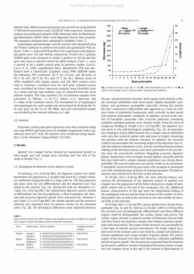
Spotted rose snapper larvae showed an exponential growth intotal length and wet weight from hatching until the end of thestudy at 40 dph (Fig. 1).
3.1. Histological development of the digestive system
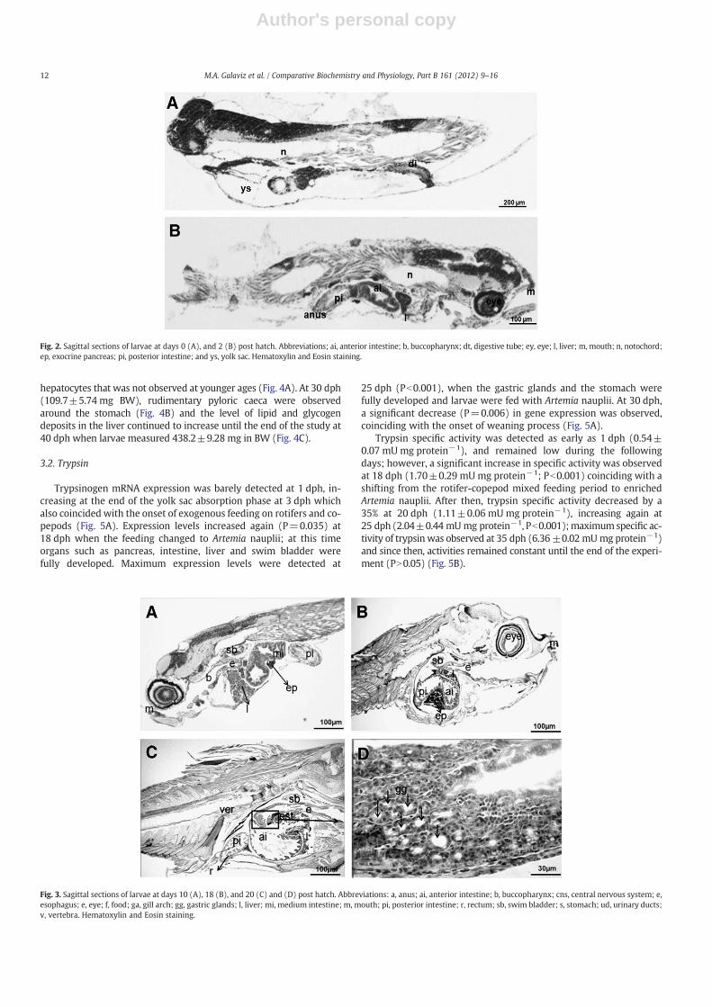
At hatching (1.4±0.34 mg BW), the digestive system was undif-ferentiated and appeared as a straight tube lined by a simple colum-nar epithelium laying dorsally to a large yolk-sac. The buccopharynxand anus were not yet differentiated and the digestive tract wasclosed to the exterior (Fig. 2A). During the yolk-sac absorption (2–3 dph, 2.0±0.25 mg BW), the rudimentary digestive system startedto differentiate into the buccopharynx, a short esophagus, the intes-tine and accessory digestive glands (liver and pancreas). Between 3and 4 dph (2.1±0.17 mg BW), the mouth opened and the posteriorintestine was separated from its anterior section by the intestinalvalve (Fig. 2B). No histological differences were observed between
the pre- and postvalvular intestine; both regions were lined by a sim-ple columnar epithelium with basal nuclei, slightly basophilic cyto-plasm, and prominent eosinophilic microvilli. During this period,the liver continued to differentiate and appeared as a mass of com-pact tissue of polyhedral hepatocytes with centrally located nucleiand reduced eosinophilic cytoplasm. In addition, several small clus-ters of basophilic pancreatic cells (exocrine pancreas) containingacidphilic zymogen granules were already visible. From the onset ofexogenous feeding to 10 dph (3.6±1.25 mg BW), the digestive sys-tem grew in size and histological complexity (Fig. 3A). In particular,the esophagus clearly differentiated into a simple cubical epitheliumwith very few scattered goblet cells and grew in length connectingthe pharynx with the anterior intestine. In addition, the intestinecoiled to accommodate the increasing length of the digestive tract in-side the reduced abdominal cavity, and the intestinal mucosa folded.Folding of the intestinal mucosa was more prominent in the prevalv-ular than the postvalvular intestine. Regarding accessory digestiveglands, hepatocytes were arranged around hepatic sinusoids and thebile duct lined with a simple cuboidal epithelium was clearly distin-guishable. The exocrine pancreas was clearly visible as an extrahepat-ic basophilic tissue formed by pancreatic cells arranged in prominentacini and containing large inclusion of zymogen granules. No lipid in-clusions were observed in the liver or the intestine.
At 18 dph (22.5±4.24 mg BW), the most relevant feature con-cerning the development of the digestive system in spotted rosesnapper was the appearance of the future stomach as a bulge of baso-philic cubical cells at the end of the esophagus (Fig. 3B). Additionalfeature characteristics of this age were the longitudinal folding ofthe esophageal mucosa and the large increase in the number of intes-tinal goblet cells, as well as the increase in size and number of muco-sal folds in the intestine.
At 20 dph (46.3±1.52 mg BW), gastric glands were clearly distin-guishable (Fig. 3C and D), whereas at 25 dph (100.5±0.70 mg BW)the stomach was morphologically differentiated and three differentregions could be distinguished: the cardias, fundus and pylorus. Thecardiac region showed a reduced number of flattened mucosal foldsand their lumen was lined by a simple short ciliated columnar epithe-lium with basal nuclei. This region of the stomach was surrounded bya thin layer of smooth circular musculature. The fundic region occu-pied most of the stomach and it was lined by a simple tall ciliated co-lumnar epithelium and a large number of gastric glands. The pyloricregion of the stomach was short and lined by a columnar epitheliumdevoid of gastric glands. The stomachwas separated from the intestineby the pyloric sphincter. Another histological distinctive feature of spot-ted rose snapper larvae at this age is the increase of lipid deposits in
Table 1Gene-specific primers for qPCR analysis and GenBank accession numbers for spottedrose snapper trypsin and pepsin partial nucleotide sequences.
Gene Primer name Primer sequence GenBank accessionnumber
Trypsin LgTryp-F 5′ ATGAGCTCCACTGCTGACAGAAAC 3′ HM754476LgTryp-R 5′ AGAGTCACCCTGGCAAGAGTCCTT 3′
Pepsin LgPepsin-F 5′ TGTCTTCGACAACATGATCAAGCA 3′ HM754478LgPepsin-R 5′ GCTGTCCATCTTGATCTGCCAGTA 3′
18S rRNA Lg18S-F 5′ CTGAACTGGGGCCATGATTAAGAG 3′ HM754479Lg18S-R 5′ GGTATCTGATCGTCGTCGAACCTC 3′
Fig. 1. Mean wet mass (mg±SD, n=30, ●), and total length (cm±SD, n=10, ■) ofspotted rose snapper larvae cultured under experimental conditions.
11M.A. Galaviz et al. / Comparative Biochemistry and Physiology, Part B 161 (2012) 9–16
Author's personal copy
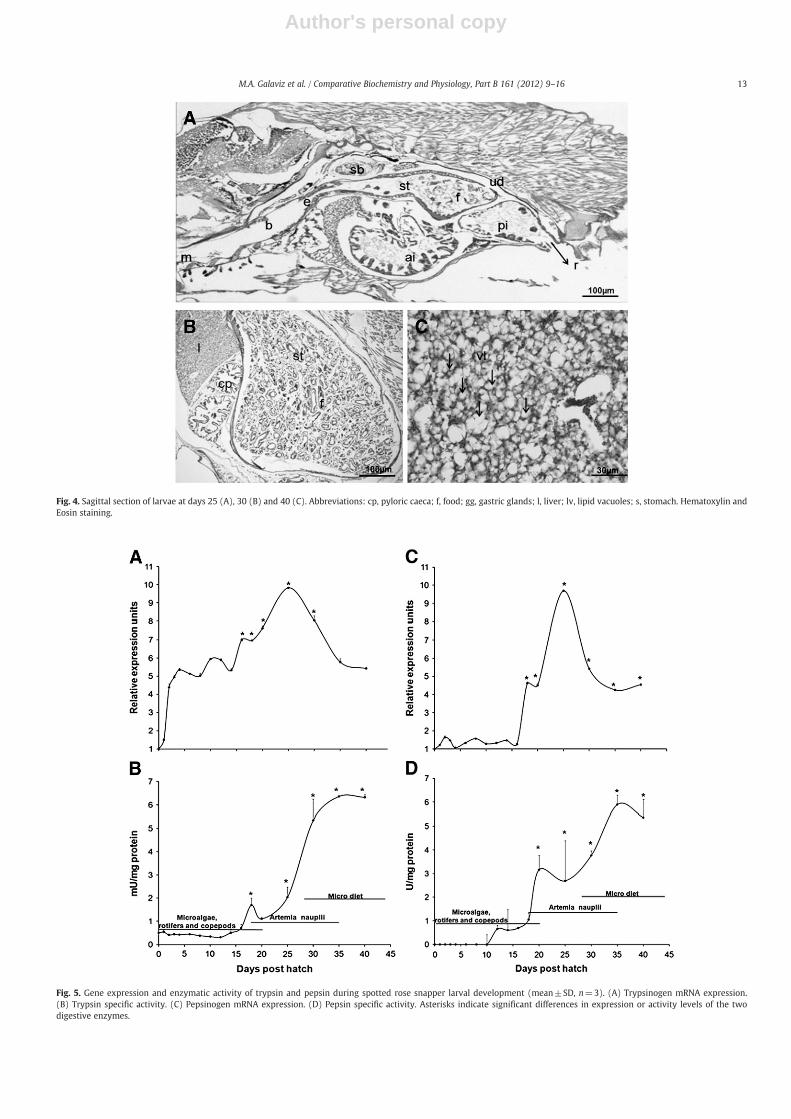
hepatocytes that was not observed at younger ages (Fig. 4A). At 30 dph(109.7±5.74 mg BW), rudimentary pyloric caeca were observedaround the stomach (Fig. 4B) and the level of lipid and glycogendeposits in the liver continued to increase until the end of the study at40 dph when larvae measured 438.2±9.28 mg in BW (Fig. 4C).
3.2. Trypsin
Trypsinogen mRNA expression was barely detected at 1 dph, in-creasing at the end of the yolk sac absorption phase at 3 dph whichalso coincided with the onset of exogenous feeding on rotifers and co-pepods (Fig. 5A). Expression levels increased again (P=0.035) at18 dph when the feeding changed to Artemia nauplii; at this timeorgans such as pancreas, intestine, liver and swim bladder werefully developed. Maximum expression levels were detected at
25 dph (Pb0.001), when the gastric glands and the stomach werefully developed and larvae were fed with Artemia nauplii. At 30 dph,a significant decrease (P=0.006) in gene expression was observed,coinciding with the onset of weaning process (Fig. 5A).
Trypsin specific activity was detected as early as 1 dph (0.54±0.07 mU mg protein−1), and remained low during the followingdays; however, a significant increase in specific activity was observedat 18 dph (1.70±0.29 mU mg protein−1; Pb0.001) coinciding with ashifting from the rotifer-copepod mixed feeding period to enrichedArtemia nauplii. After then, trypsin specific activity decreased by a35% at 20 dph (1.11±0.06 mU mg protein−1), increasing again at25 dph (2.04±0.44 mUmg protein−1, Pb0.001);maximumspecific ac-tivity of trypsin was observed at 35 dph (6.36±0.02 mUmg protein−1)and since then, activities remained constant until the end of the experi-ment (PN0.05) (Fig. 5B).
Fig. 2. Sagittal sections of larvae at days 0 (A), and 2 (B) post hatch. Abbreviations; ai, anterior intestine; b, buccopharynx; dt, digestive tube; ey, eye; l, liver; m, mouth; n, notochord;ep, exocrine pancreas; pi, posterior intestine; and ys, yolk sac. Hematoxylin and Eosin staining.
Fig. 3. Sagittal sections of larvae at days 10 (A), 18 (B), and 20 (C) and (D) post hatch. Abbreviations: a, anus; ai, anterior intestine; b, buccopharynx; cns, central nervous system; e,esophagus; e, eye; f, food; ga, gill arch; gg, gastric glands; l, liver; mi, medium intestine; m, mouth; pi, posterior intestine; r, rectum; sb, swim bladder; s, stomach; ud, urinary ducts;v, vertebra. Hematoxylin and Eosin staining.
12 M.A. Galaviz et al. / Comparative Biochemistry and Physiology, Part B 161 (2012) 9–16
Author's personal copy
Fig. 4. Sagittal section of larvae at days 25 (A), 30 (B) and 40 (C). Abbreviations: cp, pyloric caeca; f, food; gg, gastric glands; l, liver; lv, lipid vacuoles; s, stomach. Hematoxylin andEosin staining.
Fig. 5. Gene expression and enzymatic activity of trypsin and pepsin during spotted rose snapper larval development (mean±SD, n=3). (A) Trypsinogen mRNA expression.(B) Trypsin specific activity. (C) Pepsinogen mRNA expression. (D) Pepsin specific activity. Asterisks indicate significant differences in expression or activity levels of the twodigestive enzymes.
13M.A. Galaviz et al. / Comparative Biochemistry and Physiology, Part B 161 (2012) 9–16

Author's personal copy
3.3. Pepsin
Pepsinogen gene expression was significantly detected at 18 dph(Pb0.001), two days before the histological differentiation of the gas-tric glands (Fig. 5C). Maximum expression levels were detected at day25 (Pb0.001), coinciding with the full development of the stomach,after then, expression levels decreased at 30 dph (Pb0.05) andremained constant until the end of the experiment at 40 dph(PN0.05) (Fig. 5C).
Pepsin specific activity was not detected before 12 dph (0.65±0.16 U mg protein−1). A stepwise significant increase in specific ac-tivity of the acid protease was observed until the end of the experi-ment. The first significant peak in specific activity was observed at20 dph (3.16±0.60 U mg protein−1, P=0.038) coinciding with thedifferentiation of the gastric glands. Since then, pepsin activity remainedconstant until 30 dph (3.7±0.2 U mg protein−1), and increased againat 35 dph (5.89±0.4 U mg protein−1). From this age to the end of thestudy, pepsin specific activity remained constant (PN0.05) (Fig. 5D).
4. Discussion
Protein digestion in fish larvae occurs mainly by the action of alka-line proteases (such as trypsin and chymotrypsin) and cytosolic pep-tidases (Zambonino-Infante and Cahu, 2001). During larval stages,these enzymes have limited capacity for digesting macromoleculesthat are absorbed by the pinocytotic activity of the enterocytes inthe posterior intestine. In species with a stomach, the adult stage offood processing implies an acid digestion and consequently a moreefficient extracellular digestion of proteins (Segner et al., 1994).
Gastric glands can be detected a few days or weeks after hatchingand their number increase progressively, partially or completely cov-ering the stomach epithelium depending on the species (Ribeiro et al.,1999; Ortiz-Delgado et al., 2003; Elbal et al., 2004). Accordingly, thesecretions of these glands, pepsinogen and hydrochloric acid, inducea progressively lower pH environment in the lumen of the stomachand the conversion of pepsinogen into pepsin (Yúfera et al., 2004;Yúfera and Darias, 2007). The time sequence of gastric glands appear-ance and the maturation of the stomach vary among families and spe-cies (Falk-Petersen, 2005). In addition to the onset of acid digestion,the increase of aminopeptidase N activity in intestinal brush bordermembranes, and the progressive decrease of cytosolic peptidasescharacterize the progress of enterocyte maturation indicating a func-tional intestine (Zambonino-Infante and Cahu, 2001).
The development of the digestive system in tropical spotted rosesnapper larvae presented similarities with other marine fish speciessuch as gilthead sea bream Sparus aurata (Sarasquete et al., 1995),California halibut Paralichthys californicus (Gisbert et al., 2004), bull-seye puffer fish S. annulatus (García-Gasca et al., 2006), and Europeansea bass Dicentrarchux labrax (Giffard-Mena et al., 2006), amongother tropical and temperate marine fish species. In most species,the alimentary canal (buccopharynx, esophagus, intestine, and rec-tum) is differentiated at 3 dph, coinciding with the opening of themouth and the onset of exogenous feeding. Similar results were ob-served in our study, since spotted rose snapper larvae experiencedmajor changes in the development of the digestive system between3 and 4 dph, including the differentiation of enterocytes, the foldingof the intestinal mucosa, and the development of the liver, bucco-pharynx and esophagus.
According to Segner et al. (1994), the cytological differentiation ofenterocytes indicates that the intestine is suitable for absorption ofnutrients at the beginning of the exogenous feeding. In the spottedrose snapper, this differentiation coincided with the folding of the di-gestive tract and its subsequent division into anterior and posteriorintestine by the intestinal valve. Gastric glands were differentiatedat 20 dph, increasing in number and size during the next days untilthe end of the larval period. According Baglole et al. (1997), the
development of gastric glands is considered the last major event indigestive tract development, and some authors suggest that theirpresence is a characteristic feature of the end of the larval periodand the onset of the juvenile stage (Tanaka, 1972; Segner et al.,1994; Bisbal and Bengtson, 1995; Sarasquete et al., 1995; Peña et al.,2003).
The complete development of the functional stomach may in-crease the digestive capacity of the gastrointestinal system due tothe production of pepsin and hydrochloric acid by gastric glands,which involve the establishment of acid digestion and, consequently,a more efficient extracellular digestion of proteins (Yúfera and Darias,2007), enhancing the intracellular digestion in the posterior intestine(Govoni et al., 1986). In addition, the full development of the stomachplays an important role in the digestive capacity when larval feedingis changed from live food to an inert diet (Lee and Litvak, 1996; Falk-Petersen and Hansen, 2001).
Expression and activity of trypsin in L. guttatus larvae were detectedat hatch at very low levels, increasing concomitantly with larval devel-opment. These results agree with other reports in larvae of several fishspecies (Baragi and Lovell, 1986; Pedersen, 1993; Walford and Lam,1993; Zambonino-Infante and Cahu, 1994; Oozeki and Bailey, 1995;Moyano et al., 1996; Ribeiro et al., 1999; Lazo et al., 2000; Srivastavaet al., 2002; García-Gasca et al., 2006; Murray et al., 2006; Alvarez-González et al., 2010). Interestingly, Zambonino-Infante and Cahu(1994) detected enzymatic activity of trypsin and other digestive en-zymes in D. labrax larvae at 4 dph, while the first feeding occurred at6 dph. The enzymatic activity of trypsin prior to the first feeding sug-gests that the activity of this enzyme is derived fromgenetically prepro-grammed expression and not by the first exogenous feeding (See Péreset al., 1998; Ribeiro et al., 1999; Lazo et al., 2000; Zambonino-Infanteand Cahu, 2001; Alvarez-González et al., 2006, 2008).
Zambonino-Infante and Cahu (2001) mentioned that the secretionof pancreatic enzymes in the intestinal lumen increases during thefirst three weeks of larval life (at least in temperate species); this isa normal process due to the maturation of the exocrine pancreasand the production of zymogen granules. According to García-Gascaet al. (2006), the time for trypsin synthesis and enzymatic activity isspecies-specific and would depend on the larval stage of develop-ment and the type of exogenous feeding.
In L. guttatus larvae, peaks of trypsin expression preceded those ofspecific activity, and generally coincided with changes in food supply.Gene expression increased with larval development starting at 3 dphwhen the larvae opened the mouth and were fed live food (rotifersand copepods), showing maximum expression levels at 25 dph,while enzymatic activity showed maximum levels 10 days later(35 dph), suggesting that the pancreas was fully functional after25 dph. The efficient synthesis and secretion of pancreatic zymogengranules play an important role in the hydrolysis of food proteinsand the activation of other enzymes (Hjelmeland and Jørgensen,1985).
In L. guttatus, the stomach was fully developed at 25 dph, five daysafter the appearance of gastric glands. A peak in pepsinogen expres-sion was first detected at 18 dph whereas pepsin enzymatic activitywas not detected until 20 dph, coinciding with the appearance of gas-tric glands. Then a second, major peak of pepsinogen gene expressionwas detected at 25 dph, while pepsin activity was detected at 35 dph,however, the change of feeding from live food to microdiet was notperformed until 28 dph. These results indicate that the stomach wasfully functional after 25 dph. Several authors have reported for differ-ent fish species (e.g. Pagellus erythrinus, Suzer et al., 2006; Mystusnemurus, Kamarudin et al., 2011; Atractoscion nobilis, Galaviz et al.,2011, among others) a decrease in trypsin activity when the stomachbecomes functional and pepsin activity takes over the digestion pro-cess. In other words, trypsin activity decreases when pepsin activityincreases. We did not observe this pattern in trypsin and pepsin activ-ities, since activity of both enzymes remained high at the end of the
14 M.A. Galaviz et al. / Comparative Biochemistry and Physiology, Part B 161 (2012) 9–16

Author's personal copy
experiment, whereas gene expression decreased in both cases. Thismeans that both enzymes were synthesized in parallel, and whenthe amount of each enzyme was enough to perform digestion, tran-scription dropped and activity increased. Since fish are a diversegroup (from the physiological point of view), the inverse behavior be-tween trypsin and pepsin activities has not always been observed. Forinstance, Doi (2005) detected parallel increments in trypsin and pep-sin activities in mandarin fish (Siniperca chuatsi) larvae and juveniles,although there was a sharp increase in pepsin activity at 3 dph, whengastric glands first appeared, increasing even further as gastric glandscontinued to develop. Nevertheless, trypsin activity (even if lowerthan pepsin) also increased until the end of the sampling period at47 dph. In the dourado (Salminus brasiliensis) pepsin activity in-creased at 6 dph while trypsin activity remained almost constantuntil the end of the sampling period at 7 dph (Vega-Orellana et al.,2006). It is possible that in some fish species pancreatic enzymes(such as trypsin) are still required even if acid digestion starts takingplace.
Also, differences between the time of development of gastricglands and the time of pepsin secretion have been observed in somemarine fish species such as D. labrax (Vu, 1983), S. senegalensis(Ribeiro et al., 1999), and P. californicus (Alvarez-González et al.,2006). Although Zambonino-Infante and Cahu (2007) demonstratedthat gastric gland differentiation was not necessary to feed larvaewith artificial diets (since they successfully reared European seabasslarvae with artificial diets at the time of first feeding), according toour results and experience, we would not advise to wean L. guttatusbefore development of gastric glands; we suggest that weaningcould be performed between 20 and 25 dph instead of the currentweaning protocol of 28–30 dph.
5. Conclusions
In spotted rose snapper larvae, the digestive system is fully devel-oped and functional between 20 and 25 dph, as demonstrated byhistological observations and the molecular and biochemical quantifi-cation of two major proteases, trypsin and pepsin. At this time, larvaeshould be able to digest inert food and absorb nutrients. Based onthese results, the onset of the weaning period (currently performedbetween 28 and 30 dph) could be performed earlier, almost certainlybetween 20 and 25 dph.
Acknowledgments
This work was supported by the National Council for Science andTechnology (CONACyT) in Mexico, (grant number 164557), and byinternal research support of theUniversidadAutónomadeBaja California(UABC), Mexico. Thanks to Deyanira Rodarte from UABC, and RubíHernández for technical assistance, and to Noemí García, Isabel Abdo,Estela Rodríguez, and Gabriela Velasco from CIAD Mazatlan, Mexico forthe supply of fish eggs and larvae.
References
Alvarez-González, C.A., Cervantes-Trujano, M., Tovar-Ramírez, D., Conklin, D.E.,Nolasco, H., Gisbert, E., Piedrahita, R., 2006. Development of digestive enzymes inCalifornia halibut Paralichthys californicus larvae. Fish Physiol. Biochem. 31, 83–93.
Alvarez-González, C.A., Moyano-López, F.J., Civera-Cerecedo, R., Carrasco-Chávez, V.,Ortiz-Galindo, J.L., Dumas, S., 2008. Development of digestive enzyme activity inlarvae of spotted sand bass Paralabrax maculatofasciatus I: biochemical analysis.Fish Physiol. Biochem. 34, 373–384.
Alvarez-González, C.A., Moyano-López, F.J., Civera-Cerecedo, R., Carrasco-Chávez, V.,Ortiz-Galindo, J.L., Nolasco-Soria, H., Tovar-Ramírez, D., Dumas, S., 2010. Develop-ment of digestive enzyme activity in larvae of spotted sand bass Paralabraxmaculatofasciatus II: electrophoretic analysis. Fish Physiol. Biochem. 36, 29–37.
Baglole, C.J., Murray, H.M., Goff, G.P., Wright, G.M., 1997. Ontogeny of the digestivetract during larval development of yellowtail flounder: a light microscopic andmucous histochemical study. J. Fish Biol. 51, 120–134.
Baragi, V., Lovell, R.T., 1986. Digestive enzyme activities in striped bass from first feedingthrough larval development. Trans. Am. Fish. Soc. 115, 478–484.
Bisbal, G.A., Bengtson, D.A., 1995. Development of the digestive tract in larval summerflounder. J. Fish Biol. 47, 277–291.
Bradford, M.M., 1976. A rapid and sensitive method for the quantization of microgramquantities of protein utilizing the principle of protein dye binding. Anal. Biochem.72, 248–254.
Cahu, C., Rønnestad, I., Grangier, V., Zambonino-Infante, J.L., 2004. Expression andactivities of pancreatic enzymes in developing sea bass larvae Dicentrarchus labraxin relation to intact and hydrolyzed dietary protein; involvement of cholecystokinin.Aquaculture 238, 295–308.
Davis, D.A., Bootes, K.L., Arnold, C., 2000. Snapper (Family Lutjanidae) culture. In: Stickney,R.R. (Ed.), Encyclopedia of Aquaculture. JohnWiley and Sons, New York, pp. 884–889.
Doi, T., 2005. Early development of the digestive system and digestive enzyme activityof reared mandarinfish Siniperca chuatsi (Perciformes: Sinipercidae). Aquac. Sci.53, 425–431.
Douglas, S.E., Gawlicka, A., Mandlam, S., Gallant, J.W., 1999. Ontogeny of the stomach inwinter flounder: characterization and expression of the pepsinogen and protonpump genes and determination of pepsin activity. J. Fish Biol. 55, 897–915.
Elbal, M.T., García-Hernández, M.P., Lozano, M.T., Aguelleiro, B., 2004. Developmentof the digestive tract of gilthead sea bream (Sparus aurata L.). Light and electronmicroscopic studies. Aquaculture 234, 215–238.
Erlanger, B., Kokowsky, N., Cohen, W., 1961. The preparation and properties of two newchromogenic substrates of trypsin. Arch. Biochem. Biophys. 95, 271–278.
Falk-Petersen, I.B., Hansen, T.K., 2001. Organ differentiation in newly hatched commonwolffish. J. Fish Biol. 59, 1465–1482.
Falk-Petersen, I.B., 2005. Comparative organ differentiation during early life stages onmarine fish. Fish Shellfish Immunol. 19, 397–412.
Galaviz, M., García-Gasca, A., Drawbridge, M., Álvarez-González, C.A., López, L., 2011.Ontogeny of the digestive tract and enzymatic activity in white seabass, Atractoscionnobilis, larvae. Aquaculture 318, 162–168.
García-Gasca, A., Galaviz, M., Gutiérrez, J.N., García-Ortega, A., 2006. Development ofthe digestive tract, trypsin activity and gene expression in eggs and larvae of thebullseye puffer fish Sphoeroides annulatus. Aquaculture 256, 366–376.
García-Ortega, A., 2009. Nutrition and feeding research in the spotted rose snapper Lutjanusguttatus and bullseye puffer Sphoeroides annulatus, new species formarine aquaculture.Fish Physiol. Biochem. 35, 69–80.
García-Ortega, A., Abdo, I., Duncan, N., Rodríguez, E., Velasco, G., González, B., Puello, A.,Martínez, I., 2005. Larval rearing of spotted rose snapper Lutjanus guttatus underexperimental conditions. In: Hendry, C.I., Van Stappen, G., Wille, M., Sorgeloos, P.(Eds.), Larvi '05— Fish and Shellfish Larviculture Symposium: European AquacultureSociety, Special Publication No. 36, pp. 172–175. Oostende, Belgium.
Giffard-Mena, I., Charmantier, G., Grousset, E., Aujoulat, F., Castille, R., 2006. Digestivetract ontogeny of Dicentrarchus labrax: implication in osmoregulation. Dev.Growth Differ. 48, 139–151.
Gisbert, E., Piedrahita, R.H., Conklin, D.E., 2004. Ontogenetic development of the digestivesystem in California halibut Paralichthys californicus with notes on feeding practices.Aquaculture 232, 455–470.
Gisbert, E., Ortiz-Delgado, J.B., Sarasquete, C., 2008. Nutritional cellular biomarkers inearly life stages of fish. Histol. Histopathol. 23, 1525–1539.
Govoni, J.J., Boenhlert, G.W.,Watanabe, Y., 1986. The physiology of digestion in fish larvae.Environ. Biol. Fish. 16, 59–77.
Grimes, C., 1987. Reproductive biologyof the Lutjanidae: a review. In: Polovina, J.J., Ralston,S. (Eds.), Tropical Snappers and Groupers Biology and Fisheries Management. WestView Press, Boulder, CO, USA, pp. 239–294.
Hjelmeland, K., Jørgensen, T., 1985. Evaluation of radioimmunoassay as a method toquantify trypsin and trypsinogen in fish. Trans. Am. Fish. Soc. 114, 619–621.
Ibarra-Castro, L., Álvarez-Lajonchère, L., 2009. Improved induced-spawning protocolfor the spotted rose snapper Lutjanus guttatus. Isr J Aquac. - Bamidgeh 61, 121–133.
Kamarudin, M.S., Otoi, S., Saad, C.R., 2011. Changes in growth, survival and digestiveenzyme activities of Asian redtail catfish, Mystus nemurus, larvae fed on differentdiets. Afr. J. Biotechnol. 10, 4484–4493.
Lazo, J.P., Holt, G.J., Arnold, C.R., 2000. Ontogeny of pancreatic enzymes in larval reddrum Sciaenops ocellatus. Aquac. Nutr. 6, 183–192.
Lee, G.W., Litvak, M., 1996. Weaning of metamorphosed winter flounder Pleuronectesamericanus reared in the laboratory: comparison of two commercial artificialdiets on growth, survival and conversion efficiency. Aquaculture 144, 251–263.
Mai, K., Yu,H.,Ma,H., Duan,Q., Gisbert, E., Zambonino Infante, J., Cahu, C., 2005. A histologicalstudy on the development of the digestive system of Pseudosciaena crocea larvae andjuveniles. J. Fish Biol. 67, 1094–1106.
Moyano, F.J., Díaz, M., Alarcón, F.J., Sarasquete, M.C., 1996. Characterization of digestiveenzyme activity during development of gilthead sea bream Sparus aurata. FishPhysiol. Biochem. 15, 121–130.
Murray, H.M., Perez-Casanova, J.C., Gallant, J.W., Johnson, S.C., Douglas, S.E., 2004.Trypsinogen expression during the development of the exocrine pancreasin Winter flounder, Pseudopleuronectes americanus. Comp. Biochem. Physiol. A138, 53–59.
Murray, H.M., Gallant, J.W., Johnson, S.C., Douglas, S.E., 2006. Cloning and expressionanalysis of three digestive enzymes from Atlantic halibut Hippoglossus hippoglossusduring early development: predicting gastrointestinal functionality. Aquaculture252, 394–408.
Oozeki, Y., Bailey, K.M., 1995. Ontogenetic development of digestive enzyme activitiesin larval walleye pollock, Theragra chalcogramma. Mar. Biol. 122, 177–186.
Ortiz-Delgado, J.B., Darias,M.J., Cañavate, J.P., Yufera,M., Sarasquete, C., 2003. Organogenesisof the digestive tract in the White seabream, Diplodus sargus. Histological and histo-chemical approaches. Histol. Histopathol. 18, 1141–1154.
15M.A. Galaviz et al. / Comparative Biochemistry and Physiology, Part B 161 (2012) 9–16

Author's personal copy
Pedersen, B.H., 1993. Protein digestion in herring Clupea harengus larvae: trypsinogensecretion, and effects of a transitory food restriction onmortality, growth anddigestiveenzyme content. In: Walther, B.T., Fhyn, H.J. (Eds.), Physiological and BiochemicalAspects of Fish Development. University of Bergen, Norway, pp. 220–225.
Peña, R., Dumas, S., Villalejo-Fuerte, M., Ortiz-Galindo, J., 2003. Ontogenetic developmentof the digestive tract in reared spotted sand bass Paralabrax maculatofasciatus larvae.Aquaculture 219, 633–644.
Péres, A., Zambonino Infante, J.L., Cahu, C., 1998. Dietary regulation of activities andmRNA levels of trypsin and amylase in sea bass Dicentrarchus labrax larvae. FishPhysiol. Biochem. 19, 145–152.
Ribeiro, L., Zambonino-Infante, J.L., Cahu, C.L., Dinis, M.T., 1999. Development of digestiveenzymes in larvae of Solea senegalensis, Kaup 1858. Aquaculture 170, 465–473.
Rønnestad, I., Morais, S.J., 2008. Digestion. In: Finn, R.N., Kapoor, B.G. (Eds.), Fish LarvalPhysiology. Science Publishers, Enfield, New Hampshire, USA, pp. 201–262.
Rungruangsak-Torrissen, K., Moss, R., Andresen, L.H., Berg, A., Waagbø, R., 2006. Differentexpressions of trypsin and chymotrypsin in relation to growth in Atlantic salmonSalmo salar L. Fish Physiol. Biochem. 32, 7–23.
Sarasquete, M.C., Polo, A., Yufera, M., 1995. Histology and histochemistry of the developmentof the digestive system of larval gilthead sea bream Sparus aurata L. Aquaculture 130,79–82.
Sarath, G., De la Monte, R.S., Warner, F.W., 1989. Protease assay methods. In: Beyon,R.J., Bond, J.S. (Eds.), Proteolytic Enzymes: A Practical Approach. Oxford UniversityPress, New York, New York, USA, pp. 25–56.
Segner, H., Storch, V., Reinecke, M., Kloas, W., Hanke, W., 1994. The development offunctional digestive and metabolic organs in turbot Scophthalmus maximus. Mar.Biol. 119, 471–486.
Srivastava, A., Kurokawa, T., Suzuki, T., 2002. mRNA expression of pancreatic enzymeprecursors and estimation of protein digestibility infirst feeding larvae of the Japaneseflounder, Paralichthys olivaceus. Comp. Biochem. Physiol. A 132, 629–635.
Suzer, C., Saka, Ş., Firat, K., 2006. Effects of illumination on early life development anddigestive enzymeactivities in commonpandora Pagellus erythrinus L. larvae. Aquaculture260, 86–93.
Tanaka, M., 1972. Studies on the structure and function of the digestive system in teleostlarvae. Part 5: epithelial changes in the posterior gut and protein digestion. Jpn J.Ichthyol. 19, 172–180.
Ueberschär, B., 1993. Measurement of proteolytic enzyme activity: significance andapplication in larval fish research. In: Walther, B.T., Fhyn, H.J. (Eds.), Physiologicaland Biochemical Aspects of Fish Development. University of Bergen, Norway, pp.233–239.
Vega-Orellana, O.M., Fracalossi, D.M., Sugai, J.K., 2006. Dourado (Salminus brasiliensis)larviculture:weaningandontogenic development of digestive proteinases. Aquaculture252, 484–493.
Verreth, J., Torreele, E., Spazier, E., Van der Sluiszan, A., Rombout, J., Booms, R., Segner,H., 1992. The development of functional digestive system in the African catfishClarias (Burchell). J. World Aquacult. Soc. 23, 286–298.
Vu, T.T., 1983. Etude histoenzymologique des activities proteasiques dans le tube digestifdes larves et des adultes de bar, Dicentrarchus labrax (L). Aquaculture 32, 57–69.
Walford, J., Lam, T.J., 1993. Development of digestive tract and proteolytic enzyme activityin seabass (Lates calcarifer) larvae and juveniles. Aquaculture 109, 187–205.
Yúfera, M., Fernández-Díaz, C., Vidaurreta, A., Cara, J.B., Moyano, F.J., 2004. GastrointestinalpHanddevelopment of the acid digestion in larvae andearly juveniles of Sparus aurataL. (Pisces: Teleostei). Mar. Biol. 144, 863–869.
Yúfera, M., Darias, M.J., 2007. Changes in the gastrointestinal pH from larvae to adult inSenegal sole (Solea senegalensis). Aquaculture 267, 94–99.
Zambonino-Infante, J.L., Cahu, C., 1994. Development and response to a diet change of somedigestive enzymes in seabass (Dicentrarchus labrax) larvae. Fish Physiol. Biochem. 12,399–408.
Zambonino-Infante, J.L., Cahu, C.L., 2001. Ontogeny of the gastrointestinal tract of marinefish larvae. Comp. Biochem. Physiol. C 130, 477–487.
Zambonino-Infante, J.L., Cahu, C.L., 2007. Dietary modulation of some digestive enzymesand metabolic processes in developing marine fish: applications to diet formulation.Aquaculture 268, 98–105.
Zambonino-Infante, J., Gisbert, E., Sarasquete, C., Navarro, I., Gutierrez, J., Cahu, C.L.,2008. Ontogeny and physiology of the digestive system of marine fish larvae. In:Cyrino, J.E.O., Bureau, D., Kapoor, B.G. (Eds.), Feeding and Digestive Functions ofFishes. Science Publishers Inc., Enfield, USA, pp. 281–384.
16 M.A. Galaviz et al. / Comparative Biochemistry and Physiology, Part B 161 (2012) 9–16
Related Documents