Author’s Accepted Manuscript LIPOPROTEINS AS TARGETS AND MARKERS OF LIPOXIDATION Catarina B. Afonso, Corinne M. Spickett PII: S2213-2317(18)31015-2 DOI: https://doi.org/10.1016/j.redox.2018.101066 Article Number: 101066 Reference: REDOX101066 To appear in: Redox Biology Received date: 30 October 2018 Revised date: 28 November 2018 Accepted date: 5 December 2018 Cite this article as: Catarina B. Afonso and Corinne M. Spickett, LIPOPROTEINS AS TARGETS AND MARKERS OF LIPOXIDATION, Redox Biology, https://doi.org/10.1016/j.redox.2018.101066 This is a PDF file of an unedited manuscript that has been accepted for publication. As a service to our customers we are providing this early version of the manuscript. The manuscript will undergo copyediting, typesetting, and review of the resulting galley proof before it is published in its final citable form. Please note that during the production process errors may be discovered which could affect the content, and all legal disclaimers that apply to the journal pertain. www.elsevier.com/locate/redox

Welcome message from author
This document is posted to help you gain knowledge. Please leave a comment to let me know what you think about it! Share it to your friends and learn new things together.
Transcript

Author’s Accepted Manuscript
LIPOPROTEINS AS TARGETS ANDMARKERS OF LIPOXIDATION
Catarina B. Afonso, Corinne M. Spickett
PII: S2213-2317(18)31015-2DOI: https://doi.org/10.1016/j.redox.2018.101066Article Number: 101066Reference: REDOX101066
To appear in: Redox Biology
Received date: 30 October 2018Revised date: 28 November 2018Accepted date: 5 December 2018
Cite this article as: Catarina B. Afonso and Corinne M. Spickett,LIPOPROTEINS AS TARGETS AND MARKERS OF LIPOXIDATION,Redox Biology, https://doi.org/10.1016/j.redox.2018.101066
This is a PDF file of an unedited manuscript that has been accepted forpublication. As a service to our customers we are providing this early version ofthe manuscript. The manuscript will undergo copyediting, typesetting, andreview of the resulting galley proof before it is published in its final citable form.Please note that during the production process errors may be discovered whichcould affect the content, and all legal disclaimers that apply to the journal pertain.
www.elsevier.com/locate/redox
CORE Metadata, citation and similar papers at core.ac.uk
Provided by Aston Publications Explorer
1
LIPOPROTEINS AS TARGETS AND MARKERS
OF LIPOXIDATION
Catarina B. Afonso, Corinne M. Spickett*
School of Life and Health Sciences, Aston University, Aston Triangle, Aston University,
Birmingham, B4 7ET, UK
*Corresponding author: Corinne Spickett, School of Life and Health Sciences, Aston
University, Aston Triangle, Birmingham, B4 7ET, UK. Tel. +44 (0)121 204 4085;
Abstract
Lipoproteins are essential systemic lipid transport particles, composed of apolipoproteins
embedded in a phospholipid and cholesterol monolayer surrounding a cargo of diverse lipid species.
Many of the lipids present are susceptible to oxidative damage by lipid peroxidation, giving rise to
the formation of reactive lipid peroxidation products (rLPPs). In view of the close proximity of the
protein and lipid moieties within lipoproteins, the probability of adduct formation between rLPPs
and amino acid residues of the proteins, a process called lipoxidation, is high. There has been
interest for many years in the biological effects of such modifications, but the field has been limited
to some extent by the availability of methods to determine the sites and exact nature of such
modification. More recently, the availability of a wide range of antibodies to lipoxidation products,
as well as advances in analytical techniques such as liquid chromatography tandem mass
spectrometry (LC-MSMS), have increased our knowledge substantially. While most work has focused
on LDL, oxidation of which has long been associated with pro-inflammatory responses and
2
atherosclerosis, some studies on HDL, VLDL and Lipoprotein(a) have also been reported. As the
broader topic of LDL oxidation has been reviewed previously, this review focuses on lipoxidative
modifications of lipoproteins, from the historical background through to recent advances in the field.
We consider the main methods of analysis for detecting rLPP adducts on apolipoproteins, including
their advantages and disadvantages, as well as the biological effects of lipoxidized lipoproteins and
their potential roles in diseases.
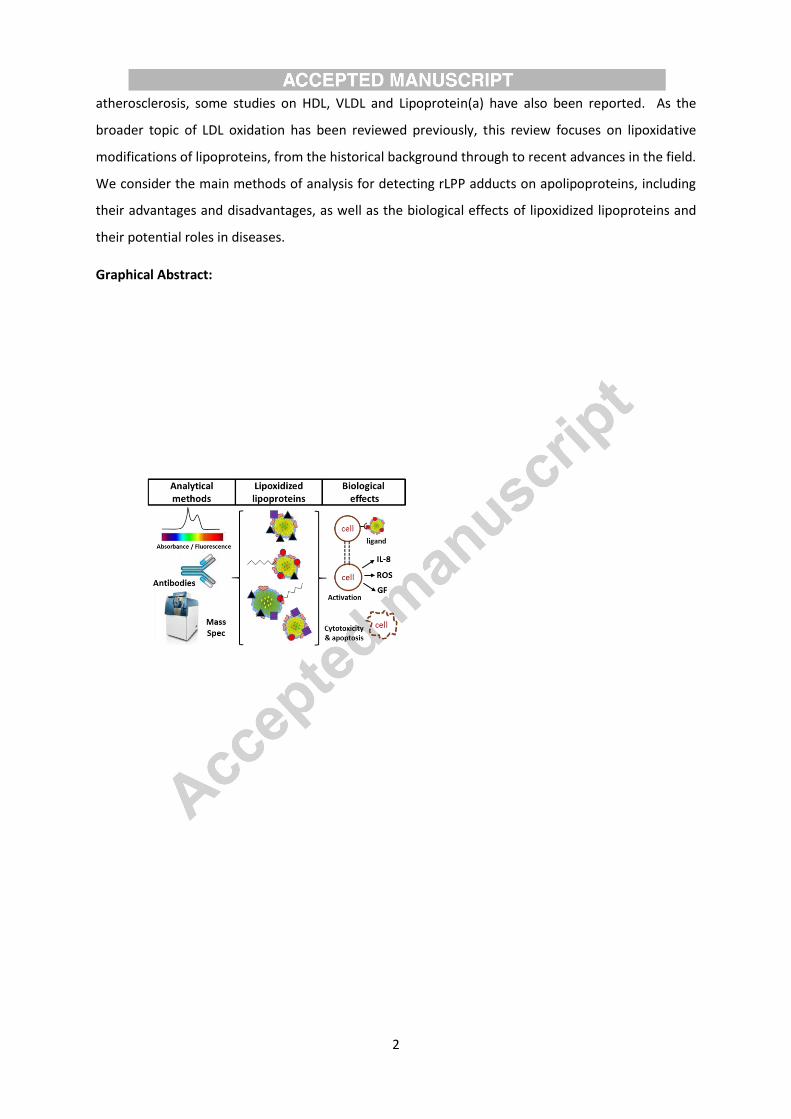
Graphical Abstract:
3
Abbreviations
AGEs, advanced glycation end products; ALEs, advanced lipoxidation end products; Apo,
Apolipoprotein; COL, Nε-(8-carboxyoctanyl)-Lysine; DNPH, 2,4-dinitrophenylhydrazine; ELISA,
enzyme-linked immunosorbent assay; GC-MS, gas chromatography mass spectrometry; HDDE, 4-
hydroxy-2E,6Z-dodecadienal; HDL, high density lipoprotein; HHE, 4-hydroxy-trans-2-hexenal; HNE, 4-
hydroxy-trans-2-nonenal; HOCl, hypochlorous acid; HPNE, 4-hydroperoxy-trans-2-nonenal; IDL,
intermediate density lipoprotein; 3-HOSCA, 3β-hydroxy-5-oxo-5,6-secocholestan-6-al; KLH, keyhole
limpet hemocyanin; (HP)LC-(ESI)-MS(MS), (high pressure) liquid chromatography (electrospray)
(tandem) mass spectrometry; LDL, low density lipoprotein; Lp(a), lipoprotein(a); MALDI-TOF, matrix-
assisted laser desorption ionization time of flight; MDA, malondialdehyde; MS, mass spectrometry;
OSE, oxidation-specific epitope; oxLDL/HDL, oxidized low/high density lipoprotein; (ox)PC, (oxidized)
phosphatidylcholine; (ox)PL, (oxidized) phospholipid; PAH, polycyclic aromatic hydrocarbons; pHA, p-
hydroxyphenylacetaldehyde; PL, phospholipid; PONPC, 1-palmitoyl-2-(9-oxo-nonanoyl)-sn-glycero-3-
phosphocholine; POVPC, 1-palmitoyl-2-(5-oxovaleroyl)-sn-glycero-3-phosphorylcholine; rLPPs,
reactive lipid peroxidation products; TBArS, thiobarbituric acid reactive substances; TNBS, 2,4,6-
trinitrobenzene sulfonic acid; VLDL, very low density lipoprotein.
Keywords
LDL; HDL; ApoB-100; Lipid peroxidation; liquid chromatography mass spectrometry;
immunoassays; atherosclerosis.
4
1. Introduction to lipoproteins and lipid oxidation
Lipoproteins are a family of complex particles consisting of amphipathic apolipoproteins
that carry a wide range of lipids, and therefore have important physiological functions in systemic
lipid transport. The basic structure is a shell formed of a monolayer of phospholipids and free
cholesterol surrounding a core of cholesteryl esters and triglycerides [1], with one or more proteins
associated with the surface phospholipids and partially embedded in the hydrophobic lipid core. The
role of the proteins is thus to solubilize and stabilize the lipoproteins in an aqueous environment
such as plasma, which is a major location of lipoproteins. Lipoproteins are classified according to
their size and density, which is determined by the ratio of protein to lipid, and therefore depends on
the lipid and apolipoprotein composition. The plasma lipoproteins are typically divided into 7
classes, which are chylomicrons, chylomicron remnants, VLDL, IDL, LDL, HDL, and Lp(a). The
chylomicrons are responsible for transport of dietary lipids from the intestine to lipid-metabolizing
tissues muscle and adipose, and the resulting chylomicron remnants deliver remaining lipid to the
liver. VLDL, IDL, LDL are involved in the second phase of triglyceride and cholesterol delivery from
the liver to peripheral tissues, whereas HDL particles are responsible for reverse cholesterol
transport from cells in the peripheral tissues to the liver to lipoproteins of the endogenous pathway.
The functions and activities of the lipoprotein particles are largely determined by the combination of
apolipoproteins they contain, which direct their interaction with receptors as well as containing a
variety of enzymatic activities. The lipid cargo is very diverse, and in addition of several classes of
phospholipid, cholesterol and cholesteryl esters, it can include free fatty acids, sphingolipids,
ceramides, and sulfolipids [2].
Many of the components of lipoproteins are susceptible to oxidative damage by free radicals
of non-radical oxidants, and this has been an energetic field of study since pioneering studies in the
1980s demonstrated that oxidation of LDL contributed to changes in its biological properties [3–5] as
described in section 4. Lipids containing unsaturated fatty acyl chains (especially poly-unsaturated
ones) can be peroxidized following hydrogen abstraction by radical attack and subsequent addition
of di-oxygen; the downstream reactions are complex and lead to a plethora of short- and long-chain
oxidation products; this topic has been extensively reviewed previously [6–9]. Peroxidation at bis-
allylic sites in the fatty acyl chains tends to result in cleavage of the hydrocarbon backbone by Hock
rearrangement or other mechanisms, resulting in formation of an aldehyde on one or both chains
[10]. Intrachain cyclization reactions can also occur, for example leading to formation of
isoprostanes or isolevuglandins. Secondary radical attack and addition of oxygen can occur at other
sites, resulting in complex structures with multiple reactive groups including aldehydes, ketones,
epoxides and cyclopentenone rings. A common motif that rises following cleavage is the -
5
substituted -alkenal, as in 4-hydroxynonenal (HNE) or 4-oxo-nonenal, although a variety of
analogous structures are possible [6,8,11]. The headgroups of amine-containing phospholipids, e.g
phosphatidylserine, can be modified by oxidative deamination, which also generates an aldehyde
[12]. Alternatively, unsaturated lipids can be attacked by electrophilic agents such as hypochlorous
acid (HOCl), which causes the formation of chlorohydrins (hydroxyl and chlorine groups on adjacent
carbons) on fatty acyl chains, or cleavage of the vinyl ether bonds of plasmalogens to yield chloro-
aldehydes. Radical nitrogen species such as nitrogen dioxide can cause peroxidation via hydrogen
abstraction, but may also undergo addition reactions leading to nitrated or nitrosylated fatty acyl
chains, some of which also contain motifs similar to the substituted alkenals mentioned above. The
key point about lipid oxidation products containing aldehydes, ketones, epoxides and substituted
alkenals is that they are electrophilic and moderately to highly reactive with nucleophilic groups,
which endows them with unusual properties through the ability to react with biological. While
several different collective names for such compounds exist, in this review we have used the term
“reactive lipid peroxidation products”, or rLPPs. Figure 1 shows the structure of the various rLPPs
discussed in this review.
Lipid oxidation in lipoproteins has been extensively investigated and the reader is directed
to reviews on this topic for more detail [13,14]. Consequently, lipid oxidation is not the main focus of
this review, which instead addresses the more specific issue of how the protein moieties of
lipoproteins are modified by the resulting rLPPs, a process known as lipoxidation and explained in
more detail in Section 2. In view of the close proximity of protein and lipids within lipoprotein, the
probability of adduct formation between rLPPs and residues of the proteins is relatively high, but to
date no lipoprotein reviews have focused specifically on lipoxidation, despite the fact that there is
growing awareness of the signaling importance of this modification [15–18]. This review addresses
mainly HDL and LDL lipoxidation, as the majority of research has investigated these lipoproteins, and
aims to provide a concise overview of lipoprotein modification by oxidized lipid adducts from early
pioneering studies to recent advances in molecular knowledge.
2. Protein oxidation and lipoxidation
In parallel to lipid oxidation, the apoprotein moieties of lipoproteins can also be oxidized
directly by a range of oxidants; usually the basic side chains (lysine, arginine and histidine), and
sidechains containing sulfur (cysteine, methionine) or aromatic groups (tyrosine, tryptophan,
phenyalanine) are most readily modified. Several studies have investigated the relative susceptibility
of lipoprotein residues to oxidants, both in vitro and ex-vivo, in an attempt to identify markers of
6
damage as well as to understand changes in lipoprotein function [19–22]. Nucleophilic sidechains,
i.e. those containing amine or thiol groups, can also be modified by reaction with rLPPs. Protein
lipoxidation can occur by one of two chemical reactions: Michael addition occurs when the
electrophilic β-carbon of a reactive lipid oxidation product reacts with the amino group of histidine
and lysine or the thiol group of a cysteine; Schiff’s base formation involves reaction of the free
amino group of a lysine, arginine or N-terminal amine with the carbonyl group of an aldehyde or
ketone. Schiff’s base adducts are less stable than Michael adducts, and both are potentially
reversible depending on the chemical environment. Moreover, depending on the electrophilic
species involved, secondary reactions such as cyclization, dehydration, oxidation, or condensation
with an additional electrophile may take place, leading to formation of more stable adducts that are
referred to as advanced lipoxidation end products, or ALEs, by analogy with the better-known
advanced glycation end products (AGEs). For example, the HNE Michael adducts tend to cyclize to
hemiacetals, which the HNE Schiff’s base with lysine can generate a stable 2-pentylpyrrole derivative
(Jacobs and Sayre, 2010); the Michael adduct of acrolein can rearrange to form formyl-
dehydropiperidino lysine (FDP-lysine) (Colzani, 2013 J Prot). The process of lipoxidation has been
very well explained previously (Sayre 2006; Jacobs and Sayre 2010; Parvez 2018), and consequently
will not be covered in depth here, although the structures most commonly discussed in the review
are shown in Figure 2.
Many approaches have been used to study lipoprotein oxidation and lipoxidation [10,16,23].
The ultimate goal is to be able to detect and identify lipoxidation under physiological conditions and
in biological or clinical samples, but many studies of modification in vitro have been carried out as a
stepping stone to this stage, to develop methodology or establish the most likely products. Oxidation
of lipoproteins in vitro is often achieved by treatment with transition metal ions, mostly commonly
Cu2+, but occasionally Fe2+ in combination with hydrogen peroxide as a Fenton reagent to generate
hydroxyl radicals is used. The use of iron also represents a model for ferretin-induced oxidation of
LDL. Other methods for oxidation of lipoproteins include radical initiators, such as 2,2′-Azobis(2-
methylpropionamidine) dihydrochloride (AAPH), or treatment with UV light. These are all able to
initiate lipid peroxidation, and therefore lead to lipoxidation of the apolipoprotein. More
physiological approaches include enzymes such as lipoxygenases and cytochrome P450 enzymes,
which catalyse radical oxidations and hydroxylations of fatty acids, or myeloperoxidase, which is
well-known for its ability to generate hypohalous acids such as HOCl, but is also able to catalyse
direct oxidations of some substrates [24]. There have also been many studies in vitro where
lipoproteins, especially LDL, have been modified by direct treatment with reactive lipid peroxidation
products; most work has been carried out with small unsaturated aldehydes, such as acrolein,
7
malondialdehyde (MDA) and HNE, but a small number of studies with less well-known rLPPs and
even esterified oxidized phospholipids have been reported, as described in the following sections.
3. Approaches to the analysis of LDL and HDL lipoxidation
The analysis of LDL and HDL is dependent on the separation of these lipoproteins from
whole plasma. A common method for separation of these particles is by gradient ultracentrifugation.
Using different salt concentrations and centrifugation at high g-forces, the lipoproteins of different
density migrate in the gradient, allowing their separation and forming bands that can be collected
and analysed further. This methodology, which dates back to 1955, was developed by Havel et al.
[25] and has been used extensively for the study of lipoproteins [26,27]. There are other methods
that take advantage of different characteristics of lipoprotein particles for their separation, such as
size exclusion chromatography, which depends on the size difference between the lipoproteins, and
gradient gel electrophoresis, which uses both size and charge for its separation [26,28,29].
All the studies included in this review used a form of the gradient ultracentrifugation
method to separate lipoproteins, followed by the analysis of oxidized lipoproteins by one or more of
the methods reviewed in this section. The analytical methods can be broadly divided into (1)
biochemical methods, involving colorimetric or fluorescence reagents and labeling with or without
additional separation by liquid chromatography; (2) antibody-based methods, including enzyme-
linked immunosorbent assay (ELISA) and immunostaining; and (3) mass spectrometric approaches,
which usually require the combination of liquid chromatography with tandem mass spectrometry.
Each of these approaches have advantages and disadvantages in terms of the complexity of the
methodology and the quality or depth of information generated, two aspects that are usually
directly related (Figure 3). Much early work used simple biochemical assays until the generation of
antibodies specific for lipoxidation products allowed the application of immunoassays. Mass
spectrometry techniques require complex and expensive instruments as well as extensive data
handling, and consequently are less widely accessible, although very valuable as they yield site-
specific information that cannot be obtained by the other approaches. Ultimately, all the techniques
provide complementary information, and are most powerful if used in combination.
3.1. Biochemical methods with chromophore or fluorophore labeling
In this section, techniques involving the use of reagents that generate a chromophore or
fluorescent signal on reaction with the lipoxidized protein or lipoxidation agent are considered.
However, it is important to bear in mind that not all methods are specific for lipoxidation: in fact, the
8
most used reagents are ones that react with aldehydes, which could also be formed by direct
oxidation of amino acid residues. Moreover, some of the methods used were indirect, for example,
deducing the amount of modification by changes in amount of free rLPP or unmodified residues.
For example, in one of the earliest studies of lipoprotein lipoxidation [23], involving the
direct reaction of HNE with LDL, the formation of HNE-LDL adducts was determined indirectly by
comparison of the amount of unreacted HNE in the supernatant of treated and untreated
lipoproteins, which was quantified by high-performance liquid chromatography on an octadecyl
silica (ODS) column with detection at 220 nm; the HNE incorporation was found to increase with
increased aldehyde concentration, with 78% bound to the protein moiety, 21% to the lipids, and 1%
as free aldehyde within the lipid fraction. To investigate more specifically the residues modified, the
apolipoprotein B-100 (ApoB-100) was delipidated, reacted with dinitrofluorobenzene to
dinitrophenylate and stabilize free hydroxyl and amine groups before total hydrolysis and
quantitation using a standard amino acid analyser. This approach was adopted as it was noted that
the adducts were not stable during the acid hydrolysis. Comparison of the levels of
dinitrophenylated amino acids (i.e. those not modified by HNE) suggested that HNE attacked mostly
lysine and tyrosine, and to a lesser extent, cysteine, serine and histidine [23]. The observation that
tyrosine and serine could be modified is interesting as these residues are not widely considered as
targets of hydroxyalkenals, but could simply have been an artefact of the experimental design with
dinitrofluorobenzene. An alternative reagent that is more specific for detection of free amine
groups, i.e. lysines, arginines and the N-terminal of proteins, is 2,4,6-trinitrobenzene sulfonic acid
(TNBS). This has been used in several studies to monitor indirectly protein modification by lipid
peroxidation products [30–36].
Amino acid analysis was also used by Uchida et al. to investigate the sites of HNE
modification in Cu2+-treated LDL, but in this case the adducts were stabilized by treatment with
sodium borohydride (NaBH4) to prevent the issues noted by Jürgens et al. (1986) [23], and the amino
acids were labeled with o-phthaldehyde for fluorescence detection during HPLC [37]. This enabled
the quantification of approximately 40 lysines and 50 histidines (mol/mol LDL) modified in HNE-
treated LDL, of which 49% and 80% respectively were identified as Michael adducts, with the
remainder as Schiff’s base adducts. However, this study also relied on complementary findings from
the use of specific antibodies, as described in the next section.
A commonly used reagent for detecting the presence of protein carbonyls is 2,4-
dinitrophenylhydrazine (DNPH) [38–40]. It reacts readily with protein-bound aldehydes or ketones to
form a hydrazone derivative with a characteristic absorbance at 365 nm, and despite its mutagenic

9
properties and explosive nature (mostly now only available as a 0.2 M solution), continues to be
widely used in studies of protein oxidation. However, its application to oxidation of lipoproteins has
been limited. The formation of protein carbonyls in HOCl-treated LDL was investigated by Yang et al.
using DNPH labeling followed by delipidation and tryptic cleavage of the ApoB-100; modified
peptides were identified on HPLC by the absorbance at 365 nm, isolated and sequenced using a gas
phase automated sequencer [41]. The modified peptides contained cysteine, lysine, tryptophan and
methionine residues; it was not clear from this study whether the modifications were due to direct
oxidation of the residues by HOCl, or formation of rLPPs that reacted with the cysteine to give
Michael adducts. In the use of DNPH as a labeling reagent for lipoxidation, it is also important to
bear in mind that Schiff’s base adducts of hydroxyalkenals do not contain a free aldehyde to react
with DNPH; instead, DNPH may react via Michael addition with them to generate 2,4-dinitrophenyl
pyrazolines, which have a slightly shifted absorbance maximum compared to hydrazones. Moreover,
when using DNPH to study lipoxidation in Cu-oxidized LDL it has been observed from the judicious
use of controls in the absence of DNPH that the tryptophan oxidation products N-formyl kynurenine
and kynurenine have significant absorbance at 365 nm, which can confound the detection of protein
carbonyls, so careful profiling of the labeled peptides is necessary [42].
Alternatively, the thiobarbituric acid reactive substances (TBArS) assay can be used to
measure malondialdehyde (MDA) equivalents present in the sample, including MDA adducts on the
protein, which are reversible under the conditions used for the assay. The reaction between
thiobarbituric acid and MDA yields a pink chromophore with an absorbance maximum at 535 nm. In
its simplest form, the TBArS assay has been widely criticized as a measure of lipid peroxidation in
complex samples (reviewed by Halliwell & Chirico [43] and Spickett et al. [44]), but under stringent
conditions, especially when combined with HPLC separation, it can be used to monitor LDL oxidation
[34,36,37,45–48]. Hoff et al. used the TBArS assay combined with TBNS analysis of free amines to
show that, similarly to the HNE-LDL modification reported previously [23], MDA modification of
ApoB-100 increased with increased MDA treatment concentration, and appeared to be correlated
with the decrease of available amines [49]. This is in accordance with previous reports of decreases
in lysine availability upon treatment of LDL with MDA [32].
While these colorimetric assays may now seem somewhat simplistic, they were
nevertheless crucial in building evidence on the occurrence of covalent modifications of LDL by short
chain non-esterified aldehydes. In contrast, a rather different approach was adopted by Karakatsani
et al. to investigate lipoxidation by phospholipid-esterified rLPPs [50]. They used both a phosphorus
assay and 31P nuclear magnetic resonance (NMR) to measure the phosphorus in the delipidated
ApoB-100 fraction of Cu-oxidized LDL. They identified an NMR peak that appeared to correspond to
10
the phospholipids bound to the protein by hydrophobic bonds, which disappeared upon treatment
with phospholipase A2 (PLA2), showing that even in the delipidated protein there is still a fraction of
bound phospholipid. Furthermore, they identified a second peak, not hydrolyzed by PLA2, which
appeared to correspond to the oxidized phosphatidylcholine fraction that formed covalent adducts
with the proteins. This finding was not corroborated until several years later by complementary
techniques involving antibodies and LC-MSMS analysis, described in later sections.
3.2. Antibody-based detection of apolipoprotein lipoxidation
Antibodies are an extremely useful tool for biomolecular and biochemical techniques used
throughout the biosciences and biomedical sector. From detection methods to therapeutics, they
are utilized in variety of fields owing to their ease of use and, at least for polyclonal antibodies,
relatively quick and inexpensive production. These characteristics also make them very attractive for
commercial development. The generation of antibodies against specific aldehydes, usually in the
form of protein adducts, has allowed the identification of lipoxidized lipoproteins by an array of
immunotechniques, and without doubt has underpinned many key advances in this field [10,16].
Several groups have worked on the development of such antibodies, characterized them, and used
them to investigate possible biological effects of oxidized lipoproteins, and their role in certain
pathological conditions, mainly atherosclerosis.
3.2.1 Development and application of anti-MDA Antibodies
The first application of antibodies against rLPP-modified lipoproteins were using either
previously existing antibodies against MDA-lysine adducts [51], or polyclonal antibodies raised
against MDA-treated LDL [52]. The MDAlys monoclonal IgG2A was selected for binding ability to
MDA-modified LDL but not to native human LDL, and was able to detect MDA-modified proteins by
immunocytochemistry and their colocalization with ApoB-100 in atheromas from rat [51]. In
contrast, an ELISA was developed using the polyclonal anti-MDA-LDL that appeared to bind to MDA-
ApoB adducts in MDA-treated LDL, while not recognizing native LDL, acetyl-LDL or MDA-HDL,
suggesting that the antibody was both modification- and protein-specific [52]. Both studies were
important for the establishment of this approach, which has subsequently been used extensively for
lipoproteins. Anti-MDA monoclonal and polyclonal (e.g. MAL-2) antibodies produced using either
rabbits or guinea pigs were later used to identify the presence of adducts of this aldehyde bound to
ApoB100 in copper-oxidized LDL, using techniques such as western blotting using the monoclonal
antibodies MDA2 and OLF4-3C10 [32,53] and a solid-phase fluorescence assay [54].
11
3.2.2 Development and application of anti-HNE Antibodies
In view of the identification of HNE as a major cytotoxic lipid peroxidation product, it is not
surprising that much effort was invested in producing anti-HNE antibodies, with both polyclonal sera
and monoclonal antibodies being produced against HNE-treated LDL, or HNE bound to other
proteins or peptides. In parallel with their work on MDA-LDL antibodies, Palinski et al. produced an
antiserum (HNE-6) and a monoclonal antibody (NA59) against HNE-LDL, which they reported were
specific for HNE-lysine adducts [53]. The monoclonal and polyclonal antibodies were used to identify
the presence of HNE-LDL adducts in copper-oxidized LDL and atherosclerotic lesions, and did not
recognize unmodified LDL [32]. Polyvalent antisera to HNE-modified LDL were also raised in rabbits
by Esterbauer’s group [47,55]. It was found that the antiserum recognized LDL treated with either
HNE or CuSO4, but not MDA, hexanal, 2,4-heptadienal or 4-hydroxyhexenal, while there was a small
cross-reactivity with 4-hydroxyoctenal. This demonstrated the specificity of the polyvalent serum to
the nine carbon hydroxyalkenal, but interestingly it was also shown that copper-oxidized Lp(a) and
VLDL both cross-reacted with the antiserum, indicating that the specificity was due to the adduct
and not the lipoprotein. The work also provided confirmation that oxidation of these lipoproteins
resulted in HNE formation and lipoxidation [47]. Later the group prepared a further polyclonal
antiserum to HNE-LDL and reported weaker binding to oxidized and HNE-treated HDL3 or bovine
serum albumin, but noted some cross-reactivity to hexanal- or 2,4-heptadienal-modified LDL [55],
illustrating the variability inherent in polyclonal preparations. They hypothesized that MDA might
react differently to HNE and other aldehydes due to its chemical structure, being a bivalent
aldehyde. Based on competition studies using HNE-treated poly(L-amino acids) lysine, tyrosine,
arginine and histidine, it was concluded that these residues were possible sites of modification; no
binding to the corresponding untreated poly(L-amino acids) was observed [55]. Other researchers
also prepared and tested antisera against HNE-LDL and MDA-LDL from rabbit by fluorescence
sandwich immunoassays or solid-phase radioassays, and used them to investigate the binding of
lipoxidized LDL to extracellular matrix components [54].
Other studies also investigated the sites of HNE-lipoxidation in other modified proteins and
the epitopes responsible for antibody recognition. The group of Uchida raised monoclonal
antibodies against HNE-keyhole limpet hemocyanin (KLH) and demonstrated using ELISAs that they
were able to recognize HNE adducts in HNE-LDL, copper-LDL and endothelial cell-treated LDL in a
concentration dependent manner [37]. Further experiments showed that the antibodies only
recognized the HNE moiety of the adduct, as their binding to the modified LDL was blocked when
incubated in the presence of HNE-modified amino-acids (HNE-N-acetyl-lysine, HNE-N-acetyl-
histidine) or HNE-glutathione. This agreed with the results of Chen et al. [55] and suggested that for
12
these antibodies the binding was not protein-specific, or indeed particularly residue-specific [37].
However, shortly afterwards the development of monoclonal antibodies with specificity against
HNE-histidine adducts and very limited recognition of HNE-lysine and -cysteine adducts was
reported [56]. In this study, the antibodies were raised against HNE-modified bovine serum albumin,
but were able to recognize HNE-protein adducts in peroxidized liver microsomes as well as in
oxidized LDL. There was no detectable cross reaction with adducts of malonaldehyde, nonanal or 4-
hydroxyhexenal, although eight and ten carbon hydroxyalkenals showed some reaction. While not
specific for lipoxidized lipoproteins, it can also be considered an advantage of these antibodies that
they can be used for the identification of any HNE-lipoxidized proteins.
3.2.3 Development and application of antibodies against other alkenals and hydroxyalkenals
While the early focus was very much on developing antibodies against MDA and HNE,
subsequently the interest expanded to include other aldehydes known to be formed upon
phospholipid peroxidation. The carcinogenic, three carbon alkenal acrolein can be formed from lipid
peroxidation but also occurs in tobacco smoke and as an environmental pollutant, and is therefore
of considerable interest. A monoclonal antibody (mAb5F6) that reacts selectively with acrolein-lysine
adducts was raised in mice using KLH treated with acrolein as an immunogen [57], and then utilized
to demonstrate lipoxidation of the N-terminal region of Apolipoprotein E-III (ApoE-III) (normally
present in HDL, VLDL and IDL) by this aldehyde with corresponding impairment of ApoEIII-heparin
binding [58]. 4-hydroxyhexanal (HHE) is the 6-carbon analogue of HNE and is also a significant
product of lipid peroxidation; antibodies raised against a protein modified with this aldehyde were
used to identify the presence of these adducts in copper oxidized LDL [46]. Specifically, the antibody
clone used (HHE53) had been shown to be inhibited in the presence of HHE-N-acetyl-histidine, but
not other amino acids, suggesting that HHE bound to the histidine residues of ApoB-100 in copper
oxLDL [46]. The same approach was used to raise an antibody against 4-hydroperoxynonenal (HPNE)
adducts (clone PM9), and it was found that 4-hydroperoxy group leads to formation of structurally
unusual lysine adducts that were specifically recognized by the antibody aldehydes [59]. It was used
to demonstrate the occurrence of HPNE lipoxidation in copper-oxidized LDL, which was verified by
western blot analysis, as well as in lesions in samples of the aortic wall of patients with generalized
arteriosclerosis, demonstrated by immunohistochemistry [59]. More recently, monoclonal
antibodies were developed against protein adducts of the twelve carbon compound 4-hydroxy-
2E,6Z-dodecadienal (HDDE); these were used to identify adducts in human aortic section, but as yet
have not been demonstrated to bind to lipoproteins or used in specific studies of their oxidation
[60].

13
3.2.4 Development and application of antibodies against oxidized lipoproteins
Besides the development of antibodies against specific aldehydes described above, several
groups also developed antibodies against copper-oxidized LDL [32,53,54,61]. Further
characterization of two antibodies developed (OB/04 and OB/09) uncovered their reaction not only
with copper-oxidized LDL, but also with azo-initiator oxidized LDL and copper-oxidized VLDL, while
not reacting with the oxidized HDL3 or any of the native forms of the lipoproteins [61]. This
suggested that the epitopes of these antibodies were generated against modified portions of
apolipoproteins present in LDL and VLDL, but not in HDL3.
An alternative approach that has proved very valuable is the isolation of autoantibodies
produced during a diseased state; for example, autoantibodies present in mice with aortic
atherosclerosis caused by a high fat diet, designated E0 antibodies, were found to bind a variety of
modified forms of LDL, including LDL modified by copper, acrolein, MDA and HNE [62–64]. One of
the autoantibodies, E06, was subsequently extensively studied, and was found to bind to the
phosphocholine head group of oxidized phosphatidylcholines [31], thus binding to both oxidized
phospholipids and oxidized phospholipid-protein adducts. As it is not ApoB-specific, it is also able to
recognize copper-oxidized HDL, demonstrating the existence of phospholipid-esterified aldehyde
adducts in lipoproteins [62]. Antibody E06 [64] is now commercially available and has been used for
detection of oxLDL in clinical samples, as discussed in Section 5. It is worth noting that in early
publications this antibody was referred to as EO6 (Horkko et al., 1999), but now the name E06 is
more commonly used, including by the commercial supplier Avanti Polar Lipids.
3.2.5 Considerations in the utilization of antibodies against lipoxidized proteins
It is important to bear in mind that there has been considerable variability in the methods of
production of the antibodies discussed above, although much of the testing was carried out against
human lipoproteins or tissue samples. As a consequence, there are substantial difference in
specificity and sensitivity between the antibodies that must be borne in mind when interpreting the
results, and which explain the different cross-reactivities reported.
Nevertheless, antibodies show great potential as a sensitive technique for the identification
of lipoxidation in lipoproteins and have facilitated the development of methods for detecting and
identifying lipoxidation, both in plasma and tissue samples. This has supported our understanding of
the biological effects of these modifications [58], which is explored further in Section 4, as well as
helping to evaluate the potential of therapeutic techniques [65], discussed in Section 5. A key
advantage of immunoassays is their simplicity and accessibility, with most biological laboratories
well set-up for ELISAs, western blotting and immunocyto/histochemistry. The potential for
14
quantitative analysis is also important, although dependent on well-characterized standards. On the
other hand, these methods provide limited information on the site of modifications within the
lipoproteins. Where the antibody is specific for a type of adduct, the residues modified can be
inferred, and has for example been used to calculate that oxidized LDL contains 3 nmol HNE-histidine
per mg protein [56]. However, identification of the precise residues that are modified requires the
application of advanced protein analysis by LC-MSMS, as explained in the next section.
3.3. Advances in mass spectrometry approaches for analysis of lipoxidation
3.3.1 LC-ESI-MS(MS) analysis of amino acid lipoxidation (non-site-specific)
With the development of LC-ESI-MS, later followed by tandem mass spectrometry and
peptide sequencing, their application for the identification of modified lipoproteins was inevitable.
One of the first reported uses of mass spectrometry for the direct measurement of aldehyde adducts
identified MDA and HNE adducts to the lysine residues of copper oxidized LDL, by protein hydrolysis
and GC-MS [34]. The study reports not only the identification of adducts of MDA (3-(Nε-
lysino)propan-1-ol) and HNE (3-(Nε-lysino)-4-hydroxynonan-1-ol), but also a lysine-MDA-lysine
iminopropene crosslink (1,3-di(Nε-lysino)propane). While lysine-MDA adducts were found even in
LDL from healthy subjects, their increase and the formation of lysine-HNE adducts during copper
oxidation of LDL was found to account for the modification of less than 1% of the lysine amino acids
present in LDL [34]. This emphasizes the challenges of studying such modifications [66].
GC-MS has been used in the identification of several fatty acids adducted to copper
oxidized LDL [45]. Acid hydrolysis was performed on delipidated apolipoproteins, and the fatty acids
released were analysed by selected ion monitoring-GS-MS (SIM-GC-MC), together with a phosphorus
assay. The carboxylic acids found to be increased in oxidized LDL included pentanedioic (glutaric)
acid (PDA), nonanedioic (azelaic) acid (NDA), hexanoate, palmitate, stearate and oleate; the latter
three showed a 10 to 20-fold increase. These results were interpreted as the formation of adducts
between proteins and phospholipids, which had also been suggested previously by antibody studies
[45], although it is known that total delipidation of lipoproteins is very difficult and non-covalently
bound lipids often remain [50,67]. LC-MS of hydrolyzed samples was also used to demonstrate that
pyridoxamine could the inhibit the formation of several advanced lipoxidation end products,
specifically N-(carboxymethyl)lysine (CML), N-(carboxyethyl)lysine (CEL), malondialdehyde-lysine,
and 4-hydroxynonenal-lysine, in the copper-catalysed oxidation of LDL [68].
Uchida’s group in Japan have worked intensively in this area using the approach of total
protein digestion by 6N HCl followed by LC-MSMS analysis of modified amino acids. Lipoxidation
adducts of trans-2-nonenal [69], 4-oxo-2(E)-nonenal [70], and acrolein [71] were detected in LDL
15
oxidized in vitro. More recently, they have developed a methodology to investigate the lipid
peroxidation “adductome” of LDL [72], based on the fact that different lipoxidation adducts of, for
example, lysine have different retention times in the LC run and different mass-to-charge ratios. In
LC-MSMS, a precursor ion scan for diagnostic fragments of the amino acid moiety was used to
identify the precursors, i.e. the modified amino acids. The effectiveness of the method was
demonstrated with Cu(II)-oxidized LDL and allowed the quantification of known adducts such as
those of 4-hydroxynonenal and acrolein, as well as identifying new adducts such as 9-oxononanoic
acid-lysine, which was detected in the reduced form as Nε-(8-carboxyoctanyl)-lysine (COL), and
thought to originate from either 9-oxo-7-nonenoic or 9-oxononanoic acid. The methodology was
further applied to identify increases in COL in LDL from hyperlipidemic rats, as well as from
hyperlipidemic subjects [72].
While clearly providing very useful information on the types and amounts of modifications
present in the lipoproteins, analysis of hydrolysed residues cannot locate the individual residues
modified within the apoprotein, information that is important for understanding the various
biological effects of lipoxidation. Localization of modified residues within a protein requires the
sequencing of peptides in a proteomics approach using tandem MS analysis, as described below.
3.3.2 LC-MSMS proteomic approach for localization of lipoxidized residues within peptides (site-
specific)
More recently, tandem mass spectrometric methods have become more common, such as
MALDI-MS/MS and LC-ESI-MS/MS. The basic principles of the methods applied to the analysis of
oxidized and lipoxidized proteins have been explained in previous reviews [73] and will not be
covered here. Most work has been carried out by the “bottom-up” approach, which involves
enzymatic digestion of proteins to peptides, which can be separated by LC, and then fragmented
within the mass spectrometer to enable sequencing. This approach is not trivial for the localization
of any oxidative modification [73], and analysis of lipoxidation is even more challenging [66]. For
lipoxidation adducts, which are not particularly stable (Schiff’s base adducts are readily reversible
and even Michael adducts can degrade), it is important to “fix” the adducts by a reduction step
before commencing the proteolysis steps. A smaller number of studies have used a “top-down”
approach, which analyses the intact protein complete with modifications, and then partially
fragments it to provide further information. Some of these techniques have also been discussed by
Colzani et al. in a review on mass spectrometric approaches for the analysis of protein adducts with
reactive carbonyl species, which provides a detailed consideration of the approaches for the analysis
of more complex biological samples, including lipoproteins, and the use of derivatization with DNPH
for the MS identification of acrolein, HNE and MDA adducts with proteins [74]. The derivatization
16
approaches can also be carried out with alternative labeling agents such as the hydroxylamine-
functionalized biotin-containing probe, aldehyde reactive probe (ARP), and have been tested by
several groups in general redox proteomic studies [16,66,75], but have not been applied to studies
of lipoproteins.
3.3.3 Application of proteomic approaches to identify sites of LDL lipoxidation by small rLPPs
One of the earliest studies to attempt to identify specific sites of lipoxidation in ApoB-100
used a targeted bottom up approach to look for HNE adducts of histidine in copper-oxidized LDL
[76]. Following treatment, the LDL was extracted and the protein digested with trypsin in solution,
before being studied using both ESI-MS to identify the mass of the peptides and precursor ion
scanning (PIS) of a fragment ion at m/z 268, which corresponds to the reduced HNE-modified
histidine immonium ion and allows peptides containing this modification to be found [77]. The group
attributed most of the parent ion peaks resulting from the PIS to theoretical HNE-modified ApoB-100
peptides, tentatively localizing the modifications to six histidine containing peptides [76]. Later they
used this approach to study the effect of HDL on LDL oxidation. By carrying out a quantitative
analysis of specific histidines, they were able to demonstrate that the presence of human HDL,
which contains the anti-oxidative enzyme paraoxonase, abrogated the lipoxidation of LDL histidines,
whereas avian HDL, which lacks paraoxonase, did not [77]. Modifications in ApoB-100 induced by
copper oxidation of LDL were also investigated using an untargeted LC-MSMS approach by Obama et
al., and a variety of amino acid oxidations were observed (mono-oxidations of histidine and
tryptophan and kynurenine), as well as HNE-histidine and N-(3-methylpyridinium)-lysine
lipoxidations resulting from acrolein modification [78]. A key novelty in this work was the testing of
both in-gel and on-membrane (polyvinylidene difluoride; PVDF) trypsin digestion, which enabled
much smaller amounts of lipoprotein to be used [78], compared to the previous work by Bolgar et al.
where milligram amounts were used [76].
3.3.4 Application of proteomic approaches to identification of sites of HDL modification by small
rLPPs
LDL is particularly challenging lipoprotein to work with for MS-based proteomic analysis, as
ApoB-100 is a very large protein consisting of 4,536 amino acid residues with a molecular weight of
over 500 kDa. In contrast, the proteins from HDL present a more amenable target: approximate
molecular weight for ApoA-I is 28 kDa, ApoA-II is 18kDa, and ApoC forms are 8-9 kDa. Consequently,
in parallel other groups have studied the oxidative modifications of HDL, with reasonable success.
Heinecke’s group carried out several investigations of apolipoprotein A-I (ApoA-I) using untargeted
LC-MSMS methods, and succeeded in obtaining 80% sequence coverage, enabling a fairly thorough
17
assessment of the modifications. While their focus was mainly on the chlorination and nitration of
tyrosine residues by myeloperoxidase [79], they also investigated the reaction of acrolein with ApoA-
I [80]. Using the endoproteinase GluC to digest the protein, they obtained 90% sequence coverage
and were able to identify eight specific lysine residues modified to Nε-(3-methylpyridinium)lysine
(MP-lysine); Nε-(3-formyl-3,4-dehydropiperidino)lysine (FDP-lysine) did not appear to be formed and
no cysteine modifications were detected because ApoA-I lacks this amino acid. Interestingly, using
the monoclonal antibody mAb5F6mention in Section 3.2.3, they were able to show that ApoA-I
colocalized with acrolein-lysine adducts in human atherosclerotic lesions. The modification of both
ApoA-I and ApoA-II by acrolein has been investigated by Chadwick et al. to determine its role in HDL
cross-linking and impairment of the reverse cholesterol transport pathway in peripheral tissues [81].
This demonstrated the occurrence of mass shifts corresponding to the addition of acrolein molecules
by both Michael addition and Schiff base on intact ApoA-I and ApoA-II proteins, and related it to the
formation of crosslinks seen in western blots, but as only intact protein analysis was carried out, the
sites of adduct formation were not identified.
Lipoxidation of the HDL protein component ApoC-II has also been studied, using a more
unusual rLLP derived from the ozonolysis of cholesterol, 3β-hydroxy-5-oxo-5,6-secocholestan-6-al (3-
HOSCA) [82]. There is evidence that co-localization of ApoC-II and serum amyloid P in atherosclerotic
plaques can lead to the formation of fibrils, and previous data from studies in vitro demonstrated
that treatment of ApoC-II with 3-HOSCA accelerates fibril formation. To investigate the formation of
covalent adducts, ApoC-II was incubated with 3-HOSCA, digested with GluC, separated by HPLC and
then analysed by MALDI-TOF-MS to identify modified peptides; however, as the peptides were large
(~40 residues), the likely site of modification within them could only be inferred. Nevertheless, the
study demonstrated that ApoC-II could be randomly modified at six different lysine residues,
typically resulting in one 3-HOSCA attached per ApoC-II molecule, and it was concluded that the
presence of this adduct in HDL leads to the formation of fibrils by both covalent Schiff base
formation, and other non-covalent mechanisms [82].
Acrolein adducts have also been identified and located in rat ApoE, which is a 294 amino
acid protein with a molecular weight of 34 kDa, and about 74% sequence homology with human
ApoEIII. The protein was digested with endopeptidases AspN and GluC to improve sequence
coverage, before analysis by matrix-assisted laser desorption/ionization time-of-flight/time-of-flight
mass spectrometry (MALDI TOF/TOF MS). Using this untargeted approach, the researchers were able
to find acrolein modifications yielding an aldimine adduct at K149 and K155; a propanal adduct at
K135 and K138; MP-lysine at K64, K67, and K254, and an FDP-lysine derivative at position K68. These

18
lipoxidations were concluded to contribute to impairment of binding to the LDL receptor and
heparin, and may also be responsible for protein unfolding [83].
3.3.5 Application of proteomic approaches to localization of adducts formed by phospholipid-
esterified rLPPs
The research described above has focused on adducts formed by small, non-esterified
alkenals, but it is well-established that phospholipid-esterified alkanals or alkenals are also major
products of lipid oxidation, and antibody-based studies described in Section 3.2 suggested that they
can also form adducts with proteins. To confirm their formation and identify the sites of
modification, studies of LDL oxidation were carried out by the group of Spickett [67]. LDL was
treated with the esterified nine-carbon alkanal 1-palmitoyl-2-(9-oxo-nonanoyl)-sn-glycero-3-
phosphocholine (PONPC), delipidated and in gel-trypsin digestion carried out, followed by LC-
MS/MS; approximately 70% sequence coverage of ApoB-100 was obtained, which was good
considering the size of the protein. In this work a novel targeted mass spectrometry approach was
developed, involving the use of narrow-window extracted ion chromatograms (XICs) to pinpoint the
presence of characteristic fragments of certain modifications. These included the m/z 184 ion, an
intense fragment commonly formed during fragmentation of phosphatidylcholines, corresponding to
the head group. They were able to identify two different peptides modified with PONPC and one
modified with the analogous 5-carbon alkanal, 1-palmitoyl-2-(5-oxovaleroyl)-sn-glycero-3-
phosphorylcholine (POVPC). This was the first published report to identify directly and localize
lipoxidation adducts of chain-shortened phospholipids.
There is also substantial evidence that adducts of oxidized PC are present on Lp(a), a
lipoprotein formed by covalent linking of ApoA to ApoB-100 [84]. The adducts are formed as Schiff
bases to two lysine residues within Kringle IV or Kringle V domains of the ApoA. Recombinant
Apo(a)s were used to investigate the lysine binding site (LBS) responsible for adduct formation, using
both E06 antibody binding and analysis of tryptic peptides by LC-MSMS. Precursor ion scanning for
m/z 184 was again employed to show the presence of a number of prominent peptide peaks
containing PC, and analysis of the lipid phase of the lipoprotein demonstrated the presence of the
short chain aldehyde POVPC, which is E06-detectable [85].
Shortly afterwards, an alternative method for identifying oxidized phospholipid adducts
was developed and applied to using the modification of ApoA in HDL [86]. A key advantage in this
study was the use of an enrichment process consisting of two separations steps. The first one
separated the phospholipid-modified peptide, which is highly hydrophobic, from less hydrophobic
molecules using a C18 column. Subsequently aminolysis was carried out; this is a specific reaction
19
that breaks the bond between the phosphoglycerol backbone and the fatty acyl chain at the sn-2
position, thus separating the PC head group and intact long hydrophobic chain from the modified
peptide, making the latter more hydrophilic and allowing it to be eluted from the column. Using this
method, the group was able to identify a plethora of peptides of ApoA-I, II and III from copper-
oxidized HDL [86] and myeloperoxidase-oxidized HDL [87], demonstrating the modification with
several different oxidized phospholipids.
It can be seen that mass spectrometry analysis of lipoxidation is a challenging but evolving
field: the rate at which papers are published in this area is increasing, the data obtained on the
modifications is getting more detailed, and the development of the technology is allowing the
analysis of more complex samples. The different approaches: amino acid analysis versus peptide
sequencing are quite complementary in the information produced; the former is very good for
quantitative studies, whereas the latter is able to identify specific residues that are modified, and
therefore enables the mechanisms of function or dysfunction to be elucidated. From the peptide
sequencing methods, a substantial body of evidence on the sites of modification of several
apolipoproteins has now been obtained and is illustrated in Figure 4. A key challenge is now to
understand the functional effects of these modifications.
4. Biological effects of lipoxidized lipoproteins
Since the discovery of the role of oxidized LDL in pathologies like atherosclerosis, some
effort has been put into understanding the biological effects this lipoprotein can have, especially in
pathology. Many reviews have been published on the subject, from its role in atherosclerosis [13,88–
90], apoptosis [91] and inflammation [92], its growth and proliferation promoting effects [93], its
involvement on endothelial dysfunction and ageing [94], among others. Although for oxidized HDL
not as much research has been done, some of its effects have also been uncovered, and it is clear
that the beneficial effects of this lipoprotein can be lost following covalent modification. Oxidation
of this lipoprotein induces the loss of its protective effect against oxidized LDL [95], and that in turn
induces further oxidative stress and cytotoxicity (although to a lower extent than oxLDL) [96]. The
effect of oxHDL has also been shown on the dysfunction of endothelial progenitor cells (EPCs) and
human renal proximal tube epithelial cells (HK-2), through activation of CD-36 receptors and
mitogen-activated protein kinase (MAPK) pathways [97,98], as well as on the induction of
adipogenesis in females with high body mass index (BMI) [99].
20
While a lot has been done to uncover the biological effects of oxidized lipoproteins,
relatively little of this work is on the effect of lipoxidized lipoproteins specifically. Figure 5
summarizes the main biological effects that have been reported to date. Much of the work has
focused on MDA- and HNE-modified LDL. As early as 1980, MDA-treated LDL was used as a model
system and was shown to be taken up by macrophages, but the mechanisms were poorly
understood; it was hypothesized that the malondialdehyde modified LDL particles shifted their
binding from the native-LDL receptor to a scavenger receptor, thus promoting their uptake [100].
While characterizing the E06 antibody, known to bind to oxidized phospholipids and oxLDL, it was
shown that this antibody blocked the uptake of oxLDL by the macrophages, and that the decrease of
the phospholipid moiety bound to ApoB-100, decreased the affinity of oxLDL to macrophages, and
the reactivity with the antibody [36]. Further studies found that several scavenger receptors bind
oxLDL, namely SRA-1, SRA-2, SRA-3, MARCO, CD36, SR-B1, CD68, and LOX-1 [101,102]. Two in
particular, CD36 and SR-B1, were shown to interact with both the protein and the lipid present in
oxLDL separately, and their binding was also inhibited by the E06 antibody and the oxidized
phospholipid POVPC, suggesting it was the oxidized moiety present in the lipid fraction or bound to
the protein fraction that mediated the high affinity binding of oxLDL to the receptor CD36 [103,104].
Other studies showed that in oxidized LDL and MDA-treated LDL with similar degrees of
modification, oxLDL showed higher binding to macrophages and degradation rate when compared
to MDA-LDL [105]. It has been reported that MDA-treated LDL induced THP-1 cell growth, which
could be suppressed by polycyclic aromatic hydrocarbons (PAHs) in an arylhydrocarbon receptor
(AhR)-dependent manner; it was suggested that this resulted from the link between AhR and p21
leading to cell cycle arrest [106]. An association between the levels of circulating MDA-LDL and
vascular inflammation has been reported, using 18F-FDG PET/CT imaging [107], and it was suggested
that part of this inflammation might be due to activation of the complement system, by the binding
of MDA and malondialdehyde acetaldehyde (MAA) adducts in treated LDL to the complement
anaphylatoxin C3a [108]. HNE-modified LDL is also thought to have altered functionality; it has been
reported that HNE-containing oxLDL can induce cell proliferation at low levels in smooth muscle
cells, while at higher levels it caused apoptosis in various cell types [109,110]. This oxLDL-induced
apoptosis might be linked to the derivatization of proteins such as tyrosine kinase receptors by HNE
[109,110] and possibly other oxidized lipids, and to the inhibition of the ubiquitin-proteasome
pathway [111].
Oxidation of LDL has also been shown to stimulate the production of antibodies against
oxidized PC species. Structural and functional similarities between the E06 antibody and T15, an
anti-oxPC secreted by B-1 cells and involved in the immune response provoked by bacterial infection
21
with S. pneumoniae, have been uncovered [112]. They suggested that oxidation of PC species,
and/or possibly their addition to protein residues, alters their conformation, exposing the PC
headgroup and making it accessible to the antigen binding site of E06/T15 antibodies. The
production of such antibodies has an important biological effect, since they have the ability to inhibit
the uptake of oxLDL by macrophages, as mentioned previously [64]. At this time, it was still unclear
whether they were autoantibodies, i.e. antibodies produced by the body against part of “self”, or if
the modified proteins and lipids were being recognized as external factors [112]. Similarly,
antibodies found in human plasma that bound to oxidized and MDA modified LDL have been tested,
and results show that one in particular, IK17, binds to MDA-LDL, MDA-HDL and Cu-oxidized LDL,
whilst not binding to LDL treated with HNE or other proteins modified with MDA, suggesting a
specific affinity to protein-bound MDA [113]. This antibody was also able to block the uptake of
oxLDL by macrophages, which is important as it could potentially be used in therapeutics. In
contrast, another study reported that when human LDL was injected into immune-competent and
immune-deficient mice (lacking B and T cells), extensively oxidized LDL was cleared faster than
native LDL but with similar clearance rates in the two mouse types, suggesting that the clearance of
oxLDL is not simply mediated by antibody production [114]. However, there is now extensive
evidence that covalent adducts such as ALEs or AGEs constitute “damage associated molecular
patterns” (DAMPs) and are recognized by the immune system. They are often referred to as
oxidation-specific epitopes, or OSEs; they occur on the surfaces of lipoproteins, apoptotic cells [115]
or microvesicles [116], and can be recognized by a wide variety of cell surface pattern recognition
receptors central in innate immunity, such as scavenger receptors and some Toll-like receptors
(reviewed by [117,118]). The lipoprotein modifications most typically studied in this context are
MDA, HNE and oxPCs, probably because these are most readily analysed, but it is highly likely that
modifications by other rLPPs also contribute but have not yet been identified. The autoantibodies
(also called natural antibodies or Nabs) produced downstream of the immune activation, and
reported in patients with a variety of inflammatory diseases, are often IgMs although IgGs also occur
[118].
Another lipoprotein that that has been quite well-studied owing to its proatherogenic
effects is Lp(a), which is an ApoA covalently linked to ApoB-100 by a disulfide bond [119]. It appears
to be quite well-established that Lp(a) can bind covalently to oxidized phosphatidylcholines via lysine
residues in the Kringle V domain [120], and it has been found that Lp(a) is the preferred carrier of
oxidized phospholipids (oxPLs) in plasma [121]. Lp(a) has been reported to have various pro-
inflammatory effects that appear to depend on the presence of the oxPL adduct. For example,
Edelstein et al. reported that human apo(a) induced the production of interleukin 8 by cultured THP-
22
1 macrophage-like cells [120]; subsequently this was confirmed in THP-1 and U937 cell lines and the
use of siRNA indicated that both CD36 and TLR2 contributed to the effect. Inhibitors of MAPKs, Jun
N-terminal kinase and ERK1/2 were found to abolish IL-8 gene expression, suggesting that these
signaling pathways are involved in the response downstream of the receptors [84]. Overall, there is
mounting evidence that a Schiff base adduct on lysine in the Kringle IV type 10 is responsible for the
pro-inflammatory and pro-atherogenic properties of this lipoprotein. There has also been interest in
anti-neoplastic effects of Lp(a), which may be caused by degradation products of the lipoprotein,
although convincing evidence is only just emerging [119] and the importance oxPC adducts in this
effect is not established.
Some studies of HDL have reported altered biological activity following lipoxidation by
acrolein. In particular, acrolein modification of ApoE was found to destabilize the protein, and
caused a significant decrease in its ability to bind 1,2-dimyristoyl-sn-glycero-3-phosphocholine
(DMPC) and support cholesterol efflux from cholesterol-loaded J774 mouse macrophages. The
modified HDL also showed decreased binding to the LDL receptor in a co-immunoprecipitation assay
and decreased binding affinity on a HiTrap heparin-sepharose column [83]. Similar findings were
reported by Chadwick et al. [81], who showed that HDL cross-linked by acrolein was less able to act
as an acceptor of free cholesterol from COS-7 cells, compared to native HDL. In fact, acrolein-HDL
increased the neutral lipid uptake into macrophages. Thus it can be seen that acrolein modification
caused substantial disruption of HDL structure, leading to a dysfunctional particle with impaired
ability to support the reverse cholesterol transport pathway, with consequent atherogenic effects.
5. Lipoxidized lipoproteins as markers of disease and their use in therapeutics discovery
The altered biological effects described in the previous section are in the main pro-
inflammatory, and therefore can be expected to contribute to a variety of diseases with underlying
inflammatory etiology. It has long been accepted that LDL oxidation and ApoB-100 modification lead
to impaired and altered biological functions that contribute to the progression and pathology of
atherosclerosis [122], but lipoproteins can also be involved in the pathophysiology of other diseases.
For example, dyslipidemia is associated with chronic kidney disease, diabetes mellitus and metabolic
syndrome, and is characterized by lipoprotein abnormalities including an increase of triacylglyceride-
rich lipoproteins such as VLDL and chylomicrons, LDL particles that are smaller and more dense, and
an overall decrease in HDL cholesterol [123,124]. HDL deficiency or dysfunction have also been
implicated in neurodegenerative disorders: high levels of this lipoprotein appear to correlate with
increased cognitive function and memory in the senior demographic [125], but in Alzheimer’s
23
disease a certain genotype of ApoE, allele APOE-ԑ4, has been shown to predict an accelerated
decline of cognitive function in carriers [125], although there is no evidence that it is related to
increased susceptibility to lipoxidation.
The development of techniques to localize and identify lipoxidation, reviewed in Section 3,
has allowed its analysis in clinical samples. A strong focus, going back more than twenty years, has
been on lipid-protein modification in samples from atherosclerotic plaques. The presence of MDA
and HNE adducts and the presence of oxLDL was reported in atheroma tissue by
immunocytochemistry with antibodies against HNE, MDA and copper-oxidized LDL [53]. The binding
was found to be specific for the lipid-rich region of the atherosclerotic lesion and this pattern was
maintained across the different antibodies [53], consistent with the expected localization of the
oxidized LDL. Several other groups also reported similar results using anti-HNE-LDL antibodies [126],
OB/04 and OB/09 antibodies against oxLDL [61], an autoantibody against MDA-LDL [113], or using
E06 and an antibody against MDA-LDL [127]. A review from 2000 summarized additional
identifications of different advanced lipoxidation end-products found in atherosclerotic lesions,
including MDA-lysine [34], HNE-lysine [33,34], and levuglandin E2 [128], which were analysed by
both immunohistochemical and chemical techniques [129]. Antibodies raised against less studied
aldehydes have also been tested in tissue from atherosclerotic lesions: the antibodies developed
against HHE-histidine adducts [46] and HPNE-lysine adducts [59], which were shown to recognize
copper-oxidized LDL, were also used to identify the presence of these adducts in human
atherosclerotic aorta. An antibody to 4-HDDE-protein adducts showed increased staining in
abdominal aorta of a cardiovascular patient with atherosclerosis and versus aorta from a healthy
normotensive 41-year-old male, with no apparent atherosclerosis [60]. Thus despite the strong focus
on smaller aldehyde adducts [130], evidence is emerging that other adducts also occur in disease,
and may become useful markers in the future.
Anti-HNE antibodies have also been used to demonstrate that HNE-treated LDL could
promote the HNE modification of other proteins, such as HSP60, a protein that is a target of
autoimmune adaptive responses, and in its modified form a ligand to scavenger receptors alongside
oxidized LDL, suggesting a synergetic effect in the progression of atherosclerosis [131]. The
mechanisms by which this happens are not well understood, but it is possible that HNE treatment
results in solubilization of HNE molecules in the LDL particles, and can then modify HSP60 when cells
are treated with HNE-LDL. Another option is that as Michael additions are reversible, HNE bound to
the ApoB100 in HNE-LDL could be released and modify other proteins.
24
The development of a well-validated ELISA assay to detect oxidized phospholipids on ApoB-
100 containing LDL, using the E06 antibody, has enabled a substantial number of studies on the
presence of oxidized phospholipids on LDL particles with several different cardiovascular diseases
(CVD), mostly carried out by the groups of Tsimikas and Witztum [132]. For example, a
reclassification of cardiovascular event in subjects from the Bruneck study followed over 15 years,
which reported that the highest tertile of OxPL/ApoB was associated with higher risk of
cardiovascular disease and stroke [133]. A study of stable subjects with coronary artery disease
showed that oxidized phospholipids on ApoB-100 (oxPL-apoB) and plasminogen (oxPL-PLG) in
plasma correlated positively with D-dimer, an end product of fibrin degradation that indicates a pro-
thrombotic state [134]. The relationship of oxPLs in Lp(a) to calcific aortic valve disease (CAVD) has
also been investigated in the Copenhagen General population study, and showed that oxPL-apoB,
oxPL-apo(a) and lipoprotein(a) levels all associated with risk of CAVD, suggesting that they may be
causal risk factors for the condition [135]. These are recent studies that expand the earlier work
reviewed in [132]. The simplicity, high-throughput and easy analysis of results of this assay makes it
suitable for clinical studies, and although the technique does not give residue-specific information
on the sites of lipoxidation, knowing the association with these pathologies should promote more
detailed research on these modifications.
Mass spectrometry techniques to detect lipoxidation markers have already been used to
study cardiovascular disease and atherosclerosis. The Uchida group used a targeted LC-MSMS to
identify an rLPP adduct, N-(8-carboxyoctanyl)lysine (COL), in oxidized LDL. They applied this method
to investigate its occurrence in sera from atherosclerosis-prone mice as well as from patients with
hyperlipidemia, and found that the modification was specifically associated with the lipoprotein
fraction of the sera. A significantly higher amount of COL was detected in both disease conditions
compared with the controls [72]. It was interesting that in hyperlipidemic humans, the COL levels
were tightly clustered while the healthy controls showed more variability, whereas mice it was the
other way round. Despite the relatively small patient and animal numbers, this study shows the
potential of MS analysis for novel biomarker discovery.
The detection of autoantibodies against known lipoxidation adducts, namely MDA, MAA,
MDA-LDL, and MAA-LDL, has been used to evaluate their potential as biomarkers of atherosclerosis
[133,136,137]. The MAA-protein adducts specifically were previously shown to be present in aortic
tissue of rabbits on a high fat diet[138] and atherosclerotic rats [139], and led to an innate and
acquired immune response, provoking the production of antibodies against it. One study developed
an ELISA assay capable of binding the anti-MAA and anti-MAA-LDL antibodies from the plasma of
human patients with non-obstructive coronary artery disease (CAD), acute myocardial infarction and
25
obstructive multi-vessel CAD [136]. While the data for the MDA-LDL and MAA-LDL adducts did not
show a significant difference between the different conditions, which is consistent with published
results from other groups [133,137], the results on the circulating anti-MAA IgG, IgM and IgA
antibodies showed a significant increase in the diseased states when compared with the controls.
The IgG/IgM/IgA profile between the different conditions also showed variability, information that
could be used as a potential biomarker.
Other conditions besides atherosclerosis have also been subject of lipoprotein lipoxidation
studies. One study investigated the association between systemic lupus erythematosus (SLE) and
both arterial and renal disease [140]. SLE is an autoimmune condition more prevalent in women,
known to be linked with an early onset of atherosclerosis. The group looked at the presence of
oxidized LDL using the E06 antibody, and determined the levels of autoantibodies against oxLDL,
MDA-LDL and cardiolipin in patients with SLE. The results showed an increase of the E06 binding
levels in the patients with the condition compared with the controls, and the levels of
autoantibodies against oxLDL, MDA-LDL and cardiolipin were also significantly increased in the
patients with SLE. It was suggested that the results obtained and the prevalence of premature
atherosclerosis can relate to excess lipid peroxidation as playing an important role in SLE [140].
Antibodies against MDA-LDL and HNE-LDL were also used to stain multiple sclerosis plaques at
different stages of disease, and these showed a localization of staining on the foam cells present
[109], suggesting that the plasma LDL that enters the parenchyma in multiple sclerosis plaques could
be oxidized in situ, leading to the development of foam cells. There has also been much interest in
the occurrence of lipoxidation in neurodegenerative diseases such as Alzheimer’s disease; while
extensive work has been done on detection of a variety of rLPP-protein adducts [141–143], there is
less evidence for specific modifications of lipoproteins in these conditions, although it has been
reported that ApoA-I is highly oxidatively modified and particularly susceptible to modification by
HNE in several neurodegenerative diseases. This may cause increased levels of tumor necrosis
factor- (TNF-) that can cross the blood-brain barrier and can contribute to neuronal death [144].
The identification and study of lipoxidation adducts ex vivo has opened doors to a new field,
the study of possible inhibitors, which could be used in the therapeutics field. One study that
exemplifies this is by Onorato et al. They reported the use of pyridoxamine, a known inhibitor of
AGEs, already used in clinical studies with diabetic subjects, as an inhibitor of protein modification by
CML and CEL, MDA and HNE, both in arachidonate-treated RNase, and in copper oxidized LDL [68].
Similarly, glucosamine has also been studied as an inhibitor of lipid peroxidation and lipoxidation on
osteoarthritis, using both an in vitro model of chondrocyte degradation by lipid peroxidation and
human LDL samples [48]. Immunoblot assays were used to assess protein oxidation and adduct

26
formation, alongside a TBARS assay, to measure MDA formation. Their findings showed a
concentration-dependent decrease of MDA production, and inhibition of protein modification,
suggesting that glucosamine might have a protective role, in line with other published studies that
show its role as a new antioxidant [145]. There has also been ongoing interest in carnosine and its
derivatives over several years, as it is well established that it can react with several aldehydes
including MDA, methylglyoxal, HNE, and acetaldehyde, which is thought to contribute to its
protective functions [146–148]. While the use of carnosine itself presents some limitations, recent
work has led to the development of (2S)-2-(3-amino propanoylamino)-3-(1H-imidazol-5-yl)propanol
(carnosinol), which is a derivative of carnosine with improved oral bioavailability and resistance to
carnosinases [74,149,150]. The compound was tested in both rat and mouse models of diet-induced
obesity and metabolic syndrome, and was found to reduce HNE adduct formation in liver and
skeletal muscle, while also improving typical symptoms of metabolic syndrome, namely
dyslipidemia, insulin resistance, steatohepatitis and inflammation. Although as yet these scavenging
compounds have not been tested specifically with lipoproteins, the likelihood is that formation of
adducts will also be attenuated, although the non-polar environment may reduce the effectiveness.
Another compound group, the kavalactones (including kawain, methystycin and dihydromethysticin)
has been identified as both advanced glycation and lipid peroxidation inhibitors. The latter was
shown by the inhibition of TBArS formation in LDL and linoleic acid, which, in conjunction with its
metal chelating properties, indicate a possible indirect inhibition of ALEs formation [151].
A somewhat different strategy has been developed by Nankar and Pande [152]. They
synthesized 11 peptides from conserved regions of human ApoE, which is known to have oxPL
binding activity. The peptides showed differential oxPL binding and native PL binding, and those that
bound exclusively to the ox-PLs also were able to inhibit IL-8 secretion in human blood, thus
demonstrating anti-inflammatory activity.
6. Summary and perspectives
In conclusion, interest in the formation of rLPP adducts with lipoproteins is growing steadily,
with many publications reporting their formation in a wide variety of conditions. Lipoproteins are
already considered as biomarkers in some conditions, but understanding of lipoxidative
modifications will add an additional layer of specificity to improve their value. Development of a
range of antibodies has been invaluable for immunohistochemistry, and has provided substantial
evidence for formation of certain adducts in vivo, frequently with increased levels in inflammatory
diseases. The current limitation is that antibodies are only available for a relatively small number of
27
rLPP adducts, compared to the number that exist and have been found by other methods, but work
is ongoing to improve this. Despite the challenging nature of the analysis, improvements in mass
spectrometry technologies are yielding very detailed qualitative and quantitative information on the
sites of modification, which is important both in understanding the biological effects of the
modifications, and designing therapeutic interventions. It is essential to appreciate the
complementary nature of the different techniques, in terms of the types of information that they
offer. It is also worth noting that currently the interest in rLLPs and lipoxidation is largely skewed
towards pathology, as such modifications have widely been considered as deleterious, but the
concept of rLPPs as signaling molecules with a role in hormesis is gaining favor. Ultimately, the
understanding in this area will depend on the sensitivity of methods for detecting the modifications,
and specific inhibitors to unravel their outcomes.
Acknowledgements
The authors gratefully acknowledge funding from Horizon 2020 for the MSCA-ITN-ETN MASSTRPLAN,
Grant Number 675132.
Declarations of interest: none
References
[1] K.R. Feingold, C. Grunfeld, Introduction to Lipids and Lipoproteins, in: and A.V. Leslie J De Groot, Editor-in-chief, George Chrousos, Kathleen Dungan, Kenneth R Feingold, Ashley Grossman, Jerome M Hershman, Christian Koch, Márta Korbonits, Robert McLachlan, Maria New, Jonathan Purnell, Robert Rebar, Frederick Singer (Ed.), Endotext, South Dartmouth (MA): MDText.com, Inc., 2000.
[2] A. Reis, A. Rudnitskaya, G.J. Blackburn, N.M. Fauzi, A.R. Pitt, C.M. Spickett, A comparison of five lipid extraction solvent systems for lipidomic studies of human LDL, J. Lipid Res. 54 (2013) 1812–1824. doi:10.1194/jlr.M034330.
[3] M.S. Brown, J.L. Goldstein, M. Krieger, Y.K. Ho, R.G. Anderson, Reversible accumulation of cholesteryl esters in macrophages incubated with acetylated lipoproteins, J. Cell Biol. (1979). doi:10.1083/jcb.82.3.597.
[4] U.P. Steinbrecher, J.L. Witztum, S. Parthasarathy, D. Steinberg, Decrease in reactive amino groups during oxidation or endothelial cell modification of LDL. Correlation with changes in receptor-mediated catabolism, Arteriosclerosis. (1987). doi:10.1161/01.ATV.7.2.135.
[5] S. Parthasarathy, D. Steinberg, J.L. Witztum, The role of oxidized low-density lipoproteins in the pathogenesis of atherosclerosis, Annu Rev Med. (1992). doi:10.1146/annurev.me.43.020192.001251.
[6] A. Catalá, Lipid peroxidation of membrane phospholipids generates hydroxy-alkenals and oxidized phospholipids active in physiological and/or pathological conditions, Chem. Phys. Lipids. 157 (2009) 1–11. doi:10.1016/j.chemphyslip.2008.09.004.
[7] A. Reis, C.M. Spickett, Chemistry of phospholipid oxidation, Biochim. Biophys. Acta - Biomembr. 1818 (2012) 2374–2387. doi:10.1016/j.bbamem.2012.02.002.
[8] B.C. Sousa, A.R. Pitt, C.M. Spickett, Chemistry and analysis of HNE and other prominent carbonyl-containing lipid
28
oxidation compounds, Free Radic. Biol. Med. (2017). doi:10.1016/j.freeradbiomed.2017.02.003.
[9] F. Guéraud, M. Atalay, N. Bresgen, A. Cipak, P.M. Eckl, L. Huc, I. Jouanin, W. Siems, K. Uchida, Chemistry and biochemistry of lipid peroxidation products, Free Radic. Res. (2010). doi:10.3109/10715762.2010.498477.
[10] C.M. Spickett, The lipid peroxidation product 4-hydroxy-2-nonenal: Advances in chemistry and analysis, Redox Biol. 1 (2013) 145–152. doi:10.1016/j.redox.2013.01.007.
[11] G.O. Fruhwirth, A. Loidl, A. Hermetter, Oxidized phospholipids: From molecular properties to disease, Biochim. Biophys. Acta - Mol. Basis Dis. 1772 (2007) 718–736. doi:10.1016/j.bbadis.2007.04.009.
[12] E. Maciel, R.N. Da Silva, C. Simões, P. Domingues, M.R.M. Domingues, Structural characterization of oxidized glycerophosphatidylserine: Evidence of polar head Oxidation, J. Am. Soc. Mass Spectrom. (2011). doi:10.1007/s13361-011-0194-9.
[13] A. Nègre-Salvayre, N. Augé, C. Camaré, T. Bacchetti, G. Ferretti, R. Salvayre, Dual signaling evoked by oxidized LDLs in vascular cells, Free Radic. Biol. Med. 106 (2017) 118–133. doi:10.1016/j.freeradbiomed.2017.02.006.
[14] A. Zmysłowski, A. Szterk, Current knowledge on the mechanism of atherosclerosis and pro-atherosclerotic properties of oxysterols, Lipids Health Dis. (2017). doi:10.1186/s12944-017-0579-2.
[15] R.M. Domingues, P. Domingues, T. Melo, D. Pérez-Sala, A. Reis, C.M. Spickett, Lipoxidation adducts with peptides and proteins: Deleterious modifications or signaling mechanisms?, J. Proteomics. 92 (2013) 110–131. doi:10.1016/j.jprot.2013.06.004.
[16] G. Aldini, M.R. Domingues, C.M. Spickett, P. Domingues, A. Altomare, F.J. Sánchez-Gómez, C.L. Oeste, D. Pérez-Sala, Protein lipoxidation: Detection strategies and challenges., Redox Biol. 5 (2015) 253–266. doi:10.1016/j.redox.2015.05.003.
[17] G. Vistoli, D. De Maddis, a Cipak, N. Zarkovic, M. Carini, G. Aldini, Advanced glycoxidation and lipoxidation end products (AGEs and ALEs): an overview of their mechanisms of formation., Free Radic. Res. 47 Suppl 1 (2013) 3–27. doi:10.3109/10715762.2013.815348.
[18] S. Sasson, Nutrient overload, lipid peroxidation and pancreatic beta cell function, Free Radic. Biol. Med. (2017). doi:10.1016/j.freeradbiomed.2016.09.003.
[19] L.J. Hazell, J.J. van den Berg, R. Stocker, Oxidation of low-density lipoprotein by hypochlorite causes aggregation that is mediated by modification of lysine residues rather than lipid oxidation, Biochem. J. (1994). doi:10.1161/01.ATV.20.8.2011.
[20] H.M. Knott, A. Baoutina, M.J. Davies, R.T. Dean, Comparative time-courses of copper-ion-mediated protein and lipid oxidation in low-density lipoprotein, Arch. Biochem. Biophys. (2002). doi:10.1016/S0003-9861(02)00018-8.
[21] E. Malle, G. Marsche, J. Arnhold, M.J. Davies, Modification of low-density lipoprotein by myeloperoxidase-derived oxidants and reagent hypochlorous acid, Biochim. Biophys. Acta - Mol. Cell Biol. Lipids. (2006). doi:10.1016/j.bbalip.2006.03.024.
[22] F.O. Ismael, J.M. Proudfoot, B.E. Brown, D.M. Van Reyk, K.D. Croft, M.J. Davies, C.L. Hawkins, Comparative reactivity of the myeloperoxidase-derived oxidants HOCl and HOSCN with low-density lipoprotein (LDL): Implications for foam cell formation in atherosclerosis, Arch. Biochem. Biophys. (2015). doi:10.1016/j.abb.2015.03.008.
[23] G. Jürgens, J. Lang, H. Esterbauer, Modification of human low-density lipoprotein by the lipid peroxidation product 4-hydroxynonenal, Biochim. Biophys. Acta - Lipids Lipid Metab. 875 (1986) 103–114. doi:10.1016/0005-2760(86)90016-0.
[24] J.W. Heinecke, Tyrosyl radical production by myeloperoxidase: A phagocyte pathway for lipid peroxidation and dityrosine cross-linking of proteins, Toxicology. (2002). doi:10.1016/S0300-483X(02)00192-0.
[25] R.J. Havel, H.A. Eder, J.H. Bragdon, The distribution and chemical composition of ultracentrifugally separated lipoproteins in human serum, J. Clin. Invest. 34 (1955) 1345–1353. doi:10.1172/JCI103182.
[26] J.M. Ordovas, Lipoprotein Protocols, Humana Press, New Jersey, 1998. doi:10.1385/1592595820.
[27] B.B.H. Chung, J.P. Segrest, M.J. Ray, J.D. Brunzell, J.E. Hokanson, R.M. Krauss, K.E.N. Beaudrie, J.T. Cone, Single Vertical Spin Density Gradient Ultracentrifugation, Methods Enzymol. 128 (1986).
[28] M. Okazaki, S. Yamashita, Recent Advances in Analytical Methods on Lipoprotein Subclasses: Calculation of
29
Particle Numbers from Lipid Levels by Gel Permeation HPLC Using “Spherical Particle Model,” J. Oleo Sci. 65 (2016) 265–282. doi:10.5650/jos.ess16020.
[29] H.R. Superko, Advanced Lipoprotein Testing and Subfractionation Are Clinically Useful, Circulation. 119 (2009) 2383–2395. doi:10.1161/CIRCULATIONAHA.108.809582.
[30] S.P. Annangudi, Y. Deng, X. Gu, W. Zhang, J.W. Crabb, R.G. Salomon, Low-density lipoprotein has an enormous capacity to bind (E)-4-hydroxynon-2-enal (HNE): Detection and characterization of lysyl and histidyl adducts containing multiple molecules of HNE, Chem. Res. Toxicol. 21 (2008) 1384–1395. doi:10.1021/tx8000303.
[31] P. Friedman, S. Hörkkö, D. Steinberg, J.L. Witztum, E.A. Dennis, Correlation of antiphospholipid antibody recognition with the structure of synthetic oxidized phospholipids. Importance of Schiff base formation and aldol condensation, J. Biol. Chem. 277 (2002) 7010–7020. doi:10.1074/jbc.M108860200.
[32] W. Palinski, S. Yla-Herttuala, M.E. Rosenfeld, S.W. Butler, S.A. Socher, S. Parthasarathy, L.K. Curtiss, J.L. Witztum, Antisera and monoclonal antibodies specific for epitopes generated during oxidative modification of low density lipoprotein, Arterioscler. Thromb. Vasc. Biol. 10 (1990) 325–335. doi:10.1161/01.ATV.10.3.325.
[33] L.M. Sayre, W. Sha, G. Xu, K. Kaur, D. Nadkarni, G. Subbanagounder, R.G. Salomon, Immunochemical Evidence Supporting 2-Pentylpyrrole Formation on Proteins Exposed to 4-Hydroxy-2-nonenal, Chem. Res. Toxicol. 9 (1996) 1194–1201. doi:10.1021/tx960094j.
[34] J.R. Requena, M.X. Fu, M.U. Ahmed, A.J. Jenkins, T.J. Lyons, J.W. Baynes, S.R. Thorpe, Quantification of malondialdehyde and 4-hydroxynonenal adducts to lysine residues in native and oxidized human low-density lipoprotein, Biochem. J. 322 (1997) 317–325. doi:10.1042/bj3220317.
[35] H.F. Hoff, N. Zyromski, D. Armstrong, J. O’Neil, Aggregation as well as chemical modification of LDL during oxidation is responsible for poor processing in macrophages., J. Lipid Res. 34 (1993) 1919–29.
[36] K.L. Gillotte, S. Hörkkö, J.L. Witztum, D. Steinberg, Oxidized phospholipids, linked to apolipoprotein B of oxidized LDL, are ligands for macrophage scavenger receptors., J. Lipid Res. 41 (2000) 824–33. http://www.ncbi.nlm.nih.gov/pubmed/10787443.
[37] K. Uchida, S. Toyokuni, K. Nishikawa, S. Kawakishi, H. Oda, H. Hiai, E.R. Stadtman, Michael Addition-Type 4-Hydroxy-2-nonenal Adducts in Modified Low-Density Lipoproteins: Markers for Atherosclerosis, Biochemistry. 33 (1994) 12487–12494. doi:10.1021/bi00207a016.
[38] E. Shacter, Quantification and significance of protein oxiation in biological samples, Drug Metab. Rev. 32 (2000) 307–326. doi:10.1081/DMR-100102336.
[39] L.J. Yan, M.J. Forster, Chemical probes for analysis of carbonylated proteins: A review, J. Chromatogr. B Anal. Technol. Biomed. Life Sci. 879 (2011) 1308–1315. doi:10.1016/j.jchromb.2010.08.004.
[40] S.B. Wall, M.R. Smith, K. Ricart, F. Zhou, P.K. Vayalil, J.Y. Oh, A. Landar, Detection of electrophile-sensitive proteins, Biochim. Biophys. Acta - Gen. Subj. 1840 (2014) 913–922. doi:10.1016/j.bbagen.2013.09.003.
[41] C.-Y. Yang, Z.-W. Gu, H.-X. Yang, M. Yang, A.M. Gotto, C. V. Smith, Oxidative Modifications of APOB-100 by Exposure of Low Density Lipoproteins to HOCl In Vitro, Free Radic. Biol. Med. 23 (1997) 82–89. doi:10.1016/S0891-5849(96)00624-7.
[42] C.Y. Yang, Z.-W.W. Gu, M. Yang, S.-N.N. Lin, G. Siuzdak, C. V. Smith, Identification of Modified Tryptophan Residues in Apolipoprotein B-100 Derived from Copper Ion-Oxidized Low-Density Lipoprotein †, Biochemistry. 38 (1999) 15903–15908. doi:10.1021/bi991464g.
[43] B. Halliwell, S. Chirico, Lipid peroxidation: its mechanism, measurement, and significance, Am. J. Clin. Nutr. 57 (1993) 715S–725S. doi:10.1093/ajcn/57.5.715S.
[44] C.M. Spickett, I. Wiswedel, W. Siems, K. Zarkovic, N. Zarkovic, Advances in methods for the determination of biologically relevant lipid peroxidation products, Free Radic. Res. 44 (2010) 1172–1202. doi:10.3109/10715762.2010.498476.
[45] A.S. Januszewski, N.L. Alderson, A.J. Jenkins, S.R. Thorpe, J.W. Baynes, Chemical modification of proteins during peroxidation of phospholipids, J. Lipid Res. 46 (2005) 1440–1449. doi:10.1194/jlr.M400442-JLR200.
[46] S. Yamada, T. Funada, N. Shibata, M. Kobayashi, Y. Kawai, E. Tatsuda, A. Furuhata, K. Uchida, Protein-bound 4-hydroxy-2-hexenal as a marker of oxidized n-3 polyunsaturated fatty acids., J. Lipid Res. 45 (2004) 626–634. doi:10.1194/jlr.M300376-JLR200.
30
[47] G. Jürgens, A. Ashy, H. Esterbauer, Detection of new epitopes formed upon oxidation of low-density lipoprotein, lipoprotein (a) and very-low-density lipoprotein. Use of an antiserum against 4-hydroxynonenal-modified low-density lipoprotein., Biochem. J. 265 (1990) 605–8. doi:10.1042/bj2650605.
[48] M.L. Tiku, H. Narla, M. Jain, P. Yalamanchili, Glucosamine prevents in vitro collagen degradation in chondrocytes by inhibiting advanced lipoxidation reactions and protein oxidation, Arthritis Res. Ther. 9 (2007) R76. doi:10.1186/ar2274.
[49] H.F. Hoff, J. O’Neil, Structural and functional changes in LDL after modification with both 4-hydroxynonenal and malondialdehyde., J. Lipid Res. 34 (1993) 1209–17.
[50] A.I. Karakatsani, T.A. Liapikos, A.N. Troganis, D.C. Tsoukatos, Involvement of phospholipids in apolipoprotein B modification during low density lipoprotein oxidation, Lipids. 33 (1998) 1159–1162. doi:10.1007/s11745-998-0318-3.
[51] M. Haberland, D. Fong, L. Cheng, Malondialdehyde-altered protein occurs in atheroma of Watanabe heritable hyperlipidemic rabbits, Science (80-. ). 241 (1988) 215–218. doi:10.1126/science.2455346.
[52] S. Salmon, C. Maziere, L. Theron, I. Beucler, M. Ayrault-Jarrier, S. Goldstein, J. Polonovski, Immunological detection of low-density lipoproteins modified by malondialdehyde in vitro or in vivo, Biochim. Biophys. Acta - Lipids Lipid Metab. 920 (1987) 215–220. doi:10.1016/0005-2760(87)90097-X.
[53] W. Palinski, M.E. Rosenfeld, S. Ylä-Herttuala, G.C. Gurtner, S.S. Socher, S.W. Butler, S. Parthasarathy, T.E. Carew, D. Steinberg, J.L. Witztum, Low density lipoprotein undergoes oxidative modification in vivo., Proc. Natl. Acad. Sci. U. S. A. 86 (1989) 1372–1376. doi:10.1073/pnas.86.4.1372.
[54] J. Greilberger, O. Schmut, G. J rgens, n itro nteractions of Oxidatively Modified LDL ith ype , , , , and Collagen, Laminin, Fibronectin, and Poly- <scp>d</scp> -Lysine, Arterioscler. Thromb. Vasc. Biol. 17 (1997) 2721–2728. doi:10.1161/01.ATV.17.11.2721.
[55] Q. Chen, H. Esterbauer, G. Jürgens, Studies on epitopes on low-density lipoprotein modified by 4-hydroxynonenal. Biochemical characterization and determination, Biochem. J. 288 ( Pt 1 (1992) 249–254. doi:10.1042/bj2880249.
[56] G. Waeg, G. Dimsity, H. Esterbauer, Monoclonal Antibodies for Detection of 4-Hydroxynonenal Modified Proteins, Free Radic. Res. 25 (1996) 149–159. doi:10.3109/10715769609149920.
[57] K. Uchida, M. Kanematsu, Y. Morimitsu, T. Osawa, N. Noguchi, E. Niki, Acrolein Is a Product of Lipid Peroxidation Reaction, J. Biol. Chem. 273 (1998) 16058–16066. doi:10.1074/jbc.273.26.16058.
[58] S. Tamamizu-Kato, J.Y. Wong, V. Jairam, K. Uchida, V. Raussens, H. Kato, J.-M.M. Ruysschaert, V. Narayanaswami, Modification by Acrolein, a Component of Tobacco Smoke and Age-Related Oxidative Stress, Mediates Functional mpairment of Human Apolipoprotein E †, Biochemistry. 46 (2007) 8392–8400. doi:10.1021/bi700289k.
[59] Y. Shimozu, K. Hirano, T. Shibata, N. Shibata, K. Uchida, 4-Hydroperoxy-2-nonenal is not just an intermediate but a reactive molecule that covalently modifies proteins to generate unique intramolecular oxidation products, J. Biol. Chem. 286 (2011) 29313–29324. doi:10.1074/jbc.M111.255737.
[60] K. Uchida, T. Shibata, S. Toyokuni, B. Daniel, K. Zarkovic, N. Zarkovic, S. Sasson, Development of a novel monoclonal antibody against 4-hydroxy-2E,6Z-dodecadienal (4-HDDE)-protein adducts: Immunochemical application in quantitative and qualitative analyses of lipid peroxidation in vitro and ex vivo, Free Radic. Biol. Med. 124 (2018) 12–20. doi:10.1016/j.freeradbiomed.2018.05.079.
[61] A. Hammer, G. Kager, G. Dohr, H. Rabl, I. Ghassempur, G. Jürgens, Generation, characterization, and histochemical application of monoclonal antibodies selectively recognizing oxidatively modified apoB-containing serum lipoproteins, Arterioscler. Thromb. Vasc. Biol. 15 (1995) 704–713. doi:10.1161/01.ATV.15.5.704.
[62] W. Palinski, S. Hörkkö, E. Miller, U.P. Steinbrecher, H.C. Powell, L.K. Curtiss, J.L. Witztum, Cloning of monoclonal autoantibodies to epitopes of oxidized lipoproteins from apolipoprotein E-deficient mice. Demonstration of epitopes of oxidized low density lipoprotein in human plasma., J. Clin. Invest. 98 (1996) 800–814. doi:10.1172/JCI118853.
[63] S. Hörkkö, E. Miller, E. Dudl, P. Reaven, L.K. Curtiss, N.J. Zvaifler, R. Terkeltaub, S.S. Pierangeli, D.W. Branch, W. Palinski, J.L. Witztum, Antiphospholipid Antibodies Are Directed against Epitopes of Oxidized Phospholipids Recognition of Cardiolipin by Monoclonal Antibodies to Epitopes of Oxidized Low Density Lipoprotein, J. Clin. Invest. 98 (1996) 815–825. doi:10.1172/JCI118854.
[64] S. Hörkkö, D.A. Bird, E. Miller, H. Itabe, N. Leitinger, G. Subbanagounder, J.A. Berliner, P. Friedman, E.A. Dennis, L.K. Curtiss, W. Palinski, J.L. Witztum, Monoclonal autoantibodies specific for oxidized phospholipids or oxidized
31
phospholipid–protein adducts inhibit macrophage uptake of oxidized low-density lipoproteins, J. Clin. Invest. 103 (1999) 117–128. doi:10.1172/JCI4533.
[65] A. Gonen, L.F. Hansen, W.W. Turner, E.N. Montano, X. Que, A. Rafia, M.-Y. Chou, P. Wiesner, D. Tsiantoulas, M. Corr, M.S. VanNieuwenhze, S. Tsimikas, C.J. Binder, J.L. Witztum, K. Hartvigsen, Atheroprotective immunization with malondialdehyde-modified LDL is hapten specific and dependent on advanced MDA adducts: implications for development of an atheroprotective vaccine, J. Lipid Res. 55 (2014) 2137–2155. doi:10.1194/jlr.M053256.
[66] Y. asil’ev, S.-C. Tzeng, L. Huang, C.S. Maier, Protein modifications by electrophilic lipoxidation products: adduct formation, chemical strategies and tandem mass spectrometry for their detection and identification., Mass Spectrom. Rev. 33 (2014) 157–82.
[67] C.M. Spickett, A. Reis, A.R. Pitt, Use of narrow mass-window, high-resolution extracted product ion chromatograms for the sensitive and selective identification of protein modifications, Anal. Chem. 85 (2013) 4621–4627. doi:10.1021/ac400131f.
[68] J.M. Onorato, A.J. Jenkins, S.R. Thorpe, J.W. Baynes, Pyridoxamine, an Inhibitor of Advanced Glycation Reactions, Also Inhibits Advanced Lipoxidation Reactions, J. Biol. Chem. 275 (2000) 21177–21184. doi:10.1074/jbc.M003263200.
[69] K. Ishino, C. Wakita, T. Shibata, S. Toyokuni, S. Machida, S. Matsuda, T. Matsuda, K. Uchida, Lipid Peroxidation Generates Body Odor Component trans -2-Nonenal Covalently Bound to Protein in Vivo, J. Biol. Chem. 285 (2010) 15302–15313. doi:10.1074/jbc.M109.068023.
[70] T. Shibata, Y. Shimozu, C. Wakita, N. Shibata, M. Kobayashi, S. Machida, R. Kato, H. Itabe, X. Zhu, L.M. Sayre, K. Uchida, Lipid peroxidation modification of protein generates Nε-(4- oxononanoyl)lysine as a pro-inflammatory ligand, J. Biol. Chem. 286 (2011) 19943–19957. doi:10.1074/jbc.M110.187047.
[71] T. Maeshima, K. Honda, M. Chikazawa, T. Shibata, Y. Kawai, M. Akagawa, K. Uchida, Quantitative analysis of acrolein-specific adducts generated during lipid peroxidation-modification of proteins in vitro: dentification of Nτ-(3-Propanal)histidine as the major adduct, Chem. Res. Toxicol. 25 (2012) 1384–1392. doi:10.1021/tx3000818.
[72] T. Shibata, K. Shimizu, K. Hirano, F. Nakashima, R. Kikuchi, T. Matsushita, K. Uchida, Adductome-based identification of biomarkers for lipid peroxidation, J. Biol. Chem. 292 (2017) 8223–8235. doi:10.1074/jbc.M116.762609.
[73] I. Verrastro, S. Pasha, K. Jensen, A. Pitt, C. Spickett, Mass Spectrometry-Based Methods for Identifying Oxidized Proteins in Disease: Advances and Challenges, Biomolecules. 5 (2015) 378–411. doi:10.3390/biom5020378.
[74] M. Colzani, G. Aldini, M. Carini, Mass spectrometric approaches for the identification and quantification of reactive carbonyl species protein adducts, J. Proteomics. 92 (2013) 28–50. doi:10.1016/j.jprot.2013.03.030.
[75] R. Chand Bollineni, M. Fedorova, R. Hoffmann, Qualitative and quantitative evaluation of derivatization reagents for different types of protein-bound carbonyl groups, Analyst. (2013). doi:10.1039/c3an00724c.
[76] M.S. Bolgar, C.-Y.Y. Yang, S.J. Gaskell, First direct evidence for lipid/protein conjugation in oxidized human low density lipoprotein, J. Biol. Chem. 271 (1996) 27999–28001. doi:10.1074/jbc.271.45.27999.
[77] P. Sangvanich, B. Mackness, S.J. Gaskell, P. Durrington, M. Mackness, The effect of high-density lipoproteins on the formation of lipid/protein conjugates during in vitro oxidation of low-density lipoprotein, Biochem. Biophys. Res. Commun. 300 (2003) 501–506. doi:10.1016/S0006-291X(02)02849-8.
[78] T. Obama, R. Kato, Y. Masuda, K. Takahashi, T. Aiuchi, H. Itabe, Analysis of modified apolipoprotein B-100 structures formed in oxidized low-density lipoprotein using LC-MS/MS, Proteomics. 7 (2007) 2132–2141. doi:10.1002/pmic.200700111.
[79] B. Shao, C. Bergt, X. Fu, P. Green, J.C. Voss, M.N. Oda, J.F. Oram, J.W. Heinecke, Tyrosine 192 in apolipoprotein A-I is the major site of nitration and chlorination by myeloperoxidase, but only chlorination markedly impairs ABCA1-dependent cholesterol transport, J. Biol. Chem. 280 (2005) 5983–5993. doi:10.1074/jbc.M411484200.
[80] B. Shao, K.D. O’Brien, .O. McDonald, X. Fu, J.F. Oram, K. Uchida, J. . Heinecke, Acrolein modifies apolipoprotein A-I in the human artery wall, Ann. N. Y. Acad. Sci. 1043 (2005) 396–403. doi:10.1196/annals.1333.046.
[81] A.C. Chadwick, R.L. Holme, Y. Chen, M.J. Thomas, M.G. Sorci-Thomas, R.L. Silverstein, K.A. Pritchard, D. Sahoo, Acrolein impairs the cholesterol transport functions of high density lipoproteins, PLoS One. 10 (2015) 1–15. doi:10.1371/journal.pone.0123138.
[82] C.R. Stewart, L.M. Wilson, Q. Zhang, C.L.L. Pham, L.J. Waddington, M.K. Staples, D. Stapleton, J.W. Kelly, G.J.
32
Howlett, Oxidized cholesterol metabolites found in human atherosclerotic lesions promote apolipoprotein C-II amyloid fibril formation, Biochemistry. 46 (2007) 5552–5561. doi:10.1021/bi602554z.
[83] T.N. Tran, M.G. Kosaraju, S. Tamamizu-Kato, O. Akintunde, Y. Zheng, J.K. Bielicki, K. Pinkerton, K. Uchida, Y.Y. Lee, V. Narayanaswami, Acrolein modification impairs key functional features of rat apolipoprotein E: Identification of modified sites by mass spectrometry, Biochemistry. 53 (2014) 361–375. doi:10.1021/bi401404u.
[84] C.A. Scipione, S.E. Sayegh, R. Romagnuolo, S. Tsimikas, S.M. Marcovina, M.B. Boffa, M.L. Koschinsky, Mechanistic insights into Lp(a)-induced IL-8 expression: a role for oxidized phospholipid modification of apo(a), J. Lipid Res. 56 (2015) 2273–2285. doi:10.1194/jlr.M060210.
[85] G. Leibundgut, C. Scipione, H. Yin, M. Schneider, M.B. Boffa, S. Green, X. Yang, E. Dennis, J.L. Witztum, M.L. Koschinsky, S. Tsimikas, Determinants of binding of oxidized phospholipids on apolipoprotein (a) and lipoprotein (a), J. Lipid Res. 54 (2013) 2815–2830. doi:10.1194/jlr.M040733.
[86] D. Gao, B. Willard, E.A. Podrez, Analysis of Covalent Modifications of Proteins by Oxidized Phospholipids Using a Novel Method of Peptide Enrichment, Anal. Chem. 86 (2014) 1254–1262. doi:10.1021/ac4035949.
[87] D. Gao, E.A. Podrez, Characterization of covalent modifications of HDL apoproteins by endogenous oxidized phospholipids, Free Radic. Biol. Med. 115 (2018) 57–67. doi:10.1016/j.freeradbiomed.2017.11.012.
[88] P. Holvoet, D. Collen, Oxidation of low density lipoproteins in the pathogenesis of atherosclerosis, Atherosclerosis. 137 (1998) S33–S38. doi:10.1016/S0021-9150(97)00305-5.
[89] S. Mitra, T. Goyal, J.L. Mehta, Oxidized LDL, LOX-1 and Atherosclerosis, Cardiovasc. Drugs Ther. 25 (2011) 419–429. doi:10.1007/s10557-011-6341-5.
[90] J.M. Meyer, A. Ji, L. Cai, D.R. van der Westhuyzen, Minimally oxidized LDL inhibits macrophage selective cholesteryl ester uptake and native LDL-induced foam cell formation, J. Lipid Res. 55 (2014) 1648–1656. doi:10.1194/jlr.M044644.
[91] R. Salvayre, N. Auge, H. Benoist, A. Negre-Salvayre, Oxidized low-density lipoprotein-induced apoptosis, Biochim. Biophys. Acta - Mol. Cell Biol. Lipids. 1585 (2002) 213–221. doi:10.1016/S1388-1981(02)00343-8.
[92] M. Estruch, J.L. Sánchez-Quesada, J. Ordóñez Llanos, S. Benítez, Electronegative LDL: A circulating modified LDL with a role in inflammation, Mediators Inflamm. 2013 (2013). doi:10.1155/2013/181324.
[93] G.M. Chisolm, Y. Chai, Regulation of cell growth by oxidized LDL., Free Radic. Biol. Med. 28 (2000) 1697–707. http://www.ncbi.nlm.nih.gov/pubmed/10946211.
[94] D. Gradinaru, C. Borsa, C. Ionescu, G.I. Prada, Oxidized LDL and NO synthesis-Biomarkers of endothelial dysfunction and ageing, Mech. Ageing Dev. 151 (2015) 101–113. doi:10.1016/j.mad.2015.03.003.
[95] L. Perségol, M.C. Brindisi, D. Rageot, J.P. Pais de Barros, S. Monier, B. Vergès, L. Duvillard, Oxidation-induced loss of the ability of HDL to counteract the inhibitory effect of oxidized LDL on vasorelaxation, Heart Vessels. 30 (2015) 845–849. doi:10.1007/s00380-014-0543-2.
[96] V.S. Soumyarani, N. Jayakumari, Oxidized HDL Induces Cytotoxic Effects: Implications for Atherogenic Mechanism, J. Biochem. Mol. Toxicol. 28 (2014) 481–489. doi:10.1002/jbt.21588.
[97] J. Wu, Z. He, X. Gao, F. Wu, R. Ding, Y. Ren, Q. Jiang, M. Fan, C. Liang, Z. Wu, Oxidized High-Density Lipoprotein mpairs Endothelial Progenitor Cells’ Function by Activation of CD36-MAPK-TSP-1 Pathways, Antioxid. Redox Signal. 22 (2015) 308–324. doi:10.1089/ars.2013.5743.
[98] X. Gao, J. Wu, Y. Qian, L. Fu, G. Wu, C. Xu, C. Mei, Oxidized high-density lipoprotein impairs the function of human renal proximal tubule epithelial cells through CD36, Int. J. Mol. Med. 34 (2014) 564–572. doi:10.3892/ijmm.2014.1799.
[99] S.J. Peterson, L. anella, K. Gotlinger, H. Jiang, S.P. Singh, K. Sodhi, E. Maher, K. O’Hanlon, J. . Shapiro, N.G. Abraham, Oxidized HDL is a potent inducer of adipogenesis and causes activation of the Ang-II and 20-HETE systems in human obese females, Prostaglandins Other Lipid Mediat. 123 (2016) 68–77. doi:10.1016/j.prostaglandins.2016.04.004.
[100] A.M. Fogelman, I. Shechter, J. Seager, M. Hokom, J.S. Child, P.A. Edwards, Malondialdehyde alteration of low density lipoproteins leads to cholesteryl ester accumulation in human monocyte-macrophages, Proc. Natl. Acad. Sci. 77 (1980) 2214–2218. doi:10.1073/pnas.77.4.2214.
[101] Y. Yamada, T. Doi, T. Hamakubo, T. Kodama, Scavenger receptor family proteins: roles for atherosclerosis, host
33
defence and disorders of the central nervous system, Cell. Mol. Life Sci. 54 (1998) 628–640. doi:10.1007/s000180050191.
[102] D. Steinberg, Low density lipoprotein oxidation and its pathobiological significance, J. Biol. Chem. 272 (1997) 20963–20966. doi:10.1074/jbc.272.34.20963.
[103] A. Boullier, K.L. Gillotte, S. Hörkkö, S.R. Green, P. Friedman, E.A. Dennis, J.L. Witztum, D. Steinberg, O. Quehenberger, The Binding of Oxidized Low Density Lipoprotein to Mouse CD36 Is Mediated in Part by Oxidized Phospholipids That Are Associated with Both the Lipid and Protein Moieties of the Lipoprotein, J. Biol. Chem. 275 (2000) 9163–9169. doi:10.1074/jbc.275.13.9163.
[104] K. Gillotte-Taylor, A. Boullier, J.L. Witztum, D. Steinberg, O. Quehenberger, Scavenger receptor class B type I as a receptor for oxidized low density lipoprotein., J. Lipid Res. 42 (2001) 1474–82.
[105] M. Zhou, Y. Chen, S. Liu, Z. Ding, Z. Pang, J. Wan, Oxidative and malondialdehyde modification of low-density lipoprotein: a comparative study of binding and degradation by macrophages and endothelial cells, Br. J. Biomed. Sci. 55 (1998) 192—198.
[106] H. Suzuki, T. Sasaki, T. Kumagai, S. Sakaguchi, K. Nagata, Malondialdehyde-modified low density lipoprotein (MDA-LDL)-induced cell growth was suppressed by polycyclic aromatic hydrocarbons (PAHs), J. Toxicol. Sci. 35 (2010) 137–147.
[107] H. Kaida, N. Tahara, A. Tahara, A. Honda, Y. Nitta, S. Igata, M. Ishibashi, S. ichi Yamagishi, Y. Fukumoto, Positive correlation between malondialdehyde-modified low-density lipoprotein cholesterol and vascular inflammation evaluated by18F-FDG PET/CT, Atherosclerosis. 237 (2014) 404–409. doi:10.1016/j.atherosclerosis.2014.10.001.
[108] M. Veneskoski, S.P. Turunen, O. Kummu, A. Nissinen, S. Rannikko, A.L. Levonen, S. Hörkkö, Specific recognition of malondialdehyde and malondialdehyde acetaldehyde adducts on oxidized LDL and apoptotic cells by complement anaphylatoxin C3a, Free Radic. Biol. Med. 51 (2011) 834–843. doi:10.1016/j.freeradbiomed.2011.05.029.
[109] J. Newcombe, H. Li, M.L. Cuzner, Low density lipoprotein uptake by macrophages in multiple sclerosis plaques: implications for pathogenesis, Neuropathol. Appl. Neurobiol. 20 (1994) 152–162. doi:10.1111/j.1365-2990.1994.tb01174.x.
[110] W. Liu, A.A. Akhand, M. Kato, I. Yokoyama, T. Miyata, K. Kurokawa, K. Uchida, I. Nakashima, 4-Hydroxynonenal Triggers an Epidermal Growth Factor Receptor-Linked Signal Pathway for Growth Inhibition., J. Cell Sci. 112 ( Pt 1 (1999) 2409–17. http://www.ncbi.nlm.nih.gov/pubmed/10381396.
[111] O. Vieira, I. Escargueil-Blanc, G. Jürgens, C. Borner, L. Almeida, R. Salvayre, A. Nègre-Salvayre, Oxidized LDLs alter the activity of the ubiquitin-proteasome pathway: potential role in oxidized LDL-induced apoptosis., FASEB J. 14 (2000) 532–42. doi:10.1096/fasebj.14.3.532.
[112] P.X. Shaw, S. Hörkkö, M.-K. Chang, L.K. Curtiss, W. Palinski, G.J. Silverman, J.L. Witztum, Natural antibodies with the T15 idiotype may act in atherosclerosis, apoptotic clearance, and protective immunity, J. Clin. Invest. 105 (2000) 1731–1740. doi:10.1172/JCI8472.
[113] P.X. Shaw, S. Hörkkö, S. Tsimikas, M.K. Chang, W. Palinski, G.J. Silverman, P.P. Chen, J.L. Witztum, Human-derived anti-oxidized LDL autoantibody blocks uptake of oxidized LDL by macrophages and localizes to atherosclerotic lesions in vivo, Arterioscler. Thromb. Vasc. Biol. 21 (2001) 1333–1339. doi:10.1161/hq0801.093587.
[114] C.A. Reardon, E.R. Miller, L. Blachowicz, J. Lukens, C.J. Binder, J.L. Witztum, G.S. Getz, Autoantibodies to OxLDL fail to alter the clearance of injected OxLDL in apolipoprotein E-deficient mice, J. Lipid Res. 45 (2004) 1347–1354. doi:10.1194/jlr.M400075-JLR200.
[115] M.-K. Chang, C.J. Binder, Y.I. Miller, G. Subbanagounder, G.J. Silverman, J. a Berliner, J.L. Witztum, Apoptotic Cells with Oxidation-specific Epitopes Are Immunogenic and Proinflammatory, J. Exp. Med. 200 (2004) 1359–1370. doi:10.1084/jem.20031763.
[116] D. Tsiantoulas, T. Perkmann, T. Afonyushkin, A. Mangold, T.A. Prohaska, N. Papac-Milicevic, V. Millischer, C. Bartel, S. Hörkkö, C.M. Boulanger, S. Tsimikas, M.B. Fischer, J.L. Witztum, I.M. Lang, C.J. Binder, Circulating microparticles carry oxidation-specific epitopes and are recognized by natural IgM antibodies, J. Lipid Res. 56 (2015) 440–448. doi:10.1194/jlr.P054569.
[117] M.Y. Chou, K. Hartvigsen, L.F. Hansen, L. Fogelstrand, P.X. Shaw, A. Boullier, C.J. Binder, J.L. Witztum, Oxidation-specific epitopes are important targets of innate immunity, J. Intern. Med. 263 (2008) 479–488. doi:10.1111/j.1365-2796.2008.01968.x.
[118] C.J. Binder, N. Papac-Milicevic, J.L. Witztum, Innate sensing of oxidation-specific epitopes in health and disease,
34
Nat. Rev. Immunol. 16 (2016) 485–497. doi:10.1038/nri.2016.63.
[119] E. Orsó, G. Schmitz, Lipoprotein(a) and its role in inflammation, atherosclerosis and malignancies, Clin. Res. Cardiol. Suppl. 12 (2017) 31–37. doi:10.1007/s11789-017-0084-1.
[120] C. Edelstein, D. Pfaffinger, J. Hinman, E. Miller, G. Lipkind, S. Tsimikas, C. Bergmark, G.S. Getz, J.L. Witztum, A.M. Scanu, Lysine-Phosphatidylcholine Adducts in Kringle V Impart Unique Immunological and Potential Pro-inflammatory Properties to Human Apolipoprotein(a), J. Biol. Chem. 278 (2003) 52841–52847. doi:10.1074/jbc.M310425200.
[121] C. Bergmark, A. Dewan, A. Orsoni, E. Merki, E.R. Miller, M.-J. Shin, C.J. Binder, S. Hörkkö, R.M. Krauss, M.J. Chapman, J.L. Witztum, S. Tsimikas, A novel function of lipoprotein [a] as a preferential carrier of oxidized phospholipids in human plasma, J. Lipid Res. (2008). doi:10.1194/jlr.M800174-JLR200.
[122] J.L. Goldstein, M.S. Brown, The low-density lipoprotein pathway and its relation to atherosclerosis., Annu. Rev. Biochem. 46 (1977) 897–930. doi:10.1146/annurev.bi.46.070177.004341.
[123] P. Barter, Lipoprotein metabolism and CKD: Overview, Clin. Exp. Nephrol. 18 (2014) 243–246. doi:10.1007/s10157-013-0866-9.
[124] M. Arca, Alterations of intestinal lipoprotein metabolism in diabetes mellitus and metabolic syndrome, Atheroscler. Suppl. 17 (2015) 12–16. doi:10.1016/S1567-5688(15)50004-4.
[125] D.A. Hottman, D. Chernick, S. Cheng, Z. Wang, L. Li, HDL and cognition in neurodegenerative disorders, Neurobiol. Dis. 72 (2014) 22–36. doi:10.1016/j.nbd.2014.07.015.
[126] G. Jürgens, Q. Chen, H. Esterbauer, S. Mair, G. Ledinski, H.P. Dinges, Immunostaining of human autopsy aortas with antibodies to modified apolipoprotein B and apoprotein(a)., Arterioscler. Thromb. A J. Vasc. Biol. 13 (1993) 1689–1699. doi:10.1161/01.ATV.13.11.1689.
[127] R.A. van Dijk, F. Kolodgie, A. Ravandi, G. Leibundgut, P.P. Hu, A. Prasad, E. Mahmud, E. Dennis, L.K. Curtiss, J.L. Witztum, B.A. Wasserman, F. Otsuka, R. Virmani, S. Tsimikas, Differential expression of oxidation-specific epitopes and apolipoprotein(a) in progressing and ruptured human coronary and carotid atherosclerotic lesions, J. Lipid Res. 53 (2012) 2773–2790. doi:10.1194/jlr.P030890.
[128] R.G. Salomon, G. Subbanagounder, J. O’Neil, K. Kaur, M.A. Smith, H.F. Hoff, G. Perry, .M. Monnier, Levuglandin E 2 −Protein Adducts in Human Plasma and Vasculature, Chem. Res. Toxicol. 10 (1997) 536–545. doi:10.1021/tx960157y.
[129] J.W. Baynes, S.R. Thorpe, Glycoxidation and lipoxidation in atherogenesis, Free Radic. Biol. Med. 28 (2000) 1708–1716. doi:10.1016/S0891-5849(00)00228-8.
[130] A. Nègre-Salvayre, S. Garoby-Salom, A. Swiader, M. Rouahi, M. Pucelle, R. Salvayre, Proatherogenic effects of 4-hydroxynonenal, Free Radic. Biol. Med. 111 (2017) 127–139. doi:10.1016/j.freeradbiomed.2016.12.038.
[131] A. Arcaro, M. Daga, G.P. Cetrangolo, E.S. Ciamporcero, A. Lepore, S. Pizzimenti, C. Petrella, M. Graf, K. Uchida, G. Mamone, P. Ferranti, P.R.J. Ames, G. Palumbo, G. Barrera, F. Gentile, Generation of adducts of 4-hydroxy-2-nonenal with heat shock 60 kDa protein 1 in human promyelocytic HL-60 and monocytic THP-1 cell lines, Oxid. Med. Cell. Longev. 2015 (2015). doi:10.1155/2015/296146.
[132] A. Taleb, J.L. Witztum, S. Tsimikas, Oxidized phospholipids on apoB-100-containing lipoproteins: a biomarker predicting cardiovascular disease and cardiovascular events, Biomark. Med. 5 (2011) 673–694. doi:10.2217/bmm.11.60.
[133] S. Tsimikas, P. Willeit, J. Willeit, P. Santer, M. Mayr, Q. Xu, A. Mayr, J.L. Witztum, S. Kiechl, Oxidation-specific biomarkers, prospective 15-year cardiovascular and stroke outcomes, and net reclassification of cardiovascular events, J. Am. Coll. Cardiol. 60 (2012) 2218–2229. doi:10.1016/j.jacc.2012.08.979.
[134] H. Kothari, A.T. Nguyen, X. Yang, Y. Hisada, S. Tsimikas, N. Mackman, A. Taylor, C.A. McNamara, Association of D-dimer with Plaque Characteristics and Plasma Biomarkers of Oxidation-Specific Epitopes in Stable Subjects with Coronary Artery Disease, J. Cardiovasc. Transl. Res. 11 (2018) 221–229. doi:10.1007/s12265-018-9790-4.
[135] P.R. Kamstrup, M.-Y. Hung, J.L. Witztum, S. Tsimikas, B.G. Nordestgaard, Oxidized Phospholipids and Risk of Calcific Aortic Valve Disease, Arterioscler. Thromb. Vasc. Biol. 37 (2017) 1570–1578. doi:10.1161/ATVBAHA.116.308761.
[136] D.R. Anderson, M.J. Duryee, S.W. Shurmur, J.Y. Um, W.D. Bussey, C.D. Hunter, R.P. Garvin, H.R. Sayles, T.R. Mikuls, L.W. Klassen, G.M. Thiele, Unique antibody responses to malondialdehyde-acetaldehyde (MAA)-protein adducts
35
predict coronary artery disease, PLoS One. 9 (2014). doi:10.1371/journal.pone.0107440.
[137] S. Tsimikas, E.S. Brilakis, R.J. Lennon, E.R. Miller, J.L. Witztum, J.P. McConnell, K.S. Kornman, P.B. Berger, Relationship of IgG and IgM autoantibodies to oxidized low density lipoprotein with coronary artery disease and cardiovascular events, J. Lipid Res. (2007). doi:10.1194/jlr.M600361-JLR200.
[138] G.E. Hill, J.A. Miller, B.T. Baxter, L.W. Klassen, M.J. Duryee, D.J. Tuma, G.M. Thiele, Association of malondialdehyde-acetaldehyde (MAA) adducted proteins with atherosclerotic-induced vascular inflammatory injury, Atherosclerosis. (1998). doi:10.1016/S0021-9150(98)00153-1.
[139] M.J. Duryee, L.W. Klassen, C.S. Schaffert, D.J. Tuma, C.D. Hunter, R.P. Garvin, D.R. Anderson, G.M. Thiele, Malondialdehyde–acetaldehyde adduct is the dominant epitope after MDA modification of proteins in atherosclerosis, Free Radic. Biol. Med. 49 (2010) 1480–1486. doi:10.1016/j.freeradbiomed.2010.08.001.
[140] J. Frostegård, E. Svenungsson, R. Wu, I. Gunnarsson, I.E. Lundberg, L. Klareskog, S. Hörkkö, J.L. Witztum, Lipid peroxidation is enhanced in patients with systemic lupus erythematosus and is associated with arterial and renal disease manifestations, Arthritis Rheum. 52 (2005) 192–200. doi:10.1002/art.20780.
[141] P.I. Moreira, L.M. Sayre, X. Zhu, A. Nunomura, M.A. Smith, G. Perry, Detection and localization of markers of oxidative stress by in situ methods: application in the study of Alzheimer disease, in: Free Radicals Antioxid. Protoc., 2010. doi:10.1007/978-1-60327-029-8_25.
[142] R. Sultana, M. Perluigi, D.A. Butterfield, Lipid peroxidation triggers neurodegeneration: A redox proteomics view into the Alzheimer disease brain, Free Radic. Biol. Med. (2013). doi:10.1016/j.freeradbiomed.2012.09.027.
[143] F. Di Domenico, A. Tramutola, D.A. Butterfield, Role of 4-hydroxy-2-nonenal (HNE) in the pathogenesis of alzheimer disease and other selected age-related neurodegenerative disorders, Free Radic. Biol. Med. 111 (2017) 253–261. doi:10.1016/j.freeradbiomed.2016.10.490.
[144] J.T.R. Keeney, A.M. Swomley, S. Förster, J.L. Harris, R. Sultana, D.A. Butterfield, Apolipoprotein A-I: Insights from redox proteomics for its role in neurodegeneration, Proteomics - Clin. Appl. 7 (2013) 109–122. doi:10.1002/prca.201200087.
[145] R. Xing, S. Liu, Z. Guo, H. Yu, C. Li, X. Ji, J. Feng, P. Li, The antioxidant activity of glucosamine hydrochloride in vitro, Bioorganic Med. Chem. 14 (2006) 1706–1709. doi:10.1016/j.bmc.2005.10.018.
[146] A.R. Hipkiss, Chapter 3 Carnosine and Its Possible Roles in Nutrition and Health, Adv. Food Nutr. Res. (2009). doi:10.1016/S1043-4526(09)57003-9.
[147] L.D. Marchette, H. Wang, F. Li, M.A. Babizhayev, A. Kasus-Jacobi, Carcinine has 4-Hydroxynonenal scavenging property and neuroprotective effect in mouse retina, Investig. Ophthalmol. Vis. Sci. (2012). doi:10.1167/iovs.11-9042.
[148] Z. Xie, S.P. Baba, B.R. Sweeney, O.A. Barski, Detoxification of aldehydes by histidine-containing dipeptides: From chemistry to clinical implications, in: Chem. Biol. Interact., 2013. doi:10.1016/j.cbi.2012.12.017.
[149] E.J. Anderson, G. Vistoli, L.A. Katunga, K. Funai, L. Regazzoni, T.B. Monroe, E. Gilardoni, L. Cannizzaro, M. Colzani, D. De Maddis, G. Rossoni, R. Canevotti, S. Gagliardi, M. Carini, G. Aldini, A carnosine analog mitigates metabolic disorders of obesity by reducing carbonyl stress, J. Clin. Invest. 128 (2018). doi:10.1172/JCI94307.
[150] G. Aldini, G. Vistoli, L. Regazzoni, M.C. Benfatto, I. Bettinelli, M. Carini, Edaravone Inhibits Protein Carbonylation by a Direct Carbonyl-Scavenging Mechanism: Focus on Reactivity, Selectivity, and Reaction Mechanisms, Antioxid. Redox Signal. (2010). doi:10.1089/ars.2009.2814.
[151] A. Upadhyay, E. Tuenter, R. Ahmad, A. Amin, V. Exarchou, S. Apers, N. Hermans, L. Pieters, Kavalactones, A novel class of protein glycation and lipid peroxidation inhibitors, Planta Med. 80 (2014) 1001–1008. doi:10.1055/s-0034-1382949.
[152] S.A. Nankar, A.H. Pande, Properties of apolipoprotein e derived peptide modulate their lipid-binding capacity and influence their anti-inflammatory function, Biochim. Biophys. Acta - Mol. Cell Biol. Lipids. 1841 (2014) 620–629. doi:10.1016/j.bbalip.2014.01.006.
36
Figure 1. Structures of the reactive lipid peroxidation products (rLPPs) reported to form adducts
with lipoproteins. Note that Ne-(3-methylpyridinium) is shown as the lysine adduct form.
Figure 2. Structures of common advanced lipoxidation end products (ALEs) or adducts detected on
lipoproteins. The type of rLPP causing adduct formation is writtten in bold above the structure(s)
and the resulting adduct names are give in plain text below. Trans-HHP-lysine is trans-Ne3-[(hept-1-
enyl)-4-hexylpyridinium]lysine.
Figure 3. The most used techniques for the analysis of lipozidized lipoproteins, and their
advantages and disadvantages. TBArs, thiobarbituric acid reactive substance assay; TNBS, 2,4,6-
trinitrobenzene sulfonic acid; UV, Ultraviolet; IHC, Immunohistochemistry; ELISA, enzyme-linked
immunosorbent assay; LC-MS, liquid chromatography-mass spectrometry; GC-MS, Gas
chromatography-mass spectrometry.
Figure 4. Scheme of the amino acid location of several types of modifications found in different
apolipoproteins. The abbreviated modifications are shown in bold and given in full as follows:
CHa,Cyclic hemiacetal adduct; FDP-Lys, Ne-(3-formyl-3, 4-dehydropiperidino)lysine (formed by MA-
MA); HF, 2,3-dihydrofuran adduct; HNE, 4-Hydroxy-2-nonenal; HODA-PL, 9-hydroxy-12-oxo-10-
dodecenoic acidb; HOHA-PLb, 4-hydroxy-7-oxo-5-heptenoic acid; HOOA-PLb, 5-hydroxy-8-oxo-6-
octenoic acid; 3-HOSCA, 3β-hydroxy-5-oxo-5,6-secocholestan-6-al; KAa, Ketoamide adduct; KODA-
PLb, 9-keto-12-oxo-10-dodecenoic acid; KOOA-PLb, 5-keto-8-oxo- 6-octenoic acid; KOHA-PLb, 4-keto-
7-oxo-5-heptenoic acid; MA, Michael Addition; MP-Lys, Ne-(3-methylpyr-idinium)lysine (formed by
MA-SB); OV-PLb, 5-oxovaleric acid; ON-PLb, 9-oxononanoic acid; POVPC, 1-palmitoyl-2-(5'-oxo-
valeroyl)-sn-glycero-3-phosphocholine; PONPC, 1-palmitoyl-2-(9-oxo-nonanoyl)-sn-glycero-3-
phosphocholine; SB, Schiff‘s base adduct. a CH and KA forms are indistinguishable by LC-MS. b These
adducts were detected by aminolysis and assumed to be derived from esters of lysophospholipids.
This figure was prepared using data obtained from references [67,77,78,82,86,87].
Figure 5. Physiological effects of lipoxidized lipoproteins, with consequences for the immune
system, inflammation, apoptosis and HDL function. (1) Oxidized LDL stimulates the production of
antibodies against itself and oxidized phospholipids, that can be recognized by the immune system;
(2) oxLDL and MDA-LDL bind to scavenger receptors, leading to their internalization and formation of
37
foam cells; oxPC-Lp(a) promotes the expression of IL-8 by macrophages, increasing inflammation; (3)
HNE-LDL leads to apoptosis by both inhibition the ubiquitin proteasome pathway and by
modification of tyrosine kinase receptors, leading to their inhibition; (4) Acrolein-HDL inhibits the
increase of HDL caused by 1,2-dimyristoyl-sn-glycero-3-phosphocholine (DMPC), and HDL uptake
and disassembly due to the decreased binding to the LDL receptor.

38
Afonso & Spickett
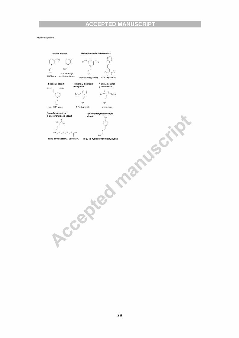
39
Afonso & Spickett

40
Afonso & Spickett

41
Afonso & Spickett
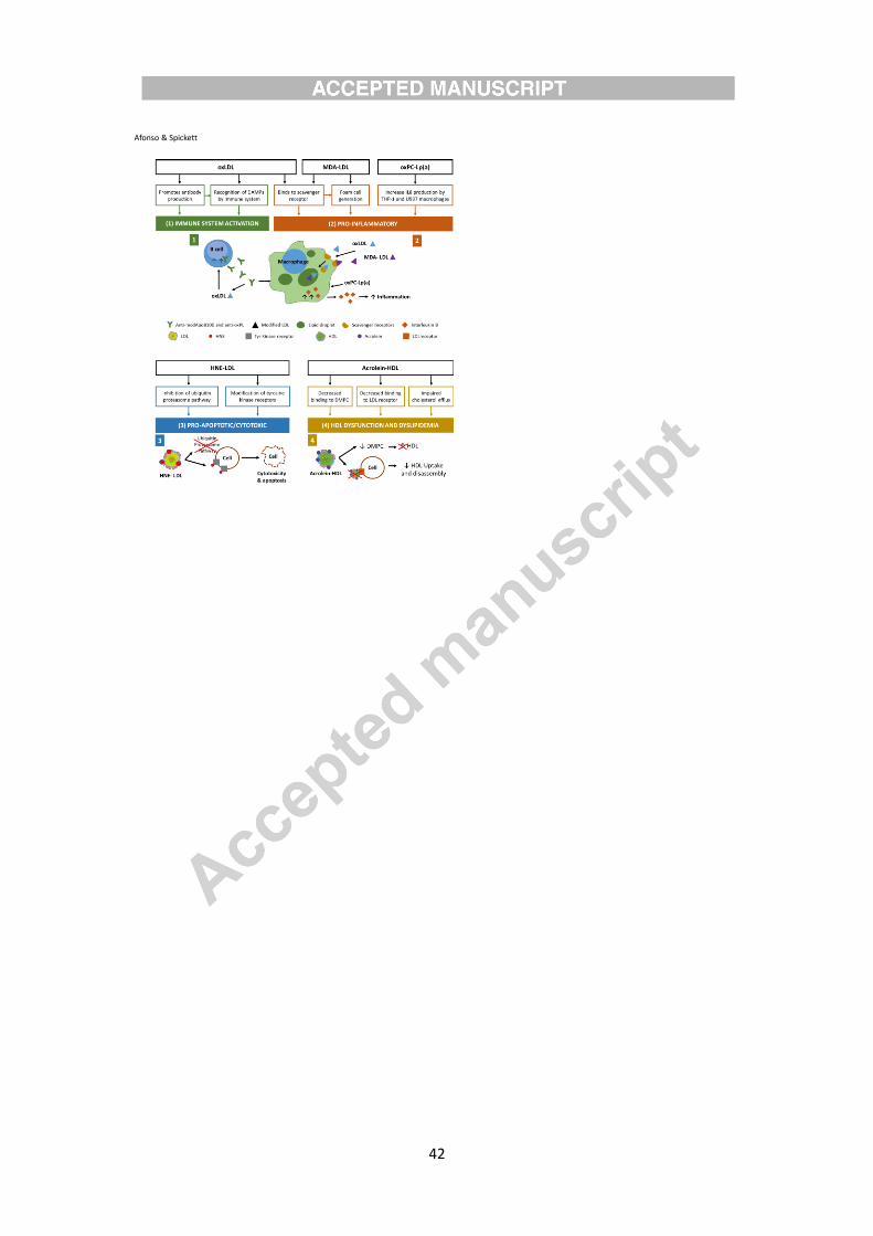
42
Afonso & Spickett

43
Highlights
• Lipoproteins can be modified by reactive Lipid Peroxidation Products (rLPPs)
• Lipoprotein lipoxidation is known to occur in several inflammatory diseases
• Biochemical, immunochemical and mass spectrometry methods can detect rLPP adducts
• Due to higher information output, MS can facilitate localization of modifications
• Antibodies against some rLPPs have been used to identify lipoxidation in vivo
Related Documents