Published Ahead of Print 15 December 2008. 10.1128/MCB.01062-08. 2009, 29(4):1059. DOI: Mol. Cell. Biol. E. D. Salmon, Terry Magnuson and Terry Van Dyke Yizhou Joseph He, Trudy G. Oliver, Lucy Lu, Ryan O'Quinn, Dale O. Cowley, Jaime A. Rivera-Pérez, Mark Schliekelman, Spindle Formation and Early Development Aurora-A Kinase Is Essential for Bipolar http://mcb.asm.org/content/29/4/1059 Updated information and services can be found at: These include: SUPPLEMENTAL MATERIAL Supplemental material REFERENCES http://mcb.asm.org/content/29/4/1059#ref-list-1 at: This article cites 70 articles, 25 of which can be accessed free CONTENT ALERTS more» articles cite this article), Receive: RSS Feeds, eTOCs, free email alerts (when new http://journals.asm.org/site/misc/reprints.xhtml Information about commercial reprint orders: http://journals.asm.org/site/subscriptions/ To subscribe to to another ASM Journal go to: on October 8, 2013 by UNIV OF UTAH http://mcb.asm.org/ Downloaded from on October 8, 2013 by UNIV OF UTAH http://mcb.asm.org/ Downloaded from on October 8, 2013 by UNIV OF UTAH http://mcb.asm.org/ Downloaded from on October 8, 2013 by UNIV OF UTAH http://mcb.asm.org/ Downloaded from on October 8, 2013 by UNIV OF UTAH http://mcb.asm.org/ Downloaded from on October 8, 2013 by UNIV OF UTAH http://mcb.asm.org/ Downloaded from on October 8, 2013 by UNIV OF UTAH http://mcb.asm.org/ Downloaded from on October 8, 2013 by UNIV OF UTAH http://mcb.asm.org/ Downloaded from on October 8, 2013 by UNIV OF UTAH http://mcb.asm.org/ Downloaded from on October 8, 2013 by UNIV OF UTAH http://mcb.asm.org/ Downloaded from on October 8, 2013 by UNIV OF UTAH http://mcb.asm.org/ Downloaded from on October 8, 2013 by UNIV OF UTAH http://mcb.asm.org/ Downloaded from on October 8, 2013 by UNIV OF UTAH http://mcb.asm.org/ Downloaded from on October 8, 2013 by UNIV OF UTAH http://mcb.asm.org/ Downloaded from

Welcome message from author
This document is posted to help you gain knowledge. Please leave a comment to let me know what you think about it! Share it to your friends and learn new things together.
Transcript
Published Ahead of Print 15 December 2008. 10.1128/MCB.01062-08.
2009, 29(4):1059. DOI:Mol. Cell. Biol. E. D. Salmon, Terry Magnuson and Terry Van DykeYizhou Joseph He, Trudy G. Oliver, Lucy Lu, Ryan O'Quinn, Dale O. Cowley, Jaime A. Rivera-Pérez, Mark Schliekelman, Spindle Formation and Early DevelopmentAurora-A Kinase Is Essential for Bipolar
http://mcb.asm.org/content/29/4/1059Updated information and services can be found at:
These include:
SUPPLEMENTAL MATERIAL Supplemental material
REFERENCEShttp://mcb.asm.org/content/29/4/1059#ref-list-1at:
This article cites 70 articles, 25 of which can be accessed free
CONTENT ALERTS more»articles cite this article),
Receive: RSS Feeds, eTOCs, free email alerts (when new
http://journals.asm.org/site/misc/reprints.xhtmlInformation about commercial reprint orders: http://journals.asm.org/site/subscriptions/To subscribe to to another ASM Journal go to:
on October 8, 2013 by U
NIV
OF
UT
AH
http://mcb.asm
.org/D
ownloaded from
on O
ctober 8, 2013 by UN
IV O
F U
TA
Hhttp://m
cb.asm.org/
Dow
nloaded from
on October 8, 2013 by U
NIV
OF
UT
AH
http://mcb.asm
.org/D
ownloaded from
on O
ctober 8, 2013 by UN
IV O
F U
TA
Hhttp://m
cb.asm.org/
Dow
nloaded from
on October 8, 2013 by U
NIV
OF
UT
AH
http://mcb.asm
.org/D
ownloaded from
on O
ctober 8, 2013 by UN
IV O
F U
TA
Hhttp://m
cb.asm.org/
Dow
nloaded from
on October 8, 2013 by U
NIV
OF
UT
AH
http://mcb.asm
.org/D
ownloaded from
on O
ctober 8, 2013 by UN
IV O
F U
TA
Hhttp://m
cb.asm.org/
Dow
nloaded from
on October 8, 2013 by U
NIV
OF
UT
AH
http://mcb.asm
.org/D
ownloaded from
on O
ctober 8, 2013 by UN
IV O
F U
TA
Hhttp://m
cb.asm.org/
Dow
nloaded from
on October 8, 2013 by U
NIV
OF
UT
AH
http://mcb.asm
.org/D
ownloaded from
on O
ctober 8, 2013 by UN
IV O
F U
TA
Hhttp://m
cb.asm.org/
Dow
nloaded from
on October 8, 2013 by U
NIV
OF
UT
AH
http://mcb.asm
.org/D
ownloaded from
on O
ctober 8, 2013 by UN
IV O
F U
TA
Hhttp://m
cb.asm.org/
Dow
nloaded from
MOLECULAR AND CELLULAR BIOLOGY, Feb. 2009, p. 1059–1071 Vol. 29, No. 40270-7306/09/$08.00�0 doi:10.1128/MCB.01062-08Copyright © 2009, American Society for Microbiology. All Rights Reserved.
Aurora-A Kinase Is Essential for Bipolar Spindle Formation andEarly Development�†
Dale O. Cowley,1‡ Jaime A. Rivera-Perez,2§ Mark Schliekelman,3 Yizhou Joseph He,3Trudy G. Oliver,1¶ Lucy Lu,1 Ryan O’Quinn,4 E. D. Salmon,4
Terry Magnuson,2 and Terry Van Dyke1*Department of Genetics and Lineberger Comprehensive Cancer Center, University of North Carolina at Chapel Hill School of Medicine,
Chapel Hill, North Carolina 275991; Department of Genetics and Carolina Center for Genome Sciences, University ofNorth Carolina, Chapel Hill, North Carolina 27599-72642; Curriculum in Genetics, University of North Carolina at
Chapel Hill, Chapel Hill, North Carolina 275993; and Department of Biology, University of North Carolina atChapel Hill, Chapel Hill, North Carolina 275994
Received 8 July 2008/Returned for modification 1 August 2008/Accepted 27 November 2008
Aurora-A is a conserved kinase implicated in mitotic regulation and carcinogenesis. Aurora-A waspreviously implicated in mitotic entry and spindle assembly, although contradictory results prevented aclear understanding of the roles of Aurora-A in mammals. We developed a conditional null mutation inthe mouse Aurora-A gene to investigate Aurora-A functions in primary cells ex vivo and in vivo. We showhere that conditional Aurora-A ablation in cultured embryonic fibroblasts causes impaired mitotic entryand mitotic arrest with a profound defect in bipolar spindle formation. Germ line Aurora-A deficiencycauses embryonic death at the blastocyst stage with pronounced cell proliferation failure, mitotic arrest,and monopolar spindle formation. Aurora-A deletion in mid-gestation embryos causes an increase inmitotic and apoptotic cells. These results indicate that murine Aurora-A facilitates, but is not absolutelyrequired for, mitotic entry in murine embryonic fibroblasts and is essential for centrosome separation andbipolar spindle formation in vitro and in vivo. Aurora-A deletion increases apoptosis, suggesting thatmolecular therapies targeting Aurora-A may be effective in inducing tumor cell apoptosis. Aurora-Aconditional mutant mice provide a valuable system for further defining Aurora-A functions and forpredicting effects of Aurora-A therapeutic intervention.
The equal partitioning of chromosomes at mitosis is criticalfor avoiding aneuploidy, a condition associated with spontane-ous miscarriage, developmental disorders, and cancer (50).Mitosis requires coordinated completion of multiple eventsincluding nuclear envelope breakdown, chromosome conden-sation and congression to the metaphase plate, centrosomeseparation, spindle formation, chromosome-spindle attach-ment and error correction, sister chromatid separation, andcytokinesis. Multiple regulators, many of which are kinases, arerequired to ensure that each event is completed in a timelyfashion and in the proper order (reviewed in reference 46).Although a number of mitotic kinases have been identified,their targets and the intricacies of mitotic signal transductionpathways are just beginning to be understood.
The Aurora kinases are key mitotic regulators in eukaryotes(reviewed in reference 45). The Aurora family includes a single
member in yeasts (Saccharomyces cerevisiae Ipl1p, Schizosac-charomyces pombe Ark1), two members each in Caenorhabditiselegans and Drosophila, and two or three members in verte-brates. Although originally given a variety of names, Aurorakinases in multicellular eukaryotes have subsequently beenclassified into A, B, and C groups based on patterns of mitoticsubcellular localization and homology, which also appear toreflect functional distinctions (8, 46). Aurora-A kinases areobserved at centrosomes and adjacent spindle fibers, and cur-rent evidence supports key roles in regulating protein localiza-tion and function at centrosomes, as well as regulation of theassembly, stability, and function of the mitotic spindle (re-viewed in reference 43). Aurora-B kinases display “chromo-somal passenger” localization, residing on mitotic chromo-somes and subsequently moving to the spindle midzone afterseparation of sister chromatids. Aurora-B family membershave been implicated in the regulation of kinetochore-spindleattachment, the spindle checkpoint, and cytokinesis (reviewedin references 1 and 8). Aurora-C kinases, which have only beenidentified in mammals, have a limited expression pattern andappear to have functions that overlap those of Aurora-B (7,53).
The human Aurora-A kinase (hAurA) was first identifiedbecause of its overexpression in cancer cell lines (5, 58). ThehAurA gene (stk15) resides on chromosome 20q13, a regionfrequently amplified in human cancers (5, 58). hAurA has beendubbed an oncogene because of the fact that its overexpressiontransforms immortalized rodent fibroblasts (5, 70). Polymor-
* Corresponding author. Mailing address: School of Medicine,CB7299, University of North Carolina at Chapel Hill, Chapel Hill, NC27599. Phone: (919) 962-2145. Fax: (919) 843-3160. E-mail: [email protected].
† Supplemental material for this article may be found at http://mcb.asm.org/.
‡ Present address: GlaxoSmithKline, Research Triangle Park, NC27509.
§ Present address: Department of Cell Biology, University of Mas-sachusetts Medical School, Worcester, MA 01655.
¶ Present address: Koch Institute for Integrative Cancer Research atMIT, Massachusetts Institute of Technology, Cambridge, MA 02142.
� Published ahead of print on 15 December 2008.
1059
phisms in hAurA are associated with an increased risk of coloncancer, while murine AurA (mAurA) polymorphisms conferincreased susceptibility to experimentally induced skin tumors(14). The mAurA gene is frequently amplified in radiation-induced lymphomas from p53 heterozygous mice, while loss ofone mAurA allele has been observed in lymphomas from p53-null mice (41). Thus, aberrant AurA expression is associatedwith tumorigenesis, suggesting that insight into AurA functionswill lead to a better understanding of tumorigenesis mecha-nisms.
A number of experimental observations suggest that AurAkinases are required for normal centrosome maturation andbipolar spindle assembly. The AurA ortholog in Drosophilamelanogaster (Aurora) was identified in a screen for mutationsthat impact the centrosome cycle (21). Syncytial embryos fromhypomorphic Aurora mutant females display a variety of mi-totic abnormalities resulting from a failure to separate centro-somes. Aurora-null flies die at the larval stage with character-istic monopolar spindles and circular chromosome arrays inlarval neuroblasts. Such monopolar spindles arise from failedcentrosome separation (21). Subsequent studies of DrosophilaAurora mutant alleles revealed additional defects in centro-some maturation (including a failure to localize transformingacidic coiled-coil protein, centrosomin, and �-tubulin at cen-trosomes) and in asymmetric localization of Numb protein insensory organ precursor cells (3, 17). Similar to the case inDrosophila, disruption of the C. elegans AurA ortholog AIR-1by RNA interference (RNAi) or mutation causes defects incentrosome maturation and monopolar spindle formation.Centrosomes undergo normal separation but collapse, leadingto monopolar spindle formation (16, 24, 56). Studies of theXenopus AurA homolog pEg2 revealed similar phenotypes af-ter overexpression of kinase-dead mutants, antibody-mediatedinhibition, or immunodepletion (18, 19, 38, 52). Furthermore,Xenopus AurA has been shown to interact with and phosphor-ylate Eg5, a mitotic kinesin required for bipolar spindle for-mation, suggesting a possible mechanism by which AurA couldinfluence bipolar spindle formation and/or stabilization (19).Thus, existing reports from these systems are quite consistentin implicating AurA in centrosome separation and function.
In contrast to the systems described above, published reportsof RNAi-mediated reduction of AurA expression in mamma-lian cell lines have contained conflicting results about the roleof AurA in mitotic entry, bipolar spindle formation, and mi-totic progression. AurA RNAi in HeLa cells was reported toblock or delay mitotic entry, prompting the conclusion thatAurA is essential for mitotic commitment in mammalian cells(27, 36). In contrast, other AurA RNAi studies showed accu-mulation of mitotic cells with monopolar spindles (12, 20, 67).These discrepancies call into question the functional conser-vation of AurA in mammals and highlight a need for additionalstudies to definitively address the roles of AurA. This is par-ticularly critical for understanding the roles of AurA in cancerand for projecting possible effects of AurA inhibitors currentlyin development as anticancer agents. We used gene targetingin mouse embryonic stem (ES) cells to produce a conditionalnull allele at the AurA locus. Here we describe cellular phe-notypes of AurA deletion in primary cells in vitro and devel-opmental phenotypes of AurA mutant mice. We show thatAurA deletion in primary embryonic fibroblasts causes delayed
mitotic entry with accumulation of cells in early prophase,consistent with a role for AurA in mitotic entry. Nevertheless,AurA-deficient cells that enter prometaphase arrest withmonopolar spindles and eventually exit mitosis without seg-regating their chromosomes. Prolonged culture of AurA-deficient cells leads to polyploidy with abnormal nuclear struc-ture. Germ line AurA deficiency causes embryonic death at theblastocyst stage with mitotic arrest and monopolar spindleformation, while AurA deletion in mid-gestation embryoscauses an increased mitotic index and increased apoptosis.Together, our findings indicate that AurA is required fortimely mitotic entry and bipolar spindle formation in vitro andin vivo.
MATERIALS AND METHODS
AurA gene targeting and mice. AurA genomic bacterial artificial chromosomeclones were isolated from a 129SvEv library by hybridization screening andsubsequent PCR verification. Bacterial strain EL350 (37) was used for “recom-bineering” to subclone a 13-kb fragment encompassing AurA exons 1 to 6 into apUC19-based plasmid including a negative-selection thymidine kinase gene (agift of R. Thresher, University of North Carolina animal models core facility). AloxP site was inserted into intron 2 by recombineering a loxP-flanked zeocincassette, followed by Cre-mediated cassette removal in EL350. A loxP- andFRT-flanked neomycin resistance cassette was subsequently inserted into aunique SalI restriction site in intron 1. The targeting vector was linearized andused for gene targeting in line E14 ES cells (129P2/OlaHsd strain). ES celltargeting was verified by PCR and Southern blotting, and correctly targetedclones were injected into C57BL/6J blastocysts for chimera generation. Chimericmales were bred with C57BL/6J females, and germ line transmission of theAurAneo allele was verified by PCR. The AurAd2 and AurAf alleles were generatedby crossing the AurAneo allele with an X-linked CMV-Cre strain (61). Recom-bined alleles were backcrossed for at least 2 generations to C57BL/6J micebefore use in this study. R26CreER (2) and R26Rep (60) mice were obtained fromJackson Laboratories. For the PCR primers used and genotyping information forthe AurA mutant alleles, see Table S1 in the supplemental material.
MEFs. Murine embryonic fibroblasts (MEFs) were generated from embryonicday 14.5 (E14.5) embryos by standard protocols. MEFs were grown in Dulbeccomodified Eagle medium (DMEM) with 4,500 mg/liter D-glucose and supple-mented with 10% fetal bovine serum (FBS), 2 mM L-glutamine, 50 U/ml peni-cillin, 50 �g/ml streptomycin, and 55 �M 2-mercaptoethanol in a humidifiedincubator with 5% CO2 and passaged every 2 to 3 days. All experiments wereperformed with cells at passages 3 to 5. �-Galactosidase detection was performedby a standard 5-bromo-4-chloro-3-indolyl-�-D-galactopyranoside (X-Gal) stain-ing protocol (59). For immunofluorescence (IF) experiments, cells were grownon acid-washed glass coverslips coated with poly-L-lysine. Coverslips were fixedin 2% formaldehyde in phosphate-buffered saline (PBS) for 20 to 30 min andstored in PBS with 0.1% sodium azide at 4°C until used. Primary antibodies wereanti-Aurora-A (IAK1; BD Biosciences no. 610938, 1:500 dilution), anti-�-tubulin(rat monoclonal, 1:200 dilution), anti-�-tubulin (Sigma T5192, 1:1,000 dilution),anti-�-tubulin (Sigma T4026, 1:200 dilution), anti-XMad2 (affinity-purified rab-bit polyclonal, 1:1,000 dilution) (66), and anti-phosphorylated histone H3 (anti-PH3; serine 10; Upstate Biotech, 06-570, 1:500 dilution). 4-Hydroxytamoxifen(OHT; Sigma H7904) was dissolved in 100% ethanol and used at 50 nM for allexperiments.
Flow cytometry. Adherent cells were removed from plates by trypsinizationand pooled with cell culture supernatant containing nonadherent cells. Cellswere washed once with PBS, fixed in cold 70% ethanol, and stored at �20°C untilanalyzed. For staining, 1 � 106 cells were washed in PBS and stained in PBS with50 �g/ml propidium iodide, 200 �g/ml boiled RNase A, and 0.1% Triton X-100.Analyses were performed on a BD FACScan flow cytometer and analyzed withSummit software (Dako).
Time-lapse imaging. AurA�/�; R26CreER/� and AurAf/f; R26CreER/� cells wereincubated in DMEM–0.1% FBS for 24 h, after which 50 nM OHT was added foran additional 24 h of incubation. Cell cycle entry was stimulated by replacing themedium with fresh DMEM with 10% FBS for 14 h of incubation, followed by theaddition of fresh DMEM–10% FBS–10 mM HEPES, pH 7.4, for an additional3 h of incubation prior to live-cell imaging. Time-lapse images were captured onan Olympus IX70 microscope with a custom-built environmental chamber. Im-ages were taken every 10 min for 50 h. Images were analyzed with OpenLab
1060 COWLEY ET AL. MOL. CELL. BIOL.
software (Improvision) and QuickTime (Apple). Mitotic entry was judged as thepoint of cell rounding, and mitotic exit was judged as the point at which cellsflattened out. Each experiment was repeated with MEFs from two differentembryos. Differences in mitotic timing were compared by Kaplan-Meier analysis.For representative movies and phenotype images, see the supplemental material.
Immunoblotting. Cells were harvested at the indicated time points, washed inPBS, counted, and lysed by boiling in sodium dodecyl sulfate (SDS) samplebuffer (60 mM Tris-HCl [pH 6.8], 10% glycerol, 2% SDS, 100 mM dithiothrei-tol). Volumes representing equal cell numbers were separated by 4 to 20%SDS-polyacrylamide gel electrophoresis and transferred to polyvinylidene diflu-oride membranes. Membranes were blocked in 5% nonfat dry milk in PBS with0.1% Tween 20 (block) and incubated with antibodies diluted in block at roomtemperature (RT) for 1 to 2 h or overnight at 4°C. Membranes were washedextensively in PBS with 0.1% Tween 20 and incubated with horseradish perox-idase-conjugated secondary antibodies for 1 h at RT in block. Following exten-sive washes, targets were detected with a SuperSignal West Pico Chemilumines-cent Substrate kit (Pierce).
Blastocyst culture and staining. Timed matings were set, and the morning ofvaginal plug detection was considered E0.5. Blastocysts were flushed from theuteri of pregnant females at E3.5. Blastocysts were cultured in drops of DMEMwith 4,500 mg/liter D-glucose, 15% FBS, 2 mM L-glutamine, 50 U/ml penicillin,50 �g/ml streptomycin, and 55 �M 2-mercaptoethanol overlaid with mineral oilin a humidified incubator with 5% CO2. Outgrowths were photographed after 5to 7 days and harvested for genotyping. DNA lysates were prepared by theHotShot method (63). For immunostaining, freshly isolated blastocysts wererinsed in PBS and fixed in fresh 4% formaldehyde in PBS for 30 to 60 min at RT.Blastocysts were subsequently rinsed in PBS; washed three times for 10 min eachin PBS with 1% bovine serum albumin and 0.5% Triton X-100 (PBT); blockedfor 1 h in PBT with 5% goat serum (Block); incubated overnight with primaryantibodies diluted in block; washed three times for 10 min each in PBT; incu-bated for 1 h with secondary antibodies in PBT; washed three times for 10 mineach in PBT, one time in PBS, once in a 1:2 mixture of glycerol-PBS, and oncein 1:1 glycerol-PBS; and mounted in mounting medium with 4�,6�-diamidino-2-phenylindole (DAPI). All incubations were performed at 25°C. The primaryantibodies used were mouse monoclonal anti-Aurora-A (IAK1; BD Biosciencesno. 610938, 1:500 dilution) and rat monoclonal anti-�-tubulin (1:200 dilution).Images were analyzed on a Leica SP2 laser scanning confocal microscope with a40� objective (1.25 numerical aperture).
RESULTS
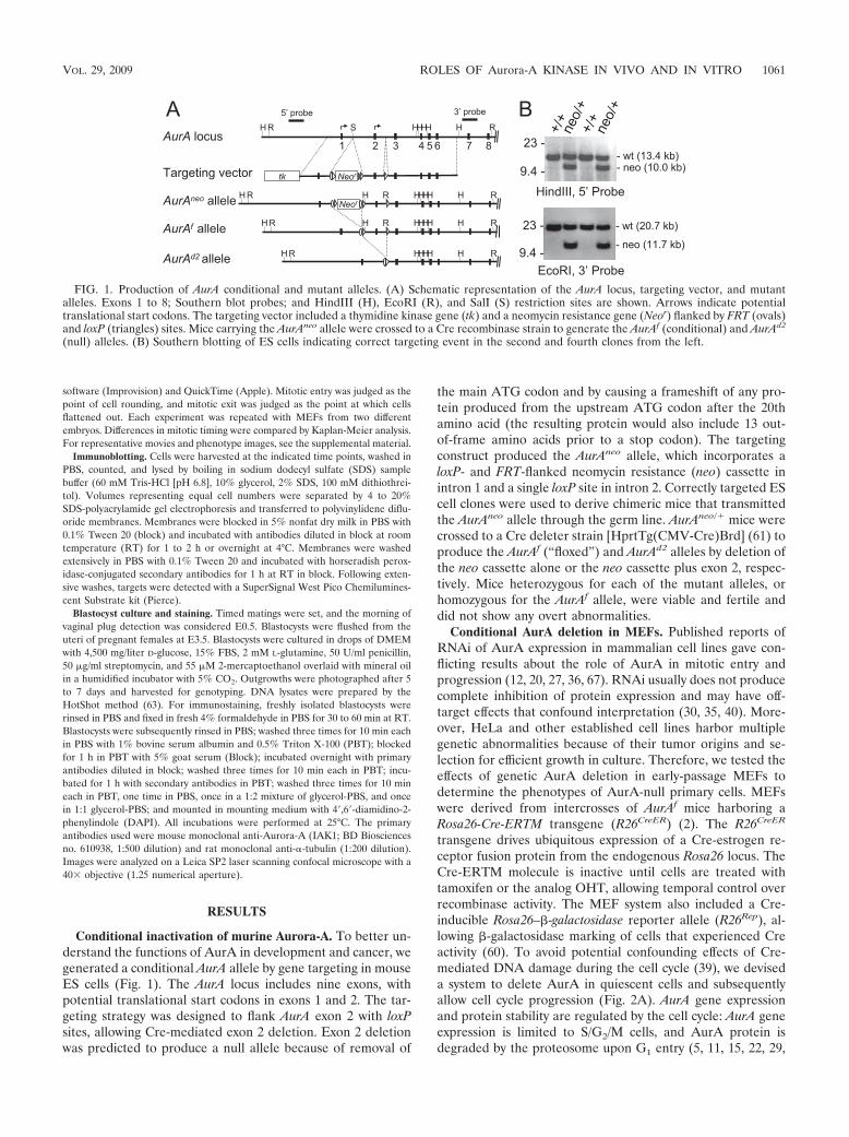
Conditional inactivation of murine Aurora-A. To better un-derstand the functions of AurA in development and cancer, wegenerated a conditional AurA allele by gene targeting in mouseES cells (Fig. 1). The AurA locus includes nine exons, withpotential translational start codons in exons 1 and 2. The tar-geting strategy was designed to flank AurA exon 2 with loxPsites, allowing Cre-mediated exon 2 deletion. Exon 2 deletionwas predicted to produce a null allele because of removal of
the main ATG codon and by causing a frameshift of any pro-tein produced from the upstream ATG codon after the 20thamino acid (the resulting protein would also include 13 out-of-frame amino acids prior to a stop codon). The targetingconstruct produced the AurAneo allele, which incorporates aloxP- and FRT-flanked neomycin resistance (neo) cassette inintron 1 and a single loxP site in intron 2. Correctly targeted EScell clones were used to derive chimeric mice that transmittedthe AurAneo allele through the germ line. AurAneo/� mice werecrossed to a Cre deleter strain [HprtTg(CMV-Cre)Brd] (61) toproduce the AurAf (“floxed”) and AurAd2 alleles by deletion ofthe neo cassette alone or the neo cassette plus exon 2, respec-tively. Mice heterozygous for each of the mutant alleles, orhomozygous for the AurAf allele, were viable and fertile anddid not show any overt abnormalities.
Conditional AurA deletion in MEFs. Published reports ofRNAi of AurA expression in mammalian cell lines gave con-flicting results about the role of AurA in mitotic entry andprogression (12, 20, 27, 36, 67). RNAi usually does not producecomplete inhibition of protein expression and may have off-target effects that confound interpretation (30, 35, 40). More-over, HeLa and other established cell lines harbor multiplegenetic abnormalities because of their tumor origins and se-lection for efficient growth in culture. Therefore, we tested theeffects of genetic AurA deletion in early-passage MEFs todetermine the phenotypes of AurA-null primary cells. MEFswere derived from intercrosses of AurAf mice harboring aRosa26-Cre-ERTM transgene (R26CreER) (2). The R26CreER
transgene drives ubiquitous expression of a Cre-estrogen re-ceptor fusion protein from the endogenous Rosa26 locus. TheCre-ERTM molecule is inactive until cells are treated withtamoxifen or the analog OHT, allowing temporal control overrecombinase activity. The MEF system also included a Cre-inducible Rosa26–�-galactosidase reporter allele (R26Rep), al-lowing �-galactosidase marking of cells that experienced Creactivity (60). To avoid potential confounding effects of Cre-mediated DNA damage during the cell cycle (39), we deviseda system to delete AurA in quiescent cells and subsequentlyallow cell cycle progression (Fig. 2A). AurA gene expressionand protein stability are regulated by the cell cycle: AurA geneexpression is limited to S/G2/M cells, and AurA protein isdegraded by the proteosome upon G1 entry (5, 11, 15, 22, 29,
FIG. 1. Production of AurA conditional and mutant alleles. (A) Schematic representation of the AurA locus, targeting vector, and mutantalleles. Exons 1 to 8; Southern blot probes; and HindIII (H), EcoRI (R), and SalI (S) restriction sites are shown. Arrows indicate potentialtranslational start codons. The targeting vector included a thymidine kinase gene (tk) and a neomycin resistance gene (Neor) flanked by FRT (ovals)and loxP (triangles) sites. Mice carrying the AurAneo allele were crossed to a Cre recombinase strain to generate the AurAf (conditional) and AurAd2
(null) alleles. (B) Southern blotting of ES cells indicating correct targeting event in the second and fourth clones from the left.
VOL. 29, 2009 ROLES OF Aurora-A KINASE IN VIVO AND IN VITRO 1061
52, 62, 64). We reasoned that if the Cre recombination oc-curred in G0- or G1-phase cells, the cells should lack AurAprotein as they traverse subsequent cell cycle phases, providinga clean system in which to detect mitotic phenotypes. Cellswere arrested by 24 h of serum starvation, followed by 2 daysof additional starvation in the presence of OHT to induceCre-ERTM activity in the quiescent cells. Cells were then fedserum-containing medium without OHT to induce cell cycleentry in the absence of Cre activity. This protocol gave efficientrecombination of the R26 reporter allele (85%) and produc-tion of the Aurd2 allele as assessed by induction of �-galacto-sidase and PCR, respectively (not shown). A background ofapproximately 5% recombination was observed in untreatedcultures (data not shown). Immunoblot analyses of controlcells (AurAf/f; R26CreER/Rep without OHT and AurA�/�;R26CreER/Rep with OHT) showed the expected pattern of AurAexpression (Fig. 2B). Very little AurA is detected in arrestedcells; the protein is abundant at 24 h, the time when the mitoticindex peaks in the population, and intermediate levels aredetected at 48 h, when the population has reverted to moreasynchronous growth and fewer cells are in mitosis (Fig. 2B).In contrast, AurA protein was undetectable in OHT-treatedAurAf/f; R26CreER/Rep MEFs at all of the time points tested (Fig.2B). Semiquantitative reverse transcription-PCR analysis dem-onstrated very similar AurA mRNA levels in OHT-treatedAurAf/f; R26CreER/Rep and AurA�/�; R26CreER/Rep MEFs, al-though primers spanning exon 2 clearly demonstrated that thedeletion had occurred in AurAf/f; R26CreER/Rep cells (Fig. S1).Thus, the deletion of exon 2 causes a loss of protein expression,as expected, despite the continued presence of the AurAmRNA with exon 2 deleted. Together, these results indicatethat the R26CreER transgene provides efficient conversion ofthe AurAf allele to the AurAd2 allele and that the AurAd2 alleleis null for protein production. The conditional AurA MEFsthus provide an excellent system in which to determine theeffects of AurA deletion on cell cycle progression.
To determine phenotypes of AurA loss, we first tested thegrowth and ploidy of AurA mutant MEFs over a 5-day timecourse after Cre-mediated deletion. OHT-treated AurAf/f;R26CreER/Rep MEFs displayed a clear growth defect relative tocontrols after serum addition (Fig. 2C). Controls included
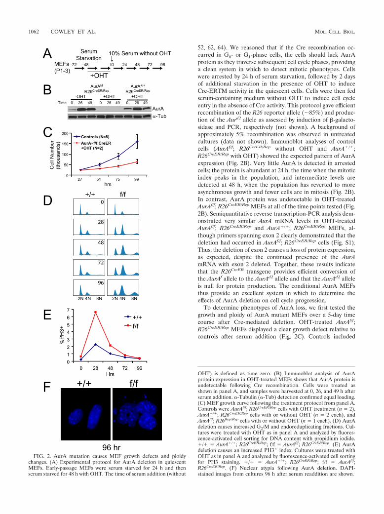
FIG. 2. AurA mutation causes MEF growth defects and ploidychanges. (A) Experimental protocol for AurA deletion in quiescentMEFs. Early-passage MEFs were serum starved for 24 h and thenserum starved for 48 h with OHT. The time of serum addition (without
OHT) is defined as time zero. (B) Immunoblot analysis of AurAprotein expression in OHT-treated MEFs shows that AurA protein isundetectable following Cre recombination. Cells were treated asshown in panel A, and samples were harvested at 0, 26, and 49 h afterserum addition. �-Tubulin (�-Tub) detection confirmed equal loading.(C) MEF growth curve following the treatment protocol from panel A.Controls were AurAf/f; R26CreER/Rep cells with OHT treatment (n 2),AurA�/�; R26CreER/Rep cells with or without OHT (n 2 each), andAurAf/f; R26Rep/Rep cells with or without OHT (n 1 each). (D) AurAdeletion causes increased G2/M and endoreduplicating fractions. Cul-tures were treated with OHT as in panel A and analyzed by fluores-cence-activated cell sorting for DNA content with propidium iodide.�/� AurA�/�; R26CreER/Rep; f/f AurAf/f; R26CreER/Rep. (E) AurAdeletion causes an increased PH3� index. Cultures were treated withOHT as in panel A and analyzed by fluorescence-activated cell sortingfor PH3 staining. �/� AurA�/�; R26CreER/Rep; f/f AurAf/f;R26CreER/Rep. (F) Nuclear atypia following AurA deletion. DAPI-stained images from cultures 96 h after serum readdition are shown.
1062 COWLEY ET AL. MOL. CELL. BIOL.
AurAf/f; R26CreER/Rep cells without OHT treatment andAurA�/�; R26CreER/Rep cells with and without OHT treatment.No growth defects were observed in response to OHT treat-ment in controls, indicating that Cre activity in quiescent cellsdoes not impact the subsequent cell cycle. We also examinedthe cell cycle and ploidy by flow cytometry (Fig. 2D). Controlcells had a normal cell cycle and ploidy profile at all of the timepoints examined. In contrast, AurA-deficient cultures dis-played a dramatic increase in cells with 4N DNA content 28 hafter serum addition, and this population remained elevatedthroughout the 96-h observation period. AurA mutant culturesalso displayed an increasing 8N population over the course ofthe experiment (Fig. 2D). The population of AurA mutantcells with 4N or greater (4N�) DNA content increased from50% at time zero to 80% at 96 h, while the 4N� populationin control cells remained near 50% across all of the time pointstested.
PH3 is widely used as a marker of mitotic cells. PH3 immu-noreactivity is detectable in punctate foci at centromeres be-ginning in G2 phase and spreads across the chromatin justprior to chromosome condensation in prophase (31). To de-termine if the increased 4N population in AurA mutant cul-tures was attributable to mitotic arrest, we analyzed the PH3index of cells with 4N and 8N DNA contents. Both wild-type(WT) and AurA-deficient cultures displayed a peak PH3 indexat 28 h, with reduced levels at each subsequent time point (Fig.2E). AurA-deficient cells showed an approximately threefoldhigher peak PH3 index than controls at 28 h and maintained ahigher PH3 index than controls at each time point until 96 h.PH3� cells were observed in the 4N population, as expected,but also in the 8N population, indicating that some polyploidcells enter mitosis (data not shown). AurA-deficient cells atlater time points displayed large, aberrant nuclei with frequentmicronuclei (Fig. 2F), indicating that AurA deficiency causesmitotic abnormalities over the course of multiple cell cycles.Together, these data suggest that AurA deficiency causes amitotic-arrest phenotype. It is notable that the peak PH3 indexin AurA mutant cultures was only 7%, while the 4N and 8Npopulations accounted for up to 80% of the total cells. Thisdiscrepancy likely reflects the fact that AurA mutant cells aredelayed in mitotic entry (see below), such that the true peakmitotic index was later in mutant cultures and was not capturedat the time points chosen for this experiment.
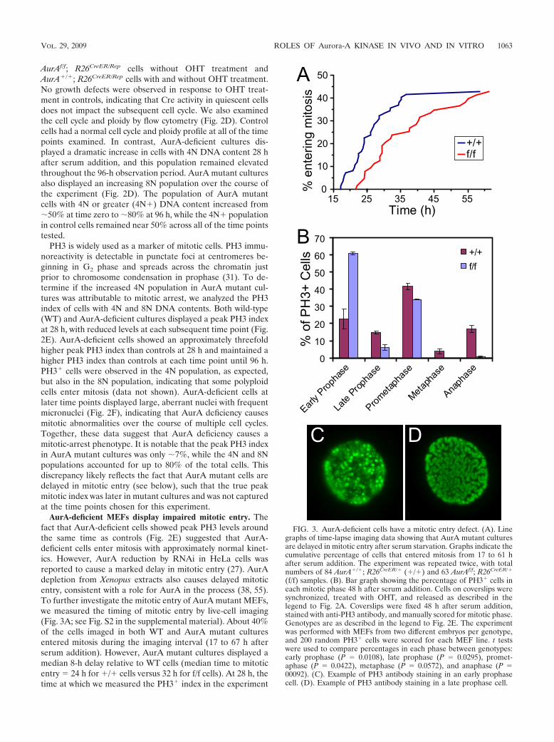
AurA-deficient MEFs display impaired mitotic entry. Thefact that AurA-deficient cells showed peak PH3 levels aroundthe same time as controls (Fig. 2E) suggested that AurA-deficient cells enter mitosis with approximately normal kinet-ics. However, AurA reduction by RNAi in HeLa cells wasreported to cause a marked delay in mitotic entry (27). AurAdepletion from Xenopus extracts also causes delayed mitoticentry, consistent with a role for AurA in the process (38, 55).To further investigate the mitotic entry of AurA mutant MEFs,we measured the timing of mitotic entry by live-cell imaging(Fig. 3A; see Fig. S2 in the supplemental material). About 40%of the cells imaged in both WT and AurA mutant culturesentered mitosis during the imaging interval (17 to 67 h afterserum addition). However, AurA mutant cultures displayed amedian 8-h delay relative to WT cells (median time to mitoticentry 24 h for �/� cells versus 32 h for f/f cells). At 28 h, thetime at which we measured the PH3� index in the experiment
FIG. 3. AurA-deficient cells have a mitotic entry defect. (A). Linegraphs of time-lapse imaging data showing that AurA mutant culturesare delayed in mitotic entry after serum starvation. Graphs indicate thecumulative percentage of cells that entered mitosis from 17 to 61 hafter serum addition. The experiment was repeated twice, with totalnumbers of 84 AurA�/�; R26CreER/� (�/�) and 63 AurAf/f; R26CreER/�
(f/f) samples. (B). Bar graph showing the percentage of PH3� cells ineach mitotic phase 48 h after serum addition. Cells on coverslips weresynchronized, treated with OHT, and released as described in thelegend to Fig. 2A. Coverslips were fixed 48 h after serum addition,stained with anti-PH3 antibody, and manually scored for mitotic phase.Genotypes are as described in the legend to Fig. 2E. The experimentwas performed with MEFs from two different embryos per genotype,and 200 random PH3� cells were scored for each MEF line. t testswere used to compare percentages in each phase between genotypes:early prophase (P 0.0108), late prophase (P 0.0295), promet-aphase (P 0.0422), metaphase (P 0.0572), and anaphase (P 00092). (C). Example of PH3 antibody staining in an early prophasecell. (D). Example of PH3 antibody staining in a late prophase cell.
VOL. 29, 2009 ROLES OF Aurora-A KINASE IN VIVO AND IN VITRO 1063
shown in Fig. 2E, only 12% of the AurA mutant cells hadentered mitosis, compared to 30% of the WT cells. In thislight, the mitotic index of 7% observed for AurA mutantcultures at 28 h suggests that AurA mutant cells are signifi-cantly delayed in mitosis, as well as prior to mitotic entry.
To better understand the delay in mitotic entry and mitoticpassage of AurA-deficient MEFs, we used IF with fixed cells toquantitate the percentage of PH3� cells in each mitotic phaseat 26, 30, and 48 h after serum addition (Fig. 3B and data notshown). AurA mutant cultures had two striking differencesfrom controls. First, we noted a marked increase in prophasecells in mutants (67% versus 38% in WT cells, P 0.018).Moreover, the majority of AurA mutant prophase cells dis-played an early prophase phenotype with moderate PH3 stain-ing and only partially condensed chromosomes (Fig. 3C, forexample). Only 6% of prophase cells in AurA mutant culturesshowed a late prophase phenotype with fully condensed chro-mosomes (Fig. 3A and D). In contrast, control cultures hadapproximately equal percentages of early- and late prophasecells. Although PH3 staining is widely used as a marker ofmitosis, phosphorylation of histone H3 begins in late G2 and isthus not a definitive mitotic marker (26). Cytological featuressuch as chromosome condensation that have historically beenused to define mitotic entry are also not necessarily true indi-cators that a cell has committed to mitosis (47). However,nuclear envelope breakdown at the prophase-prometaphasetransition clearly indicates that the cell has passed the point ofno return for mitosis (47). The accumulation of early prophasecells in AurA-deficient cultures is thus consistent with a rolefor AurA in G2 progression events that prepare cells for mi-tosis. Nevertheless, the fact that a significant percentage ofcells enter prometaphase without AurA indicates that AurA isnot absolutely required for mitotic entry in vitro.
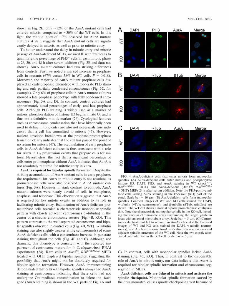
AurA is required for bipolar spindle formation. Despite thestriking accumulation of AurA mutant cells in early prophase,the requirement for AurA in mitotic entry is not absolute, asprometaphase cells were also observed in AurA mutant cul-tures (Fig. 3A). However, in stark contrast to controls, AurAmutant cultures were nearly devoid of cells in metaphase,anaphase, and telophase. This observation suggested that AurAis required for key mitotic events, in addition to its role infacilitating mitotic entry. Examination of AurA-deficient pro-metaphase cells revealed a characteristic monopolar spindlepattern with closely adjacent centrosomes (�-tubulin) in thecenter of a circular chromosome rosette (Fig. 4B, KO). Thispattern contrasts to the well-separated centrosomes and bipo-lar spindles observed in control cells (Fig. 4B, WT). �-Tubulinstaining was also slightly weaker at the centrosome(s) of someAurA-deficient cells, with a concomitant increase in punctatestaining throughout the cells (Fig. 4B and C). Although notdramatic, this phenotype is consistent with the reported im-pairment of centrosome maturation in C. elegans AurA RNAiexperiments (24). Rare cells in AurAf/f; R26CreER/Rep MEFstreated with OHT displayed bipolar spindles, suggesting thepossibility that AurA might not be absolutely required forbipolar spindle formation. However, AurA immunostainingdemonstrated that cells with bipolar spindles always had AurAstaining at centrosomes, indicating that these cells had notundergone Cre-mediated recombination to delete the AurAgene (AurA staining is shown in the WT parts of Fig. 4A and
C). In contrast, cells with monopolar spindles lacked AurAstaining (Fig. 4C, KO). Thus, in contrast to the dispensablerole of AurA in mitotic entry, our data indicate that AurA isrequired for bipolar spindle formation and chromosome seg-regation in MEFs.
AurA-deficient cells are delayed in mitosis and activate thespindle checkpoint. Monopolar spindle formation caused bythe drug monastrol causes spindle checkpoint arrest because of
FIG. 4. AurA-deficient cells that enter mitosis form monopolarspindles. (A) AurA-deficient cells enter mitosis and phosphorylatehistone H3. DAPI, PH3, and AurA staining in WT (AurA�/�;R26CreER/Rep �OHT) and AurA-deficient (AurAf/f; R26CreER/Rep
�OHT) MEFs 24 h after serum addition. Note the PH3-positive mi-totic cells lacking AurA staining in the knockout (KO) part of thepanel. Scale bar 10 �m. (B) AurA-deficient cells form monopolarspindles. Confocal images of WT and KO cells stained for DAPI,�-tubulin (�Tub; centrosomes), and �-tubulin (�Tub; spindles) areshown. The WT cell shows a normal bipolar prometaphase configura-tion. Note the characteristic monopolar spindle in the KO cell, includ-ing the circular chromosome array surrounding the single �-tubulinfocus with an astral microtubule array. Scale bar 5 �m. (C) Centro-somes duplicate but fail to separate in AurA-deficient cells. Confocalimages of WT and KO cells stained for DAPI, �-tubulin (centro-somes), and AurA are shown. AurA is localized on centrosomes andadjacent spindle structures of the WT cell. Note the two closely asso-ciated �-tubulin foci in the KO cell. Scale bar 5 �m.
1064 COWLEY ET AL. MOL. CELL. BIOL.
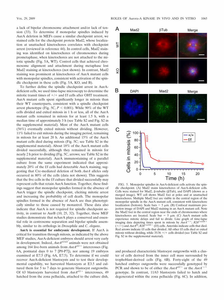
a lack of bipolar chromosome attachment and/or lack of ten-sion (33). To determine if monopolar spindles induced byAurA deletion in MEFs cause a similar checkpoint arrest, westained cells for the checkpoint protein Mad2, whose localiza-tion at unattached kinetochores correlates with checkpointarrest (reviewed in reference 44). In control cells, Mad2 stain-ing was identified on kinetochores of chromosomes duringprometaphase, when kinetochores are not attached to the mi-totic spindle (Fig. 5A, WT). Control cells that achieved chro-mosome alignment and attachment during metaphase lostMad2 staining at kinetochores (not shown). In contrast, Mad2staining was prominent at kinetochores of AurA mutant cellswith monopolar spindles, consistent with activation of the spin-dle checkpoint in these cells (Fig. 5A, KO, and B).
To further define the spindle checkpoint arrest in AurA-deficient cells, we used time-lapse microscopy to determine themitotic transit times of �/� and f/f cells after OHT treatment.AurA mutant cells spent significantly longer in mitosis thantheir WT counterparts, consistent with a spindle checkpointarrest phenotype (Fig. 5C, P � 0.001). While 90% of the WTcells divided and exited mitosis in 1 h or less, all of the AurAmutant cells remained in mitosis for at least 1.5 h, with amedian time of approximately 3 h (see Table S2 and Fig. S2 inthe supplemental material). Most of the AurA mutant cells(56%) eventually exited mitosis without dividing. However,11% failed to exit mitosis during the imaging period, remainingin mitosis for at least 20 h. An additional 15% of the AurAmutant cells died during mitosis (Fig. 5C; see Table S2 in thesupplemental material). About 18% of the AurA mutant cellsdivided successfully, although they remained in mitosis fornearly 2 h prior to dividing (Fig. 5C, arrows; see Table S2 in thesupplemental material). AurA immunostaining of a parallelculture from the same experiment indicated that approxi-mately 20% of the f/f cells had detectable AurA staining, sug-gesting that Cre-mediated deletion of both AurA alleles onlyoccurred in 80% of the cells (data not shown). This suggeststhat the five cells in the f/f culture that divided successfully mayrepresent cells that retain AurA function. Together, these find-ings suggest that monopolar spindles formed in the absence ofAurA trigger the spindle checkpoint, eliciting mitotic arrestand increasing the probability of cell death. The monopolarspindles formed in the absence of AurA are thus phenotypi-cally similar to those caused by monastrol. These data alsoindicate that AurA is not required for spindle checkpoint ac-tivity, in contrast to AurB (10, 25, 32). Together, these MEFstudies demonstrate that mAurA plays a conserved and essen-tial role in centrosome separation and bipolar spindle assem-bly, similar to its orthologs in Drosophila and C. elegans.
AurA is essential for embryonic development. If AurA iscritical for transition through mitosis, as indicated by the aboveMEF studies, AurA-deficient embryos should arrest very earlyin development. Indeed, AurAd2/d2 animals were not obtainedamong 164 live-born animals from AurAd2/� intercrosses (Fig.6A, postnatal days 0 to 7 [P0-P7]), nor among 19 embryosexamined at E7.5 (Fig. 6A, E7.5). To determine if we couldrecover AurA-deficient blastocysts and to test their develop-mental capability, we harvested blastocysts at E3.5 and cul-tured them for 5 to 7 days to generate blastocyst outgrowths.Of 63 blastocysts harvested from AurAd2/� intercrosses, 49hatched from the zona pellucida, attached to the culture dish,
and produced characteristic blastocyst outgrowths with a clus-ter of cells derived from the inner cell mass surrounded bytrophoblast-derived cells (Fig. 6B). Forty-eight of the 49hatched embryo outgrowths were successfully genotyped byPCR and shown to be of either the AurAd2/� or the AurA�/�
genotype. In contrast, 11/63 blastocysts failed to hatch anddegenerated within the zona pellucida (Fig. 6C). In addition,
FIG. 5. Monopolar spindles in AurA-deficient cells activate the spin-dle checkpoint. (A) Mad2 stains kinetochores of AurA-deficient cells.Cells were stained for Mad2, �-tubulin (�Tub), and DAPI (shown as amerged image). WT cell shows Mad2 foci at poles and at unattachedkinetochores. Multiple Mad2 foci are visible in the central region of themonopolar spindle in the AurA mutant cell, consistent with kinetochorelocalization (bottom). Scale bars 5 �m. (B) Confocal maximum pro-jection image of DAPI and Mad2 staining in an AurA mutant cell. Notethe Mad2 foci in the central region near the ends of chromosomes wherekinetochores are located. Scale bar 5 �m. (C) AurA mutant cellsexperience mitotic delays and fail to divide. Line graph of time-lapseimaging data depicting times spent in mitosis by AurA�/�; R26CreER/�
(�/�) and AurAf/f; R26CreER/� (f/f) cells. Asterisks indicate cells that died.Red arrows indicate f/f cells that divided. All other f/f cells died or exitedmitosis without dividing, while 35/36 �/� cells divided (see Table S2 andFig. S2 in the supplemental material).
VOL. 29, 2009 ROLES OF Aurora-A KINASE IN VIVO AND IN VITRO 1065
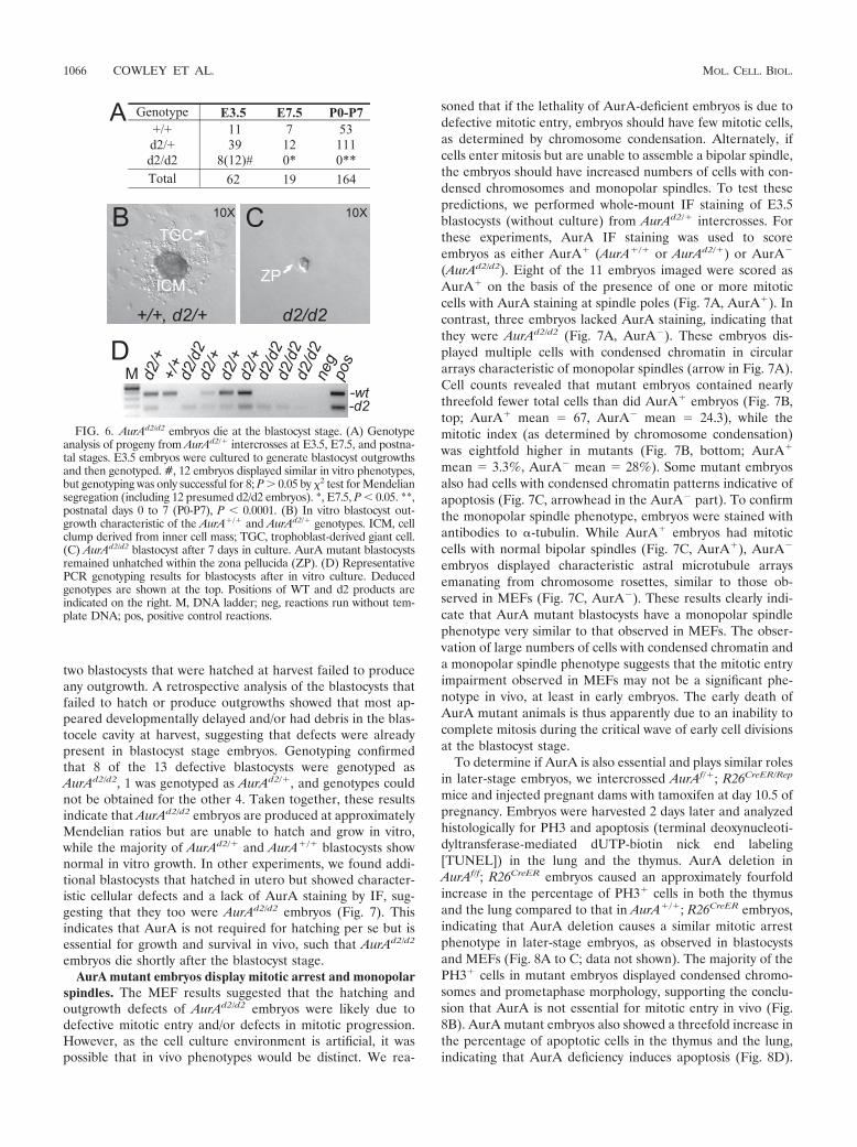
two blastocysts that were hatched at harvest failed to produceany outgrowth. A retrospective analysis of the blastocysts thatfailed to hatch or produce outgrowths showed that most ap-peared developmentally delayed and/or had debris in the blas-tocele cavity at harvest, suggesting that defects were alreadypresent in blastocyst stage embryos. Genotyping confirmedthat 8 of the 13 defective blastocysts were genotyped asAurAd2/d2, 1 was genotyped as AurAd2/�, and genotypes couldnot be obtained for the other 4. Taken together, these resultsindicate that AurAd2/d2 embryos are produced at approximatelyMendelian ratios but are unable to hatch and grow in vitro,while the majority of AurAd2/� and AurA�/� blastocysts shownormal in vitro growth. In other experiments, we found addi-tional blastocysts that hatched in utero but showed character-istic cellular defects and a lack of AurA staining by IF, sug-gesting that they too were AurAd2/d2 embryos (Fig. 7). Thisindicates that AurA is not required for hatching per se but isessential for growth and survival in vivo, such that AurAd2/d2
embryos die shortly after the blastocyst stage.AurA mutant embryos display mitotic arrest and monopolar
spindles. The MEF results suggested that the hatching andoutgrowth defects of AurAd2/d2 embryos were likely due todefective mitotic entry and/or defects in mitotic progression.However, as the cell culture environment is artificial, it waspossible that in vivo phenotypes would be distinct. We rea-
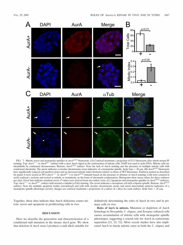
soned that if the lethality of AurA-deficient embryos is due todefective mitotic entry, embryos should have few mitotic cells,as determined by chromosome condensation. Alternately, ifcells enter mitosis but are unable to assemble a bipolar spindle,the embryos should have increased numbers of cells with con-densed chromosomes and monopolar spindles. To test thesepredictions, we performed whole-mount IF staining of E3.5blastocysts (without culture) from AurAd2/� intercrosses. Forthese experiments, AurA IF staining was used to scoreembryos as either AurA� (AurA�/� or AurAd2/�) or AurA�
(AurAd2/d2). Eight of the 11 embryos imaged were scored asAurA� on the basis of the presence of one or more mitoticcells with AurA staining at spindle poles (Fig. 7A, AurA�). Incontrast, three embryos lacked AurA staining, indicating thatthey were AurAd2/d2 (Fig. 7A, AurA�). These embryos dis-played multiple cells with condensed chromatin in circulararrays characteristic of monopolar spindles (arrow in Fig. 7A).Cell counts revealed that mutant embryos contained nearlythreefold fewer total cells than did AurA� embryos (Fig. 7B,top; AurA� mean 67, AurA� mean 24.3), while themitotic index (as determined by chromosome condensation)was eightfold higher in mutants (Fig. 7B, bottom; AurA�
mean 3.3%, AurA� mean 28%). Some mutant embryosalso had cells with condensed chromatin patterns indicative ofapoptosis (Fig. 7C, arrowhead in the AurA� part). To confirmthe monopolar spindle phenotype, embryos were stained withantibodies to �-tubulin. While AurA� embryos had mitoticcells with normal bipolar spindles (Fig. 7C, AurA�), AurA�
embryos displayed characteristic astral microtubule arraysemanating from chromosome rosettes, similar to those ob-served in MEFs (Fig. 7C, AurA�). These results clearly indi-cate that AurA mutant blastocysts have a monopolar spindlephenotype very similar to that observed in MEFs. The obser-vation of large numbers of cells with condensed chromatin anda monopolar spindle phenotype suggests that the mitotic entryimpairment observed in MEFs may not be a significant phe-notype in vivo, at least in early embryos. The early death ofAurA mutant animals is thus apparently due to an inability tocomplete mitosis during the critical wave of early cell divisionsat the blastocyst stage.
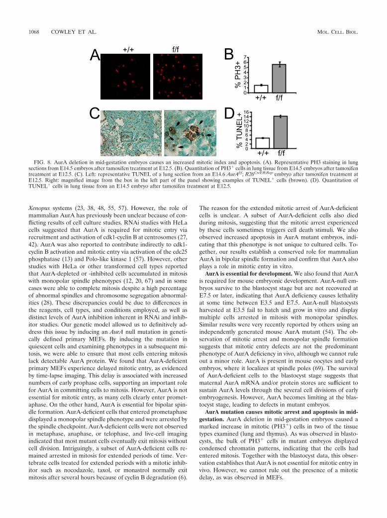
To determine if AurA is also essential and plays similar rolesin later-stage embryos, we intercrossed AurAf/�; R26CreER/Rep
mice and injected pregnant dams with tamoxifen at day 10.5 ofpregnancy. Embryos were harvested 2 days later and analyzedhistologically for PH3 and apoptosis (terminal deoxynucleoti-dyltransferase-mediated dUTP-biotin nick end labeling[TUNEL]) in the lung and the thymus. AurA deletion inAurAf/f; R26CreER embryos caused an approximately fourfoldincrease in the percentage of PH3� cells in both the thymusand the lung compared to that in AurA�/�; R26CreER embryos,indicating that AurA deletion causes a similar mitotic arrestphenotype in later-stage embryos, as observed in blastocystsand MEFs (Fig. 8A to C; data not shown). The majority of thePH3� cells in mutant embryos displayed condensed chromo-somes and prometaphase morphology, supporting the conclu-sion that AurA is not essential for mitotic entry in vivo (Fig.8B). AurA mutant embryos also showed a threefold increase inthe percentage of apoptotic cells in the thymus and the lung,indicating that AurA deficiency induces apoptosis (Fig. 8D).
FIG. 6. AurAd2/d2 embryos die at the blastocyst stage. (A) Genotypeanalysis of progeny from AurAd2/� intercrosses at E3.5, E7.5, and postna-tal stages. E3.5 embryos were cultured to generate blastocyst outgrowthsand then genotyped. #, 12 embryos displayed similar in vitro phenotypes,but genotyping was only successful for 8; P � 0.05 by 2 test for Mendeliansegregation (including 12 presumed d2/d2 embryos). *, E7.5, P � 0.05. **,postnatal days 0 to 7 (P0-P7), P � 0.0001. (B) In vitro blastocyst out-growth characteristic of the AurA�/� and AurAd2/� genotypes. ICM, cellclump derived from inner cell mass; TGC, trophoblast-derived giant cell.(C) AurAd2/d2 blastocyst after 7 days in culture. AurA mutant blastocystsremained unhatched within the zona pellucida (ZP). (D) RepresentativePCR genotyping results for blastocysts after in vitro culture. Deducedgenotypes are shown at the top. Positions of WT and d2 products areindicated on the right. M, DNA ladder; neg, reactions run without tem-plate DNA; pos, positive control reactions.
1066 COWLEY ET AL. MOL. CELL. BIOL.
Together, these data indicate that AurA deficiency causes mi-totic arrest and apoptosis in proliferating cells in vivo.
DISCUSSION
Here we describe the generation and characterization of aconditional null mutation in the mouse AurA gene. We showthat deletion of AurA exon 2 produces a null allele suitable for
definitively determining the roles of AurA in vivo and in pri-mary cells ex vivo.
Roles of AurA in mitosis. Mutation or depletion of AurAhomologs in Drosophila, C. elegans, and Xenopus cultured cellscauses accumulation of mitotic cells with monopolar spindlephenotypes, suggesting a crucial role for AurA in centrosomeseparation (21, 24, 52). More recent studies have also impli-cated AurA in timely mitotic entry in both the C. elegans and
FIG. 7. Mitotic arrest and monopolar spindles in AurAd2/d2 blastocysts. (A) Confocal maximum z projection of E3.5 blastocysts after whole-mount IFstaining. Top: AurA�/� or AurAd2/� embryo with a clear AurA signal at the centrosomes of mitotic cells. DAPI was used to stain DNA. Mitotic cells areidentifiable by condensed chromosomes. Bottom: AurAd2/d2 embryo. Note the lack of AurA staining and the presence of multiple mitotic cells withcondensed chromatin. The arrow indicates a circular chromosome array indicative of a monopolar spindle. Scale bars 20 �m. (B) AurAd2/d2 blastocystshave significantly reduced cell numbers (top) and an increased mitotic index (bottom) relative to those of WT littermates. Embryos stained as describedfor panel A were scored as WT (AurA�/� or AurAd2/�) or AurAd2/d2 mutants based on the presence or absence of AurA staining. Cells were counted inserial confocal z sections and scored as mitotic or nonmitotic on the basis of chromatin condensation. Histograms show mean values for three embryosper class. Error bars indicate standard errors. P values were derived from two-tailed t tests. (C) Apoptosis and monopolar spindles in AurAd2/d2 embryos.Top: AurA�/� or AurAd2/� embryo with normal �-tubulin (�Tub) staining. The arrow indicates a metaphase cell with a bipolar spindle. Bottom: AurAd2/d2
embryo. Note the multiple apoptotic bodies (arrowhead) and cells with circular chromosome arrays and astral microtubule patterns indicative of amonopolar spindle phenotype (arrow). Images are confocal maximum z projections of a subset of z slices for each embryo. Scale bars 20 �m.
VOL. 29, 2009 ROLES OF Aurora-A KINASE IN VIVO AND IN VITRO 1067
Xenopus systems (23, 38, 48, 55, 57). However, the role ofmammalian AurA has previously been unclear because of con-flicting results of cell culture studies. RNAi studies with HeLacells suggested that AurA is required for mitotic entry viarecruitment and activation of cdk1-cyclin B at centrosomes (27,42). AurA was also reported to contribute indirectly to cdk1-cyclin B activation and mitotic entry via activation of the cdc25phosphatase (13) and Polo-like kinase 1 (57). However, otherstudies with HeLa or other transformed cell types reportedthat AurA-depleted or -inhibited cells accumulated in mitosiswith monopolar spindle phenotypes (12, 20, 67) and in somecases were able to complete mitosis despite a high percentageof abnormal spindles and chromosome segregation abnormal-ities (28). These discrepancies could be due to differences inthe reagents, cell types, and conditions employed, as well asdistinct levels of AurA inhibition inherent in RNAi and inhib-itor studies. Our genetic model allowed us to definitively ad-dress this issue by inducing an AurA null mutation in geneti-cally defined primary MEFs. By inducing the mutation inquiescent cells and examining phenotypes in a subsequent mi-tosis, we were able to ensure that most cells entering mitosislack detectable AurA protein. We found that AurA-deficientprimary MEFs experience delayed mitotic entry, as evidencedby time-lapse imaging. This delay is associated with increasednumbers of early prophase cells, supporting an important rolefor AurA in committing cells to mitosis. However, AurA is notessential for mitotic entry, as many cells clearly enter promet-aphase. On the other hand, AurA is essential for bipolar spin-dle formation. AurA-deficient cells that entered prometaphasedisplayed a monopolar spindle phenotype and were arrested bythe spindle checkpoint. AurA-deficient cells were not observedin metaphase, anaphase, or telophase, and live-cell imagingindicated that most mutant cells eventually exit mitosis withoutcell division. Intriguingly, a subset of AurA-deficient cells re-mained arrested in mitosis for extended periods of time. Ver-tebrate cells treated for extended periods with a mitotic inhib-itor such as nocodazole, taxol, or monastrol normally exitmitosis after several hours because of cyclin B degradation (6).
The reason for the extended mitotic arrest of AurA-deficientcells is unclear. A subset of AurA-deficient cells also diedduring mitosis, suggesting that the mitotic arrest experiencedby these cells sometimes triggers cell death stimuli. We alsoobserved increased apoptosis in AurA mutant embryos, indi-cating that this phenotype is not unique to cultured cells. To-gether, our results establish a conserved role for mammalianAurA in bipolar spindle formation and confirm that AurA alsoplays a role in mitotic entry in vitro.
AurA is essential for development. We also found that AurAis required for mouse embryonic development. AurA-null em-bryos survive to the blastocyst stage but are not recovered atE7.5 or later, indicating that AurA deficiency causes lethalityat some time between E3.5 and E7.5. AurA-null blastocystsharvested at E3.5 fail to hatch and grow in vitro and displaymultiple cells arrested in mitosis with monopolar spindles.Similar results were very recently reported by others using anindependently generated mouse AurA mutant (54). The ob-servation of mitotic arrest and monopolar spindle formationsuggests that mitotic entry defects are not the predominantphenotype of AurA deficiency in vivo, although we cannot ruleout a minor role. AurA is present in mouse oocytes and earlyembryos, where it localizes at spindle poles (69). The survivalof AurA-deficient cells to the blastocyst stage suggests thatmaternal AurA mRNA and/or protein stores are sufficient tosustain AurA levels through the several cell divisions of earlyembryogenesis. However, AurA becomes limiting at the blas-tocyst stage, leading to defects in mutant embryos.
AurA mutation causes mitotic arrest and apoptosis in mid-gestation. AurA deletion in mid-gestation embryos caused amarked increase in mitotic (PH3�) cells in two of the tissuetypes examined (lung and thymus). As was observed in blasto-cysts, the bulk of PH3� cells in mutant embryos displayedcondensed chromatin patterns, indicating that the cells hadentered mitosis. Together with the blastocyst data, this obser-vation establishes that AurA is not essential for mitotic entry invivo. However, we cannot rule out the presence of a mitoticdelay, as was observed in MEFs.
FIG. 8. AurA deletion in mid-gestation embryos causes an increased mitotic index and apoptosis. (A). Representative PH3 staining in lungsections from E14.5 embryos after tamoxifen treatment at E12.5. (B). Quantitation of PH3� cells in lung tissue from E14.5 embryos after tamoxifentreatment at E12.5. (C). Left: representative TUNEL of a lung section from an E14.6 AurAf/f; R26CreER/Rep embryo after tamoxifen treatment atE12.5. Right: magnified image from the box in the left part of the panel showing examples of TUNEL� cells (brown). (D). Quantitation ofTUNEL� cells in lung tissue from an E14.5 embryo after tamoxifen treatment at E12.5.
1068 COWLEY ET AL. MOL. CELL. BIOL.
The increased mitotic index in AurA mutant mid-gestationembryos was accompanied by a parallel increase in apoptoticcells. Apoptotic chromatin patterns were also observed inAurA-deficient blastocysts, indicating that this phenotype isnot restricted to later gestation embryos. As discussed above, asubset of AurA-deficient MEFs died during mitosis. An im-portant area for future work will be the determination of thesignaling pathways that govern apoptosis in response to AurAmutation.
AurA as a therapeutic target in cancer. The important rolesof Aurora kinases in mitotic regulation, as well as the findingthat AurA is overexpressed in many human cancers, haveraised considerable interest in AurA kinase inhibitors as po-tential cancer therapeutics. Indeed, Aurora kinase inhibitorsare currently in clinical trials (reviewed in references 9 and 34).Several currently described Aurora inhibitors display activityagainst both AurA and AurB in vitro (20, 67), while com-pounds with reported selectivity for AurA or AurB inhibitionare beginning to emerge (20, 28, 68). The desirability of tar-geting AurA, AurB, or both is a matter of debate (9, 65, 67).Nevertheless, our results clarify the issue by clearly defining theAurA mutant phenotype. Thus, we predict that potent AurA-specific inhibitors should cause monopolar spindles, leading tomitotic arrest in cells with a competent spindle checkpoint. Incontrast, inhibition of AurB alone and dual inhibition of AurAand AurB have been reported to produce similar phenotypes,including rapid mitotic exit without cell division because of lossof the spindle checkpoint and cytokinesis impairment (20, 67).Activation of the spindle checkpoint in specific AurA-inhibitedcells is thus a significant functional difference that may con-tribute to distinct therapeutic outcomes. The long-term conse-quences of spindle checkpoint arrest are not fully defined butmay include apoptosis, mitotic catastrophe, mitotic exit, andsubsequent G1 arrest or mitotic exit followed by endoredupli-cation (reviewed in reference 51). Our results suggest that atleast a subset of cells treated with AurA inhibitors shouldundergo apoptosis, although it is possible that tumor cells mayrespond differently from normal primary cells. Our geneticsystem will allow future tests of these hypotheses in vivo and invitro.
The challenge of obtaining AurA-specific inhibitors raisesthe question of whether similar therapeutic effects could beachieved by targeting other molecules. Our results suggest thatdrugs such as monastrol that cause monopolar spindles byinhibiting the mitotic kinesin Eg5 may be good alternatives toAurA inhibitors. Several such drugs are under development(4). While these compounds may not show phenotypes identi-cal to that produced by AurA inhibition (because of the factthat AurA has substrates involved in multiple aspects of mito-sis and also regulates processes outside of mitosis [49]), theymay cause a phenotypically similar arrest state with similarbiological outcomes. In fact, Eg5 is a substrate of AurA inXenopus, suggesting that Eg5 activation could be downstreamof AurA. The future identification of the AurA targets medi-ating the monopolar spindle phenotype will also be helpful inexploring other ways to target this pathway for therapeuticbenefit.
In summary, these studies provide clear evidence that mam-malian AurA is essential for viability because of its key role inregulating the transition through M phase, specifically in the
establishment of bipolar spindle assembly. The conditional ge-netic system established in this report, with paired primary celland in vivo studies, will be instrumental in deciphering themechanisms and pathways involved. Furthermore, the role(s)of AurA in cancer and other diseases associated with aneu-ploidy can now be fully explored.
ACKNOWLEDGMENTS
We thank members of the Van Dyke, Magnuson, and Salmon lab-oratories for helpful discussions and technical assistance. We acknowl-edge the use of Michael Hooker Microscopy Core Facility equipmentand resources funded by an anonymous private donor and SundeepKalantry, Michael Chua, and Wendy Salmon for expert assistance withconfocal microscopy. We thank Allan Balmain for critical reading andcomments on the manuscript.
D.O.C. was a fellow of the Leukemia & Lymphoma Foundation ofAmerica (grant 5408-02). This study was supported by NCI grant2-R01-CA065773-11A1 to T.V.D., NIH grant GM24364 to E.D.S., andan NICHD grant to T.M.
REFERENCES
1. Andrews, P. D., E. Knatko, W. J. Moore, and J. R. Swedlow. 2003. Mitoticmechanics: the auroras come into view. Curr. Opin. Cell Biol. 15:672–683.
2. Badea, T. C., Y. Wang, and J. Nathans. 2003. A noninvasive genetic/phar-macologic strategy for visualizing cell morphology and clonal relationships inthe mouse. J. Neurosci. 23:2314–2322.
3. Berdnik, D., and J. A. Knoblich. 2002. Drosophila Aurora-A is required forcentrosome maturation and actin-dependent asymmetric protein localizationduring mitosis. Curr. Biol. 12:640–647.
4. Bergnes, G., K. Brejc, and L. Belmont. 2005. Mitotic kinesins: prospects forantimitotic drug discovery. Curr. Top. Med. Chem. 5:127–145.
5. Bischoff, J. R., L. Anderson, Y. Zhu, K. Mossie, L. Ng, B. Souza, B. Schryver,P. Flanagan, F. Clairvoyant, C. Ginther, C. S. Chan, M. Novotny, D. J.Slamon, and G. D. Plowman. 1998. A homologue of Drosophila aurorakinase is oncogenic and amplified in human colorectal cancers. EMBO J.17:3052–3065.
6. Brito, D. A., and C. L. Rieder. 2006. Mitotic checkpoint slippage in humansoccurs via cyclin B destruction in the presence of an active checkpoint. Curr.Biol. 16:1194–1200.
7. Brown, J. R., K. K. Koretke, M. L. Birkeland, P. Sanseau, and D. R. Patrick.2004. Evolutionary relationships of Aurora kinases: implications for modelorganism studies and the development of anti-cancer drugs. BMC. Evol.Biol. 4:39.
8. Carmena, M., and W. C. Earnshaw. 2003. The cellular geography of aurorakinases. Nat. Rev. Mol. Cell Biol. 4:842–854.
9. Carvajal, R. D., A. Tse, and G. K. Schwartz. 2006. Aurora kinases: newtargets for cancer therapy. Clin. Cancer Res. 12:6869–6875.
10. Cimini, D., X. Wan, C. B. Hirel, and E. D. Salmon. 2006. Aurora kinasepromotes turnover of kinetochore microtubules to reduce chromosome seg-regation errors. Curr. Biol. 16:1711–1718.
11. Crosio, C., G. M. Fimia, R. Loury, M. Kimura, Y. Okano, H. Zhou, S. Sen,C. D. Allis, and P. Sassone-Corsi. 2002. Mitotic phosphorylation of histoneH3: spatio-temporal regulation by mammalian Aurora kinases. Mol. Cell.Biol. 22:874–885.
12. Du, J., and G. J. Hannon. 2004. Suppression of p160ROCK bypasses cellcycle arrest after Aurora-A/STK15 depletion. Proc. Natl. Acad. Sci. USA101:8975–8980.
13. Dutertre, S., M. Cazales, M. Quaranta, C. Froment, V. Trabut, C. Dozier, G.Mirey, J. P. Bouche, N. Theis-Febvre, E. Schmitt, B. Monsarrat, C. Prigent,and B. Ducommun. 2004. Phosphorylation of CDC25B by Aurora-A at thecentrosome contributes to the G2-M transition. J. Cell Sci. 117:2523–2531.
14. Ewart-Toland, A., P. Briassouli, J. P. de Koning, J. H. Mao, J. Yuan, F.Chan, L. MacCarthy-Morrogh, B. A. Ponder, H. Nagase, J. Burn, S. Ball, M.Almeida, S. Linardopoulos, and A. Balmain. 2003. Identification of Stk6/STK15 as a candidate low-penetrance tumor-susceptibility gene in mouseand human. Nat. Genet. 34:403–412.
15. Fukuda, T., Y. Mishina, M. P. Walker, and R. P. DiAugustine. 2005. Con-ditional transgenic system for mouse aurora a kinase: degradation by theubiquitin proteasome pathway controls the level of the transgenic protein.Mol. Cell. Biol. 25:5270–5281.
16. Furuta, T., D. L. Baillie, and J. M. Schumacher. 2002. Caenorhabditiselegans Aurora A kinase AIR-1 is required for postembryonic cell divisionsand germline development. Genesis 34:244–250.
17. Giet, R., D. McLean, S. Descamps, M. J. Lee, J. W. Raff, C. Prigent, andD. M. Glover. 2002. Drosophila Aurora A kinase is required to localizeD-TACC to centrosomes and to regulate astral microtubules. J. Cell Biol.156:437–451.
VOL. 29, 2009 ROLES OF Aurora-A KINASE IN VIVO AND IN VITRO 1069
18. Giet, R., and C. Prigent. 2000. The Xenopus laevis aurora/Ip11p-relatedkinase pEg2 participates in the stability of the bipolar mitotic spindle. Exp.Cell Res. 258:145–151.
19. Giet, R., R. Uzbekov, F. Cubizolles, G. K. Le, and C. Prigent. 1999. TheXenopus laevis Aurora-related protein kinase pEg2 associates with andphosphorylates the kinesin-related protein XlEg5. J. Biol. Chem. 274:15005–15013.
20. Girdler, F., K. E. Gascoigne, P. A. Eyers, S. Hartmuth, C. Crafter, K. M.Foote, N. J. Keen, and S. S. Taylor. 2006. Validating Aurora B as ananti-cancer drug target. J. Cell Sci. 119:3664–3675.
21. Glover, D. M., M. H. Leibowitz, D. A. McLean, and H. Parry. 1995. Muta-tions in Aurora prevent centrosome separation leading to the formation ofmonopolar spindles. Cell 81:95–105.
22. Gopalan, G., C. S. Chan, and P. J. Donovan. 1997. A novel mammalian,mitotic spindle-associated kinase is related to yeast and fly chromosomesegregation regulators. J. Cell Biol. 138:643–656.
23. Hachet, V., C. Canard, and P. Gonczy. 2007. Centrosomes promote timelymitotic entry in C. elegans embryos. Dev. Cell 12:531–541.
24. Hannak, E., M. Kirkham, A. A. Hyman, and K. Oegema. 2001. Aurora-Akinase is required for centrosome maturation in Caenorhabditis elegans.J. Cell Biol. 155:1109–1116.
25. Hauf, S., R. W. Cole, S. LaTerra, C. Zimmer, G. Schnapp, R. Walter, A.Heckel, J. van Meel, C. L. Rieder, and J. M. Peters. 2003. The small moleculeHesperadin reveals a role for Aurora B in correcting kinetochore-microtu-bule attachment and in maintaining the spindle assembly checkpoint. J. CellBiol. 161:281–294.
26. Hendzel, M. J., Y. Wei, M. A. Mancini, A. Van Hooser, T. Ranalli, B. R.Brinkley, D. P. Bazett-Jones, and C. D. Allis. 1997. Mitosis-specific phos-phorylation of histone H3 initiates primarily within pericentromeric hetero-chromatin during G2 and spreads in an ordered fashion coincident withmitotic chromosome condensation. Chromosoma 106:348–360.
27. Hirota, T., N. Kunitoku, T. Sasayama, T. Marumoto, D. Zhang, M. Nitta, K.Hatakeyama, and H. Saya. 2003. Aurora-A and an interacting activator, theLIM protein Ajuba, are required for mitotic commitment in human cells.Cell 114:585–598.
28. Hoar, K., A. Chakravarty, C. Rabino, D. Wysong, D. Bowman, N. Roy, andJ. A. Ecsedy. 2007. MLN8054, a small-molecule inhibitor of Aurora A, causesspindle pole and chromosome congression defects leading to aneuploidy.Mol. Cell. Biol. 27:4513–4525.
29. Honda, K., H. Mihara, Y. Kato, A. Yamaguchi, H. Tanaka, H. Yasuda, K.Furukawa, and T. Urano. 2000. Degradation of human Aurora2 proteinkinase by the anaphase-promoting complex-ubiquitin-proteasome pathway.Oncogene 19:2812–2819.
30. Jackson, A. L., and P. S. Linsley. 2004. Noise amidst the silence: off-targeteffects of siRNAs? Trends Genet. 20:521–524.
31. Juan, G., F. Traganos, W. M. James, J. M. Ray, M. Roberge, D. M. Sauve,H. Anderson, and Z. Darzynkiewicz. 1998. Histone H3 phosphorylation andexpression of cyclins A and B1 measured in individual cells during theirprogression through G2 and mitosis. Cytometry 32:71–77.
32. Kallio, M. J., M. L. McCleland, P. T. Stukenberg, and G. J. Gorbsky. 2002.Inhibition of aurora B kinase blocks chromosome segregation, overrides thespindle checkpoint, and perturbs microtubule dynamics in mitosis. Curr.Biol. 12:900–905.
33. Kapoor, T. M., T. U. Mayer, M. L. Coughlin, and T. J. Mitchison. 2000.Probing spindle assembly mechanisms with monastrol, a small moleculeinhibitor of the mitotic kinesin, Eg5. J. Cell Biol. 150:975–988.
34. Keen, N., and S. Taylor. 2004. Aurora-kinase inhibitors as anticancer agents.Nat. Rev. Cancer 4:927–936.
35. Kulkarni, M. M., M. Booker, S. J. Silver, A. Friedman, P. Hong, N. Perri-mon, and B. Mathey-Prevot. 2006. Evidence of off-target effects associatedwith long dsRNAs in Drosophila melanogaster cell-based assays. Nat. Meth-ods 3:833–838.
36. Kunitoku, N., T. Sasayama, T. Marumoto, D. Zhang, S. Honda, O. Koba-yashi, K. Hatakeyama, Y. Ushio, H. Saya, and T. Hirota. 2003. CENP-Aphosphorylation by Aurora-A in prophase is required for enrichment ofAurora-B at inner centromeres and for kinetochore function. Dev. Cell5:853–864.
37. Lee, E. C., D. Yu, J. Martinez de Velasco, L. Tessarollo, D. A. Swing, D. L.Court, N. A. Jenkins, and N. G. Copeland. 2001. A highly efficient Esche-richia coli-based chromosome engineering system adapted for recombino-genic targeting and subcloning of BAC DNA. Genomics 73:56–65.
38. Liu, Q., and J. V. Ruderman. 2006. Aurora A, mitotic entry, and spindlebipolarity. Proc. Natl. Acad. Sci. USA 103:5811–5816.
39. Loonstra, A., M. Vooijs, H. B. Beverloo, B. A. Allak, E. Van Drunen, R.Kanaar, A. Berns, and J. Jonkers. 2001. Growth inhibition and DNA dam-age induced by Cre recombinase in mammalian cells. Proc. Natl. Acad. Sci.USA 98:9209–9214.
40. Ma, Y., A. Creanga, L. Lum, and P. A. Beachy. 2006. Prevalence of off-targeteffects in Drosophila RNA interference screens. Nature 443:359–363.
41. Mao, J. H., D. Wu, J. Perez-Losada, T. Jiang, Q. Li, R. M. Neve, J. W. Gray,W. W. Cai, and A. Balmain. 2007. Crosstalk between Aurora-A and p53:
frequent deletion or downregulation of Aurora-A in tumors from p53 nullmice. Cancer Cell 11:161–173.
42. Marumoto, T., T. Hirota, T. Morisaki, N. Kunitoku, D. Zhang, Y. Ichikawa,T. Sasayama, S. Kuninaka, T. Mimori, N. Tamaki, M. Kimura, Y. Okano,and H. Saya. 2002. Roles of aurora-A kinase in mitotic entry and G2 check-point in mammalian cells. Genes Cells 7:1173–1182.
43. Marumoto, T., D. Zhang, and H. Saya. 2005. Aurora-A—a guardian of poles.Nat. Rev. Cancer 5:42–50.
44. May, K. M., and K. G. Hardwick. 2006. The spindle checkpoint. J. Cell Sci.119:4139–4142.
45. Meraldi, P., R. Honda, and E. A. Nigg. 2004. Aurora kinases link chromo-some segregation and cell division to cancer susceptibility. Curr. Opin.Genet. Dev. 14:29–36.
46. Nigg, E. A. 2001. Mitotic kinases as regulators of cell division and its check-points. Nat. Rev. Mol. Cell Biol. 2:21–32.
47. Pines, J., and C. L. Rieder. 2001. Re-staging mitosis: a contemporary view ofmitotic progression. Nat. Cell Biol. 3:E3–E6.
48. Portier, N., A. Audhya, P. S. Maddox, R. A. Green, A. Dammermann, A.Desai, and K. Oegema. 2007. A microtubule-independent role for centro-somes and Aurora A in nuclear envelope breakdown. Dev. Cell 12:515–529.
49. Pugacheva, E. N., S. A. Jablonski, T. R. Hartman, E. P. Henske, and E. A.Golemis. 2007. HEF1-dependent Aurora A activation induces disassembly ofthe primary cilium. Cell 129:1351–1363.
50. Rajagopalan, H., and C. Lengauer. 2004. Aneuploidy and cancer. Nature432:338–341.
51. Rieder, C. L., and H. Maiato. 2004. Stuck in division or passing through:what happens when cells cannot satisfy the spindle assembly checkpoint.Dev. Cell 7:637–651.
52. Roghi, C., R. Giet, R. Uzbekov, N. Morin, I. Chartrain, R. Le Guellec, A.Couturier, M. Doree, M. Philippe, and C. Prigent. 1998. The Xenopusprotein kinase pEg2 associates with the centrosome in a cell cycle-dependentmanner, binds to the spindle microtubules and is involved in bipolar mitoticspindle assembly. J. Cell Sci. 111(Pt. 5):557–572.
53. Sasai, K., H. Katayama, D. L. Stenoien, S. Fujii, R. Honda, M. Kimura, Y.Okano, M. Tatsuka, F. Suzuki, E. A. Nigg, W. C. Earnshaw, W. R. Brinkley,and S. Sen. 2004. Aurora-C kinase is a novel chromosomal passenger proteinthat can complement Aurora-B kinase function in mitotic cells. Cell Motil.Cytoskel. 59:249–263.
54. Sasai, K., J. M. Parant, M. E. Brandt, J. Carter, H. P. Adams, S. A. Stass,A. M. Killary, H. Katayama, and S. Sen. 2008. Targeted disruption of AuroraA causes abnormal mitotic spindle assembly, chromosome misalignment andembryonic lethality. Oncogene 27:4122–4127.
55. Satinover, D. L., D. L. Brautigan, and P. T. Stukenberg. 2006. Aurora-Akinase and inhibitor-2 regulate the cyclin threshold for mitotic entry inXenopus early embryonic cell cycles. Cell Cycle 5:2268–2274.
56. Schumacher, J. M., N. Ashcroft, P. J. Donovan, and A. Golden. 1998. Ahighly conserved centrosomal kinase, AIR-1, is required for accurate cellcycle progression and segregation of developmental factors in Caenorhab-ditis elegans embryos. Development 125:4391–4402.
57. Seki, A., J. A. Coppinger, C. Y. Jang, J. R. Yates, and G. Fang. 2008. Boraand the kinase Aurora A cooperatively activate the kinase Plk1 and controlmitotic entry. Science 320:1655–1658.
58. Sen, S., H. Zhou, and R. A. White. 1997. A putative serine/threonine kinaseencoding gene BTAK on chromosome 20q13 is amplified and overexpressedin human breast cancer cell lines. Oncogene 14:2195–2200.
59. Skarnes, W. C. 2000. Gene trapping methods for the identification andfunctional analysis of cell surface proteins in mice. Methods Enzymol. 328:592–615.
60. Soriano, P. 1999. Generalized lacZ expression with the ROSA26 Cre re-porter strain. Nat. Genet. 21:70–71.
61. Su, H., A. A. Mills, X. Wang, and A. Bradley. 2002. A targeted X-linkedCMV-Cre line. Genesis 32:187–188.
62. Tanaka, M., A. Ueda, H. Kanamori, H. Ideguchi, J. Yang, S. Kitajima, andY. Ishigatsubo. 2002. Cell-cycle-dependent regulation of human aurora Atranscription is mediated by periodic repression of E4TF1. J. Biol. Chem.277:10719–10726.
63. Truett, G. E., P. Heeger, R. L. Mynatt, A. A. Truett, J. A. Walker, and M. L.Warman. 2000. Preparation of PCR-quality mouse genomic DNA with hotsodium hydroxide and Tris (HotSHOT). BioTechniques 29:52–54.
64. Walter, A. O., W. Seghezzi, W. Korver, J. Sheung, and E. Lees. 2000. Themitotic serine/threonine kinase Aurora2/AIK is regulated by phosphoryla-tion and degradation. Oncogene 19:4906–4916.
65. Warner, S. L., R. M. Munoz, P. Stafford, E. Koller, L. H. Hurley, D. D. VonHoff, and H. Han. 2006. Comparing Aurora A and Aurora B as moleculartargets for growth inhibition of pancreatic cancer cells. Mol. Cancer Ther.5:2450–2458.
66. Waters, J. C., R. H. Chen, A. W. Murray, and E. D. Salmon. 1998. Local-ization of Mad2 to kinetochores depends on microtubule attachment, nottension. J. Cell Biol. 141:1181–1191.
67. Yang, H., T. Burke, J. Dempsey, B. Diaz, E. Collins, J. Toth, R. Beckmann,and X. Ye. 2005. Mitotic requirement for Aurora A kinase is bypassed in theabsence of Aurora B kinase. FEBS Lett. 579:3385–3391.
1070 COWLEY ET AL. MOL. CELL. BIOL.

68. Yang, J., T. Ikezoe, C. Nishioka, T. Tasaka, A. Taniguchi, Y. Kuwayama, N.Komatsu, K. Bandobashi, K. Togitani, H. P. Koeffler, H. Taguchi, and A.Yokoyama. 2007. AZD1152, a novel and selective Aurora B kinase inhibitor,induces growth arrest, apoptosis, and sensitization for tubulin depolymeriz-ing agent or topoisomerase II inhibitor in human acute leukemia cells invitro and in vivo. Blood 110:2034–2040.
69. Yao, L. J., Z. S. Zhong, L. S. Zhang, D. Y. Chen, H. Schatten, and Q. Y. Sun.
2004. Aurora-A is a critical regulator of microtubule assembly and nuclearactivity in mouse oocytes, fertilized eggs, and early embryos. Biol. Reprod.70:1392–1399.
70. Zhou, H., J. Kuang, L. Zhong, W. L. Kuo, J. W. Gray, A. Sahin, B. R.Brinkley, and S. Sen. 1998. Tumour amplified kinase STK15/BTAK inducescentrosome amplification, aneuploidy and transformation. Nat. Genet. 20:189–193.
VOL. 29, 2009 ROLES OF Aurora-A KINASE IN VIVO AND IN VITRO 1071
Related Documents