CORRESPONDENCE An unusual variant of granulomatous adnexotropic cutaneous T-cell lymphoma SIR, A 40-year-old man reported the slow progressive appearance, during the previous 6 years, of pruritic erythe- matous lesions on the trunk, buttock, abdomen, axilla, genital area and forearm (Fig. 1a). Lesions consisted of follicular papules, comedones, milia and cysts. Lesional areas were alopecic (Fig. 1b), and diffuse alopecia was also present on the scalp and beard area, along with comedones and cysts. The patient reported severe skin dryness, especially in the involved areas. No impairment of salivary or lacrimary function was noted. Serological and haematological tests were all normal or negative. Because of the diffuse presence of cysts and comedones, a diagnosis of chloracne had been made in another institution; the clinical diagnosis was confirmed histologically by the presence of infundibular cysts and a granulomatous foreign body reaction to keratin scales. A further biopsy was performed: the most striking histo- logical feature was a lymphocytic infiltrate involving eccrine glands and coils along with a characteristic epithelial hyperplasia (Figs 1c,d). This picture fits perfectly with that reported in the literature 1–3 as being characteristic of syringolymphoid hyperplasia, also known as syringotropic mycosis fungoides or syringotropic cutaneous T-cell lym- phoma (CTCL). The hair follicles were involved by the lymphocytic infiltrate in a manner similar to that of the eccrine glands. Follicles were surrounded by a dense lymph- ocytic infiltrate, with extensive exocytosis. Occasional Pau- trier microabscesses were evident in the follicular sheath. This pattern is that of pilotropic mycosis fungoides, a form of folliculotropic CTCL. 4–8 Many follicles were entirely trans- Figure 1. (a) The clinical picture was characterized by erythematous plaques mostly located on the forearm, abdomen, buttock and scalp. Plaques were scattered, with follicular papules, cysts, milia and comedones. (b) Alopecia, along with cysts and comedones, was an important clinical feature, with anhidrosis in the involved areas. (c, d) Histologically, almost all the eccrine glands and hair follicles in the specimens were surrounded by a dense lymphocytic infiltrate. Exocytosis of lymphocytes within the epithelia was present, along with Pautrier-like microabscesses. Epithelia underwent a peculiar basal cell hyperplasia, resulting in a cribriform spiroadenoma-like or basaloid pattern (haematoxylin and eosin; original magnification · 132). British Journal of Dermatology 2003; 148: 363–384. ȑ 2003 British Association of Dermatologists 363

Welcome message from author
This document is posted to help you gain knowledge. Please leave a comment to let me know what you think about it! Share it to your friends and learn new things together.
Transcript
CORRESPONDENCE
An unusual variant of granulomatous adnexotropiccutaneous T-cell lymphoma
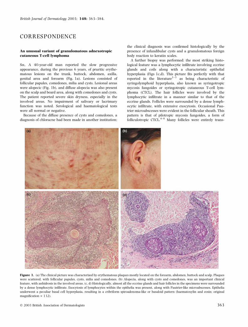
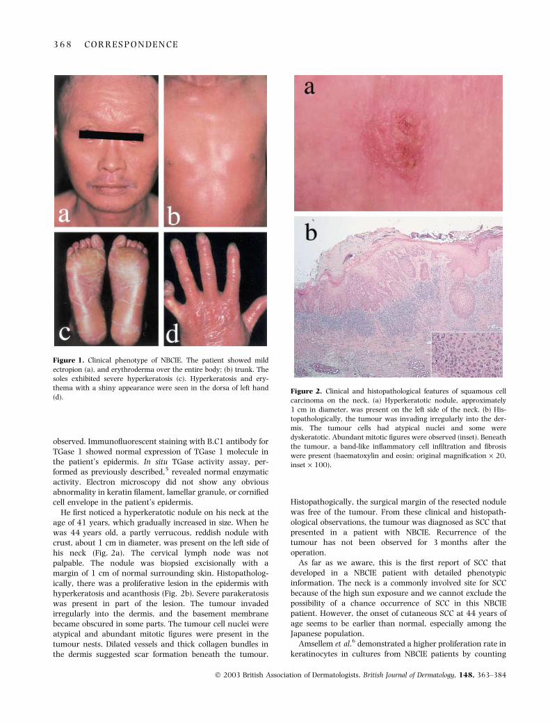
SIR, A 40-year-old man reported the slow progressiveappearance, during the previous 6 years, of pruritic erythe-matous lesions on the trunk, buttock, abdomen, axilla,genital area and forearm (Fig. 1a). Lesions consisted offollicular papules, comedones, milia and cysts. Lesional areaswere alopecic (Fig. 1b), and diffuse alopecia was also presenton the scalp and beard area, along with comedones and cysts.The patient reported severe skin dryness, especially in theinvolved areas. No impairment of salivary or lacrimaryfunction was noted. Serological and haematological testswere all normal or negative.
Because of the diffuse presence of cysts and comedones, adiagnosis of chloracne had been made in another institution;
the clinical diagnosis was confirmed histologically by thepresence of infundibular cysts and a granulomatous foreignbody reaction to keratin scales.
A further biopsy was performed: the most striking histo-logical feature was a lymphocytic infiltrate involving eccrineglands and coils along with a characteristic epithelialhyperplasia (Figs 1c,d). This picture fits perfectly with thatreported in the literature1–3 as being characteristic ofsyringolymphoid hyperplasia, also known as syringotropicmycosis fungoides or syringotropic cutaneous T-cell lym-phoma (CTCL). The hair follicles were involved by thelymphocytic infiltrate in a manner similar to that of theeccrine glands. Follicles were surrounded by a dense lymph-ocytic infiltrate, with extensive exocytosis. Occasional Pau-trier microabscesses were evident in the follicular sheath. Thispattern is that of pilotropic mycosis fungoides, a form offolliculotropic CTCL.4–8 Many follicles were entirely trans-
Figure 1. (a) The clinical picture was characterized by erythematous plaques mostly located on the forearm, abdomen, buttock and scalp. Plaques
were scattered, with follicular papules, cysts, milia and comedones. (b) Alopecia, along with cysts and comedones, was an important clinical
feature, with anhidrosis in the involved areas. (c, d) Histologically, almost all the eccrine glands and hair follicles in the specimens were surrounded
by a dense lymphocytic infiltrate. Exocytosis of lymphocytes within the epithelia was present, along with Pautrier-like microabscesses. Epithelia
underwent a peculiar basal cell hyperplasia, resulting in a cribriform spiroadenoma-like or basaloid pattern (haematoxylin and eosin; original
magnification · 132).
British Journal of Dermatology 2003; 148: 363–384.
� 2003 British Association of Dermatologists 363
formed into bizarre undifferentiated basaloid structures5
resembling a neoplasm with follicular differentiation, suchas basal cell carcinoma, trichoblastoma or cutaneous lymph-adenoma. Numerous infundibular cysts were present, scat-tered throughout the specimens.
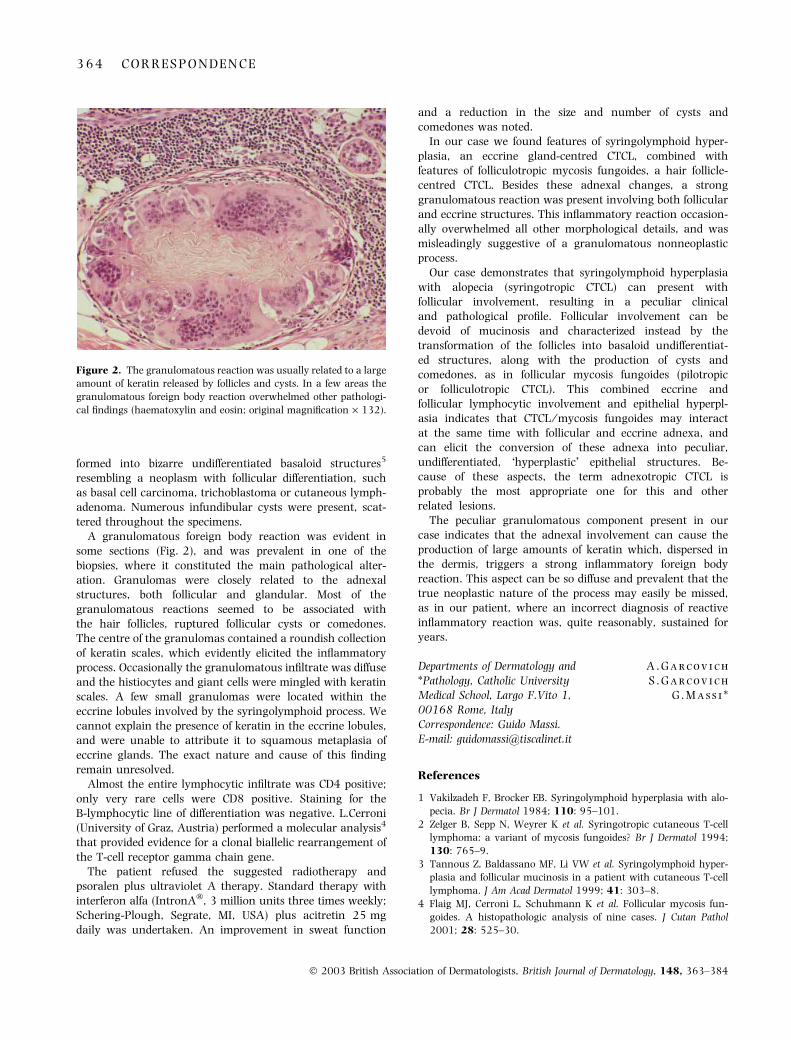
A granulomatous foreign body reaction was evident insome sections (Fig. 2), and was prevalent in one of thebiopsies, where it constituted the main pathological alter-ation. Granulomas were closely related to the adnexalstructures, both follicular and glandular. Most of thegranulomatous reactions seemed to be associated withthe hair follicles, ruptured follicular cysts or comedones.The centre of the granulomas contained a roundish collectionof keratin scales, which evidently elicited the inflammatoryprocess. Occasionally the granulomatous infiltrate was diffuseand the histiocytes and giant cells were mingled with keratinscales. A few small granulomas were located within theeccrine lobules involved by the syringolymphoid process. Wecannot explain the presence of keratin in the eccrine lobules,and were unable to attribute it to squamous metaplasia ofeccrine glands. The exact nature and cause of this findingremain unresolved.
Almost the entire lymphocytic infiltrate was CD4 positive;only very rare cells were CD8 positive. Staining for theB-lymphocytic line of differentiation was negative. L.Cerroni(University of Graz, Austria) performed a molecular analysis4
that provided evidence for a clonal biallelic rearrangement ofthe T-cell receptor gamma chain gene.
The patient refused the suggested radiotherapy andpsoralen plus ultraviolet A therapy. Standard therapy withinterferon alfa (IntronA�, 3 million units three times weekly;Schering-Plough, Segrate, MI, USA) plus acitretin 25 mgdaily was undertaken. An improvement in sweat function
and a reduction in the size and number of cysts andcomedones was noted.
In our case we found features of syringolymphoid hyper-plasia, an eccrine gland-centred CTCL, combined withfeatures of folliculotropic mycosis fungoides, a hair follicle-centred CTCL. Besides these adnexal changes, a stronggranulomatous reaction was present involving both follicularand eccrine structures. This inflammatory reaction occasion-ally overwhelmed all other morphological details, and wasmisleadingly suggestive of a granulomatous nonneoplasticprocess.
Our case demonstrates that syringolymphoid hyperplasiawith alopecia (syringotropic CTCL) can present withfollicular involvement, resulting in a peculiar clinicaland pathological profile. Follicular involvement can bedevoid of mucinosis and characterized instead by thetransformation of the follicles into basaloid undifferentiat-ed structures, along with the production of cysts andcomedones, as in follicular mycosis fungoides (pilotropicor folliculotropic CTCL). This combined eccrine andfollicular lymphocytic involvement and epithelial hyperpl-asia indicates that CTCL ⁄ mycosis fungoides may interactat the same time with follicular and eccrine adnexa, andcan elicit the conversion of these adnexa into peculiar,undifferentiated, �hyperplastic� epithelial structures. Be-cause of these aspects, the term adnexotropic CTCL isprobably the most appropriate one for this and otherrelated lesions.
The peculiar granulomatous component present in ourcase indicates that the adnexal involvement can cause theproduction of large amounts of keratin which, dispersed inthe dermis, triggers a strong inflammatory foreign bodyreaction. This aspect can be so diffuse and prevalent that thetrue neoplastic nature of the process may easily be missed,as in our patient, where an incorrect diagnosis of reactiveinflammatory reaction was, quite reasonably, sustained foryears.
A . G a r c o v i c h
S . G a r c o v i c h
G . M a s s i *
Departments of Dermatology and*Pathology, Catholic UniversityMedical School, Largo F.Vito 1,00168 Rome, ItalyCorrespondence: Guido Massi.E-mail: [email protected]
References
1 Vakilzadeh F, Brocker EB. Syringolymphoid hyperplasia with alo-
pecia. Br J Dermatol 1984; 110: 95–101.
2 Zelger B, Sepp N, Weyrer K et al. Syringotropic cutaneous T-cell
lymphoma: a variant of mycosis fungoides? Br J Dermatol 1994;
130: 765–9.
3 Tannous Z, Baldassano MF, Li VW et al. Syringolymphoid hyper-
plasia and follicular mucinosis in a patient with cutaneous T-cell
lymphoma. J Am Acad Dermatol 1999; 41: 303–8.
4 Flaig MJ, Cerroni L, Schuhmann K et al. Follicular mycosis fun-
goides. A histopathologic analysis of nine cases. J Cutan Pathol
2001; 28: 525–30.
Figure 2. The granulomatous reaction was usually related to a large
amount of keratin released by follicles and cysts. In a few areas the
granulomatous foreign body reaction overwhelmed other pathologi-
cal findings (haematoxylin and eosin; original magnification · 132).
3 6 4 C O R R E S P O N D E N C E
� 2003 British Association of Dermatologists, British Journal of Dermatology, 148, 363–384

5 Kossard S, White A, Killingsworth M. Basaloid folliculolymphoid
hyperplasia with alopecia as an expression of mycosis fungoides
(CTCL). J Cutan Pathol 1995; 22: 466–71.
6 Pereyo NG, Requena L, Galloway J, Sangueza OP. Follicular my-
cosis fungoides: a clinicohistopathologic study. J Am Acad Dermatol
1997; 36: 563–8.
7 Klemke CD, Dippel E, Assaf C et al. Follicular mycosis fungoides. Br
J Dermatol 1999; 141: 137–40.
8 Fraser-Andrews E, Ashton R, Russell-Jones R. Pilotropic mycosis
fungoides presenting with multiple cysts, comedones and alopecia.
Br J Dermatol 1999; 140: 141–4.
Comparison of melanocytic naevi with photographs,a recommended method
SIR, For many years now there have been public healthconcerns in many countries about increasing mortality ratesdue to melanoma. Part of the approach to early diagnosis hasbeen to encourage people at high risk, particularly those witha very large number of melanocytic naevi, to have standard-ized photography and to practise skin self-examination usingthe photographs as a template. Medical practitioners also usethe photographs during routine surveillance of these patients.
However, there is a dearth of standardized instructions andresearch on how to use these photographs. Anyone workingwith patients with large numbers of melanocytic naevi willappreciate how difficult it is to compare lesions on photo-graphs to the same lesions on the patient’s skin.
The prints are generally small and the lighting used whenthey are photographed is generally quite different to theincandescent lighting used at home or in clinics whencomparing the skin with the photographs. The very largenumber of lesions people have, for which the use ofphotographs is normally promoted, may require hours forspot-by-spot comparison from photographs to the skin.
It has become clear from our experience at the Skin andCancer Foundation of Victoria, where we follow up manypatients with standardized photography, that a spot-by-spotcomparison is not the way these photographs should be used.We recommend that patients or physicians using thesephotographs should compare a group of spots on thephotograph with the same group of spots on the skin.Looking for a change in size, shape or colour in a lesioncompared with a group of surrounding lesions when exam-ining the skin and comparing with the photographs isrecommended. Using a group of lesions allows the eye tobalance for the different lighting conditions in a clinic orduring skin self-examination. It also makes it very easy to saythat there has been a change in size, shape or colour of anindividual lesion compared with those around it.
It is surprising how astute the eye ⁄ brain is at detectingdifferences by comparison with surrounding objects whencomparing two very similar images. That is the basis for thechildren’s puzzles where they are asked to record how manydifferences they detect between two copies of a drawingwhere some minor changes have been made in one of thecopies.
I recommend this technique to your readers. It could bethat this is the way that dermatologists are using thesephotographs subconsciously already. However, there is noreal evidence base published on this approach. I thereforesuggest also that here is a small research project forsomeone in the future. A controlled comparison of usingphotographs of both single and grouped lesions could beperformed with both the clinicians and patients using themin the assessment of their value. Ironically, it has been notedin the past in a survey of the public’s knowledge aboutmelanoma that they say that they just look different tosurrounding lesions.1
If research did confirm my suggestion, this approach willnot only reduce the time required to use the photographs, butalso, ideally will increase the specificity and sensitivity of thetechnique by making it more akin to what we do in the visualinspection of things around us in our everyday life.
R . M a r k sDepartment of Dermatology,St Vincent’s Hospital Melbourne,Fitzroy, Melbourne, Victoria3065AustraliaCorrespondence: E-mail:[email protected]
Reference
1 Borland R, Marks R, Noy S. Public knowledge about characteristics
of moles and melanoma. Aust J Public Health 1992; 16: 370–5.
Antibiotic-resistant Propionibacteria acnes may not bethe major issue clinically or microbiologically in acne
SIR, Coates et al.1 have performed a definitive study docu-menting the prevalence of antibiotic-resistant Propionibacteriaacnes on the skin of acne patients in the May issue of theBritish Journal of Dermatology. However, we query whetherthere might be more flexibility in some of their premises. Morespecifically, we question whether reduction of the numbers ofP. acnes directly correlates with clinical success as nocorrelation between resistance and therapeutic outcome wereprovided in this study. Thus, antibiotic resistance may notnecessarily compromise therapeutic outcomes. We proposethe microbiological principle of biofilms might apply to acnein which alteration of the physical, biological, and chemicalenvironment of the pilosebaceous unit may be the primaryfunction of antibiotics and other acne therapies.2
Realistically, P. acnes probably do not live as plankton, orfree-floating microorganisms in suspension within the pilose-baceous units, but as biofilms. As such, a complex of bacteriaanchored to an internal surface of the pilosebaceous unit,enveloped by an exopolysaccharide matrix produced by thesemicroorganisms, obtain protection from the host immunesystem and antibiotics. Indeed, microbial biofilms are acommon cause of persistent infections,3 such as otitis media4
and dermatophytomas of the nails.5
C O R R E S P O N D E N C E 3 6 5
� 2003 British Association of Dermatologists, British Journal of Dermatology, 148, 363–384
Within a biofilm, multiple microenvironments exist allow-ing for the same species of bacteria to live in diverse nicheswith varying rates of metabolism, replication, and respon-siveness to antibiotic therapy. Indeed, microorganisms withinbiofilms are found to be 50–500 times more resistant toantimicrobial therapies than free-floating (planktonic) bac-teria.6 This may explain the observation of more resistantstrains of P. acnes on the skin surface than in the piloseba-ceous follicle. Bacteria in a biofilm have a natural antibioticresistance not apparent on agar plates. Regardless of thera-peutic modalities, P. acnes is always found within thepilosebaceous unit, and the number of organisms does notnecessarily correlate with clinical results. P. acnes is notpathogenic by normal standards, as a correlation has neverbeen shown between the number of bacteria and severity andtype of acne.
Thus, applying the concept of biofilms to acne, antibioticresistance in standard bacterial cultures is no longer a reliableassessment of treatment outcome. It replaces the authors’stated confusion concerning the complexity of the relation-ship between resistance and response in acne, with a myriadof other biological and chemical queries. It also naturallyleads to the development of additional acne agents, such asthe formation of benzoyl peroxide radicals, which may makea more significant alteration to the microenvironment inwhich the P. acnes reside.7 With our present state ofknowledge, antiacne antibiotics should be evaluated basedon clinical outcome, not on the unnatural resistance patternsof planktonic, agar-grown, P. acnes.
C . N . B u r k h a r t
C . G . B u r k h a r t *
Departments of Microbiology andImmunology, and *Medicine(Dermatology), Medical College ofOhio, 5600 Monroe Street,Suite 106BSylvania, OH 43623 US.A.Correspondence: E-mail:[email protected]
References
1 Coates P, Vyakrnam S, Eady EA et al. Prevalence of antibiotic-
resistant propionibacteria on the skin of acne patients: 10-year
surveillance data and snapshot distribution study. Br J Dermatol
2002; 146: 840–8.
2 Burkhart CN, Burkhart CG. Microbiology’s principle of biofilms as a
major factor in pathogenesis of acne vulgaris. Int J Dermatol, in
press.
3 Costerton JW, Stewart PS, Greenberg EP. Bacterial biofilms: a
common cause of persistent infections. Science 1999; 284: 1318–
22.
4 Ehrlich GD, Veeh R, Wang X et al. Mucosal biofilm formation on
middle-ear mucosa in the chinchilla model of otitis media. JAMA
2002; 287: 1710–15.
5 Burkhart CN, Burkhart CG, Gupta AK. Dermatophytoma: recalcit-
rance to treatment due to existence of fungal biofilm. J Am Acad
Dermatol, J Am Acad Dermatol 2002; 47: 629–31.
6 Mah TF, O’Toole GA. Mechanisms of biofilm resistance to antimi-
crobial agents. Trends Microbiol 2001; 9: 34–9.
7 Burkhart CN, Specht K, Neckers D. Synergistic activity of benzoyl
peroxide and erythromycin. Skin Pharmacol Appl Skin Physiol 2000;
13: 292–6.
Propionibacterial biofilms cannot explain antibioticresistance but might contribute to some cases ofantibiotic recalcitrant acne
SIR, Antibiotic resistance is defined as a change in suscepti-bility of a microorganism to an antibiotic such that a higherconcentration of drug is required to inhibit growth of aresistant strain compared to a fully susceptible wild typestrain. In our previous work we have defined resistance usinglaboratory criteria in that the minimum inhibitory concen-trations (MICs) of drugs for resistant strains of Propionibacteriaacnes and P. granulosum exceed those of fully susceptiblestrains. We have gone on to determine the molecular geneticbasis of this �acquired� resistance to erythromycin, clinda-mycin and tetracycline and have shown that it is mainly dueto point mutations in the target site (ribosomal RNA).However, we concede that resistance defined using laboratorycriteria may not be clinically relevant. Here the question iswhether the MIC of the drug for the organisms exceedsconcentrations that can be achieved therapeutically at thesite of action. In the case of acne there are two sites that arerelevant: the skin surface (from which previously sterilefollicles can become infected) and the ducts of pilosebaceousfollicles, both normal and lesional. Many years ago weshowed that patients colonized with erythromycin-resistantpropionibacteria responded inadequately (< 50% improve-ment) to oral erythromycin therapy.1 In this scenario, there isabsolutely no possibility that resistance is not clinicallyrelevant; MICs of erythromycin for resistant strains(512–2048 lg mL)1) are at least 100 times higher thanplasma levels on a dose of 1 g day)1.
In the case of tetracyclines, proving a link betweenresistance and treatment outcomes is much harder becauseMICs of resistant strains (4–64 lg mL)1 for tetracycline and2–16 lg mL)1 for minocycline) overlap with plasma levels sothat the actual concentration achieved in individual folliclesis critical. In acne, each follicle behaves like a separateinfection, and the response of each lesion is independent of allthe others. What matters is whether the concentration ineach follicle exceeds the MIC of the drug for the bacterialpopulation within that follicle. Some follicles will be colonizedonly with susceptible bacteria, others only with resistantbacteria and yet others with a mixture of both. In addition,drug levels will in all likelihood vary in relation to sebumexcretion rate, so it is easy to appreciate that there is nosimple relationship between resistance and outcomes exceptin the case of oral erythromycin.
So what would be the effect of propionibacterial growth inbiofilms upon response to antibiotics? Burkhart and Burk-hart2 are right in saying that many antibiotics penetratebiofilms poorly and that clinical resistance may arise froman inability of antibiotics to reach bacteria growing in
3 6 6 C O R R E S P O N D E N C E
� 2003 British Association of Dermatologists, British Journal of Dermatology, 148, 363–384
biofilms. However, we know that in acne this cannot be thecase generally. There are ample data showing > 90%reductions in viable propionibacteria in patients treatedwith all the classic antiacne antibiotics except topicalclindamycin. Bacteria that have not been exposed toselective pressure do not become resistant, thus the veryfact that propionibacteria have acquired resistance despitehaving no pre-existing pool of mobile resistance genes showsthat they have been subjected to a very considerable degreeof selective pressure arising from the extensive use ofantibiotics to treat acne.
Whether antibiotic resistance in P. acnes is clinicallyrelevant is not the only consideration. Long-term antibioticuse also has effects on other non-target commensal andsemicommensal bacteria such as streptococci, coagulasenegative staphylococci and Staphylococcus aureus. Theseorganisms have the potential both to be pathogenic and toexchange mobile resistance elements with classically pa-thogenic related organisms such as Streptococcus pyogenes.We should try to steer away from the use of long-termantibiotics as a matter of principle so as not to promoteincreased carriage of resistant organisms with pathogenicpotential. Evaluating antiacne antibiotics on clinical out-comes alone ignores this important aspect of risk benefit,resistance being the only communicable adverse drugreaction.
Assuming biofilms contribute significantly to clinicalresistance to antibiotics this would have been evident sinceantibiotics were first used to treat the disease; we would notexpect to see any change over time. Indeed there are patientswho fail to respond adequately to antibiotics and there aremany reasons why this might be the case. Any role ofbiofilms is not likely to operate at the level of individualpatients but at the follicular level. Biofilms may explain whyantibiotics work less well in vivo against resistant andsusceptible propionibacterial strains that in vitro resultswould predict. In vivo, no antibiotic-based regimen reducespropionibacterial numbers as well as benzoyl peroxide andyet MICs of antibiotics for susceptible strains in vitro are atleast an order of magnitude lower.3 This observation suggeststhat something interferes with the action of antibiotics in vivoor that the action of benzoyl peroxide is potentiated. Biofilmsmay explain why it is so difficult to eradicate propionibacteriafrom their niche.
We note with interest two recent reports that produc-tion of the polysaccharide intercellular adhesin, essentialfor biofilm formation by Staphylococcus epidermidis, ispromoted by exposure to alcohols, common componentsof topical antibiotic formulations for acne.4 This observa-tion suggests that these and other vehicle componentsmay affect interactions between resident bacterial commu-nities in vivo.
Burkhart and Burkhart are right to raise the issue ofpropionibacterial biofilms as they may not only affectresponses to antibiotics but may also interfere with theimmune response by sequestering organisms in a protectedenvironment. Therefore we look forward with interest to the
outcome of their investigation of propionibacterial biofilmsand their role in the pathogenicity of acne.
T . C o a t e s
A . E a d y
J . C o v e
The Skin Research Centre,Division of Microbiology,School of Biochemistry and MolecularBiology,University of Leeds,Leeds LS2 9JT, U.K.Correspondence: E.A.EadyE-mail: [email protected]
References
1 Eady EA, Cove JH, Holland KT, Cunliffe WJ. Erythromycin resistant
propionibacteria in antibiotic treated acne patients: association
with therapeutic failure. Br J Dermatol 1989; 121: 51–7.
2 Burkhart CN, Burkhart CG. Antibiotic-resistant Propionibacteria
acnes may not be the major issue clinically or microbiologically in
acne. Br J Dermatol 2003; 148: 365–6.
3 Eady EA, Farmery MR, Ross JI et al. Effects of benzoyl peroxide and
erythromycin alone and in combination against antibiotic-sensi-
tive and -resistant skin bacteria from acne patients. Br J Dermatol
1994; 131: 331–6.
4 Knobloch JK, Horstkotte MA, Rohde H et al. Alcoholic ingredients
in skin disinfectants increase biofilm expression of Staphylococcus
epidermidis. J Antimicrob Chemother 2002; 49: 683–7.
Squamous cell carcinoma in a patient with non-bullouscongenital ichthyosiform erythroderma
SIR, Non-bullous congenital ichthyosiform erythroderma(NBCIE) is a rare autosomal recessive disease characterizedby hyperkeratosis and generalized erythema from birth.Although there are some reports concerning skin cancersassociated with congenital ichthyoses including ichthyosishystrix,1 Netherton’s syndrome,2 KID syndrome3 and MAUIEsyndrome,4 the occurrence of squamous cell carcinoma (SCC)in NBCIE has not been reported in the literature, as far as weknow. We report here a Japanese male with NBCIE whoexhibited SCC on his neck.
A 44-year-old Japanese man has been seen for 28 years inthe Department of Dermatology, Hokkaido University Gradu-ate School of Medicine. He had been suffering from NBCIEsince birth. He had been treated with oral etretinate, 10 mgdaily, for about 20 years.
Physical examination at 42 years of age revealed general-ized erythema with slight pityriatic scales over almost hisentire body (Fig. 1). Mild ectropion was observed. There weresevere hyperkeratoses and fissures on the palms and soles,and nail deformity was present on all the fingers and toes.There were no bullous or erosive lesions. Ichthyosis lineariscircumflexa, the characteristic clinical features of Nethertonsyndrome, were not observed. His hair was sparse, but hairshafts did not show trichorrhexis invaginata. A skin biopsytaken from the left shoulder revealed parakeratotic hyper-keratosis with acanthosis. No granular degeneration was
C O R R E S P O N D E N C E 3 6 7
� 2003 British Association of Dermatologists, British Journal of Dermatology, 148, 363–384
observed. Immunofluorescent staining with B.C1 antibody forTGase 1 showed normal expression of TGase 1 molecule inthe patient’s epidermis. In situ TGase activity assay, per-formed as previously described,5 revealed normal enzymaticactivity. Electron microscopy did not show any obviousabnormality in keratin filament, lamellar granule, or cornifiedcell envelope in the patient’s epidermis.
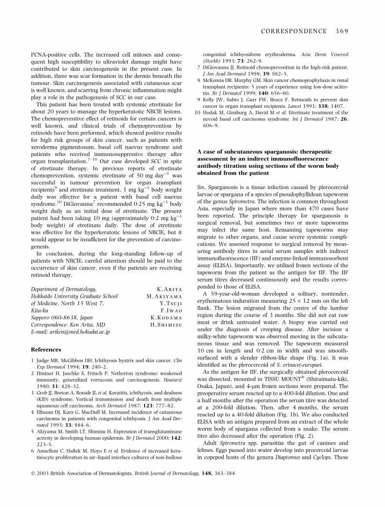
He first noticed a hyperkeratotic nodule on his neck at theage of 41 years, which gradually increased in size. When hewas 44 years old, a partly verrucous, reddish nodule withcrust, about 1 cm in diameter, was present on the left side ofhis neck (Fig. 2a). The cervical lymph node was notpalpable. The nodule was biopsied excisionally with amargin of 1 cm of normal surrounding skin. Histopatholog-ically, there was a proliferative lesion in the epidermis withhyperkeratosis and acanthosis (Fig. 2b). Severe parakeratosiswas present in part of the lesion. The tumour invadedirregularly into the dermis, and the basement membranebecame obscured in some parts. The tumour cell nuclei wereatypical and abundant mitotic figures were present in thetumour nests. Dilated vessels and thick collagen bundles inthe dermis suggested scar formation beneath the tumour.
Histopathogically, the surgical margin of the resected nodulewas free of the tumour. From these clinical and histopath-ological observations, the tumour was diagnosed as SCC thatpresented in a patient with NBCIE. Recurrence of thetumour has not been observed for 3 months after theoperation.
As far as we aware, this is the first report of SCC thatdeveloped in a NBCIE patient with detailed phenotypicinformation. The neck is a commonly involved site for SCCbecause of the high sun exposure and we cannot exclude thepossibility of a chance occurrence of SCC in this NBCIEpatient. However, the onset of cutaneous SCC at 44 years ofage seems to be earlier than normal, especially among theJapanese population.
Amsellem et al.6 demonstrated a higher proliferation rate inkeratinocytes in cultures from NBCIE patients by counting
Figure 1. Clinical phenotype of NBCIE. The patient showed mild
ectropion (a), and erythroderma over the entire body; (b) trunk. The
soles exhibited severe hyperkeratosis (c). Hyperkeratosis and ery-
thema with a shiny appearance were seen in the dorsa of left hand
(d).Figure 2. Clinical and histopathological features of squamous cell
carcinoma on the neck. (a) Hyperkeratotic nodule, approximately
1 cm in diameter, was present on the left side of the neck. (b) His-
topathologically, the tumour was invading irregularly into the der-
mis. The tumour cells had atypical nuclei and some were
dyskeratotic. Abundant mitotic figures were observed (inset). Beneath
the tumour, a band-like inflammatory cell infiltration and fibrosis
were present (haematoxylin and eosin; original magnification · 20,
inset · 100).
3 6 8 C O R R E S P O N D E N C E
� 2003 British Association of Dermatologists, British Journal of Dermatology, 148, 363–384
PCNA-positive cells. The increased cell mitoses and conse-quent high susceptibility to ultraviolet damage might havecontributed to skin carcinogenesis in the present case. Inaddition, there was scar formation in the dermis beneath thetumour. Skin carcinogenesis associated with cutaneous scaris well known, and scarring from chronic inflammation mightplay a role in the pathogenesis of SCC in our case.
This patient has been treated with systemic etretinate forabout 20 years to manage the hyperkeratotic NBCIE lesions.The chemopreventive effect of retinoids for certain cancers iswell known, and clinical trials of chemoprevention byretinoids have been performed, which showed positive resultsfor high risk groups of skin cancer, such as patients withxeroderma pigmentosum, basal cell naevus syndrome andpatients who received immunosuppressive therapy afterorgan transplantation.7–10 Our case developed SCC in spiteof etretinate therapy. In previous reports of etretinatechemoprevention, systemic etretinate of 50 mg day)1 wassuccessful in tumour prevention for organ transplantrecipients9 and etretinate treatment, 1 mg kg)1 body weightdaily was effective for a patient with basal cell naevussyndrome.10 DiGiovanna7 recommended 0Æ25 mg kg)1 bodyweight daily as an initial dose of etretinate. The presentpatient had been taking 10 mg (approximately 0Æ2 mg kg)1
body weight) of etretinate daily. The dose of etretinatewas effective for the hyperkeratotic lesions of NBCIE, but itwould appear to be insufficient for the prevention of carcino-genesis.
In conclusion, during the long-standing follow-up ofpatients with NBCIE, careful attention should be paid to theoccurrence of skin cancer, even if the patients are receivingretinoid therapy.
K . A r i t a
M . A k i y a m a
Y . T s u j i
F . I w a o
K . K o d a m a
H . S h i m i z u
Department of Dermatology,Hokkaido University Graduate Schoolof Medicine, North 15 West 7,Kita-kuSapporo 060-8638, JapanCorrespondence: Ken Arita, MDE-mail: [email protected]
References
1 Judge MR, McGibbon DH. Ichthyosis hystrix and skin cancer. Clin
Exp Dermatol 1994; 19: 240–2.
2 Hintner H, Jaschke E, Fritsch P. Netherton syndrome: weakened
immunity, generalized verrucosis and carcinogenesis. Hautarzt
1980; 31: 428–32.
3 Grob JJ, Breton A, Bonafe JL et al. Keratitis, ichthyosis, and deafness
(KID) syndrome. Vertical transmission and death from multiple
squamous cell carcinoma. Arch Dermatol 1987; 123: 777–82.
4 Elbaum DJ, Kurz G, MacDuff M. Increased incidence of cutaneous
carcinoma in patients with congenital ichthyosis. J Am Acad Der-
matol 1995; 33: 884–6.
5 Akiyama M, Smith LT, Shimizu H. Expression of transglutaminase
activity in developing human epidermis. Br J Dermatol 2000; 142:
223–5.
6 Amsellem C, Haftek M, Hoyo E et al. Evidence of increased kera-
tinocyte proliferation in air–liquid interface cultures of non-bullous
congenital ichthyosiform erythroderma. Acta Derm Venereol
(Stockh) 1993; 73: 262–9.
7 DiGiovanna JJ. Retinoid chemoprevention in the high-risk patient.
J Am Acad Dermatol 1998; 39: S82–5.
8 McKenna DB, Murphy GM. Skin cancer chemoprophylaxis in renal
transplant recipients: 5 years of experience using low-dose acitre-
tin. Br J Dermatol 1999; 140: 656–60.
9 Kelly JW, Sabto J, Gurr FW, Bruce F. Retinoids to prevent skin
cancer in organ transplant recipients. Lancet 1991; 338: 1407.
10 Hodak M, Ginzburg A, David M et al. Etretinate treatment of the
nevoid basal cell carcinoma syndrome. Int J Dermatol 1987; 26:
606–9.
A case of subcutaneous sparganosis: therapeuticassessment by an indirect immunofluorescenceantibody titration using sections of the worm bodyobtained from the patient
SIR, Sparganosis is a tissue infection caused by plerocercoidlarvae or spargana of a species of pseudophyllidean tapewormof the genus Spirometra. The infection is common throughoutAsia, especially in Japan where more than 470 cases havebeen reported. The principle therapy for sparganosis issurgical removal, but sometimes two or more tapewormsmay infect the same host. Remaining tapeworms maymigrate to other organs, and cause severe systemic compli-cations. We assessed response to surgical removal by meas-uring antibody titres in serial serum samples with indirectimmunofluorescence (IIF) and enzyme-linked immunosorbentassay (ELISA). Importantly, we utilized frozen sections of thetapeworm from the patient as the antigen for IIF. The IIFserum titres decreased continuously and the results corres-ponded to those of ELISA.
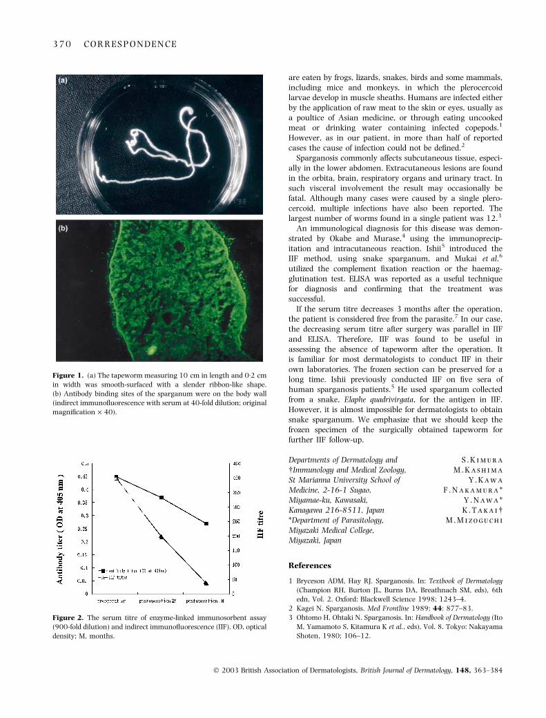
A 59-year-old-woman developed a solitary, nontender,erythematous induration measuring 25 · 12 mm on the leftflank. The lesion migrated from the centre of the lumbarregion during the course of 3 months. She did not eat rawmeat or drink untreated water. A biopsy was carried outunder the diagnosis of creeping disease. After incision amilky-white tapeworm was observed moving in the subcuta-neous tissue and was removed. The tapeworm measured10 cm in length and 0Æ2 cm in width and was smooth-surfaced with a slender ribbon-like shape (Fig. 1a). It wasidentified as the plerocercoid of S. erinacei-europaei.
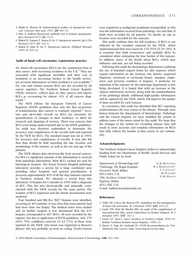
As the antigen for IIF, the surgically obtained plerocercoidwas dissected, mounted in TISSU MOUNT� (Shiraimatu-kiki,Osaka, Japan), and 4-lm frozen sections were prepared. Thepreoperative serum reacted up to a 400-fold dilution. One anda half months after the operation the serum titre was detectedat a 200-fold dilution. Then, after 4 months, the serumreacted up to a 40-fold dilution (Fig. 1b). We also conductedELISA with an antigen prepared from an extract of the wholeworm body of spargana collected from a snake. The serumtitre also decreased after the operation (Fig. 2).
Adult Spirometra spp. parasitize the gut of canines andfelines. Eggs passed into water develop into procercoid larvaein copepod hosts of the genera Diaptomus and Cyclops. These
C O R R E S P O N D E N C E 3 6 9
� 2003 British Association of Dermatologists, British Journal of Dermatology, 148, 363–384
are eaten by frogs, lizards, snakes, birds and some mammals,including mice and monkeys, in which the plerocercoidlarvae develop in muscle sheaths. Humans are infected eitherby the application of raw meat to the skin or eyes, usually asa poultice of Asian medicine, or through eating uncookedmeat or drinking water containing infected copepods.1
However, as in our patient, in more than half of reportedcases the cause of infection could not be defined.2
Sparganosis commonly affects subcutaneous tissue, especi-ally in the lower abdomen. Extracutaneous lesions are foundin the orbita, brain, respiratory organs and urinary tract. Insuch visceral involvement the result may occasionally befatal. Although many cases were caused by a single plero-cercoid, multiple infections have also been reported. Thelargest number of worms found in a single patient was 12.3
An immunological diagnosis for this disease was demon-strated by Okabe and Murase,4 using the immunoprecip-itation and intracutaneous reaction. Ishii5 introduced theIIF method, using snake sparganum, and Mukai et al.6
utilized the complement fixation reaction or the haemag-glutination test. ELISA was reported as a useful techniquefor diagnosis and confirming that the treatment wassuccessful.
If the serum titre decreases 3 months after the operation,the patient is considered free from the parasite.7 In our case,the decreasing serum titre after surgery was parallel in IIFand ELISA. Therefore, IIF was found to be useful inassessing the absence of tapeworm after the operation. Itis familiar for most dermatologists to conduct IIF in theirown laboratories. The frozen section can be preserved for along time. Ishii previously conducted IIF on five sera ofhuman sparganosis patients.5 He used sparganum collectedfrom a snake, Elaphe quadrivirgata, for the antigen in IIF.However, it is almost impossible for dermatologists to obtainsnake sparganum. We emphasize that we should keep thefrozen specimen of the surgically obtained tapeworm forfurther IIF follow-up.
S . K i m u r a
M . K a s h i m a
Y . K a w a
F . N a k a m u r a *
Y . N a w a *
K . T a k a i�M . M i z o g u c h i
Departments of Dermatology and�Immunology and Medical Zoology,St Marianna University School ofMedicine, 2-16-1 Sugao,Miyamae-ku, Kawasaki,Kanagawa 216-8511, Japan*Department of Parasitology,Miyazaki Medical College,Miyazaki, Japan
References
1 Bryceson ADM, Hay RJ. Sparganosis. In: Textbook of Dermatology
(Champion RH, Burton JL, Burns DA, Breathnach SM, eds), 6th
edn, Vol. 2. Oxford: Blackwell Science 1998; 1243–4.
2 Kagei N. Sparganosis. Med Frontline 1989; 44: 877–83.
3 Ohtomo H, Ohtaki N. Sparganosis. In: Handbook of Dermatology (Ito
M, Yamamoto S, Kitamura K et al., eds), Vol. 8. Tokyo: Nakayama
Shoten, 1980; 106–12.
Figure 2. The serum titre of enzyme-linked immunosorbent assay
(900-fold dilution) and indirect immunofluorescence (IIF). OD, optical
density; M, months.
Figure 1. (a) The tapeworm measuring 10 cm in length and 0Æ2 cm
in width was smooth-surfaced with a slender ribbon-like shape.
(b) Antibody binding sites of the sparganum were on the body wall
(indirect immunofluorescence with serum at 40-fold dilution; original
magnification · 40).
3 7 0 C O R R E S P O N D E N C E
� 2003 British Association of Dermatologists, British Journal of Dermatology, 148, 363–384
4 Okabe K, Murase M. Immunological studies on Sparganosis man-
soni. J Kurume Med Assoc 1957; 20: 907–13.
5 Ishii A. Indirect fluorescent antibody test in human sparganosis.
Jpn J Parasitol 1973; 22: 75–8.
6 Mukai H, Yamate T, Saitou R, Ito Y. Sparganosis mansoni. Jpn J Clin
Dermatol 1977; 31: 551–5.
7 Mukai H, Saito R, Ito Y. Sparganosis mansoni. Hifubyoh Shinryoh
1981; 3: 219–22.
Audit of basal cell carcinoma: registration practice
SIR, Basal cell carcinomas (BCCs) are the commonest form ofskin cancer, with increasing incidence in the U.K.1 They areassociated with significant morbidity and their cost oftreatment is an increasing burden to the health service,yet accurate information on their incidence is not available.2
For cost and volume reasons BCCs are not recorded by allcancer registries. The Northern Ireland Cancer Registry(NICR), however, collects data on skin cancers and reportsBCCs as accounting for almost 18% of all cancers diag-nosed.3
The NICR follows the European Network of CancerRegistries (ENCR) guidelines that only the first occurrenceof nonmelanoma skin cancers is routinely reported.4 Accu-rate and up-to-date records on BCCs are necessary forquantification of changes in their incidence, to allow forresearch and planning of services. There was concern thatBCCs were being under-reported due to the ENCR guidelines.An audit was therefore undertaken to determine theaccuracy and completeness of the records held and reportedby the NICR for BCCs. The standard target was set at 100%for accuracy and completeness of records. It was also feltthat data should be held regarding the size, location andmorphology of the tumour, as well as the sex and age of thepatients.
The NICR obtains data electronically from several sources.For BCCs a significant amount of the information is receivedfrom pathology laboratories. Most BCCs excised are sent forhistological analysis. The Royal Victoria Hospital pathologylaboratory provides a service for a large catchment areaincluding other hospitals and general practitioners. Itprocesses approximately 40% of all the skin tumours reportedin Northern Ireland. We obtained a record from thislaboratory of biopsies for 6 months in 1999 with a diagnosisof BCC. This list was electronically and manually cross-checked with the NICR records for the same period. Thenumber of BCCs registered and the details documented wereanalysed.
Four hundred and fifty-five �BCC� biopsies were identified,occurring in 403 patients. It was clear that some patients hadhad more than one biopsy. The medical notes were studiedand on further analysis it was determined that the 455biopsies corresponded to 407 BCCs. All were recorded by theregistry, but due to application of ENCR guidelines, only 270(66%; 95% confidence interval, CI 62–71%) of these werereported by the NICR. One lesion was registered as Bowen’sdisease: this was probably an error in coding. Twelve lesions
were registered as malignant neoplasms nonspecified, as thiswas the information received from pathology. Sex and date ofbirth were recorded for all patients. No details on size orlocation were recorded for the tumours.
This audit confirms that the full incidence of BCCs is notreflected by the numbers reported by the NICR, whichunderestimated their true extent by 33% (95% CI 29–38%). Itis essential that both recurrences and multiple BCCs beconsidered when estimating the true burden of this tumour.In addition, many of the details about BCCs, which mayinfluence outcome, are not being recorded.
Following this audit the dermatology department is pilotingthe use of standard request forms for skin tumours whichrequire information on site, location, size, history, suspecteddiagnosis, incisional or excisional biopsy, margins, origin-ation and previous numbers of biopsies. A proforma forreporting of the tumours by the pathology department is alsobeing developed. It is hoped that with an increase in theclinical information received, along with the standardizationof the pathology details, additional high-quality informationwill be captured by the NICR, which will improve the qualityof data recorded for each tumour.
In conclusion, this audit has identified that BCC reportingunderestimates the true extent of the disease. Through closeliaison between the departments of Dermatology, Pathologyand the Cancer Registry we have modified the system toaddress some of the issues raised by this audit. We hope thatthe changes to the system for recording tumour data willresult in more accurate and complete information on BCCsthat fully reflects the burden of skin cancer in our commu-nity.
Acknowledgments
The Northern Ireland Cancer Registry wishes to acknowledgefunding from the Department of Health, Social Services andPublic Safety for its work.
N . M . M c L o o n e
R . J . M i d d l e t o n *
A . T . G a v i n *
M . W a l s h�O . M . D o l a n
Departments of Dermatology and*Pathology, The Royal Hospitals,Grosvenor Road, BelfastBT12 6BA, U.K.�The Northern Ireland CancerRegistry, BelfastBT12 6BJ, U.K.E-mail: [email protected]
References
1 Telfer NR, Colver GB, Bowers PW. Guidelines for the management
of basal cell carcinoma. Br J Dermatol 1999; 141: 415–23.
2 Lucke TW, Hole DJ, MacKie RM. An audit of the completeness of
non-melanoma skin cancer registration in Greater Glasgow. Br J
Dermatol 1997; 137: 761–3.
3 Gavin AT, Reid J. Cancer Incidence in Northern Ireland, 1993–95.
Belfast: Northern Ireland Cancer Registry, 1999; 23.
4 Davies T, Page M, Coebergh W. ENCR Recommendations for Non-
Melanoma Skin Cancers. http://www-dep.arc.fr/encr.htm.
C O R R E S P O N D E N C E 3 7 1
� 2003 British Association of Dermatologists, British Journal of Dermatology, 148, 363–384
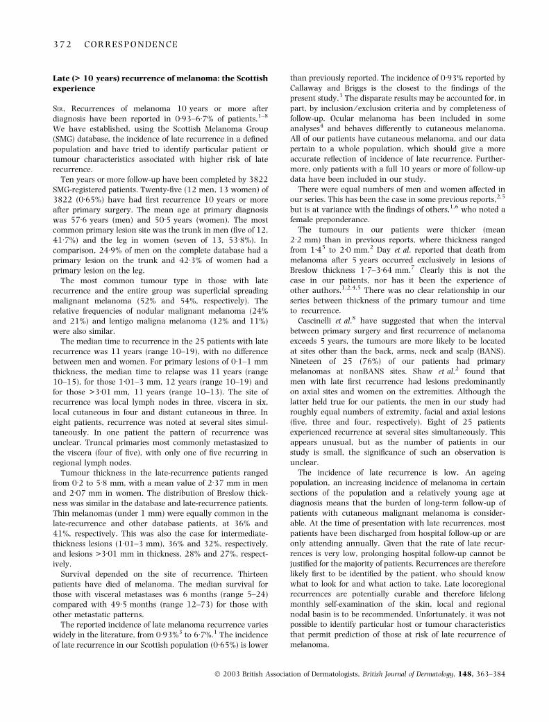
Late (> 10 years) recurrence of melanoma: the Scottishexperience
SIR, Recurrences of melanoma 10 years or more afterdiagnosis have been reported in 0Æ93–6Æ7% of patients.1–8
We have established, using the Scottish Melanoma Group(SMG) database, the incidence of late recurrence in a definedpopulation and have tried to identify particular patient ortumour characteristics associated with higher risk of laterecurrence.
Ten years or more follow-up have been completed by 3822SMG-registered patients. Twenty-five (12 men, 13 women) of3822 (0Æ65%) have had first recurrence 10 years or moreafter primary surgery. The mean age at primary diagnosiswas 57Æ6 years (men) and 50Æ5 years (women). The mostcommon primary lesion site was the trunk in men (five of 12,41Æ7%) and the leg in women (seven of 13, 53Æ8%). Incomparison, 24Æ9% of men on the complete database had aprimary lesion on the trunk and 42Æ3% of women had aprimary lesion on the leg.
The most common tumour type in those with laterecurrence and the entire group was superficial spreadingmalignant melanoma (52% and 54%, respectively). Therelative frequencies of nodular malignant melanoma (24%and 21%) and lentigo maligna melanoma (12% and 11%)were also similar.
The median time to recurrence in the 25 patients with laterecurrence was 11 years (range 10–19), with no differencebetween men and women. For primary lesions of 0Æ1–1 mmthickness, the median time to relapse was 11 years (range10–15), for those 1Æ01–3 mm, 12 years (range 10–19) andfor those >3Æ01 mm, 11 years (range 10–13). The site ofrecurrence was local lymph nodes in three, viscera in six,local cutaneous in four and distant cutaneous in three. Ineight patients, recurrence was noted at several sites simul-taneously. In one patient the pattern of recurrence wasunclear. Truncal primaries most commonly metastasized tothe viscera (four of five), with only one of five recurring inregional lymph nodes.
Tumour thickness in the late-recurrence patients rangedfrom 0Æ2 to 5Æ8 mm, with a mean value of 2Æ37 mm in menand 2Æ07 mm in women. The distribution of Breslow thick-ness was similar in the database and late-recurrence patients.Thin melanomas (under 1 mm) were equally common in thelate-recurrence and other database patients, at 36% and41%, respectively. This was also the case for intermediate-thickness lesions (1Æ01–3 mm), 36% and 32%, respectively,and lesions >3Æ01 mm in thickness, 28% and 27%, respect-ively.
Survival depended on the site of recurrence. Thirteenpatients have died of melanoma. The median survival forthose with visceral metastases was 6 months (range 5–24)compared with 49Æ5 months (range 12–73) for those withother metastatic patterns.
The reported incidence of late melanoma recurrence varieswidely in the literature, from 0Æ93%3 to 6Æ7%.1 The incidenceof late recurrence in our Scottish population (0Æ65%) is lower
than previously reported. The incidence of 0Æ93% reported byCallaway and Briggs is the closest to the findings of thepresent study.3 The disparate results may be accounted for, inpart, by inclusion ⁄ exclusion criteria and by completeness offollow-up. Ocular melanoma has been included in someanalyses4 and behaves differently to cutaneous melanoma.All of our patients have cutaneous melanoma, and our datapertain to a whole population, which should give a moreaccurate reflection of incidence of late recurrence. Further-more, only patients with a full 10 years or more of follow-updata have been included in our study.
There were equal numbers of men and women affected inour series. This has been the case in some previous reports,2,5
but is at variance with the findings of others,1,6 who noted afemale preponderance.
The tumours in our patients were thicker (mean2Æ2 mm) than in previous reports, where thickness rangedfrom 1Æ45 to 2Æ0 mm.2 Day et al. reported that death frommelanoma after 5 years occurred exclusively in lesions ofBreslow thickness 1Æ7–3Æ64 mm.7 Clearly this is not thecase in our patients, nor has it been the experience ofother authors.1,2,4,5 There was no clear relationship in ourseries between thickness of the primary tumour and timeto recurrence.
Cascinelli et al.8 have suggested that when the intervalbetween primary surgery and first recurrence of melanomaexceeds 5 years, the tumours are more likely to be locatedat sites other than the back, arms, neck and scalp (BANS).Nineteen of 25 (76%) of our patients had primarymelanomas at nonBANS sites. Shaw et al.2 found thatmen with late first recurrence had lesions predominantlyon axial sites and women on the extremities. Although thelatter held true for our patients, the men in our study hadroughly equal numbers of extremity, facial and axial lesions(five, three and four, respectively). Eight of 25 patientsexperienced recurrence at several sites simultaneously. Thisappears unusual, but as the number of patients in ourstudy is small, the significance of such an observation isunclear.
The incidence of late recurrence is low. An ageingpopulation, an increasing incidence of melanoma in certainsections of the population and a relatively young age atdiagnosis means that the burden of long-term follow-up ofpatients with cutaneous malignant melanoma is consider-able. At the time of presentation with late recurrences, mostpatients have been discharged from hospital follow-up or areonly attending annually. Given that the rate of late recur-rences is very low, prolonging hospital follow-up cannot bejustified for the majority of patients. Recurrences are thereforelikely first to be identified by the patient, who should knowwhat to look for and what action to take. Late locoregionalrecurrences are potentially curable and therefore lifelongmonthly self-examination of the skin, local and regionalnodal basin is to be recommended. Unfortunately, it was notpossible to identify particular host or tumour characteristicsthat permit prediction of those at risk of late recurrence ofmelanoma.
3 7 2 C O R R E S P O N D E N C E
� 2003 British Association of Dermatologists, British Journal of Dermatology, 148, 363–384

J . A . L e m a n
R . M . M a c K i e
University Department ofDermatology, The Western Infirmary,Glasgow G11 6NT, U.K.E-mail: [email protected]
References
1 Briele HA, Beattie CW, Ronan SG et al. Late recurrence of cuta-
neous malignant melanoma. Arch Surg 1983; 118: 800–3.
2 Shaw HM, Beattie CW, McCarthy WH, Milton GW. Late relapse
from cutaneous stage I malignant melanoma. Arch Surg 1985;
120: 1155–9.
3 Callaway MP, Briggs JC. The incidence of late recurrence (greater
than 10 years): an analysis of 536 consecutive cases of cutaneous
melanoma. Br J Plast Surg 1989; 42: 46–9.
4 Crowley NJ, Seigler HF. Late recurrence of malignant melanoma;
analysis of 168 patients. Ann Surg 1990; 212: 713–17.
5 Schmidt-Wendtner MH, Baumert J, Schmidt M et al. Late meta-
stases of cutaneous malignant melanoma: an analysis of 31
patients. J Am Acad Dermatol 2000; 43: 605–9.
6 Raderman D, Giler S, Rothem A, Ben-Bassat M. Late metastases
(beyond ten years) of cutaneous malignant melanoma. J Am Acad
Dermatol 1986; 15: 374–8.
7 Day CL, Mihm MC, Sober AJ et al. Predictors of late deaths among
patients with clinical stage I melanoma who have not had bony or
visceral metastases within the first 5 years after diagnosis. J Am
Acad Dermatol 1983; 8: 864–8.
8 Cascinelli N, Vaglini M, Bufalino R, Morabito A. BANS: a cutane-
ous region with no prognostic significance in patients with mel-
anoma. Cancer 1986; 57: 441–4.
Dermatology Life Quality Index score in vitiligo and itsimpact on the treatment outcome
SIR, Vitiligo is an acquired depigmentation disorder of greatcosmetic importance affecting 1–4% of the world’s population.The disease has a major impact on quality of life of patients,many of whom feel stigmatized by their condition. Porter et al.studied the effect of vitiligo on sexual relationships and foundthat embarrassment during sexual relationships was especi-ally frequent for men with vitiligo.1 Salzer and Schallreuter2
reported that 75% found their disfigurement moderately orseverely intolerable. Weiss et al.3 compared the difficultiesfaced by patients with vitiligo with those with leprosy in India.There may be a relationship between stress and the develop-ment of vitiligo. Al-Abadie et al.4 indicated that psychologicalstress increases levels of neuroendocrine hormones, affects theimmune system and alters the level of neuropeptides, whichmay be the initial steps in pathogenesis of vitiligo. The purposeof this study was to assess the nature and extent of the socialand psychological difficulties associated with vitiligo and theirimpact on treatment outcome by using the Dermatology LifeQuality Index (DLQI).
One hundred and fifty patients with vitiligo vulgarisinvolving more than 10% body surface area attending thepigmentary clinic of the Department of Dermatology, Post-graduate Institute of Medical Education and Research,
Chandigarh, India were enrolled in this study. They wereintroduced to the subject of this study and informed about thepersonal nature of the questionnaire, and all those who gaveconsent were given the DLQI questionnaire to complete.Before starting therapy, patients were evaluated clinically torecord the duration and progression of the disease, the sites oflesions, and the extent of cutaneous involvement. Thesepatients were treated as per protocol of our pigmentary clinic.Two parameters, repigmentation and arrest of progression ofdisease activity, were used to evaluate the response totreatment. The treatment was considered to be successful ifa patient with active disease: (i) had no new lesions; (ii) hadno increase in the size of the existing lesions; and (iii) hadmore than 25% repigmentation at the end of 1 year. Thetreatment was considered to be a failure: (i) if a patient withactive disease continued to have new lesions; or (ii) if theexisting lesions continued to increase in size; or (iii) there wasless than 25% (overall) repigmentation at the end of 1 year oftreatment. Responses on the DLQI were scored according tothe guidelines of Finlay and Khan.5
One hundred and fifty patients completed the question-naire. Their basic data are given in Table 1. Scores on theDLQI ranged from 2 to 21 (mean ± SD 10Æ67 ± 4Æ56). Therewas no statistically significant difference between the DLQIscores of the male and female patients. There were statisti-cally significant relationships between DLQI scores and age(P < 0Æ001), and between DLQI scores and disease duration(P < 0Æ05), but not between DLQI scores and other variablessuch as extent of disease.
At the end of the treatment period, 141 patients could beevaluated. The treatment outcome was considered to besuccessful in 91 patients and a failure in the remaining 50,based on the criteria mentioned above. The mean DLQIscore was 7Æ06 in patients with a successful outcome,whereas it was 13Æ12 for patients with treatment failure.There was a statistically highly significant difference(P < 0Æ0001, t-test) between mean DLQI scores of thesetwo groups.
Table 1. Basic data of patients in this study
Total patients (n ¼ 150)
Age, mean (range), years 33Æ6 (18–63)
Sex, M ⁄ F 67 ⁄ 83
Duration of disease
< 1 year 27
1–3 years 76
> 3 years 47
Treatment modalities
PUVA 12
PUVAsol 85
OMP 18
PUVAsol and OMP 7
PUVAsol and levamisole 28
PUVA, psoralen plus ultraviolet A therapy; OMP, oral mini pulse
(betamethasone).
C O R R E S P O N D E N C E 3 7 3
� 2003 British Association of Dermatologists, British Journal of Dermatology, 148, 363–384

This study provides information concerning the DLQI andits impact on treatment outcome in vitiligo sufferers. Themean DLQI score in our study (10Æ67) is higher than thatobtained by Finlay and Khan5 (mean 7Æ3) and Kent andAl-Abadie6 (4Æ82). There was no relationship of DLQI scorewith gender, which is consistent with the earlier studies.Our study clearly demonstrated that patients with highDLQI scores responded less favourably to a given thera-peutic modality. These results suggest that additionalpsychological approaches may be particularly helpful inthese patients.
In a preliminary study by Papadopoulos et al.,7 it wasshown that counselling can help to improve the body image,self-esteem and quality of life of patients with vitiligo as wellas having a positive effect on course of the disease. There isevidence that social and psychological well-being increasewhen patients with facial disfigurement are helped to developsocial skills and to confront their difficulties. In a disease suchas vitiligo, which is not only difficult to treat but is likely toprogress, it is important to recognize and deal with thepsychological component of this distressing condition notonly to improve the appearance-related stress-handlingcapability of the patient but also to obtain a better treatmentresponse. Training in assertiveness, relaxation skills and helpin building self-confidence would have substantial effects onDLQI score as well as treatment outcome. The results of thisstudy have implications in terms of our understanding of therole of the mental state and its impact on treatment outcomein vitiligo.
Acknowledgment
We express our sincere thanks to Prof. Andrew Finlay forpermission to use the Dermatology Quality Life Index.
D . P a r s a d
R . P a n d h i
S . D o g r a
A . J . K a n w a r
B . K u m a r
Department of Dermatology,Postgraduate Institute of MedicalEducation and Research,Chandigarh 160012, IndiaE-mail: [email protected]
References
1 Porter J, Beuf A, Lerner A et al. The effect of vitiligo on sexual
relationship. J Am Acad Dermatol 1990; 22: 221–2.
2 Salzer BA, Schallreuter KU. Investigations of the personality
structure in patients with vitiligo and a possible association
with catecholamine metabolism. Dermatology 1995; 190:
109–15.
3 Weiss M, Doongaji D, Siddartha S et al. The explanatory model
interview catalogue (EMIC). Br J Psychiatry 1992; 160:
819–30.
4 Al-Abadie MSK, Kent G, Gawkrodger DJ. The relationship between
stress and the onset and exacerbation of psoriasis and other skin
conditions. Br J Dermatol 1994; 130: 199–203.
5 Finlay AY, Khan G. Dermatology Life Quality Index (DLQI): a
simple practical measure for routine clinical use. Clin Exp Dermatol
1994; 19: 210–16.
6 Kent G, Al-Abadie MSK. Factors affecting responses on Dermatol-
ogy Life Quality Index among vitiligo sufferers. Clin Exp Dermatol
1996; 21: 330–3.
7 Papadopoulos L, Bor R, Legg C. Coping with the disfiguring effects
of vitiligo. a preliminary investigation into the effects of cognitive-
behaviour therapy. Br J Med Psychol 1999; 72: 385–96.
Oral lichen planus: diagnostic immunofluorescencetesting on routine histological material
SIR, Direct immunofluorescence testing on lesional biopsies isa valuable tool to establish the diagnosis of oral lichen planus(OLP) and to differentiate the condition from other immuno-logically mediated diseases of the oral mucosa, such aspemphigoid, pemphigus vulgaris and lupus erythematosus.Shaggy deposition of fibrinogen along the basement mem-brane zone and ⁄ or globoid or cytoid-like bodies with positiveIgM labelling have been shown to represent typical anddiagnostic features of OLP, in particular when used inconjunction with routine histology.1–3 However, correlativeand, most importantly, retrospective evaluations on routinehistological material do not seem possible, as specific andreliable immunolabelling is thought to require fresh-frozenbiopsy specimens.4,5 We present a simple and effectivetechnique for diagnostic immunofluorescence testing onarchival histological material from oral biopsies.
The study was performed on 30 diagnostic biopsies takenfrom the buccal or gingival mucosa of 27 patients (20women, seven men; mean age 54 years) suspected to sufferfrom OLP. All biopsies had been fixed in formaldehyde andembedded in paraffin using conventional techniques. Forimmunofluorescence testing, dewaxed and rehydrated histo-logical sections were treated with 0Æ1% (w ⁄ v) pronase E(Sigma, St Louis, MO, U.S.A.) in 0Æ1 mol L)1 Tris-bufferedsaline (TBS) for 30 min at 37 �C. After washing in TBS, thesections were incubated with fluorescein isothiocyanate(FITC)-conjugated polyclonal rabbit antibodies against IgG,IgA, IgM, complement C3 and fibrinogen (all from Dako,Hamburg, Germany) for 30 min at room temperature. Thespecificity of staining was checked by incubating the sectionswith FITC-conjugated normal rabbit serum. The labelledsections were washed in TBS, mounted in Immunomount-Shandon, coverslipped and examined with a conventionalZeiss light microscope equipped with a 200-W mercury arclamp.
Immunofluorescence examination showed excellent pre-servation of tissue morphology, and negligible backgroundstaining, nonspecific fluorescence and tissue autofluores-cence. No positive immunofluorescence staining wasobserved in normal oral mucosa biopsies (n ¼ 5) used asnegative controls. The positive controls from oral pemphigusvulgaris (n ¼ 3) and mucous membrane pemphigoid (n ¼ 3)were characterized by typical and distinct staining of theepithelial intercellular spaces and basement membrane zone,respectively. The typical immunofluorescence staining pat-terns of OLP were found in 17 of the 30 histological biopsies.
3 7 4 C O R R E S P O N D E N C E
� 2003 British Association of Dermatologists, British Journal of Dermatology, 148, 363–384
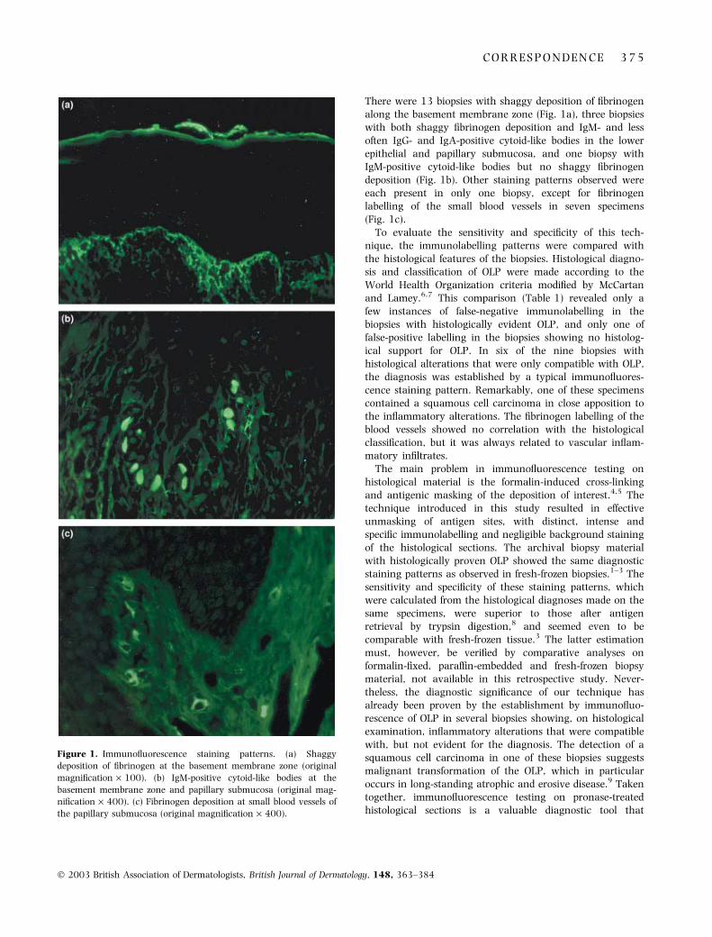
There were 13 biopsies with shaggy deposition of fibrinogenalong the basement membrane zone (Fig. 1a), three biopsieswith both shaggy fibrinogen deposition and IgM- and lessoften IgG- and IgA-positive cytoid-like bodies in the lowerepithelial and papillary submucosa, and one biopsy withIgM-positive cytoid-like bodies but no shaggy fibrinogendeposition (Fig. 1b). Other staining patterns observed wereeach present in only one biopsy, except for fibrinogenlabelling of the small blood vessels in seven specimens(Fig. 1c).
To evaluate the sensitivity and specificity of this tech-nique, the immunolabelling patterns were compared withthe histological features of the biopsies. Histological diagno-sis and classification of OLP were made according to theWorld Health Organization criteria modified by McCartanand Lamey.6,7 This comparison (Table 1) revealed only afew instances of false-negative immunolabelling in thebiopsies with histologically evident OLP, and only one offalse-positive labelling in the biopsies showing no histolog-ical support for OLP. In six of the nine biopsies withhistological alterations that were only compatible with OLP,the diagnosis was established by a typical immunofluores-cence staining pattern. Remarkably, one of these specimenscontained a squamous cell carcinoma in close apposition tothe inflammatory alterations. The fibrinogen labelling of theblood vessels showed no correlation with the histologicalclassification, but it was always related to vascular inflam-matory infiltrates.
The main problem in immunofluorescence testing onhistological material is the formalin-induced cross-linkingand antigenic masking of the deposition of interest.4,5 Thetechnique introduced in this study resulted in effectiveunmasking of antigen sites, with distinct, intense andspecific immunolabelling and negligible background stainingof the histological sections. The archival biopsy materialwith histologically proven OLP showed the same diagnosticstaining patterns as observed in fresh-frozen biopsies.1–3 Thesensitivity and specificity of these staining patterns, whichwere calculated from the histological diagnoses made on thesame specimens, were superior to those after antigenretrieval by trypsin digestion,8 and seemed even to becomparable with fresh-frozen tissue.3 The latter estimationmust, however, be verified by comparative analyses onformalin-fixed, paraffin-embedded and fresh-frozen biopsymaterial, not available in this retrospective study. Never-theless, the diagnostic significance of our technique hasalready been proven by the establishment by immunofluo-rescence of OLP in several biopsies showing, on histologicalexamination, inflammatory alterations that were compatiblewith, but not evident for the diagnosis. The detection of asquamous cell carcinoma in one of these biopsies suggestsmalignant transformation of the OLP, which in particularoccurs in long-standing atrophic and erosive disease.9 Takentogether, immunofluorescence testing on pronase-treatedhistological sections is a valuable diagnostic tool that
Figure 1. Immunofluorescence staining patterns. (a) Shaggy
deposition of fibrinogen at the basement membrane zone (original
magnification · 100). (b) IgM-positive cytoid-like bodies at the
basement membrane zone and papillary submucosa (original mag-
nification · 400). (c) Fibrinogen deposition at small blood vessels of
the papillary submucosa (original magnification · 400).
C O R R E S P O N D E N C E 3 7 5
� 2003 British Association of Dermatologists, British Journal of Dermatology, 148, 363–384

improves the often controversial histopathological assess-ment of OLP.10
G . K o l d e
C . W e s e n d a h l
H . S t e i n *
P . A . R e i c h a r t�
Department of Dermatology andAllergy, Charite,Humboldt-University of Berlin,Schumannstr. 20 ⁄ 21,D-10117 Berlin, Germany*Institute of Pathology, UniversityHospital Benjamin Franklin,Free University Berlin,Hindenburgdamm 30,D-12200 Berlin, Germany�Department of Oral Surgery andDental Radiology, Charite, Humboldt-University of Berlin, Fohrer Str. 15,D-13353 Berlin, GermanyE-mail: [email protected]
References
1 Laskaris G, Sklavounou A, Angelopoulos A. Direct immunofluo-
rescence in oral lichen planus. Oral Surg Oral Med Oral Pathol
1982; 53: 483–7.
2 Firth NA, Rich AM, Radden BG, Reade PC. Assessment of the
value of immunofluorescence microscopy in the diagnosis of oral
mucosal lichen planus. J Oral Pathol Med 1990; 19: 295–7.
3 Helander SD, Rogers RS. The sensitivity and specificity of direct
immunofluorescence testing in disorders of mucous membranes.
J Am Acad Dermatol 1994; 30: 65–75.
4 Collins AB. Immunofluorescence. In: Diagnostic Immunopathology
(Colvin RB, Bhan AK, McCluskey RT, eds). New York: Raven
Press, 1995; 699–710.
5 Brandtzaeg P. The increasing power of immunohistochemistry
and immunocytochemistry. J Immunol Methods 1998; 216:
49–67.
6 Pindborg JJ, Reichart PA, Smith CJ, van der Waal I. Histological
Typing of Cancer and Precancer of the Oral Mucosa. Berlin: Springer-
Verlag, 1997; 30.
7 McCartan BE, Lamey P. Lichen planus-specific antigen in oral
lichen planus and oral lichenoid drug eruptions. Oral Surg Oral
Med Oral Pathol Oral Radiol Endod 2000; 89: 585–7.
8 Firth NA, Rich AM, Radden BG, Reade PC. Direct immunofluo-
rescence of oral mucosal biopsies: a comparison of fresh-frozen
tissue and formalin-fixed, paraffin-embedded tissue. J Oral Pathol
Med 1992; 21: 358–63.
9 Hietanen J, Paasonen MR, Kuhlefelt M, Malmstrom M. A retro-
spective study of oral lichen planus patients with concurrent or
subsequent development of malignancy. Oral Oncol 1999; 35:
278–82.
10 van der Meij EH, Reibel J, Slootweg PJ et al. Interobserver and
intraobserver variability in the histologic assessment of oral li-
chen planus. J Oral Pathol Med 1999; 28: 274–7.
Severe pruritus in a haemodialysed patient: dramaticimprovement with granisetron
SIR, Pruritus is a common unpleasant symptom in patientsundergoing dialysis. Uraemic pruritus can be resistant toconventional antipruritic therapy and can affect the patient’squality of life. We report a haemodialysed patient withresistant pruritus who showed a dramatic improvement aftertreatment with granisetron.
A 51-year-old man on haemodialysis since 1982 presentedwith a 1-year history of pruritus. He did not improve withany major antipruritic therapy (hydroxyzine, antihistaminesclass I and II). He was depressed because he had been unableto sleep for the previous year due to the distressing pruritus.His medical history included hyperparathyroidism and hepa-titis B and C virus infection.
Examination showed global excoriated and necrotic areas,mainly over the thorax (Fig. 1a). He was started on granise-tron 1 mg daily. Within a few hours of the first dose thepatient reported a dramatic relief of the pruritic discomfort,enabling him to sleep. Pruritus, scratching and globalexcoriation gradually improved (Fig. 1b). Treatment wascontinued for 3 weeks. No side-effects were observed. Thepatient remained free of symptoms 3 months after stoppingthis drug.
Granisetron and ondansetron belong to the 5-hydroxy-tryptamine type 3 (5HT3) receptor antagonists. These drugsare selective inhibitors of the 5HT3 receptor and have apotent antiserotonin effect. The anti5HT3 drugs are usuallyemployed to relieve chemotherapy-induced emesis.1 Sero-tonin could play a key role in the generation of thissymptom.2 Additionally, serotonin and histamine have beenreported as possible mediators of uraemic pruritus. Ondan-setron has been reported to improve pruritus in dialysispatients2 and in palmoplantar pruritus resistant to othertherapies.3 These drugs have occasionally been used forcholestatic pruritus.4
Our case indicates that granisetron could be an effective,safe and well-tolerated drug to treat uraemic pruritus in
Table 1. Comparison of the histological and immunofluorescence findings
Immunofluorescence testing
Histopathology classification n OLP patternsa Other patterns Negative
Evident OLP 13 10 3 0
Compatible with OLP 9 6 2 1
No support for OLP 8 1 5 2
OLP, oral lichen planus. aShaggy deposition of fibrinogen along the basement membrane zone and ⁄ or IgM-positive cytoid-like bodies.
3 7 6 C O R R E S P O N D E N C E
� 2003 British Association of Dermatologists, British Journal of Dermatology, 148, 363–384

dialysed patients. More studies would be needed to prove theeffectiveness of this drug for uraemic pruritus.
M . P . A l b a r e s
I . B e t l l o c h
J . G u i j a r r o
G . V e r g a r a
J . C . P a s c u a l
R . B o t e l l a
Department of Dermatology,Hospital General de Alicante,C ⁄ Pintor Baeza s ⁄ n,03010 Alicante, SpainE-mail: [email protected]
References
1 Carrion Galindo JR. Los anti-5HT3: un nuevo grupo de antieme-
ticos. Rev Clin Esp 1996; 2: 65–70.
2 Balaskas EV, Bamihas GI, Karamouzis M et al. Histamine and
serotonin in uremic pruritus: effect of ondansetron in CAPD-pru-
ritic patients. Nephron 1998; 78: 395–402.
3 Downs A, Kennedy C. Successful treatment of intractable palmo-
plantar pruritus with ondansetron. Arch Dermatol 1998; 134:
925–6.
4 Schworer H, Hartmann H, Ramadori G. Relief of cholestatic pru-
ritus by a novel class of drugs: 5-hydroxytryptamine type 3
(5-HT3) receptor antagonists: effectiveness of ondansetron. Pain
1995; 61: 33–7.
Borrelia afzelii evidenced by polymerase chainreaction in a biopsy of nipple lymphocytoma
SIR, Typically located in the ear lobe, nipple areola, nose andscrotum, Borrelia lymphocytoma is not rare but is oftenmisdiagnosed.
A 57-year-old man had had an infiltration of the left areolaand nipple for 3 months. The infiltrated area strictly corres-ponded to anatomical limits; tissues were firm, elastic andhomogeneous. The colour was dark red. The patient reporteda local painful hyperaesthesia. Two months before the firstsigns appeared, in the summer, he had noticed an erythe-matous macule in the same area. After a few weeks of slowprogression, this plaque had a diameter of almost 15 cm,before resolving spontaneously. Two mammograms showed athickening of the areolar plaque. The tumour cell markersCA125, carcinoembryonic antigen and CA19Æ9 were negat-ive. Fine needle biopsy suggested an inflammatory reaction,without atypical or malignant cells. Symptoms were suggest-ive of a pseudolymphoma. The patient had not previouslynoticed any tick bite. Borrelia serodiagnosis by enzyme-linkedimmunosorbent assay (IgM and IgG) was negative. Skin
Figure 1. (a) Excoriated and necrotic areas on the thorax in a dialysed patient. (b) Improvement of the lesions after treatment with granisetron.
C O R R E S P O N D E N C E 3 7 7
� 2003 British Association of Dermatologists, British Journal of Dermatology, 148, 363–384
biopsy of the areola showed a dense perivascular andinterstitial lymphohistiocytic infiltrate, with nonspecificfibrous changes of connective tissue. A diagnosis of Borrelialymphocytoma was made by polymerase chain reaction(PCR) performed with a cryopreserved skin specimen, andwas highly positive for B. afzelii. Doxycycline 200 mg dailywas given for 4 weeks. At the end of treatment, theappearance of the breast was normal.
DNA was extracted from fresh frozen tissue using acommercially available DNeasy kit (Qiagen, Westburg, theNetherlands), following the manufacturer’s instructions. Theextracted DNA was used as a template in the PCR assays.Purified DNA from the cultured spirochaete B. burgdorferisensu strictu B31 was used as a positive control. This was akind gift from E.Godfroid (Free University of Brussels, Nivelles,Belgium). The SL primers (5¢-AATAGGTCTAATATTAGCCTTAATAGC-3¢, 5¢-CTAGTGTTTTGCCATCTTCTTTGAAAA-3¢)were used to amplify a 308-bp fragment of the ospA geneby PCR.1 The reaction mixture consisted of deoxynucleosidetriphosphates dATP, dGTP, dCTP and dUTP (200 lmol L)1),SL primers (0Æ2 pmol each), AmpliTaq Gold DNA polymerase(Applied Biosystems, Foster City, CA, U.S.A.), heat labileuracil DNA glycosylase (Roche Molecular Biochemicals,Mannheim, Germany) and 10 lL of extracted DNA. PCRamplification cycles were as follows: one cycle of 10 min at20 �C; one cycle of 10 min at 94 �C; 50 cycles of 1 min at94 �C, 1 min at 62 �C, 1 min at 72 �C; one cycle of 10 minat 72 �C. Amplicons were visualized on a 2% agarose gelstained with ethidium bromide. Southern blotting and non-radioactive hybridization were performed to confirm thespecificity of the PCR amplification product. Amplicons weretransferred on to a positively charged nylon membrane(Porablot NY, Filter Service). Hybridization with digoxigenin-labelled probe 5¢-AAGTTCCTTTAAGCTCAAGCTTGTCTACTGTT-3¢ adapted from Mansy et al.2 was performed with acommercial Dig Nucleic Acid detection kit according to themanufacturer’s instructions (Roche Molecular Biochemicals).The 308-bp ospA product was purified from agarose gel bythe QIAquick Gel Extraction kit (Qiagen). Analysis of thesequence was performed with an automatic DNA sequencer(Perkin-Elmer, Applied Biosystems) using the ABI Prism BigDye Terminator Cycle Sequencing Ready Reaction kit. Thesequence was compared with the sequence data available indatabases using BLAST. The ospA sequence exhibited 97%,89% and 90% similarity to ospA sequences of B. burgdorferiafzelii, B. burgdorferi sensu strictu and B. burgdorferi gariniistrains, respectively.
Lymphocytoma (Bafverstedt) is a late manifestation of skinborreliosis, which is mostly observed in Europe.3 Diagnosis isessentially clinical. The causal tick bite is frequently notnoticed. Serological detection has poor specificity and sensi-tivity, particularly during the first weeks of infection.4 Skinbiopsy is not discriminant: the dermal infiltrate is dense, withsmall lymphocytes and grouped lymphoblasts resemblinggerminal centres; this aspect may suggest a B-cell lymphoma.In practice, only the direct detection of Borrelia constitutes aviable diagnosis method. As culture is difficult, the best
method of diagnosis is PCR performed on a skin biopsy.5 Veryfew articles report Borrelia species identification from a skinbiopsy. In one series of five solitary lymphocytomas, fourbiopsies were positive for B. afzelii;6 this study was fromSlovenia, where B. afzelii is the predominant genospecies ofBorrelia. In another report, two cases were positive forB. garinii.7 In contrast, most American rheumatological andneurological signs of Lyme disease are due to B. burgdorferisensu strictu. As previously suggested, there may be apredilection and a relative specificity of each genospecies foreach symptom of borreliosis. In southern Belgium, 23% of489 Ixodes ricinus ticks collected were found to be infected byBorrelia sp. (53% B. burgdorferi garinii, 38% B. burgdorferisensu strictu and 9% B. burgdorferi afzelii, with numerouscoinfections).8
Many misdiagnoses could be avoided if PCR weresystematically performed for subacute areola and nippletumefaction. Most cases resolve spontaneously within a fewmonths. However, treatment is necessary to avoid tertiarysymptoms of borreliosis. Treatment is simple and effective(tetracyclines). Some decades ago, mastectomy was sometimeserroneously performed for lymphocytoma.9 Today, manyexpensive and useless examinations as well as much anxietycould still be avoided through better recognition of the disease.
P - D . G h i s l a i n
S . W o e s t y n *
D . T e n n s t e d t
J - M . L a c h a p e l l e
G . B i g a i g n o n *
M . D e l m e e *
Service de Dermatologie and*Laboratoire de MicrobiologieCliniques Universitaires Saint-Luc,Avenue Hippocrate, B-1200Brussels, BelgiumE-mail: [email protected]
References
1 Demaerschalck I, Ben Messaoud A, De Kesel M et al. Simultaneous
presence of different Borrelia burgdorferi genospecies in biological
fluids of Lyme disease patients. J Clin Microbiol 1995; 33: 602–8.
2 Mansy F, Hoyois B, de Vos MJ et al. Colorimetric solid-phase cap-
ture hybridization assay for detection of amplified Borrelia burg-
dorferi DNA. Biotechniques 1996; 21: 122–5.
3 Malane MS, Grant-Kels JM, Feder HM Jr. et al. Diagnosis of Lyme
disease based on dermatologic manifestations. Ann Intern Med
1991; 114: 490–8.
4 Gautier C, Vignolly B, Taıeb A. Lymphocytome cutane benin de
l’areole mamelonnaire et erythema chronicum migrans: une
association pathognomonique de borreliose. Arch Pediatr 1995; 2:
343–6.
5 Brettschneider S, Bruckbauer H, Klugbauer N, Hofmann H.
Diagnostic value of PCR for detection of Borrelia burgdorferi in
skin biopsy and urine samples from patients with skin borreliosis.
J Clin Microbiol 1998; 36: 2658–65.
6 Picken RN, Strle F, Ruzic-Sabljic E et al. Molecular subtyping of
Borrelia burgdorferi sensu lato isolates from five patients with solit-
ary lymphocytoma. J Invest Dermatol 1997; 108: 92–7.
7 Busch U, Hizo-Teufel C, Bohmer R et al. Borrelia burgdorferi sensu
lato strains isolated from cutaneous Lyme borreliosis biopsies dif-
ferentiated by pulsed-field gel electrophoresis. Scand J Infect Dis
1996; 28: 583–9.
3 7 8 C O R R E S P O N D E N C E
� 2003 British Association of Dermatologists, British Journal of Dermatology, 148, 363–384

8 Misonne MC, van Impe G, Hoet PP. Genetic heterogeneity of Bor-
relia burgdorferi sensu lato in Ixodes ricinus ticks collected in Bel-
gium. J Clin Microbiol 1998; 36: 3352–4.
9 Grosshans E, Meyer M, Capesius C, Stoeckel E. Les infiltrations
lymphocytaires areolo-mamelonnaires. Le lymphocytome cutane
benin de l’areole mammaire. Ann Dermatol Venereol 1980; 107:
153–61.
Arterial embolization caused by injection of hyaluronicacid (Restylane)
SIR, the report of arterial embolism caused by one of thehyaluronic acid (Restylane) fillers1 is an important reminderthat dermal fillers are not without a variety of safetyconcerns.2,3 I have observed a vascular problem in the upperlip that resulted from a delayed Restylane reaction causingvenous occlusion with a resulting varix and persistent tissueswelling that has lasted 6 years. The varix was confirmed byskin biopsy, there being no residual detectable hyaluronicacid using appropriate histopathological stains.
In addition, the authors may be interested in our recentlypublished paper on hyaluronic acid dermal filler reactionsand skin testing.2 We have in this paper documented delayedreactions to Hylaform and Restylane in approximately 0Æ4%of 709 patients. They were seen between 1996 and 1999. Itis to be noted that the reaction rate to Restylane hasdecreased since 1999 when the formulation was changed(Lowe NJ, manuscript in preparation).
The widespread use of dermal fillers by non-medicallyqualified practitioners is not, in my opinion, in the interest ofthe public; the letter by Schanz et al. highlights a new riskthat these agents can cause.
The rapid approval of dermal fillers without adequate safetytesting is also not in the interest of the general public andthe medical devices agencies should be scrutinizing anynew dermal filler in much greater detail than is currentlythe situation in Europe.4 Several other fillers have beenwithdrawn, e.g. Evolution (Europe) and Dermologen (U.S.A.),because of side-effects.
In most patients Restylane, Perlane and Hylaform hyalu-ronic acid fillers are safe when given by trained physicians.However, those treating physicians need to be familiar with thesmall potential for adverse reactions and their management.5
I thank Dr Schanz and his colleagues for bringing thisadditional potential risk from dermal fillers to our attention.
N . J . L o w eCranley Clinic and the MiddlesexHospital, London, U.K.UCLA School of Medicine, LosAngeles, CA. U.S.A.E-mail: [email protected]
References
1 Schanz S, Schippert W, Ulmer A et al. Arterial embolization caused
by injection of hyaluronic acid (Restylane). Br J Dermatol 2002;
146: 928–9.
2 Lowe NJ, Maxwell CA, Lowe PL et al. Hyaluronic acid skin fillers:
adverse reactions and skin testing. J Am Acad Dermatol 2001; 45:
930–3.
3 Lupton JR, Alster TS. Cutaneous hypersensitivity reaction to
injectable hyaluronic acid gel. Dermatol Surg 2000; 26: 135–7.
4 Lowe NJ. On the need for greater regulation of medical devices.
J Cutan Laser Ther 2000; 2: 1–2.
5 Lowe NJ. Temporary fillers – European experiences. In: Textbook of
Facial Rejuvenation (Lowe NJ, Caruthers J, Drealos Z, Griffiths CEM,
Glogau R, Klein AW, et al., eds). London: Martin Dunitz, 2002:
177–89.
Arterial embolization caused by injection of hyaluronicacid (Restylane): reply from authors
SIR, We thank Dr Lowe for his kind remarks concerning ourcase of arterial embolization after injection of hyaluronic acid(Restylane).1 We fully agree with his advice that dermal fillersshould be applied only by trained physicians who also knowabout the potential risk of arterial embolization. However, itshould be pointed out once more that this seems to occur veryrarely and our patient was the first reported case with thisserious complication after injection of Restylane. Morefrequent delayed reactions are seen, as documented byDr Lowe in 0Æ4% of the patients treated with Hylaform orRestylane.2
As an alternative option for correction of glabellarlines, treatment with botulinum toxin should be men-tioned here. Several reports show that this agent is effectiveand safe in the treatment of hyperkinetic facial lines.3–5 Theeffects are usually seen 24–72 h after injection. They last from3 to 6 months,3 which is comparable to the use of Restylane.
Although botulinum toxin is generally well tolerated,adverse reactions are also documented for this substance.Blitzer et al. describe a temporary weakness of adjacentmuscles;3 Goodman even reports temporary eyebrow ptosis.4
As in the use of filler substances, these side-effects canbe minimized with a thorough understanding of the facialsoft-tissue anatomy,5 so, in our opinion, all these facialaesthetic treatment options should be used only by experi-enced physicians.
S . S c h a n z
G . F i e r l b e c h
University Hospital Tubingen,Department of Dermatology,Liebermeisterstraße 25, 72076Tubingen,Germany.E-mail: [email protected]
References
1 Lowe NJ. Arterial embolization caused by injection of hyaluronic
acid (Restylane). Br J Dermatol 2003; 148: 379.
2 Lowe NJ, Maxwell CA, Lowe PL et al. Hyaluronic acid skin fillers:
adverse reactions and skin testing. J Am Acad Dermatol 2001; 45:
930–3.
C O R R E S P O N D E N C E 3 7 9
� 2003 British Association of Dermatologists, British Journal of Dermatology, 148, 363–384
3 Blitzer A, Binder WJ, Aviv JE et al. The management of hyper-
functional facial lines with botulinum toxin. A collaborative study
of 210 injection sites in 162 patients. Arch Otolaryngol Head Neck
Surg 1997; 123: 389–92.
4 Goodman G. Botulinum toxin for the correction of hyperkinetic
facial lines. Australas J Dermatol 1998; 39: 158–63.
5 Fagien S, Brandt FS. Primary and adjunctive use of botulinum
toxin type A (Botox) in facial aesthetic surgery: beyond the gla-
bella. Clin Plast Surg 2001; 28: 127–48.
Prevalence of hypertension among patients withatopic dermatitis
SIR, Uehara et al. recently reported the incidence of hyper-tension in a Japanese adult population with atopic derma-titis to be 1Æ1% and 1Æ6% in men and women, respectively.1
These figures may be unique to their specific patientdemographics rather than representative of all patientpopulations.
In a recent U.S. study, 89 381 patients with atopicdermatitis and eczema were analysed to determine theirannual costs of illness.2 Patients were identified throughsubmitted claims histories. Within a population of atopicdermatitis patients (n ¼ 19 664 identified by ICD-9-CMdiagnosis code 691.8) of all ages insured by a public payer,we found the prevalence of essential hypertension (ICD-9-CMdiagnosis code 401) to be 5Æ0% across all patients (3Æ8%males, 5Æ8% females). Furthermore, we found that hyperten-sion prevalence ranged from 0Æ2% in patients within the agerange 0–16 years, and went as high as 50Æ3% in patientsover the age of 60 years. The prevalence of hypertension inatopic dermatitis patients over the age of 16 years was19Æ8%.
Our research results contrast sharply with data obtaineddirectly from patients in Japan. Differences may be due topatient ethnicity and demographics, sample sizes, andmethodologies. Nonetheless, they point to the need foradditional research into the comorbidities associated withatopic dermatitis.
W . A b r a m o v i t s
L . S t e v e n s o n *
M . M . P r e n d e r g a s t�K . T o n g�
Department of Dermatology,University of Texas South-westernMedical Center at Dallas,Dallas, TX and Division ofDermatology, Baylor UniversityMedical Center, Dallas, TX, U.S.A.*Texas Dermatology Associates,Dallas, TX and Texas Tech HealthScience Center,School of Medicine,Lubbock, TX, U.S.A.�Fujisawa Healthcare, Deerfield, IL,U.S.A.�Quorum Consulting Inc., SanFrancisco, CA, U.S.A.E-mail: [email protected]
References
1 Uehara M, Sugiura H, Tanaka K. Rarity of hypertension in adult
patients with atopic dermatitis. Br J Dermatol 2002; 146: 631–5.
2 Ellis CN, Drake LA, Prendergast MM et al. Cost of atopic dermatitis
and eczema in the United States. J Am Acad Dermatol 2002; 46:
361–70.
Prevalence of hypertension among patients with atopicdermatitis: reply from author
SIR, After reading the letter by Abramovits et al. I thoughtthat the great disparity between their research results and ourdata was mainly due to differences in the method ofdiagnosing atopic dermatitis. In our study,1 we observedskin symptoms in all patients, and diagnosed their skindisease as atopic dermatitis by using the internationallyaccepted diagnostic criteria of Hanifin and Rajka.2 Thus, Ibelieve that our patients were all typical cases of atopicdermatitis.
In the study of Ellis et al.3 patients’ skin symptoms were notobserved; the diagnosis of atopic dermatitis was made on thebasis of health care claims data obtained from different payerpopulations. It is then evident that their diagnosis lackedaccuracy and might have been used too broadly. Thediagnostic confusion makes it difficult to compare theirresearch results with our data.
Nevertheless, as information on blood pressure in patientswith atopic dermatitis is scant,1 further studies in differentcountries are needed to determine the prevalence of hyper-tension in patients suffering from the disease.
M . U e h a r aDepartment of Dermatology,Shiga University of Medical Science,Tsukinowa-cho, Seta,Otsu 520–2192, JapanE-mail: [email protected]
References
1 Uehara M, Sugiura M, Tanaka K. Rarity of hypertension in adult
patients with atopic dermatitis. Br J Dermatol 2002; 146: 631–5.
2 Hanifin JM, Rajka G. Diagnostic features of atopic dermatitis. Acta
Derm Venereol (Stockh) 1980; 92 (Suppl.): 44–7.
3 Ellis CN, Drake LA, Prendergast MM et al. Cost of atopic dermatitis
and eczema in the United States. J Am Acad Dermatol 2002; 46:
361–70.
The early erosive vesicular stage of lipoid proteinosis:clinical and histopathological features
SIR, Lipoid proteinosis (LP) is a rare autosomal recessivelyinherited disorder, which is characterized by the deposition ofhyaline-like material in the skin, mucous membranes and
3 8 0 C O R R E S P O N D E N C E
� 2003 British Association of Dermatologists, British Journal of Dermatology, 148, 363–384

other tissues.1–3 The presenting manifestation of LP ishoarseness due to involvement of the larynx, which remainsthroughout life.4 The skin lesions usually appear during thefirst 2 years of life and occur in two overlapping stages. In thefirst stage, vesicles, bullae and haemorrhagic crusts appearspontaneously, and resolve with scarring. In the secondstage, the skin becomes thickened and yellowish due to thedeposits, and waxy papules and plaques develop predomin-antly on the face, scrotum and axillae.1–3,5,6 As detail aboutthe evolution and histopathology of early lesions of LP islacking, we describe the early erosive vesicular lesions indetail both clinically and histologically.
A 4-year-old girl presented with eroded lesions. She hadbeen followed up for 3 years following the diagnosis of LP. Inthis period of time the disease course showed fluctuations andshe had some severe activations, especially in summer. A life-threatening exacerbation of LP in this patient was reportedpreviously.7 Several small haemorrhagic crusts, measuring3–5 mm in diameter, had been observed at almost every visit.
On examination, small clear vesicles were observed onnormal skin. There were also quite large eroded lesions(Fig. 1a). Sheets of necrotic epidermis had slipped off theselesions, leaving red oozing erosions (Fig. 1b). During follow-
up, vesicles ruptured rapidly, erosions formed and theycontinued to spread. However, most of the lesions did notshow blisters and had a steadier pattern of shedding. Theerosions had a tendency to develop excessive crusting. Thickhaemorrhagic crusts developed as the lesions evolved, andthey healed leaving depressed scars (Fig. 1c). Healing tookplace over 20 or more days. The lesions favoured theproximal upper extremities, upper trunk and face.
A biopsy was obtained from the lesion shown in Fig. 1(a).The epidermis was atrophic, and haematoxylin and eosinstaining showed an expanded papillary dermis with homo-geneous pink material. The amorphous protein was depositedaround blood vessels and as thick bundles perpendicular tothe skin surface. The tips of the dermal papillae wereextremely narrowed and often eroded, with the base consist-ing of amorphous material (Fig. 1d). Parakeratosis andintracorneal polymorphonuclear leucocytes and extravasatederythrocytes were also seen. The papillary dermis andthickened blood vessels stained positive with periodic acid–Schiff (PAS). Amyloid stain was negative.
Treatment was given with oral prednisolone 15 mg daily(1 mg kg)1) and a combination of topical 0Æ1% diflucorto-lone-2-valerate + 1% chlorquinaldol cream twice daily. After
Figure 1. (a) Clear vesicles and erosions on the left arm. (b) Sheets of necrotic epidermis slipped off, leaving red oozing erosions. (c) Ulcers with
thick haemorrhagic crusts leaving depressed scars. (d) The tips of the dermal papillae were extremely narrowed and often eroded, with the base
consisting of amorphous material (haematoxylin and eosin; original magnification · 100).
C O R R E S P O N D E N C E 3 8 1
� 2003 British Association of Dermatologists, British Journal of Dermatology, 148, 363–384
10 days, new lesion development ceased and the oralprednisolone was tapered and stopped. When the erosionsevolved into haemorrhagic crusted ulcers we replaced thetopical corticosteroid treatment with 1% silver sulphadiazinecream twice daily until complete healing was achieved. Thepatient is still in our follow-up, and for new individual lesionsshe is applying 0Æ1% diflucortolone-2-valerate + 1% chlorqu-inaldol cream twice daily for 5 days, followed by 1% silversulphadiazine cream twice daily until the lesions are com-pletely healed. Her parents stated that this treatment accel-erated healing and that the scars of the lesions werecosmetically more acceptable.
We observed the evolution of the first stage lesions of LP inour patient. Small vesicles arose spontaneously on normalskin and were clear and noninflamed. Large sheets of skinwere shed, just as from a burn, leaving red oozing erosions;however, flaccid bullae were not a prominent feature. Thevesicles ruptured rapidly and erosions formed, but most of thelesions did not show blisters and had a steadier pattern ofshedding. The erosions had a tendency to develop excessivecrusting. Ulcers with thick haemorrhagic crusts appeared asthe lesions evolved, and they healed leaving depressed scars.
The precise pathogenesis of LP is not understood. Specificoverproduction of basement membrane type IV collagen byepithelial or endothelial cells and increased synthesis ofnoncollagenous glycoproteins by fibroblasts appear to beimportant in the pathogenesis.8,9 Histologically, lesions of LPare characterized by depositions of extracellular and perivas-cular PAS-positive and diastase-resistant amorphous mater-ial. Haematoxylin and eosin staining of the lesions revealspale pink, hyaline-like depositions especially in the papillarydermis and around dermal capillaries. The early inflamma-tory stage has not been well characterized histologically. Inthe second stage, deposits increase in the dermis and the skinbecomes thickened and waxy.3,5 In our case, the epidermiswas atrophic and the papillary dermis was expanded withhomogeneous pink material. The amorphous material wasdeposited around blood vessels and as thick bundles perpen-dicular to the skin surface. The tips of the dermal papillaewere extremely narrowed and often eroded, with the baseconsisting of amorphous material. We suggest that a possibleabrupt increase in the amorphous mass might be the cause ofepidermal atrophy; erosions and ulcerations might be due tothe blockage of the capillary circulation in the papillarydermis as a result of perivascular depositions. Furthermore,the rapid invasion of the papillary dermis and basalmembranes with amorphous material might result inmechanical damage to the tissues.
The treatment of LP remains frustrating. The case for earlysystemic corticosteroid therapy in the first stage is stillunproven.10 Although development of new lesions ceasedafter the introduction of systemic corticosteroid treatment inour patient, we do not know whether this improvement wasdue to our treatment, as the disease shows a fluctuatingcourse. However, individual lesions appeared to be improvedwith potent topical corticosteroid treatment.
We highlight the clinicopathological features of the earlyerosive vesicular stage of LP. These findings may help inunderstanding the disease process and pathogenesis of LP.According to our findings, systemic and topical corticosteroidtreatment is worth trying in early lesions of LP.
T . I . K a y a
U . T u r s e n
A . K o k t u r k
G . I k i z o g l u
D . D u s m e z *
Departments of Dermatologyand *Pathology, Mersin UniversitySchool of Medicine, 33079Zeytinlibahce, Mersin, TurkeyE-mail: [email protected]
References
1 Costagliola C, Verolino M, Landolfo P et al. Lipoid protei-
nosis (Urbach–Wiethe disease). Ophthalmologica 1999; 213:
392–6.
2 Navarro C, Fachal C, Rodriguez C et al. Lipoid proteinosis. A bio-
chemical and ultrastructural investigation of two new cases. Br J
Dermatol 1999; 141: 326–31.
3 Touart DM, Sau P. Cutaneous deposition diseases. Part I. J Am Acad
Dermatol 1998; 39: 149–71.
4 Hely T. Genealogical study of lipoid proteinosis in South Africa. Br
J Dermatol 1970; 83: 338–40.
5 Moy LS, Moy RL, Matsuola LY et al. Lipoid proteinosis: ultra-
structural and biochemical studies. J Am Acad Dermatol 1987; 16:
1193–201.
6 Newton JA, Rasbridge S, Temple A et al. Lipoid proteinosis: new
immunopathological observations. Clin Exp Dermatol 1991; 16:
350–4.
7 Kaya TI, Gunduz O, Kokturk A et al. A life threatening exacerba-
tion of lipoid proteinosis. J Eur Acad Dermatol Venereol 2002; 16:
286–8.
8 Paller AS. Histology of lipoid proteinosis. Questions and answers.
JAMA 1994; 272: 564–5.
9 Harper JI, Duance VC, Sims TJ, Light ND. Lipoid proteinosis: an
inherited disorder of collagen metabolism? Br J Dermatol 1985;
113: 145–51.
10 Buchan NG, Kemble JVH. Successful surgical treatment of lipoid
proteinosis. Br J Dermatol 1974; 91: 561–6.
3 8 2 C O R R E S P O N D E N C E
� 2003 British Association of Dermatologists, British Journal of Dermatology, 148, 363–384
Book reviews
Color Textbook of Pediatric Dermatology, 3rd edn.(2002). W.WESTON, A.LANE and J.MORELLI. St Louis: Mosby Inc.ISBN 0323018211, 369 pp. Price 70.00
This third edition of the Color Textbook of Pediatric Dermatologyis a highly readable book with excellent clinical photographs.Each chapter tackles the standard dermatological problemsand is subdivided further into individual conditions which arediscussed under the headings of clinical features, differentialdiagnosis, pathogenesis, treatment, patient education andsuggested follow up. The style, with green headings, boxedinformation, and tabulated lists, allows easy reading. It iscompact but comprehensive. The photographs are of notableclarity and the references at the end of each chapter areimpressively up to date.
All relevant topics are covered in practical detail in the 22chapters. The chapter on Dermatopharmacology and TopicalFormulary is informative although American trade names areused, this section obviously being aimed at the US reader. TheAppendix contains patient instructions, which the reader isinvited to photocopy for use in clinical practice. The advice isstandard in most examples although there are differencesbetween North American and UK practice. These are perhapshighlighted in the treatment for molluscum with imiquimod,and wet-wrap dressings for atopic eczema comprising of 1 wetpair of pyjamas with a dry pair on top. This tip could certainlyreduce the NHS wet-wrap bandage bill!
A problem-orientated differential diagnosis index inside thefront and back covers, listed in order from the commonest tothe rarest, is a further useful guide. The standard indexappears comprehensive.
The book has been written for clinicians responsible for theprimary care of children. It should certainly be most helpful tothe UK dermatology SpR in training and to any dermatologistinterested in paediatric dermatology. This is a readable, inform-ative and well-illustrated resource, which at 70 is a bargain.
A i l e e n T a y l o r
PDQ Oral Disease. Diagnosis and Management (2002).J.J.SCIUBBA, J.A.REGEZI, R.S.ROGERS III. Hamilton, Ontario: BCDecker. ISBN 1550092189, 384 pp. Price 59.99; 39.99.
This is one of several books in the PDQ series, which standsfor �pretty darned quick�, an instant giveaway that the originsare in North America. Its primary aim is to help cliniciansreach a quick diagnosis when faced with a patient who has adisease of the oral cavity and jaws. It is written by adermatologist, an ENT surgeon and an oral pathologist.
It is a well-illustrated text with clinical pictures of all thediseases discussed. In general, these are of good quality withsome impressive intra-oral close-ups. The hairy tongue onpage 15 is particularly spectacular! Unfortunately, there areno figure legends, which would have been useful, particularlyfor those figures where the area of interest is not clear.
The layout is ideal for a quick reference text, withsubheadings and division of the text into bite-sized chunksusing bullet-points. The book is further divided into sectionsaccording to the clinical presentation, such as ulcerativeconditions and pigmentary disorders, which is logical for abook in which the first goal is to make the diagnosis and onthe whole, this approach works well. However, it’s not alwaysobvious where to start. For example, there are separatevesicobullous disease and ulcerative conditions sections, andif faced with a patient who has a swollen lip, angioedema andcheilitis granulomatosa are in the inflammatory diseasessection while Melkersson–Rosenthal syndrome is with theconnective tissue lesions. In addition, there is no alphabeticalindex to guide the reader when starting from the diagnosisrather than the clinical presentation.
The information provided on each topic is brief, practicaland summarises each topic in a nutshell, entirely appropriatefor a quick reference text. The reader would have to lookelsewhere for more in-depth information and with this inmind, the authors provide an additional reading sectionwhere up-to-date references are listed. There is also a separatetherapeutics section with more detailed information than thatprovided in the main body of the book.
As a dermatologist, some sections of the book areunlikely to be useful, such as the odontogenic tumours.However, many of the diagnoses discussed are likely to beencountered occasionally and a text covering oral medi-cine, such as this, would be a useful addition to thedermatologist’s library. Being pocket-sized, it would nottake up much space on the bookshelf and each volumecomes with a copy on CD-ROM. Whether there are betterrival books on the market, I cannot comment and 60seems overpriced for a pocket-sized paperback. Therefore, itmight be better investment to put the money towards amajor textbook of oral medicine. Nevertheless, I couldimagine dermatologists and trainees dipping into this quickreference text according to the patients they’ve seen andfinding it a helpful first port of call.
K a r e n H a r m a n
B O O K R E V I E W S 3 8 3
� 2003 British Association of Dermatologists, British Journal of Dermatology, 148, 363–384

News and Notices
43rd Congress of the German Dermatological Society(Deutsche Dermatologische Gesellschaft DDG) in Ber-lin, Germany, 6–11 May 2003.
For further information contact: Beiersdorf AG, Research andDevelopment cosmed, Dept. 4210, Unnastrasse 48, D-20245Hamburg, Germany.
First Joint Meeting of the 14th International Congressfor Bioengineering and the Skin and the 8th Congressof the International Society for Skin Imaging21–24 May 2003
Current information and registration can be obtained from:http://www.AKM.ch/ISBS/ISS12003, or from the chairmanof the organizing committee: Dr K-P.Wilhelm, ConferenceSecretariat, AKM Congress Service GmbH, HauptstraBe 18,79576 Weil am Rhein, Germany.
Third World Congress of the International Academy ofCosmetic Dermatology14–17 May 2003, Beijing, China
The conference will take place under the auspices of theInternational Academy of Cosmetic Dermatology, organizedby Chinese Medical Meetings International and the ChineseSociety of Medical Esthetics & Cosmetology. This event willgather specialists to discuss all aspects of cosmetic dermatol-ogy. Topics include: basic science of cosmetic dermatology,photodermatoses and sun screen, skin care, cosmeticdermatosurgery, laser therapy, skin resurfacing, acne,pigmented disorders, and hair and nail disorders.
For further information please contact: IACD2003 Secre-taiat, Chinese Medical Meetings Internations, 42 DongsiXidajie, Beijing 100710, China. Tel.: + 86 6512 2268, ext.1604 ⁄ 1606 ⁄ 1608; fax: + 86 6524 4086; e-mail: [email protected]. The website is at: http://www.chinamed.com.cn/iacd
3 8 4 N E W S A N D N O T I C E S
� 2003 British Association of Dermatologists, British Journal of Dermatology, 148, 363–384
Related Documents