JOURNAL OF VIROLOGY, 0022-538X/98/$04.0010 Aug. 1998, p. 6283–6290 Vol. 72, No. 8 Copyright © 1998, American Society for Microbiology. All Rights Reserved. Attenuation of Influenza A Virus mRNA Levels by Promoter Mutations ERVIN FODOR, 1 ² PETER PALESE, 1 * GEORGE G. BROWNLEE, 2 AND ADOLFO GARCI ´ A-SASTRE 1 Department of Microbiology, Mount Sinai School of Medicine, New York, New York 10029, 1 and Sir William Dunn School of Pathology, University of Oxford, Oxford OX1 3RE, United Kingdom 2 Received 3 February 1998/Accepted 24 April 1998 We have engineered influenza A/WSN/33 viruses which have viral RNA (vRNA) segments with altered base pairs in the conserved double-stranded region of their vRNA promoters. The mutations were introduced into the segment coding for the neuraminidase (NA) by using a reverse genetics system. Two of the rescued viruses which share a C-G3A-U double mutation at positions 11 and 12* at the 3* and 5* ends of the NA-specific vRNA, respectively, showed approximately a 10-fold reduction of NA levels. The mutations did not dramatically affect the NA-specific vRNA levels found in virions or the NA-specific vRNA and cRNA levels in infected cells. In con- trast, there was a significant decrease in the steady-state levels of NA-specific mRNAs in infected cells. Tran- scription studies in vitro with ribonucleoprotein complexes isolated from the two transfectant viruses indicated that transcription initiation of the NA-specific segment was not affected. However, the majority of NA-specific tran- scripts lacked poly(A) tails, suggesting that mutations in the double-stranded region of the influenza virus vRNA pro- moter can attenuate polyadenylation of mRNA molecules. This is the first time that a promoter mutation in an engineered influenza virus has shown a differential effect on influenza virus RNA transcription and replication. Influenza A virus is a negative-strand RNA virus belonging to the orthomyxovirus family. It has a segmented genome con- sisting of eight single-stranded RNA molecules (27). During the replication cycle of the virus, the viral genome (vRNA) is transcribed into mRNA and replicated into cRNA molecules, which in turn are used as templates for vRNA synthesis (16). These processes are known to be catalyzed by the viral poly- merase complex consisting of three subunits, PB1, PB2, and PA (13). mRNA synthesis is initiated by capped RNA primers which are cleaved from host cell mRNA by an endonuclease associated with the viral polymerase complex. The synthesis of mRNA is prematurely terminated at a run of five to seven uridines 16 or 17 nucleotides (nt) away from the 59 end of the vRNA template, and subsequently a poly(A) tail is added. On the other hand, cRNA synthesis is believed to be initiated in the absence of primer resulting in a full-length precise copy of vRNA. The nucleoprotein (NP) has been implicated as a switching factor by acting as an antiterminator during cRNA synthesis (1). The development of ribonucleoprotein (RNP) reconstitu- tion and transfection systems allowed detailed characterization of the RNA signals involved in the regulation of transcription initiation, termination, and polyadenylation (4, 23–25, 28, 35, 37). All of these signals are known to reside in the terminal sequences of vRNA segments (22). The 59 and 39 ends contain 13 and 12 conserved nt, respectively, which have the ability to form a partially double stranded panhandle/RNA-fork or cork- screw structure (6, 8, 14). Initial in vitro transcription studies with model RNA templates implied that vRNA and cRNA promoters were located exclusively in the 39-terminal se- quences (28, 35), and the panhandle had no apparent role in the initiation of transcription in vitro. However, detailed mu- tagenesis studies of the terminal sequences showed that the 59 end forms an integral part of the promoter. These findings were based on binding experiments of the RNA polymerase to the putative promoter RNA (8, 36) and, more importantly, on in vitro transcription studies with mutant model template RNAs (8, 9, 31). In addition, activation of the viral polymerase- associated endonuclease requires interaction of the polymer- ase complex with the 59- as well as the 39-terminal sequences of vRNA segments (2, 12). The polyadenylation site for mRNAs has been mapped to the sequence of five to seven uridines near the 59 end of vRNA (34). Since the stretch of U residues is adjacent to the panhan- dle/RNA-fork structure, it has been postulated that polyade- nylation occurs by stuttering of the viral polymerase at the RNA duplex and repeated copying of the U sequence. Initial studies confirmed that the RNA duplex structure adjacent to the stretch of U residues is indeed required for polyadenyla- tion (19, 21). Identification of a major polymerase binding site at the 59 end of vRNA led to the proposal of an alternative model for polyadenylation of mRNA (8, 36). According to the new model, during mRNA elongation the RNA polymerase remains bound to the 59 end of the template vRNA. Upon reaching the U stretch, “stuttering addition” of a poly(A) se- quence occurs by the polymerase trying to transcribe the 59 end to which it is bound. Recent findings that a functional poly- merase binding site at the 59 end is required for polyadenyla- tion to occur support this hypothesis (32). In the present report, we describe the effect of base pair mutations in the conserved double-stranded region of the RNA-fork in the context of infectious viruses. Using the rescue system for the neuraminidase (NA) gene (4), we introduced double mutations into the NA segment of influenza A/WSN/33 virus. We found that a double mutation at positions 11 and 129 resulted in attenuated NA-specific mRNA levels, but vRNA and cRNA levels were not dramatically affected. * Corresponding author. Mailing address: Department of Microbi- ology, Mount Sinai School of Medicine, 1 Gustave L. Levy Place, New York, NY 10029. Phone: (212) 241-7318. Fax: (212) 722-3634. E-mail: [email protected]. ² Permanent address: Institute of Virology, Slovak Academy of Sci- ences, Bratislava, Slovak Republic. 6283 on July 9, 2015 by guest http://jvi.asm.org/ Downloaded from

Welcome message from author
This document is posted to help you gain knowledge. Please leave a comment to let me know what you think about it! Share it to your friends and learn new things together.
Transcript
JOURNAL OF VIROLOGY,0022-538X/98/$04.0010
Aug. 1998, p. 6283–6290 Vol. 72, No. 8
Copyright © 1998, American Society for Microbiology. All Rights Reserved.
Attenuation of Influenza A Virus mRNA Levels byPromoter Mutations
ERVIN FODOR,1† PETER PALESE,1* GEORGE G. BROWNLEE,2
AND ADOLFO GARCIA-SASTRE1
Department of Microbiology, Mount Sinai School of Medicine, New York, New York 10029,1
and Sir William Dunn School of Pathology, University of Oxford,Oxford OX1 3RE, United Kingdom2
Received 3 February 1998/Accepted 24 April 1998
We have engineered influenza A/WSN/33 viruses which have viral RNA (vRNA) segments with altered basepairs in the conserved double-stranded region of their vRNA promoters. The mutations were introduced intothe segment coding for the neuraminidase (NA) by using a reverse genetics system. Two of the rescued viruseswhich share a C-G3A-U double mutation at positions 11 and 12* at the 3* and 5* ends of the NA-specific vRNA,respectively, showed approximately a 10-fold reduction of NA levels. The mutations did not dramatically affectthe NA-specific vRNA levels found in virions or the NA-specific vRNA and cRNA levels in infected cells. In con-trast, there was a significant decrease in the steady-state levels of NA-specific mRNAs in infected cells. Tran-scription studies in vitro with ribonucleoprotein complexes isolated from the two transfectant viruses indicatedthat transcription initiation of the NA-specific segment was not affected. However, the majority of NA-specific tran-scripts lacked poly(A) tails, suggesting that mutations in the double-stranded region of the influenza virus vRNA pro-moter can attenuate polyadenylation of mRNA molecules. This is the first time that a promoter mutation in anengineered influenza virus has shown a differential effect on influenza virus RNA transcription and replication.
Influenza A virus is a negative-strand RNA virus belongingto the orthomyxovirus family. It has a segmented genome con-sisting of eight single-stranded RNA molecules (27). Duringthe replication cycle of the virus, the viral genome (vRNA) istranscribed into mRNA and replicated into cRNA molecules,which in turn are used as templates for vRNA synthesis (16).These processes are known to be catalyzed by the viral poly-merase complex consisting of three subunits, PB1, PB2, andPA (13). mRNA synthesis is initiated by capped RNA primerswhich are cleaved from host cell mRNA by an endonucleaseassociated with the viral polymerase complex. The synthesis ofmRNA is prematurely terminated at a run of five to sevenuridines 16 or 17 nucleotides (nt) away from the 59 end of thevRNA template, and subsequently a poly(A) tail is added. Onthe other hand, cRNA synthesis is believed to be initiated inthe absence of primer resulting in a full-length precise copy ofvRNA. The nucleoprotein (NP) has been implicated as aswitching factor by acting as an antiterminator during cRNAsynthesis (1).
The development of ribonucleoprotein (RNP) reconstitu-tion and transfection systems allowed detailed characterizationof the RNA signals involved in the regulation of transcriptioninitiation, termination, and polyadenylation (4, 23–25, 28, 35,37). All of these signals are known to reside in the terminalsequences of vRNA segments (22). The 59 and 39 ends contain13 and 12 conserved nt, respectively, which have the ability toform a partially double stranded panhandle/RNA-fork or cork-screw structure (6, 8, 14). Initial in vitro transcription studieswith model RNA templates implied that vRNA and cRNApromoters were located exclusively in the 39-terminal se-
quences (28, 35), and the panhandle had no apparent role inthe initiation of transcription in vitro. However, detailed mu-tagenesis studies of the terminal sequences showed that the59 end forms an integral part of the promoter. These findingswere based on binding experiments of the RNA polymeraseto the putative promoter RNA (8, 36) and, more importantly,on in vitro transcription studies with mutant model templateRNAs (8, 9, 31). In addition, activation of the viral polymerase-associated endonuclease requires interaction of the polymer-ase complex with the 59- as well as the 39-terminal sequences ofvRNA segments (2, 12).
The polyadenylation site for mRNAs has been mapped tothe sequence of five to seven uridines near the 59 end of vRNA(34). Since the stretch of U residues is adjacent to the panhan-dle/RNA-fork structure, it has been postulated that polyade-nylation occurs by stuttering of the viral polymerase at theRNA duplex and repeated copying of the U sequence. Initialstudies confirmed that the RNA duplex structure adjacent tothe stretch of U residues is indeed required for polyadenyla-tion (19, 21). Identification of a major polymerase binding siteat the 59 end of vRNA led to the proposal of an alternativemodel for polyadenylation of mRNA (8, 36). According to thenew model, during mRNA elongation the RNA polymeraseremains bound to the 59 end of the template vRNA. Uponreaching the U stretch, “stuttering addition” of a poly(A) se-quence occurs by the polymerase trying to transcribe the 59 endto which it is bound. Recent findings that a functional poly-merase binding site at the 59 end is required for polyadenyla-tion to occur support this hypothesis (32).
In the present report, we describe the effect of base pairmutations in the conserved double-stranded region of theRNA-fork in the context of infectious viruses. Using the rescuesystem for the neuraminidase (NA) gene (4), we introduceddouble mutations into the NA segment of influenza A/WSN/33virus. We found that a double mutation at positions 11 and 129resulted in attenuated NA-specific mRNA levels, but vRNAand cRNA levels were not dramatically affected.
* Corresponding author. Mailing address: Department of Microbi-ology, Mount Sinai School of Medicine, 1 Gustave L. Levy Place, NewYork, NY 10029. Phone: (212) 241-7318. Fax: (212) 722-3634. E-mail:[email protected].
† Permanent address: Institute of Virology, Slovak Academy of Sci-ences, Bratislava, Slovak Republic.
6283
on July 9, 2015 by guesthttp://jvi.asm
.org/D
ownloaded from
MATERIALS AND METHODS
Cells and viruses. Influenza A/WSN/33 wild-type virus and transfectant viruseswere grown in Madin-Darby bovine kidney (MDBK) cells in reinforced minimalessential medium. Influenza WSN-HK virus, a reassortant virus containing sevengenes from influenza A/WSN/33 virus and the NA gene from influenza A/HK/8/68 virus, was grown in 10-day embryonated chicken eggs. Influenza X-31 virus,a reassortant of influenza A/HK/8/68 and A/PR/8/34 viruses, was supplied byEvans Biological, Ltd., Liverpool, England. RNP transfections, selection andplaque purification of rescued transfectant viruses, and plaque assays were per-formed on MDBK cells.
Construction of plasmids. Plasmid pT3NAM1 contains the full-length cDNAof the wild-type NA gene of influenza A/WSN/33 virus flanked by a bacterio-phage T3 RNA polymerase promoter at one end and a unique BbsI restrictionsite at the other end (10). To construct plasmids encoding NA genes withmutations in the terminal noncoding sequences, PCR products were made byusing pT3NAM1 as the template and the following primers with mutations asspecified in Fig. 1: 59-CGGAATTCGAAGACGCAGCAAAAGCAGGAGTTTAAATGAATCC-39 and 59-CCAAGCTTATTAACCCTCACTAAAAGTAGAAACAAGGAGTTTTTTGAAC-39 (the residues at which mutations were intro-duced are underlined). The PCR products were digested with restriction en-zymes EcoRI and HindIII and cloned into pT3NAM1 cut with the same enzymes.NA genes and the flanking sequences in the modified plasmids were sequencedwith an automated sequencer (Applied Biosystems).
RNP transfection. The transfectant viruses were prepared as described earlier(5). Briefly, synthetic RNAs were obtained by T3 RNA polymerase transcriptionof modified pT3NAM1 plasmids linearized with restriction enzyme BbsI. RNAswere reconstituted into RNP complexes by using RNA polymerase and NPisolated from influenza X-31 virus and were transfected by the DEAE-dextrantransfection method into MDBK cells infected with influenza WSN-HK helpervirus. Rescued transfectant viruses were plaque purified three times in MDBKcells.
Sequencing of the NA genes of transfectant viruses. Viral RNA for sequencingwas isolated by phenol-chloroform extraction from transfectant viruses purifiedthrough a 30% sucrose cushion. In some cases, total RNA isolated with RNAzolB (Tel-Test, Inc., Friendswood, Tex.) from infected cells was used. Sequences ofthe 59 end were obtained either by direct RNA sequencing or by rapid amplica-tion of 59 DNA ends (59 RACE). Direct sequencing of the 59 ends was performedby using a primer complementary to nucleotide positions 1280 to 1299 (59-TGGACTAGTGGGAGCATCAT-39) of the WSN NA gene and an RNA sequenc-ing kit (United States Biochemical Corporation, Cleveland, Ohio) as instructedby the manufacturer. For 59 RACE, vRNA was reverse transcribed by using aprimer complementary to nt 879 to 898 (59-GGGTGTCCTTCGACCAAAAC-39) of the WSN NA gene. The reverse transcription product was extended withterminal deoxynucleotidyltransferase (Gibco BRL, Gaithersburg, Md.) and am-plified by PCR with the primer used for direct RNA sequencing (see above) andthe 59 RACE abridged anchor primer (Gibco BRL). PCR products cut with
restriction enzyme SpeI were cloned into the XbaI site of pUC18 and sequencedwith a DNA sequencing kit (United States Biochemical). To sequence the 39 endof the NA gene of transfectant viruses, viral RNA was 39 polyadenylated by usingpoly(A) polymerase (Gibco BRL). The polyadenylated RNA was reverse tran-scribed by using the primer 59-GCGCAAGCTTCTAGATTTTTTTTTTTTTT-39, and the cDNA was amplified by PCR with a primer containing nucleotidescorresponding to positions 115 to 98 (59-GCGCAAGCTTTATTGAGATTATATTTCC-39) of the WSN NA gene and the primer used for reverse transcription.PCR products digested with HindIII were cloned into pUC18 and sequencedwith the DNA sequencing kit.
Virus purification and PNGase F digestion. Influenza A/WSN/33 and trans-fectant viruses were grown in MDBK cells and purified by 30 to 60% sucrosegradient ultracentrifugation. About 10 mg of viral proteins was denatured with0.5% sodium dodecyl sulfate (SDS)–1% b-mercaptoethanol at 100°C for 10 minand digested with 400 U of peptide N-glycosidase F (PNGase F; New EnglandBiolabs, Inc., Beverly, Mass.) for 20 h at 37°C in a reaction buffer containing 50mM sodium phosphate (pH 7.5), 1% Nonidet P-40 (NP-40), and 5 mM Pefabloc(Boehringer Mannheim Corporation, Indianapolis, Ind.). Proteins were analyzedby SDS-polyacrylamide gel electrophoresis (PAGE) on a 12% polyacrylamidegel and staining with Coomassie brilliant blue.
NA activity determination. About 2, 0.5, 0.125, and 0.031 mg (fourfold dilu-tions) of proteins from purified virus were incubated for 10 min at 37°C in 150mM phosphate buffer (pH 6.0)–1 mM CaCl2 containing 50 nmol of 29-(4-methyl-umbelliferyl)-a-D-N-acetylneuraminic acid (MU-NANA) as the substrate in atotal volume of 100 ml (30). Then 2 ml of stop buffer (0.5 M glycine-NaOH [pH10.4]) was added, and the released 4-methylumbelliferone was determined byspectrofluorometry. A 0.1 mM solution of 4-methylumbelliferone was used as astandard control. NA activity was expressed as nanomoles of 4-methylumbelli-ferone released in 1 min per microgram of viral proteins.
RNA primer extension. vRNA from wild-type and transfectant viruses purifiedthrough a 30% sucrose cushion was extracted with phenol-chloroform. To isolatetotal RNA from infected cells, MDBK cells were infected with either wild-typeor transfectant virus at a multiplicity of infection (MOI) of 2, and RNA wasextracted from cells with RNAzol B (Tel-Test) at the indicated time pointspostinfection. Primer extension analysis of NA and nonstructural protein (NS)vRNA levels was performed as previously described (20). Briefly, 100 ng of viralRNA or 5 mg of total RNA was transcribed with 200 U of SuperScript (GibcoBRL) for 1 h at 42°C in the presence of 3 3 105 cpm of 32P-labeled NA- andNS-specific primers. The NA-specific primer, 59-GTGGCAATAACTAATCGGTCA-39, is complementary to nt 1151 to 1171 of the NA vRNA. The NS-specificprimer, 59-GGGAACAATTAGGTCAGAAGT-39, is complementary to posi-tions 695 to 715 of the NS vRNA. Primer extension analysis of NA and hemag-glutinin (HA) mRNA and cRNA levels in total RNA from infected cells wasperformed under the same conditions as described above. The primer for NA-specific mRNA and cRNA, 59-GCGCAAGCTTTATTGAGATTATATTTCC-39, contains 18 nt (underlined) corresponding to positions 115 to 98 of the NAgene. The primer for the extension of HA-specific mRNA and cRNA, 59-CATATTGTGTCTGCATCTGTAGCT-39, corresponds to positions 94 to 71 of theHA gene. Primer extension reactions were stopped by adding equal volume of90% formamide and 10 mM EDTA followed by heating to 95°C for 3 min.Extension products were analyzed on 5% polyacrylamide gels in the presence of7 M urea and quantitated by PhosphorImager (Molecular Dynamics) analysis ofdried gels.
Isolation of RNP complexes for in vitro transcription. Wild-type influenzaA/WSN/33 virus and D2 and D1/2 transfectants were grown in MDBK cells andpurified on a 30% sucrose cushion. Twelve 15-cm dishes were used for each virus.The purified viruses were resuspended in 200 ml of phosphate-buffered salineand disrupted by addition of 50 ml of 53 disruption buffer (500 mM Tris-HCl[pH 7.4], 500 mM NaCl, 25 mM MgCl2, 5 mM dithiothreitol [DTT], 25%glycerol, 2.5% NP-40, 2.5% Triton X-100, 50 mg of lysolecithin ml21) andincubation at 37°C for 30 min. The disrupted viruses were fractionated by cen-trifugation on a discontinuous glycerol gradient (70, 50, and 30%, 150 ml of each)in 100 mM Tris-HCl (pH 7.4)–100 mM NaCl–5 mM MgCl2–1 mM DTT. Thegradients were centrifuged for 4 h at 15°C in 0.8-ml tubes at 45,000 rpm in aBeckman SW55 rotor with adapters. Fractions collected from the bottom of thetubes were analyzed by SDS-PAGE (12% gel), and those enriched in RNPs wereused in transcription assays.
In vitro transcription of RNP complexes and oligo(dT)-cellulose chromatog-raphy. Transcription reactions were performed by using 6 ml of RNPs in a totalreaction volume of 20 ml containing 50 mM Tris-HCl (pH 7.8), 50 mM KCl, 10mM NaCl, 5 mM MgCl2, 5 mM DTT, 1 mM ATP, 0.5 mM each GTP and CTP,50 mM UTP, 0.1 mM [a-32P]UTP (3,000 Ci mmol21), 20 U of RNase inhibitor(Boehringer Mannheim Corporation, Indianapolis, Ind.), and 0.6 mg of rabbitglobin mRNA (Gibco BRL). After incubation at 31°C for 1.5 h, transcriptionproducts were extracted with phenol-chloroform and precipitated in the pres-ence of 5 mg of carrier yeast RNA. Oligo(dT)-cellulose separation of transcrip-tion products was performed as described previously (32). Briefly, transcriptionproducts dissolved in water were diluted to 200 ml in binding buffer (10 mMTris-HCl [pH 7.5], 500 mM NaCl) and then mixed with 50 mg of oligo(dT)-cellulose, and the mixture was incubated with agitation at room temperature for1 h. Oligo(dT)-cellulose was pelleted by microcentrifugation, and the superna-tant containing the poly(A)2 fraction was collected. The pellet was washed twice
FIG. 1. Representation of the conserved sequences of influenza A virusvRNA in the panhandle/RNA-fork conformation (8, 14). Conserved base pairs inthe double-stranded region of the RNA-fork, involving both the 59 and 39 endsof the RNA segment, are boxed. Numbering of residues starts from the 39 endand from the 59 end. The 59-end numbers are distinguished by primes. (A) Basepairs of D1, D2, D3, and D1/2 transfectant viruses in the conserved double-stranded region. Changed base pairs are highlighted. (B) Sequences of single-nucleotide mutants.
6284 FODOR ET AL. J. VIROL.
on July 9, 2015 by guesthttp://jvi.asm
.org/D
ownloaded from
in 1 ml of binding buffer, followed by incubation in 0.2 ml of low-salt buffer(10 mM Tris-HCl [pH 7.5], 250 mM NaCl) at 37°C for 15 min. The pellet waswashed with an additional 0.2 ml of low-salt buffer, and the poly(A)1 fractionwas eluted with 0.2 ml of 10 mM Tris-HCl (pH 7.5) at 50°C for 15 min. Poly(A)1
and poly(A)2 fractions were recovered by precipitation in the presence of 5 mgof carrier yeast RNA. RNA pellets were dissolved in 90% formamide–10 mMEDTA, heated to 95°C for 2 min, and analyzed on a 3% polyacrylamide gelcontaining 7 M urea.
RESULTS
Rescue of influenza viruses containing promoter mutationsin the NA segment and growth characteristics. Influenza virustransfectants were constructed with mutations in the double-stranded region of the panhandle/RNA-fork (8, 14) in order tostudy the effects of mutations on viral RNA transcription andreplication in the context of infectious viruses. The follow-ing double mutations were introduced into the NA gene of in-fluenza A/WSN/33 virus: U-A3G-C(10–119) (mutant D1),C-G3A-U(11–129) (mutant D2), and C-G3U-A(12–139) (mu-tant D3) (Fig. 1). These mutations were selected based on theresults of a previous in vivo analysis of the double-strandedregion of the vRNA promoter by using the chloramphenicolacetyltransferase (CAT) reporter gene (15). In addition, sixNA genes with the corresponding single mutations (U3G10,A3C119, C3A11, G3U129, C3U12, and G3A139) wereconstructed. NA-specific RNP complexes were reconstituted invitro and transfected into MDBK cells infected with A/WSN-HK helper virus (5). Transfection of all three NA genes withdouble mutations resulted in rescue of transfectant viruses(D1, D2, and D3). On the other hand, only three of the sixsingle-mutation constructs, carrying mutations at positions 10,119, and 139, were rescued (Fig. 1). In three attempts, none ofthe other three constructs (with mutations at positions 11, 12,and 129) was rescued.
The transfectant viruses were plaque purified three times onMDBK cells, and a single plaque was used for preparing astock virus for further analysis. The presence of the doublemutations in the D1, D2, and D3 transfectants was confirmed
by sequence analysis of the 39- and 59-terminal sequences ofthe NA gene. The mutation was also confirmed in the 59 end ofthe G3A139 transfectant virus with a single mutation. The 39end of this transfectant was wild type as expected. Confirma-tion of mutations in the two other single-mutant transfectantswas more difficult since they were unstable. Specifically, clon-ing of the 39 end of the NA vRNA of the U3G10 mutantresulted in one clone with mutant and two clones with wild-type sequences. Direct RNA sequencing of the 59 end of theNA-specific vRNA from the purified A3C119 transfectant, fol-lowing three plaque-to-plaque passages, revealed a wild-type se-quence. However, when NA-specific vRNA from MDBK cellsinfected with the original plaque of this transfectant was se-quenced, the presence of the mutation was confirmed. Thus, itseems likely that the transfectant reverted to wild type duringthe plaque purification steps. This interpretation is supportedby the observation that the transfectant initially producedsmall plaques but showed larger plaques upon passaging. Tak-en together, sequencing data for the single mutants show thattransfectant viruses with single mutations, at least those withmutations at positions 10 and 119, are unstable. Although in allcases reversion of the single mutants resulted in wild-type se-quences, we cannot exclude that phenotypic reversion may alsooccur by other compensatory mutations, since we did not an-alyze additional revertants. Because of the unstable nature ofthe single mutants, further work was focused on the doublemutants.
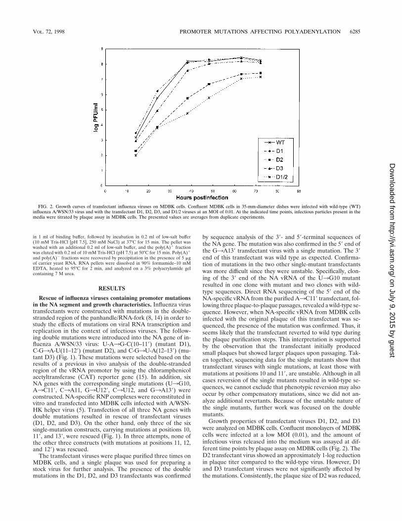
Growth properties of transfectant viruses D1, D2, and D3were analyzed on MDBK cells. Confluent monolayers of MDBKcells were infected at a low MOI (0.01), and the amount ofinfectious virus released into the medium was assayed at dif-ferent time points by plaque assay on MDBK cells (Fig. 2). TheD2 transfectant virus showed an approximately 1-log reductionin plaque titer compared to the wild-type virus. However, D1and D3 transfectant viruses were not significantly affected bythe mutations. Consistently, the plaque size of D2 was reduced,
FIG. 2. Growth curves of transfectant influenza viruses on MDBK cells. Confluent MDBK cells in 35-mm-diameter dishes were infected with wild-type (WT)influenza A/WSN/33 virus and with the transfectant D1, D2, D3, and D1/2 viruses at an MOI of 0.01. At the indicated time points, infectious particles present in themedia were titrated by plaque assay in MDBK cells. The presented values are averages from duplicate experiments.
VOL. 72, 1998 PROMOTER MUTATIONS AFFECTING POLYADENYLATION 6285
on July 9, 2015 by guesthttp://jvi.asm
.org/D
ownloaded from
but both D1 and D3 viruses showed plaque sizes similar to thatof the wild type.
Since the presence of single-base-pair mutations did notresult in severe impairment of viral growth, an attempt wasmade to introduce multiple double mutations into the sameregion of the NA gene. A construct incorporating double mu-tations from both D1 and D2 transfectants was successfullyrescued (D1/2) (Fig. 1) into infectious virus. The D1/2 trans-fectant was plaque purified three times, and the presence ofmutations was confirmed by sequencing. This virus showedsimilar reduction in plaque titers (Fig. 2) and plaque size onMDBK cells as the D2 transfectant. The effect of the D1/2mutations on viral growth was more dramatic on MDCK andVero cells, where reductions of 3 to 4 logs in plaque titers wereobserved (data not shown).
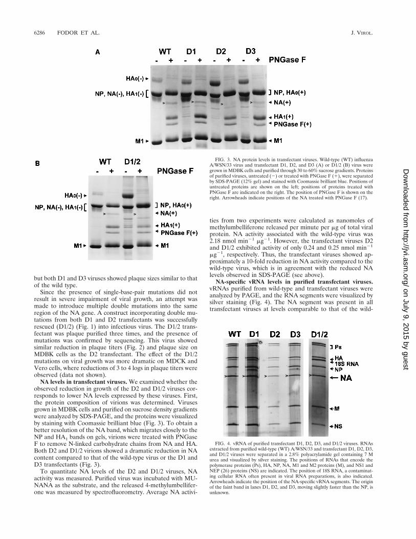
NA levels in transfectant viruses. We examined whether theobserved reduction in growth of the D2 and D1/2 viruses cor-responds to lower NA levels expressed by these viruses. First,the protein composition of virions was determined. Virusesgrown in MDBK cells and purified on sucrose density gradientswere analyzed by SDS-PAGE, and the proteins were visualizedby staining with Coomassie brilliant blue (Fig. 3). To obtain abetter resolution of the NA band, which migrates closely to theNP and HA1 bands on gels, virions were treated with PNGaseF to remove N-linked carbohydrate chains from NA and HA.Both D2 and D1/2 virions showed a dramatic reduction in NAcontent compared to that of the wild-type virus or the D1 andD3 transfectants (Fig. 3).
To quantitate NA levels of the D2 and D1/2 viruses, NAactivity was measured. Purified virus was incubated with MU-NANA as the substrate, and the released 4-methylumbellifer-one was measured by spectrofluorometry. Average NA activi-
ties from two experiments were calculated as nanomoles ofmethylumbelliferone released per minute per mg of total viralprotein. NA activity associated with the wild-type virus was2.18 nmol min21 mg21. However, the transfectant viruses D2and D1/2 exhibited activity of only 0.24 and 0.25 nmol min21
mg21, respectively. Thus, the transfectant viruses showed ap-proximately a 10-fold reduction in NA activity compared to thewild-type virus, which is in agreement with the reduced NAlevels observed in SDS-PAGE (see above).
NA-specific vRNA levels in purified transfectant viruses.vRNAs purified from wild-type and transfectant viruses wereanalyzed by PAGE, and the RNA segments were visualized bysilver staining (Fig. 4). The NA segment was present in alltransfectant viruses at levels comparable to that of the wild-
FIG. 3. NA protein levels in transfectant viruses. Wild-type (WT) influenzaA/WSN/33 virus and transfectant D1, D2, and D3 (A) or D1/2 (B) virus weregrown in MDBK cells and purified through 30 to 60% sucrose gradients. Proteinsof purified viruses, untreated (2) or treated with PNGase F (1), were separatedby SDS-PAGE (12% gel) and stained with Coomassie brilliant blue. Positions ofuntreated proteins are shown on the left; positions of proteins treated withPNGase F are indicated on the right. The position of PNGase F is shown on theright. Arrowheads indicate positions of the NA treated with PNGase F (17).
FIG. 4. vRNA of purified transfectant D1, D2, D3, and D1/2 viruses. RNAsextracted from purified wild-type (WT) A/WSN/33 and transfectant D1, D2, D3,and D1/2 viruses were separated in a 2.8% polyacrylamide gel containing 7 Murea and visualized by silver staining. The positions of RNAs that encode thepolymerase proteins (Ps), HA, NP, NA, M1 and M2 proteins (M), and NS1 andNEP (26) proteins (NS) are indicated. The position of 18S RNA, a contaminat-ing cellular RNA often present in viral RNA preparations, is also indicated.Arrowheads indicate the position of the NA-specific vRNA segments. The originof the faint band in lanes D1, D2, and D3, moving slightly faster than the NP, isunknown.
6286 FODOR ET AL. J. VIROL.
on July 9, 2015 by guesthttp://jvi.asm
.org/D
ownloaded from
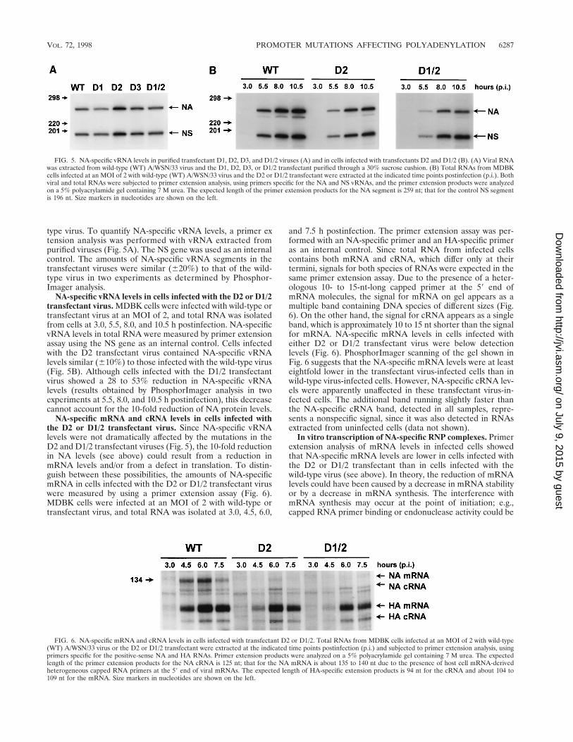
type virus. To quantify NA-specific vRNA levels, a primer extension analysis was performed with vRNA extracted frompurified viruses (Fig. 5A). The NS gene was used as an internalcontrol. The amounts of NA-specific vRNA segments in thetransfectant viruses were similar (620%) to that of the wild-type virus in two experiments as determined by Phosphor-Imager analysis.
NA-specific vRNA levels in cells infected with the D2 or D1/2transfectant virus. MDBK cells were infected with wild-type ortransfectant virus at an MOI of 2, and total RNA was isolatedfrom cells at 3.0, 5.5, 8.0, and 10.5 h postinfection. NA-specificvRNA levels in total RNA were measured by primer extensionassay using the NS gene as an internal control. Cells infectedwith the D2 transfectant virus contained NA-specific vRNAlevels similar (610%) to those infected with the wild-type virus(Fig. 5B). Although cells infected with the D1/2 transfectantvirus showed a 28 to 53% reduction in NA-specific vRNAlevels (results obtained by PhosphorImager analysis in twoexperiments at 5.5, 8.0, and 10.5 h postinfection), this decreasecannot account for the 10-fold reduction of NA protein levels.
NA-specific mRNA and cRNA levels in cells infected withthe D2 or D1/2 transfectant virus. Since NA-specific vRNAlevels were not dramatically affected by the mutations in theD2 and D1/2 transfectant viruses (Fig. 5), the 10-fold reductionin NA levels (see above) could result from a reduction inmRNA levels and/or from a defect in translation. To distin-guish between these possibilities, the amounts of NA-specificmRNA in cells infected with the D2 or D1/2 transfectant viruswere measured by using a primer extension assay (Fig. 6).MDBK cells were infected at an MOI of 2 with wild-type ortransfectant virus, and total RNA was isolated at 3.0, 4.5, 6.0,
and 7.5 h postinfection. The primer extension assay was per-formed with an NA-specific primer and an HA-specific primeras an internal control. Since total RNA from infected cellscontains both mRNA and cRNA, which differ only at theirtermini, signals for both species of RNAs were expected in thesame primer extension assay. Due to the presence of a heter-ologous 10- to 15-nt-long capped primer at the 59 end ofmRNA molecules, the signal for mRNA on gel appears as amultiple band containing DNA species of different sizes (Fig.6). On the other hand, the signal for cRNA appears as a singleband, which is approximately 10 to 15 nt shorter than the signalfor mRNA. NA-specific mRNA levels in cells infected witheither D2 or D1/2 transfectant virus were below detectionlevels (Fig. 6). PhosphorImager scanning of the gel shown inFig. 6 suggests that the NA-specific mRNA levels were at leasteightfold lower in the transfectant virus-infected cells than inwild-type virus-infected cells. However, NA-specific cRNA lev-els were apparently unaffected in these transfectant virus-in-fected cells. The additional band running slightly faster thanthe NA-specific cRNA band, detected in all samples, repre-sents a nonspecific signal, since it was also detected in RNAsextracted from uninfected cells (data not shown).
In vitro transcription of NA-specific RNP complexes. Primerextension analysis of mRNA levels in infected cells showedthat NA-specific mRNA levels are lower in cells infected withthe D2 or D1/2 transfectant than in cells infected with thewild-type virus (see above). In theory, the reduction of mRNAlevels could have been caused by a decrease in mRNA stabilityor by a decrease in mRNA synthesis. The interference withmRNA synthesis may occur at the point of initiation; e.g.,capped RNA primer binding or endonuclease activity could be
FIG. 5. NA-specific vRNA levels in purified transfectant D1, D2, D3, and D1/2 viruses (A) and in cells infected with transfectants D2 and D1/2 (B). (A) Viral RNAwas extracted from wild-type (WT) A/WSN/33 virus and the D1, D2, D3, or D1/2 transfectant purified through a 30% sucrose cushion. (B) Total RNAs from MDBKcells infected at an MOI of 2 with wild-type (WT) A/WSN/33 virus and the D2 or D1/2 transfectant were extracted at the indicated time points postinfection (p.i.). Bothviral and total RNAs were subjected to primer extension analysis, using primers specific for the NA and NS vRNAs, and the primer extension products were analyzedon a 5% polyacrylamide gel containing 7 M urea. The expected length of the primer extension products for the NA segment is 259 nt; that for the control NS segmentis 196 nt. Size markers in nucleotides are shown on the left.
FIG. 6. NA-specific mRNA and cRNA levels in cells infected with transfectant D2 or D1/2. Total RNAs from MDBK cells infected at an MOI of 2 with wild-type(WT) A/WSN/33 virus or the D2 or D1/2 transfectant were extracted at the indicated time points postinfection (p.i.) and subjected to primer extension analysis, usingprimers specific for the positive-sense NA and HA RNAs. Primer extension products were analyzed on a 5% polyacrylamide gel containing 7 M urea. The expectedlength of the primer extension products for the NA cRNA is 125 nt; that for the NA mRNA is about 135 to 140 nt due to the presence of host cell mRNA-derivedheterogeneous capped RNA primers at the 59 end of viral mRNAs. The expected length of HA-specific extension products is 94 nt for the cRNA and about 104 to109 nt for the mRNA. Size markers in nucleotides are shown on the left.
VOL. 72, 1998 PROMOTER MUTATIONS AFFECTING POLYADENYLATION 6287
on July 9, 2015 by guesthttp://jvi.asm
.org/D
ownloaded from
inhibited. Alternatively, termination or polyadenylation of vi-ral mRNA could be affected. To distinguish between all ofthese possibilities, in vitro transcription assays were performed.RNP complexes were isolated from purified D2 and D1/2transfectants and from wild-type A/WSN/33 virus, and in vitrotranscriptional activity was measured by using globin mRNA asprimer. Figure 7A shows that NA-specific transcription prod-ucts were synthesized from both the wild-type and the trans-fectant RNPs. However, there was a significant difference inthe pattern of the bands. The wild-type NA-specific transcrip-tion product appeared as a wide band corresponding to RNAspecies with poly(A) tails of different sizes. On the other hand,the NA-specific transcription products of both the D2 andD1/2 transfectants produced less diffuse bands, which impliedthat these products might not be polyadenylated. To charac-terize the transcription products, they were analyzed by oli-go(dT)-cellulose chromatography. The fractions depleted ofpoly(A)-containing molecules (Fig. 7B) showed higher levelsof NA-specific transcription products for the D2 and D1/2transfectants but lower levels for the wild-type control. On theother hand, fractions enriched in poly(A)-containing mole-cules (Fig. 7C) showed lower levels of the NA-specific tran-scription products for the D2 and D1/2 transfectants but higherlevels for the wild-type virus. This result seems to confirm thatthere is a large proportion of NA-specific transcription prod-ucts of the D2 and D1/2 transfectants which lack poly(A) tails(Fig. 7B).
DISCUSSION
In this study, we have investigated the effect of mutations inthe double-stranded region of the RNA-fork/panhandle of in-fluenza A virus vRNA in the context of infectious viruses. Thepostulated double-stranded region of the vRNA promoter con-sists of five to eight base pairs in different vRNA segments (3).The first three base pairs, those formed by nt 119 to 139 at the59 end and nt 10 to 12 at the 39 end, are strictly conservedamong different vRNA segments of all influenza A viruses.Using a reverse genetics approach, we have introduced doublemutations at the three conserved base pairs into the NA geneof influenza A/WSN/33 virus and studied their effects on viralgrowth. Interestingly, double mutations at positions 10 and 119and at positions 12 and 139 had no detectable effect on viralgrowth (D1 and D3 transfectant viruses). On the other hand, adouble mutation at positions 11 and 129 resulted in a transfec-tant (D2) which showed a 1-log reduction in plaque titers on
MDBK cells. The reduction in growth was most likely due tothe reduced levels of neuraminidase produced by this virus.
It should be noted that the D2 transfectant, as part of thebase pair mutation, has a C3A change at position 11 in the 39end, which creates an alternative AUG initiation codon in thecorresponding mRNA (Fig. 1). Since this initiation codon isupstream of the one for the NA and the two are not in frame,it is possible that the observed reduced NA levels are due topreferred initiation at the alternative AUG resulting in a 23-amino-acid peptide. However, we observed reduced steady-state levels of NA-specific mRNA in cells infected with thistransfectant, which suggests that the lower amount of mRNAis the cause for reduced NA levels. This interpretation is sup-ported by the rescue of another transfectant (D1/2), whichincorporates the double mutations from transfectant D1 andD2 viruses. The D1/2 transfectant virus shows growth charac-teristics very similar to those of the D2 transfectant. It grows asslowly as the D2 transfectant on MDBK cells and producesreduced levels of NA and NA-specific mRNA. However, dueto the presence of the additional mutation at position 10, thealternative AUG initiation codon is disrupted, thus eliminatingthe possibility of translation of an incorrect open readingframe. Taken together, the data suggest that both D2 and D1/2transfectant viruses produce reduced levels of NA due to re-duced NA-specific mRNAs.
Interestingly, reduced NA-specific mRNA levels did not cor-relate with reduced NA-specific vRNA levels for either of thetransfectant viruses. Both D2 and D1/2 purified virions con-tained NA-specific vRNA levels similar to the levels of othersegments. Also, in cells infected with D2 transfectant, we foundNA-specific vRNA levels corresponding to those found in cellsinfected with the wild-type virus. In cells infected with the D1/2transfectant, NA-specific vRNA levels showed a 1.5- to 2-foldreduction. However, this cannot account for the severe reduc-tion of NA-specific mRNA levels. We also did not see anysignificant alteration of cRNA levels in cells infected with theD2 or D1/2 transfectant compared to wild-type virus-infectedcells.
To further investigate whether mRNA synthesis was affect-ed, we performed transcription studies in vitro with RNPs iso-lated from the D2 and D1/2 transfectant viruses, using cappedRNA primer. The experiments showed that initiation of tran-scription and elongation were not affected by the mutations,but analysis of the transcription products by oligo(dT)-cellu-lose chromatography implied that the majority of the NA-specific transcription products of the D2 and D1/2 transfec-tants were not polyadenylated. This result suggests that themutations interfere with polyadenylation of mRNA transcripts.The observed low levels of mRNA in infected cells (Fig. 6) arefully consistent with these results, since nonpolyadenylatedcapped transcripts are most likely rapidly degraded in the cell(33). Lack of polyadenylation may be due to the inability of theRNA polymerase to polyadenylate after stopping at the stretchof U residues of the vRNA template. Alternatively, the RNApolymerase may not stop at the poly(U) stretch, resulting inthe synthesis of a capped full-length, nonpolyadenylated tran-script. It should be noted that efficient RNA synthesis in bothmutant and wild-type virus-derived RNPs was dependent onthe addition of primer (data not shown).
We have rescued four transfectant viruses with different basepair mutations. Neither of the mutations in the D1 and D3transfectant viruses exhibited a significant effect on transcrip-tion or replication of the NA gene, as measured by determiningNA and vRNA levels in virions. On the other hand, two of thetransfectants (D2 and D1/2) which had the same C-G3A-U(11–129) double mutation had a defect in mRNA synthesis,
FIG. 7. In vitro transcription of NA-specific RNP complexes and analysis ofthe transcription products on oligo(dT)-cellulose. In vitro transcription reactionswith RNPs isolated from wild-type (WT) A/WSN/33 virus and D2 or D1/2transfectant virus were performed with globin mRNA as primer (for details seeMaterials and Methods). One third of the transcription products was directlyanalyzed (A) on a 3% polyacrylamide gel in 7 M urea. Two-thirds of the tran-scription products were separated on oligo(dT)-cellulose, and fractions depletedof (B) and enriched in (C) poly(A)-containing molecules were analyzed on thesame gel. A longer exposure of the D1/2 products enriched in poly(A)-containingmolecules is shown. The positions of the NP- and NA-specific transcriptionproducts are indicated.
6288 FODOR ET AL. J. VIROL.
on July 9, 2015 by guesthttp://jvi.asm
.org/D
ownloaded from
although initiation of transcription was not affected, as deter-mined by the in vitro transcription assay (Fig. 7). The fact thatthe initiation of transcription and replication were not dramat-ically affected in any of the four transfectant viruses with dif-ferent double mutations suggests that the studied positions arenot crucial for initiation as long as the double-stranded struc-ture is maintained. The importance of complementarity be-tween the 59 and 39 ends in this region of the vRNA promoterhas been demonstrated in several studies performed in vitro (8,9) and in vivo (15, 21, 25). In agreement with the requirementfor complementarity, we were not able to rescue viruses withsingle mutations at all of the conserved positions involved inbase pairing. Those which we have rescued were unstable,rapidly reverting to a wild-type sequence upon passaging. Pre-vious findings that single-point mutations at positions 10 to 12in the 39 end and at positions 119 to 139 in the 59 end eliminatedor decreased reporter activity in vivo (15, 18, 25, 29) are also inagreement with the importance of complementarity. After rep-lication of vRNA into cRNA, the double mutations will also bepresent in the cRNA promoter. It has been shown in vitro thatthe cRNA promoter functions as a panhandle (31). The factthat we did not see dramatic effects of mutations on the rep-lication of vRNA in infected cells indicates that double muta-tions do not interfere with the function of the vRNA or cRNApromoter in RNA replication. However, we cannot exclude thepossibility that different base pair mutations in the duplex re-gion would have different effects.
The observed attenuation of NA-specific mRNA levels incells infected with the D2 transfectant is in agreement withprevious findings (15) that an A-U(11–129) base pair mutationin the context of a vRNA-like CAT reporter gene resulted onlyin 22% reporter activity compared to a wild-type control. How-ever, the G-C(10–119) and U-A(12–139) base pair mutations,which had no effect on the expression levels of the NA of theD1 and D3 transfectants, resulted in only 20 and 31% activi-ties, respectively, in a CAT reporter gene system (15). Thisresult suggests that base pair mutations in the context of a CATreporter gene and an NA gene might have different effects(38).
The question arises as to the mechanism of attenuation ofpolyadenylation of NA-specific mRNAs in the D2 and D1/2viruses. Recent findings suggest that poly(A) addition occursby a stuttering polymerase which is bound to the 59 end of thevRNA template (8, 32, 36). It is possible that mutations in theD2 and D1/2 transfectants interfere with the stabilization ofpolymerase binding to the 59 end of vRNA, which might beimportant for the polymerase to remain bound to the 59 end ofvRNA during polyadenylation.
The results presented in this paper show that by introducingdouble mutations into an influenza A virus segment, we canattenuate mRNA levels and consequently reduce protein levelsencoded by the mutated gene. We believe that transfectantviruses with double mutations should be stable since two spe-cific mutations would have to occur simultaneously in order torevert to the wild-type sequence. We were unable to rescue anytransfectant viruses with C3A11 or G3U129 single mutation,which suggests that such viruses might be severely impaired ornot viable at all. In addition, the double mutation in the D2transfectant was preserved during 10 passages on MDBK cells(7). It remains to be seen whether additional mutations in theNA or perhaps in the HA (11) gene may occur to compensate,at least partially, for the reduced NA levels produced by thisvirus.
In summary, we have rescued and characterized four trans-fectant viruses with promoter mutations. The mutations inthese viruses did not dramatically interfere with the replication
of vRNA, but a C-G3A-U(11–129) mutation in two of thetransfectants affected their mRNA levels, most likely by inter-fering with the efficiency of polyadenylation. This is the firstexample of an engineered influenza virus with a mutation inthe conserved region of the vRNA promoter which does notaffect the initiation of transcription or RNA replication butseverely impairs polyadenylation and, thus, mRNA synthesis.
ACKNOWLEDGMENTS
This work was supported by the National Institutes of Health (P.P.and A.G.-S.), the Max Kade Foundation (E.F.), and the Medical Re-search Council (project grant G9523972 to G.G.B.).
We thank Othmar Engelhardt, David Pritlove, and Leo Poon forhelpful discussions.
REFERENCES1. Beaton, A. R., and R. M. Krug. 1986. Transcription antitermination during
influenza viral template RNA synthesis requires the nucleocapsid proteinand the absence of a 59 capped end. Proc. Natl. Acad. Sci. USA 83:6282–6286.
2. Cianci, C., L. Tiley, and M. Krystal. 1995. Differential activation of theinfluenza virus polymerase via template RNA binding. J. Virol. 69:3995–3999.
3. Desselberger, U., V. R. Racaniello, J. J. Zazra, and P. Palese. 1980. The 39and 59 terminal sequences of influenza A, B, and C virus RNA segments arehighly conserved and show partial inverted complementarity. Gene 8:315–328.
4. Enami, M., W. Luytjes, M. Krystal, and P. Palese. 1990. Introduction of sitespecific mutations into the genome of influenza virus. Proc. Natl. Acad. Sci.USA 87:3802–3805.
5. Enami, M., and P. Palese. 1991. High-efficiency formation of influenza virustransfectants. J. Virol. 65:2711–2713.
6. Flick, R., G. Neumann, E. Hoffmann, E. Neumeier, and G. Hobom. 1996.Promoter elements in the influenza vRNA terminal structure. RNA 2:1046–1057.
7. Fodor, E. Unpublished data.8. Fodor, E., D. C. Pritlove, and G. G. Brownlee. 1994. The influenza virus
panhandle is involved in the initiation of transcription. J. Virol. 68:4092–4096.
9. Fodor, E., D. C. Pritlove, and G. G. Brownlee. 1995. Characterization of theRNA-fork model of virion RNA in the initiation of transcription in influenzaA virus. J. Virol. 69:4012–4019.
10. Garcıa-Sastre, A., T. Muster, W. S. Barclay, N. Percy, and P. Palese. 1994.Use of a mammalian internal ribosomal entry site element for expression ofa foreign protein by a transfectant influenza virus. J. Virol. 68:6254–6261.
11. Gubareva, L. V., R. Bethell, G. J. Hart, K. G. Murti, C. R. Penn, and R. G.Webster. 1996. Characterization of mutants of influenza A virus selectedwith the neuraminidase inhibitor 4-guanidino-Neu5Ac2en. J. Virol. 70:1818–1827.
12. Hagen, M., T. D. Y. Chung, J. A. Butcher, and M. Krystal. 1994. Recombi-nant influenza virus polymerase: requirement of both 59 and 39 viral ends forendonuclease activity. J. Virol. 68:1509–1515.
13. Honda, A., and A. Ishihama. 1997. The molecular anatomy of influenza virusRNA polymerase. Biol. Chem. 378:483–488.
14. Hsu, M., J. D. Parvin, S. Gupta, M. Krystal, and P. Palese. 1987. GenomicRNAs of influenza viruses are held in a circular conformation in virions andin infected cells by a terminal panhandle. Proc. Natl. Acad. Sci. USA 84:8140–8144.
15. Kim, H-J., E. Fodor, G. G. Brownlee, and B. L. Seong. 1997. Mutationalanalysis of the RNA-fork model of the influenza A virus vRNA promoter invivo. J. Gen. Virol. 78:353–357.
16. Krug, R. M., F. V. Alonso-Caplen, I. Julkunen, and M. G. Katze. 1989.Expression and replication of the influenza virus genome, p. 98–152. In R. M.Krug (ed.), The influenza viruses. Plenum, New York, N.Y.
17. Li, S., J. Schulman, S. Itamura, and P. Palese. 1993. Glycosylation of neur-aminidase determines the neurovirulence of influenza A/WSN/33 virus.J. Virol. 67:6667–6673.
18. Li, X., and P. Palese. 1992. Mutational analysis of the promoter required forinfluenza virus virion RNA synthesis. J. Virol. 66:4331–4338.
19. Li, X., and P. Palese. 1994. Characterization of the polyadenylation signal ofinfluenza virus RNA. J. Virol. 68:1245–1249.
20. Luo, G., M. Bergmann, A. Garcıa-Sastre, and P. Palese. 1992. Mechanism ofattenuation of a chimeric influenza A/B transfectant virus. J. Virol. 66:4679–4685.
21. Luo, G., W. Luytjes, M. Enami, and P. Palese. 1991. The polyadenylationsignal of influenza virus RNA involves a stretch of uridines followed by theRNA duplex of the panhandle structure. J. Virol. 65:2861–2867.
22. Luytjes, W., M. Krystal, M. Enami, J. D. Parvin, and P. Palese. 1989.Amplification, expression, and packaging of a foreign gene by influenzavirus. Cell 59:1107–1113.
VOL. 72, 1998 PROMOTER MUTATIONS AFFECTING POLYADENYLATION 6289
on July 9, 2015 by guesthttp://jvi.asm
.org/D
ownloaded from
23. Martın, J., C. Albo, J. Ortın, J. A. Melero, and A. Portela. 1992. In vitroreconstitution of active influenza virus nucleoprotein complexes using viralproteins purified from infected cells. J. Gen. Virol. 73:1855–1859.
24. Mena, I., S. de la Luna, C. Albo, J. Martın, A. Nieto, J. Ortın, and A. Portela.1994. Synthesis of biologically active influenza core proteins using a vaccin-ia-T7 RNA polymerase expression system. J. Gen. Virol. 75:2109–2114.
25. Neumann, G., and G. Hobom. 1995. Mutational analysis of influenza viruspromoter elements in vivo. J. Gen. Virol. 76:1709–1717.
26. O’Neill, R. E., J. Talon, and P. Palese. 1998. The influenza virus NEP (NS2protein) mediates the nuclear export of viral ribonucleoproteins. EMBO J.17:288–296.
27. Palese, P. 1977. The genes of influenza virus. Cell 10:1–10.28. Parvin, J. D., P. Palese, A. Honda, A. Ishihama, and M. Krystal. 1989.
Promoter analysis of the influenza virus RNA polymerase. J. Virol. 63:5142–5152.
29. Piccone, M. E., A. Fernandez-Sesma, and P. Palese. 1993. Mutational anal-ysis of the influenza virus vRNA promoter. Virus Res. 28:99–112.
30. Potier, M., L. Mameli, M. Belisle, L. Dallaire, and S. B. Melancon. 1979.Fluorometric assay of neuraminidase with a sodium (4-methylumbelliferyl-a-D-N-acetylneuraminate) substrate. Anal. Biochem. 94:287–296.
31. Pritlove, D. C., E. Fodor, B. L. Seong, and G. G. Brownlee. 1995. In vitrotranscription and polymerase binding studies of the termini of influenza Avirus complementary RNA: evidence for a cRNA panhandle. J. Gen. Virol.76:2205–2213.
32. Pritlove, D. C., L. L. M. Poon, E. Fodor, J. Sharps, and G. G. Brownlee. 1998.Polyadenylation of influenza virus mRNA transcribed in vitro from modelvirion RNA templates: requirement for 59 conserved sequences. J. Virol. 72:1280–1287.
33. Proudfoot, N. J., and E. Whitelow. 1988. Termination and 39 end processingof eukaryotic RNA, p. 97–129. In D. M. Glover and B. D. Hames (ed.),Frontiers in molecular biology—transcription and splicing. IRL Press, Ox-ford, England.
34. Robertson, J. S., M. Schubert, and R. A. Lazzarini. 1981. Polyadenylationsites for influenza virus mRNA. J. Virol. 38:157–163.
35. Seong, B. L., and G. G. Brownlee. 1992. A new method for reconstitutinginfluenza polymerase and RNA in vitro: a study of the promoter elements forcRNA and vRNA synthesis in vitro and viral rescue in vivo. Virology 186:247–260.
36. Tiley, L. S., M. Hagen, J. T. Matthews, and M. Krystal. 1994. Sequence-specific binding of the influenza virus RNA polymerase to sequences locatedat the 59 ends of the viral RNAs. J. Virol. 68:5108–5116.
37. Yamanaka, K., N. Ogasawara, H. Yoshikawa, A. Ishihama, and K. Nagata.1991. In vivo analysis of the promoter structure of the influenza genomeusing a transfection system with an engineered RNA. Proc. Natl. Acad. Sci.USA 88:5369–5373.
38. Zheng, H., P. Palese, and A. Garcıa-Sastre. 1996. Nonconserved nucleotidesat the 39 and 59 ends of an influenza A virus RNA play an important role inviral RNA replication. Virology 217:242–251.
6290 FODOR ET AL. J. VIROL.
on July 9, 2015 by guesthttp://jvi.asm
.org/D
ownloaded from
Related Documents