Atrial pacing and experimental atrial fibrillation in equines Gunther van Loon Proefschrift ter verkrijging van de graad van Doctor in de Diergeneeskundige Wetenschappen (PhD) aan de Faculteit Diergeneeskunde, Universiteit Gent Promotor: Prof. Dr. P. Deprez Copromotor: Prof. Dr. L. Jordaens Vakgroep Interne Geneeskunde en Klinische Biologie van de Grote Huisdieren Salisburylaan 133, B-9820 Merelbeke ISBN 90-5864-014-0 FACULTY OF VETERINARY MEDICINE Department of Large Animal Internal Medicine

Welcome message from author
This document is posted to help you gain knowledge. Please leave a comment to let me know what you think about it! Share it to your friends and learn new things together.
Transcript

Atrial pacing and experimental
atrial fibrillation in equines
Gunther van Loon
Proefschrift ter verkrijging van de graad van Doctor in de Diergeneeskundige
Wetenschappen (PhD) aan de Faculteit Diergeneeskunde, Universiteit Gent
Promotor: Prof. Dr. P. Deprez
Copromotor: Prof. Dr. L. Jordaens
Vakgroep Interne Geneeskunde en Klinische Biologie van de Grote Huisdieren
Salisburylaan 133, B-9820 Merelbeke
ISBN 90-5864-014-0
FACULTY OF VETERINARY MEDICINE Department of Large Animal Internal Medicine


For Fien, Emma and Sofie, who maintain my rhythms

CONTENTS
List of abbreviations
PREFACE 1
GENERAL INTRODUCTION 3
1. Atrial pacing 5 Description of cardiac pacing 5 Cardiac pacing in human medicine 12 Cardiac pacing in equine medicine 15
2. Atrial fibrillation 19 General electrophysiological considerations 19 Atrial fibrillation in equines 21
3. References 29
SCIENTIFIC AIMS 41
CHAPTER 1 TEMPORARY TRANSVENOUS ATRIAL PACING IN HORSES: THRESHOLD DETERMINATION 43
1. Summary 45 2. Introduction 46 3. Materials and methods 48 4. Results 53 5. Discussion 57 6. References 62
CHAPTER 2 INTRACARDIAC OVERDRIVE PACING AS A TREATMENT OF ATRIAL FLUTTER IN A HORSE 65
1. Summary 67 2. Introduction 68 3. Case history and clinical findings. 69 4. Discussion 73 5. References 76
CHAPTER 3 DUAL CHAMBER PACEMAKER IMPLANTATION VIA THE CEPHALIC VEIN IN HEALTHY EQUIDS 77
1. Summary 79 2. Introduction 80

Contents
3. Materials and methods 82 4. Results 87 5. Discussion 92 6. References 99
CHAPTER 4 DUAL CHAMBER RATE-ADAPTIVE PACEMAKER IMPLANTATION IN A HORSE WITH SUSPECTED SICK SINUS SYNDROME 103
1. Summary 105 2. Introduction 106 3. Case report 108 4. Discussion 114 5. References 118
CHAPTER 5 PACING-INDUCED SUSTAINED ATRIAL FIBRILLATION IN A PONY 121
1. Summary 123 2. Introduction 124 3. Materials and Methods 125 4. Results 128 5. Discussion 131 6. References 134
CHAPTER 6 AN EQUINE MODEL OF CHRONIC ATRIAL FIBRILLATION: METHODOLOGY 137
1. Summary 139 2. Introduction 140 3. Materials and Methods 142 4. Results 146 5. Discussion 151 6. References 156
CHAPTER 7 EFFECT OF EXPERIMENTAL CHRONIC ATRIAL FIBRILLATION IN EQUINES 161
1. Summary 163 2. Introduction 164 3. Materials and methods 166 4. Results 174 5. Discussion 186 6. References 201
CONCLUDING REMARKS 207

Contents
SUMMARY / SAMENVATTING 227
DANKWOORD 247
AUTHOR'S CURRICULUM 251
BIBLIOGRAPHY 253

LIST OF ABBREVIATIONS
AERP atrial effective refractory period
AF atrial fibrillation
AFCL atrial fibrillation cycle length
AV atrioventricular
CL cycle length
ECG electrocardiogram
S1 driving stimulus
S2 extrastimulus
SCL sinus cycle length
(c)SNRT (corrected) sinus node recovery time
SR sinus rhythm


1
PREFACE
Diagnosis and treatment of cardiac rhythm disturbances in equines
has remained virtually unchanged during the past decades. In human
medicine, invasive cardiac testing, using electrical stimulation or
‘pacing’ of the heart, has greatly changed the approach to arrhythmias.
Besides diagnostic and therapeutic application, the technique also
provides an excellent means to study arrhythmias in animal models.
In addition to the investigation of the applicability of cardiac pacing
in equines, this work reports results of basic research on atrial
fibrillation, clinically the most important arrhythmia in equines. The
study on atrial fibrillation in equines was performed in different pony
models making use of the atrial pacing technique.
The thesis is divided into two sections. The first section provides
information and results about atrial pacing. It describes the application
of temporary and permanent pacing. Section 2 applies atrial pacing to
develop a chronic atrial fibrillation model in equines and discusses the
consequence of pacing-induced atrial fibrillation in the equine heart.


GENERAL INTRODUCTION


GENERAL INTRODUCTION: atrial pacing
5
ATRIAL PACINGATRIAL PACING
DESCRIPTION OF CARDIAC PACING
Excitable properties of cardiac cells
Like all living cells, the inside of cardiac cells has a negative
electrical charge compared to the outside of cells (-90 mV). The
resulting voltage difference is called the transmembrane potential. Due
to the excitable properties of cardiac cells, each process reducing the
resting transmembrane potential to a critical value or threshold (- 65
mV) results in the generation of an action potential. This action
potential arises from patterned changes in transmembrane potential
due to sequential opening and closing of ion channels. When these
stereotypical voltage changes
in a single cardiac cell are
graphed against time, the
result is the action potential
(Fig. 1). The action potential
starts with a rapid, positively
directed change in
transmembrane potential,
resulting in a voltage spike
called depolarisation (phase
0). After this fast
depolarisation, a gradual normalization of ion concentrations occurs
and the cell repolarises, a process that roughly corresponds to phases
1 through 3 of the action potential. Because a second depolarisation
cannot take place until repolarisation occurs, the time from the end of
phase 0 to late in phase 3 is called the absolute refractory period of
Figure 1. Four different phases, absolute (a) and relative (b) refractory period during the cardiac action potential.

GENERAL INTRODUCTION
6
cardiac tissue. As the cells gradually recover from refractoriness, they
become excitable if a pulse of sufficient strength is delivered. The
period after the absolute refractory period and before full recovery is
called the relative refractory period (Fogoros, 1995; Kay, 1996).
Stimulation at the end of the refractory period, can initiate
tachyarrhythmias (Fishler and Thakor, 1994; Peters et al., 1994).
For most cardiac cells, after full recovery, the resting phase (the
period of time between action potentials, corresponding to phase 4) is
quiescent, and there is no net movement of ions across the cell
membrane. In some cells, a leakage of ions across the cell membrane
causes a gradual increase in transmembrane potential during phase 4,
which results in a spontaneous depolarisation, called automaticity.
Cells in the sinoatrial node have the fastest phase 4 activity. The
sinoatrial node therefore regulates heart rate and is called the natural
pacemaker of the heart.
The spontaneously generated electrical impulse from the sinoatrial
node stimulates nearby cells to depolarise. Depolarisation of one
cardiac cell tends to cause adjacent cells to depolarise. Thus, once a
cell is stimulated the wave of depolarisation (the electrical impulse) is
propagated across the atrium, cell by cell (Fogoros, 1995). The atrial
depolarisation can be recognised on the surface electrocardiogram
(ECG) by the P wave. At the atrioventricular node conduction is
slowed, which is reflected in the PR interval on the surface
electrocardiogram (ECG). Leaving the atrioventricular node, the His
bundle and Purkinje fibres rapidly spread the impulse throughout the
ventricles. On the surface ECG, the ventricular depolarisation and
subsequent repolarisation generate a QRS complex and T wave,
respectively.
Besides natural depolarisation due to sinus node activity, cardiac
cells can be stimulated artificially by electrical pulses, which is called
pacing. The electrical stimulus generally consists of a square current
pulse with variable amplitude, duration and interval. Only when the
GENERAL INTRODUCTION: atrial pacing
7
electrical current is able to reduce the resting potential of the cardiac
cells by a critical amount and within a critical time, a self-regenerating
wavefront of action potentials will propagate from the site of
stimulation across the cardiac chamber and 'capture' is being achieved
(Trautwein, 1975). Not only the amplitude but also the duration of the
stimulating current determines whether the threshold for stimulation
will be reached. Currents larger in amplitude require a shorter duration
to reach threshold and vice versa. The strength-duration curve
describes the threshold for stimulation at different pulse widths and
amplitudes during the excitable period of the tissue. Pacing during the
absolute refractory period will not result in capture. Pacing during the
relative refractory period can result in capture if the pulse has a
sufficient strength. Caution is warranted, especially during ventricular
stimulation, not to stimulate immediately after the end of the refractory
period, during the 'vulnerable period', because this can initiate
fibrillation (Fishler and Thakor, 1994; Peters et al., 1994). To avoid this
problem most pulse generators have the possibility to 'sense'
spontaneous electrical activity of the heart through the electrodes.
Immediately after sensing a cardiac depolarisation, artificial stimulation
is temporarily inhibited to minimise the risk of fibrillation induction.
Successful myocardial stimulation is dependent on (1) a source of
the electrical pulse (the pulse generator), (2) a conductor between the
source of the electrical pulse and the stimulating electrode (the lead),
(3) an electrode for pulse delivery and (4) an area of myocardium that
is excitable.
Pulse generator
Three basic electronic circuits are essential for the pulse generator
(Mond and Sloman, 1990). The timing circuit controls the pacing
interval and the output circuit controls the charging and discharging of
the impulse. The third major circuit is the sensing circuit, which
analyses the electrical signals that return to the pulse generator from
the heart via the lead and which is responsible for the recognition of

GENERAL INTRODUCTION
8
spontaneous intracardiac electrical signals. This sensing circuit allows
the pacemaker to adjust its timing intervals to changes in spontaneous
cardiac activity and prevents pacing during the vulnerable period
because this could initiate tachyarrhythmias (Fishler and Thakor, 1994;
Peters et al., 1994; Hayes and Osborn, 1996).
The pulse generator can deliver pulses of different strength and
duration at a desired rate. Different pacing protocols as overdrive
pacing, extrastimulus pacing and burst pacing are available to fulfil
electrophysiologic studies. During temporary stimulation, an external
pulse generator is used for pulse delivery, while for permanent pacing
an implantable pacemaker is applied (Fig. 2).
Using radiofrequency signals or pulsed magnetic fields, the pulse
generator is capable of both transmitting information to and receiving
information from an external programmer (Kay, 1996). Consequently,
this telemetric programmability allows changing most functions of the
implanted pacemaker non-invasively.
Originally pacemakers were classified with a three-letter
identification code according to the site of the pacing electrodes and
the mode of pacing: V = ventricle, A = atrium, D = dual, I = inhibited
and T = triggered (Mond and Sloman, 1990; Barold and Zipes, 1992;
Hayes and Osborn, 1996). The first letter indicates the chamber paced
and the second the chamber sensed. Occasionally the letter S is used
for the first or second position to indicate that a single-chamber device
Figure 2. An implantable pacemaker (1) with an atrial and ventricular lead (2) is shown. Each lead possesses an electrode on the lead-tip (3). The lead can be of the active (3a) or passive (3b) fixation type.

GENERAL INTRODUCTION: atrial pacing
9
is suitable for atrial or ventricular pacing. The third position indicates
the response of the pacemaker when a cardiac signal is sensed, i.e.
inhibition of pulse delivery (I), triggering of pulse delivery (T) or both
(D). For instance, the AAI mode means that atrial pacing and atrial
sensing occur and that an atrial stimulus will be inhibited when an
atrial signal is sensed. The (lower) heart rate in these models can be
chosen and will prevent a rate drop below this value. The major
advantage of rate programming is to allow the patient to remain in
sinus rhythm rather than in paced rhythm when intermittent
bradycardia is present (Hayes and Osborn, 1996).
During 1980 the code was extended to five letters to indicate other
complex pacing functions. However, we wish to limit the discussion to
the letter R in the fourth position, indicating that the pacemaker has
rate modulation, which can be achieved by a build-in sensor. As an
example, the DDIR mode indicates that rate adaptive atrial and
ventricular pacing occurs provided that no atrial or ventricular signals
are sensed. Depending on the kind of pacemaker, sensor activation
occurs due to patient physical activity, changes in respiratory rate,
changes in QT interval,… Upon stimulation of the sensor, a sensor-
driven response in heart rate occurs, better meeting the metabolic
demands of the patient. The upper sensor-driven heart rate, which will
be gradually achieved after continuous sensor activation, can be
programmed. When patient activity stops, sensor stimulation is ceased
and the pacing rate will gradually decrease.
Electrodes
Using properly positioned electrodes, the atria and/or ventricles can
be selectively stimulated. In general, the electrodes can be positioned
nearby the heart, on the epicardium or on the endocardium. In man,
transcutaneous atrial or ventricular pacing can be performed in
emergency situations using relatively high currents (Barold and Zipes,
1992). But this technique can be painful in conscious patients due to
stimulation of cutaneous nerves and pacing-induced skeletal muscle

GENERAL INTRODUCTION
10
contractions. Transoesophageal atrial pacing implies a markedly lower
threshold and can be performed without general anaesthesia or
sedation (Tucker and Wilson, 1993; Kantharia and Mookherjee, 1995).
The technique is relatively non-invasive and well tolerated. Ventricular
capture is inconsistent or often intolerably painful, however, thus
seriously limiting the therapeutic and emergent application of the
procedure. After a thoracotomy and incision of the pericardium,
electrodes can be attached directly on the atrial and/or ventricular
epicardium. Resistance for electrical stimulation is low and thresholds
are more easily reached. Because even low currents remain effective
to provoke myocardial excitation, stimulation itself is not painful
making this technique suitable for pacing in the conscious patient. The
necessity of a thoracotomy, however, limits the technique to temporary
pacing during cardiac surgery or to permanent pacing with an
implanted device (Amsel and Walter, 1992). In humans and small
animals, the most widely used approach for cardiac pacing is
transvenous endocardial pacing, where a lead or catheter, with
electrodes on its tip, is introduced through a vein and advanced into
the atrial or ventricular cavity. Correct positioning of the electrode can
be guided by fluoroscopy, echocardiography, intracavitary electrogram
morphology and application of testing stimuli. When the lead is
connected to an electrocardiographic device, simultaneous recording
of the intracavitary electrogram and the surface ECG can reveal the
position of the electrode. After introduction of the electrode into the
vein, the 'intravenous’ electrogram will be characterised by absent or
minimal deflections. When the electrode enters the right atrium or the
right ventricle, the largest intra-cardiac electrogram deflections will
coincide with, respectively, the P waves or QRS complexes on the
surface ECG. When the lead tip is assumed to be located in the
desired chamber, testing stimuli with a sufficient strength can be
applied to confirm its position: intra-atrial or intraventricular stimulation
will produce a P wave or QRS complex on the surface ECG,
respectively.

GENERAL INTRODUCTION: atrial pacing
11
To achieve permanent pacing, two types of transvenous leads have
been developed to preserve endocardial contact. Leads with an active
fixation invade the endocardium with screws or small jaws, while leads
with a passive fixation promote fixation to the endocardium by indirect
means. The latter can be obtained by little tines or fins to enhance
entanglement in myocardial trabeculae (Mond and Sloman, 1990;
Barold and Zipes, 1992). In general, the ventricular lead can be of the
passive or active fixation type, while the atrial lead should include an
active fixation mechanism to remain in a stable position.

GENERAL INTRODUCTION
12
CARDIAC PACING IN HUMAN MEDICINE
Therapeutic pacing
Permanent pacing
Although pacemaker implantation can be utilized to prevent
induction of certain tachyarrhythmias (Osborn, 1996), the major
therapeutic indications for permanent cardiac pacing are
bradyarrhythmias due to third-degree or second-degree AV block,
sinus node dysfunction or neurocardiogenic syncope (Barold and
Zipes, 1992; Ross and Mandel, 1995; Ellenbogen and Peters, 1996;
Hayes and Osborn, 1996). Formerly, pacemaker implantation was
performed with epicardial lead placement and therefore required major
surgery. A considerable evolution during the past decades, however,
has greatly simplified the implantation procedure. Today, virtually all
leads are transvenously inserted, making cardiac pacing a relatively
simple and safe method that is widely used in human medicine
(Holmes and Hayes David, 1990). Because major surgery is avoided,
the transvenous approach can be performed without general
anaesthesia (Barold and Zipes, 1992).
The pacing system consists of a pacemaker and 1 or 2 leads to
perform single chamber (atrial or ventricular) or dual chamber pacing.
Recent pacemakers are equipped with a build-in sensor that detects
patient activity, thus providing an exercise-dependent rate response.
Temporary pacing
Temporary cardiac pacing can be accomplished transcutaneously,
via the oesophagus, transvenously and epicardially. Temporary
cardiac pacing serves as a lifesaving therapy in the acute
management of medically refractory bradyarrhythmias (Wood and
Ellenbogen, 1996). Temporary pacing is also indicated prophylactically

GENERAL INTRODUCTION: atrial pacing
13
in patients with a high risk of developing high-degree AV block, severe
sinus node dysfunction, or asystole in acute myocardial infarction, after
cardiac surgery, during cardiac catheterisation, and occasionally
before implantation or replacement of a permanent pacemaker (Barold
and Zipes, 1992). Rapid temporary pacing can be used to terminate
atrial flutter, AV reentry tachycardia or AV nodal reentry tachycardia,
and sustained ventricular tachycardia (Barold and Zipes, 1992;
Kantharia and Mookherjee, 1995; Ross and Mandel, 1995; Osborn,
1996). Finally, temporary pacing can be applied to prevent
bradycardia-dependent ventricular tachycardia (torsade de pointes).
Non-therapeutic pacing: the electrophysiological study
The approach to the diagnosis of cardiac rhythm disturbances
starts with a detailed history, clinical examination of the patient and an
ECG recording. Further examinations include exercise testing and
long-term ECG recordings. However, during the past decades,
invasive electrophysiologic studies have proven to be of great benefit
in the management of cardiac arrhythmias.
To perform an electrophysiologic study, in general, one or more
temporary pacing catheters have to be transvenously introduced into
the cardiac chamber. Fluoroscopy and intracavitary electrogram
recordings are applied for proper catheter positioning (Ross and
Mandel, 1995). Although two simple things are being done, i.e. pacing
and recording from localized areas within the heart, complex
programmed electrical stimulation protocols are often used. These
pacing protocols apply two general types of programmed electrical
stimulation: incremental pacing and extrastimulus pacing. With
incremental pacing (or burst pacing) the heart is stimulated at a
constant rhythm in excess of the spontaneous heart rate. The
extrastimulus technique consists of introducing one or more premature
impulses, each at its own specific coupling interval.
GENERAL INTRODUCTION
14
During an electrophysiological study, automaticity and conductivity
of the sinoatrial (SA) node, and conductivity and refractoriness of the
atrioventricular (AV) node and His-Purkinje system are assessed. By
rapid atrial pacing, the automaticity and conductivity of the SA node
can be assessed. Rapid atrial pacing depolarises the SA node faster
than it can be depolarised by its intrinsic automaticity. When the
overdrive pacing is abruptly stopped there is often a relatively long
pause before the SA node recovers and begins depolarising
spontaneously again. A diseased SA node tends to have a grossly
prolonged recovery time (Zipes, 1992; Fogoros, 1995).
By delivering a properly timed extrastimulus with a short coupling
interval, myocardial refractoriness can be determined. If the coupling
interval between the last paced or spontaneous beat and the
extrastimulus is too short, the stimulus will fall in the refractory period
and no depolarisation will be initiated. However, a stimulus given after
the refractory period will cause an extrasystole or even initiate
tachycardia or fibrillation. The longest coupling interval not resulting in
a propagated cardiac depolarisation indicates the effective refractory
period of that tissue (Fogoros, 1995; Ross and Mandel, 1995).
Initiation of tachyarrhythmias is a potential hazard of cardiac
pacing. However, initiation of these tachyarrhythmias is often
attempted to assess the existence of an appropriate anatomic
substrate to encompass a reentrant circuit. Programmed electrical
stimulation also allows to terminate many reentrant arrhythmias.
Consequently, the electrophysiologic study has become vitally
important in the evaluation and treatment of reentrant
tachyarrhythmias.
Complications that can be encountered with temporary or
permanent pacing include vascular thrombosis, embolisation,
infection, lead fracture, cardiac perforation or failure of the pacing
system (Brinker and Midei, 1996).

GENERAL INTRODUCTION: atrial pacing
15
CARDIAC PACING IN EQUINE MEDICINE
Therapeutic pacing
Permanent pacing
In 1973, Berg et al. described the permanent implantation of a
ventricular pacemaker in a young donkey with third-degree AV block.
A thoracotomy was performed under general anaesthesia and an
epicardial electrode was implanted on the left ventricle. The
pacemaker was inserted subcutaneously caudal to the left elbow. After
6 weeks, however, capture was lost, whereby pacemaker function
could not be restored. The donkey died 3 weeks later. In 1979, Brown
reported a horse with third-degree AV block. In an attempt to implant a
permanent pacemaker during general anaesthesia, the horse
developed ventricular fibrillation and died. Five years later, Le
Nihouannen et al. (1984a; 1984b) reported two techniques for
experimental pacemaker implantation in equines. Under general
anaesthesia, they performed a thoracotomy in a pony to place 2
epicardial electrodes on the left ventricle and subsequently implanted
a pacemaker underneath the ascendant pectoral muscle. During a 60-
day follow-up period, the horse could be successfully paced. In
another horse, an implantable passive fixation lead was transvenously
inserted through the jugular vein into the right ventricle under
radioscopic control. This procedure was performed in the standing,
sedated animal. Implantation of a pacemaker was not reported in this
animal. In 1986, Reef et al. performed a single chamber (ventricular)
pacemaker implantation in a horse with third-degree AV block. Under
general anaesthesia, a passive fixation lead was inserted in the jugular
vein and placed in the right ventricular apex under ultrasonographic
control. The pacemaker pocket was created dorsal to the jugular vein.
The pacemaker was programmed to maintain a ventricular rate of
45/min. However, due to this fixed heart rate, the horses’ performance
was limited. Sixteen months after the implantation, a second lead with

GENERAL INTRODUCTION
16
active fixation was implanted in the right atrium under general
anaesthesia and the pacemaker was updated to a dual chamber
model, allowing AV sequential pacing of the ventricles. This type of
pacemaker delivered a ventricular stimulus each time an atrial
depolarisation was sensed. Because in this horse with third degree AV
block the sinus node still functioned properly, AV sequential pacing
allowed the ventricular rate to ‘follow’ the atrial rate, resulting in a
physiologic rate response to exercise or stress. About 3 years after the
initial implantation, this horse suddenly died. At post mortem, extensive
thrombi, a suppurative endocarditis and suspicion of a terminal
bacteraemia were present (Hamir and Reef, 1989). In 1993, Pibarot et
al. described the implantation of a dual chamber pacemaker in a
donkey with complete AV block. Epicardial electrodes were implanted
on the left atrium and the left ventricle under general anaesthesia. Due
to a high post-operative ventricular threshold, the dual chamber
system had to be programmed to a single chamber (ventricular)
pacing mode to preserve battery life. After a 12-month follow-up
period, successful ventricular pacing could still be achieved.
Temporary pacing
The first report about temporary pacing in a horse was published in
1967 by Taylor and Mero. They reported transvenous endocardial
ventricular pacing using an external pacemaker in a foal with third-
degree AV block. An electrode was introduced through the jugular vein
and right ventricular pacing was applied during 45 minutes. A similar
technique was used in some of the above-described animals with 3rd
degree AV block to avoid Adams-Stokes attacks prior to the
permanent implantation of pacemaker (Brown, 1979; Reef et al., 1986;
Pibarot et al., 1993).
GENERAL INTRODUCTION: atrial pacing
17
Non-therapeutic pacing
Permanent pacing
With a pacemaker, the effect of a sustained tachycardia can be
imitated, arrhythmias can be generated and their inducibility verified,
and furthermore, electrophysiologic measurements e.g. determination
of the refractory period, can be made. This allows studying the
pathophysiology of acute or long-term arrhythmias and developing
different therapeutic strategies. Pacemaker implantation has been
used in many animal-based research protocols using dogs (Allworth et
al., 1995; Morillo et al., 1995; Elvan et al., 1996; Yue et al., 1997),
goats (Wijffels et al., 1995), sheep (Willems et al., 2000) or pigs (Qi et
al., 2000). However, no literature data were found concerning
permanent pacemaker implantation in equines for diagnostic or
investigational purposes.
Temporary pacing
In 1977, O’Callaghan mentioned that, by analogy with human
medicine, cardiac electro-stimulation techniques could be useful to
investigate sinus node function, refractory periods, conduction times,
vulnerability of the atria for fibrillation and the effect of cardiac drugs on
conduction and refractory period in equines. Although the author
stated that pacing techniques were under investigation in their
institute, to the best of our knowledge, results could not be found in
literature.
As rapid overdrive pacing or extrastimulus pacing is able to induce
atrial or ventricular fibrillation or flutter (Brignole et al., 1986; Fogoros,
1995), the technique is suitable to study these arrhythmias. In 1975, in
a preliminary note, Senta et al. reported that, using temporary atrial
pacing, short-term periods of AF could be induced in healthy horses.
The bouts of AF persisted from 5 seconds to more than an hour and
allowed them to study the effect of short-term AF on the cardiac output
(Kubo et al., 1975). In 1978, Senta and Kubo applied rapid atrial

GENERAL INTRODUCTION
18
pacing and extrastimulus pacing to determine the ‘vulnerable period’
for AF induction, which turned out to range from 0.14 to 0.42 seconds.
Moore and Spear (1987) induced AF by rapid atrial pacing (30 stimuli
per second) during 30 seconds in different animal species including
mules and mature horses to study the duration of the induced AF
episode and the ventricular response during AF.
In equines, ventricular fibrillation has been induced in order to
investigate different ventricular defibrillation techniques. This was
performed by rapid pacing with stimuli applied to the ventricular
surface during thoracotomy (Witzel et al., 1968; Geddes et al., 1974)
or to the ventricular endocardium using a temporary pacing catheter
(Tacker et al., 1973; Tacker et al., 1975),
Yamaya et al. (1997a; 1997b) applied atrial overdrive pacing to
investigate AV conductive function in horses.

GENERAL INTRODUCTION: atrial fibrillation
19
ATRIAL FIBRILLATIONATRIAL FIBRILLATION
In human medicine, acute and chronic AF have been described
extensively. The latter form is further subdivided in paroxysmal,
persistent and permanent AF. Paroxysmal AF includes cases in which
episodes of AF terminate spontaneously without any therapy. If AF
persists until a successful treatment is initiated it is called persistent
AF. When AF continues despite treatment, permanent AF is said to be
present (Gallagher and Camm, 1998; Allessie et al., 2001).
GENERAL ELECTROPHYSIOLOGICAL CONSIDERATIONS
The reentry phenomenon
Under normal circumstances, cardiac activation starts with a
depolarisation from the sinus node. This impulse spreads throughout
the atria, generating a P wave on the surface ECG. After a slow
conduction through the AV node, corresponding with the P-R segment,
the impulse conducts over the His-Purkinje network to depolarise the
ventricles (Petch, 1986). At the start of cardiac depolarisation, each
cell becomes activated in turn and the cardiac impulse dies out when
all fibres have been discharged and are completely refractory. During
this absolute refractory period, the cardiac impulse has “no place to
go”. It must be extinguished and restarted by the next sinus impulse.
If, however a group of fibres not activated during the initial wave of
depolarisation recovers excitability in time to be discharged before the
impulse dies out, they may serve as a link to reexcite areas that were
just discharged and have now recovered from the initial depolarisation.
Such a process is called reentry, reentrant excitation or circus
movement (Zipes, 1992).

GENERAL INTRODUCTION
20
Reentry requires a pathway with unidirectional block and slow
conduction and in order to continue, the anatomical length of the
circuit travelled should equal or exceed the reentrant wavelength. The
latter is equal to the mean conduction velocity of the impulse multiplied
by the longest refractory period of the elements in the circuit.
Conditions that depress conduction velocity or abbreviate the
refractory period will promote the development of reentry.
Already around 1960, Moe and co-workers (Moe et al., 1959; Moe,
1962; Moe et al., 1964) introduced the hypothesis that atrial fibrillation
consists of multiple reentry waves wandering around in the atria. The
more wavelets present at the same time, the smaller the probability
that they die out simultaneously and the smaller the chance AF
terminates. Therefore large atria are more likely to maintain AF. A
decreased conduction velocity and a decreased refractory period
shorten the wavelength and allow more wavelets to coexist in the atria,
thereby favouring AF perpetuation (Zipes, 1997). In addition of this
pathological triad of chronic fibrillation (atrial dilatation, shortened
refractoriness and depressed conduction), also increased
heterogeneity in intra-atrial conduction and spatial dispersion in
recovery of excitability may be of crucial importance (Wijffels et al.,
1995; Allessie, 1998).
Initiation and perpetuation of AF
In man, diverse triggers can initiate AF especially if a vulnerable
substrate is present. These triggers include sympathetic or
parasympathetic stimulation, bradycardia, atrial premature beats or
tachycardia, and acute atrial stretch (Allessie et al., 2001). Also ectopic
foci in sleeves of atrial tissue within the pulmonary veins or vena cava
junctions can initiate AF (Haissaguerre et al., 1998; Tsai et al., 2000).
In experimental animal models, AF induction can be triggered by rapid
atrial pacing (Morillo et al., 1995; Wijffels et al., 1995; Elvan et al.,
1996). Moreover, it appeared that AF itself produced changes in the

GENERAL INTRODUCTION: atrial fibrillation
21
electrophysiological properties of the myocardium that further
promoted the AF perpetuation.
From human medicine, it is known that any process that infiltrates,
irritates, inflames, scars, or stretches the atria may predispose them to
fibrillate (Gallagher and Camm, 1998). This entails that AF often is the
result of underlying disease. The most important predisposing
conditions in human medicine include myocardial infiltration or
inflammation (neoplasia, pericarditis, myocarditis), atrial scars, atrial
stretch or hypertrophy (valvular lesions, pulmonary hypertension,
congenital heart disease), myocardial degeneration and hormonal,
neural or metabolic imbalances (thyrotoxicosis, electrolyte
disturbances, autonomic status, systemic infection) (Gallagher and
Camm, 1998). Besides, AF can present without evidence of other
cardiac or systemic disease known to promote AF, and since 1954 this
is usually described by the term lone AF (Evans and Swann, 1954) or
idiopathic AF.
ATRIAL FIBRILLATION IN EQUINES
About a century ago, irregularity of the pulse was termed pulsus
irregularis perpetuus (Hering, 1908). The rapid but hemodynamically
ineffectual movement of atrium or ventricle, inducible by electrical
stimulation, was known as ‘delerium cordis’ (Hoffa and Ludwig, 1850),
‘frémissement fibrillaire’ (Vulpian, 1874) and ‘undulatory movement’
(Gaskell, 1900). In 1909, Sir Thomas Lewis merged the different
concepts under the term auricular fibrillation. But only in 1911, Thomas
Lewis was the first to demonstrate the link between an irregular heart
rate and atrial fibrillation. From in situ studies in horses he concluded
that the ‘tumultuous action of the heart’, i.e. the irregularity which
occurs in the action of the ventricles, was in reality the outcome of
fibrillation of the auricles. As such, research in equines contributed
significantly to the early understanding of AF (Moore and Spear, 1987).

GENERAL INTRODUCTION
22
Atrial fibrillation (AF) is the most common, clinically significant,
pathological arrhythmia in the horse (Bertone and Wingfield, 1987;
Manohar and Smetzer, 1992; Reef et al., 1995). The prevalence of AF
in horses is described to range from 0.23 to 5.3% (Holmes et al.,
1969; Deegen, 1971; Else and Holmes, 1971; Deem and Fregin,
1982). All breeds can be affected but AF more frequently occurs in
large breed horses and is virtually never seen in ponies (Holmes et al.,
1969; Else and Holmes, 1971; Deem and Fregin, 1982; Reef et al.,
1988). Some authors suggest that males are more frequently affected
(Holmes et al., 1969; Else and Holmes, 1971; Deem and Fregin, 1982;
Reef et al., 1988; Reef et al., 1995), while others didn’t find a gender
predilection. AF occurs at all ages and is even encountered in
neonatal foals (Machida et al., 1989; Yamamoto et al., 1992).
Etiology
Horses with AF may be classified into 2 groups: one in which
existence of underlying disease is obvious (Else and Holmes, 1971)
and the other in which underlying disease is not apparent, so-called
lone fibrillators (Holmes et al., 1986). Horses in the latter group tend to
be young and generally are referred for examination because of
exercise intolerance (Deem and Fregin, 1982).
Underlying diseases that may predispose horses to AF include
electrolyte imbalances (Holmes et al., 1986), respiratory disease
(Glazier and Kavanagh, 1977; Deegen, 1986; Gelberg et al., 1991),
gastrointestinal disorders (Reef et al., 1988) and especially heart
disease. Horses with cardiac failure commonly develop AF (Deem and
Fregin, 1982; Belgrave, 1990; Taylor et al., 1991; Seahorn and
Hormanski, 1993; Blissitt, 1999). Animals with atrial dilatation are more
predisposed to the development of AF (Else and Holmes, 1971;
Bertone et al., 1987; Detweiler, 1989; Stadler et al., 1994) and this
most commonly occurs secondary to mitral or tricuspid valvular
regurgitation (Holmes et al., 1969; Else and Holmes, 1971; Kiryu et al.,
1974; Deem and Fregin, 1982; Morris and Fregin, 1982; Deegen,

GENERAL INTRODUCTION: atrial fibrillation
23
1986; Reef et al., 1988; Blissitt, 1999). Increased atrial fibrosis has
been reported in AF affected horses (Else and Holmes, 1971; Gerber
et al., 1972; Button et al., 1980; Muylle et al., 1981; Nuytten et al.,
1981; Deegen, 1986). The cause of this fibrosis was thought to be
related to local circulatory disturbances caused by arteriolar wall
thickening (Amada and Kiryu, 1987) and to over-exertion and strain
(Else and Holmes, 1971). From human medicine we know that atrial
myocardial damage can be the source of atrial premature
depolarisations and might increase the chance for AF initiation and
perpetuation (Allessie et al., 2001). However, atrial fibrosis is also
commonly found in horses in normal sinus rhythm (Else and Holmes,
1971; Bertone and Wingfield, 1987).
Besides AF secondary to other disease, horses most frequently
present lone AF (Amada et al., 1974; Rose and Davis, 1977b; Deem
and Fregin, 1982; Reef et al., 1988; Detweiler, 1989; Stewart et al.,
1990; Collatos, 1995). Lone AF is said to be present if no other
abnormalities are found on clinical and biochemical examination, ECG,
and cardiac ultrasound. Often these animals respond better to medical
antiarrhythmic treatment. Up to 87.5% of these animals may be
treated successfully and many return to their previous level of
performance (Reef et al., 1988). Although some of these horses might
present the aforementioned myocardial lesions that remain
undiagnosed, lone AF is still said to be present because high-level
performances can be achieved after treatment.
The paroxysmal form of AF is occasionally encountered, usually in
the absence of underlying cardiac disease. Occurrence of paroxysmal
AF has been related to the presence of atrial premature
depolarisations, increased intra-atrial pressure and increased atrial
strain, myocardial ischemia, electrolyte disturbances such as
potassium depletion (which might be related to administration of
diuretics in racehorses), and changes in autonomic tone (Donald and
Elliott, 1948; Detweiler, 1952; Glazier et al., 1959; Else and Holmes,

GENERAL INTRODUCTION
24
1971; Machida et al., 1989; Gallagher and Camm, 1998). Possibly due
to a high vagal tone, paroxysmal AF has also been described in
neonatal foals (Machida et al., 1989; Yamamoto et al., 1992) and
during eye enucleation in a horse (Gasthuys et al., 1988).
Hemodynamics
During atrial fibrillation, multiple reentry wavelets meander around
in the atria causing contraction of individual areas of myocardium
rather than a synchronous atrial contraction. Due to the loss of
concerted atrial contraction, filling of the ventricles occurs passively
and is attributable mainly to the flow and pressure gradient transmitted
from the venous and pulmonary capillary beds (Holmes et al., 1986;
Bertone and Wingfield, 1987). As a result, ventricular preload is
reduced and stroke volume decreases (Abildskov et al., 1971; Kubo et
al., 1975; Deegen and Buntenkotter, 1976; Wingfield et al., 1980;
Deem and Fregin, 1982; Miller and Holmes, 1984; Muir and McGuirk,
1984; Deegen, 1986; Betsch, 1991; Marr et al., 1994; Marr et al.,
1995). Atrial asystole prevents presystolic atrioventricular (AV) valve
closure, leading to AV valve regurgitation. Ventricular performance in
AF can vary strikingly because of the influence of variations in beat-to-
beat intervals on the contractile performance (Manohar and Smetzer,
1992). A pressure increase in right and left atrium is often present
which might explain the occurrence of epistaxis, ventral oedema or
lung oedema in AF horses (Else and Holmes, 1971; Muir and
McGuirk, 1984; Bonagura, 1985; Amada and Kiryu, 1987; Bertone and
Wingfield, 1987; Stadler et al., 1994).
At rest, the aforementioned alterations are of little importance
because most of the ventricular filling occurs early in diastole during
the rapid ventricular filling phase and because reflex mechanisms act
to maintain an adequate circulation if no other abnormalities are
present (Oldham et al., 1967). As a result of compensatory
mechanisms and because of the high vagal tone in horses reducing
AV conduction, resting heart rate in horses without underlying heart

GENERAL INTRODUCTION: atrial fibrillation
25
disease remains normal (Roos, 1924; Deem and Fregin, 1982;
Deegen, 1986).
During exercise, however, when tachycardia abbreviates ventricular
diastole, booster pump function of the atria becomes very important in
achieving adequate ventricular filling to maintain appropriate stroke
volume (Miller and Holmes, 1984; Manohar and Smetzer, 1992).
Moreover, AF horses present an abnormally rapid heart rate during
excitement or exercise, further hampering cardiac performance
(Fregin, 1971; Deegen and Buntenkotter, 1976; Steel et al., 1976;
Maier-Bock and Ehrlein, 1978; Deegen, 1986). Accordingly, exercise
intolerance is the most important complaint in AF affected horses.
Symptoms
Symptoms of AF largely vary according to the degree of underlying
disease and the exercise load demanded from the patient. In breeding
or pleasure horses, AF is usually an incidental finding as physical
strain is to small to elicit complaints about performance. Obvious
symptoms in this group of patients are usually indicative of
concomitant congestive heart failure. Performance horses usually
present exercise intolerance (Mitten, 1996). Increased left and right
atrial pressures may result in lung oedema, epistaxis and ventral
oedema. Due to the decreased ventricular function, the abnormal
pressures, the lung oedema and possibly the occurrence of ventricular
tachyarrhythmias, horses may show weakness, incoordination,
collapse or sudden death (Donald and Elliott, 1948; Deem and Fregin,
1982; Amada and Kiryu, 1987; Bertone and Wingfield, 1987).
Occasionally, paroxysmal AF is encountered in racehorses,
causing a sudden decrease in performance (Amada and Kurita, 1975;
Rose and Davis, 1977a; Holmes et al., 1986; Miller et al., 1992; Hiraga
and Kubo, 1999). In these animals AF has been suggested to occur
without underlying heart disease.
GENERAL INTRODUCTION
26
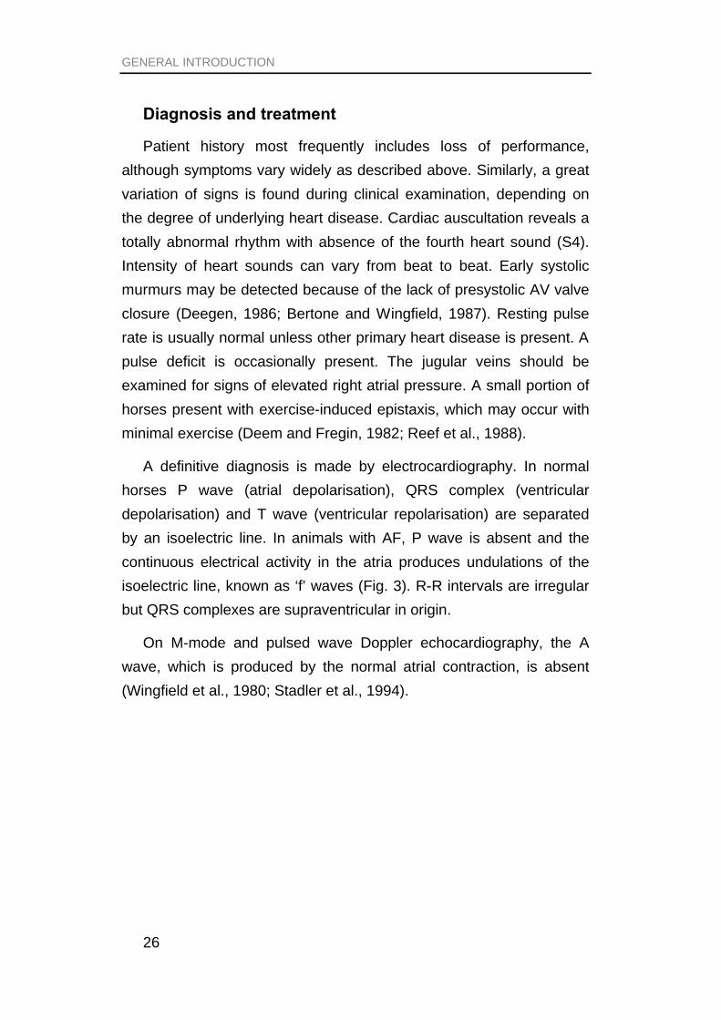
Diagnosis and treatment
Patient history most frequently includes loss of performance,
although symptoms vary widely as described above. Similarly, a great
variation of signs is found during clinical examination, depending on
the degree of underlying heart disease. Cardiac auscultation reveals a
totally abnormal rhythm with absence of the fourth heart sound (S4).
Intensity of heart sounds can vary from beat to beat. Early systolic
murmurs may be detected because of the lack of presystolic AV valve
closure (Deegen, 1986; Bertone and Wingfield, 1987). Resting pulse
rate is usually normal unless other primary heart disease is present. A
pulse deficit is occasionally present. The jugular veins should be
examined for signs of elevated right atrial pressure. A small portion of
horses present with exercise-induced epistaxis, which may occur with
minimal exercise (Deem and Fregin, 1982; Reef et al., 1988).
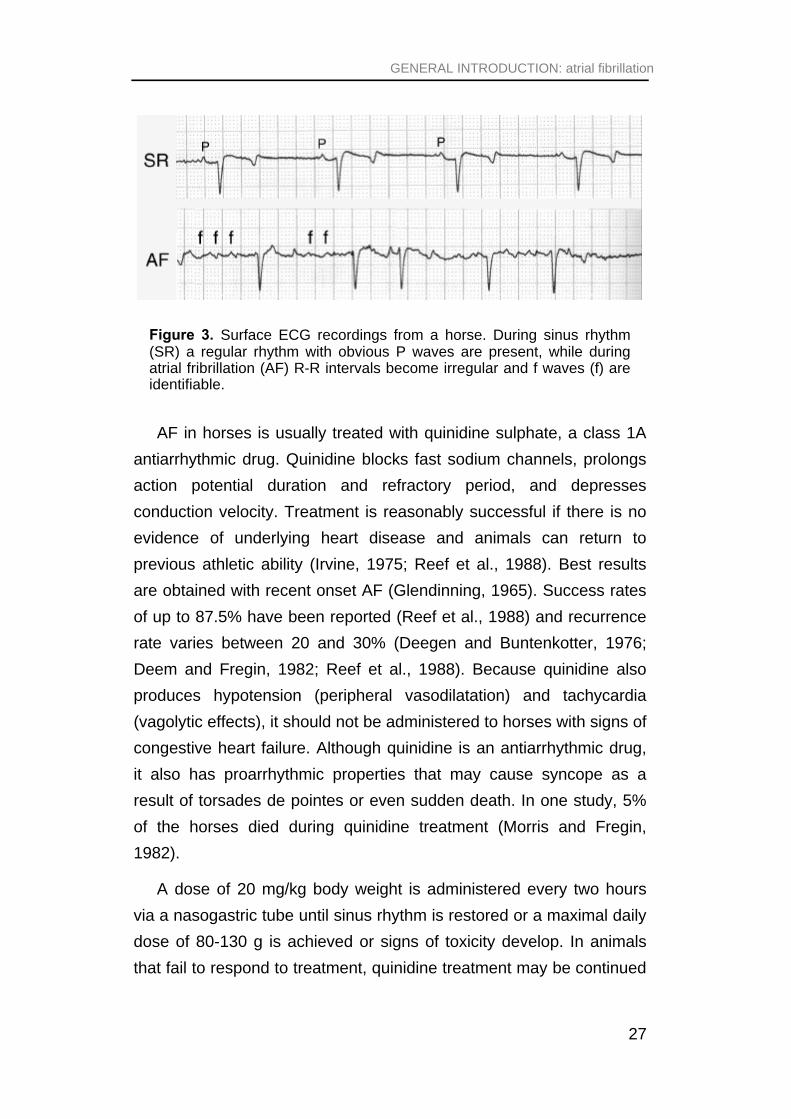
A definitive diagnosis is made by electrocardiography. In normal
horses P wave (atrial depolarisation), QRS complex (ventricular
depolarisation) and T wave (ventricular repolarisation) are separated
by an isoelectric line. In animals with AF, P wave is absent and the
continuous electrical activity in the atria produces undulations of the
isoelectric line, known as ‘f’ waves (Fig. 3). R-R intervals are irregular
but QRS complexes are supraventricular in origin.
On M-mode and pulsed wave Doppler echocardiography, the A
wave, which is produced by the normal atrial contraction, is absent
(Wingfield et al., 1980; Stadler et al., 1994).
GENERAL INTRODUCTION: atrial fibrillation
27
AF in horses is usually treated with quinidine sulphate, a class 1A
antiarrhythmic drug. Quinidine blocks fast sodium channels, prolongs
action potential duration and refractory period, and depresses
conduction velocity. Treatment is reasonably successful if there is no
evidence of underlying heart disease and animals can return to
previous athletic ability (Irvine, 1975; Reef et al., 1988). Best results
are obtained with recent onset AF (Glendinning, 1965). Success rates
of up to 87.5% have been reported (Reef et al., 1988) and recurrence
rate varies between 20 and 30% (Deegen and Buntenkotter, 1976;
Deem and Fregin, 1982; Reef et al., 1988). Because quinidine also
produces hypotension (peripheral vasodilatation) and tachycardia
(vagolytic effects), it should not be administered to horses with signs of
congestive heart failure. Although quinidine is an antiarrhythmic drug,
it also has proarrhythmic properties that may cause syncope as a
result of torsades de pointes or even sudden death. In one study, 5%
of the horses died during quinidine treatment (Morris and Fregin,
1982).
A dose of 20 mg/kg body weight is administered every two hours
via a nasogastric tube until sinus rhythm is restored or a maximal daily
dose of 80-130 g is achieved or signs of toxicity develop. In animals
that fail to respond to treatment, quinidine treatment may be continued
Figure 3. Surface ECG recordings from a horse. During sinus rhythm (SR) a regular rhythm with obvious P waves are present, while during atrial fribrillation (AF) R-R intervals become irregular and f waves (f) are identifiable.

GENERAL INTRODUCTION
28
at 6 hourly intervals or an additional digoxin treatment may be given to
slow down ventricular rate during quinidine treatment (Reef et al.,
1995). Concurrent digoxin and quinidine treatment should be given
cautiously as this enhances digoxin toxicity (Bertone and Wingfield,
1987; Parraga et al., 1995).
Up to 76% of the horses show quinidine side effects, including
urticaria, nasal oedema, anorexia, tachycardia, weakness, ataxia,
colic, diarrhoea, laminitis or convulsions (Morris and Fregin, 1982;
Reef et al., 1988). ECG recordings prior to quinidine administration are
vital: 25% increase in the width of the QRS complex indicates toxicity
and precludes further treatment.
Intravenous treatment with quinidine gluconate has also been
reported but success rate is slightly lower (Gerber et al., 1971; Deegen
and Buntenkotter, 1974; Lekeux et al., 1981; McGuirk et al., 1981;
Muir et al., 1990).
Following conversion to sinus rhythm, horses usually are not
immediately returned to training. However, the recommended rest
period is empirical, not based on clinical findings. Therefore, different
authors recommend a period of rest after conversion ranging from a
few days (Gerber et al., 1971; Amada and Kurita, 1975), 1 to 2 weeks
(Bonagura, 1990; Patteson, 1996), up to 3 months (Rose and Davis,
1977b). In horses, not treated for AF because of congestive heart
failure, therapy is best aimed at alleviating the clinical signs of heart
failure and, obviously, these animals should avoid any effort.
GENERAL INTRODUCTION: References
29
REFERENCESREFERENCES
Abildskov, J.A., Millar, K. & Burgess, M.J. (1971). Atrial fibrillation. Am J Cardiol 28, 263-267.
Allessie, M.A. (1998). Atrial electrophysiologic remodeling: another vicious circle? J Cardiovasc Electrophysiol 9, 1378-1393.
Allessie, M.A., Boyden, P.A., Camm, A.J., Kleber, A.G., Lab, M.J., Legato, M.J., Rosen, M.R., Schwartz, P.J., Spooner, P.M., Van Wagoner, D.R. & Waldo, A.L. (2001). Pathophysiology and Prevention of Atrial Fibrillation. Circulation 103, 769-777.
Allworth, M.S., Church, D.B., Maddison, J.E., Einstein, R., Brennan, P., Abdul Hussein, N. & Matthews, R. (1995). Effect of enalapril in dogs with pacing-induced heart failure. Am J Vet Res 56, 85-94.
Amada, A. & Kiryu, K. (1987). Atrial fibrillation in the race horse. Heart Vessels Suppl 2, 2-6.
Amada, A. & Kurita, H. (1975). Five cases of paroxysmal atrial fibrillation in the racehorse. Exp Rep Equine Hlth Lab 12, 89-100.
Amada, A., Senta, T., Kubo, K., Oh-ishi, S. & Kiryu, K. (1974). Atrial fibrillation in the horse: clinical and histopathological studies of two cases. 1. Clinical study. Exp Rep Equine Hlth Lab 11, 51-69.
Amsel, B.J. & Walter, P.J. (1992). Salvage transvenous rapid atrial pacing to terminate atrial flutter after cardiac operations. Ann Thorac Surg 53, 648-649.
Barold, S.S. & Zipes, D. (1992). Cardiac pacemakers and antiarrhythmic devices. In Heart Disease, Braunwald, E. (ed) pp. 726-755. W.B. Saunders: Philadelphia, PA.
Belgrave, J. (1990). A case of atrial fibrillation with congestive heart failure. Equine Vet Educ 2, 2-4.
Berg, F., Weber, Pfanzelt, S., Wenzl, J. & Oeppert, G. (1973). [Use of an internal pacemaker in a donkey with the Adams-Stokes syndrome]. Tierarztliche Umschau 28, 616-618.
Bertone, J.J., Traub-Dargatz, J.L. & Wingfield, W.E. (1987). Atrial fibrillation in a pregnant mare: treatment with quinidine sulfate. J Am Vet Med Assoc 190, 1565-1566.

GENERAL INTRODUCTION
30
Bertone, J.J. & Wingfield, W.E. (1987). Atrial fibrillation in horses. Comp Cont Educ Pract 9, 763-771.
Betsch, J.M. (1991). Study of atrial fibrillation in horses: four case reports. Prat Vet Equine 23, 13--24.
Blissitt, K.J. (1999). Diagnosis and treatment of atrial fibrillation. Equine Vet Educ 11, 11-19.
Bonagura, J.D. (1985). Equine heart disease. An overview. Vet Clin North Am Equine Pract 1, 267-274.
Bonagura, J.D. (1990). Clinical evaluation and management of heart disease. Equine Vet Educ 2, 31-37.
Brignole, M., Menozzi, C., Sartore, B., Barra, M. & Monducci, I. (1986). The use of atrial pacing to induce atrial fibrillation and flutter. Int J Cardiol 12, 45-54.
Brinker, J. & Midei, M. (1996). Techniques of pacemaker implantation. In: Cardiac pacing, 2nd edn. Ed: K.A. Ellenbogen. Blackwell Science, Abingdon. pp. 216-277.
Brown. (1979). ECG of the month. J Am Vet Med Assoc 175, 1076-1077.
Button, C., Scrutchfield, W.L., Clark, R.G., Knauer, K.W. & Schmitz, D.G. (1980). Multiple atrial dysrhythmias in a horse. J Am Vet Med Assoc 177, 714-719.
Collatos, C. (1995). Treating atrial fibrillation in horses. Comp Cont Educ Pract 17, 243-245.
Deegen, E. (1971). [Treatment of auricular fibrillation in horses with quinidinum sulfuricum.] Dtsch Tierarztl Wochenschr 78, 655-660.
Deegen, E. (1986). [Clinical significance of atrial fibrillation in horses.] Pferdeheilkunde 2, 179-186.
Deegen, E. & Buntenkotter, S. (1974). [Intravenous infusion of quinidine sulfate for therapy of equine auricular fibrillation. Preliminary report]. Dtsch Tierarztl Wochenschr 81, 161-162.
Deegen, E. & Buntenkotter, S. (1976). Behaviour of the heart rate of horses with auricular fibrillation during exercise and after treatment. Equine Vet J 8, 26-29.
Deem, D.A. & Fregin, G.F. (1982). Atrial fibrillation in horses: a review of 106 clinical cases, with consideration of prevalence, clinical signs, and prognosis. J Am Vet Med Assoc 180, 261-265.

GENERAL INTRODUCTION: References
31
Detweiler, D.K. (1952). Experimental and clinical observations on auricular fibrillation in horses. Proc Am Vet Med Ass 89, 119-129.
Detweiler, D.K. (1989). The mamalian electrocardiogram: comparative features. In Comprehensive electrocardiology, Macfarlane, P.W. & Veitch Lawrie, T.D. (eds), Vol. 2. pp. 1332-1377. Pergamon Press: New York.
Donald, D.E. & Elliott, F.J. (1948). Auricular fibrillation in horses. Vet Rec 60, 473-474.
Ellenbogen, K.A. & Peters, L.W. (1996). Indications for permanent and temporary cardiac pacing. In Cardiac pacing, Ellenbogen, K.A. (ed) pp. 1-36. Practical cardiac diagnosis. Blackwell Science: Abingdon.
Else, R.W. & Holmes, J.R. (1971). Pathological changes in atrial fibrillation in the horse. Equine Vet J 3, 56-64.
Elvan, A., Wylie, K. & Zipes, D.P. (1996). Pacing-induced chronic atrial fibrillation impairs sinus node function in dogs. Electrophysiological remodeling. Circulation 94, 2953-2960.
Evans, W. & Swann, P. (1954). Lone auricular fibrillation. Br Heart J 16, 189-194.
Fishler, M.G. & Thakor, N.V. (1994). A computer model study of the ventricular fibrillation vulnerable window: sensitivity to regional conduction depressions. Ann Biomed Eng 22, 610-621.
Fogoros, R.N. (1995). Electrophysiologic testing. 2nd edition. Blackwell Science: Oxford: 1-270.
Fregin, G.F. (1971). Atrial fibrillation in the horse. Proc Am Ass Equine Practnrs 16, 383-388.
Gallagher, M.M. & Camm, J. (1998). Classification of atrial fibrillation. Am J Cardiol 82, 18N-28N.
Gaskell, W.H. (1900). Schaefer's Textbook of Physiology, pp. 103, London.
Gasthuys, F., De Moor, A., Picavet, M., Muylle, E. & Steenhaut, M. (1988). Atrial fibrillation during eye enucleation in a horse. Vlaams Diergen Tijdschr 57, 207-212.
Geddes, L.A., Tacker, W.A., Rosborough, J., Moore, A.G., Cabler, P., Bailey, M., McCrady, J.D. & Witzel, D. (1974). The electrical dose for ventricular defibrillation with electrodes applied directly to the heart. J Thorac Cardiovasc Surg 68, 593-602.

GENERAL INTRODUCTION
32
Gelberg, H.B., Smetzer, D.L. & Foreman, J.H. (1991). Pulmonary hypertension as a cause of atrial fibrillation in young horses: four cases (1980-1989). J Am Vet Med Assoc 198, 679-682.
Gerber, H., Chuit, P. & Schatzmann, H.J. (1971). Treatment of atrial fibrillation in the horse with intravenous dihydroquinidine gluconate. Equine Vet J 3, 110-113.
Gerber, H., Chuit, P., Schatzmann, H.J., Straub, R., Schatzmann, U. & Pauli, B. (1972). [Intravenous treatment of atrial fibrillation in the horse]. Schweiz Arch Tierheilkd 114, 57-72.
Glazier, D.B. & Kavanagh, J.F. (1977). An unusual case of atrial fibrillation in a racing Thoroughbred filly. Irish Vet J 21, 107-110.
Glazier, D.B., Nicholsons, J.A. & Kelly, W.R. (1959). Atrial fibrillation in the horse. Irish Vet J 13, 47-55.
Glendinning, S.A. (1965). The use of quinidine sulphate for the treatment of atrial fibrillation in twelve horses. Vet Rec 77, 951-960.
Haissaguerre, M., Jais, P., Shah, D.C., Takahashi, A., Hocini, M., Quiniou, G., Garrigue, S., Le Mouroux, A., Le Metayer, P. & Clementy, J. (1998). Spontaneous initiation of atrial fibrillation by ectopic beats originating in the pulmonary veins. N Engl J Med 339, 659-666.
Hamir, A.N. & Reef, V.B. (1989). Complications of a permanent transvenous pacing catheter in a horse. J Comp Pathol 101, 317-326.
Hayes, D.L. & Osborn, M.J. (1996). Pacing: antibradycardia devices. In Mayo Clinic Practice of Cardiology, Guiliani, E.R., Gersh, B.J., McGoon, M.D., Hayes, D.L. & Schaff, H.V. (eds) pp. 909-976. Mosby: St. Louis.
Hering, H.E. (1908). Das elektrogram des pulsus irregularis perpetuus. Dtsch Arch Klin Med 94, 205.
Hiraga, A. & Kubo, K. (1999). Two cases of paroxysmal atrial fibrillation during exercise in horses. Equine Vet Educ 11, 6-10.
Hoffa, M. & Ludwig, C.F. (1850). Zeitschr F Rat Med 9, 104-144.
Holmes, D.R., Jr. & Hayes David, L. (1990). Pacemaker implantation techniques. In Electrical therapy of cardiac arrhythmia, Saksena, S. & Goldschlaber, N. (eds) pp. 173-190. W.B. Saunders: London.
Holmes, J.R., Darke, P.G. & Else, R.W. (1969). Atrial fibrillation in the horse. Equine Vet J 1, 211-222.

GENERAL INTRODUCTION: References
33
Holmes, J.R., Henigan, M., Williams, R.B. & Witherington, D.H. (1986). Paroxysmal atrial fibrillation in racehorses. Equine Vet J 18, 37-42.
Irvine, C.H. (1975). Electrocardiographic anomalies in the racehorse. N Z Vet J 23, 262-269.
Kantharia, B.K. & Mookherjee, S. (1995). Clinical utility and the predictors of outcome of overdrive transesophageal atrial pacing in the treatment of atrial flutter. Am J Cardiol 76, 144-147.
Kay, G.N. (1996). Basic concepts of pacing. In Cardiac pacing, Ellenbogen, K.A. (ed) pp. 37-123. Practical cardiac diagnosis. Blackwell Science: Abingdon.
Kiryu, K., Amada, A., Kaneko, M. & Satoh, H. (1974). Atrial fibrillation in the horse: clinical and histopathological studies of two cases. 2 Formal pathogenesis. Exp Rep Equine Hlth Lab 11, 70-86.
Kubo, K., Senta, T. & Sugimoto, O. (1975). Changes in cardiac output with experimentally induced atrial fibrillation in the horse. Exp Rep Equine Hlth Lab 12, 101-108.
Le Nihouannen, J.C., Sevestre, J., Dorso, Y., Petit, J.C. & Ozoux, C. (1984a). Implantation of a cardiac pacemaker into horses. I. Equipment and techniques. Revue Med Vet 135, 91-95.
Le Nihouannen, J.C., Sevestre, J., Dorso, Y., Petit, J.C. & Ozoux, C. (1984b). Implantation of a cardiac pacemaker into horses. II. Postoperative monitoring of a pacemaker with epicardial and myocardial electrodes in a pony. Revue Med Vet 135, 165-168.
Lekeux, P., Muylle, E., Henroteaux, M. & Bienfet, V. (1981). Comparison of different treatments of atrial fibrillation in the horse. Zentralbl Veterinarmed A 28, 475-480.
Lewis, T. (1909). Auricular fibrillation: a common clinical condition. Brit Med J 2, 1909.
Lewis, T. (1911). Irregularity of the heart's action in horses and its relationship to fibrillation of the auricles in experiment and to complete irregularity of the human heart. Heart: a journal for the study of the circulation 3, 161-171.
Machida, N., Yasuda, J. & Too, K. (1989). Three cases of paroxysmal atrial fibrillation in the Thoroughbred newborn foal. Equine Vet J 21, 66-68.
GENERAL INTRODUCTION
34
Maier-Bock, H. & Ehrlein, H.J. (1978). Heart rate during a defined exercise test in horses with heart and lung diseases. Equine Vet J 10, 235-242.
Manohar, M. & Smetzer, D.L. (1992). Atrial fibrillation. Comp Cont Educ Pract 14, 1327-1333.
Marr, C.M., Reef, V.B., Reimer, J.M., Sweeney, R.W. & Reid, S.W. (1995). An echocardiographic study of atrial fibrillation in horses: before and after conversion to sinus rhythm. J Vet Intern Med 9, 336-340.
Marr, C.M., Robertson, S.A., Johnstone, G.M., Johnson, C.B., Taylor, P.M. & Schumacher, J. (1994). Expert opinion case series. Is it appropriate to perform an ovariectomy for treatment of granulosa cell tumour in an aged Thoroughbred mare with atrial fibrillation. Equine Vet Educ 6, 293-300.
McGuirk, S.M., Muir, W.W. & Sams, R.A. (1981). Pharmacokinetic analysis of intravenously and orally administered quinidine in horses. Am J Vet Res 42, 938-942.
Miller, M.S., Gertsen, K.E. & Dawson, H. (1992). Paroxysmal atrial fibrillation: a case report. Equine Vet Sc 7, 95-97.
Miller, P.J. & Holmes, J.R. (1984). Relationships of left side systolic time intervals to beat-by-beat heart rate and blood pressure variables in some cardiac arrhythmias of the horse. Res Vet Sci 37, 18-25.
Mitten, L.A. (1996). Cardiovascular causes of exercise intolerance. Vet Clin North Am Equine Pract 12, 473-494.
Moe, G.K. (1962). On the multiple wavelet hypothesis of atrial fibrillation. Arch Int Pharmacodyn 140, 183-188.
Moe, G.K., Albildskov, J.A. & Syracuse, N.Y. (1959). Atrial fibrillation as a self-sustaining arrhythmia independent of focal discharge. Am Heart J 58, 59-70.
Moe, G.K., Rheinboldt, W.C. & Albildskov, J.A. (1964). A computer model of atrial fibrillation. Am Heart J 67, 200-220.
Mond, H.G. & Sloman, J.G. (1990). The indications for and types of artificial cardiac pacemakers. In The heart, Schlant Robert, C. & Hurst, J.W. (eds) pp. 561-580. McGraw-Hill: New York.
Moore, E.N. & Spear, J.F. (1987). Electrophysiological studies on atrial fibrillation. Heart Vessels Suppl 2, 32-39.
Morillo, C.A., Klein, G.J., Jones, D.L. & Guiraudon, C.M. (1995). Chronic rapid atrial pacing. Structural, functional, and electrophysiological characteristics of a new model of sustained atrial fibrillation. Circulation 91, 1588-1595.

GENERAL INTRODUCTION: References
35
Morris, D.D. & Fregin, G.F. (1982). Atrial fibrillation in horses: factors associated with response to quinidine sulfate in 77 clinical cases. Cornell Vet 72, 339-349.
Muir, W.W., 3rd, Reed, S.M. & McGuirk, S.M. (1990). Treatment of atrial fibrillation in horses by intravenous administration of quinidine. J Am Vet Med Assoc 197, 1607-1610.
Muir, W.W. & McGuirk, S.M. (1984). Hemodynamics before and after conversion of atrial fibrillation to normal sinus rhythm in horses. J Am Vet Med Assoc 184, 965-970.
Muylle, E., Vandenhende, C., Oyaert, W., Thoonen, H. & Vlaeminck, K. (1981). Delayed monensin sodium toxicity in horses. Equine Vet J 13, 107-108.
Nuytten, J., Bruynooghe, D., Muylle, E., Vandenhende, C., Vlaminck, K. & Oyaert, W. (1981). Accidental monensin intoxication in horses - acute and subacute symptoms. Vlaams Diergen Tijdschr 50, 242-249.
Oldham, H.N., Vasko, J.S., Brawley, R.K., Henney, R.P. & Morrow, A.G. (1967). The hemodynamic effects of atrial fibrillation. J Surg Res 7, 587-590.
Osborn, M.J. (1996). Pacing: antitachycardia devices. In Mayo Clinic Practice of Cardiology, Guiliani, E.R., Gersh, B.J., McGoon, M.D., Hayes, D.L. & Schaff, H.V. (eds) pp. 977-1016. Mosby: St. Louis.
Parraga, M.E., Kittleson, M.D. & Drake, C.M. (1995). Quinidine administration increases steady state serum digoxin concentration in horses. Equine Vet J Suppl19, 114-119.
Patteson, M.W. (1996). Equine cardiology. Blackwell Science: Oxford: 1-254.
Petch, M.C. (1986). Atrial fibrillation: bad news for man and horse? Equine Vet J 18, 3-4.
Peters, R.W., Weiss, D.N., Carliner, N.H., Feliciano, Z., Shorofsky, S.R. & Gold, M.R. (1994). Overdrive pacing for atrial flutter. Am J Cardiol 74, 1021-1023.
Pibarot, P., Vrins, A., Salmon, Y. & Difruscia, R. (1993). Implantation of a programmable atrioventricular pacemaker in a donkey with complete atrioventricular block and syncope. Equine Vet J 25, 248-251.
Qi, W., Kjekshus, H., Klinge, R., Kjekshus, J.K. & Hall, C. (2000). Cardiac natriuretic peptides and continuously monitored atrial pressures during chronic rapid pacing in pigs. Acta Physiol Scand 169, 95-102.

GENERAL INTRODUCTION
36
Reef, V.B., Clark, E.S., Oliver, J.A. & Donawick, W.J. (1986). Implantation of a permanent transvenous pacing catheter in a horse with complete heart block and syncope. J Am Vet Med Assoc 189, 449-452.
Reef, V.B., Levitan, C.W. & Spencer, P.A. (1988). Factors affecting prognosis and conversion in equine atrial fibrillation. J Vet Intern Med 2, 1-6.
Reef, V.B., Reimer, J.M. & Spencer, P.A. (1995). Treatment of atrial fibrillation in horses: new perspectives. J Vet Intern Med 9, 57-67.
Roos, J. (1924). Auricular fibrillation in the domestic animals. Heart 1, 1-7.
Rose, R.J. & Davis, P.E. (1977a). Paroxysmal atrial fibrillation in a racehorse. Aust Vet J 53, 545-549.
Rose, R.J. & Davis, P.E. (1977b). Treatment of atrial fibrillation in three racehorses. Equine Vet J 9, 68-71.
Ross, T.R. & Mandel, W.J. (1995). Invasive cardiac electrophysiologic testing. In Cardiac arrhythmias, Mandel, W.J. (ed) pp. 193-235. Lippincott: Philadelphia.
Seahorn, T.L. & Hormanski, C.E. (1993). Ventricular septal defect and atrial fibrillation in an adult horse. J Equine Vet Sc 13, 36-38.
Senta, T. & Kubo, K. (1978). Experimental induction of atrial fibrillation by electrical stimulation in the horse. Exp Rep Equine Hlth Lab 15, 37-46.
Senta, T., Kubo, K., Sugimoto, O. & Amada, A. (1975). Induction of atrial fibrillation by electrical stimulation in the horse. Exp Rep Equine Hlth Lab 12, 109-112.
Stadler, P., Deegen, E. & Kroker, K. (1994). [Echocardiography and therapy of atrial fibrillation in horses]. Dtsch Tierarztl Wochenschr 101, 190-194.
Steel, J.D., Hall, M.C. & Stewart, G.A. (1976). Cardiac monitoring during exercise tests in the horse. 3. Changes in the electrocardiogram during and after exercise. Aust Vet J 52, 6-10.
Stewart, G.A., Fulton, L.J. & McKellar, C.D. (1990). Idiopathic atrial fibrillation in a champion Standardbred racehorse. Aust Vet J 67, 187-191.
Tacker, W.A., Geddes, L.A., Rosborough, J.P. & Moore, A.G. (1973). Ventricular defribrillation of a 341 kg horse using precordial electrodes. Can J Comp Med 37, 382-390.

GENERAL INTRODUCTION: References
37
Tacker, W.A., Jr., Geddes, L.A., Rosborough, J.P., Witzel, D., Cabler, P.S., Chapman, R.J. & Rivera, R.A. (1975). Trans-chest ventricular defibrillation of heavy subjects using trapezoidal current waveforms. J Electrocardiol 8, 237-240.
Taylor, D.H. & Mero, M.A. (1967). The use of an internal pacemaker in a horse with Adams-Stokes syndrome. J Am Vet Med Assoc 151, 1172-1176.
Taylor, F.G., Wotton, P.R., Hillyer, M.H., Barr, F.J. & Lucke, V.M. (1991). Atrial septal defect and atrial fibrillation in a foal. Vet Rec 128, 80-81.
Trautwein, W. (1975). Electrophysiological aspects of cardiac stimulation. In Advances in pacemaker technology, Schaldach, M. & Furman, S. (eds) pp. 11-23. Springer: Berlin.
Tsai, C.F., Tai, C.T., Hsieh, M.H., Lin, W.S., Yu, W.C., Ueng, K.C., Ding, Y.A., Chang, M.S. & Chen, S.A. (2000). Initiation of atrial fibrillation by ectopic beats originating from the superior vena cava: electrophysiological characteristics and results of radiofrequency ablation. Circulation 102, 67-74.
Tucker, K.J. & Wilson, C. (1993). A comparison of transoesophageal atrial pacing and direct current cardioversion for the termination of atrial flutter: a prospective, randomised clinical trial. Br Heart J 69, 530-535.
Vulpian, E.F. (1874). Arch De Physiol 1, 975.
Wijffels, M.C., Kirchhof, C.J., Dorland, R. & Allessie, M.A. (1995). Atrial fibrillation begets atrial fibrillation. A study in awake chronically instrumented goats. Circulation 92, 1954-1968.
Willems, R., Ector, H. & Heidbüchel, H. (2000). Slow development of atrial fibrillation in a transvenously paced sheep model. In Abstract book: Cardiac arrhythmias and device therapy: Results and perspectives for the new century, Ovsyshcher, E. (ed) pp. 95-99. Futura Publishing Company: Armonk, New York.
Wingfield, W.E., Miller, C.W., Voss, J.L., Bennett, D.G. & Breukels, J. (1980). Echocardiography in assessing mitral valve motion in 3 horses with atrial fibrillation. Equine Vet J 12, 181-184.
Witzel, D.A., Geddes, L.A., Hoff, H.E. & McFarlane, J. (1968). Electrical defibrillation of the equine heart. Am J Vet Res 29, 1279-1285.
Wood, M. & Ellenbogen, K.A. (1996). Temporary cardiac pacing. In Cardiac pacing, Ellenbogen, K.A. (ed) pp. 168-215. Practical cardiac diagnosis. Blackwell Science: Abingdon.

GENERAL INTRODUCTION
38
Yamamoto, K., Yasuda, J. & Too, K. (1992). Arrhythmias in newborn thoroughbred foals. Equine Vet J 24, 169-173.
Yamaya, Y., Kubo, K. & Amada, A. (1997a). Relationship between atrioventricular conduction and hemodynamics during atrial pacing in horses. J Equine Sci 59, 149-151.
Yamaya, Y., Kubo, K., Amada, A. & Sato, K. (1997b). Intrinsic atrioventricular conductive function in horses with a second degree atrioventricular block. J Vet Med Sci 59, 149-151.
Yue, L., Feng, J., Gaspo, R., Li, G.R., Wang, Z. & Nattel, S. (1997). Ionic remodeling underlying action potential changes in a canine model of atrial fibrillation. Circ Res 81, 512-525.
Zipes, D.P. (1992). Genesis of cardiac arrhythmias: electrophysiological considerations. In Heart Disease, Braunwald, E. (ed) pp. 588-627. W. B. Saunders: Philadelphia.
Zipes, D.P. (1997). The seventh annual Gordon K. Moe Lecture. Atrial fibrillation: from cell to bedside. J Cardiovasc Electrophysiol 8, 927-938.

SCIENTIFIC AIMS


Scientific Aims
41
Horses can suffer from cardiac rhythm disturbances that can affect
their performance or can even be life threatening. Clinically, the most
important arrhythmia in equines is atrial fibrillation. During the past
decades, only little progress has been made concerning arrhythmias in
equines. In general, the surface electrocardiogram (ECG) is the only
diagnostic aid while treatment options merely consist of a few drugs,
such as quinidine and digoxin. Conversely, in human medicine,
extensive facilities are available in the cardiology department. In man,
electrophysiological studies and different cardiac pacing techniques
have become a mainstay in the diagnosis and treatment of many
dysrhythmias. Short-term pacing is applied with temporary catheters
while long-term pacing can be achieved by permanent implantation of
an electrical pulse generator.
Besides diagnosis and treatment, cardiac pacing also provides an
excellent means to study pathophysiology of arrhythmias in animal
models.
The aims of our study were investigating the applicability of atrial
pacing in horses and subsequently using the technique to study the
pathophysiology of atrial fibrillation in equines. Particular aims of this
study were as follows:
Section 1: atrial pacing
1. Temporary pacing
a. Development of a technique
b. Application in the treatment of atrial flutter
2. Permanent pacing
a. Development of a technique
b. Application to treat sinus node dysfunction
Section 2: research on atrial fibrillation
1. Development of a model for chronic atrial fibrillation in healthy equines
2. Application of the model to study the pathophysiology of chronic atrial fibrillation


CHAPTER 11
Temporary transvenous atrial pacing in horses: Temporary transvenous atrial pacing in horses:
threshold determination threshold determination
G. van Loon1, H. Laevens2, P. Deprez1
1 Department of Large Animal Internal Medicine, Faculty of Veterinary
Medicine, Ghent University, Salisburylaan 133, B-9820 Merelbeke,
Belgium 2
Department of Reproduction, Obstetrics and Herd Health, Faculty of
Veterinary Medicine, Ghent University, Salisburylaan 133, B-9820
Merelbeke, Belgium
Adapted from: van Loon G., Laevens H., Deprez P. (2001). Temporary
transvenous atrial pacing in horses: threshold
determination. Equine Vet J 33, 290-295.

CHAPTER 1: Summary
45
SUMMARYSUMMARY
The purpose of this study was to perform temporary atrial
pacing and to determine the atrial strength-duration (S-D) curve,
which displays the minimal pulse intensity necessary to achieve
atrial capture. In seven horses atrial pacing was applied using a
temporary pacing catheter and a pacemaker as electrical pulse
generator. Using the stimulus reduction method, three
approaches for atrial threshold determination were used. With
the fixed pulse width method, at several pulse widths the
corresponding minimal amplitudes to achieve capture were
determined, describing a S-D curve. With the fixed amplitude
method, the corresponding threshold pulse widths were
determined at several fixed amplitudes. The third method proved
to be the best one and was a combination of both
aforementioned methods to determine 2 points of the S-D curve.
From these two points the whole S-D curve was calculated using
a mathematical equation. Temporary pacing can be used to
terminate atrial flutter, to induce atrial arrhythmias or to obtain
more information about the electrophysiologic properties of the
heart such as the atrial refractory period, atrial vulnerability and
atrioventricular conduction.

Temporary transvenous atrial pacing in horses: threshold determination
46
INTRODUCTIONINTRODUCTION
The excitable properties of the cardiac cells make artificial
stimulation of the heart possible (Irnich 1989). Artificial stimulation can
be performed by mechanical or electrical stimulation. Electrical stimuli
are delivered by an external or an implantable pulse generator and are
conducted to the heart by means of a lead wire with electrodes on its
tip. The transvenous insertion of a lead or catheter electrode into the
cardiac cavity is the most widely used method for cardiac pacing,
although transcutaneous, transoesophageal or epicardial stimulation
are also possible.
The electrical stimulus, which is a square current pulse, has to
reduce the resting potential of the cardiac cells by a critical amount
and within a critical time in order to reach the threshold for the
propagated response, known as the action potential (Trautwein 1975).
The threshold for cardiac pacing can be defined as the smallest
amount of electrical activity that produces consistent myocardial
capture outside the refractory period of the heart (Hayes and Osborn
1996). It is determined by its amplitude or strength and by its pulse
width (PW) or duration. Within limits, values with a lower amplitude
must have a longer duration, while the duration of high amplitude
values may be shorter. These threshold values describe a hyperbolic-
like strength-duration curve (Geddes and Bourland 1985). Knowledge
of this strength-duration relationship is fundamental to safe and
effective cardiac pacing (Ayers et al. 1986). A threshold within normal
limits is a means of verifying that the electrode is in a secure position
and is in contact with viable cardiac tissue (Schuenemeyer 1986).
Temporary cardiac pacing can be used therapeutically, as during
third degree atrioventricular block, to prevent problems during
anaesthesia of animals with documented conduction system disease

CHAPTER 1: Introduction
47
or as a treatment of atrial flutter (van Loon et al. 1997). It can also be
used to induce atrial arrhythmias in order to study their effect on
cardiac function, as performed by Kubo et al. (1975), Senta and Kubo
(1978) and Moore and Spear (1987). Furthermore temporary pacing
can provide useful information about the electrophysiologic properties
of the heart, such as atrial refractory period, atrioventricular conduction
(Yamaya et al. 1994, 1997a and 1997b) or atrial vulnerability (Senta
and Kubo 1978).
The purpose of this study was to assess the feasibility of temporary
atrial pacing in horses, to determine the minimal pulse intensity
necessary to achieve atrial stimulation and to evaluate different
techniques to determine the atrial S-D curve.

Temporary transvenous atrial pacing in horses: threshold determination
48
MATERIALS AND METHODMATERIALS AND METHODSS
Threshold values for temporary transvenous atrial pacing were
determined in seven healthy standardbred horses. Clinical,
electrocardiographic and echocardiographic examinations were
normal in all horses.
Instrumentation
A catheter electrode or lead1, which possessed two electrodes at
its tip, was used for bipolar stimulation of the right atrium. The lead
was straight in 3 horses and had a J shaped tip in 4 horses. A
pacemaker2 served as an external pulse generator and was
connected with the electrodes of the lead. A programmer3 had
telemetric contact with the pulse generator and was at the same time
connected with a surface electrocardiogram (ECG). With the
programmer, the pulse generator could be programmed to deliver
electrical pulses with a specific interval, amplitude and duration.
Furthermore, electrical activity of the heart could be 'sensed' through
the same electrode in order to obtain an endocardial electrogram. The
endocardial electrogram and the surface ECG were simultaneously
visible on the programmer screen and could be recorded on paper.
After local anaesthesia (xylocaine 2 %4), an introducer sheath was
inserted in the lower third of the jugular vein using a modified
Seldinger technique. Via the jugular vein, the catheter electrode was
inserted into the cardiac chamber. Location of the catheter tip was
confirmed by the ECG recordings and by echocardiography. Initially,
while the electrode tip was still in the blood vessels, no or minimal
electrical activity could be sensed. Once the electrode entered the
right atrium, largest deflections on the endocardial electrogram
coincided with the P waves on the surface ECG. Further advancement
CHAPTER 1: Materials & methods
49
of the catheter into the right ventricle produced large endocavitary
deflections simultaneous with the QRS-complexes.
On echocardiography, catheter movement was visible once the
catheter entered the right atrium. Due to lung interference, the dorsal
part of the right atrium could only be imaged partially.
Lead positioning
First the lead was inserted into the right atrium and advanced until
it reached the tricuspid valve or entered the right ventricle.
Subsequently, the lead was withdrawn until large intra-atrial deflections
with an equal morphology were present, indicating a stable contact
between the electrode and the endocardium. The lead was left in place
to perform threshold measurements. When no efficacious electrode
position was obtained during withdrawal, when diaphragmatic nerve
stimulation occurred or when endocardial contact was lost during
threshold measurement, the electrode was repositioned and
measurements were repeated.
The whole procedure was performed in the unsedated horse.
Measurements
In order to obtain an artificial stimulation of the heart, the
pacemaker was programmed to deliver bipolar electrical pulses at a
faster rate than the intrinsic heart rate, empirically at 60 beats per
minute. The most distal electrode (tip) was used as cathode while the
proximal electrode (ring) was the anode. If intrinsic atrial activity was
sensed by the pulse generator, stimulation was shortly inhibited to
avoid induction of atrial fibrillation.
For the threshold measurements the stimulus reduction method
was used. This means that stimulation started with strong pulses, with
a long duration and/or high amplitude, and that gradually the intensity
of the pulses was decreased until capture, i.e. cardiac contraction, was
lost. The decrease of pulse intensity was not continuous but stepwise.

Temporary transvenous atrial pacing in horses: threshold determination
50
The PW varied in steps of 0.03 milliseconds (ms) between 0.03 and
0.15 ms, in steps of 0.05 ms between 0.15 and 1 ms and in steps of
0.1 ms between 1 ms and 1.5 ms. Variations of the amplitude were
restricted to 0.5 volt (V), 1 V, 1.5 V, 2 V, 2.5 V, 3 V, 3.5 V, 4 V, 5 V and
7.5 V.
Three different ways for threshold determination were applied. With
the constant PW method (method 1), the pulse duration was
programmed at a fixed value. Subsequently, pacing started with stimuli
with high amplitude. Every 4 stimuli, the pulse generator gradually
decreased the amplitude. Capture was present as long as every
electrical pulse was followed by a P wave on the surface ECG. When
the pulse amplitude became too low and thus too weak, a P wave was
not initiated, capture was lost and intrinsic heart rate reappeared. At
that time, stimulation was aborted. The weakest pulse still able to
achieve capture was taken as the threshold value for stimulation. The
minimal amplitude for stimulation was successively determined at
fixed PW of 0.8 ms, 0.6 ms, 0.4 ms, 0.3 ms, 0.2 ms, 0.12 ms and 0.06
ms. A plot of these values, with stimulus strength (V) on the ordinate
and duration (ms) on the abscissa, describes the strength-duration (S-
D) curve.
With the constant amplitude procedure (method 2), the amplitude
of the stimuli was kept constant and the PW was gradually decreased
until capture was lost. The minimal PW for stimulation was
successively determined at fixed amplitudes of 7.5 V, 4 V, 3.5 V, 3 V,
2.5 V, 2 V and 1.5 V to obtain a new S-D curve.
For the third method, only two threshold values were determined.
The constant voltage method was used to determine the threshold PW
at a high amplitude pulse, while the threshold amplitude for a long PW
stimulus was determined with the constant PW method. With these
two values the theoretical strength-duration curve was calculated using
a hyperbolic equation, described by Lapicque (1907):

CHAPTER 1: Materials & methods
51
+×=t
t1II c
r [1]
where I is the stimulus intensity (voltage or current), t is the PW, Ir
is the rheobase and tc is the chronaxie.
The rheobase was defined as the lowest amplitude with indefinite
pulse duration that just stimulates the myocardium (Irnich 1980). The
chronaxie was the threshold pulse duration at a stimulus amplitude of
twice that of rheobase. Using equation [1] rheobase and chronaxie can
be calculated when 2 threshold values on the S-D curve are known.
With the rheobase and chronaxie all points of the S-D curve can be
computed using the same equation (Fig. 1.1).
After determination of the S-D curve using these three methods,
the lead was advanced again until the tricuspid valve or the right
ventricle and was withdrawn to perform a new series of measurements
at a second location in the atrium. Thus in each horse 6 S-D curves
were determined.
Statistical analysis
Statistical comparison of methods 1 and 2 was not performed as
variation was along the ordinate for method 1 while variation was along
the abscissa for method 2. Therefore, accuracy and precision of
methods 1 and 2 were compared with method 3. First method 1 and 3
were compared. At the same fixed PW values as used in method 1,
the threshold amplitudes were calculated for method 3 using equation
[1] resulting in a variation along the ordinate. Subsequently, to
compare methods 2 and 3, threshold PW values for method 3 were
calculated for each fixed amplitude used in method 2, resulting in a
variation along the abscissa.
Statistical analyses were performed after a loge-transformation of
the data to obtain a normal distribution, as indicated by the Wilk-
Shapiro/Rankit Plot (Statistix 4.15). For each PW or amplitude level

Temporary transvenous atrial pacing in horses: threshold determination
52
respectively, a paired t-test was performed to compare the accuracy of
threshold determination of methods 1 or 2 with method 3 (Statistix
4.1). Similarly, precision of threshold determination with methods 1 or
2, i.e. variances at each fixed level, was compared with precision of
threshold determination with method 3 using an F-test (Statistix 4.1).
Correlation (Pearson's) was used to determine the degree of linear
association between method 1 and 2 with method 3. A probability of <
0.05 was considered significant.

CHAPTER 1: Results
53
RESULTSRESULTS
Successful atrial pacing was obtained in all horses. Adequate
electrode positioning and determination of threshold values could be
easily performed. The procedure was well tolerated by the unsedated
horse and cardiac pacing initiated no adverse reactions of the horse,
even when diaphragmatic nerve stimulation occurred.
The J shaped catheter was found to obtain more easily a stable
endocardial contact than the straight lead.
Threshold values for atrial stimulation using the constant PW
method and the constant voltage method are listed in Table 1.1 and
1.2, respectively. Geometric mean and 95% confidence interval of
each method and the corresponding values of the third method are
displayed. The correlation coefficient and p-value of method 1 and
method 2 with method 3 are recorded. In addition, Table 1.1 contains
rheobasic voltage and chronaxie pulse duration of each horse, and the
geometric mean values and the 95% confidence interval, obtained
from method 3. The mean rheobase and chronaxie are indicated in
Figure 1.1, which displays the mean S-D curve of method 3.
Due to limitations of the system, the amplitude and PW varied
stepwise and not continuously. This means that values are
overestimated. Furthermore in some horses at a fixed PW of 0.06 ms,
threshold amplitude could not be obtained because the threshold was
higher than the maximal available strength of 7.5 V. Consequently
capture did not occur. In these cases data were excluded from
statistical analyses. At a fixed PW of 0.8 ms, the amplitude values of
horse 3 might have been lower than 0.5 V but the amplitude could not
be decreased further. Similarly for the second method, in some horses
the PW values at a fixed amplitude of 4 V and 7.5 V might have been
shorter than 0.03 ms but this was the shortest available PW. In some

Temporary transvenous atrial pacing in horses: threshold determination
54
horses minimal PW for stimulation could not be determined at a fixed
amplitude of 2 V and/or 1.5 V because these values were below their
rheobase. At an amplitude below the rheobase, even an infinitely long
PW will not be sufficient to initiate a depolarisation. In these cases,
data were excluded from statistical analyses.
Statistical analyses showed that accuracy and precision at each
PW level were similar for both methods 1 and 3 and that there was a
linear association between the threshold values of both methods at
each PW level (Table 1.1).
Statistical comparison of method 2 with method 3 indicated that
precision was similar for both methods at each amplitude level. Also,
at each amplitude level, a linear association was found between the
threshold values of both methods as indicated by the significant
correlation coefficients in Table 1.2. On the other hand, accuracy
evaluation showed that threshold values were significantly lower when
determined with method 2 at the amplitude levels 1.5, 2 and 2.5 V
(Table 1.2).
Figure 1.1. This S-D curve displays the geometric mean values and 95% confidence interval (error bars) for all horses and all positions using method 3. The geometric mean values for rheobase and chronaxie are shown.

CHAPTER 1: Results
55
Table 1.1. Measured amplitude values (V), geometric means (GM) and 95% confidence intervals (95% CI) of the constant PW method, and the rheobase (V) and chronaxie (ms) values with their GM and 95% CI of the third method are displayed for each horse in each position. Values of the GM and 95% CI of the third method are shown. At each PW level, similar superscript letters for the GM values of method 1 and method 3 indicate that no significant difference in accuracy exists between both methods. Correlation (Pearson's), and corresponding p-value, between both methods is shown at each PW level. Some data are missing, indicated by M, because capture could not be achieved at the maximal amplitude of 7.5 V.
Constant pulse width method (Method 1) Method 3 Horse Position
0.06 ms 0.12 ms 0.2 ms 0.3 ms 0.4 ms 0.6 ms 0.8 ms rheobase chronaxie
1 1 3 2 1.5 1.5 1 1 1 0.76 0.26
2 M 7.5 4 3.5 3.5 3 3 2.39 0.26
2 1 7.5 4 3 2.5 2.5 2 2 2.25 0.11
2 5 3.5 3 2.5 3 2.5 2.5 2.24 0.09
3 1 3 2.5 1 1 1 0.5 0.5 0.82 0.21
2 2.5 1.5 1 1 1 0.5 0.5 1.25 0.20
4 1 5 2 2 1.5 1 1 1 0.62 0.49
2 7.5 7.5 3.5 2.5 2.5 2.5 2 1.65 0.17
5 1 M 7.5 4 4 3.5 3 1.5 1.47 0.36
2 M 5 4 3.5 3 2.5 2 1.09 0.67
6 1 5 2.5 1.5 1.5 1 1 1 0.80 0.25
2 3 2 1.5 1.5 1 1 1 0.80 0.25
7 1 M 4 3.5 2 2 1.5 1.5 1.30 0.16
2 7.5 4 2.5 2 2 2 2 1.13 0.33
Method 1 GM 4.52 a 3.46
a2.30
a 1.99
a 1.76
a 1.47
a 1.35
a - -
95% CI (3.33 ; 6.14)
(2.53 ; 4.73)
(1.72 ; 3.09)
(1.54 ; 2.57)
(1.29 ; 2.40)
(1.03 ; 2.09)
(0.98 ; 1.86) - -
Method 3 GM 5.16 a 3.71
a2.73
a 2.24
a 1.99
a 1.74
a 1.61
a 1.21 0.24
95% CI (4.35 ; 6.11)
(2.95 ; 4.67)
(2.19 ; 3.42)
(1.79 ; 2.80)
(1.59 ; 2.48)
(1.38 ; 2.17)
(1.28 ; 2.02)
(0.94 ; 1.56)
(0.18 ; 0.32)
Correlation 0.76 0.77 0.81 0.86 0.94 0.79 0.77 - -
p-value 0.011 0.001 < 0.001 < 0.001 < 0.001 < 0.001 0.001 - -

Temporary transvenous atrial pacing in horses: threshold determination
56
Table 1.2. Measured PW values (ms), geometric means (GM) and 95% confidence intervals (95% CI) of the constant amplitude method are displayed for each horse in each position. GM values and 95% CI for the third method are shown. At each amplitude level, different superscript letters for the GM values of methods 2 and method 3 indicate a significant difference in accuracy of both methods. Correlation (Pearson's), and corresponding p-value, between both methods is shown at each PW level. In some horses data are missing, as indicated by M, because capture could not be achieved at the maximal pulse width of 1.5 ms.
Constant amplitude method (Method 2) Horse
Position 1.5 V 2 V 2.5 V 3 V 3.5 V 4 V 7.5 V
1 0.2 0.12 0.12 0.09 0.09 0.06 0.03 1
2 M M 1.3 0.45 0.25 0.2 0.09
1 M 1.2 0.45 0.25 0.2 0.12 0.06 2
2 M 0.9 0.65 0.15 0.15 0.12 0.06
1 0.12 0.12 0.09 0.06 0.06 0.03 0.03 3
2 0.12 0.09 0.06 0.06 0.03 0.03 0.03
1 0.2 0.2 0.2 0.12 0.09 0.09 0.03 4
2 0.5 0.4 0.2 0.15 0.12 0.12 0.09
1 1.5 0.7 0.55 0.45 0.35 0.3 0.09 5
2 1.1 0.6 0.4 0.3 0.3 0.25 0.12
1 0.15 0.15 0.12 0.09 0.09 0.06 0.06 6
2 0.12 0.09 0.06 0.06 0.06 0.06 0.06
1 0.8 0.3 0.2 0.15 0.15 0.12 0.03 7
2 1 0.3 0.3 0.3 0.2 0.15 0.09
GM 0.33 a 0.22
a 0.23
a 0.15
a 0.12
a 0.10
a 0.06
a
Method 2 95 % CI
(0.15 ; 0.69)
(0.13 ; 0.36)
(0.13 ; 0.39)
(0.10 ; 0.23)
(0.08 ; 0.19)
(0.07 ; 0.15)
(0.04 ; 0.07)
GM 0.76 b 0.32
b 0.31
b 0.19
a 0.14
a 0.11
a 0.05
a
Method 3 95 % CI
(0.29 ; 1.95)
(0.20 ; 0.52)
(0.17 ; 0.60)
(0.12 ; 0.28)
(0.10 ; 0.20)
(0.08 ; 0.16)
(0.04 ; 0.06)
Correlation 0.79 0.87 0.89 0.86 0.76 0.81 0.74
p-value 0.007 <0.001 <0.001 <0.001 0.002 <0.001 0.002

CHAPTER 1: Discussion
57
DISCUSSIONDISCUSSION
During the measurements, factors that could influence the
threshold values were avoided as much as possible. The horses were
made used to stand in the stocks in order to avoid sympathetic
influence. In man, increased sympathetic tone and catecholamines are
known to decrease threshold values while increased vagal tone
increases threshold values (Preston et al. 1967; Kay 1996). Lignocaine
without epinephrine was used as local anaesthetic at the level of the
jugular vein, because the sympathomimetic agent epinephrine can
lower the threshold for stimulation (Preston et al. 1967; Furman et al.
1977b; Wood and Ellenbogen 1996).
In three horses, a straight lead was used while in four horses
measurements were performed with a J tipped lead. The straight lead
was found to remain more difficult in stable endocardial contact than
the J shaped catheter. The J shape was thought to enhance
entanglement in the atrial trabeculae while the catheter was withdrawn.
The shape and surface area of the electrodes were identical in both
leads, because these parameters influence the threshold for
temporary pacing (Irnich 1980; Schuenemeyer 1986). Smaller
electrodes have a larger electric field and thus a lower threshold than
large surface electrodes (Furman et al. 1977b).
Correct intra-atrial location of the electrodes with a stable
endocardial contact is characterized by an endocardial electrogram
with large monomorphic deflections concomitant with the peripheral P
wave (Furman et al. 1977a). Since voltage threshold increases rapidly
with increasing distance between the electrode and the excitable
myocardium (Furman et al. 1977b), consistent capture with low
threshold values was an additional verification for a good contact
between electrode and myocardium. Due to areas of fibrotic

Temporary transvenous atrial pacing in horses: threshold determination
58
myocardium local variations in threshold values may occur (Furman et
al. 1977b; Ector et al. 1985).
Ultrasonography was helpful to position the electrode tip in the
atrium but the dorsal part of the right atrium could not completely be
visualized. Moreover, even when the catheter was visible on
ultrasound, endocardial contact of the electrode tip could not always
be assured. While in human medicine fluoroscopy is an important tool
to localize the electrode tip, no fluoroscopic control was performed
during this study. Due to the large equine thorax, the usefulness of this
technique would have been limited in adult horses.
During catheter positioning, single atrial or ventricular extrasystoles
occurred occasionally but never initiated sustained dysrhythmias.
Bipolar stimulation means that pulses are delivered between two
electrodes within the heart and is most commonly applied for
temporary pacing (Wood and Ellenbogen 1996). The most distal (tip)
electrode is usually the cathode, while the proximal (ring) is the anode
(Furman et al. 1977b; Chou T. C. 1986; Schuenemeyer 1986). The
voltage threshold for bipolar pacing is slightly higher than for unipolar
pacing although the difference is very small (Furman et al. 1977b).
Immediately after a pulse is delivered, the endocardial electrogram
presents a large deflection, known as the afterpotential (Kay 1996).
During this deflection the intrinsic electrical activity of the heart is often
not visible. Consequently, capture is not always visible on the
intracardiac electrogram and can better be established on the surface
ECG.
Occasionally, diaphragmatic contractions were seen simultaneous
with pacing rate. Stimulation of the lateral, dorsal part of the right
atrium, at the level of the diaphragmatic nerve, was likely to be the
cause. Decreasing the stimulation intensity, i.e. the amplitude and/or
pulse duration, inhibited the diaphragmatic flutter. However, when

CHAPTER 1: Discussion
59
diaphragmatic stimulation occurred, measurements were not
performed and the lead was repositioned to avoid this phenomenon.
With the stimulus reduction method, stimulation intensity starts at a
high level and decreases gradually, while the opposite is true with the
stimulus advance method. In human medicine, the stimulus reduction
method is mostly used because patients often depend on pacemaker
stimulation. Threshold values determined by the stimulus advance
method are slightly higher than those of the stimulus reduction method
(Sylvén et al. 1982; Schuenemeyer 1986).
For the fixed PW method, method 1, in some horses no capture
could be obtained at 0.12 and 0.06 ms because the maximal available
amplitude of 7.5 V was not sufficient to achieve capture. Therefore the
left side of the curve could not always be obtained and the best results
for the fixed PW method were obtained at the right side of the curve.
For horse 3, voltage threshold at 0.6 and/or 0.8 ms might have been
lower than 0.5 V but 0.5 V was the lowest available amplitude.
For the fixed amplitude procedure, method 2, at low amplitudes
there was a large variation in the determined PW values and in some
horses no values could be obtained. The reason was that for some
horses the low voltage was close to their rheobasic voltage that
resulted in a very long PW while in other horses the PW was still very
short. And in some horses a threshold PW could not be obtained at a
low amplitude because that amplitude was already below their
rheobasic voltage. Therefore the fixed amplitude method seemed
most suitable for measurement of the left side of the curve, and less
appropriate to obtain the right side of the curve.
The third method, which calculated the S-D curve with Lapicque's
law, was a combination of the aforementioned methods in order to
simplify S-D curve determination. A point at the left side of the curve
was determined with the most suitable method being the fixed
amplitude method, while a point at the right side of the curve was
measured using the most appropriate fixed PW method. From these

Temporary transvenous atrial pacing in horses: threshold determination
60
two points rheobase and chronaxie can be calculated using equation
[1]. The rheobase is the lowest pulse intensity at infinitely long pulse
duration still able to elicit a cardiac contraction. For practical purposes
rheobase is usually determined as the threshold stimulus voltage at a
pulse width of 2.0 ms (Trautwein 1975; Kay 1996). The lowest
rheobase will be achieved when the distance between electrode and
excitable tissue is minimal (Geddes and Bourland 1985). The
chronaxie is tissue dependent (Ayers et al. 1986) and is a measure of
the excitability (Trautwein 1975): the smaller the chronaxie, the more
excitable the tissue. It describes where the S-D curve rises with
decreasing stimulus duration (Irnich 1980; Geddes and Bourland
1985). When the rheobase and chronaxie are known the whole S-D
curve can be quickly synthesized using equation [1]. At all points of the
curve this method proved to be comparable with the fixed PW method.
The mean values of both methods were different at 0.06 ms but this
difference was not significant (p=0.189). The reason for this difference
was that for some horses at 0.06 ms the maximal available pulse
amplitude of 7.5 V was not sufficient to achieve capture as indicated
by 'M' in Table 1.1, by which method 1 underestimated the mean
amplitude. Because method 3 calculated the threshold amplitude at
0.06 ms, the mean value was higher and more accurate. When
method 3 was compared with the constant amplitude procedure, mean
values proved to be significantly different at 1.5, 2 and 2.5 V. This
could be explained by the fact that in some horses the fixed amplitude
was very close to the rheobasic voltage, resulting in an extremely long
PW calculated with method 3. Controversially, for method 2 the mean
PW at 2.5 V was longer than the mean value at 2 V. The explanation
was that the rheobase of horse 1 in position 2 was 2.39 and thus just
below the fixed amplitude of 2.5 V. For method 3 this resulted in a
calculated PW of 5 ms, which caused a substantial increase of the
mean value. This also explains the large variation along the abscissa
in method 3.

CHAPTER 1: Discussion
61
Limitations of the study
In this study temporary catheters were used for measurements.
Such catheters have no fixation mechanism and small movements of
the electrode tip during measurements can change threshold values.
This can be prevented using an active fixation electrode to determine
thresholds. Such an electrode extends a helix into the
endomyocardium to maintain a better endocardial contact.
During our study an implantable pacemaker was used as an
external electrical pulse generator. However, this device was not able
to deliver electrical stimuli of any desired amplitude or PW and
variation was stepwise. Accuracy of the S-D curve determination could
be increased using a device that generates pulses of any intensity and
with a stepless variation.
This paper describes a method to measure atrial threshold values
in horses. These threshold values provide a guide for the required
intensity of electrical stimuli to achieve successful cardiac pacing in
horses. However, because the threshold values depend on the kind of
electrode, the lead, intra-atrial location and also on individual variation,
threshold measurements must be determined for each procedure to
ensure adequate capture during cardiac pacing. Determination of 2
threshold points and subsequent calculation of the S-D curve using
Lapicque's law proved to be the easiest and fastest way to obtain the
S-D curve.
Manufacturer's address
1 Temporary Pervenous Lead, Cordis, Miami, Florida, USA 2 Thera D, Medtronic, Minneapolis, USA 3 Programmer 9790, Medtronic, Minneapolis, USA 4 Xylocaine 2 %, Faculty Vet. Medicine, Merelbeke, Belgium 5 Analytical Software, Tallahassee FL, USA

Temporary transvenous atrial pacing in horses: threshold determination
62
REFERENCESREFERENCES
Ayers G.M., Aronson S.W., Geddes L.A. (1986) Comparison of the ability of the Lapicque and exponential strength-duration curves to fit experimentally obtained perception threshold data. Australas. Phys. Eng. Sci. Med. 9, 111-6.
Brinker, J. & Midei, M. (1996). Techniques of pacemaker implantation. In: Cardiac pacing, 2nd edn. Ed: K.A. Ellenbogen. Blackwell Science, Abingdon. pp. 216-277.
Chou T.C. (1986) Artificial electronic pacemakers. In: Electrocardiography in clinical practice, 2nd edn. Ed: T.C. Chou. Grune and Stratton, London. pp 609-637.
Ector H., Witters E., Tanghe L., Aubert A. and De Geest H. (1985) Measurement of pacing threshold. Pacing Clin. Electrophysiol. 8, 66-72.
Furman S., Hurzeler P. and De Caprio V. (1977a) Cardiac pacing and pacemakers. III Sensing the cardiac electrogram. Am. Heart J. 93, 794-801.
Furman S., Hurzeler P. and Mehra R. (1977b) Cardiac pacing and pacemakers. IV Threshold of cardiac stimulation. Am. Heart J. 94, 115-124.
Geddes L.A. and Bourland J.D (1985) The strength-duration curve. IEEE Trans. Biomed. Eng. 6, 458-9
Hayes D.L. and Osborn M.J. (1996) Pacing - Antibradycardia devices. In: Mayo clinic practice of cardiology, 3rd edn. Ed: E.R. Guiliani, B.J. Gersh, M.D. McGoon, D.L. Hayes and H.V. Schaff. Mosby, St. Louis. pp 909-976.
Irnich W. (1980) The chronaxie time and its practical importance. PACE 3, 292-301.
Irnich W. (1989) Das Grundgesetz der Electrostimulation. Biomed. Technik 34, 158-167.
Sylvén J.C. H., Hellerstedt M. and Levander-Lindgren M.A.J. (1982) Pacing threshold interval with decreasing and increasing output. PACE 5, 646-649.
Kay G.N. (1996). Basic concepts of pacing. In: Cardiac pacing, 2nd edn. Ed: K.A. Ellenbogen. Blackwell Science, Abingdon. pp 37-123.

CHAPTER 1: References
63
Kubo K., Senta T. and Sugimoto O. (1975) Changes in cardiac output with experimentally induced atrial fibrillation in the horse. Exp. Rep. Equine Hlth. Lab. 12, 101-108.
Lapicque L. (1907) Considérations préalables sur la nature du phénomène par lequel l'électricité excite les nerfs. J. Physiol. Pathol. Génér. 9, 565-578.
Moore E.N. and Spear J.F. (1987) Electrophysiological studies on atrial fibrillation. Heart Vessels Supplement 2, 32-39.
Preston T.A., Fletcher R.D., Lucchesi B.R., Judge R.D. and Arbor A (1967) Changes in myocardial threshold. Physiologic and pharmacologic factors in patients with implanted pacemakers. Am. Heart J. 74, 235-242.
Schuenemeyer T.D. (1986) Acute and chronic testing of pacemaker thresholds and sensitivity. In: Practical cardiac pacing, 1st edn. Ed: P. C. Gillette and J.C. Griffin. Williams and Wilkins, Baltimore. pp 127-136.
Senta T. and Kubo K. (1978) Experimental induction of atrial fibrillation by electrical stimulation in the horse. Exp. Rep. Equine Hlth. Lab. 15, 37-46.
Trautwein W. (1975) Electrophysiological aspects of cardiac stimulation. In: Advances in pacemaker technology, 1st edn. Ed: M. Schaldach, S. Furman. Springer, Berlin. pp 11-23.
van Loon G., Jordaens L., Muylle E., Nollet H., Sustronck B. (1998) Intracardiac overdrive pacing as a treatment of atrial flutter in a horse. Vet. Rec. 142, 301-303.
Wood M. and Ellenbogen K.A. (1996). Temporary cardiac pacing. In: Cardiac pacing, 2nd edn. Ed.: K.A. Ellenbogen. Blackwell Science, Abingdon. pp 168-215.
Yamaya Y., Kubo K., Amada A. and Sato K. (1994) Assessment of atrio-ventricular conductive function in horses by atrial pacing. J. Japan. Vet. Med. Association 47, 658-662.
Yamaya Y., Kubo K. and Amada A. (1997a). Relationship between atrioventricular conduction and hemodynamics during atrial pacing in horses. J. Equine Sci. 8, 35-38.
Yamaya Y., Kubo K., Amada A. and Sato K. (1997b). Intrinsic atrioventricular conductive function in horses with a second degree atrioventricular block. J. Vet. Med. Sci. 59, 149-151.


CHAPTER 22
Intracardiac overdrive pacing as a treatment of Intracardiac overdrive pacing as a treatment of
atrial flutter atrial flutter in a horsein a horse
G. van Loon1, L. Jordaens2, E. Muylle1, H. Nollet1, B.
Sustronck1
1 Department of Large Animal Internal Medicine, Faculty of Veterinary
Medicine, Ghent University, Salisburylaan 133, B-9820 Merelbeke,
Belgium
2 Department of Cardiology, University Hospital, Ghent University, De
Pintelaan 185, B-9000 Ghent, Belgium
Adapted from: van Loon G., Jordaens L., Muylle E., Nollet H. &
Sustronck B. (1998). Intracardiac overdrive pacing as
a treatment of atrial flutter in a horse. Vet Rec 142,
301-303.

CHAPTER 2: Summary
67
SUMMARYSUMMARY
A 5-year-old Warmblood mare with atrial fibrillation was
treated with quinidine sulphate. Atrial rhythm changed into atrial
flutter and, since toxic effects occurred, the treatment was
discontinued. Seven months after the occurrence of atrial flutter,
treatment with a rapid atrial pacing technique restored normal
sinus rhythm. One year after the pacing therapy the horse was
still in sinus rhythm and was brought back into training. At
present, 5 years after the pacing procedure, sinus rhythm is still
maintained and no complaints are reported.

Intracardiac overdrive pacing as a treatment of atrial flutter in a horse
68
INTRODUCTIONINTRODUCTION
Atrial fibrillation is a common type of cardiac arrhythmia in horses.
Treatment is mostly attempted by oral administration of quinidine
sulphate and is reported to be successful in more than 80 % of the
cases. In some horses however, quinidine treatment changes atrial
fibrillation into atrial flutter without restoration of normal sinus rhythm
(Deem and Fregin 1982). In human medicine, medical therapy of
persistent atrial flutter is often disappointing and in those cases a rapid
atrial pacing technique can be used to obtain a normal sinus rhythm
(Jordaens and others 1993).
CHAPTER 2: Case history and clinical findings
69
CASE HISTORY AND CLICASE HISTORY AND CLINICAL FINDINGSNICAL FINDINGS
A 5-year-old Warmblood mare experienced a severe episode of
exertional rhabdomyolysis. At the same time a cardiac arrhythmia with
severe bradycardia (pulse rate of 16/min) and a cardiac murmur were
detected. Since the mare was too weak for transportation, she was
kept at rest and was referred to the clinic one month later.
On presentation the horse was in a good condition. The body
temperature was normal. Faecal consistency was rather soft. There
was no pathologic pulsation of the jugular veins. The pulse rate was
24/min and irregular. A systolic cardiac murmur grade III/VI was
audible over the tricuspid valve area. Electrocardiography revealed
atrial fibrillation. On echocardiography, subjective assessment
indicated a right atrial enlargement. Doppler echocardiography showed
a mild tricuspid valve insufficiency. No valvular abnormalities were
seen on B-mode or M-mode. Blood analysis was normal. The LDH
and LDH1 iso-enzymes were within normal limits.
Since there were no signs of congestive heart failure, a treatment
with oral quinidine sulphate was started. After a test dose of 11 mg/kg,
the horse was given 22 mg/kg quinidine sulphate every 2 hours for 6
doses. Therapy was continued with 22 mg/kg every 6 hours. On the
third day of treatment atrial rhythm had changed into atrial flutter. On
the same day intoxication signs occurred and therapy had to be
discontinued. During the following days the atrial flutter and the heart
murmur remained unchanged. Atrial flutter rate was 185/min, which
corresponds to a flutter cycle length (CL) of 324 ms. Ventricular rate
was 24/min. Atrioventricular conduction was 4:1 but the conduction
was irregular.
The horse returned home and was kept at rest. Seven months after
the onset of atrial flutter, the horse was represented and a treatment

Intracardiac overdrive pacing as a treatment of atrial flutter in a horse
70
with an intracardiac pacing technique was attempted. At that time a
systolic cardiac murmur grade III/VI was still present over the tricuspid
valve area. A distinct jugular pulsation was visible. Electrocardiography
revealed atrial flutter with a flutter CL of 300 ms and a ventricular rate
of 24/min with severe second degree AV block. The atrioventricular
conduction was approximately 4:1. On echocardiography the right
atrial dilatation was more pronounced. Left atrial diameter was normal.
On Doppler echocardiography, the tricuspid valve insufficiency was
more significant.
Pacing procedure
The mare was prepared for cardiac catheterisation. No sedation
was used. The left external jugular vein was used to introduce a
bipolar pacing catheter (U.S.C.I., Bard, Ireland) into the right ventricle.
Into the right external jugular vein a quadripolar pacing catheter
(U.S.C.I., Bard, Ireland) was inserted and advanced into the right
atrium. Exact positioning of each catheter was obtained both by
evaluation of the intracardiac electrocardiogram of each electrode and
by echocardiographic control. Subsequently, testing stimuli were
applied (Medtronic 5345 DDD Temporary Pulse Generator, Medtronic
Inc., U.S.A.) to the atrial and ventricular catheter to check 'capture', i.e.
whether stimulation initiated a P-wave and a QRS-complex
respectively. A burst of atrial stimuli, 4 - 15 seconds in duration, was
given and the effect on ECG was evaluated. The initial stimulation CL
was 350 ms which was slightly greater than the flutter CL of 300 ms.
Subsequently, as no effect occurred, the CL of pacing bursts was
gradually shortened by 30 ms. At a stimulation rate of 230 times per
minute, i.e. a pacing CL of 260 ms, atrial flutter changed into atrial
fibrillation (Fig. 2.1) and the pacing technique was discontinued.
During the next five days, electrocardiographic examination was
performed twice daily. Since there was no spontaneous restoration of
sinus rhythm, a treatment with quinidine sulphate was attempted. After
three oral doses of 22 mg quinidine sulphate per kg every two hours,

CHAPTER 2: Case history and clinical findings
71
stable atrial flutter waves reoccurred and persisted during the next
days.
Before the application of a second overdrive pacing, the horse was
twice orally treated with quinidine sulphate (22 mg/kg). After this
treatment plasma quinidine level was 4·2 µg/ml. Due to quinidine the
flutter CL was now 460 ms. First pacing attempts at a stimulation CL
of 360 ms were unsuccessful. Immediately after a pacing burst with a
CL of 330 ms, a change in flutter rate occurred. At that time lead II and
lead III of the surface ECG resembled very closely atrial fibrillation,
while the intracardiac ECG showed a clear flutter wave with a CL of
365 ms (Fig. 2.2).
Figure 2.1. During atrial fibrillation intra-atrial waves are completely chaotic and the surface ECG presents typical f-waves (f). (trace 1: atrial electrogram; trace 2: lead II of the surface ECG)
Figure 2.2. Clear atrial flutter waves on intra-atrial ECG (arrows) corresponding to P-waves on the surface ECG (P). After a burst of stimuli (small arrows) with capture (P') a change in the atrial rhythm occurs: the surface ECG resembles atrial fibrillation with f-waves (f), while the intracardiac ECG shows atrial flutter with regular flutter waves (arrow-heads). (trace 1: atrial electrogram; trace 2: lead II of the surface ECG)

Intracardiac overdrive pacing as a treatment of atrial flutter in a horse
72
After an overdrive pacing with a CL of 300 ms, flutter waves
continued for a short period and changed suddenly into a normal sinus
rhythm (Fig. 2.3). On the ECG a second degree atrioventricular (AV)
block was present. The whole pacing procedure was well tolerated by
the unsedated horse and one week later it was discharged and kept at
rest. An electrocardiographic and echocardiographic control was
performed every three months.
One year later the mare was re-evaluated. For the previous three
months she had been trained everyday for one hour without any
complaints. Electrocardiographic examination revealed a sinus rhythm
with a second degree AV block. The jugular pulsation had disappeared
completely. The systolic murmur over the tricuspid valve area had
evolved to grade I/VI. The right atrium was still dilated but its diameter
was reduced. On Doppler echocardiography the tricuspid valve
insufficiency was less pronounced. At present, 5 years after the pacing
procedure, sinus rhythm is still maintained and the owner reports no
complaints.
Figure 2.3. After overdrive pacing flutter waves continued for a short period (arrows) and changed suddenly into a normal sinus rhythm (arrow-heads). (trace 1: atrial electrogram; trace 2: lead II of the surface ECG)
CHAPTER 2: Discussion
73
DISCUSSIONDISCUSSION
Horses suffering from atrial fibrillation but without other significant
cardiac disease usually respond well to quinidine sulphate treatment.
A success rate of more than 80 % is reported (Deem and Fregin 1982,
Reef and others 1988). In these horses, conversion to sinus rhythm
sometimes occurs after a short period of atrial flutter (Betsch 1991).
However, in some horses (Matsuda 1992), as in the present case,
conversion to sinus rhythm does not occur and atrial flutter persists.
Atrial flutter is a cardiac arrhythmia that tends to be relatively resistant
to medical therapy (Peters and others 1994). Furthermore, because of
the vagolytic action and the ability to slow the flutter rate, quinidine
therapy can facilitate AV conduction sufficiently to result in a 1:1
ventricular response to the atrial flutter causing ventricular tachycardia
or ventricular fibrillation (Fregin 1982, Zipes 1992). This could be an
explanation for those horses dying during quinidine therapy as has
been reported in literature (Deem and Fregin 1982, Betsch 1991).
Being a reentrant arrhythmia, atrial flutter can be successfully
entrained and interrupted by overdrive pacing (Kantharia and
Mookherjee 1995, Osborn 1996). In human medicine, rapid atrial
pacing is generally considered to be a simple and safe method (Amsel
and Walter 1992). Through a venous access a bipolar electrode has to
be positioned into the right atrium. In humans accurate positioning is
usually performed during fluoroscopy. Since this is barely possible in
mature horses, successful positioning was obtained by monitoring the
intracardiac ECG to search for the largest endocavitary P-wave
deflection and by simultaneous B-mode echocardiography. Since rapid
stimulation of the ventricle through an incorrect positioned atrial
catheter could cause ventricular fibrillation, exact catheter localisation
is essential.
Intracardiac overdrive pacing as a treatment of atrial flutter in a horse
74
Optimal pacing rate in humans is reported to be 35 - 50 % higher
than the mean atrial flutter rate, i.e. at a stimulation CL of 66 - 75 % of
the flutter CL (Hii and others 1992, Kantharia and Mookherjee 1995).
In this horse restoration of sinus rhythm was obtained at a stimulation
CL of 65 % of the mean flutter CL.
In humans, rapid atrial stimulation can change atrial flutter into
atrial fibrillation especially at higher stimulation rates (Hii and others
1992, Zipes 1992, Osborn 1996). If atrial fibrillation occurs, overdrive
pacing must be discontinued since it is useless in cases of atrial
fibrillation. Atrial fibrillation however, is generally better tolerated than
atrial flutter (Tucker and Wilson 1993). In humans, 10 - 80 % of the
pacing-induced cases of atrial fibrillation convert spontaneously to
sinus rhythm. Patients remaining in atrial fibrillation, as this horse, are
believed to have a more diseased or abnormal myocardium or cardiac
conduction system (Kantharia and Mookherjee 1995).
During a period the surface ECG resembled atrial fibrillation, while
the intracardiac ECG revealed atrial flutter. This finding might indicate
that some of the horses, which are thought to suffer from atrial
fibrillation, have in fact atrial flutter.
It is well known that a long period of atrial flutter or atrial fibrillation
in combination with atrial dilatation worsens the prognosis for
treatment (Reef and others 1988, Collatos C. 1995, Kantharia and
Mookherjee 1995, Stadler and others 1994). Exact measurement of
the right atrial diameter is difficult because of a lack of good
echocardiographic landmarks. In the present case however, subjective
assessment of the right atrial diameter was made by means of
videotape analysis of ultrasound images taken by the same
investigator. Although these assessments might not represent the
exact diameter, they were thought to be an acceptable criterion to
evaluate the evolution of the right atrial dimension.

CHAPTER 2: Discussion
75
Since antiarrhythmic drugs are believed to improve the results of
pacing, quinidine was given prior to the second and successful
application of atrial overdrive pacing.
Because temporary asystole can occur immediately after a burst of
atrial stimuli, a back-up pacing catheter was inserted into the right
ventricle of the horse for safety reasons. If asystole would have
appeared, this catheter could deliver stimuli to the ventricle in order to
restore a normal ventricular rate.
This is the first report of successful intracardiac overdrive pacing
therapy in the horse. It illustrates that a medically resistant and
chronically persistent atrial flutter, even in the presence of a marked
atrial dilatation, may be converted to sinus rhythm by means of
intracardiac overdrive pacing. The pacing technique is well tolerated
and could be used to investigate the electrophysiological properties of
the heart.

Intracardiac overdrive pacing as a treatment of atrial flutter in a horse
76
REFERENCESREFERENCES
Amsel B. J. & Walter P. J. (1992) Annales of Thoracic Surgery 53, 648.
Betsch J. M. (1991) Pratique Vétérinaire Equine 23, 13.
Collatos C. (1995) The compendium on continuing education 17, 243.
Deem D. A. & Fregin G. F. (1982) Journal of the American Veterinary Medical Association 180, 261.
Fregin G. F. (1982) Equine Medicine and surgery. 3rd edn. Eds. Mannsmann R. A., McAllister E. S., Pratt P. W., Santa Barbara, American Veterinary Publications, p 645.
Hii J. T. Y., Mitchell L. B., Duff H. J., Wyse D. G. & Gillis A. M. (1992) American journal of Cardiology 70, 463.
Jordaens L., Missault L., Germonpré E., Callens B., Adang L., Vandenbogaerde J. & Clement D. (1993) American Journal of Cardiology 71, 63.
Kantharia B. K. & Mookherjee S. (1995) American Journal of Cardiology 76, 144.
Matsuda H. (1992) Japanese Journal of Veterinary Research 40, 44.
Osborn M. J. (1996) Mayo Clinic Practice of Cardiology 3th edn. Eds. Giuliani E. R., Gersh B. J., McGoon M. D., Hayes D. L., Schaff H. V. St. Louis, Mosby, p 977.
Peters R. W., Weiss D. N., Carliner N. H., Feliciano Z., Shorofsky S. R. & Gold M. R. (1994) American Journal of Cardiology 74, 1021.
Reef V. B., Levitan C. W. & Spencer P. A. (1988) Journal of Veterinary Internal Medicine 2, 1.
Stadler P., Deegen E. & Kroker K. (1994) Deutsche Tierärztliche Wochenschrift 101, 173.
Tucker, K. J. & Wilson C. (1993) British Heart Journal 69, 530.
Zipes D. P. (1992) Heart Disease. 4th edn. Ed. Braunwald E., Philadelphia, W. B. Saunders, p 667.
CHAPTER 33
Dual chamber pacemaker implantation via the Dual chamber pacemaker implantation via the
cephalic vein in healthy equidscephalic vein in healthy equids
G. van Loon1, W. Fonteyne2, H. Rottiers1, R. Tavernier2, L.
Jordaens2, L. D'Hont2, R. Colpaert2, T. De Clercq3, P. Deprez1
1 Department of Large Animal Internal Medicine, Faculty of Veterinary
Medicine, Ghent University, Salisburylaan 133, B-9820 Merelbeke,
Belgium
2 Department of Cardiology, University Hospital, Ghent University, De
Pintelaan 185, B-9000 Ghent, Belgium
3 Department of Medical Imaging of Domestic Animals, Faculty of
Veterinary Medicine, Ghent University, Salisburylaan 133, B-9820
Merelbeke, Belgium
Adapted from: van Loon G., Fonteyne W., Rottiers H., Tavernier R.,
Jordaens L., D'Hont L., Colpaert R., De Clercq T. &
Deprez P. (2001). Dual chamber pacemaker
implantation via the cephalic vein in healthy equids. J
Vet Intern Med 15, in press.


CHAPTER 3: Summary
79
SUMMARYSUMMARY
The purpose of the present study was to develop a feasible
and safe technique for dual chamber pacemaker implantation in
healthy horses. Implantation was performed in a standing,
tranquillized horse and ponies. Atrial and ventricular leads were
transvenously inserted through the cephalic vein and a
subcutaneous pacemaker pocket was created between the lateral
pectoral groove and the manubrium sterni in 6 equids.
Positioning of each lead was guided by echocardiography and by
measuring the electrical characteristics of the lead. The
implantation procedure lasted about 4 hours in each animal and
was well tolerated. In all animals dual chamber pacemaker
function was obtained and these results remained good
throughout the follow-up period. At the time of implantation atrial
and ventricular sensing were between 2.1 and 7.2 mV and 7.8 and
16.8 mV, respectively, and atrial and ventricular pacing
thresholds at 0.5 ms varied from 0.5 to 0.7 V and 0.3 to 1.0 V,
respectively. Six months after the implantation sensing values
varied from 2 to 10 mV for the atrial lead and from 2 to 16 mV for
the ventricular lead, while pacing thresholds at 0.5 ms varied
from less than 0.5 to 2.5 V for the right atrium and from less than
0.5 to 5.0 V for the right ventricle. Atrial lead dislodgement
occurred in 2 animals, requiring insertion of a new lead.
Ventricular lead dislodgement was not observed.

Dual chamber pacemaker implantation via the cephalic vein in healthy equids
80
INTRODUCTIONINTRODUCTION
A cardiac pacemaker is an implantable device that delivers battery-
supplied electrical stimuli through electrodes in contact with the
myocardium in order to produce an artificially triggered depolarisation
of the atria and/or ventricles.1,2 The electrodes can be attached to the
epicardial surface, which necessitates a thoracotomy, or can be
attached to the endocardium, by means of transvenous lead insertion.3
The pacing system contains 1 or 2 leads to perform single chamber or
dual chamber pacing. Compared with a single chamber pacemaker,
dual chamber systems ensure a more physiological cardiac function.2,4
This system not only allows a physiological rate-responsiveness, but
also re-establishes atrioventricular (AV) synchrony which preserves
the contribution of the atrial contraction to the ventricular filling,
preventing systemic and pulmonary venous congestion, decreasing
mitral valve regurgitation and thus increasing cardiac output.5
Transvenous lead placement makes cardiac pacing relatively
simple and safe and is widely used in human medicine. In small
animal medicine, permanent pacing has gained considerable
importance in the management of bradycardias caused by third
degree atrioventricular (AV) block, high grade second degree AV block
(Mobitz type II), sick sinus syndrome or persistent atrial standstill.2,4,6-10
Besides the therapeutic use in cases of rhythm disturbances,
pacemakers can also be used for diagnostic or investigational
purposes. With a pacemaker, electrophysiological measurements can
be made and arrhythmias can be artificially induced. Many animal-
based studies, using sheep 11,12, goats 13-16, pigs 17,18 and especially
dogs 19-23, have been described but the authors found no published
data on equids.

CHAPTER 3: Introduction
81
To our knowledge, dual chamber pacemaker implantation in horses
has only been described twice for the treatment of a third degree AV
block and the applied techniques were quite diverse. In 1986, Reef et
al. implanted a dual chamber pacemaker in the ventral neck region of
a horse using transvenous electrodes through the jugular vein.25 This
horse survived for 3 years.26 In 1993, Pibarot et al. described the
implantation of an atrioventricular pacemaker in a donkey using
epicardial electrodes.27 They created a subcutaneous pacemaker
pocket on the lateral chest wall.
The purpose of the present study was to develop a reproducible
technique for dual chamber pacemaker implantation in healthy equids
and to investigate the feasibility and safety of the procedure.

Dual chamber pacemaker implantation via the cephalic vein in healthy equids
82
MATERIALS AND METHODMATERIALS AND METHODSS
Animals
This study was approved by the Ethics committee of the Faculty of
Veterinary Medicine, Ghent University.
Five ponies and a Thoroughbred horse were selected. Body weight
and height at the withers ranged between 250 and 440 kg, and 1.25 m
and 1.55 m, respectively (Table 3.1). All animals were healthy as
indicated by the clinical and biochemical exam. There were no
abnormalities on electrocardiographic (ECG) and echocardiographic
examination.
Table 3.1. The height at the withers and the characteristics of the leads (Medtronic, Minneapolis, MN) are displayed for each animal. The last column indicates how long the pacemaker had been implanted at the time of submission of the paper.
horse height
(m) lead lead
length (cm)
polarity fixation steroid- eluting
model number
implantation time
(months)
atrial 58 bipolar screw-in yes 4068 1 1.25
ventr. 58 bipolar screw-in yes 4068 12
atrial 52 bipolar screw-in no 4058M 2 1.32
ventr. 58 unipolar tined no 6971 44
atrial 52 bipolar screw-in no 4058M 3 1.25
ventr. 52 unipolar tined no 6971 46
atrial 85 bipolar screw-in yes 4068 4 1.37
ventr. 85 bipolar screw-in yes 4068 29
atrial 52 bipolar screw-in no 4058M 5 1.30
ventr. 52 unipolar screw-in no 4057M 27
atrial 58 bipolar screw-in no 4058M 6 1.55
ventr. 110 bipolar screw-in yes 4068 19
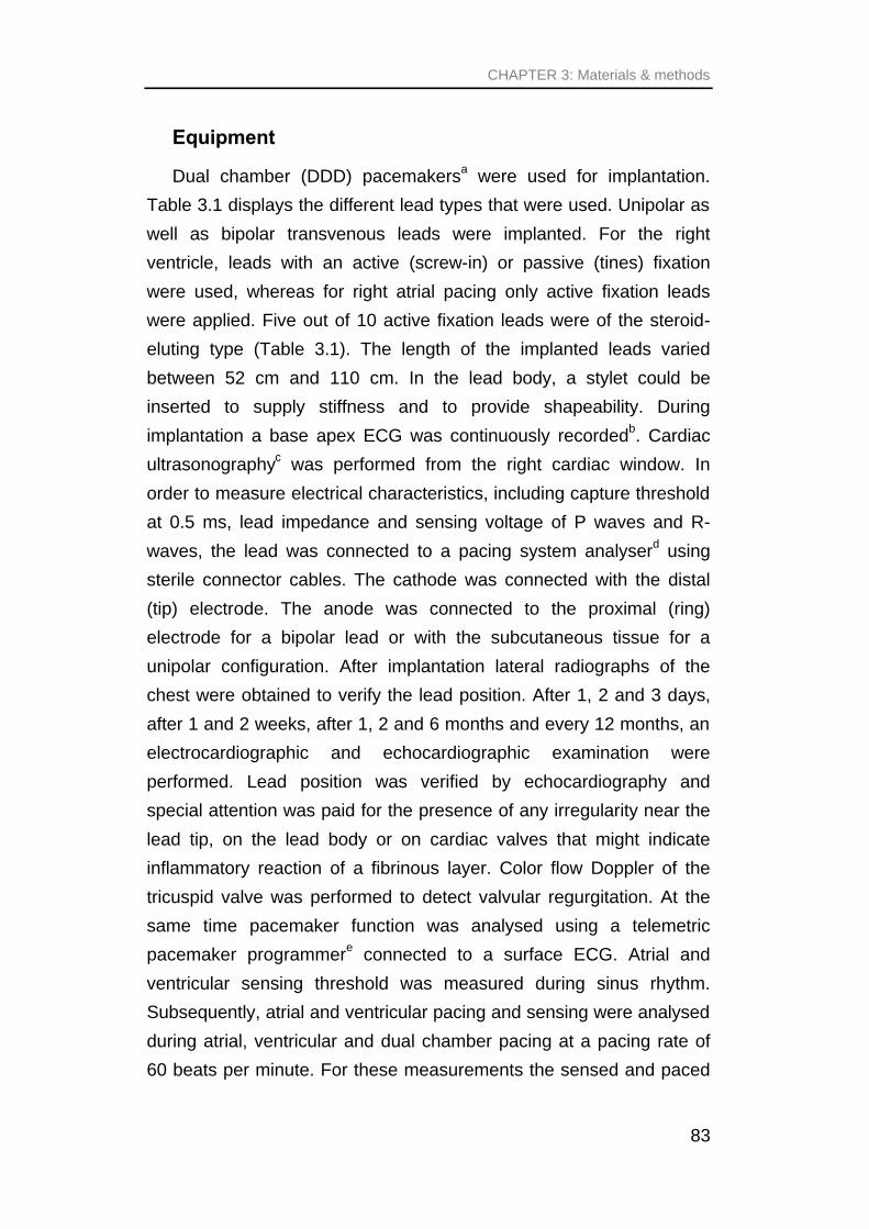
CHAPTER 3: Materials & methods
83
Equipment
Dual chamber (DDD) pacemakersa were used for implantation.
Table 3.1 displays the different lead types that were used. Unipolar as
well as bipolar transvenous leads were implanted. For the right
ventricle, leads with an active (screw-in) or passive (tines) fixation
were used, whereas for right atrial pacing only active fixation leads
were applied. Five out of 10 active fixation leads were of the steroid-
eluting type (Table 3.1). The length of the implanted leads varied
between 52 cm and 110 cm. In the lead body, a stylet could be
inserted to supply stiffness and to provide shapeability. During
implantation a base apex ECG was continuously recordedb. Cardiac
ultrasonographyc was performed from the right cardiac window. In
order to measure electrical characteristics, including capture threshold
at 0.5 ms, lead impedance and sensing voltage of P waves and R-
waves, the lead was connected to a pacing system analyserd using
sterile connector cables. The cathode was connected with the distal
(tip) electrode. The anode was connected to the proximal (ring)
electrode for a bipolar lead or with the subcutaneous tissue for a
unipolar configuration. After implantation lateral radiographs of the
chest were obtained to verify the lead position. After 1, 2 and 3 days,
after 1 and 2 weeks, after 1, 2 and 6 months and every 12 months, an
electrocardiographic and echocardiographic examination were
performed. Lead position was verified by echocardiography and
special attention was paid for the presence of any irregularity near the
lead tip, on the lead body or on cardiac valves that might indicate
inflammatory reaction of a fibrinous layer. Color flow Doppler of the
tricuspid valve was performed to detect valvular regurgitation. At the
same time pacemaker function was analysed using a telemetric
pacemaker programmere connected to a surface ECG. Atrial and
ventricular sensing threshold was measured during sinus rhythm.
Subsequently, atrial and ventricular pacing and sensing were analysed
during atrial, ventricular and dual chamber pacing at a pacing rate of
60 beats per minute. For these measurements the sensed and paced

Dual chamber pacemaker implantation via the cephalic vein in healthy equids
84
atrioventricular interval were programmed to 350 ms, the atrial and
ventricular blanking were 100 ms and 28 ms, respectively, the post-
ventricular atrial refractory period was 150 ms and the ventricular
refractory period was 300 ms. During the whole follow-up period
horses were kept in stalls and every two weeks they were given free
access to an arena. The pacemaker was intermittently active to
perform cardiac studies, including echocardiographic examinations
and blood pressure measurements. Except from horse 1, which
developed chronic laminitis, each horse completed treadmill exercise,
including performing at walk, trot, canter and gallop, with heart rates
up to 175 beats per minute. Horse 2, 3, 4, 5 and 6 completed 12, 4,
16, 12 and 6 treadmill sessions, respectively. Treadmill exercise and
free access to an arena started 2 months after implantation.
Implantation procedure
The animals were given 15 µg/kg detomidinef IV and 3 µg/kg
buprenorphineg IV and remained in standing position during the
implantation. The horses were placed in stocks to allow the surgeons
to sit in a safer position in front of the horse. If the horse showed any
discomfort during the implantation, 7 µg/kg detomidine was
administered IV. The ventral neck and pectoral region was clipped.
The region at the level of the lateral pectoral groove was surgically
prepared and was blocked with 2 per cent lidocaineh. An incision of
about 8 cm was made along the lateral pectoral groove and the
cephalic vein was exposed by blunt dissection. A suturei was placed
around both ends of the exposed vein but only the distal part of the
vessel was ligated, preventing blood from the leg entering the surgical
site. Between the 2 sutures, a partial venotomy was made (Fig. 3.1).
The vein lumen was opened with a vein lifter and the atrial lead, with a
straight stylet inserted, was introduced in cardiac direction. The lead
was advanced until it became ultrasonographically visible in the right
atrium. Subsequently, the ventricular lead, with a straight stylet
inserted, was also advanced in the cephalic vein. The lead became
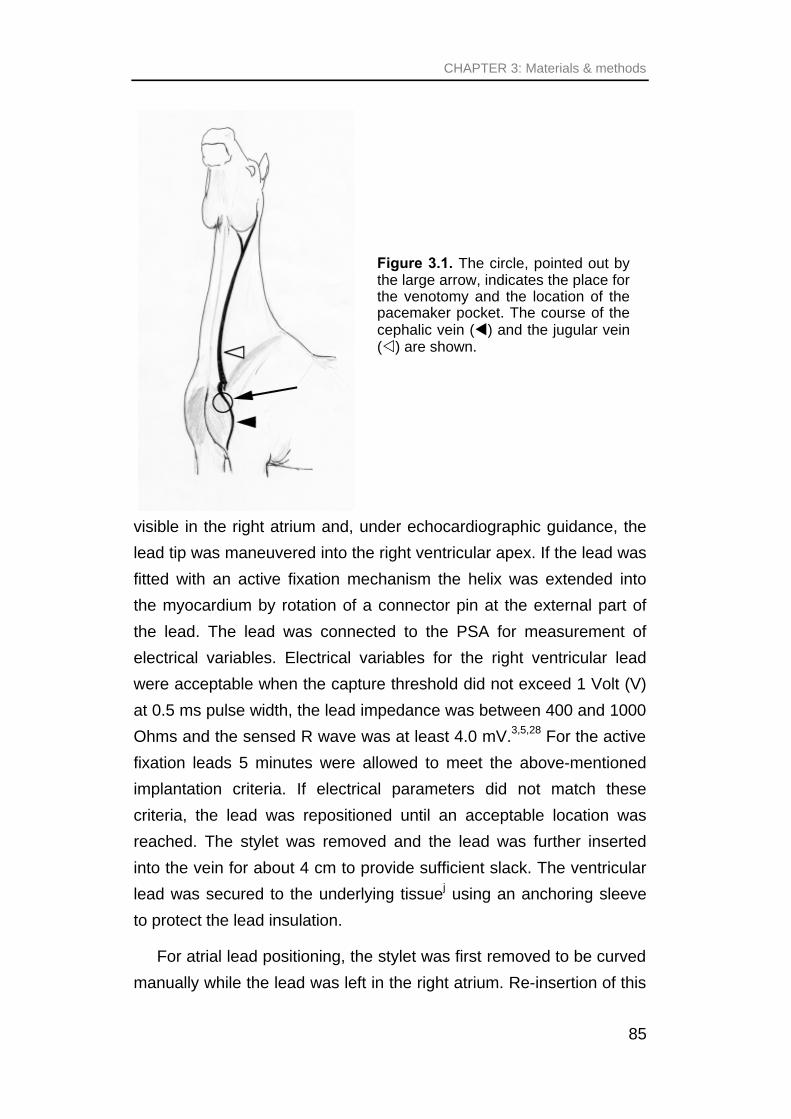
CHAPTER 3: Materials & methods
85
visible in the right atrium and, under echocardiographic guidance, the
lead tip was maneuvered into the right ventricular apex. If the lead was
fitted with an active fixation mechanism the helix was extended into
the myocardium by rotation of a connector pin at the external part of
the lead. The lead was connected to the PSA for measurement of
electrical variables. Electrical variables for the right ventricular lead
were acceptable when the capture threshold did not exceed 1 Volt (V)
at 0.5 ms pulse width, the lead impedance was between 400 and 1000
Ohms and the sensed R wave was at least 4.0 mV.3,5,28 For the active
fixation leads 5 minutes were allowed to meet the above-mentioned
implantation criteria. If electrical parameters did not match these
criteria, the lead was repositioned until an acceptable location was
reached. The stylet was removed and the lead was further inserted
into the vein for about 4 cm to provide sufficient slack. The ventricular
lead was secured to the underlying tissuej using an anchoring sleeve
to protect the lead insulation.
For atrial lead positioning, the stylet was first removed to be curved
manually while the lead was left in the right atrium. Re-insertion of this
Figure 3.1. The circle, pointed out by the large arrow, indicates the place for the venotomy and the location of the pacemaker pocket. The course of the cephalic vein (t) and the jugular vein (v) are shown.

Dual chamber pacemaker implantation via the cephalic vein in healthy equids
86
curved stylet provided a J shape to the lead and supplied body. After
connection of the PSA and under echocardiographic control, the lead
was slowly manipulated to obtain contact with the endocardium. Once
in the right position the helix of the active fixation mechanism was
extended into the endocardium and electrical parameters were
checked. A good atrial position was achieved when the capture
threshold at 0.5 ms did not exceed 1.5 V, lead resistance was between
400 and 1000 Ohms and P wave sensing was at least 1.5 mV.3,5,28
After withdrawal of the stylet, pacing was performed at 5 V and 0.5 ms
to check if no diaphragmatic stimulation occurred. If the electrical
characteristics did not match the implantation criteria or if
diaphragmatic stimulation occurred, the lead was repositioned. If the
lead was properly positioned, it was advanced slightly into the vein and
the proximal part was secured to the underlying tissue using the
anchoring sleevej. Gentle traction was applied to the ventricular and
the atrial lead to verify if lead migration occurred and electrical
variables were again examined. Subsequently the proximal part of the
cephalic vein was ligatedi. Between the lateral pectoral groove and the
manubrium sterni, a subcutaneous pacemaker pocket was created by
blunt dissection (Fig. 3.1). The pacemaker was connected to both
leads and inserted in the pocket taking care that no acute angulations
of the electrodes occurred. Redundant leads were looped along the
sides of the device or underneath it. The pocket was irrigated with
antibioticsk, and the subcutaneous tissue and skin were closed in a
routine manner. Flunixin megluminel (0.3 mg/kg IV q8h) and sodium
ceftiofurm (2 mg/kg IV q24h) were given during 5 days. Oral
trimethoprim sulphadiazin treatmentn (1 mg/kg trimetoprim and 5
mg/kg sulphadiazin PO q12h) was continued for 10 days. During this
recovery period, recumbency of the horse was prevented by attaching
the horse to the stable wall.
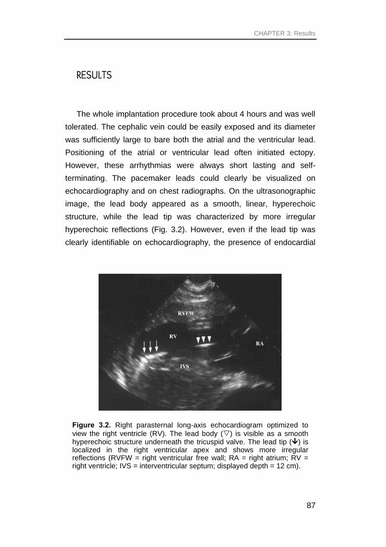
CHAPTER 3: Results
87
RESULTSRESULTS
The whole implantation procedure took about 4 hours and was well
tolerated. The cephalic vein could be easily exposed and its diameter
was sufficiently large to bare both the atrial and the ventricular lead.
Positioning of the atrial or ventricular lead often initiated ectopy.
However, these arrhythmias were always short lasting and self-
terminating. The pacemaker leads could clearly be visualized on
echocardiography and on chest radiographs. On the ultrasonographic
image, the lead body appeared as a smooth, linear, hyperechoic
structure, while the lead tip was characterized by more irregular
hyperechoic reflections (Fig. 3.2). However, even if the lead tip was
clearly identifiable on echocardiography, the presence of endocardial
Figure 3.2. Right parasternal long-axis echocardiogram optimized to view the right ventricle (RV). The lead body (s) is visible as a smooth hyperechoic structure underneath the tricuspid valve. The lead tip (Ü) is localized in the right ventricular apex and shows more irregular reflections (RVFW = right ventricular free wall; RA = right atrium; RV = right ventricle; IVS = interventricular septum; displayed depth = 12 cm).

Dual chamber pacemaker implantation via the cephalic vein in healthy equids
88
contact could not always be ascertained and had to be confirmed by
determination of electrical variables. During ventricular lead positioning
echocardiographic control was useful to follow the lead entering into
the right ventricular apex and to avoid it migrating towards the
pulmonary artery. With a straight stylet inserted, the lead easily
entered the right ventricular apex (Fig. 3.2) and an acceptable position
was readily accomplished. Atrial lead positioning was more difficult.
The atrial lead had to curve in the right atrium towards the atrial wall in
order to allow the helix to be extended into the myocardium. On
echocardiography the most dorsal parts of the right atrium could not
completely be imaged and the visualization of both the atrial and the
ventricular lead in the right atrium complicated ultrasonographic
guidance. Therefore, echocardiography served mainly to prevent atrial
lead fixation too close to the tricuspid valve and lead positioning was
performed relying mainly on the measurements of electrical variables.
Although satisfactory electrical characteristics were often readily
achieved after extension of the helix, withdrawal of the stylet
sometimes resulted in an immediate loss of capture or sensing,
indicating that active fixation of the lead had not been accomplished.
Several attempts were sometimes necessary to achieve active fixation
of the atrial lead. Furthermore diaphragmatic stimulation occasionally
occurred at 5 V and 0.5 ms, which also necessitated lead
repositioning. However, lead implantation criteria could always be met
(Table 3.2).
On post-operative radiographs the ventricular lead tip was visible in
the right ventricular apex in all animals. The atrial lead tip was
identifiable in the right atrium and was located in the cranial region of
the right atrium in pony 3 and in the horse, in the mid portion in pony 2
and in the caudal part in pony 1, 4 and 5.

CHAPTER 3: Results
89
Table 3.2. The electrical characteristics of the atrial and ventricular lead at the time of implantation and 6 months after the implantation, including the stimulation threshold at 0.5 ms pulse duration, shown in volts (V) and milli-ampere (mA), the lead impedance (Ohm, Ω) and the sensing amplitude (mV), are shown for each horse. (* indicates the values of the re-implanted lead)
atrial lead ventricular lead
horse time threshold (0.5 ms)
impedance
sensing
threshold (0.5 ms)
impedance
sensing
V mA
Ù mV V mA
Ù mV
implantation
0.6 1.5 410 2.1 0.5 1.1 440 11.2 1
6 months < 0.5 < 0.8 587 2.8 < 0.5 < 0.8 610 8
implantation
0.5 1.0 490 3.5 0.6 1.5 415 8.9 2
6 months 1 1.3 741 2.8 5.0 6.7 752 2
implantation
0.7 1.7 420 3.9 0.6 0.7 860 16.8 3
6 months 2 3.4 592 2 1.5 1.3 1116 15.7
implantation
0.7 1.6 440 2.6 1.0 1.6 620 7.8 4
6 months 0.5 0.7 741 4.2 0.5 0.6 791 16
implantation
0.5 0.8 405 7.2 0.3 0.6 520 16.1 5*
6 months 0.5 1.3 396 5.6 1.6 1.9 855 8.2
implantation
0.6 0.9 450 6 0.9 1.5 610 13.6 6*
6 months 2.5 3.9 634 10 < 0.5 < 0.6 784 8
In all animals the dual chamber pacemaker was successfully
implanted and atrial and ventricular sensing and pacing were all
achieved, allowing an effective atrial, ventricular and dual chamber
pacing function (Fig. 3.3). In the Thoroughbred horse (animal 6), at a
heart rate of 30 to 35 beats per minute, the normal intrinsic AV interval
was about 400 ms, which is within normal limits. Due to a maximal
programmable AV interval of the pacemaker of 350 ms, however, in
the dual chamber pacing mode ventricular pacing occurred at slow
heart rates although AV conduction in that horse was normal.

Dual chamber pacemaker implantation via the cephalic vein in healthy equids
90
All animals had a normal wound healing with formation of a small
seroma during the first days post-implantation. Ligation of the cephalic
vein caused no circulatory problems and the position of the pacemaker
did not impede equids in lying down or performing treadmill exercise.
In none of the horses was treadmill exercise associated with lead
dislodgement. Atrial lead dislodgement occurred 2 days after
implantation in pony 5 causing a loss of atrial capture and atrial
sensing. On echocardiography the lead tip was identified in the right
ventricle near the tricuspid valve and radiographs were taken to
confirm the lead displacement. The pacemaker pocket was reopened
and after retraction of the helix, the lead was removed and replaced by
a new one. In the horse the atrial lead dislodgement occurred 1 month
Figure 3.3. Print from the pacemaker programmer showing a surface ECG (1), calibrated at 0.1 mV/mm, a simultaneous intra-atrial and intraventricular ECG (2), calibrated at 1 mV/mm, and a marker channel (3) indicating atrial pacing (AP) and ventricular pacing (VP). The pacemaker is programmed at dual chamber pacing and sensing (DDD-mode) at 60 beats per minute with an atrioventricular interval of 350 ms. Each atrial and ventricular stimulus is followed by a P wave and QRS complex on the surface ECG, respectively, indicating successful dual chamber pacing

CHAPTER 3: Results
91
after implantation. At that time treadmill exercise had not been
performed. Atrial capture could no longer be obtained, even at 1.5 ms
and 7.5 V. Atrial sensing dropped from 8 mV on the previous day, to
1.4 mV on the day of dislodgement. On cardiac ultrasound the
dislodged lead tip was identifiable in the right atrium near the tricuspid
valve. The lead, which was of the steroid-eluting type, was withdrawn
and a new non-steroid-eluting lead was inserted in the contra-lateral
cephalic vein. After positioning and fixation of this new lead, the
proximal part was tunnelled subcutaneously and connected to the
pacemaker, restoring normal pacemaker function. Ventricular lead
dislodgement was never observed.
Long-term follow-up revealed no abnormalities on
echocardiographic examinations. The appearance and position of
each lead remained identical. The lead body surface remained smooth
and no irregularities were detected near the lead tip. The tricuspid
valve showed no valvular lesions or valvular regurgitation. In all
animals atrial and ventricular sensing remained possible and both
chambers could be successfully stimulated with acceptable threshold
values, indicating a good pacemaker function (Table 3.2). Arrhythmias,
other than occasional second-degree atrioventricular block, were not
seen. In the horse, diaphragmatic stimulation occasionally occurred
during atrial pacing. Pony 1 had to be euthanized 1 year after
implantation because of a chronic laminitis. Necropsy revealed that
both the atrial and the ventricular lead tip were closely attached to the
myocardium and were surrounded by a small amount of fibrous tissue.
There were no lesions on the tricuspid valve. A small fibrous layer was
present on the proximal intravascular part of the lead. A firm fibrous
capsule surrounded the pacemaker box.

Dual chamber pacemaker implantation via the cephalic vein in healthy equids
92
DISCUSSIONDISCUSSION
The described standardized technique for dual chamber
pacemaker implantation in horses proved to be efficient and safe. The
whole procedure could be performed in the standing horse and was
well tolerated. Successful sensing and pacing of the atrium as well as
the ventricle were obtained on long-term follow-up.
Transvenous implantation of a permanent pacemaker is the
preferred method in both human and small animal medicine.2,10 In
contrast to epicardial lead placement it is much less invasive, cheaper
and more rapidly accomplished. A transvenous approach avoids major
surgery and can be performed without general anesthesia,4,6 which is
an advantage in animals with compromised cardiovascular system in
which general anesthesia is associated with considerable risk of
adverse events29,30 The occurrence of a lethal ventricular fibrillation
during general anesthesia has been described in a horse during an
attempt to implant a pacemaker.31 In equids requiring pacemaker
implantation because of a symptomatic bradycardia, administration of
detomidine could worsen cardiac function because of its negative
chronotropic effect. In these animals, a temporary pacing catheter
should therefore be inserted into the right ventricular apex prior to
sedation, to allow pacing during the implantation procedure.
The only way to perform transvenous pacemaker implantation in
equids is to use a superficial vein that is located close to the heart and
that is large enough to accommodate 2 leads. The vein will be ligated.
Furthermore the pacemaker pocket has to be located close to the vein
and close to the heart to avoid the use of extremely long leads and be
implanted at a safe location in order to allow recumbency and
exercise. For these reasons, the cephalic vein at the level of the lateral
pectoral groove was chosen for lead insertion. The left as well as the
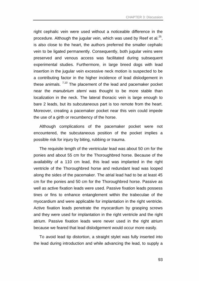
CHAPTER 3: Discussion
93
right cephalic vein were used without a noticeable difference in the
procedure. Although the jugular vein, which was used by Reef et al.25,
is also close to the heart, the authors preferred the smaller cephalic
vein to be ligated permanently. Consequently, both jugular veins were
preserved and venous access was facilitated during subsequent
experimental studies. Furthermore, in large breed dogs with lead
insertion in the jugular vein excessive neck motion is suspected to be
a contributing factor in the higher incidence of lead dislodgement in
these animals. 7,10 The placement of the lead and pacemaker pocket
near the manubrium sterni was thought to be more stable than
localization in the neck. The lateral thoracic vein is large enough to
bare 2 leads, but its subcutaneous part is too remote from the heart.
Moreover, creating a pacemaker pocket near this vein could impede
the use of a girth or recumbency of the horse.
Although complications of the pacemaker pocket were not
encountered, the subcutaneous position of the pocket implies a
possible risk for injury by biting, rubbing or trauma.
The requisite length of the ventricular lead was about 50 cm for the
ponies and about 55 cm for the Thoroughbred horse. Because of the
availability of a 110 cm lead, this lead was implanted in the right
ventricle of the Thoroughbred horse and redundant lead was looped
along the sides of the pacemaker. The atrial lead had to be at least 45
cm for the ponies and 50 cm for the Thoroughbred horse. Passive as
well as active fixation leads were used. Passive fixation leads possess
tines or fins to enhance entanglement within the trabeculae of the
myocardium and were applicable for implantation in the right ventricle.
Active fixation leads penetrate the myocardium by grasping screws
and they were used for implantation in the right ventricle and the right
atrium. Passive fixation leads were never used in the right atrium
because we feared that lead dislodgement would occur more easily.
To avoid lead tip distortion, a straight stylet was fully inserted into
the lead during introduction and while advancing the lead, to supply a

Dual chamber pacemaker implantation via the cephalic vein in healthy equids
94
degree of stiffness and shapeability to the lead. Echocardiography
facilitated atrial and ventricular lead positioning, but measurements of
electrical parameters were crucial for final adjustments during lead
placement because they allow selection of the optimal electrode
stimulation and sensing site.2 For the electrical measurements the
distal (tip) electrode of the lead was always connected with the
cathode, because cathodal stimulation is less likely to induce
arrhythmias.32 The same acceptance values for the electrical
parameters of the atrial and ventricular lead were taken as in human
medicine. Because the short- and long-term success of the pacing
system is related to the initial lead position, effort was expended to
obtain the best possible initial location in terms of both stability and
electrical performance.3 For the active fixation leads, 5 minutes were
sometimes allowed for the lead to meet the implantation criteria,
because it is common for capture thresholds to decrease significantly
shortly after active fixation.3,33
The ventricular lead was positioned first because it may supply
back-up pacing in animals with severe bradycardia and because it is
usually considered the most important of the leads.3 Ventricular lead
placement was performed with a straight stylet inserted in the lead
body to guide the lead tip straight toward the right ventricular apex.
Except from an occasional insertion of the tip between the right
ventricular wall and a papillary muscle or a migration towards the
pulmonary artery, the lead tip readily moved into the right ventricular
apex to remain in a stable position. The active as well as the passive
fixation leads remained well in place and lead dislodgement was never
encountered.
Atrial lead positioning was much more difficult. First because the
atrial lead had to bend with its tip toward the atrial endocardium by
using a curved stylet and second because a firm endocardial contact
had to be obtained with the active fixation mechanism. Even when
acceptable electrical parameters were obtained after extension of the

CHAPTER 3: Discussion
95
helix, atrial capture was sometimes lost when the stylet was
withdrawn. Possibly the helix only partially entered the myocardium, or
the lead tip was located parallel to the endocardium instead of
perpendicular, preventing the helix to enter the myocardium
sufficiently. Furthermore, the presence of large atrial trabeculae might
have interfered with the lead fixation. In contrast with the ventricular
lead, the exact anatomical position of the atrial lead in the right atrium
could not always be determined. However, any atrial position that
allows adequate pacing and sensing thresholds can be used as long
as no extracardiac stimulation occurs with pacing.5
Occasionally, diaphragmatic stimulation was observed during atrial
pacing at 5 V and 0.5 ms. Stimulation of the lateral, dorsal part of the
right atrium, at the level of the right diaphragmatic nerve, was likely to
be the cause.34 When diaphragmatic stimulation occurred the lead
was always repositioned.
Fluoroscopy is used during pacemaker implantation in humans and
small animals, and could have been helpful during the implantation in
horses. Especially during atrial lead placement, fluoroscopy might
have been useful to identify the typical to-and-fro motion of the lead tip
with atrial activity. However, ultrasonography proved to be sufficient
and is also used in human medicine when fluoroscopy is contra-
indicated, as in pregnant women.3,35-37 Post-operative
echocardiographic examinations are useful, not only to check lead
position but also to look for strands or vegetative lesions, because the
latter can often be found in cases of lead infection. 38-40
Once satisfactory lead position was obtained, the lead was
advanced slightly into the vein to impart a small bend and thereby
allow for movements of the neck and foreleg that might otherwise
exert traction on the lead.1,2,19 However, care was taken not to enter
the lead too far in the vein. Too large a loop may predispose to ectopy,
lead displacement or myocardial perforation at the tip.2,3 When
securing the outer part of the lead to the underlying tissue and when

Dual chamber pacemaker implantation via the cephalic vein in healthy equids
96
closing the pocket, special care was taken not to damage the lead
insulation.
After lead implantation, the tissue around the lead tip becomes
inflamed due to the presence of foreign material in the body and due
to the pressure of the lead-electrode system in contact with the
myocardium.2,41 With time the inflammatory reaction subsides leaving
a thin capsule of fibrous tissue between the electrode and the active
myocardium supplying an extra fixation of the lead tip to the
myocardium.41 However, the inflammatory reaction can be excessive,
causing the capture threshold to rise, sometimes above the output of
the pacemaker, causing exit block. In this context the use of a steroid-
eluting electrode may offer benefits in terms of subacute and chronic
thresholds.3 Such an electrode elutes small amounts of
dexamethasone from its tip which attenuates the local inflammatory
reaction and which reduces the thickness of the fibrous capsule that
surrounds the electrode, lowering the chronic threshold for
stimulation.3,33 This study suggests that lower chronic threshold values
are observed with the steroid-eluting leads (Table 3.2).
The main complication of the transvenous implantation technique is
a lead displacement during the early postoperative period 4,6,7,10 as it
was seen in pony 5. Therefore exercise should be restricted during the
first weeks after implantation because at that time a fibrous connection
of the lead tip is not yet achieved.4,6 In the horse lead dislodgement
occurred 1 month after implantation. Such a late dislodgement was
probably related to the animal's movement and was caused by
inadequate fixation of the anchoring sleeve to the lead or to the
surrounding tissue, insufficient slack, or a poor fixation of the lead tip
to the endocardium.41 The latter could have been influenced by the
use of the steroid-eluting electrode in this horse because the elution of
dexamethasone could have reduced the thickness of the fibrous
capsule surrounding the lead tip.

CHAPTER 3: Discussion
97
The maximal programmable AV interval for the pacemaker was
limited to 350 ms. In a dual chamber pacing mode, this means that, if
the pacemaker hasn't sensed a ventricular depolarization within 350
ms following an atrial event, a ventricular stimulus will be delivered. At
slow heart rates, horses with a normal AV conduction can present a
PR interval of more than 350 ms. This implies that, even in the
presence of a normal AV conduction, dual chamber pacing can give
rise to unnecessary ventricular pacing due to the restricted
programmable AV interval. When the heart rate increases slightly,
however, PR interval reduces to below 350 ms and inappropriate
ventricular pacing ceases.
We can conclude that the implantation of a dual chamber
pacemaker using endocardial leads via the cephalic vein is feasible
and safe in healthy equids and can be performed on the standing
animal. The advantage of the described method is that the cephalic
venous access and the subcutaneous implantation of the pacemaker
near the manubrium sterni require only minor surgery and can be
performed under local anesthesia, avoiding a general anesthesia. The
cephalic venous approach preserves both jugular veins, facilitating
experimental studies after pacemaker implantation. The subcutaneous
position of the pacemaker permits post-operative reprogramming. No
major complications were encountered in this study and the implanted
pacemakers did not impede the ability of the horse and ponies to lie
down, nor to perform treadmill exercise. Limitations of the technique
included the lack of fluoroscopy and the limited echocardiographic
visualization of the high right atrium, which hampered atrial lead
positioning. Implantations were performed in healthy animals and
further studies are required to investigate the applicability of the
technique in clinical patients who would need temporary pacing prior to
sedation. The use of transvenous endocardial leads can imply an
increased risk for lead dislodgement. Therefore regular examination of
pacemaker function should be performed in a horse with symptomatic
bradycardia, especially if the animal is intended to perform exercise.

Dual chamber pacemaker implantation via the cephalic vein in healthy equids
98
Although no problems were encountered with pacemaker pockets in
this study, the subcutaneously positioned pocket could be injured by
external trauma. Besides the implantation of a pacemaker in animals
with extreme bradycardia, this relatively simple technique also
provides an excellent tool to collect information about cardiac
arrhythmias and electrophysiological characteristics of the equine
heart. In combination with various drugs, the influence of these drugs
on electrophysiological properties of the heart could be studied.
Furthermore, arrhythmias can be artificially induced which makes
research about their effect on heart function and about the application
of a feasible therapy possible.
Acknowledgements
The authors wish to thank the Special Research Fund, Ghent
University, for financial support.
Footnotes a Thera D(R), Medtronic, Minneapolis, MN b Cardiolife TEC-7511K, Nihon Kohden, Tokyo, Japan c Vingmed CFM 800 SV, GE Vingmed, Horten, Norway d PSA Model 5309, Medtronic, Minneapolis, MN e Programmer 9790®, Medtronic, Minneapolis, MN f Domosedan®, Pfizer Animal Health, Nossegem, Belgium g Temgesic®, Schering-Plough, Brussels, Belgium h Xylocaine 2%®, Astra Pharmaceuticals, Brussels, Belgium i Vicryl 4/1, Johnson & Johnson, Dilbeek, Belgium j Mersutures 4/1, Johnson & Johnson, Dilbeek, Belgium k Kanacillin Trifortis Vet®, Continental Pharma, Brussels, Belgium l Finadyne®, Shering-Plough, Brussels, Belgium m Excenel®, Upjohn, Puurs, Belgium n Tribrissen Oral Paste, Mallinckrodt Veterinary, Brussels, Belgium
CHAPTER 3: References
99
REFERENCESREFERENCES
1. Klement P, Del-Nido PJ, Wilson GJ. The use of cardiac pacemakers in veterinary practice. Comp Cont Educ Pract 1984; 6: 893-902.
2. Kittleson MD, Kienle RD. Small animal cardiovascular medicine. St. Louis, MO: Mosby; 1998; 525-539.
3. Brinker J, Midei M. Techniques of pacemaker implantation. In: Ellenbogen KA, ed. Cardiac pacing. Abingdon, UK: Blackwell Science; 1996: 216-277.
4. Darke PGG. Update: cardiac pacing. In: Kirk RW, Bonagura JD, ed. Current Veterinary Therapy. Philadelphia, PA: WB Saunders; 1992: 708-713.
5. Hayes DL, Osborn MJ. Pacing. Antibradycardia devices. In: Guiliani ER, Gersh BJ, McGoon MD, et al., ed. Mayo Clinic Practice of Cardiology. St. Louis, MO: Mosby; 1996: 909-976.
6. Sisson DD. Bradyarrhythmias and cardiac pacing. In: Kirk RW, ed. Current Veterinary Therapy. Philadelphia, PA: WB Saunders; 1989: 286-294.
7. Sisson D, Thomas WP, Woodfield J, Pion PD, Luethy M, DeLellis LA. Permanent transvenous pacemaker implantation in forty dogs. J Vet Intern Med 1991; 130: 322-331.
8. Roberts DH, Tennant B, Brockman D, Bellchambers S, Charles RG. Successful use of a QT-sensing rate-adaptive pacemaker in a dog. Vet Rec 1992; 130: 471-472.
9. Neu H, Mulch J. Subcutaneous cardiac pacemaker implantation on the left side of the neck in the dog: indications, technique and clinical experiences. Kleintierpraxis 1994; 39: 211-226.
10. Flanders JA, Moise NS, Gelzer AR, Waskiewicz JC, MacGregor JM. Introduction of an endocardial pacing lead through the costocervical vein in six dogs. J Am Vet Med Assoc 1999; 215: 46-48.
11. Liddicoat JR, Klein JR, Reddy VM, Klautz RJ, Teitel DF, Hanley FL. Hemodynamic effects of chronic prenatal ventricular pacing for the treatment of complete atrioventricular block. Circulation 1997; 96: 1025-1030.
12. Willems R, Ector H, Heidbüchel H. Slow development of atrial fibrillation in a transvenously paced sheep model. In:

Dual chamber pacemaker implantation via the cephalic vein in healthy equids
100
Ovsyshcher E, ed. Abstract book: Cardiac Arrhythmias And Device Therapy: Results and Perspectives for the New Century. Armonk, NY: Futura Publishing Company; 2000: 95-99.
13. Wijffels MC, Kirchhof CJ, Dorland R, Allessie MA. Atrial fibrillation begets atrial fibrillation. A study in awake chronically instrumented goats. Circulation 1995; 92: 1954-1968.
14. Ausma J, Wijffels M, Thone F, Wouters L, Allessie M, Borgers M. Structural changes of atrial myocardium due to sustained atrial fibrillation in the goat. Circulation 1997; 96: 3157-3163.
15. van der Velden HM, Ausma J, Rook MB, Hellemons AJ, van Veen TA, Allessie MA, Jongsma H. Gap junctional remodeling in relation to stabilization of atrial fibrillation in the goat. Cardiovasc Res 2000; 46: 476-486.
16. Li D, Benardeau A, Nattel S. Contrasting efficacy of dofetilide in differing experimental models of atrial fibrillation. Circulation 2000; 102: 104-112.
17. Klinge R, Hystad M, Kjekshus J, Karlberg BE, Djoseland O, Aakvaag A, Hall C. An experimental study of cardiac natriuretic peptides as markers of development of congestive heart failure. Scand J Clin Lab Invest 1998; 58: 683-691.
18. Qi W, Kjekshus H, Klinge R, Kjekshus JK, Hall C. Cardiac natriuretic peptides and continuously monitored atrial pressures during chronic rapid pacing in pigs. Acta Physiol Scand 2000; 169: 95-102.
19. Bellenger CR, Ilkiw JE, Nicholson AI, et al. Transvenous pacemaker leads in the dog: an experimental study. Res Vet Sci 1990; 49: 211-215.
20. O'Brien PJ, Ianuzzo CD, Moe GW, et al. Rapid ventricular pacing of dogs to heart failure: biochemical and physiological studies. Can J Physiol Pharmacol 1990; 68: 34-39.
21. Burkett DE, Seymour AA, Knight DH, et al. Myocardial effects of repetitive episodes of rapid ventricular pacing in conscious dogs: surgical creation, echocardiographic evaluation, and morphometric analysis. Lab Anim Sci 1994; 44: 453-461.
22. Allworth MS, Church DB, Maddison JE, et al. Effect of enalapril in dogs with pacing-induced heart failure. Am J Vet Res 1995; 56: 85-94.
23. Morillo CA, Klein GJ, Jones DL, et al. Chronic rapid atrial pacing. Structural, functional and electrophysiological characteristics of a new model of sustained atrial fibrillation. Circulation 1995; 91: 1588-1595.

CHAPTER 3: References
101
24. Yue L, Feng J, Gaspo R, Li GR, Wang Z, Nattel S. Ionic remodeling underlying action potential changes in a canine model of atrial fibrillation. Circ Res 1997; 81: 512-525.
25. Reef VB, Clark ES, Oliver JA, et al. Implantation of a permanent transvenous pacing catheter in a horse with complete heart block and syncope. J Am Vet Med Assoc 1986; 189: 449-452.
26. Hamir AN, Reef VB. Complications of a permanent transvenous pacing catheter in a horse. J Comp Path 1989; 101: 317-326.
27. Pibarot P, Vrins A, Salmon Y, et al. Implantation of a programmable atrioventricular pacemaker in a donkey with complete atrioventricular block and syncope. Equine Vet J 1993; 25: 248-251.
28. Holmes DR, Hayes DL. Pacemaker implantation techniques. In: Saksena S, Goldschlaber N, ed. Electrical therapy of cardiac arrhythmia. London, UK: WB Saunders; 1990: 173-190.
29. Bonagura JD, Helphrey ML, Muir WW. Complications associated with permanent pacemaker implantation in the dog. J Am Vet Med Assoc 1983; 182: 149-155.
30. Tilley LP. Essentials of canine and feline electrocardiography. Philadelphia, PA: Lea & Febiger; 1992, 365-382.
31. Brown CM. ECG of the month. J Am Vet Med Assoc 1979; 175: 1076-1077.
32. Furman S, Hurzeler P, Mehra R. Cardiac pacing and pacemakers. IV Threshold of cardiac stimulation. Am Heart J 1977; 94: 115-124.
33. Kay GM. Basic concepts of pacing. In: Ellenbogen KA, ed. Cardiac pacing. Abingdon, UK: Blackwell Science; 1996: 37-123.
34. Schoenfeld MH. Follow-up of the pacemaker patient. In: Ellenbogen KA, ed. Cardiac pacing. Abingdon, UK: Blackwell Science; 1996: 456-498.
35. Gudal M, Kervancioglu C, Oral D, et al. Permanent pacemaker implantation in a pregnant woman with guidance of ECG and two-dimensional echocardiography. PACE 1987; 10: 543-545.
36. Jordaens LJ, Vandenbogaerde JF, VandeBruquene P, DeBuyzere M. Transoesophageal echocardiography for insertion of a physiological pacemaker in early pregnancy. PACE 1990; 13: 955-957.
37. Leclercq F, Hager FX, Macia JC, Mariottini CJ, Pasquie JL, Grolleau R. Left ventricular lead insertion using a modified transseptal catheterization technique: A totally endocardiological
Dual chamber pacemaker implantation via the cephalic vein in healthy equids
102
approach for permanent biventricular pacing in end-stage heart failure. PACE 1999; 22: 1570-1575.
38. Victor F, De Place C, Camus C, Le Breton H, Leclercq C, Pavin D, Mabo P, Daubert C. Pacemaker lead infection: echocardiographic features, management, and outcome. Heart 1999; 81: 82-87.
39. Voet JG, Vandekerckhove YR, Muyldermans LL, Missault LH, Matthys LJ. Pacemaker lead infection: report of three cases and review of the literature. Heart 1999; 81: 88-91.
40. Tan HH, Ling LH, Ng WL, Cheng A. Diagnosis of pacemaker lead infection using transoesophageal echocardiography: a case report. Ann Acad Med Singapore 2000; 29: 97-100.
41. Levine PA. Differential diagnosis, evaluation, and management of pacing system malfunction. In: Ellenbogen KA, ed. Cardiac pacing. Abingdon, UK: Blackwell Science; 1996: 333-429.

CHAPTER 44
Dual chamber rateDual chamber rate--adaptive pacemaker adaptive pacemaker
implantation in a horse with suspected sick implantation in a horse with suspected sick
sinus syndromesinus syndrome
G. van Loon1, W. Fonteyne2, H. Rottiers1, R. Tavernier2, P.
Deprez1
1 Department of Large Animal Internal Medicine, Faculty of Veterinary
Medicine, Ghent University, Salisburylaan 133, B-9820 Merelbeke,
Belgium
2 Department of Cardiology, University Hospital, Ghent University, De
Pintelaan 185, B-9000 Ghent, Belgium
Adapted from: van Loon G., Fonteyne W., Rottiers H., Tavernier R. &
Deprez P. (2001). Dual chamber rate-adaptive
pacemaker implantation in a horse with suspected
sick sinus syndrome. Vet Rec, submitted.


CHAPTER 4: Summary
105
SUMMARYSUMMARY
A 5-year-old gelding presented with syncope at termination of
exercise. A 24-hour ECG recording revealed intermittent pauses
in the sinus rhythm of up to 10 seconds indicating sinus node
disease. Especially at termination of exercise, pauses in sinus
rhythm were repeatedly present. A dual chamber rate-adaptive
pacemaker was successfully implanted. Rate-adaptive pacing
based on a build-in activity sensor, prevented excessive post-
exercise bradycardia and syncope, allowing the horse to return to
work.

Dual chamber rate-adaptive pacemaker implantation in a horse with suspected SSS
106
INTRODUCTIONINTRODUCTION
Cardiac pacing is the treatment of choice for many symptomatic
bradycardias due to high-degree atrioventricular (AV) block, sick sinus
syndrome (SSS) or persistent atrial standstill (Sisson 1989, Sisson and
others 1991, Darke 1992, Roberts and others 1992, Neu and Mulch
1994, Kittleson and Kienle 1998, Flanders and others 1999). The
implantation of a pacemaker is a relative safe and simple procedure,
commonly used, not only in human clinical practice, but also in small
animals (Klement and others 1984, Darke 1992, Flanders and others
1999). One of the most important technologic advances in implantation
technique has been the development of endocardial leads that can be
introduced into the heart from a peripheral vein. Single chamber and
dual chamber pacemaker models are available with a variety of
programmable pacing modalities. Current pacemakers can vary their
pacing rate in response to changes in activity. These types are called
rate-adaptive, rate-modulated or sensor-driven pacemakers and are
often used in patients with sick sinus syndrome (Barold and Zipes
1992).
In equines, little information is available concerning therapeutic
pacemaker implantation. In 1986, Reef et al. performed a successful
transvenous implantation of a single chamber model in a horse
suffering from third-degree AV block. Sixteen months after the
implantation, this model was upgraded to a dual chamber model and
continued to function for another 2 years. In 1993, Pibarot et al.
implanted a dual chamber pacemaker, using epicardial leads, in a
donkey with complete heart block. To the best of our knowledge, no
data are available in literature related to the applicability of rate-
responsive pacing in equines for symptomatic sinus node dysfunction.
We report our experience with the transvenous implantation of a rate-

CHAPTER 4: Introduction
107
adaptive dual chamber pacemaker in a horse with syncope due to sick
sinus syndrome.
Dual chamber rate-adaptive pacemaker implantation in a horse with suspected SSS
108
CASE REPORTCASE REPORT
History and clinical findings
A 5-year-old gelding was used for dressage and for recreation.
During a ride, shortly after a gallop, the horse suddenly collapsed.
After a few seconds, the horse was able to stand up again, but still
presented a weak gait. The horse was immediately transferred to the
Faculty of Veterinary Medicine, Ghent University. At admission, the
horse showed a normal behaviour and no abnormalities were seen at
walk. During the clinical examination the pulse rate was 22 beats per
min (bpm) and slightly irregular. A grade 2/6 holodiastolic murmur was
detected over the aortic valve region. Respiratory rate and lung
auscultation were normal. No abnormalities were found on
neurological examination. Complete blood count and biochemical
profile were normal.
Electrocardiography and cardiac ultrasound
An electrocardiogram showed a heart rate of 24 beats per minute
with a sinus arrhythmia and an occasional second degree AV block.
An echocardiogram, including M-mode and colour flow Doppler
showed mild aortic regurgitation. Left ventricular diameter was normal
and fractional shortening was 33%. During lunging, the horses’ heart
rate increased but immediately after exercise, heart rate dropped very
quickly to below 35 bpm and on several occasions pauses in sinus
rhythm up to 7 seconds were detected, without a ventricular escape
beat occurring. An incremental exercise test on a treadmill revealed a
maximal heart rate of 183 bpm. A 24-hour ECG showed sinus
bradycardia and occasional second degree AV block. Several
episodes of sinus arrest or sinus exit block up to 10 seconds in
duration were observed without any ventricular escape beat (Fig. 4.1).
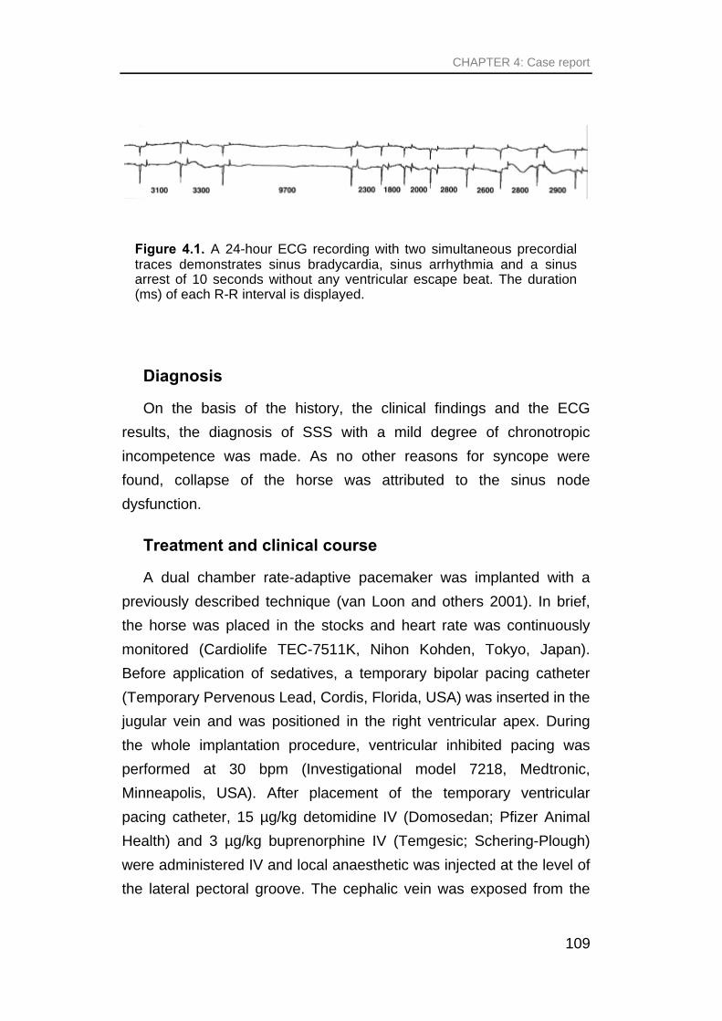
CHAPTER 4: Case report
109
Diagnosis
On the basis of the history, the clinical findings and the ECG
results, the diagnosis of SSS with a mild degree of chronotropic
incompetence was made. As no other reasons for syncope were
found, collapse of the horse was attributed to the sinus node
dysfunction.
Treatment and clinical course
A dual chamber rate-adaptive pacemaker was implanted with a
previously described technique (van Loon and others 2001). In brief,
the horse was placed in the stocks and heart rate was continuously
monitored (Cardiolife TEC-7511K, Nihon Kohden, Tokyo, Japan).
Before application of sedatives, a temporary bipolar pacing catheter
(Temporary Pervenous Lead, Cordis, Florida, USA) was inserted in the
jugular vein and was positioned in the right ventricular apex. During
the whole implantation procedure, ventricular inhibited pacing was
performed at 30 bpm (Investigational model 7218, Medtronic,
Minneapolis, USA). After placement of the temporary ventricular
pacing catheter, 15 µg/kg detomidine IV (Domosedan; Pfizer Animal
Health) and 3 µg/kg buprenorphine IV (Temgesic; Schering-Plough)
were administered IV and local anaesthetic was injected at the level of
the lateral pectoral groove. The cephalic vein was exposed from the
Figure 4.1. A 24-hour ECG recording with two simultaneous precordial traces demonstrates sinus bradycardia, sinus arrhythmia and a sinus arrest of 10 seconds without any ventricular escape beat. The duration (ms) of each R-R interval is displayed.

Dual chamber rate-adaptive pacemaker implantation in a horse with suspected SSS
110
surrounding tissue and through a venotomy 2 endocardial bipolar
screw-in leads were inserted. Guided by echocardiography from the
right hemithorax (Vingmed CFM 800 SV, GE Vingmed, Horten,
Norway) and by the intracardiac electrogram, the ventricular
(CapsureFix 4068 - 110cm, steroid eluting, Medtronic, Minneapolis,
USA) and the atrial active fixation lead (Stela BS45, bipolar, screw-in,
atrial endocardial lead, 52 cm, Ela Medical, France) were properly
positioned and the screw was extended into the endomyocardium. For
the ventricular lead, the threshold for stimulation was 0.6 V at a pulse
width of 0.5 ms with an impedance of 600 Ohm at 5 V. The ventricular
intracardiac signal was sensed at 6.2 mV. The atrial lead presented a
threshold of 0.4 V at 0.5 ms and an impedance of 620 Ohm at 5 V,
with a sensing voltage of 5.5 mV. The proximal part of both leads was
ligated to the surrounding tissue and connected with the pacemaker,
positioned in a subcutaneous pacemaker pocket between the lateral
pectoral groove and the manubrium sterni. The temporary pacing
catheter was disconnected and removed. With a lower rate of 30 bpm,
the pacemaker was programmed in the DDI mode, which indicates
dual chamber pacing, dual chamber sensing and inhibition of pacing
upon atrial or ventricular sensing.
After the implantation, radiography and fluoroscopy confirmed the
lead position. The horse remained at rest and was not allowed to lie
down for 2 weeks following the implantation. Flunixin meglumine (0.3
mg/kg IV q8h; Finadyne; Shering-Plough) and sodium ceftiofur (2
mg/kg IV q24h; Excenel; Upjohn) were given during 5 days. On the 5th
day the horse presented a temperature of 39.2 °C and sodium
ceftiofur was continued for another 5 days. Threshold and sensing
values were closely monitored. Between 2 and 3 weeks after the
implantation, the horse showed an intermittent fever ranging from 38.1
to 38°6. Appetite remained normal and no abnormalities were seen at
the surgical site or on cardiac ultrasound. During this period however,
atrial threshold increased up to 7.5 V at 0.5 ms. The fever was thought
to be caused by an inflammatory reaction at the lead tip and 1 mg/kg

CHAPTER 4: Case report
111
oral prednisolone therapy was started (daily for 5 days and every other
day for 2 weeks). Fever disappeared and atrial threshold values
normalized.
Two months after the implantation, the rate adaptive function of the
pacemaker was activated by programming the DDIR mode (Fig. 4.2).
Exercise tests at walk, trot and canter were performed, first on a
treadmill, afterwards while the horse was ridden by the owner, to
search for programming values emulating as close as possible a
normal heart rate response both at rest and during exercise. The
lowest heart rate was set at 30 bpm and the paced AV interval was
programmed at 300 ms. This implicated that the upper sensor-driven
heart rate of the pacemaker was 150 bpm. A high threshold for rate
adaptation and a low rate response produced the best results. An
acceleration time of 1 minute was chosen, which means that, after
sensor activation, a slow increase in rate occurs over 1 minute of time.
A deceleration time of 2.5 min was selected, which resulted in a
gradual decrease in heart rate over 2.5 min after cessation of sensor
Figure 4.2. Surface ECG (1) and marker channel (2) obtained while the pacemaker is in DDIR mode with a paced AV interval of 350 ms. The first complex is still a native beat and atrial (AS) and ventricular sensing (VS) occur. Because sinus node activity decreases, atrial pacing (AP) occurs in the second beat but, as normal AV conduction appears within 350 ms, no ventricular pacing occurs. Because of the slow native heart rate atrial pacing continues in the third and fourth complex, but as the AV conduction slows, no ventricular depolarisation occurs within the paced AV interval of 350 ms and a ventricular stimulus (VP) is delivered.
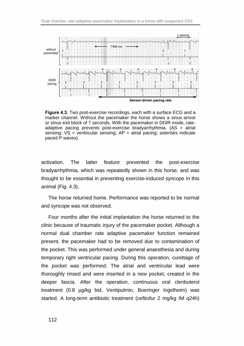
Dual chamber rate-adaptive pacemaker implantation in a horse with suspected SSS
112
activation. The latter feature prevented the post-exercise
bradyarrhythmia, which was repeatedly shown in this horse, and was
thought to be essential in preventing exercise-induced syncope in this
animal (Fig. 4.3).
The horse returned home. Performance was reported to be normal
and syncope was not observed.
Four months after the initial implantation the horse returned to the
clinic because of traumatic injury of the pacemaker pocket. Although a
normal dual chamber rate adaptive pacemaker function remained
present, the pacemaker had to be removed due to contamination of
the pocket. This was performed under general anaesthesia and during
temporary right ventricular pacing. During this operation, curettage of
the pocket was performed. The atrial and ventricular lead were
thoroughly rinsed and were inserted in a new pocket, created in the
deeper fascia. After the operation, continuous oral clenbuterol
treatment (0.8 µg/kg bid, Ventipulmin, Boeringer Ingelheim) was
started. A long-term antibiotic treatment (ceftiofur 2 mg/kg IM q24h)
Figure 4.3. Two post-exercise recordings, each with a surface ECG and a marker channel. Without the pacemaker the horse shows a sinus arrest or sinus exit block of 7 seconds. With the pacemaker in DDIR mode, rate-adaptive pacing prevents post-exercise bradyarrhythmia. (AS = atrial sensing; VS = ventricular sensing; AP = atrial pacing; asterisks indicate paced P waves).

CHAPTER 4: Case report
113
was given in order to try to resolve pocket and lead infection and to
avoid the occurrence of endocarditis. After 4 weeks however,
fistulation of the pocket recurred. After sedation and analgesia, during
temporary ventricular pacing, the pocket was opened. After
unscrewing of the lead helix, both leads could be removed with gentle
traction. Clenbuterol treatment was continued until, four weeks later,
after wound healing, a reimplantation was performed on the contra-
lateral side. Electrical characteristics for the atrial lead (Stelid BS45D
52 cm, bipolar, ELA Medical, France) were a threshold of 1.0 V at 0.5
ms with an impedance of 438 Ohm at 5 V, and a sensing voltage of
5.2 mV. For the ventricular lead (Sweet Tip Rx, 4245-59 cm, bipolar,
CPI, Guidant, St.-Paul, USA), a threshold of 1.6 V at 0.5 ms, a lead
impedance of 346 Ohm and a ventricular sensing voltage of 3.2 mV.
The pacemaker pocket was created slightly more dorsal, underneath
the m. cutaneus colli. The horse presented a normal recovery period
and returned to his normal work 2 months post-implantation. At the
time of submission of this paper, 4 months after the second
implantation, the owner reported no complaints and successful dual
chamber rate-adaptive pacemaker function remained present.

Dual chamber rate-adaptive pacemaker implantation in a horse with suspected SSS
114
DISCUSSIONDISCUSSION
A syncopal episode is defined as a sudden temporary loss of
consciousness associated with a deficit of postural tone with
spontaneous recovery. Syncope in horses is uncommon and therefore
has been virtually unstudied (Crisman 1998).
Sick sinus syndrome (SSS) is a term given to a number of
abnormalities of the sinoatrial (SA) node, including persistent
spontaneous sinus bradycardia, sinus arrest or SA exit block,
combinations of SA and AV conduction disturbances or alternation of
paroxysms of atrial tachyarrhythmias and periods of slow atrial and
ventricular rates (bradycardia and tachycardia syndrome) (Kapoor
1992, Zipes 1992). Little is known about sinus node disease in horses
and its relation to syncope.
In this horse, periods of sinus arrest or sinus exit block up to 10
seconds in duration were seen at rest. Because long periods of sinus
arrest were repeatedly demonstrated in the post-exercise period and
because the horse had presented syncope at termination of exercise,
this bradyarrhythmia was the most likely explanation for the syncopal
episode. In humans, postexertional asystole is known to cause
syncope (Hirata and others 1987, Huycke and others 1987, Kapoor
1989, O'Connor and others 1999, Crisafulli and others 2000). SSS in
human patients generally requires permanent pacemaker implantation
and represents the most common indication for pacemaker
implantation (Barold and Zipes 1992, Ellenbogen and Peters 1996). In
human patients with SSS, it has been proven that atrial based pacing
systems (single chamber or dual chamber) are superior to ventricular
based pacing (Barold and Zipes 1992, Benditt and others 1995) and
this modality was applied in the horse. However, as atrial lead
dislodgement represents a potential hazard of endocardial leads

CHAPTER 4: Discussion
115
(Sisson 1989, Sisson and others 1991, Darke 1992, Flanders and
others 1999), a dual chamber system was implanted. The ventricular
lead in this system was considered as an extra safety factor because it
would preserve a normal ventricular rate at any time, even if the atrial
lead would be dislodged. The use of a rate-adaptive pacing system
further expands the treatment possibilities. The described pacemaker
was fitted with a sensor, a piezoelectric ceramic crystal, which detects
mechanical vibration of the pacemaker box induced by body
movements or muscle contractions. Upon sensor activation, pacing
rate is gradually increased to a maximal level. At termination of
activity, the pacing rate is slowly decreased. Especially the gradual
decrease in heart rate was thought to be of importance in this horse to
avoid symptomatic bradycardia at termination of exercise, allowing the
horse to be ridden again.
The pacemaker pocket was created at the level of the pectoral
muscles. This implies that pacemaker vibrations will easily occur even
with slight body movements. Therefore, by programming the threshold
for rate adaptation at the highest value, the rate adaptive function was
set to react slowly and only after vigorous pacemaker vibration,
allowing only large sensor signals to increase heart rate. Furthermore,
the rate response, which determines the extent to which an increase in
the patient’s activity raises the pacing rate, was set to its lowest value.
A long acceleration time of 1 minute was chosen and the deceleration
time was set at 2.5 min. These values resulted in the best rate-
adaptive pacing results and avoided excessive pacing, especially at
low activities.
The subcutaneous position of the pacemaker pocket implies that it
is vulnerable to external trauma. However, deeper localisation of the
pacemaker implies a greater distance between the pulse generator
and the programming instrument and may prevent post-operative
programming (Fox and others 1986). During the second implantation,
the pacemaker pocket was created slightly more dorsal and
Dual chamber rate-adaptive pacemaker implantation in a horse with suspected SSS
116
underneath the m. cutaneus colli, to obtain a better protection against
traumatic injury but still allow telemetric programming.
Due to pocket laceration, the pacing system is contaminated. In an
ultimate attempt to preserve the leads in the horse, extensive
debridement of the pocket and long-term antibiotic treatment were
implemented. Although this approach has been described in humans
(Hurst and others 1986) and dogs (Sisson and others 1991), antibiotic
treatment alone rarely is sufficient to cure infection, and removal of the
generator and the leads is usually indicated (Morgan and others 1979).
In such patients, a new system may be placed at the time of removal
of the infected hardware or a two-step approach may be taken with
temporary pacing bridging the time between explantation and the new
implant (Lewis and others 1985). However, as SSS in the horse was
not an acute life-threatening situation (Barold and Zipes 1992),
temporary pacing was not performed. Instead the horse was treated
with the β2-adrenoreceptor agonist clenbuterol in an attempt to
decrease the probability of symptomatic bradycardia (Young and
others 1999) during the period of pocket healing.
Administration of the α2 adrenergic agonist detomidine is
associated with the occurrence of bradycardia, sinus block and second
degree AV block (Clarke and Taylor 1986, Short and others 1986). As
drug-induced chronotropic and dromotropic suppression was expected
to be emphasized in this horse, administration of detomidine probably
would have resulted in syncope due to marked bradycardia.
Temporary pacing of the right ventricle allows avoiding syncope by
preserving a minimal ventricular rate and should be initiated prior to
administration of sedatives. In animals suffering from 3rd degree AV
block temporary ventricular pacing can also be applied as a lifesaving
therapy in expectation of a more definitive treatment, i.e. pacemaker
implantation.
The DDIR pacing mode was selected for permanent programming,
which means AV sequential, non-P-synchronous, rate-modulated
CHAPTER 4: Discussion
117
pacing with dual chamber sensing. As AV conduction was normal in
this horse, P synchronous pacing (DDDR) was not required. Also in
humans with SSS, DDIR pacing is frequently used (Barold and Zipes
1992). The paced AV interval, i.e. the time allowed to elapse between
a paced atrial and ventricular complex (if no spontaneous ventricular
depolarisation occurs), was programmed at 300 ms. With this value
the maximal achievable fully pacemaker dependant heart rate was 150
bpm. Programming of a shorter AV interval would allow a completely
paced heart rate of 180 bpm. However, at slow resting heart rates, the
AV interval in this horse varied between 300 and 420 ms, which is a
normal value in horses. As AV conduction was normal, in the presence
of a short paced AV interval, unnecessary ventricular pacing would
occur at rest, resulting in impaired hemodynamics at rest (Reynolds
1996) and a reduced battery life. Although the maximal paced heart
rate was 150 bpm, during maximal treadmill exercise the horse
showed a natural rate of 183 bpm, indicating that a considerable
chronotropic response was still present.
We can conclude that dual-chamber pacemaker implantation is a
feasible therapy in horses. Rate-adaptive pacing proved to be
applicable in horses and was effective to prevent syncope due to post-
exertional bradyarrhythmia in this horse.
Acknowledgements
The authors wish to thank the Department of Surgery and
Anaesthesiology of Domestic Animals, Ghent University for their help.
The Special Research Fund, Ghent University, is gratefully
acknowledged for financial support

Dual chamber rate-adaptive pacemaker implantation in a horse with suspected SSS
118
REFERENCESREFERENCES
BAROLD, S. S. & ZIPES, D. (1992) Cardiac pacemakers and antiarrhythmic devices. In Heart Disease Ed E. Braunwald. Philadelphia, PA, W.B. Saunders. pp. 726-755.
BENDITT, D. G., SAKAGUCHI, S., GOLDSTEIN, M. A., LURIE, K. G., GORNICK, C. C. & ADLER, S. W. (1995) Sinus node dysfunction: pathophysiology, clinical features, evaluation, and treatment. In Cardiac electrophysiology: from cell to bedside Eds D. Zipes, J. Jalife. Philadelphia, W.B. Saunders. pp. 1215-1247.
CLARKE, K. W. & TAYLOR, P. M. (1986) Detomidine: a new sedative for horses. Equine Veterinary Journal 18, 366-370.
CRISAFULLI, A., MELIS, F., ORRU, V., LENER, R., LAI, C. & CONCU, A. (2000) Hemodynamic during a postexertional asystolia in a healthy athlete: a case study. Medicine and science in sports and exercise 32, 4-9.
CRISMAN, M. V. (1998) Clinical approach to commonly encountered problems: syncope and weakness. In Equine internal medicine Eds S. M. Reed, W. M. Bayly. Philadelphia, PA, W.B. Saunders. pp. 124-126.
DARKE, P. G. (1992) Update: Cardiac pacing. In Current veterinary therapy Eds R. W. Kirk, J. D. Bonagura. Philadelphia, PA, W.B. Saunders. pp. 708-713.
ELLENBOGEN, K. A., PETERS, R. W. (1996) Hemodynamics of cardiac pacing. In Cardiac pacing Ed K. A. Ellenbogen. Abingdon, Blackwell Science. pp. 124-167.
FLANDERS, J. A., MOISE, N. S., GELZER, A. R., WASKIEWICZ, J. C. & MACGREGOR, J. M. (1999) Introduction of an endocardial pacing lead through the costocervical vein in six dogs. Journal of the American Veterinary Medical Association 215, 46-48, 34.
FOX, P. R., MATTHIESEN, D. T., PURSE, D. & BROWN, N. O. (1986) Ventral abdominal, transdiaphragmatic approach for implantation of cardiac pacemakers in the dog. Journal of the American Veterinary Medical Association 189, 1303-1308.
HIRATA, T., YANO, K., OKUI, T., MITSUOKA, T. & HASHIBA, K. (1987) Asystole with syncope following strenuous exercise in a

CHAPTER 4: References
119
man without organic heart disease. Journal of Electrocardiology 20, 280-283.
HURST, L. N., EVANS, H. B., WINDLE, B. & KLEIN, G. J. (1986) The salvage of infected cardiac pacemaker pockets using a closed irrigation system. Pacing and Clinical Electrophysiology 9, 785-792.
HUYCKE, E. C., CARD, H. G., SOBOL, S. M., NGUYEN, N. X. & SUNG, R. J. (1987) Postexertional cardiac asystole in a young man without organic heart disease. Annals of Internal Medicine 106, 844-845.
KAPOOR, W. N. (1989) Syncope with abrupt termination of exercise. American Journal of Medicine 87, 597-599.
KAPOOR, W. N. (1992) Hypotension and syncope. In Heart Disease Ed E. Braunwald. Philadelphia, PA, W.B. Saunders. pp. 875-886.
KITTLESON, M. D. & KIENLE, R. D. (1998) Small animal cardiovascular medicine. St. Louis, MO, Mosby.
KLEMENT, P., DEL NIDO, P. J. & WILSON, G. J. (1984) The use of cardiac pacemakers in veterinary practice. Compendium on Continuing Education for the Practicing Veterinarian 6, 893-902.
LEWIS, A. B., HAYES, D. L., HOLMES, D. R., JR., VLIETSTRA, R. E., PLUTH, J. R. & OSBORN, M. J. (1985) Update on infections involving permanent pacemakers. Characterization and management. Journal of Thoracic and Cardiovascular Surgery 89, 758-763.
MORGAN, G., GINKS, W., SIDDONS, H. & LEATHAM, A. (1979) Septicemia in patients with an endocardial pacemaker. The American Journal of Cardiology 44, 221-224.
NEU, H. & MULCH, J. (1994) Subcutaneous cardiac pacemaker implantation on the left side of the neck in the dog; indications, technique and clinical experiences. Kleintierpraxis 39, 211-226.
O'CONNOR, F. G., ORISCELLO, R. G. & LEVINE, B. D. (1999) Exercise-related syncope in the young athlete: reassurance, restriction or referral? American Family Physician 60, 2001-2008.
PIBAROT, P., VRINS, A., SALMON, Y. & DIFRUSCIA, R. (1993) Implantation of a programmable atrioventricular pacemaker in a donkey with complete atrioventricular block and syncope. Equine Veterinary Journal 25, 248-251.

Dual chamber rate-adaptive pacemaker implantation in a horse with suspected SSS
120
REEF, V. B., CLARK, E. S., OLIVER, J. A. & DONAWICK, W. J. (1986) Implantation of a permanent transvenous pacing catheter in a horse with complete heart block and syncope. Journal of the American Veterinary Medical Association 189, 449-452.
REYNOLDS, D. W. (1996) Hemodynamics of cardiac pacing. In Cardiac pacing Ed K. A. Ellenbogen. Abingdon, Blackwell Science. pp. 124-167.
ROBERTS, D. H., TENNANT, B., BROCKMAN, D., BELLCHAMBERS, S. & CHARLES, R. G. (1992) Successful use of a QT-sensing rate-adaptive pacemaker in a dog. Veterinary Record 130, 471-472.
SHORT, C. E., STAUFFER, J. L., GOLDBERG, G. & VAINIO, O. (1986) The use of atropine to control heart rate responses during detomidine sedation in horses. Acta Veterinaria Scandinavica 27, 548-559.
SISSON, D. (1989) Bradyarrhythmias and cardiac pacing. In Current veterinary therapy Eds R. W. Kirk, J. D. Bonagura. Philadelphia, PA, W.B. Saunders. pp. 286-294.
SISSON, D., THOMAS, W. P., WOODFIELD, J., PION, P. D., LUETHY, M. & DELELLIS, L. A. (1991) Permanent transvenous pacemaker implantation in forty dogs. Journal of Veterinary Internal Medicine 5, 322-331.
VAN LOON, G., FONTEYNE, W., ROTTIERS, H., TAVERNIER, R., JORDAENS, L., D'HONT, L., COLPAERT, R., DE CLERCQ, T. & DEPREZ, P. (2001) Dual chamber pacemaker implantation via the cephalic vein in healthy equids. Journal of Veterinary Internal Medicine, accepted.
YOUNG, L. E., FRENCH, D. A., BEARD, J. & MUNROE, G. A. (1999). Brady-arrythmia in a 6-year-old Arabian mare. In Veterinary Cardiovascular Society Autumn Meeting: Loughborough University, UK.
ZIPES, D. (1992) Specific arrhythmias: diagnosis and treatment. In Heart Disease Ed E. Braunwald. Philadelphia, PA, W.B. Saunders. pp. 667-725.

CHAPTER 55
PacingPacing--induced sustained atrial fibrillation in a induced sustained atrial fibrillation in a
ponypony
G. van Loon1, R. Tavernier2, M. Duytschaever2, W. Fonteyne2,
P. Deprez1, L. Jordaens2
1 Department of Large Animal Internal Medicine, Faculty of Veterinary
Medicine, Ghent University, Salisburylaan 133, B-9820 Merelbeke,
Belgium
2 Department of Cardiology, University Hospital, Ghent University, De
Pintelaan 185, B-9000 Ghent, Belgium
Adapted from: van Loon G., Tavernier R., Duytschaever M., Fonteyne
W., Deprez P. & Jordaens L. (2000). Pacing-induced
sustained atrial fibrillation in a pony. Can J Vet Res 64,
254-258.


CHAPTER 5: Summary
123
SUMMARYSUMMARY
A transvenous screw-in electrode was implanted in the right
atrium of a healthy pony and connected with an implantable
pulse generator programmed to deliver bursts of electrical
stimuli to the atrium. Initially, cessation of burst pacing resulted
in short (less than 1 minute) self-terminating episodes of atrial
fibrillation. As burst pacing continued, the episodes of induced
atrial fibrillation became longer. After 3 weeks of continuous
atrial pacing, atrial fibrillation became sustained (56 hours). This
model of pacing-induced atrial fibrillation can be used to study
the mechanisms leading to atrial fibrillation, its perpetuation and
therapy. Our preliminary observations support the concept that
once atrial fibrillation starts it sets up changes in the electrical
characteristics of the atrium that favour its own perpetuation.

Pacing-induced sustained atrial fibrillation in a pony
124
INTRODUCTIONINTRODUCTION
Atrial fibrillation (AF) is the most important symptomatic arrhythmia
in horses. It can occur as a result of myocardial disease or valvular
insufficiency, but in horses AF is frequently encountered without
structural heart disease (lone AF). AF produces adverse
hemodynamic effects since atrial contraction is absent and no longer
contributes to optimal ventricular filling. During atrial fibrillation
electrical activity is continuous and chaotic. The rapid irregular atrial
rate is caused by multiple reentrant circuits that produce fibrillating
wavefronts sweeping across the myocardial surface (1). In the healthy
atrial myocardium the arrhythmia quickly extinguishes itself unless a
critical amount of contiguous myocardial surface is present (1). This
may explain why AF is more frequently seen in animals with a large
heart (e.g. horses) and occurs less in animals with a small heart (1-3).
To study the pathophysiology of AF and possible therapies, many
animal models have been developed. Most of them were performed in
dogs and have been developed in short-term settings. In these models
AF has been maintained by pharmacological or electrical stimulation of
the vagal nerve, or after surgically-induced mitral regurgitation or
sterile pericarditis (3-6). Recently, chronic AF models have been
developed in dogs (5) and goats (7) by means of rapid atrial pacing or
intermittent burst pacing. We investigated whether chronic atrial burst
pacing in a pony might also lead to an increased atrial vulnerability,
thus yielding a model of sustained AF. In the present study an
electrical pulse generator was connected to an electrode positioned in
the right atrium to deliver bursts of electrical stimuli to the atrial
myocardium. The pulse generator was subcutaneously implanted
facilitating prolonged atrial pacing.
CHAPTER 5: Materials & methods
125
MATERMATERIALS AND METHODSIALS AND METHODS
A 6-year-old pony mare, weighing 250 kg and measuring 125 cm at
the withers, was used for implantation of an electric pulse generator
(Itrel II 7424 multi-programmable neurological pulse generator,
Medtronic, Minneapolis). Clinical examination was normal. The
electrocardiogram (ECG) revealed a sinus rhythm (SR) and no
abnormalities were found on echocardiography.
The day of implantation antibiotic prophylaxis was started (15
mg/kg Trimethoprim-Sulfadiazin IV, Borgal®, Hoechst Roussel Vet,
Brussels, Belgium). During the whole implantation procedure the pony
remained in standing position and was given 20 mg/kg detomidine IV
(Domosedan®, Pfizer Animal Health, Nossegem, Belgium) and 2
µg/kg buprenorphine IV (Temgesic®, Schering-Plough, Brussels,
Belgium). A base-apex ECG (Cardiolife TEC-7511K, Nihon Kohden,
Tokyo, Japan) was connected and cardiac ultrasonography was
performed from the right hemithorax. The region of the left lateral
pectoral groove was surgically prepared and injected with local
anaesthetic. An incision was made along the tract of the cephalic vein.
The vein was exposed by blunt dissection of the connective tissue
over a length of approximately 3 cm. The distal part of the vein was
ligated (Vicryl 4/1, Johnson & Johnson, Dilbeek, Belgium). After a
venotomy, a bipolar, active fixation lead (CapSureFix® 4568,
Medtronic, Minneapolis) was introduced in the vein and advanced
towards the heart. When entrance of the lead tip in the right atrium
was visualised on echocardiography, a curved stylet was inserted in
the lead body to provide a J shape and to supply stiffness. The
external part of the lead was connected with a pacing system analyser
(Pacing System Analyser Model 5309, Medtronic, Minneapolis). With
the pacing system analyser electrical pulses of variable intensity could
be generated and lead impedance and intrinsic cardiac activity could
be measured. Subsequently the lead was slowly manoeuvred until
Pacing-induced sustained atrial fibrillation in a pony
126
contact with the atrial endocardium was achieved. An appropriated
atrial position was obtained when the atrium could be stimulated with
electrical pulses not exceeding 1.5 Volt (V) at 0.5 ms pulse width, the
lead impedance was between 400 and 1000 Ohms and the sensed P
wave was at least 1.5 mV (8, 9). At a stimulation threshold of 0.5 ms,
2.4 mA and 1 V with a resistance of 420 Ohms, and a P wave sensing
of 11 mV, the helix of the active fixation mechanism was extended by
rotation of a connector pin at the external part of the lead and the stylet
was withdrawn. Subsequently, the proximal part of the cephalic vein
was ligated and the external part of the lead was secured to the
underlying muscle with nonabsorbable material (Mersutures 4/1,
Johnson & Johnson, Dilbeek, Belgium). Between the lateral pectoral
groove and the manubrium sterni a subcutaneous pocket was created
by blunt dissection. After connection of the lead to the pulse generator,
the latter was inserted in the pocket, which was closed in a routine
manner. Antibiotic treatment (Tribrissen Oral Paste, Mallinckrodt
Veterinary, Brussels, Belgium) was continued for 2 weeks.
Four weeks after implantation the stimulator was activated with a
programmer (Model 7432 Console Programmer, Medtronic,
Minneapolis) and was programmed to apply intermittent burst pacing.
Each burst consisted of a 2-second lasting train of electrical stimuli (20
Hz, 2 Volt in amplitude and 0.5 ms pulse width). Every 4 seconds a
burst was delivered to the right atrial myocardium.
Measurements were made on the day pacing was started (day 0),
after 1 and 3 days and after 1, 2 and 3 weeks. A surface ECG was
recorded with a base apex lead and a unipolar lead at the left side of
the thorax, 8 cm above the olecranon. The duration of the induced AF
episodes was measured by switching the pulse generator off after a
burst was delivered, and by recording the time needed for SR to
restore. After restoration of SR, the pulse generator was switched on
and off again to measure new AF episodes to obtain a mean value
and range for the AF duration. On day 0 the pulse generator was

CHAPTER 5: Materials & methods
127
turned on and off 20 times, while on the following examinations, this
was only performed for 3 to 5 times. The number of times an atrial
response occurred after cessation of the burst was recorded.
This research was approved by the Ethical Committee of the
Faculty of Veterinary Medicine.

Pacing-induced sustained atrial fibrillation in a pony
128
RESULTSRESULTS
After full recovery of the pony, the safety of the stimulation program
was tested. Burst pacing did not provoke any adverse reactions of the
pony. During electrical pacing small stimulation spikes were present
on the surface ECG and atrial capture occurred.
Characteristics of the pacing-induced atrial arrhythmia
During the experiment, the configuration of the atrial responses
changed. This was best visualised on the unipolar ECG. On day 0 a
rapid repetition of electrically induced atrial depolarisations (P' waves)
separated by isoelectric segments was seen during and sometimes
after the burst, indicating that rapid atrial responses with organised
atrial activation were present, rather than AF (Fig. 5.1). On day 0, only
in12 out of 20 bursts, rapid atrial responses were present after
cessation of the burst.
Figure 5.1. Surface ECG (unipolar lead on the upper trace and lead II on the lower trace) on day 0. During the burst, small spikes, resulting from the pacing stimulus artefact, and rapid atrial responses are present. The electrically induced atrial depolarisations, P' waves (arrows), are separated by isoelectric segments. After cessation of the burst atrial responses continue during 3 seconds before restoration of sinus rhythm (SR).

CHAPTER 5: Results
129
From day 1 onwards, clear identification of separate P' waves was
no longer possible and the surface ECG showed fibrillation waves and
an irregular ventricular response, suggesting that AF was present
during and after the burst (Fig. 5.2). From this day onwards, every
burst was followed by an episode of AF.
Duration of the pacing-induced atrial arrhythmia
The day burst pacing was started (day 0), the induced rapid atrial
responses were very short lasting and always self-terminating within a
few seconds. Over 20 attempts the mean duration of these responses
was 1.5 seconds with a range of 0.5 to 3 seconds. After 1 day of
pacing the induced AF episodes were short with a mean of 2 seconds
ranging from 0.5 to 10 seconds (over 5 inductions) (Fig. 5.3). During
the study we observed a progressive increase in the AF duration. On
day 3 the mean duration of the induced AF episodes after 5 bursts
was 10 seconds (range 2 to 20 seconds). After 1 and 2 weeks of
pacing respectively, the mean AF duration had further increased to 6
minutes (range 4 to 8 minutes over 3 attempts) and 10 minutes (range
4 to 15 minutes over 3 attempts). After 3 weeks of burst pacing there
was no restoration of SR after inactivation of the stimulator and
Figure 5.2. Surface ECG (unipolar lead on the upper trace and lead II on the lower trace) after 1 day of burst pacing. The pulse generator is just turned off. AF is present.

Pacing-induced sustained atrial fibrillation in a pony
130
sustained AF, i.e. AF lasting for more than 24 hours, was present. The
pulse generator was left inactive and daily ECG examinations
indicated that fibrillation continued. Finally after 56 hours of AF, SR
restored spontaneously.
0,01
1
100
10000
0 5 10 15 20Days of stimulation
AF duration (min)
Figure 5.3. Duration of AF in the ordinate (logarithmic scale) versus the time atrial burst pacing is applied. The longer burst pacing is continued, the longer the induced AF episodes. On day 21 sustained AF is present.

CHAPTER 5: Discussion
131
DISCUSSIONDISCUSSION
Atrial burst pacing with an implantable pulse generator induced
episodes of atrial arrhythmia in a healthy pony. Initially the atrial
arrhythmia resembled rapid repetitive responses. However, episodes
were very short and not every burst was followed by atrial responses.
Chronic burst pacing resulted in a changed morphology of the atrial
response on the surface ECG: fibrillation waves instead of rapid atrial
responses became obvious. The vulnerability of the atria to fibrillation
increased and the duration of the induced AF episodes progressively
prolonged. Three weeks of burst pacing resulted in sustained AF (> 24
hours).
As AF consists of multiple reentry wavelets, its persistence
depends on the number of wavelets that can coexist in the atria (10).
When a small number of wavelets are present, the probability that they
die out all together is high, and the arrhythmia is likely to terminate
itself. The higher the number of wavelets, the smaller the chance they
all extinguish simultaneously and the longer AF will persist. The
number of wavelets simultaneously present during AF depends on the
amount of atrial tissue mass and the wavelength of the atrial impulse
(3,7) being the product of conduction velocity and refractory period. In
this context, the small size of the pony's heart and therefore the limited
amount of atrial tissue may be the underlying reason that the initially
induced AF episodes were short (1). In the clinical setting, AF is also
encountered more frequently in large breed horses and rather rarely in
ponies. Furthermore, many horses have lone AF, i.e. AF without
identifiable heart disease, while species with a relatively small heart
like humans, dogs and even ponies often present AF in the setting of
an underlying heart disease that caused atrial dilatation (1). A
progressive increase in AF duration due to chronic burst pacing has
also been observed in dogs and goats. Wijffels et al. (7) reported that

Pacing-induced sustained atrial fibrillation in a pony
132
chronic burst pacing and AF itself caused a marked shortening in atrial
refractoriness, a process referred to as electrical remodeling. The
decreased atrial refractory period shortens the wavelength of the atrial
impulse and allows more fibrillation waves to coexist in the atria. AF
therefore seems to lead to its own progression. Besides a shortening
in refractory period, in dogs burst pacing resulted in a slowing of intra-
atrial conduction, and thus a shortened wavelength (11), and in an
atrial enlargement (5), both leading to an increased AF stability. These
elements might also have contributed to the progressive increase in
the AF duration in our pony. The above-mentioned observations
support the theory that recent onset AF is more likely to convert to
sinus rhythm than long standing AF (12).
On day 0, the rapid repetition of P' waves, separated by isoelectric
segments, might have been due to a local reentry in the atrium. But
from day 1 onwards, fibrillation waves were visible on the surface ECG
indicating the presence of AF. A change in configuration of the atrial
response and an increase in the rate of fibrillation have been
encountered in goats (7). It was suggested that during the onset of AF
the atrium was activated uniformly by broad activation waves, while
after chronic burst pacing activation of the atrium had become more
complex, by multiple wavelets.
AF is inducible by delivering a single extra-stimulus with a short
coupling interval or a burst of electrical stimuli to the right atrium (2,11,
13-17). Until now the inducibility of atrial fibrillation in horses has only
been studied with temporary catheters and external pulse generators
able to deliver extra-stimuli or bursts of electrical stimuli to the right
atrium over a short period of time (2,18). The major limitation of this
approach is the short duration of the induced AF episodes and the
inability to study the effects of chronic AF over longer periods of time.
By the transvenous implantation of a programmable pulse generator
under local anaesthesia this problem can be overcome. During our
study, burst pacing was only performed during 3 weeks, but, by leaving

CHAPTER 5: Discussion
133
the burst program activated, AF can be maintained as long as
necessary. In the near future these devices will not only be able to
deliver bursts of electrical stimuli but will also allow programmed
electrical stimulation with different driving cycle lengths and various
coupling intervals of the extrastimuli. This will allow to study the
electrical atrial characteristics in more detail. Furthermore, this
approach would lead to the development of reliable methods to induce
atrial fibrillation. This in term will allow evaluating the effect of different
interventions on the inducibility and therefore the prevention of atrial
fibrillation. These interventions can include the administration of drugs
but also the use of pacing algorithms to prevent atrial fibrillation.
Acknowledgements
The authors wish to thank the Special Research Fund, Ghent
University, for financial support.

Pacing-induced sustained atrial fibrillation in a pony
134
REFERENCESREFERENCES
1. Fogoros RN. Electrophysiologic testing. Oxford: Blackwell Science, 1995.
2. Moore EN, Spear JF. Electrophysiological studies on atrial fibrillation. Heart Vessels 1987; Suppl 2: 32-39.
3. De Luna AB, GENIS AB, Guindo J, Vinolas X, Boveda S, Torner P, Oter R, Sobral J, Sztajzel J. Méchanismes favorisant et déclenchant la fibrillation auriculaire. Arch Mal Coeur 1994; 87: 19-25.
4. Benditt DG, Dunbar D, Fetter J, Sakaguchi S, Lurie KG, Adler SW. Low-energy transvenous cardioversion defibrillation of atrial tachyarrhythmias in the canine: An assessment of electrode configurations and monophasic pulse sequencing. Am Heart J 1994; 127: 994-1002.
5. Morillo CA, Klein GJ, Jones DL, Guiraudon CM. Chronic rapid atrial pacing. Structural, functional and electrophysiological characteristics of a new model of sustained atrial fibrillation. Circulation 1995; 91: 1588-1595.
6. Sokoloski MC, Ayers GM, Kumagai K, Khrestian CM, Niwano S, Waldo AL. Safety of transvenous atrial defibrillation: studies in the canine sterile pericarditis model. Circulation 1997; 96: 1343-1350.
7. Wijffels MC, Kirchhof CJ, Dorland R, Allessie MA. Atrial fibrillation begets atrial fibrillation. A study in awake chronically instrumented goats. Circulation 1995; 92: 1954-1968.
8. Holmes DR, Hayes DL. Pacemaker implantation techniques. In: Saksena S, Goldschlaber N, eds. Electrical therapy of cardiac arrhythmia. London: WB Saunders, 1990: 173-190.
9. Brinker J and Midei M. Techniques of pacemaker implantation. In: Ellenbogen KA, ed. Cardiac pacing. Abingdon: Blackwell Science, 1996: 216-277.
10. Moe GK. On the multiple wavelet hypothesis of atrial fibrillation. Arch Int Pharmacodyn 1962; 140: 183-188.
11. Elvan A, Wylie K, Zipes DP. Pacing-induced chronic atrial fibrillation impairs sinus node function in dogs. Electrophysiological remodeling. Circulation 1996; 94: 2953-2960.
12. Blissitt KJ. Diagnosis and treatment of atrial fibrillation. Equine Vet Educ 1999; 11: 11-19.

CHATER 5: References
135
13. Brignole M, Menozzi C, Sartore B, Barra M, Monducci I. The use of atrial pacing to induce atrial fibrillation and flutter. Int J Cardiol 1986; 12: 45-54.
14. Cooper RA, Alferness CA, Smith WM, Ideker RE. Internal cardioversion of atrial fibrillation in sheep. Circulation 1993; 87: 1673-1686.
15. Sideris DA, Toumanidis ST, Tselepatiotis E, Kostopoulos K, Stringli T, Kitsiou T, Moulopoulos SD. Atrial pressure and experimental atrial fibrillation. Pacing Clin Electrophysiol 1995; 18: 1679-1685.
16. Power JM, Beacom GA, Alferness CA, Raman J, Wijffels M, Farish SJ, Burrell LM, Tonkin AM. Susceptibility to atrial fibrillation: a study in an ovine model of pacing-induced early heart failure. J Cardiovasc Electrophysiol 1998; 9: 423-435.
17. Osswald S, Trouton TG, Roelke M, O'nunain SS, Fallon JT, Holden HB, Ruskin JN, Garan H. Transvenous single lead atrial defibrillation: efficacy and risk of ventricular fibrillation in an ischemic canine model. Pacing Clin Electrophysiol 1998; 21: 580-589.
18. Senta T And Kubo K. Experimental induction of atrial fibrillation by electrical stimulation in the horse. Exp Rep Equine Hlth Lab 1978; 15: 37-46.


CHAPTER 66
An equine model of chronic atrial fibrillation: An equine model of chronic atrial fibrillation:
methodologymethodology
G. van Loon1, M. Duytschaever2, R. Tavernier2, W. Fonteyne2,
L. Jordaens2, P. Deprez1
1 Department of Large Animal Internal Medicine, Faculty of Veterinary
Medicine, Ghent University, Salisburylaan 133, B-9820 Merelbeke,
Belgium
2 Department of Cardiology, University Hospital, Ghent University, De
Pintelaan 185, B-9000 Ghent, Belgium
Adapted from: van Loon G., Duytschaever M., Tavernier R., Fonteyne
W., Jordaens L. & Deprez P. (2001). An equine model
of chronic atrial fibrillation: methodology. The
Veterinary Journal, submitted.


CHAPTER 6: Summary
139
SUMMARYSUMMARY
We describe the development and the different features of an
experimental model of chronic atrial fibrillation (AF) in equines. In
4 healthy ponies a dual chamber pacemaker, with an adapted
pacemaker program, was implanted transvenously in the
standing animal. This adapted pacemaker induced episodes of
AF by delivering a 2 second burst of electrical stimuli (42 Hz) as
soon as sinus rhythm was detected. Simultaneous with a surface
ECG, the intra-atrial electrogram could be recorded to determine
the atrial electrogram morphology. Programmed electrical
stimulation (PES) was used to determine the atrial effective
refractory period (AERP) and the rate adaptation of the AERP, the
sinus node recovery time (SNRT) and the corrected SNRT, AF
vulnerability, AF cycle length and AF duration.
This experimental AF model can be used to study the
pathophysiology of chronic AF in equines.
An equine model of chronic atrial fibrillation: methodology
140
INTRODUCTIONINTRODUCTION
Atrial fibrillation (AF) is a common arrhythmia in horses (Bertone
and Wingfield, 1987; Manohar and Smetzer, 1992; Reef et al., 1995)
with a prevalence ranging from 0.6% to 5.3 % (Holmes et al., 1969;
Deegen, 1971; Else and Holmes, 1971; Deem and Fregin, 1982). AF
can occur as ‘lone’ AF, i.e. without underlying heart disease, or it can
be the result of cardiac pathology. It has been reported that up to 80%
of the horses with AF show histopathological lesions on post mortem
examination (Else and Holmes, 1971; Kiryu et al., 1974; Kiryu et al.,
1977; Deem and Fregin, 1982; Deegen, 1986; Bertone and Wingfield,
1987). On the other hand, about 20% of the horses with AF exhibit no
or only minor histopathological lesions, and many of the described
lesions also occur in horses without AF (Else and Holmes, 1971; Kiryu
et al., 1974; Kiryu et al., 1977; Deem and Fregin, 1982; Bertone and
Wingfield, 1987). Furthermore, Reef et al. (1988) reported that about
57% of the horses with AF had no detectable underlying heart disease.
Knowledge of AF in horses is mainly based on findings in horses
with naturally occurring AF (Muir and McGuirk, 1984; Marr et al.,
1995). It is however impossible to determine whether an animal
developed AF due to, possibly distinct, cardiac pathology or whether it
represents a real case of ‘lone’ AF. If for instance atrial dilatation or
structural changes are found, these could be the cause as well as the
consequence of AF. An experimental equine model of chronic AF is
therefore needed to study the pathophysiology of this arrhythmia.
In recent years, numerous animal models have been developed to
study the pathophysiology of human AF (Janse et al., 1998). In most
animal models, acute AF has been studied by applying electrical
stimulation of the atria and by pharmacological or electrical stimulation
of the vagal nerve, surgically-induced mitral regurgitation or sterile

CHAPTER 6: Introduction
141
pericarditis. These experiments were mainly performed in dogs
(Shimizu et al., 1991; Benditt et al., 1994; Sideris et al., 1995;
Sokoloski et al., 1997), sheep (Cooper et al., 1993; Maixent et al.,
2000) or pigs (Leistad et al., 1993; Leistad et al., 1996). The major
limitation of these models has been their failure to sustain AF over
prolonged periods. During recent years, however, long-term AF
models have been developed in dogs (Morillo et al., 1995; Elvan et al.,
1996; Yue et al., 1997), goats (Wijffels et al., 1995), sheep (Willems et
al., 2000) and pigs (Qi et al., 2000).
The aims of the present study were to develop a reproducible
model of chronic AF in healthy equines and to describe the different
features of this model. The application of this model could provide
useful information about the pathophysiology of this arrhythmia in
equines.
An equine model of chronic atrial fibrillation: methodology
142
MATERIALS AND METHODMATERIALS AND METHODSS
Equine model of AF
Animal handling was carried out according to The European
Directive for Protection of Vertebrate Animals Used for Experimental
and Other Scientific Purposes. The research was approved by the
Ethics Committee of the Faculty of Veterinary Medicine, Ghent
University.
Four healthy ponies, aged between 3 and 6 years and between
1.25 m and 1.37 m in height, were selected. Biochemical analysis,
clinical examination, electrocardiography and echocardiography were
used to exclude animals with structural heart disease.
In all animals, a dual chamber pacemaker was implanted using a
previously described technique (van Loon et al., 2001). In brief, the
animals were placed in stocks and detomidine (15 µg/kg IV,
Domosedan, Pfizer Animal Health) and buprenophine (3 µg/kg IV,
Temgesic, Schering-Plough) were administered. A base-apex
electrocardiogram (ECG) was continuously recorded (Cardiolife TEC-
7511K, Nihon Kohden). After local anaesthesia (lignocain 2%,
Xylocaine 2%, Astra Pharmaceuticals), a skin incision was made at
the level of the lateral pectoral groove in order to expose the cephalic
vein. Through a venotomy, 2 electrode leads (Medtronic) were
introduced and positioned in the right atrium and right ventricular apex.
Exact positioning of each lead was guided by ultrasonography
(Vingmed CFM 800 SV) from the right cardiac window, and by
determining the electrical characteristics of each lead with a pacing
system analyser (PSA Model 5309, Medtronic), including the threshold
for stimulation, the lead impedance, and the voltage of the intracavitary
signals. Care was taken not to stimulate the diaphragm during high
output atrial pacing.
CHAPTER 6: Materials & methods
143
After correct positioning, fixation of distal end of the lead to the right
atrium (screw) and the right ventricle (screw or tines) was achieved.
The proximal ends of the leads were secured to the underlying tissue
and were connected to a dual chamber pacemaker (Thera D(R),
Medtronic). The pacemaker box was positioned in a subcutaneous
pocket between the lateral pectoral groove and the cranial part of the
sternum. After implantation lead position was verified using
radiography and fluoroscopy. Flunixin meglumine (0.3 mg/kg IV q8h;
Finadyne, Shering-Plough) and sodium ceftiofur (2 mg/kg IV q24h;
Excenel, Upjohn) were given for 5 days. Oral trimethoprim
sulphadiazine treatment (1 mg/kg trimethoprim and 5 mg/kg
sulphadiazine PO q12h; Tribrissen Oral Paste, Mallinckrodt Veterinary)
was continued for 10 days. A recovery period of at least 1 month was
allowed.
After full recovery, the normal pacemaker program was modified
into a custom made ‘fibrillation’ program (Medtronic). This fibrillation
program continuously analyses the intra-atrial and intraventricular
electrogram. Below a ventricular rate of 80 beats per minute (bpm), an
algorithm was activated that analysed atrioventricular synchrony. Sinus
rhythm (SR) was defined as 1/1 AV synchrony during 3 to 4
consecutive heart cycles. Upon SR detection, a 2 second burst of
stimuli (42 Hz) was delivered to the right atrium at twice the threshold
for stimulation. If, due to burst pacing, rapid irregular atrial activity
lasting for more than 1 second was present, AF was considered to be
induced (Wijffels et al., 1995). As a result of repeated pacing, the heart
was kept continuously in AF. The actual time of burst delivery was
stored in the pacemakers’ memory and could be used to assess the
duration of the AF paroxysms.
Electrophysiological measurements
In all animals, baseline electrophysiological measurements were
performed, using a telemetric programmer (Programmer 9790,
Medtronic), which allowed simultaneous recording of a surface ECG
An equine model of chronic atrial fibrillation: methodology
144
and an intracardiac electrogram. The resting heart rate was recorded
to calculate the sinus cycle length (SCL). To determine the threshold
for stimulation, pacing was performed slightly faster than sinus rate,
0.5 ms pulse width and high amplitude. Every 8 stimuli, amplitude was
decreased. Capture was present as long as every atrial or ventricular
stimulus was followed by a P wave or QRS complex, respectively. The
lowest output which still captured the atrium or ventricle was taken as
the threshold for stimulation. Pacemaker output was subsequently
programmed at twice threshold both for electrophysiological
measurements and for atrial burst pacing.
To determine the atrial effective refractory period (AERP),
programmed electrical stimulation (PES) was performed at twice the
threshold amplitude. During pacing (S1-S1) with an interval of 1000
ms, an extrastimulus (S2) was introduced with a coupling interval
below the expected refractory period. If no capture of the extrastimulus
occurred, i.e. if the S2 was not followed by a P wave on the surface
ECG, the S1-S2 interval was slightly prolonged in steps of 8 ms and
the procedure was repeated, until capture of the extrastimulus
occurred. The longest S1-S2 interval that was not followed by an atrial
depolarisation was taken as the AERP (Fogoros, 1995; Morillo et al.,
1995). This measurement of AERP was performed at pacing cycle
lengths (CL) of 1000, 800, 600, 500, 400 and 333 ms.
To determine the sinus node recovery time (SNRT), at each pacing
CL, the pacemaker output was inhibited for a short period until an
intrinsic atrial depolarisation occurred. The time between the last
paced beat and the first spontaneous atrial depolarisation was
recorded. In each animal, this measurement was repeated 3 times at
each pacing rate. The longest recorded time was taken as the SNRT
(Zipes, 1992). The corrected SNRT (cSNRT) was calculated by
subtracting the SCL from the SNRT. The ratio of SNRT and SCL,
another index used in human medicine to assess sinus node function,
was determined (SNRT/SCL X 100).

CHAPTER 6: Materials & methods
145
To establish baseline AF characteristics, the fibrillation program
was briefly switched on and the duration of 3 pacing-induced but self-
perpetuating AF paroxysms was recorded. To determine the atrial
fibrillation cycle length (AFCL) during AF, the atrial electrogram of an
AF episode was recorded on paper. Over a time-window of 1 to 18
seconds, consecutive atrial depolarisations were counted to calculate
the mean AFCL. From the recorded atrial electrogram, the AF
morphology was determined using the criteria described by Wells et al.
(1978). These authors defined more organized AF, with atrial
potentials separated by isoelectric segments as Type I AF. Recordings
with a similar electrogram morphology but with a disturbed baseline
were designated Type II, while Type III AF comprised those with a
chaotic pattern without discrete electrograms, indicating a more
complex activation pattern. The term Type IV was reserved for cases
in which the level of organisation varied over the short period of
recording, alternating between Type III and either Type I or II.
When these baseline electrophysiological measurements had been
determined, the fibrillation program was permanently enabled during 5
days to verify if AF could be maintained successfully. During this
period the ponies were stabled. Daily, the animals’ behaviour was
monitored simultaneous with a telemetric ECG recording. On day 5, a
re-study was performed determining AF duration, AFCL and AF
morphology.

An equine model of chronic atrial fibrillation: methodology
146
RESULTSRESULTS
Pacemaker implantation succeeded in all animals. One animal
presented atrial lead dislodgement 2 days after implantation, resulting
in a loss of atrial capture and atrial sensing. The pacemaker pocket
was reopened in this animal and the atrial lead was replaced by a new
one. Successful dual chamber pacing was achieved in all animals with
a mean atrial and ventricular threshold for pacing of 1 ± 0.7 V and 1.9
± 2.1 V respectively.
Baseline electrophysiologic measurements
The mean resting heart rate during sinus rhythm was 42 ± 5 bpm
which means a SCL of 1439 ms ± 185 ms.
A representative example of an AERP measurement is given in
Figure 6.1. In the upper part atrial pacing is performed at 75 bpm, i.e.
at a S1-S1 CL of 800 ms. An extrastimulus (S2) is delivered 281 ms
after the last S1. This short coupling interval does not result in atrial
capture (no P wave). However, an extrastimulus with an S1-S2
coupling interval of 289 ms did capture the atrium as evidenced by a
clear P wave on the surface ECG. Consequently, for this horse the
AERP was 281 ms. In all animals, the AERP was determined at
different atrial pacing rates in order to obtain a rate adaptation curve
for the AERP (Fig. 6.2). We observed a marked shortening of the
refractory period at increasing pacing rates. At a driving CL of 1000 ms
(60 bpm), AERP was 287 ± 29 ms. During pacing with an S1-S1 CL of
333 ms (180 bpm) AERP was 234 ms ± 20 ms.
The mean SNRT was 1946 ± 175 ms. The cSNRT was 508 ms ±
167 ms and the mean ratio of SNRT to SCL was 137% ± 16%.

CHAPTER 6: Results
147
Figure 6.1. In panel A and B, a marker channel (upper trace) and a surface ECG (lower trace) are displayed from the same animal. Atrial pacing (S1) is performed at 75 bpm, which means at a cycle length of 800 ms. Each pacing stimulus (S1) is followed by a P wave on the surface ECG, indicating capture occurs. In panel A, an extrastimulus (S2) is delivered 281 ms after the last S1 but no capture occurs. In panel B, the S1-S2 interval of 289 ms initiates a P wave (↓). In this animal the AERP at a pacing CL of 800 ms is 281 ms. After each test, sinus rhythm re-establishes and the spontaneous atrial depolarisation is sensed by the atrial lead (AS).
Figure 6.2. Mean values (± standard error) for the AERP at different driving CL in four healthy ponies are displayed on an AERP rate adaptation curve. AERP shortens at shorter driving CL.
200
250
300
350
200 400 600 800 1000S1-S1 (ms)
AERP(ms)

An equine model of chronic atrial fibrillation: methodology
148
Atrial fibrillation study
Activation of the ‘fibrillation’ program resulted in an immediate
detection of SR and automatically a 2 second 42 Hz burst of atrial
stimuli was delivered (Fig. 6.3). During the bursts, AF was induced as
evidenced by the rapid and irregular rate of the f-waves on the surface
ECG and the irregular ventricular rhythm. During application of burst
stimuli, due to saturation, the intra-atrial electrogram could not be used
to evaluate the atrial rhythm. After cessation of the burst, AF was only
short lived and terminated mostly within 3 seconds. On the intra-atrial
electrogram, the rapid atrial rate during AF became apparent (Fig.
6.3).
Figure 6.3. A simultaneous recording of the atrial electrogram (0.2 mV/mm), marker channel and surface ECG (0.05 mV/mm) is displayed. The marker channel indicates when atrial pacing (AP) or when sensing of spontaneous atrial and ventricular activity occurs (AS, VS). The fibrillation program is activated. Upon sinus rhythm detection, a burst (42 Hz) is delivered to the atrium and capture occurs (↓). The induced AF episode is recognized by the pacemaker and inhibits the delivery of a new burst.

CHAPTER 6: Results
149
The fibrillation program was enabled and AF duration, AFCL and
atrial electrogram morphology were measured first at baseline and
again after 5 days of continuous AF maintenance (Fig. 6.4). In the
normal atria, the induced AF episodes were always self-terminating
and the mean AF duration was 3 seconds ± 1 seconds. After 5 days of
repeated induction of AF the mean AF duration had increased to 182
seconds (range 6-600 seconds). The mean AFCL was calculated from
the atrial electrogram. In acute AF, AFCL was 247 ms ± 33 ms. Due to
5 days of maintained AF, the AFCL shortened to 212 ms (± 27 ms). At
baseline, the induced AF episodes were of Type I (n=3) or Type II
(n=1). After 5 days of AF, complexity of AF had increased. Type I AF
had changed into Type II (n=1) or Type III (n=1) and Type II was
transformed into Type III.
Both burst pacing and electrophysiological measurements did not
elicit any behavioural reaction of the animal. When observing the
animal at rest in a stable it could not be determined when sinus rhythm
or AF was present, or when automatic burst pacing occurred.

An equine model of chronic atrial fibrillation: methodology
150
Figure 6.4. Due to a 5-day period of AF maintenance AFCL shortens, the atrial electrogram becomes more complex and AF duration increases
0%
25%
50%
75%
100%
baseline 5 days AF
% o
f an
imal
s (n
=4)
TypeIII
Type II
Type I
AF Type
0
100
200
300
400
baseline 5 days AF
AF
dur
atio
n (s
eco
nd
s)
200
225
250
275
300
baseline 5 days AF
AF
CL
(m
s)

CHAPTER 6: Discussion
151
DISCUSSIONDISCUSSION
Main findings
Implantation of a pacemaker, modified with a fibrillation program,
proved to be an effective means of inducing AF in healthy equines.
Besides repetitive AF induction, electrophysiological parameters,
essential for the development and perpetuation of AF, such as the
AERP, SNRT, AF duration, AFCL and atrial electrogram morphology,
could be studied. As the ponies were apparently completely oblivious
to pacing, all measurements could be performed in the conscious,
unsedated animal, avoiding any drug-related interference. This also
implied that the fibrillation program could remain active day and night
in order to maintain AF continuously. In this study, a 5-day period of
repeated AF induction increased the AF duration, shortened the AFCL
and increased the complexity of the atrial electrogram morphology.
AF research in horses
With a prevalence ranging from 0.6 to 5.3%, AF represents the
most important arrhythmia affecting performance in horses (Holmes et
al., 1969; Deegen, 1971; Else and Holmes, 1971; Deem and Fregin,
1982). AF is frequently associated with cardiac disease. However, as a
number of horses lack evidence of valvular or structural heart disease
and because many of them return to their previous level of exercise
after AF conversion, lone AF is thought to be present in these animals.
However, only few experimental studies investigating the
pathophysiology of AF have been performed in equines. Senta et al.
(1975) and Senta and Kubo (1978) have reported that temporary rapid
atrial pacing could induce short-term paroxysms of AF, perpetuating
for a few seconds up to 90 minutes. In 1975, this technique was used
to induce short-term AF episodes in 7 healthy horses to study the

An equine model of chronic atrial fibrillation: methodology
152
effect of AF on the cardiac output (Kubo et al., 1975). However,
because the induced AF episodes were always short and self-
terminating, cardiac output measurements had to be completed
immediately after burst pacing. Moore and Spear (1987) applied atrial
burst pacing at a rate of 1800 bpm to induce short episodes of AF in
goats, calves, cows and also mules and mature horses to study AF
duration and ventricular response during AF.
Only recently, we described the first model of chronic AF in a
healthy pony by implantation of a neurostimulator (van Loon et al.,
2000). This electrical pulse generator, connected with a right atrial
screw-in electrode, was subcutaneously implanted. Every 4 seconds,
this pulse generator delivered a 2 second burst of electrical stimuli (20
Hz) to the right atrium. Initially cessation of burst pacing resulted in
short self-terminating paroxysms of AF. Three weeks of burst pacing
resulted in a sustained AF episode of 56 hours.
Besides the automatic maintenance of AF for a prolonged period of
time, the major advantage of the currently described model with the
fibrillation pacemaker is the ability to study different
electrophysiological characteristics, critical in the development and
perpetuation of AF (Morillo et al., 1995; Wijffels et al., 1995; Elvan et
al., 1996).
Pathophysiology of AF
From human medicine, we know that AF is triggered by ectopic
beats, bradycardia or precursor arrhythmias such as atrial tachycardia
or atrial flutter (Allessie et al., 2001). Recently, it has been shown that
most of the ectopic foci originate from atrial musculature extending in
the pulmonary veins or the superior caval vein (Jais et al., 1997;
Haissaguerre et al., 1998; Tsai et al., 2000). Whether such foci exist in
horses is yet unknown. The role of autonomic balance on the
occurrence of AF still remains unclear. These triggers may initiate
reentry wavelets, which, meandering through both atria, may interact

CHAPTER 6: Discussion
153
with anatomical and/or functional obstacles leading to fragmentation
and consequently a source of new wavelets (Allessie et al., 2001). On
the other hand, dying out of existing wavelets can be caused by fusion
with another wavelet, by reaching the border of the atrium and
because the advancing depolarisation wave meets an area where the
myocardium has not recovered its excitability from a foregoing
activation (Allessie et al., 1985). During AF, a critical number of
wavelets is required for self-perpetuating fibrillation. If the number of
wavelets simultaneously present in the atria is too small, AF may be
short-lived. The more wavelets present at the same time in the atria,
the smaller the statistical chance that they die out simultaneously and
the more stable AF becomes (Moe, 1962; Wijffels et al., 1995). The
number of wavelets depends mainly on the atrial size and on the
wavelength of the excitation waves. The larger the atria, the more
wavelets they can contain and the more susceptible they become for
AF. Besides, the atria can include a higher number of wavelets when
the wavelength of each wavelet is decreased. As wavelength is
defined as the product of refractory period and conduction velocity, a
decrease in these parameters results in a shorter wavelength and an
increased AF stability. AF by itself, once initiated, results in a
shortening of the AERP, a process referred to as electrical remodeling,
and thereby leads to its own progression (Wijffels et al., 1995).
In our study, AERP was determined by applying extrastimuli to the
right atrium. When a premature pulse is delivered with a short coupling
interval, the tissue is still in the refractory state and the impulse will fail
to propagate. The longest coupling interval without propagation of the
impulse reflects the effective refractory period (ERP) of that tissue
(Fogoros, 1995; Ross and Mandel, 1995). If the coupling interval was
any longer, the tissue would be recovered and depolarisation would
occur. In the normal pony atria, we determined the AERP at different
driving cycle lengths, which demonstrated an AERP shortening at
higher heart rates. In other animal models it has been shown that
repetitive induction of AF caused a decrease in AERP and that the

An equine model of chronic atrial fibrillation: methodology
154
decrease was more pronounced at slower pacing rates (Morillo et al.,
1995; Wijffels et al., 1995; Elvan et al., 1996; Willems et al., 2000). In
dogs and goats, pacing-induced decrease in AERP and shortening of
the wavelength are known to increase AF inducibility and AF
perpetuation. In the present model, the induced AF paroxysms were
initially short. Five days of repetitive AF induction and presumptive
electrical remodeling resulted in an increased AF duration.
Furthermore, our ponies showed a decrease in AFCL and thus a
higher rate of fibrillation, which was probably related to a pacing-
induced decrease in AERP (Morillo et al., 1995). The higher rate of
fibrillation and the more chaotic nature of AF resulted in an increased
complexity of the atrial electrogram (Fig. 6.4).
In dogs, it has been shown that changes in sinus node function
might develop as a consequence of AF (Elvan et al., 1996). Using
overdrive suppression, the sinus node function can be established by
determining the sinus node recovery time (SNRT). In human medicine,
atrial pacing is performed near the sinus node at a rate slightly faster
than the basic sinus rate for at least 30 seconds and than abruptly
stopped. Due to overdrive pacing, the sinus node can present a pause.
The interval from the last paced atrial complex to the first spontaneous
sinus node depolarisation represents the degree of overdrive
suppression induced by pacing. In humans, the procedure is
performed at a series of pacing rates and the longest interval observed
is considered to be the SNRT for that patient (Zipes, 1992; Fogoros,
1995). Because the SNRT largely depends on the basic sinus cycle
length (BCL) a correction (c) can be made: cSNRT = SNRT – BCL. To
measure sinus node function the tip of the catheter should be located
close to the sinus node. In the present study, the atrial lead tip was
clearly visible on radiography and fluoroscopy but its position in
relation to the sinus node was not exactly known. However, as the lead
is chronically implanted and thus remains at the same place, several
measurements over time might be compared with each other to detect

CHAPTER 6: Discussion
155
changes due to AF. As far as the authors know, normal values for
SNRT and cSNRT in equines have not been reported yet.
Clinical implications
The present study demonstrated the feasibility of developing a
reproducible chronic AF model in equines. As lone AF represents a
substantial part of the clinical AF cases (Bertone and Wingfield, 1987;
Reef et al., 1988), the transvenous approach for pacemaker
implantation in healthy subjects, providing a closed-chest animal
model and avoiding surgery-related complications such as pericarditis,
closely resembled the natural state of AF in equines. Therefore, the
present model may enhance the possibility of expanding our
understanding of the pathophysiology of AF in equines. Furthermore,
this model may facilitate developing new therapeutic strategies.
Acknowledgements
The authors wish to thank Professor M.A. Allessie from the
university Maastricht for his advise. The Special Research Fund,
Ghent University, is acknowledged for the financial support.

An equine model of chronic atrial fibrillation: methodology
156
REFERENCESREFERENCES
Allessie, M.A., Boyden, P.A., Camm, A.J. et al. (2001). Pathophysiology and Prevention of Atrial Fibrillation. Circulation 103, 769-77.
Allessie, M.A., Lammers, W.J., Bonke, F.I. & Hollen, J. (1985). Experimental evaluation of Moe's multiple wavelet hypothesis of atrial fibrillation. In Cardiac electrophysiology and arrhythmias, eds. D.P. Zipes & J. Jalife pp. 265-275. Orlando: Grune & Stratton.
Benditt, D.G., Dunbar, D., Fetter, J., Sakaguchi, S., Lurie, K.G. & Adler, S.W. (1994). Low-energy transvenous cardioversion defibrillation of atrial tachyarrhythmias in the canine: an assessment of electrode configurations and monophasic pulse sequencing. American Heart Journal 127, 994-1003.
Bertone, J.J. & Wingfield, W.E. (1987). Atrial fibrillation in horses. Compendium on Continuing Education for the Practicing Veterinarian 9, 763-71.
Cooper, R.A., Alferness, C.A., Smith, W.M. & Ideker, R.E. (1993). Internal cardioversion of atrial fibrillation in sheep. Circulation 87, 1673-86.
Deegen, E. (1971). Treatment of auricular fibrillation in horses with quinidinum sulfuricum. Deutsche Tierarztliche Wochenschrift 78, 655-60.
Deegen, E. (1986). Clinical significance of atrial fibrillation in horses. Pferdeheilkunde 2, 179-86.
Deem, D.A. & Fregin, G.F. (1982). Atrial fibrillation in horses: a review of 106 clinical cases, with consideration of prevalence, clinical signs, and prognosis. Journal of the American Veterinary Medical Association 180, 261-5.
Else, R.W. & Holmes, J.R. (1971). Pathological changes in atrial fibrillation in the horse. Equine Veterinary Journal 3, 56-64.
Elvan, A., Wylie, K. & Zipes, D.P. (1996). Pacing-induced chronic atrial fibrillation impairs sinus node function in dogs. Electrophysiological remodeling. Circulation 94, 2953-60.
Fogoros, R.N. (1995). Electrophysiologic testing. pp. 1-270. Oxford: Blackwell Science.
CHAPTER 6: References
157
Haissaguerre, M., Jais, P., Shah, D.C. et al. (1998). Spontaneous initiation of atrial fibrillation by ectopic beats originating in the pulmonary veins. The New England Journal of Medicine 339, 659-66.
Holmes, J.R., Darke, P.G. & Else, R.W. (1969). Atrial fibrillation in the horse. Equine Veterinary Journal 1, 211-22.
Jais, P., Haissaguerre, M., Shah, D.C. et al. (1997). A focal source of atrial fibrillation treated by discrete radiofrequency ablation. Circulation 95, 572-6.
Janse, M.J., Opthof, T. & Kleber, A.G. (1998). Animal models of cardiac arrhythmias. Cardiovascular Research 39, 165-77.
Kiryu, K., Amada, A., Kaneko, M. & Satoh, H. (1974). Atrial fibrillation in the horse: clinical and histopathological studies of two cases. 2 Formal pathogenesis. Experimental Reports of Equine Health Laboratory 11, 70-86.
Kiryu, K., Kaneko, M., Oikawa, M., Kanemaru, T., Yoshihara, T. & Satoh, H. (1977). Histopathogenesis of atrial fibrillation in the horse. Cardiopathology of an additional case. Experimental Reports of Equine Health Laboratory 14, 54-63.
Kubo, K., Senta, T. & Sugimoto, O. (1975). Changes in cardiac output with experimentally induced atrial fibrillation in the horse. Experimental Reports of Equine Health Laboratory 12, 101-8.
Leistad, E., Aksnes, G., Verburg, E. & Christensen, G. (1996). Atrial contractile dysfunction after short-term atrial fibrillation is reduced by verapamil but increased by BAY K8644. Circulation 93, 1747-54.
Leistad, E., Christensen, G. & Ilebekk, A. (1993). Effects of atrial fibrillation on left and right atrial dimensions, pressures, and compliances. American Journal of Physiology 264, H1093-7.
Maixent, J.M., Barbey, O., Pierre, S. et al. (2000). Inhibition of Na,K-ATPase by external electrical cardioversion in a sheep model of atrial fibrillation. Journal of Cardiovascular Electrophysiology 11, 439-45.
Manohar, M. & Smetzer, D.L. (1992). Atrial fibrillation. Compendium on Continuing Education for the Practicing Veterinarian 14, 1327-33.
Marr, C.M., Reef, V.B., Reimer, J.M., Sweeney, R.W. & Reid, S.W. (1995). An echocardiographic study of atrial fibrillation in horses: before and after conversion to sinus rhythm. Journal of Veterinary Internal Medicine 9, 336-40.

An equine model of chronic atrial fibrillation: methodology
158
Moe, G.K. (1962). On the multiple wavelet hypothesis of atrial fibrillation. Archives internationales de pharmacodynamie 140, 183-8.
Moore, E.N. & Spear, J.F. (1987). Electrophysiological studies on atrial fibrillation. Heart Vessels Supplement 2, 32-9.
Morillo, C.A., Klein, G.J., Jones, D.L. & Guiraudon, C.M. (1995). Chronic rapid atrial pacing. Structural, functional, and electrophysiological characteristics of a new model of sustained atrial fibrillation. Circulation 91, 1588-95.
Muir, W.W. & McGuirk, S.M. (1984). Hemodynamics before and after conversion of atrial fibrillation to normal sinus rhythm in horses. Journal of the American Veterinary Medical Association 184, 965-70.
Qi, W., Kjekshus, H., Klinge, R., Kjekshus, J.K. & Hall, C. (2000). Cardiac natriuretic peptides and continuously monitored atrial pressures during chronic rapid pacing in pigs. Acta Physiologica Scandinavica 169, 95-102.
Reef, V.B., Levitan, C.W. & Spencer, P.A. (1988). Factors affecting prognosis and conversion in equine atrial fibrillation. Journal of Veterinary Internal Medicine 2, 1-6.
Reef, V.B., Reimer, J.M. & Spencer, P.A. (1995). Treatment of atrial fibrillation in horses: new perspectives. Journal of Veterinary Internal Medicine 9, 57-67.
Ross, T.R. & Mandel, W.J. (1995). Invasive cardiac electrophysiologic testing. In Cardiac arrhythmias, ed. W.J. Mandel pp. 193-235. Philadelphia: Lippincott.
Senta, T. & Kubo, K. (1978). Experimental induction of atrial fibrillation by electrical stimulation in the horse. Experimental Reports of Equine Health Laboratory 15, 37-46.
Senta, T., Kubo, K., Sugimoto, O. & Amada, A. (1975). Induction of atrial fibrillation by electrical stimulation in the horse. Experimental Reports of Equine Health Laboratory 12, 109-12.
Shimizu, A., Nozaki, A., Rudy, Y. & Waldo, A.L. (1991). Multiplexing studies of effects of rapid atrial pacing on the area of slow conduction during atrial flutter in canine pericarditis model. Circulation 83, 983-94.
Sideris, D.A., Toumanidis, S.T., Tselepatiotis, E. et al. (1995). Atrial pressure and experimental atrial fibrillation. Pacing and Clinical Electrophysiology 18, 1679-85.
Sokoloski, M.C., Ayers, G.M., Kumagai, K., Khrestian, C.M., Niwano, S. & Waldo, A.L. (1997). Safety of transvenous atrial

CHAPTER 6: References
159
defibrillation: studies in the canine sterile pericarditis model. Circulation 96, 1343-50.
Tsai, C.F., Tai, C.T., Hsieh, M.H. et al. (2000). Initiation of atrial fibrillation by ectopic beats originating from the superior vena cava: electrophysiological characteristics and results of radiofrequency ablation. Circulation 102, 67-74.
van Loon, G., Fonteyne, W., Rottiers, H. et al. (2001). Dual chamber pacemaker implantation via the cephalic vein in healthy equids. Journal of Veterinary Internal Medicine, in press.
van Loon, G., Tavernier, R., Duytschaever, M., Fonteyne, W., Deprez, P. & Jordaens, L. (2000). Pacing-induced sustained atrial fibrillation in a pony. Canadian Journal of Veterinary Research 64, 254-8.
Wells, J.L., Jr., Karp, R.B., Kouchoukos, N.T., MacLean, W.A., James, T.N. & Waldo, A.L. (1978). Characterization of atrial fibrillation in man: studies following open heart surgery. Pacing and Clinical Electrophysiology 1, 426-38.
Wijffels, M.C., Kirchhof, C.J., Dorland, R. & Allessie, M.A. (1995). Atrial fibrillation begets atrial fibrillation. A study in awake chronically instrumented goats. Circulation 92, 1954-68.
Willems, R., Ector, H. & Heidbüchel, H. (2000). Slow development of atrial fibrillation in a transvenously paced sheep model. In Abstract book: Cardiac arrhythmias and device therapy: Results and perspectives for the new century, ed. E. Ovsyshcher pp. 95-9. Armonk, New York: Futura Publishing Company.
Yue, L., Feng, J., Gaspo, R., Li, G.R., Wang, Z. & Nattel, S. (1997). Ionic remodeling underlying action potential changes in a canine model of atrial fibrillation. Circulation Research 81, 512-25.
Zipes, D.P. (1992). Genesis of cardiac arrhythmias: electrophysiological considerations. In Heart Disease, ed. E. Braunwald pp. 588-627. Philadelphia: W. B. Saunders.


CHAPTER 77
Effect of experimental chronic atrial fibrillation Effect of experimental chronic atrial fibrillation
in equinesin equines
G. van Loon1, P. Deprez1, M. Duytschaever2, W. Fonteyne2, R.
Tavernier2, L. Jordaens2
1 Department of Large Animal Internal Medicine, Faculty of Veterinary
Medicine, Ghent University, Salisburylaan 133, B-9820 Merelbeke,
Belgium
2 Department of Cardiology, University Hospital, Ghent University, De
Pintelaan 185, B-9000 Ghent, Belgium


CHAPTER 7: Summary
163
SUMMARYSUMMARY
In 4 healthy ponies a dual chamber pacemaker, with an
adapted pacemaker program, was implanted transvenously. The
implantation procedure was performed in the standing, sedated
animal. With this pulse generator atrial fibrillation was maintained
during 6 months by applying intermittent burst pacing.
Electrophysiologic measurements, determination of intracardiac
pressure and echocardiography were performed before, during
and after the 6-month fibrillation period. All blood pressures and
echocardiographic examinations were performed in sinus rhythm
during regular atrial pacing. As a result of maintained AF, atrial
refractoriness decreased and the rate of fibrillation increased.
Additionally, a slight increase in right atrial pressure and a
significant left atrial dilatation became apparent. Non-invasive
assessment of atrial shortening fraction indicated a total loss of
atrial contractile function as a result of fibrillation, which resulted
in a decreased stroke volume. During the fibrillation period, the
duration of the induced atrial fibrillation paroxysms increased
progressively and became persistent in one animal. Duration of
the fibrillation episodes was associated with atrial diameter, atrial
refractory period and rate of fibrillation.
After restoration of SR, the electrophysiological parameters
returned to normal within 10 days. Atrial size and atrial
contractile function normalized only after 1 to 2 months of SR. All
fibrillation-induced alterations were reversible within 2 months
after cardioversion.

Effect of experimental chronic atrial fibrillation in equines
164
INTRODUCTION INTRODUCTION
Atrial fibrillation (AF) is the most frequently encountered arrhythmia
affecting performance in the horse. Although AF can originate from
underlying cardiac disorders such as valvular dysfunction or congenital
disorders, this arrhythmia can also affect young healthy horses without
detectable heart disease (Amada et al., 1974; Rose and Davis, 1977;
Deem and Fregin, 1982; Reef et al., 1988; Detweiler, 1989; Stewart et
al., 1990; Collatos, 1995). Especially animals from the latter group
often respond well to antiarrhythmic treatment and frequently return to
their previous athletic ability (Irvine, 1975; Amada and Kurita, 1978;
Reef et al., 1988). Knowledge about AF in horses mainly has been
gathered from animals with naturally occurring AF. In these animals,
however, the exact duration of the arrhythmia is generally unknown
and it is usually not clear whether these horses present, possibly
distinct, underlying cardiac pathology. When abnormalities such as
atrial dilatation are found in an animal with spontaneously occurring AF
it remains to be discussed whether these are cause or consequence
of AF.
More information about the pathogenesis of AF in horses might be
obtained under more controlled, experimental situations. In the
seventies, Senta et al. were able to induce short bouts of AF in healthy
horses by applying rapid atrial pacing (Senta et al., 1975; Senta and
Kubo, 1978). This technique was subsequently used to study the
influence of acute AF on cardiac output (Kubo et al., 1975). Recently,
an identical experimental protocol was used to study the effectiveness
of an antiarrhythmic drug on acute AF in healthy horses (Ohmura et
al., 2000). However, the major limitation of both studies was the short
duration of the induced AF episodes, ranging from a few minutes to
about one and a half hour.

CHAPTER 7: Introduction
165
We recently described 2 experimental models to maintain long-
term AF in healthy equines (van Loon et al., 2000; van Loon et al.,
2001). In these models, an implanted electrical pulse generator was
able to maintain AF continuously by rapid atrial pacing.
The major objective of the present study was validating the
applicability of such a model and studying the consequence of long-
term AF on electrophysiologic and echocardiographic parameters and
on intra-cardiac blood pressures in healthy ponies.

Effect of experimental chronic atrial fibrillation in equines
166
MATERIALS AND METHODMATERIALS AND METHODSS
Animal handling was carried out according to The European
Directive for Protection of Vertebrate Animals Used for Experimental
and Other Scientific Purposes. The research was approved by the
Ethics Committee of the Faculty of Veterinary Medicine, Ghent
University.
Animals
Four ponies, two mares and two geldings, between 3 and 6 years
old and between 1.25 m and 1.37 m at the withers, were selected for
the study. Body weight ranged from 250 to 275 kg. These animals
showed no abnormalities on clinical and biochemical examination.
Electrocardiography and echocardiography, including M-mode and
colour flow Doppler, were normal in all animals.
A dual chamber pacemaker (Thera (D)R, Medtronic) was implanted
in each animal as described previously (van Loon et al., 2001). After
full recovery (at least 1 month), a fibrillation program was uploaded
into the pacemakers’ memory. This adapted pacemaker repeatedly
induced episodes of AF by delivering a 2 second burst of electrical
stimuli (42 Hz) as soon as sinus rhythm (SR) was detected, whereas
no stimulation occurred during the induced AF episodes.
Transcutaneous programming allowed enabling or disabling the
fibrillation program at any time. With the fibrillation pacemaker, AF was
maintained during 180 days in each animal.
Experimental protocol
Measurements were performed before AF induction (control
values), during the 6 months of AF (effect of AF) and after this AF
period when SR was restored (reversibility). First, control
measurements were performed in all animals. At this stage, the

CHAPTER 7: Materials & methods
167
fibrillation program was only briefly enabled to induce AF paroxysms in
order to study baseline AF characteristics. After the baseline
measurements, the fibrillation program was permanently enabled
during 6 months to maintain AF. During this period, the fibrillation
program was only temporarily disabled to allow SR to restore.
Subsequently, electrophysiologic studies, echocardiography and
invasive blood pressure monitoring were performed. When these
measurements were completed, the fibrillation program was enabled
again. After the 180 days of AF, the fibrillation program was
permanently disabled to study the reversibility. During this phase, the
program was only briefly enabled to restudy AF features.
Electrophysiology
Electrophysiologic measurements were performed using a
telemetric programmer (Programmer 9790, Medtronic, Minneapolis),
which allowed simultaneous recording of the surface ECG and the
intracardiac electrogram. Electrophysiological studies were performed
at days -2, -1, 0 (control values), at days 5, 10, 20, 30, 60, 120, 180
(AF values), and at days 190, 210 (recovery values).
At baseline and during the reversibility study, first the
measurements during SR were performed and afterwards the
fibrillation program was temporarily enabled to study AF features.
During the fibrillation period (day 0 – 180), first the AF characteristics
were documented. After disabling the fibrillation program and
spontaneous cardioversion, measurements during SR were carried
out.
Measurements during SR
Heart rate at rest was determined from a surface ECG and the
sinus cycle length was calculated. The threshold for stimulation was
determined at 0.5 ms and pacemaker output was programmed at
twice diastolic threshold.

Effect of experimental chronic atrial fibrillation in equines
168
Regular atrial pacing was started at 60 beats per minute (bpm),
which corresponds to a pacing cycle length of 1000 ms. After 2 min of
pacing, the stimulator output was temporarily inhibited until a
spontaneous atrial depolarisation occurred. The time between the last
paced beat and the first spontaneous atrial depolarisation was
recorded to assess the sinus node recovery time (SNRT). This
measurement was performed 3 times. Subsequently, programmed
electrical stimulation was applied to determine the atrial effective
refractory period (AERP). During regular atrial pacing an extrastimulus
with a short coupling interval was delivered early in atrial diastole. If
the extrastimulus was not captured, the coupling interval was slightly
prolonged and the procedure was repeated, until capture of the
extrastimulus occurred. The longest coupling interval not followed by
an atrial depolarisation was taken as the AERP (Fogoros, 1995;
Morillo et al., 1995) at that specific pacing rate. The number of atrial
depolarisations that followed the first captured extrastimulus was
recorded to assess atrial vulnerability.
The above-mentioned measurements were all performed at atrial
pacing rates of 60, 75, 100, 120, 150 and 180 bpm, corresponding to a
pacing CL of 1000, 800, 600, 500, 400 and 333 ms, respectively. The
longest time from all the SNRT measurements at the different pacing
rates was taken as the SNRT of that day (Zipes, 1992). The corrected
SNRT (cSNRT) was calculated by the following formula:
cSNRT = SNRT – sinus cycle length
Measurements during AF
During AF, mean ventricular rate at rest was calculated. The atrial
electrogram was recorded on paper. The local atrial fibrillation cycle
length (AFCL) was calculated by measuring the interval between the
steepest negative deflections in an 18-second window, averaged to
obtain the mean AFCL (Morillo et al., 1995). Shorter time windows
were used in case of shorter AF paroxysms. From the recorded atrial

CHAPTER 7: Materials & methods
169
electrogram, the AF morphology was determined using the criteria
described by Wells et al. (1978). These authors defined more
organized AF, with atrial potentials separated by isoelectric segments
as Type I AF. Recordings with a similar electrogram morphology but
with a disturbed baseline were designated Type II, while Type III AF
comprised those with a chaotic pattern without discrete electrograms,
indicating a more complex activation pattern. The term Type IV was
reserved for cases in which the level of organisation varied over the
short period of recording, alternating between Type III and either Type
I or Type II AF.
Subsequently, the fibrillation program was disabled, and the time
until sinus rhythm restored spontaneously was recorded as the AF
duration. Sustained AF was defined as AF of more than 24 hours
(Wijffels et al., 1995).
Echocardiography
Echocardiography was performed on days –2, -1, 0 (control
period), at days 10, 30, 60, 120, 180 (during the induced AF period),
and at days 190, 210, 240 (recovery period after AF).
It should be emphasized that all echocardiographic examinations
were not performed during fibrillation, but were completed immediately
after spontaneous restoration of sinus rhythm, while regular atrial
pacing at a rate of 60 or 75 bpm was performed. Care was taken that
no diaphragmatic stimulation appeared during atrial pacing. When
second-degree atrioventricular block occurred, measurements during
and following AV block were discarded. Cardiac ultrasound was
performed from the left and the right thoracic window (Vingmed CFM
800 SV, General Electric, Horten, Norway). Ten cardiac cycles were
measured for each variable to obtain a mean value.

Effect of experimental chronic atrial fibrillation in equines
170
Left ventricular and aortic measurements
During atrial pacing at 75 bpm, standard echocardiographic
measurements were performed (Patteson et al., 1995). Left ventricular
internal diameter (LVID) and interventricular septal thickness (IVS)
were determined during systole (syst) and diastole (diast) from the right
parasternal short axis M-mode view. In addition to M-mode views, B-
mode echocardiograms were obtained because the ventricular lead
occasionally produced an acoustic shadow on the M-mode images. A
small dropout on the B-mode image still allowed calliper positioning by
extrapolation of the endocardial border. Left ventricular fractional
shortening (FS) was calculated from established methods
(Feigenbaum, 1986). Fractional wall thickening (FWT) was calculated
for the IVS from the formula:
100IVS
IVSIVSFWT
diast
diastsyst ×−
=
Systolic and diastolic left ventricular internal area (LVA) was
measured by planimetry from the right parasternal short axis
ventricular B-mode images at chordal level. Left ventricular fractional
area change (FAC) was calculated from the formula:
100LVA
LVALVAFAC
diast
systdiast ×−
=
Systolic aortic diameter was measured by 2-D echocardiography
from the right parasternal long-axis left ventricular outflow-tract view at
the level of the sino-tubular junction using both the leading-edge to
leading-edge method (Sahn et al., 1978) and the inner-edge technique
(Schiller et al., 1989).
Doppler studies
For the aortic outflow, pulsed wave Doppler spectra were recorded
from the left parasternal long-axis 5-chambered view (Long et al.,
1992) with the sample volume placed in the centre of the aorta.

CHAPTER 7: Materials & methods
171
Velocity time integral (VTI) and peak velocity (Vmax) were measured
from the Doppler waveforms. Stroke volume was calculated using the
equation:
Stroke volume = VTI x aortic cross section area
Aortic cross sectional area was calculated using the equation:
2
2
diameter systolic aortic3.14area sectional cross Aortic
×=
The leading-edge to leading-edge aortic systolic diameter was used
for cardiac output calculation.
Colour flow Doppler from the tricuspid valve and the mitral valve
was performed from a left and right parasternal view, respectively
(Blissitt and Bonagura, 1995) to assess valvular regurgitation.
Left atrial measurements
Left atrial diameters were measured during regular atrial pacing at
a rate of 60 and 75 bpm. Left atrial internal diameters were measured
from 2-D long-axis left and right parasternal images, optimised to
produce the largest diameter of the left atrium using standardised
imaging techniques (Long et al., 1992). On the left parasternal views,
lung artefacts sometimes blurred delineation of the left atrial lateral
wall. Therefore, attention was paid that the coronary vein remained
visible throughout the cardiac cycle. Left atrial diameter was measured
along a line, parallel to the closed mitral valves, and callipers were
placed between the lateral atrial wall near the dorsal part of the
coronary vein and the interatrial septum. During a single cardiac cycle,
left atrial diameter was measured at specific points in time. Left atrial
diameter was determined just prior to atrial contraction, which was at
the onset of the P wave on the surface ECG (LADp). Subsequently,
diameter was measured during maximal atrial contraction (LADa), at
the end of ventricular diastole (LADdiast) and at the end of ventricular

Effect of experimental chronic atrial fibrillation in equines
172
systole (LADsyst). Shortening fraction of the left atrium was calculated
using the equation:
100LAD
LADLADfraction shortening atrialLeft
p
ap ×−
=
Invasive blood pressure recordings
Right heart catheterisation was performed on day 0 (control), day
30 and 180 (AF period), and day 210 and 240 (reversibility).
Similar to echocardiography, all catheterisations were not
completed during AF, but were carried out while regular atrial pacing at
an arbitrary rate of 75 bpm was performed. Measurements were not
made during or following second-degree atrioventricular block. Ten
cardiac cycles were measured for each variable to obtain a mean
value.
Cardiac catheterisation was performed in the unsedated animal,
using a high-fidelity catheter-tip micromanometer (MTC catheter, PPG
Biomedical Systems Divisions, Best, The Netherlands). Catheter
calibration against a mercury manometer was performed prior to the
study. Via an 8.5-F introducer sheath (Intro-flex, Baxter, Germany), the
catheter was inserted in the jugular vein and advanced into the right
ventricle. Correct catheter position was confirmed by the characteristic
pressure trace (van Loon et al., 1994; Nollet et al., 1999). The
pressure module (Servomed 104, Hellige, Freiburg im Breisgam,
Germany) automatically calculated the first derivate of the obtained
pressure recording. Surface ECG, pressure (mm Hg) and rate of
pressure change (dP/dt) were simultaneously recorded on paper. After
ventricular measurements had been completed, the catheter was
slowly withdrawn into the right atrium to obtain atrial recordings. During
the entire procedure, the animals’ head position was kept at the same
level.
For each cardiac cycle, different variables were calculated from the
pressure recordings. For the right ventricle, pressure during atrial

CHAPTER 7: Materials & methods
173
contraction (RVa), pressure at end-diastole (RVdiast), peak systolic
pressure (RVsyst), and the maximal rate of pressure change (RVdP/dt)
were calculated. For the right atrium, pressure was determined just
prior to atrial contraction, which was at the onset of the P wave on the
surface ECG (RAp), at maximal atrial contraction (RAa-wave), at AV
valve closure (RAc-wave), and at AV valve opening (RAv-wave). The
maximal rate of pressure change during the atrial contraction (RAdP/dt)
was recorded. The pressure difference generated by right atrial
contraction was calculated from the following formula:
Pressure difference = RAa-wave – RAp
Statistical analyses
Statistical analyses for each parameter were done with repeated
measures analysis of variance (Proc Mixed, SAS v8, SAS Institute
Inc., SAS Campus Drive, Cary, NC, USA). Time was considered a
repeated measure, and horse a random effect. An autoregressive
covariance structure of order 1 was included in the analyses to take
into account correlations between measurements at different time
point intervals. A probability of < 0.05 was considered significant.
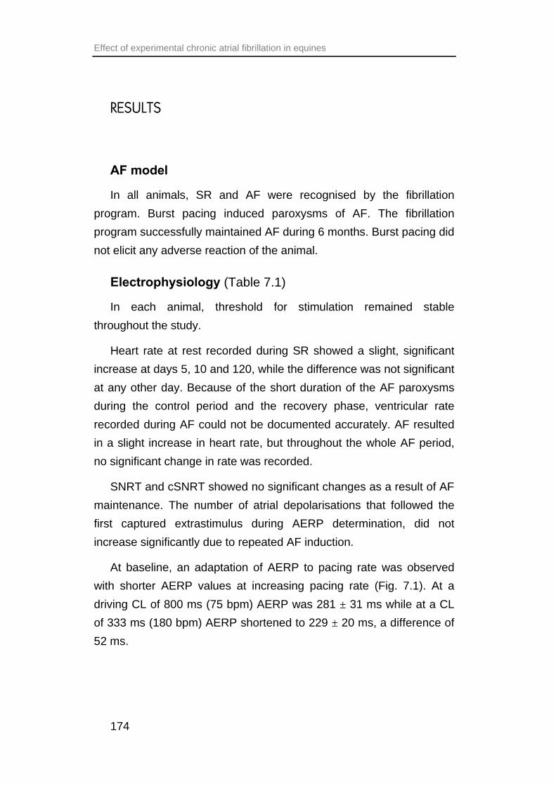
Effect of experimental chronic atrial fibrillation in equines
174
RESULTSRESULTS
AF model
In all animals, SR and AF were recognised by the fibrillation
program. Burst pacing induced paroxysms of AF. The fibrillation
program successfully maintained AF during 6 months. Burst pacing did
not elicit any adverse reaction of the animal.
Electrophysiology (Table 7.1)
In each animal, threshold for stimulation remained stable
throughout the study.
Heart rate at rest recorded during SR showed a slight, significant
increase at days 5, 10 and 120, while the difference was not significant
at any other day. Because of the short duration of the AF paroxysms
during the control period and the recovery phase, ventricular rate
recorded during AF could not be documented accurately. AF resulted
in a slight increase in heart rate, but throughout the whole AF period,
no significant change in rate was recorded.
SNRT and cSNRT showed no significant changes as a result of AF
maintenance. The number of atrial depolarisations that followed the
first captured extrastimulus during AERP determination, did not
increase significantly due to repeated AF induction.
At baseline, an adaptation of AERP to pacing rate was observed
with shorter AERP values at increasing pacing rate (Fig. 7.1). At a
driving CL of 800 ms (75 bpm) AERP was 281 ± 31 ms while at a CL
of 333 ms (180 bpm) AERP shortened to 229 ± 20 ms, a difference of
52 ms.

CHAPTER 7: Results
175
Table 7.1. Mean values for electrophysiologic measurements are given. AERP (ms) was measured at different pacing cycle lengths between 1000 and 333 ms. During every AERP measurement the number of atrial depolarisations following the first captured extrastimulus was recorded (+: 1 atrial depolarisation; ++: 2 to 5 depolarisations; +++: more than 5 depolarisations).
Control values Atrial fibrillation period Recovery
Day -2 -1 0 5 10 20 30 60 120 180 190 210
SR 42 46 43 51* 54* 51 48 47 57* 49 46 43 Heart rate
AF NA NA NA 55 63 56 54 56 57 54 NA NA
SNRT (ms) 1983 1848 1761 1613 1705 1626 1910 1551 1596 1769 1964 1945 cSNRT
(ms) 136 140 126 136 151 138 150 120 150 144 150 140
1000 ++ +++ ++ + + ++ ++ ++ + ++ ++ +
800 ++ +++ + +++ ++ ++ ++ + ++ ++ ++ ++
600 +++ +++ ++ + ++ ++ ++ ++ ++ ++ ++ ++
500 +++ ++ ++ + ++ ++ ++ ++ ++ ++ ++ ++
400 ++ +++ ++ ++ + ++ + ++ ++ ++ ++ ++
Atrial depol
333 +++ +++ +++ ++ ++ ++ ++ ++ ++ ++ ++ ++
1000 284 283 275 250* 240* 238* 236* 225* 233* 229* 275 279
800 283 281 279 252* 240* 240* 240 227* 231* 227* 275 283
600 272 273 264 248 236* 240 236 230 228 229 266 270
500 258 258 258 235* 229* 231 227* 223* 225* 226 258 260
400 244 244 250 225* 211* 223* 217 219 219 214 242 248
AERP (ms)
333 233 229 235 211* 203* 197* 203* 203* 207 203* 233 235
NA: not available; SR: sinus rhythm; AF: atrial fibrillation; (c)SNRT: (corrected) sinus node recovery time (ms) * indicates significant difference compared to control values with p< 0.05
Figure 7.1. Mean values for the AERP at different pacing CL, describing the rate adaptation of AERP. Measurements of day 0 (control) and day 180 (AF) are displayed.
175
200
225
250
275
300
300 400 500 600 700 800 900 1000
Pacing CL (ms)
AERP (ms)
-31 ms
-46 ms
Day 0
Day 180

Effect of experimental chronic atrial fibrillation in equines
176
Repeated AF induction resulted in a significant decrease in AERP
(Fig. 7.2), although a large inter-individual variation existed. From day
5 onward, the AERP decrease was significant and a plateau was
reached after about 2 months of AF. The AF-induced decrease in
AERP was more pronounced at slower pacing rates (-46 ms) than at
higher pacing rates (-31 ms) (Fig. 7.1). At baseline, the difference
between the AERP determined at CL of 800 and 333 ms was 52 ms,
while the difference was 23 ms after 6 months of AF. Subsequently,
the AERP rate adaptation curve was attenuated. After termination of
AF at day 180, within 10 days, AERP values returned to baseline and
the normal rate adaptation of atrial refractoriness recurred.
AF characteristics
At baseline, AF paroxysms showed a mean AFCL of 247 ± 36 ms
(Fig. 7.3). Atrial depolarisations could be clearly identified. Three
animals presented Type I AF and one animal Type II fibrillation (Fig.
7.4). As a result of maintained AF, an obvious decrease in AFCL
occurred. Two months of AF resulted in an AFCL of 170 ± 16 ms and
after 6 months of AF, AFCL had further shortened to 159 ± 16 ms.
AFCL was directly proportional to AERP but there was an obvious
inter-individual variation. Pony 4 showed a slight decrease in AFCL
although AERP values remained virtually unchanged. Maintained AF
resulted in a more complex atrial electrogram. Type I AF changed into
Type II (n=1) or Type III (n=2). Type II AF changed into Type III (n=1)
(Fig. 7.4). At baseline, AF paroxysms were short (Fig. 7.5) with a
mean duration of 2 ± 0.5 seconds. Repeated AF induction resulted in a
progressive increase in AF duration. After 4 months, mean AF duration
was 57 hours, ranging from 18 minutes (Pony 3) to 9 days (Pony 4).
Because of maintained fibrillation during 180 days, AF duration ranged
from 60 minutes (Pony 3) to 12 hours (Pony 1) and became persistent
in Pony 4, requiring defibrillation.

CHAPTER 7: Results
177
Figure 7.2. Mean AERP values (± standard error) at a pacing CL of 800 ms (* indicates significant difference compared to control values).
150
200
250
300
350
-30 0 30 60 90 120 150 180 210days
AERP (ms)
**
**
* *
cardioversion
Figure 7.3. Mean AFCL (± standard error) during the study.
100
150
200
250
300
-30 0 30 60 90 120 150 180 210Day
*
** * *
* *
cardioversion
AFCL (ms)
Figure 7.5. AF duration for each animal is displayed throughout the study. AF was maintained from day 0 to day 180.
0,01
0,1
1
10
100
1000
10000
100000
-30 0 30 60 90 120 150 180 210day
p o ny 1
p o ny 2
p o ny 3
p o ny 4
AF duration (min)(log scale)
Persistent AF
Figure 7.4. AF type at baseline and at the end of the AF period.
0%
25%
50%
75%
100%
baseline 180 days AF
TypeIII
Type II
Type I
% a n im a ls
( n = 4 )

Effect of experimental chronic atrial fibrillation in equines
178
Echocardiography
Left ventricle – Aorta (Table 7.2)
Following maintained AF, left ventricular internal diameter at end-
diastole (LVIDdiast) decreased and internal diameter at end-systole
(LVIDsyst) increased although both differences were not significant. Left
ventricular fractional shortening, however, was significantly decreased
from day 10 onward (Fig. 7.6). Similar results were obtained for left
ventricular internal area. A non-significant decrease in diastolic area
and increase in systolic area, and a significant decrease in left
ventricular area change were observed. Interventricular septal
thickness during systole decreased significantly, while diastolic septal
thickness showed no significant changes. Septal fractional wall
thickening was not significantly decreased.
No change was observed in the aortic systolic diameter at sino-
tubular junction, determined with the leading-edge to leading-edge
method as well as with the inner-edge technique.
Pulsed wave and colour flow Doppler (Table 7.2)
As a result of experimentally induced atrial fibrillation, spectral
Doppler recordings from the aortic outflow showed a significant
decrease in both maximal velocity and velocity time integral from day
10 onward. Because no change in aortic systolic diameter was
observed throughout the study, the significant decrease in velocity
time integral is accompanied by a significant decrease in stroke
volume (Fig. 7.6).
No evidence of significant tricuspid or mitral valve dysfunction was
observed at either baseline or restudy on colour flow Doppler.

CHAPTER 7: Results
179
Table 7.2. Echocardiographic measurements of left ventricular internal diameter (LVID; cm) and area (LVA; cm) with calculated left ventricular fraction shortening, of interventricular septal thickness (IVS; cm) and of aortic diameter (cm) and flow.
Control values Atrial fibrillation period Recovery
Day -2 -1 0 10 30 60 120 180 190 210 240
diast 8,4 8,5 8,4 8,2 8,3 8,3 8,3 8,1 8,4 8,3 8,3 syst 5,5 5,5 5,6 5,7 5,9 6,1 5,9 5,7 5,9 5,5 5,5
LVID B-mode
FS 34,3 34,7 33,7 31,0 28,1 26,9 29,4 29,4 30,9 33,5 33,5
diast 8,2 8,3 8,3 8,2 8,1 8,2 8,3 8,2 8,3 8,3 8,4 syst 5,5 5,6 5,6 5,8 5,9 6,0 5,9 5,8 5,6 5,7 5,5 LVID
M-mode FS 32,8 32,5 32,6 28,4 27,6 27,5 28,8 28,8 32,0 32,3 33,8
diast 2,5 2,3 2,3 2,3 2,3 2,2 2,2 2,3 2,3 2,4 2,4 syst 3,2 3,2 3,2 3,1 2,9 3,0 3,0 3,0 3,0 3,1 3,2
IVS B-mode
FWT 23,8 29,2 28,8 25,6 21,5 24,2 24,7 24,6 23,3 24,1 25,1
diast 2,6 2,5 2,4 2,4 2,4 2,4 2,3 2,3 2,4 2,5 2,4 syst 3,4 3,3 3,2 3,0 3,0 3,0 3,0 3,0 3,2 3,3 3,3 IVS
M-mode FWT 23,9 23,3 24,4 20,6 20,5 19,8 22,1 22,1 25,5 23,5 25,8
diast 39,2 41,1 39,1 41,0 40,8 40,0 39,6 36,3 38,2 37,4 38,1 syst 15,7 16,1 16,1 17,4 19,1 19,6 19,1 17,5 17,0 15,1 15,4
LVA B-mode
FAC 60,4 60,3 58,4 58,1 53,1 51,5 51,7 52,5 55,5 59,5 59,4
LELE 6,0 6,0 6,0 6,0 6,0 6,0 6,1 6,0 6,0 6,1 6,0 Aorta B-mode IE 5,3 5,3 5,3 5,3 5,3 5,3 5,4 5,3 5,4 5,4 5,4
Vmax 0,9 0,9 0,9 0,8 0,8 0,8 0,8 0,8 0,8 0,9 0,9 Aorta Doppler VTI 25,4 25,2 25,0 22,2 22,0 20,3 20,9 20,1 23,6 25,3 25,2
FS: fractional shortening (%); FWT: fractional wall thickening (%); FAC: fractional area change (%); LELE: leading-edge to leading-edge technique (cm); IE: inner edge technique (cm); Vmax: maximal flow velocity (m/s); VTI: velocity time integral (cm) (bold, underlined values differ significantly from baseline p<0.05)
Figure 7.6. Left ventricular fractional shortening (FS) and stroke volume (SV) are displayed. AF was maintained between day 0 and day180
20
25
30
35
40
-30 0 30 60 90 120 150 180 210 240
0,4
0,5
0,6
0,7
0,8
FS (%) SV (L)
Day

Effect of experimental chronic atrial fibrillation in equines
180
Left atrium (Table 7.3)
Left atrial diameters were determined during atrial pacing at both
60 and 75 bpm. Due to the shorter diastolic time at a rate of 75 bpm,
the rapid ventricular filling phase in early diastole was immediately
followed by an atrial contraction. As both events occurred as a single
smooth movement and could not clearly be distinguished, left atrial
diameter before initiation of atrial contraction (LADp) could not be
determined accurately at 75 bpm and was only measured during atrial
pacing at 60 bpm. Consequently, left atrial shortening fraction was only
calculated for the latter rate.
Left atrial diameters obtained at different time points during the
cardiac cycle, measured from the left parasternal as well as from the
right parasternal views, at a rate of 60 as well as 75 bpm, showed a
significant increase in diameter from day 10 or day 30 onward. Figure
7.7 shows different atrial diameters obtained from a left parasternal
view at a pacing rate of 60 bpm. At baseline, left atrial diameter at the
onset of atrial contraction (LADp), at the end of ventricular diastole
(LADdiast) and at the end of ventricular systole (LADsyst) was 7.8 ± 0.47,
7.5 ± 0.36 and 8.9 ± 0.51 cm, respectively. After 6 months of
maintained AF, diameters had increased to 8.6 ± 0.38, 8.9 ± 0.23, and
9.6 ± 0.50 cm, respectively, which means an increase of 0.8, 1.4 and
0.7 cm. The most obvious increase in diameter occurred for left atrial
diameter during atrial contraction (LADa). At baseline this diameter
was 6.7 ± 0.35 cm while at the end of the AF period it was 8.7 ± 0.39
cm, an increase of 2.0 cm. During the control period, atrial contraction
was able to decrease atrial diameter by 1.1 cm, representing a left
atrial shortening fraction of 13.4 ± 3.2 % (Fig. 7.8). Ten days of AF
were already sufficient to result in a significantly decreased atrial
inotropy. Because of long-term AF, atrial contraction no longer resulted
in a decrease in atrial diameter. Atrial shortening fraction became
negative (-0.9 ± 1 %) indicating that a slight increase in atrial diameter
occurred even though the atrium was contracting maximally.
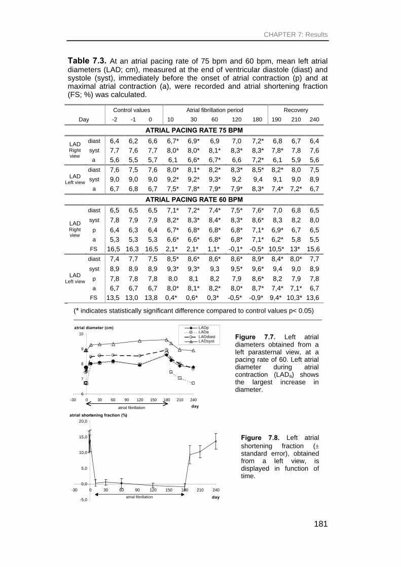
CHAPTER 7: Results
181
Table 7.3. At an atrial pacing rate of 75 bpm and 60 bpm, mean left atrial diameters (LAD; cm), measured at the end of ventricular diastole (diast) and systole (syst), immediately before the onset of atrial contraction (p) and at maximal atrial contraction (a), were recorded and atrial shortening fraction (FS; %) was calculated.
Control values Atrial fibrillation period Recovery
Day -2 -1 0 10 30 60 120 180 190 210 240
ATRIAL PACING RATE 75 BPM
diast 6,4 6,2 6,6 6,7* 6,9* 6,9 7,0 7,2* 6,8 6,7 6,4 syst 7,7 7,6 7,7 8,0* 8,0* 8,1* 8,3* 8,3* 7,8* 7,8 7,6
LAD Right view
a 5,6 5,5 5,7 6,1 6,6* 6,7* 6,6 7,2* 6,1 5,9 5,6
diast 7,6 7,5 7,6 8,0* 8,1* 8,2* 8,3* 8,5* 8,2* 8,0 7,5 syst 9,0 9,0 9,0 9,2* 9,2* 9,3* 9,2 9,4 9,1 9,0 8,9 LAD
Left view a 6,7 6,8 6,7 7,5* 7,8* 7,9* 7,9* 8,3* 7,4* 7,2* 6,7
ATRIAL PACING RATE 60 BPM
diast 6,5 6,5 6,5 7,1* 7,2* 7,4* 7,5* 7,6* 7,0 6,8 6,5
syst 7,8 7,9 7,9 8,2* 8,3* 8,4* 8,3* 8,6* 8,3 8,2 8,0
p 6,4 6,3 6,4 6,7* 6,8* 6,8* 6,8* 7,1* 6,9* 6,7 6,5 a 5,3 5,3 5,3 6,6* 6,6* 6,8* 6,8* 7,1* 6,2* 5,8 5,5
LAD Right view
FS 16,5 16,3 16,5 2,1* 2,1* 1,1* -0,1* -0,5* 10,5* 13* 15,6
diast 7,4 7,7 7,5 8,5* 8,6* 8,6* 8,6* 8,9* 8,4* 8,0* 7,7
syst 8,9 8,9 8,9 9,3* 9,3* 9,3 9,5* 9,6* 9,4 9,0 8,9
p 7,8 7,8 7,8 8,0 8,1 8,2 7,9 8,6* 8,2 7,9 7,8 a 6,7 6,7 6,7 8,0* 8,1* 8,2* 8,0* 8,7* 7,4* 7,1* 6,7
LAD Left view
FS 13,5 13,0 13,8 0,4* 0,6* 0,3* -0,5* -0,9* 9,4* 10,3* 13,6
(* indicates statistically significant difference compared to control values p< 0.05)
Figure 7.8. Left atrial shortening fraction (± standard error), obtained from a left view, is displayed in function of time.
-5,0
0,0
5,0
10,0
15,0
20,0
-30 0 30 60 90 120 150 180 210 240
atrial shortening fraction (%)
dayatrial fibrillation
Figure 7.7. Left atrial diameters obtained from a left parasternal view, at a pacing rate of 60. Left atrial diameter during atrial contraction (LADa) shows the largest increase in diameter.
6
7
8
9
10
-30 0 30 60 90 120 150 180 210 240
LADpLADaLADdiastLADsyst
atrial diameter (cm)
dayatrial fibrillation

Effect of experimental chronic atrial fibrillation in equines
182
After termination of fibrillation, atrial diameters and left atrial shortening
fraction gradually returned to normal values after 1 to 2 months of SR.
Invasive blood pressure monitoring
Right ventricle (Table 7.4)
During the induced fibrillation period, right ventricular pressure
during atrial contraction (RVa), at end-diastole (RVdiast) and peak
systolic pressure (RVsyst) showed a decrease at day 30 but was not
significantly altered at day 180. Right ventricular maximal dP/dt was
significantly decreased at day 180.
Right atrium (Table 7.4)
As a result of AF, right atrial pressure prior to atrial contraction
(RAp), at the c-wave (RAc-wave) and at the v-wave (RAv-wave) showed an
increased pressure although the increase did not reach significance.
AF resulted in a significant decrease in the atrial pressure during atrial
contraction (RAa-wave) and in the maximal dP/dt during atrial contraction
(RAdP/dt). Calculation of the pressure difference produced by atrial
contraction showed a significant decrease during the AF period.
AF duration
Although an increase in AF duration was associated with shorter
AERP values, AERP had to drop below a critical value of about 258
ms before AF paroxysms could persist for more than an hour (Fig.
7.9). Above this value, shortening of AERP was not associated with an
increase in AF duration. Yet, below an AERP of 258 ms, association
between AERP and AF duration remained rather poor.
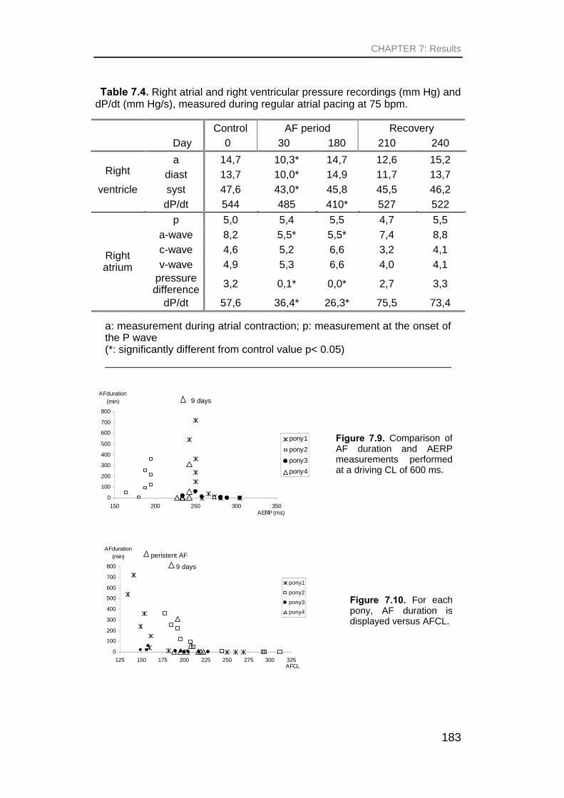
CHAPTER 7: Results
183
Table 7.4. Right atrial and right ventricular pressure recordings (mm Hg) and dP/dt (mm Hg/s), measured during regular atrial pacing at 75 bpm.
Control AF period Recovery
Day 0 30 180 210 240
a 14,7 10,3* 14,7 12,6 15,2
diast 13,7 10,0* 14,9 11,7 13,7
syst 47,6 43,0* 45,8 45,5 46,2
Right
ventricle
dP/dt 544 485 410* 527 522
p 5,0 5,4 5,5 4,7 5,5
a-wave 8,2 5,5* 5,5* 7,4 8,8
c-wave 4,6 5,2 6,6 3,2 4,1
v-wave 4,9 5,3 6,6 4,0 4,1 pressure difference
3,2 0,1* 0,0* 2,7 3,3
Right atrium
dP/dt 57,6 36,4* 26,3* 75,5 73,4
a: measurement during atrial contraction; p: measurement at the onset of the P wave (*: significantly different from control value p< 0.05)
0
100
200
300
400
500
600
700
800
125 150 175 200 225 250 275 300 325AFCL
AFduration (min)
pony1
pony2
pony3
pony4
9 days
peristent AF
Figure 7.10. For each pony, AF duration is displayed versus AFCL.
Figure 7.9. Comparison of AF duration and AERP measurements performed at a driving CL of 600 ms.
0
100
200
300
400
500
600
700
800
150 200 250 300 350AERP (ms)
AFduration (min)
pony1
pony2
pony3
pony4
9 days

Effect of experimental chronic atrial fibrillation in equines
184
Pony 4 hardly showed any change in AERP although this was the
animal with the longest AF duration, requiring defibrillation. In pony 2,
longest AF paroxysms were not associated with shortest AERP
values.
Figure 7.10 compares AF duration and AFCL. As long as AFCL
was longer than 215 ms, AFCL and AF duration were not associated
and AF paroxysms remained short. Once AFCL further decreased
below 215 ms as a result of repeated AF induction, a decrease of
AFCL was associated with an increase in AF duration, and paroxysms
of more than an hour could be induced. Sustained AF was only seen in
pony 4 at an AFCL of 182 ms. In this animal AF became persistent at
an AFCL of 153. Although in pony 3 AFCL shortens below 158 ms,
paroxysms in this animal were short.
The animals’ height and atrial size importantly influenced AF
duration. Largest animals, showing the largest left atrial diameter at
baseline, reached the longest AF duration during the study. In Figure
7.11, for each animal, left atrial diameter at the onset of atrial
contraction (LADp) obtained from a left view, is shown from baseline
until day 180. An atrial diameter of less than 8.2 cm was associated
with short paroxysms of AF (< 60 min). When atrial diameter
exceeded this critical value as a result of maintained AF, atrial
diameter was related to AF duration. Pony 3, with the smallest left
0
100
200
300
400
500
600
700
800
7 7,5 8 8,5 9left atrial diameter (cm)
AF duration (min)
pony 1
Pony 2
Pony 3
Pony 4
persistent AF
Figure 7.11. Comparison of AF duration and left atrial diameter (left view) measured at the onset of the P wave, during atrial pacing at a rate of 60 bpm.

CHAPTER 7: Results
185
atrial diameter throughout the study, showed the shortest AF
paroxysms. In pony 4, with the largest atrial size at the end of the
fibrillation period, SR did not restore spontaneously and in this animal
defibrillation was required.
Recovery phase
As described above, all variables from electrophysiology,
echocardiography and blood pressure returned towards normal within
2 months after restoration of SR. While electrophysiological values
returned to normal within 10 days, atrial size and atrial contractile
function gradually normalized over 1 to 2 months of time. AF duration
decreased abruptly to less than a minute within 10 days after
restoration of SR, although at that time atrial dilatation was still present
(Fig. 7.12).
Figure 7.12. Comparison of mean AF cycle length (AFCL), left atrial diameter (left view, pacing rate 60 bpm) and AF duration during the recovery phase. Ten days after cardioversion, AF duration is less than a minute although left atrial diameter has only slightly decreased. (Exact values for AF duration are not displayed because of the individual variation.)
140
160
180
200
220
240
260
280
170 180 190 200 210 220
7,8
7,9
8
8,1
8,2
8,3
8,4
8,5
8,6
8,7
day
atrial diameter (cm
)
AF
CL
(m
s) AF
duration (min)
0
more than 60 min

Effect of experimental chronic atrial fibrillation in equines
186
DISCUSSIONDISCUSSION
In the present study AF was maintained during 6 months in healthy
ponies by applying burst pacing. AF resulted in a fast decrease in
AERP and AERP rate-adaptation. Consequently, AFCL shortened and
the atrial electrogram became more complex. A fast loss of atrial
contractile function was observed, which resulted in an increased atrial
pressure and a decreased ventricular function. Subsequently, a slow
increase in atrial size occurred. As a result of these AF-induced
electrophysiological and morphological alterations a progressive
increase in AF duration was seen during the 6-month AF period. After
restoration of sinus rhythm, all variables returned to normal.
Electrophysiological changes and AF duration showed a short time
course for normalization, while echocardiographic values needed 1 to
2 months to recover.
Study protocol
This model proved to be efficient to induce and maintain long-term
atrial fibrillation in healthy ponies. Implantation of a pacemaker allowed
to perform programmed electrical stimulation in order to study atrial
electrophysiologic characteristics. Type and rate of fibrillation could be
determined from atrial electrogram recordings. Because the model
required a minimally invasive implantation technique, reliable blood
pressure recordings and echocardiographic studies could be carried
out.
Horses with naturally occurring AF mostly are presented with
subacute or chronic AF. Therefore, to mimic more closely the natural
state of the disease, we developed an equine model in which AF was
maintained over a prolonged period of time by permanent implantation
of an electrical pulse generator.

CHAPTER 7: Discussion
187
Many AF studies have been performed during anaesthesia, in
opened-chest animals or in animals with induced pericarditis and
these conditions are likely to have an important impact on cardiac
function. A major advantage of the closed-chest pony model was that
invasive surgery was avoided, thereby preserving normal cardiac
function and allowing reliable blood pressure measurements and
cardiac ultrasound to be performed. During the whole study, all
measurements were performed in the conscious, unsedated animal,
thereby avoiding any drug-related interference.
It is known that AF is favoured by underlying cardiac pathology.
Valvular insufficiency might cause atrial stretch and atrial dilatation
leading to increased susceptibility and perpetuation of AF (Morillo et
al., 1995; Allessie, 1998; Power et al., 1998). Local disorders of the
myocardium, such as fibrosis, are known promoters of AF (Allessie et
al., 2001). In humans, increasing age is strongly associated with an
increased risk for atrial fibrillation. A survey in horses also
demonstrated increased incidence of AF in aged animals (Else and
Holmes, 1971). However, in many horses it is suggested that lone
atrial fibrillation is present because cardiac ultrasound reveals no
abnormalities or because the animals respond well to quinidine
treatment and because they return to their previous level of exercise. It
should be mentioned, however, that response to treatment and
regaining athletic ability after cardioversion of AF not necessarily
indicates absence of underlying cardiac pathology. Moreover, our
present diagnostic facilities might be insufficient to detect distinct atrial
pathology.
The purpose of our research was to study lone AF in equines.
Therefore, young and healthy animals were selected. These animals
did not show any abnormalities on cardiologic examinations and were
considered to be free of myocardial lesions. Subsequently, by artificial
induction and long-term maintenance of AF in healthy subjects, any
observed changes would be induced, simply and solely by the
Effect of experimental chronic atrial fibrillation in equines
188
arrhythmia, and would not be confounded by any other cardiac
pathology.
Although AF rarely occurs in ponies and is predominantly seen in
mature horses (Else and Holmes, 1971; Bertone and Wingfield, 1987;
Detweiler, 1989), we used ponies in our study. In a previous report we
demonstrated that sustained AF could be successfully induced in the
pony heart (van Loon et al., 2000). We suggested that spontaneous
restoration of SR after long-term AF maintenance would occur more
easily in ponies than in horses, which would be advantageous because
during the 6-month AF period, spontaneous restoration of SR was
required to perform measurements. Besides, during preliminary
research we validated non-invasive echocardiographic cardiac output
determination using the well-documented thermodilution technique
(Blissitt et al., 1997), which could be performed more easily in smaller
sized animals (unpublished data).
AF is characterised by an irregularly irregular ventricular rate. As
differences in diastolic times result in different preload and afterload
conditions, important alterations in cardiac function occur, and
confusing results might be found during AF (Leistad et al., 1993a;
Leistad et al., 1993b; Hardman et al., 1998). In the present study,
echocardiography and blood pressure recordings were performed after
spontaneous reversion of sinus rhythm, and during regular atrial
pacing. Subsequently, confounding effects of ventricular arrhythmia or
changes in heart rate that might occur during the study were
eliminated. The reason why a pacing rate of 75 bpm was chosen to
perform blood pressure measurement and cardiac ultrasound was that
some animals showed a SR of about 60 bpm during the study. To be
sure that consistent atrial pacing occurred at any time, a rate of 75
bpm was chosen to perform ultrasound and blood pressure
measurements. However, at this rate, rapid ventricular filling phase in
early diastole and atrial contraction were not clearly distinguishable.
Consequently, left atrial diameter before initiation of atrial contraction
CHAPTER 7: Discussion
189
(LADp) could not be determined accurately at 75 bpm, which made
calculation of left atrial shortening fraction impossible. Additional left
atrial measurements were therefore performed at a pacing rate of 60
bpm.
In other animal models, induction of AF is often associated with an
important increase in ventricular rate. When subsequently, alterations
in electrophysiological properties, or in atrial or ventricular structure
and function, are found, these might be biased by the ventricular rate
or by ventricular tachycardiomyopathy. In the present study, heart rate
during AF was only slightly higher than during SR and was unlikely to
affect experimental results.
Electrophysiologic changes induced by AF
In animals, contrasting results about the effect of AF on sinus node
function have been reported. While in the isolated rabbit heart, during
atrial fibrillation, sinus automaticity was still present in the centre of the
sinus node and hardly overdrive suppressed due to a high degree of
sinoatrial entrance block (Kirchhof and Allessie, 1992), in dogs, 2 to 6
weeks of maintained AF caused an impaired sinus node function
(Elvan et al., 1996), confirmed by a prolongation of cSNRT. Also in
humans, chronic AF can be associated with a depressed sinus node
function (Manios et al., 2001). In our ponies, no significant changes in
SNRT or cSNRT could be demonstrated after 6 months of AF. This
could be partly explained by the technique for SNRT measurement in
the present study. While in human medicine it is generally accepted
that SNRT should be determined with an electrode that is positioned
near to the sinus node (Fogoros, 1995), the exact position of the
implanted atrial electrode in relation to the sinus node was not known.
Electrode position too far away from the sinus node might have
influenced our results. However, in each animal the permanently
implanted electrode remained in the same position during the whole
study, suggesting that alterations in sinus node function during the
study still might be detected. To the best of our knowledge, information
Effect of experimental chronic atrial fibrillation in equines
190
about SNRT determination in horses is not available in literature. It
could be that, because measurements were performed in the
conscious ponies, changes in autonomic tone occurred during these
measurements, confounding the results. Determination of SNRT after
autonomic blockade with propranolol and atropine might have provided
more conclusive results.
As a result of prolonged pacing-induced AF, the previously normal
atrium now maintained longer paroxysms of AF. The increased AF
duration might be explained, at least in part, by the shortening in
AERP. Because the length of a reentry wavelet is defined as the
product of AERP and conduction velocity, AERP shortening resulted in
a shortening of the wavelength. According to Moe’s multiple wavelet
hypothesis (Moe, 1962), a long wavelet may not permit reentry to
sustain, causing fibrillation to terminate, whereas a short wavelength
allows multiple wavelets to coexist in the atria, thereby favouring AF
persistence. Although atrial conduction velocity could not be measured
in this study, it presumably remained similar or even decreased as a
result of AF, as it was shown in other species (Morillo et al., 1995;
Wijffels et al., 1995; Elvan et al., 1996).
It has been reported that in human patients a poor or absent rate
adaptation of the AERP might be the cause of AF (Attuel et al., 1982;
Boutjdir et al., 1986). Normal ponies used in the present study showed
an adaptation of AERP to rate, which was attenuated by pacing-
induced AF. After cardioversion to SR the normal adaptation to
changes in heart rate was restored within 10 days. The present
observations thus suggest that maladaptation of AERP is rather the
result of AF than the cause of it.
Repeated AF induction resulted in a faster rate of fibrillation as
evidenced by the decreased AFCL. Previous studies have
demonstrated a high correlation between local fibrillation interval and
refractory period (Wijffels et al., 1995). In that case AFCL could be
used as an index for AERP estimation. Such an index would be
CHAPTER 7: Discussion
191
advantageous as it could be applied during AF and make restoration
of SR during the study redundant. However, median fibrillation interval
is not equal to local refractory period as during AF a small excitable
gap is still present (Allessie et al., 1991; Kirchhof et al., 1993;
Duytschaever et al., 2001).
AF-induced changes in atrial size and function
In our study, atrial diameters were measured rather close to the
mitral valve annulus because apical views cannot be obtained in
horses. Measurements of atrial area obtained from apical views more
closely reflect true atrial size and might provide a higher sensitivity in
exposing subtle changes in atrial dimension. However, in our study a
significant gradual increase in atrial size could be demonstrated as a
result of atrial fibrillation. It should be emphasized that we did not
perform echocardiography during a fibrillating rhythm but during
regular atrial pacing. Measurements of atrial size during AF are not
only confounded by the irregular ventricular rate but also by decreased
atrial compliance that reduces atrial diameter (White et al., 1982;
Leistad et al., 1993a). The decreased compliance during AF is caused
by the unsynchronised, continuous muscle fibre contractions, by
higher intracellular calcium concentrations in atrial fibres during the
noncontracting phase, or by a greater myocardial turgor due to an
increase in atrial blood flow during fibrillation (Leistad et al., 1993a).
The AF-induced increase in atrial size was suggested to be partly
caused by an increase in atrial pressure (Oldham et al., 1967; White et
al., 1982; Leistad et al., 1993a). In the present study, increase in atrial
pressure did not reach statistical significance. The reason why atrial
diameters changed significantly while pressure differences were less
obvious, could be the limited number of animals, but could also be
explained by the fact that pressures were measured in the right atrium
while diameters were measured in the left atrium. It has been shown in
other animals that AF-induced changes in pressure and size are less
pronounced in the right heart compared to the left heart (White et al.,

Effect of experimental chronic atrial fibrillation in equines
192
1982; Leistad et al., 1993a). In our study, pressures were measured in
the right heart because left atrial catheterisation using a high-fidelity
catheter is technically more difficult in horses, and echocardiographic
variables were determined for the left heart because reliable
echocardiographic landmarks for the right heart are lacking.
As a result of maintained AF, atrial pressure during maximal atrial
contraction, the a-wave, was decreased. Although this could suggest a
decreased atrial contractility, a-wave was also influenced by pre- and
afterload conditions, and did not reflect atrial contractile function.
Therefore, in addition, atrial pressure immediately before the onset of
atrial contraction was measured, which allowed calculating the
pressure difference generated by atrial contraction. This generated
pressure difference was completely abolished by pacing-induced AF,
indicating that, even during the short restoration of SR, atrial
contractility was almost completely lost. As right atrial a-wave nearly
disappeared on the atrial pressure tracings, right atrial maximal dP/dt
during atrial contraction was significantly depressed. However, it
should be emphasized that maximal dP/dt in the right atrium only
indicates the pressure rise generated by atrial contraction and does
not provide a true reflection of atrial inotropic state, as the latter only
applies for measurements performed during the isovolumetric phase
of the contraction (Van den Bos et al., 1973; Brown and Holmes,
1978), a condition that was not full-filled in the atrium.
Left atrial contractions are difficult to visualize on echocardiography
(Wingfield et al., 1980) and M-mode analysis of mitral valve movement
has been used as an attractive alternative in visualising the effects of
AF in horses. However, mitral valve movement is highly affected by
atrial and ventricular loading conditions and thus poorly reflects atrial
inotropic state. To the best of our knowledge, real estimation of atrial
contractility in equines has not been reported in literature. In equine
literature, ultrasonographic estimation of atrial size is generally
performed at the end of ventricular diastole and systole, but does not

CHAPTER 7: Discussion
193
allow evaluation of atrial contractile function. In the present study, we
introduced additional left atrial measurements at different timings
during the cardiac cycle to estimate atrial contractility. By determining
atrial diameter immediately prior to atrial contraction and again during
maximal atrial contraction, shortening fraction of the left atrium was
calculated by adapting the well-documented formula for left ventricular
shortening fraction (Feigenbaum, 1986). The index proved to be
sensitive in detecting changes in atrial inotropic state non-invasively.
Ten days of AF already reduced left atrial shortening fraction from
16.5% at baseline to 2.1%. After 6 months of AF a nadir of –0.5% was
reached. The negative value of atrial fractional shortening indicates
that passive atrial filling resulted in an increase in atrial diameter, even
though the atrium was contracting maximally.
Effect of AF on the ventricle
Similar remarks as for the atria can be given concerning pressure
measurement performed in the right ventricle and ultrasound
completed on the left ventricle.
A decreased diastolic ventricular diameter and area indicated an
incomplete left ventricular filling caused by a loss of atrial contraction.
By the Frank-Starling mechanism, the decreased ventricular preload
resulted in a reduced inotropy, which was evidenced by the increased
systolic diameter and area, and concomitant decreased fractional
shortening and fractional area change, and also the reduced right
ventricular maximal dP/dt. The depressed ventricular performance
subsequently resulted in a lower ventricular peak pressure and a
reduced aortic flow velocity. The final result was an obvious decrease
in stroke volume and thus cardiac output. An AF episode as short as
10 days was already sufficient to result in a significantly reduced
ventricular performance. As ventricular rate during pacing-induced
fibrillation was not markedly increased during our study, occurrence of
ventricular tachycardiomyopathy could be prevented and reduced
Effect of experimental chronic atrial fibrillation in equines
194
ventricular function was predominantly attributed to the decreased
preload.
Other studies in equines described contradictory results concerning
cardiac output. Muir et al. (1984) reported normal cardiac output
values measured during AF in 7 horses with naturally occurring AF,
provided that they didn’t show signs of congestive heart failure. In 4 of
these horses, cardiac output was again measured after restoration of
sinus rhythm. After cardioversion an increase in output was observed
in one animal, while no changes were detected in the remaining 3
horses. However, in none of these horses the duration of fibrillation
was known. Furthermore, it remained unclear to what extent quinidine
treatment, influenced hemodynamic results. In 1975, Kubo et al.
investigated the effect of experimentally induced short-term AF
paroxysms on cardiac output in 7 healthy horses. Compared to SR,
they found increases as well as decreases in cardiac output after
experimental AF induction. On the average, cardiac output increased
by 12%, which turned out to be not significant. However, a significant
increase in heart rate of 43% was observed, resulting in a significant
decrease in stroke volume (22%). It should be stressed that in both
above-mentioned reports measurements were performed during AF.
Consequently, hemodynamic alterations are the result of multiple
factors such as reduced atrial compliance during AF, AF-induced
depressed atrial contractility, irregular ventricular rhythm and different
ventricular rate.
Reversibility of AF-induced changes
Although AF resulted in important changes in atrial
electrophysiology, size and function, all these changes appeared to be
reversible within 2 months after restoration of sinus rhythm. A short
time-course was observed for the electrophysiologic changes,
normalizing within 10 days after cardioversion. Atrial size and
contractility, and concomitant left ventricular function, needed 1 to 2
months to recover after the 6 month AF period. These results are in

CHAPTER 7: Discussion
195
agreement with the findings of Allessie (1998) who suggested that
metabolic changes, such as differences in ion concentrations or in ion
pump activities, occurred and disappeared within seconds to minutes,
while changes in electrical remodeling needed hours or days. Many
studies reported a postfibrillatory atrial contractile dysfunction,
requiring weeks to months to recover (Jordaens et al., 1993; Van
Gelder et al., 1993; Manning et al., 1994; Allessie, 1998; Schotten et
al., 2001). Cellular mechanisms responsible for AF-induced contractile
dysfunction are still poorly understood. Alterations in atrial cellular
ultrastructure and a reduction in L-type Ca2+ current have been
suggested as plausible mechanisms (Ausma et al., 1997; Yue et al.,
1997; Bosch et al., 1999; Van Wagoner et al., 1999; Schotten et al.,
2001).
What can we conclude about AF duration?
Immediately after induction of AF, the AFCL started to shorten.
Initially, the decrease in AFCL was not accompanied by an increase in
AF duration. Only when AFCL had shortened to a critical value, AF
duration started to increase. Similarly, only after an increase in atrial
size above a critical value, AF paroxysms progressively increased.
These findings are in agreement with the multiple wavelet hypothesis
(Moe, 1962). Because of the shorter atrial refractoriness, and
concomitant shorter wavelength and fibrillation CL, and because of the
increase in atrial diameter, a larger number of wavelets wander around
in the atria, decreasing the statistical probability of simultaneous
extinction and favouring AF perpetuation. In the isolated Langendorff-
perfused canine hearts, the critical number of wandering wavelets for
perpetuation of fibrillation was between 3 and 6 (Allessie et al., 1985).
The lifetime of individual wavelets was very short, about a few hundred
milliseconds. Continuously, ‘old’ wavelets extinguished by fusion or
collision with another wavelet, by reaching the border of the atrium,
and because the advancing depolarisation wave met an area where
the myocardium had not recovered its excitability from a foregoing
Effect of experimental chronic atrial fibrillation in equines
196
activation. At the same time new wavelets were formed by division of
an existing wave at an area of conduction block, an offspring
traversing toward the other atrium, and possible sources of impulse
formation. The approximate number of atrial wavelets during AF can
be determined during mapping studies or can be estimated when
conduction velocity, refractory period and atrial size are known.
However, studies in dogs have shown a spatial dispersion and
temporal inhomogeneity in AERP and, in addition, conduction velocity
during AF was shown to vary from 20 to 139 cm per second in the
same animal, resulting in a continuously changing wavelength and a
varying number of wavelets (Allessie et al., 1994).
Both a decrease in AFCL and an increase in atrial diameter
appeared to be required in order for AF to become sustained. In dogs,
a minimal reduction of 15% in AERP associated with an increase of at
least 40% in atrial area was highly predictive of sustained AF (Morillo
et al., 1995). This also explains the abrupt reduction in AF duration
during the recovery period: although after restoration of SR, initially,
atrial diameter had barely changed, the fast occurring increase in
AERP and AFCL toward normal values, prevented AF to perpetuate.
With these findings we can explain why naturally occurring AF is never
seen in ponies: their atrial electrophysiological properties and their
atrial size simply prevent sufficient wavelets to coexist in the atria.
Surprisingly, after pacing-induced electrical and morphological
remodeling, long-term AF could be induced in ponies.
Clinical implications
In equine medicine it is believed that AF often is present as ‘lone’
AF, i.e. without overt underlying heart disease. However, very little
information is available on the initiation of AF, and what AF actually
does in a healthy animal. The present model demonstrated the
feasibility of developing a reproducible model of long-term AF in
healthy equines to expand our understanding of the mechanisms

CHAPTER 7: Discussion
197
leading to AF. In the future, this model will facilitate the development of
different therapeutic modalities in horses.
In the present study adapted left atrial measurements were
introduced in order to establish atrial contractile function non-
invasively. Such an index could be applied to reveal atrial disease in
equines. Especially after cardioversion of AF, calculation of atrial
shortening fraction is useful to assess restoration of atrial contractile
function.
The results of this study indicate that, after initiation of atrial
fibrillation, changes in electrophysiological parameters develop fast to
reach a plateau, and that they return quickly to normal after restoration
of SR. Dilatation of the left atrium appears slower and continues to
progress over a longer period of time, normalizing again about 2
months after cardioversion. Therefore, it is suggested that after
successful treatment of chronic atrial fibrillation, training should only be
resumed after a period of about 1 to 2 months, i.e. after restitution of
atrial electrophysiologic characteristics, atrial contractile function and
atrial diameter. As atrial diameter seems to be an important predictor,
not only for AF stability but also for successful restoration of SR and
for the risk of AF recurrence, the atrial diameter is often determined
prior to initiation of AF therapy, where horses with a dilated left atrium
are given an unfavourable prognosis. As in the present study, AF-
induced atrial dilatation occurred in normal individuals, this parameter
should be interpreted cautiously in chronic fibrillating animals without
valvular disease. A long-term follow-up of animals with a normal left
atrial diameter and recent onset lone AF should be performed to
provide more information about changes in left atrial diameter in
horses with naturally occurring AF. Furthermore, little information is
currently available about the correlation between left atrial diameters
measured during SR and during AF. The present model could be
applied to study this relationship.

Effect of experimental chronic atrial fibrillation in equines
198
At present, little is known about the effect of high-level performance
on the inducibility or persistence of AF. During high performance,
cardiac load may be higher and this might affect atrial pressure,
diameter and stretch, which might be important factors explaining AF
susceptibility (Leistad et al., 1993a; Morillo et al., 1995; Sideris et al.,
1995). The time-course of the occurrence of electrophysiologic,
pressure or echocardiographic alterations might therefore differ in
horses that remain in training.
As mostly seen in lone AF horses, AF-induced tachycardia at rest
did not develop during the present study. The high vagal tone in
equines results in a high degree of concealed conduction at the AV
node. This confirms that in horses with lone atrial fibrillation rate
control is not required and only rhythm control has to be considered.
In humans, AF is associated with an increased risk of systemic
thromboembolic complications, particularly strokes, although it has
been reported that the risk would not be increased in patients with lone
AF (Kopecky et al., 1987; Elvan et al., 1999). In the present study,
during echocardiographic examinations special attention was paid to
the presence of atrial thrombi, but none could be identified. Also in
clinical cases thromboembolic events are not reported and seem to be
negligible in horses, making anticoagulation therapy redundant.
In human medicine, antiarrhythmic therapy is generally continued
after restoration of SR to prevent early recurrence of AF. One of the
possible explanations for the high incidence of early recurrence of AF
could be the inversed rate adaptation curve of the AERP that can
occur. When inversed rate adaptation is present, restoration of SR
implies a decreased atrial rate and thus a shortening of the refractory
period instead of an increase. The shorter refractory period again
favours AF occurrence. As a reversed rate adaptation was not seen
during our study, this might be a contributing factor for the low
incidence of early AF recurrence seen in equines and might therefore

CHAPTER 7: Discussion
199
suggest that antiarrhythmic therapy after successful restoration of SR
is not required in this species.
Limitations of the study
In the present study only 4 animals were implanted and followed
during the 6-month AF period. Due to the limited number of animals,
the results and statistical analyses must be interpreted cautiously.
However, most variables altered similarly in all animals, suggesting
that valuable conclusions could be drawn.
Pacemaker output was set at 2 times diastolic threshold. In some
studies, a stimulation output of 4 times threshold was used, yielding a
more aggressive pacing protocol for AF induction. Furthermore, during
our study, SR detection appeared during 3 to 4 consecutive sinus
beats. As the resting sinus cycle length in the ponies was about 1200
to 1500 ms, each SR episode persisted for about 4 to 6 seconds
before AF was re-induced. As electrical reversed remodeling appeared
fast, this short period of SR detection might have influenced induction
and perpetuation of AF. Measurements of electrical parameters,
echocardiography and blood pressures were performed after
spontaneous reversion of SR. These periods of SR also might have
influenced the experimental results.
One atrial electrode was used for determination of
electrophysiologic properties. It is known that, at a certain point in time,
AERP and AFCL vary among the location in the atrium (spatial
dispersion). In addition, a local area may show different AERP and
AFCL values in time (temporal dispersion). Therefore, multiple
electrodes are desirable to make detailed electrophysiological
measurements. However, a continuing problem exists with chronic AF
models in achieving a balance between the area from which
information is gathered by electrode implantation and any effects of
the implantation per se, which may be related to the epicardial area in
contact with the electrodes (Power et al., 1998).
Effect of experimental chronic atrial fibrillation in equines
200
From this study we can conclude that, in healthy equines, AF
induces changes in electrophysiological properties that lead to AF
perpetuations. Furthermore, six months of maintained AF results in
atrial dilatation and atrial contractile dysfunction, which needs 1 to 2
months to recover after cardioversion. All AF-induced changes were
fully reversible.
Acknowledgements
The authors wish to thank Prof. M. A. Allessie from the university
Maastricht for his advise. The Special Research Fund, Ghent
University, is acknowledged for the financial support.

CHAPTER 7: References
201
REFERENCESREFERENCES
Allessie, M., Kirchhof, C., Scheffer, G.J., Chorro, F. & Brugada, J. (1991). Regional control of atrial fibrillation by rapid pacing in conscious dogs. Circulation 84, 1689-1697.
Allessie, M.A. (1998). Atrial electrophysiologic remodeling: another vicious circle? J Cardiovasc Electrophysiol 9, 1378-1393.
Allessie, M.A., Boyden, P.A., Camm, A.J., Kleber, A.G., Lab, M.J., Legato, M.J., Rosen, M.R., Schwartz, P.J., Spooner, P.M., Van Wagoner, D.R. & Waldo, A.L. (2001). Pathophysiology and Prevention of Atrial Fibrillation. Circulation 103, 769-777.
Allessie, M.A., Lammers, W.J., Bonke, F.I. & Hollen, J. (1985). Experimental evaluation of Moe's multiple wavelet hypothesis of atrial fibrillation. In Cardiac electrophysiology and arrhythmias, Zipes, D.P. & Jalife, J. (eds) pp. 265-275. Grune & Stratton: Orlando.
Allessie, M.A., Wijffels, M.C. & Kirchhof, C.J. (1994). Experimental models of arrhythmias: toys or truth? Eur Heart J 15 Suppl A, 2-8.
Amada, A. & Kurita, H. (1978). Treatment of atrial fibrillation with Quinidine Sulphate in the racehorse. Exp Rep Equine Hlth Lab 15, 47-61.
Amada, A., Senta, T., Kubo, K., Oh-ishi, S. & Kiryu, K. (1974). Atrial fibrillation in the horse: clinical and histopathological studies of two cases. 1. Clinical study. Exp Rep Equine Hlth Lab 11, 51-69.
Attuel, P., Childers, R., Cauchemez, B., Poveda, J., Mugica, J. & Coumel, P. (1982). Failure in the rate adaptation of the atrial refractory period: its relationship to vulnerability. Int J Cardiol 2, 179-197.
Ausma, J., Wijffels, M., Thone, F., Wouters, L., Allessie, M. & Borgers, M. (1997). Structural changes of atrial myocardium due to sustained atrial fibrillation in the goat. Circulation 96, 3157-3163.
Bertone, J.J. & Wingfield, W.E. (1987). Atrial fibrillation in horses. Comp Cont Educ Pract 9, 763-771.
Blissitt, K.J. & Bonagura, J.D. (1995). Colour flow Doppler echocardiography in normal horses. Equine Vet J Suppl 19, 47-55.
Effect of experimental chronic atrial fibrillation in equines
202
Blissitt, K.J., Young, L.E., Jones, R.S., Darke, P.G. & Utting, J. (1997). Measurement of cardiac output in standing horses by Doppler echocardiography and thermodilution. Equine Vet J 29, 18-25.
Bosch, R.F., Zeng, X., Grammer, J.B., Popovic, K., Mewis, C. & Kuhlkamp, V. (1999). Ionic mechanisms of electrical remodeling in human atrial fibrillation. Cardiovasc Res 44, 121-131.
Boutjdir, M., Le Heuzey, J.Y., Lavergne, T., Chauvaud, S., Guize, L., Carpentier, A. & Peronneau, P. (1986). Inhomogeneity of cellular refractoriness in human atrium: factor of arrhythmia? Pacing Clin Electrophysiol 9, 1095-1100.
Brown, C.M. & Holmes, J.R. (1978). Haemodynamics in the horse: 1. Pressure pulse contours. Equine Vet J 10, 188-194.
Collatos, C. (1995). Treating atrial fibrillation in horses. Comp Cont Educ Pract 17, 243-245.
Deem, D.A. & Fregin, G.F. (1982). Atrial fibrillation in horses: a review of 106 clinical cases, with consideration of prevalence, clinical signs, and prognosis. J Am Vet Med Assoc 180, 261-265.
Detweiler, D.K. (1989). The mamalian electrocardiogram: comparative features. In Comprehensive electrocardiology, Macfarlane, P.W. & Veitch Lawrie, T.D. (eds), Vol. 2. pp. 1332-1377. Pergamon Press: New York.
Else, R.W. & Holmes, J.R. (1971). Pathological changes in atrial fibrillation in the horse. Equine Vet J 3, 56-64.
Elvan, A., Wylie, K. & Zipes, D.P. (1996). Pacing-induced chronic atrial fibrillation impairs sinus node function in dogs. Electrophysiological remodeling. Circulation 94, 2953-2960.
Elvan, A., Ramanna, H. & Robles de Medina, E.O. (1999). New insights into the pathophysiology and treatment of atrial fibrillation. ACC Current Journal Review 8, 11-14.
Feigenbaum, H. (1986). Echocardiography. Lea and Febiger: Philadelphia, 1-662.
Fogoros, R.N. (1995). Electrophysiologic testing. Blackwell Science: Oxford, 1-270.
Hardman, S.M., Noble, M.I., Biggs, T. & Seed, W.A. (1998). Evidence for an influence of mechanical restitution on beat-to-beat variations in haemodynamics during chronic atrial fibrillation in patients. Cardiovasc Res 38, 82-90.
Irvine, C.H. (1975). Electrocardiographic anomalies in the racehorse. N Z Vet J 23, 262-269.

CHAPTER 7: References
203
Jordaens, L., Missault, L., Germonpre, E., Callens, B., Adang, L., Vandenbogaerde, J. & Clement, D.L. (1993). Delayed restoration of atrial function after conversion of atrial flutter by pacing or electrical cardioversion. Am J Cardiol 71, 63-67.
Kirchhof, C., Chorro, F., Scheffer, G.J., Brugada, J., Konings, K., Zetelaki, Z. & Allessie, M. (1993). Regional entrainment of atrial fibrillation studied by high-resolution mapping in open-chest dogs. Circulation 88, 736-749.
Kirchhof, C.J. & Allessie, M.A. (1992). Sinus node automaticity during atrial fibrillation in isolated rabbit hearts. Circulation 86, 263-271.
Kopecky, S.L., Gersh, B.J., McGoon, M.D., Whisnant, J.P., Holmes, D.R., Jr., Ilstrup, D.M. & Frye, R.L. (1987). The natural history of lone atrial fibrillation. A population-based study over three decades. N Engl J Med 317, 669-674.
Kubo, K., Senta, T. & Sugimoto, O. (1975). Changes in cardiac output with experimentally induced atrial fibrillation in the horse. Exp Rep Equine Hlth Lab 12, 101-108.
Leistad, E., Christensen, G. & Ilebekk, A. (1993a). Effects of atrial fibrillation on left and right atrial dimensions, pressures, and compliances. Am J Physiol 264, H1093-1097.
Leistad, E., Christensen, G. & Ilebekk, A. (1993b). Significance of increased atrial pressure on stroke volume during atrial fibrillation in anaesthetized pigs. Acta Physiol Scand 149, 157-161.
Long, K.J., Bonagura, J.D. & Darke, P.G. (1992). Standardised imaging technique for guided M-mode and Doppler echocardiography in the horse. Equine Vet J 24, 226-235.
Manios, E.G., Kanoupakis, E.M., Mavrakis, H.E., Kallergis, E.M., Dermitzaki, D.N. & Vardas, P.E. (2001). Sinus pacemaker function after cardioversion of chronic atrial fibrillation: Is sinus node remodeling related with recurrence? J Cardiovasc Electrophysiol 12, 800-806.
Manning, W.J., Silverman, D.I., Katz, S.E., Riley, M.F., Come, P.C., Doherty, R.M., Munson, J.T. & Douglas, P.S. (1994). Impaired left atrial mechanical function after cardioversion: relation to the duration of atrial fibrillation. J Am Coll Cardiol 23, 1535-1540.
Moe, G.K. (1962). On the multiple wavelet hypothesis of atrial fibrillation. Arch Int Pharmacodyn 140, 183-188.
Morillo, C.A., Klein, G.J., Jones, D.L. & Guiraudon, C.M. (1995). Chronic rapid atrial pacing. Structural, functional, and electrophysiological characteristics of a new model of sustained atrial fibrillation. Circulation 91, 1588-1595.

Effect of experimental chronic atrial fibrillation in equines
204
Muir, W.W. & McGuirk, S.M. (1984). Hemodynamics before and after conversion of atrial fibrillation to normal sinus rhythm in horses. J Am Vet Med Assoc 184, 965-970.
Nollet, H., van Loon, G., Deprez, P., Sustronck, B. & Muylle, E. (1999). Use of right ventricular pressure increase rate to evaluate cardiac contractility in horses. Am J Vet Res 60, 1508-1512.
Ohmura, H., Nukada, T., Mizuno, Y., Yamaya, Y., Nakayama, T. & Amada, A. (2000). Safe and efficacious dosage of flecainide acetate for treating equine atrial fibrillation. J Vet Med Sci 62, 711-715.
Oldham, H.N., Vasko, J.S., Brawley, R.K., Henney, R.P. & Morrow, A.G. (1967). The hemodynamic effects of atrial fibrillation. J Surg Res 7, 587-590.
Patteson, M.W., Gibbs, C., Wotton, P.R. & Cripps, P.J. (1995). Echocardiographic measurements of cardiac dimensions and indices of cardiac function in normal adult thoroughbred horses. Equine Vet J Suppl 19, 18-27.
Power, J.M., Beacom, G.A., Alferness, C.A., Raman, J., Wijffels, M., Farish, S.J., Burrell, L.M. & Tonkin, A.M. (1998). Susceptibility to atrial fibrillation: a study in an ovine model of pacing-induced early heart failure. J Cardiovasc Electrophysiol 9, 423-435.
Reef, V.B., Levitan, C.W. & Spencer, P.A. (1988). Factors affecting prognosis and conversion in equine atrial fibrillation. J Vet Intern Med 2, 1-6.
Rose, R.J. & Davis, P.E. (1977). Paroxysmal atrial fibrillation in a racehorse. Aust Vet J 53, 545-549.
Sahn, D.J., DeMaria, A., Kisslo, J. & Weyman, A. (1978). Recommendations regarding quantitation in M-mode echocardiography: results of a survey of echocardiographic measurements. Circulation 58, 1072-1083.
Schiller, N.B., Shah, P.M., Crawford, M., DeMaria, A., Devereux, R., Feigenbaum, H., Gutgesell, H., Reichek, N., Sahn, D., Schnittger, I. & et al. (1989). Recommendations for quantitation of the left ventricle by two- dimensional echocardiography. American Society of Echocardiography Committee on Standards, Subcommittee on Quantitation of Two-Dimensional Echocardiograms. J Am Soc Echocardiogr 2, 358-367.
Schotten, U., Ausma, J., Stellbrink, C., Sabatschus, I., Vogel, M., Frechen, D., Schoendube, F., Hanrath, P. & Allessie, M.A. (2001). Cellular Mechanisms of Depressed Atrial Contractility in Patients With Chronic Atrial Fibrillation. Circulation 103, 691-698.

CHAPTER 7: References
205
Senta, T. & Kubo, K. (1978). Experimental induction of atrial fibrillation by electrical stimulation in the horse. Exp Rep Equine Hlth Lab 15, 37-46.
Senta, T., Kubo, K., Sugimoto, O. & Amada, A. (1975). Induction of atrial fibrillation by electrical stimulation in the horse. Exp Rep Equine Hlth Lab 12, 109-112.
Sideris, D.A., Toumanidis, S.T., Tselepatiotis, E., Kostopoulos, K., Stringli, T., Kitsiou, T. & Moulopoulos, S.D. (1995). Atrial pressure and experimental atrial fibrillation. Pacing Clin Electrophysiol 18, 1679-1685.
Stewart, G.A., Fulton, L.J. & McKellar, C.D. (1990). Idiopathic atrial fibrillation in a champion Standardbred racehorse. Aust Vet J 67, 187-191.
Van den Bos, G.C., Elzinga, C., Westerhof, N. & Noble, M.I. (1973). Problems in the use of indices of myocardial contractility. Cardiovasc Res 7, 834-848.
Van Gelder, I.C., Crijns, H.J., Blanksma, P.K., Landsman, M.L., Posma, J.L., Van Den Berg, M.P., Meijler, F.L. & Lie, K.I. (1993). Time course of hemodynamic changes and improvement of exercise tolerance after cardioversion of chronic atrial fibrillation unassociated with cardiac valve disease. Am J Cardiol 72, 560-566.
van Loon, G., Deprez, P., Sustronck, B., Muylle, E. & Muylle S. (1994). Ventricular Pressure Patterns in 2 Horses with an Augmented Ldh1 Isoenzyme Fraction. Vlaams Diergen Tijdschr 63, 54-59.
van Loon, G., Duytschaever, M., Tavernier, R., Fonteyne, W., Jordaens, L. & Deprez, P. (2001). An equine model of chronic atrial fibrillation: methodology. The Veterinary Journal, submitted.
van Loon, G., Tavernier, R., Duytschaever, M., Fonteyne, W., Deprez, P. & Jordaens, L. (2000). Pacing-induced sustained atrial fibrillation in a pony. Can J Vet Res 64, 254-258.
Van Wagoner, D.R., Pond, A.L., Lamorgese, M., Rossie, S.S., McCarthy, P.M. & Nerbonne, J.M. (1999). Atrial L-type Ca2+ currents and human atrial fibrillation. Circ Res 85, 428-436.
Wells, J.L., Jr., Karp, R.B., Kouchoukos, N.T., MacLean, W.A., James, T.N. & Waldo, A.L. (1978). Characterization of atrial fibrillation in man: studies following open heart surgery. Pacing Clin Electrophysiol 1, 426-438.
White, C.W., Kerber, R.E., Weiss, H.R. & Marcus, M.L. (1982). The effects of atrial fibrillation on atrial pressure-volume and flow relationships. Circ Res 51, 205-215.
Effect of experimental chronic atrial fibrillation in equines
206
Wijffels, M.C., Kirchhof, C.J., Dorland, R. & Allessie, M.A. (1995). Atrial fibrillation begets atrial fibrillation. A study in awake chronically instrumented goats. Circulation 92, 1954-1968.
Wingfield, W.E., Miller, C.W., Voss, J.L., Bennett, D.G. & Breukels, J. (1980). Echocardiography in assessing mitral valve motion in 3 horses with atrial fibrillation. Equine Vet J 12, 181-184.
Yue, L., Feng, J., Gaspo, R., Li, G.R., Wang, Z. & Nattel, S. (1997). Ionic remodeling underlying action potential changes in a canine model of atrial fibrillation. Circ Res 81, 512-525.
Zipes, D.P. (1992). Genesis of cardiac arrhythmias: electrophysiological considerations. In Heart Disease, Braunwald, E. (ed) pp. 588-627. W. B. Saunders: Philadelphia.

CONCLUDING REMARKS


CONCLUDING REMARKS
209
This thesis shows that temporary and permanent pacing can be
readily achieved in equines opening new perspectives to diagnose,
treat and understand equine dysrhythmias. Temporary atrial pacing
cannot be applied to terminate atrial fibrillation (AF) (Allessie et al.,
1994), but it can successfully entrain and interrupt atrial flutter
(Kantharia and Mookherjee, 1995; Osborn, 1996). Atrial flutter
sometimes occurs as intermediate rhythm during quinidine treatment
in AF horses (Betsch, 1991; Matsuda, 1992; van Loon et al., 1998).
Because of the vagolytic action of quinidine and its ability to slow flutter
rate, quinidine therapy facilitates atrioventricular (AV) conduction and
occasionally results in a 1:1 ventricular response to atrial flutter,
initiating ventricular tachycardia or even ventricular fibrillation (Fregin,
1982; Zipes, 1992). This could be a possible explanation for the death
of some horses during quinidine therapy (Brooijmans, 1957; Deem
and Fregin, 1982; Betsch, 1991). In this context, overdrive pacing
might well be an aid in the treatment in AF. It is well known that
quinidine has a narrow therapeutic index (Bouckaert et al., 1994).
During AF treatment in horses, the quinidine plasma level is
progressively increased toward therapeutic but also toxic levels. In
case atrial flutter would occur during the quinidine treatment, overdrive
pacing could be applied to terminate atrial flutter. As such, conversion
to sinus rhythm might be achieved earlier during the AF treatment,
reducing the quinidine dose and thereby the risk of lethal arrhythmias
or other toxic side effects.
The presence of atrial flutter or atrial fibrillation is not always
identifiable on a surface ECG. The use of a temporary pacing catheter
allows distinguishing between AF and atrial flutter, and determining AF
type and AF cycle length (Gallagher and Camm, 1998; van Loon et al.,
1998). The temporary pacing technique can be applied for further
research in normal horses and horses with AF in order to determine
atrial electrophysiological characteristics, such as sinus node recovery
time, atrial effective refractory period and rate adaptation of atrial
refractoriness, and in order to develop pacing protocols for estimating

CONCLUDING REMARKS
210
atrial vulnerability for AF. The latter would be useful in horses
suspected of having paroxysmal AF during high performance and in
horses treated for AF in order to estimate the risk for relapse into
fibrillation.
Permanent pacemaker implantation expands our ability to treat
equine rhythm disturbances. Nowadays, refurbished pacemakers
intended for veterinary use only, are available and reduce the cost of
the device. This thesis describes a transvenous implantation technique
that is applicable for implantation of single chamber as well as dual
chamber pacemakers, with or without rate-responsiveness.
Consequently, the pacemaker type that best meets the patient’s needs
can be selected for implantation.
The simplest implantation is the one of a single chamber model
with a ventricular lead. Such a pacemaker could be applied to
preserve a minimal ventricular rate in cases of symptomatic
bradycardia. Absent AV synchrony, however, results in a loss of atrial
contribution to ventricular filling, decreasing ventricular preload and
thereby stroke volume (Reynolds, 1996). Because presystolic valve
closure is lacking, mitral valve regurgitation may occur early in systole,
further reducing cardiac output (Hayes and Osborn, 1996). The
ventricular demand pacemaker only ascertains a minimal heart rate
and cannot adapt its rate to exercise. The use of a rate-adaptive
ventricular pacemaker with a built-in sensor, allows achieving a heart
rate that varies in response to exercise.
In patients suffering from sinus node dysfunction but with a normal
AV conduction, a single chamber atrial pacemaker could theoretically
be applied. But atrial lead displacement is a potential complication of
transvenous leads (Sisson, 1989; Sisson et al., 1991; Darke, 1992;
Flanders et al., 1999) and such a dislocation would result in loss of
pacing. Therefore, symptomatic horses should rather receive a dual
chamber pacemaker programmed in dual chamber pacing mode to
CONCLUDING REMARKS
211
preserve ventricular pacing in case atrial lead dislodgement would
occur.
Dual chamber models have the capability to perform both pacing
and sensing in atrium and ventricle, which is advantageous in a variety
of situations. Four different rhythms can be observed as a result of
normal dual chamber pacemaker function: (1) normal sinus rhythm,
which inhibits atrial and ventricular pacing; (2) atrial pacing as a result
of sinus (atrial) bradycardia with intact AV conduction; (3) AV
sequential pacing when atrial and ventricular bradycardia exist
independently; and (4) atrial synchronous pacing, which occurs in case
of heart block with normal atrial (sinus node) activity (Hayes and
Osborn, 1996). The latter means that ventricular pacing is performed
each time an atrial activity is sensed, thus producing an atrial-triggered
adaptation of ventricular rate, for example in response to exercise.
This also means that in patients with paroxysmal atrial
tachyarrhythmias automatic mode switching to a non-tracking mode
must be present to avoid ventricular pacing at the upper rate limit
during an inappropriately rapid atrial rhythm (Hayes and Levine, 1996;
Provenier et al., 1994; Reynolds, 1996). When normal sinus node
function is lost, in the presence of chronotropic incompetence, the use
of a rate-adaptive pacemaker model is recommended to obtain a heart
rate that varies according to physical activity.
Although the main purpose of pacemaker implantation is the
treatment of bradycardia, pacemakers have been used in numerous
cardiac studies in dogs, pigs, goats and sheep (Morillo et al., 1995;
Wijffels et al., 1995; Elvan et al., 1996; Yue et al., 1997; Qi et al., 2000;
Willems et al., 2000). Indeed, pacemaker programmability and
availability of different pacing modalities allow emulating acute or
chronic arrhythmias and studying electrophysiologic variables over
time. In this thesis pacemakers were used to develop a model for
chronic AF in equines. Although AF can be induced by delivering
single extrastimuli, preliminary results indicated that AF in ponies could

CONCLUDING REMARKS
212
not be consistently maintained in this way. Pacemaker adaptation with
a fibrillation-induction program applying atrial burst pacing was needed
to maintain AF continuously. As horses with naturally occurring AF are
often only presented after days, weeks or months, a chronic AF model
was essential to study AF pathophysiology. A major advantage of the
equine model is the non-invasive character, which implies that
accurate blood pressure measurements and cardiac ultrasonography
can still be performed. Both the absence of iatrogenic cardiac
pathology, such as pericarditis, and the normal ventricular rate during
AF, make the model an excellent means to study the effects simply
and solely induced by AF, without other interfering factors. The equine
model may therefore contribute to the understanding of AF and may
provide additional information transferable to human cardiology.
During AF, cardiac output is generally decreased (Kubo et al.,
1975; Deegen and Buntenkotter, 1976; Wingfield et al., 1980; Deem
and Fregin, 1982; Miller and Holmes, 1984; Muir and McGuirk, 1984;
Deegen, 1986; Betsch, 1991; Marr et al., 1995). Output is defined by
heart rate and stroke volume, and stroke volume depends on the
inotropic state of the myocardium and on pre- and afterload conditions.
As in equines little or no increase in ventricular rate is seen during AF,
ventricular tachycardiomyopathy is unlikely to be the cause of the
decreased output. There is also little reason to believe that noticeable
differences in afterload, other than the varying diastolic intervals, are
present. A decreased cardiac output can therefore be attributed to
changes in heart rate and, by the Frank-Starling mechanism, to an
altered ventricular filling. AF results in an irregular ventricular rate and
in consequence in varying diastolic intervals that alter ventricular filling
and thereby stroke volume (Miller and Holmes, 1984; Prystowsky et
al., 1996). First, regardless of irregular heart rate, ventricular filling is
reduced by a decreased atrial emptying. During AF, waves of
excitation circle continuously around the atria causing independent
contraction of individual muscle fibres rather than a synchronous
contraction of the atria. Consequently, the atria fail to contract as a
CONCLUDING REMARKS
213
whole and ventricular filling, and thereby stroke volume, is reduced
(Kubo et al., 1975; Blissitt, 1999). Secondly, the inotropic state of the
atrial myocardium might play a role in the reduced ventricular filling. In
order to estimate the importance of atrial contractile function per se,
not biased by the irregular atrial and ventricular activity, measurements
at a regularly paced rhythm were performed. During the maintained
fibrillation period this could be achieved after spontaneous restoration
of sinus rhythm. It was shown that not only chaotic atrial and
ventricular rhythm but also atrial contractile dysfunction importantly
contributes to the depressed ventricular function. In this study, atrial
contractile function was estimated by intra-atrial pressure
measurements, but, in addition, adapted echocardiographic variables
were introduced to establish atrial contractility non-invasively. It should
be mentioned that due to technical limitations, atrial diameter was
measured not at the largest atrial cross-section but rather close to the
mitral valve annulus, possibly reducing the sensitivity of the variables.
However, atrial shortening fraction showed a highly significant
decrease, even after a short AF episode, suggesting that it is a
valuable means to estimate atrial contractile function in clinical
patients. A last factor influencing cardiac output is AV valve function.
Under normal conditions, atrial contraction increases ventricular
pressure sufficiently during early atrial diastole to reverse the AV
pressure gradient, thus placing the AV valve leaflets in approximation
before or at the onset of ventricular systole (Hayes and Osborn, 1996).
Deficient atrial contraction prevents presystolic AV valve closure. As a
result, further reduction of stroke volume may occur due to AV valve
regurgitation (Holmes et al., 1969; Leistad et al., 1993). In humans,
however, this kind of AV valve incompetence is thought to be of
negligible importance and in the pony model no evidence of significant
AV valve regurgitation could be observed on colour flow Doppler.
In horses with AF, loss of performance is the major clinical sign
(Bertone and Wingfield, 1987; Reef et al., 1988; Reef et al., 1995;
Mitten, 1996). The decreased athletic ability is predominantly ascribed

CONCLUDING REMARKS
214
to a reduced cardiac output caused by an insufficient ventricular filling.
At rest, passive ventricular filling and compensatory mechanisms are
mostly sufficient to retain cardiac output. During exercise, atrial
contribution to ventricular preload becomes increasingly important.
Under normal circumstances, atrial contraction is reported to
contribute up to 20 to 30% in the filling of the ventricles (Brooijmans,
1957; Nolan et al., 1969; Hayes and Osborn, 1996). During AF,
ventricular filling is not only altered by the irregular rhythm but also by
the reduced atrial contraction. Because of the atrial contractile
dysfunction and possibly AV valve regurgitation (Leistad et al., 1993)
atrial pressure rises. Consequently, venous return is reduced and
systemic and pulmonary venous congestion might occur. Especially
during exercise, left atrial hypertension may arise and cause exercise-
induced epistaxis associated with some cases of AF (Deem and
Fregin, 1982). Left atrial hypertension may also lead to the occurrence
of lung oedema, which hampers oxygen exchange and further reduces
performance. Although in equine literature little attention is paid to the
influence of heart rate, this should not be underestimated. From
literature data and clinical observations it is known that, even during
slight efforts, heart rate in the AF horse increases disproportionately to
work (Deegen and Buntenkotter, 1976; Steel et al., 1976; Maier-Bock
and Ehrlein, 1978). In part, this tachycardia might be a compensatory
mechanism for the decreased cardiac output. However, tachycardia is
often excessive, which might have a negative effect on cardiac output
due to the short diastolic intervals, hampering ventricular filling,
especially in the absence of atrial contribution (Miller and Holmes,
1984). AF-related tachycardia could also be attributed to a change in
autonomic tone. Unless underlying cardiac pathology is present,
ventricular rate in the resting horse with atrial fibrillation is normal
because of a high degree of concealed conduction, caused by the high
vagal tone and the size of the AV node (Meijler and van der Tweel,
1987; Meijler, 1990; Prystowsky et al., 1996; Kuwahara et al., 1998). In
contrast, increased resting heart rates are often associated with AF in
CONCLUDING REMARKS
215
man and dogs (Ruffy, 1995; Prystowsky et al., 1996). During moderate
exercise, the decreased vagal tone and the predominance of the
sympathetic tone reduce the concealed conduction (Prystowsky et al.,
1996). Many atrial depolarisation waves are suddenly conducted
through the AV node and a disproportionately high ventricular rate is
observed.
To be maintained, AF requires a critical number of wavelets
simultaneously present in the atria (Allessie et al., 1985). A large
number of wavelets can coexist when the atria are large, the
conduction velocity is slow and the refractory period is short, and
additionally, when there is heterogeneity in conduction velocity and
refractoriness (Wijffels et al., 1995; Zipes, 1997; Allessie, 1998). In
horses, AF is favoured both by the large atrial mass and the high vagal
tone. A high vagal tone shortens the refractory period of individual
cells to a varying degree and consequently causes differences in the
length and phase of the action potential (Moe, 1962; Bertone and
Wingfield, 1987; Moore and Spear, 1987; Liu and Nattel, 1997; Wijffels
et al., 1997; Zipes, 1997; Blissitt, 1999). In addition, some authors
suggest that in large mammals (such as horses), wavelength of the
atrial impulse does not increase proportionally to the size of the atria
(Allessie et al., 1990). Horses are so vulnerable to AF that most of the
AF horses don’t need any underlying cardiac pathology to develop the
disease but present the so-called lone AF (Fregin, 1971; Rose and
Davis, 1977; Amada and Kurita, 1978; Deem and Fregin, 1982; Reef
et al., 1988; Detweiler, 1989; Collatos, 1995). In the study of Reef et al.
(1988), almost 60% of the horses with AF were diagnosed to suffer
from lone AF. The high occurrence of lone AF in horses could be
supported by the high vagal tone. In man, it is known that lone
fibrillators often present attacks during predominant parasympathetic
stimulation (Huang et al., 1998), while paroxysms in patients with
structural heart disease more frequently occur in a sympathetic setting
(Allessie et al., 2001). Horses are diagnosed of having lone AF when
no cardiac disease other than AF is found during examinations, or

CONCLUDING REMARKS
216
when they respond well to quinidine treatment and are able to return to
work after successful cardioversion (Deem and Fregin, 1982; Reef et
al., 1988; Detweiler, 1989). It should be stressed, however, that lone
AF is AF without evidence of other cardiac or systemic disease known
to promote AF (Evans and Swann, 1954), and like any diagnosis of
exclusion, its quality varies according to how rigorously one searches
for an alternative (Gallagher and Camm, 1998). In a series of 230
human patients referred to a specialized AF clinic, all were found to
have some predisposing condition (Hurst et al., 1964). A more recent
study in man evidenced that all patients with lone AF presented
abnormal atrial histology (Frustaci et al., 1997). Literature data
describing atrial pathology in normal horses and horses with AF are
limited. In a survey of 45 horses with AF, post-mortem examination of
the atria revealed both macroscopic changes, such as dilatation, and
microscopic lesions, such as fibrosis (Else and Holmes, 1971).
Overexertion and strain were suggested to belong to the possible
mechanisms leading to these atrial lesions. In another study,
histopathological examination on 2 AF horses revealed myocardial
fibrosis in both of them (Kiryu et al., 1974). In a study on 19 horses
without arrhythmias, however, 4 normal animals also presented focal
fibrosis in the atrial myocardium (Kiryu et al., 1981). The precise
importance of such lesions in the occurrence of AF therefore still
remains uncertain (Bertone and Wingfield, 1987). In horses,
successfully treated for ‘lone’ AF, recurrence rates of 25% have been
reported (Reef et al., 1995). It should be noticed that these animals
might present distinct microscopic lesions that were not diagnosed.
Besides, recurrence of AF is said to be more likely in horses that are
brought back into training (Glendinning, 1965; Kroneman and
Breukink, 1966). This could be explained by exercise-related changes
in heart rate and autonomic tone. Additionally, during exercise,
increase in atrial pressure is significantly higher in horses than in other
species (Manohar et al., 1994). Consequently, a higher increase in
atrial diameter and stretch is to be expected, which is known to have
CONCLUDING REMARKS
217
profibrillatory effects (Zipes, 1997; Allessie, 1998; Janse et al., 1998;
Franz, 2000). As a result, electrical remodeling, increase in AF
duration, atrial dilatation and atrial contractile dysfunction might be
more pronounced or show a different time course when the animals
continue high-level performance.
Apart from the influence of atrial lesions on AF occurrence, it has
been shown in other animal models that AF itself also induces
changes in atrial structure, such as myolysis, fragmentation of
sarcoplasmatic reticulum, and loss of myofibrils. (Morillo et al., 1995;
Ausma et al., 1997a; Ausma et al., 1997b; Thijssen et al., 2000). To
what extent structural remodeling occurs in horses with AF is yet
unknown.
As induced fibrillation in ponies results in electrical remodeling and
atrial dilatation further leading to AF perpetuation, the present work
supports the concept of ‘AF begets AF’ (Wijffels et al., 1995). These
findings explain why animals with longstanding AF are more difficult to
convert to SR (Deem and Fregin, 1982; Reef et al., 1988; Reef et al.,
1995). However, research in horses will have to prove whether or not
AF leads to a similar atrial dilatation as in ponies, because thickness of
the atrial wall might play a role in occurrence of dilatation.
Future research should focus on the question whether horses
actually present ‘lone’ fibrillation and whether AF occurrence or
recurrence is predictable in an individual horse. Ultrasonography and
estimation of atrial contractile function might help identifying animals
with overt atrial fibrosis. In cardioverted horses atrial contractile
function should be restored before training is recommenced. If proper
restoration is not observed an underlying atrial disease might be
present and relapse of AF could be more likely. In this thesis it was
shown that atrial size importantly contributes to AF perpetuation. In the
pony model, a difference in atrial diameter of 1 cm resulted in an
obvious increase in AF duration. In horses, reference values for atrial
diameter show a large variation. Slight increases in atrial diameter, as

CONCLUDING REMARKS
218
observed in the pony model for AF, probably would fall within normal
limits. It should therefore be attempted to narrow reference values for
horses by examining a large number of them and by clearly
differentiating between breeds. If horses are presented with AF,
echocardiography is always performed prior to the onset of treatment
to intercept patients with congestive heart failure. Further studies are
also needed to clarify the correlation between atrial diameter
measured during SR or measured during AF.
Some animals possibly present a faint balance between atrial size
and refractory period, favouring AF occurrence. Application of
temporary pacing to perform electrophysiologic studies in a large
number of normal horses will permit to compare reference values for
refractoriness and adaptation of refractory period with those of AF
converted animals. Individual electrophysiological differences might
thereby indicate an increased risk for AF. A pacing protocol, adapted
to horses, will enable assessment of atrial fibrillation threshold, which
will be an additional aid in identifying animals suspected of having
paroxysmal AF during strenuous work. The combination of
electrophysiologic characteristics and atrial size might provide a
predictive value for the likelihood of AF occurrence in an individual
animal.
Although in human medicine AF has been intensively studied and
has been called the ‘darling arrhythmia’ of the past decade (Zipes,
1997), many questions still remain to be answered. The present pony
model affords a glance behind the scenes of AF pathophysiology in
equines and could be applied in future to gain further insight into this
mystifying arrhythmia.
CONCLUDING REMARKS: references
219
REFERENCESREFERENCES
Allessie, M.A. (1998). Atrial electrophysiologic remodeling: another vicious circle? J Cardiovasc Electrophysiol 9, 1378-1393.
Allessie, M.A., Boyden, P.A., Camm, A.J., Kleber, A.G., Lab, M.J., Legato, M.J., Rosen, M.R., Schwartz, P.J., Spooner, P.M., Van Wagoner, D.R. & Waldo, A.L. (2001). Pathophysiology and Prevention of Atrial Fibrillation. Circulation 103, 769-777.
Allessie, M.A., Lammers, W.J., Bonke, F.I. & Hollen, J. (1985). Experimental evaluation of Moe's multiple wavelet hypothesis of atrial fibrillation. In Cardiac electrophysiology and arrhythmias, Zipes, D.P. & Jalife, J. (eds) pp. 265-275. Grune & Stratton: Orlando.
Allessie, M.A., Rensma, P.L., Brugada, J., Smeets, J.L., Penn, O.C. & Kirchhof, C. (1990). Pathophysiology of atrial fibrillation. In Cardiac electrophysiology: from cell to bedside, Zipes, D. & Jalife, J. (eds) pp. 548-559. W.B. Saunders: Philadelphia.
Allessie, M.A., Wijffels, M.C. & Kirchhof, C.J. (1994). Experimental models of arrhythmias: toys or truth? Eur Heart J 15 Suppl A, 2-8.
Amada, A. & Kurita, H. (1978). Treatment of atrial fibrillation with Quinidine Sulphate in the racehorse. Exp Rep Equine Hlth Lab 15, 47-61.
Ausma, J., Wijffels, M., Thone, F., Wouters, L., Allessie, M. & Borgers, M. (1997a). Structural changes of atrial myocardium due to sustained atrial fibrillation in the goat. Circulation 96, 3157-3163.
Ausma, J., Wijffels, M., van Eys, G., Koide, M., Ramaekers, F., Allessie, M. & Borgers, M. (1997b). Dedifferentiation of atrial cardiomyocytes as a result of chronic atrial fibrillation. Am J Pathol 151, 985-997.
Bertone, J.J. & Wingfield, W.E. (1987). Atrial fibrillation in horses. Comp Cont Educ Pract 9, 763-771.
Betsch, J.M. (1991). Study of atrial fibrillation in horses: four case reports. Prat Vet Equine 23, 13--24.
Blissitt, K.J. (1999). Diagnosis and treatment of atrial fibrillation. Equine Vet Educ 11, 11-19.
CONCLUDING REMARKS
220
Bouckaert, S., Voorspoels, J., Vandenbossche, G., Deprez, P. & Remon, J.P. (1994). Effect of drug formulation and feeding on the pharmacokinetics of orally administered quinidine in the horse. J Vet Pharmacol Ther 17, 275-278.
Brooijmans, A.W.M. (1957). Electrocardiography in horses and cattle. In Collected papers pp. 1-265: Utrecht.
Collatos, C. (1995). Treating atrial fibrillation in horses. Comp Cont Educ Pract 17, 243-245.
Darke, P.G. (1992). Update: Cardiac pacing. In Current veterinary therapy, Kirk, R.W. & Bonagura, J.D. (eds) pp. 708-713. W.B. Saunders: Philadelphia, PA.
Deegen, E. (1986). Clinical significance of atrial fibrillation in horses. Pferdeheilkunde 2, 179-186.
Deegen, E. & Buntenkotter, S. (1976). Behaviour of the heart rate of horses with auricular fibrillation during exercise and after treatment. Equine Vet J 8, 26-29.
Deem, D.A. & Fregin, G.F. (1982). Atrial fibrillation in horses: a review of 106 clinical cases, with consideration of prevalence, clinical signs, and prognosis. J Am Vet Med Assoc 180, 261-265.
Detweiler, D.K. (1989). The mamalian electrocardiogram: comparative features. In Comprehensive electrocardiology, Macfarlane, P.W. & Veitch Lawrie, T.D. (eds), Vol. 2. pp. 1332-1377. Pergamon Press: New York.
Else, R.W. & Holmes, J.R. (1971). Pathological changes in atrial fibrillation in the horse. Equine Vet J 3, 56-64.
Elvan, A., Wylie, K. & Zipes, D.P. (1996). Pacing-induced chronic atrial fibrillation impairs sinus node function in dogs. Electrophysiological remodeling. Circulation 94, 2953-2960.
Evans, W. & Swann, P. (1954). Lone auricular fibrillation. Br Heart J 16, 189-194.
Flanders, J.A., Moise, N.S., Gelzer, A.R., Waskiewicz, J.C. & MacGregor, J.M. (1999). Introduction of an endocardial pacing lead through the costocervical vein in six dogs. J Am Vet Med Assoc 215, 46-48, 34.
Franz, M.R. (2000). Mechano-electrical feedback. Cardiovasc Res 45, 263-266.
Fregin, G.F. (1971). Atrial fibrillation in the horse. In Proc Am Ass Equine Practnrs, Vol. 16. pp. 383-388.

CONCLUDING REMARKS: references
221
Fregin, G.F. (1982). The equine electrocardiogram with standardized body and limb positions. Cornell Vet 72, 304-324.
Frustaci, A., Chimenti, C., Bellocci, F., Morgante, E., Russo, M.A. & Maseri, A. (1997). Histological Substrate of Atrial Biopsies in Patients With Lone Atrial Fibrillation. Circulation 96, 1180-1184.
Gallagher, M.M. & Camm, J. (1998). Classification of atrial fibrillation. Am J Cardiol 82, 18N-28N.
Glendinning, S.A. (1965). The use of quinidine sulphate for the treatment of atrial fibrillation in twelve horses. Vet Rec 77, 951-960.
Hayes, D. L. & Levine, P.A. (1996). Pacemaker timing cycles. In Cardiac pacing, Ellenbogen, K.A. (ed) pp. 278-332. Blackwell Science: Abingdon.
Hayes, D.L. & Osborn, M.J. (1996). Pacing: antibradycardia devices. In Mayo Clinic Practice of Cardiology, Guiliani, E.R., Gersh, B.J., McGoon, M.D., Hayes, D.L. & Schaff, H.V. (eds) pp. 909-976. Mosby: St. Louis.
Holmes, J.R., Darke, P.G. & Else, R.W. (1969). Atrial fibrillation in the horse. Equine Vet J 1, 211-222.
Huang, J.L., Wen, Z.C., Lee, W.L., Chang, M.S. & Chen, S.A. (1998). Changes of autonomic tone before the onset of paroxysmal atrial fibrillation. Int J Cardiol 66, 275-283.
Hurst, J.W., Paulk, E.A., Procter, H.D. & Schlant, R.C. (1964). Management of patients with atrial fibrillation. Am J Med 37, 728-741.
Janse, M.J., Opthof, T. & Kleber, A.G. (1998). Animal models of cardiac arrhythmias. Cardiovascular Research 39, 165-177.
Kantharia, B.K. & Mookherjee, S. (1995). Clinical utility and the predictors of outcome of overdrive transesophageal atrial pacing in the treatment of atrial flutter. Am J Cardiol 76, 144-147.
Kiryu, K., Amada, A., Kaneko, M. & Satoh, H. (1974). Atrial fibrillation in the horse: clinical and histopathological studies of two cases. 2 Formal pathogenesis. Exp Rep Equine Hlth Lab 11, 70-86.
Kiryu, K., Kaneko, M., Oikawa, M., Yoshihara, T., Hasegawa, M. & Tomioka, Y. (1981). Microscopic observations on the heart in horses which exhibited no arrhythmias. Bull Equine Res Inst 18, 141-147.
CONCLUDING REMARKS
222
Kroneman, J. & Breukink, H.J. (1966). Treatment of atrial fibrillation in the horse with digitalis tincture and quinidine sulfate. Tijdschr Diergeneeskd 91, 223-229.
Kubo, K., Senta, T. & Sugimoto, O. (1975). Changes in cardiac output with experimentally induced atrial fibrillation in the horse. Exp Rep Equine Hlth Lab 12, 101-108.
Kuwahara, M., Hiraga, A., Nishimura, T., Tsubone, H. & Sugano, S. (1998). Power spectral analysis of heart rate variability in a horse with atrial fibrillation. J Vet Med Sci 60, 111-114.
Leistad, E., Christensen, G. & Ilebekk, A. (1993). Effects of atrial fibrillation on left and right atrial dimensions, pressures, and compliances. Am J Physiol 264, H1093-1097.
Liu, L. & Nattel, S. (1997). Differing sympathetic and vagal effects on atrial fibrillation in dogs: role of refractoriness heterogeneity. Am J Physiol 273, H805-816.
Maier-Bock, H. & Ehrlein, H.J. (1978). Heart rate during a defined exercise test in horses with heart and lung diseases. Equine Vet J 10, 235-242.
Manohar, M., Goetz, T.E., Hutchens, E. & Coney, E. (1994). Atrial and ventricular myocardial blood flows in horses at rest and during exercise. Am J Vet Res 55, 1464-1469.
Marr, C.M., Reef, V.B., Reimer, J.M., Sweeney, R.W. & Reid, S.W. (1995). An echocardiographic study of atrial fibrillation in horses: before and after conversion to sinus rhythm. J Vet Intern Med 9, 336-340.
Matsuda, H. (1992). Treatment of atrial fibrillation in horses. Jpn J Vet Res 40, 40.
Meijler, F.L. (1990). [Comparative pathophysiology of the atrioventricular node]. Verh K Acad Geneeskd Belg 52, 127-138.
Meijler, F.L. & van der Tweel, I. (1987). Comparative study of atrial fibrillation and AV conduction in mammals. Heart Vessels Suppl 2, 24-31.
Miller, P.J. & Holmes, J.R. (1984). Relationships of left side systolic time intervals to beat-by-beat heart rate and blood pressure variables in some cardiac arrhythmias of the horse. Res Vet Sci 37, 18-25.
Mitten, L.A. (1996). Cardiovascular causes of exercise intolerance. Vet Clin North Am Equine Pract 12, 473-494.
Moe, G.K. (1962). On the multiple wavelet hypothesis of atrial fibrillation. Arch Int Pharmacodyn 140, 183-188.

CONCLUDING REMARKS: references
223
Moore, E.N. & Spear, J.F. (1987). Electrophysiological studies on atrial fibrillation. Heart Vessels Suppl 2, 32-39.
Morillo, C.A., Klein, G.J., Jones, D.L. & Guiraudon, C.M. (1995). Chronic rapid atrial pacing. Structural, functional, and electrophysiological characteristics of a new model of sustained atrial fibrillation. Circulation 91, 1588-1595.
Muir, W.W. & McGuirk, S.M. (1984). Hemodynamics before and after conversion of atrial fibrillation to normal sinus rhythm in horses. J Am Vet Med Assoc 184, 965-970.
Nolan, S.P., Dixon, S.H., Jr., Fisher, R.D. & Morrow, A.G. (1969). The influence of atrial contraction and mitral valve mechanics on ventricular filing. A study of instantaneous mitral valve flow in vivo. Am Heart J 77, 784-791.
Osborn, M.J. (1996). Pacing: antitachycardia devices. In Mayo Clinic Practice of Cardiology, Guiliani, E.R., Gersh, B.J., McGoon, M.D., Hayes, D.L. & Schaff, H.V. (eds) pp. 977-1016. Mosby: St. Louis.
Provenier, F., Jordaens, L., Verstraeten, T. & Clement, D.L. (1994). The "automatic mode switch" function in successive generations of minute ventilation sensing dual chamber rate responsive pacemakers. Pacing Clin Electrophysiol 17, 1913-1919.
Prystowsky, E.N., Benson, D.W., Jr., Fuster, V., Hart, R.G., Kay, G.N., Myerburg, R.J., Naccarelli, G.V. & Wyse, D.G. (1996). Management of patients with atrial fibrillation. A Statement for Healthcare Professionals. From the Subcommittee on Electrocardiography and Electrophysiology, American Heart Association. Circulation 93, 1262-1277.
Qi, W., Kjekshus, H., Klinge, R., Kjekshus, J.K. & Hall, C. (2000). Cardiac natriuretic peptides and continuously monitored atrial pressures during chronic rapid pacing in pigs. Acta Physiol Scand 169, 95-102.
Reef, V.B., Levitan, C.W. & Spencer, P.A. (1988). Factors affecting prognosis and conversion in equine atrial fibrillation. J Vet Intern Med 2, 1-6.
Reef, V.B., Reimer, J.M. & Spencer, P.A. (1995). Treatment of atrial fibrillation in horses: new perspectives. J Vet Intern Med 9, 57-67.
Reynolds, D.W. (1996). Hemodynamics of cardiac pacing. In Cardiac pacing, Ellenbogen, K.A. (ed) pp. 124-167. Practical cardiac diagnosis. Blackwell Science: Abingdon.
CONCLUDING REMARKS
224
Rose, R.J. & Davis, P.E. (1977). Treatment of atrial fibrillation in three racehorses. Equine Vet J 9, 68-71.
Ruffy, R. (1995). Atrial fibrillation. In Cardiac electrophysiology: from cell to bedside, Zipes, D. & Jalife, J. (eds) pp. 682-690. W. B. Saunders: Philadelphia.
Sisson, D. (1989). Bradyarrhythmias and cardiac pacing. In Current veterinary therapy, Kirk, R.W. & Bonagura, J.D. (eds) pp. 286-294. W.B. Saunders: Philadelphia, PA.
Sisson, D., Thomas, W.P., Woodfield, J., Pion, P.D., Luethy, M. & DeLellis, L.A. (1991). Permanent transvenous pacemaker implantation in forty dogs. J Vet Intern Med 5, 322-331.
Steel, J.D., Hall, M.C. & Stewart, G.A. (1976). Cardiac monitoring during exercise tests in the horse. 3. Changes in the electrocardiogram during and after exercise. Aust Vet J 52, 6-10.
Thijssen, V.L., Ausma, J., Liu, G.S., Allessie, M.A., van Eys, G.J. & Borgers, M. (2000). Structural changes of atrial myocardium during chronic atrial fibrillation. Cardiovasc Pathol 9, 17-28.
van Loon, G., Jordaens, L., Muylle, E., Nollet, H. & Sustronck, B. (1998). Intracardiac overdrive pacing as a treatment of atrial flutter in a horse. Vet Rec 142, 301-303.
Wijffels, M.C., Kirchhof, C.J., Dorland, R. & Allessie, M.A. (1995). Atrial fibrillation begets atrial fibrillation. A study in awake chronically instrumented goats. Circulation 92, 1954-1968.
Wijffels, M.C., Kirchhof, C.J., Dorland, R., Power, J. & Allessie, M.A. (1997). Electrical remodeling due to atrial fibrillation in chronically instrumented conscious goats: roles of neurohumoral changes, ischemia, atrial stretch, and high rate of electrical activation. Circulation 96, 3710-3720.
Willems, R., Ector, H. & Heidbüchel, H. (2000). Slow development of atrial fibrillation in a transvenously paced sheep model. In Abstract book: Cardiac arrhythmias and device therapy: Results and perspectives for the new century, Ovsyshcher, E. (ed) pp. 95-99. Futura Publishing Company: Armonk, New York.
Wingfield, W.E., Miller, C.W., Voss, J.L., Bennett, D.G. & Breukels, J. (1980). Echocardiography in assessing mitral valve motion in 3 horses with atrial fibrillation. Equine Vet J 12, 181-184.
Yue, L., Feng, J., Gaspo, R., Li, G.R., Wang, Z. & Nattel, S. (1997). Ionic remodeling underlying action potential changes in a canine model of atrial fibrillation. Circ Res 81, 512-525.

CONCLUDING REMARKS: references
225
Zipes, D.P. (1992). Genesis of cardiac arrhythmias: electrophysiological considerations. In Heart Disease, Braunwald, E. (ed) pp. 588-627. W. B. Saunders: Philadelphia.
Zipes, D.P. (1997). The seventh annual Gordon K. Moe Lecture. Atrial fibrillation: from cell to bedside. J Cardiovasc Electrophysiol 8, 927-938.


SUMMARY / SAMENVATTING


SUMMARY
229
SUMMARY
Cardiac pacing has become a mainstay in human cardiology for
diagnosis and treatment of many dysrhythmias and has been applied
in numerous animal models to study the pathophysiology of rhythm
disturbances. In equines, however, little research has been performed
concerning rhythm disturbances and in this species diagnostic and
therapeutic modalities are limited. Cardiac pacing has hardly been
studied in equines, and therefore research on its applicability was
tempting, especially to study pathophysiological aspects of atrial
fibrillation (AF), clinically the most important arrhythmia in horses.
In this thesis a General introduction discusses general features of
atrial pacing and AF. The first part of the introduction gives an
overview of physiologic aspects and required equipment to perform
pacing. The use of cardiac pacing in man is summarized, discussing
therapeutic and diagnostic features. Subsequently, a review is
presented of all available literature data on cardiac pacing in equines.
Reports describing therapeutic pacing as well as pacing during cardiac
studies are briefly discussed. In the second part of the introduction,
general electrophysiological aspects on AF are summarized and an
overview about knowledge on AF in equines is given. It is obvious that
AF results in exercise intolerance. However, the association between
AF and pathologic changes such as atrial dilatation, atrial contractile
dysfunction, ventricular dysfunction, histological changes and
electrophysiologic alterations remains unclear.
The research of the thesis consists of two major sections. The first
section (Chapters 1 – 4) describes the development of a technique to
perform temporary as well as permanent atrial pacing. Both
techniques are applied to treat clinical patients.

SUMMARY
230
In the second section (Chapters 5 - 7) the atrial pacing techniques
are applied to develop an equine model for chronic atrial fibrillation.
This model was subsequently used to study the pathophysiology of
AF.
The first Chapter of the thesis describes how temporary pacing in
horses can be performed. A pacing catheter, connected with an
external electrical pulse generator, is introduced in the external jugular
vein and advanced into the right atrium. Electrogram recordings and
echocardiography are used to verify the position of the catheter tip.
Once the electrodes of the catheter tip make contact with the atrial
endocardium, cardiac pacing can be performed.
One must be aware that, in order to initiate a cardiac depolarisation
in excitable myocardial tissue, i.e. in order to achieve capture, the
electrical pulse must be of sufficient intensity to exceed the threshold
for stimulation. The intensity of the electrical pulse is determined both
by the pulse strength (amplitude) and by the duration (width) of the
pulse. The strength-duration curve describes which combinations of
pulse strength and duration are sufficient to reach the threshold for
stimulation. In the study, three different approaches were used to
determine the atrial strength-duration curve. With the fixed pulse width
method, at a series of pulse widths, minimal amplitudes were
determined to achieve capture. The strength-duration curve is
obtained by connecting the different points. With the fixed amplitude
method, the corresponding threshold pulse widths were determined at
several fixed amplitudes. The third method proved to be the best one
and was a combination of both aforementioned methods. At high
amplitude the minimal pulse width was determined and at long pulse
duration the minimal pulse strength was determined. From these two
points the strength-duration curve was calculated using a
mathematical equation.
Chapter 2 describes how the pacing technique was subsequently
used to treat a horse with atrial flutter. This horse was a 5-year-old

SUMMARY
231
Warmblood mare that was presented with loss of performance due to
atrial fibrillation. Cardiac ultrasound indicated mild tricuspid valve
insufficiency with right atrial enlargement. A standard treatment with
quinidine sulphate changed the atrial rhythm from AF into atrial flutter
but, because toxic effects occurred, the treatment had to be
discontinued. After side effects had abolished, atrial flutter persisted
and the horse was not able to return to work.
In man, termination of atrial flutter can be achieved by rapid atrial
pacing. Seven months after the occurrence of atrial flutter, rapid atrial
pacing was performed in the horse and sinus rhythm was successfully
restored. Re-examination after a rest period indicated that sinus
rhythm was still present and that tricuspid valve insufficiency and right
atrial enlargement were less pronounced. The horse was brought back
into training and returned to its previous level of athletic performance.
At present, 5 years after the pacing procedure, sinus rhythm is still
maintained and no complaints are reported.
The study proofs that atrial pacing provides an alternative treatment
for atrial flutter in equines.
After research on temporary atrial pacing, the study was extended
to perform permanent pacing. Chapter 3 illustrates the development of
a feasible and safe technique to perform permanent pacing in 6
healthy equines by implantation of a pacemaker. Dual chamber
pacemakers were used, which means that an atrial as well as a
ventricular electrode were implanted, allowing stimulation of both
cardiac chambers. A major advantage of the technique was that the
whole implantation procedure could be performed in the standing,
sedated animal, thereby avoiding a general anaesthesia. Atrial and
ventricular leads were transvenously inserted through the cephalic vein
and a subcutaneous pacemaker pocket was created between the
lateral pectoral groove and the manubrium sterni. Positioning of each
lead was guided by echocardiography and by measuring the electrical
characteristics of the lead. The implantation procedure lasted about 4

SUMMARY
232
hours in each animal and was well tolerated. In all animals dual
chamber pacemaker function was obtained and results remained good
throughout the follow-up period. At the time of implantation atrial and
ventricular sensing were between 2.1 and 7.2 millivolt and 7.8 and
16.8 millivolt, respectively, and atrial and ventricular pacing thresholds
at 0.5 millisecond varied from 0.5 to 0.7 volt and 0.3 to 1.0 volt,
respectively. Six months after the implantation sensing values varied
from 2 to 10 millivolt for the atrial lead and from 2 to 16 millivolt for the
ventricular lead, while pacing thresholds at 0.5 millisecond varied from
less than 0.5 to 2.5 volt for the right atrium and from less than 0.5 to
5.0 volt for the right ventricle. Atrial lead dislodgement occurred in 2
animals, requiring insertion of a new lead. Ventricular lead
dislodgement was not observed.
The current research opens new prospects in the treatment of
cardiac rhythm disturbances in equines.
Because now, implantation of a dual chamber pacemaker could be
performed in a reproducible manner, the technique was put into
practice by treatment of a horse with symptomatic bradycardia
(Chapter 4). The horse was a 5-year-old gelding that presented
syncope at termination of exercise. A 24-hour ECG recording revealed
intermittent pauses in the sinus rhythm of up to 10 seconds, without
any ventricular escape beat, indicating sinus node disease. Especially
at termination of exercise, pauses in sinus rhythm were repeatedly
present.
We assumed that implantation of a dual chamber pacemaker
would prevent further syncope by preserving a minimal heart rate.
However, this would be a fixed minimal rate and would not allow the
horse to perform exercise. Therefore, a dual chamber pacing system
with rate-adaptive function was implanted. Rate-adaptive pacing based
on a built-in activity sensor, prevented excessive post-exercise
bradycardia and syncope, allowing the horse to perform exercise. The
location of the pacemaker at the level of the pectoral muscles proved
SUMMARY
233
to be suitable resulting in appropriate sensor activation. At present,
one year after initial admission, proper pacemaker function has
allowed the horse to return back to its normal work.
This chapter illustrates that pacemaker implantation is a feasible
therapy in horses and the use of a rate-adaptive pacing system further
expands our treatment possibilities.
In the second section of the thesis, atrial pacing was applied to
perform research on AF in horses. Lone AF, i.e. AF without underlying
heart disease, is thought to be present in many AF horses and most of
the time it is encountered in a subacute or chronic form. However, little
information is available on the cause and effect of AF in horses. In the
second section of the thesis, first a model for chronic AF is developed,
trying to mimic the natural state of the disease as close as possible.
Subsequently the AF model was applied to study AF pathophysiology
in equines.
The first model for chronic AF, discussed in Chapter 5, consisted of
the implantation of an electrical pulse generator, connected with a
transvenous screw-in electrode that had been positioned in the right
atrium of a healthy pony. The pulse generator was programmed to
deliver every 4 seconds a burst of electrical stimuli to the right atrium
to induce bouts of AF. Each burst consisted of a 2-second lasting train
of electrical stimuli (20 Hz, 2 volt in amplitude and 0.5 millisecond
pulse width). Initially, cessation of burst pacing resulted in short (less
than 1 minute) self-terminating episodes of AF. As burst pacing
continued, the duration of induced AF paroxysms became longer. After
3 weeks of atrial pacing, AF became sustained (56 hours). Although
AF is hardly ever seen in healthy ponies, this model supports the
concept that once AF starts it sets up changes in the electrical
characteristics of the atrium that favour AF perpetuation.
SUMMARY
234
This model of pacing-induced AF can be used to study the
mechanisms of AF occurrence, its perpetuation and possible
therapies.
In order to study the electrophysiologic aspects that contribute to
AF pathophysiology, a new equine model for chronic AF was
developed (Chapter 6). In 4 healthy ponies a dual chamber
pacemaker, with an adapted pacemaker program, was implanted
transvenously in the standing animal. The fibrillation program
continuously analysed the intra-atrial and intraventricular electrogram.
Below a ventricular rate of 80 beats per minute, an algorithm was
activated that analysed atrioventricular synchrony. Sinus rhythm was
defined as 1/1 atrioventricular synchrony during 3 to 4 consecutive
heart cycles. Upon SR detection, a 2 second burst of stimuli (42 Hz)
was delivered to the right atrium at double threshold in order to induce
AF paroxysms. As a result of repeated pacing, the heart was kept
continuously in AF. The advantage of this model is that simultaneous
with a surface ECG, the intra-atrial electrogram can be recorded to
determine the atrial electrogram morphology and rate of fibrillation.
Programmed electrical stimulation can also be used to determine
sinus node recovery time and atrial effective refractory period. By
measuring atrial refractoriness at different pacing rates, rate
adaptation of refractoriness can be assessed. The number of atrial
depolarisations following the first captured extrastimulus during
refractory period measurement estimates atrial vulnerability.
In conclusion, this model is suited to study AF in equines because it
reflects closely the natural appearance of the disease and because it
provides an excellent means to analyse electrophysiologic alterations
that might be induced by AF.
The latter model was applied to study the effect of experimentally
induced AF on healthy equines (Chapter 7). Four ponies, with the
above-mentioned fibrillation pacemaker implanted, were used in this
study. With the pulse generator, atrial fibrillation was maintained during

SUMMARY
235
6 months by applying intermittent burst pacing. Electrophysiologic
measurements, determination of intracardiac blood pressure and
echocardiography were performed at baseline, at several time points
during the 6 months of fibrillation and after the fibrillation period. In the
present study, additional left atrial measurements were introduced to
estimate atrial contractility. To avoid confounding effects of irregular
heart rate and to obtain reproducible results for pressure and cardiac
ultrasound, heart catheterisations and cardiac ultrasound were
performed during sinus rhythm when the atrium was paced at a fixed
rate. During the fibrillation period, this could be achieved by
temporarily disabling the fibrillation program in order to allow sinus
rhythm to restore spontaneously.
As a result of maintained AF, atrial refractoriness decreased and
the adaptation to rate was attenuated. Fibrillation rate increased and
the atrial electrogram morphology became more complex. No change
in sinus node function was demonstrated. A slight increase in right
atrial pressure was observed and a significant left atrial dilatation
became apparent. Non-invasive assessment of atrial shortening
fraction indicated a total loss of atrial contractile function as a result of
fibrillation. These findings were confirmed by blood pressure values:
pressure change and rate of pressure rise during atrial contraction
were both significantly decreased. Because of atrial contractile
dysfunction, ventricular filling was decreased. Consequently, a
depressed ventricular function by the Frank-Starling mechanism
resulted in a decreased stroke volume.
During the fibrillation period, the duration of the induced atrial
fibrillation paroxysms increased progressively and became persistent
in one animal, requiring defibrillation. Duration of the fibrillation
episodes was associated with atrial diameter, atrial refractory period
and rate of fibrillation.
After restoration of SR, the electrophysiological variables returned
to normal within 10 days. Atrial size and atrial contractile function only

SUMMARY
236
normalized after 1 to 2 months of SR. The duration of the induced AF
paroxysms abruptly decreased to less than a minute. All fibrillation-
induced alterations were reversible within 2 months after
cardioversion.
The final conclusion of the thesis is that atrial pacing can be
successfully applied in equines and that the pacing procedure is well
tolerated, even without application of sedatives. Temporary as well as
permanent pacing techniques open new perspectives on the diagnosis
and treatment of equine rhythm disorders. The chronic AF model
provided essential information on AF pathophysiology and can be used
for future research developing new therapeutic strategies.
SAMENVATTING
237
SAMENVATTING
In de humane cardiologie is elektrische stimulatie van het hart,
meestal kortweg ‘pacing’ genoemd, een onmisbare techniek geworden
voor de diagnosestelling en de behandeling van verschillende
aritmieën. Om de pathofysiologie van hartritmestoornissen bij de mens
beter te bestuderen wordt het pacen vaak toegepast in experimentele
diermodellen. Paarden worden echter slechts zelden gebruikt voor
dergelijke modellen en omdat bij het paard ook nauwelijks diepgaand
onderzoek wordt verricht op gebied van hartritmestoornissen zijn
zowel de diagnostische als de therapeutische mogelijkheden bij deze
diersoort sterk gelimiteerd.
Bij het paard is atriale fibrillatie (AF) klinisch de belangrijkste
hartaritmie, maar zelfs van deze aandoening is de onderliggende
pathofysiologie nog grotendeels onbekend. Deze doctoraatsthesis
heeft dan ook als doel om door middel van het ontwikkelen van een
techniek om het paardenhart te pacen, de pathofysiologie van
hartritmestoornissen op te helderen en zo nieuwe perspectieven te
openen voor de behandeling ervan.
De algemene aspecten van atriale pacing en van atriale fibrillatie
worden uiteengezet in de introductie van dit proefschrift.
In het eerste gedeelte ervan wordt een overzicht gegeven van de
prikkelbaarheid van het hart en van het materiaal dat nodig is om een
hart elektrisch te kunnen stimuleren. Het gebruik van elektrische
hartstimulatie in de humane geneeskunde wordt aangehaald waarbij
vooral nadruk wordt gelegd op de diagnostische en de therapeutische
toepassingen. Vervolgens worden de beschikbare literatuurgegevens
over elektrische hartstimulatie bij paarden weergegeven. Er wordt
hierbij zowel ingegaan op pacen met therapeutische doeleinden als op
pacen toegepast tijdens cardiale studies.

SAMENVATTING
238
Het tweede deel van de introductie bespreekt de
elektrofysiologische aspecten van atriumfibrillatie in het algemeen en
gaat vervolgens dieper in op atriale fibrillatie bij het paard in het
bijzonder. Klinische gegevens wijzen erop dat AF aanleiding geeft tot
verminderde prestaties. Het juiste verband echter tussen AF en
pathologische veranderingen aan het hart, zoals bijvoorbeeld atriale
dilatatie, verminderde atriale contractiliteit, ventriculaire disfunctie,
histologische en elektrofysiologische veranderingen, blijft nog altijd
onopgehelderd.
Het onderzoek van deze doctoraatsthesis bestaat uit twee grote
onderdelen. De eerste sectie (Hoofdstukken 1-4) beschrijft de
totstandkoming van een techniek om het paardenhart elektrisch te
stimuleren. Zowel tijdelijke als permanente stimulatie komen hierbij
aan bod en beiden worden gebruikt ter behandeling van klinische
patiënten. In de tweede sectie van de thesis (Hoofdstukken 5-7)
worden de pacingtechnieken aangewend om een experimenteel
paardenmodel voor chronische atriale fibrillatie te ontwikkelen. Dit
model wordt ten slotte gebruikt om de pathofysiologische aspecten
van AF bij het paard te bestuderen.
Het eerste Hoofdstuk van de thesis beschrijft de ontwikkeling van
een techniek om temporaire pacing bij paarden uit te voeren door
middel van een pacing-katheter. Ter hoogte van de top van de
katheter zitten twee elektrodes ingebouwd. Het andere uiteinde van de
katheter is voorzien van connecties die verbonden worden met een
elektrische pulsgenerator. De top van de katheter wordt in de vena
jugularis externa ingebracht en vervolgens intraveneus opgeschoven
naar het rechter atrium tot de elektrodes contact maken met het atriale
endocard. De juiste positie van de kathetertop wordt gecontroleerd
door middel van echocardiografie en een intracardiaal elektrogram.
Een belangrijke voorwaarde om een hart elektrisch te kunnen
stimuleren, dus om een depolarisatie van myocardiaal weefsel te
SAMENVATTING
239
kunnen veroorzaken, is het toedienen van een elektrische prikkel die
sterk genoeg is om de drempel voor stimulatie te overschrijden. De
intensiteit van een elektrische prikkel wordt bepaald door zowel de
amplitude (strength) als de duur (duration) van de prikkel. In
Hoofdstuk 1 worden drie verschillende manieren beschreven om een
strength-duration curve op te stellen. Dit is een curve die beschrijft
welke combinaties van amplitude en duur geschikt zijn om de
stimulatiedrempel te bereiken. Bij de vaste-prikkelduur-methode
worden minimale amplitudes bepaald die nodig zijn om bij een
welbepaalde prikkelduur de drempel te bereiken. Bij de vaste-
prikkelamplitude-methode wordt vastgesteld welke minimale
prikkelduur nodig is om bij een welbepaalde prikkelamplitude de
stimulatiedrempel te bereiken. Bij een derde methode worden twee
punten op de strength-duration curve bepaald met behulp van beide
voorgaande methodes: voor een hoge amplitude wordt de
overeenkomstige pulsduur bepaald en voor een lange pulsduur wordt
de amplitude bepaald om de drempelwaarde voor stimulatie te
bereiken. Aan de hand van deze 2 punten wordt de rest van de curve
berekend op basis van een formule.
Het wordt duidelijk dat temporaire atriale pacing wel degelijk kan
toegepast worden bij het paard mits de prikkelintensiteit de
drempelwaarde voor stimulatie overschrijdt.
Hoofdstuk 2 beschrijft hoe de temporaire pacing techniek kan
gebruikt worden om een paard met atriale flutter te behandelen. Een
5-jarige warmbloed merrie werd aangeboden wegens verminderde
prestaties ten gevolge van boezemfibrilleren. Echocardiografie toonde
een milde insufficiëntie aan van de tricuspidaalklep met vergroting van
het rechter atrium. De standaardbehandeling met quinidinesulfaat
veranderde het hartritme van atriale fibrillatie in atriale flutter, maar
aangezien toxische neveneffecten optraden, moest de therapie
gestaakt worden.

SAMENVATTING
240
In de humane geneeskunde kan atriale flutter stopgezet worden
door middel van snelle atriale pacing. Omdat een medicamenteuze
therapie uitgesloten was, werd bij de warmbloedmerrie eveneens de
snelle atriale pacing techniek toegepast. Zeven maand na het
ontstaan van de atriale flutter werd met behulp van snel atriaal pacen
het normaal sinusritme weer hersteld. Na een rustperiode toonde
grondig hartonderzoek aan dat sinusritme nog steeds aanwezig was
en dat zowel de tricuspidaalinsufficiëntie als de rechter atriumdilatatie
minder uitgesproken waren. Daarop werd het paard weer in training
gebracht waarbij het opnieuw zijn vorig prestatieniveau bereikte. Nu,
vijf jaar na de pacing procedure, slaat het hart nog steeds in
sinusritme en zijn er verder geen klachten.
Deze casuï stiek bewijst dat atriale pacing gebruikt kan worden als
alternatieve behandelingsmethode voor atriale flutter.
In een volgende fase van de studie werd getracht om naast
temporaire pacing eveneens een permanente pacing techniek voor het
paard op punt te stellen. Hoofdstuk 3 illustreert de ontwikkeling van
een reproduceerbare en tevens veilige techniek om door middel van
pacemakerimplantatie het hart permanent elektrisch te stimuleren.
In deze studie werd bij zes gezonde dieren een tweekamer
pacemaker geï mplanteerd, wat betekent dat zowel een atriale als een
ventriculaire elektrode worden ingeplant zodat zowel het atrium als het
ventrikel apart gestimuleerd kunnen worden. De implantatie werd
uitgevoerd op gesedeerde doch rechtstaande dieren, waardoor een
algemene anesthesie kon vermeden worden. Zowel de atriale als de
ventriculaire lead werden ingeplant via de vena cephalica en de
pacemaker zelf werd subcutaan ingebracht tussen het manubrium
sterni en de zijdelingse borstgroeve. Beide leads werden in het hart
gepositioneerd met behulp van echocardiografie en met behulp van
metingen van sensing- en pacingvoltage en leadweerstand. De
implantatieprocedure duurde gemiddeld vier uur en werd goed
verdragen door alle dieren. Bij allen werd een tweekamer pacemaker
SAMENVATTING
241
functie verkregen en de meetresultaten waren bevredigend gedurende
de ganse follow-up periode. Op het tijdstip van implantatie waren de
atriale en de ventriculaire sensing waarden respectievelijk 2,1 tot 7,2
millivolt en 7,8 tot 16,8 millivolt. De atriale en de ventriculaire
drempelwaarden voor stimulatie bij 0,5 milliseconden varieerden van
0,5 tot 0,7 volt en van 0,3 tot 1 volt, respectievelijk. Zes maanden na
de implantatie varieerden de sensingwaarden van de atriale lead van 2
tot 10 millivolt en van de ventriculaire lead van 2 tot 16 millivolt. Pacing
drempelwaarden bij 0,5 milliseconden varieerden dan van minder dan
0,5 tot 2,5 volt voor het rechter atrium en van minder dan 0,5 tot 5 volt
voor het rechter ventrikel.
Bij twee dieren kwam de atriale lead terug los en werd een nieuwe
lead ingebracht. Loslating van een ventriculaire lead werd bij geen
enkel dier geobserveerd.
In Hoofdstuk 4 werd de hierboven beschreven techniek voor een
permanente pacemakerimplantatie aangewend als therapie bij een
paard met symptomatische bradycardie. Een 5-jarige ruin werd
immers aangeboden op de kliniek met de klacht van syncope na
inspanning. Een 24-uur ECG toonde intermitterende pauzes in het
sinusritme aan die tot 10 seconden duurden zonder dat er enig
ventriculair escaperitme optrad, gegevens die wijzen op een
aandoening van de sinusknoop. Vooral vlak na inspanning waren er
herhaaldelijk pauzes in het sinusritme te zien.
Implantatie van een tweekamer pacemaker zou verdere syncopes
kunnen vermijden door steeds een minimaal hartritme te garanderen.
Zo'n constant maar laag hartritme zou echter geen fysieke
inspanningen toelaten. Daarom werd hier geopteerd voor een
tweekamer pacemaker met een ritmerespons. Pacing met
ritmerespons, steunend op een ingebouwde activiteitssensor,
verhinderde succesvol bradycardie en syncopes na inspanning, en
maakte het mogelijk om het paard terug op een normale manier in
training te brengen. De lokalisatie van de pacemaker ter hoogte van

SAMENVATTING
242
de voorste oppervlakkige borstspier bleek geschikt te zijn om een
gepaste sensor activatie te verkrijgen. Momenteel, 1 jaar na de
implantatie, zorgt het goed functioneren van de pacemaker ervoor dat
het paard nog steeds zonder problemen bereden kan worden.
Pacemaker implantatie is dus een haalbare therapie bij het paard
en door de toepasbaarheid van de ritmerespons functie kan de
pacemaker functie nog beter afgestemd worden aan de behoefte van
het individu waardoor zelfs fysieke inspanning opnieuw mogelijk wordt.
In de tweede sectie van dit doctoraatswerk werd atriale pacing niet
therapeutisch maar experimenteel aangewend om verder onderzoek
uit te voeren met betrekking tot atriale fibrillatie bij het paard.
Bij de meeste paarden met ‘natuurlijke’ AF wordt de aandoening in
het subacuut of chronisch stadium vastgesteld en vaak kan er geen
onderliggende hartafwijking aangetoond worden. Tot op heden is er
slechts weinig informatie voorhanden over de oorzaak van AF bij het
paard en de gevolgen ervan op de hartspier op langere termijn.
In het tweede deel van de huidige studie was het daarom de
bedoeling eerst een experimenteel model te ontwikkelen om
chronische AF bij het paard te induceren en daarbij het natuurlijke
ziekteproces zo getrouw mogelijk na te bootsen. Vervolgens werd dit
model dan gebruikt om de pathofysiologische gevolgen van AF op het
paardenhart te bestuderen.
Het eerst ontwikkelde model voor chronische atriale fibrillatie
(Hoofdstuk 5) werd toegepast bij een gezonde pony en bestond uit
een ingeplante elektrische pulsgenerator, verbonden met een
transveneuze schroefelektrode ter hoogte van het rechter atrium. De
pulsgenerator was geprogrammeerd om elke vier seconden een trein
(burst) van elektrische stimuli (20 Hz, 2 volt en 0,5 milliseconden) in
het rechter atrium te genereren met de bedoeling een AF episode te
induceren. Initieel resulteerde een stopzetting van burst pacing in korte
SAMENVATTING
243
(<1 minuut) zelfuitdovende episodes van AF. Hoe langer burst pacing
echter werd toegepast hoe langer het geï nduceerde AF paroxisme
bleef bestaan. Na drie weken van atriaal pacen bleek de
geï nduceerde fibrillatie episode gedurende een periode van 56 uur op
zichzelf verder bestaan. Hoewel AF zelden voorkomt bij gezonde
pony’s doen deze bevindingen vermoeden dat AF, eens aanwezig in
een hart, zodanige veranderingen teweegbrengt in de elektrische
eigenschappen van het atrium, dat AF als het ware zichzelf in stand
houdt.
Om het onderzoek toe te spitsen op de elektrofysiologische
aspecten die bijdragen tot de pathofysiologie van fibrillatie werd een
nieuw model voor chronische AF ontwikkeld (Hoofdstuk 6).
Bij 4 gezonde pony’s werd een tweekamer pacemaker, die van
aangepaste software voorzien was, ingeplant. Deze software
analyseerde voortdurend het intra-atriaal en intraventriculair
elektrogram. Wanneer het ventriculair ritme van de pony’s lager lag
dan 80 slagen/min werd een algoritme geactiveerd dat de
atrioventriculaire doorgeleiding analyseerde. Sinusritme werd
gedefinieerd als 1/1 atrioventriculaire doorgeleiding gedurende 3 à 4
opeenvolgende hartcycli. Bij detectie van sinusritme werd een twee
seconden durende burst van elektrische stimuli (42 Hz) gegenereerd
ter hoogte van het rechter atrium met een intensiteit van twee maal de
drempelwaarde voor stimulatie, en dit om telkens een AF paroxisme te
induceren. Door dit herhaald pacen werd het hart dus continu in AF
gehouden.
Het grote voordeel van dit model is dat, tegelijkertijd met een
oppervlakte ECG, een intra-atriaal elektrogram kan opgenomen
worden ter bepaling van de morfologie van dit atriaal elektrogram en
van de snelheid van fibrilleren. Het toedienen van geprogrammeerde
stimuli kan eveneens gebruikt worden om de sinusknoop activiteit
(recovery-time) evenals de atriale effectieve refractaire periode te
bepalen. Door het meten van de atriale refractaire periode bij

SAMENVATTING
244
verschillende pacing frequenties kan ook de aanpassing van de
refractaire periode aan het atriale ritme bepaald worden. Het meten
van het aantal atriale depolarisaties volgend op de eerste ‘gevolgde’
extrastimulus tijdens de refractaire periode kan gebruikt worden om de
atriale gevoeligheid in te schatten.
Omdat al deze metingen gemakkelijk uitvoerbaar zijn, mag men
concluderen dat dit chronisch AF-model de ideale middelen verschaft
voor de studie van elektrofysiologische veranderingen die mogelijks
een 'natuurlijke' AF induceren of in stand houden.
Het model werd daarom gebruikt om bij gezonde paarden het
effect van experimenteel geï nduceerde AF na te gaan (Hoofdstuk 7).
Bij de vier gezonde pony’s die voor deze proefopzet werden gebruikt
werd, met bovengenoemde pacemaker implantatie, AF gedurende zes
maand in stand gehouden met behulp van intermitterende burst
pacing. Elektrofysiologische metingen, intracardiale
bloeddrukmetingen en echocardiografie werden uitgevoerd voor,
tijdens en na de fibrillatieperiode. Bij deze proefopzet werden tevens
nieuwe metingen geï ntroduceerd om gedurende elke hartcyclus de
atriale contractiliteit te kunnen beoordelen. Om storende effecten van
een onregelmatig hartritme hierbij te vermijden en om
reproduceerbare resultaten qua bloeddruk en echocardiografie te
bekomen, werden de hartkatheterisaties uitgevoerd bij een sinusritme
terwijl het atrium gestimuleerd werd aan een vooraf vastgelegde
snelheid. Gedurende de fibrillatieperiode kon dit gebeuren door het
fibrillatieprogramma tijdelijk uit te schakelen en te wachten tot
sinusritme spontaan heroptrad.
Op basis van de meetresultaten werd vastgesteld dat tengevolge
van de langdurige AF de atriale refractaire periode verkort was en de
frequentierespons verminderd. De fibrillatiesnelheid verhoogde en de
morfologie van het atriale elektrogram werd steeds complexer. In de
sinusknoopfunctie zelf konden geen veranderingen aangetoond
worden. Een lichte stijging van de rechter atriale bloeddruk werd
SAMENVATTING
245
waargenomen evenals een significantie dilatatie van het linker atrium.
De niet-invasieve bepaling van de atriale verkortingsfractie duidde op
een totaal verlies van atriale contractiliteit ten gevolge van de fibrillatie.
Deze bevindingen werden tevens bevestigd door de gemeten
bloeddrukwaarden: de gegenereerde druk en de snelheid van de
bloeddrukstijging gedurende een atriale contractie waren beiden
significant gedaald. Ten gevolge van de atriale contractiele disfunctie
was de ventriculaire vulling eveneens gedaald. De daardoor
verminderde ventriculaire functie (Frank-Starling mechanisme)
resulteerde in een verminderd slagvolume. Gedurende de
fibrillatieperiode nam de duur van de geï nduceerde AF progressief
toe en werd bij 1 dier zelfs blijvend. In dit laatste geval moest
defibrillatie uitgevoerd worden om het sinusritme te herstellen. De duur
van de fibrillatie-episodes was gecorreleerd met de atriale diameter,
de atriale refractaire periode en de fibrillatiesnelheid. Na herstel van
het sinusritme keerden de elektrofysiologische variabelen terug naar
de normaalwaarden binnen een periode van 10 dagen. Ook de duur
van het geï nduceerde AF paroxisme daalde abrupt tot minder dan 1
minuut. Atriale diameter en atriale contractiliteit normaliseerden pas na
1 à 2 maand.
Uit de studie is dus gebleken dat AF verscheidene veranderingen
teweegbrengt ter hoogte van het hart maar dat ze volledig reversibel
blijken te zijn en dit binnen de twee maand na cardioversie.
De globale conclusie van dit doctoraal proefschrift is dat atriale
pacing wel degelijk kan uitgevoerd worden bij paarden en dat de
gehele pacing procedure, zelfs zonder gebruik van sedativa, goed
verdragen wordt. Zowel tijdelijke als permanente pacing technieken
openen nieuwe perspectieven wat betreft diagnosestelling en
behandelingsmogelijkheden voor hartritmestoornissen bij het paard.

SAMENVATTING
246
Het chronisch AF model verschaft essentiële informatie over de
pathofysiologie van AF en kan zeker gebruikt worden bij de
ontwikkeling van nieuwe therapeutische strategieën.
247
DANKWOORD
Hoewel alleen mijn naam op het titelblad van dit proefschrift
verschijnt zou dit eigenlijk een lange lijst moeten zijn van alle personen
die geholpen hebben bij het realiseren van dit werk. Ongetwijfeld
worden sommige mensen niet vermeld hoewel ik ze toch wens te
danken.
Het was nog aan het Casinoplein toen ik mijn intrede maakte aan
de vakgroep Inwendige Ziekten, alwaar ik al vrij snel door Prof. Dr. E.
Muylle geï noculeerd werd met het ‘I-love-cardiology’ virus, subtype
equi. Hoewel het initieel wat onwennig aanvoelde moet ik zeggen dat
ik al altijd enorm dankbaar ben geweest dat hij mij, met al zijn
begeestering en kennis, op weg zette in de cardiologie. Het was
vervolgens onder leiding van Prof. Dr. P. Deprez dat ik mijn weg in de
cardiologie en inwendige ziekten kon vervolgen. Met een enorm
uithoudingsvermogen, enthousiasme en kennis wist hij me altijd te
helpen en te stimuleren in alle aspecten van de kliniek- en
onderzoeksaspecten. Ten allen tijde en tot in de late uren stond zijn
bureau letterlijk en figuurlijk open om hem met allerlei vragen en
problemen lastig te vallen die telkens opnieuw rustig en met doorzicht
opgelost werden. Piet, ik moet zeggen dat je door al die jaren heen
meer een vriend dan een baas geweest bent.
Prof. Dr. L. Jordaens wens ik erg te bedanken. Bij onze eerste
ontmoeting om een hartprobleem bij een paard te bespreken,
verdwenen mijn knikkende knieën meteen toen ik merkte met wat voor
een enthousiasme ik onthaald werd: “Een paard met flutter, dat moet
ik in de les tonen!”. Het is dankzij jouw inzet dat het huidig onderzoek
van de grond gekomen is. Het onderzoek werd verder uitgebouwd

Dankwoord
248
onder de vleugels van Dr. R. Tavernier bij wie ik gepast of ongepast
kon komen aandraven met nieuwe problemen en aan wie ik heel veel
te danken heb. Dr. W. Fonteyne, jou wens ik van harte te danken voor
de vele uren die je geduldig knielend voor de paarden, weliswaar in de
aanwezigheid van een lieftallige assistente, hebt weten door te
brengen om elke implantatie tot een goed einde te brengen. Je weet
dat dat natuurlijk ook voor jou geldt Heidi: het was aangenaam
samenwerken met jullie. Dat artikel ‘Atrial pacing in equines and (its)
relationships’ is inmiddels submitted in ‘Heart, Affairs and Beyond’. Dr.
M. Duytschaever wens ik erg te bedanken voor de vele uren die we al
pacend doorgebracht hebben en voor zijn klare kijk op de
elektrofysiologie. Ook Ludwig, Rudi, Bert, Etienne, Thea en de andere
mensen van de afdeling ritmestoornissen, hartelijk dank voor jullie
hulp.
Natuurlijk ben ik ook veel dank verschuldigd aan alle (ex-)leden
van de vakgroep Inwendige Ziekten. Heidi Rottiers en Hendrik Nollens,
jullie hebben mij vele uren in mijn ‘kotje’ weten te trotseren. Even
dacht ik dat er een significante correlatie was tussen het helpen met
mijn proeven en jullie vertrek uit de dienst maar gelukkig bleek dat
toch niet het geval. Dominique wist jullie taak perfect over te nemen en
stond altijd klaar om te helpen bij de eindeloze implantaties, echo’s,
katheterisaties en pacingprocedures. Bedankt Dominique. Daarnaast
kon ik gelukkig ook rekenen op alle andere assistenten: Heidi Nollet,
Mieke, Laurence, Cathérine, Katleen, Marc, ... Naast het helpen bij de
proeven hebben jullie mij, vooral naar het einde van het proefschrift
toe, enorm gesteund met het overnemen van de kliniekdiensten. De
nooit ophoudende stroom van patiënten betekent toch een zware extra
belasting voor jonge onderzoekers om een doctoraat binnen een
beperkte periode te kunnen voltooien. Daarnaast waren er dan nog de
bloed onderzoeken, katheters wassen, facturen maken, medicaties
brouwen en niet te vergeten emmers koffie, alles werd met de
glimlach gedaan: bedankt Christiane, Bea, Fernand en apotheker
Soenen. Jozef, Tony, Christiaan, Danny en Julien wisten de pony’s

Acknowledgements
249
een koliekvrij dieet (dieet Jozef?) te geven: ‘Ja mijne man, ‘t is een
goeie sfeer op inwendige!’
Maar ook van andere vakgroepen kreeg ik veel steun. Bedankt
Prof. Dr. F. Gasthuys en Dr. L. Vlaminck voor de chirurgische en
verdovende bijstand. Dr. T. De Clercq wist de duistere draden feilloos
aan het licht te brengen. We zullen je missen Tom. En een niet te
verwaarlozen, significante bijdrage werd ongetwijfeld geleverd door Dr.
H. Laevens (p<0.0001). Dr. D. De Groote, bedankt voor de hulp bij de
lay-out: zo moest ik enkel nog de inhoud van het doctoraat wat
aanpassen. Ik zou ook Dhr. C. Puttevils willen bedanken, die geen
seconde twijfelde om me te helpen met de foto van de kaft.
Mijn dank gaat ook uit naar Prof. M. Allessie (Universiteit
Maastricht) voor zijn interesse en bijdrage aan dit werk. Uit zijn niet te
evenaren inzichten in de pathofysiologie van atriale fibrillatie heb ik
veel informatie kunnen putten. Een woord van dank ook aan Prof.
Borgers voor de talloze analyses die hij voor ons reeds uitvoerde.
Een belangrijke financiële en logistieke bijdrage werd geleverd door
het Bijzonder Onderzoeksfonds van de Universiteit Gent en door de
firma’s Medtronic en Ela Medical. Dhr. B. van Veen wens ik in het
bijzonder te danken voor de hulp bij het fibrillatieprogramma.
Speciale dank ook aan alle leden van de begeleidingscommissie
voor hun interesse en waardevolle opmerkingen bij het nalezen van
het manuscript.
Tot slot wil ik ook mijn familie bedanken. Vooreerst mijn vader die
in mij geloofde en mij steunde om de studies diergeneeskunde aan te
vatten. Jammer dat je er vandaag niet bij kan zijn. Een dikke pluim
voor mijn moeder, die altijd achter mij staat en door dik en dun
optimistisch door het leven gaat. En voor Fernand, die met openheid
voor iedereen klaar staat. Ook Frauke, Leena, Monique en Erwin,
bedankt voor jullie steun door de jaren heen.

Dankwoord
250
Aan het einde van deze dankbetuiging kom ik dan bij Fien, Emma
en Sofie. Moest ik het goed kunnen verwoorden dan zou deze
paragraaf de langste moeten zijn. Sofie, al die tijd wist je me te helpen,
te stimuleren om vol te houden maar vaak ook te kalmeren: ‘het komt
allemaal wel goed’. Dank zij jou heb ik dit werk kunnen voltooien. Ik
weet dat ik soms niet al te gemakkelijk was en me te veel druk maakte
in dit doctoraat maar toch wist je mij met liefde en geduld bij te staan.
Dank zij jou heb ik ook twee fantastische dochters die ons leven
zoveel meer waard maken en die ons leren om van alles te genieten.
Fien en Emma, jullie zijn het mooiste geschenk in mijn leven!
251
AUTHOR’S CURRICULUM
Gunther van Loon werd geboren op 8 september 1968 te Essen.
Na het beëindigen van het secundair onderwijs, afdeling Latijn-
Wetenschappen, aan het College van het Eucharistisch Hart te Essen,
is hij in 1986 gestart met de studies Diergeneeskunde aan de
Universiteit van Gent. In 1992 behaalde hij het diploma van Doctor in
de Diergeneeskunde met onderscheiding.
In september 1992 trad hij in dienst bij de vakgroep Interne
Geneeskunde en Klinische Biologie van de Grote Huisdieren, eerst als
vrij assistent en daarna als voltijds assistent, onder leiding van de
professoren E. Muylle en P. Deprez. Zijn voornaamste taak bestond
erin deel te nemen aan de kliniekdiensten en klinisch onderricht in de
inwendige ziekten te geven aan de studenten van het laatste jaar. Hij
genoot een algemene opleiding in de inwendige ziekten van de grote
huisdieren, maar heeft zich vooral toegelegd op het cardiovasculair
onderzoek en transabdominale echografie bij de grote huisdieren.
Door zijn bijzondere interesse in cardiologie werd hij lid van de
Veterinary Cardiovascular Society en nam deel aan de meeste
activiteiten van deze organisatie.
Zijn interesse in de cardiologie bij het paard ontstond vanuit de
ervaring in de vakgroep en de klinische gevallen die ter onderzoek en
behandeling werden aangeboden. Het eigenlijke onderzoek voor de
thesis werd gestart in 1996 met de studie van temporaire pacing. Later
werd dit onderzoek uitgebreid met het ontwikkelen van een
experimenteel model voor de studie van de pathofysiologie van atriale
fibrillatie bij het paard. De resultaten van dit onderzoek hebben geleid
tot deze thesis. Tevens volgde hij tijdens zijn mandaat de

Author’s Curriculum
252
doctoraatsopleiding in de diergeneeskundige wetenschappen waarvan
op 10 september 1999 het getuigschrift behaald werd.
Gunther van Loon is auteur of medeauteur van 26 publicaties in
internationale en nationale tijdschriften en was 4 maal spreker op een
internationaal congres.

253
BIBLIOGRAPHY
PAPERPAPERSS
van Loon G., Deprez P., Sustronck B., Muylle E. & Muylle S.
(1994). Abnormal Right-Ventricular Pressure Patterns in 2
Horses with an Augmented LDH1 Isoenzyme Fraction. Vlaams
Diergen Tijdschr 63, 54-59.
van Loon G., Deprez P., Muylle E. & Sustronck B. (1995). Larval
Cyathostomiasis as a Cause of Death in 2 Regularly
Dewormed Horses. J Vet Med Ser A 42, 301-306.
van Loon G., Deprez P., Sustronck B. & Muylle E. (1995).
Cyathostominosis as a cause of chronic diarrhoea in the
horse: A review. Vlaams Diergen Tijdschr 64, 197-202.
Deprez P., Sustronck B., van Loon G. & Muylle E. (1995).
Persistent hyperbilirubinemia in a horse: A case report.
Vlaams Diergen Tijdschr 64, 212-215.
Sustronck B., van Loon G., Gasthuys F., Foubert L., Deprez P. &
Muylle E. (1996). Effect of the inhalation of nitric oxide on 5-
hydroxytryptamine-induced pulmonary hypertension in calves.
J Vet Med Ser A 43, 513-520.
Sustronck B., Deprez P., van Loon G. & Muylle E. (1996). A field
trial evaluating the efficacy of a Pasteurella haemolytic
bacterial extract vaccine in calves. Vlaams Diergen Tijdschr
65, 197-203.

BIBLIOGRAPHY
254
van Loon G., Muylle S., Sustronck B. & Deprez P. (1996).
Univentricular heart with atrial septal defect in a calf. Vlaams
Diergen Tijdschr 65, 209-212.
van Loon G. (1996). Necrobacillosis of the larynx in calves.
Vlaams Diergen Tijdschr 65, 104-104.
Muylle S., van Loon G., Simoens P. & Lauwers H. (1996). Les
incisives de cheval: sont-elles vraiment des chronomètres de
l'âge? Prat Vet Equine 28, 47-50.
Sustronck B., van Loon G., Deprez P., Muylle E., Gasthuys F. &
Foubert L. (1997). Effect of inhaled nitric oxide on the hypoxic
pulmonary vasoconstrictor response in anaesthetised calves.
Res Vet Sci 63, 193-197.
Sustronck B., Deprez P., van Loon G., Coghe J. & Muylle E.
(1997). Efficacy of the combination sodium ceftiofur-
flumethasone in the treatment of experimental Pasteurella
haemolytica bronchopneumonia in calves. J Vet Med Ser A
44, 179-187.
Muylle S., van Loon G., Deprez P. & Simoens P. (1997). Anatomic
validation of guttural pouch endoscopy in the horse. Vlaams
Diergen Tijdschr 66, 75-80.
Muylle S., Simoens P., Lauwers H. & van Loon G. (1997). Ageing
draft and trotter horses by their dentition. Vet Rec 141, 17-20.
van Loon G., Jordaens L., Muylle E., Nollet H. & Sustronck B.
(1998). Intracardiac overdrive pacing as a treatment of atrial
flutter in a horse. Vet Rec 142, 301-303.
Muylle S., Simoens P., Lauwers H. & van Loon G. (1998). Ageing
Arab horses by their dentition. Vet Rec 142, 659-662.
van Loon G. & Deprez P. (1998). Echographic diagnosis of
complex congenital cardiac anomalies in two adult horses.
Vlaams Diergen Tijdschr 67, 288-292.
BIBLIOGRAPHY
255
Nollet H., van Loon G., Deprez P., Sustronck B. & Muylle E.
(1999). Use of right ventricular pressure increase rate to
evaluate cardiac contractility in horses. Am J Vet Res 60,
1508-1512.
Muylle S., Simoens P., Lauwers H. & van Loon G. (1999). Age
determination in mini-Shetland ponies and donkeys. J Vet Med
Ser A 46, 421-429.
van Loon G., Tavernier R., Duytschaever M., Fonteyne W.,
Deprez P. & Jordaens L. (2000). Pacing-induced sustained
atrial fibrillation in a pony. Can J Vet Res 64, 254-258.
Verschooten F., Deprez P., De Clercq T., Nollet H., van Loon G.,
Delesalle C., Lefère L. & Saunders J. (2000). Het onderzoek
van dekhengsten: een opfrissertje. Vlaams Diergeneesk
Tijdschr 69, 441-444.
van Loon G., Tavernier R., Fonteyne W., Duytschaever M.,
Jordaens L. & Deprez P. (2001). Pacing-induced long-term
atrial fibrillation in horses. Europace 2 Suppl, A84.
van Loon G. (2001). Infusies bij kalveren. Vlaams Diergeneesk
Tijdschr 70, 72.
van Loon G., Laevens H. & Deprez P. (2001). Temporary
transvenous atrial pacing in horses: threshold determination.
Equine Vet J 33, 290-295.
van Loon G., Fonteyne W., Rottiers H., Tavernier R., Jordaens L.,
D'Hont L., Colpaert R., De Clercq T. & Deprez P. (2001). Dual
chamber pacemaker implantation via the cephalic vein in
healthy equids. J Vet Intern Med 15, in press.
Bos M., De Bosschere H., Deprez P., van Loon G., De Vriese S.
R., Christophe A. B. & Ducatelle R. (2001). Chemical
identification of the (causative) lipids in a case of exogenous

BIBLIOGRAPHY
256
lipoid pneumonia in a horse. Equine Vet J, accepted for
publication.
van Loon G., Duytschaever M., Tavernier R., Fonteyne W.,
Jordaens L. & Deprez P. (2001). An equine model of chronic
atrial fibrillation: methodology. Vet J, accepted for publication.
van Loon G., Fonteyne W., Rottiers H., Tavernier R. & Deprez P.
(2001). Dual chamber rate-adaptive pacemaker implantation
in a horse with suspected sick sinus syndrome. Vet Rec,
submitted.
ORAL PRESENTATIONS AORAL PRESENTATIONS AND POSTERSND POSTERS
Sustronck B., Gasthuys F., van Loon G., Foubert L. (1996). Effect
of nitric oxide (NO) on the hypoxic vasoconstrictor response in
calves. Proc. of the Spring Meeting of the Association of
Veterinary Anaesthetists, Febr. 29th –March 1st 1996, Bern,
Switserland, p 20.
Sustronck B., van Loon G., Gasthuys F., Foubert L., Deprez P.,
Muylle E. (1996). Inhaled nitric oxide selectively reverses the
hypoxic vasoconstrictor response in calves. Proc. of the XIX
World Buiatrics Congress, July 8th –July 12th 1996, Edingburg,
Scotland, p 644-647.
Muylle S., Simoens P., Lauwers H. and van Loon G. (1997).
Dental ageing of horses: a comparison between three breeds.
Proc. of the 5th World Veterinary Dental congress, April 1st –
April 3rd 1997, Birmingham, England, p 245.
van Loon G., Jordaens L., Muylle E., Nollet H., Sustronck B.
(1997). New considerations on atrial flutter in the horse. Proc.

BIBLIOGRAPHY
257
of the 5th World Equine Veterinary Association, September
10th –September 12th 1997, Padova, Italy, p 75.
Gasthuys F., Sustronck B., van Loon G., Foubert L. (1997). Effect
of nitric oxide (NO) in pulmonary hypertension models in
calves. Proc. of the 6th International Congress of Veterinary
Anaesthesiology, September 23rd – September 27th 1997,
Thessaloniki, Greece, p 138.
van Loon G., Deprez P., Tavernier R., Fonteyne W. and Jordaens
L. (1999). Pacemaker implantation: technique. Proc. of the
British Equine Veterinary Association congress, September
12th – September 15th 1999, Harrogate, England, p 184-185.
van Loon G., Duytschaever M., Deprez P., Tavernier R. and
Jordaens L. (1999). Neurostimulator implantation for induction
of chronic atrial fibrillation. Proc. of the British Equine
Veterinary Association congress, September 12th – September
15th 1999, Harrogate, England, p 185.
Deprez P. and van Loon G. (1999). Electrical interventions in
equine cardiology. Proc. of the World Equine Veterinary
Association congress, September 30th – October 3rd 1999,
Paris, France, p 83-86.
van Loon G., Duytschaever M., Deprez P., Fonteyne W., Tavernier
R., Jordaens L. (1999). An experimental model for sustained
atrial fibrillation in equines. Proc. of the Veterinary
Cardiovascular Society, November 5th – November 6th 1999,
Loughborough, England.
van Loon G., Fonteyne W., Rottiers H., Tavernier R. Deprez P.
(2000). Dual chamber, rate-adaptive pacemaker implantation
in a horse with sinus node dysfunction. Proc. of the Veterinary
Cardiovascular Society, November 10th – November 12th 2000,
Shrewsbury, England.
BIBLIOGRAPHY
258
van Loon G., Tavernier R., Fonteyne W., Duytschaever M.,
Jordaens L., Deprez P. (2000). Pacing-induced long-term
atrial fibrillation in horses. Proc. of the IXth International
Symposium on Progress in Clinical Pacing, December 5th –
December 8th 2000, Rome, Italy.
Amory H., van Loon G., Deprez P. (2001). Approche clinique des
souffles cardiaques. Proc. Of Journées Association Vétérinaire
Equine Française, Octobre 11th – Octobre 13th 2001, Pau,
France, Octobre 2001.
Related Documents