electronic reprint Acta Crystallographica Section D Biological Crystallography ISSN 1399-0047 Atomic resolution crystal structure of glutaredoxin 1 from Plasmodium falciparum and comparison with other glutaredoxins Manickam Yogavel, Timir Tripathi, Ankita Gupta, Mudassir Meraj Banday, Stefan Rahlfs, Katja Becker, Hassan Belrhali and Amit Sharma Acta Cryst. (2014). D70, 91–100 Copyright c International Union of Crystallography Author(s) of this paper may load this reprint on their own web site or institutional repository provided that this cover page is retained. Republication of this article or its storage in electronic databases other than as specified above is not permitted without prior permission in writing from the IUCr. For further information see http://journals.iucr.org/services/authorrights.html Acta Crystallographica Section D: Biological Crystallography welcomes the submission of papers covering any aspect of structural biology, with a particular emphasis on the struc- tures of biological macromolecules and the methods used to determine them. Reports on new protein structures are particularly encouraged, as are structure–function papers that could include crystallographic binding studies, or structural analysis of mutants or other modified forms of a known protein structure. The key criterion is that such papers should present new insights into biology, chemistry or structure. Papers on crystallo- graphic methods should be oriented towards biological crystallography, and may include new approaches to any aspect of structure determination or analysis. Papers on the crys- tallization of biological molecules will be accepted providing that these focus on new methods or other features that are of general importance or applicability. Crystallography Journals Online is available from journals.iucr.org Acta Cryst. (2014). D70, 91–100 Yogavel et al. · Glutaredoxin 1

Welcome message from author
This document is posted to help you gain knowledge. Please leave a comment to let me know what you think about it! Share it to your friends and learn new things together.
Transcript
electronic reprint
Acta Crystallographica Section D
BiologicalCrystallography
ISSN 1399-0047
Atomic resolution crystal structure of glutaredoxin 1 fromPlasmodium falciparum and comparison with otherglutaredoxins
Manickam Yogavel, Timir Tripathi, Ankita Gupta, Mudassir Meraj Banday,Stefan Rahlfs, Katja Becker, Hassan Belrhali and Amit Sharma
Acta Cryst. (2014). D70, 91–100
Copyright c© International Union of Crystallography
Author(s) of this paper may load this reprint on their own web site or institutional repository provided thatthis cover page is retained. Republication of this article or its storage in electronic databases other than asspecified above is not permitted without prior permission in writing from the IUCr.
For further information see http://journals.iucr.org/services/authorrights.html
Acta Crystallographica Section D: Biological Crystallography welcomes the submission ofpapers covering any aspect of structural biology, with a particular emphasis on the struc-tures of biological macromolecules and the methods used to determine them. Reportson new protein structures are particularly encouraged, as are structure–function papersthat could include crystallographic binding studies, or structural analysis of mutants orother modified forms of a known protein structure. The key criterion is that such papersshould present new insights into biology, chemistry or structure. Papers on crystallo-graphic methods should be oriented towards biological crystallography, and may includenew approaches to any aspect of structure determination or analysis. Papers on the crys-tallization of biological molecules will be accepted providing that these focus on newmethods or other features that are of general importance or applicability.
Crystallography Journals Online is available from journals.iucr.org
Acta Cryst. (2014). D70, 91–100 Yogavel et al. · Glutaredoxin 1
research papers
Acta Cryst. (2014). D70, 91–100 doi:10.1107/S1399004713025285 91
Acta Crystallographica Section D
BiologicalCrystallography
ISSN 1399-0047
Atomic resolution crystal structure of glutaredoxin1 from Plasmodium falciparum and comparisonwith other glutaredoxins
Manickam Yogavel,a Timir
Tripathi,b Ankita Gupta,b
Mudassir Meraj Banday,a Stefan
Rahlfs,c Katja Becker,c Hassan
Belrhalid,e and Amit Sharmaa*
aStructural and Computational Biology Group,
International Centre for Genetic Engineering and
Biotechnology (ICGEB), Aruna Asaf Ali Road,
New Delhi 110 067, India, bDepartment of
Biochemistry, North-Eastern Hill University,
Shillong 792 022, India, cBiochemistry and
Molecular Biology, Interdisciplinary Research
Center, Justus Liebig University Giessen,
35392 Giessen, Germany, dEuropean Molecular
Biology Laboratory, 6 Rue Jules Horowitz, BP
181, 38042 Grenoble, France, and eUnit for
Virus Host-Cell Interactions, University
Grenoble Alpes-EMBL-CNRS, 6 rue Jules
Horowitz, 38042 France
Correspondence e-mail: [email protected]
# 2014 International Union of Crystallography
Glutaredoxins (Grxs) are redox proteins that use glutathione
(�Glu-Cys-Gly; GSH) as a cofactor. Plasmodium falciparum
has one classic dithiol (CXXC) glutaredoxin (glutaredoxin 1;
PfGrx1) and three monothiol (CXXS) Grx-like proteins
(GLPs), which have five residue insertions prior to the
active-site Cys. Here, the crystal structure of PfGrx1 has been
determined by the sulfur single-wavelength anomalous
diffraction (S-SAD) method utilizing intrinsic protein and
solvent S atoms. Several residues were modelled with
alternate conformations, and an alternate position was refined
for the active-site Cys29 owing to radiation damage. The GSH-
binding site is occupied by water polygons and buffer
molecules. Structural comparison of PfGrx1 with other Grxs
and Grx-like proteins revealed that the GSH-binding motifs
(CXXC/CXXS, TVP, CDD, Lys26 and Gln/Arg63) are
structurally conserved. Both the monothiol and dithiol Grxs
possess three conserved water molecules; two of these were
located in the GSH-binding site. PfGrx1 has several polar and
charged amino-acid substitutions that provide structurally
important additional hydrogen bonds and salt bridges missing
in other Grxs.
Received 28 June 2013
Accepted 11 September 2013
PDB References: glutaredoxin
1, 4hjm; 4kje; 4kjf
1. Introduction
The intracellular redox milieu influences cellular functions,
including those of gene expression, DNA synthesis, apoptosis
and cellular signalling. Glutaredoxins (Grxs) are a family of
small thiol–disulfide oxidoreductases that are ubiquitous and
conserved in evolution. They are involved in the maintenance
of thiol–disulfide redox equilibrium within cells (Fernandes &
Holmgren, 2004; Holmgren et al., 2005; Lillig et al., 2008) and
utilize electrons provided by the tripeptide glutathione (�Glu-
Cys-Gly; GSH) to catalyze thiol–disulfide exchange reactions.
Structurally, all Grxs share a common topological fold, the
thioredoxin fold, which consists of four-stranded �-sheets
flanked by three to five �-helices. Grxs were discovered as
GSH-dependent hydrogen donors for ribonucleotide reduc-
tase in an Escherichia coli mutant lacking Trxs (Holmgren,
1976). Since then, Grxs have been assigned several other
functions such as the reduction of methionine sulfoxides and
sulfates (Gonzalez Porque et al., 1970; Tsang, 1981). Grxs act
as general thiol–disulfide oxidoreductases and help to protect
cells against oxidative stress by detoxifying oxidizing agents
(Holmgren, 1979, 2000; Axelsson & Mannervik, 1980). Grxs
are also involved in regulating transcription-factor binding
activity (Matthews et al., 1992; Pineda-Molina et al., 2001),
electronic reprint
apoptosis (Chrestensen et al., 2000), redox-signal transduction
and protein translocation (Shelton et al., 2005). Grxs catalyze
important steps in oxidative protein folding by making
protein–protein interactions and conducting covalent catalysis
to act as chaperones and isomerases of disulfides (Berndt et al.,
2008). Several Grxs bind to Fe–S clusters both in vitro and
in vivo, thereby helping in intracellular iron sensing, electron
transfer, enzyme catalysis and regulation (Lillig et al., 2005;
Feng et al., 2006; Lill et al., 2006; Iwema et al., 2009; Hoff et al.,
2009; Muhlenhoff et al., 2010). The functionally and mechan-
istically heterogeneous class of Grxs can be subdivided into
monothiol (CXXS) and dithiol (CXXC) Grxs depending on
the number of cysteine residues present in the active site. In
parallel to this, two distinct mechanisms have been proposed
to explain Grx activity based on one or two active-site
cysteines. In the monothiol mechanism, only the N-terminal
cysteine is required for reduction of Grx–GSH mixed disul-
fides, while in the dithiol mechanism Grxs can reduce both
low-molecular-weight ligands and protein disulfides using both
active-site cysteines (Fernandes & Holmgren, 2004; Gallogly
et al., 2009). Both pathways use GSH as a reductant and
concomitantly produce GSSG, which is in turn reduced back
to GSH via glutathione reductase (GR) and NADPH.
Besides having three monothiol Grx-like proteins (GLPs),
Plasmodium falciparum harbours a single gene encoding a
typical dithiol Grx (PfGrx1), which is cytoplasmic (Rahlfs et
al., 2001; Kehr et al., 2010). PfGrx1 displays glutathione:HEDS
activity and can reduce ribonucleotide reductase and the
thioredoxin-like protein plasmoredoxin in vitro (Rahlfs et al.,
2001). PfGrx1 is highly thermostable and resistant to de-
naturants and pH changes (Tripathi et al., 2010). Several
studies have identified interactions between Grxs and their
partner proteins in plants, eukaryotic parasites and other
organisms (Motohashi et al., 2001; Lindahl & Florencio, 2003;
Balmer et al., 2003; Lemaire et al., 2004; Wong et al., 2004;
Rouhier et al., 2005; Sturm et al., 2009). These interacting
proteins are involved in many processes, including oxidative
stress response (peroxiredoxins, ascorbate peroxidase and
catalase), carbon/nitrogen/sulfur metabolism (S-adenosyl-
methionine synthetase, S-adenosyl-l-homocysteine hydrolase
and phosphoglycerate kinase), protein biosynthesis (elonga-
tion factors) and protein folding (heat-shock proteins and
protein disulfide isomerase). Recently, it has been shown that
the parasite P. falciparum antioxidant protein (PfAOP; 1-Cys
peroxiredoxin) requires PfGrx1 and glutathione as substrates
for the reduction of hydroperoxides (Djuika et al., 2013). In
this work, we provide atomic resolution structural information
on PfGrx1. The latter has several additional hydrogen bonds
and salt bridges owing to polar and charged amino-acid
substitutions when compared with other Grxs. PfGrx1 has a
unique hydrophobic pocket filled with the crystallization
solvent molecule MPD. We also show that the disulfide bond
between Cys29 and Cys32 is broken up on exposure to
synchrotron radiation. Finally, our analysis of residue conser-
vation and surface electrostatic potential distributions in Grxs
provides insights into the intriguing structural diversity of
Grxs.
2. Materials and methods
2.1. Expression and purification of the recombinant protein
Recombinant PfGrx1 was produced as described by Rahlfs
et al. (2001). Escherichia coli M15 cells were transformed with
the respective pQE30grx plasmid. 5 ml of LB medium was
inoculated with a single colony and used as a starter culture
for a 500 ml culture. The cells were grown at 310 K in LB
medium containing ampicillin (100 mg ml�1) and kanamycin
(50 mg ml�1) to an A600 of 0.6; subsequently, protein expres-
sion was induced by adding 1 mM isopropyl �-d-1-thio-
galactopyranoside (IPTG). The cells were grown for an
additional 4 h, harvested, and either directly used for protein
purification or frozen at 253 K. For purification, bacterial cells
were disintegrated via sonication in the presence of a
protease-inhibitor cocktail. The protein was purified using
Ni2+–NTA beads (Qiagen) and gel-filtration chromatography.
Fractions containing the protein were analyzed by SDS–
PAGE (Supplementary Fig. S11). The purified protein was
finally concentrated and the buffer was exchanged to 50 mM
HEPES, 50 mM KCl, 1 mM EDTA pH 8.0.
2.2. Crystallization, sulfur-SAD and atomic resolution datacollection
Crystallization experiments were performed using the
hanging-drop vapour-diffusion method at 293 K. Initial crys-
tallization screening was carried out in a Costar 96-well tissue-
culture plate (Corning, Lowell, Massachusetts, USA) with
ClearSeal Film (Hampton Research, USA) using the Crystal
Screen, Crystal Screen 2, PEG/Ion, PEG/Ion 2 (Hampton
Research, USA) and Morpheus (Molecular Dimensions, UK)
screens. Three different drop ratios were aliquoted using a
Mosquito nanolitre dispenser system (TTP LabTech,
Melbourn, England) by mixing 75, 100 or 50 nl protein solu-
tions with 75, 50 or 100 nl reservoir solutions, respectively. All
drops were equilibrated against a 75 ml reservoir solution. A
single crystal appeared in 2 d from a 1:1 ratio drop in condition
No. 44 of Morpheus [the mixture of precipitants consisted of
12.5%(w/v) PEG 1000, 12.5%(w/v) PEG 3350 and 12.5%(v/v)
MPD and the mixture of additives consisted of 0.02 M amino
acids (0.2 M sodium l-glutamate, 0.2 M dl-alanine, 0.2 M
glycine, 0.2 M dl-lysine–HCl, 0.2 M dl-serine) with buffer
system 2 (0.1 M MOPS/HEPES–Na pH 7.5: a mixture of 26 ml
1 M MOPS with 24 ml 1 M HEPES–Na)]. Manual optimiza-
tion of the crystal-growth conditions was performed using the
hanging-drop vapour-diffusion method in 24-well VDX plates
(Hampton Research, USA). The best crystals were grown at
293 K in drops consisting of a mixture of 1 ml protein solution
and 1 ml reservoir solution; these drops were equilibrated
against 200 ml reservoir solution.
A single crystal was flash-cooled directly in a liquid N2
stream at 100 K, as the mother liquor was an adequate cryo-
protectant. The S-SAD data set (referred to as PfGrx1-SAD)
was collected using Cu K� radiation (� = 1.54 A) on a MAR
research papers
92 Yogavel et al. � Glutaredoxin 1 Acta Cryst. (2014). D70, 91–100
1 Supporting information has been deposited in the IUCr electronic archive(Reference: RR5049).
electronic reprint
345 image-plate detector mounted on a Rigaku MicroMax-007
rotating-anode X-ray generator operated at 40 kV and 20 mA
with VariMax HR optics. A total range of 360� was covered
with 1.0� oscillation and 30 s exposure per frame. The crystal-
to-detector distance was set to 100 mm. X-ray diffraction data
were collected to 1.55 A resolution. Other atomic resolution
data sets (referred to as PfGrx1-AR1 and PfGrx1-AR2) were
collected to 1.04 and 0.95 A resolution using two different
crystals (Supplementary Fig. S2) and two different wave-
lengths (0.83 and 0.73 A) on BM14, ESRF, Grenoble. All data
sets were indexed, processed, and scaled using the HKL-2000
package (Otwinowski & Minor, 1997).
Bijvoet mates were treated as equiva-
lent reflections during scaling but were
merged separately as I+ and I� for the
S-SAD data. Crystal parameters and
data-processing statistics are summar-
ized in Table 1.
2.3. Structure determination andrefinement
PfGrx1 consists of 111 amino acids
(�12.4 kDa) with three methionines
and three cysteines. From the crystal
parameters, it was clear that there was
one molecule per asymmetric unit, with
a Matthews coefficient of 2.21 A3 Da�1
and a solvent content of 44%. The
observed anomalous signal (h�Fi/hFi)was 0.02, which was higher than the
expected value of 0.01 at the Cu K�radiation wavelength (� = 1.54 A) for a
111-residue protein with six S atoms
(Hendrickson & Teeter, 1981; Dauter et
al., 2002). The mean anomalous signal-
to-noise ratio [h�F/�(�F)i] was 1.47
and was significant to 2.0 A resolution
(Supplementary Table S1). Therefore,
we chose the S-SAD approach to solve
the phase problem. A heavy-atom
position search was performed to 2.0 A
resolution with SHELXD (Sheldrick,
2008), and the positions of six sulfur
anomalous scatterers with occupancy
>0.5 were obtained (Supplementary Fig.
S3). These anomalous scatterers were
further used to estimate the phases
with SHELXE (Sheldrick, 2008). The
electron-density maps generated by
SHELXE had a clear, interpretable
electron density, and 105 out of 111
polyalanine residues could be built in
three chains (Fig. 1a). Furthermore,
automatic model building was carried
out with ARP/wARP (Langer et al.,
2008) using the amino-acid sequence of
PfGrx1, a polyalanine model, and the phases from SHELXE.
The unit-cell parameters of the atomic resolution data sets
(PfGrx1-AR1 and PfGrx1-AR2) were isomorphous to those
for the PfGrx1-SAD data set. Therefore, atomic resolution
structures were determined by 20 cycles of rigid-body refine-
ment using REFMAC5 (Murshudov et al., 2011) from the
CCP4 suite (Winn et al., 2011) with the PfGrx1-SAD structure
as a model Subsequently, a few runs of ten cycles of restrained
refinement were carried out using REFMAC5. After each step
of refinement, the models were inspected and manually
adjusted to correspond to computed 2Fo � Fc and Fo � Fc
research papers
Acta Cryst. (2014). D70, 91–100 Yogavel et al. � Glutaredoxin 1 93
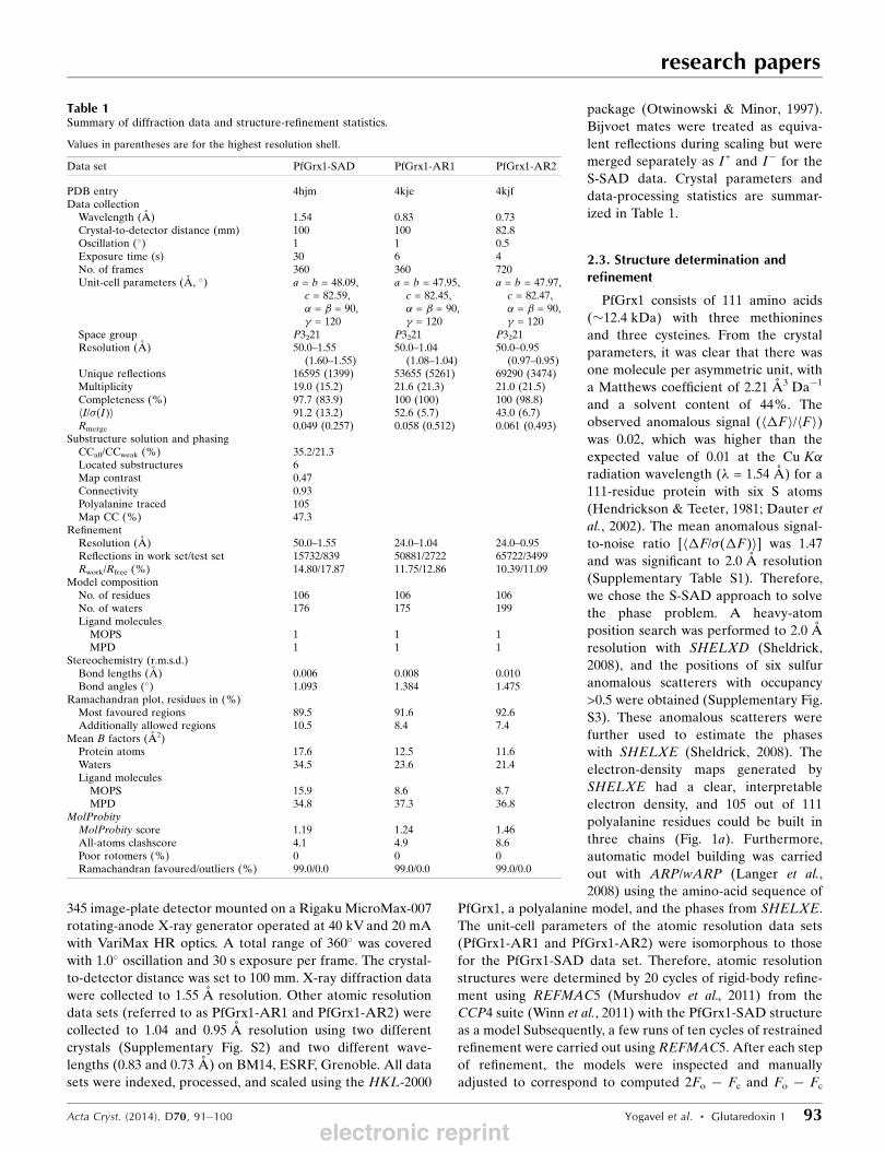
Table 1Summary of diffraction data and structure-refinement statistics.
Values in parentheses are for the highest resolution shell.
Data set PfGrx1-SAD PfGrx1-AR1 PfGrx1-AR2
PDB entry 4hjm 4kje 4kjfData collection
Wavelength (A) 1.54 0.83 0.73Crystal-to-detector distance (mm) 100 100 82.8Oscillation (�) 1 1 0.5Exposure time (s) 30 6 4No. of frames 360 360 720Unit-cell parameters (A, �) a = b = 48.09,
c = 82.59,� = � = 90,� = 120
a = b = 47.95,c = 82.45,� = � = 90,� = 120
a = b = 47.97,c = 82.47,� = � = 90,� = 120
Space group P3221 P3221 P3221Resolution (A) 50.0–1.55
(1.60–1.55)50.0–1.04
(1.08–1.04)50.0–0.95
(0.97–0.95)Unique reflections 16595 (1399) 53655 (5261) 69290 (3474)Multiplicity 19.0 (15.2) 21.6 (21.3) 21.0 (21.5)Completeness (%) 97.7 (83.9) 100 (100) 100 (98.8)hI/�(I)i 91.2 (13.2) 52.6 (5.7) 43.0 (6.7)Rmerge 0.049 (0.257) 0.058 (0.512) 0.061 (0.493)
Substructure solution and phasingCCall/CCweak (%) 35.2/21.3Located substructures 6Map contrast 0.47Connectivity 0.93Polyalanine traced 105Map CC (%) 47.3
RefinementResolution (A) 50.0–1.55 24.0–1.04 24.0–0.95Reflections in work set/test set 15732/839 50881/2722 65722/3499Rwork/Rfree (%) 14.80/17.87 11.75/12.86 10.39/11.09
Model compositionNo. of residues 106 106 106No. of waters 176 175 199Ligand molecules
MOPS 1 1 1MPD 1 1 1
Stereochemistry (r.m.s.d.)Bond lengths (A) 0.006 0.008 0.010Bond angles (�) 1.093 1.384 1.475
Ramachandran plot, residues in (%)Most favoured regions 89.5 91.6 92.6Additionally allowed regions 10.5 8.4 7.4
Mean B factors (A2)Protein atoms 17.6 12.5 11.6Waters 34.5 23.6 21.4Ligand molecules
MOPS 15.9 8.6 8.7MPD 34.8 37.3 36.8
MolProbityMolProbity score 1.19 1.24 1.46All-atoms clashscore 4.1 4.9 8.6Poor rotomers (%) 0 0 0Ramachandran favoured/outliers (%) 99.0/0.0 99.0/0.0 99.0/0.0
electronic reprint
electron-density maps using Coot (Emsley & Cowtan, 2004).
During the progress of the refinement, 3-(N-morpholino)-
propanesulfonic acid (MOPS) and 2-methyl-2,4-pentanediol
(MPD) solvent molecules and water molecules were manually
added and included in the refinement based on positive peaks
in difference Fourier maps. The final round of refinement was
carried out using phenix.refine (Afonine et al., 2012). The
occupancies of alternate conformations of protein residues
and water molecules were also refined. The quality of the
atomic model was assessed with PROCHECK (Laskowski et
al., 1993) and MolProbity (Chen et al., 2010). The figures were
generated using Chimera (Pettersen et al., 2004) and PyMOL
(http://www.pymol.org). Atomic coordinates and structure
factors for the PfGrx1-SAD, PfGrx1-AR1 and PfGrx1-AR2
data sets have been deposited in the Protein Data Bank,
Research Collaboratory for Structural Bioinformatics,
Rutgers University, New Brunswick, New Jersey, USA (http://
www.rcsb.org/) with accession codes 4hjm, 4kje and 4kjf,
respectively. The Friedel pairs for the S-SAD data were also
included in the deposited structure factors.
3. Results and discussion
3.1. Model quality and overall description of PfGrx1
PfGrx1 has the typical glutaredoxin/thioredoxin fold with a
core four-stranded �-sheet (strands �1 and �2 are parallel, and
strands �3 and �4 are antiparallel) flanked by five �-helices
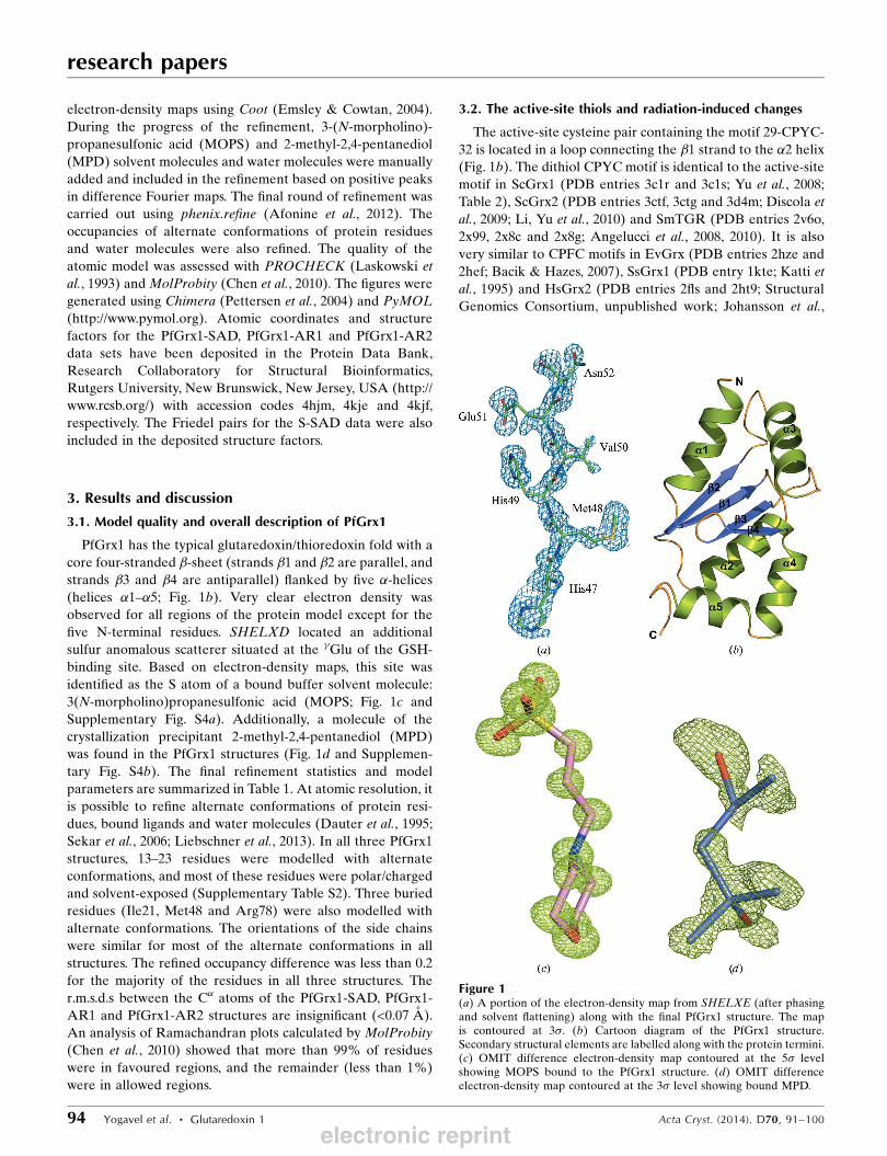
(helices �1–�5; Fig. 1b). Very clear electron density was
observed for all regions of the protein model except for the
five N-terminal residues. SHELXD located an additional
sulfur anomalous scatterer situated at the �Glu of the GSH-
binding site. Based on electron-density maps, this site was
identified as the S atom of a bound buffer solvent molecule:
3(N-morpholino)propanesulfonic acid (MOPS; Fig. 1c and
Supplementary Fig. S4a). Additionally, a molecule of the
crystallization precipitant 2-methyl-2,4-pentanediol (MPD)
was found in the PfGrx1 structures (Fig. 1d and Supplemen-
tary Fig. S4b). The final refinement statistics and model
parameters are summarized in Table 1. At atomic resolution, it
is possible to refine alternate conformations of protein resi-
dues, bound ligands and water molecules (Dauter et al., 1995;
Sekar et al., 2006; Liebschner et al., 2013). In all three PfGrx1
structures, 13–23 residues were modelled with alternate
conformations, and most of these residues were polar/charged
and solvent-exposed (Supplementary Table S2). Three buried
residues (Ile21, Met48 and Arg78) were also modelled with
alternate conformations. The orientations of the side chains
were similar for most of the alternate conformations in all
structures. The refined occupancy difference was less than 0.2
for the majority of the residues in all three structures. The
r.m.s.d.s between the C� atoms of the PfGrx1-SAD, PfGrx1-
AR1 and PfGrx1-AR2 structures are insignificant (<0.07 A).
An analysis of Ramachandran plots calculated by MolProbity
(Chen et al., 2010) showed that more than 99% of residues
were in favoured regions, and the remainder (less than 1%)
were in allowed regions.
3.2. The active-site thiols and radiation-induced changes
The active-site cysteine pair containing the motif 29-CPYC-
32 is located in a loop connecting the �1 strand to the �2 helix
(Fig. 1b). The dithiol CPYC motif is identical to the active-site
motif in ScGrx1 (PDB entries 3c1r and 3c1s; Yu et al., 2008;
Table 2), ScGrx2 (PDB entries 3ctf, 3ctg and 3d4m; Discola et
al., 2009; Li, Yu et al., 2010) and SmTGR (PDB entries 2v6o,
2x99, 2x8c and 2x8g; Angelucci et al., 2008, 2010). It is also
very similar to CPFC motifs in EvGrx (PDB entries 2hze and
2hef; Bacik & Hazes, 2007), SsGrx1 (PDB entry 1kte; Katti et
al., 1995) and HsGrx2 (PDB entries 2fls and 2ht9; Structural
Genomics Consortium, unpublished work; Johansson et al.,
research papers
94 Yogavel et al. � Glutaredoxin 1 Acta Cryst. (2014). D70, 91–100
Figure 1(a) A portion of the electron-density map from SHELXE (after phasingand solvent flattening) along with the final PfGrx1 structure. The mapis contoured at 3�. (b) Cartoon diagram of the PfGrx1 structure.Secondary structural elements are labelled along with the protein termini.(c) OMIT difference electron-density map contoured at the 5� levelshowing MOPS bound to the PfGrx1 structure. (d) OMIT differenceelectron-density map contoured at the 3� level showing bound MPD.
electronic reprint
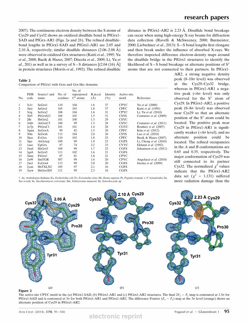
2007). The continuous electron density between the S atoms of
Cys29 and Cys32 shows an oxidized disulfide bond in PfGrx1-
SAD and PfGrx-AR1 (Figs. 2a and 2b). The refined disulfide-
bond lengths in PfGrx1-SAD and PfGrx1-AR1 are 2.05 and
2.10 A, respectively; similar disulfide distances (2.06–2.08 A)
were observed in oxidized Grx structures (Katti et al., 1995; Yu
et al., 2008; Bacik & Hazes, 2007; Discola et al., 2009; Li, Yu et
al., 201) as well as in a survey of S—S distances [(2.04 (16) A]
in protein structures (Morris et al., 1992). The refined disulfide
distance in PfGrx1-AR2 is 2.23 A. Disulfide bond breakage
can occur when using high-energy X-ray beams for diffraction
data collection (Ravelli & McSweeney, 2000; Burmeister,
2000; Liebschner et al., 2013): S—S bond lengths first elongate
and then break under the influence of absorbed X-rays. We
therefore inspected difference electron-density maps around
the disulfide bridge in the PfGrx1 structures to identify the
likelihood of S—S bond breakage or alternate positions of S�
atoms that are not connected to their partners. In PfGrx1-
AR2, a strong negative density
peak (8–10� level) was observed
at the Cys29–Cys32 bridge,
whereas in PfGrx1-AR1 a nega-
tive peak (<6� level) was only
observed for the S� atom of
Cys29. In PfGrx1-AR2, a positive
peak (6–8� level) was observed
near Cys29 so that an alternate
position of the S� atom could be
located. The positive peak near
Cys29 in PfGrx1-AR1 is signifi-
cantly weaker (<4� level), and no
alternate position could be
located. The refined occupancies
in the A and B conformations are
0.65 and 0.35, respectively. The
major conformation of Cys29 was
still connected to its partner
Cys32. The normalized �2 values
indicate that the PfGrx1-AR2
data set (�2 = 1.131) suffered
more radiation damage than the
research papers
Acta Cryst. (2014). D70, 91–100 Yogavel et al. � Glutaredoxin 1 95
Figure 2The active-site CPYC motif in the (a) PfGrx1-SAD, (b) PfGrx1-AR1 and (c) PfGrx1-AR2 structures. The final 2Fo � Fc map is contoured at 1.5� forPfGrx1-SAD and is contoured at 3� for both PfGrx1-AR1 and PfGrx1-AR2. The difference Fourier (Fo � Fc) map at the 3� level (orange) shows analternate position of Cys29 in PfGrx1-AR2.
Table 2Comparison of PfGrx1 with Grxs and Grx-like domains.
No.PDBcode
Source† andname
No. ofresidues
No. ofsuperposedC� atoms
R.m.s.d.(A)
Identity(%)
Active-sitemotif Reference
1 3c1r ScGrx1 110 104 1.6 37 CPYC Yu et al. (2008)2 1kte SsGrx1 105 103 1.8 37 CPFC Katti et al. (1995)3 3ctg ScGrx2 108 104 1.6 31 CPYC Li, Yu et al. (2010)4 3fz9 PtGrxS12 106 102 1.5 31 CSYS Couturier et al. (2009)5 2fls HsGrx2 101 100 1.3 29 CSYC6 3rhb AtGrxC5 100 99 1.3 28 CSYC Couturier et al. (2011)7 2e7p PtGrxC1 101 101 1.4 28 CGYC Rouhier et al. (2007)8 3qmx SyGrxA 99 82 1.5 28 CPFC Kim et al. (2012)9 3l4n ScGrx6 113 104 2.0 26 CSYS Luo et al. (2010)10 2hze EvGrx 110 103 1.8 25 CPFC Bacik & Hazes (2007)11 3ipz AtGrxcp 109 99 1.9 23 CGFS Li, Cheng et al. (2010)12 1aaz EpGrx 87 74 2.2 23 CVYC Eklund et al. (1992)13 2wul HsGrx5 109 99 1.7 22 CGFS Johansson et al. (2011)14 3gx8 ScGrx5 111 102 1.6 21 CGFS15 3msz FtGrx1 87 81 1.8 21 CPYC16 2x99 SmTGR 587 99 1.6 20 CPYC Angelucci et al. (2010)17 2wci EcGrx4 113 99 2.0 20 CGFS Iwema et al. (2009)18 2yan HsTXLN2 105 97 2.1 16 CGFS19 3zyw HsGrx3D1 111 99 2.3 16 CGFS
† At, Arabidopsis thaliana; Ec, Escherichia coli; Ev, Ectromelia virus; Hs, Homo sapiens; Pt, Populus tremula � P. tremuloides; Ss,Sus scrofa; Sc, Saccharomyces cerevisiae; Sm, Schistosoma mansoni; Sy, Synechocystis sp.
electronic reprint
research papers
96 Yogavel et al. � Glutaredoxin 1 Acta Cryst. (2014). D70, 91–100
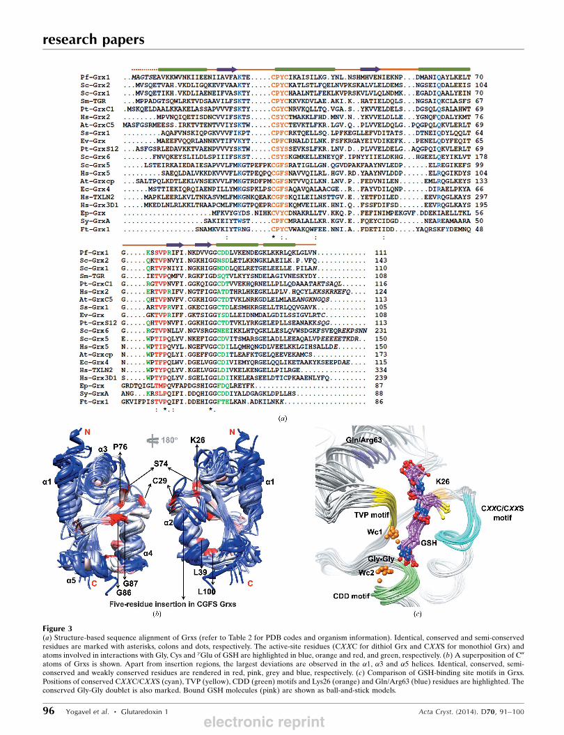
Figure 3(a) Structure-based sequence alignment of Grxs (refer to Table 2 for PDB codes and organism information). Identical, conserved and semi-conservedresidues are marked with asterisks, colons and dots, respectively. The active-site residues (CXXC for dithiol Grx and CXXS for monothiol Grx) andatoms involved in interactions with Gly, Cys and �Glu of GSH are highlighted in blue, orange and red, and green, respectively. (b) A superposition of C�
atoms of Grxs is shown. Apart from insertion regions, the largest deviations are observed in the �1, �3 and �5 helices. Identical, conserved, semi-conserved and weakly conserved residues are rendered in red, pink, grey and blue, respectively. (c) Comparison of GSH-binding site motifs in Grxs.Positions of conserved CXXC/CXXS (cyan), TVP (yellow), CDD (green) motifs and Lys26 (orange) and Gln/Arg63 (blue) residues are highlighted. Theconserved Gly-Gly doublet is also marked. Bound GSH molecules (pink) are shown as ball-and-stick models.
electronic reprint
PfGrx1-AR1 data set (�2 = 0.839). The deposited radiation
dose was calculated using RADDOSE (Zeldin et al., 2013).
The average and maximum doses deposited were 6.9 and
67.9 kGy, respectively, for the PfGrx1-AR1 crystals and 7.9
and 78.6 kGy, respectively, for the PfGrx1-AR2 crystals.
Therefore, radiation damage may be limited. The average B
factor of the active-site CPYC motif in the PfGrx1 structures
(14.4, 8.4 and 8.5 A2 for PfGrx1-SAD, PfGrx1-AR1 and
PfGrx1-AR2, respectively) is lower than the overall B values
(Table 1), which are similar to those of other dithiol Grxs
(Rouhier et al., 2007; Yu et al., 2008; Discola et al., 2009; Li, Yu
et al., 2010).
3.3. Structural comparison of PfGrx1 with otherGrxs/Grx-like domains
The sequence identity between Grxs and Grx-like domains
is in the range 16–37% and few residues (<15) are conserved
among them (Fig. 3a and Table 2). The superposition of 74–
104 C� atoms of the PfGrx1 structure with monothiol/dithiol,
oxidized/reduced and GSH-bound/unbound forms of Grx
structures from different organisms shows an r.m.s.d. range of
1.3–2.3 A (Fig. 3b and Table 2), suggesting that the overall
folding of PfGrx1 is similar to that of other Grxs and Grx-like
domains. Superposition of Grxs and Grx-like domains shows
very good agreement for the core �-strands and the active-site
motif containing the �2 helix and the �4 helix (Fig. 3b). Apart
from the insertion loops, the maximum displacement was
observed in the �1, �3 and �5 helices. Most conserved residues
are present around the active-site regions of these enzymes
(Fig. 3b). The GSH-binding motifs (CXXC or CXXS, TVP and
CDD) are structurally conserved in oxidized/reduced and
GSH-bound/unbound forms of both the monothiol and dithiol
Grxs except for the Gly-binding residue at position 63 (Fig. 3c).
In most Grxs, a conserved hydrophobic residue followed by
a Gly-Gly doublet precedes the CDD motif. The Gly-Gly
doublet provides room for two conserved water molecules
(Wc1 and Wc2) which interact with the �Glu of GSH. Bound
GSH molecules adopt extended conformations apart from in
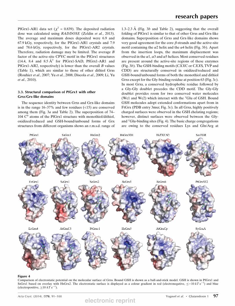
FtGrx (PDB entry 3msz; Fig. 3c). In all Grxs, highly positively
charged surfaces were observed in the GSH chelating regions;
however, distinct surfaces were observed between the Gly-
and �Glu-binding sites (Fig. 4). The basic charge congregations
are owing to the conserved residues Lys and Gln/Arg at
research papers
Acta Cryst. (2014). D70, 91–100 Yogavel et al. � Glutaredoxin 1 97
Figure 4Comparison of electrostatic potential on the molecular surface of Grxs. Bound GSH is shown as a ball-and-stick model. GSH is shown in PfGrx1 andSsGrx1 based on overlay with HsGrx2. The electrostatic surface is displayed as a colour gradient in red (electronegative, ��10 kT e�1) and blue(electropositive, �10 kT e�1).
electronic reprint
research papers
98 Yogavel et al. � Glutaredoxin 1 Acta Cryst. (2014). D70, 91–100
positions 26 (based on the PfGrx1 residue numbering) and 63,
respectively. The distinct potential distribution in the �Glu-
binding region is a consequence of fewer conserved residues in
the CDD motif as well as at positions 72 (Lys/Arg/Gln/Trp)
and 77 (Arg/Gln/Asn) (Fig. 3a).
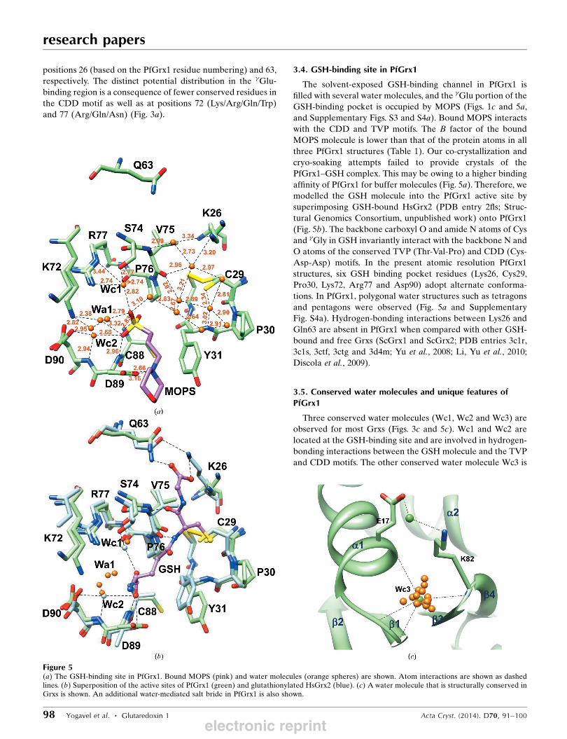
3.4. GSH-binding site in PfGrx1
The solvent-exposed GSH-binding channel in PfGrx1 is
filled with several water molecules, and the �Glu portion of the
GSH-binding pocket is occupied by MOPS (Figs. 1c and 5a,
and Supplementary Figs. S3 and S4a). Bound MOPS interacts
with the CDD and TVP motifs. The B factor of the bound
MOPS molecule is lower than that of the protein atoms in all
three PfGrx1 structures (Table 1). Our co-crystallization and
cryo-soaking attempts failed to provide crystals of the
PfGrx1–GSH complex. This may be owing to a higher binding
affinity of PfGrx1 for buffer molecules (Fig. 5a). Therefore, we
modelled the GSH molecule into the PfGrx1 active site by
superimposing GSH-bound HsGrx2 (PDB entry 2fls; Struc-
tural Genomics Consortium, unpublished work) onto PfGrx1
(Fig. 5b). The backbone carboxyl O and amide N atoms of Cys
and �Gly in GSH invariantly interact with the backbone N and
O atoms of the conserved TVP (Thr-Val-Pro) and CDD (Cys-
Asp-Asp) motifs. In the present atomic resolution PfGrx1
structures, six GSH binding pocket residues (Lys26, Cys29,
Pro30, Lys72, Arg77 and Asp90) adopt alternate conforma-
tions. In PfGrx1, polygonal water structures such as tetragons
and pentagons were observed (Fig. 5a and Supplementary
Fig. S4a). Hydrogen-bonding interactions between Lys26 and
Gln63 are absent in PfGrx1 when compared with other GSH-
bound and free Grxs (ScGrx1 and ScGrx2; PDB entries 3c1r,
3c1s, 3ctf, 3ctg and 3d4m; Yu et al., 2008; Li, Yu et al., 2010;
Discola et al., 2009).
3.5. Conserved water molecules and unique features ofPfGrx1
Three conserved water molecules (Wc1, Wc2 and Wc3) are
observed for most Grxs (Figs. 3c and 5c). Wc1 and Wc2 are
located at the GSH-binding site and are involved in hydrogen-
bonding interactions between the GSH molecule and the TVP
and CDD motifs. The other conserved water molecule Wc3 is
Figure 5(a) The GSH-binding site in PfGrx1. Bound MOPS (pink) and water molecules (orange spheres) are shown. Atom interactions are shown as dashedlines. (b) Superposition of the active sites of PfGrx1 (green) and glutathionylated HsGrx2 (blue). (c) A water molecule that is structurally conserved inGrxs is shown. An additional water-mediated salt bride in PfGrx1 is also shown.
electronic reprint
located at the position of the �1 helix, the �1 strand, and the
loop between the �3 and �4 strands. In PfGrx1, Glu28
precedes the active-site residues, and the corresponding
position in other Grxs is populated by polar Ser/Thr/Asn,
aromatic Tyr/Trp, or Gly residues. Similarly, Glu51 is located
between the �1 helix and the �2 strand, and the corresponding
position in other Grxs has a conserved hydrophobic residue
Val/Leu/Ile or aromatic residue Phe/Tyr (Fig. 3a). A water-
mediated salt bridge between the �1 helix and the loop
between the �3 and �4 strands is found in PfGrx1, and this
interaction is absent in other Grxs (Fig. 5c). Unique salt
bridges between the �1 and �3 helices and the �4 strand and
the �4 and �5 helices were found only in PfGrx1, PtGrxS12
and SsGrx1 (Supplementary Fig. S5).
4. Conclusions
In this work, we have determined the atomic structure of the
dithiol Grx PfGrx1 and compared it in depth with those of
other Grxs from different organisms. Our results indicate that
monothiol (CXXS) and dithiol (CXXC) Grxs differ signifi-
cantly in their helix-capping hydrogen bonds (Supplementary
Figs. S6, S7 and S8). Both monothiol and dithiol Grxs contain
three conserved water molecules, of which two are located in
the GSH-binding site while the third is located between �-
strands and the �1 helix. The dithiol-containing redox proteins
thioredoxin (Trx), glutaredoxin (Grx) and plasmoredoxin
(Plrx), with the latter being exclusively found in Plasmodium
species, play central roles in maintaining redox homeostasis in
malarial parasites (Sturm et al., 2009). The PfGrx1 structure
presented here in complex with MOPS and MPD provides
novel insights concerning interacting surfaces whose roles in in
vivo interactions with parasite biomolecules remain to be
explored in further detail.
The X-ray facility at ICGEB was funded by the Wellcome
Trust. This work was generously supported with grants from
the Department of Biotechnology (DBT), Government of
India to TT, AS and MY, and with the Deutsche
Forschungsgemeinschaft (BE 1540/18-1 to KB and SR).
References
Afonine, P. V., Grosse-Kunstleve, R. W., Echols, N., Headd, J. J.,Moriarty, N. W., Mustyakimov, M., Terwilliger, T. C., Urzhumtsev,A., Zwart, P. H. & Adams, P. D. (2012). Acta Cryst. D68, 352–367.
Angelucci, F., Dimastrogiovanni, D., Boumis, G., Brunori, M., Miele,A. E., Saccoccia, F. & Bellelli, A. (2010). J. Biol. Chem. 285, 32557–32567.
Angelucci, F., Miele, A. E., Boumis, G., Dimastrogiavanni, D.,Brunori, M. & Belleli, A. (2008). Proteins, 72, 936–945.
Axelsson, K. & Mannervik, B. (1980). Biochim. Biophys. Acta, 613,324–336.
Bacik, J. P. & Hazes, B. (2007). J. Mol. Biol. 365, 1545–1558.Balmer, Y., Koller, A., del Val, G., Manieri, W., Schurmann, P. &
Buchanan, B. B. (2003). Proc. Natl Acad. Sci. USA, 100, 370–375.Berndt, C., Lillig, C. H. & Holmgren, A. (2008). Biochim. Biophys.
Acta, 1783, 641–650.Burmeister, W. P. (2000). Acta Cryst. D56, 328–341.
Chen, V. B., Arendall, W. B., Headd, J. J., Keedy, D. A., Immormino,R. M., Kapral, G. J., Murray, L. W., Richardson, J. S. & Richardson,D. C. (2010). Acta Cryst. D66, 12–21.
Chrestensen, C. A., Starke, D. W. & Mieyal, J. J. (2000). J. Biol. Chem.275, 26556–26565.
Couturier, J., Koh, C. S., Zaffagnini, M., Winger, A. M., Gualberto,J. M., Corbier, C., Decottignies, P., Jacquot, J.-P., Lemaire, S. D.,Didierjean, C. & Rouhier, N. (2009). J. Biol. Chem. 284, 9299–9310.
Couturier, J., Stroher, E., Albetel, A. N., Roret, T., Muthurama-lingam, M., Tarrago, L., Seidel, T., Tsan, P., Jacquot, J.-P., Johnson,M. K., Dietz, K. J., Didierjean, C. & Rouhier, N. (2011). J. Biol.Chem. 286, 27515–27527.
Dauter, Z., Dauter, M. & Dodson, E. J. (2002). Acta Cryst. D58,494–506.
Dauter, Z., Lamzin, V. S. & Wilson, K. S. (1995). Curr. Opin. Struct.Biol. 5, 784–790.
Discola, K. F., de Oliveira, M. A., Rosa Cussiol, J. R., Monteiro, G.,Barcena, J. A., Porras, P., Padilla, C. A., Guimaraes, B. G. & Netto,L. E. (2009). J. Mol. Biol. 385, 889–901.
Djuika, C. F., Fiedler, S., Schnolzer, M., Sanchez, C., Lanzer, M. &Deponte, M. (2013). Biochim. Biophys. Acta, 1830, 4073–4090.
Eklund, H., Ingelman, M., Soderberg, B. O., Uhlin, T., Nordlund, P.,Nikkola, M., Sonnerstam, U., Joelson, T. & Petratos, K. (1992). J.Mol. Biol. 228, 596–618.
Emsley, P. & Cowtan, K. (2004). Acta Cryst. D60, 2126–2132.Feng, Y., Zhong, N., Rouhier, N., Hase, T., Kusunoki, M., Jacquot,
J.-P., Jin, C. & Xia, B. (2006). Biochemistry, 45, 7998–8008.Fernandes, A. P. & Holmgren, A. (2004). Antioxid. Redox Signal. 6,
63–74.Gallogly, M. M., Starke, D. W. & Mieyal, J. J. (2009). Antioxid. Redox
Signal. 11, 1059–1081.Gonzalez Porque, P., Baldesten, A. & Reichard, P. (1970). J. Biol.
Chem. 245, 2371–2374.Hendrickson, W. A. & Teeter, M. M. (1981). Nature (London), 290,
107–113.Hoff, K. G., Culler, S. J., Nguyen, P. Q., McGuire, R. M., Silberg, J. J. &
Smolke, C. D. (2009). Chem. Biol. 16, 1299–1308.Holmgren, A. (1976). Proc. Natl Acad. Sci. USA, 73, 2275–2279.Holmgren, A. (1979). J. Biol. Chem. 254, 3672–3678.Holmgren, A. (2000). Antioxid. Redox Signal. 2, 811–820.Holmgren, A., Johansson, C., Berndt, C., Lonn, M. E., Hudemann, C.
& Lillig, C. H. (2005). Biochem. Soc. Trans. 33, 1375–1377.Iwema, T., Picciocchi, A., Traore, D. A. K., Ferrer, J.-L., Chauvat, F. &
Jacquamet, L. (2009). Biochemistry, 48, 6041–6043.Johansson, C., Kavanagh, K. L., Gileadi, O. & Oppermann, U. (2007).
J. Biol. Chem. 282, 3077–3082.Johansson, C., Roos, A. K., Montano, S. J., Sengupta, R.,
Filippakopoulos, P., Guo, K., von Delft, F., Holmgren, A.,Oppermann, U. & Kavanagh, K. L. (2011). Biochem. J. 433,303–311.
Katti, S. K., Robbins, A. H., Yang, Y. & Wells, W. W. (1995). ProteinSci. 4, 1998–2005.
Kehr, S., Sturm, N., Rahlfs, S., Przyborski, J. M. & Becker, K. (2010).PLoS Pathog. 6, e1001242.
Kim, S. G., Chung, J.-S., Sutton, R. B., Lee, J.-S., Lopez-Maury, L.,Lee, S. Y., Florencio, F. J., Lin, T., Zabet-Moghaddam, M., Wood,M. J., Nayak, K., Madem, V., Tripathy, J. N., Kim, S.-K. & Knaff,D. B. (2012). Biochim. Biophys. Acta, 1824, 392–403.
Langer, G., Cohen, S. X., Lamzin, V. S. & Perrakis, A. (2008). NatureProtoc. 3, 1171–1179.
Laskowski, R. A., MacArthur, M. W., Moss, D. S. & Thornton, J. M.(1993). J. Appl. Cryst. 26, 283–291.
Lemaire, S. D., Guillon, B., Le Marechal, P., Keryer, E., Miginiac-Maslow, M. & Decottignies, P. (2004). Proc. Natl Acad. Sci. USA,101, 7475–7480.
Li, L., Cheng, N., Hirschi, K. D. & Wang, X. (2010). Acta Cryst. D66,725–732.
research papers
Acta Cryst. (2014). D70, 91–100 Yogavel et al. � Glutaredoxin 1 99electronic reprint
Li, W.-F., Yu, J., Ma, X.-X., Teng, Y.-B., Luo, M., Tang, Y.-J. & Zhou,C.-Z. (2010). Biochem. Biophys. Acta, 1804, 1542–1547.
Liebschner, D., Dauter, M., Brzuszkiewicz, A. & Dauter, Z. (2013).Acta Cryst. D69, 1447–1462.
Lill, R., Dutkiewicz, R., Elsasser, H. P., Hausmann, A., Netz, D. J.,Pierik, A. J., Stehling, O., Urzica, E. & Muhlenhoff, U. (2006).Biochim. Biophys. Acta, 1763, 652–667.
Lillig, C. H., Berndt, C. & Holmgren, A. (2008). Biochim. Biophys.Acta, 1780, 1304–1317.
Lillig, C. H., Berndt, C., Vergnolle, O., Lonn, M. E., Hudemann, C.,Bill, E. & Holmgren, A. (2005). Proc. Natl Acad. Sci. USA, 102,8168–8173.
Lindahl, M. & Florencio, F. J. (2003). Proc. Natl Acad. Sci. USA, 100,16107–16112.
Luo, M., Jiang, Y.-L., Ma, X.-X., Tang, Y.-J., He, Y.-X., Yu, J., Zhang,R.-G., Chen, Y. & Zhou, C.-Z. (2010). J. Mol. Biol. 398, 614–622.
Matthews, J. R., Wakasugi, N., Virelizier, J. L., Yodoi, J. & Hay, R. T.(1992). Nucleic Acids Res. 20, 3821–3830.
Morris, A. L., MacArthur, M. W., Hutchinson, E. G. & Thornton, J. M.(1992). Proteins, 12, 345–364.
Motohashi, K., Kondoh, A., Stumpp, M. T. & Hisabori, T. (2001).Proc. Natl Acad. Sci. USA, 98, 11224–11229.
Muhlenhoff, U., Molik, S., Godoy, J. R., Uzarska, M. A., Richter, N.,Seubert, A., Zhang, Y., Stubbe, J., Pierrel, F., Herrero, E., Lillig,C. H. & Lill, R. (2010). Cell Metab. 12, 373–385.
Murshudov, G. N., Skubak, P., Lebedev, A. A., Pannu, N. S., Steiner,R. A., Nicholls, R. A., Winn, M. D., Long, F. & Vagin, A. A. (2011).Acta Cryst. D67, 355–367.
Otwinowski, Z. & Minor, W. (1997). Methods Enzymol. 276, 307–326.
Pettersen, E. F., Goddard, T. D., Huang, C. C., Couch, G. S.,Greenblatt, D. M., Meng, E. C. & Ferrin, T. E. (2004). J. Comput.Chem. 25, 1605–1612.
Pineda-Molina, E., Klatt, P., Vazquez, J., Marina, A., Garcıa deLacoba, M., Perez-Sala, D. & Lamas, S. (2001). Biochemistry, 40,14134–14142.
Rahlfs, S., Fischer, M. & Becker, K. (2001). J. Biol. Chem. 276, 37133–37140.
Ravelli, R. B. G. & McSweeney, S. M. (2000). Structure, 8, 315–328.Rouhier, N., Unno, H., Bandyopadhyay, S., Masip, L., Kim, S.-K.,
Hirasawa, M., Gualberto, J. M., Lattard, V., Kusunoki, M., Knaff,D. B., Georgiou, G., Hase, T., Johnson, M. K. & Jacquot, J.-P. (2007).Proc. Natl Acad. Sci. USA, 104, 7379–7384.
Rouhier, N., Villarejo, A., Srivastava, M., Gelhaye, E., Keech, O.,Droux, M., Finkemeier, I., Samuelsson, G., Dietz, K. J., Jacquot,J.-P. & Wingsle, G. (2005). Antioxid. Redox Signal. 7, 919–929.
Sekar, K., Yogavel, M., Gayathri, D., Velmurugan, D., Krishna, R.,Poi, M.-J., Dauter, Z., Dauter, M. & Tsai, M.-D. (2006). Acta Cryst.F62, 1–5.
Sheldrick, G. M. (2008). Acta Cryst. A64, 112–122.Shelton, M. D., Chock, P. B. & Mieyal, J. J. (2005). Antioxid. Redox
Signal. 7, 348–366.Sturm, N., Jortzik, E., Mailu, B. M., Koncarevic, S., Deponte, M.,
Forchhammer, K., Rahlfs, S. & Becker, K. (2009). PLoS Pathog. 5,e1000383.
Tripathi, T., Roseler, A., Rahlfs, S., Becker, K. & Bhakuni, V. (2010).Biochimie, 92, 284–291.
Tsang, M. L. (1981). J. Bacteriol. 146, 1059–1066.Winn, M. D. et al. (2011). Acta Cryst. D67, 235–242.Wong, J. H., Cai, N., Balmer, Y., Tanaka, C. K., Vensel, W. H.,
Hurkman, W. J. & Buchanan, B. B. (2004). Phytochemistry, 65,1629–1640.
Yu, J., Zhang, N.-N., Yin, P.-D., Cui, P.-X. & Zhou, C.-Z. (2008).Proteins, 72, 1077–1083.
Zeldin, O. B., Gerstel, M. & Garman, E. F. (2013). J. Appl. Cryst. 46,1225–1230.
research papers
100 Yogavel et al. � Glutaredoxin 1 Acta Cryst. (2014). D70, 91–100
electronic reprint
Related Documents