Atomic Force Microscopy of Microwear Traces on Mousterian Tools From Myshtylagty Lagat (Weasel Cave), Russia NATHAN R. FAULKS 1 ,LARRY R. KIMBALL 2 ,NAZIM HIDJRATI 3 , AND TONYA S. COFFEY 1 1 Department of Physics and Astronomy, Appalachian State University, Boone, North Carolina 2 Department of Anthropology, Appalachian State University, Boone, North Carolina 3 Institute of History and Archaeology, North Ossetian State University, Vladikavkaz, North Ossetia, Russia Summary: Since durable technology emerged be- tween 3.4 and 3.2 million years ago, stone tools served as a major material means that hominins used to survive. Determining how different lithic tools functioned is a principal question in human evolution. The main experimentally based approach to the functional study of lithic technology uses stereo and incident-light microscopy, and is known as the Keeley Method. Although this method has demonstrated success in linking the morphology of microwear traces on flint tools to the function of the tool, there is no agreed upon model of how these microwear polishes form. At the same time, the characterization of these polishes has been a largely qualitative process. Herein, we use the atomic force microscope (AFM) to scan microwear traces on Middle Palaeolithic (Mousterian) tools from Weasel Cave, Russia to show quantitative data and small scale features of microwear polishes interpreted (using the Keeley Method) as due to contact with meat, fresh hide, dry hide, bone, wood, and hafting. These results follow those of to the previous AFM study on the experimental tools, namely that the meat and dry hide polishes are the least developed polishes with smaller changes in roughness and that the bone polish and wood polishes are more highly developed polishes and exhibit larger changes in roughness. SCANNING 33: 1–12, 2011. r 2011 Wiley Periodicals, Inc. Key words: atomic force microscope, microwear, scanning probe, palaeolithic stone tool function Introduction Since flaked lithic technology emerged between 3.4–3.2 and 2.5–2.6 mya (Semaw, 2000; McPherron et al., 2010), stone tools served as a crucial adaptive means in early hominin evolution. The determina- tion of how stone tools were used is important to our understanding of human evolution. The first experimentally based approach to the functional study of stone tools was by Russian archaeologist Sergei A. Semenov (’64) using both stereo- and incident-light microscopy. He was able to observe use-wear traces which, when compared with micro- wear traces on experimentally used replicas, per- mitted the identification of the kind of material being worked and the kinematics of individual archaeological tools. The first replication of Seme- nov’s method in the West was by Lawrence H. Keeley (’77, ’80) using incident-light microscopy with magnifications of 50–400 . The ‘‘Keeley Method’’ permitted the observation of additional microwear traces and a more refined characteriza- tion of the polishes themselves. Accordingly, a microwear polish ‘‘can be described in terms of its brightness or dullness (that is, how much light it reflects) and its roughness or smoothness, as well as the presence of certain topographical features, like pits, undulations, and so forth’’ (Keeley, ’80). Thus, reflectivity, roughness, and microtopography were the major axes of variation that differentiated microwear polishes. DOI 10.1002/sca.20273 Published online in Wiley Online Library (wileyonlinelibrary.com) Received 8 February 2011; Accepted with revision 11 July 2011 Address for reprints: Tonya S. Coffey, 327 CAP Science Building, 525 Rivers St, Boone, NC 28608 E-mail: [email protected] Contract grant sponsor: NSF; Contract grant number: DMR 0821124; Contract grant sponsor: Wenner-Gren Foundation for Anthropological Research; Contract grant number: 6157; Contract grant sponsors: L.S.B. Leakey Foundation; IREX; St. George Russian-American Archaeological Program; Appalachian State University; Institute for History and Archaeology at North Ossetian State University; NC Space Grant. SCANNING VOL. 33, 1–12 (2011) & Wiley Periodicals, Inc.

Welcome message from author
This document is posted to help you gain knowledge. Please leave a comment to let me know what you think about it! Share it to your friends and learn new things together.
Transcript
Atomic Force Microscopy of Microwear Traces on Mousterian ToolsFrom Myshtylagty Lagat (Weasel Cave), Russia
NATHAN R. FAULKS1, LARRY R. KIMBALL
2, NAZIM HIDJRATI3, AND TONYA S. COFFEY
1
1Department of Physics and Astronomy, Appalachian State University, Boone, North Carolina2Department of Anthropology, Appalachian State University, Boone, North Carolina3Institute of History and Archaeology, North Ossetian State University, Vladikavkaz, North Ossetia, Russia
Summary: Since durable technology emerged be-tween 3.4 and 3.2 million years ago, stone toolsserved as a major material means that homininsused to survive. Determining how different lithictools functioned is a principal question in humanevolution. The main experimentally based approachto the functional study of lithic technology usesstereo and incident-light microscopy, and is knownas the Keeley Method. Although this method hasdemonstrated success in linking the morphology ofmicrowear traces on flint tools to the function of thetool, there is no agreed upon model of how thesemicrowear polishes form. At the same time, thecharacterization of these polishes has been a largelyqualitative process. Herein, we use the atomic forcemicroscope (AFM) to scan microwear traces onMiddle Palaeolithic (Mousterian) tools from WeaselCave, Russia to show quantitative data and smallscale features of microwear polishes interpreted(using the Keeley Method) as due to contact withmeat, fresh hide, dry hide, bone, wood, and hafting.These results follow those of to the previous AFMstudy on the experimental tools, namely that themeat and dry hide polishes are the least developedpolishes with smaller changes in roughness and thatthe bone polish and wood polishes are more highlydeveloped polishes and exhibit larger changes in
roughness. SCANNING 33: 1–12, 2011. r 2011Wiley Periodicals, Inc.
Key words: atomic force microscope, microwear,scanning probe, palaeolithic stone tool function
Introduction
Since flaked lithic technology emerged between3.4–3.2 and 2.5–2.6 mya (Semaw, 2000; McPherronet al., 2010), stone tools served as a crucial adaptivemeans in early hominin evolution. The determina-tion of how stone tools were used is important toour understanding of human evolution. The firstexperimentally based approach to the functionalstudy of stone tools was by Russian archaeologistSergei A. Semenov (’64) using both stereo- andincident-light microscopy. He was able to observeuse-wear traces which, when compared with micro-wear traces on experimentally used replicas, per-mitted the identification of the kind of materialbeing worked and the kinematics of individualarchaeological tools. The first replication of Seme-nov’s method in the West was by Lawrence H.Keeley (’77, ’80) using incident-light microscopywith magnifications of 50–400� . The ‘‘KeeleyMethod’’ permitted the observation of additionalmicrowear traces and a more refined characteriza-tion of the polishes themselves. Accordingly, amicrowear polish ‘‘can be described in terms of itsbrightness or dullness (that is, how much light itreflects) and its roughness or smoothness, as well asthe presence of certain topographical features, likepits, undulations, and so forth’’ (Keeley, ’80). Thus,reflectivity, roughness, and microtopographywere the major axes of variation that differentiatedmicrowear polishes.
DOI 10.1002/sca.20273
Published online in Wiley Online Library (wileyonlinelibrary.com)
Received 8 February 2011; Accepted with revision 11 July 2011
Address for reprints: Tonya S. Coffey, 327 CAP Science Building,
525 Rivers St, Boone, NC 28608
E-mail: [email protected]
Contract grant sponsor: NSF; Contract grant number: DMR
0821124; Contract grant sponsor: Wenner-Gren Foundation for
Anthropological Research; Contract grant number: 6157; Contract
grant sponsors: L.S.B. Leakey Foundation; IREX; St. George
Russian-American Archaeological Program; Appalachian State
University; Institute for History and Archaeology at North Ossetian
State University; NC Space Grant.
SCANNING VOL. 33, 1–12 (2011)& Wiley Periodicals, Inc.
A formal classification of the Keeley Method byPlisson (’85) described microwear polishes moresystematically, whereby discrete microwear tracesare recognized by the specific attribute states for asuite of characteristics: localization, extent, texture,contour, brightness, striae, and polish (or coales-cence). He defined microwear traces for the contactmaterials of meat, wet hide, fresh hide, dry hide,ochred hide, bone, antler, horn, ivory, shell, fish,fresh wood, dry wood, soft plant, and amber(Plisson, ’85). The importation of striations (seeKeeley, ’80; Mansur, ’82, for classifications anddiscussions of striae formation) was recognized bySemenov (’64) as particularly important in ascer-taining the direction of tool use or kinematics.Kimball (’89) extended Plisson’s classification toinclude cane and hafting polishes. And, recently,Rots (2010) published a systematic, experimentally-based study of hafting traces.
Today, this revised, ‘‘high-power Keeley Meth-od’’ is the most generally accepted method for thedetermination of stone tool function (Juel Jensen,’88; Yerkes and Kardulias, ’93). Although themethod relies on the qualitative differentiation ofmicrowear polishes through incident-light micro-scopy at magnifications from 50� to 500� , in-creasingly microwear analysts employ severalinstruments with variable kinds of magnification:loupe (5–10� ), stereo-microscope (15–180� ),SEM, laser confocal, laser profilometer, and inter-ferometer, as well. Our use of the atomic force mi-croscope (AFM) is an additional method in thequantitative characterization of use-wear patterningand the understanding of microwear polish forma-tion.
One of the problems with contemporary micro-wear analysis is the lingering perception (Klein,2009) that it is not predicatably accurate. Thisis largely due to variable blind testing results(Bamforth, ’88; Bamforth et al., ’90), the relianceupon qualitative charateristics of microwear traces,and the lack of agreement about how microwearpolishes form. And so, the central questions of thisstudy are: whether microwear polishes on archae-ological tools can be quantitatively discerned usingthe AFM; and can other features of microwearpolishes be observed at this small scale that will helpto better model microwear polish formation.
Quantitative Microwear
The first attempt to quantify microwear polisheswere largely based upon reflected brightnessfrom photometry—Keeley (’77, ’80). He was able toshow quantitative differences in reflectivity of these
polishes in two clusters (soft plant and antler; versuswood, dry hide, and fresh hide) using a photometerand contrasting polish reflectivity for dark-field andlight-field reflection in microamperes. Next, moreconcerted efforts included: direct measurement ofsurface from roughometry (Beyries et al., ’88); ormore frequently, from indirect measures of thepolish surfaces themselves—texture analysis ofvariable grey scale values from image analysis ofscanned photomicrographs or negatives (Dumont,’82; Grace et al., ’85, ’87; Knutsson et al., ’88). Theimportance of these initial studies was manifestin subsequent programs with atomic force micro-scopy (Kimball et al., ’95, ’98), optical inter-ferometry (Anderson et al., ’98; Gonzalez-Urquijoand Ibanez-Estevez, 2003) tribology/vertical-scanning interferometer (Anderson et al., 2006),laser profilometry (Stemp and Stemp, 2001, 2003;Stemp et al., 2010), and laser scanning confocalmicroscopy (Evans and Donahue, 2008). Thesestudies have each provided important contributions.
Stemp et al. (2001, 2003, 2009, 2010) have usedthe laser profilometer to differentiate use-wear tra-ces on experimental tools used to work potsherds,marine shell, antler, wood, and dry hide. Usinglength-scaled measurements of surface profiles byroughness (Rq), fractal dimension (Dr), and length-scale fractal analysis, they demonstrated that thequantitative differentiation of polishes is possible(more accurately with length-scale fractal analysisthan that for Rq). In addition, it was observedthat proximity to the worn edge of the tool is im-portant, and that the texture of tool raw materialhas a significant effect. The importance of tool rawmaterial is also demonstrated by Bradley andClayton (’87) and Lerner et al. (2007), as is thechemical composition of polishes themselves (Evansand Donahue, 2005). Both studies have importantimplications for polish formation, in addition topolish identification.
The use of the laser scanning confocal micro-scope by Evans and Donahue (2008) demonstratesthe striking images of polishes that can be obtainedat magnifications that span those of the stereo andincident-light microscopes. Their work is importantbecause details of microwear polishes difficult toappreciate with lab microscopes (i.e., the attributesof contour, extent, localization, and patterns ofsurface undulations of Plisson, ’85) are clearly visi-ble. The approach also enables the quantitativemeasurement of the polish (in their case by Rq)differentiation for fresh hide, greasy hide, dry hide,antler, wood, and an unused control.
The first study using the AFM (Kimball et al.,’95, ’98) demonstrated that the AFM showedpromise in the analytical study of the function ofstone tools. Important data was gleaned as to how
2 SCANNING VOL. 33, 0 (2011)
microwear polishes vary quantitatively, and howpolishes are formed. The study imaged experimentalflint tools in the AFM and from these imagesdetermined the average roughness values for flintsurfaces’ peaks and valleys before and after differenttypes of polishes. However, this initial study wasconstrained by the size restriction of the stage of thisAFM, which necessitated the use of small (o2 cm)replica tools, and thus prevented the expansion ofthe findings to actual archaeological specimens. Inaddition, the effective scans were 15� 15 mm. TheAFM used in this study is a Veeco Dimension Iconwhich has a large sample stage that can accom-modate tools up to several centimeters in height andup to 20� 20 cm in size, which allows us to imageactual archaeological tools. And, scans of 50� 50micrometers are possible.
In this study, we combine the Keeley Methodwith the AFM to analyze six different polishes fromworking bone, fresh hide, dry hide, and wood, cut-ting meat; and hafting polish on archaeologicaltools. The Keeley Method is ideal for first identifyingthe locations and types of the wear traces. The AFMthen provides high-magnification imaging andquantitative information about the polish micro-topography; and when compared to unused controlsurfaces on the same artifact, we can understandsomething about wear formation. However, low-magnification images are not always possible as thescan head is controlled by a piezo which limits scansizes to a maximum of roughly 0.1 mm� 0.1 mm.Also, the piezo limits the heights of the features inthe scan that can be imaged to 20 mm or less,depending on the microscope. This means thatmany use traces cannot be imaged in the AFM, asthe tool surface is simply too rough.
Materials and Methods
Sample of Palaeolithic Tools
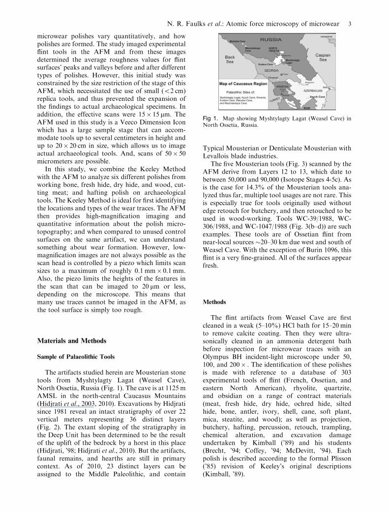
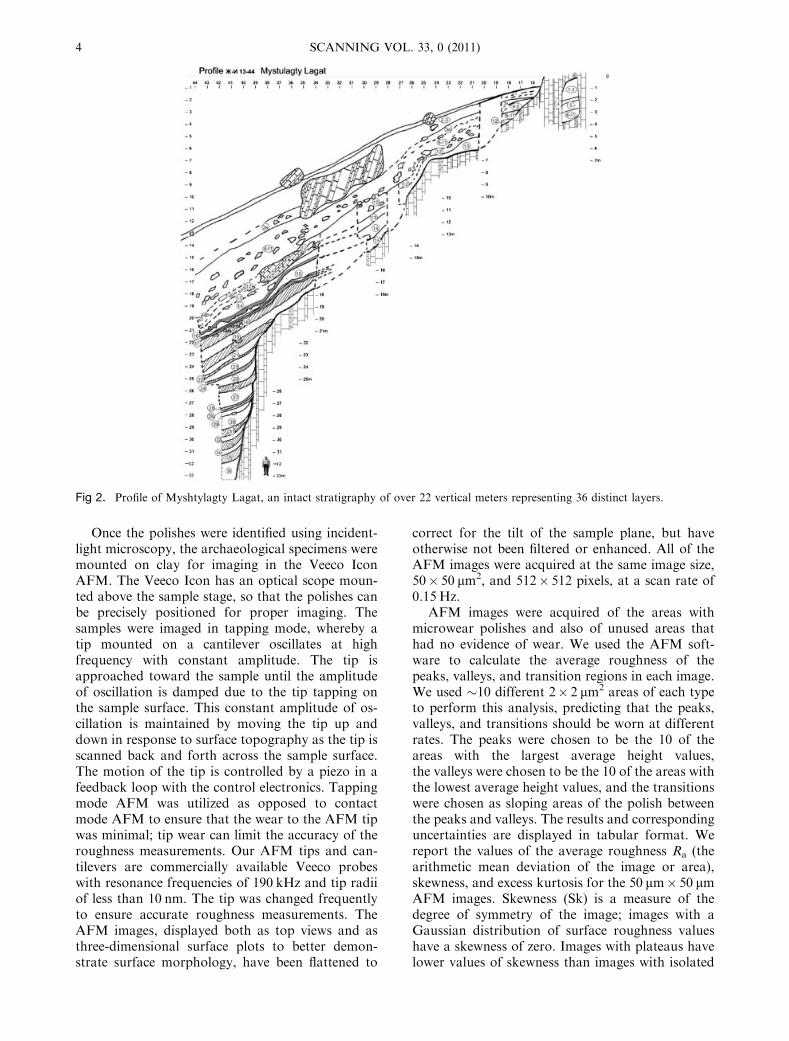
The artifacts studied herein are Mousterian stonetools from Myshtylagty Lagat (Weasel Cave),North Ossetia, Russia (Fig. 1). The cave is at 1125 mAMSL in the north-central Caucasus Mountains(Hidjrati et al., 2003, 2010). Excavations by Hidjratisince 1981 reveal an intact stratigraphy of over 22vertical meters representing 36 distinct layers(Fig. 2). The extant sloping of the stratigraphy inthe Deep Unit has been determined to be the resultof the uplift of the bedrock by a horst in this place(Hidjrati, ’98; Hidjrati et al., 2010). But the artifacts,faunal remains, and hearths are still in primarycontext. As of 2010, 23 distinct layers can beassigned to the Middle Paleolithic, and contain
Typical Mousterian or Denticulate Mousterian withLevallois blade industries.
The five Mousterian tools (Fig. 3) scanned by theAFM derive from Layers 12 to 13, which date tobetween 50,000 and 90,000 (Isotope Stages 4-5c). Asis the case for 14.3% of the Mousterian tools ana-lyzed thus far, multiple tool usages are not rare. Thisis especially true for tools originally used withoutedge retouch for butchery, and then retouched to beused in wood-working. Tools WC-39/1988, WC-306/1988, and WC-1047/1988 (Fig. 3(b–d)) are suchexamples. These tools are of Ossetian flint fromnear-local sources �20–30 km due west and south ofWeasel Cave. With the exception of Burin 1096, thisflint is a very fine-grained. All of the surfaces appearfresh.
Methods
The flint artifacts from Weasel Cave are firstcleaned in a weak (5–10%) HCl bath for 15–20 minto remove calcite coating. Then they were ultra-sonically cleaned in an ammonia detergent bathbefore inspection for microwear traces with anOlympus BH incident-light microscope under 50,100, and 200� . The identification of these polishesis made with reference to a database of 303experimental tools of flint (French, Ossetian, andeastern North American), rhyolite, quartzite,and obsidian on a range of contract materials(meat, fresh hide, dry hide, ochred hide, siltedhide, bone, antler, ivory, shell, cane, soft plant,mica, steatite, and wood); as well as projection,butchery, hafting, percussion, retouch, trampling,chemical alteration, and excavation damageundertaken by Kimball (’89) and his students(Brecht, ’94; Coffey, ’94; McDevitt, ’94). Eachpolish is described according to the formal Plisson(’85) revision of Keeley’s original descriptions(Kimball, ’89).
Fig 1. Map showing Myshtylagty Lagat (Weasel Cave) inNorth Ossetia, Russia.
N. R. Faulks et al.: Atomic force microscopy of microwear 3
Once the polishes were identified using incident-light microscopy, the archaeological specimens weremounted on clay for imaging in the Veeco IconAFM. The Veeco Icon has an optical scope moun-ted above the sample stage, so that the polishes canbe precisely positioned for proper imaging. Thesamples were imaged in tapping mode, whereby atip mounted on a cantilever oscillates at highfrequency with constant amplitude. The tip isapproached toward the sample until the amplitudeof oscillation is damped due to the tip tapping onthe sample surface. This constant amplitude of os-cillation is maintained by moving the tip up anddown in response to surface topography as the tip isscanned back and forth across the sample surface.The motion of the tip is controlled by a piezo in afeedback loop with the control electronics. Tappingmode AFM was utilized as opposed to contactmode AFM to ensure that the wear to the AFM tipwas minimal; tip wear can limit the accuracy of theroughness measurements. Our AFM tips and can-tilevers are commercially available Veeco probeswith resonance frequencies of 190 kHz and tip radiiof less than 10 nm. The tip was changed frequentlyto ensure accurate roughness measurements. TheAFM images, displayed both as top views and asthree-dimensional surface plots to better demon-strate surface morphology, have been flattened to
correct for the tilt of the sample plane, but haveotherwise not been filtered or enhanced. All of theAFM images were acquired at the same image size,50� 50 mm2, and 512� 512 pixels, at a scan rate of0.15 Hz.
AFM images were acquired of the areas withmicrowear polishes and also of unused areas thathad no evidence of wear. We used the AFM soft-ware to calculate the average roughness of thepeaks, valleys, and transition regions in each image.We used �10 different 2� 2 mm2 areas of each typeto perform this analysis, predicting that the peaks,valleys, and transitions should be worn at differentrates. The peaks were chosen to be the 10 of theareas with the largest average height values,the valleys were chosen to be the 10 of the areas withthe lowest average height values, and the transitionswere chosen as sloping areas of the polish betweenthe peaks and valleys. The results and correspondinguncertainties are displayed in tabular format. Wereport the values of the average roughness Ra (thearithmetic mean deviation of the image or area),skewness, and excess kurtosis for the 50 mm� 50 mmAFM images. Skewness (Sk) is a measure of thedegree of symmetry of the image; images with aGaussian distribution of surface roughness valueshave a skewness of zero. Images with plateaus havelower values of skewness than images with isolated
Fig 2. Profile of Myshtylagty Lagat, an intact stratigraphy of over 22 vertical meters representing 36 distinct layers.
4 SCANNING VOL. 33, 0 (2011)

steep peaks.
Sk ¼ 1
nRq
Xni¼1ðZi � �ZÞ3 ð1Þ
Here, n is the number of data points, Rq is the rootmean square roughness of the image, Zi is the heightof the ith data point, and �Z is the average height ofthe image. Kurtosis (K) shows the pointedness orbluntness of the distribution of the roughnessvalues. Smoother profiles have less variation in theirroughness values, and therefore the distribution ofroughness values is more narrow than a Gaussiandistribution. This causes the kurtosis for more uni-form surfaces to be higher than surfaces with greater
variation in roughness values.
K ¼ 1
nR2q
Xni¼1ðZi � �ZÞ4
" #� 3 ð2Þ
Results and Analysis
WC-1096/1097, Wood Polish
Burin WC-1096/1097 (Fig. 3(a)) is interpreted tohave been used to plane wood along three burinatededges, but was not hafted. The scanned use-trace 1cis indicated by the black rectangle in the digitalmicrophotograph of the microwear polish inFigure 4(a). The identification of this microweartrace as wood polish is based upon an invasive to
Fig 3. Mousterian stone tools scanned by AFM: (a) Burin WC-1096/1097 use-trace 1c; (b) Atypical Mousterian Point WC-39/1988 use-trace 4a; (c) Mousterian Point WC-306/1988 use-trace 3a; (d) Levallois Blade WC-1047/1988 use-trace 6b; (e) AtypicalLevallois Point WC-520 use-trace 2f; and (f) Mousterian Point WC-306/1988 hafting trace 1e. AFM scans were taken in the centerof the polish (microphotograph locations indicated by rectangles).
N. R. Faulks et al.: Atomic force microscopy of microwear 5

spreading extent, smooth, united texture, a fluidpolish with a coalescence following the entire mi-crotopography. The polish is most pronounced atthe higher elevations, and continues over the edgerather than rounding or otherwise significantlymodifying it. The contour of the polish is irregularlyclear. It is a very bright polish.
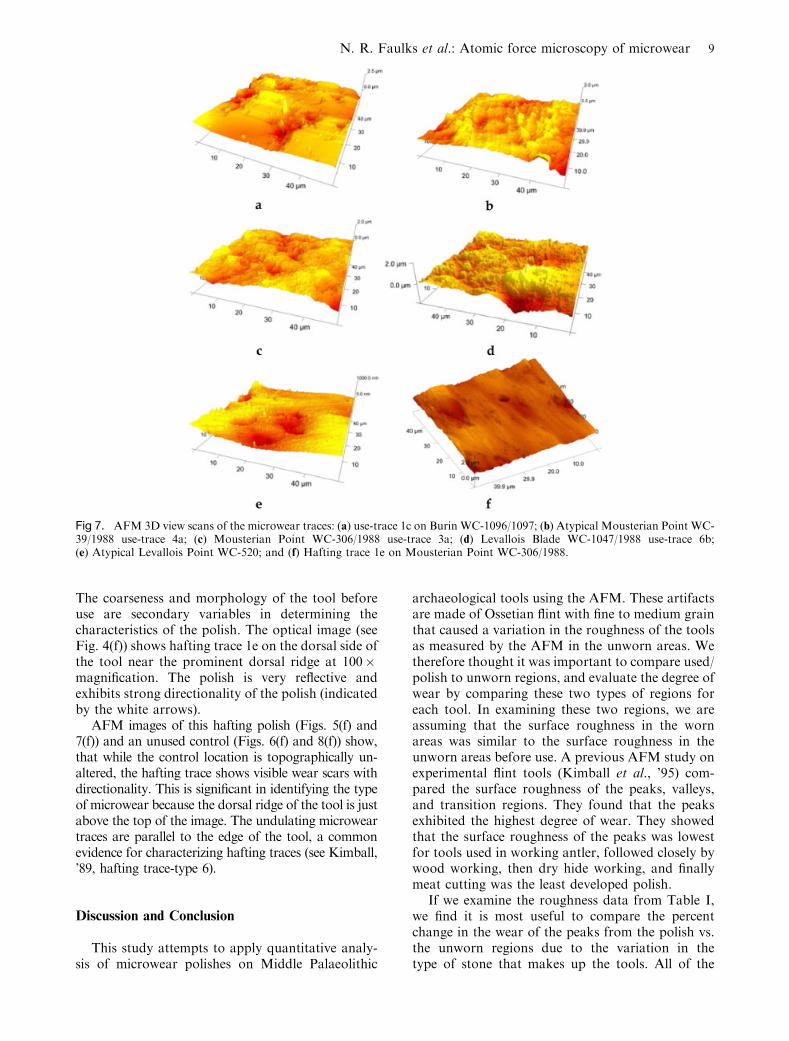
The AFM images were acquired close to thecenter of the polish (Figs. 5(a) and 7(a)), as well asan adjacent unmodified surface (Figs. 6(a) and 8(a)).
(It is important to keep in mind that there is somevariation in the graininess of these Ossetian flints,especially this specimen, which is more coarse-grained than the other specimens.) The edge of thetool is close to the location of the area imaged to theleft of the image. The image of the unworn area(Figs. 6(a) and 8(a)) has been cropped to 50� 45 mmdue to streaking in the AFM image caused by therelatively rough surface of the tool. It is easy to seethe areas of microwear in these top view scans. Thepeaks of the surface have been almost completelyworn away and flattened smooth. The valleysremain rough, with no evidence of filling in withwear debris at this place. This is reflected in theanalysis of the roughness data from the peaks, val-leys, and transition regions, as shown in Table I.The roughness of the valleys from the polish regionis significantly larger than the roughness of thevalleys from the unworn region. This is most likelydue to the extreme wear, which flattened out thetool in this area to a great extent. The wear left only
Fig 4. Digital microphotographs of the microwear traces:(a) wood polish on Burin WC-1096—AFM scan in center ofpolish; (b) fresh hide polish on Atypical Mousterian PointWC-39—AFM scan at domed polish at rounded tool edge;(c) dry hide polish on Mousterian Point WC-306—AFM scanat the matte, rounded edge; (d) well-developed meat polish onLevallois Blade WC-1047—AFM scan near the ridgeadjacent to the middle arrow; (e) bone polish on atypicalLevallois Point WC-520—AFM scan in center of polish nearthe tool edge; and (f) bright, undulating hafting polish onMousterian Point WC-306—AFM scan in center of polish(image has been rotated 901 clockwise). The height ofFigures 4(a) and (d) are �347 mm (all 200� ), andFigure 4(f) is �712 mm (100� ). Striations are indicated byblack arrows, and undulating polish by white arrows.
Fig 5. AFM top view scans of the microwear traces: (a) use-trace 1c on Burin WC-1096/1097; (b) Atypical MousterianPoint WC-39/1988 use-trace 4a; (c) Mousterian Point WC-306/1988 use-trace 3a; (d) Levallois Blade WC-1047/1988use-trace 6b; (e) Atypical Levallois Point WC-520; and (f)Hafting trace 1e on Mousterian Point WC-306/1988.
6 SCANNING VOL. 33, 0 (2011)

the deepest crevices untouched which skewed theresults toward a high roughness value (Table I).
WC-39/1988, Fresh Hide Polish
An Atypical Mousterian Point WC-39/1988(Fig. 3(b)) is interpreted to have been used to cleanfresh hide (at the distal end) and whittling wood(along the lateral right edge) in a hafted mode. Use-trace 4a, illustrated here (Fig. 4(b)), was used toclean hide in a fresh state—that is, to remove ad-hering tissues on the interior hide surface. Theidentification of this microwear trace as fresh hide-working polish is based upon a polish that is fluidand grainy following the microtopography. Withintensive work, the polish modifies the higher por-tion of the microtopography more significantly. Itsextent is invasive and exhibits an average texture.The contour of the polish is fuzzy and exhibits anaverage or ‘‘matte’’ brightness. Striations arepresent (indicated by black arrows) and are short,wide, and deep into the fresh hide polish. Their
orientation suggests cutting motions parallel, ob-lique, and perpendicular to the tool edge.
The acquired AFM scans of the microweartrace (Figs. 5(b) and 7(b)) and an unused region(Figs. 6(b) and 8(b)), and associated roughness va-lues indicate that this interpreted fresh hide polish isless rough along the peaks, transitions, and even thevalleys. The AFM image reveals striations (Mansur,’82, type 1) that are roughly parallel to the workingedge, thus indicating the tool kinematics. This isindicative of a cutting motion of the tool throughthe tissues attached to the hide.
WC-306/1988, Dry Hide Polish
Mousterian Point WC-306/1988 (Fig. 3(c)) is in-terpreted to have been used in two functions: (1)butchery along the lateral right edge; and (2) plan-ing wood along the lateral left edge. The illustrateduse-trace 3a (Fig. 4(c)) exhibits dry hide polish inthis location (along with microwear polishes fromcutting through the hide in fresh condition, cuttingthrough meat, and in one place contacting bone.This is the classic manifestation of heavy butchery,as opposed to simply cutting through meaty tissues.In this case, it appears that processing of the carcasscontinued until the hide was relatively dry. Theillustrated dry hide polish is defined as a soft, grainypolish with significant edge rounding and mod-ification of the microtopography. The texture isdense, the contour is fuzzy, and exhibits a matte/weak brightness. The numerous striations (indicatedby the black arrows) are long, wide, and deep(Mansur, ’82, type 1). The extent of the polish ismoderate. The image also shows that abrasive wearof the tool edge caused a rounding and smoothingof the working edge of the tool. AFM images wereacquired of the microwear polish (Figs. 5(c) and7(c)) and an unworn location nearby (Figs. 6(c) and8(c)). Unlike the fresh hide polish example (above),these striations were not observed in the AFMscans. The quantitative data suggest that dry hidepolish is as rough as the unused surface.
WC-1047/1988, Meat Polish
Levallois Blade WC-1047/1988 (Fig. 3(d)) is in-terpreted to have been used in butchery (lateral rightedge) and wood planing (proximal edge). Use-trace6b is identified as a polish from cutting meat, but allalong this working edge were meat and fresh hidepolishes. The identification of this microwear traceas meat is defined by a very fluid polish whichaffects the entire microtopography alike without
Fig 6. AFM top view scans of the unworn areas: (a) BurinWC-1096/1097; (b) Atypical Mousterian Point WC-39/1988;(c) Mousterian Point WC-306/1988; (d) Levallois Blade WC-1047/1988; (e) Atypical Levallois Point WC-520; and (f)Mousterian Point WC-306/1988—same as Figure 6(c).
N. R. Faulks et al.: Atomic force microscopy of microwear 7
major alteration of the relief. The texture is denseand the contour is fuzzy, but more evident at50� –100� . Yet, it forms a continuous linear bandof polish along the working edge. The brightness isaverage and somewhat lustrous. A few striations(Mansur, ’82, type 2) are evident and they are nar-row, straight, and short; and indicate the directionof tool use.
AFM images were acquired of the micropolish(Figs. 5(d) and 7(d)) and an unworn location(Figs. 6(d) and 8(d)) to the right from the edge of thetool. The roughness values of the peaks and valleysindicate that meat polish, while subtle is measurablyless rough than the unused control surface. Inaddition, there are two distinct groups of striae,running both oblique-to-parallel (top of image) andperpendicular (bottom of image) to the edge.
WC-520, Bone Polish
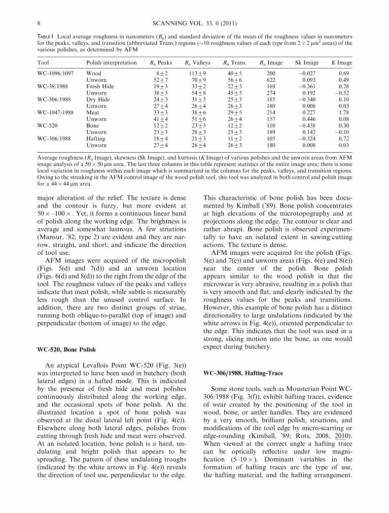
An atypical Levallois Point WC-520 (Fig. 3(e))was interpreted to have been used in butchery (bothlateral edges) in a hafted mode. This is indicatedby the presence of fresh hide and meat polishescontinuously distributed along the working edge,and the occasional spots of bone polish. At theillustrated location a spot of bone polish wasobserved at the distal lateral left point (Fig. 4(e)).Elsewhere along both lateral edges, polishes fromcutting through fresh hide and meat were observed.At an isolated location, bone polish is a hard, un-dulating and bright polish that appears to bespreading. The pattern of these undulating troughs(indicated by the white arrows in Fig. 4(e)) revealsthe direction of tool use, perpendicular to the edge.
This characteristic of bone polish has been docu-mented by Kimball (’89). Bone polish concentratesat high elevations of the microtopography and atprojections along the edge. The contour is clear andrather abrupt. Bone polish is observed experimen-tally to have an isolated extent in sawing/cuttingactions. The texture is dense.
AFM images were acquired for the polish (Figs.5(e) and 7(e)) and unworn areas (Figs. 6(e) and 8(e))near the center of the polish. Bone polishappears similar to the wood polish in that themicrowear is very abrasive, resulting in a polish thatis very smooth and flat; and clearly indicated by theroughness values for the peaks and transitions.However, this example of bone polish has a distinctdirectionality to large undulations (indicated by thewhite arrows in Fig. 4(e)), oriented perpendicular tothe edge. This indicates that the tool was used in astrong, slicing motion into the bone, as one wouldexpect during butchery.
WC-306/1988, Hafting-Trace
Some stone tools, such as Mousterian Point WC-306/1988 (Fig. 3(f)), exhibit hafting traces, evidenceof wear created by the positioning of the tool inwood, bone, or antler handles. They are evidencedby a very smooth, brilliant polish, striations, andmodifications of the tool edge by micro-scarring oredge-rounding (Kimball, ’89; Rots, 2008, 2010).When viewed at the correct angle a hafting tracecan be optically reflective under low magni-fication (5–10� ). Dominant variables in theformation of hafting traces are the type of use,the hafting material, and the hafting arrangement.
TABLE I Local average roughness in nanometers (Ra) and standard deviation of the mean of the roughness values in nanometersfor the peaks, valleys, and transition (abbreviated Trans.) regions (�10 roughness values of each type from 2� 2 mm2 areas) of thevarious polishes, as determined by AFM
Tool Polish interpretation Ra Peaks Ra Valleys Ra Trans. Ra Image Sk Image K Image
WC-1096/1097 Wood 872 11379 4075 200 �0.027 0.69Unworn 5277 7079 5676 622 0.093 0.49
WC-38/1988 Fresh Hide 1973 3372 2273 169 �0.361 0.28Unworn 3875 5478 4575 274 0.192 �0.52
WC-306/1988 Dry Hide 2473 3173 2573 185 �0.340 0.10Unworn 2774 2674 2673 180 0.008 0.03
WC-1047/1988 Meat 3373 3876 2975 214 0.227 1.78Unworn 4174 5176 2674 157 0.446 0.08
WC-520 Bone 1272 2373 1272 110 �0.438 0.30Unworn 2373 2873 2573 189 0.142 �0.10
WC-306/1988 Hafting 1874 2173 1172 105 �0.324 0.72Unworn 2774 2674 2673 180 0.008 0.03
Average roughness (Ra Image), skewness (Sk Image), and kurtosis (K Image) of various polishes and the unworn areas from AFMimage analysis of a 50� 50 mm area. The last three columns in this table represent statistics of the entire image area; there is somelocal variation in roughness within each image which is summarized in the columns for the peaks, valleys, and transition regions.Owing to the streaking in the AFM control image of the wood polish tool, this tool was analyzed in both control and polish imagefor a 44� 44 mm area.
8 SCANNING VOL. 33, 0 (2011)
The coarseness and morphology of the tool beforeuse are secondary variables in determining thecharacteristics of the polish. The optical image (seeFig. 4(f)) shows hafting trace 1e on the dorsal side ofthe tool near the prominent dorsal ridge at 100�magnification. The polish is very reflective andexhibits strong directionality of the polish (indicatedby the white arrows).
AFM images of this hafting polish (Figs. 5(f) and7(f)) and an unused control (Figs. 6(f) and 8(f)) show,that while the control location is topographically un-altered, the hafting trace shows visible wear scars withdirectionality. This is significant in identifying the typeof microwear because the dorsal ridge of the tool is justabove the top of the image. The undulating microweartraces are parallel to the edge of the tool, a commonevidence for characterizing hafting traces (see Kimball,’89, hafting trace-type 6).
Discussion and Conclusion
This study attempts to apply quantitative analy-sis of microwear polishes on Middle Palaeolithic
archaeological tools using the AFM. These artifactsare made of Ossetian flint with fine to medium grainthat caused a variation in the roughness of the toolsas measured by the AFM in the unworn areas. Wetherefore thought it was important to compare used/polish to unworn regions, and evaluate the degree ofwear by comparing these two types of regions foreach tool. In examining these two regions, we areassuming that the surface roughness in the wornareas was similar to the surface roughness in theunworn areas before use. A previous AFM study onexperimental flint tools (Kimball et al., ’95) com-pared the surface roughness of the peaks, valleys,and transition regions. They found that the peaksexhibited the highest degree of wear. They showedthat the surface roughness of the peaks was lowestfor tools used in working antler, followed closely bywood working, then dry hide working, and finallymeat cutting was the least developed polish.
If we examine the roughness data from Table I,we find it is most useful to compare the percentchange in the wear of the peaks from the polish vs.the unworn regions due to the variation in thetype of stone that makes up the tools. All of the
Fig 7. AFM 3D view scans of the microwear traces: (a) use-trace 1c on Burin WC-1096/1097; (b) Atypical Mousterian Point WC-39/1988 use-trace 4a; (c) Mousterian Point WC-306/1988 use-trace 3a; (d) Levallois Blade WC-1047/1988 use-trace 6b;(e) Atypical Levallois Point WC-520; and (f) Hafting trace 1e on Mousterian Point WC-306/1988.
N. R. Faulks et al.: Atomic force microscopy of microwear 9
polishes studied except for WC-306/1988 use-trace3a (interpreted as a dry hide polish) had loweraverage surface roughness for the peaks vs. the un-worn areas. WC-306 use-trace 3a showed no dif-ference in roughness between the unworn vs. polishregions, according to t-test results. Ranked fromlargest to smallest percent change in the roughnessof the peaks of the polish vs. unworn areas, wefound the following: 85% for WC-1096/1097 (in-terpreted as wood polish), 50% for WC-39 (inter-preted as fresh hide polish), 48% for WC-520(interpreted as bone polish), 33% for WC-306hafting-trace 1e (interpreted as a hafting trace), and20% for WC-1047 (interpreted as a meat polish). Itshould be noted that WC-39 and WC-520 give thesame percent change in roughness, according tot-test results. Ranked from largest to smallestpercent change in the roughness of the transitionregions of the polish vs. unworn areas, we foundthe following: 58% for WC-306 spot 1e (interpretedas a hafting trace), 51% for both WC-520 andWC-39 (interpreted as bone polish and fresh hidepolish), and 29% for WC-1096/1097 (interpretedas wood polish). WC-306 spot 3a and WC-1047(interpreted as dry hide and meat polish) showed no
difference in roughness of the transition regionsbetween the unworn vs. polish regions, according tot-test results.
Examining the roughness data for the50� 50 mm2 regions from Table I, we see a 68%decrease in roughness in the polish vs. the unwornregion for WC-1096/1097 (interpreted as woodpolish), a 42% decrease in roughness for both WC-520 and WC-306 spot 1e (interpreted as bone andhafting polishes), and a 38% decrease in roughnessfor WC-39 (interpreted as fresh hide polish). ForWC-306 use-trace 3a, there is a negligible differentin roughness for the polish vs. the unworn region,and for WC-1047 the roughness increases for thepolish vs. unworn region, even though we can seeobvious wear patterns in the AFM image. If weassume that the interpretation of the polishes notedon the artifacts is correct, then we see similar resultsto the previous AFM study on the experimentaltools, namely that the meat and dry hide polishesare the least developed polishes (i.e., smallestchanges in roughness) and that the bone polish(similar to an antler polish) and wood polishes aremore highly developed polishes (i.e., exhibit smallerchanges in roughness). These AFM results also
Fig 8. AFM 3D view scans of the unworn areas: (a) Burin WC-1096/1097; (b) Atypical Mousterian Point WC-39/1988;(c) Mousterian Point WC-306/1988; (d) Levallois Blade WC-1047/1988; (e) Atypical Levallois Point WC-520; and (f) MousterianPoint WC-306/1988—same as (c).
10 SCANNING VOL. 33, 0 (2011)
agree with prior work done by Evans and Donahue(2008) using laser scanning confocal microscopy andwith Stemp and colleagues (2009) using a laserprofilometer. It should be noted that WC-1096/1097is of a more coarsely grained flint, and as such theextreme abrasive wearing of the peaks during use isnot unexpected.
In the Mansur-Franchomme (’83) study onexperimental stone tools, it was noted that whenthere is moisture present during use the wear is moreextreme and less localized to the peaks. If we assumethat our interpretation of the polishes on the arti-facts in this study is correct, then this agrees withour results for WC-39 and WC-306 use-trace 3a,interpreted as fresh hide and dry hide polishes, asWC-39 showed a 50% change in the roughness ofthe peaks and transition regions in the polish vs. theunworn regions and the dry hide polish roughnessfor both was the same, according to t-test results.Studying the morphology from the images of thevarious techniques, it can be seen that the wear ismore uniform and widespread for the fresh hideworking than for the dry hide working. Our resultsalso agree with the Evans and Donahue (2008) studythat used laser scanning confocal microscopy tomeasure the surface roughness of various experi-mental stone tools and reported higher surfaceroughness for polish due to dry hide working thanfor polish due to fresh hide working.
For all of the traces, the skewness determinedfrom the AFM images is reduced for the polish vs.unworn regions. As the skewness is lower for sur-faces with more plateaus and higher for surfaceswith isolated steep peaks, lower values of skewnessimply a more worn surface. For all of the traces, theexcess kurtosis determined from the AFM images isincreased for the polish vs. control regions. An in-crease in kurtosis implies a more uniform surfaceroughness. Skewness and kurtosis are thereforeparameters that can help give a quantitative mea-sure to distinguish use traces for both experimentaltools and archeological artifacts. It is exciting thattools WC-1047 and WC-306 use-trace 3a, whosepolishes were interpreted to be meat and dry hide,show measurable differences in these parameters, asthese polishes are difficult to verify qualitatively bysimply viewing the morphology or examining theroughness alone.
The AFM can provide quantitative data in thequestion of the differentiation of microwear polishesof archeological tools at the very small scale.As opposed to other micro and nanoscale techni-ques, such as scanning electron microscopy, itcan provide quantitative roughness analysiswithout sputter coating the artifact or casting. TheAFM can also provide higher-resolution imagesthan optical interferometry or laser scanning
confocal microscopy, techniques commonly usedfor quantitative analysis of lithic artifacts. It isdifficult to draw conclusions at this time due to apaucity of AFM analysis of experimental tools,and we therefore believe that future work shouldinclude AFM analysis of experimental tools madefrom the types of flints used by these Neanderthals.
Acknowledgements
We are thankful to the NC Space Grant andAppalachian State University’s College of Arts andSciences for funding of this work, in addition tofunding from NSF DMR 0821124. The excavationsat Weasel Cave have benefited from grants from theWenner-Gren Foundation for AnthropologicalResearch (1997 Grant No. 6157), L.S.B. LeakeyFoundation (1998), IREX (1995 and 1996),St. George Russian-American ArchaeologicalProgram, Appalachian State University, and theInstitute for History and Archaeology at NorthOssetian State University. In addition to the seniorauthor, this research was undertaken with theassistance of ASU undergraduate students IsaacBryan and Zachary Bryan. Finally, we appreciatethe helpful comments from three reviewers thatgreatly improved the manuscript.
References
Anderson PC, Astruc L, Vargiolu R, Zahouani H. 1998.Contribution of quantitative analysis of surface states toa multi-method approach for characterising plant-pro-cessing traces on flint tools with gloss. In: Longo L,editor. Functional analyses of the lithic artifacts (),Proceedings of the XIII Congress of International Unionof Prehistoric and Protohistoric Sciences. Forli, Italy:ABACO. p 1151–1160.
Anderson PC, Georges J-M, Vargiolu R, Zahouani H. 2006.Insights from a tribological analysis of the tribulum.J Arch Sci 33:1559–1568.
Bamforth D. 1988. Investigating microwear polishes withblind tests: the Institute results in context. J Arch Sci15:11–24.
Bamforth D, Burns G, Woodman C. 1990. Ambiguous usetraces and blind test results: new data. J Arch Sci17:413–430.
Beyries S, Delamare F, Quantin JC. 1988. Traceologieet rugosimetrie tridimensionnelle. In: Beyries S, editor.Industries lithiques: traceologie et technologie, BritishArchaeological Reports International Series 411.p 115–132.
Bradley R, Clayton C. 1987. The influence of flint micro-structure on the formation of microwear polishes. In:Sieveking D, Newcomer MH, editors. The human uses offlint and chert, Papers presented at the Fourth Interna-tional Flint Symposium. Cambridge: University ofCambridge Press. p 81–89.
Brecht T. 1994. A description of the microwear formed byworking charred wood. Lithic Technol 19:83–87.
N. R. Faulks et al.: Atomic force microscopy of microwear 11

Coffey BP. 1994. The chemical alteration of microwear pol-ishes: an evaluation of the Plisson and Mauger findingsthrough replicative experimentation. Lithic Technol19:88–92.
Dumont JV. 1982. The quantification of microwear traces: anew use for interferometry. World Archaeol 14:206–217.
Evans AA, Donahue RE. 2005. The elemental chemistry oflithic microwear: an experiment. J Arch Sci 32:1733–1740.
Evans AA, Donahue RE. 2008. Laser scanning confocalmicroscopy: a potential technique for the study of lithicmicrowear. J Arch Sci 35:2223–2230.
Gonzalez-Urquijo JE, Ibanez-Estevez JJ. 2003. The quanti-fication of use-wear polish using image analysis. Firstresults. J Arch Sci 30:481–489.
Grace R, Graham ID, Newcomer MH. 1985. The quantifi-cation of microwear polishes. World Archaeol 17:112–120.
Grace R, Graham ID, Newcomer MH. 1987. Preliminaryinvestigation into the quantification of wear traces onflint tools. In: Sieveking D, Newcomer MH, editors. Thehuman uses of flint and chert. Cambridge: CambridgeUniversity Press. p 63–69.
Hidjrati N. 1998. Some clarification of the issues of siteformation, stratigraphy, hearths, and preservation atWeasel Cave, North Ossetia, Russia. Paper presented atthe 63rd annual meeting of the Society for AmericanArchaeology, Seattle.
Hidjrati NI, Kimball LR, Koetje T. 2003. Middle and LatePleistocene Investigations of Myshtylgaty Lagat (WeaselCave) North Ossetia, Russia. Antiquity 77:298.
Hidjrati N, Kimball L, Koetje T, Cleghorn N, Coffey T,Kanukova M, Nesmeynov S, Voeykova O, Sautiyeva-Maslennikova V. 2010. Nekotorye rezultaty mezhdisci-plinarnykh issledovanyi pleistocenovikh kulturnykxsloiev Mystylagty-lagat i Fynaigandzity-uat v SevernoiOsetii. In: Bzarov RS, Hidjrati NI, Dzitzzoity YA,Salbiev TK, Fidarov RF, editors. Voprosy istorii ikultury narodov rossii. Vladikavkaz: North OssetianState University, Insititute of History and Archaeology.p 221–244.
Juel Jensen H. 1988. Functional analysis of prehistoric flinttools by high-power microscopy: a review of west Eur-opean research. J World Prehistory 2:53–88.
Keeley LH. 1977. The function of Palaeolithic flint tools. SciAm 237:108–126.
Keeley LH. 1980. Experimental determination of stone tooluse. Chicago, IL: University of Chicago Press.
Kimball LR. 1989. Planning and functional variability in theUpper Palaeolithic: microwear analysis of Upper Peri-gordian tools from Le Flageolet I (Dordogne). Ph.D.Dissertation, Evanston, IL: Northwestern University.
Kimball LR, Allen PE, Kimball JF. 1995. Microwear pol-ishes as viewed through the atomic force microscope.Lithic Technol 20:26–28.
Kimball L, Allen PE, Kimball J, Schlichting B, Pham K.1998. The analysis of microwear polishes with the atomicforce microscope. Proceedings of the XIII Congress ofthe International Union of Prehistoric and ProtohistoricSciences. Forli, Italy: ABACO. p 1121–1132.
Klein R. 2009. The human career: human biological andcultural origins. Chicago: University of Chicago Press.
Knutsson K, Dahlquist B, Knutsson H. 1988. Patterns oftool use: the microwear analysis of the quartz andflint assemblage from Bjurselet site, Vasterbotten,northern Sweden. In: Beyries S, editor. Industrieslithiques: traceologie et technologie. British Archae-ological Reports International Series 411. p 253–294.
Lerner H, Xiangdong D, Costopoulos A, Ostoja-Starzewski M.2007. Lithic raw material physical properties and use-wearaccrual. J Arch Sci 34:711–722.
Mansur ME. 1982. Microwear analysis of natural and usestriations: new clues to the mechanisms of striation for-mation. In: Cahen D, editor. Tailler! pour quoi faire:prehistoire et technologie lithique II, Recent progress inmicrowear studies, Vol. 2. Studia Praehistorica Belgica.Koninklijk Museum voor Midden-Afrika, Tervuren,Belgium. p 213–233.
Mansur-Franchomme ME. 1983. Scanning electron micro-scopy of dry hide working tools: the role of abrasives andhumidity in microwear polish formation. J Arch Sci10:223–230.
McDevitt K. 1994. Results of replicative hide-working ex-periments: the roles of raw material, hide condition anduse wear patterns in the determination of rhyolite endscraper function. Lithic Technol 19:93–97.
McPherron S, Alemseged Z, Marean C, Wynn J, Reed D,Geraads D, Bobe R, Bearat H. 2010. Evidence for stone-tool-assisted consumption of animal tissues before 3.39million years ago at Dikika, Ethiopia. Nature466:857–860.
Plisson H. 1985. Etude fonctionnelle d’outillages lithiquesprehistoriques par l’analyse des micro-usures: recherchemethodologique et archeologique. These de Doctorat,Paris, France: l’Universite de Paris I.
Rots V. 2008. Hafting traces on flint tools. In ‘‘Prehistorictechnology’’ 40 years later: functional studies and theRussian legacy, British Archaeological Reports, Inter-national Series 1783, Verona, Italy. p 75–84.
Rots V. 2010. Prehension and hafting traces on flint tools: amethodology. Leuven: Leuven University Press.
Semaw S. 2000. The world’s oldest stone artefacts fromGona, Ethiopia: their implications for understandingstone technology and patterns of human evolution be-tween 2.6-1.5 million years ago. J Arch Sci 27:1197–1214.
Semenov S. 1964. Prehistoric technology. London, England:Cory, Adams and Mackay.
Stemp WJ, Stemp M. 2001. UBM laser profilometryand lithic use-wear analysis: a variable length scaleinvestigation of surface topography. J Arch Sci 28:81–88.
Stemp WJ, Stemp M. 2003. Documenting stages of polishdevelopment on experimental stone tools: surface char-acterization by fractal geometry using UBM laser profi-lometry. J Arch Sci 30:287–296.
Stemp WJ, Childs BE, Vionnet S, Brown CA. 2009. Quan-tification and discrimination of lithic use-wear: surfaceprofile measurements and length-scale fractal analysis.Archaeometry 51:366–382.
Stemp WJ, Childs BE, Vionnet S. 2010. Laser profilometryand length-scale analysis of stone tools: second seriesexperiment results. Scanning 32:233–243.
Yerkes RW, Kardulias PN. 1993. Recent developments in theanalysis of lithic artifacts. J Archaeol Res 1:89–119.
12 SCANNING VOL. 33, 0 (2011)
Related Documents