ATLAS Occlusion Diagnosis by BruxChecker Kanagawa Dental College Research Institute of Occlusion Medicine http://www.kdcnet.ac.jp/occmed/

Welcome message from author
This document is posted to help you gain knowledge. Please leave a comment to let me know what you think about it! Share it to your friends and learn new things together.
Transcript

ATLASOcclusion Diagnosis
byBruxChecker
Kanagawa Dental CollegeResearch Institute of Occlusion Medicine
http://www.kdcnet.ac.jp/occmed/
CONTENTS
IntroductionI. Concept of Organic OcclusionII. Concept of Craniomandibular System (CMS)III. Relationship between Organic Occlusion and CMSIV. Relationship between CMS and Occlusal GuidanceV. Physiological significance of BruxismVI. Basic concept of Functional Occlusion
1. Passive centric and Active centric2. Various lines on occlusal surface3. Intercuspation of first molar4. Passive centric and Occlusal guidance5. Retrusive barrier6. Guidance and Mediotrusion
VII. Bruxism movement1. Occlusion during sleep bruxism2. Bruxism movement of mandible3. Bruxism movement of mandible and Canine guidance4. Bruxism movement of mandible and Anterior teeth guidance5. Bruxism movement of mandible and working side condyle
VIII. Bruxism and Occlusal pattern1. Compensation curves and Disocclusion2. Compensation curve : Curve of Wilson
IX. Occlusal plane and DisocclusionX. Occlusal GuidanceXI. Occlusion Analysis formReferences
ATLASOcclusion Diagnosis by BruxChecker
日常臨床での
日常臨床での
ブラキシズム診断
ブラキシズム診断
神奈川歯科大学咬合医学研究所
1234567
14
24
29313233
Contact us:[email protected]

INTRODUCTION
Applying all these concepts to the clinical is a principle, NOT an average value.The attainment of a healthy functional occlusion, should be based on the individualization of each patient’s condition and its complete understanding. That is, the form of skeletal frame, the form and the function of the temporomandibular joint, the inclination of the condyler path, the occlusal plane, the dental arch and the dental morphology. The occlusion treatment should be planned on that. “Needed treatment for the patient at that time” is the functional principle of the occlusion. If you do not know the functional meaning and apply it as well as the knowledge of the inclination of the tooth axis, the alignment of the teeth and the cant of the occlusal plane, even so, it is worthless. Therefore, once again, applying it to the clinical p r a c t i c e i s a p r i n c i p l e a n d n o t a n a v e r a g e v a l u e .
In Dentistry, the theory for occlusion refers to the occlusal patterns such as canine guidance and group function in general. However, those occlusal patterns can only work for mandibular border movements and when the mandible moves furthermore the limit; the scene in which these occlusal patterns are effective is not useful anymore. Since the mastication movement is not a border movement, this concept of the occlusal patterns can not be fitted for it. A real situation where the occlusal patterns are necessary is the sleep bruxism function. For instance, the term canine guidance is used for the grinding movement of the mandible, but during chewing movement, the upper and lower canines do not touch or g r i n d s o m u c h . Therefore, the term canine control should be used whenever we are thinking about the role o f t h e c a n i n e d u r i n g t h e m a s t i c a t i o n m o v e m e n t .
Always when occlusion is constructed from a dental-medical perspective to the problem, one of the important considerations has to be the grinding movement of the mandible during the sleep bruxism. Because the force tangencies of teeth during the grinding movement of the mandible might strongly influence the masticatory muscle activity, it is closely related to the collapse of occlusions and a lot of dental diseases. However, the clinical examination of the lateral grinding movement usually obtained is different from the actual grinding movement during sleep bruxism. So, the occlusal pattern at the sleep bruxism can not be reproduced in t h e c l i n i c a l s i t u a t i o n i n m o s t o f t h e c a s e s . Therefore, the evaluation and diagnosis in consideration of concrete approach for the sleep bruxism will constitute an important area which provides basis for the dental-medical t r e a t m e n t i n t h e n e a r f u t u r e . Currently, the treatment of hard tissue diseases, temporomandibular joint diseases and related diseases from which sleep bruxism is assumed to be the cause; is mainly passive treatment of decreasing the bruxism by using a night guard and the autosuggestion method, e t c .
The sleep bruxism as an stress releaser has been reported as one of the importantfunctions of the masticatory organ which maintains the homeostasis of the human body. If the sleep bruxism plays the key role for maintaining human life, a physiological dental occlusion which adapts to the reaction of the human body should be obtained; different from the concept of the mechanical dental occlusion from the past. For that reason, the examination, diagnosis, and treatment planning methods based on the occlusal pattern at t h e s l e e p b r u x i s m s h o u l d b e a c c o m p l i s h e d .
June. 2005
Sadao SatoKanagawa Dental College
Institute of occlusion medicine- 1 -

I. Concept of Organic Occlusion
The term Organic Occlusion recognized as a basis of occlusion is a concept based on mutual protection that works when the occlusal system is overloaded. The molars will protect the anterior teeth when clenching occurs and the anterior teeth will protect the molars when grinding occurs. This scene in which an excessive overload is present is chiefly known as bruxismf u n c t i o n .The bruxism is basically an appearing activity of the masticatory muscles as
a response for a strong clenching and grinding movements of the mandible. When this occurs, abnormal contacts of the upper and lower teeth become a problem because of the muscle activity. The muscle activity according to the bruxism movement increases abnormally (hyperactivity) whenever there is a contact in the molar area. That represents a high risk to turn in a non-physiological bruxism. Therefore, molar disocclusion is indispensable during t h e b r u x i s m t o a v o i d a n y a b n o r m a l m u s c u l a r a c t i v i t y . Thinking about the occlusion from such a point of view, the malocclusion is an occlusion in which there are cuspid dental contacts of upper and lower teeth with a lack of occlusal support which generates a significant amount of the muscle activity during the strong clenching and grinding movements of b r u x i s m .
- 2 -
II. Concept of Cranio Mandibular System(CMS)
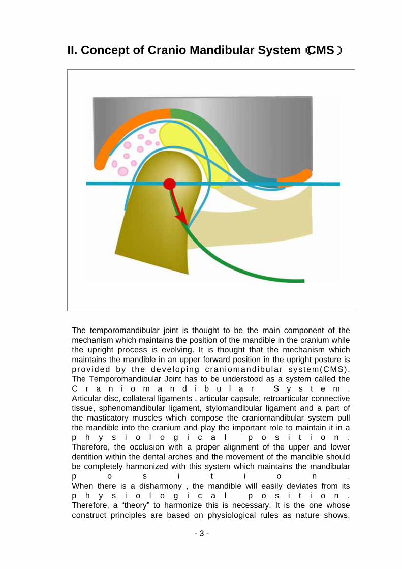
The temporomandibular joint is thought to be the main component of the mechanism which maintains the position of the mandible in the cranium while the upright process is evolving. It is thought that the mechanism which maintains the mandible in an upper forward position in the upright posture is p rov ided by the deve lop ing c ran iomand ibu la r sys tem(CMS). The Temporomandibular Joint has to be understood as a system called the C r a n i o m a n d i b u l a r S y s t e m . Articular disc, collateral ligaments , articular capsule, retroarticular connective tissue, sphenomandibular ligament, stylomandibular ligament and a part of the masticatory muscles which compose the craniomandibular system pull the mandible into the cranium and play the important role to maintain it in a p h y s i o l o g i c a l p o s i t i o n .Therefore, the occlusion with a proper alignment of the upper and lower dentition within the dental arches and the movement of the mandible should be completely harmonized with this system which maintains the mandibular p o s i t i o n . When there is a disharmony , the mandible will easily deviates from its p h y s i o l o g i c a l p o s i t i o n .Therefore, a “theory” to harmonize this is necessary. It is the one whose construct principles are based on physiological rules as nature shows.
- 3 -

III. Relationship of CMS and Organic Occlusion
An important factor which influences the mandibular position is the occlusalsupport. The masticatory organ is an unit that can be loaded by excessive forces of the bruxism and clenching, etc. Therefore, the occlusion should support these forces in such a function so that an excessive load could not compromise the temporomandibular joint (craniomandibular system).For instance, when a strong clenching is done, the temporomandibular joint should be supported by the dental occlusion. It should be a quiet down or repose state compared with the mastication and pronunciation functions’movements , etc. However, the occlusal support is often lost asconsequence of a malintercuspation factor in the case of class II malocclusions and the premature lost of the molars syndrome. In those cases the generating powerful forces push the mandible in a retruded position caused by the increase of the muscular activity during the bruxism and clenching. On the other hand, a muscular group of the craniomandibularsystem is trying to maintain the mandible forward. That means that during the bruxism and the clenching, an important confrontation takes place between a muscular group which makes the mandible moves backward and the craniomandibular system muscular group which tries to locate mandible forward in the state where the occlusal support was lost. Therefore, the symptoms caused by an abnormal tension (tenderness) of these muscular g r o u p s a p p e a r i n s u c h a p a t i e n t s w h e n w a k i n g u p .
- 4 -
IV. Relationship between CMS and Occlusal Guidance
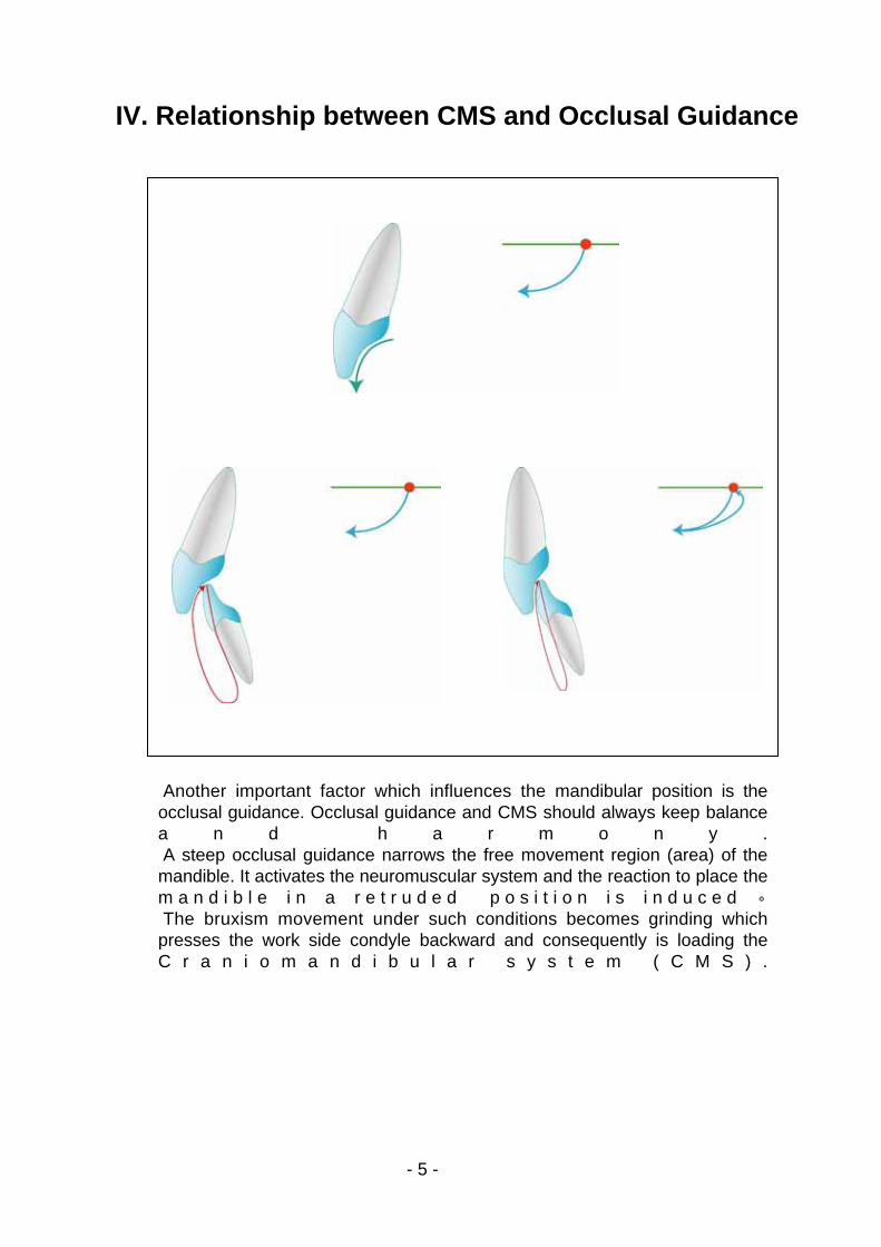
Another important factor which influences the mandibular position is the occlusal guidance. Occlusal guidance and CMS should always keep balance a n d h a r m o n y .A steep occlusal guidance narrows the free movement region (area) of the
mandible. It activates the neuromuscular system and the reaction to place the m a n d i b l e i n a r e t r u d e d p o s i t i o n i s i n d u c e d 。The bruxism movement under such conditions becomes grinding which
presses the work side condyle backward and consequently is loading the C r a n i o m a n d i b u l a r s y s t e m ( C M S ) .
- 5 -
STRESS
Limbic system
Visceralmasticatory
muscle activity
ClenchingBruxism
Hypothalamus
Autonomic nervous system
Pituitary adrenocortical
system
Stress Syndrome
Occlusal patternBalanced occlusion
Group functionCanine guidance
Dental diseaseCaries
Periodontal diseaseHypersensitivity
AbfractionTMD
Muscle symptoms
V. Physiological meaning of Bruxism
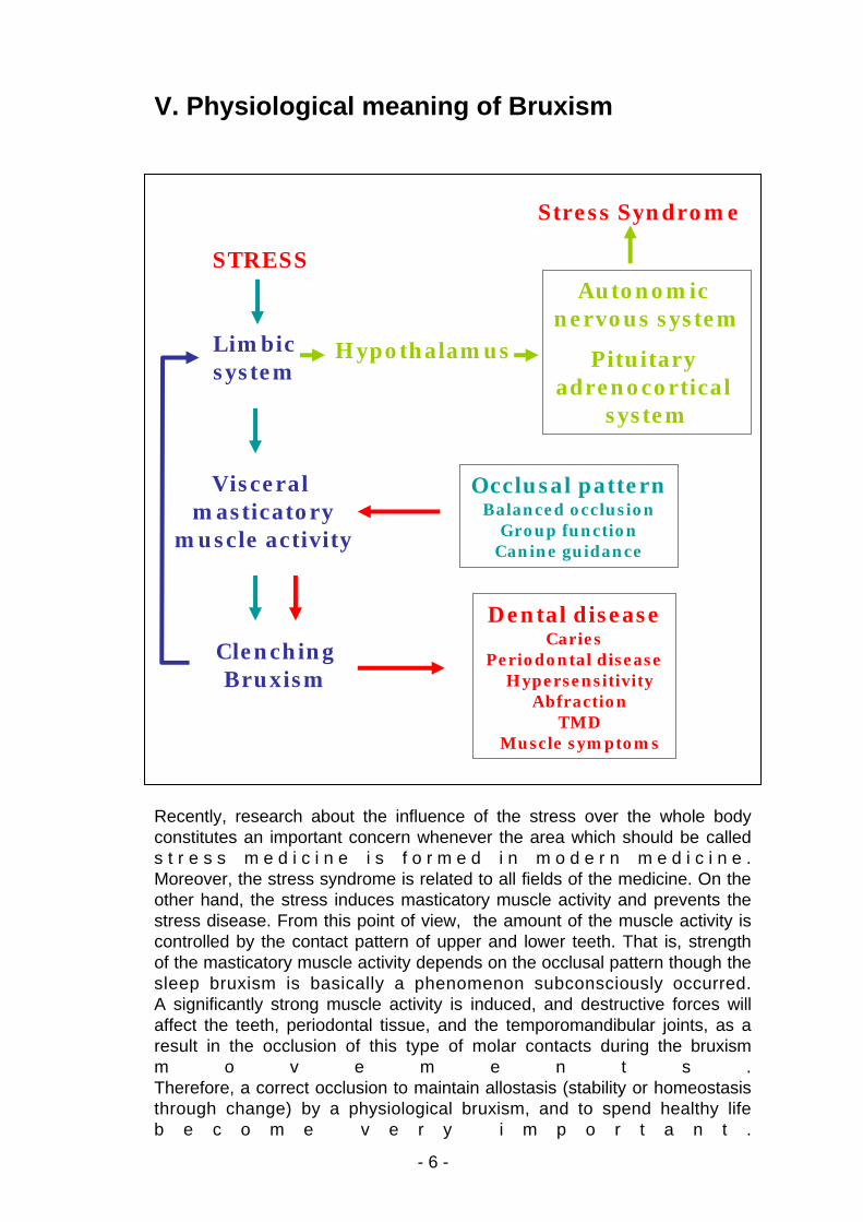
Recently, research about the influence of the stress over the whole body constitutes an important concern whenever the area which should be called s t r e s s m e d i c i n e i s f o r m e d i n m o d e r n m e d i c i n e . Moreover, the stress syndrome is related to all fields of the medicine. On the other hand, the stress induces masticatory muscle activity and prevents the stress disease. From this point of view, the amount of the muscle activity is controlled by the contact pattern of upper and lower teeth. That is, strength of the masticatory muscle activity depends on the occlusal pattern though the sleep bruxism is basically a phenomenon subconsciously occurred. A significantly strong muscle activity is induced, and destructive forces will affect the teeth, periodontal tissue, and the temporomandibular joints, as a result in the occlusion of this type of molar contacts during the bruxism m o v e m e n t s . Therefore, a correct occlusion to maintain allostasis (stability or homeostasis through change) by a physiological bruxism, and to spend healthy life b e c o m e v e r y i m p o r t a n t .
- 6 -

VI. Basic concept of Functional Occlusion1. Passive Centric and Active Centric
Centric StopThe position of the mandible is mainly established by the teeth intercuspation of the maxilla and mandible. The contact points resulting from the engagement (occlusion) of the upper and lower teeth arec a l l e d c e n t r i c s t o p s .
Passive CentricThe centric stop of the maxilla that exists on the upper dentition in connection with the cranium. It is thought as a fixed static state compared with the dynamic movement of the mandible. Moreover, it is thought as a passive contact point which catches the functional cusp of m a n d i b u l a r t e e t h . T h e r e f o r e , i t i s c a l l e d p a s s i v e c e n t r i c .
Active CentricThe centric stop of the mandible moves dynamically compared with the passive centric of the maxilla. Therefore, a centric stop of the mandible i s c a l l e d a c t i v e c e n t r i c .
Passive centric line
Active centric line
Upper and lower centric stops are in a complete evenly and simultaneously relation. In class I occlusion, the passive centric stops with the upper first and second premolars are located on the mesial marginal ridges. The guidance at the lateral movement of the mandible passes through the mesial marginal r idge from this passive centric or very close to the labial cusp.
- 7 -
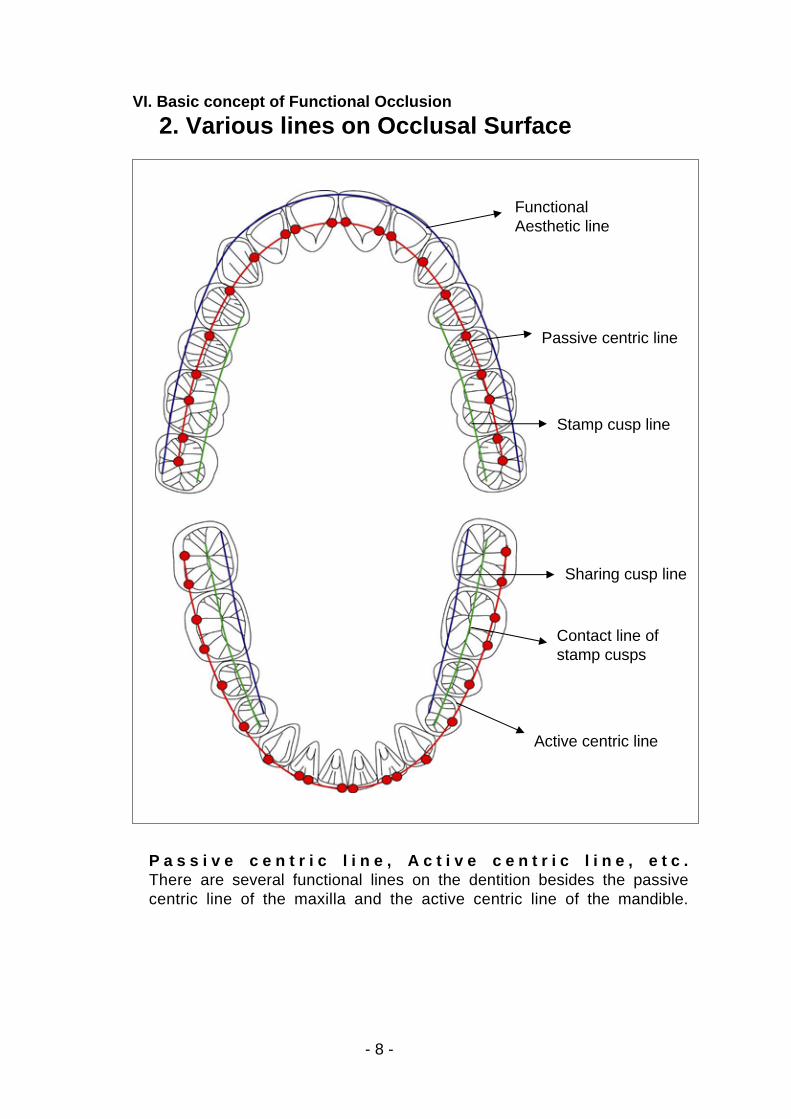
FunctionalAesthetic line
Passive centric line
Active centric line
Stamp cusp line
Sharing cusp line
Contact line of stamp cusps
VI. Basic concept of Functional Occlusion2. Various lines on Occlusal Surface
- 8 -
P a s s i v e c e n t r i c l i n e , A c t i v e c e n t r i c l i n e , e t c .There are several functional lines on the dentition besides the passive centric line of the maxilla and the active centric line of the mandible.

The first molar erupts earliest in the permanent dentition, and plays an important role in maintaining the occlusal support and the mandibular position. The mesio-lingual cusp of the upper first molar and the disto-buccal cusp of the lower first molar are the largest cusps in the whole dentition.Occlusal support and the stability of the mandibular position are maintained by their e n g a g e m e n t ( o c c l u s i o n ) .
- 9 -
VI. Basic concept of Functional Occlusion 3. Intercuspation of the first molar -- 1

VI. Basic concept of Functional Occlusion 3. Intercuspation of the first molar -- 2
ABC
In examining the occlusal relationships in the permanent dentition, much a t t e n t i o n i s c e n t e r e d o n t h e f i r s t m o l a r s .The engagement of an upper and lower first molar is the most important occlusion in the permanent dental articulation. The mesio-lingual cusp of the upper first molar occludes in the central fossa of the lower first molar, and the disto-buccal cusp of the lower first molar occludes in mesial with the transverse ridge of the upper first molar. Then, the contacts of cusps and ridges are obtained by these ABC contacts, and the Class I occlusion is maintained. Harmony with CMS is kept by such steady engagement.
- 10 -

The active centric stops of the mandible occlude with the passive centric stops of the maxilla moving on the lingual surface of the incisors and canine and on the mesial marginal ridges of molars and premolars of the working side along the lateral excursions of the mandible. It moves consecutively on n o n - w o r k i n g s i d e t o w a r d t h e l i n g u a l c u s p s .
It moves from the passive centric line toward out side during the mandibularlateral excursions. Therefore, this area is called guiding area.The limit of the guiding area is the mesial marginal ridge of the upper first m o l a r s , a n d t h e r e i s n o t g u i d e p o s t e r i o r t o t h i s .
- 11 -
VI. Basic concept of Functional Occlusion4. Passive Centric and Occlusal Guidance
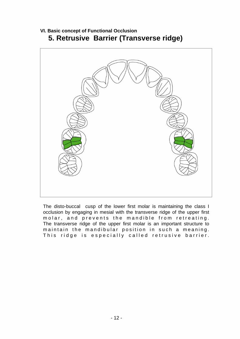
The disto-buccal cusp of the lower first molar is maintaining the class Iocclusion by engaging in mesial with the transverse ridge of the upper first m o l a r , a n d p r e v e n t s t h e m a n d i b l e f r o m r e t r e a t i n g .The transverse ridge of the upper first molar is an important structure to m a i n t a i n t h e m a n d i b u l a r p o s i t i o n i n s u c h a m e a n i n g .T h i s r i d g e i s e s p e c i a l l y c a l l e d r e t r u s i v e b a r r i e r .
- 12 -
VI. Basic concept of Functional Occlusion5. Retrusive Barrier (Transverse ridge)
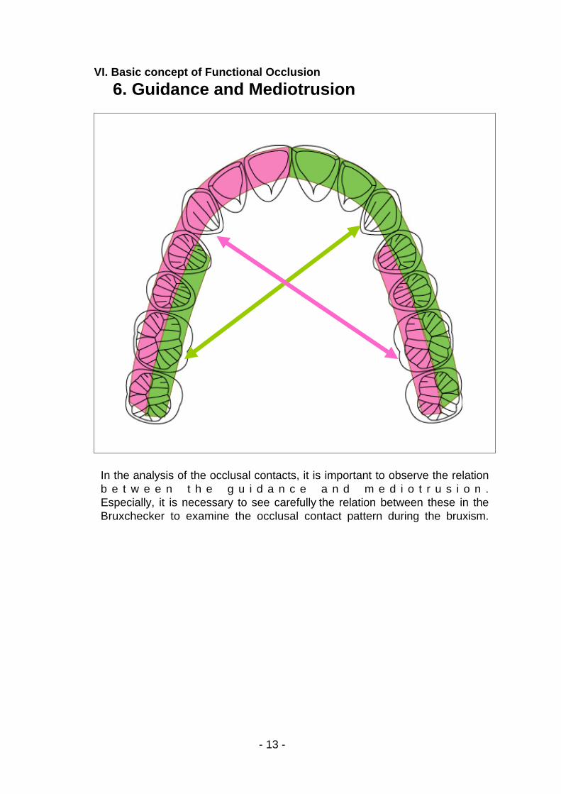
In the analysis of the occlusal contacts, it is important to observe the relation b e t w e e n t h e g u i d a n c e a n d m e d i o t r u s i o n . Especially, it is necessary to see carefully the relation between these in the Bruxchecker to examine the occlusal contact pattern during the bruxism.
- 13 -
VI. Basic concept of Functional Occlusion6. Guidance and Mediotrusion
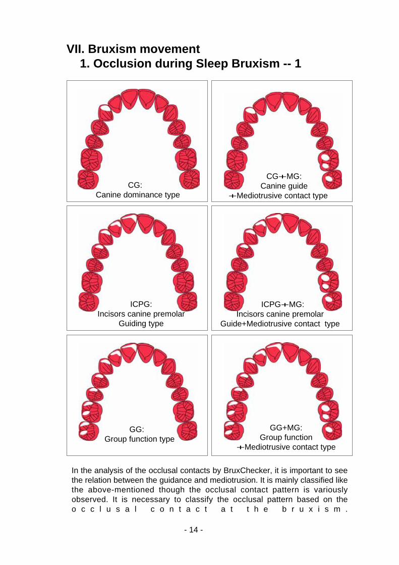
In the analysis of the occlusal contacts by BruxChecker, it is important to see the relation between the guidance and mediotrusion. It is mainly classified like the above-mentioned though the occlusal contact pattern is variously observed. It is necessary to classify the occlusal pattern based on the o c c l u s a l c o n t a c t a t t h e b r u x i s m .
CG:Canine dominance type
CG+MG:Canine guide
+Mediotrusive contact type
ICPG+MG:Incisors canine premolar
Guide+Mediotrusive contact type
ICPG:Incisors canine premolar
Guiding type
GG:Group function type
GG+MG:Group function
+Mediotrusive contact type
- 14 -
VII. Bruxism movement1. Occlusion during Sleep Bruxism -- 1

ICP
ICP
- 15 -
VII. Bruxism movement1. Occlusion during Sleep Bruxism -- 2
Grinding movement at the bruxism is basically a latero-retrusive movement, and the mandible does not move in a forward direction. There is a type including a centric stop at the intercuspal position and a type without a centric stop in the grinding area. Perhaps, this difference is thought to be dependent on the relation between the condylar inclination and the canine guidance. It is thought that grinding movement from an intercuspal position becomes difficult when the inclination of the canine guidance is too strong (steep) compared with the condylar inclination; it pass beyond to the canine cusp, and the b r u x i s m i s d o n e .

- 16 -
VII. Bruxism movement1. Occlusion during Sleep Bruxism -- 3
Example of grinding during sleep bruxism by Bruxchecker.
In this case, right and left sides, they both show contacts of the group function type. Moreover, the right and left second molar lingual cusps has c o m e s t r o n g l y i n c o n t a c t .

CG
CG+MG
GG
GG+MG
Various examples of grinding patterns during the bruxism
CG : canine dominant typeCG+MG : canine guide + mediotrusive contact typeGG : group function typeGG+MG : group function + mediotrusive contact type
- 17 -
VII. Bruxism movement1. Occlusion during Sleep Bruxism -- 4
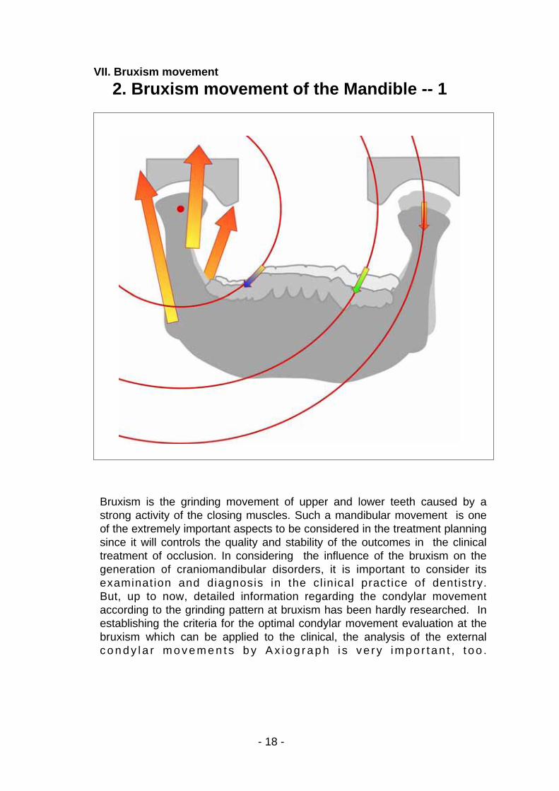
Bruxism is the grinding movement of upper and lower teeth caused by a strong activity of the closing muscles. Such a mandibular movement is one of the extremely important aspects to be considered in the treatment planning since it will controls the quality and stability of the outcomes in the clinical treatment of occlusion. In considering the influence of the bruxism on the generation of craniomandibular disorders, it is important to consider its examinat ion and diagnosis in the cl inical practice of dentistry.But, up to now, detailed information regarding the condylar movement according to the grinding pattern at bruxism has been hardly researched. In establishing the criteria for the optimal condylar movement evaluation at the bruxism which can be applied to the clinical, the analysis of the external c o n d y l a r m o v e m e n t s b y A x i o g r a p h i s ve ry impo r tan t , t oo .
- 18 -
VII. Bruxism movement2. Bruxism movement of the Mandible -- 1

VII. Bruxism movement2. Bruxism movement of the Mandible -- 2
Bruxism is the grinding movement of upper and lower teeth by a strong closing muscle activity. The mandibular movement is a whirling motion (rotational) of the non-working side condyle to make the working side condyle a rotation center. Such a movement demands a comparatively flat slope as a guiding path in the working side canine. When average tooth arrangement and the distance between the condylar heads to be 120 mm are assumed, the inclination on the canine slope becomes about 37 degrees. In this angle, the grinding movement which does not overwork from a geometrical relation between the condyle and the canine becomes possible. However, in a natural occlusion, there is an inclination which is about eight degrees steeper than a geometrical whirling motion, because the current average inclinations of the Japanese guiding path canine are about 45 degrees. Perhaps, it is thought that the inclination of canine means the limit of the canine guidance. It is necessary to note it because various problems on the occlusal function will be c a u s e d b y a v e r y s t r o n g i n c l i n a t i o n o f c a n i n e .
- 19 -
VII. Bruxism movement2. Bruxism movement of the Mandible -- 3
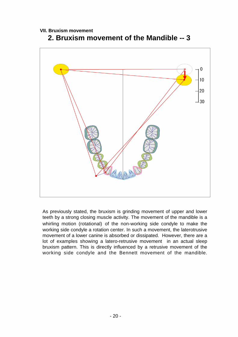
As previously stated, the bruxism is grinding movement of upper and lower teeth by a strong closing muscle activity. The movement of the mandible is a whirling motion (rotational) of the non-working side condyle to make the working side condyle a rotation center. In such a movement, the laterotrusivemovement of a lower canine is absorbed or dissipated. However, there are a lot of examples showing a latero-retrusive movement in an actual sleep bruxism pattern. This is directly influenced by a retrusive movement of the working side condyle and the Bennett movement of the mandible.
- 20 -

Each average inclination of the guiding path of the canine and eminence for the Japanese is 44 degrees, and these are parallel relations. Smooth grinding of the mandible without any overwork because of that parallelism, becomes possible eventhough the bruxism is grinding movement of upper and lower t e e t h b y a s t r o n g c l o s i n g m u s c l e a c t i v i t y .
44º
44º
- 21 -
VII. Bruxism movement3. Bruxism movement of the Mandible
and Canine guidance

44°55°
Averages of the anterior teeth guiding path for the Japanese population are 55 degrees, and averages of the eminence inclination are 44 degrees. The guiding path of the front teeth is a relation approximately ten degrees steeper. Thus, smooth grinding of the mandible becomes possible because of such a harmonized relation with the canine even though the bruxism is grinding movement caused by a strong closing muscle activity laterotrusively. Because a greatly steep guiding path of the anterior teeth induces the retrusive movement of the mandible, it causes a retruded mandible.
- 22 -
VII. Bruxism movement4. Bruxism movement of the Mandible
and Anterior teeth guidance
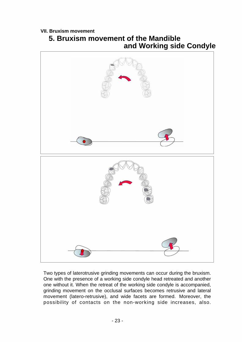
Two types of laterotrusive grinding movements can occur during the bruxism. One with the presence of a working side condyle head retreated and another one without it. When the retreat of the working side condyle is accompanied, grinding movement on the occlusal surfaces becomes retrusive and lateral movement (latero-retrusive), and wide facets are formed. Moreover, the possibil i ty of contacts on the non-working side increases, also.
- 23 -
VII. Bruxism movement5. Bruxism movement of the Mandible
and Working side Condyle

Several compensation curves exist in the dental occlusion. These are closely related to the cusp contact according to the grinding movement of the mandible. Curve of Spee is called sagittal compensating curve. A strong curve increases the possibility of molar contacts. Curve of Wilson is called lateral compensating curve. A strong curve of Wilson increases the p o s s i b i l i t y o f n o n - w o r k i n g s i d e c o n t a c t s .
Note:The curve of the canine is not a curve of Wilson .
- 24 -
VIII. Bruxism and Occlusal pattern1. Compensation curves and Disocclusion
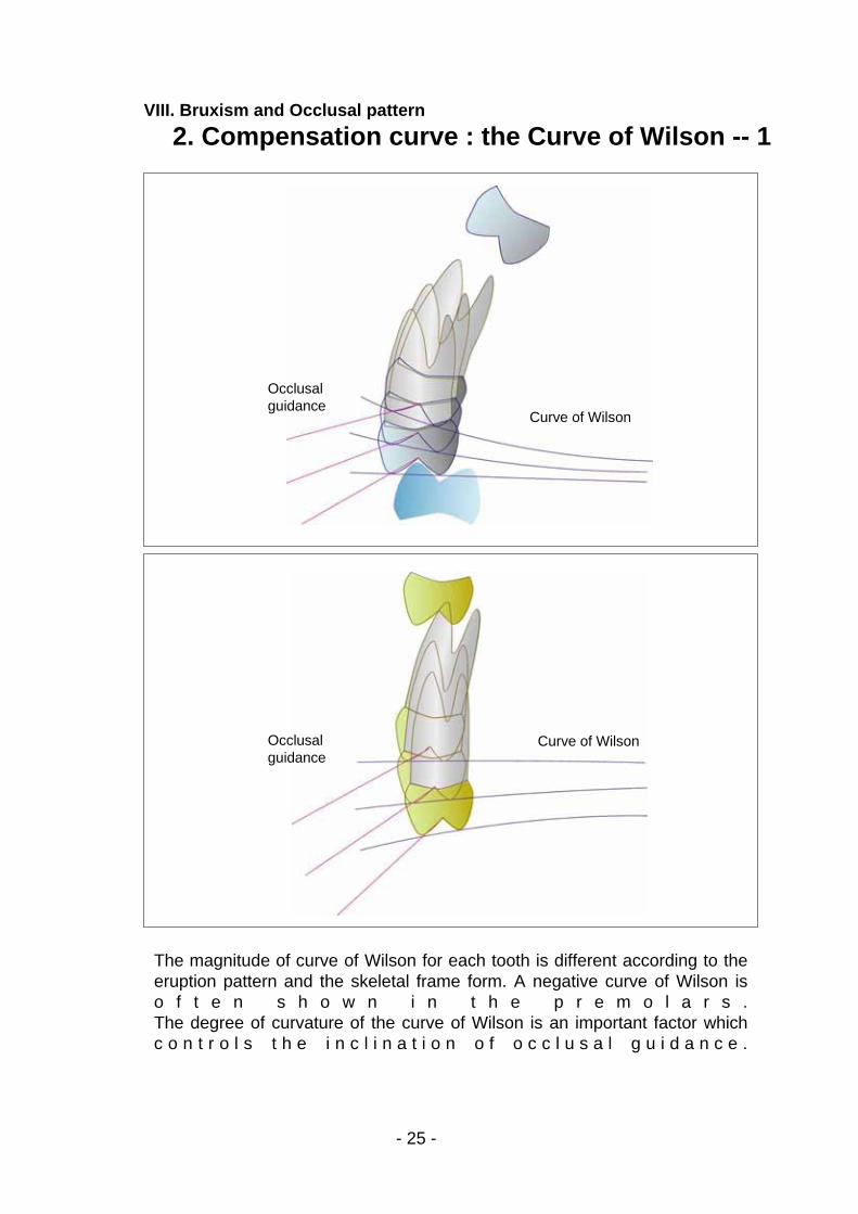
The magnitude of curve of Wilson for each tooth is different according to the eruption pattern and the skeletal frame form. A negative curve of Wilson is o f t e n s h o w n i n t h e p r e m o l a r s . The degree of curvature of the curve of Wilson is an important factor which c o n t r o l s t h e i n c l i n a t i o n o f o c c l u s a l g u i d a n c e .
Curve of Wilson
Occlusalguidance
Curve of WilsonOcclusalguidance
- 25 -
VIII. Bruxism and Occlusal pattern2. Compensation curve : the Curve of Wilson -- 1
VIII. Bruxism and Occlusal pattern2. Compensation curve : the Curve of Wilson -- 2
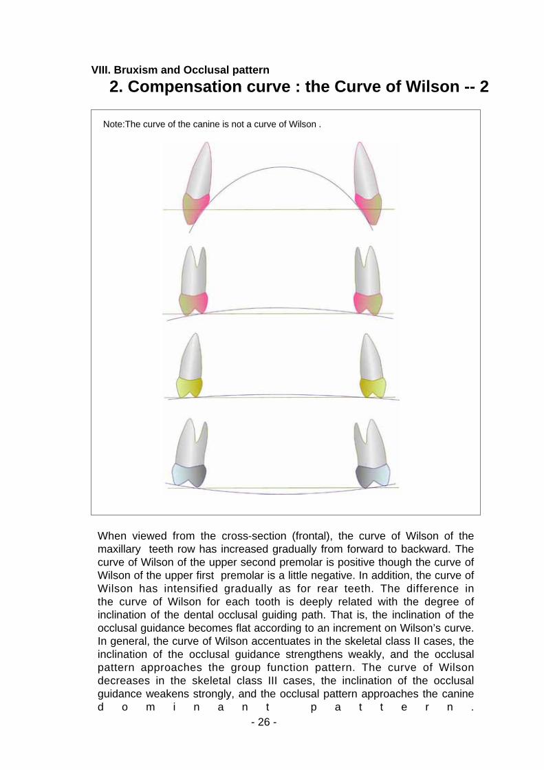
When viewed from the cross-section (frontal), the curve of Wilson of the maxillary teeth row has increased gradually from forward to backward. The curve of Wilson of the upper second premolar is positive though the curve of Wilson of the upper first premolar is a little negative. In addition, the curve of Wilson has intensified gradually as for rear teeth. The difference in the curve of Wilson for each tooth is deeply related with the degree of inclination of the dental occlusal guiding path. That is, the inclination of the occlusal guidance becomes flat according to an increment on Wilson’s curve. In general, the curve of Wilson accentuates in the skeletal class II cases, the inclination of the occlusal guidance strengthens weakly, and the occlusal pattern approaches the group function pattern. The curve of Wilson decreases in the skeletal class III cases, the inclination of the occlusal guidance weakens strongly, and the occlusal pattern approaches the canine d o m i n a n t p a t t e r n .
Note:The curve of the canine is not a curve of Wilson .
- 26 -
VIII. Bruxism and Occlusal pattern2. Compensation curve : the Curve of Wilson -- 3
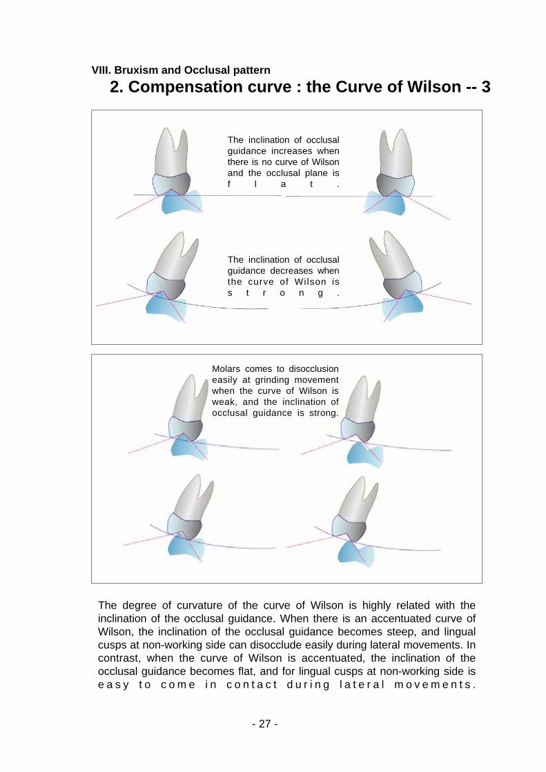
The degree of curvature of the curve of Wilson is highly related with the inclination of the occlusal guidance. When there is an accentuated curve of Wilson, the inclination of the occlusal guidance becomes steep, and lingual cusps at non-working side can disocclude easily during lateral movements. In contrast, when the curve of Wilson is accentuated, the inclination of the occlusal guidance becomes flat, and for lingual cusps at non-working side is e a s y t o c o m e i n c o n t a c t d u r i n g l a t e r a l m o v e m e n t s .
The inclination of occlusalguidance increases when there is no curve of Wilson and the occlusal plane is f l a t .
The inclination of occlusal guidance decreases when the curve of Wilson is s t r o n g .
Molars comes to disocclusion easily at grinding movement when the curve of Wilson is weak, and the inclination of occlusal guidance is strong.
- 27 -

VIII. Bruxism and Occlusal pattern2. Compensation curve : the Curve of Wilson -- 4
From a growing observation, the curve of Wilson during the first molar eruption is comparatively strong. The occlusal pattern while laterotrusive movement is balanced occlusion. Disocclusion of the non-working side molar is achieved as shown in the figure by the premolar eruption.
- 28 -

RCIRCI
Occlusal planeSteep:Group functionFlat :Canine guidance
Considering the functional role of the masticatory organ, the interferences in the molar area in dynamic functions of mastication, deglutition, pronunciation and bruxism are thought to be the principal cause of problems which appear to the teeth, periodontal tissue, temporomandibular joints, and masticatory muscles. Therefore, it is extremely important, as a basic problem in the dental occlusion construct ion to remove the interferences in molars. The most important factor in the reconstruction of occlusion which evades the molar interferences is the occlusal plane and the curve of Wilson. The occlusal plane becomes steeper, the molars do not disocclude easily, a n d t h e o c c l u s a l p a t t e r n a p p r o a c h e s t h e g r oup f u n c t i o n . Oppositely, the occlusal plane becomes flatter, the molars do disoccludemore easily, and the occlusal pattern becomes a canine dominant pattern.
- 29 -
IX. Occlusal Plane and Disocclusion
Angle of Disocclusion(AOD)
AOD=SCI-OP-CI=8-13
RCIRCI
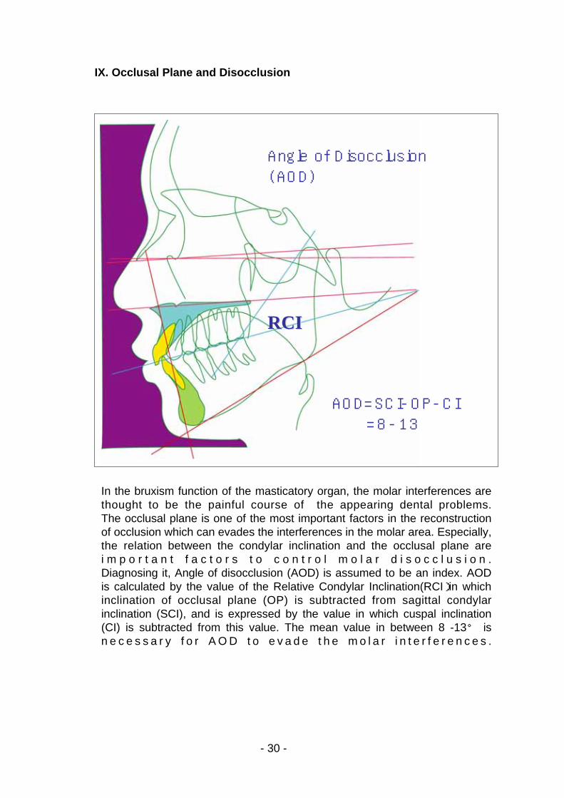
In the bruxism function of the masticatory organ, the molar interferences are thought to be the painful course of the appearing dental problems. The occlusal plane is one of the most important factors in the reconstruction of occlusion which can evades the interferences in the molar area. Especially, the relation between the condylar inclination and the occlusal plane are i m p o r t a n t f a c t o r s t o c o n t r o l m o l a r d i s o c c l u s i o n . Diagnosing it, Angle of disocclusion (AOD) is assumed to be an index. AOD is calculated by the value of the Relative Condylar Inclination(RCI)in which inclination of occlusal plane (OP) is subtracted from sagittal condylarinclination (SCI), and is expressed by the value in which cuspal inclination (CI) is subtracted from this value. The mean value in between 8 -13° is n e c e s s a r y f o r A O D t o e v a d e t h e m o l a r i n t e r f e r e n c e s .
- 30 -
IX. Occlusal Plane and Disocclusion

X. Occlusal Guidance
Guidance from the front teeth to the molar is known as the guiding path inclination and distance to the rear molar and premolar from canine one by one flatter and shorter. It is a condition for establish an easy disocclusion. The condylar inclination and the lingual surface of canine incline almost ina p a r a l l e l r e l a t i o n . The condylar inclination and the lingual surface of canine incline almost ina p a r a l l e l r e l a t i o n .
Therefore, molar disocclusion at grinding movement is not the one obtained only by the strongly lingual surface inclination of canine. It should be recognized what should be achieved by canine guidance which harmonizes with the condyle path and guiding path inclination of anterior teeth, canine, p r e m o l a r s , a n d m o l a r s s e q u e n t i a l l y .
- 31 -

XI. Occlusion analysis form
- 32 -

References
Sato S. and Slavicek R.: Bruxism as a stress management function of the masticatory organ. Bull. Kanagawa Dent. Coll. 29: 101-110, 2001.
Sato S., Yuyama N., Tamura K., Tamaki K., Hori N., Kaneko M., Sasaguri K., Lee M. C-il., Onozuka M. and Slavicek R.: The masticatory organ, brain function, stress-relase, and a proposal to add a new category to the taxonomy of the healing arts: Occlusion medicine. Bull Kanagawa Dent Coll 30: 2002.
Sato S.: Role of masticatory organ and concept of functional occlusion. Hotetsurinsho (in Japanese) 29;265-279,1996.
Sato S. and Tamaki K.: Meaning of bruxism seen from functional occlusion restructuring. Nihon shika hyoron (in Japanese), 201-218,1997.
Slavicek R.: The function of stress management. In: The Masticatory Organ – Function and Dysfunction, Slavicek, R. (Ed), Klosterneuburg, Gamma Medizinisch-wissenschaftliche Fortdungs-AG, pp. 281-291. 2002..
Kulmer S, Ruzicka B, Niederwanger A, Moschen I. : Incline and length of guiding elements in untreated naturally grown dentition. J Oral Rehabilitation 26;650-660,1999.
Leja W, Hilbe M, Stainer M, Kulmer S. : Nicht-kariöse zervikale läsionen in relation zum okklusionstypus und zur neigung der individullen führungselemente. Dtsch Aahnärztl Z, 45;411-414,1999.
Williamson E H, Lundquist D O.: Anterior guidance: its effect of electromyographic activity of the temporal and masseter muscles. J Prosthet Dent, 49: 816, 1983.
Grubwieser G, Flatz A, Grunert I, Kofler M, Ulmer H, Gausch K, Kulmer S.: Quantitative analysis of masseter and temporalis EMGs : a comparison of anterior guided versus balanced occlusal concepts in patients wearing complete dentures. J Oral Rehabilitation, 26: 731-736, 1999.
Toubol, J-P., Michel, J-F.: le mouvement initial de Bennett. Experimentation clinique, Consequences therapeutiques. Les Cahiers Proth, 42 : 69-87, 1983.
McHorris W H.: Focus on anterior guidance. J Gnathology, 8; 3-13, 1989.
Sato S.: Relationship between occlusion and whole body seen from role of masticatory organ. (in Japanese) Nihon zenshin kougou academy J. 6(2):101-109,2000.
Tamaki K: Occlusion and function of the Craniomandibular System. Bull of Kanagawa Dental College, 29(2), 111-119, 2001.
Tamaki K, Ales G Celar, Toshio Teranaka, Sadao Sato, Slavicek R: Interdisciplinary Approach to the Patient with Mandibular Lateral Displacement and Complex Craniomandibular Symptoms. Information Orthop Kieferortho, (in press) 2005.
Onodera K, Kawagoe T, Sasaguri K, Protacio-Quismundo C, Sato S.: Evaluation of the condylar movements in healthy and symptomatic temporomandibular joint patients during mastication and simulated bruxism utilizing condylograph. Stomatologie 101.8:187-190,2004.
Slavicek R, Sato S: Bruxism—a function of the masticatory organ to cope with stress. Wien Med Wochenschr. 154: 584-9, 2004 (German)
Onodera K., Kawagoe T., Sasaguri K. and Sato S.: Development of Bruxchecker - simple device for occlusion evaluation at sleep bruxism. (in Japanese) Kanagawashigaku 39:133-138,2004.
Sato S. and Sasaguri K.: Physiology function and occlusion medical aspect of bruxism. (in Japanese)Nihon shika sangyo academy J. 18:3-10,2004.
- 33 -
Related Documents