© 2015 Akkus et al. This work is published by Dove Medical Press Limited, and licensed under Creative Commons Attribution – Non Commercial (unported, v3.0) License. The full terms of the License are available at http://creativecommons.org/licenses/by-nc/3.0/. Non-commercial uses of the work are permitted without any further permission from Dove Medical Press Limited, provided the work is properly attributed. Permissions beyond the scope of the License are administered by Dove Medical Press Limited. Information on how to request permission may be found at: http://www.dovepress.com/permissions.php Medical Devices: Evidence and Research 2015:8 1–10 Medical Devices: Evidence and Research Dovepress submit your manuscript | www.dovepress.com Dovepress 1 REVIEW open access to scientific and medical research Open Access Full Text Article http://dx.doi.org/10.2147/MDER.S50594 Atherectomy devices: technology update Nuri I Akkus 1 Abdulrahman Abdulbaki 1 Enrique Jimenez 2 Neeraj Tandon 2 1 Department of Cardiology, Louisiana State University Health Sciences Center Shreveport, Shreveport, LA, USA; 2 Department of Cardiology, Overton Brooks VA Medical Center, Shreveport, LA, USA Correspondence: Nuri I Akkus Louisiana State University Health Sciences Center Shreveport, Division of Cardiovascular Diseases, 1501 Kings Hwy, Shreveport, LA 71130, USA Tel +1 318 675 5943 Email [email protected] Abstract: Atherectomy is a procedure which is performed to remove atherosclerotic plaque from diseased arteries. Atherosclerotic plaques are localized in either coronary or peripheral arterial vasculature and may have different characteristics depending on the texture of the plaque. Atherectomy has been used effectively in treatment of both coronary and peripheral arterial disease. Atherectomy devices are designed differently to either cut, shave, sand, or vaporize these plaques and have different indications. In this article, current atherectomy devices are reviewed. Keywords: coronary artery disease, peripheral arterial disease SilverHawk and TurboHawk directional atherectomy systems The SilverHawk and TurboHawk plaque excision systems are the two US Food and Drug Administration-approved directional atherectomy devices in use today. Both are approved for use in atherectomy of the peripheral vasculature and are not approved for use in coronary, carotid, iliac, or renal arteries. Directional atherectomy is con- sidered a minimally invasive treatment that removes plaque and restores blood flow in the native artery. Besides removing plaque from the body, the other advantages of directional atherectomy include lack of barotrauma, which decreases the risk of neointimal hyperplasia and lesser risk of dissection. Atherectomy does not preclude the use of surgical bypass at a future time and has the advantage of being able to change the bypass site, if needed. SilverHawk plaque excision system (ev3 Inc., Plymouth, MN, USA) is a forward cutting directional atherectomy device. This can be used with or without concurrent percutaneous balloon angioplasty and stenting. The device consists of a rotating blade inside a tubular housing with a collection area (nosecone) (Figure 1A and B). This catheter is connected to a battery-driven motor which spins the cutter. The TurboHawk system is similar except in the number of inner blades. While SilverHawk has one inner blade, TurboHawk has four contoured blades, thus favor- ing use in highly calcified lesions and more plaque removal per pass. Both devices come in various sizes to enable atherectomy in vessels with diameters of 1.5–7 mm. Atherectomy using the SilverHawk device carries with it the advantage of directional control, making it easier to remove eccentric lesions. As the device is advanced through the lesion, plaque is excised and packed in the nosecone. Different planes of excision are achieved by rotation of the device. Distal embolization remains a major

Welcome message from author
This document is posted to help you gain knowledge. Please leave a comment to let me know what you think about it! Share it to your friends and learn new things together.
Transcript
© 2015 Akkus et al. This work is published by Dove Medical Press Limited, and licensed under Creative Commons Attribution – Non Commercial (unported, v3.0) License. The full terms of the License are available at http://creativecommons.org/licenses/by-nc/3.0/. Non-commercial uses of the work are permitted without any further
permission from Dove Medical Press Limited, provided the work is properly attributed. Permissions beyond the scope of the License are administered by Dove Medical Press Limited. Information on how to request permission may be found at: http://www.dovepress.com/permissions.php
Medical Devices: Evidence and Research 2015:8 1–10
Medical Devices: Evidence and Research Dovepress
submit your manuscript | www.dovepress.com
Dovepress 1
R E v i E w
open access to scientific and medical research
Open Access Full Text Article
http://dx.doi.org/10.2147/MDER.S50594
Atherectomy devices: technology update
Nuri i Akkus1
Abdulrahman Abdulbaki1
Enrique Jimenez2
Neeraj Tandon2
1Department of Cardiology, Louisiana State University Health Sciences Center Shreveport, Shreveport, LA, USA; 2Department of Cardiology, Overton Brooks vA Medical Center, Shreveport, LA, USA
Correspondence: Nuri i Akkus Louisiana State University Health Sciences Center Shreveport, Division of Cardiovascular Diseases, 1501 Kings Hwy, Shreveport, LA 71130, USA Tel +1 318 675 5943 Email [email protected]
Abstract: Atherectomy is a procedure which is performed to remove atherosclerotic plaque
from diseased arteries. Atherosclerotic plaques are localized in either coronary or peripheral
arterial vasculature and may have different characteristics depending on the texture of the plaque.
Atherectomy has been used effectively in treatment of both coronary and peripheral arterial
disease. Atherectomy devices are designed differently to either cut, shave, sand, or vaporize
these plaques and have different indications. In this article, current atherectomy devices are
reviewed.
Keywords: coronary artery disease, peripheral arterial disease
SilverHawk and TurboHawk directional atherectomy systemsThe SilverHawk and TurboHawk plaque excision systems are the two US Food and
Drug Administration-approved directional atherectomy devices in use today. Both are
approved for use in atherectomy of the peripheral vasculature and are not approved
for use in coronary, carotid, iliac, or renal arteries. Directional atherectomy is con-
sidered a minimally invasive treatment that removes plaque and restores blood flow
in the native artery. Besides removing plaque from the body, the other advantages
of directional atherectomy include lack of barotrauma, which decreases the risk of
neointimal hyperplasia and lesser risk of dissection. Atherectomy does not preclude the
use of surgical bypass at a future time and has the advantage of being able to change
the bypass site, if needed.
SilverHawk plaque excision system (ev3 Inc., Plymouth, MN, USA) is a forward
cutting directional atherectomy device. This can be used with or without concurrent
percutaneous balloon angioplasty and stenting. The device consists of a rotating blade
inside a tubular housing with a collection area (nosecone) (Figure 1A and B). This
catheter is connected to a battery-driven motor which spins the cutter.
The TurboHawk system is similar except in the number of inner blades. While
SilverHawk has one inner blade, TurboHawk has four contoured blades, thus favor-
ing use in highly calcified lesions and more plaque removal per pass. Both devices
come in various sizes to enable atherectomy in vessels with diameters of 1.5–7 mm.
Atherectomy using the SilverHawk device carries with it the advantage of directional
control, making it easier to remove eccentric lesions. As the device is advanced
through the lesion, plaque is excised and packed in the nosecone. Different planes of
excision are achieved by rotation of the device. Distal embolization remains a major

Medical Devices: Evidence and Research 2015:8submit your manuscript | www.dovepress.com
Dovepress
Dovepress
2
Akkus et al
disadvantage with these systems and hence use of embolic
protection devices is recommended in large and heavily
calcified vessels.
Clinical dataThe TALON (Treating PeripherAls with SiLverHawk:
Outcomes CollectioN) registry enrolled more than 601 patients
with over 1,258 infrainguinal arterial lesions that were treated
with the SilverHawk device.1 It included patients with both
claudication and critical limb ischemia (CLI). About half of
the patient population was diabetic and a third was Rutherford
class 4 or greater. Both above- and below-knee lesions were
treated. The device achieved 50% or less diameter stenosis in
94.7% of the lesions. The procedural success rate was 97.6%.
Stent placement was required in only 6.3% of the lesions after
plaque excision. The 6- and 12-month rates of survival free
of target lesion revascularization (TLR) were 90% and 80%,
respectively. Rates of TLR were similar among patients with
diabetes (11%) and without diabetes (9%). The 12-month
outcomes compare favorably to angioplasty and stenting.
However, this being an observational registry, it did not have
any independent assessment of outcomes.
McKinsey et al, prospectively analyzed 579 lesions
treated with SilverHawk in 275 patients with above- and
below-knee interventions (claudicants 36.7%, CLI 63.3%).2
Eighteen-month primary and secondary patency was
52.7% and 75%, respectively. Overall limb salvage was
92.4% at 18 months with 4.4% requiring bypass. Kandzari
et al prospectively evaluated 69 patients with critical limb
ischemia for 6 months after treatment with SilverHawk
plaque excision.3 A total of 76 limbs were treated with 40%
infrapopliteal lesions. Procedural success was achieved in
99% of the cases and TLR rate was 4% at 6 months. Zeller
et al reported 1-year and 2-year results after SilverHawk
directional atherectomy (DA) of 49 below-the-knee
lesions in 36 patients.4 Sixty-seven percent of lesions were
treated with primary DA, 39% required additional balloon
angioplasty (BA), while 4% required bail-out stenting.
Primary and secondary patency rates were 67% and 91%,
respectively, after 1 year, and 60% and 80%, respectively,
after 2 years. Keeling et al report 1-year primary and second-
ary patency of 61.7% and 76.4%, respectively, from their
database of 60 patients in whom 70 plaque excisions were
performed.5 Restenosis developed in 2.8% of the patients
at 3 months. Sixt et al prospectively treated de novo and
restenotic lesions in 161 patients (166 lesions) with plaque
excision.6 The overall technical success rate was 76%
(124/164) and the procedural success rate was 95%. At 12
months, primary patency rate was 61% and the secondary
patency rate was 75% in the entire cohort. Although Silver-
Hawk is not indicated for treating in-stent restenosis, it has
been used for this condition with varying results. Shammas
et al studied the effectiveness of plaque excision in manage-
ment of lower-limb in-stent restenosis with the SilverHawk
atherectomy catheter.7 They reported from their retrospective
analysis of 41 patients that 1-year TLR and target-vessel
revascularization occurred in 31.7% and 34.1% of cases,
respectively. Bailout stenting was used in 24.4%. Distal
embolization requiring treatment occurred in 7.3% and stent
thrombosis rate was 4.9%. Another study, DEFINITIVE LE
(Determination of Effectiveness of SilverHawk Peripheral
Plaque Excision [SilverHawk Device] for the Treatment of
Infrainguinal Vessels/Lower Extremities) is a global registry
that enrolled patients with both claudication and CLI across
50 sites in the United States and Europe.8 This registry is
the largest ever conducted, with enrollment of 799 patients
worldwide, evaluating a real-world patient population with
lesions up to 20 cm in length and multilevel lesions with
the same lesion lengths. Device success was reported at
89%, with a post-atherectomy BA rate of 33% and bail-out
stenting rate of 3%. Rates of distal embolization, dissection,
and perforation were 3.8%, 2.3%, and 5.3%, respectively.
All-complication rate needing treatment was 7.6%. At
12 months, superficial femoral artery patency was 83% and
infrapopliteal artery patency was 78%. Limb salvage rate
in CLI patients was 95%. Diabetics were found to perform
equally well when compared to nondiabetics. In a study,
the analysis of atherectomy samples from peripheral arter-
ies showed 21% medial and 1% adventitial component of
the arterial wall.9 In addition to the reported complications
above, SilverHawk atherectomy can cause pseudoaneurysm
formation (Figures 2–4),10 no flow, and ischemia.1 The next-
generation catheters that contain imaging sensors (optical
coherence tomography or intravascular ultrasound) will
Figure 1 SilverHawk atherectomy catheter.Notes: (A) shows the catheter; (B) shows a close-up view of the tip of the catheter, cutter, and nosecone. Reproduced from: Radvany MG, Kiesz RS. Plaque Excision in Management of Lower Extremity Peripheral Arterial Disease with the SilverHawk Atherectomy Catheter. Semin Intervent Radiol. 2008;25(1):11–19.11 images courtesy of Covidien plc, Peripheral Vascular Division, Mansfield, MA, USA.

Medical Devices: Evidence and Research 2015:8 submit your manuscript | www.dovepress.com
Dovepress
Dovepress
3
Atherectomy devices: technology update
and first used in 1988. It is currently available as Rotablator
System (Boston Scientific Corporation; Scimed, Plymouth,
MN, USA) and consists of an elliptical, nickel-plated, brass
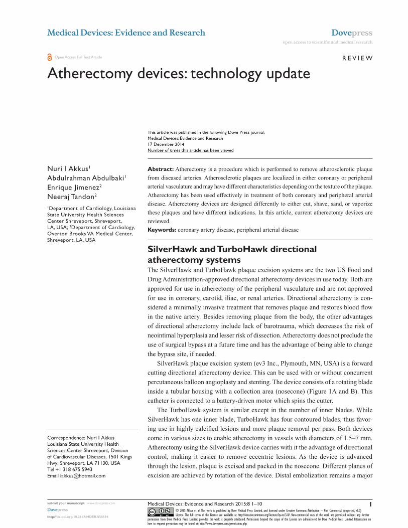
burr (Figure 5) which is coated with 2,000–3,000 microscopic
diamond crystals on the leading edge. The diamond crystals
are 20 µm in size, with only 5 µm extruding from the nickel
coating, and the burr rotates at 140,000–190,000 rpm.12,13
The burr is advanced over the 0.009 inch dia meter, 325 cm,
in length stainless steel RotaWire™ with a 2.2–2.6 cm spring
tip. The burr should not be advanced close to the spring tip
of the wire, and a wire clip torquer should be placed on the
wire which will prevent the guide wire from spinning. Avail-
able burr sizes are; 1.25, 1.5, 1.75, 2.0, 2.15, 2.25, 2.38, and
2.5 mm. In addition to the burr (which is bonded to the drive
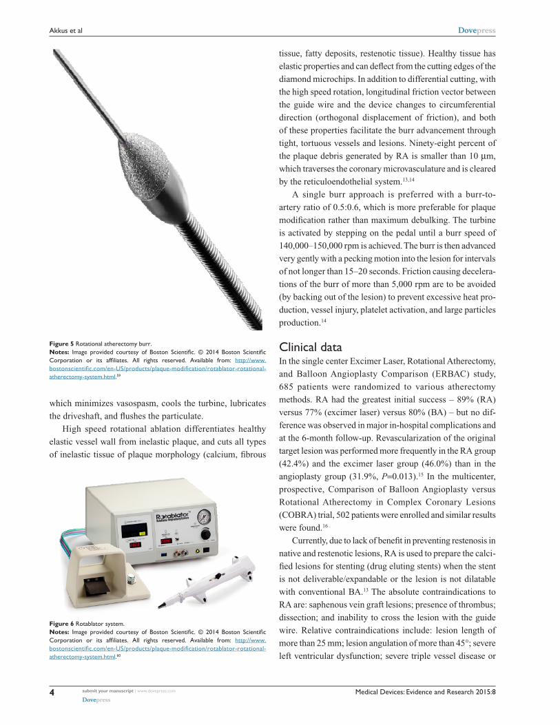
shaft), the Rotablator system has a console, a drive shaft, and
a turbine (Figure 6). The console is reusable and controls the
rotational speed of the drive shaft and burr. The drive shaft is
flexible, connected to the turbine, and housed by a 4.3 French
Teflon sheath. The Teflon sheath works as a flush delivery
conduit and also protects the arterial wall from the spinning
drive shaft.12 The turbine is driven by compressed air or
nitrogen and has the capacity to rotate the shaft and burr at
the desired rpm. The turbine is activated by a foot pedal and
controlled by the console.12 During rotational atherectomy
(RA), a cocktail of Rotaglide® lubricant, nitroglycerine,
verapamil, and heparin infuses through the Teflon sheath
Figure 2 Angiogram showing occluded left superficial femoral artery (arrow).
Figure 3 Angiogram showing no significant disease in left superficial femoral artery (arrow) after SilverHawk atherectomy and balloon angioplasty.
Figure 4 Angiogram showing pseudoaneurysm (arrow) formation in proximal left superficial femoral artery.
provide real-time imaging during atherectomy and will help
operators to direct the plaque excision.11
Rotational atherectomyThe Rotablator system was developed by David C Auth,
PhD (Boston Scientific Corporation, Redmond, WA, USA)
Medical Devices: Evidence and Research 2015:8submit your manuscript | www.dovepress.com
Dovepress
Dovepress
4
Akkus et al
which minimizes vasospasm, cools the turbine, lubricates
the driveshaft, and flushes the particulate.
High speed rotational ablation differentiates healthy
elastic vessel wall from inelastic plaque, and cuts all types
of inelastic tissue of plaque morphology (calcium, fibrous
tissue, fatty deposits, restenotic tissue). Healthy tissue has
elastic properties and can deflect from the cutting edges of the
diamond microchips. In addition to differential cutting, with
the high speed rotation, longitudinal friction vector between
the guide wire and the device changes to circumferential
direction (orthogonal displacement of friction), and both
of these properties facilitate the burr advancement through
tight, tortuous vessels and lesions. Ninety-eight percent of
the plaque debris generated by RA is smaller than 10 µm,
which traverses the coronary microvasculature and is cleared
by the reticuloendothelial system.13,14
A single burr approach is preferred with a burr-to-
artery ratio of 0.5:0.6, which is more preferable for plaque
modification rather than maximum debulking. The turbine
is activated by stepping on the pedal until a burr speed of
140,000–150,000 rpm is achieved. The burr is then advanced
very gently with a pecking motion into the lesion for intervals
of not longer than 15–20 seconds. Friction causing decelera-
tions of the burr of more than 5,000 rpm are to be avoided
(by backing out of the lesion) to prevent excessive heat pro-
duction, vessel injury, platelet activation, and large particles
production.14
Clinical dataIn the single center Excimer Laser, Rotational Atherectomy,
and Balloon Angioplasty Comparison (ERBAC) study,
685 patients were randomized to various atherectomy
methods. RA had the greatest initial success – 89% (RA)
versus 77% (excimer laser) versus 80% (BA) – but no dif-
ference was observed in major in-hospital complications and
at the 6-month follow-up. Revascularization of the original
target lesion was performed more frequently in the RA group
(42.4%) and the excimer laser group (46.0%) than in the
angioplasty group (31.9%, P=0.013).15 In the multicenter,
prospective, Comparison of Balloon Angioplasty versus
Rotational Atherectomy in Complex Coronary Lesions
(COBRA) trial, 502 patients were enrolled and similar results
were found.16
Currently, due to lack of benefit in preventing restenosis in
native and restenotic lesions, RA is used to prepare the calci-
fied lesions for stenting (drug eluting stents) when the stent
is not deliverable/expandable or the lesion is not dilatable
with conventional BA.13 The absolute contraindications to
RA are: saphenous vein graft lesions; presence of thrombus;
dissection; and inability to cross the lesion with the guide
wire. Relative contraindications include: lesion length of
more than 25 mm; lesion angulation of more than 45°; severe
left ventricular dysfunction; severe triple vessel disease or
Figure 6 Rotablator system.Notes: Image provided courtesy of Boston Scientific. © 2014 Boston Scientific Corporation or its affiliates. All rights reserved. Available from: http://www.bostonscientific.com/en-US/products/plaque-modification/rotablator-rotational-atherectomy-system.html.60
Figure 5 Rotational atherectomy burr.Notes: Image provided courtesy of Boston Scientific. © 2014 Boston Scientific Corporation or its affiliates. All rights reserved. Available from: http://www.bostonscientific.com/en-US/products/plaque-modification/rotablator-rotational-atherectomy-system.html.59

Medical Devices: Evidence and Research 2015:8 submit your manuscript | www.dovepress.com
Dovepress
Dovepress
5
Atherectomy devices: technology update
unprotected left main disease; and no candidacy to coronary
artery bypass surgery, either because of patient ineligibility
or lack of onsite surgical backup.13,17 Some of the reported
complications of RA are: Q wave myocardial infarction (MI)
(0.8%), urgent coronary artery bypass surgery (2.0%), non-Q
MI (8.9%), acute closures (1.1%), slow flow (2%), perfora-
tion (1.0%), side-branch closure (5%), dissection (4%), and
spasm (5%).18,19 Peripheral Rotablator atherectomy has also
recently started being used for calcified below-knee arteries.
There is no published outcome data available yet about its
use in peripheral arterial disease.
Pathway Jetstream PV Atherectomy SystemPathway Jetstream PV Atherectomy System (Pathway Medical
Technologies, Inc., Redmond, WA, USA) consists of a single-
use catheter with control pod and a reusable, compact console
power source which can be placed on a standard intravenous
stand. The system is indicated for both thrombectomy and RA
by the same catheter. The catheter is advanced over the 0.014″
wire with a maximum rate of 1 mm/second to avoid significant
drops in rotational speeds; it has a front-cutting tip that makes
it go through tight lesions without predilation. The electric
motor spins catheters at 60–70 krpm, and for every 40 seconds
of treatment, a 10-second pause in device activation is recom-
mended.20 During treatment, saline solution is delivered to the
proximal end of the catheter using two lines: one line to flush
the motor assembly to maintain an airtight seal, maximizing
embolic protection; the other line to infuse saline solution
in the treatment area through ports located on the distal
body of the catheter to facilitate the catheter’s de bulking and
aspiration capabilities.20 Its differentially cutting catheter
tip preferentially removes both hard and soft diseased tissue
from peripheral arteries with minimal damage to the vessel
wall. The rotational design potentially leads to concentric
lumens, which can facilitate laminar flow. JETSTREAM®
expandable catheters 2.1 mm/3.0 mm and 2.4 mm/3.4 mm
have a catheter tip that remains at a defined nominal diameter
(2.1mm or–2.4 mm) when spinning clockwise (blades within
fenestrated metal housing at the tip of the catheter used), but
expands to a defined maximum dia meter (3.0 mm or 3.4 mm,
respectively) when rotating counterclockwise (blades that are
hinged and mounted just proximal to the distal housing used)
(Figure 7); these sizes are indicated for above-knee arteries.20
For the below-knee use, there are fixed cutters (single cutter)
with sizes of 1.6 mm and 1.85 mm. Pathway Jetstream is the
only atherectomy device to offer continuous active aspiration,
and actively removes atherosclerotic debris and thrombus
from the treatment site and delivers it to a collection bag
located on the console.
Clinical dataZeller et al21 used Pathway Jetstream PV Atherectomy System
in treatment of 172 patients – 210 lesions (femoropopliteal
and infrapopliteal vessels) – with 99% device success,
and 6-month and 12-month target-lesion revascularization
rates of 15% and 26%, respectively. The 1-year restenosis
rate was 38.2% based on duplex imaging.21 In a six-patient
intravascular ultrasound study, after Pathway PV use, the
lumen area was greater than burr-sized lumen expectancy
at cross-sections in the treated segments, which suggested a
complementary role of aspiration in luminal gain achieved
with this device.22 Another intravascular ultrasound study
showed substantial plaque volume reduction by removing
fibrotic and fibro-fatty plaque by Jetstream atherectomy,
which resulted in substantial luminal volume expansion
without concomitant vessel expansion with no appreciable
effect on necrotic core and dense calcium.23
Abrupt vessel occlusion, dissection, distal emboli, hema-
toma at access site, infection, perforation, pseudoaneurysm,
renal failure, restenosis, and thrombus formation are some
of the reported complications of Jetstream atherectomy.24 In
a review of 2,137 lesions treated in 1,029 patients, Jetstream
(Pathway Medical Technologies Inc. Kirkland, WA, USA.) and
DiamondBack 360° (Cardiovascular Systems Inc., Saint Paul,
MN, USA) devices had a combined embolization rate of 22%
(eight of 36), four of 18 (22%) in each group, which was signifi-
cantly higher than with BA alone (five of 570, 0.9%), BA and
stent (five of 740, 0.7%), SilverHawk atherectomy (14 of 736,
1.9%), and laser atherectomy (two of 55, 3.6%; P,0.001).25
The use of embolization protection may be beneficial when
using this device.
Figure 7 JETSTREAM® expandable catheters.Notes: Image provided courtesy of Boston Scientific. © 2014 Boston Scientific Corporation or its affiliates. All rights reserved. Available from: http://www.medrad.com/en-us/info/products/Pages/jetstream-xc-systems.aspx.61

Medical Devices: Evidence and Research 2015:8submit your manuscript | www.dovepress.com
Dovepress
Dovepress
6
Akkus et al
Excimer laser atherectomyLaser is an acronym for light amplification by stimulated
emission of radiation. Although the history of laser begins
in 1951, the first medical application is reported by Goldman
in 1962 and used in 1963 for the experimental ablation of
atherosclerotic plaques.26 The first clinical applications were
performed by Choy and Ginsburg in 1983.26
Laser atherectomy uses the high energy, monochromatic
light beam to alter or dissolve (vaporize) the plaque without
damaging the surrounding tissue. Fiber-optic catheters are
used to deliver this light beam. For endovascular applica-
tions, xenon chloride excimer laser is used and its fiber-optic
catheter has multiple small fibers, rather than just a few
large fibers, in order to be flexible enough to navigate in the
arterial tree.27
Laser sources can vary depending on the wavelength of
their emitted light, how the light is transmitted (pulsed or
continuous), and the effective power of the light beam. In
addition to the source, effectiveness of a given laser depends
on how the light interacts with the tissue. The absorption
depth is determined by the wavelength and the tissue. In
the near-infrared regions (2,000 to 3,000 nm wavelength)
the light penetration depth is roughly 1–0.1 mm, whereas
in the ultraviolet (UV) B region with a shorter wavelength
(300 nm), the absorption depth is less. For example, at
308 nm, where the XeCL excimer laser emits, the typical
absorption depth is about 0.05 mm (50 µm).28 UV B light
has another advantage in that it uses direct photochemical
lytic affect to break molecular bonds rather than thermal
affect.29 UV B photons absorbed by the proteins and lipids
in cells actually break chemical bonds which facilitates lyses
of cellular structures.30
In the past, neodymium-yttrium aluminum garnet laser
(1,060 nm wavelength) and the argon laser (500 nm wave-
length) with deeper penetration levels were used with a
constant/continuous power output and resulted in excess
thermal damage, leading to thrombosis, vasospasm, and high
restenosis rates.28,31–33 On the other hand, with the excimer
laser (with less of a penetration depth), the high energy is
delivered with short interaction time (pulsed). In this way,
chemical bonds are broken only in the tissue that the laser is
touching without damaging surrounding material or increas-
ing the heat.28
While using laser, pulse width is also important in order
to keep the energy delivered to the tip of the catheter faster
than the time it will take for the heat to diffuse away from
its tip, so that the effect will be only localized to the tissue
on contact without a consequent rise in temperature to
surrounding tissue.28 The excimer laser catheter removes tissue
with a thickness of 10 µm with each pulse of energy.29
There is a unique threshold of energy density for each
laser type. If more energy is given, more reaction will be cre-
ated in the tissue (in expense of more heat generation) which
can be used as an advantage in particularly fibrotic or calci-
fied lesions. When laser is used, two factors are controlled
by the operators: number of pulses per second (frequency)
given and energy amount (fluence).28 Pulsed-wave xenon
chloride laser (CVX-300; Spectranetics, Colorado Springs,
CO, USA) is commonly used in clinical practice; it oper-
ates within a wavelength of 308 nm, with relatively long
pulses (pulse duration of 135 nanoseconds), and produces
an output of 165 mJ per pulse.27,34 The long pulse length is
required for successful delivery of the UV light by silica fibers
at the fluences necessary for therapy – typically between 30
and 80 mJ/mm2. After this pulse of 135 nanoseconds, laser
energy is not emitted. Typically, pulse repetition rates of
25–40 pulses/second are used. By doing this, the total power
emitted from the catheter tip is less than 3 W for the larg-
est catheters and this minimizes thermal effects during the
tissue-ablation process.27 The size of the laser catheters used
are chosen based on reference coronary/peripheral vessel
diameter, and available sizes are 0.9 mm, 1.4 mm, 1.7 mm,
2.0 mm, 2.3 mm, and 2.5 mm.
While using excimer laser, advancement of the catheter
should be slow (0.5 mm/second and no faster than 1 mm/
second) for effective plaque removal due to shallow penetra-
tion energy depth (35–50 µm).31 Slower advancement will
also create a larger and smoother channel. Since iodinated dye
absorbs the excimer laser energy nearly completely and will
cause cavitation bubbles, vapor bubbles, and percussive waves
which will lead to dissections/perforations,35,36 laser catheter
should never be activated in contrast media. In addition to
angiographic contrast media, blood (hemoglobin) strongly
absorbs excimer laser light at 308 nm.37 In order to remove
blood and contrast, saline flushes need to be given before
and during advancement of the laser catheter so that both
will be removed from the artery. The application of saline
infusion has resulted in improvements in both angiographic
and clinical outcomes.31,37 Excimer laser can also vaporize
thrombi, suppressing platelet aggregation while ablating the
underlying plaque.38 There are newer laser catheter designs
such as eccentric laser catheters (fiber-optic bundle disposed
opposite the guide-wire lumen), which are optimally spaced
concentric catheters (fiber bundle placed concentrically
around the guide-wire lumen) designed to achieve maximal
debulking in complex lesions.27
Medical Devices: Evidence and Research 2015:8 submit your manuscript | www.dovepress.com
Dovepress
Dovepress
7
Atherectomy devices: technology update
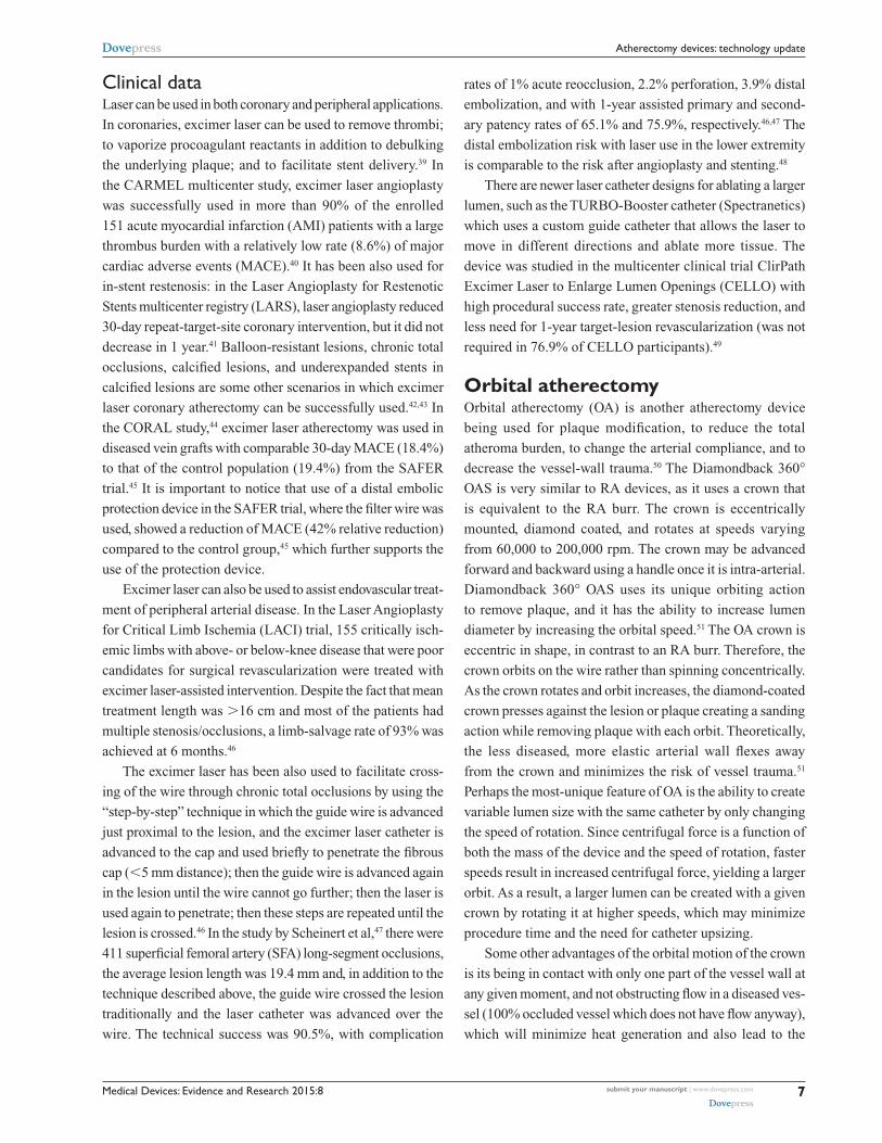
Clinical dataLaser can be used in both coronary and peripheral applications.
In coronaries, excimer laser can be used to remove thrombi;
to vaporize procoagulant reactants in addition to debulking
the underlying plaque; and to facilitate stent delivery.39 In
the CARMEL multicenter study, excimer laser angioplasty
was successfully used in more than 90% of the enrolled
151 acute myocardial infarction (AMI) patients with a large
thrombus burden with a relatively low rate (8.6%) of major
cardiac adverse events (MACE).40 It has been also used for
in-stent restenosis: in the Laser Angioplasty for Restenotic
Stents multicenter registry (LARS), laser angioplasty reduced
30-day repeat-target-site coronary intervention, but it did not
decrease in 1 year.41 Balloon-resistant lesions, chronic total
occlusions, calcified lesions, and underexpanded stents in
calcified lesions are some other scenarios in which excimer
laser coronary atherectomy can be successfully used.42,43 In
the CORAL study,44 excimer laser atherectomy was used in
diseased vein grafts with comparable 30-day MACE (18.4%)
to that of the control population (19.4%) from the SAFER
trial.45 It is important to notice that use of a distal embolic
protection device in the SAFER trial, where the filter wire was
used, showed a reduction of MACE (42% relative reduction)
compared to the control group,45 which further supports the
use of the protection device.
Excimer laser can also be used to assist endovascular treat-
ment of peripheral arterial disease. In the Laser Angioplasty
for Critical Limb Ischemia (LACI) trial, 155 critically isch-
emic limbs with above- or below-knee disease that were poor
candidates for surgical revascularization were treated with
excimer laser-assisted intervention. Despite the fact that mean
treatment length was .16 cm and most of the patients had
multiple stenosis/occlusions, a limb-salvage rate of 93% was
achieved at 6 months.46
The excimer laser has been also used to facilitate cross-
ing of the wire through chronic total occlusions by using the
“step-by-step” technique in which the guide wire is advanced
just proximal to the lesion, and the excimer laser catheter is
advanced to the cap and used briefly to penetrate the fibrous
cap (,5 mm distance); then the guide wire is advanced again
in the lesion until the wire cannot go further; then the laser is
used again to penetrate; then these steps are repeated until the
lesion is crossed.46 In the study by Scheinert et al,47 there were
411 superficial femoral artery (SFA) long-segment occlusions,
the average lesion length was 19.4 mm and, in addition to the
technique described above, the guide wire crossed the lesion
traditionally and the laser catheter was advanced over the
wire. The technical success was 90.5%, with complication
rates of 1% acute reocclusion, 2.2% perforation, 3.9% distal
embolization, and with 1-year assisted primary and second-
ary patency rates of 65.1% and 75.9%, respectively.46,47 The
distal embolization risk with laser use in the lower extremity
is comparable to the risk after angioplasty and stenting.48
There are newer laser catheter designs for ablating a larger
lumen, such as the TURBO-Booster catheter (Spectranetics)
which uses a custom guide catheter that allows the laser to
move in different directions and ablate more tissue. The
device was studied in the multicenter clinical trial ClirPath
Excimer Laser to Enlarge Lumen Openings (CELLO) with
high procedural success rate, greater stenosis reduction, and
less need for 1-year target-lesion revascularization (was not
required in 76.9% of CELLO participants).49
Orbital atherectomyOrbital atherectomy (OA) is another atherectomy device
being used for plaque modification, to reduce the total
atheroma burden, to change the arterial compliance, and to
decrease the vessel-wall trauma.50 The Diamondback 360°
OAS is very similar to RA devices, as it uses a crown that
is equivalent to the RA burr. The crown is eccentrically
mounted, diamond coated, and rotates at speeds varying
from 60,000 to 200,000 rpm. The crown may be advanced
forward and backward using a handle once it is intra-arterial.
Diamondback 360° OAS uses its unique orbiting action
to remove plaque, and it has the ability to increase lumen
diameter by increasing the orbital speed.51 The OA crown is
eccentric in shape, in contrast to an RA burr. Therefore, the
crown orbits on the wire rather than spinning concentrically.
As the crown rotates and orbit increases, the diamond-coated
crown presses against the lesion or plaque creating a sanding
action while removing plaque with each orbit. Theoretically,
the less diseased, more elastic arterial wall flexes away
from the crown and minimizes the risk of vessel trauma.51
Perhaps the most-unique feature of OA is the ability to create
variable lumen size with the same catheter by only changing
the speed of rotation. Since centrifugal force is a function of
both the mass of the device and the speed of rotation, faster
speeds result in increased centrifugal force, yielding a larger
orbit. As a result, a larger lumen can be created with a given
crown by rotating it at higher speeds, which may minimize
procedure time and the need for catheter upsizing.
Some other advantages of the orbital motion of the crown
is its being in contact with only one part of the vessel wall at
any given moment, and not obstructing flow in a diseased ves-
sel (100% occluded vessel which does not have flow anyway),
which will minimize heat generation and also lead to the
Medical Devices: Evidence and Research 2015:8submit your manuscript | www.dovepress.com
Dovepress
Dovepress
8
Akkus et al
continuous clearance of the sanded microscopic particulate
matter rather than having it build up into a large load of matter
(and micro embolization), which can be seen when the central
RA catheter is disengaged from the plaque.
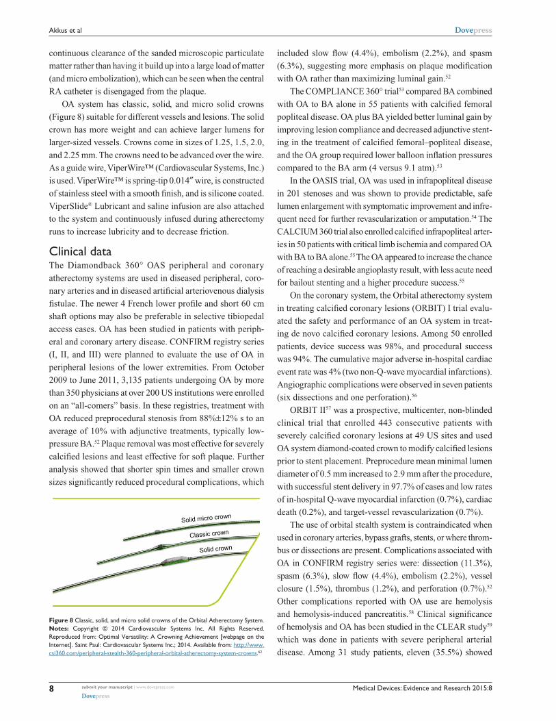
OA system has classic, solid, and micro solid crowns
(Figure 8) suitable for different vessels and lesions. The solid
crown has more weight and can achieve larger lumens for
larger-sized vessels. Crowns come in sizes of 1.25, 1.5, 2.0,
and 2.25 mm. The crowns need to be advanced over the wire.
As a guide wire, ViperWire™ (Cardiovascular Systems, Inc.)
is used. ViperWire™ is spring-tip 0.014″ wire, is constructed
of stainless steel with a smooth finish, and is silicone coated.
ViperSlide® Lubricant and saline infusion are also attached
to the system and continuously infused during atherectomy
runs to increase lubricity and to decrease friction.
Clinical dataThe Diamondback 360° OAS peripheral and coronary
atherectomy systems are used in diseased peripheral, coro-
nary arteries and in diseased artificial arteriovenous dialysis
fistulae. The newer 4 French lower profile and short 60 cm
shaft options may also be preferable in selective tibiopedal
access cases. OA has been studied in patients with periph-
eral and coronary artery disease. CONFIRM registry series
(I, II, and III) were planned to evaluate the use of OA in
peripheral lesions of the lower extremities. From October
2009 to June 2011, 3,135 patients undergoing OA by more
than 350 physicians at over 200 US institutions were enrolled
on an “all-comers” basis. In these registries, treatment with
OA reduced preprocedural stenosis from 88%±12% s to an
average of 10% with adjunctive treatments, typically low-
pressure BA.52 Plaque removal was most effective for severely
calcified lesions and least effective for soft plaque. Further
analysis showed that shorter spin times and smaller crown
sizes significantly reduced procedural complications, which
included slow flow (4.4%), embolism (2.2%), and spasm
(6.3%), suggesting more emphasis on plaque modification
with OA rather than maximizing luminal gain.52
The COMPLIANCE 360° trial53 compared BA combined
with OA to BA alone in 55 patients with calcified femoral
popliteal disease. OA plus BA yielded better luminal gain by
improving lesion compliance and decreased adjunctive stent-
ing in the treatment of calcified femoral–popliteal disease,
and the OA group required lower balloon inflation pressures
compared to the BA arm (4 versus 9.1 atm).53
In the OASIS trial, OA was used in infrapopliteal disease
in 201 stenoses and was shown to provide predictable, safe
lumen enlargement with symptomatic improvement and infre-
quent need for further revascularization or amputation.54 The
CALCIUM 360 trial also enrolled calcified infrapop liteal arter-
ies in 50 patients with critical limb ischemia and compared OA
with BA to BA alone.55 The OA appeared to increase the chance
of reaching a desirable angioplasty result, with less acute need
for bailout stenting and a higher procedure success.55
On the coronary system, the Orbital atherectomy system
in treating calcified coronary lesions (ORBIT) I trial evalu-
ated the safety and performance of an OA system in treat-
ing de novo calcified coronary lesions. Among 50 enrolled
patients, device success was 98%, and procedural success
was 94%. The cumulative major adverse in-hospital cardiac
event rate was 4% (two non-Q-wave myocardial infarctions).
Angiographic complications were observed in seven patients
(six dissections and one perforation).56
ORBIT II57 was a prospective, multicenter, non-blinded
clinical trial that enrolled 443 consecutive patients with
severely calcified coronary lesions at 49 US sites and used
OA system diamond-coated crown to modify calcified lesions
prior to stent placement. Preprocedure mean minimal lumen
diameter of 0.5 mm increased to 2.9 mm after the procedure,
with successful stent delivery in 97.7% of cases and low rates
of in-hospital Q-wave myocardial infarction (0.7%), cardiac
death (0.2%), and target-vessel revascularization (0.7%).
The use of orbital stealth system is contraindicated when
used in coronary arteries, bypass grafts, stents, or where throm-
bus or dissections are present. Complications associated with
OA in CONFIRM registry series were: dissection (11.3%),
spasm (6.3%), slow flow (4.4%), embolism (2.2%), vessel
closure (1.5%), thrombus (1.2%), and perforation (0.7%).52
Other complications reported with OA use are hemolysis
and hemolysis-induced pancreatitis.58 Clinical significance
of hemolysis and OA has been studied in the CLEAR study59
which was done in patients with severe peripheral arterial
disease. Among 31 study patients, eleven (35.5%) showed
Solid micro crown
Classic crown
Solid crown
Figure 8 Classic, solid, and micro solid crowns of the Orbital Atherectomy System.Notes: Copyright © 2014 Cardiovascular Systems Inc. All Rights Reserved. Reproduced from: Optimal versatility: A Crowning Achievement [webpage on the internet]. Saint Paul: Cardiovascular Systems inc.; 2014. Available from: http://www.csi360.com/peripheral-stealth-360-peripheral-orbital-atherectomy-system-crowns.62
Medical Devices: Evidence and Research 2015:8 submit your manuscript | www.dovepress.com
Dovepress
Dovepress
9
Atherectomy devices: technology update
laboratory evidence of hemolysis without any clinically
significant hemolysis. Lower glomerular filtration rates, calci-
fied plaque, long atherectomy runs, and solid crown selection
were independent predictors of hemolysis in this study.59
ConclusionWe have discussed in this article current atherectomy tech-
nologies including: SilverHawk directional atherectomy,
where the direction can be adjusted, which is useful for
eccentric lesions and is used in peripheral arterial disease;
RA, where the burr spins concentrically and mostly is used
in calcified coronaries; OA, where the crown orbits the wire
and is used in both coronary and peripheral arterial disease;
excimer laser atherectomy, which ablates the tissue and is
used in both coronary and peripheral arterial disease; and
Pathway Jetstream PV Atherectomy, which can be used for
RA as well as thrombectomy and is used in peripheral arte-
rial disease. As was discussed in the article, these current
atherectomy devices have been used efficiently in treatment
of coronary and/or peripheral arterial disease, and atherec-
tomy technologies continuously evolve to become even more
effective treatment modalities, which hopefully will also be
reflected as improved clinical outcomes in the patients.
DisclosureThe authors report no conflicts of interest in this work.
References1. Ramaiah V, Gammon R, Kiesz S, et al. Midterm outcomes from the
TALON Registry: treating peripherals with SilverHawk: outcomes collection. J Endovasc Ther. 2006;13:592–602.
2. McKinsey JF, Goldstein L, Khan HU, et al. Novel treatment of patients with lower extremity ischemia: use of percutaneous atherectomy in 579 lesions. Ann Surg. 2008;248(4):519–528.
3. Kandzari DE, Kiesz RS, Allie D, et al. Procedural and clinical outcomes with catheter-based plaque excision in critical limb ischemia. J Endovasc Ther. 2006;13:12–22.
4. Zeller T, Sixt S, Schwarzwälder U, et al. Two-year results after directional atherectomy of infrapopliteal arteries with the SilverHawk device. J Endovasc Ther. 2007;14(2):232–240.
5. Keeling WB, Shames ML, Stone PA, et al. Plaque excision with the Silverhawk catheter: early results in patients with claudication or critical limb ischemia. J Vasc Surg. 2007;45(1):25–31.
6. Sixt S, Rastan A, Beschorner U, et al. Acute and long-term outcome of Silverhawk assisted atherectomy for femoro-popliteal lesions according the TASC II classification: a single-center experience. Vasa. 2010;39(3): 229–236.
7. Shammas NW, Shammas GA, Helou TJ, Voelliger CM, Mrad L, Jerin M. Safety and 1-year revascularization outcome of SilverHawk atherectomy in treating in-stent restenosis of femoropopliteal arteries: a retrospective review from a single center. Cardiovasc Revasc Med. 2012;13(4): 224–227.
8. Garcia LA. Late Breaking Clinical Trials: DEFINITIVE LE 12 month outcomes. Presented at: VIVA 2012; October 9–12; 2012; Las Vegas, NV.
9. Kaid KA, Gopinathapillai R, Qian F, Salvaji M, Wasty N, Cohen M. Analysis of particulate debris after superficial femoral artery atherectomy. J Invasive Cardiol. 2009;21(1):7–10.
10. Akkus NI, Fay M, Varma J. Percutaneous treatment of delayed post-atherectomy superficial femoral artery pseudoaneurysm. J Invasive Cardiol. 2012;24(10):E212–E214.
11. Radvany MG, Kiesz RS. Plaque Excision in Management of Lower Extremity Peripheral Arterial Disease with the SilverHawk Atherectomy Catheter. Semin Intervent Radiol. 2008;25(1):11–19.
12. Spencer B, Yeung AC. Rotational Atherectomy: Concepts and Practice. In: Interventional Cardiology. New York: McGraw-Hill; 2007:333–347.
13. Tran T, Brown M, Lasala J. An evidence-based approach to the use of rotational and directional coronary atherectomy in the era of drug-eluting stents: when does it make sense? Catheter Cardiovasc Interv. 2008;72(5):650–662.
14. Tomey MI, Kini AS, Sharma SK. Current status of rotational atherectomy. JACC Cardiovasc Interv. 2014;7(4):345–353.
15. Reifart N, Vandormael M, Krajcar M, et al. Randomized comparison of angioplasty of complex coronary lesions at a single center. Excimer Laser, Rotational Atherectomy, and Balloon Angioplasty Comparison (ERBAC) Study. Circulation. 1997;96:91–98.
16. Dill T, Dietz U, Hamm CW, et al. A randomized comparison of balloon angioplasty versus rotational atherectomy in complex coronary lesions (COBRA study). Eur Heart J. 2000;21:1759–1766.
17. Sharma SK, Dangas G, Mehran R, et al. Risk factors for the development of slow flow during rotational coronary atherectomy. Am J Cardiol. 1997;80:219–222.
18. Maclsaac AI, Bass TA, Buchbinder M, et al. High speed rotational atherectomy: outcome in calcified and noncalcified coronary artery lesions. J Am Coll Cardiol. 1995;26:731–736.
19. Kini A, Marmur JD, Duvvuri S, Dangas G, Choudhary S, Sharma SK. Rotational atherectomy: Improved procedural outcome with evolution of technique and equipment. Single-center results of first 1,000 patients. Catheter Cardiovasc Interv. 1999;46:305–311.
20. Zeller T, Krankenberg H, Rastan A, et al. Percutaneous rotational and aspiration atherectomy in infrainguinal peripheral arterial occlusive disease: a multicenter pilot study. J Endovasc Ther. 2007;14(3): 357–364.
21. Zeller T, Krankenberg H, Steinkamp H, et al. One-year outcome of percutaneous rotational atherectomy with aspiration in infrainguinal peripheral arterial occlusive disease: the multicenter pathway PVD trial. J Endovasc Ther. 2009;16(6):653–662.
22. Hassan AH, Ako J, Waseda K, et al. Mechanism of lumen gain with a novel rotational aspiration atherectomy system for peripheral arterial disease: examination by intravascular ultrasound. Cardiovasc Revasc Med. 2010;11(3):155–158.
23. Singh T, Koul D, Szpunar S, et al. Tissue removal by ultrasound evaluation (the TRUE study): the Jetstream G2 system post-market peripheral vascular IVUS study. J Invasive Cardiol. 2011;23(7):269–273.
24. Sixt S, Scheinert D, Rastan A, et al. One-year outcome after percutane-ous rotational and aspiration atherectomy in infrainguinal arteries in patient with and without type 2 diabetes mellitus. Ann Vasc Surg. 2011; 25(4):520–529.
25. Shrikhande GV, Khan SZ, Hussain HG, Dayal R, McKinsey JF, Morrissey N. Lesion types and device characteristics that predict distal embolization during percutaneous lower extremity interventions. J Vasc Surg. 2011;53(2):347–352.
26. Choy DS. History of lasers in medicine. Thorac Cardiovasc Surg. 1988;36 Suppl 2:114–117.
27. Taylor K, Reiser C. Next generation catheters for excimer laser coronary angioplasty. Lasers Med Sci. 2001;16:133–140.
28. Biamino G. The excimer laser: science fiction fantasy or practical tool? J Endovasc Ther. 2004;11 Suppl 2:II207–II222.
29. Rogers JH, Laird JR. Overview of new technologies for lower extremity revascularization. Circulation. 2007;116(18):2072–2085.
30. Oraevsky AA, Jacques SL, Pettit GH, Saidi IS, Tittel FK, Henry PD. XeCl laser ablation of atherosclerotic aorta: optical properties and energy pathways. Lasers Surg Med. 1992;12:585–597.
Medical Devices: Evidence and Research
Publish your work in this journal
Submit your manuscript here: http://www.dovepress.com/medical-devices-evidence-and-research-journal
Medical Devices: Evidence and Research is an international, peer-reviewed, open access journal that focuses on the evidence, technology, research, and expert opinion supporting the use and application of medical devices in the diagnosis, treatment and management of clini-cal conditions and physiological processes. The identification of novel
devices and optimal use of existing devices which will lead to improved clinical outcomes and more effective patient management and safety is a key feature. The manuscript management system is completely online and includes a quick and fair peer-review system. Visit http://www.dovepress.com/testimonials.php to read real quotes from authors.
Medical Devices: Evidence and Research 2015:8submit your manuscript | www.dovepress.com
Dovepress
Dovepress
Dovepress
10
Akkus et al
31. Grundfest WS, Litvack F, Forrester JS, et al. Laser ablation of human atherosclerotic plaque without adjacent tissue injury. J Am Coll Cardiol. 1985;5:929–933.
32. Geschwind HJ, Boussignac G, Teisseire B, Benhaiem N, Bittoun R, Laurent D. Conditions for effective Nd-YAG laser angioplasty. Br Heart J. 1984;52:484–489.
33. Abela GS, Crea F, Smith W, Pepine CJ, Conti CR. In vitro effects of argon laser radiation on blood: quantitative and morphologic analysis. J Am Coll Cardiol. 1985;5:231–237.
34. Ebersole D, Dahm JB, Das T, et al. Excimer laser revascularization of saphenous vein grafts in acute myocardial infarction. J Invasive Cardiol. 2004;16(4):177–180.
35. Isner JM, Pickering JG, Mosseri M. Laser-induced dissections: pathogenesis and implications for therapy. J Am Coll Cardiol. 1992;19: 1619–1621.
36. van Leeuwen TG, Meertens JH, Velema E, Post MJ, Borst C. Intraluminal vapor bubble induced by excimer laser pulse causes microsecond arterial dilation and invagination leading to extensive wall damage in the rabbit. Circulation. 1993;87(4):1258–1263.
37. Tcheng JE. Saline infusion in excimer laser coronary angioplasty. Semin Interv Cardiol. 1996;1:135–141.
38. CARMEL Excimer Laser Interventional Study Group, Topaz O, Ebersole D, et al. Excimer laser in myocardial infarction: a comparison between STEMI patients with established Q-wave versus patients with non-STEMI (non-Q). Lasers Med Sci. 2008;23(1):1–10.
39. Topaz O, Bernardo NL, Shah R, et al. Effectiveness of excimer laser coronary angioplasty in acute myocardial infarction or in unstable angina pectoris. Am J Cardiol. 2001;87:849–855.
40. Topaz O, Ebersole D, Das T, et al. Excimer laser angioplasty in acute myocardial infarction (the CARMEL multicenter trial). Am J Cardiol. 2004;93:694–701.
41. Giri S, Ito S, Lansky AJ, Mehran R. Clinical and angiographic outcome in the laser angioplasty for restenotic stents (LARS) multicenter registry. Cathet Cardiovasc Intervent. 2001;52(1):24–34.
42. Ben-Dor I, Maluenda G, Pichard AD, et al. The use of excimer laser for complex coronary artery lesions. Cardiovasc Revasc Med. 2011; 12(1):69. e1–e8.
43. Fernandez JP, Hobson AR, McKenzie D, et al. Beyond the balloon: excimer coronary laser atherectomy used alone or in combination with rotational atherectomy in the treatment of chronic total occlusions, non-crossable and non-expansible coronary lesions. EuroIntervention. 2013;9(2):243–250.
44. Giugliano GR, Falcone MW, Mego D, Ebersole D, et al. A prospective multicenter registry of laser therapy for degenerated saphenous vein graft stenosis: the COronary graft Results following Atherectomy with Laser (CORAL) trial. Cardiovasc Revasc Med. 2012;13(2):84–89.
45. Baim DS, Wahr D, George B, et al. Randomized trial of a distal embolic protection device during percutaneous intervention of saphenous vein aorto-coronary bypass grafts. Circulation. 2002;105(11):1285–1290.
46. Laird JF, Zeller T, Gray BH, et al. Limb salvage following laser-assisted angioplasty for critical limb ischemia: results of the LACI multicenter trial. J Endovasc Ther. 2006;13:1–11.
47. Scheinert D, Laird JR Jr, Schröder M, Steinkamp H, Balzer JO, Biamino G. Excimer laser-assisted recanalization of long, chronic superficial femo-ral artery occlusions. J Endovasc Ther. 2001;8:156–166.
48. Shammas NW, Coiner D, Shammas GA, Christensen L, Dippel EJ, Jerin M. Distal embolic event protection using excimer laser ablation in peripheral vascular interventions: results of the DEEP EMBOLI registry. J Endovasc Ther. 2009;16(2):197–202.
49. Dave RM, Patlola R, Kollmeyer K, et al. Excimer laser recanalization of femoropopliteal lesions and 1-year patency: results of the CELLO registry. J Endovasc Ther. 2009;16(6):665–675.
50. Staniloae CS, Korabathina R. Orbital atherectomy: device evolution and clinical data. J Invasive Cardiol. 2014;26(5):215–219.
51. Heuser RR. Treatment of lower extremity vascular disease: the Diamondback 360 degrees Orbital Atherectomy System. Expert Rev Med Devices. 2008;5(3):279–286.
52. Das T, Mustapha J, Indes J, et al. Technique optimization of orbital atherectomy in calcified peripheral lesions of the lower extremities: the CONFIRM series, a prospective multicenter registry. Catheter Cardiovasc Interv. 2014;83(1):115–122.
53. Dattilo R, Himmelstein SI, Cuff RF. The COMPLIANCE 360° Trial: A Randomized, Prospective, Multicenter, Pilot Study Comparing Acute and Long-Term Results of Orbital Atherectomy to Balloon Angioplasty for Calcified Femoropopliteal Disease. J Invasive Cardiol. 2014;26(8): 355–360.
54. Safian RD, Niazi K, Runyon JP, et al. Orbital atherectomy for infrapo-pliteal disease: device concept and outcome data for the OASIS trial. Catheter Cardiovasc Interv. 2009;73(3):406–412.
55. Shammas NW, Lam R, Mustapha J, et al. Comparison of orbital atherec-tomy plus balloon angioplasty vs balloon angioplasty alone in patients with critical limb ischemia: results of the CALCIUM 360 randomized pilot trial. J Endovasc Ther. 2012;19(4):480–488.
56. Parikh K, Chandra P, Choksi N, Khanna P, Chambers J. Safety and feasibility of orbital atherectomy for the treatment of calcified coronary lesions: the ORBIT I trial. Catheter Cardiovasc Interv. 2013;81(7): 1134–1139.
57. Chambers JW, Feldman RL, Himmelstein SI, Bhatheja R, et al. Pivotal trial to evaluate the safety and efficacy of the orbital atherectomy system in treating de novo, severely calcified coronary lesions (ORBIT II). JACC Cardiovasc Interv. 2014;7(5):510–518.
58. Mehta SK, Laster SB. Hemolysis induced pancreatitis after orbital atherectomy in a heavily calcified superficial femoral artery. Catheter Cardiovasc Interv. 2008;72(7):1009–1011.
59. Staniloae CS, Korabathina R, Lane TA, et al. Study to determine the clinical significance of Hemolysis During Orbital AtheRectomy (CLEAR study). J Endovasc Ther. 2011;18(1):57–63.
60. Rotablator™ Rotational Atherectomy System [webpage on the Internet]. Marlborough: Boston Scientific Corporation; 2014. Available from: http://www.bostonscientific.com/en-US/products/plaque-modification/rotablator-rotational-atherectomy-system.html. Accessed. August 26, 2014
61. Boston Scientific Acquires the Interventional Division of Bayer [webpage on the Internet]. Warrendale: MEDRAD, INC.; 2014. Available from: http://www.medrad.com/en-us/info/products/Pages/jetstream-xc-systems.aspx. Accessed. August 26, 2014
62. Optimal Versatility: A Crowning Achievement [webpage on the Inter-net]. Saint Paul: Cardiovascular Systems Inc.; 2014. Available from: http://www.csi360.com/peripheral-stealth-360-peripheral-orbital-atherectomy-system-crowns. Accessed August 26, 2014
Related Documents