Astroglial Inhibition of NF-kB Does Not Ameliorate Disease Onset and Progression in a Mouse Model for Amyotrophic Lateral Sclerosis (ALS) Claudia Crosio 1,2 *, Cristiana Valle 2,3 , Arianna Casciati 2 , Ciro Iaccarino 1,2 , Maria Teresa Carrı` 2,4 1 Department of Physiological, Biochemical and Cell Science, University of Sassari, Sassari, Italy, 2 Fondazione Santa Lucia IRCCS, c/o CERC, Rome, Italy, 3 Cell Biology and Neurobiology Institute, National Research Council, Monterotondo Scalo, Italy, 4 Department of Biology, University of Rome ‘‘Tor Vergata’’, Rome, Italy Abstract Motor neuron death in amyotrophic lateral sclerosis (ALS) is considered a ‘‘non-cell autonomous’’ process, with astrocytes playing a critical role in disease progression. Glial cells are activated early in transgenic mice expressing mutant SOD1, suggesting that neuroinflammation has a relevant role in the cascade of events that trigger the death of motor neurons. An inflammatory cascade including COX2 expression, secretion of cytokines and release of NO from astrocytes may descend from activation of a NF-kB-mediated pathway observed in astrocytes from ALS patients and in experimental models. We have attempted rescue of transgenic mutant SOD1 mice through the inhibition of the NF-kB pathway selectively in astrocytes. Here we show that despite efficient inhibition of this major pathway, double transgenic mice expressing the mutant SOD1 G93A ubiquitously and the dominant negative form of IkBa (IkBaAA) in astrocytes under control of the GFAP promoter show no benefit in terms of onset and progression of disease. Our data indicate that motor neuron death in ALS cannot be prevented by inhibition of a single inflammatory pathway because alternative pathways are activated in the presence of a persistent toxic stimulus. Citation: Crosio C, Valle C, Casciati A, Iaccarino C, Carrı ` MT (2011) Astroglial Inhibition of NF-kB Does Not Ameliorate Disease Onset and Progression in a Mouse Model for Amyotrophic Lateral Sclerosis (ALS). PLoS ONE 6(3): e17187. doi:10.1371/journal.pone.0017187 Editor: Mark Cookson, National Institutes of Health, United States of America Received August 2, 2010; Accepted January 25, 2011; Published March 18, 2011 Copyright: ß 2011 Crosio et al. This is an open-access article distributed under the terms of the Creative Commons Attribution License, which permits unrestricted use, distribution, and reproduction in any medium, provided the original author and source are credited. Funding: This work was supported by Telethon (GGP07018) and Sisal to M.T.C. The funders had no role in study design, data collection and analysis, decision to publish, or preparation of the manuscript. Competing Interests: The authors have declared that no competing interests exist. * E-mail: [email protected] Introduction Amyotrophic Lateral Sclerosis (ALS), the most common adult- onset motor neuron disease, is usually fatal within five years of onset and is characterized by the degeneration of upper and lower motor neurons. Most ALS cases are sporadic, but about 5–10% of patients inherit the disease, typically in an autosomal dominant manner (familial ALS, FALS). Family-based linkage studies have led to the identification of twelve loci and eight genes for FALS, as well as three loci for ALS with frontotemporal dementia [1]. Approximately 20% of familial cases are caused by mutations in the gene coding for Cu/Zn superoxide dismutase (SOD1), and following linkage studies published in 1993, many different transgenic animal and cellular models of human SOD1 mutations have been developed, increasing our knowledge about the pathogenesis of both sporadic and familial forms of ALS [2]. Current hypotheses for the biology underlying sporadic and familial ALS forms in humans represent non-competing mecha- nisms that are likely to converge in various unfortunate patterns to mediate selective motor neuron degeneration [3]. Mutant SOD1 toxicity has been linked to oxidative damage, accumulation of intracellular aggregates, mitochondrial dysfunction, defects in axonal transport, growth factor deficiency, glial cell pathology, and glutamate excitotoxicity. A growing body of evidence indicates that non-neuronal cells contribute to the disease process in animal [4,5,6,7,8] and cellular [4,9,10] models overexpressing mutant SOD1. As a consequence, motor neuron death in ALS is considered as a ‘‘non-cell autonomous’’ process, with astrocytes playing a critical role in disease progression [11]. Astrocytes have many functions relevant to motor neuron physiology. First, they express the most important glutamate transporter EAAT2/GLT- 1, thus contributing to the clearance of this neurotransmitter; deficiency of astroglial EAAT2/GLT-1 causes severe motor neuron loss [12] and alteration of this transporter has been repeatedly invoked as a cause contributing to ALS [3]. Second, astrocytes are the major source of both trophic [13] and toxic factors [4] for motor neurons. Several cytokines have been proposed to play a role in ALS as reinforcing signals from glia cells, including interleukin-6 (IL6), tumour necrosis factor a (TNFa), monocyte chemoattractant protein-1, monocyte colony- stimulating factor (MCSF) and transforming growth factor b1 (TGFb1) that were found increased in cerebrospinal fluid, plasma and epidermis from ALS patients, although with sometimes conflicting results [14]. In addition, the production of nitric oxide and the activation of cyclooxygenase type 2 (COX2) aggravate the toxic effects of mutant SOD1 in several experimental models for ALS. The production of all those proinflammatory mediators may be secondary to the induction of the transcription factor NF-kB, which is activated in the presence of reactive oxygen species (ROS) and by many other different signalling molecules associated with ALS onset and progression [15,16]. NF-kB activation has been observed in astrocytes from ALS patients and in human cells expressing mutant SOD1 [17]. NF-kB also regulates the expression of COX2 that may cause an increase in the synthesis PLoS ONE | www.plosone.org 1 March 2011 | Volume 6 | Issue 3 | e17187

Welcome message from author
This document is posted to help you gain knowledge. Please leave a comment to let me know what you think about it! Share it to your friends and learn new things together.
Transcript
Astroglial Inhibition of NF-kB Does Not AmeliorateDisease Onset and Progression in a Mouse Model forAmyotrophic Lateral Sclerosis (ALS)Claudia Crosio1,2*, Cristiana Valle2,3, Arianna Casciati2, Ciro Iaccarino1,2, Maria Teresa Carrı̀2,4
1 Department of Physiological, Biochemical and Cell Science, University of Sassari, Sassari, Italy, 2 Fondazione Santa Lucia IRCCS, c/o CERC, Rome, Italy, 3 Cell Biology and
Neurobiology Institute, National Research Council, Monterotondo Scalo, Italy, 4 Department of Biology, University of Rome ‘‘Tor Vergata’’, Rome, Italy
Abstract
Motor neuron death in amyotrophic lateral sclerosis (ALS) is considered a ‘‘non-cell autonomous’’ process, with astrocytesplaying a critical role in disease progression. Glial cells are activated early in transgenic mice expressing mutant SOD1,suggesting that neuroinflammation has a relevant role in the cascade of events that trigger the death of motor neurons. Aninflammatory cascade including COX2 expression, secretion of cytokines and release of NO from astrocytes may descendfrom activation of a NF-kB-mediated pathway observed in astrocytes from ALS patients and in experimental models. Wehave attempted rescue of transgenic mutant SOD1 mice through the inhibition of the NF-kB pathway selectively inastrocytes. Here we show that despite efficient inhibition of this major pathway, double transgenic mice expressing themutant SOD1G93A ubiquitously and the dominant negative form of IkBa (IkBaAA) in astrocytes under control of the GFAPpromoter show no benefit in terms of onset and progression of disease. Our data indicate that motor neuron death in ALScannot be prevented by inhibition of a single inflammatory pathway because alternative pathways are activated in thepresence of a persistent toxic stimulus.
Citation: Crosio C, Valle C, Casciati A, Iaccarino C, Carrı̀ MT (2011) Astroglial Inhibition of NF-kB Does Not Ameliorate Disease Onset and Progression in a MouseModel for Amyotrophic Lateral Sclerosis (ALS). PLoS ONE 6(3): e17187. doi:10.1371/journal.pone.0017187
Editor: Mark Cookson, National Institutes of Health, United States of America
Received August 2, 2010; Accepted January 25, 2011; Published March 18, 2011
Copyright: � 2011 Crosio et al. This is an open-access article distributed under the terms of the Creative Commons Attribution License, which permitsunrestricted use, distribution, and reproduction in any medium, provided the original author and source are credited.
Funding: This work was supported by Telethon (GGP07018) and Sisal to M.T.C. The funders had no role in study design, data collection and analysis, decision topublish, or preparation of the manuscript.
Competing Interests: The authors have declared that no competing interests exist.
* E-mail: [email protected]
Introduction
Amyotrophic Lateral Sclerosis (ALS), the most common adult-
onset motor neuron disease, is usually fatal within five years of
onset and is characterized by the degeneration of upper and lower
motor neurons. Most ALS cases are sporadic, but about 5–10% of
patients inherit the disease, typically in an autosomal dominant
manner (familial ALS, FALS). Family-based linkage studies have
led to the identification of twelve loci and eight genes for FALS, as
well as three loci for ALS with frontotemporal dementia [1].
Approximately 20% of familial cases are caused by mutations in
the gene coding for Cu/Zn superoxide dismutase (SOD1), and
following linkage studies published in 1993, many different
transgenic animal and cellular models of human SOD1 mutations
have been developed, increasing our knowledge about the
pathogenesis of both sporadic and familial forms of ALS [2].
Current hypotheses for the biology underlying sporadic and
familial ALS forms in humans represent non-competing mecha-
nisms that are likely to converge in various unfortunate patterns to
mediate selective motor neuron degeneration [3]. Mutant SOD1
toxicity has been linked to oxidative damage, accumulation of
intracellular aggregates, mitochondrial dysfunction, defects in
axonal transport, growth factor deficiency, glial cell pathology, and
glutamate excitotoxicity. A growing body of evidence indicates
that non-neuronal cells contribute to the disease process in animal
[4,5,6,7,8] and cellular [4,9,10] models overexpressing mutant
SOD1. As a consequence, motor neuron death in ALS is
considered as a ‘‘non-cell autonomous’’ process, with astrocytes
playing a critical role in disease progression [11]. Astrocytes have
many functions relevant to motor neuron physiology. First, they
express the most important glutamate transporter EAAT2/GLT-
1, thus contributing to the clearance of this neurotransmitter;
deficiency of astroglial EAAT2/GLT-1 causes severe motor
neuron loss [12] and alteration of this transporter has been
repeatedly invoked as a cause contributing to ALS [3]. Second,
astrocytes are the major source of both trophic [13] and toxic
factors [4] for motor neurons. Several cytokines have been
proposed to play a role in ALS as reinforcing signals from glia
cells, including interleukin-6 (IL6), tumour necrosis factor a(TNFa), monocyte chemoattractant protein-1, monocyte colony-
stimulating factor (MCSF) and transforming growth factor b1
(TGFb1) that were found increased in cerebrospinal fluid, plasma
and epidermis from ALS patients, although with sometimes
conflicting results [14]. In addition, the production of nitric oxide
and the activation of cyclooxygenase type 2 (COX2) aggravate the
toxic effects of mutant SOD1 in several experimental models for
ALS. The production of all those proinflammatory mediators may
be secondary to the induction of the transcription factor NF-kB,
which is activated in the presence of reactive oxygen species (ROS)
and by many other different signalling molecules associated with
ALS onset and progression [15,16]. NF-kB activation has been
observed in astrocytes from ALS patients and in human cells
expressing mutant SOD1 [17]. NF-kB also regulates the
expression of COX2 that may cause an increase in the synthesis
PLoS ONE | www.plosone.org 1 March 2011 | Volume 6 | Issue 3 | e17187
of prostaglandins, which trigger astrocytic glutamate release and
induce free radical formation, thus contributing to both excito-
toxicity and oxidative damage. Indeed, treatment with COX2
inhibitors markedly protects motor neurones and significantly
prolongs survival of ALS mice [18].
An approach that has been widely used to study cell specific NF-
kB function in mice is to inhibit its activation by the
(over)expression of various degradation-resistant mutant isoforms
of IkBa, the physiological inhibitor of NF-kB. These proteins, that
may be collectively termed IkB-DR (IkB-degradation resistant,
[19], act in a dominant negative manner to block NF-kB
activation, by impairing its nuclear translocation and transcrip-
tional activation [20]. To address the contribution of astroglial
NF-kB and, more generally, the contribution of astrocytosis to
ALS onset and progression, we generated a mouse line expressing
an IkBa-DR (IkBaAA) in astrocytes only, under control of the
astrocyte-specific glial fibrillary acidic protein (GFAP) promoter,
and crossbred them with transgenic mice over-expressing ALS-
typical mutant SOD1G93A. We demonstrate that GFAP-IkBaAA
transgenic mice grow normally, although they are highly sensitive
to lipopolysaccharides (LPS)-induced toxaemia. However,
SOD1G93A mice made deficient for NF-kB activation in astrocytes
do not show an ameliorated ALS phenotype despite a slight but
significant reduction in astrogliosis at onset.
Results
Generation of IkBaAA transgenic mice and phenotypicalcharacterization
We generated tissue-restricted NF-kB knockout mice through
targeting of a dominant-negative form of IkBa in glial cells. When
the two N-terminal serines of IkBa have been mutated into
alanines IkBa-S32/36A, IkBaAA), the protein cannot be
phosphorylated and degraded and therefore acts as a super-
repressor of NF-kB activity. In its presence, every NF-kB/Rel
complex is sequestered in the cytoplasm, prevented from DNA
binding and consequently from activating transcription [21]. We
have generated transgenic mice over-expressing the dominant
negative form of IkBa selectively in astrocytes (GFAP-IkBaAA),
using the human GFAP promoter [22]. As shown in Figure 1, we
have generated four different independent transgenic lines,
indicated from A to D, that have integrated in their genome a
number of transgene copies included between 10–20 and 200 as
suggested by band intensity comparison (Figure 1B).
Transgene expression was analyzed by RT-PCR on total RNA
extracted from different tissues of mice generated from founder A
and D (indicated as TgA and TgD, respectively). As shown in
Figure 1C, different levels of transgene expression were obtained,
but in all cases expression of GFAP-IkBaAA was predominant in
neuronal tissue, with no ectopic expression in peripheral tissues,
except faintly in muscle of transgenic line A. Total proteins,
extracted from spinal cord samples from TgA, TgD and non-
transgenic mice (-) were tested by Western blot for IkBaexpression. As shown in Figure 1D–E, IkBa overexpression is
clearly detectable and significantly increased in both transgenic
lines in tissue samples in which astrocytes are not the only cellular
population. Expression of IkBa-AA in both transgenic lines does
not induce any obvious phenotype as confirmed by their normal
growth of mice (Figure 1F). Standard cresyl violet histopathology
did not reveal any gross morphological anomalies in the
architecture of the naive spinal cord in GFAP- IkBaAA-A/D
mice (figure 1G).
To confirm the ability of IkBaAA to prevent NF-kB activation,
electrophoretic mobility shift assay (EMSA) was performed on
nuclear extracts from astrocytes isolated from brain (figure 2A,
upper panels) and spinal cord (figure 2A, lower panels) of non-
transgenic (-) and GFAP-IkBaAA transgenic mice (Tg-A)
(Figure 2A). Astrocytes from control mice stimulated with TNFaexhibited a significant induction of NF-kB DNA binding activity,
whereas no induction was detected in astrocytes from transgenic
mice (Figure 2A, left panels). To confirm the cell specificity of
transgene expression, similar EMSA experiments were performed
on microglial cells isolated from brain and spinal cord of GFAP-
IkBaAA mice and non-transgenic littermates (Fig. 2A, right
panels). As expected, when stimulated with TNFa, GFAP- IkBa-
AA microglial primary cultures showed an increase in NF-kB
DNA binding activity comparable to non-transgenic microglial
cells, demonstrating the complete functionality of the NF-kB
pathway in this cell type. Quantification of results is shown in
Figure 2B. Similar results were obtained with line TgD in primary
cultures (data not shown). At molecular level, the impairment of
NF-k signalling in primary astrocytes of GFAP-IkBaAA trans-
genic mice causes a severe reduction in the expression of many
different kB controlled genes upon challenge with lipopolysaccha-
ride (LPS). Cox2 levels were assayed at protein level (figure 2C),
while iNOS, IL-1b, TNFa, FAS at mRNA level (figure 2D and 2).
NF-kB inhibition in glial cells does not influence onsetand progression of ALS, caused by SOD 1 mutations
To assess the contribution of NF-kB inhibition in glial cells in
the pathogenesis of ALS, we have generated two different double
transgenic mice lines by crossing SOD1G93A mice with the two
independent GFAP-IkBaAA transgenic lines described above, in
order to exclude a founder effect. The effect of NF-kB deficiency
in glial cells on severity of motor neuron disease was examined
using cohorts of GFAP-IkBaAA (indicated as Tg), SOD1G93A
(indicated as G93A) and GFAP-IkBaAA/SOD1G93A (indicated as
double Tg) transgenic mice.
We initially confirmed cell-specific inhibition of NF-kB by
stimulating primary cultures of motor neurons, microglial (data
not shown) and glial cells from transgenic (GFAP-IkBaAA,
indicated as TgA and SOD1G93A, indicated as G93A), double
transgenic (SOD1G93A/GFAP-IkBaAA, indicated as double Tg)
and non-transgenic littermates (-) with LPS (or TNFa data not
shown) for 20 min and determining NF-kB (p65) nuclear
translocation by immunofluorescence. As expected, we did not
observe NF-kB nuclear translocation in glial cells isolated from
spinal cord (Figure 3A) of mice expressing IkBaAA, independently
from SOD1G93A presence (TgA and double Tg) in contrast of
motor neurons of all genotypes (Figure 3B). Notably, despite the
observation that mutant SOD1 stably expressed in a neuronal cell
line may induce up-regulation of NF-kB [17], in primary
astrocytic cultures derived from double transgenic mice the
super-repressor IkBaAA is still able to prevent p65 nuclear
translocation upon LPS (or TNFa data not shown) stimulation.
The impairment of NF-kB signalling in double transgenic mice
was confirmed by the lack of Cox2 induction upon LPS
stimulation, as in TgA mice (figure 3C).
Alteration of NF-kB-signalling has been widely linked to ALS
onset and progression, [15,17,23,24]. Using an antibody that
specifically recognizes an epitope overlapping the nuclear
localization signal of the p65 subunit and, thus, selectively binding
to the activated form of p65 (indicated as p65*), we evaluated the
activation of NF-kB in single and double transgenic mice at the
onset stage by Western blot. As expected, we observed in G93A
mice an increased NF-kB activation, that is prevented in double
transgenic mice (figure 4A and 4B). These data indicate the
Astroglial Inhibition of NF-kB in ALS Mice
PLoS ONE | www.plosone.org 2 March 2011 | Volume 6 | Issue 3 | e17187

Astroglial Inhibition of NF-kB in ALS Mice
PLoS ONE | www.plosone.org 3 March 2011 | Volume 6 | Issue 3 | e17187
effectiveness of GFAP-IkBaAA transgenic mice in inhibiting NF-
kB signalling in the astrocytes of G93A spinal cord.
Nonetheless, as shown in Figure 4, the impairment of NF-kB
signalling in glial cells did not influence the rate of mortality in the
double transgenic mice lines. SOD1G93A and double transgenic
mice developed ALS in a comparable manner (data reported in
Figure 4 refer only to the double transgenic line generated by
crossing GFAP-IkBaAA line A with SOD1G93A, but they are
indistinguishable from those obtained with the other double
transgenic line). There was no difference in disease onset measured
either evaluating motor performance in grip test (Figure 4C) and
rotaroad (Figure 4D) or by 10% of weight loss (Figure 4E). Disease
progression was also similar and the median lifespan was 160 and
157 days for SOD1G93A and double transgenic mice, respectively
(figure 4F). Notably, the life span we observed in our transgenic
lines is similar to the one observed in the original B6.Cg-
Tg(SOD1-G93A)1Gur line (from The Jackson Laboratory, 50%
survive at 157.1+/29.3 days, 99,99% B57BL/6J, http://jaxmice.
jax.org/strain/004435.html).
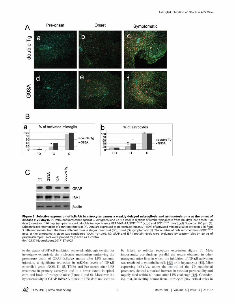
Selective expression of IkBaAA in astrocytes causes adelayed microgliosis and astrocytosis at the onset ofdisease only
Astrocytosis and microgliosis are non-neuronal events that likely
contribute to the neurodegenerative process in ALS. NF-kB is a
transcription factor involved in gene expression of many different
inflammatory molecules whose function is crucial for microglial
activation and induction of reactive astrogliosis. We therefore
investigated whether glial NF-kB deficiency in mutant SOD1 mice
had any effect on the expression of CD11b, a marker of microglial
activation, and GFAP, a marker of astrogliosis. Immunoreactivity
was assessed in the spinal cord of normal mice, SOD1G93A and
double transgenic mice at pre-onset (100 days), onset (120 days)
and symptomatic phase (140 days) of disease progression. As
shown in Figure 6, we observed a slight, but statistically significant,
reduction of both CD11b and GFAP positive cells at the onset
stage in double transgenic mice with respect to SOD1G93A mice.
This effect was lost at later stages of disease and the astroglial
inhibition of NF-kB in the SOD1G93A background was not
sufficient to reduce neuroinflammation in the spinal cord of double
transgenic mice. Western blot analysis confirms the reduction of
GFAP reactive protein and IBA1, a marker of microglial
activation, in spinal cord extract from double Tg mice with
respect to G93A mice (figure 5C).
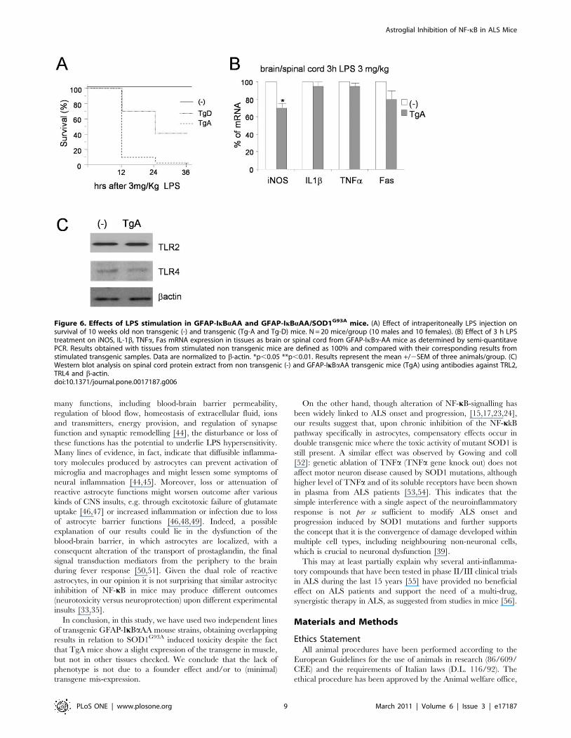
Astroglial inhibition of NF-kB is deleterious uponLPS-induced septic shock
The data presented above indicate that astroglial inhibition of
NF-kB does not affect motor neuron survival in an ALS genetic
model, although astrocyte dysfunction, via a number of pathways
including the NF-kB one, has been invoked as a potential
mediator of disease progression. Previous studies have demon-
strated that a chronic stimulation of innate immunity, via sub-
lethal systemic injection of LPS, can exacerbate ALS disease
progression [25,26]. Thus, we decided to investigate the effect of
LPS on G93A/GFAP-IkBaAA double transgenic mice to evaluate
whether the NF-kB impairment in astrocytes may have any effect
on LPS exacerbation of ALS disease progression. This experiment
revealed unfeasible since, surprisingly, when 10-week-old GFAP-
IkBaAA single transgenic mice were challenged with a sub-lethal
dose of LPS (1, 3 and 5 mg/kg, Sigma-Aldrich, from E. coli strain
111:B4), at all doses tested we observed a premature death of mice
from both GFAP-IkBaAA transgenic lines (Figure 6A).
At the molecular level, we observed that 3 h hours after LPS
treatment, a significant reduction of iNOS induction occurred in
the spinal cord of TgA (figure 6B). IL1b, TNFa and Fas showed a
modest reduction in spinal cord from TgA mice, that does not
reach statistical significance (figure 6B).
In the attempt of investigating the hypersensitivity of GFAP-
IkBaAA transgenic mice to LPS, we have analyzed the expression
level of the toll-like receptors 2 and 4, which have been indicated
as LPS mediators [27,28,29]. As shown in figure 6C, TRL2 and
TRL4 levels are equal in the spinal cord from TgA compared to
non transgenic mice.
Discussion
In the past five years, neuroinflammation has been recognized
as a key event in disease onset and progression, modulating death
of motor neurons in ALS. Yet, several lines of evidence indicate
that gliosis may actually exert very different effects in the diseased
spinal cord as it may mediate either beneficial or harmful events.
Indeed, activated astrocytes can mediate neuroprotection by
preserving motor neuron survival through the release of anti-
inflammatory cytokines, neurotrophins, and growth factors [30].
On the other hand, reactive astrocytes can participate directly in
inflammatory reactions expressing inflammation markers includ-
ing iNOS and COX2 and other mediators including prostaglan-
dins, IL-6, TNFa, IL-1b and NGF [30].
As demonstrated by two independent groups, astrocytes may be
the primary cell types where mutant SOD1 exert its toxic effects on
motor neurons by releasing some not yet identified molecule(s) [9,10].
Moreover, genetic evidence obtained in mice models for ALS
indicate that lack of expression of mutant SOD1 in GFAP expressing
astrocytes [7], but not astrocyte ablation [31], sharply slowed disease
progression. These findings demonstrate that while astrocyte
signalling is an important factor in the aetiology of motor neuron
diseases, astrocyte proliferation itself does not play a significant role.
A number of different signalling molecules released by
astrocytes are likely to be under the control of the transcription
factor NF-kB, a key molecule responding to both redox and
inflammatory stimuli [20]. In order to explore the contribution of
NF-kB regulated gene expression in astrocytes to ALS onset and
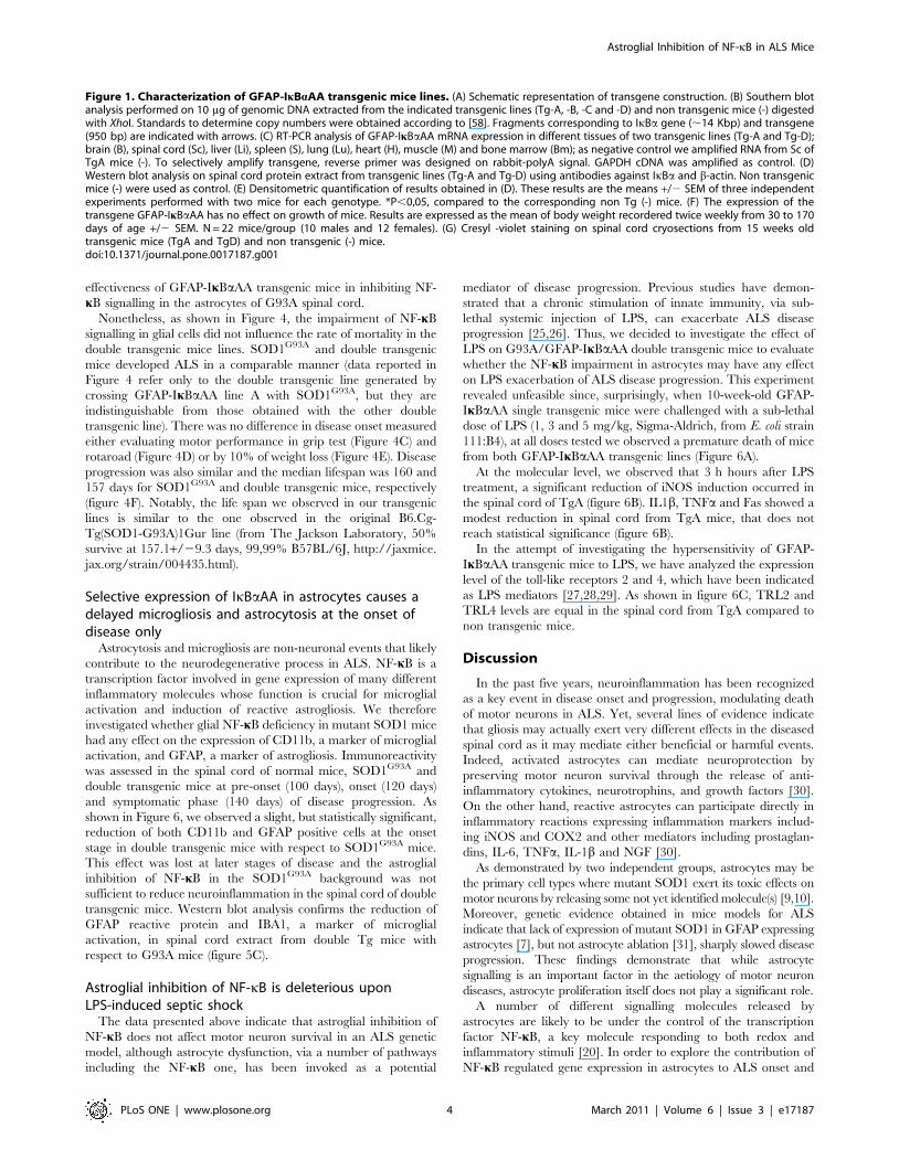
Figure 1. Characterization of GFAP-IkBaAA transgenic mice lines. (A) Schematic representation of transgene construction. (B) Southern blotanalysis performed on 10 mg of genomic DNA extracted from the indicated transgenic lines (Tg-A, -B, -C and -D) and non transgenic mice (-) digestedwith XhoI. Standards to determine copy numbers were obtained according to [58]. Fragments corresponding to IkBa gene (,14 Kbp) and transgene(950 bp) are indicated with arrows. (C) RT-PCR analysis of GFAP-IkBaAA mRNA expression in different tissues of two transgenic lines (Tg-A and Tg-D);brain (B), spinal cord (Sc), liver (Li), spleen (S), lung (Lu), heart (H), muscle (M) and bone marrow (Bm); as negative control we amplified RNA from Sc ofTgA mice (-). To selectively amplify transgene, reverse primer was designed on rabbit-polyA signal. GAPDH cDNA was amplified as control. (D)Western blot analysis on spinal cord protein extract from transgenic lines (Tg-A and Tg-D) using antibodies against IkBa and b-actin. Non transgenicmice (-) were used as control. (E) Densitometric quantification of results obtained in (D). These results are the means +/2 SEM of three independentexperiments performed with two mice for each genotype. *P,0,05, compared to the corresponding non Tg (-) mice. (F) The expression of thetransgene GFAP-IkBaAA has no effect on growth of mice. Results are expressed as the mean of body weight recordered twice weekly from 30 to 170days of age +/2 SEM. N = 22 mice/group (10 males and 12 females). (G) Cresyl -violet staining on spinal cord cryosections from 15 weeks oldtransgenic mice (TgA and TgD) and non transgenic (-) mice.doi:10.1371/journal.pone.0017187.g001
Astroglial Inhibition of NF-kB in ALS Mice
PLoS ONE | www.plosone.org 4 March 2011 | Volume 6 | Issue 3 | e17187
progression, we developed transgenic mice expressing a super-
repressor of NF-kB specifically in GFAP-positive astrocytes
(GFAP-IkBaAA). This type of cell-specific repression has been
widely used to efficiently block NF-kB activation, because the
degradation-resistant IkB mutant interferes preferentially with the
activity of canonical NF-kB dimers [19,32].
Using a similar approach, it has been demonstrated that
inactivation of NF-kB activity in astrocytes can either protect
neurons from different insults (improving functional recovery
following spinal cord injury [33,34] and experimental autoim-
mune encephalomyelitis [35], and promoting survival of retinal
neurons following ischemic injury [36]), or have no effect on
preventing neuronal death in cerebral ischemia [37]. The two
transgenic GFAP-IkBaAA mouse strains used in this study grow
normally and they display impairment in NF-kB activation upon
LPS or TNFa stimulation specifically in astrocytes (Figures 2, 3).
Unexpectedly, we report here that NF-kB downregulation in
astrocytes fails to influence onset, severity, or progression of disease
in a mutant SOD1-based ALS mice model. Though we observed a
slight but significant reduction in the percentage of activated
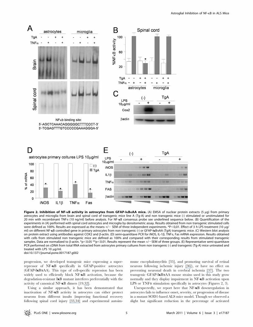
Figure 2. Inhibition of NF-kB activity in astrocytes from GFAP-IkBaAA mice. (A) EMSA of nuclear protein extracts (5 mg) from primaryastrocytes and microglia from brain and spinal cord of transgenic mice line A (Tg-A) and non transgenic mice (-) stimulated or unstimulated for20 min with recombinant TNFa (10 ng/ml) before analysis. For NF-kB consensus probe see underlined sequence below. (B) Quantification of theexperiments in (A) performed with spinal cord astrocytes and microglia by densitometric assay. Results obtained from non transgenic stimulated cellswere defined as 100%. Results are expressed as the means +/2 SEM of three independent experiments. *P,0,01. Effect of 3 h LPS treatment (10 mg/ml) on different NF-kB controlled gene in primary astrocytes from non transgenic (-) or GFAP-IkBaAA (TgA) transgenic mice. (C) Western blot analysison protein extract using antibodies against COX2 and b-actin. (D) semi-quantitave PCR for iNOS, IL-1b, TNFa, Fas mRNA expression. Results obtainedwith cells from stimulated non transgenic mice are defined as 100% and compared with their corresponding results from stimulated transgenicsamples. Data are normalized to b-actin. *p,0.05 **p,0.01. Results represent the mean +/2SEM of three groups. (E) Representative semi-quantitavePCR performed on cDNA from total RNA extracted from astrocytes primary cultures from non transgenic (-) and transgenic (Tg-A) mice untreated andtreated with LPS 10 mg/ml.doi:10.1371/journal.pone.0017187.g002
Astroglial Inhibition of NF-kB in ALS Mice
PLoS ONE | www.plosone.org 5 March 2011 | Volume 6 | Issue 3 | e17187
microglia and astrocytes at onset stage, neither survival nor motor
performance of double transgenic mice differ from those of single
transgenic ALS mice (Figure 4–5). We may speculate that the
reduction observed in GFAP markers in double transgenic mice
with respect to G93A mice at the onset of the disease is due to a
delay in astrogliosis for different reasons: i) we observe a similar
reduction in CD11b positive cells, that do not express the
transgene (figure 2); ii) astroglial and neuronal NF-kB has been
shown not to be a critical regulator of survival under non
pathological conditions [19,33]; iii) at end stage of disease the level
of GFAP positive cells in double transgenic mice is comparable to
the G93A transgenic mice.
Diminished expression of mutant SOD1 in astrocytes delays
microglial activation and significantly reduces disease progression
in ALS mice [38,39], a fact suggesting that noxious signals from
astrocytes do contribute significantly to the progression of non-cell
autonomous killing of motor neurons in ALS via activation of both
microglia and astrocytes, although ablation of proliferating
microglia or astrocytes does not affect motor neuron degeneration
in SOD1-ALS [31,40]. Here we show that the inhibition of NF-kB
signalling in astrocytes does not affect motor neuron survival in a
SOD1-linked ALS mouse model. This result indicates that the
toxic effects generated by expression mutant SOD1 in astrocytes
are independent from NF-kB signalling. Other pathways can be
activated in astrocytes that parallel or compensate the NF-kB
inhibition leading to the same inflammatory response at late stage
of the disease in both G93A and G93A/GFAP-IkBaAA transgenic
mice.
Although inhibition of NF-kB in astrocytes does not affect
motor neuron death upon expression of mutant SOD1, it is crucial
in experiments of LPS-induced toxaemia (Figure 6). In fact in our
GFAP-IkBaAA transgenic mice relatively low doses of LPS evoke
a striking reduction in survival, roughly dependent on transgene
copy number. Our results are partially in contrast with those
obtained in another GFAP-IkBa-AA transgenic line, which
responds to LPS treatment as non-transgenic mice [41]. This
discrepancy can at least partially be explained by the slightly
different transgenic construction and considering that individual
transgenic mouse strains over-expressing IkBa-DR proteins in a
given tissue often show phenotypes of varying severity in relation
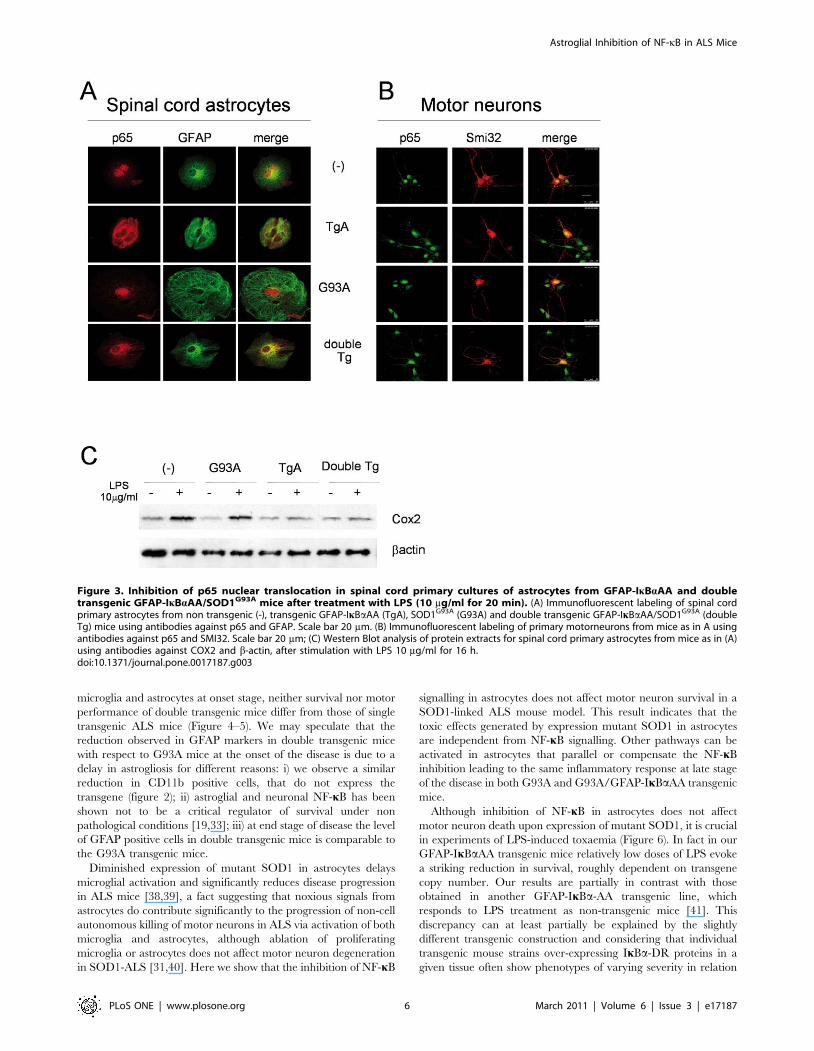
Figure 3. Inhibition of p65 nuclear translocation in spinal cord primary cultures of astrocytes from GFAP-IkBaAA and doubletransgenic GFAP-IkBaAA/SOD1G93A mice after treatment with LPS (10 mg/ml for 20 min). (A) Immunofluorescent labeling of spinal cordprimary astrocytes from non transgenic (-), transgenic GFAP-IkBaAA (TgA), SOD1G93A (G93A) and double transgenic GFAP-IkBaAA/SOD1G93A (doubleTg) mice using antibodies against p65 and GFAP. Scale bar 20 mm. (B) Immunofluorescent labeling of primary motorneurons from mice as in A usingantibodies against p65 and SMI32. Scale bar 20 mm; (C) Western Blot analysis of protein extracts for spinal cord primary astrocytes from mice as in (A)using antibodies against COX2 and b-actin, after stimulation with LPS 10 mg/ml for 16 h.doi:10.1371/journal.pone.0017187.g003
Astroglial Inhibition of NF-kB in ALS Mice
PLoS ONE | www.plosone.org 6 March 2011 | Volume 6 | Issue 3 | e17187
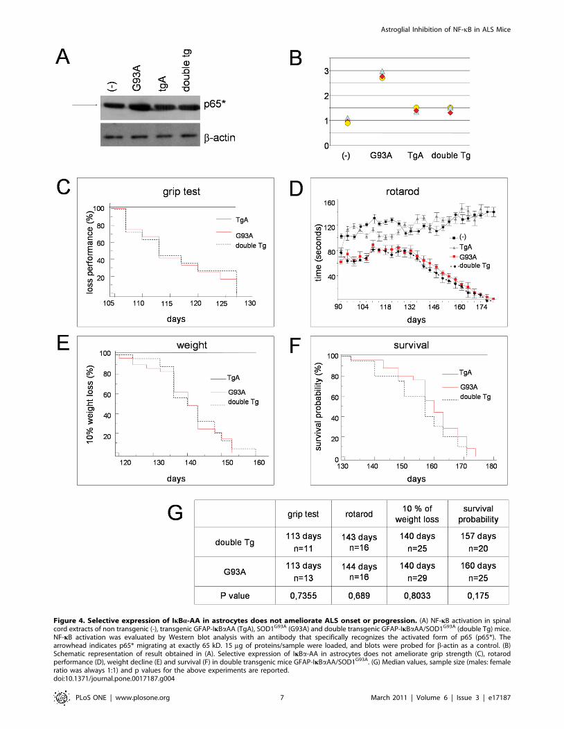
Figure 4. Selective expression of IkBa-AA in astrocytes does not ameliorate ALS onset or progression. (A) NF-kB activation in spinalcord extracts of non transgenic (-), transgenic GFAP-IkBaAA (TgA), SOD1G93A (G93A) and double transgenic GFAP-IkBaAA/SOD1G93A (double Tg) mice.NF-kB activation was evaluated by Western blot analysis with an antibody that specifically recognizes the activated form of p65 (p65*). Thearrowhead indicates p65* migrating at exactly 65 kD. 15 mg of proteins/sample were loaded, and blots were probed for b-actin as a control. (B)Schematic representation of result obtained in (A). Selective expression of IkBa-AA in astrocytes does not ameliorate grip strength (C), rotarodperformance (D), weight decline (E) and survival (F) in double transgenic mice GFAP-IkBaAA/SOD1G93A. (G) Median values, sample size (males: femaleratio was always 1:1) and p values for the above experiments are reported.doi:10.1371/journal.pone.0017187.g004
Astroglial Inhibition of NF-kB in ALS Mice
PLoS ONE | www.plosone.org 7 March 2011 | Volume 6 | Issue 3 | e17187
to the extent of NF-kB inhibition achieved. Although we did not
investigate extensively the molecular mechanism underlying the
premature death of GFAP-IkBaAA mouse after LPS systemic
injection, a significant reduction in mRNAs levels of NF-kB
controlled genes iNOS, IL-1b, TNFa and Fas occurs after LPS
treatment in primary astrocytes and to a lower extent in spinal
cord and brain of transgene mice (figure 2 and 6). Moreover the
hypersensitivity of GFAP-IkBaAA mouse to LPS does not seem to
be linked to toll-like receptors expression (figure 6). Most
importantly, our findings parallel the results obtained in other
transgenic mice lines in which the inhibition of NF-kB activation
was restricted to endothelial cells [42] or in hepatocytes [43]. Mice
expressing IkBaAA, under the control of the Tie endothelial
promoter, showed a marked increase in vascular permeability and
rapidly died within 60 hours after LPS challenge [42]. Consider-
ing that, in healthy neural tissue, astrocytes play critical roles in
Figure 5. Selective expression of IkBaAA in astrocytes causes a weakly delayed microgliosis and astrocytosis only at the onset ofdisease (120 days). (A) Immunofluorescence against GFAP (green) and Cd11b (red) in sections of lumbar spinal cord from 100 days (pre-onset), 120days (onset) and 140 days (symptomatic) old double transgenic mice GFAP-IkBaAA/SOD1G93A (a,b,c) and SOD1G93A mice (d,e,f). Scale bar 100 mm. (B).Schematic representation of counting results in (A). Data are expressed as percentage (mean+/2SEM) of activated microglia (a) or astrocytes (b) from3 different animals from the three different disease stages: pre-onset (PO); onset (O); symptomatic (S). The number of cells recorded from SOD1G93A
mice at the symptomatic stage was considered 100%. *p,0.05. (C) GFAP and IBA1 protein levels were evaluated by Western blot on 20 mg ofproteins/sample. Blots were probed for b-actin as a control.doi:10.1371/journal.pone.0017187.g005
Astroglial Inhibition of NF-kB in ALS Mice
PLoS ONE | www.plosone.org 8 March 2011 | Volume 6 | Issue 3 | e17187
many functions, including blood-brain barrier permeability,
regulation of blood flow, homeostasis of extracellular fluid, ions
and transmitters, energy provision, and regulation of synapse
function and synaptic remodelling [44], the disturbance or loss of
these functions has the potential to underlie LPS hypersensitivity.
Many lines of evidence, in fact, indicate that diffusible inflamma-
tory molecules produced by astrocytes can prevent activation of
microglia and macrophages and might lessen some symptoms of
neural inflammation [44,45]. Moreover, loss or attenuation of
reactive astrocyte functions might worsen outcome after various
kinds of CNS insults, e.g. through excitotoxic failure of glutamate
uptake [46,47] or increased inflammation or infection due to loss
of astrocyte barrier functions [46,48,49]. Indeed, a possible
explanation of our results could lie in the dysfunction of the
blood-brain barrier, in which astrocytes are localized, with a
consequent alteration of the transport of prostaglandin, the final
signal transduction mediators from the periphery to the brain
during fever response [50,51]. Given the dual role of reactive
astrocytes, in our opinion it is not surprising that similar astrocityc
inhibition of NF-kB in mice may produce different outcomes
(neurotoxicity versus neuroprotection) upon different experimental
insults [33,35].
In conclusion, in this study, we have used two independent lines
of transgenic GFAP-IkBaAA mouse strains, obtaining overlapping
results in relation to SOD1G93A induced toxicity despite the fact
that TgA mice show a slight expression of the transgene in muscle,
but not in other tissues checked. We conclude that the lack of
phenotype is not due to a founder effect and/or to (minimal)
transgene mis-expression.
On the other hand, though alteration of NF-kB-signalling has
been widely linked to ALS onset and progression, [15,17,23,24],
our results suggest that, upon chronic inhibition of the NF-kkB
pathway specifically in astrocytes, compensatory effects occur in
double transgenic mice where the toxic activity of mutant SOD1 is
still present. A similar effect was observed by Gowing and coll
[52]: genetic ablation of TNFa (TNFa gene knock out) does not
affect motor neuron disease caused by SOD1 mutations, although
higher level of TNFa and of its soluble receptors have been shown
in plasma from ALS patients [53,54]. This indicates that the
simple interference with a single aspect of the neuroinflammatory
response is not per se sufficient to modify ALS onset and
progression induced by SOD1 mutations and further supports
the concept that it is the convergence of damage developed within
multiple cell types, including neighbouring non-neuronal cells,
which is crucial to neuronal dysfunction [39].
This may at least partially explain why several anti-inflamma-
tory compounds that have been tested in phase II/III clinical trials
in ALS during the last 15 years [55] have provided no beneficial
effect on ALS patients and support the need of a multi-drug,
synergistic therapy in ALS, as suggested from studies in mice [56].
Materials and Methods
Ethics StatementAll animal procedures have been performed according to the
European Guidelines for the use of animals in research (86/609/
CEE) and the requirements of Italian laws (D.L. 116/92). The
ethical procedure has been approved by the Animal welfare office,
Figure 6. Effects of LPS stimulation in GFAP-IkBaAA and GFAP-IkBaAA/SOD1G93A mice. (A) Effect of intraperitoneally LPS injection onsurvival of 10 weeks old non transgenic (-) and transgenic (Tg-A and Tg-D) mice. N = 20 mice/group (10 males and 10 females). (B) Effect of 3 h LPStreatment on iNOS, IL-1b, TNFa, Fas mRNA expression in tissues as brain or spinal cord from GFAP-IkBa-AA mice as determined by semi-quantitavePCR. Results obtained with tissues from stimulated non transgenic mice are defined as 100% and compared with their corresponding results fromstimulated transgenic samples. Data are normalized to b-actin. *p,0.05 **p,0.01. Results represent the mean +/2SEM of three animals/group. (C)Western blot analysis on spinal cord protein extract from non transgenic (-) and GFAP-IkBaAA transgenic mice (TgA) using antibodies against TRL2,TRL4 and b-actin.doi:10.1371/journal.pone.0017187.g006
Astroglial Inhibition of NF-kB in ALS Mice
PLoS ONE | www.plosone.org 9 March 2011 | Volume 6 | Issue 3 | e17187
Dept. of Public Health and Veterinary, Nutrition and Food Safety,
General Management of Animal Care and Veterinary Drugs of
the Italian Ministry of Health (Application number 32/08 of July
7, 2008; Approval number 744 of January 9, 2009).
At the indicated time, mice were anesthetized with chloral
hydrate 500 mg/kg, sacrificed and dissected for the different
experiments. All efforts were made to minimize suffering. All
animals have been raised and crossed in the indoor animal house
in a 12 h light/dark cycle in a virus/antigen-free facility with
controlled temperature and humidity and have been provided with
water and food ad libitum.
Generation of GFAP-IkkBaAA transgenic miceThe cDNA corresponding to mouse IkBaa (accession nu
NM_010907) was PCR amplified from mouse brain mRNA and
subcloned into the XhoI site of pSK-Bluescript II (Stratagene). The
point mutations S32A and S36A were generated using the
Quikchange site-directed mutagenesis kit (Stratagene). Rabbit b-
globin poly-A signal (from nt 906 to nt 2560 of accession number
K03256) was subcloned into the KpnI site of pSK-IkBaAA vector.
A 2.2 kb fragment, corresponding to human GFAP promoter
[22,57], was subcloned into the pSK-IkBaAA-polyA vector
described above using a SalI restriction site. The resulting
GFAP-IkBaAA expression cassette was gel purified, and trans-
ferred to the Transgenic Facility of EMBL (Monterotondo, Italy).
DNA was microinjected into fertilized eggs of hybrid strains (e.g.
B6CBA F1) and then introduced into pseudo-pregnant females.
Transgenic offspring were identified originally by PCR, using the
following oligos For 59-ACTCCACTCCACTTGGCTGT-39 and
Rev 59-CAAGTGCTCCACGATGGCCA-39, and confirmed by
Southern analyses.
Two GFAP-IkBaAA transgenic mice lines (Tg-A and Tg-D)
were backcrossed for six generations onto C57BL/6 background
(98,4% C57BL/6 genetic background) before breeding with
SOD1G93A transgenic mice (strain B6.Cg-Tg(SOD1-G93A)-1Gur
from The Jackson Laboratory, 99,99% C57BL/6 genetic
background).
Southern blot analysis and transgene copy numberevaluation
10 mg of genomic DNA for each genotype, as well as the
transgenic plasmidic construction, were digested with XhoI and
DNA analized by standard Southern blot analysis. Considering
that the aploid content of a mammalian genome is 36109 bp, that
the transgenic mice are hemizygous and finally that GFAP-
IkBaAA trasnsgene is about 56103 bp, 10 copies of the transgene
correspond to 0,1 ng of transgene DNA to 10 mg tail DNA [58].
Western blot analysisProtein samples (20 mg) were resolved on 12% SDS–polyacryl-
amide gel and transferred to Hybond-P membrane (Amersham).
Membranes were blocked for 1 h in TBS, 0.1% Tween 20 and 5%
non-fat dry milk, followed by an overnight incubation with
primary antibodies (rabbit anti-COX2 Cayman, mouse anti-IkBaCalbiochem, mouse anti-p65 active subunit Millipore, mouse anti-
GFAP Sigma-Aldrich, rabbit anti-TRL2 Cell Signaling, rabbit
anti-TRL4 Cell Signaling, rabbit anti-IBA1 Wako) diluted in the
same buffer. After washing with 0.1% Tween in Tris-buffered
saline, the membrane was incubated with peroxidase-conjugated
secondary antibody (Amersham) for 1 h, then washed and
developed using the ECL chemiluminescent detection system
(Roche). Densitometric analyses were performed using Image
Quant T2 software program (GE Healthcare Life Science) and
normalized against the signal obtained by reprobing the
membranes with mouse anti-bactin (Sigma-Aldrich).
Southern blot analysisGenomic DNA from both transgenic lines and non transgenic
mouse were digested with XhoI (BioLabs), run on 1% agarose gel
and blotted on Hybond nylon membrane. Blots were incubated
overnight with a labeled probe consisting of nucleotides 122–1066
of mouse IkBa gene (accession nu NM_010907), washed for
10 min at room temperature in 26SSC and then for 20 min at
55uC in 0.56SSC, 0.1% SDS. Gene copy number was evaluated
by densitometric analysis with ImageQuant T2 software program
(GE Healthcare Life Science) using endogenous IkBa gene as an
internal standard.
Electrophoretic mobility shift assayNuclear extracts were prepared and band-shift assays were
performed as reported [17], using the oligonucleotide 59-
AGCTCAAACAGGGGGCTTTCCCT-39 for NF-kB site se-
quence (underlined sequence in fig. 2A).
Primary astrocyte, microglial and neuron culturesPrimary astrocytes and microglia were prepared from 1- to 2-
day-old mice using a modification of a published method [59].
Brains or spinal cords were dissected and meningeal tissue was
stripped off. Brains were mechanically dissociated with fire-
polished Pasteur pipettes and the resulting cell suspension was
passed through a sterile nylon mesh (pore size, 70 mm; Falcon) in
DMEM. After being washed by centrifugation at 200 g for 5 min,
all cells from one brain were seeded into 25-cm2 culture flasks
(Falcon) in DMEM supplemented with 20% FCS and antibiotics
and were grown for 10–14 days at 37uC. In those conditions,
neurons do not survive; microglial cells were collected by intensive
shaking of culture flasks for 1 h at 37uC at 250 rpm in orbital
shaker. Purified astrocytes and microglia were seeded on poly-L-
lysine-coated coverslips and mouse recombinant TNFa (Sigma-
Aldrich) (10 ng/ml) or LPS (Sigma-Aldrich, from E. coli strain
111:B4,) (10 mg/ml) was added to the cell culture medium for
20 min to 16 h. Cortical primary neurons were prepared from
E16–E18 mouse embryo according to [60] and primary motor
neurons according to [4].
ImmunofluorescenceThree mice per genotype at different disease stage (before onset,
onset, symptomatic phase) were anesthetized with chloral hydrate
(500 mg/kg) and perfused transcardially with 4% paraformal-
deyde. The whole spinal cords were rapidly removed and postfixed
overnight in 4% paraformaldeyde, cryoprotected in 20% sucrose
and then stored at 280uC. Lumbar spinal cords were embedded
in OCT freezing medium and 10 mm-thick sections were
prepared with a cryostat. All sections were permeabilized with
0.1% Triton X-100 in PBS and non-specific binding was blocked
with 10% goat serum, 2% bovine serum albumin, 0.1% Triton X-
100 diluted in PBS for 1 h at room temperature. Sections were
incubated with primary antibodies (mouse anti-GFAP diluted
1:500, Sigma-Aldrich, rat anti-CD11b diluted 1:200, Serotec)
diluted in blocking solution, overnight at 4uC and then with
secondary antibodies Cy3- and Alexa488-conjugated (Molecular
Probe) diluted 1:200 in blocking solution for 1 h at room
temperature.
Primary cell cultures were grown on poly-L-lysine-coated glass
slides, fixed in 4% paraformaldehyde for 10 min at 4uC and
subsequently washed in PBS. Immunofluorescence analysis was
Astroglial Inhibition of NF-kB in ALS Mice
PLoS ONE | www.plosone.org 10 March 2011 | Volume 6 | Issue 3 | e17187
performed as describe above. Rabbit anti-p65 (diluted 1:200,
Calbiochem), mouse anti-GFAP (diluted 1:500, Sigma-Aldrich)
and mouse anti-Smi32 (diluted 1:500, Sternberg) were used as
primary antibodies.
Sections and cells were analyzed with a confocal microscopy
Leica TCS SP5 with LAS lite 170 image software.
Quantification of microglia and astrocytes in the spinalcord sections
Images of the anterior horn area from every 10th lumbar spinal
cord section (a total of 12 sections) from mice were photographed
in the same conditions, followed by counting of CD11b-positive
cells for activated microglia and GFAP-positive cells for astrocytes.
The values for each sample were plotted and Pearson’s correlation
coefficient and significance of correlation were determined.
RNA extraction and reverse transcriptionTotal RNA was extracted from mouse tissues treated with
3 mg/kg LPS (Sigma-Aldrich, from E. coli strain 111:B4) or saline
and from astrocyte primary cultures treated with either 10 mg/ml
LPS or 10 mg/ml recombinant TNFa Sigma-Aldrich), or saline at
the selected time points using Trizol reagent (Invitrogen). The
SuperScriptTM III First-Strand reverse transcription system
(Invitrogen) was used to synthesize cDNA, with 1 mg of total
RNA and random hexamers, according to the manufacturer’s
instructions. Appropriate RT negative controls were included
(without reverse transcriptase) to determine the presence of
genomic DNA contamination. Samples with genomic contamina-
tion were treated with DNase I, Amp Grade (Invitrogen) following
the manufacturer’s instructions.
Semi-quantitative RT-PCRPrimers were designed using Primer-3 software selecting a Tm
around 54uC to allow amplification with the same cycling program.
Primer sequences are: TNFa For 59-CTGTGAAGGGAAT-
GGGTGTT-39, TNFa Rev 59-CCCAGCATCTTGGTTTCTG-
39, IL-1b For 59-CTCATTGTGGCTGTGGAGAA-39, IL-1b Rev
59-GCTGTCTAATGGGAACGTCA-39, Fas For 59-TATCAAG-
GAGGCCCATTTTGC-39, Fas Rev 59-TGTTTCCACTTC-
TAAACCATGCT-39, iNOS For 59-CATGCCATTGAGTTCAT-
CAACC-39, iNOS Rev 59-TGTGAATTCCAGAGCCTGAAG-39,
b-actin For 59-ATCCTGTGGCATCCATGAAAC-39, b-actin Rev
59-AACGCAGCTCAGTAACAGTC-39. The number of cycles for
amplification was determined empirically to allow quantification in
the linear range of PCR. After reverse transcription, 1/10th of the
cDNA was used for each PCR reaction, except for b-actin where 1/
30th was used. PCR reactions were assembled with 0.2 mM of each
primer, 2 mM dNTPs (Promega) and 2.5 U Go Taq (Promega).
Cycling conditions were the same for all primer pairs: 94uC for
2 min, and then 30 cycles at 94uC for 30 s followed by 50uC for 45 s
and 72uC for 45 s. PCR was carried out in GeneAmp PCR
System2700 thermocycler (Applied Biosystems). 5 ml aliquots of the
reaction mix were withdrawn at preestablished cycles, electropho-
resed in 1% agarose gels, stained with ethidium bromide and
analysed using VersaDoc Model 3000 (Biorad) coupled to Image
Quant T2 software (GE Healthcare Life Science).
Behavioural analysisBehavioural analysis were performed according to the standard
operating procedures indicated by [61]. All animals were tested
twice a week for deficit in grip strength, Rotarod performance and
body weight by the same operator who was blind to the genotype
of mice. The progressive body weight loss was calculated as the
difference from the maximum weight recorded for each animal.
Analyses started at 30 days (progressive body weight) and 12 weeks
(motor performances) of age. In the grip strength test, the time the
mouse held on the inverted grid with both hind limbs was
recorded. Each mouse was given up to three attempts to hold on to
the inverted lid for a maximum of 90 s and the longest latency was
recorded. Rotarod testing was performed using the accelerating
Rotarod apparatus (Ugo Basile 7650 model). Time was started
once the animals were positioned on the rotating bar, the rod was
accelerated at a constant rate of 0.1 rpm/s from 3 rpm to 30 rpm
for a maximum of 4 min 300. The time (seconds) at which the
animal fell from the bar was recorded. Three trials were given to
each animal and the longest retention time was recorded. The
onset of clear symptoms was considered when the mice showed the
first impairment in grip strength. The symptomatic phase stage of
disease was considered when the mice showed a 10% weight loss
that was usually accompanied with the first impairment in
Rotarod performance.
Statistical analysisEach experiment was repeated at least three times. Groups of at
least three animals were used for biochemical analysis and unless
indicated, all data are reported as mean+/2SEM. Behavioural
analysis and survival data were analyzed with Kaplan–Meier
curves and log rank test. Multiple group comparison was
performed by one-way ANOVA with Bonferroni’s post test and
differences were declared statistically significant if p,0.05. All
statistical computations were performed using GraphPad Prism
4.0 (GraphPad Software).
Acknowledgments
We wish to thank Alberto Ferri and Mauro Cozzolino for constant support
and critical reading of the manuscript and Manuela Galioto for technical
assistance.
Author Contributions
Conceived and designed the experiments: CC CV AC CI MTC.
Performed the experiments: CC AC CV CI. Analyzed the data: CC CV
CI MTC. Wrote the paper: CC CI CV MTC.
References
1. Dion PA, Daoud H, Rouleau GA (2009) Genetics of motor neuron disorders:
new insights into pathogenic mechanisms. Nat Rev Genet 10: 769–782.
2. Bendotti C, Carri MT (2004) Lessons from models of SOD1-linked familial
ALS. Trends Mol Med 10: 393–400.
3. Rothstein JD (2009) Current hypotheses for the underlying biology ofamyotrophic lateral sclerosis. Ann Neurol 65 Suppl 1: S3–9.
4. Ferri A, Nencini M, Casciati A, Cozzolino M, Angelini DF, et al. (2004) Celldeath in amyotrophic lateral sclerosis: interplay between neuronal and glial cells.
Faseb J 18: 1261–1263.
5. Boillee S, Yamanaka K, Lobsiger CS, Copeland NG, Jenkins NA, et al. (2006)
Onset and progression in inherited ALS determined by motor neurons and
microglia. Science 312: 1389–1392.
6. Clement AM, Nguyen MD, Roberts EA, Garcia ML, Boillee S, et al. (2003)Wild-type nonneuronal cells extend survival of SOD1 mutant motor neurons in
ALS mice. Science 302: 113–117.
7. Yamanaka K, Chun SJ, Boillee S, Fujimori-Tonou N, Yamashita H, et al. (2008)Astrocytes as determinants of disease progression in inherited amyotrophic
lateral sclerosis. Nat Neurosci 11: 251–253.
8. Yamanaka K, Boillee S, Roberts EA, Garcia ML, McAlonis-Downes M, et al. (2008)
Mutant SOD1 in cell types other than motor neurons and oligodendrocytesaccelerates onset of disease in ALS mice. Proc Natl Acad Sci U S A 105: 7594–7599.
9. Di Giorgio FP, Carrasco MA, Siao MC, Maniatis T, Eggan K (2007) Non-cell
autonomous effect of glia on motor neurons in an embryonic stem cell-based
ALS model. Nat Neurosci 10: 608–614.
Astroglial Inhibition of NF-kB in ALS Mice
PLoS ONE | www.plosone.org 11 March 2011 | Volume 6 | Issue 3 | e17187
10. Nagai M, Re DB, Nagata T, Chalazonitis A, Jessell TM, et al. (2007) Astrocytes
expressing ALS-linked mutated SOD1 release factors selectively toxic to motorneurons. Nat Neurosci 10: 615–622.
11. Staats KA, Van Den Bosch L (2009) Astrocytes in amyotrophic lateral sclerosis:
direct effects on motor neuron survival. J Biol Phys 35: 337–346.12. Foran E, Trotti D (2009) Glutamate transporters and the excitotoxic path to
motor neuron degeneration in amyotrophic lateral sclerosis. Antioxid RedoxSignal 11: 1587–1602.
13. Ekestern E (2004) Neurotrophic factors and amyotrophic lateral sclerosis.
Neurodegener Dis 1: 88–100.14. Papadimitriou D, Le Verche V, Jacquier A, Ikiz B, Przedborski S, et al. (2010)
Inflammation in ALS and SMA: sorting out the good from the evil. NeurobiolDis 37: 493–502.
15. Migheli A, Piva R, Atzori C, Troost D, Schiffer D (1997) c-Jun, JNK/SAPKkinases and transcription factor NF-kappa B are selectively activated in
astrocytes, but not motor neurons, in amyotrophic lateral sclerosis.
J Neuropathol Exp Neurol 56: 1314–1322.16. Kaltschmidt B, Widera D, Kaltschmidt C (2005) Signaling via NF-kappaB in the
nervous system. Biochim Biophys Acta 1745: 287–299.17. Casciati A, Ferri A, Cozzolino M, Celsi F, Nencini M, et al. (2002) Oxidative
modulation of nuclear factor-kappaB in human cells expressing mutant fALS-
typical superoxide dismutases. J Neurochem 83: 1019–1029.18. Pramatarova A, Laganiere J, Roussel J, Brisebois K, Rouleau GA (2001)
Neuron-specific expression of mutant superoxide dismutase 1 in transgenic micedoes not lead to motor impairment. J Neurosci 21: 3369–3374.
19. Pasparakis M, Luedde T, Schmidt-Supprian M (2006) Dissection of the NF-kappaB signalling cascade in transgenic and knockout mice. Cell Death Differ
13: 861–872.
20. Ghosh S, Karin M (2002) Missing pieces in the NF-kappaB puzzle. Cell 109Suppl: S81–96.
21. Fridmacher V, Kaltschmidt B, Goudeau B, Ndiaye D, Rossi FM, et al. (2003)Forebrain-specific neuronal inhibition of nuclear factor-kappaB activity leads to
loss of neuroprotection. J Neurosci 23: 9403–9408.
22. Nolte C, Matyash M, Pivneva T, Schipke CG, Ohlemeyer C, et al. (2001) GFAPpromoter-controlled EGFP-expressing transgenic mice: a tool to visualize
astrocytes and astrogliosis in living brain tissue. Glia 33: 72–86.23. Tolosa L, Caraballo-Miralles V, Olmos G, Llado J (2010) TNF-alpha potentiates
glutamate-induced spinal cord motoneuron death via NF-kappaB. Mol CellNeurosci.
24. Maruyama H, Morino H, Ito H, Izumi Y, Kato H, et al. (2010) Mutations of
optineurin in amyotrophic lateral sclerosis. Nature 465: 223–226.25. Li B, Guo YS, Sun MM, Dong H, Wu SY, et al. (2008) The NADPH oxidase is
involved in lipopolysaccharide-mediated motor neuron injury. Brain Res 1226:199–208.
26. Nguyen MD, D’Aigle T, Gowing G, Julien JP, Rivest S (2004) Exacerbation of
motor neuron disease by chronic stimulation of innate immunity in a mousemodel of amyotrophic lateral sclerosis. J Neurosci 24: 1340–1349.
27. Kawai T, Akira S (2007) Signaling to NF-kappaB by Toll-like receptors. TrendsMol Med 13: 460–469.
28. Qin L, Li G, Qian X, Liu Y, Wu X, et al. (2005) Interactive role of the toll-likereceptor 4 and reactive oxygen species in LPS-induced microglia activation. Glia
52: 78–84.
29. Phulwani NK, Esen N, Syed MM, Kielian T (2008) TLR2 expression inastrocytes is induced by TNF-alpha- and NF-kappa B-dependent pathways.
J Immunol 181: 3841–3849.30. Moisse K, Strong MJ (2006) Innate immunity in amyotrophic lateral sclerosis.
Biochim Biophys Acta 1762: 1083–1093.
31. Lepore AC, Dejea C, Carmen J, Rauck B, Kerr DA, et al. (2008) Selectiveablation of proliferating astrocytes does not affect disease outcome in either acute
or chronic models of motor neuron degeneration. Exp Neurol 211: 423–432.32. Memet S (2006) NF-kappaB functions in the nervous system: from development
to disease. Biochem Pharmacol 72: 1180–1195.
33. Brambilla R, Bracchi-Ricard V, Hu WH, Frydel B, Bramwell A, et al. (2005)Inhibition of astroglial nuclear factor kappaB reduces inflammation and
improves functional recovery after spinal cord injury. J Exp Med 202: 145–156.34. Brambilla R, Hurtado A, Persaud T, Esham K, Pearse DD, et al. (2009)
Transgenic inhibition of astroglial NF-kappa B leads to increased axonal sparingand sprouting following spinal cord injury. J Neurochem 110: 765–778.
35. Brambilla R, Persaud T, Hu X, Karmally S, Shestopalov VI, et al. (2009)
Transgenic inhibition of astroglial NF-kappa B improves functional outcome inexperimental autoimmune encephalomyelitis by suppressing chronic central
nervous system inflammation. J Immunol 182: 2628–2640.
36. Dvoriantchikova G, Barakat D, Brambilla R, Agudelo C, Hernandez E, et al.
(2009) Inactivation of astroglial NF-kappaB promotes survival of retinal neuronsfollowing ischemic injury. Eur J Neurosci 30: 175–185.
37. Zhang W, Potrovita I, Tarabin V, Herrmann O, Beer V, et al. (2005) Neuronal
activation of NF-kappaB contributes to cell death in cerebral ischemia. J CerebBlood Flow Metab 25: 30–40.
38. Wang L, Gutmann DH, Roos RP (2010) Astrocyte loss of mutant SOD1 delaysALS disease onset and progression in G85R transgenic mice. Hum Mol Genet.
39. Ilieva H, Polymenidou M, Cleveland DW (2009) Non-cell autonomous toxicity
in neurodegenerative disorders: ALS and beyond. J Cell Biol 187: 761–772.
40. Gowing G, Philips T, Van Wijmeersch B, Audet JN, Dewil M, et al. (2008)Ablation of proliferating microglia does not affect motor neuron degeneration in
amyotrophic lateral sclerosis caused by mutant superoxide dismutase. J Neurosci
28: 10234–10244.
41. Juttler E, Inta I, Eigler V, Herrmann O, Maegele I, et al. (2007) Neuronal NF-kappaB influences thermoregulation and survival in a sepsis model.
J Neuroimmunol 189: 41–49.
42. Kisseleva T, Song L, Vorontchikhina M, Feirt N, Kitajewski J, et al. (2006) NF-
kappaB regulation of endothelial cell function during LPS-induced toxemia andcancer. J Clin Invest 116: 2955–2963.
43. Lavon I, Goldberg I, Amit S, Landsman L, Jung S, et al. (2000) High
susceptibility to bacterial infection, but no liver dysfunction, in micecompromised for hepatocyte NF-kappaB activation. Nat Med 6: 573–577.
44. Sofroniew MV, Vinters HV (2010) Astrocytes: biology and pathology. ActaNeuropathol 119: 7–35.
45. Abbott N (2002) Astrocyte-endothelial interactions and blood-brain barrier
permeability. J Anat 200: 527.
46. Bush TG, Puvanachandra N, Horner CH, Polito A, Ostenfeld T, et al. (1999)Leukocyte infiltration, neuronal degeneration, and neurite outgrowth after
ablation of scar-forming, reactive astrocytes in adult transgenic mice. Neuron 23:
297–308.
47. Swanson RA, Ying W, Kauppinen TM (2004) Astrocyte influences on ischemicneuronal death. Curr Mol Med 4: 193–205.
48. Faulkner JR, Herrmann JE, Woo MJ, Tansey KE, Doan NB, et al. (2004)
Reactive astrocytes protect tissue and preserve function after spinal cord injury.
J Neurosci 24: 2143–2155.
49. Voskuhl RR, Peterson RS, Song B, Ao Y, Morales LB, et al. (2009) Reactiveastrocytes form scar-like perivascular barriers to leukocytes during adaptive
immune inflammation of the CNS. J Neurosci 29: 11511–11522.
50. Nishioku T, Dohgu S, Takata F, Eto T, Ishikawa N, et al. (2009) Detachment of
brain pericytes from the basal lamina is involved in disruption of the blood-brainbarrier caused by lipopolysaccharide-induced sepsis in mice. Cell Mol Neurobiol
29: 309–316.
51. Steiner AA, Ivanov AI, Serrats J, Hosokawa H, Phayre AN, et al. (2006) Cellularand molecular bases of the initiation of fever. PLoS Biol 4: e284.
52. Gowing G, Dequen F, Soucy G, Julien JP (2006) Absence of tumor necrosis
factor-alpha does not affect motor neuron disease caused by superoxide
dismutase 1 mutations. J Neurosci 26: 11397–11402.
53. Cereda C, Baiocchi C, Bongioanni P, Cova E, Guareschi S, et al. (2008) TNFand sTNFR1/2 plasma levels in ALS patients. J Neuroimmunol 194: 123–131.
54. Moreau C, Devos D, Brunaud-Danel V, Defebvre L, Perez T, et al. (2005)
Elevated IL-6 and TNF-alpha levels in patients with ALS: inflammation or
hypoxia? Neurology 65: 1958–1960.
55. Aggarwal S, Cudkowicz M (2008) ALS drug development: reflections from thepast and a way forward. Neurotherapeutics 5: 516–527.
56. Carri MT, Grignaschi G, Bendotti C (2006) Targets in ALS: designing
multidrug therapies. Trends Pharmacol Sci 27: 267–273.
57. Mucke L, Oldstone MB, Morris JC, Nerenberg MI (1991) Rapid activation of
astrocyte-specific expression of GFAP-lacZ transgene by focal injury. New Biol3: 465–474.
58. Camper SA (1987) Research applications of transgenic mice. Biotechniques 5:
638–650.
59. Chen Y, Balasubramaniyan V, Peng J, Hurlock EC, Tallquist M, et al. (2007)
Isolation and culture of rat and mouse oligodendrocyte precursor cells. NatProtoc 2: 1044–1051.
60. Keilhoff G, Erdo SL (1991) Parallel development of excitotoxic vulnerability to
N-methyl-D-aspartate and kainate in dispersed cultures of the rat cerebralcortex. Neuroscience 43: 35–40.
61. Ludolph AC, Bendotti C, Blaugrund E, Chio A, Greensmith L, et al. (2010)Guidelines for preclinical animal research in ALS/MND: A consensus meeting.
Amyotroph Lateral Scler 11: 38–45.
Astroglial Inhibition of NF-kB in ALS Mice
PLoS ONE | www.plosone.org 12 March 2011 | Volume 6 | Issue 3 | e17187
Related Documents