Review Astrocytes as secretory cells of the central nervous system: idiosyncrasies of vesicular secretion Alexei Verkhratsky 1,2,3,4,5,6,*,† , Michela Matteoli 7,8,**,† , Vladimir Parpura 9,***,† , Jean-Pierre Mothet 10,****,† & Robert Zorec 5,6,*****,† Abstract Astrocytes are housekeepers of the central nervous system (CNS) and are important for CNS development, homeostasis and defence. They communicate with neurones and other glial cells through the release of signalling molecules. Astrocytes secrete a wide array of classic neurotransmitters, neuromodulators and hormones, as well as metabolic, trophic and plastic factors, all of which contribute to the gliocrine system. The release of neuroactive substances from astrocytes occurs through several distinct pathways that include diffusion through plasmalemmal channels, translocation by multi- ple transporters and regulated exocytosis. As in other eukaryotic cells, exocytotic secretion from astrocytes involves divergent secre- tory organelles (synaptic-like microvesicles, dense-core vesicles, lysosomes, exosomes and ectosomes), which differ in size, origin, cargo, membrane composition, dynamics and functions. In this review, we summarize the features and functions of secretory organelles in astrocytes. We focus on the biogenesis and trafficking of secretory organelles and on the regulation of the exocytotic secretory system in the context of healthy and diseased astrocytes. Keywords astrocytes; exocytosis; secretion; secretory vesicles; SNARE proteins DOI 10.15252/embj.201592705 | Received 29 July 2015 | Revised 10 September 2015 | Accepted 1 December 2015 | Published online 12 January 2016 The EMBO Journal (2016) 35: 239–257 See the Glossary for abbreviations used in this article. Astrocytes, secretory cells of the CNS The concept of astrocytes as secretory cells is almost as old as the discovery of these glial cells. The secretory potential of astrocytes became known only 15 years after Michael von Lenhosse ´k coined the term “astrocyte” (von Lenhosse ´k, 1895). In 1909, Hans Held observed, using the molybdenum haematoxylin stain, granular inclusions in neuroglial processes, which he interpreted as a sign of active secretion (Held, 1909). A year later, Jean Nageotte reported secretory granules in glial cells of the grey matter (i.e. astrocytes) using the Altmann method of fucsin labelling. Nageotte concluded that he was “able to present evidence of a robust and active secre- tion phenomenon in the protoplasm of these cells” (Nageotte, 1910). These granules, later called gliosomes by Alois Alzheimer (see (Glees, 1955) for historic narration), were often observed, and the hypothesis of astroglial secretion was also entertained by Wilder Penfield (Penfield, 1932). Of note, this early 20 th century term should not be confused with the recent use of the name gliosomes for describing glial sub-cellular re-sealed particles (Nakamura et al, 1993) containing transmitter-laden vesicles (Stigliani et al, 2006). Be this as it may, both Nageotte and Penfield regarded astrocytes as true endocrine elements that release their secretions into the blood from their endfeet tightly associated with the brain vasculature. This endocrine role of astroglia has not been experimentally confirmed. However, research carried out in recent years has provided a remarkable body of evidence indicating that astrocytes secrete diverse substances that contribute to the regulation of CNS develop- ment and homeostasis, synaptogenesis and cognitive function. In that, astrocytes act as a part of a neuroglial secretory network, which, by analogy with the endocrine system, can be defined as the 1 Faculty of Life Sciences, The University of Manchester, Manchester, UK 2 Achucarro Center for Neuroscience, IKERBASQUE, Basque Foundation for Science, Bilbao, Spain 3 Department of Neurosciences, University of the Basque Country UPV/EHU and CIBERNED, Leioa, Spain 4 University of Nizhny Novgorod, Nizhny Novgorod, Russia 5 Laboratory of Neuroendocrinology-Molecular Cell Physiology, Faculty of Medicine, Institute of Pathophysiology, University of Ljubljana, Ljubljana, Slovenia 6 Celica BIOMEDICAL, Ljubljana, Slovenia 7 CNR Institute of Neuroscience, Milano, Italy 8 Humanitas Research Hospital, Rozzano, Italy 9 Department of Neurobiology, Civitan International Research Center and Center for Glial Biology in Medicine, Evelyn F. McKnight Brain Institute, Atomic Force Microscopy & Nanotechnology Laboratories, University of Alabama at Birmingham, Birmingham, AL, USA 10 Team Gliotransmission & Synaptopathies, Aix-Marseille University, CNRS, CRN2M UMR7286, Marseille, France *Corresponding author. Tel: +44 161 2755414; E-mail: [email protected] **Corresponding author. Tel: +39 2 82245202; E-mail: [email protected] ***Corresponding author. Tel: +1 205 996 7369; E-mail: [email protected]; ****Corresponding author. Tel: +33 4 9169 8769; E-mail: [email protected] *****Corresponding author. Tel: +386 1 543 7081; E-mail: [email protected] † All authors contributed equally to this work ª 2016 The Authors The EMBO Journal Vol 35 | No 3 | 2016 239 Published online: January 12, 2016

Welcome message from author
This document is posted to help you gain knowledge. Please leave a comment to let me know what you think about it! Share it to your friends and learn new things together.
Transcript

Review
Astrocytes as secretory cells of the central nervoussystem: idiosyncrasies of vesicular secretionAlexei Verkhratsky1,2,3,4,5,6,*,†, Michela Matteoli7,8,**,†, Vladimir Parpura9,***,†, Jean-Pierre Mothet10,****,† &
Robert Zorec5,6,*****,†
Abstract
Astrocytes are housekeepers of the central nervous system (CNS)and are important for CNS development, homeostasis and defence.They communicate with neurones and other glial cells through therelease of signalling molecules. Astrocytes secrete a wide array ofclassic neurotransmitters, neuromodulators and hormones, as wellas metabolic, trophic and plastic factors, all of which contribute tothe gliocrine system. The release of neuroactive substances fromastrocytes occurs through several distinct pathways that includediffusion through plasmalemmal channels, translocation by multi-ple transporters and regulated exocytosis. As in other eukaryoticcells, exocytotic secretion from astrocytes involves divergent secre-tory organelles (synaptic-like microvesicles, dense-core vesicles,lysosomes, exosomes and ectosomes), which differ in size, origin,cargo, membrane composition, dynamics and functions. In thisreview, we summarize the features and functions of secretoryorganelles in astrocytes. We focus on the biogenesis and traffickingof secretory organelles and on the regulation of the exocytoticsecretory system in the context of healthy and diseased astrocytes.
Keywords astrocytes; exocytosis; secretion; secretory vesicles; SNARE proteins
DOI 10.15252/embj.201592705 | Received 29 July 2015 | Revised 10 September
2015 | Accepted 1 December 2015 | Published online 12 January 2016
The EMBO Journal (2016) 35: 239–257
See the Glossary for abbreviations used in this article.
Astrocytes, secretory cells of the CNS
The concept of astrocytes as secretory cells is almost as old as the
discovery of these glial cells. The secretory potential of astrocytes
became known only 15 years after Michael von Lenhossek coined
the term “astrocyte” (von Lenhossek, 1895). In 1909, Hans Held
observed, using the molybdenum haematoxylin stain, granular
inclusions in neuroglial processes, which he interpreted as a sign of
active secretion (Held, 1909). A year later, Jean Nageotte reported
secretory granules in glial cells of the grey matter (i.e. astrocytes)
using the Altmann method of fucsin labelling. Nageotte concluded
that he was “able to present evidence of a robust and active secre-
tion phenomenon in the protoplasm of these cells” (Nageotte,
1910). These granules, later called gliosomes by Alois Alzheimer
(see (Glees, 1955) for historic narration), were often observed, and
the hypothesis of astroglial secretion was also entertained by Wilder
Penfield (Penfield, 1932). Of note, this early 20th century term
should not be confused with the recent use of the name gliosomes
for describing glial sub-cellular re-sealed particles (Nakamura et al,
1993) containing transmitter-laden vesicles (Stigliani et al, 2006).
Be this as it may, both Nageotte and Penfield regarded astrocytes as
true endocrine elements that release their secretions into the blood
from their endfeet tightly associated with the brain vasculature. This
endocrine role of astroglia has not been experimentally confirmed.
However, research carried out in recent years has provided a
remarkable body of evidence indicating that astrocytes secrete
diverse substances that contribute to the regulation of CNS develop-
ment and homeostasis, synaptogenesis and cognitive function. In
that, astrocytes act as a part of a neuroglial secretory network,
which, by analogy with the endocrine system, can be defined as the
1 Faculty of Life Sciences, The University of Manchester, Manchester, UK2 Achucarro Center for Neuroscience, IKERBASQUE, Basque Foundation for Science, Bilbao, Spain3 Department of Neurosciences, University of the Basque Country UPV/EHU and CIBERNED, Leioa, Spain4 University of Nizhny Novgorod, Nizhny Novgorod, Russia5 Laboratory of Neuroendocrinology-Molecular Cell Physiology, Faculty of Medicine, Institute of Pathophysiology, University of Ljubljana, Ljubljana, Slovenia6 Celica BIOMEDICAL, Ljubljana, Slovenia7 CNR Institute of Neuroscience, Milano, Italy8 Humanitas Research Hospital, Rozzano, Italy9 Department of Neurobiology, Civitan International Research Center and Center for Glial Biology in Medicine, Evelyn F. McKnight Brain Institute, Atomic Force Microscopy &
Nanotechnology Laboratories, University of Alabama at Birmingham, Birmingham, AL, USA10 Team Gliotransmission & Synaptopathies, Aix-Marseille University, CNRS, CRN2M UMR7286, Marseille, France
*Corresponding author. Tel: +44 161 2755414; E-mail: [email protected]**Corresponding author. Tel: +39 2 82245202; E-mail: [email protected]***Corresponding author. Tel: +1 205 996 7369; E-mail: [email protected];****Corresponding author. Tel: +33 4 9169 8769; E-mail: [email protected]*****Corresponding author. Tel: +386 1 543 7081; E-mail: [email protected]†All authors contributed equally to this work
ª 2016 The Authors The EMBO Journal Vol 35 | No 3 | 2016 239
Published online: January 12, 2016
gliocrine system of the CNS (Vardjan & Zorec, 2015). Other known
cellular components of the gliocrine system are microglia and oligo-
dendroglia, which all secrete numerous factors important for trophic
support, homeostatic control and defence of the nervous tissue. It is
highly likely that NG2 cells could be annexed to the gliocrine
system, albeit experimental evidence on this account is lacking at
present. Astroglia-derived secretory substances include (Table 1):
(i) classical neurotransmitters, (ii) neurotransmitter precursors, (iii)
neuromodulators, (iv) hormones and peptides, (v) eicosanoids, (vi)
metabolic substrates, (vii) scavengers of ROS, (viii) growth factors,
(ix) various factors that can be defined as “plastic” (e.g. factors
that regulate synaptogenesis and synaptic connectivity) and,
finally, (x) pathologically relevant molecules such as inflammatory
factors. These different molecules are released by astrocytes
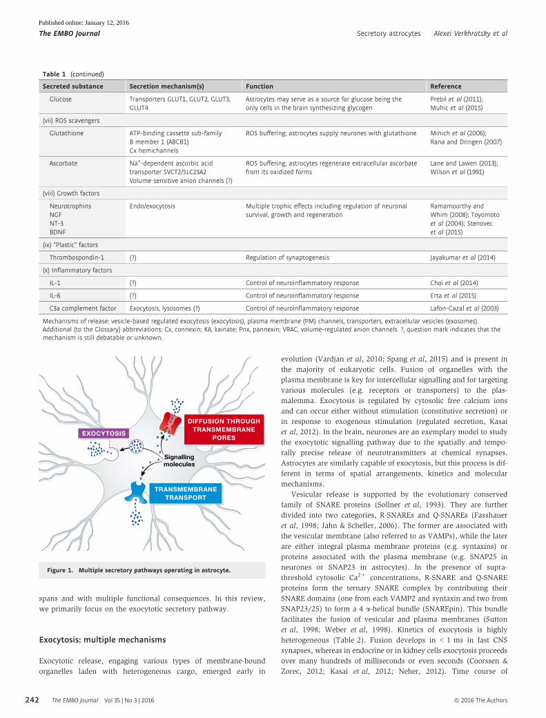
through several pathways (Fig 1 and see Malarkey & Parpura,
2008 for details) represented by: (i) vesicle-based exocytosis (e.g.
that of D-serine (Martineau et al, 2013) or glutamate (Montana
et al, 2004); (ii) diffusion through plasmalemmal pores/channels
(e.g. release of ATP and/or glutamate through anion channels,
connexin hemichannels or dilated P2X7 receptors, Cotrina et al,
1998; Suadicani et al, 2006) and (iii) extrusion through plas-
malemmal transporters (e.g. the release of GABA via the reversed
operation of GAT-3 transporters, Unichenko et al, 2012). Often,
the same molecule can be released through different pathways,
which affects the complexity/specificity of its action. The release
of these molecules to the extracellular space, along with their
subsequent transport by the convective glymphatic system (Thrane
et al, 2014), occurs within various brain regions in different time
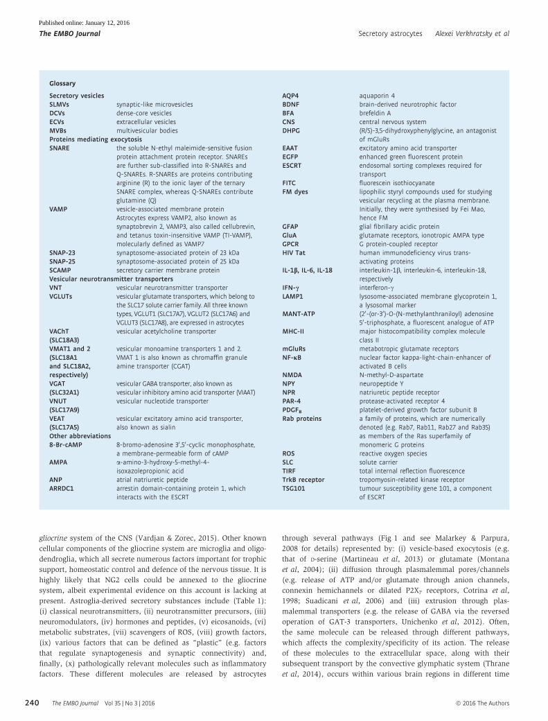
Glossary
Secretory vesiclesSLMVs synaptic-like microvesiclesDCVs dense-core vesiclesECVs extracellular vesiclesMVBs multivesicular bodiesProteins mediating exocytosisSNARE the soluble N-ethyl maleimide-sensitive fusion
protein attachment protein receptor. SNAREsare further sub-classified into R-SNAREs andQ-SNAREs. R-SNAREs are proteins contributingarginine (R) to the ionic layer of the ternarySNARE complex, whereas Q-SNAREs contributeglutamine (Q)
VAMP vesicle-associated membrane proteinAstrocytes express VAMP2, also known assynaptobrevin 2, VAMP3, also called cellubrevin,and tetanus toxin-insensitive VAMP (TI-VAMP),molecularly defined as VAMP7
SNAP-23 synaptosome-associated protein of 23 kDaSNAP-25 synaptosome-associated protein of 25 kDaSCAMP secretory carrier membrane proteinVesicular neurotransmitter transportersVNT vesicular neurotransmitter transporterVGLUTs vesicular glutamate transporters, which belong to
the SLC17 solute carrier family. All three knowntypes, VGLUT1 (SLC17A7), VGLUT2 (SLC17A6) andVGLUT3 (SLC17A8), are expressed in astrocytes
VAChT(SLC18A3)
vesicular acetylcholine transporter
VMAT1 and 2(SLC18A1and SLC18A2,respectively)
vesicular monoamine transporters 1 and 2.VMAT 1 is also known as chromaffin granuleamine transporter (CGAT)
VGAT(SLC32A1)
vesicular GABA transporter, also known asvesicular inhibitory amino acid transporter (VIAAT)
VNUT(SLC17A9)
vesicular nucleotide transporter
VEAT(SLC17A5)
vesicular excitatory amino acid transporter,also known as sialin
Other abbreviations8-Br-cAMP 8-bromo-adenosine 30 ,50-cyclic monophosphate,
a membrane-permeable form of cAMPAMPA a-amino-3-hydroxy-5-methyl-4-
isoxazolepropionic acidANP atrial natriuretic peptideARRDC1 arrestin domain-containing protein 1, which
interacts with the ESCRT
AQP4 aquaporin 4BDNF brain-derived neurotrophic factorBFA brefeldin ACNS central nervous systemDHPG (R/S)-3,5-dihydroxyphenylglycine, an antagonist
of mGluRsEAAT excitatory amino acid transporterEGFP enhanced green fluorescent proteinESCRT endosomal sorting complexes required for
transportFITC fluorescein isothiocyanateFM dyes lipophilic styryl compounds used for studying
vesicular recycling at the plasma membrane.Initially, they were synthesised by Fei Mao,hence FM
GFAP glial fibrillary acidic proteinGluA glutamate receptors, ionotropic AMPA typeGPCR G protein-coupled receptorHIV Tat human immunodeficiency virus trans-
activating proteinsIL-1b, IL-6, IL-18 interleukin-1b, interleukin-6, interleukin-18,
respectivelyIFN-c interferon-cLAMP1 lysosome-associated membrane glycoprotein 1,
a lysosomal markerMANT-ATP (20-(or-30)-O-(N-methylanthraniloyl) adenosine
50-triphosphate, a fluorescent analogue of ATPMHC-II major histocompatibility complex molecule
class IImGluRs metabotropic glutamate receptorsNF-jB nuclear factor kappa-light-chain-enhancer of
activated B cellsNMDA N-methyl-D-aspartateNPY neuropeptide YNPR natriuretic peptide receptorPAR-4 protease-activated receptor 4PDGFB platelet-derived growth factor subunit BRab proteins a family of proteins, which are numerically
denoted (e.g. Rab7, Rab11, Rab27 and Rab35)as members of the Ras superfamily ofmonomeric G proteins
ROS reactive oxygen speciesSLC solute carrierTIRF total internal reflection fluorescenceTrkB receptor tropomyosin-related kinase receptorTSG101 tumour susceptibility gene 101, a component
of ESCRT
The EMBO Journal Vol 35 | No 3 | 2016 ª 2016 The Authors
The EMBO Journal Secretory astrocytes Alexei Verkhratsky et al
240
Published online: January 12, 2016
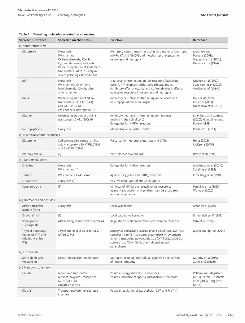
Table 1. Signalling molecules secreted by astrocytes.
Secreted substance Secretion mechanism(s) Function Reference
(i) Neurotransmitters
Glutamate ExocytosisPM channels:Cx hemichannels; P2X7RsCystine-glutamate antiporterReversed operation of glutamatetransporters EAAT1/2—only insevere pathological conditions
Excitatory neurotransmitter acting on glutamate ionotropic(AMPA, KA and NMDA) and metabotropic receptors inneurones and neuroglia
Malarkey andParpura (2008);Mazzanti et al (2001);Parpura et al (1994)
ATP ExocytosisPM channels: Cx or Panxhemichannels, P2X7Rs, otheranion channels
Neurotransmitter acting on P2X receptors (excitatoryaction), P2Y receptors (pleiotropic effects), and A1(inhibitory effects), A2A, A2B and A3 (metabotropic effects)adenosine receptors in neurones and neuroglia
Queiroz et al (1997);Suadicani et al (2012);Vardjan et al (2014a)
GABA Reversed operation of GABAtransporters GAT1 (SLC6A1)and GAT3 (SLC6A11)PM channels: bestrophin1 (?)
Inhibitory neurotransmitter acting on neurones andon subpopulations of neuroglia
Heja et al (2009);Lee et al (2011);Unichenko et al (2013)
Glycine Reversed operation of glycinetransporters GlyT1 (SLC6A9)
Inhibitory neurotransmitter acting on neurones(mainly in the spinal cord)Co-agonist for NMDA receptors
Eulenburg and Gomeza(2010); Holopainen andKontro (1989)
Neuropeptide Y Exocytosis Metabotropic neurotransmitter Prada et al (2011)
(ii) Neurotransmitter precursors
Glutamine Sodium-coupled neutral aminoacid transporters SNAT3/SLC38A3and SNAT5/SLC38A5
Precursor for neuronal glutamate and GABA Hertz (2013);McKenna (2007)
Pro-enkephalin (?) Precursor for enkephalins Batter et al (1991)
(iii) Neuromodulators
D-Serine ExocytosisPM channels (?)
Co-agonist for NMDA receptors Martineau et al (2014);Schell et al (1995)
Taurine PM channels: Cx43, VRAC Agonist for glycine and GABAA receptors Kimelberg et al (1990)
L-aspartate Exocytosis (?) Positive modulator of NMDA receptors
Kynurenic acid (?) Inhibitor of NMDA and acetylcholine receptors;aberrant production and synthesis can be associatedwith schizophrenia
Pershing et al (2015);Wu et al (2010)
(iv) Hormones and peptides
Atrial natriureticpeptide (ANP)
Exocytosis Local vasodilator Krzan et al (2003)
Endothelin-3 (?) Local vasoactive hormone Ehrenreich et al (1991)
Sphingosine1-phosphate
ATP-binding cassette transporter A1 Regulation of cell proliferation and immune response Sato et al (2007)
Thyroid hormonesthyroxine (T4) andtriiodothyronine(T3)
L-type amino acid transporter 2LAT2/SLC7A8
Astrocytes exclusively express type 2 deiodinase (D2) thatconverts T4 to T3. Astrocytes accumulate T4 by organicanion transporting polypeptide 1C1 (OATP1C1/SLCO1C1),convert it to T3, which is then released to brainparenchyma
Morte and Bernal (2014)
(v) Eicosanoids
Arachidonic acidProstanoids
Direct release from membranes Multiple; including intercellular signalling and controlof innate immunity
Murphy et al (1988);Xu et al (2003a,b)
(vi) Metabolic substrates
Lactate Membrane transporterMonocarboxylate TransporterMCT1/SLC16A1Lactate channels
Possible energy substrate in neuronesPossible activator of specific metabotropic receptors
Pellerin and Magistretti(2012); Sotelo-Hitschfeldet al (2015); Tang et al(2014)
Citrate Transporters/Volume-regulatedchannels
Possible regulation of extracellular Ca2+ and Mg2+ (?)
ª 2016 The Authors The EMBO Journal Vol 35 | No 3 | 2016
Alexei Verkhratsky et al Secretory astrocytes The EMBO Journal
241
Published online: January 12, 2016
spans and with multiple functional consequences. In this review,
we primarily focus on the exocytotic secretory pathway.
Exocytosis: multiple mechanisms
Exocytotic release, engaging various types of membrane-bound
organelles laden with heterogeneous cargo, emerged early in
evolution (Vardjan et al, 2010; Spang et al, 2015) and is present in
the majority of eukaryotic cells. Fusion of organelles with the
plasma membrane is key for intercellular signalling and for targeting
various molecules (e.g. receptors or transporters) to the plas-
malemma. Exocytosis is regulated by cytosolic free calcium ions
and can occur either without stimulation (constitutive secretion) or
in response to exogenous stimulation (regulated secretion, Kasai
et al, 2012). In the brain, neurones are an exemplary model to study
the exocytotic signalling pathway due to the spatially and tempo-
rally precise release of neurotransmitters at chemical synapses.
Astrocytes are similarly capable of exocytosis, but this process is dif-
ferent in terms of spatial arrangements, kinetics and molecular
mechanisms.
Vesicular release is supported by the evolutionary conserved
family of SNARE proteins (Sollner et al, 1993). They are further
divided into two categories, R-SNAREs and Q-SNAREs (Fasshauer
et al, 1998; Jahn & Scheller, 2006). The former are associated with
the vesicular membrane (also referred to as VAMPs), while the later
are either integral plasma membrane proteins (e.g. syntaxins) or
proteins associated with the plasma membrane (e.g. SNAP25 in
neurones or SNAP23 in astrocytes). In the presence of supra-
threshold cytosolic Ca2+ concentrations, R-SNARE and Q-SNARE
proteins form the ternary SNARE complex by contributing their
SNARE domains (one from each VAMP2 and syntaxin and two from
SNAP23/25) to form a 4 a-helical bundle (SNAREpin). This bundle
facilitates the fusion of vesicular and plasma membranes (Sutton
et al, 1998; Weber et al, 1998). Kinetics of exocytosis is highly
heterogeneous (Table 2). Fusion develops in < 1 ms in fast CNS
synapses, whereas in endocrine or in kidney cells exocytosis proceeds
over many hundreds of milliseconds or even seconds (Coorssen &
Zorec, 2012; Kasai et al, 2012; Neher, 2012). Time course of
Table 1 (continued)
Secreted substance Secretion mechanism(s) Function Reference
Glucose Transporters GLUT1, GLUT2, GLUT3,GLUT4
Astrocytes may serve as a source for glucose being theonly cells in the brain synthesizing glycogen
Prebil et al (2011);Muhic et al (2015)
(vii) ROS scavengers
Glutathione ATP-binding cassette sub-familyB member 1 (ABCB1)Cx hemichannels
ROS buffering; astrocytes supply neurones with glutathione Minich et al (2006);Rana and Dringen (2007)
Ascorbate Na+-dependent ascorbic acidtransporter SVCT2/SLC23A2Volume-sensitive anion channels (?)
ROS buffering; astrocytes regenerate extracellular ascorbatefrom its oxidized forms
Lane and Lawen (2013);Wilson et al (1991)
(viii) Growth factors
NeurotrophinsNGFNT-3BDNF
Endo/exocytosis Multiple trophic effects including regulation of neuronalsurvival, growth and regeneration
Ramamoorthy andWhim (2008); Toyomotoet al (2004); Stenovecet al (2015)
(ix) “Plastic” factors
Thrombospondin-1 (?) Regulation of synaptogenesis Jayakumar et al (2014)
(x) Inflammatory factors
IL-1 (?) Control of neuroinflammatory response Choi et al (2014)
IL-6 (?) Control of neuroinflammatory response Erta et al (2015)
C3a complement factor Exocytosis, lysosomes (?) Control of neuroinflammatory response Lafon-Cazal et al (2003)
Mechanisms of release: vesicle-based regulated exocytosis (exocytosis), plasma membrane (PM) channels, transporters, extracellular vesicles (exosomes).Additional (to the Glossary) abbreviations: Cx, connexin; KA, kainate; Pnx, pannexin; VRAC, volume-regulated anion channels. ?, question mark indicates that themechanism is still debatable or unknown.
EXOCYTOSIS
DIFFUSION THROUGHTRANSMEMBRANE
PORES
TRANSMEMBRANETRANSPORT
Signallingmolecules
Figure 1. Multiple secretory pathways operating in astrocyte.
The EMBO Journal Vol 35 | No 3 | 2016 ª 2016 The Authors
The EMBO Journal Secretory astrocytes Alexei Verkhratsky et al
242
Published online: January 12, 2016
exocytotic release is determined by several factors. First, it is the
sensitivity of secretory apparatus to [Ca2+]i, which is heterogeneous
in different cell types. Second, the spatiotemporal progression of local
[Ca2+]i signals differs markedly between cells. For instance, in synap-
tic terminals excitation–secretion coupling is exceedingly fast due to
the organisation of Ca2+ nanodomains that reflect a close proximity
of the Ca2+ source and exocytotic machinery (Eggermann et al,
2012). Finally, slow regulated exocytosis may also evince a distinct
vesicle nanoarchitecture (e.g. arrangement and density of R-SNAREs,
see Fig 2) and the heterogeneity of Q-SNAREs (Takamori et al, 2006;
Singh et al, 2014). Multiple mechanisms controlling exocytosis may
coexist within the confinement of a single cell resulting in complex
kinetics of secretion (Rupnik et al, 2000).
Diversity of astroglial secretory organelles
Eukaryotic cells produce different types of membranous secretory
organelles that are classified as intracellular or extracellular. Intra-
cellular vesicles are represented by transport vesicles, lysosomes
and various types of secretory vesicles, whereas extracellular
vesicles are ectosomes, exosomes, microvesicles (microparticles),
membrane particles and apoptotic vesicles (van der Pol et al, 2012;
Cocucci & Meldolesi, 2015). Intracellular vesicles are cellular
organelles that may completely fuse with cellular membranes,
whereas extracellular vesicles are membranous compartments
released into the surrounding environment. Generally, vesicles
undergoing constitutive or regulated exocytosis derive either from
the trans-Golgi network or from early or recycling endosomes,
although multivesicular bodies and lysosomes have been reported
to undergo exocytosis under certain conditions.
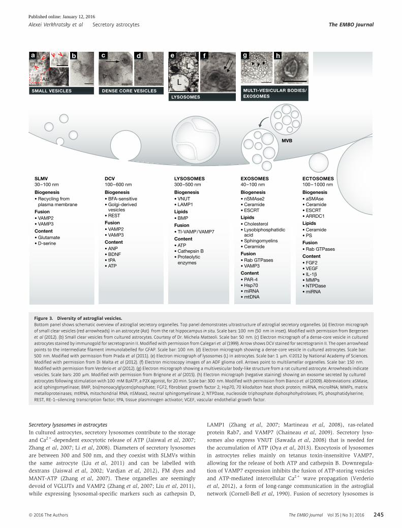
Several secretory organelles undergo regulated exocytosis in
astrocytes (Fig 3). These include clear electron lucent SLMVs that
morphologically resemble synaptic vesicles (Bezzi et al, 2004;
Crippa et al, 2006; Jourdain et al, 2007; Bergersen & Gundersen,
2009; Martineau et al, 2013), DCVs (Calegari et al, 1999; Parpura &
Zorec, 2010) and secretory lysosomes (Zhang et al, 2007; Li et al,
2008; Verderio et al, 2012). All these organelles can store and
release low (amino acids) and/or high (peptides and proteins)
molecular weight chemical transmitters (Parpura & Zorec, 2010;
Gucek et al, 2012; Vardjan & Zorec, 2015). Secretory vesicles can
also act as recycling vesicles that take up extracellular molecules
(e.g. by endocytosis) and promote their subsequent release (Vardjan
et al, 2014b). This function may be essential for defining the compo-
sition of the cerebrospinal fluid and for the function of the glym-
phatic system (Thrane et al, 2014).
Synaptic-like microvesicles carry amino acids
Astroglial SLMVs typically have a diameter of 30–100 nm and
appear in pairs/groups of 2–15 vesicles (Bezzi et al, 2004; Jourdain
et al, 2007; Bergersen et al, 2012; Martineau et al, 2013). They are
much less numerous compared to synaptic vesicles in nerve termi-
nals where these organelles exist in groups of hundreds to thou-
sands. Larger SLMVs (diameter of 1–3 lm) have also been
identified in astrocytes in hippocampal slices. These vesicles may be
generated by intracellular fusion of smaller vesicles and/or other
organelles in response to a sustained increase in [Ca2+]i or mechani-
cal stimulation (Kang et al, 2013), but it is not clear whether they
contribute to physiological secretion.
Concentrating neurotransmitters into vesicles is accomplished by
vesicular neurotransmitter transporters or VNTs, which differ from
the transporters at the plasma membrane with respect to energy
coupling, substrate specificity and affinity. Six types of VNTs
have been identified so far, including transporters for glutamate
(VGLUT1-3), acetylcholine (vAChT), monoamines (VMAT1-2),
GABA/glycine (VIAAT, also named VGAT), and more recently trans-
porters for ATP (VNUT) and, possibly, for aspartate (sialin/VEAT)
(Chaudhry et al, 2008; Sawada et al, 2008; Blakely & Edwards,
2012). Accumulation of D-serine in SLMVs is mediated by vesicular
D-serine transporter, VSerT (Martineau et al, 2013), although its
molecular identity remains elusive. The VNTs are essential molecu-
lar components of chemical transmission and the fingerprint of regu-
lated exocytosis. Some VNTs such as VGLUT1-3 have been identified
in cultured astrocytes (Fremeau et al, 2002; Bezzi et al, 2004; Kreft
et al, 2004; Crippa et al, 2006; Montana et al, 2006). Analyses of
astrocytes in situ using gene chip microarray, single-cell RT–PCR
and immunostainings (Bezzi et al, 2004; Li et al, 2013; Sahlender
et al, 2014) have produced variable results and in some cases
have challenged the presence of VNTs and thus the concept of
astroglial exocytosis. Nonetheless, immunogold electron micro-
scopy, confocal microscopy and single-cell RT–PCR have shown that
sub-populations of astrocytes in the brain express VGLUT1 (Bezzi
et al, 2004; Bergersen et al, 2012; Ormel et al, 2012), VGLUT2 (Bezzi
et al, 2004) and VGLUT3 (Ormel et al, 2012).
In astrocytes, SLMVs primarily store glutamate and D-serine, an
agonist of glycine regulatory site of NMDA receptor (Martineau et al,
2008, 2013; Bergersen et al, 2012). In cultured astrocytes, SLMVs
co-localise with D-serine (Mothet et al, 2005; Martineau et al, 2013)
and VGLUTs, suggesting that glutamate and D-serine may reside in
Table 2. Comparison of maximal rates of regulated exocytosis indifferent secretory cell types recorded as an increase in whole-cellmembrane capacitance evoked by flash photolysis-induced elevationsin cytosolic [Ca2+].
Cell type
Max rate inregulatedexocytosis References
Endocrine cells
Endocrine pituitarymelanotrophes
25 s�1
44 s�1Rupnik et al (2000);Thomas et al, 1993)
Endocrinepancreatic b cells
70 s�1 Barg et al (2001);Wan et al (2004)
Chromaffin cells 1,500 s�1 Voets (2000)
Neurones
Rod photoreceptors 300 s�1
400 s�1Kreft et al (2003);Thoreson et al (2004)
Retinal bipolar neurones 3,000 s�1 Heidelberger et al (1994)
Inhibitory basket cell 5,000 s�1 Sakaba (2008)
Calyx of Heldneurones
6,000 s�1 Bollmann et al (2000);Schneggenburgerand Neher (2000)
Neuroglia
Astrocytes 0.1–2 s�1 Kreft et al (2004)
See also (Neher, 2012).
ª 2016 The Authors The EMBO Journal Vol 35 | No 3 | 2016
Alexei Verkhratsky et al Secretory astrocytes The EMBO Journal
243
Published online: January 12, 2016
the same secretory organelle (Bezzi et al, 2004; Ormel et al, 2012).
This contrasts the in situ evidence showing that glutamate and
D-serine are stored in distinct SLMVs within the same astrocyte
(Bergersen et al, 2012). Direct comparison of astroglial SLMVs
(Crippa et al, 2006; Martineau et al, 2013) and neuronal synaptic
vesicles shows that astrocytic vesicles in bulk contain D-serine and
glutamate, whereas neuronal synaptic vesicles in bulk contain gluta-
mate, glycine and GABA but are devoid of D-serine (Martineau et al,
2013; Sild & Van Horn, 2013). Astrocytes from various brain regions,
including the hippocampus and cortex, and Bergmann glial cells
in the cerebellum contain SLMVs (Bergersen et al, 2012; Ormel et al,
2012). These vesicles are present in perisynaptic processes as well as
in somata (Bezzi et al, 2004; Montana et al, 2004; Ormel et al,
2012). The release of both glutamate and D-serine from astrocytes is
Ca2+-dependent and is blocked by tetanus toxin that cleaves astro-
cytic R-SNAREs VAMP2 and VAMP3 (Bezzi et al, 2004; Mothet et al,
2005; Martineau et al, 2008, 2014; Henneberger et al, 2010; Parpura
& Zorec, 2010; Kang et al, 2013; Shigetomi et al, 2013).
Dense-core vesicles carry peptides
The DCVs are the main component for the storage and release of
neuropeptides and hormones from neuroendocrine cells (Burgoyne
& Morgan, 2003) and neurones (Klyachko & Jackson, 2002). These
vesicles also contain ATP, which is likely accumulated into DCVs
via VNUTs, albeit the presence of this transporter on these orga-
nelles has not yet been reported. The ultrastructure characteristics
of astroglial DCVs are similar to those of neuroendocrine cells and
neurones, although their core seems not as dense as in neuroen-
docrine cells (Potokar et al, 2008). The actual fraction of DCVs in
astrocytes is quite small; for example, VAMP2-positive DCVs repre-
sent only 2% of the total number of vesicles examined (i.e. clear
and dense-core vesicles, Crippa et al, 2006). Astroglial DCVs are
generally larger than SLMVs, being ~100–600 nm in diameter
(Calegari et al, 1999; Hur et al, 2010; Prada et al, 2011), albeit
ANP-storing vesicles can have diameters as small as 50 nm (Potokar
et al, 2008). DCVs from cultured astrocytes contain the secretory
proteins secretogranins II (Calegari et al, 1999; Paco et al, 2009;
Prada et al, 2011) and III (Paco et al, 2010), chromogranins
(Hur et al, 2010), ANP (Kreft et al, 2004; Paco et al, 2009),
neuropeptide Y (Ramamoorthy & Whim, 2008; Prada et al, 2011)
and ATP (Coco et al, 2003; Pangrsic et al, 2007). The DCVs contain-
ing secretogranins were also identified in astrocytes from human
brain samples (Hur et al, 2010), confirming the existence of DCVs
in situ.
YpH
Atto594
Atto594
~62 nm
~62 nm
Primaryantibody
Distance betweenYpH and Atto594
Co
unts
Distance (nm)
Secondary antibodytagged with Atto594
A
40
20
00 100 200 300
B
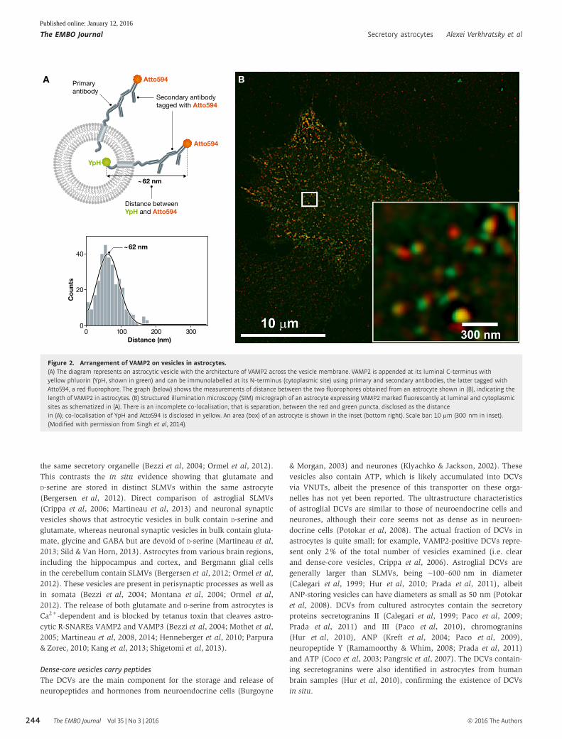
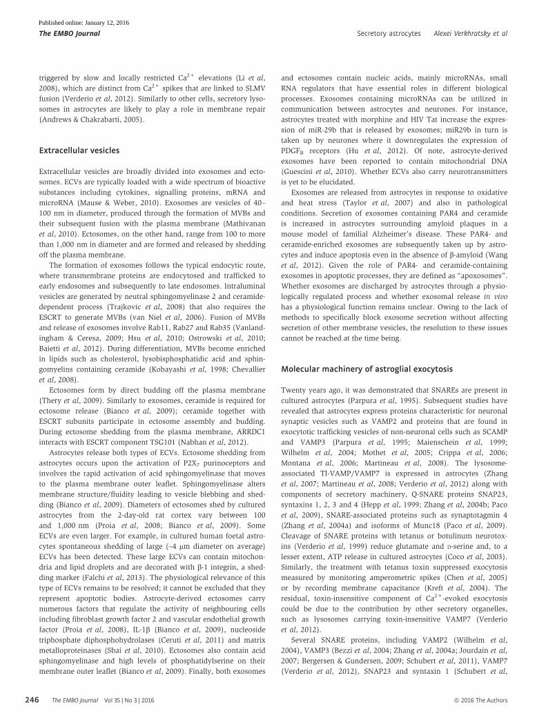
Figure 2. Arrangement of VAMP2 on vesicles in astrocytes.(A) The diagram represents an astrocytic vesicle with the architecture of VAMP2 across the vesicle membrane. VAMP2 is appended at its luminal C-terminus withyellow phluorin (YpH, shown in green) and can be immunolabelled at its N-terminus (cytoplasmic site) using primary and secondary antibodies, the latter tagged withAtto594, a red fluorophore. The graph (below) shows the measurements of distance between the two fluorophores obtained from an astrocyte shown in (B), indicating thelength of VAMP2 in astrocytes. (B) Structured illumination microscopy (SIM) micrograph of an astrocyte expressing VAMP2marked fluorescently at luminal and cytoplasmicsites as schematized in (A). There is an incomplete co-localisation, that is separation, between the red and green puncta, disclosed as the distancein (A); co-localisation of YpH and Atto594 is disclosed in yellow. An area (box) of an astrocyte is shown in the inset (bottom right). Scale bar: 10 µm (300 nm in inset).(Modified with permission from Singh et al, 2014).
The EMBO Journal Vol 35 | No 3 | 2016 ª 2016 The Authors
The EMBO Journal Secretory astrocytes Alexei Verkhratsky et al
244
Published online: January 12, 2016
Secretory lysosomes in astrocytes
In cultured astrocytes, secretory lysosomes contribute to the storage
and Ca2+-dependent exocytotic release of ATP (Jaiswal et al, 2007;
Zhang et al, 2007; Li et al, 2008). Diameters of secretory lysosomes
are between 300 and 500 nm, and they coexist with SLMVs within
the same astrocyte (Liu et al, 2011) and can be labelled with
dextrans (Jaiswal et al, 2002; Vardjan et al, 2012), FM dyes and
MANT-ATP (Zhang et al, 2007). These organelles are seemingly
devoid of VGLUTs and VAMP2 (Zhang et al, 2007; Liu et al, 2011),
while expressing lysosomal-specific markers such as cathepsin D,
LAMP1 (Zhang et al, 2007; Martineau et al, 2008), ras-related
protein Rab7, and VAMP7 (Chaineau et al, 2009). Secretory lyso-
somes also express VNUT (Sawada et al, 2008) that is needed for
the accumulation of ATP (Oya et al, 2013). Exocytosis of lysosomes
in astrocytes relies mainly on tetanus toxin-insensitive VAMP7,
allowing for the release of both ATP and cathepsin B. Downregula-
tion of VAMP7 expression inhibits the fusion of ATP-storing vesicles
and ATP-mediated intercellular Ca2+ wave propagation (Verderio
et al, 2012), a form of long-range communication in the astroglial
network (Cornell-Bell et al, 1990). Fusion of secretory lysosomes is
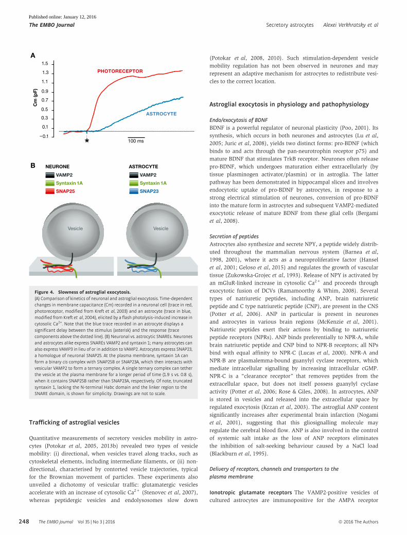
DCV100–600 nm
Biogenesis• BFA-sensitive
• Golgi-derived vesicles
• REST
Fusion• VAMP2
• VAMP3
Content• ANP
• BDNF
• tPA
• ATP
SLMV30–100 nm
Biogenesis• Recycling from
plasma membrane
Fusion• VAMP2
• VAMP3
Content• Glutamate
• D-serine
EXOSOMES40–100 nm
Biogenesis• nSMAse2
• Ceramide
• ESCRT
Lipids• Cholesterol
• Lysobiphosphatidic acid
• Sphingomyelins
• Ceramide
Fusion• Rab GTPases
• VAMP3
Content• PAR-4
• Hsp70
• miRNA
• mtDNA
LYSOSOMES300–500 nm
Biogenesis• VNUT
• LAMP1
Lipids• BMP
Fusion• TI-VAMP/VAMP7
Content• ATP
• Cathepsin B
• Proteolytic
enzymes
ECTOSOMES100– 1000 nm
Biogenesis• aSMAse
• Ceramide
• ESCRT
• ARRDC1
Lipids• Ceramide
• PS
Fusion• Rab GTPases
Content• FGF2
• VEGF
• IL-1β• MMPs
• NTPDase
• miRNA
MVB
MULTI-VESICULAR BODIES/EXOSOMES
DENSE CORE VESICLESSMALL VESICLESLYSOSOMES
a b c d e f g h
Figure 3. Diversity of astroglial vesicles.Bottom panel shows schematic overview of astroglial secretory organelles. Top panel demonstrates ultrastructure of astroglial secretory organelles. (a) Electron micrographof small clear vesicles (red arrowheads) in an astrocyte (Ast) from the rat hippocampus in situ. Scale bars: 100 nm (50 nm in inset). Modified with permission from Bergersenet al (2012). (b) Small clear vesicles from cultured astrocytes. Courtesy of Dr. Michela Matteoli. Scale bar: 50 nm. (c) Electron micrograph of a dense-core vesicle in culturedastrocytes stained by immunogold for secretogranin II. Modified with permission from Calegari et al (1999). Arrow shows DCV stained for secretogranin II. The open arrowheadpoints to the intermediate filament immunolabelled for GFAP. Scale bar: 100 nm. (d) Electron micrograph showing a dense-core vesicle in cultured astrocytes. Scale bar:500 nm. Modified with permission from Prada et al (2011). (e) Electron micrograph of lysosomes (L) in astrocytes. Scale bar: 1 µm. ©2012 by National Academy of Sciences.Modified with permission from Di Malta et al (2012). (f) Electron microscopy images of an ADF glioma cell. Arrows point to multilamellar organelles. Scale bar: 150 nm.Modified with permission from Verderio et al (2012). (g) Electron micrograph showing a multivesicular body-like structure from a rat cultured astrocyte. Arrowheads indicatevesicles. Scale bars: 200 µm. Modified with permission from Brignone et al (2015). (h) Electron micrograph (negative staining) showing an exosome secreted by culturedastrocytes following stimulation with 100 mMBzATP, a P2X agonist, for 20min. Scale bar: 300 nm. Modified with permission from Bianco et al (2009). Abbreviations: aSMase,acid sphingomyelinase; BMP, bis(monoacylglycero)phosphate; FGF2, fibroblast growth factor 2; Hsp70, 70 kilodalton heat shock protein; miRNA, microRNA; MMPs, matrixmetalloproteinases; mtRNA, mitochondrial RNA; nSMase2, neutral sphingomyelinase 2; NTPDase, nucleoside triphosphate diphosphohydrolases; PS, phosphatidylserine;REST, RE-1–silencing transcription factor; tPA, tissue plasminogen activator; VGEF, vascular endothelial growth factor.
ª 2016 The Authors The EMBO Journal Vol 35 | No 3 | 2016
Alexei Verkhratsky et al Secretory astrocytes The EMBO Journal
245
Published online: January 12, 2016
triggered by slow and locally restricted Ca2+ elevations (Li et al,
2008), which are distinct from Ca2+ spikes that are linked to SLMV
fusion (Verderio et al, 2012). Similarly to other cells, secretory lyso-
somes in astrocytes are likely to play a role in membrane repair
(Andrews & Chakrabarti, 2005).
Extracellular vesicles
Extracellular vesicles are broadly divided into exosomes and ecto-
somes. ECVs are typically loaded with a wide spectrum of bioactive
substances including cytokines, signalling proteins, mRNA and
microRNA (Mause & Weber, 2010). Exosomes are vesicles of 40–
100 nm in diameter, produced through the formation of MVBs and
their subsequent fusion with the plasma membrane (Mathivanan
et al, 2010). Ectosomes, on the other hand, range from 100 to more
than 1,000 nm in diameter and are formed and released by shedding
off the plasma membrane.
The formation of exosomes follows the typical endocytic route,
where transmembrane proteins are endocytosed and trafficked to
early endosomes and subsequently to late endosomes. Intraluminal
vesicles are generated by neutral sphingomyelinase 2 and ceramide-
dependent process (Trajkovic et al, 2008) that also requires the
ESCRT to generate MVBs (van Niel et al, 2006). Fusion of MVBs
and release of exosomes involve Rab11, Rab27 and Rab35 (Vanland-
ingham & Ceresa, 2009; Hsu et al, 2010; Ostrowski et al, 2010;
Baietti et al, 2012). During differentiation, MVBs become enriched
in lipids such as cholesterol, lysobisphosphatidic acid and sphin-
gomyelins containing ceramide (Kobayashi et al, 1998; Chevallier
et al, 2008).
Ectosomes form by direct budding off the plasma membrane
(Thery et al, 2009). Similarly to exosomes, ceramide is required for
ectosome release (Bianco et al, 2009); ceramide together with
ESCRT subunits participate in ectosome assembly and budding.
During ectosome shedding from the plasma membrane, ARRDC1
interacts with ESCRT component TSG101 (Nabhan et al, 2012).
Astrocytes release both types of ECVs. Ectosome shedding from
astrocytes occurs upon the activation of P2X7 purinoceptors and
involves the rapid activation of acid sphingomyelinase that moves
to the plasma membrane outer leaflet. Sphingomyelinase alters
membrane structure/fluidity leading to vesicle blebbing and shed-
ding (Bianco et al, 2009). Diameters of ectosomes shed by cultured
astrocytes from the 2-day-old rat cortex vary between 100
and 1,000 nm (Proia et al, 2008; Bianco et al, 2009). Some
ECVs are even larger. For example, in cultured human foetal astro-
cytes spontaneous shedding of large (~4 lm diameter on average)
ECVs has been detected. These large ECVs can contain mitochon-
dria and lipid droplets and are decorated with b-1 integrin, a shed-
ding marker (Falchi et al, 2013). The physiological relevance of this
type of ECVs remains to be resolved; it cannot be excluded that they
represent apoptotic bodies. Astrocyte-derived ectosomes carry
numerous factors that regulate the activity of neighbouring cells
including fibroblast growth factor 2 and vascular endothelial growth
factor (Proia et al, 2008), IL-1b (Bianco et al, 2009), nucleoside
triphosphate diphosphohydrolases (Ceruti et al, 2011) and matrix
metalloproteinases (Sbai et al, 2010). Ectosomes also contain acid
sphingomyelinase and high levels of phosphatidylserine on their
membrane outer leaflet (Bianco et al, 2009). Finally, both exosomes
and ectosomes contain nucleic acids, mainly microRNAs, small
RNA regulators that have essential roles in different biological
processes. Exosomes containing microRNAs can be utilized in
communication between astrocytes and neurones. For instance,
astrocytes treated with morphine and HIV Tat increase the expres-
sion of miR-29b that is released by exosomes; miR29b in turn is
taken up by neurones where it downregulates the expression of
PDGFB receptors (Hu et al, 2012). Of note, astrocyte-derived
exosomes have been reported to contain mitochondrial DNA
(Guescini et al, 2010). Whether ECVs also carry neurotransmitters
is yet to be elucidated.
Exosomes are released from astrocytes in response to oxidative
and heat stress (Taylor et al, 2007) and also in pathological
conditions. Secretion of exosomes containing PAR4 and ceramide
is increased in astrocytes surrounding amyloid plaques in a
mouse model of familial Alzheimer’s disease. These PAR4- and
ceramide-enriched exosomes are subsequently taken up by astro-
cytes and induce apoptosis even in the absence of b-amyloid (Wang
et al, 2012). Given the role of PAR4- and ceramide-containing
exosomes in apoptotic processes, they are defined as “apoxosomes”.
Whether exosomes are discharged by astrocytes through a physio-
logically regulated process and whether exosomal release in vivo
has a physiological function remains unclear. Owing to the lack of
methods to specifically block exosome secretion without affecting
secretion of other membrane vesicles, the resolution to these issues
cannot be reached at the time being.
Molecular machinery of astroglial exocytosis
Twenty years ago, it was demonstrated that SNAREs are present in
cultured astrocytes (Parpura et al, 1995). Subsequent studies have
revealed that astrocytes express proteins characteristic for neuronal
synaptic vesicles such as VAMP2 and proteins that are found in
exocytotic trafficking vesicles of non-neuronal cells such as SCAMP
and VAMP3 (Parpura et al, 1995; Maienschein et al, 1999;
Wilhelm et al, 2004; Mothet et al, 2005; Crippa et al, 2006;
Montana et al, 2006; Martineau et al, 2008). The lysosome-
associated TI-VAMP/VAMP7 is expressed in astrocytes (Zhang
et al, 2007; Martineau et al, 2008; Verderio et al, 2012) along with
components of secretory machinery, Q-SNARE proteins SNAP23,
syntaxins 1, 2, 3 and 4 (Hepp et al, 1999; Zhang et al, 2004b; Paco
et al, 2009), SNARE-associated proteins such as synaptotagmin 4
(Zhang et al, 2004a) and isoforms of Munc18 (Paco et al, 2009).
Cleavage of SNARE proteins with tetanus or botulinum neurotox-
ins (Verderio et al, 1999) reduce glutamate and D-serine and, to a
lesser extent, ATP release in cultured astrocytes (Coco et al, 2003).
Similarly, the treatment with tetanus toxin suppressed exocytosis
measured by monitoring amperometric spikes (Chen et al, 2005)
or by recording membrane capacitance (Kreft et al, 2004). The
residual, toxin-insensitive component of Ca2+-evoked exocytosis
could be due to the contribution by other secretory organelles,
such as lysosomes carrying toxin-insensitive VAMP7 (Verderio
et al, 2012).
Several SNARE proteins, including VAMP2 (Wilhelm et al,
2004), VAMP3 (Bezzi et al, 2004; Zhang et al, 2004a; Jourdain et al,
2007; Bergersen & Gundersen, 2009; Schubert et al, 2011), VAMP7
(Verderio et al, 2012), SNAP23 and syntaxin 1 (Schubert et al,
The EMBO Journal Vol 35 | No 3 | 2016 ª 2016 The Authors
The EMBO Journal Secretory astrocytes Alexei Verkhratsky et al
246
Published online: January 12, 2016

2011), have been detected in astrocytes in situ. VAMP2 and 3
co-localise with VGLUT1 and 2 on SLMVs that store glutamate
(Bezzi et al, 2004; Zhang et al, 2004a; Jourdain et al, 2007;
Bergersen & Gundersen, 2009) and likely D-serine (Martineau et al,
2013). Several synaptotagmin isoforms including synaptotagmins 4,
5, 7 and 11 are also present in astrocytes (Zhang et al, 2004a;
Mittelsteadt et al, 2009). However, typical neuronal SNARE-associated
proteins, such as synaptotagmins 1 and 2, and synaptophysin
(Wilhelm et al, 2004) have not been observed in astrocytes in situ.
Inactivation of VAMP2 and/or VAMP3 by tetanus neurotoxin
abolished the release of glutamate (Jourdain et al, 2007; Perea &
Araque, 2007) and, likely, D-serine in astrocytes in brain slices
(Henneberger et al, 2010). A transgenic mouse model expressing a
dominant negative (dn) SNARE (i.e. the cytosolic tail of VAMP2) in
astrocytes (Pascual et al, 2005; Halassa et al, 2009) showed changes
in behaviour, synaptic transmission and maturation of neurones
(Pascual et al, 2005; Hines & Haydon, 2013; Nadjar et al, 2013;
Turner et al, 2013; Lalo et al, 2014; Sultan et al, 2015), suggesting a
role for astrocytic VAMP2-dependent exocytosis in vivo. Of note,
VAMP2 cytosolic tail is supposed to compete with VAMP2 for bind-
ing to other components forming the ternary complexes, leading to
the reduced number of complexes formed and hence inhibiting regu-
lated exocytosis. Although these experiments provide strong support
for a function of SNARE proteins in astroglial regulated exocytosis,
there are indications that neurones might also express dnSNARE in
the transgenic mice, thus raising the possibility that the impairment
of neuronal, rather than astroglial, exocytosis may account for the
phenotype observed (Fujita et al, 2014). The debate that ensued
(Sloan & Barres, 2014) highlights technical matters, and particular
aspects of astroglial glutamate secretion in the context of synaptic
transmission, without questioning the general concept of exocyto-
sis-mediated astroglial secretion. These technical dissensions
nonetheless emphasize the need for refining the existing experimen-
tal strategies and developing new approaches directly attacking the
various facets of astroglial secretion in physiological and pathophys-
iological contexts (Jahn et al, 2015).
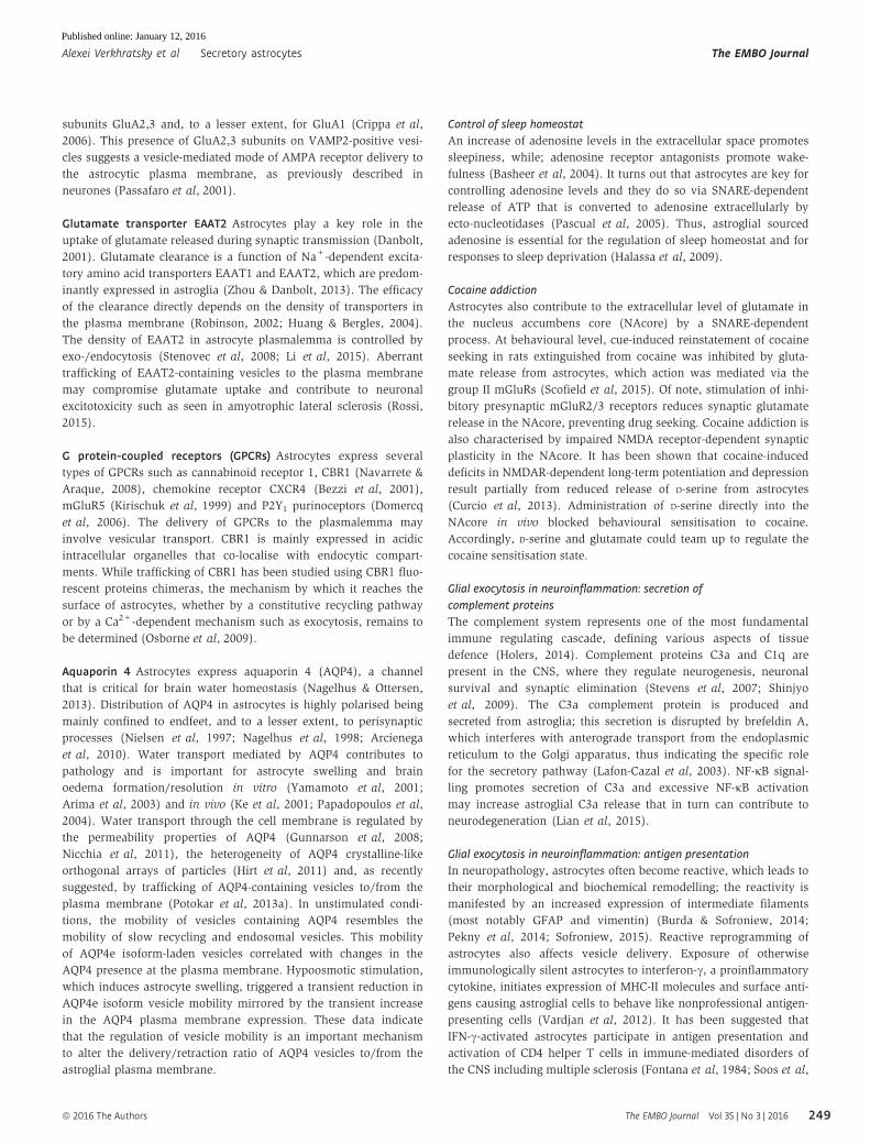
Astroglial exocytosis is slow
Visualisation of fluorescently labelled VGLUT1/2-containing vesicles
revealed that fusion events in isolated astrocytes occur within
hundreds of milliseconds after the increase in cytosolic Ca2+ (Bezzi
et al, 2004; Cali et al, 2008; Marchaland et al, 2008; Santello et al,
2011). Even slower kinetics of vesicular fusions has been reported
by using synapto-pHluorin (spH), a fluorescently tagged-VAMP2,
(Bowser & Khakh, 2007). Treatment of astrocytes with the Ca2+
ionophore ionomycin triggered exocytotic fusion of spH-labelled
SLMVs within seconds (Liu et al, 2011). Similarly, the TIRF micro-
scopy (Malarkey & Parpura, 2011) showed slow exocytotic bursts
occurring within seconds after mechanical stimulation of astrocytes.
Secretion of NPY from peptidergic vesicles occurred with a > 1-min
delay after stimulation (Ramamoorthy & Whim, 2008; Prada et al,
2011). Exocytotic release from peptidergic vesicles in 8-Br-cAMP-
matured astrocytes also began minutes after the stimulation (Paco
et al, 2009). Similar observations have been made for secretory
lysosomes, which labelled with FM dyes fused with the plasma
membrane with an ~1-min delay after exposure of astrocytes to
Ca2+ ionophores or ATP (Zhang et al, 2007; Li et al, 2008). Exocy-
totic fusion of quinacrine-loaded vesicles that express lysosomal
VAMP7 occurred with a > 2-min delay after exposure to various
stimuli including ionomycin, glutamate, ATP or UV-induced Ca2+
uncaging (Kreft et al, 2004; Pangrsic et al, 2007; Pryazhnikov &
Khiroug, 2008). Likewise, EGFP-LAMP1- and FITC-dextran-labelled
lysosomes underwent exocytotic fusion with a > 40-s delay after
administration of ionomycin (Liu et al, 2011) or the group I mGluR
agonist DHPG (Jaiswal et al, 2007).
Taken together, these imaging data indicate that in contrast to
neurones, where the fusion occurs within < 0.5 ms after the Ca2+
entry into the cytosol (Neher, 2012; Sudhof, 2012), exocytotic
release of various molecules from astrocytes is a much slower
process, occurring with a substantial post-stimulus delay (Vardjan
et al, 2015). Indeed, capacitance measurements on isolated
astrocytes confirm that the kinetics of vesicle fusion is at least 2
orders of magnitude slower than in neurones (Fig 4 and Table 2;
Kreft et al, 2003, 2004). Incidentally, inhibition of astroglial
exocytosis (using astroglia targeted expression of dnSNARE or
pharmacological tools) affects only slow electrical oscillations in
the cortex (Fellin et al, 2009), while fast neuronal electrical activ-
ity seems to be unaffected by corrupted (using a mouse model
rendering a reduction of VAMP1-3 expression in Muller cells, a
specialized astroglia of the retina) gliotransmission (Slezak et al,
2012).
The somewhat lethargic kinetics of astroglial vesicular release
likely reflects distinct organisation of the exocytotic machinery.
First, electron microscopy studies (Bezzi et al, 2004; Jourdain et al,
2007; Bergersen et al, 2012) have shown that astrocytes lack struc-
turally organised vesicle clusters typical of the active zone present
in presynaptic terminals, which may make the stimulus–secretion
coupling looser. In neurones, SNARE proteins are associated with
vesicles clustered at active zones that are essentially release sites.
This spatial localisation arguably is linked to the minimisation of
the delay between the stimulus and the secretory output
(Kasai et al, 2012). Second, the SNARE components and SNARE-
associated proteins of the exocytotic apparatus are not identical in
astrocytes and neurones, neither is the stability of SNARE
complexes, nor are the numbers of SNARE molecules associated
with a single vesicle.
VAMP isoforms with similar structural properties can participate
in the formation of several different SNARE complexes (Wilhelm
et al, 2004; Montana et al, 2009), which may affect the mechanism
of vesicle fusion with the plasma membrane. In neuronal terminals,
the ternary fusion complex forms between VAMP2, SNAP25 and
syntaxin, whereas in astrocytes the ternary SNARE fusion complex
assembles from VAMP2/3 or TI-VAMP, SNAP23 and syntaxin
(Montana et al, 2009; Hamilton & Attwell, 2010). At a single
molecule level, the presence of SNAP23A (as opposed to SNAP25B)
in the ternary complex decreases the complex stability by half,
arguably retarding the tethering/docking/fusion process (Fig 4B).
Moreover, the density of R-SNAREs associated with a single vesicle
in astrocytes is lesser than in neurones; in the latter, a single synap-
tic vesicle contains ~70 VAMP2 molecules (Takamori et al, 2006)
vs. ~25 in a single astroglial vesicle (Singh et al, 2014). This paucity
of VAMP2 would lead to the reduced density of ternary SNARE
complexes, which would contribute to further retardation of docking
and fusion process in astrocytes.
ª 2016 The Authors The EMBO Journal Vol 35 | No 3 | 2016
Alexei Verkhratsky et al Secretory astrocytes The EMBO Journal
247
Published online: January 12, 2016
Trafficking of astroglial vesicles
Quantitative measurements of secretory vesicles mobility in astro-
cytes (Potokar et al, 2005, 2013b) revealed two types of vesicle
mobility: (i) directional, when vesicles travel along tracks, such as
cytoskeletal elements, including intermediate filaments, or (ii) non-
directional, characterised by contorted vesicle trajectories, typical
for the Brownian movement of particles. These experiments also
unveiled a dichotomy of vesicular traffic: glutamatergic vesicles
accelerate with an increase of cytosolic Ca2+ (Stenovec et al, 2007),
whereas peptidergic vesicles and endolysosomes slow down
(Potokar et al, 2008, 2010). Such stimulation-dependent vesicle
mobility regulation has not been observed in neurones and may
represent an adaptive mechanism for astrocytes to redistribute vesi-
cles to the correct location.
Astroglial exocytosis in physiology and pathophysiology
Endo/exocytosis of BDNF
BDNF is a powerful regulator of neuronal plasticity (Poo, 2001). Its
synthesis, which occurs in both neurones and astrocytes (Lu et al,
2005; Juric et al, 2008), yields two distinct forms: pro-BDNF (which
binds to and acts through the pan-neurotrophin receptor p75) and
mature BDNF that stimulates TrkB receptor. Neurones often release
pro-BDNF, which undergoes maturation either extracellularly (by
tissue plasminogen activator/plasmin) or in astroglia. The latter
pathway has been demonstrated in hippocampal slices and involves
endocytotic uptake of pro-BDNF by astrocytes, in response to a
strong electrical stimulation of neurones, conversion of pro-BDNF
into the mature form in astrocytes and subsequent VAMP2-mediated
exocytotic release of mature BDNF from these glial cells (Bergami
et al, 2008).
Secretion of peptides
Astrocytes also synthesize and secrete NPY, a peptide widely distrib-
uted throughout the mammalian nervous system (Barnea et al,
1998, 2001), where it acts as a neuroproliferative factor (Hansel
et al, 2001; Geloso et al, 2015) and regulates the growth of vascular
tissue (Zukowska-Grojec et al, 1993). Release of NPY is activated by
an mGluR-linked increase in cytosolic Ca2+ and proceeds through
exocytotic fusion of DCVs (Ramamoorthy & Whim, 2008). Several
types of natriuretic peptides, including ANP, brain natriuretic
peptide and C type natriuretic peptide (CNP), are present in the CNS
(Potter et al, 2006). ANP in particular is present in neurones
and astrocytes in various brain regions (McKenzie et al, 2001).
Natriuretic peptides exert their actions by binding to natriuretic
peptide receptors (NPRs). ANP binds preferentially to NPR-A, while
brain natriuretic peptide and CNP bind to NPR-B receptors; all NPs
bind with equal affinity to NPR-C (Lucas et al, 2000). NPR-A and
NPR-B are plasmalemma-bound guanylyl cyclase receptors, which
mediate intracellular signalling by increasing intracellular cGMP.
NPR-C is a “clearance receptor” that removes peptides from the
extracellular space, but does not itself possess guanylyl cyclase
activity (Potter et al, 2006; Rose & Giles, 2008). In astrocytes, ANP
is stored in vesicles and released into the extracellular space by
regulated exocytosis (Krzan et al, 2003). The astroglial ANP content
significantly increases after experimental brain infarction (Nogami
et al, 2001), suggesting that this gliosignalling molecule may
regulate the cerebral blood flow. ANP is also involved in the control
of systemic salt intake as the loss of ANP receptors eliminates
the inhibition of salt-seeking behaviour caused by a NaCl load
(Blackburn et al, 1995).
Delivery of receptors, channels and transporters to the
plasma membrane
Ionotropic glutamate receptors The VAMP2-positive vesicles of
cultured astrocytes are immunopositive for the AMPA receptor
1.5
1.3
1.1
0.9
0.7
100 ms
0.5
0.3
0.1
–0.1
Cm
(pF)
PHOTORECEPTOR
ASTROCYTE
VAMP2
Syntaxin 1A
SNAP25
VAMP2
Syntaxin 1A
SNAP23
A
B NEURONE
Vesicle
ASTROCYTE
Vesicle
Figure 4. Slowness of astroglial exocytosis.(A) Comparison of kinetics of neuronal and astroglial exocytosis. Time-dependentchanges in membrane capacitance (Cm) recorded in a neuronal cell (trace in red,photoreceptor, modified from Kreft et al, 2003) and an astrocyte (trace in blue,modified from Kreft et al, 2004), elicited by a flash photolysis-induced increase incytosolic Ca2+. Note that the blue trace recorded in an astrocyte displays asignificant delay between the stimulus (asterisk) and the response (tracecomponents above the dotted line). (B) Neuronal vs. astrocytic SNAREs. Neuronesand astrocytes alike express SNAREs VAMP2 and syntaxin 1; many astrocytes canalso express VAMP3 in lieu of or in addition to VAMP2. Astrocytes express SNAP23,a homologue of neuronal SNAP25. At the plasma membrane, syntaxin 1A canform a binary cis complex with SNAP25B or SNAP23A, which then interacts withvesicular VAMP2 to form a ternary complex. A single ternary complex can tetherthe vesicle at the plasma membrane for a longer period of time (1.9 s vs. 0.8 s),when it contains SNAP25B rather than SNAP23A, respectively. Of note, truncatedsyntaxin 1, lacking the N-terminal Habc domain and the linker region to theSNARE domain, is shown for simplicity. Drawings are not to scale.
The EMBO Journal Vol 35 | No 3 | 2016 ª 2016 The Authors
The EMBO Journal Secretory astrocytes Alexei Verkhratsky et al
248
Published online: January 12, 2016
subunits GluA2,3 and, to a lesser extent, for GluA1 (Crippa et al,
2006). This presence of GluA2,3 subunits on VAMP2-positive vesi-
cles suggests a vesicle-mediated mode of AMPA receptor delivery to
the astrocytic plasma membrane, as previously described in
neurones (Passafaro et al, 2001).
Glutamate transporter EAAT2 Astrocytes play a key role in the
uptake of glutamate released during synaptic transmission (Danbolt,
2001). Glutamate clearance is a function of Na+-dependent excita-
tory amino acid transporters EAAT1 and EAAT2, which are predom-
inantly expressed in astroglia (Zhou & Danbolt, 2013). The efficacy
of the clearance directly depends on the density of transporters in
the plasma membrane (Robinson, 2002; Huang & Bergles, 2004).
The density of EAAT2 in astrocyte plasmalemma is controlled by
exo-/endocytosis (Stenovec et al, 2008; Li et al, 2015). Aberrant
trafficking of EAAT2-containing vesicles to the plasma membrane
may compromise glutamate uptake and contribute to neuronal
excitotoxicity such as seen in amyotrophic lateral sclerosis (Rossi,
2015).
G protein-coupled receptors (GPCRs) Astrocytes express several
types of GPCRs such as cannabinoid receptor 1, CBR1 (Navarrete &
Araque, 2008), chemokine receptor CXCR4 (Bezzi et al, 2001),
mGluR5 (Kirischuk et al, 1999) and P2Y1 purinoceptors (Domercq
et al, 2006). The delivery of GPCRs to the plasmalemma may
involve vesicular transport. CBR1 is mainly expressed in acidic
intracellular organelles that co-localise with endocytic compart-
ments. While trafficking of CBR1 has been studied using CBR1 fluo-
rescent proteins chimeras, the mechanism by which it reaches the
surface of astrocytes, whether by a constitutive recycling pathway
or by a Ca2+-dependent mechanism such as exocytosis, remains to
be determined (Osborne et al, 2009).
Aquaporin 4 Astrocytes express aquaporin 4 (AQP4), a channel
that is critical for brain water homeostasis (Nagelhus & Ottersen,
2013). Distribution of AQP4 in astrocytes is highly polarised being
mainly confined to endfeet, and to a lesser extent, to perisynaptic
processes (Nielsen et al, 1997; Nagelhus et al, 1998; Arcienega
et al, 2010). Water transport mediated by AQP4 contributes to
pathology and is important for astrocyte swelling and brain
oedema formation/resolution in vitro (Yamamoto et al, 2001;
Arima et al, 2003) and in vivo (Ke et al, 2001; Papadopoulos et al,
2004). Water transport through the cell membrane is regulated by
the permeability properties of AQP4 (Gunnarson et al, 2008;
Nicchia et al, 2011), the heterogeneity of AQP4 crystalline-like
orthogonal arrays of particles (Hirt et al, 2011) and, as recently
suggested, by trafficking of AQP4-containing vesicles to/from the
plasma membrane (Potokar et al, 2013a). In unstimulated condi-
tions, the mobility of vesicles containing AQP4 resembles the
mobility of slow recycling and endosomal vesicles. This mobility
of AQP4e isoform-laden vesicles correlated with changes in the
AQP4 presence at the plasma membrane. Hypoosmotic stimulation,
which induces astrocyte swelling, triggered a transient reduction in
AQP4e isoform vesicle mobility mirrored by the transient increase
in the AQP4 plasma membrane expression. These data indicate
that the regulation of vesicle mobility is an important mechanism
to alter the delivery/retraction ratio of AQP4 vesicles to/from the
astroglial plasma membrane.
Control of sleep homeostat
An increase of adenosine levels in the extracellular space promotes
sleepiness, while; adenosine receptor antagonists promote wake-
fulness (Basheer et al, 2004). It turns out that astrocytes are key for
controlling adenosine levels and they do so via SNARE-dependent
release of ATP that is converted to adenosine extracellularly by
ecto-nucleotidases (Pascual et al, 2005). Thus, astroglial sourced
adenosine is essential for the regulation of sleep homeostat and for
responses to sleep deprivation (Halassa et al, 2009).
Cocaine addiction
Astrocytes also contribute to the extracellular level of glutamate in
the nucleus accumbens core (NAcore) by a SNARE-dependent
process. At behavioural level, cue-induced reinstatement of cocaine
seeking in rats extinguished from cocaine was inhibited by gluta-
mate release from astrocytes, which action was mediated via the
group II mGluRs (Scofield et al, 2015). Of note, stimulation of inhi-
bitory presynaptic mGluR2/3 receptors reduces synaptic glutamate
release in the NAcore, preventing drug seeking. Cocaine addiction is
also characterised by impaired NMDA receptor-dependent synaptic
plasticity in the NAcore. It has been shown that cocaine-induced
deficits in NMDAR-dependent long-term potentiation and depression
result partially from reduced release of D-serine from astrocytes
(Curcio et al, 2013). Administration of D-serine directly into the
NAcore in vivo blocked behavioural sensitisation to cocaine.
Accordingly, D-serine and glutamate could team up to regulate the
cocaine sensitisation state.
Glial exocytosis in neuroinflammation: secretion of
complement proteins
The complement system represents one of the most fundamental
immune regulating cascade, defining various aspects of tissue
defence (Holers, 2014). Complement proteins C3a and C1q are
present in the CNS, where they regulate neurogenesis, neuronal
survival and synaptic elimination (Stevens et al, 2007; Shinjyo
et al, 2009). The C3a complement protein is produced and
secreted from astroglia; this secretion is disrupted by brefeldin A,
which interferes with anterograde transport from the endoplasmic
reticulum to the Golgi apparatus, thus indicating the specific role
for the secretory pathway (Lafon-Cazal et al, 2003). NF-jB signal-
ling promotes secretion of C3a and excessive NF-jB activation
may increase astroglial C3a release that in turn can contribute to
neurodegeneration (Lian et al, 2015).
Glial exocytosis in neuroinflammation: antigen presentation
In neuropathology, astrocytes often become reactive, which leads to
their morphological and biochemical remodelling; the reactivity is
manifested by an increased expression of intermediate filaments
(most notably GFAP and vimentin) (Burda & Sofroniew, 2014;
Pekny et al, 2014; Sofroniew, 2015). Reactive reprogramming of
astrocytes also affects vesicle delivery. Exposure of otherwise
immunologically silent astrocytes to interferon-c, a proinflammatory
cytokine, initiates expression of MHC-II molecules and surface anti-
gens causing astroglial cells to behave like nonprofessional antigen-
presenting cells (Vardjan et al, 2012). It has been suggested that
IFN-c-activated astrocytes participate in antigen presentation and
activation of CD4 helper T cells in immune-mediated disorders of
the CNS including multiple sclerosis (Fontana et al, 1984; Soos et al,
ª 2016 The Authors The EMBO Journal Vol 35 | No 3 | 2016
Alexei Verkhratsky et al Secretory astrocytes The EMBO Journal
249
Published online: January 12, 2016
1998) and experimental autoimmune encephalomyelitis (Shrikant &
Benveniste, 1996).
The delivery of MHC-II molecules to the cell surface of antigen-
presenting cells is mediated via a cytoskeletal network and requires
the fusion of MHC-II-carrying late endolysosomes with the plasma
membrane. Actin microfilaments (Barois et al, 1998), microtubules
(Wubbolts et al, 1999; Vyas et al, 2007) and their motor proteins
(Wubbolts et al, 1999; Vascotto et al, 2007) mediate trafficking of
MHC-II compartments in antigen-presenting cells. Recently, the role
of intermediate filaments (GFAP and vimentin) in MHC-II trafficking
was investigated in IFN-c-activated astrocytes (Vardjan et al, 2012).
In IFN-c-activated astrocytes, upregulation of intermediate filaments
allows for a faster and therefore more efficient delivery of MHC-II
molecules to the cell surface (Vardjan et al, 2012). Reduced mobility
of late endolysosomes due to an increase in [Ca2+]i may increase
their probability of docking and fusion to the plasmalemma
(Potokar et al, 2010), which, in astrocytes acting as antigen-
presenting cells, may provide an additional regulatory mechanism
that controls the delivery of MHC-II molecules to the cell surface
(Vardjan et al, 2012). Besides IFN-c, endogenous suppressors,
including norepinephrine, regulate the expression of MHC-II mole-
cules in astrocytes (Frohman et al, 1988; De Keyser et al, 2004).
The effects of norepinephrine are mediated through the activation of
G protein-coupled b-adrenergic receptors on astrocytes and the acti-
vation of the cAMP signalling pathway (Vardjan et al, 2014b).
However, it is unclear how this pathway controls the vesicular
delivery of MHC-II molecules to the plasma membrane. These regu-
latory mechanisms may enable antigen-presenting reactive astro-
cytes to respond rapidly and in a controlled manner during CNS
inflammation. Incidentally, cultured astrocytes expressing mutated
(M164V) presenilin 1 have impaired vesicular trafficking, which
may be related to compromised defensive capabilities of astrocytes
in the neurodegeneration context (Stenovec et al, 2016).
Glial exocytosis in neuroinflammation: release of cytokines with ECVs
Human astrocytes express a large number of cytokines (Choi et al,
2014). The mechanisms by which astrocytes secrete these cytokines
are still to be defined. However, the release of pro-inflammatory
cytokines, and in particular IL-1b, has been extensively character-
ised in microglia. Microglia extracellular vesicles express IL-1b, IL-6,inducible nitric oxide synthase and cyclooxygenase-2 (Bianco et al,
2009; Verderio et al, 2012). Microglial ectosomes contain the cyto-
kine IL-1b (Bianco et al, 2005, 2009). Pro-IL-1b is incorporated into
ectosomes together with pro-caspase-1, the enzyme responsible for
IL-1b maturation, P2X7 receptor (Bianco et al, 2005), and likely with
other inflammosome components, as described in monocytes (Qu
et al, 2007; Sarkar et al, 2009). As a consequence of the assembly of
this multiprotein complex, mature IL-1 b (as well as IL-18) is
released from ectosomes upon ATP stimulation. It is possible that
proinflammatory cytokines from astrocytes follows a similar route,
employing extracellular vesicles.
Concluding remarks
Astrocytes express a complex exocytotic machinery that is associated
with several types of secretory vesicles involved in the secretion of a
wide variety of neurotransmitters, neurotransmitter precursors,
hormones, trophic and plastic factors, etc. Astroglial secretion contri-
butes to the intrinsic CNS gliocrine network that provides for the
regulation of multiple physiological and pathophysiological
processes. Likely owing to the difference in secretory machinery,
astroglial exocytosis is much slower that the neuronal counterpart.
This fundamental difference reflects distinct physiological
specialisation of astroglia as a key homeostatic component of the
neural network.
AcknowledgementsAuthors’ research was supported by the Alzheimer’s Research Trust (UK) to AV
and by the grant (agreement from August 27 2013 no. 02.B.49.21.0003)
between The Ministry of Education and Science of the Russian Federation and
Lobachevsky State University of Nizhny Novgorod and by the grant of the
Russian Scientific Foundation no. 14-15-00633; M.M was supported by Italian
Ministry of Health GR-2011-02347377, Cariplo 2014-0655, Progetto CNR Invec-
chiamento- CUP B44G13000080005; V.P.’s work is supported by the National
Institutes of Health (HD078678). J.P.M.’s work is supported by the CNRS, the
University Aix Marseille, and by the Fondation pour la Recherche Medicale.
R.Z.’s work is supported by The Research Agency of Slovenia grants no. P3 310,
J3 3236, J3 4051, J3-4146, J3 6790 and J3 6789.
Conflict of interestThe authors declare that they have no conflict of interest.
References
Andrews NW, Chakrabarti S (2005) There’s more to life than
neurotransmission: the regulation of exocytosis by synaptotagmin VII.
Trends Cell Biol 15: 626 – 631
Arcienega II, Brunet JF, Bloch J, Badaut J (2010) Cell locations for AQP1, AQP4
and 9 in the non-human primate brain. Neuroscience 167: 1103 – 1114
Arima H, Yamamoto N, Sobue K, Umenishi F, Tada T, Katsuya H, Asai K (2003)
Hyperosmolar mannitol simulates expression of aquaporins 4 and 9
through a p38 mitogen-activated protein kinase-dependent pathway in
rat astrocytes. J Biol Chem 278: 44525 – 44534
Baietti MF, Zhang Z, Mortier E, Melchior A, Degeest G, Geeraerts A, Ivarsson Y,
Depoortere F, Coomans C, Vermeiren E, Zimmermann P, David G (2012)
Syndecan-syntenin-ALIX regulates the biogenesis of exosomes. Nat Cell
Biol 14: 677 – 685
Barg S, Ma X, Eliasson L, Galvanovskis J, Gopel SO, Obermuller S, Platzer J,
Renstrom E, Trus M, Atlas D, Striessnig J, Rorsman P (2001) Fast exocytosis
with few Ca2+ channels in insulin-secreting mouse pancreatic b cells.
Biophys J 81: 3308 – 3323
Barnea A, Aguila-Mansilla N, Bigio EH, Worby C, Roberts J (1998) Evidence for
regulated expression of neuropeptide Y gene by rat and human cultured
astrocytes. Regul Pept 75–76: 293 – 300
Barnea A, Roberts J, Keller P, Word RA (2001) Interleukin-1beta induces
expression of neuropeptide Y in primary astrocyte cultures in a cytokine-
specific manner: induction in human but not rat astrocytes. Brain Res 896:
137 – 145
Barois N, Forquet F, Davoust J (1998) Actin microfilaments control the MHC
class II antigen presentation pathway in B cells. J Cell Sci 111: 1791 – 1800
Basheer R, Strecker RE, Thakkar MM, McCarley RW (2004) Adenosine and
sleep-wake regulation. Prog Neurobiol 73: 379 – 396
Batter DK, Vilijn MH, Kessler J (1991) Cultured astrocytes release
proenkephalin. Brain Res 563: 28 – 32
The EMBO Journal Vol 35 | No 3 | 2016 ª 2016 The Authors
The EMBO Journal Secretory astrocytes Alexei Verkhratsky et al
250
Published online: January 12, 2016

Bergami M, Santi S, Formaggio E, Cagnoli C, Verderio C, Blum R, Berninger B,
Matteoli M, Canossa M (2008) Uptake and recycling of pro-BDNF for
transmitter-induced secretion by cortical astrocytes. J Cell Biol 183:
213 – 221
Bergersen LH, Gundersen V (2009) Morphological evidence for vesicular
glutamate release from astrocytes. Neuroscience 158: 260 – 265
Bergersen LH, Morland C, Ormel L, Rinholm JE, Larsson M, Wold JF, Roe AT,
Stranna A, Santello M, Bouvier D, Ottersen OP, Volterra A, Gundersen V
(2012) Immunogold detection of L-glutamate and D-serine in small
synaptic-like microvesicles in adult hippocampal astrocytes. Cereb Cortex
22: 1690 – 1697
Bezzi P, Domercq M, Brambilla L, Galli R, Schols D, De Clercq E, Vescovi A,
Bagetta G, Kollias G, Meldolesi J, Volterra A (2001) CXCR4-activated
astrocyte glutamate release via TNFa: amplification by microglia triggers
neurotoxicity. Nat Neurosci 4: 702 – 710
Bezzi P, Gundersen V, Galbete JL, Seifert G, Steinhauser C, Pilati E, Volterra A
(2004) Astrocytes contain a vesicular compartment that is competent for
regulated exocytosis of glutamate. Nat Neurosci 7: 613 – 620
Bianco F, Perrotta C, Novellino L, Francolini M, Riganti L, Menna E, Saglietti L,
Schuchman EH, Furlan R, Clementi E, Matteoli M, Verderio C (2009) Acid
sphingomyelinase activity triggers microparticle release from glial cells.
EMBO J 28: 1043 – 1054
Bianco F, Pravettoni E, Colombo A, Schenk U, Moller T, Matteoli M, Verderio C
(2005) Astrocyte-derived ATP induces vesicle shedding and IL-1 b release
from microglia. J Immunol 174: 7268 – 7277
Blackburn RE, Samson WK, Fulton RJ, Stricker EM, Verbalis JG (1995) Central
oxytocin and ANP receptors mediate osmotic inhibition of salt appetite in
rats. Am J Physiol 269: R245 –R251
Blakely RD, Edwards RH (2012) Vesicular and plasma membrane
transporters for neurotransmitters. Cold Spring Harb Perspect Biol 4:
a005595
Bollmann JH, Sakmann B, Borst JG (2000) Calcium sensitivity of glutamate
release in a calyx-type terminal. Science 289: 953 – 957
Bowser DN, Khakh BS (2007) Two forms of single-vesicle astrocyte exocytosis
imaged with total internal reflection fluorescence microscopy. Proc Natl
Acad Sci USA 104: 4212 – 4217
Brignone MS, Lanciotti A, Camerini S, De Nuccio C, Petrucci TC, Visentin S,
Ambrosini E (2015) MLC1 protein: a likely link between leukodystrophies
and brain channelopathies. Front Cell Neurosci 9: 66
Burda JE, Sofroniew MV (2014) Reactive gliosis and the multicellular response
to CNS damage and disease. Neuron 81: 229 – 248
Burgoyne RD, Morgan A (2003) Secretory granule exocytosis. Physiol Rev 83:
581 – 632
Calegari F, Coco S, Taverna E, Bassetti M, Verderio C, Corradi N, Matteoli M,
Rosa P (1999) A regulated secretory pathway in cultured hippocampal
astrocytes. J Biol Chem 274: 22539 – 22547
Cali C, Marchaland J, Regazzi R, Bezzi P (2008) SDF 1-a (CXCL12) triggers
glutamate exocytosis from astrocytes on a millisecond time scale: imaging
analysis at the single-vesicle level with TIRF microscopy. J Neuroimmunol
198: 82 – 91
Ceruti S, Colombo L, Magni G, Vigano F, Boccazzi M, Deli MA, Sperlagh B,
Abbracchio MP, Kittel A (2011) Oxygen-glucose deprivation increases the
enzymatic activity and the microvesicle-mediated release of
ectonucleotidases in the cells composing the blood-brain barrier.
Neurochem Int 59: 259 – 271
Chaineau M, Danglot L, Galli T (2009) Multiple roles of the vesicular-SNARE
TI-VAMP in post-Golgi and endosomal trafficking. FEBS Lett 583:
3817 – 3826
Chaudhry FA, Boulland JL, Jenstad M, Bredahl MK, Edwards RH (2008)
Pharmacology of neurotransmitter transport into secretory vesicles. Handb
Exp Pharmacol 184: 77 – 106
Chen X, Wang L, Zhou Y, Zheng LH, Zhou Z (2005) “Kiss-and-run” glutamate
secretion in cultured and freshly isolated rat hippocampal astrocytes. J
Neurosci 25: 9236 – 9243
Chevallier J, Chamoun Z, Jiang G, Prestwich G, Sakai N, Matile S, Parton RG,
Gruenberg J (2008) Lysobisphosphatidic acid controls endosomal
cholesterol levels. J Biol Chem 283: 27871 – 27880
Choi SS, Lee HJ, Lim I, Satoh J, Kim SU (2014) Human astrocytes: secretome
profiles of cytokines and chemokines. PLoS ONE 9: e92325
Coco S, Calegari F, Pravettoni E, Pozzi D, Taverna E, Rosa P, Matteoli M,
Verderio C (2003) Storage and release of ATP from astrocytes in culture. J
Biol Chem 278: 1354 – 1362
Cocucci E, Meldolesi J (2015) Ectosomes and exosomes: shedding the
confusion between extracellular vesicles. Trends Cell Biol 25: 364 – 372
Coorssen JR, Zorec R (2012) Regulated exocytosis per partes. Cell Calcium 52:
191 – 195
Cornell-Bell AH, Finkbeiner SM, Cooper MS, Smith SJ (1990) Glutamate
induces calcium waves in cultured astrocytes: long-range glial signaling.
Science 247: 470 – 473
Cotrina ML, Lin JH, Alves-Rodrigues A, Liu S, Li J, Azmi-Ghadimi H, Kang J,
Naus CC, Nedergaard M (1998) Connexins regulate calcium
signaling by controlling ATP release. Proc Natl Acad Sci USA 95:
15735 – 15740
Crippa D, Schenk U, Francolini M, Rosa P, Verderio C, Zonta M, Pozzan T,
Matteoli M, Carmignoto G (2006) Synaptobrevin2-expressing vesicles in
rat astrocytes: insights into molecular characterization, dynamics and
exocytosis. J Physiol 570: 567 – 582
Curcio L, Podda MV, Leone L, Piacentini R, Mastrodonato A, Cappelletti P, Sacchi
S, Pollegioni L, Grassi C, D’Ascenzo M (2013) Reduced D-serine levels in the
nucleus accumbens of cocaine-treated rats hinder the induction of NMDA
receptor-dependent synaptic plasticity. Brain 136: 1216 – 1230
Danbolt NC (2001) Glutamate uptake. Prog Neurobiol 65: 1 – 105
De Keyser J, Zeinstra E, Mostert J, Wilczak N (2004) b2-adrenoceptor
involvement in inflammatory demyelination and axonal degeneration in
multiple sclerosis. Trends Pharmacol Sci 25: 67 – 71
Di Malta C, Fryer JD, Settembre C, Ballabio A (2012) Astrocyte dysfunction
triggers neurodegeneration in a lysosomal storage disorder. Proc Natl Acad
Sci USA 109: E2334 – E2342
Domercq M, Brambilla L, Pilati E, Marchaland J, Volterra A, Bezzi P (2006) P2Y1receptor-evoked glutamate exocytosis from astrocytes: control by tumor
necrosis factor-alpha and prostaglandins. J Biol Chem 281: 30684 – 30696
Eggermann E, Bucurenciu I, Goswami SP, Jonas P (2012) Nanodomain
coupling between Ca2+ channels and sensors of exocytosis at fast
mammalian synapses. Nat Rev Neurosci 13: 7 – 21
Ehrenreich H, Kehrl JH, Anderson RW, Rieckmann P, Vitkovic L, Coligan JE,
Fauci AS (1991) A vasoactive peptide, endothelin-3, is produced by and
specifically binds to primary astrocytes. Brain Res 538: 54 – 58
Erta M, Giralt M, Esposito FL, Fernandez-Gayol O, Hidalgo J (2015) Astrocytic
IL-6 mediates locomotor activity, exploration, anxiety, learning and social
behavior. Horm Behav 73: 64 – 74
Eulenburg V, Gomeza J (2010) Neurotransmitter transporters expressed in
glial cells as regulators of synapse function. Brain Res Rev 63:
103 – 112
Falchi AM, Sogos V, Saba F, Piras M, Congiu T, Piludu M (2013) Astrocytes
shed large membrane vesicles that contain mitochondria, lipid droplets
and ATP. Histochem Cell Biol 139: 221 – 231
ª 2016 The Authors The EMBO Journal Vol 35 | No 3 | 2016
Alexei Verkhratsky et al Secretory astrocytes The EMBO Journal
251
Published online: January 12, 2016
Fasshauer D, Sutton RB, Brunger AT, Jahn R (1998) Conserved structural
features of the synaptic fusion complex: SNARE proteins reclassified as Q-
and R-SNAREs. Proc Natl Acad Sci USA 95: 15781 – 15786
Fellin T, Halassa MM, Terunuma M, Succol F, Takano H, Frank M, Moss SJ,
Haydon PG (2009) Endogenous nonneuronal modulators of synaptic
transmission control cortical slow oscillations in vivo. Proc Natl Acad Sci
USA 106: 15037 – 15042
Fontana A, Fierz W, Wekerle H (1984) Astrocytes present myelin basic protein
to encephalitogenic T-cell lines. Nature 307: 273 – 276
Fremeau RT Jr, Burman J, Qureshi T, Tran CH, Proctor J, Johnson J, Zhang H,
Sulzer D, Copenhagen DR, Storm-Mathisen J, Reimer RJ, Chaudhry FA,
Edwards RH (2002) The identification of vesicular glutamate transporter 3
suggests novel modes of signaling by glutamate. Proc Natl Acad Sci USA
99: 14488 – 14493
Frohman EM, Vayuvegula B, Gupta S, van den Noort S (1988)
Norepinephrine inhibits gamma-interferon-induced major
histocompatibility class II (Ia) antigen expression on cultured astrocytes
via beta-2-adrenergic signal transduction mechanisms. Proc Natl Acad
Sci USA 85: 1292 – 1296
Fujita T, Chen MJ, Li B, Smith NA, Peng W, Sun W, Toner MJ, Kress BT, Wang
L, Benraiss A, Takano T, Wang S, Nedergaard M (2014) Neuronal transgene
expression in dominant-negative SNARE mice. J Neurosci 34: 16594 – 16604
Geloso MC, Corvino V, Di Maria V, Marchese E, Michetti F (2015) Cellular
targets for neuropeptide Y-mediated control of adult neurogenesis. Front
Cell Neurosci 9: 85
Glees P (1955) Neuroglia morphology and function. Oxford: Blackwell
Gucek A, Vardjan N, Zorec R (2012) Exocytosis in astrocytes: transmitter
release and membrane signal regulation. Neurochem Res 37: 2351 – 2363
Guescini M, Genedani S, Stocchi V, Agnati LF (2010) Astrocytes and
Glioblastoma cells release exosomes carrying mtDNA. J Neural Transm 117:
1 – 4
Gunnarson E, Zelenina M, Axehult G, Song Y, Bondar A, Krieger P, Brismar H,
Zelenin S, Aperia A (2008) Identification of a molecular target for
glutamate regulation of astrocyte water permeability. Glia 56: 587 – 596
Halassa MM, Florian C, Fellin T, Munoz JR, Lee SY, Abel T, Haydon PG, Frank
MG (2009) Astrocytic modulation of sleep homeostasis and cognitive
consequences of sleep loss. Neuron 61: 213 – 219
Hamilton NB, Attwell D (2010) Do astrocytes really exocytose
neurotransmitters? Nat Rev Neurosci 11: 227 – 238
Hansel DE, Eipper BA, Ronnett GV (2001) Neuropeptide Y functions as a
neuroproliferative factor. Nature 410: 940 – 944
Heidelberger R, Heinemann C, Neher E, Matthews G (1994) Calcium
dependence of the rate of exocytosis in a synaptic terminal. Nature 371:
513 – 515
Heja L, Barabas P, Nyitrai G, Kekesi KA, Lasztoczi B, Toke O, Tarkanyi G,
Madsen K, Schousboe A, Dobolyi A, Palkovits M, Kardos J (2009) Glutamate
uptake triggers transporter-mediated GABA release from astrocytes. PLoS
ONE 4: e7153
Held H (1909) Über die Neuroglia marginalis der menschlichen
Grosshirnrinde. Monatschr f Psychol u Neurol 26 Rdg.-Heft: 360 – 416
Henneberger C, Papouin T, Oliet SH, Rusakov DA (2010) Long-term potentiation
depends on release of D-serine from astrocytes. Nature 463: 232 – 236
Hepp R, Perraut M, Chasserot-Golaz S, Galli T, Aunis D, Langley K, Grant NJ
(1999) Cultured glial cells express the SNAP-25 analogue SNAP-23. Glia 27:
181 – 187
Hertz L (2013) The Glutamate-Glutamine (GABA) Cycle: importance of Late
Postnatal Development and Potential Reciprocal Interactions between
Biosynthesis and Degradation. Front Endocrinol (Lausanne) 4: 59
Hines DJ, Haydon PG (2013) Inhibition of a SNARE-sensitive pathway in
astrocytes attenuates damage following stroke. J Neurosci 33: 4234 – 4240
Hirt B, Gleiser C, Eckhard A, Mack AF, Muller M, Wolburg H, Lowenheim H
(2011) All functional aquaporin-4 isoforms are expressed in the rat
cochlea and contribute to the formation of orthogonal arrays of particles.
Neuroscience 189: 79 – 92
Holers VM (2014) Complement and its receptors: new insights into human
disease. Annu Rev Immunol 32: 433 – 459
Holopainen I, Kontro P (1989) Uptake and release of glycine in cerebellar
granule cells and astrocytes in primary culture: potassium-stimulated
release from granule cells is calcium-dependent. J Neurosci Res 24:
374 – 383
Hsu C, Morohashi Y, Yoshimura S, Manrique-Hoyos N, Jung S, Lauterbach MA,
Bakhti M, Gronborg M, Mobius W, Rhee J, Barr FA, Simons M (2010)
Regulation of exosome secretion by Rab35 and its GTPase-activating
proteins TBC1D10A-C. J Cell Biol 189: 223 – 232
Hu G, Yao H, Chaudhuri AD, Duan M, Yelamanchili SV, Wen H, Cheney PD,
Fox HS, Buch S (2012) Exosome-mediated shuttling of microRNA-29
regulates HIV Tat and morphine-mediated neuronal dysfunction. Cell
Death Dis 3: e381
Huang YH, Bergles DE (2004) Glutamate transporters bring competition to
the synapse. Curr Opin Neurobiol 14: 346 – 352
Hur YS, Kim KD, Paek SH, Yoo SH (2010) Evidence for the existence of
secretory granule (dense-core vesicle)-based inositol 1,4,5-trisphosphate-
dependent Ca2+ signaling system in astrocytes. PLoS ONE 5: e11973
Jahn HM, Scheller A, Kirchhoff F (2015) Genetic control of astrocyte function
in neural circuits. Front Cell Neurosci 9: 310
Jahn R, Scheller RH (2006) SNAREs–engines for membrane fusion. Nat Rev
Mol Cell Biol 7: 631 – 643
Jaiswal JK, Andrews NW, Simon SM (2002) Membrane proximal lysosomes are
the major vesicles responsible for calcium-dependent exocytosis in
nonsecretory cells. J Cell Biol 159: 625 – 635
Jaiswal JK, Fix M, Takano T, Nedergaard M, Simon SM (2007) Resolving vesicle
fusion from lysis to monitor calcium-triggered lysosomal exocytosis in
astrocytes. Proc Natl Acad Sci USA 104: 14151 – 14156
Jayakumar AR, Tong XY, Curtis KM, Ruiz-Cordero R, Shamaladevi N, Abuzamel
M, Johnstone J, Gaidosh G, Rama Rao KV, Norenberg MD (2014) Decreased
astrocytic thrombospondin-1 secretion after chronic ammonia treatment
reduces the level of synaptic proteins: in vitro and in vivo studies. J
Neurochem 131: 333 – 347
Jourdain P, Bergersen LH, Bhaukaurally K, Bezzi P, Santello M, Domercq M,
Matute C, Tonello F, Gundersen V, Volterra A (2007) Glutamate
exocytosis from astrocytes controls synaptic strength. Nat Neurosci 10:
331 – 339
Juric DM, Loncar D, Carman-Krzan M (2008) Noradrenergic stimulation of
BDNF synthesis in astrocytes: mediation via a1- and b1/b2-adrenergic
receptors. Neurochem Int 52: 297 – 306
Kang N, Peng H, Yu Y, Stanton PK, Guilarte TR, Kang J (2013) Astrocytes
release D-serine by a large vesicle. Neuroscience 240: 243 – 257
Kasai H, Takahashi N, Tokumaru H (2012) Distinct initial SNARE
configurations underlying the diversity of exocytosis. Physiol Rev 92:
1915 – 1964
Ke C, Poon WS, Ng HK, Pang JC, Chan Y (2001) Heterogeneous responses of
aquaporin-4 in oedema formation in a replicated severe traumatic brain
injury model in rats. Neurosci Lett 301: 21 – 24
Kimelberg HK, Goderie SK, Higman S, Pang S, Waniewski RA (1990) Swelling-
induced release of glutamate, aspartate, and taurine from astrocyte
cultures. J Neurosci 10: 1583 – 1591
The EMBO Journal Vol 35 | No 3 | 2016 ª 2016 The Authors
The EMBO Journal Secretory astrocytes Alexei Verkhratsky et al
252
Published online: January 12, 2016
Kirischuk S, Kirchhoff F, Matyash V, Kettenmann H, Verkhratsky A (1999)
Glutamate-triggered calcium signalling in mouse bergmann glial cells
in situ: role of inositol-1,4,5-trisphosphate-mediated intracellular calcium
release. Neuroscience 92: 1051 – 1059
Klyachko VA, Jackson MB (2002) Capacitance steps and fusion pores of
small and large-dense-core vesicles in nerve terminals. Nature 418:
89 – 92
Kobayashi T, Stang E, Fang KS, de Moerloose P, Parton RG, Gruenberg J (1998)
A lipid associated with the antiphospholipid syndrome regulates
endosome structure and function. Nature 392: 193 – 197
Kreft M, Krizaj D, Grilc S, Zorec R (2003) Properties of exocytotic response in
vertebrate photoreceptors. J Neurophysiol 90: 218 – 225
Kreft M, Stenovec M, Rupnik M, Grilc S, Krzan M, Potokar M, Pangrsic T,
Haydon PG, Zorec R (2004) Properties of Ca2+-dependent exocytosis in
cultured astrocytes. Glia 46: 437 – 445
Krzan M, Stenovec M, Kreft M, Pangrsic T, Grilc S, Haydon PG, Zorec R (2003)
Calcium-dependent exocytosis of atrial natriuretic peptide from astrocytes.
J Neurosci 23: 1580 – 1583
Lafon-Cazal M, Adjali O, Galeotti N, Poncet J, Jouin P, Homburger V, Bockaert
J, Marin P (2003) Proteomic analysis of astrocytic secretion in the mouse.
Comparison with the cerebrospinal fluid proteome. J Biol Chem 278:
24438 – 24448
Lalo U, Palygin O, Rasooli-Nejad S, Andrew J, Haydon PG, Pankratov Y (2014)
Exocytosis of ATP from astrocytes modulates phasic and tonic inhibition
in the neocortex. PLoS Biol 12: e1001747
Lane DJ, Lawen A (2013) The glutamate aspartate transporter (GLAST)
mediates L-glutamate-stimulated ascorbate-release via swelling-activated
anion channels in cultured neonatal rodent astrocytes. Cell Biochem
Biophys 65: 107 – 119
Lee M, McGeer EG, McGeer PL (2011) Mechanisms of GABA release from
human astrocytes. Glia 59: 1600 – 1611
von Lenhossék M (1895) Der feinere Bau des Nervensystems im Lichte neuester
Forschung, 2nd edn. Berlin: Fischer’s Medicinische Buchhandlung H.
Kornfield
Li D, Herault K, Silm K, Evrard A, Wojcik S, Oheim M, Herzog E, Ropert N
(2013) Lack of evidence for vesicular glutamate transporter expression in
mouse astrocytes. J Neurosci 33: 4434 – 4455
Li D, Herault K, Zylbersztejn K, Lauterbach MA, Guillon M, Oheim M,
Ropert N (2015) Astrocyte VAMP3 vesicles undergo Ca2+ -independent
cycling and modulate glutamate transporter trafficking. J Physiol 593:
2807 – 2832
Li D, Ropert N, Koulakoff A, Giaume C, Oheim M (2008) Lysosomes are the
major vesicular compartment undergoing Ca2+-regulated exocytosis from
cortical astrocytes. J Neurosci 28: 7648 – 7658
Lian H, Yang L, Cole A, Sun L, Chiang AC, Fowler SW, Shim DJ, Rodriguez-
Rivera J, Taglialatela G, Jankowsky JL, Lu HC, Zheng H (2015) NFkappaB-
activated astroglial release of complement C3 compromises neuronal
morphology and function associated with Alzheimer’s disease. Neuron 85:
101 – 115
Liu T, Sun L, Xiong Y, Shang S, Guo N, Teng S, Wang Y, Liu B, Wang C, Wang
L, Zheng L, Zhang CX, Han W, Zhou Z (2011) Calcium triggers exocytosis
from two types of organelles in a single astrocyte. J Neurosci 31:
10593 – 10601
Lu B, Pang PT, Woo NH (2005) The yin and yang of neurotrophin action. Nat
Rev Neurosci 6: 603 – 614
Lucas KA, Pitari GM, Kazerounian S, Ruiz-Stewart I, Park J, Schulz S, Chepenik
KP, Waldman SA (2000) Guanylyl cyclases and signaling by cyclic GMP.
Pharmacol Rev 52: 375 – 414
Maienschein V, Marxen M, Volknandt W, Zimmermann H (1999) A plethora of
presynaptic proteins associated with ATP-storing organelles in cultured
astrocytes. Glia 26: 233 – 244
Malarkey EB, Parpura V (2008) Mechanisms of glutamate release from
astrocytes. Neurochem Int 52: 142 – 154
Malarkey EB, Parpura V (2011) Temporal characteristics of vesicular fusion in
astrocytes: examination of synaptobrevin 2-laden vesicles at single vesicle
resolution. J Physiol 589: 4271 – 4300
Marchaland J, Cali C, Voglmaier SM, Li H, Regazzi R, Edwards RH, Bezzi P
(2008) Fast subplasma membrane Ca2+ transients control
exo-endocytosis of synaptic-like microvesicles in astrocytes. J Neurosci
28: 9122 – 9132
Martineau M, Galli T, Baux G, Mothet JP (2008) Confocal imaging and
tracking of the exocytotic routes for D-serine-mediated gliotransmission.
Glia 56: 1271 – 1284
Martineau M, Parpura V, Mothet JP (2014) Cell-type specific mechanisms of
D-serine uptake and release in the brain. Front Synaptic Neurosci 6: 12
Martineau M, Shi T, Puyal J, Knolhoff AM, Dulong J, Gasnier B, Klingauf J,
Sweedler JV, Jahn R, Mothet JP (2013) Storage and uptake of D-serine into
astrocytic synaptic-like vesicles specify gliotransmission. J Neurosci 33:
3413 – 3423
Mathivanan S, Ji H, Simpson RJ (2010) Exosomes: extracellular organelles
important in intercellular communication. J Proteomics 73: 1907 – 1920
Mause SF, Weber C (2010) Microparticles: protagonists of a novel
communication network for intercellular information exchange. Circ Res
107: 1047 – 1057
Mazzanti M, Sul JY, Haydon PG (2001) Glutamate on demand: astrocytes as a
ready source. Neuroscientist 7: 396 – 405
McKenna MC (2007) The glutamate-glutamine cycle is not stoichiometric:
fates of glutamate in brain. J Neurosci Res 85: 3347 – 3358
McKenzie JC, Juan YW, Thomas CR, Berman NE, Klein RM (2001) Atrial
natriuretic peptide-like immunoreactivity in neurons and astrocytes of
human cerebellum and inferior olivary complex. J Histochem Cytochem 49:
1453 – 1467
Minich T, Riemer J, Schulz JB, Wielinga P, Wijnholds J, Dringen R (2006) The
multidrug resistance protein 1 (Mrp1), but not Mrp5, mediates export of
glutathione and glutathione disulfide from brain astrocytes. J Neurochem
97: 373 – 384
Mittelsteadt T, Seifert G, Alvarez-Baron E, Steinhauser C, Becker AJ, Schoch S
(2009) Differential mRNA expression patterns of the synaptotagmin gene
family in the rodent brain. J Comp Neurol 512: 514 – 528
Montana V, Liu W, Mohideen U, Parpura V (2009) Single molecule
measurements of mechanical interactions within ternary SNARE
complexes and dynamics of their disassembly: SNAP25 vs. SNAP23. J
Physiol 587: 1943 – 1960
Montana V, Malarkey EB, Verderio C, Matteoli M, Parpura V (2006) Vesicular
transmitter release from astrocytes. Glia 54: 700 – 715
Montana V, Ni Y, Sunjara V, Hua X, Parpura V (2004) Vesicular glutamate
transporter-dependent glutamate release from astrocytes. J Neurosci 24:
2633 – 2642
Morte B, Bernal J (2014) Thyroid hormone action: astrocyte-neuron
communication. Front Endocrinol (Lausanne) 5: 82
Mothet JP, Pollegioni L, Ouanounou G, Martineau M, Fossier P, Baux G (2005)
Glutamate receptor activation triggers a calcium-dependent and SNARE
protein-dependent release of the gliotransmitter D-serine. Proc Natl Acad
Sci USA 102: 5606 – 5611
Muhic M, Vardjan N, Chowdhury HH, Zorec R, Kreft M (2015) Insulin and
Insulin-like Growth Factor 1 (IGF-1) Modulate Cytoplasmic Glucose and
ª 2016 The Authors The EMBO Journal Vol 35 | No 3 | 2016
Alexei Verkhratsky et al Secretory astrocytes The EMBO Journal
253
Published online: January 12, 2016
Glycogen Levels but Not Glucose Transport across the Membrane in
Astrocytes. J Biol Chem 290: 11167 – 11176
Murphy S, Pearce B, Jeremy J, Dandona P (1988) Astrocytes as eicosanoid-
producing cells. Glia 1: 241 – 245
Nabhan JF, Hu R, Oh RS, Cohen SN, Lu Q (2012) Formation and release of
arrestin domain-containing protein 1-mediated microvesicles (ARMMs) at
plasma membrane by recruitment of TSG101 protein. Proc Natl Acad Sci
USA 109: 4146 – 4151
Nadjar A, Blutstein T, Aubert A, Laye S, Haydon PG (2013) Astrocyte-derived
adenosine modulates increased sleep pressure during inflammatory
response. Glia 61: 724 – 731
Nagelhus EA, Ottersen OP (2013) Physiological roles of aquaporin-4 in brain.
Physiol Rev 93: 1543 – 1562
Nagelhus EA, Veruki ML, Torp R, Haug FM, Laake JH, Nielsen S, Agre P,
Ottersen OP (1998) Aquaporin-4 water channel protein in the rat retina
and optic nerve: polarized expression in Muller cells and fibrous
astrocytes. J Neurosci 18: 2506 – 2519
Nageotte J (1910) Phenomenes de secretion dans le protoplasma des
cellules nevrogliques de la substance grise. C R Soc Biol (Paris) 68:
1068 – 1069
Nakamura Y, Iga K, Shibata T, Shudo M, Kataoka K (1993) Glial plasmalemmal
vesicles: a subcellular fraction from rat hippocampal homogenate distinct
from synaptosomes. Glia 9: 48 – 56
Navarrete M, Araque A (2008) Endocannabinoids mediate neuron-astrocyte
communication. Neuron 57: 883 – 893
Neher E (2012) Introduction: regulated exocytosis. Cell Calcium 52: 196 – 198
Nicchia GP, Ficarella R, Rossi A, Giangreco I, Nicolotti O, Carotti A, Pisani F,
Estivill X, Gasparini P, Svelto M, Frigeri A (2011) D184E mutation in
aquaporin-4 gene impairs water permeability and links to deafness.
Neuroscience 197: 80 – 88
van Niel G, Porto-Carreiro I, Simoes S, Raposo G (2006) Exosomes: a common
pathway for a specialized function. J Biochem 140: 13 – 21
Nielsen S, Nagelhus EA, Amiry-Moghaddam M, Bourque C, Agre P, Ottersen
OP (1997) Specialized membrane domains for water transport in glial
cells: high-resolution immunogold cytochemistry of aquaporin-4 in rat
brain. J Neurosci 17: 171 – 180
Nogami M, Shiga J, Takatsu A, Endo N, Ishiyama I (2001)
Immunohistochemistry of atrial natriuretic peptide in brain infarction.
Histochem J 33: 87 – 90
Ormel L, Stensrud MJ, Chaudhry FA, Gundersen V (2012) A distinct set of
synaptic-like microvesicles in atroglial cells contain VGLUT3. Glia 60:
1289 – 1300
Osborne KD, Lee W, Malarkey EB, Irving AJ, Parpura V (2009) Dynamic
imaging of cannabinoid receptor 1 vesicular trafficking in cultured
astrocytes. ASN Neuro 1: e00022
Ostrowski M, Carmo NB, Krumeich S, Fanget I, Raposo G, Savina A, Moita
CF, Schauer K, Hume AN, Freitas RP, Goud B, Benaroch P, Hacohen N,
Fukuda M, Desnos C, Seabra MC, Darchen F, Amigorena S, Moita LF,
Thery C (2010) Rab27a and Rab27b control different steps of
the exosome secretion pathway. Nat Cell Biol 12: 19 – 30;
sup pp 11–13
Oya M, Kitaguchi T, Yanagihara Y, Numano R, Kakeyama M, Ikematsu K,
Tsuboi T (2013) Vesicular nucleotide transporter is involved in ATP storage
of secretory lysosomes in astrocytes. Biochem Biophys Res Commun 438:
145 – 151
Paco S, Margeli MA, Olkkonen VM, Imai A, Blasi J, Fischer-Colbrie R, Aguado F
(2009) Regulation of exocytotic protein expression and Ca2+-dependent
peptide secretion in astrocytes. J Neurochem 110: 143 – 156
Paco S, Pozas E, Aguado F (2010) Secretogranin III is an astrocyte
granin that is overexpressed in reactive glia. Cereb Cortex 20:
1386 – 1397
Pangrsic T, Potokar M, Stenovec M, Kreft M, Fabbretti E, Nistri A, Pryazhnikov
E, Khiroug L, Giniatullin R, Zorec R (2007) Exocytotic release of ATP from
cultured astrocytes. J Biol Chem 282: 28749 – 28758
Papadopoulos MC, Manley GT, Krishna S, Verkman AS (2004) Aquaporin-4
facilitates reabsorption of excess fluid in vasogenic brain edema. FASEB J
18: 1291 – 1293
Parpura V, Basarsky TA, Liu F, Jeftinija K, Jeftinija S, Haydon PG (1994)
Glutamate-mediated astrocyte-neuron signalling. Nature 369:
744 – 747
Parpura V, Fang Y, Basarsky T, Jahn R, Haydon PG (1995) Expression of
synaptobrevin II, cellubrevin and syntaxin but not SNAP-25 in cultured
astrocytes. FEBS Lett 377: 489 – 492
Parpura V, Zorec R (2010) Gliotransmission: exocytotic release from
astrocytes. Brain Res Rev 63: 83 – 92
Pascual O, Casper KB, Kubera C, Zhang J, Revilla-Sanchez R, Sul JY,
Takano H, Moss SJ, McCarthy K, Haydon PG (2005) Astrocytic
purinergic signaling coordinates synaptic networks. Science 310:
113 – 116
Passafaro M, Piech V, Sheng M (2001) Subunit-specific temporal and spatial
patterns of AMPA receptor exocytosis in hippocampal neurons. Nat
Neurosci 4: 917 – 926
Pekny M, Wilhelmsson U, Pekna M (2014) The dual role of astrocyte
activation and reactive gliosis. Neurosci Lett 565: 30 – 38
Pellerin L, Magistretti PJ (2012) Sweet sixteen for ANLS. J Cereb Blood Flow
Metab 32: 1152 – 1166
Penfield W (1932) Neuroglia, normal and pathological. In Cytology and
Cellular pathology of the Nervous system, Penfield W (ed.), Vol. 2, pp
421 – 479. New York: Paul B. Hoeber, Inc.
Perea G, Araque A (2007) Astrocytes potentiate transmitter release at single
hippocampal synapses. Science 317: 1083 – 1086
Pershing ML, Bortz DM, Pocivavsek A, Fredericks PJ, Jorgensen CV, Vunck SA,
Leuner B, Schwarcz R, Bruno JP (2015) Elevated levels of kynurenic acid
during gestation produce neurochemical, morphological, and cognitive
deficits in adulthood: implications for schizophrenia. Neuropharmacology
90: 33 – 41
van der Pol E, Boing AN, Harrison P, Sturk A, Nieuwland R (2012)
Classification, functions, and clinical relevance of extracellular vesicles.
Pharmacol Rev 64: 676 – 705
Poo MM (2001) Neurotrophins as synaptic modulators. Nat Rev Neurosci 2:
24 – 32
Potokar M, Kreft M, Pangrsic T, Zorec R (2005) Vesicle mobility studied in
cultured astrocytes. Biochem Biophys Res Commun 329: 678 – 683
Potokar M, Stenovec M, Gabrijel M, Li L, Kreft M, Grilc S, Pekny M,
Zorec R (2010) Intermediate filaments attenuate stimulation-
dependent mobility of endosomes/lysosomes in astrocytes. Glia 58:
1208 – 1219
Potokar M, Stenovec M, Jorgacevski J, Holen T, Kreft M, Ottersen OP, Zorec R
(2013a) Regulation of AQP4 surface expression via vesicle mobility in
astrocytes. Glia 61: 917 – 928
Potokar M, Stenovec M, Kreft M, Kreft ME, Zorec R (2008) Stimulation
inhibits the mobility of recycling peptidergic vesicles in astrocytes. Glia 56:
135 – 144
Potokar M, Vardjan N, Stenovec M, Gabrijel M, Trkov S, Jorgacevski J, Kreft M,
Zorec R (2013b) Astrocytic vesicle mobility in health and disease. Int J Mol
Sci 14: 11238 – 11258
The EMBO Journal Vol 35 | No 3 | 2016 ª 2016 The Authors
The EMBO Journal Secretory astrocytes Alexei Verkhratsky et al
254
Published online: January 12, 2016
Potter LR, Abbey-Hosch S, Dickey DM (2006) Natriuretic peptides, their
receptors, and cyclic guanosine monophosphate-dependent signaling
functions. Endocr Rev 27: 47 – 72
Prada I, Marchaland J, Podini P, Magrassi L, D’Alessandro R, Bezzi P, Meldolesi
J (2011) REST/NRSF governs the expression of dense-core vesicle
gliosecretion in astrocytes. J Cell Biol 193: 537 – 549
Prebil M, Vardjan N, Jensen J, Zorec R, Kreft M (2011) Dynamic monitoring of
cytosolic glucose in single astrocytes. Glia 59: 903 – 913
Proia P, Schiera G, Mineo M, Ingrassia AM, Santoro G, Savettieri G, Di
Liegro I (2008) Astrocytes shed extracellular vesicles that contain
fibroblast growth factor-2 and vascular endothelial growth factor. Int J
Mol Med 21: 63 – 67
Pryazhnikov E, Khiroug L (2008) Sub-micromolar increase in
[Ca2+]i triggers delayed exocytosis of ATP in cultured astrocytes. Glia 56:
38 – 49
Qu Y, Franchi L, Nunez G, Dubyak GR (2007) Nonclassical IL-1 b secretion
stimulated by P2X7 receptors is dependent on inflammasome activation
and correlated with exosome release in murine macrophages. J Immunol
179: 1913 – 1925
Queiroz G, Gebicke-Haerter PJ, Schobert A, Starke K, von Kugelgen I (1997)
Release of ATP from cultured rat astrocytes elicited by glutamate receptor
activation. Neuroscience 78: 1203 – 1208
Ramamoorthy P, Whim MD (2008) Trafficking and fusion of neuropeptide
Y-containing dense-core granules in astrocytes. J Neurosci 28:
13815 – 13827
Rana S, Dringen R (2007) Gap junction hemichannel-mediated release of
glutathione from cultured rat astrocytes. Neurosci Lett 415: 45 – 48
Robinson MB (2002) Regulated trafficking of neurotransmitter transporters:
common notes but different melodies. J Neurochem 80: 1 – 11
Rose RA, Giles WR (2008) Natriuretic peptide C receptor signalling in the
heart and vasculature. J Physiol 586: 353 – 366
Rossi D (2015) Astrocyte physiopathology: at the crossroads of
intercellular networking, inflammation and cell death. Prog Neurobiol
130: 86 – 120
Rupnik M, Kreft M, Sikdar SK, Grilc S, Romih R, Zupancic G, Martin TF, Zorec
R (2000) Rapid regulated dense-core vesicle exocytosis requires the CAPS
protein. Proc Natl Acad Sci USA 97: 5627 – 5632
Sahlender DA, Savtchouk I, Volterra A (2014) What do we know about
gliotransmitter release from astrocytes? Philos Trans R Soc Lond B Biol Sci
369: 20130592
Sakaba T (2008) Two Ca2+-dependent steps controlling synaptic vesicle fusion
and replenishment at the cerebellar basket cell terminal. Neuron 57:
406 – 419
Santello M, Bezzi P, Volterra A (2011) TNFalpha controls glutamatergic
gliotransmission in the hippocampal dentate gyrus. Neuron 69:
988 – 1001
Sarkar A, Mitra S, Mehta S, Raices R, Wewers MD (2009) Monocyte derived
microvesicles deliver a cell death message via encapsulated caspase-1.
PLoS ONE 4: e7140
Sato K, Malchinkhuu E, Horiuchi Y, Mogi C, Tomura H, Tosaka M, Yoshimoto
Y, Kuwabara A, Okajima F (2007) Critical role of ABCA1 transporter in
sphingosine 1-phosphate release from astrocytes. J Neurochem 103:
2610 – 2619
Sawada K, Echigo N, Juge N, Miyaji T, Otsuka M, Omote H, Yamamoto A,
Moriyama Y (2008) Identification of a vesicular nucleotide transporter.
Proc Natl Acad Sci USA 105: 5683 – 5686
Sbai O, Ould-Yahoui A, Ferhat L, Gueye Y, Bernard A, Charrat E, Mehanna A,
Risso JJ, Chauvin JP, Fenouillet E, Rivera S, Khrestchatisky M (2010)
Differential vesicular distribution and trafficking of MMP-2, MMP-9, and
their inhibitors in astrocytes. Glia 58: 344 – 366
Schell MJ, Molliver ME, Snyder SH (1995) D-serine, an endogenous synaptic
modulator: localization to astrocytes and glutamate-stimulated release.
Proc Natl Acad Sci USA 92: 3948 – 3952
Schneggenburger R, Neher E (2000) Intracellular calcium dependence of
transmitter release rates at a fast central synapse. Nature 406:
889 – 893
Schubert V, Bouvier D, Volterra A (2011) SNARE protein expression in synaptic
terminals and astrocytes in the adult hippocampus: a comparative
analysis. Glia 59: 1472 – 1488
Scofield MD, Boger HA, Smith RJ, Li H, Haydon PG, Kalivas PW (2015) Gq-
DREADD selectively initiates glial glutamate release and inhibits cue-
induced cocaine seeking. Biol Psychiatry 78: 441 – 451
Shigetomi E, Jackson-Weaver O, Huckstepp RT, O’Dell TJ, Khakh BS (2013)
TRPA1 channels are regulators of astrocyte basal calcium levels and long-
term potentiation via constitutive D-serine release. J Neurosci 33:
10143 – 10153
Shinjyo N, Stahlberg A, Dragunow M, Pekny M, Pekna M (2009) Complement-
derived anaphylatoxin C3a regulates in vitro differentiation and migration
of neural progenitor cells. Stem Cells 27: 2824 – 2832
Shrikant P, Benveniste EN (1996) The central nervous system as an
immunocompetent organ: role of glial cells in antigen presentation. J
Immunol 157: 1819 – 1822
Sild M, Van Horn MR (2013) Astrocytes use a novel transporter to fill
gliotransmitter vesicles with D-serine: evidence for vesicular synergy. J
Neurosci 33: 10193 – 10194
Singh P, Jorgacevski J, Kreft M, Grubisic V, Stout RF Jr, Potokar M, Parpura V,
Zorec R (2014) Single-vesicle architecture of synaptobrevin2 in astrocytes.
Nat Commun 5: 3780
Slezak M, Grosche A, Niemiec A, Tanimoto N, Pannicke T, Munch TA, Crocker
B, Isope P, Hartig W, Beck SC, Huber G, Ferracci G, Perraut M, Reber M,
Miehe M, Demais V, Leveque C, Metzger D, Szklarczyk K, Przewlocki R
et al (2012) Relevance of exocytotic glutamate release from retinal glia.
Neuron 74: 504 – 516
Sloan SA, Barres BA (2014) Looks can be deceiving: reconsidering the
evidence for gliotransmission. Neuron 84: 1112 – 1115
Sofroniew MV (2015) Astrocyte barriers to neurotoxic inflammation. Nat Rev
Neurosci 16: 249 – 263
Sollner T, Whiteheart SW, Brunner M, Erdjument-Bromage H, Geromanos S,
Tempst P, Rothman JE (1993) SNAP receptors implicated in vesicle
targeting and fusion. Nature 362: 318 – 324
Soos JM, Morrow J, Ashley TA, Szente BE, Bikoff EK, Zamvil SS (1998)
Astrocytes express elements of the class II endocytic pathway and process
central nervous system autoantigen for presentation to encephalitogenic T
cells. J Immunol 161: 5959 – 5966
Sotelo-Hitschfeld T, Niemeyer MI, Machler P, Ruminot I, Lerchundi R, Wyss
MT, Stobart J, Fernandez-Moncada I, Valdebenito R, Garrido-Gerter P,
Contreras-Baeza Y, Schneider BL, Aebischer P, Lengacher S, San Martin A,
Le Douce J, Bonvento G, Magistretti PJ, Sepulveda FV, Weber B et al (2015)
Channel-mediated lactate release by K+-stimulated astrocytes. J Neurosci
35: 4168 – 4178
Spang A, Saw JH, Jorgensen SL, Zaremba-Niedzwiedzka K, Martijn J, Lind AE,
van Eijk R, Schleper C, Guy L, Ettema TJ (2015) Complex archaea that
bridge the gap between prokaryotes and eukaryotes. Nature 521: 173 – 179
Stenovec M, Kreft M, Grilc S, Pangrsic T, Zorec R (2008) EAAT2 density at the
astrocyte plasma membrane and Ca2+-regulated exocytosis. Mol Membr
Biol 25: 203 – 215
ª 2016 The Authors The EMBO Journal Vol 35 | No 3 | 2016
Alexei Verkhratsky et al Secretory astrocytes The EMBO Journal
255
Published online: January 12, 2016
Stenovec M, Kreft M, Grilc S, Potokar M, Kreft ME, Pangrsic T, Zorec R (2007)
Ca2+-dependent mobility of vesicles capturing anti-VGLUT1 antibodies. Exp
Cell Res 313: 3809 – 3818
Stenovec M, Lasic E, Bozic M, Bobnar ST, Stout RF Jr, Grubisic V, Parpura V,
Zorec R (2015) Ketamine inhibits ATP-evoked exocytotic release of brain-
derived neurotrophic factor from vesicles in cultured rat astrocytes. Mol
Neurobiol doi:10.1007/s12035-015-9562-y
Stenovec M, Trkov S, Lasic E, Terzieva S, Kreft M, Rodriguez Arellano JJ,
Parpura V, Verkhratsky A, Zorec R (2016) Expression of familial Alzheimer
disease presenilin 1 gene attenuates vesicle traffic and reduces peptide
secretion in cultured astrocytes devoid of pathologic tissue environment.
Glia 64: 317 – 329
Stevens B, Allen NJ, Vazquez LE, Howell GR, Christopherson KS, Nouri N,
Micheva KD, Mehalow AK, Huberman AD, Stafford B, Sher A, Litke AM,
Lambris JD, Smith SJ, John SW, Barres BA (2007) The classical
complement cascade mediates CNS synapse elimination. Cell 131:
1164 – 1178
Stigliani S, Zappettini S, Raiteri L, Passalacqua M, Melloni E, Venturi C,
Tacchetti C, Diaspro A, Usai C, Bonanno G (2006) Glia re-sealed particles
freshly prepared from adult rat brain are competent for exocytotic release
of glutamate. J Neurochem 96: 656 – 668
Suadicani SO, Brosnan CF, Scemes E (2006) P2X7 receptors mediate ATP
release and amplification of astrocytic intercellular Ca2+ signaling. J
Neurosci 26: 1378 – 1385
Suadicani SO, Iglesias R, Wang J, Dahl G, Spray DC, Scemes E (2012) ATP
signaling is deficient in cultured Pannexin1-null mouse astrocytes. Glia 60:
1106 – 1116
Sudhof TC (2012) Calcium control of neurotransmitter release. Cold Spring
Harb Perspect Biol 4: a011353
Sultan S, Li L, Moss J, Petrelli F, Casse F, Gebara E, Lopatar J, Pfrieger FW,
Bezzi P, Bischofberger J, Toni N (2015) Synaptic integration of adult-born
hippocampal neurons is locally controlled by astrocytes. Neuron 88:
957 – 972
Sutton RB, Fasshauer D, Jahn R, Brunger AT (1998) Crystal structure of a
SNARE complex involved in synaptic exocytosis at 2.4 A resolution. Nature
395: 347 – 353
Takamori S, Holt M, Stenius K, Lemke EA, Gronborg M, Riedel D, Urlaub H,
Schenck S, Brugger B, Ringler P, Muller SA, Rammner B, Grater F, Hub JS,
De Groot BL, Mieskes G, Moriyama Y, Klingauf J, Grubmuller H, Heuser J
et al (2006) Molecular anatomy of a trafficking organelle. Cell 127:
831 – 846
Tang F, Lane S, Korsak A, Paton JF, Gourine AV, Kasparov S, Teschemacher AG
(2014) Lactate-mediated glia-neuronal signalling in the mammalian brain.
Nat Commun 5: 3284
Taylor AR, Robinson MB, Gifondorwa DJ, Tytell M, Milligan CE (2007)
Regulation of heat shock protein 70 release in astrocytes: role of signaling
kinases. Dev Neurobiol 67: 1815 – 1829
Thery C, Ostrowski M, Segura E (2009) Membrane vesicles as conveyors of
immune responses. Nat Rev Immunol 9: 581 – 593
Thomas P, Wong JG, Lee AK, Almers W (1993) A low affinity Ca2+ receptor
controls the final steps in peptide secretion from pituitary melanotrophs.
Neuron 11: 93 – 104
Thoreson WB, Rabl K, Townes-Anderson E, Heidelberger R (2004) A highly
Ca2+-sensitive pool of vesicles contributes to linearity at the rod
photoreceptor ribbon synapse. Neuron 42: 595 – 605
Thrane AS, Rangroo Thrane V, Nedergaard M (2014) Drowning stars:
reassessing the role of astrocytes in brain edema. Trends Neurosci 37:
620 – 628
Toyomoto M, Ohta M, Okumura K, Yano H, Matsumoto K, Inoue S, Hayashi K,
Ikeda K (2004) Prostaglandins are powerful inducers of NGF and BDNF
production in mouse astrocyte cultures. FEBS Lett 562: 211 – 215
Trajkovic K, Hsu C, Chiantia S, Rajendran L, Wenzel D, Wieland F, Schwille P,
Brugger B, Simons M (2008) Ceramide triggers budding of exosome
vesicles into multivesicular endosomes. Science 319: 1244 – 1247
Turner JR, Ecke LE, Briand LA, Haydon PG, Blendy JA (2013) Cocaine-related
behaviors in mice with deficient gliotransmission. Psychopharmacology
226: 167 – 176
Unichenko P, Dvorzhak A, Kirischuk S (2013) Transporter-mediated
replacement of extracellular glutamate for GABA in the developing murine
neocortex. Eur J Neurosci 38: 3580 – 3588
Unichenko P, Myakhar O, Kirischuk S (2012) Intracellular Na+ concentration
influences short-term plasticity of glutamate transporter-mediated
currents in neocortical astrocytes. Glia 60: 605 – 614
Vanlandingham PA, Ceresa BP (2009) Rab7 regulates late endocytic trafficking
downstream of multivesicular body biogenesis and cargo sequestration. J
Biol Chem 284: 12110 – 12124
Vardjan N, Gabrijel M, Potokar M, Svajger U, Kreft M, Jeras M, de Pablo Y,
Faiz M, Pekny M, Zorec R (2012) IFN-c-induced increase in the mobility of
MHC class II compartments in astrocytes depends on intermediate
filaments. J Neuroinflammation 9: 144
Vardjan N, Kreft M, Zorec R (2014a) Regulated Exocytosis in Astrocytes is as
Slow as the Metabolic Availability of Gliotransmitters: Focus on Glutamate
and ATP. Adv Neurobiol 11: 81 – 101
Vardjan N, Parpura V, Zorec R (2015) Loose excitation-secretion coupling in
astrocytes. Glia doi:10.1002/glia.22920
Vardjan N, Potokar M, Stenovec M, Jorga�cevski J, Sa�sa T, Kreft M, Zorec R
(2014b) Pathophysiology of vesicle dynamics in astrocytes. In Pathological
Potential of Neuroglia: Possible New Targets for Medical Intervention,
Parpura V, Verkhratsky A (eds), pp 33 – 60. Heidelberg: Springer
Vardjan N, Stenovec M, Jorgacevski J, Kreft M, Zorec R (2010) Fusion pore: an
evolutionary invention of nucleated cells. Eur Rev 18: 347 – 364
Vardjan N, Zorec R (2015) Excitable astrocytes: Ca- and cAMP-regulated
exocytosis. Neurochem Res 40: 2414 – 2424
Vascotto F, Lankar D, Faure-Andre G, Vargas P, Diaz J, Le Roux D, Yuseff MI,
Sibarita JB, Boes M, Raposo G, Mougneau E, Glaichenhaus N, Bonnerot C,
Manoury B, Lennon-Dumenil AM (2007) The actin-based motor protein
myosin II regulates MHC class II trafficking and BCR-driven antigen
presentation. J Cell Biol 176: 1007 – 1019
Verderio C, Cagnoli C, Bergami M, Francolini M, Schenk U, Colombo A, Riganti
L, Frassoni C, Zuccaro E, Danglot L, Wilhelm C, Galli T, Canossa M,
Matteoli M (2012) TI-VAMP/VAMP7 is the SNARE of secretory lysosomes
contributing to ATP secretion from astrocytes. Biol Cell 104: 213 – 228
Verderio C, Coco S, Rossetto O, Montecucco C, Matteoli M (1999)
Internalization and proteolytic action of botulinum toxins in CNS neurons
and astrocytes. J Neurochem 73: 372 – 379
Voets T (2000) Dissection of three Ca2+-dependent steps leading to secretion
in chromaffin cells from mouse adrenal slices. Neuron 28: 537 – 545
Vyas JM, Kim YM, Artavanis-Tsakonas K, Love JC, Van der Veen AG, Ploegh HL
(2007) Tubulation of class II MHC compartments is microtubule
dependent and involves multiple endolysosomal membrane proteins in
primary dendritic cells. J Immunol 178: 7199 – 7210
Wan QF, Dong Y, Yang H, Lou X, Ding J, Xu T (2004) Protein kinase activation
increases insulin secretion by sensitizing the secretory machinery to Ca2+.
J Gen Physiol 124: 653 – 662
Wang G, Dinkins M, He Q, Zhu G, Poirier C, Campbell A, Mayer-Proschel M,
Bieberich E (2012) Astrocytes secrete exosomes enriched with proapoptotic
The EMBO Journal Vol 35 | No 3 | 2016 ª 2016 The Authors
The EMBO Journal Secretory astrocytes Alexei Verkhratsky et al
256
Published online: January 12, 2016
ceramide and prostate apoptosis response 4 (PAR-4): potential mechanism
of apoptosis induction in Alzheimer disease (AD). J Biol Chem 287:
21384 – 21395
Weber T, Zemelman BV, McNew JA, Westermann B, Gmachl M, Parlati F,
Sollner TH, Rothman JE (1998) SNAREpins: minimal machinery for
membrane fusion. Cell 92: 759 – 772
Wilhelm A, Volknandt W, Langer D, Nolte C, Kettenmann H, Zimmermann H
(2004) Localization of SNARE proteins and secretory organelle proteins in
astrocytes in vitro and in situ. Neurosci Res 48: 249 – 257
Wilson JX, Jaworski EM, Dixon SJ (1991) Evidence for electrogenic sodium-
dependent ascorbate transport in rat astroglia. Neurochem Res 16: 73 – 78
Wu HQ, Pereira EF, Bruno JP, Pellicciari R, Albuquerque EX, Schwarcz R
(2010) The astrocyte-derived a7 nicotinic receptor antagonist kynurenic
acid controls extracellular glutamate levels in the prefrontal cortex. J Mol
Neurosci 40: 204 – 210
Wubbolts R, Fernandez-Borja M, Jordens I, Reits E, Dusseljee S, Echeverri C,
Vallee RB, Neefjes J (1999) Opposing motor activities of dynein and kinesin
determine retention and transport of MHC class II-containing
compartments. J Cell Sci 112: 785 – 795
Xu J, Chalimoniuk M, Shu Y, Simonyi A, Sun AY, Gonzalez FA, Weisman GA,
Wood WG, Sun GY (2003a) Prostaglandin E2 production in astrocytes:
regulation by cytokines, extracellular ATP, and oxidative agents.
Prostaglandins Leukot Essent Fatty Acids 69: 437 –448
Xu J, Yu S, Sun AY, Sun GY (2003b) Oxidant-mediated AA release from
astrocytes involves cPLA2 and iPLA2. Free Radic Biol Med 34:
1531 – 1543
Yamamoto N, Yoneda K, Asai K, Sobue K, Tada T, Fujita Y, Katsuya H,
Fujita M, Aihara N, Mase M, Yamada K, Miura Y, Kato T (2001)
Alterations in the expression of the AQP family in cultured rat
astrocytes during hypoxia and reoxygenation. Brain Res Mol Brain Res
90: 26 – 38
Zhang Q, Fukuda M, Van Bockstaele E, Pascual O, Haydon PG (2004a)
Synaptotagmin IV regulates glial glutamate release. Proc Natl Acad Sci USA
101: 9441 – 9446
Zhang Q, Pangrsic T, Kreft M, Krzan M, Li N, Sul JY, Halassa M,
Van Bockstaele E, Zorec R, Haydon PG (2004b) Fusion-related
release of glutamate from astrocytes. J Biol Chem 279:
12724 – 12733
Zhang Z, Chen G, Zhou W, Song A, Xu T, Luo Q, Wang W, Gu XS, Duan S
(2007) Regulated ATP release from astrocytes through lysosome
exocytosis. Nat Cell Biol 9: 945 – 953
Zhou Y, Danbolt NC (2013) GABA and Glutamate Transporters in Brain. Front
Endocrinol (Lausanne) 4: 165
Zukowska-Grojec Z, Pruszczyk P, Colton C, Yao J, Shen GH, Myers AK,
Wahlestedt C (1993) Mitogenic effect of neuropeptide Y in rat vascular
smooth muscle cells. Peptides 14: 263 – 268
ª 2016 The Authors The EMBO Journal Vol 35 | No 3 | 2016
Alexei Verkhratsky et al Secretory astrocytes The EMBO Journal
257
Published online: January 12, 2016
Related Documents