Submitted 10 December 2012 Accepted 15 January 2013 Published 26 February 2013 Corresponding author Leo Lahti, leo.lahti@iki.fi Academic editor Simon Silver Additional Information and Declarations can be found on page 19 DOI 10.7717/peerj.32 Copyright 2013 Lahti et al. Distributed under Creative Commons CC-BY 3.0 OPEN ACCESS Associations between the human intestinal microbiota, Lactobacillus rhamnosus GG and serum lipids indicated by integrated analysis of high-throughput profiling data Leo Lahti 1,2,6 , Anne Salonen 1,3,6 , Riina A. Kekkonen 4 , Jarkko Saloj¨ arvi 1 , Jonna Jalanka-Tuovinen 1 , Airi Palva 1 , Matej Oreˇ siˇ c 5 and Willem M. de Vos 1,2,3 1 Department of Veterinary Biosciences, University of Helsinki, Finland 2 Laboratory of Microbiology, Wageningen University, Wageningen, Netherlands 3 Department of Bacteriology and Immunology, Haartman Institute, University of Helsinki, Finland 4 Valio R&D, Helsinki, Finland 5 Quantitative Biology and Bioinformatics, VTT Technical Research Centre of Finland, Espoo, Finland 6 These authors contributed equally to this work. ABSTRACT Accumulating evidence indicates that the intestinal microbiota regulates our physiology and metabolism. Bacteria marketed as probiotics confer health benefits that may arise from their ability to affect the microbiota. Here high-throughput screening of the intestinal microbiota was carried out and integrated with serum lipidomic profiling data to study the impact of probiotic intervention on the intestinal ecosystem, and to explore the associations between the intestinal bacteria and serum lipids. We performed a comprehensive intestinal microbiota analysis using a phylogenetic microarray before and after Lactobacillus rhamnosus GG intervention. While a specific increase in the L. rhamnosus-related bacteria was observed during the intervention, no other changes in the composition or stability of the microbiota were detected. After the intervention, lactobacilli returned to their initial levels. As previously reported, also the serum lipid profiles remained unaltered during the intervention. Based on a high-resolution microbiota analysis, intake of L. rhamnosus GG did not modify the composition of the intestinal ecosystem in healthy adults, indicating that probiotics confer their health effects by other mechanisms. The most prevailing association between the gut microbiota and lipid profiles was a strong positive correlation between uncultured phylotypes of Ruminococcus gnavus-group and polyunsaturated serum triglycerides of dietary origin. Moreover, a positive correlation was detected between serum cholesterol and Collinsella (Coriobacteriaceae). These associations identified with the spectrometric lipidome profiling were corroborated by enzymatically determined cholesterol and triglyceride levels. Actinomycetaceae correlated negatively with triglycerides of highly unsaturated fatty acids while a set of Proteobacteria showed negative correlation with ether phosphatidylcholines. Our results suggest that several members of the How to cite this article Lahti et al. (2013), Associations between the human intestinal microbiota, Lactobacillus rhamnosus GG and serum lipids indicated by integrated analysis of high-throughput profiling data. PeerJ 1:e32; DOI 10.7717/peerj.32

Welcome message from author
This document is posted to help you gain knowledge. Please leave a comment to let me know what you think about it! Share it to your friends and learn new things together.
Transcript

Submitted 10 December 2012Accepted 15 January 2013Published 26 February 2013
Corresponding authorLeo Lahti, [email protected]
Academic editorSimon Silver
Additional Information andDeclarations can be found onpage 19
DOI 10.7717/peerj.32
Copyright2013 Lahti et al.
Distributed underCreative Commons CC-BY 3.0
OPEN ACCESS
Associations between the humanintestinal microbiota, Lactobacillusrhamnosus GG and serum lipidsindicated by integrated analysis ofhigh-throughput profiling dataLeo Lahti1,2,6, Anne Salonen1,3,6, Riina A. Kekkonen4,Jarkko Salojarvi1, Jonna Jalanka-Tuovinen1, Airi Palva1, Matej Oresic5
and Willem M. de Vos1,2,3
1 Department of Veterinary Biosciences, University of Helsinki, Finland2 Laboratory of Microbiology, Wageningen University, Wageningen, Netherlands3 Department of Bacteriology and Immunology, Haartman Institute, University of Helsinki,
Finland4 Valio R&D, Helsinki, Finland5 Quantitative Biology and Bioinformatics, VTT Technical Research Centre of Finland, Espoo,
Finland6 These authors contributed equally to this work.
ABSTRACTAccumulating evidence indicates that the intestinal microbiota regulates ourphysiology and metabolism. Bacteria marketed as probiotics confer health benefitsthat may arise from their ability to affect the microbiota. Here high-throughputscreening of the intestinal microbiota was carried out and integrated with serumlipidomic profiling data to study the impact of probiotic intervention on theintestinal ecosystem, and to explore the associations between the intestinal bacteriaand serum lipids. We performed a comprehensive intestinal microbiota analysisusing a phylogenetic microarray before and after Lactobacillus rhamnosus GGintervention. While a specific increase in the L. rhamnosus-related bacteria wasobserved during the intervention, no other changes in the composition or stabilityof the microbiota were detected. After the intervention, lactobacilli returned to theirinitial levels. As previously reported, also the serum lipid profiles remained unalteredduring the intervention. Based on a high-resolution microbiota analysis, intakeof L. rhamnosus GG did not modify the composition of the intestinal ecosystemin healthy adults, indicating that probiotics confer their health effects by othermechanisms. The most prevailing association between the gut microbiota andlipid profiles was a strong positive correlation between uncultured phylotypes ofRuminococcus gnavus-group and polyunsaturated serum triglycerides of dietaryorigin. Moreover, a positive correlation was detected between serum cholesterol andCollinsella (Coriobacteriaceae). These associations identified with the spectrometriclipidome profiling were corroborated by enzymatically determined cholesterol andtriglyceride levels. Actinomycetaceae correlated negatively with triglycerides of highlyunsaturated fatty acids while a set of Proteobacteria showed negative correlationwith ether phosphatidylcholines. Our results suggest that several members of the
How to cite this article Lahti et al. (2013), Associations between the human intestinal microbiota, Lactobacillus rhamnosus GG andserum lipids indicated by integrated analysis of high-throughput profiling data. PeerJ 1:e32; DOI 10.7717/peerj.32

Firmicutes, Actinobacteria and Proteobacteria may be involved in the metabolismof dietary and endogenous lipids, and provide a scientific rationale for furtherhuman studies to explore the role of intestinal microbes in host lipid metabolism.
Subjects Microbiology, Clinical Trials, NutritionKeywords Microbiota, Lipidomics, Probiotics, Lactobacillus rhamnosus GG, Gastrointestinaltract, Physiology, High-throughput profiling
INTRODUCTIONOver 90% of the cells in the human body constitute microbes, the majority of them
living in the lower part of the gastrointestinal (GI) tract. Hence, we are composite
organisms programmed not only by the inherited and stable human genome but also
by the environmentally acquired and plastic microbiome. The coding capacity of the
microbiome vastly surpasses the human genome with more than three million genes
(Qin et al., 2010). While many of the microbial functions have not yet been characterized,
various mechanisms have been described by which the intestinal microbes impact our
life, including the metabolism of our food, exclusion of pathogens and many signaling
functions that range from modulation of the mucosal immune response to development
of metabolic diseases (Holmes et al., 2011; Sekirov, 2010). Bacterial metabolism of dietary
and endogenous substances is known to generate a wide repertoire of metabolites that
may have beneficial or harmful effects on the host (O’Keefe, 2008; Blaut & Clavel, 2007).
Hence, the intestinal microbiota has a remarkable potential to influence the physiology and
biology of the host (Tremaroli & Backhed, 2012), and unlike the human genome, its gene
pool can be modulated by changing the environmental conditions, such as food or drug
intake, affecting the composition and function of the intestinal microbiota (Zoetendal,
Rajilic-Stojanovic & de Vos, 2008).
Consumption of lactic acid bacteria marketed as probiotics is a common approach
to maintain health (Saxelin et al., 2005). Lactobacillus rhamnosus GG is one of the most
widely used probiotic bacteria that is assumed to interact with the host via binding to
human mucus via its extracellular pili (Kankainen et al., 2009). However, the further
molecular details of the probiotic signaling are not yet understood and it remains to be
established whether the effect is direct, through metabolites or structural components
modulating for instance the immune responses of the host or indirect, via alteration
of the intestinal microbiota. From the ecological perspective, a single bacterial strain
is not likely to radically alter the established intestinal community, which in adults
typically consists of hundreds of different species-level phylotypes that represent
approximately ten bacterial phyla, vastly dominated by Firmicutes, Bacteroidetes and
Actinobacteria, followed by Proteobacteria, Fusobacteria and Verrucomicrobia (Zoetendal,
Rajilic-Stojanovic & de Vos, 2008). Many of the currently characterized phylotypes are
strict anaerobes, have not yet been cultured, and can only be recognized based on
molecular methods (Zoetendal, Rajilic-Stojanovic & de Vos, 2008). The development of
Lahti et al. (2013), PeerJ, DOI 10.7717/peerj.32 2/25

culture-independent molecular techniques has provided insights in the composition of the
intestinal microbiota before and following probiotic intake. Targeted microbiota analyses
as well as the more recent community-level profiling studies support the view that the
effects of probiotic intake are limited and only affect bacteria related to the probiotics
(Satokari et al., 2001; Vaughan, Mollet & de Vos, 1999; Palaria, Johnson-Kanda & O’Sullivan,
2011; McNulty et al., 2011).
Animal and in vitro studies have shown that the intestinal microbiota can regulate
host lipid metabolism via numerous microbial activities (Fava, 2006; Martin et al., 2007).
The best characterized mechanism is through the biotransformation of bile acids, which
regulates the digestion and absorption of fats, and profoundly affects the cholesterol
and other lipid metabolism in the body (Gerard, 2010; Ridlon, Kang & Hylemon, 2006).
However, global monitoring of the serum and organ lipid profiles of germ-free and
conventionally raised mice suggests an even more widespread and profound influence
of the intestinal microbiota on host lipid metabolism, in particular on triglycerides and
phosphatidylcholines (Velagapudi et al., 2010; Oresic, Hanninen, & Vidal-Puig, 2008).
In the present study, we characterized the impact of a probiotic intervention on the
composition and stability of the intestinal microbiota and serum lipid profiles in healthy
adults. A comprehensive intestinal microbiota profiling and phylogenetic analysis was
carried out with the Human Intestinal Tract Chip (HITChip), a phylogenetic microarray
that provides a robust and sensitive measurement platform to assessing the abundance
of over 1000 microbial species-like phylotypes representing the majority of the known
bacterial diversity of the human intestinal tract (Rajilic-Stojanovic et al., 2009). Stability
of the microbiota was quantified by inter- and intra-individual correlations within
and between time points. The study subjects were sampled prior and after three weeks
consumption of dairy milks supplemented with either the probiotic L. rhamnosus GG or
a placebo. Moreover, the microbiota composition was analyzed from follow-up samples
collected three weeks after the intervention trial. With phylogenetic microarray analysis
we could both deepen and expand the typical bacterial analysis in probiotic trials, often
limited to specific bacterial groups, such as lactic acid bacteria, or to the dominant fraction
of the microbiota. Previously, we have shown that the intake of L. rhamnosus GG did not
elicit any significant changes in the serum lipids (Kekkonen et al., 2008b). The present study
complements these results by providing longitudinal data and evidence that the probiotic
intervention with L. rhamnosus GG does not affect the overall composition or stability
of the intestinal microbiota. Furthermore, we have integrated the lipid profiling and
global microbiota data sets to investigate the overall associations between the intestinal
microbes and systemic metabolites, in particular serum lipids. Quantitative analyses
suggest that the abundance of specific intestinal microbes is correlated to that of specific
lipid species. We identified mainly uncultured members of the Firmicutes, Actinobacteria
and Proteobacteria that seem to be involved in the absorption and metabolism of the
dietary and endogenous lipids, providing a scientific rationale for further human studies
designed to explore the role of intestinal microbes in lipid metabolism, and associations to
health.
Lahti et al. (2013), PeerJ, DOI 10.7717/peerj.32 3/25
MATERIALS AND METHODSEthics statementThe trial and its protocol have been approved by the Ethics Committee of the Hospital
District of Helsinki and Uusimaa (Ethical protocol no HUS 357/E0/05). The subjects
provided written informed consent. The details of this trial have been published previously
(Kekkonen et al., 2008a; Kekkonen et al., 2008b).
SubjectsThe subjects were healthy Finnish adults (n = 25, 18 females, 7 males) with a mean
age of 42 years (23–55) and a mean BMI of 24 kg/m2 (18–30) from Helsinki region,
representing a subset of a larger cohort (Kekkonen et al., 2008a; Kekkonen et al., 2008b). The
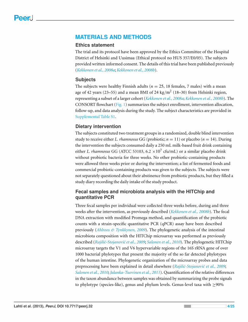
CONSORT flowchart (Fig. 1) summarizes the subject enrollment, intervention allocation,
follow-up, and data analysis during the study. The subject characteristics are provided in
Supplemental Table S1.
Dietary interventionThe subjects constituted two treatment groups in a randomized, double blind intervention
study to receive either L. rhamnosus GG (probiotic; n = 11) or placebo (n = 14). During
the intervention the subjects consumed daily a 250 mL milk-based fruit drink containing
either L. rhamnosus GG (ATCC 53103, 6.2 ×10⁷ cfu/mL) or a similar placebo drink
without probiotic bacteria for three weeks. No other probiotic-containing products
were allowed three weeks prior or during the intervention; a list of fermented foods and
commercial probiotic-containing products was given to the subjects. The subjects were
not separately questioned about their abstinence from probiotic products, but they filled a
study diary recording the daily intake of the study product.
Fecal samples and microbiota analysis with the HITChip andquantitative PCR
Three fecal samples per individual were collected three weeks before, during and three
weeks after the intervention, as previously described (Kekkonen et al., 2008b). The fecal
DNA extraction with modified Promega method, and quantification of the probiotic
counts with a strain-specific quantitative PCR (qPCR) assay have been described
previously (Ahlroos & Tynkkynen, 2009). The phylogenetic analysis of the intestinal
microbiota composition with the HITChip microarray was performed as previously
described (Rajilic-Stojanovic et al., 2009; Salonen et al., 2010). The phylogenetic HITChip
microarray targets the V1 and V6 hypervariable regions of the 16S rRNA gene of over
1000 bacterial phylotypes that present the majority of the so far detected phylotypes
of the human intestine. Phylogenetic organization of the microarray probes and data
preprocessing have been explained in detail elsewhere (Rajilic-Stojanovic et al., 2009;
Salonen et al., 2010; Jalanka-Tuovinen et al., 2011). Quantification of the relative differences
in the taxon abundance between samples was obtained by summarizing the probe signals
to phylotype (species-like), genus and phylum levels. Genus-level taxa with ≥90%
Lahti et al. (2013), PeerJ, DOI 10.7717/peerj.32 4/25

Figure 1 Flowchart of the study subjects during the trial.
sequence similarity in the 16S rRNA gene are referred to as Species and relatives, the latter
being shortened in the text as “et rel.” (Rajilic-Stojanovic et al., 2009).
Previously, HITChip-derived microbial profiles have been shown to correlate well
with those obtained with fluorescence in situ hybridization (FISH) (Rajilic-Stojanovic
et al., 2009) and pyrosequencing (Claesson et al., 2009). Samples were also used for
absolute quantification of total bacteria, methanogenic Archaea, Lactobacillus group and
Bifidobacterium spp. with previously described qPCR primers and reaction conditions
(Salonen et al., 2010).
Lahti et al. (2013), PeerJ, DOI 10.7717/peerj.32 5/25

Blood samples and their biochemical analyses
Of the 25 study subjects used for the microbiota analysis, 22 (8 from probiotic group and
14 from placebo group) were available for the parallel analysis of serum lipid profiles before
and after the intervention (44 samples in total). Venous blood samples from the antecubital
vein were taken at baseline and after the three-week intervention. The blood samples
were stored at −20 ◦C for further analyses. Total cholesterol, high-density lipoprotein
(HDL) and low-density lipoprotein (LDL) cholesterol as well as triglyceride levels were
enzymatically determined from the serum as previously described (Kekkonen et al., 2008a).
Global lipid profiling: sample preparation and analysis byUPLC-MSExtraction of lipids from the serum samples, analysis of the lipid extracts with Waters
Q-Tof Premier mass spectrometer combined with an Acquity Ultra Performance LCTM
(UPLC), data preprocessing and identification of the lipid molecular species is described
in detail elsewhere (Kekkonen et al., 2008a). Lipids have been named according to Lipid
Maps (http://www.lipidmaps.org) with the following abbreviations: Cer: ceramide;
ChoE: cholesteryl ester; lysoPC: lysophosphatidylcholine; PA: phosphatidic acid; PG:
phosphatidylglycerol; PC: phosphatidylcholine; PS: phosphatidylserine; SM: sphin-
gomyelin; TG: triglyceride. Where the fatty acid composition could not be determined,
the total number of carbons and double bonds is indicated. The first number indicates
the amount of carbon atoms in the fatty acid molecule, followed by the number of double
bonds.
Statistical analyses
The effects of probiotic intervention on the intestinal microbiota were investigated with
a combination of linear models, sparse principal component analysis (PCA; Shen &
Huang, 2008; Le Cao, Gonzalez & Dejean, 2009), unsupervised hierarchical clustering,
and significance testing. The log-transformed HITChip and lipid profiling values were
approximately Gaussian distributed and fulfilled the general statistical assumptions
underlying the selected computational approaches. Background variables, including
age, body-mass index and gender were compared in the baseline samples to exclude
potentially confounding effects associated with these variables. The Wilcoxon test was
used with continuous variables and the Fisher exact test for categorical variables, followed
by Benjamini–Hochberg p-value correction for multiple testing. No significant differences
between the background variables were observed between the treatment groups (p> 0.05in all comparisons). The two-group comparisons between the time points and between
the treatment groups were quantified based on a linear model with group-wise fixed
effects and sample-specific random effects, as implemented in the limma R package
(Smyth, 2004), to identify bacterial taxa with significant changes induced by the probiotic
intervention and to assess the magnitude and significance of the effects. The function
lmFit was used to fit the linear model, followed by significance estimation by empirical
Bayes as described in Smyth (2004). In addition, power calculation was carried out to
Lahti et al. (2013), PeerJ, DOI 10.7717/peerj.32 6/25

assess statistical power of the current study. The original data was randomly permuted,
one significant 2-fold alteration was inserted, and Gaussian noise was added using the
same average standard deviation as in the original data. Empirical power calculation with
1000 random permutations showed that 2-fold and higher alterations that follow the
noise levels in the original data were detected in>99.8% of the cases with sample size of
8 or more, based on the same detection criteria than in the current study, confirming the
high statistical power of the analysis at the present sample size. PCA is a linear dimension
reduction method used to compress information in the high-dimensional phylogenetic
(genus-level) and lipid profiles into few informative features that capture the main
variation in the data and allow two-dimensional visualization of sample similarities. The
temporal and inter-individual similarity of the microbiota and lipid profiles was assessed
by average intra- and inter-individual Pearson correlations (r) between and within the time
points, respectively. The differences in profile similarity between the probiotic and placebo
groups were estimated with Wilcoxon test. The biweight midcorrelation, which is more
robust to outliers than the standard Pearson correlation, was used to quantify correlation
between the microbiota and lipid profiles across the individuals. High-throughput
screening studies involve considerable multiple testing and the traditional multiple
testing correction approaches are prohibitively conservative in this context due to their
emphasis on estimating the probability of a single false positive finding. Hence, we have
used q-values for multiple testing correction in the high-throughput screening tests that
include parallel comparisons of large numbers of lipids and bacterial phylotypes.
Correlation analysis of the intestinal microbiota and serum lipids
The biweight midcorrelation measure was used to quantify associations between the
microbiota and lipid profiling data sets, and between the microbiota and biochemically
determined lipids. The correlations between genus-level bacterial groups and lipid species
were calculated together with significance estimates that were corrected for multiple
testing. The significantly correlated (q < 0.05) phylotypes and lipids were selected for
further analysis. It is important to note, however, that while the present sample size
of 22 samples across 2 time points (before and during the probiotic intervention) is
sufficient for highlighting significant correlation patterns, the present experimental
design cannot uncover causal relationships and the observed correlations may be partly
associated with diet or other confounding variables that simultaneously affect both
lipid and microbiota profiles. Two-way average linkage hierarchical clustering of the
bacterial taxa and lipids was applied to highlight and visualize groups of phylotypes
and lipids sharing similar correlation patterns. Analogous ‘second-order’ correlations
have previously been applied for instance in the context of gene expression studies
(Parmigiani et al., 2004; Lahti et al., 2013). A constant plaid model biclustering was
used for systematic detection of significantly correlated bacterial and lipid groups on
the correlation heatmap (Lazzeroni & Owen, 2002). To interpret the detected biclusters,
statistical enrichment (over-representation) of the implicated bacteria was quantified.
Lahti et al. (2013), PeerJ, DOI 10.7717/peerj.32 7/25

Table 1 Stability of microbiota and lipid profiles in the probiotic and placebo groups. We determinedthe similarity (expressed as Pearson’s correlation) both within and between the time points (TP) for themicrobiota and lipid profiles by the average scatter r of the profiles. Lipid data is available for the first twotime points (TP1 and TP2, three weeks before the intervention and during the intervention, respectively),and not available (−) for the third time point (TP3) measured three weeks after the intervention.
Between subjects Within subjects
Microbiota TP1 TP2 TP3 TP1 vs TP2 TP3 vs TP2
Probiotic 0.78 0.78 0.78 0.94 0.95
Placebo 0.76 0.77 0.77 0.94 0.95
Lipids
Probiotic 0.90 0.89 – 0.92 –
Placebo 0.91 0.89 – 0.93 –
Moreover, enrichment analysis was carried out for lipids containing even or odd number
of carbon atoms, as well as the enrichment of lipids with zero, one or more double bonds.
The enrichment analyses were carried out with Fisher’s exact test (Rivals et al., 2007). All
analyses were performed within the R statistical environment (R Development Core Team,
2010).
RESULTSImpact of the L. rhamnosus GG intervention in the intestinalmicrobiota
This study characterized the impact of L. rhamnosus GG intervention on the stability and
composition of the intestinal microbiota. The subjects in the probiotic group consumed
daily approximately 1010 (10.2log10) colony forming units (cfu) of L. rhamnosus GG.
The compliance was verified with the quantification of L. rhamnosus GG in the feces with
strain-specific qPCR (Kekkonen et al., 2008b). The average excretion of L. rhamnosus GG in
the probiotic group was more than 1000-fold higher than that in the placebo group (8.52;
sd 0.73log10 versus 5.20; sd 1.09log10 genome copies per gram of feces, respectively).
The stability of the microbiota during the trial was quantified by the similarity of the
microbiota profiles between the three time points with Pearson correlation (Table 1). The
average intra-individual correlations were high; 0.94–0.95 (sd 0.02–0.03). No significant
difference in the temporal stability of the microbiota between the probiotic and placebo
groups was observed, indicating that the probiotic intervention did not alter the overall
microbial stability. Principal Component Analysis (PCA) visualization of the relationships
between the intestinal microbiota profiles (Fig. 2A) and hierarchical clustering (data not
shown) further supported the conclusion that there were no systematic differences in the
microbiota between the intervention groups. The average inter-individual microbiota
correlation was 0.76–0.78 with no significant differences between the probiotic and
placebo groups (Table 1). The intra-individual microbiota correlations (r = 0.94–0.95)
Lahti et al. (2013), PeerJ, DOI 10.7717/peerj.32 8/25

Figure 2 Intervention effects on the abundance of L. rhamnosus. Mean abundance of L. rhamnosusamong the study subjects before, during and after the probiotic intervention (the time points 1–3,respectively) quantified by the HITChip hybridization signal. The error bars denote the Gaussian 95%confidence limits based on standard deviation of the mean.
were notably higher than the inter-individual correlations (r = 0.76–0.78), stressing the
subject-specificity of the microbiota.
Linear models were used to quantify the effects of the L. rhamnosus GG intervention
on individual taxa using genus- and phylotype-level microarray data. A specific and
transient increase of bacteria related to L. rhamnosus was detected in the probiotic group
immediately after the intervention (Fig. 2). There were no intervention-related effects in
the other lactobacilli, Bifidobacteria or any other taxa either in the probiotic or placebo
group; however substantial individual variation was evident.
To complement the HITChip microarray analysis, we also determined the absolute
counts of total bacteria, methanogenic Archaea, Lactobacillus group and Bifidobacterium
spp. using real-time PCR. The ingestion of L. rhamnosus GG was reflected in the total
lactobacilli that showed a highly significant increase in the probiotic group after the
intervention, returning to baseline levels in the follow up (q< 0.05 in both comparisons).
No other significant changes were observed in the amount of targeted microbes neither
in the probiotic or placebo group. We detected substantial inter- and intra-individual
variation in the methanogenic Archaea but that was independent of the time point or the
treatment group (data not shown). These observations support the conclusion that the
L. rhamnosus GG intervention did not change the overall microbiota composition.
Lahti et al. (2013), PeerJ, DOI 10.7717/peerj.32 9/25

High-throughput profiling of serum lipidsGlobal profiling of the serum lipids was performed using the UPLC-MS platform to
assess the serum lipid profiles at the molecular species level (Kekkonen et al., 2008a). In
total, 407 lipid species from 11 different classes were identified. The lipid profiles were
dominated with triglycerides (TG; 37%), phosphatidylcholines (PC; 25%) and phos-
phatidyletanolamines (PE; 13%) while phosphatidic acids (PA), phosphatidylglycerols
(PG), sphingomyelins (SM), cholesteryl esters (ChoE), lysophosphatidylcholines (lysoPC),
phosphatidylserines (PS), ceramides (Cer) and lysophosphatidylethanolamines (lysoPE)
each contributed with 7% to 0.3% to the total lipid pool.
The stability of the lipid profiles was quantified based on the same correlation analysis
approach than in the HITChip microbiota profiling analysis. No significant differences
in the lipid profile stability were observed between the treatment groups (average
intra-individual r = 0.92 and r = 0.93 for the probiotic and placebo groups, respectively;
Table 1). In contrast to microbiota profiles, the serum lipid profiles were remarkably
similar not only within the subjects but also between the subjects (average inter-individual
r = 0.89–0.91; Table 1). No significant differences were seen between the probiotic and
placebo groups, or between the time points. The stability of the lipid and microbiota
profiles showed a weak positive correlation (r = 0.27) across the subjects independent of
a treatment group. The global lipid profiles did not separate according to the treatment
group in PCA visualization (Fig. 2B), and no statistically significant lipid alterations were
detected between the time points neither in the probiotic or placebo group (data not
shown; Kekkonen et al., 2008b). The fold-changes for each lipid species between the two
time points followed normal distribution in both intervention groups (data not shown),
which further supports the conclusion that the intervention did not induce systematic
alterations on the lipid profiles.
Associations between the intestinal microbiota and serum lipidsIn addition to studying the effects of probiotic L. rhamnosus GG intervention in the
intestinal microbiota, we investigated the co-variation of the microbiota and serum
lipid profiles. As discussed above, our results (Table 1, Fig. 2) conclusively show that the
probiotic intervention did not affect the lipid profiles or the microbiota beyond the L.
rhamnosus that was ingested. Hence, the subjects from probiotic and placebo groups
were pooled to increase the sample size and statistical power in exploring the associations
between the intestinal microbiota and serum lipids.
Heatmap visualization of the microbiota–lipid correlations across the 22 subjects
from 2 time points revealed intestinal bacteria and serum lipids with significantly
correlated abundance patterns (Fig. 3). In total, 86 bacterial group-lipid pairs with notable
correlations (±0.5 or higher) were identified at q< 0.05 significance level (Fig. 3). Among
the significant correlations, 23 of the 131 genus-level taxa detectable by the HITChip were
represented (Supplemental Table S3). Most of these taxa belonged to Proteobacteria (6)
and Firmicutes within Clostridium cluster XIVa (5) and Bacilli (3). From the lipid side,
the vast majority of the significant correlations were attributable to the most dominant
Lahti et al. (2013), PeerJ, DOI 10.7717/peerj.32 10/25

lipids, TGs (62%) and PCs (30%). When analyzed at the genus-level, the significant
correlations were strongly dominated by the positive correlations between bacteria
related to Ruminococcus gnavus and different TG species (31 of 40 significant positive
correlations, average r = 0.61; Fig. 3A). Detailed analysis at the phylotype-level data
indicated that within the R. gnavus group, two uncultured phylotypes, uncultured human
gut bacterium JW1G3 and JW1H4a, dominated the correlations while the type species
R. gnavus or related R. torques did not correlate significantly with TG or any other lipid
species. Other representatives of the Clostridium cluster XIVa included bacteria related to
Dorea formicigenerans that also correlated positively with four TG species (Fig. 3; average
r = 0.58). The remaining TG correlations were negative and involved phylogenetically
diverse bacteria (Supplemental Table S3). Also PCs correlated significantly with numerous
taxa, most of which correlated negatively with ester- or ether linked PCs. Interestingly,
a set of Proteobacteria (Campylobacter, Helicobacter and Moraxellaceae) with low signal
intensities had a clear negative correlation with PC. Similarly, two Lactobacillus species
(L. rhamnosus that was ingested during the trial and L. salivarius) together with another
group of Bacillus, Enterococcus spp., also correlated negatively with PC while few Firmi-
cutes exhibited a positive correlation with this lipid species. Among the less abundant lipids
(<10% of the total lipid pool), a positive correlation was detected between cholesteryl ester
and Collinsella (r = 0.59; Supplemental Table S3, Fig. 5A). On the phylotype level, the
correlation was mainly attributable to an uncultured bacterium clone Eldhufec074 within
genus Collinsella. Another group of Actinobacteria, namely Actinomycetaceae, showed a
significant negative correlation to TG and PA.
Hierarchical clustering and heatmap visualization of the significantly correlated
lipid–bacteria pairs revealed groups of lipids and bacteria sharing similar correlation pat-
terns. A constant plaid model biclustering approach was applied to detect coherent groups
of significantly correlating microbe–lipid pairs (see Methods). The analysis revealed three
major clusters of significantly correlated lipid–microbe pairs (Supplemental Table S2).
One of the observed biclusters highlights the above discussed positive association
between bacteria related to R. gnavus and TGs. Another TG cluster, containing highly
unsaturated long-chain fatty acids, consisted of negatively correlating bacteria related to
Megamonas hypermegale (family Veillonellaceae, Clostridium cluster IX) or belonging to
the Actinomycetaceae. The third cluster contained a set of Proteobacteria (Helicobacter,
Oceanospirillum and Moraxellaceae spp.) and bacteria related to Eubacterium cylindroides
within the family Erysipelotrichaceae, which all correlated negatively with ether lipids.
To explore the nature of the identified lipid–microbe pairs, we quantified the
enrichment of specific, functionally relevant lipid categories in the biclusters. In particular,
we investigated the enrichment of lipids with even and odd number of carbon atoms and
the degree of saturation (0, 1, or more double bonds). The origin of lipid can be inferred
from its chemical composition: Polyunsaturated fatty acids (PUFAs; more than one double
bond) and lipids that have uneven number of carbon atoms originate from plants, bacteria
or marine organism as humans cannot synthesize them. Significant enrichment of long
acyl chain (p< 0.05; Fisher exact test) was detected among the TG lipids that significantly
Lahti et al. (2013), PeerJ, DOI 10.7717/peerj.32 11/25

Figure 3 Correlations between intestinal genus-level phylogenetic groups and serum lipids. The correlations between the intestinal bacteriaand serum lipids are indicated by colors (red: positive; blue: negative). The significant correlations (q < 0.05) are indicated by ‘+’; only lipidsand bacteria with at least one significant correlation are shown. Hierarchical clustering of the rows and columns highlights groups of significantlycorrelated bacteria and lipids. Lipids have been named according to Lipid Maps (http://www.lipidmaps.org) with the following abbreviations: Cer:ceramide; ChoE: cholesteryl ester; lysoPC: lysophosphatidylcholine; PA: phosphatidic acid; PG: phosphatidylglycerol; PC: phosphatidylcholine; PS:phosphatidylserine; SM: sphingomyelin; TG: triglyceride. Where the fatty acid composition could not be determined, the total number of carbonsand double bonds is indicated. The first number indicates the amount of carbon atoms in the fatty acid molecule, followed by the number of doublebonds. For further details, see the Methods section.
correlated with bacteria related to R. gnavus (Supplemental Fig. S1; Supplemental Table
S2). Such a positive association suggests a role for these R. gnavus-related bacteria in the
absorption of dietary lipids. In support of this interpretation, TGs containing fatty acids
with odd number of carbons were also relatively common in this group (p = 0.1). In the
other two biclusters, no significant enrichment of odd/even carbon count or saturation
level was detected but within the bicluster 2 (Supplemental Table S2), Actinomycetaceae
correlated exclusively with highly unsaturated PUFAs (Fig. 3).
Associations between the intestinal microbiota and biochemicallydetermined serum lipids
The mean values (SD) for the major serum lipids were total cholesterol 5.10 (1.02), LDL
cholesterol 3.00 (1.21), HDL cholesterol 1.50 (0.33) and TG 1.20 (0.71) mmol/L (Kekkonen
et al., 2008a). No significant changes were detected in the lipids of the probiotic or the
placebo groups during the intervention (Kekkonen et al., 2008b). Hence, we analyzed
Lahti et al. (2013), PeerJ, DOI 10.7717/peerj.32 12/25

Figure 4 Association between Ruminococcus gnavus et rel. and serum triglyceride (TG) lipids. Therelative amounts of R. gnavus et rel. were quantified by the HITChip analysis and the triglycerideconcentration was determined based on two independent techniques: A the triglyceride TG(54:5) (seeFig. 3 for explanation) by mass spectrometry (r = 0.61); B triglyceride by an enzymatic assay (r = 0.60).
the potential associations of the enzymatically determined major blood lipids with the
intestinal microbiota (Table 2). A positive correlation between bacteria related to R. gnavus
and TG was observed (q< 0.01; r= 0.60; Fig. 4B), corroborating the association identified
within the global lipid analysis. In line with the spectroscopic lipid analysis, representatives
of Bacteroidetes and Uncultured Clostridiales correlated negatively and other implicated
Firmicutes positively with enzymatically determined TG (Table 2). Representatives of
Proteobacteria and Actinobacteria that correlated negatively with TG in the lipid profiling
data did not correlate significantly with enzymatically determined TG. Such inconsistency
may partly arise from methodological reasons as the enzymatic assay captures not only TG
but also diacylglyceride, monoacylglyceride and free glycerol, while lipid profiling captures
TGs at the molecular level.
Collinsella spp. and Eubacterium biforme et rel. showed statistically significant (q <0.05) and positive correlations to enzymatically determined total and LDL cholesterol
(Fig. 5B; Supplemental Table S2). No other significant correlations were identified for total
or LDL cholesterol while HDL cholesterol correlated significantly with numerous taxa.
Eight different Firmicutes including taxa related to Ruminococcus obeum and D. formici-
generans were found to correlate negatively with HDL, while Uncultured Clostridiales I was
the only taxon showing positive correlation to HDL (Supplemental Table S2).
DISCUSSIONIn the present study, we analyzed the effect of probiotic intake on the stability and
composition of the intestinal microbiota and of the serum lipids, and the overall
associations between the microbiota and lipid profiles. The microbiota was analyzed
using the HITChip, a phylogenetic microarray, providing one of the first holistic
and community-level microbiota assessments after a probiotic intervention. The data
published so far is largely dominated with targeted microbiota analyses that have
Lahti et al. (2013), PeerJ, DOI 10.7717/peerj.32 13/25

Table 2 Associations between genus-level bacterial groups and enzymatically determined lipids. As-sociations between the relative amounts of genus-level bacterial groups as determined by the HITChipanalysis and the serum lipid concentrations are quantified with a biweight midcorrelation. Only signifi-cant positive and negative correlations are shown (q< 0.05; otherwise ‘−’). Abbreviations: Total choles-terol (TC), high-density lipoprotein (HDL) and low-density lipoprotein (LDL) cholesterol, triglyceride(TG). Correlations between genus-level bacterial groups and mass spectrometry-determined lipids areprovided in Supplemental Table S3.
Phyla/Firmicute order Genus-level taxon TC LDL HDL TG
Actinobacteria Collinsella 0.56 0.57 – –
Bacilli Aneurinibacillus – – −0.58 0.50
Bacteroidetes Bacteroides plebeius et rel. – – – −0.47
Bacteroidetes Bacteroides vulgatus et rel. – – – −0.47
Bacteroidetes Tannerella et rel. – – – −0.45
Clostridium cluster XI Anaerovorax odorimutans et rel. – – −0.48 0.49
Clostridium cluster XIVa Clostridium nexile et rel. – – −0.45 –
Clostridium cluster XIVa Clostridium sphenoides et rel. – – – 0.46
Clostridium cluster XIVa Dorea formicigenerans et rel. – – −0.56 0.57
Clostridium cluster XIVa Eubacterium hallii et rel. – – – 0.47
Clostridium cluster XIVa Ruminococcus gnavus et rel. – – −0.46 0.60
Clostridium cluster XIVa Ruminococcus obeum et rel. – – −0.48 0.51
Clostridium cluster XV Anaerofustis – – −0.45 –
Clostridium cluster XVI Eubacterium biforme et rel. 0.48 0.47 – –
Clostridium cluster XVI Eubacterium cylindroides et rel. – – −0.45 –
Uncultured Clostridiales Uncultured Clostridiales I – – 0.53 −0.45
Uncultured Clostridiales Uncultured Clostridiales II – – – −0.54
Figure 5 Association between Collinsella spp. and serum cholesterol. The relative amounts ofCollinsella spp. were quantified by the HITChip analysis, and serum cholesterol levels were determinedby two independent techniques: A Cholesterol ester ChoE(20:5) (see Fig. 3 for explanation) by massspectrometry (r = 0.59); B low-density lipoprotein (LDL) cholesterol by enzymatic assay (r = 0.57).
Lahti et al. (2013), PeerJ, DOI 10.7717/peerj.32 14/25

reported a generic increase of lactic acid bacteria after intake of individual lactobacilli
strains (McNulty et al., 2011; Palaria, Johnson-Kanda & O’Sullivan, 2011; Satokari et
al., 2001; Savard et al., 2011; Vaughan, Mollet & de Vos, 1999; Yamanoa et al., 2006).
Our community-level analysis showed that the probiotic intake did not introduce any
changes in the microbiota composition or stability except the specific increase of L.
rhamnosus and total lactobacilli, which likely reflected the excretion of the ingested strain.
Similarly, ingestion of a mixture of five different probiotic strains did not induce any
significant changes in the microbiota composition neither in adults nor in simplified
model community in mice (McNulty et al., 2011). Even when a combination of probiotic
and prebiotic (synbiotic) food was consumed, two studies based on microbiota profiling
and qPCR did not identify any differences in the microbiota composition between placebo
and treatment besides the ingested strains (Palaria, Johnson-Kanda & O’Sullivan, 2011;
Vitali et al., 2010).
We have shown previously using the same cohort that the ingestion of L. rhamnosus GG
had apparent effects on the host immunology (Kekkonen et al., 2008b). Hence, our results
does not support the hypothesis that probiotic bacteria would modulate the endogenous
microbiota but rather points towards direct signaling to host or altered gene expression of
the resident microbiota as a mode of probiotic action as recently proposed (McNulty et al.,
2011).
Our study was carried out in healthy adults with seemingly well-established and
balanced microbial communities. In subjects whose intestinal ecosystem is unbalanced
e.g. due to pathogen overgrowth, gastrointestinal symptoms or recent intake of antibiotics,
intake of probiotic bacteria may modulate the microbiota. Similarly, the developing
microbiota of children is potentially more susceptible to environmental modulators
including probiotics. Although substantial changes in infant microbiota following daily
intake of L. rhamnosus GG have indeed been reported (Cox et al., 2010), these results must
be interpreted with caution as the phylogenetic microarray used in that study provided
signals for close to 50 phyla (Midgley et al., 2012) whereas the human intestinal ecosystem
only contains a maximum of 10 phyla (Rajilic-Stojanovic, Smidt & de Vos, 2007).
To the best of our knowledge, this cohort is the first addressing the potential impact of
probiotics on global serum lipid profiles in adults. Daily intake of L. rhamnosus GG for
three weeks did not introduce any consistent alterations in the serum lipids or in the bio-
chemically determined cholesterol and triglyceride levels of the healthy, normolipidemic
adults analyzed in this and previous study (Kekkonen et al., 2008a). Probiotic bacteria
have been suggested as a potential non-drug treatment to lower serum cholesterol levels
based on their in vitro described ability to deconjugate bile acids and directly assimilate
cholesterol. A recent meta-analysis implies that probiotic intake can lower the total and
LDL cholesterol (Guo et al., 2011), although numerous human studies have failed to
observe any effects on serum lipids after probiotic intake (see e.g. Sadrzadeh-Yeganeh et al.,
2010; Pereira & Gibson, 2002). One explanation for the controversy is that similarly to the
strain-specificity of the immunomodulatory effects of probiotics (Kekkonen et al., 2008b),
also their effects on lipid metabolism are likely to vary between different strains. In atopic
Lahti et al. (2013), PeerJ, DOI 10.7717/peerj.32 15/25

infants the consumption of L. rhamnosus GG supplemented formulas for several months
resulted in a reduced proportion of α-linolenic acid and of total n-3 PUFAs, leading to
increased n-6 to n-3 PUFA ratio in sera of probiotic-fed infants (Kankaanpaa et al., 2002).
Decrease of n-3 fatty acids has negative health implication, underlining the need for more
in vivo research to specify the bacterial strains and target population where probiotics can
have beneficial effects on the host lipid metabolism.
Our data indicates that the variation of the intestinal microbiota is considerably higher
across the individuals than the variation in serum lipid profiles, highlighting the tight
homeostatic control of systemically circulating lipids. The intestinal microbiota is known
to play an important role in the regulation of systemic lipid metabolism (Martin et al.,
2007; Velagapudi et al., 2010). In this study, we identified several novel associations between
the human intestinal commensals and serum lipids. From the lipid side, TGs (62%)
and PCs (30%) dominated the significant lipid–microbe correlations. TGs are used for
energy storage while PCs are abundant constituents of cell membranes. Both of these
abundant lipid classes were affected upon colonization of germ-free mice (Velagapudi et al.,
2010). The most prominent correlation was between TGs and uncultured phylotypes
related to R. gnavus that belong to family Ruminococcaceae in Clostridium cluster
XIVa. Two characterized species within the group, R. gnavus and R. torques, have been
implicated in intestinal disorders as they appeared to be overrepresented both in IBS
(Kassinen et al., 2007; Rajilic-Stojanovic et al., 2011) and in IBD (Joossens et al., 2011;
Prindiville, Cantrell & Wilson, 2004). The TG lipids that were positively associated with
R. gnavus-related phylotypes were enriched with polyunsaturated and odd-chain fatty
acids. Since the odd-chain fatty acids are not synthesized in the body, these positive asso-
ciations suggest that the implicated bacteria facilitate the absorption of polyunsaturated
dietary lipids. Moreover, it cannot be excluded that these bacteria are even involved
in their biosynthesis as related R. obeum together with other Firmicutes are among
the intestinal bacteria capable of producing isomers of conjugated linoleic acids (CLA;
McIntosh et al., 2009).
Triglycerides carry different types of fatty acids, and accordingly not all TGs displayed
the same pattern regarding microbial correlations. For example, bacteria related to D.
formicigenerans showed positive association to saturated triglycerides with a small carbon
number, i.e. including palmitic acid. These fatty acids are mainly produced de novo in the
liver (Westerbacka et al., 2010; Kotronen et al., 2009). D. formicigenerans produces formate
and acetate that are precursors of hepatic lipogenesis. Recent metaproteomic work verifies
that the acetate kinases involved in acetate production are highly expressed in the intestinal
ecosystem (Kolmeder et al., 2012), suggesting that several intestinal bacteria have potential
to regulate lipogenesis. On the other hand, Actinomycetaceae were inversely associated with
TGs containing highly unsaturated triglycerides with a high carbon number. Such TGs
carry physiologically important fatty acids such as C22:6 (docosahexanoic acid) and C20:4
(arachidonic acid). Both are important cell membrane components, the former especially
in visual and neural tissues, and the latter is involved in inflammatory signaling. While not
Lahti et al. (2013), PeerJ, DOI 10.7717/peerj.32 16/25

much is known about the intestinal Actinomycetaceae, both commensal and pathogenic
forms of these Actinobacteria are abundant in the oral cavity.
We identified multiple taxa correlating negatively with normal or ether linked forms of
PC. Dietary PC was recently identified as compound that after conversion by the intestinal
microbiota promotes heart disease (Wang et al., 2011). Unfortunately, that study did
not address the identity of bacteria involved in the metabolism of dietary PC, but our
data suggest that different Firmicutes, Proteobacteria and Fusobacteria may participate
in this metabolic conversion (Fig. 3). Remarkably, the presence of serum ether PCs was
negatively associated with various taxa, mainly with Gram-negative genera that include
pathogens and commensals, such as Helicobacter, Moraxellaceae and Campylobacter.
Among the ether lipids, plasmalogens, known as endogenous antioxidants (Wallner &
Schmitz, 2011), are the most abundant. Their negative association with Proteobacteria
could indicate that the implicated bacteria may induce oxidative stress in host cells, leading
to depletion of plasmalogens. In support of this hypothesis, lipopolysaccharide (LPS), a
ubiquitous and toxic surface component of Gram-negative bacteria, induces oxidative
stress in mammalian cell cultures (Aly, Lightfoot & El-Shemy, 2010).
Finally, we identified positive correlation between the abundance of serum cholesterol
and genus Collinsella (Actinobacteria, family Coriobacteriacea). Biochemical lipid analysis
indicated that Collinsella spp. together with bacteria related to E. biforme correlated
specifically with total cholesterol and LDL but not HDL. To our knowledge, our work
provides the first in vivo evidence about the implication of Coriobacteriacea in human
lipid metabolism. Our present findings are supported by the data generated with rodent
models. In a hamster model, the proportion of Coriobacteriaceae showed high positive
correlation with non-HDL plasma cholesterol levels and reacted to the intake of dietary
lipids (Martinez et al., 2009). In a mouse model, a strong positive correlation between
Coriobacteriaceae and hepatic triglycerides was identified (Claus et al., 2011), providing
cumulative indication for the involvement of Coriobacteriaceae in mammalian lipid
metabolism. While only one taxon (Collinsella) correlated statistically significantly with
cholesterol in the lipid profiling data, numerous taxa showed significant correlation to en-
zymatically determined HDL cholesterol. This may arise from technical differences, as the
lipid profiling does not quantify free cholesterol but its derivative cholesteryl ester, where
cholesterol is esterified to long-chain fatty acids. All taxa that were associated with both
HDL cholesterol and TG had opposite correlation with these lipids (Supplemental Table
S2), in accordance with the fact that serum TG and HDL show strong inverse relationship,
and their ratio is used as an atherogenic index that predicts cardiovascular risk (Jeppesen
et al., 1997). Hence, identification of bacteria that potentially affect the athreogenic index
may provide important clinical implications for dyslipidemic individuals.
While good correspondence between the HITChip and pyrosequencing studies
have been previously demonstrated, the main advantage of the HITChip microarray
compared to the pyrosequencing studies is that the microarray technology provides
very standardized and cost-efficient tools for deep and reproducible analysis of intestinal
Lahti et al. (2013), PeerJ, DOI 10.7717/peerj.32 17/25

microbiota including phylotypes that are only present in low concentrations (Claesson et
al., 2009; Salonen et al., 2012). We expect that our major findings, the associations between
specific lipid species and bacteria related to Collinsella or R. gnavus could be detected
also in a standard pyrosequencing study due to the relatively high abundance of these
organisms. However, the associations involving less abundant taxa (Bacilli, Proteobacteria,
Actinomycetaeae) would likely have been missed with conventional sequencing depth.
In summary, quantitative analysis of high-throughput profiling data identified several
significant correlations between the intestinal microbiota and serum lipids. These
results partly confirm and extend previous observations in animal studies, and provide
hypotheses for follow-up studies in humans. It is important to note, however, that the
present analysis is based on a Finnish cohort and only two time points, and does not
consider causal relationships between these variables. Therefore it cannot be excluded that
some of the observed correlations may be associated with the diet or other confounding
variables that simultaneously affect both lipid and microbiota profiles. However, as all
subjects ate their habitual diets, individual food components are not likely to cause
systematic bias to the observed lipid–microbe correlations, which is further supported
by the observation of distinct groups of correlated lipid–microbe pairs in the bicluster
analysis. It can also be ruled out that the intervention would have caused the correlations
as control measurements from the baseline were included, the ingested probiotics
L. rhamnosus GG did not correlate significantly with any serum lipid, and the drink did
not otherwise alter the microbiota. Confirmation of the findings in an independent cohort
and a more thorough longitudinal analysis will be necessary to assess the effect of external
variables on the microbiota–lipid correlations and their potential causal associations.
None of the implicated bacterial taxa are functionally characterized, and thus the potential
mechanisms of how human intestinal bacteria relate to the host lipid metabolism are
currently unknown. Among the potential mechanisms, bacterial modification of the
bile acid pool is by far the best characterized. Collinsella spp., R. gnavus as well as the
genus Eubacterium are among the intestinal bacteria capable of deconjugating bile acids
(Lepercq et al., 2004). Bile acids are cholesterol-derived detergents that play a central role
in the absorption of fat in the intestine but also in signaling with systemic endocrine
functions (Swann et al., 2011). It has been known for long that intestinal bacteria can
chemically modify bile acids but recent work suggest that the microbiota may also control
their production and degradation (Antunes et al., 2011). In mice the intestinal bacteria
profoundly affect the emulsification, absorption and transport of dietary fat as well as
their storage and peroxidation through the metabolic and signaling properties of bile acids
(Martin et al., 2007).
CONCLUSIONOur data supports the concept that the overall lipid content in human serum is a composite
of host and microbial metabolic activity, and the intestinal commensals are implicated
in the metabolism of various lipid species that human body utilizes for membranes,
energy storage and signaling. Considering that a single gene in an intestinal bacterium
Lahti et al. (2013), PeerJ, DOI 10.7717/peerj.32 18/25
could alter host fatty acid composition (Rosberg-Cody et al., 2011), we can only envisage
the metabolic capacity and the functional consequences from the million genes in the
intestinal microbiome. As epidemiological data do not support a link between dietary
cholesterol and serum cholesterol levels (Lecerf & de Lorgeril, 2011), the role of genetic
factors in the individual variability of cholesterol levels is evident. Identification of the
bacteria and their mechanisms that regulate host fatty acid and lipid metabolism have
considerable potential for clinical implications due to the profound role of these molecules
for instance in cardiovascular disease and regulation of inflammatory cytokine signaling
(Calder, 2011). Future trials involving controlled diet and dyslipidemic individuals will
provide further insights on the role of intestinal microbes on human lipid metabolism.
ADDITIONAL INFORMATION AND DECLARATIONS
FundingThis work was partly funded by the Finnish Funding Agency for Technology and Innova-
tion (TEKES; grant number 40274/06), the ERC grant Microbes Inside (grant 250172),
and the Academy of Finland (grant 137389) to WMdV. We gratefully acknowledge the
HITChip team (Wageningen University, NL supported by the Spinoza Award of the
Netherlands Foundation for Scientific Research to WMdV) for the HITChip analysis.
The work at the University of Helsinki was performed at the Centre of Excellence on
Microbial Food Safety Research, Academy of Finland. LL received funding from the
Academy of Finland (decision 256950) and Alfred Kordelin Foundation. Support was
received from Valio; a manufacturer of probiotics. The funders had no role in study design,
data collection and analysis, decision to publish, or preparation of the manuscript.
Grant DisclosuresThe following grant information was disclosed by the authors:
Finnish Funding Agency for Technology and Innovation: (TEKES; grant number
40274/06).
European Research Council: (ERC; grant 250172).
Academy of Finland: (decisions 256950 to LL; 137389 to WMdV).
Competing InterestsWMdV is an Academic Editor for PeerJ. The work was partially supported by Valio, a
manufacturer of probiotics.
Author Contributions• Leo Lahti analyzed the data, wrote the paper.
• Anne Salonen performed the experiments, analyzed the data, wrote the paper.
• Riina A. Kekkonen conceived and designed the experiments, performed the
experiments.
• Jarkko Salojarvi and Jonna Jalanka-Tuovinen analyzed the data.
Lahti et al. (2013), PeerJ, DOI 10.7717/peerj.32 19/25

• Airi Palva contributed reagents/materials/analysis tools.
• Matej Oresic and Willem M. de Vos conceived and designed the experiments,
contributed reagents/materials/analysis tools, wrote the paper.
Clinical Trial EthicsThe following information was supplied relating to ethical approvals (i.e. approving body
and any reference numbers):
The trial and its protocol have been approved by the Ethics Committee of the Hospital
District of Helsinki and Uusimaa (Ethical protocol no HUS 357/E0/05). The subjects
provided written informed consent. The details of this trial have been published previously
(Kekkonen et al., 2008a; Kekkonen et al., 2008b).
Microarray Data DepositionThe following information was supplied regarding the deposition of microarray data:
The microarray data presented in the manuscript are phylogenetic microarray data for
which the standards of commercial microarrays are not as such applicable. Therefore we
have attached the data as Supplemental material of this manuscript, and details of the
samples, experimental design, and study protocol are provided in the main text. Prior to
this manuscript, this particular phylogenetic microarray, the HITChip, has been utilized
in various publications, including: Claesson et al. PLoS ONE. 2009; 4(8):e6669, Biagi et
al. PLoS ONE. 2010 May 17; 5(5):e10667 and Jalanka-Tuovinen et al. PLoS ONE. 2011;
6(7):e23035.
Clinical Trial RegistrationThe following information was supplied regarding Clinical Trial registration:
The trial and its protocol have been approved by the Ethics Committee of the Hospital
District of Helsinki and Uusimaa (Ethical protocol no HUS 357/E0/05).
Supplemental InformationSupplemental information for this article can be found online at http://dx.doi.org/
10.7717/peerj.32.
REFERENCESAhlroos T, Tynkkynen S. 2009. Quantitative strain-specific detection of Lactobacillus rhamnosus
GG in human faecal samples by real-time PCR. Journal of Applied Microbiology 106:506–514DOI ./j.-...x.
Aly HA, Lightfoot DA, El-Shemy HA. 2010. Bacterial lipopolysaccharide-induced oxidative stressin adult rat Sertoli cells in vitro. Toxicology in Vitro 24:1266–1272 DOI ./j.tiv....
Antunes LCM, Han J, Ferreira RBR, Lolic P, Borchers CH, Finlay BB. 2011. Effect of antibiotictreatment on the intestinal metabolome. Antimicrobial Agents and Chemotheraphy55:1494–1503 DOI ./AAC.-.
Blaut M, Clavel T. 2007. Metabolic diversity of the intestinal microbiota: implications for healthand disease. The Journal of Nutrition 137:751S–755S.
Lahti et al. (2013), PeerJ, DOI 10.7717/peerj.32 20/25

Calder P. 2011. Fatty acids and inflammation: the cutting edge between food and pharma.European Journal of Pharmacology 668:S50–58 DOI ./j.ejphar....
Claesson MJ, O’Sullivan O, Wang Q, Nikkila J, Marchesi JR, Smidt H, de Vos VM, Ross RP,O’Toole PW. 2009. Comparative analysis of pyrosequencing and a phylogenetic microarrayfor exploring microbial community structures in the human distal intestine. PLoS ONE 4:e6669DOI ./journal.pone..
Claus SP, Ellero SL, Berger B, Krause L, Bruttin A, Molina J, Paris A, Want EJ,de Waziers I, Cloarec O, Richards SE, Wang Y, Dumas M-E, Ross A, Rezzi S, Kochhar S,van Bladeren P, Lindon JC, Holmes E, Nicholson JK. 2011. Colonization-induced host-gutmicrobial metabolic interaction. mBio 2:e00271-10 DOI ./mBio.-.
Cox MJ, Huang YJ, Fujimura KE, Liu JT, McKean M, Homer Boushey HA, Segal MR, Brodie EL,Cabana MD, Lynch SV. 2010. Lactobacillus casei abundance is associated with profound shiftsin the infant gut microbiome. PLoS ONE 5: e8745.
Fava F. 2006. The gut microbiota and lipid metabolism: implications for human health andcoronary heart disease. Current Medicinal Chemistry 13:3005–3021DOI ./.
Gerard P. 2010. Gastrointestinal tract: microbial metabolism of steroids. In: Timmis KN, ed.Handbook of hydrocarbon and lipid microbiology. Berlin Heidelberg: Springer, 3133–3140.
Guo Z, Liu XM, Zhang QX, Shen Z, Tian FW, Sun ZH, Zhang HP, Chen W. 2011. Influenceof consumption of probiotics on the plasma lipid profile: a meta-analysis of randomisedcontrolled trials. Nutrition, Metabolism and Cardiovascular Diseases 21:844–850DOI ./j.numecd....
Holmes E, Li JV, Athanasiou T, Ashrafian H, Nicholson JK. 2011. Understanding the role of gutmicrobiome-host metabolic signal disruption in health and disease. Trends in Microbiology19:349–359 DOI ./j.tim....
Jalanka-Tuovinen J, Salonen A, Nikkila J, Immonen O, Kekkonen R, Lahti L, Palva A,de Vos WM. 2011. Intestinal microbiota in healthy adults: temporal analysis revealsindividual and common core and relation to intestinal symptoms. PLoS ONE 6:e23035DOI ./journal.pone..
Jeppesen J, Hein HO, Suadicani P, Gyntelberg F. 1997. Relation of high TG-low HDL cholesteroland LDL cholesterol to the incidence of ischemic heart disease. An 8-year follow-up in theCopenhagen Male Study. Arteriosclerosis, Thrombosis and Vascular Biology 17:1114–1120DOI ./.ATV....
Joossens M, Huys G, Cnockaert M, de Preter V, Verbeke K, Rutgeerts P, Vandamme P,Vermeire S. 2011. Dysbiosis of the faecal microbiota in patients with Crohn’s disease and theirunaffected relatives. Gut 60:631–637 DOI ./gut...
Kankaanpaa PE, Yang B, Kallio HP, Isolauri E, Salminen SJ. 2002. Influence of probioticsupplemented infant formula on composition of plasma lipids in atopic infants. The Journalof Nutritional Biochemistry 13:364–369 DOI ./S-()-.
Kankainen M, Paulin L, Tynkkynen S, von Ossowski I, Reunanen J, Partanen P, Satokari R,Vesterlund S, Hendrickx APA, Lebeer S, De Keersmaecker SCJ, Vanderleyden J,Hamalainen T, Laukkanen S, Salovuori N, Ritari J, Alatalo E, Korpela R,Mattila-Sandholm T, Lassig A, Hatakka K, Kinnunen KT, Karjalainen H, Saxelin M,Laakso K, Surakka A, Palva A, Salusjarvi T, Auvinen P, de Vos WM. 2009. Comparativegenomic analysis of Lactobacillus rhamnosus GG reveals pili containing a human-mucusbinding protein. Proceedings of the National Academy of Sciences United States of America106:17193–17198 DOI ./pnas..
Lahti et al. (2013), PeerJ, DOI 10.7717/peerj.32 21/25

Kassinen A, Krogius-Kurikka L, Makivuokko H, Rinttila T, Paulin L, Corander J, Malinen E,Apajalahti J, Palva A. 2007. The fecal microbiota of irritable bowel syndromepatients differs significantly from that of healthy subjects. Gastroenterology 133:24–33DOI ./j.gastro....
Kekkonen RA, Sysi-Aho M, Seppanen-Laakso T, Julkunen I, Vapaatalo H, Oresic M, Korpela R.2008a. Effect of probiotic Lactobacillus rhamnosus GG intervention on global serumlipidomic profiles in healthy adults. World Journal of Gastroenterology 14:3188–3194DOI ./wjg...
Kekkonen RA, Lummela N, Karjalainen H, Latvala S, Tynkkynen S, Jarvenpaa S, Kautiainen H,Julkunen I, Vapaatalo H, Korpela R. 2008b. Probiotic intervention has strain-specificanti-inflammatory effects in healthy adults. World Journal of Gastroenterology 14:2029–2036DOI ./wjg...
Kolmeder CA, de Been M, Nikkila J, Ritamo I, Matto J, Valmu L, Salojarvi J, Palva A, Salonen A,de Vos WM. 2012. Comparative metaproteomics and diversity analysis of human intestinalmicrobiota testifies for its temporal stability and expression of core functions. PLoS ONE7:e29913 DOI ./journal.pone..
Kotronen A, Velagapudi VR, Yetukuri L, Westerbacka J, Bergholm R, Ekroos K, Makkonen J,Taskinen MR, Oresic M, Yki-Jarvinen H. 2009. Serum saturated fatty acids containingtriacylglycerols are better markers of insulin resistance than total serum triacylglycerolconcentrations. Diabetologia 52:684–690 DOI ./s---.
Lahti L, Schafer M, Klein H-U, Bicciato S, Dugas M. 2013. Cancer gene prioritization byintegrative analysis of mRNA expression and DNA copy number data: a comparative review.Briefings in Bioinformatics 14:27–35 DOI ./bib/bbs.
Lazzeroni L, Owen A. 2002. Plaid models for gene expression data. Statistica Sinica 12:61–86.
Le Cao K-A, Gonalez I, Dejean S. 2009. IntergrOmics: an R package to unravel relationshipsbetween two omics data sets. Bioinformatics 25:2855–2856 DOI ./bioinformatics/btp.
Lecerf JM, de Lorgeril M. 2011. Dietary cholesterol: from physiology to cardiovascular risk. BritishJournal of Nutrition 106:6–14 DOI ./S.
Lepercq P, Gerard P, Beguet F, Grill J-P, Relano P, Cayuela C, Juste C. 2004. Isolates fromnormal human intestinal flora but not lactic acid bacteria exhibit 7α- and7β-hydroxysteroid dehydrogenase activities. Microbial Ecology in Health and Disease16:195–201 DOI ./.
Martin FP, Dumas ME, Wang Y, Legido-Quigley C, Yap IK, Tang H, Zirah S, Murphy GM,Cloarec O, Lindon JC, Sprenger N, Fay LB, Kochhar S, van Bladeren P, Holmes E,Nicholson JK. 2007. A top-down systems biology view of microbiome-mammalian metabolicinteractions in a mouse model. Molecular Systems Biology 3:112 DOI ./msb.
Martinez I, Wallace G, Zhang C, Legge R, Benson AK, Carr TP, Moriyama EN, Walter J. 2009.Diet-induced metabolic improvements in a hamster model of hypercholesterolemia are stronglylinked to alterations of the gut microbiota. Applied Environmental Microbiology 75:4175–4184DOI ./AEM.-.
McIntosh FM, Shingfield KJ, Devillard E, Russell WR, Wallace RJ. 2009. Mechanism ofconjugated linoleic acid and vaccenic acid formation in human faecal suspensions and purecultures of intestinal bacteria. Microbiology 155:285–294 DOI ./mic..-.
McNulty NP, Yatsunenko T, Hsiao A, Faith JJ, Muegge BD, Goodman AL, Henrissat B,Oozeer R, Cools-Portier S, Gobert G, Chervaux C, Knights D, Lozupone CA, Knight R,Duncan AE, Bain JR, Muehlbauer MJ, Newgard CB, Heath AC, Gordon JI. 2011. The impact
Lahti et al. (2013), PeerJ, DOI 10.7717/peerj.32 22/25

of a consortium of fermented milk strains on the gut microbiome of gnotobiotic mice andmonozygotic twins. Science Translational Medicine 3:106 DOI ./scitranslmed..
Midgley DJ, Greenfield P, Shaw JM, Oytam Y, Li D, Kerr CA, Hendry P. 2012. Reanalysisand simulation suggest a phylogenetic microarray does not accurately profile microbialcommunities. PLoS ONE 7:e33875 DOI ./journal.pone..
O’Keefe SJ. 2008. Nutrition and colonic health: the critical role of the microbiota. Current Opinionin Gastroenterology 24:51–58 DOI ./MOG.beff.
Oresic M, Hanninen VA, Vidal-Puig A. 2008. Lipidomics: a new window to biomedical frontiers.Trends in Biotechnology 26:647–652 DOI ./j.tibtech....
Palaria A, Johnson-Kanda I, O’Sullivan DJ. 2011. Effect of a synbiotic yogurt on levels of fecalbifidobacteria, clostridia and enterobacteria. Applied Environmental Microbiology 78:933–940DOI ./AEM.-.
Parmigiani G, Garrett-Mayer ES, Anbazhagan R, Gabrielson E. 2004. A cross-study comparisonof gene expression studies for the molecular classification of lung cancer. Clinical CancerResearch 10:2922–2927 DOI ./-.CCR--.
Pereira DI, Gibson GR. 2002. Effects of consumption of probiotics and prebiotics on serumlipid levels in humans. Critical Reviews in Biochemistry and Molecular Biology 37:259–281DOI ./.
Prindiville T, Cantrell M, Wilson KH. 2004. Ribosomal DNA sequence analysis ofmucosa-associated bacteria in Crohn’s disease. Inflammatory Bowel Diseases 10:824–833DOI ./--.
Qin J, Li R, Raes J, Arumugam M, Burgdorf KS, Manichanh C, Nielsen T, Pons N, Levenez F,Yamada T, Mende DR, Li J, Xu J, Li S, Li D, Cao J, Wang B, Liang H, Zheng H, Xie Y, Tap J,Lepage P, Bertalan M, Batto J-M, Hansen T, De Paslier D, Linneberg A, Nielsen HB,Pelletier E, Renault P, Sicheritz-Ponten T, Turner K, Zhu H, Yu C, Li S, Jian M, Zhou Y,Li Y, Zhang X, Li S, Qin N, Yang H, Wang J, Brunak S, Dore J, Guarner F, Kristiansen K,Pedersen O, Parkhill J, Weissenbach J, MetaHIT Consortium, Bork P, Ehrlich SD, Wang J.2010. A human gut microbial gene catalogue established by metagenomic sequencing. Nature464:59–65 DOI ./nature.
R Development Core Team. 2010. R: a language and environment for statistical computing. Vienna,Austria: R Foundation for Statistical Computing.
Rajilic-Stojanovic M, Smidt H, de Vos WM. 2007. Diversity of the human gastrointestinaltract microbiota revisited. Environmental Microbiology 9:2125–2136 DOI ./j.-...x.
Rajilic-Stojanovic M, Heilig HG, Molenaar D, Kajander K, Surakka A, Smidt H,de Vos WM. 2009. Development and application of the human intestinal tract chip, aphylogenetic microarray: analysis of universally conserved phylotypes in the abundantmicrobiota of young and elderly adults. Environmental Microbiology 11:1736–1751DOI ./j.-...x.
Rajilic-Stojanovic M, Biagi E, Heilig HG, Kajander K, Kekkonen RA, Tims S,de Vos WM. 2011. Global and deep molecular analysis of microbiota signatures in fecalsamples from patients with irritable bowel syndrome. Gastroenterology 141:1792–1801DOI ./j.gastro....
Ridlon JM, Kang DJ, Hylemon PB. 2006. Bile salt biotransformations by human intestinalbacteria. Journal of Lipid Research 47:241–259 DOI ./jlr.R-JLR.
Lahti et al. (2013), PeerJ, DOI 10.7717/peerj.32 23/25

Rivals I, Personnaz L, Taing L, Potier M-C. 2007. Enrichment or depletion of a GO categorywithin a class of genes: which test? Bioinformatics 23(4):401–407 DOI ./bioinformat-ics/btl.
Rosberg-Cody E, Stanton C, O’Mahony L, Wall R, Shanahan F, Quigley EM, Fitzgerald GF,Ross RP. 2011. Recombinant lactobacilli expressing linoleic acid isomerase can modulatethe fatty acid composition of host adipose tissue in mice. Microbiology 157:609–615DOI ./mic..-.
Sadrzadeh-Yeganeh H, Elmadfa I, Djazayery A, Jalali M, Heshmat R, Chamary M. 2010. Theeffects of probiotic and conventional yoghurt on lipid profile in women. British Journal ofNutrition 103:1778–1783 DOI ./S.
Salonen A, Nikkila J, Jalanka-Tuovinen J, Immonen O, Rajilic-Stojanovic M, Kekkonen RA,Palva A, de Vos WM. 2010. Comparative analysis of fecal DNA extraction methods withphylogenetic microarray: effective recovery of bacterial and archaeal DNA using mechanicalcell lysis. Journal of Microbiological Methods 81:127–134 DOI ./j.mimet....
Salonen A, Salojarvi J, Lahti L, de Vos WM. 2012. The adult intestinal core microbiotais determined by analysis depth and health status. Clinical Microbiology and Infection18(Suppl. 4):16–20 DOI ./j.-...x.
Satokari RM, Vaughan EE, Akkermans AD, Saarela M, de Vos WM. 2001. Polymerase chainreaction and denaturing gradient gel electrophoresis monitoring of fecal bifidobacteriumpopulations in a prebiotic and probiotic feeding trial. Systematic and Applied Microbiology24:227–231 DOI ./--.
Savard P, Lamarche B, Paradis ME, Thiboutot H, Laurin E, Roy D. 2011. Impact ofBifidobacterium animalis subsp. lactis BB-12 and Lactobacillus acidophilus LA-5-containingyoghurt on fecal bacterial counts of healthy adults. International Journal of Food Microbiology149:50–57 DOI ./j.ijfoodmicro....
Saxelin M, Tynkkynen S, Mattila-Sandholm T, de Vos WM. 2005. Probiotic and other functionalmicrobes: from markets to mechanisms. Current Opinion in Biotechnology 16:204–211DOI ./j.copbio....
Shen HJ, Huang JZ. 2008. Sparse principal component analysis via regularized low rank matrixapproximation. Journal of Multivariate Analysis 99:1015–1034 DOI ./j.jmva....
Sekirov I. 2010. Gut microbiota in health and disease. Physiological Reviews 90:859–904DOI ./physrev...
Smyth GK. 2004. Linear models and empirical Bayes methods for assessing differential expressionin microarray experiments. Statistical Applications in Genetics and Molecular Biology 3:Article 3DOI ./-..
Storey JD, Tibshirani R. 2003. Statistical significance for genomewide studies. Proceedingsof the National Academy of Sciences United States of America 100:9440–9445DOI ./pnas..
Swann JR, Want EJ, Geier FM, Spagou K, Wilson ID, Sidaway JE, Nicholson JK, Holmes E.2011. Systemic gut microbial modulation of bile acid metabolism in host tissuecompartments. Proceedings of the National Academy of Sciences United States of America108(Suppl 1):4523–4530 DOI ./pnas..
Tremaroli V, Backhed F. 2012. Functional interactions between the gut microbiota and hostmetabolism. Nature 489:242–249 DOI ./nature.
Vaughan EE, Mollet B, de Vos WM. 1999. Functionality of probiotics and intestinallactobacilli: light in the intestinal tract tunnel. Current Opinion in Biotechnology 10:505–510DOI ./S-()-X.
Lahti et al. (2013), PeerJ, DOI 10.7717/peerj.32 24/25
Velagapudi VR, Hezaveh R, Reigstad CS, Gopalacharyulu P, Yetukuri L, Islam S, Felin J,Perkins R, Boren J, Oresic M, Backhed F. 2010. The gut microbiota modulates host energyand lipid metabolism in mice. Journal of Lipid Research 51:1101–1112 DOI ./jlr.M.
Vitali B, Ndagijimana M, Cruciani F, Carnevali P, Candela M, Guerzoni ME, Brigidi P. 2010.Impact of a synbiotic food on the gut microbial ecology and metabolic profiles. BMCMicrobiology 10:4 DOI ./---.
Wallner S, Schmitz G. 2011. Plasmalogens the neglected regulatory and scavenging lipid species.Chemistry and Physics of Lipids 164:573–589 DOI ./j.chemphyslip....
Wang Z, Klipfell E, Bennett BJ, Koeth R, Levison BS, DuGar B, Feldstein AE, Britt EB, Fu X,Chung Y-M, Wu Y, Schauer P, Smith JD, Allayee H, Tang WHW, DiDonato JA, Lusis AJ,Hazen SL. 2011. Gut flora metabolism of phosphatidylcholine promotes cardiovascular disease.Nature 472:57–63 DOI ./nature.
Westerbacka J, Kotronen A, Fielding BA, Wahren J, Hodson L, Perttila J, Seppanen-Laakso T,Suortti T, Arola J, Hultcrantz R, Castillo S, Olkkonen VM, Frayn KN, Oresic M,Yki-Jarvinen H. 2010. Splanchnic balance of free fatty acids, endocannabinoids, andlipids in subjects with nonalcoholic fatty liver disease. Gastroenterology 139:1961–1971DOI ./j.gastro....
Yamanoa T, Iinoa H, Takadaa M, Bluma S, Rochata F, Fukushima Y. 2006. Improvement of thehuman intestinal flora by ingestion of the probiotic strain Lactobacillus johnsonii La1. BritishJournal of Nutrition 95:303–312 DOI ./BJN.
Zoetendal EG, Rajilic-Stojanovic M, de Vos WM. 2008. High-throughput diversity andfunctionality analysis of the gastrointestinal tract microbiota. Gut 57:1605–1615DOI ./gut...
Lahti et al. (2013), PeerJ, DOI 10.7717/peerj.32 25/25
Related Documents


