10.1128/MCB.20.23.8793-8802.2000. 2000, 20(23):8793. DOI: Mol. Cell. Biol. Ana Aranda Rosa M. Tolón, Ana I. Castillo, Ana M. Jiménez-Lara and Receptors AF2-Independent Activation of Nuclear Association with Ets-1 Causes Ligand- and http://mcb.asm.org/content/20/23/8793 Updated information and services can be found at: These include: REFERENCES http://mcb.asm.org/content/20/23/8793#ref-list-1 at: This article cites 36 articles, 15 of which can be accessed free CONTENT ALERTS more» articles cite this article), Receive: RSS Feeds, eTOCs, free email alerts (when new http://journals.asm.org/site/misc/reprints.xhtml Information about commercial reprint orders: http://journals.asm.org/site/subscriptions/ To subscribe to to another ASM Journal go to: on February 14, 2014 by guest http://mcb.asm.org/ Downloaded from on February 14, 2014 by guest http://mcb.asm.org/ Downloaded from

Welcome message from author
This document is posted to help you gain knowledge. Please leave a comment to let me know what you think about it! Share it to your friends and learn new things together.
Transcript
10.1128/MCB.20.23.8793-8802.2000.
2000, 20(23):8793. DOI:Mol. Cell. Biol. Ana ArandaRosa M. Tolón, Ana I. Castillo, Ana M. Jiménez-Lara and ReceptorsAF2-Independent Activation of Nuclear Association with Ets-1 Causes Ligand- and
http://mcb.asm.org/content/20/23/8793Updated information and services can be found at:
These include:
REFERENCEShttp://mcb.asm.org/content/20/23/8793#ref-list-1at:
This article cites 36 articles, 15 of which can be accessed free
CONTENT ALERTS more»articles cite this article),
Receive: RSS Feeds, eTOCs, free email alerts (when new
http://journals.asm.org/site/misc/reprints.xhtmlInformation about commercial reprint orders: http://journals.asm.org/site/subscriptions/To subscribe to to another ASM Journal go to:
on February 14, 2014 by guest
http://mcb.asm
.org/D
ownloaded from
on F
ebruary 14, 2014 by guesthttp://m
cb.asm.org/
Dow
nloaded from
MOLECULAR AND CELLULAR BIOLOGY,0270-7306/00/$04.0010
Dec. 2000, p. 8793–8802 Vol. 20, No. 23
Copyright © 2000, American Society for Microbiology. All Rights Reserved.
Association with Ets-1 Causes Ligand- and AF2-IndependentActivation of Nuclear Receptors
ROSA M. TOLON,1,2 ANA I. CASTILLO,1 ANA M. JIMENEZ-LARA,1 AND ANA ARANDA1*
Instituto de Investigaciones Biomedicas “Alberto Sols,” Consejo Superior de Investigaciones Cientıficas andUniversidad Autonoma de Madrid, 28029 Madrid,1 and Instituto de Investigacion,
Fundacion Hospital Alcorcon, 29022 Alcorcon,2 Spain
Received 8 May 2000/Returned for modification 16 June 2000/Accepted 13 September 2000
The vitamin D receptor (VDR) normally functions as a ligand-dependent transcriptional activator. Here weshow that, in the presence of Ets-1, VDR stimulates the prolactin promoter in a ligand-independent manner,behaving as a constitutive activator. Mutations in the AF2 domain abolish vitamin D-dependent transactiva-tion but do not affect constitutive activation by Ets-1. Therefore, in contrast with the actions of vitamin D,activation by Ets-1 is independent of the AF2 domain. Ets-1 also conferred a ligand-independent activation tothe estrogen receptor and to peroxisome proliferator-activated receptor a. In addition, Ets-1 cooperated withthe unliganded receptors to stimulate the activity of reporter constructs containing consensus responseelements fused to the thymidine kinase promoter. There is a direct interaction of the receptors with Ets-1 whichrequires the DNA binding domains of both proteins. Interaction with Ets-1 induces a conformational changein VDR which can be detected by an increased resistance to proteolytic digestion. Furthermore, a retinoid Xreceptor-VDR heterodimer in which both receptors lack the core C-terminal AF2 domain can recruit coacti-vators in the presence, but not in the absence, of Ets-1. This suggests that Ets-1 induces a conformationalchange in the receptor which creates an active interaction surface with coactivators even in the AF2-defectivemutants. These results demonstrate the existence of a novel mechanism, alternative to ligand binding, whichcan convert an unliganded receptor from an inactive state into a competent transcriptional activator.
Nuclear receptors normally act as ligand-inducible transcrip-tional factors by binding as homodimers or as heterodimerswith the retinoid X receptor (RXR) to hormone responseelements (HREs) in target genes (15). Transcriptional regula-tion by nuclear receptors is achieved through autonomous ac-tivation functions (AFs): a constitutive N-terminal AF1 and aC-terminal ligand-dependent AF2. Ligand binding causes aconformational change in the receptors that allows recruitmentof CREB binding protein (CBP)- and p160-related coactivatorproteins with histone acetylase activity (26). The multisubunitcoactivator complex TRAC-DRIP also binds the nuclear re-ceptors in a ligand- and AF2-dependent manner and may in-teract directly with the basic transcriptional machinery (9, 21).However, recent evidence has shown that several receptorsmay also be activated in a ligand-independent manner. A va-riety of agents including growth factors and cyclic AMP acti-vate the receptors, presumably by stimulation of cellular pro-tein kinases which cause receptor phosphorylation (3, 12, 22,24, 31). This activation appears to involve a ligand- and AF2-independent recruitment of p160 coactivators (8, 27). Further-more, the estrogen receptor (ER) can be also stimulated in aligand-independent manner by association with cyclin D1 (35).Cyclin D1 interacts with p160 coactivators (36) and also withthe acetylase p/CAF (CBP-associated factor) (18) and can re-cruit the coactivators to ER in the absence of estrogens.
The vitamin D receptor (VDR), ERa, and peroxisome pro-liferator-activated receptor a (PPARa) stimulate prolactingene expression (5, 16, 25). In the context of the prolactingene, the nuclear receptors require the presence of the pitu-
itary-specific transcription factor GHF-1 (or Pit-1) to activatethe promoter, and a direct protein-to-protein interaction be-tween the receptors and GHF-1 appears to be involved in thisregulation. The prolactin promoter contains several bindingsites for Ets factors. The Ets family of transcription factors, atarget of the Ras–mitogen-activated protein kinase signalingpathway, plays an important role in cell growth and develop-ment (29). The Ets family is defined by a conserved DNAbinding domain (DBD), also known as the ETS domain. Etsfactors bind DNA as monomers and recognize a consensussequence that contains a core 59-GGA(A/T)-39 motif. Ets-1acts in conjunction with GHF-1 to fully reconstitute prolactinpromoter activity in nonpituitary cells. This functional interac-tion also involves a physical association between both tran-scription factors. It has been shown that Ets-1 physically asso-ciates with GHF-1 and that both factors synergistically activatethe prolactin promoter (2, 4). Additionally, CBP also interactswith Ets-1 (33) and GHF-1 (25, 32, 34) and plays a coactivatorrole in transactivation by these factors. Therefore, a multicom-ponent activating complex appears to be responsible for pro-lactin gene transcription.
In this study we have analyzed the effect of Ets factors ontranscriptional regulation by the receptors. We have foundthat, in the presence of Ets-1, VDR stimulates the prolactinpromoter in a ligand-independent manner, behaving as a con-stitutive activator. There is a direct interaction of the VDRwith Ets-1 which induces a conformational change in VDR andrenders an active receptor in the absence of vitamin D. Thisactivation is AF2 independent, since Ets-1 also conferred ac-tivation to AF2-defective VDR mutants. Furthermore, recep-tors lacking the AF2 domain can recruit the p160 coactivatorACTR in the presence but not in the absence of Ets-1. Ets-1also conferred ligand-independent activation to other nuclearreceptors such as ERa and PPARa. These observations dem-onstrate the existence of a novel mechanism of activation of
* Corresponding author. Mailing address: Instituto de Investigacio-nes Biomedicas “Alberto Sols,” Consejo Superior de InvestigacionesCientıficas and Universidad Autonoma de Madrid, Arturo Duperier 4,28029 Madrid, Spain. Phone: 34-91-585-4642. Fax: 34-91-585-4587.E-mail: [email protected].
8793
on February 14, 2014 by guest
http://mcb.asm
.org/D
ownloaded from
nuclear receptors, different from ligand binding, which couldhave important effects on transcriptional regulation.
MATERIALS AND METHODS
Expression vectors and transfections. Reporter plasmids containing differentfragments of the rat prolactin promoter fused to the chloramphenicol acetyl-transferase (CAT) gene have been previously described (4). The consensusvitamin D response element (VDRE), peroxisome proliferator response element(PPRE), and estrogen response element (ERE) were cloned upstream of thethymidine kinase (TK) promoter of pBL-CAT8 (5, 10). Expression vectors forVDR, VDR mutants, ERa, PPARa, RXR, GHF-1, SRC-1, CBP, Ets-1 (p54),and dominant-negative Ets have been also described (4, 5, 10, 11). HeLa cellswere transfected by calcium phosphate with 5 mg of reporter plasmids and theamounts of expression vectors indicated in the figure legends. Unless otherwisestated cells were incubated in the presence and absence of vitamin D (100 nM),estradiol (1 mM), Wy14,643 (100 mM), or 9-cis-retinoic acid (1 mM), for 48 h inDulbecco’s modified Eagle medium supplemented with 10% AG1-X8 resin andcharcoal-stripped newborn calf serum. All data shown are means 6 standarddeviations obtained from at least four independent transfections, and the exper-iments were repeated at least twice with similar relative differences in regulatedexpression.
Immunoprecipitation and GST pull-down assays. Coding sequences for VDR,PPARa, and Ets-1 were fused in frame with that for glutathione S-transferase(GST) in the pGEX 2TK-P vector. GST-ACTR (6), GST-SMRT (7), and GST-CBP (11) were also used. Recombinant proteins were synthetized, purified onglutathione-Sepharose resin, and analyzed by sodium dodecyl sulfate-polyacryl-amide gel electrophoresis (SDS-PAGE). GST alone and GST–Ets-1 (1 mg) wereexposed to 900 mg of whole-cell extract (WCE) from HeLa cells transfected with15 mg of VDR 24 h before. Proteins were eluted from the resin and resolved bySDS-PAGE. VDR was detected by Western blotting with a VDR antibody [VDR(C-20); Santa Cruz Biotechnology] and visualized with ECL (Amersham). 35S-labeled Ets-1, VDR, ER, PPAR, RXR, and SRC-1 were generated with TNT T7Quick coupled in vitro transcription and translation and used in pull-down assayswith 1 mg of GST or GST-fused proteins as described previously (5). 35S-labeledp68 Ets-1 deletion mutants in pSG5 (2) were also used in the assays. The p54Ets-1 isoform lacks the extra N-terminal domain. For immunoprecipitation,HeLa cells were transfected with 15 mg of expression vectors for Ets-1 and/orVDR. The cells were harvested in 200 ml of lysis buffer, and 700 mg of WCE wasincubated with anti-Ets antibody [Ets-1/Ets-2 (C-275); Santa Cruz Biotechnolo-gy] and protein A-Sepharose at 4°C overnight. WCEs (5 mg) of untransfectedpituitary GH4C1 cells were also used for immunoprecipitation with the Etsantibody. The cells were either untreated or treated with 100 nM vitamin D for24 h. The immunocomplexes were resolved by SDS-PAGE and analyzed byWestern blotting with the VDR antibody.
Gel retardation assays. Mobility shift assays were performed with 1 ml of invitro-translated VDR and/or RXR in the presence and absence of recombinantEts-1 (300 ng) and ACTR (600 ng) as previously described (10, 11). The VDREoligonucleotide used was 59-AGCTCAGGTCAAGGAGGTCAG-39.
Limited proteolytic digestion. In vitro-translated 35S-VDR (8 ml) or 35S-VDR(112–427) (8 ml) was incubated in the presence of 400 ng of GST–Ets-1, the
same amount of GST alone, or 100 nM vitamin D for 20 min at room temper-ature. The receptors were then incubated for 2 min with increasing concentra-tions of trypsin, and the proteolytic fragments were separated and identified byautoradiography as described previously (11).
RESULTS
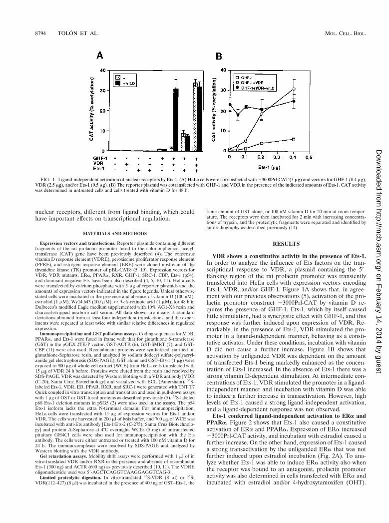
VDR shows a constitutive activity in the presence of Ets-1.In order to analyze the influence of Ets factors on the tran-scriptional response to VDR, a plasmid containing the 59-flanking region of the rat prolactin promoter was transientlytransfected into HeLa cells with expression vectors encodingEts-1, VDR, and/or GHF-1. Figure 1A shows that, in agree-ment with our previous observations (5), activation of the pro-lactin promoter construct 23000Prl-CAT by vitamin D re-quires the presence of GHF-1. Ets-1, which by itself causedlittle stimulation, had a synergistic effect with GHF-1, and thisresponse was further induced upon expression of VDR. Re-markably, in the presence of Ets-1, VDR stimulated the pro-moter in a ligand-independent manner, behaving as a consti-tutive activator. Under these conditions, incubation with vitaminD did not cause a further increase. Figure 1B shows thatactivation by unliganded VDR was dependent on the amountof transfected Ets-1 being markedly enhanced as the concen-tration of Ets-1 increased. In the absence of Ets-1 there was astrong vitamin D-dependent stimulation. At intermediate con-centrations of Ets-1, VDR stimulated the promoter in a ligand-independent manner and incubation with vitamin D was ableto induce a further increase in transactivation. However, highlevels of Ets-1 caused a strong ligand-independent activation,and a ligand-dependent response was not observed.
Ets-1 conferred ligand-independent activation to ERa andPPARa. Figure 2 shows that Ets-1 also caused a constitutiveactivation of ERa and PPARa. Expression of ERa increased23000Prl-CAT activity, and incubation with estradiol caused afurther increase. On the other hand, expression of Ets-1 causeda strong transactivation by the unliganded ERa that was notfurther induced upon estradiol incubation (Fig. 2A). To ana-lyze whether Ets-1 was able to induce ERa activity also whenthe receptor was bound to an antagonist, prolactin promoteractivity was also determined in cells transfected with ERa andincubated with estradiol and/or 4-hydroxytamoxifen (OHT).
FIG. 1. Ligand-independent activation of nuclear receptors by Ets-1. (A) HeLa cells were cotransfected with 23000Prl-CAT (5 mg) and vectors for GHF-1 (0.4 mg),VDR (2.5 mg), and/or Ets-1 (0.5 mg). (B) The reporter plasmid was cotransfected with GHF-1 and VDR in the presence of the indicated amounts of Ets-1. CAT activitywas determined in untreated cells and cells treated with vitamin D for 48 h.
8794 TOLON ET AL. MOL. CELL. BIOL.
on February 14, 2014 by guest
http://mcb.asm
.org/D
ownloaded from
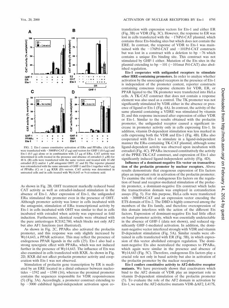
As shown in Fig. 2B, OHT treatment markedly reduced basalCAT activity as well as estradiol-induced stimulation in theabsence of Ets-1. After expression of Ets-1, the unligandedERa stimulated the promoter even in the presence of OHT.Although promoter activity was lower in cells incubated withthe antagonist, stimulation of ERa transcriptional activity byEts-1 in cells incubated with OHT was similar to that in cellsincubated with estradiol when activity was expressed as foldinduction. Furthermore, identical results were obtained withthe pure antiestrogen ICI182.780, which blocks not only AF2but also AF1 functions (not illustrated).
As shown in Fig. 2C, PPARa also activated the prolactinpromoter, and this response was only slightly increased byWy14,643, a PPAR activator. This may reflect the presence ofendogenous PPAR ligands in the cells (25). Ets-1 also had astrong synergistic effect with PPARa, which was not inducedfurther in the presence of Wy14,643. The influence of Ets wasalso examined in cells transfected with RXR. As shown in Fig.2D, RXR did not affect prolactin promoter activity and coop-eration with Ets-1 was not observed.
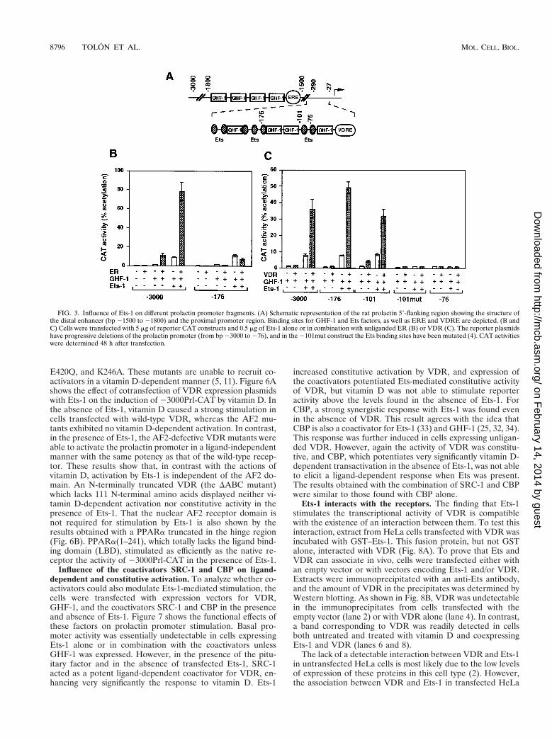
Stimulation of prolactin gene transcription by ER is medi-ated by an ERE located in a distal enhancer between nucleo-tides 21592 and 21580 (16), whereas the proximal promotercontains the sequences responsible for VDR responsiveness(5) (Fig. 3A). Accordingly, a promoter construct extending tobp 23000 exhibited ligand-independent activation upon co-
transfection with expression vectors for Ets-1 and either ER(Fig. 3B) or VDR (Fig. 3C). However, the response to ER waslost in cells transfected with the 2176Prl-CAT plasmid, whichcontains three Ets-binding sites but which does not contain theERE. In contrast, the response of VDR to Ets-1 was main-tained with the 2176Prl-CAT and 2101Prl-CAT constructsbut was lost in a construct with a deletion to bp 276 whichcontains a unique Ets binding site. This construct was notstimulated by GHF-1 either. Mutation of the Ets sites in theplasmid extending to bp 2101 (2101mut Prl-CAT) also abol-ished regulation.
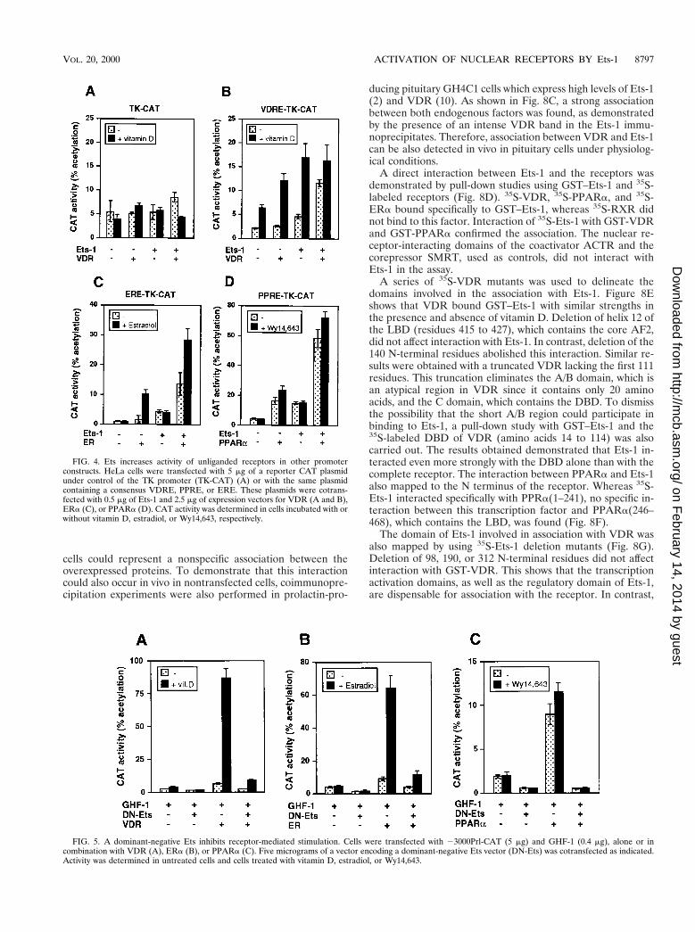
Ets-1 cooperates with unliganded receptors to stimulateother HRE-containing promoters. In order to analyze whetheractivation by the unoccupied receptors in the presence of Ets-1is independent of the promoter context, reporter constructscontaining consensus response elements for VDR, ER, orPPAR ligated to the TK promoter were transfected into HeLacells. A TK-CAT construct that does not contain a responseelement was also used as a control. The TK promoter was notsignificantly stimulated by VDR either in the absence or pres-ence of ligand or Ets-1 (Fig. 4A). In contrast, the activity of thesame plasmid containing a VDRE was stimulated by vitaminD, and this response increased after expression of either VDRor Ets-1. Similar to the results obtained with the prolactinpromoter, the unliganded receptor caused a significant in-crease in promoter activity only in cells expressing Ets-1. Inaddition, vitamin D-dependent stimulation was less marked incells expressing both the VDR and Ets-1 (Fig. 4B). ERa alsocooperated with Ets-1 to stimulate in a ligand-independentmanner the ERa-containing TK-CAT plasmid, although someligand-dependent activity was observed upon incubation withestradiol (Fig. 4C). PPARa increased constitutively the activityof the PPRE-TK-CAT construct, and expression of Ets-1 alsosignificantly induced ligand-independent activity (Fig. 4D).
Influence of a dominant-negative Ets vector on transactiva-tion of the prolactin promoter by nuclear receptors. Aboveresults demonstrate that exogenous expression of Ets factorsplays an important role in activation of the prolactin promoter.To examine the role of endogenous Ets factors on the regula-tion of basal and receptor-mediated stimulation of the prolac-tin promoter, a dominant-negative Ets construct which lacksthe transactivation domain was employed in cotransfectionassays (Fig. 5). For this purpose, HeLa cells were transfectedwith 23000Prl-CAT and an expression vector encoding theETS domain of Ets-2. The DBD is highly conserved among themembers of the Ets family, and therefore overexpression ofthis domain interferes with the action of the different Etsfactors. Expression of dominant-negative Ets had little effecton basal promoter activity, which was essentially undetectablein the absence of GHF-1 (data not shown), and reduced sig-nificantly GHF-1-mediated activation. In addition, the domi-nant-negative vector interfered strongly with VDR and vitaminD-dependent stimulation (Fig. 5A). Similar results were ob-tained in cells transfected with ER (Fig. 5B), in which expres-sion of this vector abolished estrogen regulation. The domi-nant-negative Ets also neutralized the responses to PPARa,which again were similar in the presence and absence ofWy14,643 (Fig. 5C). Therefore, endogenous Ets factors play acrucial role not only in basal activity but also in activation ofthe prolactin promoter by the nuclear receptors.
Ets-1 confers constitutive activity to AF2-defective receptormutants. We have previously shown that coactivators whichbind to the AF2 domain of VDR play an important role invitamin D-dependent stimulation of the prolactin promoter(5). To evaluate the role of the AF2 domain in activation byEts-1, we used the AF2-defective mutants VDR-DAF2, L417S,
FIG. 2. Ets-1 causes constitutive activation of ERa and PPARa. (A) Cellswere transfected with 23000Prl-CAT (5 mg) and vectors for GHF-1 (0.4 mg) andEts-1 (0.5 mg) alone or in combination with 2.5 mg of ERa. CAT activity wasdetermined in cells treated in the presence and absence of estradiol (1 mM) for48 h. (B) cells were transfected with the same vectors and treated with 10 nMestradiol (E2) and/or 1 mM antagonist OHT. (C and D) The reporter plasmidwas cotransfected with the same amounts of GHF-1 and Ets-1 together with 5 mgof PPARa (C) or 1 mg RXR (D) vectors. CAT activity was determined inuntreated cells and in cells treated with Wy14,643 or 9-cis-retinoic acid.
VOL. 20, 2000 ACTIVATION OF NUCLEAR RECEPTORS BY Ets-1 8795
on February 14, 2014 by guest
http://mcb.asm
.org/D
ownloaded from
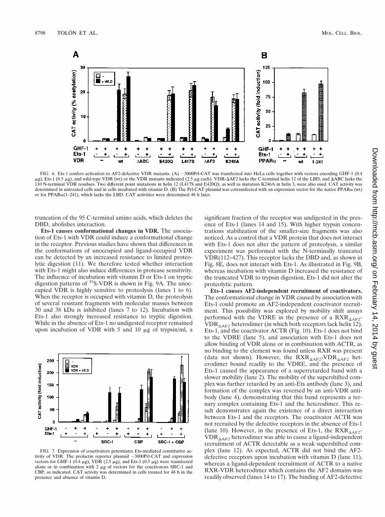
E420Q, and K246A. These mutants are unable to recruit co-activators in a vitamin D-dependent manner (5, 11). Figure 6Ashows the effect of cotransfection of VDR expression plasmidswith Ets-1 on the induction of 23000Prl-CAT by vitamin D. Inthe absence of Ets-1, vitamin D caused a strong stimulation incells transfected with wild-type VDR, whereas the AF2 mu-tants exhibited no vitamin D-dependent activation. In contrast,in the presence of Ets-1, the AF2-defective VDR mutants wereable to activate the prolactin promoter in a ligand-independentmanner with the same potency as that of the wild-type recep-tor. These results show that, in contrast with the actions ofvitamin D, activation by Ets-1 is independent of the AF2 do-main. An N-terminally truncated VDR (the DABC mutant)which lacks 111 N-terminal amino acids displayed neither vi-tamin D-dependent activation nor constitutive activity in thepresence of Ets-1. That the nuclear AF2 receptor domain isnot required for stimulation by Ets-1 is also shown by theresults obtained with a PPARa truncated in the hinge region(Fig. 6B). PPARa(1–241), which totally lacks the ligand bind-ing domain (LBD), stimulated as efficiently as the native re-ceptor the activity of 23000Prl-CAT in the presence of Ets-1.
Influence of the coactivators SRC-1 and CBP on ligand-dependent and constitutive activation. To analyze whether co-activators could also modulate Ets-1-mediated stimulation, thecells were transfected with expression vectors for VDR,GHF-1, and the coactivators SRC-1 and CBP in the presenceand absence of Ets-1. Figure 7 shows the functional effects ofthese factors on prolactin promoter stimulation. Basal pro-moter activity was essentially undetectable in cells expressingEts-1 alone or in combination with the coactivators unlessGHF-1 was expressed. However, in the presence of the pitu-itary factor and in the absence of transfected Ets-1, SRC-1acted as a potent ligand-dependent coactivator for VDR, en-hancing very significantly the response to vitamin D. Ets-1
increased constitutive activation by VDR, and expression ofthe coactivators potentiated Ets-mediated constitutive activityof VDR, but vitamin D was not able to stimulate reporteractivity above the levels found in the absence of Ets-1. ForCBP, a strong synergistic response with Ets-1 was found evenin the absence of VDR. This result agrees with the idea thatCBP is also a coactivator for Ets-1 (33) and GHF-1 (25, 32, 34).This response was further induced in cells expressing unligan-ded VDR. However, again the activity of VDR was constitu-tive, and CBP, which potentiates very significantly vitamin D-dependent transactivation in the absence of Ets-1, was not ableto elicit a ligand-dependent response when Ets was present.The results obtained with the combination of SRC-1 and CBPwere similar to those found with CBP alone.
Ets-1 interacts with the receptors. The finding that Ets-1stimulates the transcriptional activity of VDR is compatiblewith the existence of an interaction between them. To test thisinteraction, extract from HeLa cells transfected with VDR wasincubated with GST–Ets-1. This fusion protein, but not GSTalone, interacted with VDR (Fig. 8A). To prove that Ets andVDR can associate in vivo, cells were transfected either withan empty vector or with vectors encoding Ets-1 and/or VDR.Extracts were immunoprecipitated with an anti-Ets antibody,and the amount of VDR in the precipitates was determined byWestern blotting. As shown in Fig. 8B, VDR was undetectablein the immunoprecipitates from cells transfected with theempty vector (lane 2) or with VDR alone (lane 4). In contrast,a band corresponding to VDR was readily detected in cellsboth untreated and treated with vitamin D and coexpressingEts-1 and VDR (lanes 6 and 8).
The lack of a detectable interaction between VDR and Ets-1in untransfected HeLa cells is most likely due to the low levelsof expression of these proteins in this cell type (2). However,the association between VDR and Ets-1 in transfected HeLa
FIG. 3. Influence of Ets-1 on different prolactin promoter fragments. (A) Schematic representation of the rat prolactin 59-flanking region showing the structure ofthe distal enhancer (bp 21500 to 21800) and the proximal promoter region. Binding sites for GHF-1 and Ets factors, as well as ERE and VDRE are depicted. (B andC) Cells were transfected with 5 mg of reporter CAT constructs and 0.5 mg of Ets-1 alone or in combination with unliganded ER (B) or VDR (C). The reporter plasmidshave progressive deletions of the prolactin promoter (from bp 23000 to 276), and in the 2101mut construct the Ets binding sites have been mutated (4). CAT activitieswere determined 48 h after transfection.
8796 TOLON ET AL. MOL. CELL. BIOL.
on February 14, 2014 by guest
http://mcb.asm
.org/D
ownloaded from
cells could represent a nonspecific association between theoverexpressed proteins. To demonstrate that this interactioncould also occur in vivo in nontransfected cells, coimmunopre-cipitation experiments were also performed in prolactin-pro-
ducing pituitary GH4C1 cells which express high levels of Ets-1(2) and VDR (10). As shown in Fig. 8C, a strong associationbetween both endogenous factors was found, as demonstratedby the presence of an intense VDR band in the Ets-1 immu-noprecipitates. Therefore, association between VDR and Ets-1can be also detected in vivo in pituitary cells under physiolog-ical conditions.
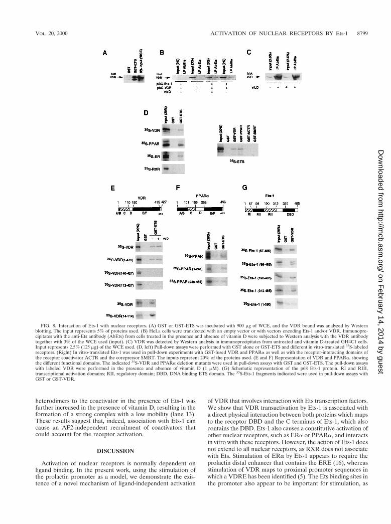
A direct interaction between Ets-1 and the receptors wasdemonstrated by pull-down studies using GST–Ets-1 and 35S-labeled receptors (Fig. 8D). 35S-VDR, 35S-PPARa, and 35S-ERa bound specifically to GST–Ets-1, whereas 35S-RXR didnot bind to this factor. Interaction of 35S-Ets-1 with GST-VDRand GST-PPARa confirmed the association. The nuclear re-ceptor-interacting domains of the coactivator ACTR and thecorepressor SMRT, used as controls, did not interact withEts-1 in the assay.
A series of 35S-VDR mutants was used to delineate thedomains involved in the association with Ets-1. Figure 8Eshows that VDR bound GST–Ets-1 with similar strengths inthe presence and absence of vitamin D. Deletion of helix 12 ofthe LBD (residues 415 to 427), which contains the core AF2,did not affect interaction with Ets-1. In contrast, deletion of the140 N-terminal residues abolished this interaction. Similar re-sults were obtained with a truncated VDR lacking the first 111residues. This truncation eliminates the A/B domain, which isan atypical region in VDR since it contains only 20 aminoacids, and the C domain, which contains the DBD. To dismissthe possibility that the short A/B region could participate inbinding to Ets-1, a pull-down study with GST–Ets-1 and the35S-labeled DBD of VDR (amino acids 14 to 114) was alsocarried out. The results obtained demonstrated that Ets-1 in-teracted even more strongly with the DBD alone than with thecomplete receptor. The interaction between PPARa and Ets-1also mapped to the N terminus of the receptor. Whereas 35S-Ets-1 interacted specifically with PPRa(1–241), no specific in-teraction between this transcription factor and PPARa(246–468), which contains the LBD, was found (Fig. 8F).
The domain of Ets-1 involved in association with VDR wasalso mapped by using 35S-Ets-1 deletion mutants (Fig. 8G).Deletion of 98, 190, or 312 N-terminal residues did not affectinteraction with GST-VDR. This shows that the transcriptionactivation domains, as well as the regulatory domain of Ets-1,are dispensable for association with the receptor. In contrast,
FIG. 4. Ets increases activity of unliganded receptors in other promoterconstructs. HeLa cells were transfected with 5 mg of a reporter CAT plasmidunder control of the TK promoter (TK-CAT) (A) or with the same plasmidcontaining a consensus VDRE, PPRE, or ERE. These plasmids were cotrans-fected with 0.5 mg of Ets-1 and 2.5 mg of expression vectors for VDR (A and B),ERa (C), or PPARa (D). CAT activity was determined in cells incubated with orwithout vitamin D, estradiol, or Wy14,643, respectively.
FIG. 5. A dominant-negative Ets inhibits receptor-mediated stimulation. Cells were transfected with 23000Prl-CAT (5 mg) and GHF-1 (0.4 mg), alone or incombination with VDR (A), ERa (B), or PPARa (C). Five micrograms of a vector encoding a dominant-negative Ets vector (DN-Ets) was cotransfected as indicated.Activity was determined in untreated cells and cells treated with vitamin D, estradiol, or Wy14,643.
VOL. 20, 2000 ACTIVATION OF NUCLEAR RECEPTORS BY Ets-1 8797
on February 14, 2014 by guest
http://mcb.asm
.org/D
ownloaded from
truncation of the 95 C-terminal amino acids, which deletes theDBD, abolishes interaction.
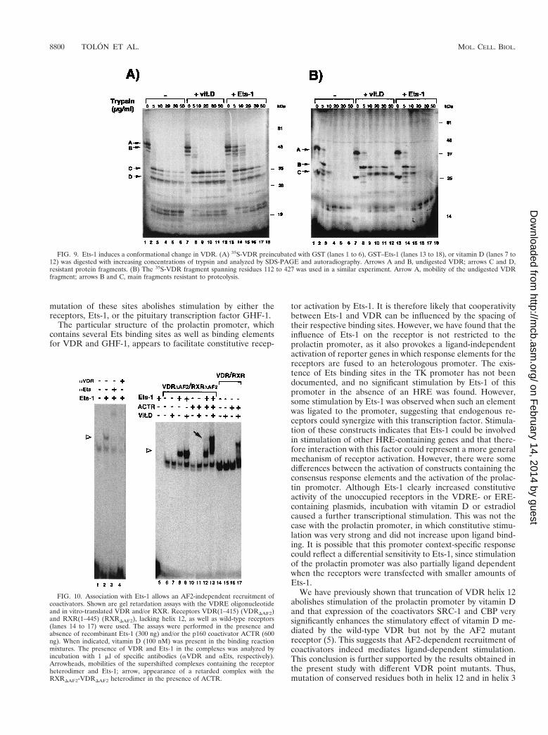
Ets-1 causes conformational changes in VDR. The associa-tion of Ets-1 with VDR could induce a conformational changein the receptor. Previous studies have shown that differences inthe conformations of unoccupied and ligand-occupied VDRcan be detected by an increased resistance to limited proteo-lytic digestion (11). We therefore tested whether interactionwith Ets-1 might also induce differences in protease sensitivity.The influence of incubation with vitamin D or Ets-1 on trypticdigestion patterns of 35S-VDR is shown in Fig. 9A. The unoc-cupied VDR is highly sensitive to proteolysis (lanes 1 to 6).When the receptor is occupied with vitamin D, the proteolysisof several resistant fragments with molecular masses between30 and 38 kDa is inhibited (lanes 7 to 12). Incubation withEts-1 also strongly increased resistance to tryptic digestion.While in the absence of Ets-1 no undigested receptor remainedupon incubation of VDR with 5 and 10 mg of trypsin/ml, a
significant fraction of the receptor was undigested in the pres-ence of Ets-1 (lanes 14 and 15). With higher trypsin concen-trations stabilization of the smaller-size fragments was alsonoticed. As a control that a VDR protein that does not interactwith Ets-1 does not alter the pattern of proteolysis, a similarexperiment was performed with the N-terminally truncatedVDR(112–427). This receptor lacks the DBD and, as shown inFig. 8E, does not interact with Ets-1. As illustrated in Fig. 9B,whereas incubation with vitamin D increased the resistance ofthe truncated VDR to trypsin digestion, Ets-1 did not alter theproteolytic pattern.
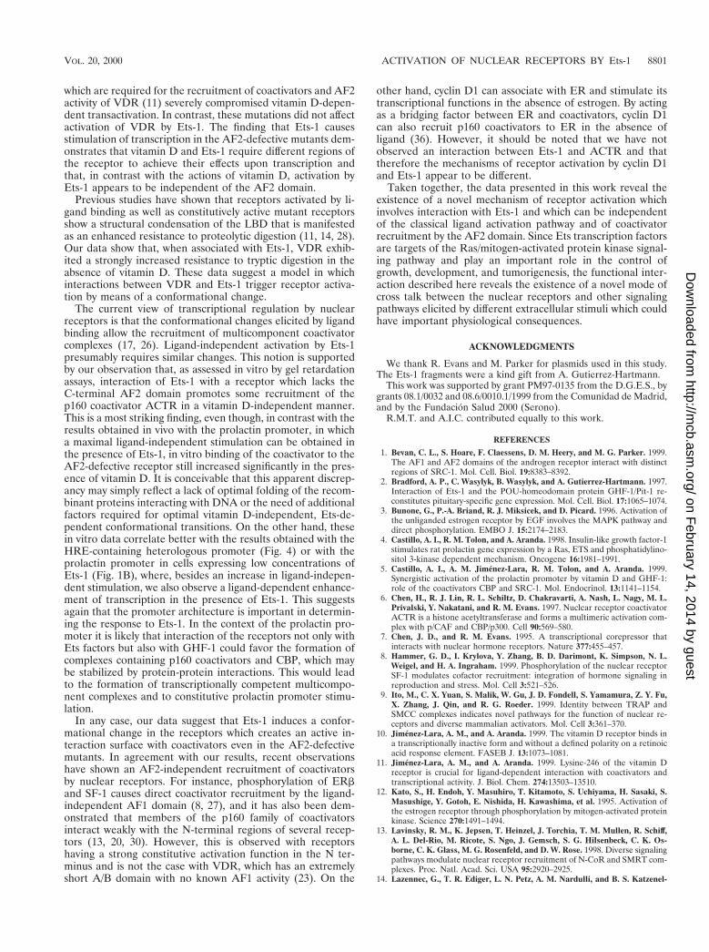
Ets-1 causes AF2-independent recruitment of coactivators.The conformational change in VDR caused by association withEts-1 could promote an AF2-independent coactivator recruit-ment. This possibility was explored by mobility shift assaysperformed with the VDRE in the presence of a RXRDAF2-VDRDAF2 heterodimer (in which both receptors lack helix 12),Ets-1, and the coactivator ACTR (Fig. 10). Ets-1 does not bindto the VDRE (lane 5), and association with Ets-1 does notallow binding of VDR alone or in combination with ACTR, asno binding to the element was found unless RXR was present(data not shown). However, the RXRDAF2-VDRDAF2 het-erodimer bound readily to the VDRE, and the presence ofEts-1 caused the appearance of a superretarded band with aslower mobility (lane 2). The mobility of the supershifted com-plex was further retarded by an anti-Ets antibody (lane 3), andformation of the complex was reversed by an anti-VDR anti-body (lane 4), demonstrating that this band represents a ter-nary complex containing Ets-1 and the heterodimer. This re-sult demonstrates again the existence of a direct interactionbetween Ets-1 and the receptors. The coactivator ACTR wasnot recruited by the defective receptors in the absence of Ets-1(lane 10). However, in the presence of Ets-1, the RXRDAF2-VDRDAF2 heterodimer was able to cause a ligand-independentrecruitment of ACTR detectable as a weak supershifted com-plex (lane 12). As expected, ACTR did not bind the AF2-defective receptors upon incubation with vitamin D (lane 11),whereas a ligand-dependent recruitment of ACTR to a nativeRXR-VDR heterodimer which contains the AF2 domains wasreadily observed (lanes 14 to 17). The binding of AF2-defective
FIG. 6. Ets-1 confers activation to AF2-defective VDR mutants. (A) 23000Prl-CAT was transfected into HeLa cells together with vectors encoding GHF-1 (0.4mg), Ets-1 (0.5 mg), and wild-type VDR (wt) or the VDR mutants indicated (2.5 mg each). VDR-DAF2 lacks the C-terminal helix 12 of the LBD, and DABC lacks the110 N-terminal VDR residues. Two different point mutations in helix 12 (L417S and E420Q), as well as mutation K246A in helix 3, were also used. CAT activity wasdetermined in untreated cells and in cells incubated with vitamin D. (B) The Prl-CAT plasmid was cotransfected with an expression vector for the native PPARa (wt)or for PPARa(1–241), which lacks the LBD. CAT activities were determined 48 h later.
FIG. 7. Expression of coactivators potentiates Ets-mediated constitutive ac-tivity of VDR. The prolactin reporter plasmid 23000Prl-CAT and expressionvectors for GHF-1 (0.4 mg), VDR (2.5 mg), and Ets-1 (0.5 mg) were transfectedalone or in combination with 2 mg of vectors for the coactivators SRC-1 andCBP, as indicated. CAT activity was determined in cells treated for 48 h in thepresence and absence of vitamin D.
8798 TOLON ET AL. MOL. CELL. BIOL.
on February 14, 2014 by guest
http://mcb.asm
.org/D
ownloaded from
heterodimers to the coactivator in the presence of Ets-1 wasfurther increased in the presence of vitamin D, resulting in theformation of a strong complex with a low mobility (lane 13).These results suggest that, indeed, association with Ets-1 cancause an AF2-independent recruitment of coactivators thatcould account for the receptor activation.
DISCUSSION
Activation of nuclear receptors is normally dependent onligand binding. In the present work, using the stimulation ofthe prolactin promoter as a model, we demonstrate the exis-tence of a novel mechanism of ligand-independent activation
of VDR that involves interaction with Ets transcription factors.We show that VDR transactivation by Ets-1 is associated witha direct physical interaction between both proteins which mapsto the receptor DBD and the C terminus of Ets-1, which alsocontains the DBD. Ets-1 also causes a constitutive activation ofother nuclear receptors, such as ERa or PPARa, and interactsin vitro with these receptors. However, the action of Ets-1 doesnot extend to all nuclear receptors, as RXR does not associatewith Ets. Stimulation of ERa by Ets-1 appears to require theprolactin distal enhancer that contains the ERE (16), whereasstimulation of VDR maps to proximal promoter sequences inwhich a VDRE has been identified (5). The Ets binding sites inthe promoter also appear to be important for stimulation, as
FIG. 8. Interaction of Ets-1 with nuclear receptors. (A) GST or GST-ETS was incubated with 900 mg of WCE, and the VDR bound was analyzed by Westernblotting. The input represents 5% of proteins used. (B) HeLa cells were transfected with an empty vector or with vectors encoding Ets-1 and/or VDR. Immunopre-cipitates with the anti-Ets antibody (AbEts) from cells treated in the presence and absence of vitamin D were subjected to Western analysis with the VDR antibodytogether with 3% of the WCE used (input). (C) VDR was detected by Western analysis in immunoprecipitates from untreated and vitamin D-treated GH4C1 cells.Input represents 2.5% (125 mg) of the WCE used. (D, left) Pull-down assays were performed with GST alone or GST-ETS and different in vitro-translated 35S-labeledreceptors. (Right) In vitro-translated Ets-1 was used in pull-down experiments with GST-fused VDR and PPARa as well as with the receptor-interacting domains ofthe receptor coactivator ACTR and the corepressor SMRT. The inputs represent 20% of the proteins used. (E and F) Representation of VDR and PPARa, showingthe different functional domains. The indicated 35S-VDR and PPARa deletion mutants were used in pull-down assays with GST and GST-ETS. The pull-down assayswith labeled VDR were performed in the presence and absence of vitamin D (1 mM). (G) Schematic representation of the p68 Ets-1 protein. RI and RIII,transcriptional activation domains; RII, regulatory domain; DBD, DNA binding ETS domain. The 35S-Ets-1 fragments indicated were used in pull-down assays withGST or GST-VDR.
VOL. 20, 2000 ACTIVATION OF NUCLEAR RECEPTORS BY Ets-1 8799
on February 14, 2014 by guest
http://mcb.asm
.org/D
ownloaded from
mutation of these sites abolishes stimulation by either thereceptors, Ets-1, or the pituitary transcription factor GHF-1.
The particular structure of the prolactin promoter, whichcontains several Ets binding sites as well as binding elementsfor VDR and GHF-1, appears to facilitate constitutive recep-
tor activation by Ets-1. It is therefore likely that cooperativitybetween Ets-1 and VDR can be influenced by the spacing oftheir respective binding sites. However, we have found that theinfluence of Ets-1 on the receptor is not restricted to theprolactin promoter, as it also provokes a ligand-independentactivation of reporter genes in which response elements for thereceptors are fused to an heterologous promoter. The exis-tence of Ets binding sites in the TK promoter has not beendocumented, and no significant stimulation by Ets-1 of thispromoter in the absence of an HRE was found. However,some stimulation by Ets-1 was observed when such an elementwas ligated to the promoter, suggesting that endogenous re-ceptors could synergize with this transcription factor. Stimula-tion of these constructs indicates that Ets-1 could be involvedin stimulation of other HRE-containing genes and that there-fore interaction with this factor could represent a more generalmechanism of receptor activation. However, there were somedifferences between the activation of constructs containing theconsensus response elements and the activation of the prolac-tin promoter. Although Ets-1 clearly increased constitutiveactivity of the unoccupied receptors in the VDRE- or ERE-containing plasmids, incubation with vitamin D or estradiolcaused a further transcriptional stimulation. This was not thecase with the prolactin promoter, in which constitutive stimu-lation was very strong and did not increase upon ligand bind-ing. It is possible that this promoter context-specific responsecould reflect a differential sensitivity to Ets-1, since stimulationof the prolactin promoter was also partially ligand dependentwhen the receptors were transfected with smaller amounts ofEts-1.
We have previously shown that truncation of VDR helix 12abolishes stimulation of the prolactin promoter by vitamin Dand that expression of the coactivators SRC-1 and CBP verysignificantly enhances the stimulatory effect of vitamin D me-diated by the wild-type VDR but not by the AF2 mutantreceptor (5). This suggests that AF2-dependent recruitment ofcoactivators indeed mediates ligand-dependent stimulation.This conclusion is further supported by the results obtained inthe present study with different VDR point mutants. Thus,mutation of conserved residues both in helix 12 and in helix 3
FIG. 9. Ets-1 induces a conformational change in VDR. (A) 35S-VDR preincubated with GST (lanes 1 to 6), GST–Ets-1 (lanes 13 to 18), or vitamin D (lanes 7 to12) was digested with increasing concentrations of trypsin and analyzed by SDS-PAGE and autoradiography. Arrows A and B, undigested VDR; arrows C and D,resistant protein fragments. (B) The 35S-VDR fragment spanning residues 112 to 427 was used in a similar experiment. Arrow A, mobility of the undigested VDRfragment; arrows B and C, main fragments resistant to proteolysis.
FIG. 10. Association with Ets-1 allows an AF2-independent recruitment ofcoactivators. Shown are gel retardation assays with the VDRE oligonucleotideand in vitro-translated VDR and/or RXR. Receptors VDR(1–415) (VDRDAF2)and RXR(1–445) (RXRDAF2), lacking helix 12, as well as wild-type receptors(lanes 14 to 17) were used. The assays were performed in the presence andabsence of recombinant Ets-1 (300 ng) and/or the p160 coactivator ACTR (600ng). When indicated, vitamin D (100 nM) was present in the binding reactionmixtures. The presence of VDR and Ets-1 in the complexes was analyzed byincubation with 1 ml of specific antibodies (aVDR and aEts, respectively).Arrowheads, mobilities of the supershifted complexes containing the receptorheterodimer and Ets-1; arrow, appearance of a retarded complex with theRXRDAF2-VDRDAF2 heterodimer in the presence of ACTR.
8800 TOLON ET AL. MOL. CELL. BIOL.
on February 14, 2014 by guest
http://mcb.asm
.org/D
ownloaded from
which are required for the recruitment of coactivators and AF2activity of VDR (11) severely compromised vitamin D-depen-dent transactivation. In contrast, these mutations did not affectactivation of VDR by Ets-1. The finding that Ets-1 causesstimulation of transcription in the AF2-defective mutants dem-onstrates that vitamin D and Ets-1 require different regions ofthe receptor to achieve their effects upon transcription andthat, in contrast with the actions of vitamin D, activation byEts-1 appears to be independent of the AF2 domain.
Previous studies have shown that receptors activated by li-gand binding as well as constitutively active mutant receptorsshow a structural condensation of the LBD that is manifestedas an enhanced resistance to proteolytic digestion (11, 14, 28).Our data show that, when associated with Ets-1, VDR exhib-ited a strongly increased resistance to tryptic digestion in theabsence of vitamin D. These data suggest a model in whichinteractions between VDR and Ets-1 trigger receptor activa-tion by means of a conformational change.
The current view of transcriptional regulation by nuclearreceptors is that the conformational changes elicited by ligandbinding allow the recruitment of multicomponent coactivatorcomplexes (17, 26). Ligand-independent activation by Ets-1presumably requires similar changes. This notion is supportedby our observation that, as assessed in vitro by gel retardationassays, interaction of Ets-1 with a receptor which lacks theC-terminal AF2 domain promotes some recruitment of thep160 coactivator ACTR in a vitamin D-independent manner.This is a most striking finding, even though, in contrast with theresults obtained in vivo with the prolactin promoter, in whicha maximal ligand-independent stimulation can be obtained inthe presence of Ets-1, in vitro binding of the coactivator to theAF2-defective receptor still increased significantly in the pres-ence of vitamin D. It is conceivable that this apparent discrep-ancy may simply reflect a lack of optimal folding of the recom-binant proteins interacting with DNA or the need of additionalfactors required for optimal vitamin D-independent, Ets-de-pendent conformational transitions. On the other hand, thesein vitro data correlate better with the results obtained with theHRE-containing heterologous promoter (Fig. 4) or with theprolactin promoter in cells expressing low concentrations ofEts-1 (Fig. 1B), where, besides an increase in ligand-indepen-dent stimulation, we also observe a ligand-dependent enhance-ment of transcription in the presence of Ets-1. This suggestsagain that the promoter architecture is important in determin-ing the response to Ets-1. In the context of the prolactin pro-moter it is likely that interaction of the receptors not only withEts factors but also with GHF-1 could favor the formation ofcomplexes containing p160 coactivators and CBP, which maybe stabilized by protein-protein interactions. This would leadto the formation of transcriptionally competent multicompo-nent complexes and to constitutive prolactin promoter stimu-lation.
In any case, our data suggest that Ets-1 induces a confor-mational change in the receptors which creates an active in-teraction surface with coactivators even in the AF2-defectivemutants. In agreement with our results, recent observationshave shown an AF2-independent recruitment of coactivatorsby nuclear receptors. For instance, phosphorylation of ERband SF-1 causes direct coactivator recruitment by the ligand-independent AF1 domain (8, 27), and it has also been dem-onstrated that members of the p160 family of coactivatorsinteract weakly with the N-terminal regions of several recep-tors (13, 20, 30). However, this is observed with receptorshaving a strong constitutive activation function in the N ter-minus and is not the case with VDR, which has an extremelyshort A/B domain with no known AF1 activity (23). On the
other hand, cyclin D1 can associate with ER and stimulate itstranscriptional functions in the absence of estrogen. By actingas a bridging factor between ER and coactivators, cyclin D1can also recruit p160 coactivators to ER in the absence ofligand (36). However, it should be noted that we have notobserved an interaction between Ets-1 and ACTR and thattherefore the mechanisms of receptor activation by cyclin D1and Ets-1 appear to be different.
Taken together, the data presented in this work reveal theexistence of a novel mechanism of receptor activation whichinvolves interaction with Ets-1 and which can be independentof the classical ligand activation pathway and of coactivatorrecruitment by the AF2 domain. Since Ets transcription factorsare targets of the Ras/mitogen-activated protein kinase signal-ing pathway and play an important role in the control ofgrowth, development, and tumorigenesis, the functional inter-action described here reveals the existence of a novel mode ofcross talk between the nuclear receptors and other signalingpathways elicited by different extracellular stimuli which couldhave important physiological consequences.
ACKNOWLEDGMENTS
We thank R. Evans and M. Parker for plasmids used in this study.The Ets-1 fragments were a kind gift from A. Gutierrez-Hartmann.
This work was supported by grant PM97-0135 from the D.G.E.S., bygrants 08.1/0032 and 08.6/0010.1/1999 from the Comunidad de Madrid,and by the Fundacion Salud 2000 (Serono).
R.M.T. and A.I.C. contributed equally to this work.
REFERENCES
1. Bevan, C. L., S. Hoare, F. Claessens, D. M. Heery, and M. G. Parker. 1999.The AF1 and AF2 domains of the androgen receptor interact with distinctregions of SRC-1. Mol. Cell. Biol. 19:8383–8392.
2. Bradford, A. P., C. Wasylyk, B. Wasylyk, and A. Gutierrez-Hartmann. 1997.Interaction of Ets-1 and the POU-homeodomain protein GHF-1/Pit-1 re-constitutes pituitary-specific gene expression. Mol. Cell. Biol. 17:1065–1074.
3. Bunone, G., P.-A. Briand, R. J. Miksicek, and D. Picard. 1996. Activation ofthe unliganded estrogen receptor by EGF involves the MAPK pathway anddirect phosphorylation. EMBO J. 15:2174–2183.
4. Castillo, A. I., R. M. Tolon, and A. Aranda. 1998. Insulin-like growth factor-1stimulates rat prolactin gene expression by a Ras, ETS and phosphatidylino-sitol 3-kinase dependent mechanism. Oncogene 16:1981–1991.
5. Castillo, A. I., A. M. Jimenez-Lara, R. M. Tolon, and A. Aranda. 1999.Synergistic activation of the prolactin promoter by vitamin D and GHF-1:role of the coactivators CBP and SRC-1. Mol. Endocrinol. 13:1141–1154.
6. Chen, H., R. J. Lin, R. L. Schiltz, D. Chakravarti, A. Nash, L. Nagy, M. L.Privalski, Y. Nakatani, and R. M. Evans. 1997. Nuclear receptor coactivatorACTR is a histone acetyltransferase and forms a multimeric activation com-plex with p/CAF and CBP/p300. Cell 90:569–580.
7. Chen, J. D., and R. M. Evans. 1995. A transcriptional corepressor thatinteracts with nuclear hormone receptors. Nature 377:455–457.
8. Hammer, G. D., I. Krylova, Y. Zhang, B. D. Darimont, K. Simpson, N. L.Weigel, and H. A. Ingraham. 1999. Phosphorylation of the nuclear receptorSF-1 modulates cofactor recruitment: integration of hormone signaling inreproduction and stress. Mol. Cell 3:521–526.
9. Ito, M., C. X. Yuan, S. Malik, W. Gu, J. D. Fondell, S. Yamamura, Z. Y. Fu,X. Zhang, J. Qin, and R. G. Roeder. 1999. Identity between TRAP andSMCC complexes indicates novel pathways for the function of nuclear re-ceptors and diverse mammalian activators. Mol. Cell 3:361–370.
10. Jimenez-Lara, A. M., and A. Aranda. 1999. The vitamin D receptor binds ina transcriptionally inactive form and without a defined polarity on a retinoicacid response element. FASEB J. 13:1073–1081.
11. Jimenez-Lara, A. M., and A. Aranda. 1999. Lysine-246 of the vitamin Dreceptor is crucial for ligand-dependent interaction with coactivators andtranscriptional activity. J. Biol. Chem. 274:13503–13510.
12. Kato, S., H. Endoh, Y. Masuhiro, T. Kitamoto, S. Uchiyama, H. Sasaki, S.Masushige, Y. Gotoh, E. Nishida, H. Kawashima, et al. 1995. Activation ofthe estrogen receptor through phosphorylation by mitogen-activated proteinkinase. Science 270:1491–1494.
13. Lavinsky, R. M., K. Jepsen, T. Heinzel, J. Torchia, T. M. Mullen, R. Schiff,A. L. Del-Rio, M. Ricote, S. Ngo, J. Gemsch, S. G. Hilsenbeck, C. K. Os-borne, C. K. Glass, M. G. Rosenfeld, and D. W. Rose. 1998. Diverse signalingpathways modulate nuclear receptor recruitment of N-CoR and SMRT com-plexes. Proc. Natl. Acad. Sci. USA 95:2920–2925.
14. Lazennec, G., T. R. Ediger, L. N. Petz, A. M. Nardulli, and B. S. Katzenel-
VOL. 20, 2000 ACTIVATION OF NUCLEAR RECEPTORS BY Ets-1 8801
on February 14, 2014 by guest
http://mcb.asm
.org/D
ownloaded from
lenbogen. 1997. Mechanistic aspects of estrogen receptor activation probedwith constitutively active estrogen receptors: correlations with DNA andcoregulator interactions and receptor conformational changes. Mol. Endo-crinol. 11:1375–1386.
15. Mangelsdorf, D. J., C. Thummel, M. Beato, P. Herrlich, G. Schutz, K.Umesono, B. Blumberg, P. Kastner, M. Mark, P. Chambon, and R. M.Evans. 1995. The nuclear receptor superfamily: the second decade. Cell83:835–839.
16. Maurer, R. A., and A. C. Notides. 1987. Identification of an estrogen-respon-sive element from the 59-flanking region of the rat prolactin gene. Mol. Cell.Biol. 7:4247–4254.
17. McKenna, N. J., R. B. Lanz, and B. W. O’Malley. 1999. Nuclear receptorcoregulators: cellular and molecular biology. Endocr. Rev. 20:321–344.
18. McMahon, C., T. Suthiphongchai, J. DiRenzo, and M. E. Ewen. 1999. P/CAFassociates with cyclin D1 and potentiates its activation of the estrogen re-ceptor. Proc. Natl. Acad. Sci. USA 96:5382–5387.
19. Oberste-Berghaus, C., K. Zanger, K. Hashimoto, R. N. Cohen, A. N. Hol-lenberg, and F. E. Wondisdorf. 2000. Thyroid hormone-independent inter-action between the thyroid hormone receptor beta2 amino terminus andcoactivators. J. Biol. Chem. 275:1787–1792.
20. Onate, S. A., V. Boonyaratanakorkit, T. E. Spencer, S. Y. Tsai, M.-J. Tsai,D. P. Edwards, and B. W. O’Malley. 1998. The steroid receptor coactivator-1contains multiple receptor interacting and activation domains that cooper-atively enhance the activation function 1 (AF1) and AF2 domains of steroidreceptor. J. Biol. Chem. 273:12101–12108.
21. Rachez, C., B. D. Lemon, Z. Suldan, V. Bromleigh, M. Gamble, A. M. Naar,H. Erdjument-Bromage, P. Tempst, and L. P. Freedman. 1999. Ligand-dependent transcription activation by nuclear receptors requires the DRIPcomplex. Nature 398:824–828.
22. Rochette-Egly, C., S. Adam, M. Rossignol, J.-M. Egly, and P. Chambon.1997. Stimulation of RARa activation function AF-1 through binding to thegeneral transcription factor TFIIH and phosphorylation by CDK7. Cell 90:97–107.
23. Sone, T., S. Kerner, and J. W. Pike. 1991. Vitamin D receptor interactionwith specific DNA: association as a 1,24-hydroxyvitamin D3-modulated het-erodimer. J. Biol. Chem. 266:23296–23305.
24. Taneja, R., C. Rochette-Egly, J. L. Plassat, L. Penna, M. P. Gaub, and P.Chambon. 1997. Phosphorylation of activation functions AF-1 and AF-2 ofRARa and RARg is indispensable for differentiation of F9 cells upon reti-noic acid and cAMP treatment. EMBO J. 16:6452–6455.
25. Tolon, R. M., A. I. Castillo, and A. Aranda. 1998. Activation of the prolactingene by peroxisome proliferator activated receptor-a appears to be DNAbinding-independent. J. Biol. Chem. 273:26652–26661.
26. Torchia, J., C. K. Glass, and M. G. Rosenfeld. 1998. Co-activators andco-repressors in the integration of transcriptional responses. Curr. Opin. CellBiol. 10:373–383.
27. Tremblay, A., G. B. Tremblay, F. Labrie, and V. Guiguere. 1999. Ligand-independent recruitment of SRC-1 to estrogen receptor b through phos-phorylation of activation function AF-1. Mol. Cell 3:513–519.
28. Vivat, V., C. Zechel, J. M. Wurtz, W. Bourguet, H. Kagechika, H. Umemiya,K. Shudo, D. Moras, H. Gronemeyer, and P. Chambon. 1997. A mutationmimicking ligand-induced conformational change yields a constitutive RXRthat senses allosteric effects in heterodimers. EMBO J. 15:5697–5709.
29. Wasylyk, B., J. Hagman, and A. Gutierrez-Hartmann. 1998. Ets transcriptionfactors: nuclear effectors of the Ras-MAP-kinase signaling pathway. TrendsBiochem. Sci. 23:213–216.
30. Webb, P., P. Neguyen, J. Sinshako, C. Anderson, W. Feng, M. P. Nguyen, D.Chen, S.-M. Huang, S. Subramanian, E. McKinerney, B. Katzenellenbogen,M. R. Stallcup, and P. J. Kushner. 1998. Estrogen receptor activation func-tion 1 works by binding p160 coactivator proteins. Mol. Endocrinol. 12:1605–1618.
31. Weigel, N. L., and Y. Zhang. 1998. Ligand-independent activation of steroidhormone receptors. J. Mol. Med. 76:469–479.
32. Xu, L., R. M. Lavinsky, J. S. Dasen, S. E. Flyn, E. M. McInerney, T.-M.Mullen, T. Heinzel, D. Szeto, E. Korzus, R. Kurokawal, A. K. Aggarwal,D. W. Rose, C. K. Glass, and M. G. Rosenfeld. 1998. Signal-specific co-activator domain requirements for Pit-1 activation. Nature 395:301–306.
33. Yang, C., L. H. Shapiro, M. Ribera, A. Kumar, and P. K. Brindle. 1998. Arole for CREB binding protein and p300 transcriptional coactivators in Ets-1transactivation functions. Mol. Cell. Biol. 18:2218–2229.
34. Zanger, K., L. E. Cohen, K. Hashimoto, S. Radovick, and F. E. Wondisdorf.1999. A novel mechanism for cyclic adenosine 39,59-monophosphate regula-tion of gene expression by CREB-binding protein. Mol. Endocrinol. 13:268–275.
35. Zwijsen, R. M. L., E. Wientjens, R. Klompmaker, J. van der Sman, R.Bernards, and R. Michalides. 1997. CDK-independent activation of estrogenreceptor by cyclin D1. Cell 88:405–415.
36. Zwijsen, R. M. L., R. S. Buckle, E. M. Hijmans, C. J. M. Loomans, and R.Bernards. 1998. Ligand-independent recruitment of steroid receptor coac-tivators to estrogen receptor by cyclin D1. Genes Dev. 12:3488–3498.
8802 TOLON ET AL. MOL. CELL. BIOL.
on February 14, 2014 by guest
http://mcb.asm
.org/D
ownloaded from
Related Documents