Br. J. Cancer (1991), 63, 136 142 Association of INT2/HSTI coamplification in primary breast cancer with hormone-dependent phenotype and poor prognosis A. Borg', H. Sigurdsson, G.M. Clark2, M. Fern6, S.A.W. Fuqua2, H. Olsson, D. Killanderl & W.L. McGurie2 'Department of Oncology, University Hospital, S-221 85 Lund, Sweden; 2Department of Oncology/Medicine, University of Texas Health Science Center at San Antonio, San Antonio, Texas 78284-7884, USA. Summary The human proto-oncogene INT2 (homologous to the mouse INT2 gene, implicated in proviral induced mammary carcinoma) has been mapped to chromosome q 13 and found to share band localisation with, among others, the HSTJ proto-oncogene. Both genes are members of the fibroblast growth factor family. In the present study, coamplification (2-15 copies) of the INT2/HSTJ genes was found in 27 (9%) of 311 invasive human breast carcinomas using slot blot and Southern blot analyses. Amplification was not correlated to tumour size, axillary lymph node status or stage of disease, neither to patient age nor menopausal status. However, 26 (96%) of the 27 amplified tumours were, often strongly, Oestrogen receptor positive compared to 65% of the unamplified cases (P = 0.001). These findings are in sharp contrast to the strong correlations of HER-2/neu proto-oncogene amplification with advanced stage and steroid receptor negativity, previously observed in the same series of tumours. Patients with INT2/HSTI amplified breast cancer had a significantly shorter disease-free survival compared to those with unamplified genes (P = 0.015, median follow up 45 months). This correlation was confined to node-negative patients and persisted in multivariate analysis. No significant correlation to survival from breast cancer was found. It is concluded that amplification of the II q 13 region in breast cancer occurs in a particular subset of aggressive tumours, quite different from that identified by HER-2/neu amplification. It still remains to be shown that the selection for amplified genes at I 1q13 is due to the activity of INT2, HSTJ or yet another, still unidentified, neighbouring gene. However, the results are potentially of clinical value in separating a group of node-negative breast cancer for more intense treatment. The putative proto-oncogene INT2 is known as one of several integration sites for mouse mammary tumour virus (MMTV), a retrovirus implicated in mammary tumouri- genesis in certain strains of mice (Nusse, 1988a). INT2 encodes a predicted member of the fibroblast growth factor (FGF) family of potent mitogens or morphogens involved in angiogenesis, tissue induction and cell migration (Dickson & Peters, 1987; Thomas, 1988; Burgess, 1988). The human INT2 gene has been cloned, found to be 89% homologous to the mouse INT2, and mapped to chromosome 11q13 (Brookes et al., 1989; Casey et al., 1986). Interestingly, another proto-oncogene, HSTJ (HSTFI), was localised to the same chromosomal site and also found to be a FGF member (Adelaide et al., 1988, Yoshida et al., 1987, 1988a). HSTI was initially detected as a transforming gene in DNA from human stomach cancer (Sakamoto et al., 1986), found virtually identical to the KS oncogene from Kaposis sarcoma (Delli Bovi et al., 1987), and recently also identified as an alternative integration site for MMTV in mouse mammary tumours (Peters et al., 1989). INT2 and HST1 are closely linked in the mouse genome (Yoshida et al., 1988b) and only 35 kilobasepairs apart in the same transcriptional orientation in the human genome (Wada et al., 1988). This suggests that they originated through duplication of a common ancestral gene during evolution, and that this region may involve still other, yet unknown, related genes. Amplification of the 1 1q13 region have been reported from various solid tumours including, besides breast cancer, squa- mous cell carcinomas (Zhou et al., 1988, Berenson et al., 1989), a stomach cancer and the vulvar carcinoma cell line A431 (Yoshida et al., 1988a), melanomas (Adelaide et al., 1988, Theillet et al., 1989), bladder and Oesophageal carcin- omas (Tsutsumi et al., 1988; Tsuda et al., 1988; Theillet et al., 1989), and a hepatocellular carcinoma (Hatada et al., 1988). It usually entails the INT2 and HSTJ genes and also the BCLI locus, recognised as a chromosomal breakpoint in B-cell leukaemia (Tsujimoto et al., 1984), but not other genes located at the same or neighbouring bands (Ali et al., 1989). Multiple endocrine neoplasia type I (MEN-I), the patho- genesis of which seems to involve a putative FGF-member, have also been linked to a locus proximal to the INT2 gene at I Iql2-q13 (Nakamura et al., 1989; Bale et al., 1989). Consequently, although no evidence yet exists, the plain fact that both selective amplifications and non-random transloca- tion encompass the same chromosomal region, points to its importance in the development of human cancer. In breast cancer, amplification of the INT2 and HSTI genes have been found in 9-23% (Zhou et al., 1988, Lide- reau et al., 1988; Varley et al., 1988; Tsuda et al., 1989; Theillet et al., 1989; Adnane et al., 1989; Fantl et al., 1989). When clinical follow-up was available, a correlation to poor survival was noted. In the present study we report on INT2/ HSTI coamplification in a particular subset of human breast cancer with hormone-dependent phenotype, and on the cor- relation to disease outcome among low-risk patient cate- gories. Materials and methods Patients and tumour material Patients were all from the southern Sweden health care region, diagnosed for breast disease during the time interval of October 1982 and February 1985 and had a tumour sent for steroid receptor analysis. Tumours used for the present study were consecutive cases with a tissue amount allowing gene analysis, representing about 25% of all new cases of breast disease diagnosed during this time period. Cases ineligible for the study (e.g. benign disease, cancer in situ or samples from metatases) were excluded, as were tumours judged to be too cell-poor after cytopathological examination of tissue imprints. Patients presenting with bilateral cancer were not excluded if it was clear which primary tumour recurred. Of the remaining 311 tumours, 27% were classified accord- ing to UICC as Stage I, 36% as Stage IIa, 24% as Stage IIb, 7% as Stage III, and 7% as Stage IV (distant spread at diagnosis or within 2 months after primary operation). Ten Correspondence: A. Borg. Received 22 March 1990; and in revised form 28 August 1990. Br. J. Cancer (1991), 63, 136-142 17" Macmillan Press Ltd., 1991

Welcome message from author
This document is posted to help you gain knowledge. Please leave a comment to let me know what you think about it! Share it to your friends and learn new things together.
Transcript
Br. J. Cancer (1991), 63, 136 142
Association of INT2/HSTI coamplification in primary breast cancer withhormone-dependent phenotype and poor prognosis
A. Borg', H. Sigurdsson, G.M. Clark2, M. Fern6, S.A.W. Fuqua2, H. Olsson, D. Killanderl &W.L. McGurie2
'Department of Oncology, University Hospital, S-221 85 Lund, Sweden; 2Department of Oncology/Medicine, University of TexasHealth Science Center at San Antonio, San Antonio, Texas 78284-7884, USA.
Summary The human proto-oncogene INT2 (homologous to the mouse INT2 gene, implicated in proviralinduced mammary carcinoma) has been mapped to chromosome q 13 and found to share band localisationwith, among others, the HSTJ proto-oncogene. Both genes are members of the fibroblast growth factorfamily. In the present study, coamplification (2-15 copies) of the INT2/HSTJ genes was found in 27 (9%) of311 invasive human breast carcinomas using slot blot and Southern blot analyses. Amplification was notcorrelated to tumour size, axillary lymph node status or stage of disease, neither to patient age normenopausal status. However, 26 (96%) of the 27 amplified tumours were, often strongly, Oestrogen receptorpositive compared to 65% of the unamplified cases (P = 0.001). These findings are in sharp contrast to thestrong correlations of HER-2/neu proto-oncogene amplification with advanced stage and steroid receptornegativity, previously observed in the same series of tumours. Patients with INT2/HSTI amplified breastcancer had a significantly shorter disease-free survival compared to those with unamplified genes (P = 0.015,median follow up 45 months). This correlation was confined to node-negative patients and persisted inmultivariate analysis. No significant correlation to survival from breast cancer was found. It is concluded thatamplification of the II q 13 region in breast cancer occurs in a particular subset of aggressive tumours, quitedifferent from that identified by HER-2/neu amplification. It still remains to be shown that the selection foramplified genes at I 1q13 is due to the activity of INT2, HSTJ or yet another, still unidentified, neighbouringgene. However, the results are potentially of clinical value in separating a group of node-negative breast cancerfor more intense treatment.
The putative proto-oncogene INT2 is known as one ofseveral integration sites for mouse mammary tumour virus(MMTV), a retrovirus implicated in mammary tumouri-genesis in certain strains of mice (Nusse, 1988a). INT2encodes a predicted member of the fibroblast growth factor(FGF) family of potent mitogens or morphogens involved inangiogenesis, tissue induction and cell migration (Dickson &Peters, 1987; Thomas, 1988; Burgess, 1988). The humanINT2 gene has been cloned, found to be 89% homologous tothe mouse INT2, and mapped to chromosome 11q13(Brookes et al., 1989; Casey et al., 1986). Interestingly,another proto-oncogene, HSTJ (HSTFI), was localised tothe same chromosomal site and also found to be a FGFmember (Adelaide et al., 1988, Yoshida et al., 1987, 1988a).HSTI was initially detected as a transforming gene in DNAfrom human stomach cancer (Sakamoto et al., 1986), foundvirtually identical to the KS oncogene from Kaposis sarcoma(Delli Bovi et al., 1987), and recently also identified as analternative integration site for MMTV in mouse mammarytumours (Peters et al., 1989). INT2 and HST1 are closelylinked in the mouse genome (Yoshida et al., 1988b) and only35 kilobasepairs apart in the same transcriptional orientationin the human genome (Wada et al., 1988). This suggests thatthey originated through duplication of a common ancestralgene during evolution, and that this region may involve stillother, yet unknown, related genes.Amplification of the 1 1q13 region have been reported from
various solid tumours including, besides breast cancer, squa-mous cell carcinomas (Zhou et al., 1988, Berenson et al.,1989), a stomach cancer and the vulvar carcinoma cell lineA431 (Yoshida et al., 1988a), melanomas (Adelaide et al.,1988, Theillet et al., 1989), bladder and Oesophageal carcin-omas (Tsutsumi et al., 1988; Tsuda et al., 1988; Theillet etal., 1989), and a hepatocellular carcinoma (Hatada et al.,1988). It usually entails the INT2 and HSTJ genes and alsothe BCLI locus, recognised as a chromosomal breakpoint inB-cell leukaemia (Tsujimoto et al., 1984), but not other genes
located at the same or neighbouring bands (Ali et al., 1989).Multiple endocrine neoplasia type I (MEN-I), the patho-genesis of which seems to involve a putative FGF-member,have also been linked to a locus proximal to the INT2 geneat I Iql2-q13 (Nakamura et al., 1989; Bale et al., 1989).Consequently, although no evidence yet exists, the plain factthat both selective amplifications and non-random transloca-tion encompass the same chromosomal region, points to itsimportance in the development of human cancer.
In breast cancer, amplification of the INT2 and HSTIgenes have been found in 9-23% (Zhou et al., 1988, Lide-reau et al., 1988; Varley et al., 1988; Tsuda et al., 1989;Theillet et al., 1989; Adnane et al., 1989; Fantl et al., 1989).When clinical follow-up was available, a correlation to poorsurvival was noted. In the present study we report on INT2/HSTI coamplification in a particular subset of human breastcancer with hormone-dependent phenotype, and on the cor-relation to disease outcome among low-risk patient cate-gories.
Materials and methods
Patients and tumour material
Patients were all from the southern Sweden health care
region, diagnosed for breast disease during the time intervalof October 1982 and February 1985 and had a tumour sentfor steroid receptor analysis. Tumours used for the presentstudy were consecutive cases with a tissue amount allowinggene analysis, representing about 25% of all new cases ofbreast disease diagnosed during this time period. Casesineligible for the study (e.g. benign disease, cancer in situ orsamples from metatases) were excluded, as were tumoursjudged to be too cell-poor after cytopathological examinationof tissue imprints. Patients presenting with bilateral cancerwere not excluded if it was clear which primary tumourrecurred.Of the remaining 311 tumours, 27% were classified accord-
ing to UICC as Stage I, 36% as Stage IIa, 24% as Stage IIb,7% as Stage III, and 7% as Stage IV (distant spread atdiagnosis or within 2 months after primary operation). Ten
Correspondence: A. Borg.Received 22 March 1990; and in revised form 28 August 1990.
Br. J. Cancer (1991), 63, 136-142 17" Macmillan Press Ltd., 1991
INT2/HSTI COAMPLIFICATION IN HUMAN BREAST CANCER 137
patients not treated with axillary resection were unclassified.However, none of these had metastases at diagnosis. Therange of patient age at operation was 31-92 years (median63 years), 22% were premenopausal and 78% were post-menopausal. Adjuvant tamoxifen was given to 38% of thepatients, adjuvant chemotherapy (cyclosphosphamide) to 6%,whereas 45% received postoperative radiation (Sigurdsson etal., 1990). Recurrences were clinically confirmed and register-ed as loco-regional or distant. Deaths were distinguished asdue to breast cancer or to intercurrent disease. Distant recur-rences were found in 94 cases and loco-regional recurrencesin 11 cases. Of 115 deaths, 81 were due to breast cancer.Median follow-up for all patients was 46 months, for thosestill living 53 months, and for those still living or dead inintercurrent disease 51 months. Only distant recurrences wereconsidered in the calculation of distant disease-free survival,which also locoregional recurrences were included in disease-free survival. Death due to breast cancer was used as end-point in breast cancer survival and death due to other causeswere censored. Only patients with Stage I-III (MO) diseasewere included in the survival analyses.
Results
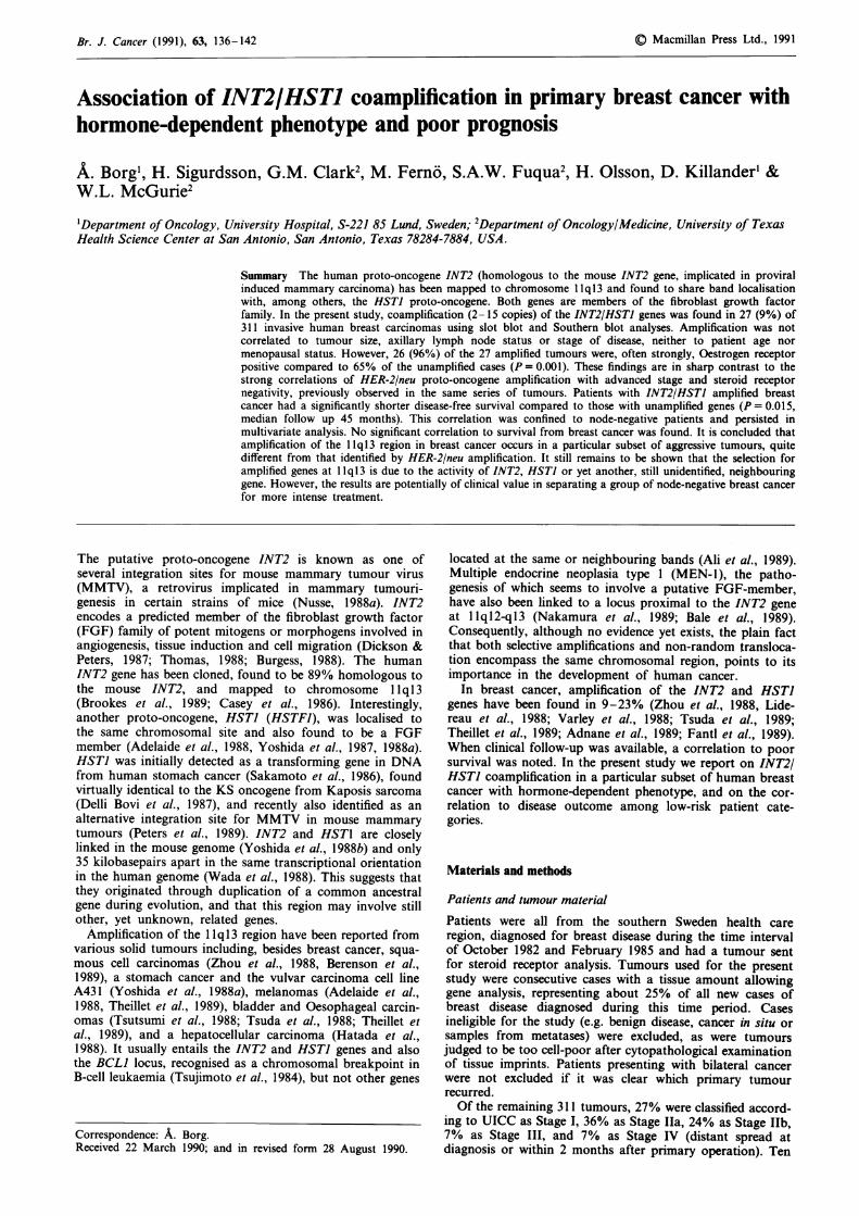
INT2/HSTI amplificationHybridisation of the INT2 SS6 probe to BamHI digestedDNA (Figure 1) revealed the known polymorphism of thislocus (two alleles; 8.4 and 5.6/2.8 kb fragments, respectively(Casey et al., 1986)) and the normal, approximately, 2:1distribution of alleles among studied tumours. Both alleleswere found affected by amplification but, in tumours heter-ozygous for the site, in no cases simultaneously. The HSTpORFI probe recognised four constant ECORI digestedDNA fragments (Figure 2). The three shortest fragments(5.8, 2.8 and 0.8 kb) harbour the HSTJ gene, while thelargest fragment (8.0 kb) represents binding to the HST2gene (Yoshida et al., 1988a). Amplification was found toexclusively affect the HSTJ gene.INT2 amplification was detected in 27 (9%) of 311 slot
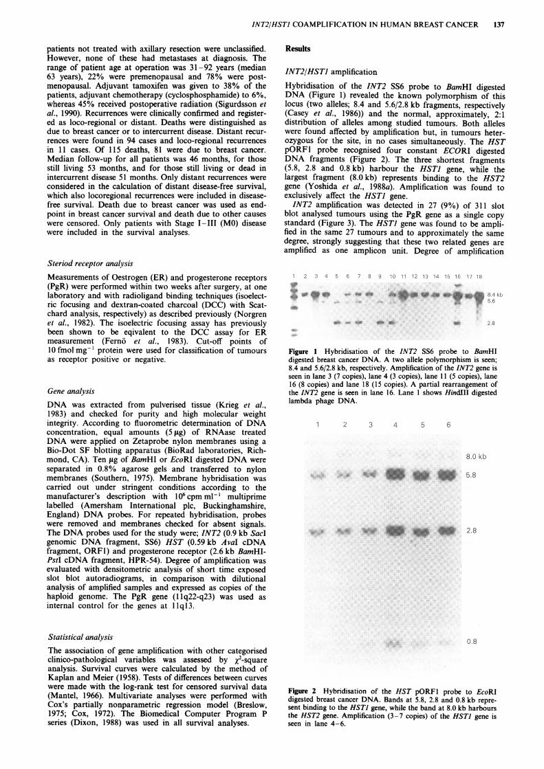
blot analysed tumours using the PgR gene as a single copystandard (Figure 3). The HSTI gene was found to be ampli-fied in the same 27 tumours and to approximately the samedegree, strongly suggesting that these two related genes areamplified as one amplicon unit. Degree of amplification
Steriod receptor analysis
Measurements of Oestrogen (ER) and progesterone receptors(PgR) were performed within two weeks after surgery, at onelaboratory and with radioligand binding techniques (isoelect-ric focusing and dextran-coated charcoal (DCC) with Scat-chard analysis, respectively) as described previously (Norgrenet al., 1982). The isoelectric focusing assay has previouslybeen shown to be eqivalent to the DCC assay for ERmeasurement (Ferno et al., 1983). Cut-off points of10 fmol mg-' protein were used for classification of tumoursas receptor positive or negative.
Gene analysisDNA was extracted from pulverised tissue (Krieg et al.,1983) and checked for purity and high molecular weightintegrity. According to fluorometric determination of DNAconcentration, equal amounts (5 gig) of RNAase treatedDNA were applied on Zetaprobe nylon membranes using aBio-Dot SF blotting apparatus (BioRad laboratories, Rich-mond, CA). Ten tig of BamHI or EcoRI digested DNA wereseparated in 0.8% agarose gels and transferred to nylonmembranes (Southern, 1975). Membrane hybridisation wascarried out under stringent conditions according to themanufacturer's description with 106cpm ml-' multiprimelabelled (Amersham International plc, Buckinghamshire,England) DNA probes. For repeated hybridisation, probeswere removed and membranes checked for absent signals.The DNA probes used for the study were; INT2 (0.9 kb Saclgenomic DNA fragment, SS6) HST (0.59 kb AvaI cDNAfragment, ORFI) and progesterone receptor (2.6 kb BamHI-PstI cDNA fragment, HPR-54). Degree of amplification wasevaluated with densitometric analysis of short time exposedslot blot autoradiograms, in comparison with dilutionalanalysis of amplified samples and expressed as copies of thehaploid genome. The PgR gene (1 lq22-q23) was used asinternal control for the genes at 1 1q13.
Statistical analysisThe association of gene amplification with other categorisedclinico-pathological variables was assessed by X2-squareanalysis. Survival curves were calculated by the method ofKaplan and Meier (1958). Tests of differences between curveswere made with the log-rank test for censored survival data(Mantel, 1966). Multivariate analyses were performed withCox's partially nonparametric regression model (Breslow,1975; Cox, 1972). The Biomedical Computer Program Pseries (Dixon, 1988) was used in all survival analyses.
2 3 4 5 6 7 8 9 10 11 12 13 14 15 16 17 18
Figure 1 Hybridisation of the INT2 SS6 probe to BamHIdigested breast cancer DNA. A two allele polymorphism is seen;8.4 and 5.6/2.8 kb, respectively. Amplification of the INT2 gene isseen in lane 3 (7 copies), lane 4 (3 copies), lane 11 (5 copies), lane16 (8 copies) and lane 18 (15 copies). A partial rearrangement ofthe INT2 gene is seen in lane 16. Lane I shows HindIII digestedlambda phage DNA.
1 2 3 4 5 6
8.0 kb
5.8
2.8
0.8
Figure 2 Hybridisation of the HST pORFl probe to EcoRIdigested breast cancer DNA. Bands at 5.8, 2.8 and 0.8 kb repre-sent binding to the HSTI gene, while the band at 8.0 kb harboursthe HST2 gene. Amplification (3-7 copies) of the HSTI gene isseen in lane 4 -6.
8.4 kb5.6
2.8
_
4

138 A. BORG et al.
INT2 PgR
7 Copies
3 Copies
8 Copi's
Figure 3 Hybridisation of the INT2 SS6 probe to a breastcancer DNA slot blot, and rehybridisation with a PgR HPR-54probe. INT2 copy numbers of amplified tumours are indicated.
ranged from 2-15 copies; 18 samples having 2-4 copies, 8samples 5-10 copies, and 1 sample > 10 (15) copies of thegenes.
INT2/HSTI amplification in relation to other prognosticfactors
INT2/HSTI amplification was not statistically correlatedwith axillary lymph node status. The tendency of gene ampli-fication being more prevalent in node positive tumours wascaused by the relatively high incidence of amplification in thenode positive subgroup with few involved nodes. No associa-tion with tumour size or stage of disease was seen, neither topatient age or menopausal status (Table I).
However, 26 (96%) of the 27 amplified tumours were ERpositive, compared with 65% of the unamplified cases(P = 0.001). The ER concentration in amplified tumours wasmost often of high levels (>200 fmol/mg-' protein, Figure4) and, furthermore, the single deviating sample was nottotally devoid of ER, but just below the cut-off value used toclassify tumours as ER positive. A similar trend, althoughnot significant, was observed with PgR status. Also shown inFigure 4 is the quite different pattern of ER concentrations inHER-2/neu amplified tumours. HER-2/neu amplification wasfound in 52 (17%) of the 311 cases (Borg et al., 1990). Threeof these were also amplified for the 1 1q13 region and, notice-ably, also those with the lowest ER concentration amongINT2/HSTI amplified cases.As not all patients were given postoperative radiation and
the same, or any, adjuvant therapy, a bias might be intro-duced in the calculation of survival differences. However,there was no distinction whatever between amplified andunamplified tumours in respect of therapy. Adjuvant tamox-ifen, adjuvant chemotherapy and postoperative radiationwere given to, respectively, 12 (44%), 2 (7.4%) and 12 (44%)of the 27 amplified tumours, and to, respectively, 106 (37%),18 (6.3%) and 127 (45%) of the 284 unamplified tumours.
INT2/HSTI amplification in relation to survival
INT2/HSTI amplification was found to be a significant pre-dictor of a shorter disease-free survival (DFS, P = 0.015)when analysing all MO patients (n = 291, Figure 5a). Therewas a trend towards a worse prognosis of tumours with a
Table I INT2/HSTJ amplification in relation to other categorisedprognostic factors
Variable Amplified/total (%) P-value
Node statusNegative 10/161 (6%)Positive 16/139 (12%) P= 0.10
No. of positive nodes1-3 12/72 (17%)4- 10 3/53 (6%)> 10 1/14 7%) P= 0.14
Tumour size< 2 cm 10/118 (8%)2-5 14/163 (9%)> 5 3/30 (10%) P = 0.96
Clinical stageI 3/80 (4%)Ila 12/108 (11%)Ilb 8/71 (11%)III 1/22 (5%)IV 2/20 (10%) P = 0.36
MenopausePre 6/68 (9%)Post 21/243 (9%) P= 0.96
ER<10 1/99 (1%)" 10 26/212 (12%) P= 0.001
PgR<10 8/123 (6%)" 10 18/171 (10%) P= 0.23
HER-2/neuSingle copy 23/258 (9%)Amplified 3/52 (6%) P=0.46
>.iooo4l80f
... '. '-t.....,-:77!
G ;. i...
'. .....
xl,--;. -: .:
II
S,1.
I. - 5i5
_ X
S * . ...
100 -*
Mdi'_n. k -
0
Figure 4 Oestrogen receptor concentrations in breast cancerscontaining a single and amplified copy number of the INT2/HSTI genes and in cases amplified for HER-2/neu gene. Medianvalues are indicated.
lowkr.
.
*
INT2/HSTJ COAMPLIFICATION IN HUMAN BREAST CANCER 139
a)a)
U1)cn
n
U0
0
Time to disease recurrence (Years)
80
m' 60
(n. 40Q
20-
0
P= 0.19
N Events INT2/HST1266 54 unampl. o25 8 ampl.
D 1 2 3 4 66 7Time to death (Years)
Figure 5 Relationship between INT2/HSTJ amplification anddisease-free survival a, or breast cancer survival b in a MOpatients (n = 291).
high copy number () 5 copies) compared to those with a lowdegree of amplification (6 recurrences in 8 cases compared to7 in 17 cases). Of a total of 13 recurrences in the 25 includedamplified tumours, 3 were of loco-regional type while 10 weredistant metastases. Thus, the calculation of amplification inrelation to distant DFS resulted in a less prominent associa-tion (P = 0.12). Neither did the prediction of breast cancersurvival (P = 0.19, Figure Sb) reach significance.The presence of axillary lymph node metastases is widely
considered as the most reliable risk factor in breast cancer,and it was natural to perform separate analyses of geneamplification in relation to survival in the node-negative andnode-positive patient subgroups. As revealed in Figure 6, thesignificance of INT2/HSTI amplification as a predictor ofDFS was totaly confined to the node-negative group (P =0.030, n = 160). No association was seen in node-positivepatients (P = 0.73, n = 120). The corresponding correlationsin node-negative patients to distant DFS (P = 0.092) orbreast cancer survival (P = 0.21) were, again, not statisticallysignificant.As INT2/HSTI amplification affected mainly ER positive
tumours, this category was analysed separately (Figure 7). Ahighly significant correlation (P = 0.002) was found to ashorter DFS in amplified ER positive tumours as comparedwith unamplified ER positive tumours. ER negative tumourshad an intermediate DFS pattern (Figure 7). In the ER
a) I , . . . P, >,,,.
60- 9 4 ampl. een ico
5a) P- 0.73 L
O 40- Node-positives8_0 N Events INT2/HST1
.105 47 unampl.-----20- 15 8 ampl.--
20
0 1 2 3 4 5 6 7Time to disease recurrence (Years)
Figure 6 Relationship between INT2/HSTJ amplification anddisease-free survival in node-negative and node-positive MObreast cancer patients.
100 S
. +,, P~~~~~P= 0.002
80- *
CDa)I 60- , 'ER- (N =90)U,0 n
40-
ER+N Events INT2/HST1
20- 177 43 unampl. 024 13 ampl. .
0.0 1 2 3 4 5 6 7
Time to disease recurrence (Years)
Figure 7 Relationship between INT2/HSTI amplification anddisease-free survival in ER positive MO breast cancer. Also shownis the disease-free survival for ER negative. MO breast cancer.
positive category, amplification was significantly or nearlysignificantly correlated also to distant DFS (P = 0.026) andbreast cancer survival (P = 0.070).To examine the significance of INT2/HSTI amplification
as a predictor of DFS in combination with other prognosticfactors, multivariate analyses (Table II) were performed onall MO patients (actually 264, since lymph node status andPgR status were unavailable in 10 and 16 cases, respectively)as well as on node-negative patients separately (n = 148, PgRstatus not available in 12 cases). When analysing all MOpatients, lymph node status was the single most powerfulpredictor of DFS, followed by PgR status and tumour size.INT2/HSTI amplification was retained in the model as anearly significant variable (P = 0.060) with a relative risk of1.7 for amplified tumours. In node-negative patients, PgRstatus and INT2/HSTI amplification were the only andapproximately equally significant independent variables (P =0.011 and P = 0.0 13, respectively), with a relative risk of 4.0(95% confidence interval 1.3-12) for amplified tumours. Itshould be pointed out that the number of tumours andevents in some analyses are small and that the results of thesemust be cautiously interpreted.
140 A. BORG et al.
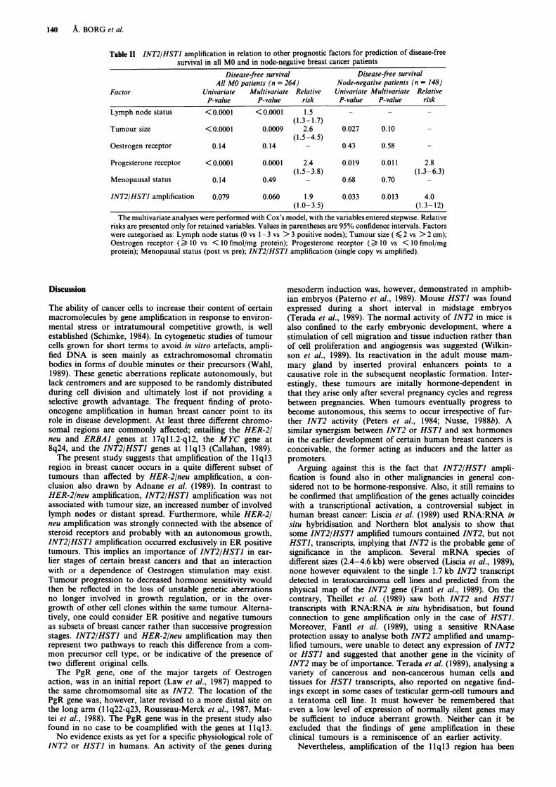
Table II INT2/HSTJ amplification in relation to other prognostic factors for prediction of disease-freesurvival in all MO and in node-negative breast cancer patients
Disease-free survival Disease-free survivalAll MO patients (n = 264) Node-negative patients (n = 148)
Factor Univariate Multivariate Relative Univariate Multivariate RelativeP-value P-value risk P-value P-value risk
Lymph node status < 0.0001 <0.0001 1.5 - -
(1.3- 1.7)Tumour size <0.0001 0.0009 2.6 0.027 0.10
(1.5-4.5)Oestrogen receptor 0.14 0.14 - 0.43 0.58
Progesterone receptor <0.0001 0.0001 2.4 0.019 0.011 2.8(1.5-3.8) (1.3-6.3)
Menopausal status 0.14 0.49 - 0.68 0.70 -
INT2/HSTJ amplification 0.079 0.060 1.9 0.033 0.013 4.0(1.0-3.5) (1.3- 12)
The multivariate analyses were performed with Cox's model, with the variables entered stepwise. Relativerisks are presented only for retained variables. Values in parentheses are 95% confidence intervals. Factorswere categorised as: Lymph node status (0 vs 1 - 3 vs > 3 positive nodes); Tumour size ( < 2 vs > 2 cm);Oestrogen receptor (> 10 vs <10 fmol/mg protein); Progesterone receptor (>10 vs <10 fmol/mgprotein); Menopausal status (post vs pre); INT2/HSTJ amplification (single copy vs amplified).
Discussion
The ability of cancer cells to increase their content of certainmacromolecules by gene amplification in response to environ-mental stress or intratumoural competitive growth, is wellestablished (Schimke, 1984). In cytogenetic studies of tumourcells grown for short terms to avoid in vitro artefacts, ampli-fied DNA is seen mainly as extrachromosomal chromatinbodies in forms of double minutes or their precursors (Wahl,1989). These genetic aberrations replicate autonomously, butlack centromers and are supposed to be randomly distributedduring cell division and ultimately lost if not providing aselective growth advantage. The frequent finding of proto-oncogene amplification in human breast cancer point to itsrole in disease development. At least three different chromo-somal regions are commonly affected; entailing the HER-2/neu and ERBAI genes at 17qll.2-ql2, the MYC gene at8q24, and the INT2/HSTI genes at 1lql3 (Callahan, 1989).The present study suggests that amplification of the 1 1q13
region in breast cancer occurs in a quite different subset oftumours than affected by HER-2/neu amplification, a con-
clusion also drawn by Adnane et al. (1989). In contrast toHER-2/neu amplification, INT2/HSTJ amplification was notassociated with tumour size, an increased number of involvedlymph nodes or distant spread. Furthermore, while HER-2/neu amplification was strongly connected with the absence ofsteroid receptors and probably with an autonomous growth,INT2/HSTI amplification occurred exclusively in ER positivetumours. This implies an importance of INT2/HSTI in ear-
lier stages of certain breast cancers and that an interactionwith or a dependence of Oestrogen stimulation may exist.Tumour progression to decreased hormone sensitivity wouldthen be reflected in the loss of unstable genetic aberrationsno longer involved in growth regulation, or in the over-growth of other cell clones within the same tumour. Alterna-tively, one could consider ER positive and negative tumoursas subsets of breast cancer rather than successive progressionstages. INT2/HSTI and HER-2/neu amplification may thenrepresent two pathways to reach this difference from a com-mon precursor cell type, or be indicative of the presence oftwo different original cells.The PgR gene, one of the major targets of Oestrogen
action, was in an initial report (Law et al., 1987) mapped tothe same chromomsomal site as INT2. The location of thePgR gene was, however, later revised to a more distal site onthe long arm (1 lq22-q23, Rousseau-Merck et al., 1987, Mat-tei et al., 1988). The PgR gene was in the present study alsofound in no case to be coamplified with the genes at 1q13.No evidence exists as yet for a specific physiological role of
INT2 or HSTJ in humans. An activity of the genes during
mesoderm induction was, however, demonstrated in amphib-ian embryos (Paterno et al., 1989). Mouse HSTJ was foundexpressed during a short interval in midstage embryos(Terada et al., 1989). The normal activity of INT2 in mice isalso confined to the early embryonic development, where astimulation of cell migration and tissue induction rather thanof cell proliferation and angiogensis was suggested (Wilkin-son et al., 1989). Its reactivation in the adult mouse mam-mary gland by inserted proviral enhancers points to acausative role in the subsequent neoplastic formation. Inter-estingly, these tumours are initally hormone-dependent inthat they arise only after several pregnancy cycles and regressbetween pregnancies. When tumours eventually progress tobecome autonomous, this seems to occur irrespective of fur-ther INT2 activity (Peters et al., 1984; Nusse, 1988b). Asimilar synergism between INT2 or HSTI and sex hormonesin the earlier development of certain human breast cancers isconceivable, the former acting as inducers and the latter aspromoters.
Arguing against this is the fact that INT2/HSTI ampli-fication is found also in other malignancies in general con-sidered not to be hormone-responsive. Also, it still remains tobe confirmed that amplification of the genes actually coincideswith a transcriptional activation, a controversial subject inhuman breast cancer: Liscia et al. (1989) used RNA:RNA insitu hybridisation and Northern blot analysis to show thatsome INT2/HSTJ amplified tumours contained INT2, but notHSTI, transcripts, implying that INT2 is the probable gene ofsignificance in the amplicon. Several mRNA species ofdifferent sizes (2.4-4.6 kb) were observed (Liscia et al., 1989),none however equivalent to the single 1.7 kb INT2 transcriptdetected in teratocarcinoma cell lines and predicted from thephysical map of the INT2 gene (Fantl et al., 1989). On thecontrary, Theillet et al. (1989) saw both INT2 and HSTItranscripts with RNA:RNA in situ hybridisation, but foundconnection to gene amplification only in the case of HSTI.Moreover, Fantl et al. (1989), using a sensitive RNAaseprotection assay to analyse both INT2 amplified and unamp-lified tumours, were unable to detect any expression of INT2or HSTI and suggested that another gene in the vicinity ofINT2 may be of importance. Terada et al. (1989), analysing avariety of cancerous and non-cancerous human cells andtissues for HSTI transcripts, also reported on negative find-ings except in some cases of testicular germ-cell tumours anda teratoma cell line. It must however be remembered thateven a low level of expression of normally silent genes maybe sufficient to induce aberrant growth. Neither can it beexcluded that the findings of gene amplification in theseclinical tumours is a reminiscence of an earlier activity.
Nevertheless, amplification of the l1ql3 region has been
INT2/HSTI COAMPLIFICATION IN HUMAN BREAST CANCER 141
shown to be associated with a poor clinical outcome (Lider-eau et al., 1988; Zhou et al., 1988; Tsuda et al., 1989). Aprognostic value of gene amplification in prediction ofdisease-free survival was confirmed in the present study,found to persist in multivariate analysis and to be confined tonode-negative patients. A subset of these latter patients, agroup in general considered as being of good prognosis,could be separated and shown to have a disease-free survivalas bad as node-positive patients. At this median time of 46months follow-up, the correlations had not yet translatedinto survival differences. As the number of cases and relapseevents in the node-negative group are small, the results mustbe critically interpreted. However, if shown to be valid infuture investigations, amplification of this chromosomalregion may become an important prognostic factor and use-ful in selection of node-negative patients for adjuvant
therapy. Also, an increased knowledge of the genes at 1 1q13will most certainly contribute to a deeper understanding ofhuman breast cancer etiology.This paper has been produced in collaboration with the SouthernSwedish Breast Cancer Study Group. The work was supported inpart by the Fru Berta Kamprads Foundation, the John and AugustaPerssons Foundation, the Swedish Cancer Society, the Gunnar Nil-sson Cancer Research Trust Fund, NIH Grant CA 30195, and ACSGrant IN-1161.We are indepted to Clive Dickson and Gordon Peters (Imperial
Cancer Research Fund Laboratories, London, UK) for the INT2probe, to Masaaki Terada (National Cancer Center Research Insti-tute Tokyo, Japan) for the HST probe, to Mark R. Hughes and BertW.O. O'Malley (Baylor College of Medicine, Houston, Texas) forthe PgR probe, and to Mrs Eva Henriksson (Department of Onco-logy, Lund, Sweden) for illustration preparation and Dr Ingrid Idvallfor tissue imprint examination.
References
ADELAIDE, J., MATTEI, M.-G., MARICS, I. & 4 others (1988). Chro-mosomal localization of the hst oncogene and its co-amplificationwith the int.2 oncogene in a human melanoma. Oncogene, 2, 413.
ADNANE, J., GAUDRAY, P., SIMON, M.-P., SIMONY-LAFONTAINE,J., JEANTEUR, P. & THEILLET, C. (1989). Proto-oncogene ampli-fication and human breast cancer phenotype. Oncogene, 4, 1389.
ALI, I.U., MERLO, G., CALLAHAN, R. & LIDEREAU, R. (1989). Theamplification unit on chromosome 1 q 13 in aggressive primaryhuman breast tumour entails the bel-i, int-2 and hst loci. Onco-gene, 4, 89.
BALE, S.J., BALE, A.E., STEWART, K. & 9 others (1989). Linkageanalysis of multiple endocrine neoplasia type 1 with INT2 andother markers on chromosome 11. Genomics, 4, 320.
BERENSON, J.R., YANG, J. & MICKEL, R.A. (1989). Frequent ampli-fication of the bel-1 locus in head and neck squamous cell car-cinomas. Oncogene, 4, 1111.
BORG, A., TANDON, A.K., SIGURDSSON, H. & 5 others (1990). HER-2/neu amplification predicts poor survival in node-positive breastcancer. Cancer Res., 50, 4332.
BRESLOW, N.E. (1975). Analysis of survival data under the propor-tional hazards model. Int. Stat. Rev., 43, 45.
BROOKES, S., SMITH, R., CASEY, G., DICKSON, C. & PETERS, G.(1989). Sequence organisation of the human int-2 gene and itsexpression in teratocarcinoma cells. Oncogene, 4, 429.
BURGESS, A.W. (1988). Int-l and int-2: oncogenic proteins, mitogensand morphogens? BioEssays, 8, 40.
CALLAHAN, R. (1989). Genetic alterations in primary breast cancer.Breast Cancer Res. Treat., 13, 191.
CASEY, G., SMITH, R., McGILLIVRAY, D., PETERS, G. & DICKSON,C. (1986). Characterisation and chromosome assignment of thehuman homolog of int-2, a potential proto-oncogene. Mol. Cell.Biol., 6, 502.
COX, D.R. (1972). Regression models and life-tables. J. Roy. Stat.Soc. (B), 34, 187.
DELLI BOVI, P., CURATOLA, A.M., KERN, F.G., GRECO, A., ITFr-MANN, M. & BASILICO, C. (1987). An oncogene isolated bytransfection of Kapsosi's sarcoma DNA encodes a growth factorthat is a member of the FGF family, Cell, 50, 729.
DICKSON, C. & PETERS, G. (1987). Potential oncogene productrelated to growth factors. Nature, 326, 833.
DIXON, W.J. (1988). BMDP statistical software. Berkeley, California:University of California Press.
FANTL, V., BROOKES, S., SMITH, R. & 5 others (1989). Characterisa-tion of the proto-oncogene int-2 and its potential for the diag-nosis of human breast cancers. In Cancer Cells 7, Furth M &Greaves, M. (eds) p 283. Cold Spring Harbor Press: New York.
FERNO, M., BORG, A. & NORGREN, A. (1983). A comparison of twosteroid receptor assays in breast cancer: dextran coated charcoaland isoelectric focusing. Anticancer Res., 3, 243.
HATADA, I., TOKINO, T., OCHIYA, T. & MATSUBARA, K. (1988).Co-amplification of integrated hepatitis B virus DNA and trans-forming gene hst-J in a hepatocellular carcinoma. Oncogene, 3,537.
KAPLAN, E.L. & MEIER, P. (1958). Nonparametric estimation fromincomplete observations. J. Am. Stat. Assoc., 53, 457.
KRIEG, P., AMTMANN, E. & SAUER, G. (1983). The simultaneousextraction of high molecular weight DNA and of RNA fromsolid tumours. Anal. Biochem., 134, 288.
LAW, M.L., KAO, F.T., WEI, Q. & 8 others (1987). The progesteronereceptor gene maps to human chromosome band 11q13, the siteof the mammary oncogene int-2. Proc. Natl. Acad. Sci. USA, 84,2877.
LIDEREAU, R., CALLAHAN, R., DICKSON, C., PETERS, G., ESCOT, C.& ALI, I.U. (1988). Amplification of the int-2 gene in primaryhuman breast tumours. Oncogene Res., 2, 285.
LISCIA, D.S., MERLO, G.R., GARRETT, C., FRENCH, D., MARIANAI-COSTATINI, R. & CALLAHAN, R. (1989). Expression of int-2mRNA in human tumours amplified at the int-2 locus. Oncogene,4, 1219.
MANTEL, N. (1966). Evaluation of survival data and two new rankorder statistics arising in its consideration. Cancer Chemother.Rep., 50, 163.
MATTEI, M.-G., KRUST, A., STROPP, U., MATTEI, J.-F. & CHAMBON,P. (1988). Assignment of the human progesterone receptor to theq22 band or chromosome 11. Hum. Genet., 78, 96.
NAKAMURA, Y., LARSSON, C., JULIER, C. & 11 others (1989).Localisation of the genetic defect in multiple endocrine neoplasiatype I within a small region of chromosome 11. Am. J. Hum.Genet., 44, 751.
NORGREN, A., BORG, A., FERNO, M., JOHANSSON, U., LINDAHL, B.& TSIOBANELIS, K. (1982). Improved method for assay of estra-diol and progesterone receptors with special reference to breastcancer. Anticancer Res., 2, 315.
NUSSE, R. (1988a). The activation of cellular oncogenes by proviralinsertion in murine mammary cancer. In Breast Cancer: Cellularand Molecular Biology, Lippman, M.E. & Dickson, R.B. (eds).p. 283, Kluwer Academic Publishers: Boston.
NUSSE, R. (1988b). The int genes in mammary tumorigenesis and innormal development. Trends in Genet., 4, 291.
PATERNO, G.D., GILLESPIE, L.L., DIXON, M.S., SLACK, J.M.W. &HEATH, J.K. (1989). Mesoderm-inducing properties of INT-2 andkFGF: two oncogene-encoded growth factors related to FGF.Development, 106, 79.
PETERS, G., LEE, A.E. & DICKSON, C. (1984). Activation of cellulargene by mouse mammary tumour virus may occur early in mam-mary tumour development. Nature, 309, 273.
PETERS, G., BROOKES, S., SMITH, R., PLACZEK, M. & DICKSON, C.(1989). The mouse homolog of the hst/k-FGF gene is adjacent toint-2 and is activated by proviral insertion in some virally inducedmammary tumors. Proc. Natl Acad. Sci. USA, 86, 5678.
ROUSSEAU-MERCK, M.F., BERNHEIM, A., CHERIF, D. & 5 others(1987). Localisation of the human progesterone receptor gene(PGR) to chromosome 1lq22-q23. Cytogenet. Cell Genet., 46,685.
SAKAMOTO, H., MORI, M., TAIRA, M. & 6 others (1986). Transform-ing gene from human stomach cancers and a noncancerous por-tion of stomach mucosa. Proc. Natl Acad. Sci. USA, 83, 3997.
SCHIMKE, R.T. (1984). Gene amplification, drug resistance, andcancer. Cancer Res., 44, 1735.
SIGURDSSON, H., BALDETORP, B., BORG, A. & 4 others (1990).Indicators of prognosis in node-negative breast cancer. N. Engl.J. Med., 322, 1045.
SOUTHERN, E.M. (1975). Detection of specific sequences amongDNA fragments separated by gel electrophoresis. J. Mol. Biol.,98, 503.
142 A. BORG et at.
TERADA, M., YOSHIDA, T., MIYAGAWA, K., SAKAMOTO, H. &SUGIMURA, T. (1989). Transforming growth factor gene hst-1. InCancer Cells 7, Furth, M. & Greaves, M. (eds). p. 311. ColdSpring Harbor Press: New York.
THEILLET, C., LE ROY, X., DE LAPEYRIERE, 0. & 8 others (1989).Amplification of FGF-related genes in human tumours: possibleinvolvement of HST in breast carcinomas. Oncogene, 4, 915.
THOMAS, K.A. (1988). Transforming potential of fibroblast growthfactor genes. Trends in Biochem. Sci., 13, 327.
TSUDA, T., NAKATANI, H., MATSUMURA, T. & 7 others (1988).Amplification of the hst- 1 gene in human Oesophageal car-cinomas. Jpn. J. Cancer Res., 79, 584.
TSUDA, H., HIROHASHI, S., SHIMOSATO, Y. & 11 others (1989).Correlation between long-term survival in breast cancer patientsand amplification of two putative oncogene-coamplification units:hst-l/int-2 and c-erbB-2/ear-1. Cancer Res., 49, 3104.
TSUJIMOTO, Y., YUNIS, J., ONORATO-SHOWE, L., ERIKSON, J.,NOWELL, P.C. & CROCE, C.M. (1984). Molecular cloning of thechromosomal breakpoint of B-cell lymphomas and leukemiaswith the t(I 1;14) chromosome translocation. Science, 224, 1403.
TSUTSUMI, M., SAKAMOTO, H., YOSHIDA, T. & 4 others (1988).Coamplification of the hst-I and int-2 genes in human cancers.Jpn. J. Cancer Res., 79, 428.
VARLEY, J.M., WALKER, R.A., CASEY, G. & BRAMMAR, W.J. (1988).A common alteration to the int-2 proto-oncogene in DNA fromprimary breast carcinomas. Oncogene, 3, 87.
WADA, A., SAKAMOTO, H., KATOH, 0. & 5 others (1988). Thehomologous oncogenes, HSTI and INT2, are closely located inhuman genome. Biochem. Biophys. Res. Commun., 157, 828.
WAHL, G.M. (1989). The importance of circular DNA in mammaliangene amplification. Cancer Res., 49, 1333.
WILKINSON, D.G., PETERS, G., DICKSON, C. & MCMAHON, A.P.(1988). Expression of the FGF-related proto-oncogene int-2 dur-ing gastrulation and neurulation in the mouse. EMBO J., 7, 691.
YOSHIDA, T., MIYAGAWA, K., ODAGIRI, H. & 4 others (1987).Genomic sequence of hst, a transforming gene encoding a proteinhomologous to fibroblast growth factors and the int-2-encodedprotein. Proc. Natl. Acad. Sci. USA, 84, 7305.
YOSHIDA, M.C., WADA, M., SATOH, H. & 8 others (1988a). HumanHSTI (HSTFI) gene maps to chromosome band 1 lql3 andcoamplifies with the INT2 gene in human cancer. Proc. NatlAcad. Sci. USA, 85, 4861.
YOSHIDA, T., MURAMATSU, H., MURAMATSU, T. & 4 others(1988b). Differential expression of two homologous and clusteredoncogenes, hstl and int-2, during differentiation of F9 cells.Biochem. Biophys. Res. Commun., 157, 618.
ZHOU, D.J., CASEY, G. & CLINE, M.J. (1988). Amplification ofhuman int-2 in breast cancers and squamous carcinomas. Onco-gene, 2, 279.
Related Documents