Cardiac Imaging Association of Annular Calcification and Aortic Valve Sclerosis With Brain Findings on Magnetic Resonance Imaging in Community Dwelling Older Adults The Cardiovascular Health Study Carlos J. Rodriguez, MD, MPH,* Traci M. Bartz, MS,† W. T. Longstreth, JR, MD, MPH,‡ Jorge R. Kizer, MD, MSC,§ Eddy Barasch, MD,¶ Donald M. Lloyd-Jones, MD, MSC,# John S. Gottdiener, MD** Winston-Salem, North Carolina; Seattle, Washington; New York, Roslyn, and Stony Brook, New York; Chicago, Illinois; and Baltimore, Maryland Objectives The objective of this study was to investigate the associations of mitral annular calcification, aortic annular calci- fication, and aortic valve sclerosis with covert magnetic resonance imaging (MRI)–defined brain infarcts. Background Clinically silent brain infarcts defined by MRI are associated with increased risk for cognitive decline, dementia, and future overt stroke. Left-sided cardiac valvular and annular calcifications are suspected as risk factors for clinical ischemic stroke. Methods A total of 2,680 CHS (Cardiovascular Health Study) participants without clinical histories of stroke or transient isch- emic attack underwent brain MRI in 1992 and 1993, 1 to 2 years before echocardiographic exams (1994 to 1995). Results The mean age of the participants was 74.5 4.8 years, and 39.3% were men. The presence of any annular or valvu- lar calcification (mitral annular calcification, aortic annular calcification, or aortic valve sclerosis), mitral annular calci- fication alone, or aortic annular calcification alone was significantly associated with a higher prevalence of covert brain infarcts in unadjusted analyses (p 0.01 for all). In models adjusted for age, sex, race, body mass index, physi- cal activity, creatinine, systolic blood pressure, total cholesterol, high-density lipoprotein cholesterol, smoking, diabe- tes, coronary heart disease, and congestive heart failure, the presence of any annular or valve calcification remained associated with covert brain infarcts (risk ratio: 1.24; 95% confidence interval: 1.05 to 1.47). The degree of annular or valvular calcification severity showed a direct relation with the presence of covert MRI findings. Conclusions Left-sided cardiac annular and valvular calcifications are associated with covert MRI-defined brain infarcts. Fur- ther study is warranted to identify mechanisms and determine whether intervening in the progression of annular and valvular calcification could reduce the incidence of covert brain infarcts as well as the associated risk for cognitive impairment and future stroke. (J Am Coll Cardiol 2011;57:2172–80) © 2011 by the American Col- lege of Cardiology Foundation Mitral annular calcification (MAC), aortic annular calcifi- cation (AAC), and aortic valve (AV) sclerosis (AVSc) are characterized by calcium and lipid deposition on the fibrous skeleton at the base of the heart (mitral and aortic annuli) From the *Department of Epidemiology and Medicine, Wake Forest University School of Medicine, Winston-Salem, North Carolina; †Department of Biostatistics, University of Washington, Seattle, Washington; ‡Departments of Neurology and Epidemiology, University of Washington, Seattle, Washington; §Departments of Medicine and Public Health, Weill Cornell Medical College, New York, New York; St. Francis Hospital, Roslyn, New York; ¶Stony Brook University, Stony Brook, New York; #Department of Preventive Medicine, Northwestern University Feinberg School of Medicine, Chicago, Illinois; and the **Department of Medicine, University of Maryland, Baltimore, Maryland. This research was supported by contract numbers N01-HC-85079 through N01-HC-85086, N01-HC-35129, N01 HC-15103, N01 HC-55222, N01-HC-75150, and N01-HC-45133 and grant number U01 HL080295 from the National Heart, Lung, and Blood Institute, with additional contribution from the National Institute of Neurological Disorders and Stroke. A full list of principal Cardiovascular Health Study investigators and institutions can be found at http://www.chs-nhlbi.org/pi.htm. Dr. Rodriguez is partially supported by the Robert Wood Johnson Foundation Amos Medical Faculty Development Program and a National Heart, Lung, and Blood Institute Mentored Patient-Oriented Research Career Development Award (K23 HL079343-01A3). Manuscript received July 24, 2010; revised manuscript received December 10, 2010, accepted January 2, 2011. Journal of the American College of Cardiology Vol. 57, No. 21, 2011 © 2011 by the American College of Cardiology Foundation ISSN 0735-1097/$36.00 Published by Elsevier Inc. doi:10.1016/j.jacc.2011.01.034

Welcome message from author
This document is posted to help you gain knowledge. Please leave a comment to let me know what you think about it! Share it to your friends and learn new things together.
Transcript

Journal of the American College of Cardiology Vol. 57, No. 21, 2011© 2011 by the American College of Cardiology Foundation ISSN 0735-1097/$36.00
Cardiac Imaging
Association of Annular Calcification andAortic Valve Sclerosis With Brain Findingson Magnetic Resonance Imaging inCommunity Dwelling Older AdultsThe Cardiovascular Health Study
Carlos J. Rodriguez, MD, MPH,* Traci M. Bartz, MS,† W. T. Longstreth, JR, MD, MPH,‡Jorge R. Kizer, MD, MSC,§ Eddy Barasch, MD,�¶ Donald M. Lloyd-Jones, MD, MSC,#John S. Gottdiener, MD**
Winston-Salem, North Carolina; Seattle, Washington; New York, Roslyn, and Stony Brook, New York;Chicago, Illinois; and Baltimore, Maryland
Objectives The objective of this study was to investigate the associations of mitral annular calcification, aortic annular calci-fication, and aortic valve sclerosis with covert magnetic resonance imaging (MRI)–defined brain infarcts.
Background Clinically silent brain infarcts defined by MRI are associated with increased risk for cognitive decline, dementia,and future overt stroke. Left-sided cardiac valvular and annular calcifications are suspected as risk factors forclinical ischemic stroke.
Methods A total of 2,680 CHS (Cardiovascular Health Study) participants without clinical histories of stroke or transient isch-emic attack underwent brain MRI in 1992 and 1993, 1 to 2 years before echocardiographic exams (1994 to 1995).
Results The mean age of the participants was 74.5 � 4.8 years, and 39.3% were men. The presence of any annular or valvu-lar calcification (mitral annular calcification, aortic annular calcification, or aortic valve sclerosis), mitral annular calci-fication alone, or aortic annular calcification alone was significantly associated with a higher prevalence of covertbrain infarcts in unadjusted analyses (p � 0.01 for all). In models adjusted for age, sex, race, body mass index, physi-cal activity, creatinine, systolic blood pressure, total cholesterol, high-density lipoprotein cholesterol, smoking, diabe-tes, coronary heart disease, and congestive heart failure, the presence of any annular or valve calcification remainedassociated with covert brain infarcts (risk ratio: 1.24; 95% confidence interval: 1.05 to 1.47). The degree of annular orvalvular calcification severity showed a direct relation with the presence of covert MRI findings.
Conclusions Left-sided cardiac annular and valvular calcifications are associated with covert MRI-defined brain infarcts. Fur-ther study is warranted to identify mechanisms and determine whether intervening in the progression of annularand valvular calcification could reduce the incidence of covert brain infarcts as well as the associated risk forcognitive impairment and future stroke. (J Am Coll Cardiol 2011;57:2172–80) © 2011 by the American Col-lege of Cardiology Foundation
Published by Elsevier Inc. doi:10.1016/j.jacc.2011.01.034
Mitral annular calcification (MAC), aortic annular calcifi-cation (AAC), and aortic valve (AV) sclerosis (AVSc) are
From the *Department of Epidemiology and Medicine, Wake Forest UniversitySchool of Medicine, Winston-Salem, North Carolina; †Department of Biostatistics,University of Washington, Seattle, Washington; ‡Departments of Neurology andEpidemiology, University of Washington, Seattle, Washington; §Departments ofMedicine and Public Health, Weill Cornell Medical College, New York, New York;�St. Francis Hospital, Roslyn, New York; ¶Stony Brook University, Stony Brook,New York; #Department of Preventive Medicine, Northwestern University FeinbergSchool of Medicine, Chicago, Illinois; and the **Department of Medicine, University
of Maryland, Baltimore, Maryland. This research was supported by contract numbersN01-HC-85079 through N01-HC-85086, N01-HC-35129, N01 HC-15103, N01characterized by calcium and lipid deposition on the fibrousskeleton at the base of the heart (mitral and aortic annuli)
HC-55222, N01-HC-75150, and N01-HC-45133 and grant number U01HL080295 from the National Heart, Lung, and Blood Institute, with additionalcontribution from the National Institute of Neurological Disorders and Stroke. A fulllist of principal Cardiovascular Health Study investigators and institutions can befound at http://www.chs-nhlbi.org/pi.htm. Dr. Rodriguez is partially supported bythe Robert Wood Johnson Foundation Amos Medical Faculty Development Programand a National Heart, Lung, and Blood Institute Mentored Patient-OrientedResearch Career Development Award (K23 HL079343-01A3).
Manuscript received July 24, 2010; revised manuscript received December 10,2010, accepted January 2, 2011.

mmiuEwudpcih
ificMjl4diM1d
2173JACC Vol. 57, No. 21, 2011 Rodriguez et al.May 24, 2011:2172–80 Cardiac Calcification and Silent Brain Infarcts
and on the aortic cusps, respectively (1–4). Clinical precur-sors of atherosclerosis are also risk factors for MAC andAVSc (5). MAC and AVSc have been documented to beindependent predictors of cardiovascular events (5,6), al-though their relationship with clinical ischemic strokeremains less well defined. Not all studies examining theassociation between MAC and stroke have reported asignificant relationship (7–12), whereas an association be-tween AV calcification and overt stroke has been demon-strated only in the presence of AV stenosis and not withAVSc (13).
With the increased use of magnetic resonance imaging(MRI), covert brain infarcts and white matter lesions(WMLs) are often seen in elderly people free of prior clinicaltransient ischemic attacks (TIA) and stroke (14–16). Thesecovert MRI findings are not benign, because they areassociated with cognitive decline and future overt stroke(17). Covert brain infarcts and WMLs are thought to havea vascular origin possibly related to microembolism alongwith microvascular ischemia (18). Data are lacking on theprevalence and relevance of these covert MRI findings inpatients with MAC, AAC, and AVSc and without historiesof TIA and stroke. Whether cardiac annular and valvularcalcification is related to prevalent covert brain infarcts andWMLs independent of known and putative stroke riskfactors remains untested.
The CHS (Cardiovascular Health Study) is a large,community-based study of cardiovascular disease in theelderly that included cranial MRI and echocardiography.We used data from the CHS to investigate the hypothesisthat the prevalence of covert brain infarcts and WMLswould be higher in participants with MAC and calcific AVdisease (defined as AVSc and AAC) than those without.
Methods
Study population. Details of the CHS have been pub-lished elsewhere (19,20). Briefly, a random sample of menand women age 65 years and older were recruited fromMedicare eligibility lists in 4 U.S. communities (SacramentoCounty, California; Washington County, Maryland; For-syth County, North Carolina; and Pittsburgh, Pennsylva-nia). In 1989 and 1990, the original study enrolled 5,201subjects, and in 1992 and 1993, an additional 687 AfricanAmericans were enrolled. Exclusions included the use of awheelchair, institutionalization, inability to give informedconsent, plans to move away from the area within 3 years, oractive treatment for malignancy. Each participating centerreceived institutional review board approval, and all partic-ipants gave informed consent.
Of the 5,888 elderly participants in the CHS, 3,101underwent both MRI in 1992 or 1993 and echocardiogra-phy in 1994 or 1995. We excluded 245 participants withclinical histories of stroke or TIA before their MRI orechocardiography. An additional 140 subjects were excluded
in whom necessary variables from the MRI or echocardio- pgraphic studies could not be de-fined, as were 36 participants whowere missing covariate informa-tion. Thus, the sample size for ourprimary analysis of covert braininfarcts was 2,680 participants.When WMLs were analyzed, oursample included 2,665 partici-pants, because 15 participants werefurther excluded for lack of a WMgrade measurement.Measurements. The baselineexamination (closest to and be-fore MRI) included a standard-ized interview conducted bytrained CHS study personnel as-sessing demographics, plus a va-riety of risk factors, includingsmoking, alcohol intake, historyof TIA, stroke, congestive heartfailure, and prior coronary heartdisease (CHD). The physical ex-amination included standardizedmeasurements of height, weight,and seated blood pressure mea-sured using a random-zero sphygmomanometer. Bloodpressure was measured in triplicate 5 minutes apart. Allpatients were instructed to fast. Diabetes was defined as aclinical history of diabetes, fasting glucose level �126
g/dl, or the use of a diabetic medication. Lipid measure-ents were made at the Laboratory for Clinical Biochem-
stry Research, and low-density lipoprotein was calculatedsing the Friedewald equation.chocardiography. Two-dimensional echocardiogramsere recorded on videotape using a Toshiba SSH-160Altrasound machine (Toshiba Corporation, Tokyo, Japan)uring the 1994 and 1995 CHS examinations, as detailedreviously (21,22). The echocardiograms were evaluated at aentralized core laboratory (Georgetown University, Wash-ngton, DC) by observers blinded to participants’ clinicalistories.Definitions of AVSc, MAC, and AAC were the same as
n previous CHS studies (2,23). AVSc was identified asocal or diffuse aortic cusp thickening, stiffness, and/orncreased echogenicity (calcification) with normal aorticusp excursion and a peak trans-AV flow velocity �2.0 m/s.
AC was defined by increased echodensity located at theunction of the atrioventricular groove and posterior mitraleaflet on the parasternal long-axis, short-axis, or apical-chamber view. The presence of AAC was similarlyefined as increased echodensity of the aortic root at thensertion of the aortic cusps.
RI. Brain MRI was performed during the 1992 and993 CHS examinations, 1 to 2 years before the echocar-iographic exams. Brain MRI was performed and inter-
Abbreviationsand Acronyms
AAC � aortic annularcalcification
AV � aortic valve
AVSc � aortic valvesclerosis
CHD � coronary heartdisease
CRP � C-reactive protein
HDL � high-densitylipoprotein
LV � left ventricular
MAC � mitral annularcalcification
MRI � magnetic resonanceimaging
NT-proBNP � N-terminalpro-brain natriuretic peptide
TIA � transient ischemicattack
WM � white matter
WML � white matter lesion
reted without any information ab
out the participant, as
idAttNw(ICuIBNaSyscpiacrAvb3rmtt“
oissoAPccaipccMclmCeaficsnicntcT
R
T74otnpabna0dEphwv(ac
2174 Rodriguez et al. JACC Vol. 57, No. 21, 2011Cardiac Calcification and Silent Brain Infarcts May 24, 2011:2172–80
previously described (24). A brain infarct was defined as anarea of low signal intensity of at least 3 mm on T1-weightedimages that was also visible as a hyperintense lesion onT2-weighted images. MRI-defined brain infarcts were cat-egorized as absent or present. WMLs were consideredpresent if visible as hyperintense on proton-density andT2-weighted images, without prominent hypointensity onT1-weighted scans. WMLs were assigned a grade from 0(best) to 9 (worst) and analyzed as a dichotomous variablewith WM grade �4. Covert MRI findings were defined as:1) high WM grade of 5 or more; 2) the presence of infarcts;or 3) both. Infarct location was determined as corticalinfarcts (any infarct involving the cortex or the cerebellarsurface) and noncortical infarcts (any infarct involving basalganglia, thalamus, internal capsule, or centrum ovale andsparing the cortical surface). Noncortical infarcts werealmost entirely �20 mm (25).Secondary exposure variables. Echocardiographic leftatrial dimensions (in centimeters) were defined in theparasternal long-axis view, as described (26). Measurementof left ventricular (LV) internal dimensions in diastole wasused to determine LV mass using an established validatedmethod (26). LV mass was normalized for height2.7 accord-ng to published guidelines (26). LV hypertrophy wasefined on the basis of established LV mass cutoffs (26).trial fibrillation was determined on electrocardiography at
he time of examination or by self-report at any visit uphrough the last visit before the MRI. Measurement of-terminal pro-brain natriuretic peptide (NT-proBNP)as performed using a commercially available immunoassay
Elescys proBNP Assay, Roche Diagnostics, Indianapolis,ndiana) on the Elescys 2010 instrument. High-sensitivity-reactive protein (CRP) measurement was performedsing a latex-enhanced reagent (Dade Behring, Deerfield,llinois). Cystatin C concentration was measured using aNII nephelometer (N Latex Cystatin-C, Dade Behring).T-proBNP, CRP, and cystatin C were all log transformed
nd analyzed as continuous variables.tatistical analysis. We performed a cross-sectional anal-sis in participants without histories of clinical TIA ortroke. Echocardiographic variables of annular and valvularalcification were our exposure variables to be related to theresence of MRI findings. Because the prevalence of brainnfarcts was close to 27% in our population, unadjusted anddjusted risk ratios and 95% confidence intervals werealculated by relative risk regression (27) using Poissonegression with robust standard errors. MAC, AAC, andVSc were analyzed as separate and combined independent
ariables. Analyses tested whether: 1) MAC was related torain infarct; 2) MAC was related to high WM grade; and) MAC was related to any MRI finding. This analysis wasepeated for AAC and AVSc, as well as for combinedeasures of annular and valvular calcification. Specifically,
he variable “any annular or valvular calcification” includedhe presence of MAC, AAC, or AVSc, whereas the variable
all annular or valvular calcification” included the presence bf MAC, AAC, and AVSc together; “any AV calcification”ncluded AAC and AVSc. Our primary exposure of interest,pecified a priori, was the presence of the combined mea-ures of any annular or valvular calcification. The primaryutcome of interest was the presence of covert brain infarcts.ll other exposure and outcome measures were secondary.otential confounders associated with annular or valvularalcification and established stroke risk factors were in-luded in adjusted models: model 1 (age, sex, and racedjusted) and model 2 (adjusted for age, sex, race, body massndex, physical activity, creatinine, average systolic bloodressure, total cholesterol, high-density lipoprotein [HDL]holesterol, smoking, diabetes, and the presence of CHD orongestive heart failure at the closest clinic visit before
RI). On the basis of their known associations witherebrovascular events (left atrial enlargement, atrial fibril-ation, and LV hypertrophy) or their potential role as
ediators or confounders (NT-proBNP, CRP, and cystatin), independent predictor variables were analyzed in a priori
xploratory models: model 3 (model 1 plus LV hypertrophynd left atrial enlargement) and model 4 (model 1 plus atrialbrillation, CRP, NT-proBNP, and cystatin C). We usedhi-square and regression analysis to examine associations intrata defined by infarct location: cortical infarct versusoncortical infarct. Subjects with multiple infarcts were
ncluded in the cortical infarct group if at least 1 infarct wasortical and in the noncortical group if all infarcts wereoncortical. In regression analyses, those infarcts not of theype being examined in the particular stratum were ex-luded. Stata version 10.1 (StataCorp LP, College Station,exas) was used for all analyses.
esults
he mean age of the participants in these analyses was4.5 � 4.8 years, and 39.3% were men. MAC was found in0.1%, AAC in 44.3%, and AVSc in 53.3%. The prevalencef any annular or valvular calcification was 77.0%, whereashe prevalence for all calcification was 16.0%. Age, creati-ine level, male sex, white race, history of CHD, NT-roBNP, and cystatin C levels were all associated withnnular or valvular calcification (MAC, AAC, or AVSc) inivariate comparisons (Table 1). Neither total cholesterolor HDL cholesterol individually showed a significantssociation with any annular or valvular calcification (p �.08), but the ratio of total cholesterol to HDL cholesterolid (p � 0.03).ffects on covert brain infarcts. On MRI, 712 partici-ants (26.6%) had 1 or more covert infarcts, and 161 (6.0%)ad WM grades �4. The presence of covert brain infarctsas significantly associated with detection of any annular oralvular calcification (p � 0.001) in bivariate comparisonsFig. 1). Relative risk regression analysis showed that anynnular or valvular calcification, MAC, AAC, and any AValcification were associated with the presence of covert
rain infarcts in unadjusted models (Table 2). In minimally
Ic(iws
ive hean
2175JACC Vol. 57, No. 21, 2011 Rodriguez et al.May 24, 2011:2172–80 Cardiac Calcification and Silent Brain Infarcts
adjusted models, the presence of MAC, any AV calcifi-cation, and any annular or valvular calcification wereassociated with covert brain infarcts. In models adjustedfor age, sex, race, body mass index, physical activity,systolic blood pressure, smoking, diabetes, total choles-terol, HDL cholesterol, creatinine, CHD, and congestiveheart failure, only the presence of any annular or valvecalcification was associated with covert brain infarcts.Additional a priori exploratory models (models 3 and 4)adjusting for factors such as LV hypertrophy, left atrialsize, NT-proBNP, CRP, atrial fibrillation, and cystatin Cdid not meaningfully change the results (Table 2). Whenall annular or valvular calcification was included as theexposure variable, only unadjusted and minimally ad-justed models were statistically significant (data notshown).Effects on high WM grade. With high WM grade as theoutcome, the presence of any annular or valvular calcifica-tion, the presence of any AV calcification, and the presenceof AAC were significant determinants associated in unad-justed models. Only the presence of any annular or valvularcalcification was associated with high WM grade in mini-mally adjusted and fully adjusted models. The association ofany AV calcification with WM grade was only marginallysignificant (Table 3).Effects on covert brain infarcts and high WM grade.
Descriptive Statistics for the Total Study Cohort and According toTable 1 Descriptive Statistics for the Total Study Cohort and A
Risk FactorEntire Cohort(n � 2,680)
Any Annular o(n
Age (yrs) 74.53 � 4.81 74
BMI (kg/m2) 26.68 � 4.37 26
Men 1,053 (39.3%) 8
White race 2,275 (84.9%) 1,7
Physical activity (kcal) 1,620.59 � 1,837.96 1,621
Serum creatinine (mg/dl) 1.03 � 0.26 1
Systolic BP (mm Hg) 134.22 � 20.13 134
TC/HDL cholesterol ratio 4.09 � 1.22 4
TC (mg/dl) 209.31 � 37.34 210
HDL cholesterol (mg/dl) 54.40 � 14.73 54
Smoking status
Never smoker 1,224 (45.7%) 9
Former smoker 1,219 (45.5%) 9
Current smoker 237 (8.8%) 1
Diabetes prevalence 317 (11.8%) 2
Presence of CHD 466 (17.4%) 3
Presence of CHF 92 (3.4%)
CRP (mg/dl) 4.68 � 7.71 4
Natural log of NT-proBNP 229.08 � 427.25 245
Natural log of cystatin C 1.07 � 0.26 1
Left atrial size (cm) 4.00 � 0.93 4
Atrial fibrillation 202 (7.5%) 1
LV hypertrophy 474 (24.4%)
Data are mean � SD and n (%). *Any annular or valve calcification combines the presence of mitBMI � body mass index; BP � blood pressure; CHD � congenital heart disease; CHF � congest
atriuretic peptide; TC � total cholesterol.
When high WM grade and brain infarcts were consid- a
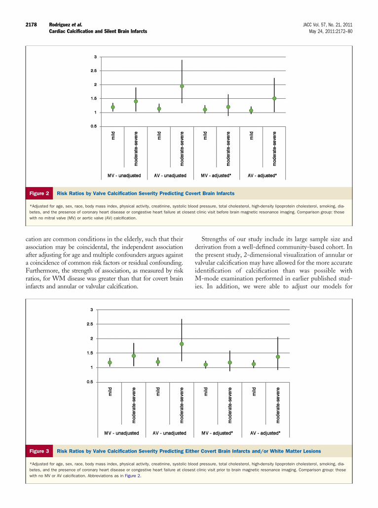
ered as a combined outcome (Table 4), the results wereconsistent with our above models. The presence of anyannular or valvular calcification, any AV calcification,MAC, and AAC showed significant associations withcombined outcomes in our unadjusted and minimallyadjusted models. In our fully adjusted model and explor-atory models, only the presence of any annular or valvularcalcification, the presence of any AV calcification, andthe presence of AAC were related to the combinedoutcome. AVSc alone was not associated with any brainMRI finding in any of our models. The degree of valvularcalcification showed a direct relation with the presence ofcovert brain MRI lesions. In fully adjusted models,moderate to severe annular or valvular calcification in themitral and aortic position showed a 21% and 19% higherrisk for covert brain infarcts and a 51% and 38% higherrisk for covert brain infarcts and/or high WM grade thanno valvular lesions, respectively, although the differencewas statistically significant only for the relation of AVlesions with covert brain infarcts (Figs. 2 and 3).
nfarct location analysis. Of the 712 participants withovert infarcts, 65 (9%) had any cortical infarcts and 64791%) had noncortical infarcts. The prevalence of corticalnfarcts versus noncortical infarcts in those with versusithout any annular or valvular calcification was not
ignificantly different (p � 0.51). Relative risk regression
lar or Valvular Calcification Statusding to Annular or Valvular Calcification Status
e Calcification*63)
No Annular or Valve Calcification(n � 617) p Value
4.87 73.67 � 4.52 �0.0001
4.31 26.86 � 4.56 0.22
.8%) 211 (34.2%) 0.003
.8%) 506 (82.0%) 0.02
1,850.79 1,618.67 � 1,795.90 0.98
0.27 1.01 � 0.22 0.003
19.82 134.28 � 21.15 0.93
1.21 3.99 � 1.24 0.027
37.55 207.00 � 36.56 0.081
14.61 55.32 � 15.07 0.076
.7%) 301 (48.8%)
.7%) 256 (41.5%)
6%) 60 (9.7%) 0.07
.0%) 70 (11.4%) 0.85
.4%) 86 (13.9%) 0.01
7%) 15 (2.4%) 0.12
.02 4.68 � 6.57 0.99
66.02 171.80 � 241.27 0.0006
.27 1.05 � 0.21 0.003
.01 3.94 � 0.58 0.084
4%) 29 (4.7%) 0.002
.3%) 91 (18.8%) 0.001
ular calcification, aortic annular calcification, or aortic valve sclerosis.rt failure; HDL � high-density lipoprotein; LV � left ventricular; NT-proBNP � N-terminal pro-brain
Annuccor
r Valv� 2,0
.79 �
.62 �
42 (40
69 (85
.16 �
.04 �
.20 �
.12 �
.00 �
.12 �
23 (44
63 (46
77 (8.
47 (12
80 (18
77 (3.
.68 � 8
.57 � 4
.08 � 0
.01 � 1
73 (8.
18 (26
ral ann
nalysis of any annular or valvular calcification using

2176 Rodriguez et al. JACC Vol. 57, No. 21, 2011Cardiac Calcification and Silent Brain Infarcts May 24, 2011:2172–80
model 2 showed that risk ratios were only slightlystronger for any cortical infarct than for noncorticalinfarcts, but as expected given the small number ofcortical infarcts, the association for this stratum was notsignificant (Table 5).
Figure 1 Proportion of Participants With Covert Brain InfarctsAccording to Annular or Valve Lesion Status
Data showing a significantly greater proportion of covert brain infarcts in partici-pants with any annular or valvular calcification compared with those withoutany annular or valvular calcification. MRI � magnetic resonance imaging.
Risk Ratios (95% Confidence Intervals) Describing the AssociationMAC, AAC, and AVSc and Covert Brain InfarctsTable 2 Risk Ratios (95% Confidence Intervals) Describing theMAC, AAC, and AVSc and Covert Brain Infarcts
Variable Unadjusted p Value Model 1 p Value
Any annular orvalve calcification
1.33 (1.12–1.57) 0.001 1.27 (1.08–1.50) 0.005
MAC 1.21 (1.06–1.37) 0.003 1.15 (1.01–1.30) 0.035
AAC 1.17 (1.03–1.33) 0.014 1.11 (0.98–1.26) 0.108
AVSc 1.08 (0.95–1.23) 0.224 1.07 (0.94–1.21) 0.323
Any AV calcification* 1.21 (1.05–1.41) 0.009 1.16 (1.00–1.35) 0.044
*Any AV calcification: combination of AAC or AVSc. Model 1: adjusted for age, sex, and race (whitblood pressure, total cholesterol, high-density lipoprotein cholesterol, smoking status (never, formevisit before brain magnetic resonance imaging. Model 3: adjusted for age, race, sex, left ventricular
peptide, C-reactive protein, cystatin C, and atrial fibrillation.AAC � aortic annular calcification; AV � aortic valve; AVSc � aortic valve sclerosis; MAC � mitral ann
Discussion
In a community-based sample of older adults, we found thatthe presence of any left-sided cardiac annular or valvularcalcification was significantly associated with a 33% greaterrisk for covert brain infarcts on MRI in participants withouthistories of TIA or stroke. This observed association per-sisted after full adjustment for potential confounders. Sim-ilar associations were observed with the presence of highWM grade and when covert brain infarcts and high WMgrade were analyzed as a combined MRI-defined outcome.Additionally, we used explanatory models with comprehen-sive adjustment for clinical confounders and echocardio-graphic and inflammatory covariates. Our findings remainedindependent of NT-proBNP, CRP, and cystatin C, sug-gesting that the primary association represents more thansimply shared risk factors.
Prior studies have reported inconsistent findings withrespect to the association between left-sided cardiacannular or valvular calcifications and the risk for clinicalstroke. MAC and AAC are characterized by calcium andlipid deposition in the annular fibrosa, whereas AVScresults from similar accumulation involving the AVleaflets (1,28). Although numerous case reports provideevidence of brain infarction due to calcific emboli fromAVs (29 –33), an independent association between AVcalcification and clinical stroke has been demonstratedonly in the presence of hemodynamically significant AVstenosis (4,13). Several studies (7,9,10) have detailed arelationship between MAC and clinical stroke, but thiswas not confirmed by a prior report from the CHS or bya separate study using a matched control group (3,8,34).The weight of the evidence, although varied and seem-ingly conflicted, favors the presence of some associationbetween mitral and/or aortic calcification and clinicalischemic stroke.
To our knowledge, our study is the first to demonstratean association between left-sided cardiac annular or valvularcalcifications and covert brain infarcts. Most studies ofcalcific AV disease do not detail the presence of concomi-tant AAC, and it may be that AAC is an underappreciatedexposure in stroke risk. Our results are important because
ongciations Among
odel 2 p Value Model 3 p Value Model 4 p Value
(1.05–1.47) 0.011 1.24 (1.02–1.50) 0.035 1.27 (1.05–1.53) 0.013
(0.99–1.27) 0.081 1.09 (0.93–1.27) 0.303 1.11 (0.97–1.28) 0.140
(0.96–1.24) 0.179 1.09 (0.93–1.27) 0.290 1.08 (0.94–1.24) 0.288
(0.92–1.18) 0.538 1.05 (0.90–1.22) 0.559 1.08 (0.93–1.24) 0.313
(0.99–1.32) 0.075 1.11 (0.93–1.32) 0.240 1.15 (0.98–1.35) 0.096
hite). Model 2: adjusted for age, sex, race, body mass index, physical activity, creatinine, systolict), diabetes, and the presence of coronary heart disease or congestive heart failure at closest clinicphy, and left atrial dimension. Model 4: adjusted for age, race, sex, N-terminal pro-brain natriuretic
s AmAsso
M
1.24
1.12
1.09
1.04
1.14
e, nonwr, currenhypertro
ular calcification.

2177JACC Vol. 57, No. 21, 2011 Rodriguez et al.May 24, 2011:2172–80 Cardiac Calcification and Silent Brain Infarcts
covert MRI-defined brain infarcts and WMLs are associ-ated with a higher risk for cognitive decline and futurestroke (14,16,17). The implication of our findings is that therelationship between left-sided cardiac annular or valvularcalcifications and brain infarcts may have been underesti-mated in prior studies that considered only clinical ischemicstroke. This relationship may be stronger and of greaterclinical relevance when both covert and overt brain infarctsare considered.
Several explanations may account for the independentassociation of annular or valvular calcific lesions with covertMRI-defined brain infarcts. These cardiac lesions may notbe just markers of stroke risk based on their association withatherosclerotic vascular disease (35,36) but may also play acausative role. Embolization from left-sided cardiac annularor valvular calcifications can vary from friable ulceratedcalcium deposits to noncalcific (thrombus) embolization(32,37–40). Evidence of systemic embolism to cerebral,coronary, renal, and retinal arteries and the peripheralcirculation has been found on autopsy in one-third ofpatients with calcific AV disease (30), with evidence oforganized microthrombi observed in 53% of calcified AVs(41). Wilson et al. (32) noted calcific AV disease as the mostcommon cardiac abnormality in patients with central retinalartery occlusion. A potential mechanistic pathway for mi-cothrombi formation on left-sided calcified structures couldinvolve turbulent blood flow at the mitral or aortic position,followed by fragmentation of red cells and release of
Risk Ratios (95% Confidence Intervals) Describing the Associationand Either Covert Brain Infarcts or High WM Grade (>4)Table 4 Risk Ratios (95% Confidence Intervals) Describing theand Either Covert Brain Infarcts or High WM Grade (>
Variable Unadjusted p Value Model 1 p Value
Any annular orvalve calcification
1.39 (1.19–1.64) �0.001 1.33 (1.13–1.56) 0.001
MAC 1.20 (1.07–1.35) 0.002 1.13 (1.00–1.27) 0.042
AAC 1.22 (1.09–1.38) 0.001 1.15 (1.02–1.29) 0.021
AVSc 1.10 (0.97–1.24) 0.123 1.08 (0.96–1.22) 0.201
Any AV calcification* 1.28 (1.11–1.47) 0.001 1.22 (1.06–1.40) 0.006
*Any AV calcification: combination of AAC or AVSc. Model 1: adjusted for age, sex, race (white, nonpressure, total cholesterol, high-density lipoprotein cholesterol, smoking status (never, former, currbefore brain magnetic resonance imaging. Model 3: adjusted for age, race, sex, left ventricular hy
Risk Ratios (95% Confidence Intervals) Describing the AssociationAVSc, MAC, and AAC and High WM Grade (>4)Table 3 Risk Ratios (95% Confidence Intervals) Describing theAVSc, MAC, and AAC and High WM Grade (>4)
Variable Unadjusted p Value Model 1 p Value
Any annular orvalve calcification
1.8 (1.17–2.77) 0.008 1.59 (1.04–2.45) 0.033
MAC 1.34 (0.99–1.81) 0.055 1.14 (0.85–1.54) 0.390
AAc 1.48 (1.10–2.00) 0.010 1.25 (0.92–1.69) 0.154
AvSc 1.29 (0.95–1.76) 0.099 1.25 (0.92–1.69) 0.153
Any AV calcification* 1.64 (1.13–2.38) 0.009 1.44 (1.00–2.10) 0.053
*Any AV calcification: combination of AAC or AVSc. Model 1: adjusted for age, sex, race (white, nonpressure, total cholesterol, high-density lipoprotein cholesterol, smoking status (never, former, currbefore brain magnetic resonance imaging. Model 3: adjusted for age, race, sex, left ventricular hypeptide, C-reactive protein, cystatin C, and atrial fibrillation.
Abbreviations as in Table 2.
peptide, C-reactive protein, cystatin C, and atrial fibrillation.Abbreviations as in Table 2.
adenosine diphosphate and thromboplastin, with resultingmicrothrombus formation and noncalcific embolism(41,42). The incidence of cerebral thromboembolism fromleft-sided cardiac annular or valvular calcifications is likelyunderestimated. Patients may remain asymptomatic in somecases depending on the size of emboli (30), and anyneurologic symptoms may be attributed to other competingcauses, such as low cardiac output, small vessel disease, oratrial fibrillation. A caveat to this argument is that mostinfarcts in the CHS were noncortical, which are typicallynot cardioembolic (25), suggesting that embolism may notbe the only explanation for covert infarcts in those withannular or valvular calcification. The number of corticalinfarcts in these CHS participants was not sufficient toanswer the question of whether or not the risk for covertinfarcts with any annular or valvular calcification differs byinfarct location. In addition, the association between annu-lar or valvular calcification may be through either known orunknown shared risk factors that were not considered inthese analyses, such as atherosclerotic disease of the aortaand cervicocranial vasculature.
Regarding WM disease and annular or valvular calcifica-tion, the mechanism of association is somewhat unclear.WM disease is found in those with vascular dementia andmay be the consequence of microvasculopathy, edema,gliosis, as well as chronic ischemia and small infarcts such asthose caused by embolic calcific disease (43). Although it istrue that both WM disease and annular or valvular calcifi-
ong AVSc, MAC, and AACciations Among AVSc, MAC, and AAC
odel 2 p Value Model 3 p Value Model 4 p Value
(1.11–1.54) 0.001 1.30 (1.07–1.57) 0.007 1.33 (1.11–1.59) 0.002
(0.99–1.25) 0.074 1.08 (0.93–1.25) 0.307 1.10 (0.96–1.25) 0.169
(1.01–1.28) 0.035 1.15 (0.99–1.32) 0.062 1.13 (0.99–1.29) 0.072
(0.94–1.19) 0.326 1.06 (0.92–1.23) 0.394 1.09 (0.95–1.24) 0.212
(1.05–1.39) 0.009 1.19 (1.01–1.40) 0.043 1.21 (1.04–1.41) 0.016
. Model 2: adjusted for age, sex, race, body mass index, physical activity, creatinine, systolic bloodbetes, and the presence of coronary heart disease or congestive heart failure at closest clinic visit
hy, and left atrial dimension. Model 4: adjusted for age, race, sex, N-terminal pro-brain natriuretic
ongciations Among
odel 2 p Value Model 3 p Value Model 4 p Value
1.04–2.49) 0.033 1.65 (0.98–2.77) 0.058 1.63 (0.99–2.70) 0.055
(0.86–1.57) 0.334 1.13 (0.79–1.62) 0.510 1.12 (0.80–1.58) 0.502
(0.91–1.68) 0.177 1.29 (0.90–1.85) 0.173 1.23 (0.87–1.74) 0.237
(0.91–1.68) 0.166 1.37 (0.94–1.99) 0.097 1.39 (0.98–1.97) 0.061
(1.00–2.11) 0.050 1.60 (1.01–2.53) 0.045 1.49 (0.97–2.28) 0.068
. Model 2: adjusted for age, sex, race, body mass index, physical activity, creatinine, systolic bloodbetes, and the presence of coronary heart disease or congestive heart failure at closest clinic visit
hy, and left atrial dimension. Model 4: adjusted for age, race, sex, N-terminal pro-brain natriuretic
s AmAsso4)
M
1.31
1.11
1.14
1.06
1.20
white)ent), diapertrop
s AmAsso
M
1.61 (
1.16
1.23
1.24
1.45
white)ent), diapertrop
2178 Rodriguez et al. JACC Vol. 57, No. 21, 2011Cardiac Calcification and Silent Brain Infarcts May 24, 2011:2172–80
cation are common conditions in the elderly, such that theirassociation may be coincidental, the independent associationafter adjusting for age and multiple confounders argues againsta coincidence of common risk factors or residual confounding.Furthermore, the strength of association, as measured by riskratios, for WM disease was greater than that for covert braininfarcts and annular or valvular calcification.
Figure 2 Risk Ratios by Valve Calcification Severity Predicting
*Adjusted for age, sex, race, body mass index, physical activity, creatinine, systolbetes, and the presence of coronary heart disease or congestive heart failure at cwith no mitral valve (MV) or aortic valve (AV) calcification.
Figure 3 Risk Ratios by Valve Calcification Severity Predicting
*Adjusted for age, sex, race, body mass index, physical activity, creatinine, systolbetes, and the presence of coronary heart disease or congestive heart failure at cwith no MV or AV calcification. Abbreviations as in Figure 2.
Strengths of our study include its large sample size andderivation from a well-defined community-based cohort. Inthe present study, 2-dimensional visualization of annular orvalvular calcification may have allowed for the more accurateidentification of calcification than was possible withM-mode examination performed in earlier published stud-ies. In addition, we were able to adjust our models for
rt Brain Infarcts
d pressure, total cholesterol, high-density lipoprotein cholesterol, smoking, dia-clinic visit before brain magnetic resonance imaging. Comparison group: those
r Covert Brain Infarcts and/or White Matter Lesions
d pressure, total cholesterol, high-density lipoprotein cholesterol, smoking, dia-clinic visit prior to brain magnetic resonance imaging. Comparison group: those
Cove
ic bloolosest
Eithe
ic bloolosest

2179JACC Vol. 57, No. 21, 2011 Rodriguez et al.May 24, 2011:2172–80 Cardiac Calcification and Silent Brain Infarcts
markers of inflammation, echocardiographic variables, andother important potential confounders.
Our study also had several limitations. First, we per-formed brain MRI 1 to 2 years before the echocardiographicexaminations. Thus, causality cannot be inferred. In doingthis as a cross-sectional analysis, we are assuming thatchanges in measures determined on MRI are slow, so thatthose variables remain close to what they would be had theybeen obtained at the time of the echocardiographic exami-nation. Although the issue of reverse causality exists, it isnot biologically plausible that covert brain infarcts wouldcause annular or valvular calcification. Moreover, becausesuch covert infarcts would not lead to immediate disability,there would be less of a short-term clinical impact of theselesions than would be observed from clinically apparentovert stroke. Second, although our statistical models wereextensive, unmeasured confounders could potentially ex-plain the observed associations. Third, our classificationscheme for left-sided annular or valvular calcification can becriticized. MAC, AAC, and AVSc are distinct diseaseprocesses but with significant overlap and correlation char-acterized by common initial pathology of lipid infiltration,components of chronic inflammation, and calcification (28).This disease process is manifested and progresses differently,possibly because of different flow hemodynamic parametersbetween the mitral and aortic annuli. We looked at thesecardiac lesions as an aggregate because of the threat ofcalcific and thrombotic systemic embolism, which is typi-cally unique to left-sided lesions unless there is a patentforamen ovale present. We defined AV calcification ascalcification within the AV leaflets or valve annulus. Thus,AAC, AVSc, and aortic stenosis represent different pointsalong the same histopathologic spectrum. Last, the majorityof CHS participants were Caucasian, possibly reducing thegeneralizability of our findings to other ethnic groups ofolder adults.
Conclusions
Our results indicate that left-sided cardiac annular orvalvular calcification is associated with covert MRI-definedbrain infarcts. Prior estimates of the association betweencalcific annular or valve disease and stroke may be underes-timated, because they accounted only for clinical overt
Covert Brain Infarct Location Accordingto Calcific Annular or Valve Disease StatusTable 5 Covert Brain Infarct Location Accordingto Calcific Annular or Valve Disease Status
InfarctLocation
In Those WithoutAny Annular or Valve
Calcification
In Those With AnyAnnular or Valve
Calcification
Risk Ratio(95% Confidence
Interval)*
Noncortical 18% (121/647) 81% (526/647) 1.23 (1.03–1.47)
Cortical 15% (10/65) 85% (55/65) 1.64 (0.84–3.21)
*Adjusted for age, sex, race, body mass index, physical activity, creatinine, systolic blood pressure,total cholesterol, high-density lipoprotein cholesterol, smoking status (never, former, current),diabetes, and the presence of coronary heart disease or congestive heart failure at closest clinicvisit before brain magnetic resonance imaging.
stroke. Further study is warranted to identify the mecha-
nisms relating these 2 sets of findings in the heart and brainand determine whether intervening in the progression ofcalcific annular or valvular disease can reduce the incidenceof these covert MRI findings as well as the associated riskfor cognitive decline and future stroke.
Reprint requests and correspondence: Dr. Carlos J. Rodriguez,Division of Public Health Sciences, Wake Forest UniversitySchool of Medicine, Medical Center Boulevard, Winston-Salem,North Carolina 27157. E-mail: [email protected].
REFERENCES
1. Ashida T, Kiraku J, Takahashi N, Sugiyama T, Fujii J. Experimentalstudy on the pathogenesis of mitral annular calcification: calciumdeposits in mitral complex lesions induced by vagal stimulation inrabbits [article in Japanese]. J Cardiol 1997;29 Suppl:13–7.
2. Barasch E, Gottdiener JS, Larsen EK, Chaves PH, Newman AB,Manolio TA. Clinical significance of calcification of the fibrousskeleton of the heart and aortosclerosis in community dwelling elderly.The Cardiovascular Health Study (CHS). Am Heart J 2006;151:39–47.
3. Boon A, Cheriex E, Lodder J, Kessels F. Cardiac valve calcification:characteristics of patients with calcification of the mitral annulus oraortic valve. Heart 1997;78:472–4.
4. Cosmi JE, Kort S, Tunick PA, et al. The risk of the development ofaortic stenosis in patients with “benign” aortic valve thickening. ArchIntern Med 2002;162:2345–7.
5. Agmon Y, Khandheria BK, Meissner I, et al. Aortic valve sclerosis andaortic atherosclerosis: different manifestations of the same disease?Insights from a population-based study. J Am Coll Cardiol 2001;38:827–34.
6. Fox CS, Vasan RS, Parise H, et al. Mitral annular calcification predictscardiovascular morbidity and mortality: the Framingham Heart Study.Circulation 2003;107:1492–6.
7. Benjamin EJ, Plehn JF, D’Agostino RB, et al. Mitral annular calcifi-cation and the risk of stroke in an elderly cohort. N Engl J Med1992;327:374–9.
8. Boon A, Lodder J, Cheriex E, Kessels F. Mitral annulus calcificationis not an independent risk factor for stroke: a cohort study of 657patients. J Neurol 1997;244:535–41.
9. Aronow WS, Ahn C, Kronzon I, Gutstein H. Association of mitralannular calcium with new thromboembolic stroke at 44-monthfollow-up of 2,148 persons, mean age 81 years. Am J Cardiol1998;81:105–6.
10. Kizer JR, Wiebers DO, Whisnant JP, et al. Mitral annular calcifica-tion, aortic valve sclerosis, and incident stroke in adults free of clinicalcardiovascular disease: the Strong Heart Study. Stroke 2005;36:2533–7.
11. Krause I, Lev S, Fraser A, et al. Close association between valvar heartdisease and central nervous system manifestations in the antiphospho-lipid syndrome. Ann Rheum Dis 2005;64:1490–3.
12. Morelli S, Bernardo ML, Viganego F, et al. Left-sided heart valveabnormalities and risk of ischemic cerebrovascular accidents in patientswith systemic lupus erythematosus. Lupus 2003;12:805–12.
13. Otto CM, Lind BK, Kitzman DW, Gersh BJ, Siscovick DS. Associ-ation of aortic-valve sclerosis with cardiovascular mortality and mor-bidity in the elderly. N Engl J Med 1999;341:142–7.
14. Kario K, Pickering TG, Umeda Y, et al. Morning surge in bloodpressure as a predictor of silent and clinical cerebrovascular disease inelderly hypertensives: a prospective study. Circulation 2003;107:1401–6.
15. Longstreth WT Jr. Brain vascular disease overt and covert. Stroke2005;36:2062–3.
16. Vermeer SE, Hollander M, van Dijk EJ, Hofman A, Koudstaal PJ,Breteler MM. Silent brain infarcts and white matter lesions increasestroke risk in the general population: the Rotterdam Scan Study.
Stroke 2003;34:1126–9.
2180 Rodriguez et al. JACC Vol. 57, No. 21, 2011Cardiac Calcification and Silent Brain Infarcts May 24, 2011:2172–80
17. Vermeer SE, Prins ND, den Heijer T, Hofman A, Koudstaal PJ,Breteler MM. Silent brain infarcts and the risk of dementia andcognitive decline. N Engl J Med 2003;348:1215–2.
18. Kruit MC, van Buchem MA, Hofman PA, et al. Migraine as a riskfactor for subclinical brain lesions. JAMA 2004;291:427–34.
19. Fried LP, Borhani NO, Enright P, et al. The Cardiovascular HealthStudy: design and rationale. Ann Epidemiol 1991;1:263–76.
20. Psaty BM, Kuller LH, Bild D, et al. Methods of assessing prevalentcardiovascular disease in the Cardiovascular Health Study. AnnEpidemiol 1995;5:270–7.
21. Gardin JM, Wong ND, Bommer W, et al. Echocardiographic designof a multicenter investigation of free-living elderly subjects: theCardiovascular Health Study. J Am Soc Echocardiogr 1992;5:63–72.
22. Stewart BF, Siscovick D, Lind BK, et al. Clinical factors associatedwith calcific aortic valve disease. Cardiovascular Health Study. J AmColl Cardiol 1997;29:630–4.
23. Novaro GM, Katz R, Aviles RJ, et al. Clinical factors, but notC-reactive protein, predict progression of calcific aortic-valve disease:the Cardiovascular Health Study. J Am Coll Cardiol 2007;50:1992–8.
24. Longstreth WT Jr., Arnold AM, Beauchamp NJ Jr., et al. Incidence,manifestations, and predictors of worsening white matter on serialcranial magnetic resonance imaging in the elderly: the CardiovascularHealth Study. Stroke 2005;36:56–61.
25. Longstreth WT Jr., Bernick C, Manolio TA, Bryan N, Jungreis CA,Price TR. Lacunar infarcts defined by magnetic resonance imaging of3660 elderly people: the Cardiovascular Health Study. Arch Neurol1998;55:1217–25.
26. Lang RM, Bierig M, Devereux RB, et al. Recommendations forchamber quantification: a report from the American Society ofEchocardiography’s Guidelines and Standards Committee and theChamber Quantification Writing Group, developed in conjunctionwith the European Association of Echocardiography, a branch of theEuropean Society of Cardiology. J Am Soc Echocardiogr 2005;18:1440–63.
27. Zou G. A modified poisson regression approach to prospective studieswith binary data. Am J Epidemiol 2004;159:702–6.
28. Sell S, Scully RE. Aging changes in the aortic and mitral valves.Histologic and histochemical studies, with observations on the patho-genesis of calcific aortic stenosis and calcification of the mitral annulus.Am J Pathol 1965;46:345–65.
29. Brockmeier LB, Adolph RJ, Gustin BW, Holmes JC, Sacks JG.Calcium emboli to the retinal artery in calcific aortic stenosis. AmHeart J 1981;101:32–7.
30. Holley KE, Bahn RC, McGoon DC, Mankin HT. Spontaneous
calcific embolization associated with calcific aortic stenosis. Circulation1963;27:197–202.31. Oliveira-Filho J, Massaro AR, Yamamoto F, Bustamante L, Scaff M.Stroke as the first manifestation of calcific aortic stenosis. CerebrovascDis 2000;10:413–6.
32. Wilson JH, Cranley JJ. Recurrent calcium emboli in a patient withaortic stenosis. Chest 1989;96:1433–4.
33. Vernhet H, Torres GF, Laharotte JC, et al. Spontaneous calcificcerebral emboli from calcified aortic valve stenosis. J Neuroradiol1993;20:19–23.
34. Gardin JM, McClelland R, Kitzman D, et al. M-mode echocardio-graphic predictors of six- to seven-year incidence of coronary heartdisease, stroke, congestive heart failure, and mortality in an elderlycohort (the Cardiovascular Health Study). Am J Cardiol 2001;87:1051–7.
35. Karas MG, Francescone S, Segal AZ, et al. Relation between mitralannular calcium and complex aortic atheroma in patients with cerebralischemia referred for transesophageal echocardiography. Am J Cardiol2007;99:1306–11.
36. Adler Y, Levinger U, Koren A, et al. Relation of nonobstructive aorticvalve calcium to carotid arterial atherosclerosis. Am J Cardiol 2000;86:1102–5.
37. Rancurel G, Marelle L, Vincent D, Catala M, Arzimanoglou A,Vacheron A. Spontaneous calcific cerebral embolus from a calcificaortic stenosis in a middle cerebral artery infarct. Stroke 1989;20:691–3.
38. Hamon M, Gomes S, Oppenheim C, et al. Cerebral microembolismduring cardiac catheterization and risk of acute brain injury: aprospective diffusion-weighted magnetic resonance imaging study.Stroke 2006;37:2035–8.
39. Willens HJ, Ferreira AC, Gallagher AJ, Morytko JA. Mobile compo-nents associated with rapidly developing mitral annulus calcification inpatients with chronic renal failure: review of mobile elements associ-ated with mitral annulus calcification. Echocardiography 2003;20:363–7.
40. Lin CS, Schwartz IS, Chapman I. Calcification of the mitral annulusfibrosus with systemic embolization. A clinicopathologic study of 16cases. Arch Pathol Lab Med 1987;111:411–4.
41. Stein PD, Sabbah HN, Pitha JV. Continuing disease process of calcificaortic stenosis. Role of microthrombi and turbulent flow. Am J Cardiol1977;39:159–63.
42. Johnson S. Platelets in hemostasis. In: Seegers W, ed. Blood ClottingEnzymology. New York: Academic Press, 1967:379–420.
43. Breteler MM, van Swieten JC, Bots ML, et al. Cerebral white matterlesions, vascular risk factors, and cognitive function in a population-based study: the Rotterdam Study. Neurology 1994;44:1246–52.
Key Words: aortic valve y calcification y covert brain infarcts yepidemiology.
Related Documents