Association of Alzheimer’s disease GWAS loci with MRI markers of brain aging Ganesh Chauhan a, b,1 , Hieab H.H. Adams c, d, 1 , Joshua C. Bis e , Galit Weinstein f, g , Lei Yu h, i , Anna Maria Töglhofer j , Albert Vernon Smith k, l , Sven J. van der Lee c , Rebecca F. Gottesman m, n , Russell Thomson o , Jing Wang g, p , Qiong Yang g, p , Wiro J. Niessen d , Oscar L. Lopez q, r , James T. Becker q, r, s , Thanh G. Phan t , Richard J. Beare t, u , Konstantinos Arfanakis h, v , Debra Fleischman h , Meike W. Vernooij c, d , Bernard Mazoyer w , Helena Schmidt j , Velandai Srikanth t, o , David S. Knopman x , Clifford R. Jack, Jr y , Philippe Amouyel z, aa, ab , Albert Hofman c , Charles DeCarli ac , Christophe Tzourio a , Cornelia M. van Duijn c, ad, ae , David A. Bennett h, i , Reinhold Schmidt af , William T. Longstreth, Jr ag , Thomas H. Mosley ah , Myriam Fornage ai , Lenore J. Launer aj , Sudha Seshadri f, g, 2 , M. Arfan Ikram c, d, ad, 2 , Stephanie Debette a, b, f, ak, * , 2 a INSERM U897, University of Bordeaux, Bordeaux, France b University of Bordeaux, Bordeaux, FranceINSERM U897, University of Bordeaux, Bordeaux, France c Department of Epidemiology, Erasmus Medical Center, Rotterdam, the Netherlands d Department of Radiology, Erasmus Medical Center, Rotterdam, the Netherlands e Cardiovascular Health Research Unit, Department of Medicine, University of Washington, Seattle, WA, USA f Department of Neurology, Boston University School of Medicine, Boston, MA, USA g The Framingham Heart Study, Boston, MA, USA h Rush Alzheimer’s Disease Center, Rush University Medical Center, Chicago, IL, USA i Department of Neurological Sciences, Rush University Medical Center, Chicago, IL, USA j Institute of Molecular Biology and Biochemistry, Centre for Molecular Medicine, Medical University of Graz, Graz, Austria k Icelandic Heart Association, Iceland l Department of Medicine, University of Iceland, Reykjavik, Iceland m Department of Neurology, Johns Hopkins School of Medicine, Baltimore, USA n Department of Epidemiology, Johns Hopkins Bloomberg School of Public Health, Baltimore, USA o Menzies Research Institute Tasmania, University of Tasmania, Hobart, Tasmania, Australia p Department of Biostatistics, Boston University School of Public Health, Boston, MA, USA q Department of Neurology, University of Pittsburgh School of Medicine, Pittsburgh, PA, USA r Department of Psychiatry, University of Pittsburgh School of Medicine, Pittsburgh, PA, USA s Department of Psychology, University of Pittsburgh School of Medicine, Pittsburgh, PA, USA t Stroke and Ageing Research Centre, Southern Clinical School, Department of Medicine, Monash University, Melbourne, Victoria, Australia u Developmental Imaging Group, Murdoch Childrens Research Institute, The Royal Children’s Hospital, Parkville, Victoria, Australia v Department of Biomedical Engineering, Illinois Institute of Technology, Chicago, IL, USA w Groupe d’Imagerie Neurofonctionnelle, UMR5296, CNRS, CEA, Université de Bordeaux, Bordeaux, France x Department of Neurology, Mayo Clinic College of Medicine, Rochester, MN, USA y Department of Radiology, Mayo Clinic College of Medicine, Rochester, MN, USA z Department of Epidemiology and Public Health, Pasteur Institute of Lille, Lille, France aa INSERM, U744, Lille, France ab Université Lille 2, Lille, France ac Department of Neurology, University of California at Davis, Davis, CA, USA ad Netherlands Consortium for Healthy Aging, Leiden, the Netherlands ae Center for Medical Systems Biology, Leiden, the Netherlands af Department of Neurology, Clinical Division of Neurogeriatrics, Medical University of Graz, Austria ag Departments of Neurology and Epidemiology, University of Washington, Seattle, WA, USA ah Department of Medicine-Geriatrics/Gerontology, University of Mississippi Medical Center, Jackson, MS, USA ai The Human Genetics Center and Institute of Molecular Medicine, The University of Texas Health Science Center at Houston, Houston, USA * Corresponding author at: Université de Bordeaux, Centre Inserm U 897, Service de Neuroépidémiologie, Bâtiment ISPED e Case 11,146 rue Léo Saignat- CS 61292, 33076 BORDEAUX CEDEX, France. Tel.: þ33 (0) 6 84 07 72 53; fax: þ33 (0) 5 47 30 42 09. E-mail addresses: [email protected], [email protected] (S. Debette). 1 These authors contributed equally to the article. 2 These authors jointly directed this work. Contents lists available at ScienceDirect Neurobiology of Aging journal homepage: www.elsevier.com/locate/neuaging 0197-4580/$ e see front matter Ó 2015 Elsevier Inc. All rights reserved. http://dx.doi.org/10.1016/j.neurobiolaging.2014.12.028 Neurobiology of Aging xxx (2015) 1e10

Welcome message from author
This document is posted to help you gain knowledge. Please leave a comment to let me know what you think about it! Share it to your friends and learn new things together.
Transcript
lable at ScienceDirect
Neurobiology of Aging xxx (2015) 1e10
Contents lists avai
Neurobiology of Aging
journal homepage: www.elsevier .com/locate/neuaging
Association of Alzheimer’s disease GWAS loci with MRI markers ofbrain aging
Ganesh Chauhan a,b,1, Hieab H.H. Adams c,d,1, Joshua C. Bis e, Galit Weinstein f,g, Lei Yu h,i,Anna Maria Töglhofer j, Albert Vernon Smith k,l, Sven J. van der Lee c,Rebecca F. Gottesmanm,n, Russell Thomson o, Jing Wang g,p, Qiong Yang g,p,Wiro J. Niessen d, Oscar L. Lopez q,r, James T. Becker q,r,s, Thanh G. Phan t,Richard J. Beare t,u, Konstantinos Arfanakis h,v, Debra Fleischmanh, Meike W. Vernooij c,d,Bernard Mazoyerw, Helena Schmidt j, Velandai Srikanth t,o, David S. Knopmanx,Clifford R. Jack, Jr y, Philippe Amouyel z,aa,ab, Albert Hofman c, Charles DeCarli ac,Christophe Tzourio a, Cornelia M. van Duijn c,ad,ae, David A. Bennett h,i, Reinhold Schmidt af,William T. Longstreth, Jr ag, Thomas H. Mosley ah, Myriam Fornage ai, Lenore J. Launer aj,Sudha Seshadri f,g,2, M. Arfan Ikramc,d,ad,2, Stephanie Debette a,b,f,ak,*,2
a INSERM U897, University of Bordeaux, Bordeaux, FrancebUniversity of Bordeaux, Bordeaux, FranceINSERM U897, University of Bordeaux, Bordeaux, FrancecDepartment of Epidemiology, Erasmus Medical Center, Rotterdam, the NetherlandsdDepartment of Radiology, Erasmus Medical Center, Rotterdam, the NetherlandseCardiovascular Health Research Unit, Department of Medicine, University of Washington, Seattle, WA, USAfDepartment of Neurology, Boston University School of Medicine, Boston, MA, USAg The Framingham Heart Study, Boston, MA, USAhRush Alzheimer’s Disease Center, Rush University Medical Center, Chicago, IL, USAiDepartment of Neurological Sciences, Rush University Medical Center, Chicago, IL, USAj Institute of Molecular Biology and Biochemistry, Centre for Molecular Medicine, Medical University of Graz, Graz, Austriak Icelandic Heart Association, IcelandlDepartment of Medicine, University of Iceland, Reykjavik, IcelandmDepartment of Neurology, Johns Hopkins School of Medicine, Baltimore, USAnDepartment of Epidemiology, Johns Hopkins Bloomberg School of Public Health, Baltimore, USAoMenzies Research Institute Tasmania, University of Tasmania, Hobart, Tasmania, AustraliapDepartment of Biostatistics, Boston University School of Public Health, Boston, MA, USAqDepartment of Neurology, University of Pittsburgh School of Medicine, Pittsburgh, PA, USArDepartment of Psychiatry, University of Pittsburgh School of Medicine, Pittsburgh, PA, USAsDepartment of Psychology, University of Pittsburgh School of Medicine, Pittsburgh, PA, USAt Stroke and Ageing Research Centre, Southern Clinical School, Department of Medicine, Monash University, Melbourne, Victoria, AustraliauDevelopmental Imaging Group, Murdoch Childrens Research Institute, The Royal Children’s Hospital, Parkville, Victoria, AustraliavDepartment of Biomedical Engineering, Illinois Institute of Technology, Chicago, IL, USAwGroupe d’Imagerie Neurofonctionnelle, UMR5296, CNRS, CEA, Université de Bordeaux, Bordeaux, FrancexDepartment of Neurology, Mayo Clinic College of Medicine, Rochester, MN, USAyDepartment of Radiology, Mayo Clinic College of Medicine, Rochester, MN, USAzDepartment of Epidemiology and Public Health, Pasteur Institute of Lille, Lille, Franceaa INSERM, U744, Lille, FranceabUniversité Lille 2, Lille, FranceacDepartment of Neurology, University of California at Davis, Davis, CA, USAadNetherlands Consortium for Healthy Aging, Leiden, the NetherlandsaeCenter for Medical Systems Biology, Leiden, the NetherlandsafDepartment of Neurology, Clinical Division of Neurogeriatrics, Medical University of Graz, AustriaagDepartments of Neurology and Epidemiology, University of Washington, Seattle, WA, USAahDepartment of Medicine-Geriatrics/Gerontology, University of Mississippi Medical Center, Jackson, MS, USAai The Human Genetics Center and Institute of Molecular Medicine, The University of Texas Health Science Center at Houston, Houston, USA
* Corresponding author at: Université de Bordeaux, Centre Inserm U 897, Service de Neuroépidémiologie, Bâtiment ISPED e Case 11, 146 rue Léo Saignat- CS 61292, 33076BORDEAUX CEDEX, France. Tel.: þ33 (0) 6 84 07 72 53; fax: þ33 (0) 5 47 30 42 09.
E-mail addresses: [email protected], [email protected] (S. Debette).1 These authors contributed equally to the article.2 These authors jointly directed this work.
0197-4580/$ e see front matter � 2015 Elsevier Inc. All rights reserved.http://dx.doi.org/10.1016/j.neurobiolaging.2014.12.028

G. Chauhan et al. / Neurobiology of Aging xxx (2015) 1e102
aj Laboratory of Epidemiology and Population Sciences, Intramural Research Program, National Institute on Aging, National Institutes of Health,Bethesda, MD, USAakDepartment of Neurology, Bordeaux University Hospital, Bordeaux, France
a r t i c l e i n f o
Article history:Received 18 June 2014Received in revised form 22 December 2014Accepted 26 December 2014
Keywords:AlzheimerMRI-MarkersGenetic risk scoreGWASHippocampal volume
a b s t r a c t
Whether novel risk variants of Alzheimer’s disease (AD) identified through genome-wide associationstudies also influence magnetic resonance imaging-based intermediate phenotypes of AD in the generalpopulation is unclear. We studied association of 24 AD risk loci with intracranial volume, total brainvolume, hippocampal volume (HV), white matter hyperintensity burden, and brain infarcts in a meta-analysis of genetic association studies from large population-based samples (N ¼ 8175e11,550). Insingle-SNP based tests, AD risk allele of APOE (rs2075650) was associated with smaller HV (p ¼ 0.0054)and CD33 (rs3865444) with smaller intracranial volume (p ¼ 0.0058). In gene-based tests, there wasassociations of HLA-DRB1 with total brain volume (p ¼ 0.0006) and BIN1 with HV (p ¼ 0.00089).A weighted AD genetic risk score was associated with smaller HV (beta � SE ¼ �0.047 � 0.013, p ¼0.00041), even after excluding the APOE locus (p ¼ 0.029). However, only association of AD genetic riskscore with HV, including APOE, was significant after multiple testing correction (including number ofindependent phenotypes tested). These results suggest that novel AD genetic risk variants maycontribute to structural brain aging in nondemented older community persons.
� 2015 Elsevier Inc. All rights reserved.
1. Introduction
Alzheimer’s disease (AD) is the leading cause of dementia andrepresents a major public health burden (Ballard et al., 2011).Converging evidence suggests that pathologic processes leading tothis progressive neurodegenerative disorder start many yearsbefore clinical diagnosis of dementia (Sperling et al., 2011). Mag-netic resonance imaging (MRI) markers of brain aging, includingintracranial volume (ICV), total brain volume (TBV) and hippo-campal volume (HV), and markers of vascular brain injury,including white matter hyperintensities (WMH) and brain infarcts,are powerful predictors of dementia and may, at least in part,represent intermediate markers reflecting pathologic processesleading to AD (Debette and Markus, 2010; Jack et al., 2010, 2013;Kaye et al., 1997; Sperling et al., 2011; Vermeer et al., 2007). ICV,an imaging marker reflecting brain growth during developmentand maturation, was suggested to be correlated with resilience tobrain damage (Negash et al., 2013).
Recently, large scale genome-wide association studies (GWAS)and candidate gene-based studies have identified novel suscepti-bility loci for late-onset AD (Boada et al., 2013; Carrasquillo et al.,2009; Harold et al., 2009; Hollingworth et al., 2011; Jonsson et al.,2012, 2013; Lambert et al., 2009, 2013; Naj et al., 2011; Seshadriet al., 2010). These AD risk variants have recently been used toexamine the genotypic overlap between AD and other types ofdementia (Carrasquillo et al., 2014). Some of these variants havebeen studied with respect to various MRI measures in a mixedstudy sample of AD patients, mildly cognitive impaired and healthycontrol subjects (Biffi et al., 2010; Furney et al., 2011). They couldalso be implemented to explore the impact of genetic determinantsof AD on MRI markers of structural brain changes in nondementedcommunity persons. Indeed, this could provide important infor-mation on the disease mechanisms through which these genesaffect the risk of AD and could be of interest for the design of pre-ventative interventions. Whether all previously and newly discov-ered AD risk loci influence brain structure in advance of clinicallydetectable dementia has never been systematically investigated inlarge community samples to our knowledge. Our aim was to studyassociation of known AD GWAS loci with ICV, TBV, HV, WMHburden, and brain infarcts in nondemented participants from 10population-based studies.
2. Methods
2.1. Population
Analyses were performed on 8175e11,550 dementia-free par-ticipants of European ancestry with quantitative brain MRI andgenome-wide genotypes (N ¼ 8175 for ICV, N ¼ 8673 for TBV, N ¼11,550 for HV, N ¼ 9361 for WMH burden, and N ¼ 9401 for braininfarcts), from up to 10 population-based cohort studies partici-pating in the Cohorts of Heart and Aging Research in GenomicEpidemiology consortium: Aging Gene-Environment Susceptibili-tyeReykjavik Study, Atherosclerosis Risk in Communities Study,Austrian Stroke Prevention Study, Cardiovascular Health Study,Framingham Heart Study (FHS), Rotterdam Study, Erasmus Ruc-phen Family study, Religious Order Study & Rush Memory andAging Project, Tasmanian Study of Cognition and Gait, and the 3C-Dijon study. Each study secured approval from institutional reviewboards, and all participants provided written informed consent forstudy participation, brain MRI, and use of DNA for genetic research.Individual studies are described in the Supplementary Materials.
2.2. MRI scans
In each study, MRI scans were performed and interpreted in astandardized fashion, without reference to clinical or genetic in-formation. Details on MRI parameters and phenotype definition areprovided in the Supplementary Materials. Briefly, automated orsemiquantitative post-processing software was used to measureICV and TBV. TBV was expressed as percentage of ICV to correct fordifferences in head size (Ikram et al., 2012). HV was evaluated usingoperator-defined boundaries drawn on serial coronal sections orautomated methods (Bis et al., 2012). WMH burden was estimatedon a quantitative scale using custom-written computer programs inAging Gene-Environment Susceptibility-Reykjavik, Austrian StrokePrevention Study, FHS, and Rotterdam Study; in AtherosclerosisRisk in Communities Study and Cardiovascular Health Study, WMHburden was estimated on a semiquantitative scale (Fornage et al.,2011). Brain infarcts were defined as areas of abnormal signal in-tensity in a vascular distribution that lacked mass effect,�3e4mm,distinct from dilated perivascular spaces (Debette et al., 2010).
G. Chauhan et al. / Neurobiology of Aging xxx (2015) 1e10 3
2.3. AD GWAS loci
We manually scanned the GWAS catalog (http://www.genome.gov/gwastudies/) and Alzgene (www.alzgene.org/) for GWAS onAD. We only chose studies performed on European subjects,including a replication stage, examining single marker-based as-sociations and having loci reaching genome-wide significance (p <
5.0 � 10�8). This led to the identification of 24 independent loci.Effect estimates for SNPs with the lowest p-value in each locus(defined as the index SNP of the locus) are presented inSupplementary Table 1. We included the CD33 locus (rs3865444)despite the absence of replication in the latest AD GWAS meta-analysis (Lambert et al., 2013); this locus was previously repli-cated in several AD GWAS (Hollingworth et al., 2011; Naj et al.,2011); and recent functional studies provide strong evidence forinvolvement of rs3865444 and CD33 in AD pathology (Bradshawet al., 2013). For the APOE-ε polymorphism, we used rs2075650 asa proxy (r2 ¼ 0.48 with rs429358, the APOE-ε SNP) because APOE-εgenotypes cannot be reliably imputed on commercial genome-widechips. The AD risk variants near HLA-DRB1 (Lambert et al., 2013),ATP5H/KCTD2 (Boada et al., 2013), in TREM2 (Jonsson et al., 2013),and APP(Jonsson et al., 2012) were not included for single-SNPbased association and genetic risk score-based association as noindex SNP or proxy (r2 > 0.3) was available among the genome-wide genotypes for MRI markers of brain aging.
2.4. Power calculation
Quanto software V 1.2.3 (Gauderman, 2002a, 2002b) was used tocompute power of the 5 MRI-marker studies, assuming additivemodel of inheritance at a ¼ 0.0025 (Supplementary Fig. 1). Powerfor the quantitative traits (ICV, TBV, HV, WMH burden) wascomputed for different percentage variance explained whereas forbrain infarcts, a dichotomous trait, it was computed for differentodds ratios at different allele frequencies.
2.5. Correlation between phenotypes and equivalent number ofindependent phenotypes
Correlation between the 5 MRI phenotypes in 3C-Dijon and FHSwas calculated based on Pearson correlation using the “rcorr” func-tion in R. These correlations were used to compute the equivalentnumber of independent phenotypes using the online toolmatSpDlite(http://neurogenetics.qimrberghofer.edu.au/matSpDlite/). MatSp-Dlite which is based on the same principles used to identify numberof independent SNPs in a locus, gives the equivalent number of in-dependent variables in a correlation (r) matrix, depending on theratio of observed eigenvalue variance (after spectral decomposiiton)to its theoretical maximum (Nyholt, 2004).
2.6. Association analyses
Three analytical approaches were taken to examine the associ-ations of interest.
2.6.1. Single-SNPebased association analysisWe tested for association of AD GWAS loci with MRI markers of
brain aging using association estimates obtained from meta-analyses of GWAS for ICV (Ikram et al., 2012), TBV (Ikram et al.,2012), HV (Bis et al., 2012), WMH burden (Fornage et al., 2011),and brain infarcts (Debette et al., 2010) using genotypes imputedon the HapMap2 CEU reference panel. AD risk alleles, as describedin the latest AD GWAS meta-analysis (Lambert et al., 2013), weremodeled as the effect alleles for associations with MRI markers ofbrain aging. Logistic (brain infarcts) or linear (ICV, TBV, HV, and
WMH burden) regression was performed within each study,adjusting for age, gender, and principal components of populationstratification, and for familial relationships or study center ifrelevant. For WMH burden, data were log transformed to achievenormal distribution and associations were additionally adjustedfor ICV (except for studies measuring WMH burden on a semi-quantitative visual scale, visual grades being inherently normal-ized for brain size) (Fornage et al., 2011). For most phenotypes(ICV, TBV, HV, and brain infarcts), meta-analyses were performedusing fixed effects inverse variance weighted meta-analysis. ForWMH burden, meta-analysis was performed using effective sam-ple size weighted meta-analysis because WMH burden wasmeasured on different scales across studies. If the lead SNP at aspecific AD GWAS locus was not available, a proxy SNP (r2> 0.70 in1000G CEU) of the lead SNP was used to check single-SNPebasedassociation results (Supplementary Table 1). After Bonferronicorrection, for testing 20 independent loci, p < 0.0025 wasconsidered significant for single-SNPebased associations. How-ever, application of a more stringent threshold additionally ac-counting for the number of independent phenotypes tested led toa Bonferroni correction of p < 0.000625.
2.6.2. Gene-based association analysisGene-based association tests can be more powerful in compar-
ison with single-SNPebased association tests when there are manycausal variants in a gene with small effects (Liu et al., 2010). Single-SNPebased association results from the respective MRI markerGWAS meta-analysis were used to compute gene-based associationresults using the Versatile Gene-Based Association Study 2 software(https://vegas2.qimrberghofer.edu.au/) (Liu et al., 2010). The geneannotations and linkage disequilibrium (LD) calculation in VersatileGene-Based Association Study 2 are based on 1000 genomes (phase1 version 3). This tool annotated all but 1 gene (MS4A4E) within50 kb of the index SNPs. The test incorporates information from allmarkers within a gene and accounts for LD between markers byusing simulations from the multivariate normal distribution. Gene-based association analyses were performed for all protein-codinggenes (N ¼ 65 genes) which lie within a 50 kb distance of indexSNP of the AD risk loci. Gene boundaries were defined as 50 kbupstream and downstream of the start and end of gene (Liu et al.,2010). The choice of 50 kb boundary to cover a gene was chosenas a trade-off between a longer boundary which would have causedexcess overlap between nearby genes and a shorter boundarywhich would have ignored potential regulatory regions (Liu et al.,2010). Maximum permutation limits were set to 1000,000. Aftercorrecting for the number of genes (N ¼ 65) tested the multipletesting threshold was p < 0.00077. A more stringent correctionadditionally accounting for number of independent phenotypes(n¼ 4) tested, lead to amultiple testing threshold of p< 0.00019 forgene-based association.
2.6.3. Construction of genetic risk scoreWe constructed a genetic risk score comprising all selected AD
risk variants from 20 independent AD risk loci to estimate jointeffect of these SNPs on MRI markers of brain aging. Methods havebeen recently developed to apply a genetic risk score to meta-analysis summary estimates without requiring access to raw datafrom individual studies (Dastani et al., 2012). For each MRI markerof interest, the beta-coefficient for a given SNP, as obtained from theGWAS meta-analysis for this MRI marker, was weighted with thepublished AD beta-coefficient for the given SNP. The weighted sumof beta-coefficients for all 20 SNPs (Equation 1) was used as thebeta-coefficient of the genetic risk score. Similarly, for each MRImarker of interest, the inverse of the variance for a given SNP (fromthe GWAS meta-analysis for this MRI marker) was weighted by the
G. Chauhan et al. / Neurobiology of Aging xxx (2015) 1e104
square of the published AD beta-coefficient for the given SNP. Theseweighted inverse of variances were then summed, and the inverseof this sum was used as the variance of the genetic risk score(Equation 2). The Wald statistic was used to test for significance ofassociations between the genetic risk score and each MRI marker(Dastani et al., 2012). For WMH burden, betas and standard errorswere estimated from Z-statistics provided by the effective samplesize weighted meta-analysis using Equation 3. AD beta-coefficientsused as weights for the score were all drawn from the discoverystage of the recent largest AD GWASmeta-analysis (17,008 AD casesand 37,154 control subjects, Supplementary Table 1) (Lambert et al.,2013). Associations with p < 0.05 were considered significant forgenetic risk score-based associations.
bgrs ¼Pm
1 wbSE�2Pm
1 w2SE�2(1)
SE2grs ¼ 1Pm
1 w2SE�2(2)
bgrs ¼ beta of genetic risk score; SEgrs ¼ SE of genetic risk score;w¼weight applied (SNP-specific beta of AD GWAS); b ¼ SNP-specificbeta of association with MRI phenotype; SE ¼ SNP-specific SE ofassociation with MRI phenotype
SEw ¼ffiffiffiffiffiffiffiffiffiffiffiffiffiffiffiffiffiffiffiffiffiffiffiffiffiffiffiffiffiffiffiVP=ðES� 2pqÞ
p(3)
Beta ¼ SE � Z (4)
VP ¼ phenotypic variance (approximated to 1); ES ¼ effectivesample size; p¼minor allele frequency; q¼major allele frequency.
After correcting for 4 independent phenotypes tested, themultiple testing threshold for genetic risk score associationwas p<
0.0125.
3. Results
3.1. Correlation and heritability of the 5-MRI traits
Based on data from 2 studies which were part of the originalmeta-analysis, the 2 MRI markers of structural brain aging, ICV, andTBV showed high correlation with each other but were onlymoderately correlated with HV (Supplementary Table 2). The 2 MRImarkers of vascular brain aging WMH burden and brain infarctsshowed low correlation with each other and very little or no cor-relation with the 3 markers of structural brain aging. Depending onthis correlation the equivalent number of independent phenotypescalculated using matSpDlite was 4 for both studies. Publishedliterature showed that the 5 MRI markers had moderate to highheritability (Supplementary Table 3).
3.1.1. Single-SNPebased associationsIn total, 9 of 20 AD risk variants that could be analyzed showed
association with at least 1 MRI-marker at p < 0.05 (Table 1). Withonly 2 exceptions (CD33 locus with brain infarcts [p ¼ 0.048] andPTK2B locus with ICV [p ¼ 0.028]), betas were in the expected di-rection, that is, the AD risk allele was associated with increased riskfor brain infarcts and with lower ICV, TBV, and HV. The most sig-nificant associations were for APOE-rs2075650 with HV (beta �SE ¼ �0.042 � 0.015, p ¼ 0.0054) and CD33-rs3865444 with ICV(beta � SE ¼ �5.209 � 1.886, p ¼ 0.0058) (Table 1). However, noneof the single-SNPebased associations were significant after cor-recting for multiple testing. None of the AD risk variants showedassociations with WMH burden.
3.1.2. Gene-based associationsOf the 24 loci investigated, 23 had at least 1 protein-coding
gene within 50 kb distance. Only rs3851179 (11q14) had noprotein-coding gene within 50 kb and was not represented in thegene-based association analysis (nearest genes: PICALM 87.72 kbdownstream and EED 86.95 kb upstream). In total, 65 protein-coding genes from 23 independent loci were assessed for gene-based association analyses (Supplementary Table 4).
A total of 27 protein-coding genes within 50 kb of 15 index SNPswere associated with ICV, TBV, HV, or brain infarcts at p < 0.05(Table 2). For ICV, we observed association with 13 genes within 50kb of 5 index SNPs (MEF2C, NME8, PILRB, PILRA, ZCWPW1, MEPCE,PPP1R35, C7orf61, MS4A6A, PVRL2, TOMM40, APOE, and APOC1;p-range: 0.04e0.0078). Eight genes within 50 kb of 6 index SNPswere associated with TBV (CR1, HLA-DRB1, HLA-DQA1, HLA-DQB1,TAS2R60, SCARA3, ICT1, and CD33; p-range: 0.047e0.0006). BIN1,TREML1, andMS4A6Awere associatedwithHV (p¼ 0.00089, 0.03, and0.048, respectively) whereas MEF2C, AURKA, CSTF1, and TAS2R60showed association with brain infarcts (p-range: 0.049e0.033). ForWMH burden, we observed association with 3 genes from 2 loci(HLA-DQB1, HMHA1, and ABCA7; p ¼ 0.01, 0.046, and 0.049, respec-tively). If we correct for the number of genes tested the association ofHLA-DRB1 with TBV remains significant, but if we additionally correctfor the number of phenotypes tested this association is not significant.
3.2. Genetic risk score-based associations
The AD genetic risk score was associated with smaller HV (beta� SE ¼ �0.047 � 0.013, p ¼ 0.00041) (Table 3). This associationwasalso observed after removing the APOE locus from the AD geneticscore (beta � SE ¼ �0.050 � 0.023, p ¼ 0.029). There was alsonominal association of the AD genetic risk score with smaller TBV(beta � SE ¼ �0.127 � 0.064, p ¼ 0.046), but this association wasnot significant after excluding the APOE locus from the genetic riskscore (p ¼ 0.13). Only association of the AD genetic risk score withHV including APOE locus was significant after correcting for thenumber of independent phenotypes tested.
4. Discussion
We investigated associations of 24 genome-wide significant ADrisk loci with 5 MRI markers of brain structure and aging (ICV, TBV,HV, WMH burden, and brain infarcts), in over 8000 dementia-freeolder community participants from the Cohorts of Heart and Ag-ing Research in Genomic Epidemiology consortium. Although nosingle SNP-based association met the significance threshold aftercorrection for multiple testing, index AD risk variants mapping to 8of the 21 AD risk loci showed nominal association with at least 1MRI marker, the most interesting being association for APOE(rs2075650) with smaller HV and for CD33 (rs3865444) withsmaller ICV. In gene-based association, analyses of HLA-DRB1 weresignificantly associated with TBV after correction for number ofgenes tested. A weighted AD genetic risk score was significantlyassociated with smaller HV.
In single-SNP based associations, none of the associations weresignificant after correcting for multiple testing. Nominally, signifi-cant associations of an APOE risk variant with HV (p¼ 0.0054), and aCD33 variant with ICV (p ¼ 0.0058) were observed. Since the mid1990s (Supplementary Table 5), some studies have described sig-nificant associations between the APOE-ε4 allele and smaller HV(den Heijer et al., 2002; Lehtovirta et al., 1995, 1996; Lind et al.,2006; Liu et al., 2014; Lu et al., 2011; Morra et al., 2009; O’Dwyeret al., 2012; Plassman et al., 1997; Schuff et al., 2009; Soininenet al., 1995); however, other studies did not find such an associa-tion (Ferencz et al., 2013; Khan et al., 2014; Reiman et al., 1998;

Table 1Single-SNP based association of the AD loci with MRI markers of brain aging
Index SNPa Proxy Closest gene Chr:positionb Distancefrom genec
Intracranial volume(ICV) (in cm3)
Total brain volume(in % ICV)
Hippocampal volume(in cm3)
WMH burdend Brain infarcts (yes/no)
b SE p b SE p b SE p Z-statistics p b SE p
rs2075650 APOE 19:45395619 13.39 kb 4.405 2.605 0.091 �0.1 0.072 0.168 �0.042 0.015 0.0054 1.089 0.276 �0.081 0.062 0.195rs9331896 rs2279590 CLU 8:27467686 wg �3.112 1.795 0.083 �0.104 0.051 0.04 �0.009 0.011 0.416 �1.546 0.122 �0.012 0.043 0.771rs10792832 PICALM 11:85867875 86.95 kb 0.763 1.681 0.65 0.064 0.047 0.18 �0.001 0.01 0.863 1.243 0.214 0.003 0.04 0.932rs6656401 CR1 1:207692049 wg �2.834 2.19 0.196 0.023 0.061 0.713 0.016 0.013 0.211 0.375 0.708 �0.069 0.054 0.197rs6733839 rs744373 BIN1 2:127892810 27.91kb �1.943 1.862 0.297 �0.07 0.052 0.183 �0.024 0.011 0.027 �0.168 0.867 0.079 0.043 0.064rs4147929 rs3752246 ABCA7 19:1063443 wg 0.103 2.342 0.965 �0.018 0.065 0.786 �0.017 0.014 0.226 NA NA 0.017 0.058 0.773rs983392 rs11230161 MS4A6A 11:59923508 15.57 kb �3.093 1.675 0.065 �0.059 0.047 0.214 �0.023 0.01 0.021 �1.42 0.156 �0.012 0.043 0.782rs10948363 CD2AP 6:47487762 wg 1.537 1.845 0.405 �0.017 0.052 0.742 0.003 0.011 0.87 1.089 0.276 �0.035 0.044 0.433rs11771145 EPHA1 7:143110762 4.78 kb 3.353 1.901 0.078 �0.026 0.053 0.625 0.003 0.011 0.912 NA NA �0.023 0.042 0.592rs3865444 CD33 19:51727962 0.36 kb �5.209 1.886 0.0058 0.025 0.053 0.638 �0.019 0.011 0.087 �0.362 0.717 �0.088 0.045 0.048rs9271192 HLA-DRB1e 6:32578530 20.92 kb NA NA NA NA NA NA NA NA NA NA NA NA NA NArs28834970 rs2322599 PTK2B 8:27195121 wg 3.675 1.67 0.028 �0.006 0.047 0.898 �0.003 0.01 0.762 �0.824 0.41 �0.006 0.04 0.89rs11218343 rs7939826 SORL1 11:121435587 wg 4.525 6.239 0.468 �0.165 0.174 0.341 0.011 0.037 0.768 NA NA 0.316 0.155 0.041rs10498633 SLC24A4 14:92926952 wg �2.052 2.042 0.315 0.01 0.057 0.858 �0.012 0.012 0.329 0.363 0.717 0.049 0.048 0.304rs35349669 rs7607736 INPP5D 2:234068476 wg �3.625 1.723 0.035 �0.063 0.048 0.196 �0.01 0.01 0.313 0.856 0.392 �0.003 0.041 0.935rs190982 MEF2C 5:88223420 23.50 kb �1.611 1.918 0.401 0.034 0.054 0.525 0.005 0.011 0.687 0.545 0.586 0.046 0.044 0.3rs2718058 rs12155159 NME8 7:37841534 46.67 kb 2.168 1.762 0.218 0 0.05 0.994 0.012 0.01 0.271 �0.438 0.662 0.027 0.042 0.523rs1476679 ZCWPW1 7:100004446 wg �0.22 1.8 0.903 �0.017 0.051 0.738 �0.01 0.011 0.36 �0.053 0.958 �0.014 0.044 0.754rs10838725 rs10838726 CELF1 11:47557871 wg �0.433 1.795 0.809 0.085 0.05 0.092 0 0.011 0.992 �1.012 0.312 �0.068 0.043 0.115rs17125944 FERMT2 14:53400629 wg 0.465 2.767 0.867 �0.025 0.078 0.744 0.015 0.016 0.347 �0.574 0.566 0.069 0.071 0.332rs7274581 rs927174 CASS4 20:55018260 wg �0.435 2.956 0.883 �0.119 0.083 0.152 �0.014 0.017 0.421 0.055 0.956 0.084 0.07 0.228
p < 0.0025 (a ¼ 0.05/20) was considered significant after correcting for number of independent loci tested.Key: AD, Alzheimer’s disease; b, beta (meta-analysis effect estimate) per allele increase of the risk allele; LD, linkage disequilibrium;MRI, magnetic resonance imaging; SE, standard error; SNPs, single-nucleotide polymorphisms;WMH, white matter hyperintensities; Z-statistic, meta-analysis of Z-statistics (beta/SE) from each study, weighted by effective sample size (product of the sample size and the ratio of the empirically observed dosage variance tothe expected binomial dosage variance for imputed SNPs).
a Index SNP was defined as the SNP with the lowest p at the locus.b Chr:position has been provided for the index SNP as per NCBI build 37 (GRCh37.p10).c Distance from gene start or end (whichever is the shortest) is provided in kilo bases (kb) and if within gene, wg notation used.d Expressed in cm3 or on a semiquantitative 10-point scale in the original study.e Neither the index SNP nor any SNP in LD with index SNP is available in the HapMapebased imputed data meta-analysis results.
G.Chauhan
etal./
Neurobiology
ofAging
xxx(2015)
1e10
5
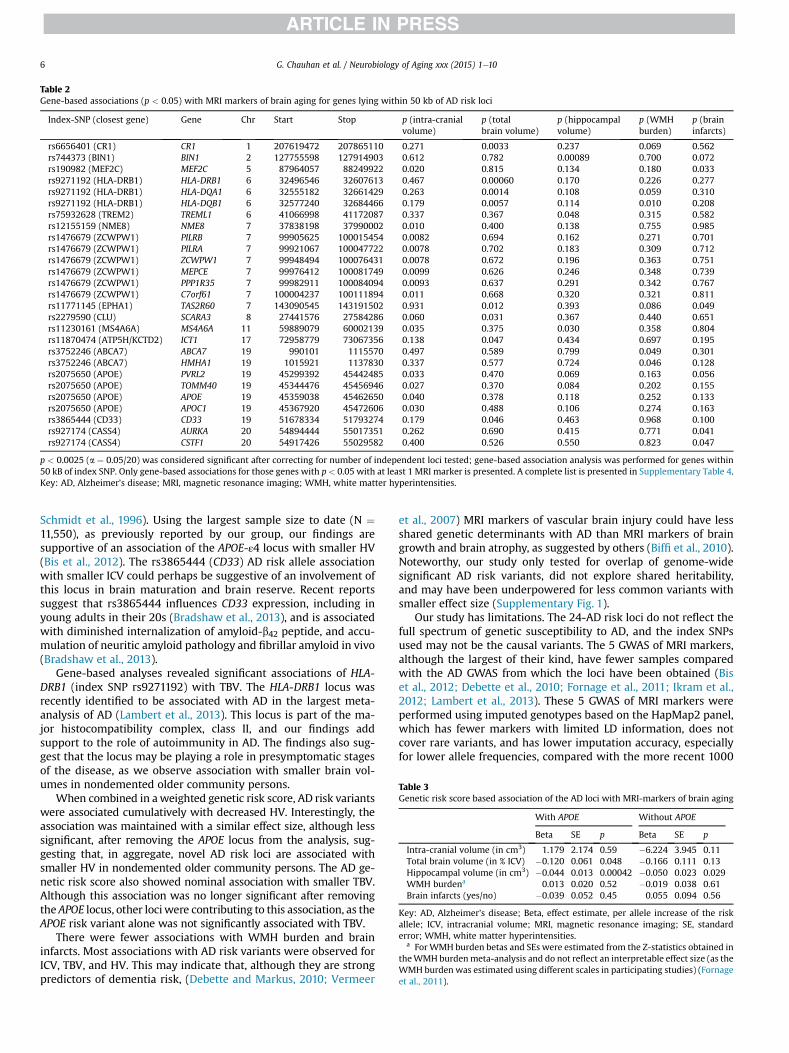
Table 3Genetic risk score based association of the AD loci with MRI-markers of brain aging
With APOE Without APOE
Beta SE p Beta SE p
Intra-cranial volume (in cm3) 1.179 2.174 0.59 �6.224 3.945 0.11Total brain volume (in % ICV) �0.120 0.061 0.048 �0.166 0.111 0.13Hippocampal volume (in cm3) �0.044 0.013 0.00042 �0.050 0.023 0.029WMH burdena 0.013 0.020 0.52 �0.019 0.038 0.61Brain infarcts (yes/no) �0.039 0.052 0.45 0.055 0.094 0.56
Key: AD, Alzheimer’s disease; Beta, effect estimate, per allele increase of the riskallele; ICV, intracranial volume; MRI, magnetic resonance imaging; SE, standarderror; WMH, white matter hyperintensities.
a For WMH burden betas and SEs were estimated from the Z-statistics obtained intheWMH burdenmeta-analysis and do not reflect an interpretable effect size (as theWMH burden was estimated using different scales in participating studies) (Fornageet al., 2011).
Table 2Gene-based associations (p < 0.05) with MRI markers of brain aging for genes lying within 50 kb of AD risk loci
Index-SNP (closest gene) Gene Chr Start Stop p (intra-cranialvolume)
p (totalbrain volume)
p (hippocampalvolume)
p (WMHburden)
p (braininfarcts)
rs6656401 (CR1) CR1 1 207619472 207865110 0.271 0.0033 0.237 0.069 0.562rs744373 (BIN1) BIN1 2 127755598 127914903 0.612 0.782 0.00089 0.700 0.072rs190982 (MEF2C) MEF2C 5 87964057 88249922 0.020 0.815 0.134 0.180 0.033rs9271192 (HLA-DRB1) HLA-DRB1 6 32496546 32607613 0.467 0.00060 0.170 0.226 0.277rs9271192 (HLA-DRB1) HLA-DQA1 6 32555182 32661429 0.263 0.0014 0.108 0.059 0.310rs9271192 (HLA-DRB1) HLA-DQB1 6 32577240 32684466 0.179 0.0057 0.114 0.010 0.208rs75932628 (TREM2) TREML1 6 41066998 41172087 0.337 0.367 0.048 0.315 0.582rs12155159 (NME8) NME8 7 37838198 37990002 0.010 0.400 0.138 0.755 0.985rs1476679 (ZCWPW1) PILRB 7 99905625 100015454 0.0082 0.694 0.162 0.271 0.701rs1476679 (ZCWPW1) PILRA 7 99921067 100047722 0.0078 0.702 0.183 0.309 0.712rs1476679 (ZCWPW1) ZCWPW1 7 99948494 100076431 0.0078 0.672 0.196 0.363 0.751rs1476679 (ZCWPW1) MEPCE 7 99976412 100081749 0.0099 0.626 0.246 0.348 0.739rs1476679 (ZCWPW1) PPP1R35 7 99982911 100084094 0.0093 0.637 0.291 0.342 0.767rs1476679 (ZCWPW1) C7orf61 7 100004237 100111894 0.011 0.668 0.320 0.321 0.811rs11771145 (EPHA1) TAS2R60 7 143090545 143191502 0.931 0.012 0.393 0.086 0.049rs2279590 (CLU) SCARA3 8 27441576 27584286 0.060 0.031 0.367 0.440 0.651rs11230161 (MS4A6A) MS4A6A 11 59889079 60002139 0.035 0.375 0.030 0.358 0.804rs11870474 (ATP5H/KCTD2) ICT1 17 72958779 73067356 0.138 0.047 0.434 0.697 0.195rs3752246 (ABCA7) ABCA7 19 990101 1115570 0.497 0.589 0.799 0.049 0.301rs3752246 (ABCA7) HMHA1 19 1015921 1137830 0.337 0.577 0.724 0.046 0.128rs2075650 (APOE) PVRL2 19 45299392 45442485 0.033 0.470 0.069 0.163 0.056rs2075650 (APOE) TOMM40 19 45344476 45456946 0.027 0.370 0.084 0.202 0.155rs2075650 (APOE) APOE 19 45359038 45462650 0.040 0.378 0.118 0.252 0.133rs2075650 (APOE) APOC1 19 45367920 45472606 0.030 0.488 0.106 0.274 0.163rs3865444 (CD33) CD33 19 51678334 51793274 0.179 0.046 0.463 0.968 0.100rs927174 (CASS4) AURKA 20 54894444 55017351 0.262 0.690 0.415 0.771 0.041rs927174 (CASS4) CSTF1 20 54917426 55029582 0.400 0.526 0.550 0.823 0.047
p < 0.0025 (a ¼ 0.05/20) was considered significant after correcting for number of independent loci tested; gene-based association analysis was performed for genes within50 kB of index SNP. Only gene-based associations for those genes with p< 0.05 with at least 1 MRI marker is presented. A complete list is presented in Supplementary Table 4.Key: AD, Alzheimer’s disease; MRI, magnetic resonance imaging; WMH, white matter hyperintensities.
G. Chauhan et al. / Neurobiology of Aging xxx (2015) 1e106
Schmidt et al., 1996). Using the largest sample size to date (N ¼11,550), as previously reported by our group, our findings aresupportive of an association of the APOE-ε4 locus with smaller HV(Bis et al., 2012). The rs3865444 (CD33) AD risk allele associationwith smaller ICV could perhaps be suggestive of an involvement ofthis locus in brain maturation and brain reserve. Recent reportssuggest that rs3865444 influences CD33 expression, including inyoung adults in their 20s (Bradshaw et al., 2013), and is associatedwith diminished internalization of amyloid-b42 peptide, and accu-mulation of neuritic amyloid pathology and fibrillar amyloid in vivo(Bradshaw et al., 2013).
Gene-based analyses revealed significant associations of HLA-DRB1 (index SNP rs9271192) with TBV. The HLA-DRB1 locus wasrecently identified to be associated with AD in the largest meta-analysis of AD (Lambert et al., 2013). This locus is part of the ma-jor histocompatibility complex, class II, and our findings addsupport to the role of autoimmunity in AD. The findings also sug-gest that the locus may be playing a role in presymptomatic stagesof the disease, as we observe association with smaller brain vol-umes in nondemented older community persons.
When combined in aweighted genetic risk score, AD risk variantswere associated cumulatively with decreased HV. Interestingly, theassociation was maintained with a similar effect size, although lesssignificant, after removing the APOE locus from the analysis, sug-gesting that, in aggregate, novel AD risk loci are associated withsmaller HV in nondemented older community persons. The AD ge-netic risk score also showed nominal association with smaller TBV.Although this association was no longer significant after removingthe APOE locus, other lociwere contributing to this association, as theAPOE risk variant alone was not significantly associated with TBV.
There were fewer associations with WMH burden and braininfarcts. Most associations with AD risk variants were observed forICV, TBV, and HV. This may indicate that, although they are strongpredictors of dementia risk, (Debette and Markus, 2010; Vermeer
et al., 2007) MRI markers of vascular brain injury could have lessshared genetic determinants with AD than MRI markers of braingrowth and brain atrophy, as suggested by others (Biffi et al., 2010).Noteworthy, our study only tested for overlap of genome-widesignificant AD risk variants, did not explore shared heritability,and may have been underpowered for less common variants withsmaller effect size (Supplementary Fig. 1).
Our study has limitations. The 24-AD risk loci do not reflect thefull spectrum of genetic susceptibility to AD, and the index SNPsused may not be the causal variants. The 5 GWAS of MRI markers,although the largest of their kind, have fewer samples comparedwith the AD GWAS from which the loci have been obtained (Biset al., 2012; Debette et al., 2010; Fornage et al., 2011; Ikram et al.,2012; Lambert et al., 2013). These 5 GWAS of MRI markers wereperformed using imputed genotypes based on the HapMap2 panel,which has fewer markers with limited LD information, does notcover rare variants, and has lower imputation accuracy, especiallyfor lower allele frequencies, compared with the more recent 1000
G. Chauhan et al. / Neurobiology of Aging xxx (2015) 1e10 7
genomes reference panels. We therefore could not analyze rare ADrisk variants in the present study, and we cannot exclude that themore limited LD information might have introduced some bias inthe results of the gene-based analyses. In addition, despite majorefforts to harmonize phenotype definitions across studies, theremay be some residual heterogeneity in methods for quantifyingMRImarkers of brain aging. These elements could have reduced ourpower to detect associations of AD GWAS loci with MRI markers ofbrain aging. The choice of 50 kb window for a gene-based test doesnot account for potential regulatory effects on more distant genes.Our findings cannot be generalized to populations of non-Europeanancestry. Ongoing, larger multiethnic GWAS of MRI markers ofbrain aging, as well as sequencing projects searching for rare vari-ants associated with AD risk, and MRI phenotypes may enable us toexpand our findings in the future.
5. Conclusion
In conclusion, we have shown that novel AD genetic risk variantsare associated with MRI markers of structural brain aging in oldernondemented community persons. In aggregate, novel AD geneticrisk variants were associated with smaller brain volumes, especiallyHV. Significant gene-based associations and suggestive single SNP-based associations with ICV, TBV, and HV also provide interestinghypotheses for mechanisms underlying genetic associations withAD.
Disclosure statement
The authors have no conflicts of interest to disclose.
Acknowledgements
The authors thank the staff and participants of all participatingstudy for their important contributions.
Aging Gene-Environment Susceptibility-Reykjavik Study: Theresearch has been funded by NIA contract N01-AG-12100 withcontributions from NEI, NIDCD, and NHLBI, the NIA IntramuralResearch Program, Hjartavernd (the Icelandic Heart Association),and the Althingi (the Icelandic Parliament).
The Atherosclerosis Risk in Communities Study: The researchis carried out as a collaborative study supported by NationalHeart, Lung, and Blood Institute contracts (HHSN268201100005C,HHSN268201100006C, HHSN268201100007C, HHSN268201100008C,HHSN268201100009C, HHSN268201100010C, HHSN268201100011C,and HHSN268201100012C), R01HL087641, R01HL59367 andR01HL086694 and R01HL7825; National Human Genome ResearchInstitute contract U01HG004402; and National Institutes of Healthcontract HHSN268200625226C. The authors thank the staff and par-ticipants of the ARIC study for their important contributions. Infra-structure was partly supported by Grant Number UL1RR025005, acomponent of the National Institutes of Health (UL1RR025005) andNIH Roadmap for Medical Research. Funds for these projects were alsosupported by grant HL093029 to Myriam Fornage.
Cardiovascular Health Study: This CHS research was supported byNHLBI contracts HHSN268201200036C, HHSN268200800007C,N01HC55222, N01HC85079, N01HC85080, N01HC85081, N01HC85082,N01HC85083, N01HC85086, N01HC15103; and NHLBI grantsU01HL080295, R01HL087652, R01HL105756, R01HL103612, andR01HL120393 with additional contribution from the National Instituteof Neurological Disorders and Stroke (NINDS).
The Austrian Stroke Prevention Study: The research reported inthis article was funded by the Austrian Science Fond (FWF) grantnumber P20545-P05 and P13180. The Medical University of Grazsupports the databank of the ASPS. The authors thank the staff and
the participants of the ASPS for their valuable contributions. Theauthors thank Birgit Reinhart for her long-term administrativecommitment and Ing Johann Semmler for the technical assistanceat creating the DNA bank.
Erasmus Rucphen Family Study: This study is financially sup-ported by the Netherlands Organization for Scientific Research(NWO), the Internationale Stichting Alzheimer Onderzoek (ISAO),the Hersenstichting Nederland (HSN), and the Centre for MedicalSystems Biology (CMSB) in the framework of the Netherlands Ge-nomics Initiative (NGI). The authors thank the participants from theGenetic Research in Isolated Populations, Erasmus Rucphen Familywho made this work possible.
Framingham Heart Study: This work was supported by theFramingham Heart Study’s National Heart, Lung, and Blood Insti-tute contract (N01-HC-25195) and its contract with Affymetrix, Incfor genotyping services (Contract No. N02-HL-6e4278). A portionof this research utilized the Linux Cluster for Genetic Analysis(LinGA-II) funded by the Robert Dawson Evans Endowment of theDepartment of Medicine at Boston University School of Medicineand Boston Medical Center. It was also funded by grants from theNational Institute on Aging (R01 AG08122, AG033193, P30AG0101029) and the National Institute of Neurological Disordersand Stroke (R01 NS17950).
The Religious Order Study (ROS) and Rush Memory and AgingProject (R-MAP): The R-MAP and ROS data used in this article wassupported by National Institute on Aging grants P30AG10161,R01AG17917, and R01AG15819, National Institutes of Health grants(R01 AG040039, R21 NS076827, P20 MD006886) and the IllinoisDepartment of Public Health.
The Rotterdam Study: The GWA database of the RotterdamStudy was funded through the Netherlands Organization of Scien-tific Research NWO (nr. 175.010.2005.011). This study was furthersupported by the Netherlands Genomics Initiative (NGI)/Netherlands Organisation for Scientific Research (NWO) project nr.050-060-810. The Rotterdam Study is supported by the ErasmusMedical Center and Erasmus University, Rotterdam; theNetherlands Organization for Scientific Research (NWO), theNetherlands Organization for the Health Research and Develop-ment (ZonMw; Veni-grant 916.13.054), the Research Institute forDiseases in the Elderly (RIDE), the Ministry of Education, Cultureand Science, the Ministry for Health, Welfare and Sports, the Eu-ropean Commission (DG XII), the Municipality of Rotterdam, andthe Internationale Stichting Alzheimer Onderzoek.
The Tasmanian Study of Gait and Cognition (TASCOG) is sup-ported by Project Grants from the National Health and MedicalResearch Council (NHMRC IDs 403000, 491109, 606543), and agrant from the Wicking Dementia Education and Research Centre,Hobart. Velandai Srikanth is supported by an NHMRC/NationalHeart Foundation Career Development Fellowship (ID 606544).
Three City Study (3C): The authors thank the staff and the par-ticipants of the 3C Study for their important contributions. The 3CStudy is conducted under a partnership agreement between theInstitut National de la Santé et de la Recherche Médicale (INSERM),the Victor SegaleneBordeaux II University, and Sanofi-Aventis. TheFondation pour la Recherche Médicale funded the preparation andinitiation of the study. The 3C Study is also supported by the CaisseNationaleMaladie des Travailleurs Salariés, Direction Générale de laSanté, Mutuelle Générale de l’Education Nationale (MGEN), Institutde la Longévité, Conseils Régionaux of Aquitaine and Bourgogne,Fondation de France, and Ministry of ResearcheINSERM Pro-gramme “Cohortes et collections de données biologiques.” LilleGénopôle received an unconditional grant from Eisai. The authorsthank A. Boland (Centre National de Génotypage) for her technicalhelp in preparing the DNA samples for analyses. This work wassupported by the National Foundation for Alzheimer’s Disease and
G. Chauhan et al. / Neurobiology of Aging xxx (2015) 1e108
Related Disorders, the Institut Pasteur de Lille and the Centre Na-tional de Génotypage. Ganesh Chauhan and Stéphanie Debette aresupported by a grant from the Fondation Leducq and the AgenceNationale de la Recherche (Chaire d’Excellence).
The authors also thank Josée Dupuis (Department of Biostatis-tics, Boston University School of Public Health, Boston, Massachu-setts, USA) and Toby Johnson (Department of Medical Genetics,University of Lausanne, Lausanne, Switzerland) for their statisticaladvice.
Appendix A. Supplementary data
Supplementary data associated with this article can be found, inthe online version, at http://dx.doi.org/10.1016/j.neurobiolaging.2014.12.028.
References
Ballard, C., Gauthier, S., Corbett, A., Brayne, C., Aarsland, D., Jones, E., 2011. Alz-heimer’s disease. Lancet 377, 1019e1031.
Biffi, A., Anderson, C.D., Desikan, R.S., Sabuncu, M., Cortellini, L., Schmansky, N.,Salat, D., Rosand, J., 2010. Genetic variation and neuroimaging measures inAlzheimer disease. Arch. Neurol. 67, 677e685.
Bis, J.C., DeCarli, C., Smith, A.V., van der Lijn, F., Crivello, F., Fornage, M., Debette, S.,Shulman, J.M., Schmidt, H., Srikanth, V., Schuur, M., Yu, L., Choi, S.H.,Sigurdsson, S., Verhaaren, B.F., DeStefano, A.L., Lambert, J.C., Jack Jr., C.R.,Struchalin, M., Stankovich, J., Ibrahim-Verbaas, C.A., Fleischman, D.,Zijdenbos, A., den Heijer, T., Mazoyer, B., Coker, L.H., Enzinger, C., Danoy, P.,Amin, N., Arfanakis, K., van Buchem, M.A., de Bruijn, R.F., Beiser, A., Dufouil, C.,Huang, J., Cavalieri, M., Thomson, R., Niessen, W.J., Chibnik, L.B., Gislason, G.K.,Hofman, A., Pikula, A., Amouyel, P., Freeman, K.B., Phan, T.G., Oostra, B.A.,Stein, J.L., Medland, S.E., Vasquez, A.A., Hibar, D.P., Wright, M.J., Franke, B.,Martin, N.G., Thompson, P.M., Nalls, M.A., Uitterlinden, A.G., Au, R., Elbaz, A.,Beare, R.J., van Swieten, J.C., Lopez, O.L., Harris, T.B., Chouraki, V., Breteler, M.M.,De Jager, P.L., Becker, J.T., Vernooij, M.W., Knopman, D., Fazekas, F., Wolf, P.A., vander Lugt, A., Gudnason, V., Longstreth Jr., W.T., Brown, M.A., Bennett, D.A., vanDuijn, C.M., Mosley, T.H., Schmidt, R., Tzourio, C., Launer, L.J., Ikram, M.A.,Seshadri, S., 2012. Common variants at 12q14 and 12q24 are associated withhippocampal volume. Nat. Genet. 44, 545e551.
Boada, M., Antunez, C., Ramirez-Lorca, R., Destefano, A.L., Gonzalez-Perez, A.,Gayan, J., Lopez-Arrieta, J., Ikram, M.A., Hernandez, I., Marin, J., Galan, J.J.,Bis, J.C., Mauleon, A., Rosende-Roca, M., Moreno-Rey, C., Gudnasson, V.,Moron, F.J., Velasco, J., Carrasco, J.M., Alegret, M., Espinosa, A., Vinyes, G.,Lafuente, A., Vargas, L., Fitzpatrick, A.L., Launer, L.J., Saez, M.E., Vazquez, E.,Becker, J.T., Lopez, O.L., Serrano-Rios, M., Tarraga, L., van Duijn, C.M., Real, L.M.,Seshadri, S., Ruiz, A., 2013. ATP5H/KCTD2 locus is associated with Alzheimer’sdisease risk. Mol. Psychiatry 19, 682e687.
Bradshaw, E.M., Chibnik, L.B., Keenan, B.T., Ottoboni, L., Raj, T., Tang, A.,Rosenkrantz, L.L., Imboywa, S., Lee, M., Von Korff, A., Morris, M.C., Evans, D.A.,Johnson, K., Sperling, R.A., Schneider, J.A., Bennett, D.A., De Jager, P.L., 2013.CD33 Alzheimer’s disease locus: altered monocyte function and amyloidbiology. Nat. Neurosci. 16, 848e850.
Carrasquillo, M.M., Khan, Q.U., Murray, M.E., Krishnan, S., Aakre, J., Pankratz, V.S.,Nguyen, T., Ma, L., Bisceglio, G., Petersen, R.C., Younkin, S.G., Dickson, D.W.,Boeve, B.F., Graff-Radford, N.R., Ertekin-Taner, N., 2014. Late-onset Alzheimerdisease genetic variants in posterior cortical atrophy and posterior AD.Neurology 82, 1455e1462.
Carrasquillo, M.M., Zou, F., Pankratz, V.S., Wilcox, S.L., Ma, L., Walker, L.P.,Younkin, S.G., Younkin, C.S., Younkin, L.H., Bisceglio, G.D., Ertekin-Taner, N.,Crook, J.E., Dickson, D.W., Petersen, R.C., Graff-Radford, N.R., 2009. Geneticvariation in PCDH11X is associated with susceptibility to late-onset Alzheimer’sdisease. Nat. Genet. 41, 192e198.
Dastani, Z., Hivert, M.F., Timpson, N., Perry, J.R., Yuan, X., Scott, R.A., Henneman, P.,Heid, I.M., Kizer, J.R., Lyytikainen, L.P., Fuchsberger, C., Tanaka, T., Morris, A.P.,Small, K., Isaacs, A., Beekman, M., Coassin, S., Lohman, K., Qi, L., Kanoni, S.,Pankow, J.S., Uh, H.W., Wu, Y., Bidulescu, A., Rasmussen-Torvik, L.J.,Greenwood, C.M., Ladouceur, M., Grimsby, J., Manning, A.K., Liu, C.T., Kooner, J.,Mooser, V.E., Vollenweider, P., Kapur, K.A., Chambers, J., Wareham, N.J.,Langenberg, C., Frants, R., Willems-Vandijk, K., Oostra, B.A., Willems, S.M.,Lamina, C., Winkler, T.W., Psaty, B.M., Tracy, R.P., Brody, J., Chen, I., Viikari, J.,Kahonen, M., Pramstaller, P.P., Evans, D.M., St Pourcain, B., Sattar, N., Wood, A.R.,Bandinelli, S., Carlson, O.D., Egan, J.M., Bohringer, S., van Heemst, D., Kedenko, L.,Kristiansson, K., Nuotio, M.L., Loo, B.M., Harris, T., Garcia, M., Kanaya, A.,Haun, M., Klopp, N., Wichmann, H.E., Deloukas, P., Katsareli, E., Couper, D.J.,Duncan, B.B., Kloppenburg, M., Adair, L.S., Borja, J.B., Wilson, J.G., Musani, S.,Guo, X., Johnson, T., Semple, R., Teslovich, T.M., Allison, M.A., Redline, S.,Buxbaum, S.G., Mohlke, K.L., Meulenbelt, I., Ballantyne, C.M., Dedoussis, G.V.,Hu, F.B., Liu, Y., Paulweber, B., Spector, T.D., Slagboom, P.E., Ferrucci, L., Jula, A.,Perola, M., Raitakari, O., Florez, J.C., Salomaa, V., Eriksson, J.G., Frayling, T.M.,
Hicks, A.A., Lehtimaki, T., Smith, G.D., Siscovick, D.S., Kronenberg, F., vanDuijn, C., Loos, R.J., Waterworth, D.M., Meigs, J.B., Dupuis, J., Richards, J.B.,Voight, B.F., Scott, L.J., Steinthorsdottir, V., Dina, C., Welch, R.P., Zeggini, E.,Huth, C., Aulchenko, Y.S., Thorleifsson, G., McCulloch, L.J., Ferreira, T., Grallert, H.,Amin, N., Wu, G., Willer, C.J., Raychaudhuri, S., McCarroll, S.A., Hofmann, O.M.,Segre, A.V., van Hoek, M., Navarro, P., Ardlie, K., Balkau, B., Benediktsson, R.,Bennett, A.J., Blagieva, R., Boerwinkle, E., Bonnycastle, L.L., Bostrom, K.B.,Bravenboer, B., Bumpstead, S., Burtt, N.P., Charpentier, G., Chines, P.S.,Cornelis, M., Crawford, G., Doney, A.S., Elliott, K.S., Elliott, A.L., Erdos, M.R.,Fox, C.S., Franklin, C.S., Ganser, M., Gieger, C., Grarup, N., Green, T., Griffin, S.,Groves, C.J., Guiducci, C., Hadjadj, S., Hassanali, N., Herder, C., Isomaa, B.,Jackson, A.U., Johnson, P.R., Jorgensen, T., Kao, W.H., Kong, A., Kraft, P.,Kuusisto, J., Lauritzen, T., Li, M., Lieverse, A., Lindgren, C.M., Lyssenko, V.,Marre, M., Meitinger, T., Midthjell, K., Morken, M.A., Narisu, N., Nilsson, P.,Owen, K.R., Payne, F., Petersen, A.K., Platou, C., Proenca, C., Prokopenko, I.,Rathmann, W., Rayner, N.W., Robertson, N.R., Rocheleau, G., Roden, M.,Sampson, M.J., Saxena, R., Shields, B.M., Shrader, P., Sigurdsson, G., Sparso, T.,Strassburger, K., Stringham, H.M., Sun, Q., Swift, A.J., Thorand, B., Tichet, J.,Tuomi, T., van Dam, R.M., van Haeften, T.W., van Herpt, T., van Vliet-Ostaptchouk, J.V., Walters, G.B., Weedon, M.N., Wijmenga, C., Witteman, J.,Bergman, R.N., Cauchi, S., Collins, F.S., Gloyn, A.L., Gyllensten, U., Hansen, T.,Hide, W.A., Hitman, G.A., Hofman, A., Hunter, D.J., Hveem, K., Laakso, M.,Morris, A.D., Palmer, C.N., Rudan, I., Sijbrands, E., Stein, L.D., Tuomilehto, J.,Uitterlinden, A., Walker, M., Watanabe, R.M., Abecasis, G.R., Boehm, B.O.,Campbell, H., Daly, M.J., Hattersley, A.T., Pedersen, O., Barroso, I., Groop, L.,Sladek, R., Thorsteinsdottir, U., Wilson, J.F., Illig, T., Froguel, P., van Duijn, C.M.,Stefansson, K., Altshuler, D., Boehnke, M., McCarthy, M.I., Soranzo, N.,Wheeler, E., Glazer, N.L., Bouatia-Naji, N., Magi, R., Randall, J., Elliott, P., Rybin, D.,Dehghan, A., Hottenga, J.J., Song, K., Goel, A., Lajunen, T., Doney, A., Cavalcanti-Proenca, C., Kumari, M., Timpson, N.J., Zabena, C., Ingelsson, E., An, P.,O’Connell, J., Luan, J., Elliott, A., Roccasecca, R.M., Pattou, F., Sethupathy, P.,Ariyurek, Y., Barter, P., Beilby, J.P., Ben-Shlomo, Y., Bergmann, S., Bochud, M.,Bonnefond, A., Borch-Johnsen, K., Bottcher, Y., Brunner, E., Bumpstead, S.J.,Chen, Y.D., Chines, P., Clarke, R., Coin, L.J., Cooper, M.N., Crisponi, L., Day, I.N., deGeus, E.J., Delplanque, J., Fedson, A.C., Fischer-Rosinsky, A., Forouhi, N.G.,Franzosi, M.G., Galan, P., Goodarzi, M.O., Graessler, J., Grundy, S., Gwilliam, R.,Hallmans, G., Hammond, N., Han, X., Hartikainen, A.L., Hayward, C., Heath, S.C.,Hercberg, S., Hillman, D.R., Hingorani, A.D., Hui, J., Hung, J., Kaakinen, M.,Kaprio, J., Kesaniemi, Y.A., Kivimaki, M., Knight, B., Koskinen, S., Kovacs, P.,Kyvik, K.O., Lathrop, G.M., Lawlor, D.A., Le Bacquer, O., Lecoeur, C., Li, Y.,Mahley, R., Mangino, M., Martinez-Larrad, M.T., McAteer, J.B., McPherson, R.,Meisinger, C., Melzer, D., Meyre, D., Mitchell, B.D., Mukherjee, S., Naitza, S.,Neville, M.J., Orru, M., Pakyz, R., Paolisso, G., Pattaro, C., Pearson, D., Peden, J.F.,Pedersen, N.L., Pfeiffer, A.F., Pichler, I., Polasek, O., Posthuma, D., Potter, S.C.,Pouta, A., Province, M.A., Rice, K., Ripatti, S., Rivadeneira, F., Rolandsson, O.,Sandbaek, A., Sandhu, M., Sanna, S., Sayer, A.A., Scheet, P., Seedorf, U., Sharp, S.J.,Shields, B., Sigurethsson, G., Sijbrands, E.J., Silveira, A., Simpson, L., Singleton, A.,Smith, N.L., Sovio, U., Swift, A., Syddall, H., Syvanen, A.C., Tonjes, A.,Uitterlinden, A.G., van Dijk, K.W., Varma, D., Visvikis-Siest, S., Vitart, V.,Vogelzangs, N., Waeber, G., Wagner, P.J., Walley, A., Ward, K.L., Watkins, H.,Wild, S.H., Willemsen, G., Witteman, J.C., Yarnell, J.W., Zelenika, D., Zethelius, B.,Zhai, G., Zhao, J.H., Zillikens, M.C., Borecki, I.B., Meneton, P., Magnusson, P.K.,Nathan, D.M., Williams, G.H., Silander, K., Bornstein, S.R., Schwarz, P.,Spranger, J., Karpe, F., Shuldiner, A.R., Cooper, C., Serrano-Rios, M., Lind, L.,Palmer, L.J., Hu, F.B.s., Franks, P.W., Ebrahim, S., Marmot, M., Wright, A.F.,Stumvoll, M., Hamsten, A., Buchanan, T.A., Valle, T.T., Rotter, J.I., Penninx, B.W.,Boomsma, D.I., Cao, A., Scuteri, A., Schlessinger, D., Uda, M., Ruokonen, A.,Jarvelin, M.R., Peltonen, L., Mooser, V., Musunuru, K., Smith, A.V.,Edmondson, A.C., Stylianou, I.M., Koseki, M., Pirruccello, J.P., Chasman, D.I.,Johansen, C.T., Fouchier, S.W., Peloso, G.M., Barbalic, M., Ricketts, S.L., Bis, J.C.,Feitosa, M.F., Orho-Melander, M., Melander, O., Li, X., Cho, Y.S., Go, M.J., Kim, Y.J.,Lee, J.Y., Park, T., Kim, K., Sim, X., Ong, R.T., Croteau-Chonka, D.C., Lange, L.A.,Smith, J.D., Ziegler, A., Zhang, W., Zee, R.Y., Whitfield, J.B., Thompson, J.R.,Surakka, I., Smit, J.H., Sinisalo, J., Scott, J., Saharinen, J., Sabatti, C., Rose, L.M.,Roberts, R., Rieder, M., Parker, A.N., Pare, G., O’Donnell, C.J., Nieminen, M.S.,Nickerson, D.A., Montgomery, G.W., McArdle, W., Masson, D., Martin, N.G.,Marroni, F., Lucas, G., Luben, R., Lokki, M.L., Lettre, G., Launer, L.J., Lakatta, E.G.,Laaksonen, R., Konig, I.R., Khaw, K.T., Kaplan, L.M., Johansson, A., Janssens, A.C.,Igl, W., Hovingh, G.K., Hengstenberg, C., Havulinna, A.S., Hastie, N.D., Harris, T.B.,Haritunians, T., Hall, A.S., Groop, L.C., Gonzalez, E., Freimer, N.B., Erdmann, J.,Ejebe, K.G., Doring, A., Dominiczak, A.F., Demissie, S., de Faire, U., Caulfield, M.J.,Boekholdt, S.M., Assimes, T.L., Quertermous, T., Seielstad, M., Wong, T.Y., Tai, E.S.,Feranil, A.B., Kuzawa, C.W., Taylor Jr., H.A., Gabriel, S.B., Holm, H., Gudnason, V.,Krauss, R.M., Ordovas, J.M., Munroe, P.B., Kooner, J.S., Tall, A.R., Hegele, R.A.,Kastelein, J.J., Schadt, E.E., Strachan, D.P., Reilly, M.P., Samani, N.J., Schunkert, H.,Cupples, L.A., Sandhu, M.S., Ridker, P.M., Rader, D.J., Kathiresan, S., 2012. Novelloci for adiponectin levels and their influence on type 2 diabetes and metabolictraits: a multi-ethnic meta-analysis of 45,891 individuals. PLoS Genet. 8,e1002607.
Debette, S., Bis, J.C., Fornage, M., Schmidt, H., Ikram, M.A., Sigurdsson, S., Heiss, G.,Struchalin, M., Smith, A.V., van der Lugt, A., DeCarli, C., Lumley, T.,Knopman, D.S., Enzinger, C., Eiriksdottir, G., Koudstaal, P.J., DeStefano, A.L.,Psaty, B.M., Dufouil, C., Catellier, D.J., Fazekas, F., Aspelund, T., Aulchenko, Y.S.,Beiser, A., Rotter, J.I., Tzourio, C., Shibata, D.K., Tscherner, M., Harris, T.B.,Rivadeneira, F., Atwood, L.D., Rice, K., Gottesman, R.F., van Buchem, M.A.,
G. Chauhan et al. / Neurobiology of Aging xxx (2015) 1e10 9
Uitterlinden, A.G., Kelly-Hayes, M., Cushman, M., Zhu, Y., Boerwinkle, E.,Gudnason, V., Hofman, A., Romero, J.R., Lopez, O., van Duijn, C.M., Au, R.,Heckbert, S.R., Wolf, P.A., Mosley, T.H., Seshadri, S., Breteler, M.M., Schmidt, R.,Launer, L.J., Longstreth Jr., W.T., 2010. Genome-wide association studies of MRI-defined brain infarcts: meta-analysis from the CHARGE consortium. Stroke 41,210e217.
Debette, S., Markus, H.S., 2010. The clinical importance of white matter hyper-intensities on brain magnetic resonance imaging: systematic review and meta-analysis. BMJ 341, c3666.
den Heijer, T., Oudkerk, M., Launer, L.J., van Duijn, C.M., Hofman, A., Breteler, M.M.,2002. Hippocampal, amygdalar, and global brain atrophy in different apolipo-protein E genotypes. Neurology 59, 746e748.
Ferencz, B., Laukka, E.J., Lovden, M., Kalpouzos, G., Keller, L., Graff, C., Wahlund, L.O.,Fratiglioni, L., Backman, L., 2013. The influence of APOE and TOMM40 poly-morphisms on hippocampal volume and episodic memory in old age. Front.Hum. Neurosci. 7, 198.
Fornage, M., Debette, S., Bis, J.C., Schmidt, H., Ikram, M.A., Dufouil, C., Sigurdsson, S.,Lumley, T., DeStefano, A.L., Fazekas, F., Vrooman, H.A., Shibata, D.K., Maillard, P.,Zijdenbos, A., Smith, A.V., Gudnason, H., de Boer, R., Cushman, M., Mazoyer, B.,Heiss, G., Vernooij, M.W., Enzinger, C., Glazer, N.L., Beiser, A., Knopman, D.S.,Cavalieri, M., Niessen, W.J., Harris, T.B., Petrovic, K., Lopez, O.L., Au, R.,Lambert, J.C., Hofman, A., Gottesman, R.F., Garcia, M., Heckbert, S.R.,Atwood, L.D., Catellier, D.J., Uitterlinden, A.G., Yang, Q., Smith, N.L., Aspelund, T.,Romero, J.R., Rice, K., Taylor, K.D., Nalls, M.A., Rotter, J.I., Sharrett, R., vanDuijn, C.M., Amouyel, P., Wolf, P.A., Gudnason, V., van der Lugt, A.,Boerwinkle, E., Psaty, B.M., Seshadri, S., Tzourio, C., Breteler, M.M., Mosley, T.H.,Schmidt, R., Longstreth, W.T., DeCarli, C., Launer, L.J., 2011. Genome-wide as-sociation studies of cerebral white matter lesion burden: the CHARGE con-sortium. Ann. Neurol. 69, 928e939.
Furney, S.J., Simmons, A., Breen, G., Pedroso, I., Lunnon, K., Proitsi, P., Hodges, A.,Powell, J., Wahlund, L.O., Kloszewska, I., Mecocci, P., Soininen, H., Tsolaki, M.,Vellas, B., Spenger, C., Lathrop, M., Shen, L., Kim, S., Saykin, A.J., Weiner, M.W.,Lovestone, S., 2011. Genome-wide association with MRI atrophy measures as aquantitative trait locus for Alzheimer’s disease. Mol. Psychiatry 16, 1130e1138.
Gauderman, W.J., 2002a. Sample size requirements for association studies of gene-gene interaction. Am. J. Epidemiol. 155, 478e484.
Gauderman, W.J., 2002b. Sample size requirements for matched case-controlstudies of gene-environment interaction. Stat. Med. 21, 35e50.
Harold, D., Abraham, R., Hollingworth, P., Sims, R., Gerrish, A., Hamshere, M.L.,Pahwa, J.S., Moskvina, V., Dowzell, K., Williams, A., Jones, N., Thomas, C.,Stretton, A., Morgan, A.R., Lovestone, S., Powell, J., Proitsi, P., Lupton, M.K.,Brayne, C., Rubinsztein, D.C., Gill, M., Lawlor, B., Lynch, A., Morgan, K.,Brown, K.S., Passmore, P.A., Craig, D., McGuinness, B., Todd, S., Holmes, C.,Mann, D., Smith, A.D., Love, S., Kehoe, P.G., Hardy, J., Mead, S., Fox, N., Rossor, M.,Collinge, J., Maier, W., Jessen, F., Schurmann, B., van den Bussche, H., Heuser, I.,Kornhuber, J., Wiltfang, J., Dichgans, M., Frolich, L., Hampel, H., Hull, M.,Rujescu, D., Goate, A.M., Kauwe, J.S., Cruchaga, C., Nowotny, P., Morris, J.C.,Mayo, K., Sleegers, K., Bettens, K., Engelborghs, S., De Deyn, P.P., VanBroeckhoven, C., Livingston, G., Bass, N.J., Gurling, H., McQuillin, A., Gwilliam, R.,Deloukas, P., Al-Chalabi, A., Shaw, C.E., Tsolaki, M., Singleton, A.B., Guerreiro, R.,Muhleisen, T.W., Nothen, M.M., Moebus, S., Jockel, K.H., Klopp, N.,Wichmann, H.E., Carrasquillo, M.M., Pankratz, V.S., Younkin, S.G., Holmans, P.A.,O’Donovan, M., Owen, M.J., Williams, J., 2009. Genome-wide association studyidentifies variants at CLU and PICALM associated with Alzheimer’s disease. Nat.Genet. 41, 1088e1093.
Hollingworth, P., Harold, D., Sims, R., Gerrish, A., Lambert, J.C., Carrasquillo, M.M.,Abraham, R., Hamshere, M.L., Pahwa, J.S., Moskvina, V., Dowzell, K., Jones, N.,Stretton, A., Thomas, C., Richards, A., Ivanov, D., Widdowson, C., Chapman, J.,Lovestone, S., Powell, J., Proitsi, P., Lupton, M.K., Brayne, C., Rubinsztein, D.C.,Gill, M., Lawlor, B., Lynch, A., Brown, K.S., Passmore, P.A., Craig, D.,McGuinness, B., Todd, S., Holmes, C., Mann, D., Smith, A.D., Beaumont, H.,Warden, D., Wilcock, G., Love, S., Kehoe, P.G., Hooper, N.M., Vardy, E.R., Hardy, J.,Mead, S., Fox, N.C., Rossor, M., Collinge, J., Maier, W., Jessen, F., Ruther, E.,Schurmann, B., Heun, R., Kolsch, H., van den Bussche, H., Heuser, I., Kornhuber, J.,Wiltfang, J., Dichgans, M., Frolich, L., Hampel, H., Gallacher, J., Hull, M.,Rujescu, D., Giegling, I., Goate, A.M., Kauwe, J.S., Cruchaga, C., Nowotny, P.,Morris, J.C., Mayo, K., Sleegers, K., Bettens, K., Engelborghs, S., De Deyn, P.P., VanBroeckhoven, C., Livingston, G., Bass, N.J., Gurling, H., McQuillin, A., Gwilliam, R.,Deloukas, P., Al-Chalabi, A., Shaw, C.E., Tsolaki, M., Singleton, A.B., Guerreiro, R.,Muhleisen, T.W., Nothen, M.M., Moebus, S., Jockel, K.H., Klopp, N.,Wichmann, H.E., Pankratz, V.S., Sando, S.B., Aasly, J.O., Barcikowska, M.,Wszolek, Z.K., Dickson, D.W., Graff-Radford, N.R., Petersen, R.C., van Duijn, C.M.,Breteler, M.M., Ikram, M.A., DeStefano, A.L., Fitzpatrick, A.L., Lopez, O.,Launer, L.J., Seshadri, S., Berr, C., Campion, D., Epelbaum, J., Dartigues, J.F.,Tzourio, C., Alperovitch, A., Lathrop, M., Feulner, T.M., Friedrich, P., Riehle, C.,Krawczak, M., Schreiber, S., Mayhaus, M., Nicolhaus, S., Wagenpfeil, S.,Steinberg, S., Stefansson, H., Stefansson, K., Snaedal, J., Bjornsson, S.,Jonsson, P.V., Chouraki, V., Genier-Boley, B., Hiltunen, M., Soininen, H.,Combarros, O., Zelenika, D., Delepine, M., Bullido, M.J., Pasquier, F., Mateo, I.,Frank-Garcia, A., Porcellini, E., Hanon, O., Coto, E., Alvarez, V., Bosco, P.,Siciliano, G., Mancuso, M., Panza, F., Solfrizzi, V., Nacmias, B., Sorbi, S., Bossu, P.,Piccardi, P., Arosio, B., Annoni, G., Seripa, D., Pilotto, A., Scarpini, E.,Galimberti, D., Brice, A., Hannequin, D., Licastro, F., Jones, L., Holmans, P.A.,Jonsson, T., Riemenschneider, M., Morgan, K., Younkin, S.G., Owen, M.J.,O’Donovan, M., Amouyel, P., Williams, J., 2011. Common variants at ABCA7,
MS4A6A/MS4A4E, EPHA1, CD33 and CD2AP are associated with Alzheimer’sdisease. Nat. Genet. 43, 429e435.
Ikram, M.A., Fornage, M., Smith, A.V., Seshadri, S., Schmidt, R., Debette, S.,Vrooman, H.A., Sigurdsson, S., Ropele, S., Taal, H.R., Mook-Kanamori, D.O.,Coker, L.H., Longstreth Jr., W.T., Niessen, W.J., DeStefano, A.L., Beiser, A.,Zijdenbos, A.P., Struchalin, M., Jack Jr., C.R., Rivadeneira, F., Uitterlinden, A.G.,Knopman, D.S., Hartikainen, A.L., Pennell, C.E., Thiering, E., Steegers, E.A.,Hakonarson, H., Heinrich, J., Palmer, L.J., Jarvelin, M.R., McCarthy, M.I., Grant, S.F.,St Pourcain, B., Timpson, N.J., Smith, G.D., Sovio, U., Nalls, M.A., Au, R.,Hofman, A., Gudnason, H., van der Lugt, A., Harris, T.B., Meeks, W.M.,Vernooij, M.W., van Buchem, M.A., Catellier, D., Jaddoe, V.W., Gudnason, V.,Windham, B.G., Wolf, P.A., van Duijn, C.M., Mosley Jr., T.H., Schmidt, H.,Launer, L.J., Breteler, M.M., DeCarli, C., 2012. Common variants at 6q22 and17q21 are associated with intracranial volume. Nat. Genet. 44, 539e544.
Jack Jr., C.R., Knopman, D.S., Jagust, W.J., Petersen, R.C., Weiner, M.W., Aisen, P.S.,Shaw, L.M., Vemuri, P., Wiste, H.J., Weigand, S.D., Lesnick, T.G., Pankratz, V.S.,Donohue, M.C., Trojanowski, J.Q., 2013. Tracking pathophysiological processes inAlzheimer’s disease: an updated hypothetical model of dynamic biomarkers.Lancet Neurol. 12, 207e216.
Jack Jr., C.R., Knopman, D.S., Jagust, W.J., Shaw, L.M., Aisen, P.S., Weiner, M.W.,Petersen, R.C., Trojanowski, J.Q., 2010. Hypothetical model of dynamic bio-markers of the Alzheimer’s pathological cascade. Lancet Neurol. 9, 119e128.
Jonsson, T., Atwal, J.K., Steinberg, S., Snaedal, J., Jonsson, P.V., Bjornsson, S.,Stefansson, H., Sulem, P., Gudbjartsson, D., Maloney, J., Hoyte, K., Gustafson, A.,Liu, Y., Lu, Y., Bhangale, T., Graham, R.R., Huttenlocher, J., Bjornsdottir, G.,Andreassen, O.A., Jonsson, E.G., Palotie, A., Behrens, T.W., Magnusson, O.T.,Kong, A., Thorsteinsdottir, U., Watts, R.J., Stefansson, K., 2012. A mutation in APPprotects against Alzheimer’s disease and age-related cognitive decline. Nature488, 96e99.
Jonsson, T., Stefansson, H., Steinberg, S., Jonsdottir, I., Jonsson, P.V., Snaedal, J.,Bjornsson, S., Huttenlocher, J., Levey, A.I., Lah, J.J., Rujescu, D., Hampel, H.,Giegling, I., Andreassen, O.A., Engedal, K., Ulstein, I., Djurovic, S., Ibrahim-Verbaas, C., Hofman, A., Ikram, M.A., van Duijn, C.M., Thorsteinsdottir, U.,Kong, A., Stefansson, K., 2013. Variant of TREM2 associated with the risk ofAlzheimer’s disease. N. Engl. J. Med. 368, 107e116.
Kaye, J.A., Swihart, T., Howieson, D., Dame, A., Moore, M.M., Karnos, T., Camicioli, R.,Ball, M., Oken, B., Sexton, G., 1997. Volume loss of the hippocampus and tem-poral lobe in healthy elderly persons destined to develop dementia. Neurology48, 1297e1304.
Khan, W., Giampietro, V., Ginestet, C., Dell’Acqua, F., Bouls, D., Newhouse, S.,Dobson, R., Banaschewski, T., Barker, G.J., Bokde, A.L., Buchel, C., Conrod, P.,Flor, H., Frouin, V., Garavan, H., Gowland, P., Heinz, A., Ittermann, B., Lemaitre, H.,Nees, F., Paus, T., Pausova, Z., Rietschel, M., Smolka, M.N., Strohle, A., Gallinat, J.,Westman, E., Schumann, G., Lovestone, S., Simmons, A., 2014. No differences inhippocampal volume between carriers and non-carriers of the ApoE epsilon4and epsilon2 alleles in young healthy adolescents. J. Alzheimers Dis. 40, 37e43.
Lambert, J.C., Heath, S., Even, G., Campion, D., Sleegers, K., Hiltunen, M.,Combarros, O., Zelenika, D., Bullido, M.J., Tavernier, B., Letenneur, L., Bettens, K.,Berr, C., Pasquier, F., Fievet, N., Barberger-Gateau, P., Engelborghs, S., De Deyn, P.,Mateo, I., Franck, A., Helisalmi, S., Porcellini, E., Hanon, O., de Pancorbo, M.M.,Lendon, C., Dufouil, C., Jaillard, C., Leveillard, T., Alvarez, V., Bosco, P.,Mancuso, M., Panza, F., Nacmias, B., Bossu, P., Piccardi, P., Annoni, G., Seripa, D.,Galimberti, D., Hannequin, D., Licastro, F., Soininen, H., Ritchie, K., Blanche, H.,Dartigues, J.F., Tzourio, C., Gut, I., Van Broeckhoven, C., Alperovitch, A.,Lathrop, M., Amouyel, P., 2009. Genome-wide association study identifies var-iants at CLU and CR1 associated with Alzheimer’s disease. Nat. Genet. 41,1094e1099.
Lambert, J.C., Ibrahim-Verbaas, C.A., Harold, D., Naj, A.C., Sims, R., Bellenguez, C.,Jun, G., Destefano, A.L., Bis, J.C., Beecham, G.W., Grenier-Boley, B., Russo, G.,Thornton-Wells, T.A., Jones, N., Smith, A.V., Chouraki, V., Thomas, C., Ikram, M.A.,Zelenika, D., Vardarajan, B.N., Kamatani, Y., Lin, C.F., Gerrish, A., Schmidt, H.,Kunkle, B., Dunstan, M.L., Ruiz, A., Bihoreau, M.T., Choi, S.H., Reitz, C., Pasquier, F.,Hollingworth, P., Ramirez, A., Hanon, O., Fitzpatrick, A.L., Buxbaum, J.D.,Campion, D., Crane, P.K., Baldwin, C., Becker, T., Gudnason, V., Cruchaga, C.,Craig, D., Amin, N., Berr, C., Lopez, O.L., De Jager, P.L., Deramecourt, V.,Johnston, J.A., Evans, D., Lovestone, S., Letenneur, L., Moron, F.J.,Rubinsztein, D.C., Eiriksdottir, G., Sleegers, K., Goate, A.M., Fievet, N.,Huentelman, M.J., Gill, M., Brown, K., Kamboh, M.I., Keller, L., Barberger-Gateau, P., McGuinness, B., Larson, E.B., Green, R., Myers, A.J., Dufouil, C.,Todd, S., Wallon, D., Love, S., Rogaeva, E., Gallacher, J., St George-Hyslop, P.,Clarimon, J., Lleo, A., Bayer, A., Tsuang, D.W., Yu, L., Tsolaki, M., Bossu, P.,Spalletta, G., Proitsi, P., Collinge, J., Sorbi, S., Sanchez-Garcia, F., Fox, N.C.,Hardy, J., Naranjo, M.C., Bosco, P., Clarke, R., Brayne, C., Galimberti, D.,Mancuso, M., Matthews, F., Moebus, S., Mecocci, P., Del Zompo, M., Maier, W.,Hampel, H., Pilotto, A., Bullido, M., Panza, F., Caffarra, P., Nacmias, B., Gilbert, J.R.,Mayhaus, M., Lannfelt, L., Hakonarson, H., Pichler, S., Carrasquillo, M.M.,Ingelsson, M., Beekly, D., Alvarez, V., Zou, F., Valladares, O., Younkin, S.G.,Coto, E., Hamilton-Nelson, K.L., Gu, W., Razquin, C., Pastor, P., Mateo, I.,Owen, M.J., Faber, K.M., Jonsson, P.V., Combarros, O., O’Donovan, M.C.,Cantwell, L.B., Soininen, H., Blacker, D., Mead, S., Mosley Jr., T.H., Bennett, D.A.,Harris, T.B., Fratiglioni, L., Holmes, C., de Bruijn, R.F., Passmore, P., Montine, T.J.,Bettens, K., Rotter, J.I., Brice, A., Morgan, K., Foroud, T.M., Kukull, W.A.,Hannequin, D., Powell, J.F., Nalls, M.A., Ritchie, K., Lunetta, K.L., Kauwe, J.S.,Boerwinkle, E., Riemenschneider, M., Boada, M., Hiltunen, M., Martin, E.R.,Schmidt, R., Rujescu, D., Wang, L.S., Dartigues, J.F., Mayeux, R., Tzourio, C.,

G. Chauhan et al. / Neurobiology of Aging xxx (2015) 1e1010
Hofman, A., Nothen, M.M., Graff, C., Psaty, B.M., Jones, L., Haines, J.L.,Holmans, P.A., Lathrop, M., Pericak-Vance, M.A., Launer, L.J., Farrer, L.A., vanDuijn, C.M., Van Broeckhoven, C., Moskvina, V., Seshadri, S., Williams, J.,Schellenberg, G.D., Amouyel, P., 2013. Meta-analysis of 74,046 individualsidentifies 11 new susceptibility loci for Alzheimer’s disease. Nat. Genet. 45,1452e1458.
Lehtovirta, M., Laakso, M.P., Soininen, H., Helisalmi, S., Mannermaa, A., Helkala, E.L.,Partanen, K., Ryynanen, M., Vainio, P., Hartikainen, P, Riekkinen, P.J., 1995.Volumes of hippocampus, amygdala and frontal lobe in Alzheimer patients withdifferent apolipoprotein E genotypes. Neuroscience 67, 65e72.
Lehtovirta, M., Soininen, H., Laakso, M.P., Partanen, K., Helisalmi, S., Mannermaa, A.,Ryynanen, M., Kuikka, J., Hartikainen, P., Riekkinen Sr., P.J., 1996. SPECT and MRIanalysis in Alzheimer’s disease: relation to apolipoprotein E epsilon 4 allele.J. Neurol. Neurosurg. Psychiatry 60, 644e649.
Lind, J., Larsson, A., Persson, J., Ingvar, M., Nilsson, L.G., Backman, L., Adolfsson, R.,Cruts, M., Sleegers, K., Van Broeckhoven, C., Nyberg, L., 2006. Reduced hippo-campal volume in non-demented carriers of the apolipoprotein E epsilon4:relation to chronological age and recognitionmemory. Neurosci. Lett. 396, 23e27.
Liu, J.Z., McRae, A.F., Nyholt, D.R., Medland, S.E., Wray, N.R., Brown, K.M.,Hayward, N.K., Montgomery, G.W., Visscher, P.M., Martin, N.G., Macgregor, S.,2010. A versatile gene-based test for genome-wide association studies. Am. J.Hum. Genet. 87, 139e145.
Liu, Y., Yu, J.T., Wang, H.F., Han, P.R., Tan, C.C., Wang, C., Meng, X.F., Risacher, S.L.,Saykin, A.J., Tan, L., 2014. APOE genotype and neuroimaging markers of Alz-heimer’s disease: systematic review and meta-analysis. J. Neurol. Neurosurg.Psychiatry 86, 127e134.
Lu, P.H., Thompson, P.M., Leow, A., Lee, G.J., Lee, A., Yanovsky, I., Parikshak, N.,Khoo, T., Wu, S., Geschwind, D., Bartzokis, G., 2011. Apolipoprotein E genotype isassociated with temporal and hippocampal atrophy rates in healthy elderlyadults: a tensor-based morphometry study. J. Alzheimers Dis. 23, 433e442.
Morra, J.H., Tu, Z., Apostolova, L.G., Green, A.E., Avedissian, C., Madsen, S.K.,Parikshak, N., Toga, A.W., Jack Jr., C.R., Schuff, N., Weiner, M.W., Thompson, P.M.,2009. Automated mapping of hippocampal atrophy in 1-year repeat MRI datafrom 490 subjects with Alzheimer’s disease, mild cognitive impairment, andelderly controls. Neuroimage 45 (1 Suppl), S3eS15.
Naj, A.C., Jun, G., Beecham, G.W., Wang, L.S., Vardarajan, B.N., Buros, J., Gallins, P.J.,Buxbaum, J.D., Jarvik, G.P., Crane, P.K., Larson, E.B., Bird, T.D., Boeve, B.F., Graff-Radford, N.R., De Jager, P.L., Evans, D., Schneider, J.A., Carrasquillo, M.M., Ertekin-Taner, N., Younkin, S.G., Cruchaga, C., Kauwe, J.S., Nowotny, P., Kramer, P.,Hardy, J., Huentelman, M.J., Myers, A.J., Barmada, M.M., Demirci, F.Y.,Baldwin, C.T., Green, R.C., Rogaeva, E., St George-Hyslop, P., Arnold, S.E.,Barber, R., Beach, T., Bigio, E.H., Bowen, J.D., Boxer, A., Burke, J.R., Cairns, N.J.,Carlson, C.S., Carney, R.M., Carroll, S.L., Chui, H.C., Clark, D.G., Corneveaux, J.,Cotman, C.W., Cummings, J.L., DeCarli, C., DeKosky, S.T., Diaz-Arrastia, R.,Dick, M., Dickson, D.W., Ellis, W.G., Faber, K.M., Fallon, K.B., Farlow, M.R.,Ferris, S., Frosch, M.P., Galasko, D.R., Ganguli, M., Gearing, M., Geschwind, D.H.,Ghetti, B., Gilbert, J.R., Gilman, S., Giordani, B., Glass, J.D., Growdon, J.H.,Hamilton, R.L., Harrell, L.E., Head, E., Honig, L.S., Hulette, C.M., Hyman, B.T.,Jicha, G.A., Jin, L.W., Johnson, N., Karlawish, J., Karydas, A., Kaye, J.A., Kim, R.,Koo, E.H., Kowall, N.W., Lah, J.J., Levey, A.I., Lieberman, A.P., Lopez, O.L.,Mack, W.J., Marson, D.C., Martiniuk, F., Mash, D.C., Masliah, E., McCormick, W.C.,McCurry, S.M., McDavid, A.N., McKee, A.C., Mesulam, M., Miller, B.L., Miller, C.A.,Miller, J.W., Parisi, J.E., Perl, D.P., Peskind, E., Petersen, R.C., Poon, W.W.,Quinn, J.F., Rajbhandary, R.A., Raskind, M., Reisberg, B., Ringman, J.M.,Roberson, E.D., Rosenberg, R.N., Sano, M., Schneider, L.S., Seeley, W.,Shelanski, M.L., Slifer, M.A., Smith, C.D., Sonnen, J.A., Spina, S., Stern, R.A.,Tanzi, R.E., Trojanowski, J.Q., Troncoso, J.C., Van Deerlin, V.M., Vinters, H.V.,Vonsattel, J.P., Weintraub, S., Welsh-Bohmer, K.A., Williamson, J., Woltjer, R.L.,
Cantwell, L.B., Dombroski, B.A., Beekly, D., Lunetta, K.L., Martin, E.R.,Kamboh, M.I., Saykin, A.J., Reiman, E.M., Bennett, D.A., Morris, J.C., Montine, T.J.,Goate, A.M., Blacker, D., Tsuang, D.W., Hakonarson, H., Kukull, W.A.,Foroud, T.M., Haines, J.L., Mayeux, R., Pericak-Vance, M.A., Farrer, L.A.,Schellenberg, G.D., 2011. Common variants at MS4A4/MS4A6E, CD2AP, CD33and EPHA1 are associated with late-onset Alzheimer’s disease. Nat. Genet. 43,436e441.
Negash, S., Xie, S., Davatzikos, C., Clark, C.M., Trojanowski, J.Q., Shaw, L.M.,Wolk, D.A., Arnold, S.E., 2013. Cognitive and functional resilience despite mo-lecular evidence of Alzheimer’s disease pathology. Alzheimer’s Dement. 9,e89e95.
Nyholt, D.R., 2004. A simple correction for multiple testing for single-nucleotidepolymorphisms in linkage disequilibrium with each other. Am. J. Hum. Genet.74, 765e769.
O’Dwyer, L., Lamberton, F., Matura, S., Tanner, C., Scheibe, M., Miller, J., Rujescu, D.,Prvulovic, D., Hampel, H., 2012. Reduced hippocampal volume in healthy youngApoE4 carriers: an MRI study. PLoS One 7, e48895.
Plassman, B.L., Welsh-Bohmer, K.A., Bigler, E.D., Johnson, S.C., Anderson, C.V.,Helms, M.J., Saunders, A.M., Breitner, J.C., 1997. Apolipoprotein E epsilon 4 alleleand hippocampal volume in twins with normal cognition. Neurology 48,985e989.
Reiman, E.M., Uecker, A., Caselli, R.J., Lewis, S., Bandy, D., de Leon, M.J., De Santi, S.,Convit, A., Osborne, D., Weaver, A., Thibodeau, S.N., 1998. Hippocampal volumesin cognitively normal persons at genetic risk for Alzheimer’s disease. Ann.Neurol. 44, 288e291.
Schmidt, H., Schmidt, R., Fazekas, F., Semmler, J., Kapeller, P., Reinhart, B.,Kostner, G.M., 1996. Apolipoprotein E e4 allele in the normal elderly: neuro-psychologic and brain MRI correlates. Clin. Genet. 50, 293e299.
Schuff, N., Woerner, N., Boreta, L., Kornfield, T., Shaw, L.M., Trojanowski, J.Q.,Thompson, P.M., Jack Jr., C.R., Weiner, M.W., 2009. MRI of hippocampal volumeloss in early Alzheimer’s disease in relation to ApoE genotype and biomarkers.Brain 132 (Pt 4), 1067e1077.
Seshadri, S., Fitzpatrick, A.L., Ikram, M.A., DeStefano, A.L., Gudnason, V., Boada, M.,Bis, J.C., Smith, A.V., Carassquillo, M.M., Lambert, J.C., Harold, D., Schrijvers, E.M.,Ramirez-Lorca, R., Debette, S., Longstreth Jr., W.T., Janssens, A.C., Pankratz, V.S.,Dartigues, J.F., Hollingworth, P., Aspelund, T., Hernandez, I., Beiser, A.,Kuller, L.H., Koudstaal, P.J., Dickson, D.W., Tzourio, C., Abraham, R., Antunez, C.,Du, Y., Rotter, J.I., Aulchenko, Y.S., Harris, T.B., Petersen, R.C., Berr, C., Owen, M.J.,Lopez-Arrieta, J., Varadarajan, B.N., Becker, J.T., Rivadeneira, F., Nalls, M.A., Graff-Radford, N.R., Campion, D., Auerbach, S., Rice, K., Hofman, A., Jonsson, P.V.,Schmidt, H., Lathrop, M., Mosley, T.H., Au, R., Psaty, B.M., Uitterlinden, A.G.,Farrer, L.A., Lumley, T., Ruiz, A., Williams, J., Amouyel, P., Younkin, S.G., Wolf, P.A.,Launer, L.J., Lopez, O.L., van Duijn, C.M., Breteler, M.M., 2010. Genome-wideanalysis of genetic loci associated with Alzheimer disease. JAMA 303,1832e1840.
Soininen, H., Partanen, K., Pitkanen, A., Hallikainen, M., Hanninen, T., Helisalmi, S.,Mannermaa, A., Ryynanen, M., Koivisto, K., Riekkinen Sr., P., 1995. Decreasedhippocampal volume asymmetry on MRIs in nondemented elderly subjectscarrying the apolipoprotein E epsilon 4 allele. Neurology 45, 391e392.
Sperling, R.A., Aisen, P.S., Beckett, L.A., Bennett, D.A., Craft, S., Fagan, A.M.,Iwatsubo, T., Jack Jr., C.R., Kaye, J., Montine, T.J., Park, D.C., Reiman, E.M.,Rowe, C.C., Siemers, E., Stern, Y., Yaffe, K., Carrillo, M.C., Thies, B., Morrison-Bogorad, M., Wagster, M.V., Phelps, C.H., 2011. Toward defining the preclinicalstages of Alzheimer’s disease: recommendations from the National Institute onAging-Alzheimer’s Association workgroups on diagnostic guidelines for Alz-heimer’s disease. Alzheimers Dement. 7, 280e292.
Vermeer, S.E., Longstreth Jr., W.T., Koudstaal, P.J., 2007. Silent brain infarcts: a sys-tematic review. Lancet Neurol. 6, 611e619.
Related Documents