B BAŞ EDİTÖR - EDITOR IN CHIEF Dr. N. Kürşad TOKEL, Türkiye BAŞ EDİTÖR YARDIMCISI - ASSOCIATE EDITOR IN CHIEF Dr. Nazmi NARİN, Türkiye EDİTÖR YARDIMCILARI - ASSOCIATE EDITORS Dr. Osman BAŞPINAR, Türkiye Dr. Ali BAYKAN, Türkiye Dr. Ersin EREK, Türkiye Dr. Yakup ERGÜL, Türkiye Dr. Fırat KARDELEN, Türkiye Dr. Sedef TUNAOĞLU, Türkiye DİL EDİTÖRÜ - LANGUAGE EDITOR Dr. Ayhan KILIÇ, Türkiye YAYIN KU URULU - EDITORIAL BOARD Dr. Riyadh M. ABU -SULAIMAN, Saudi Arabia Dr. Hakan AKINTÜRK, Germany Dr. Dursun ALEHAN, Türkiye Dr. Zahid AMIN, USA Dr. Semra ATALAY, Türkiye Dr. İhsan BAKIR, Türkiye Dr. Emre BELLİ, France Dr. Mario CARMINATI, Italy Dr. Mehta CHETAN, United Kingdom Dr. Ahmet ÇELEBİ, Türkiye Dr. Alpay ÇELİKER, Türkiye Dr. Sertaç ÇİÇEK, Türkiye Dr. Baharat DAVI, India Dr. Ziyad HIJAZI, USA Dr. Afksendiyos KALANGOS, Switzerland Dr. Tevfik KARAGÖZ, Türkiye Dr. Zübeyir KILIÇ, Türkiye Dr. Serdar KULA, Türkiye Dr. Matthias MULLER, Germany Dr. Kemal NİŞLİ, Türkiye Dr. Öztekin OTO, Türkiye Dr. Ender ÖDEMİŞ, Türkiye Dr. Nazan ÖZBARLAS, Türkiye Dr. Mustafa PAÇ, Türkiye Dr. Levent SALTIK, Türkiye Dr. C. Tayyar SARIOĞLU, Türkiye Dr. Dietmar SCHRANZ, Germany Dr. Shakeel QURESHI, United Kingdom Dr. Ruhi ÖZYÜREK, Türkiye Dr. Thomas PAUL, Germany Dr. Ercan TUTAR, Türkiye Dr. Volkan TUZCU, Türkiye Dr. Orhan UZUN, United Kingdom Dr. Birgül VARAN, Türkiye

Welcome message from author
This document is posted to help you gain knowledge. Please leave a comment to let me know what you think about it! Share it to your friends and learn new things together.
Transcript

BBAŞ EDİTÖR - EDITOR IN CHIEF
Dr. N. Kürşad TOKEL, Türkiye
BAŞ EDİTÖR YARDIMCISI - ASSOCIATE EDITOR IN CHIEF
Dr. Nazmi NARİN, Türkiye
EDİTÖR YARDIMCILARI - ASSOCIATE EDITORS
Dr. Osman BAŞPINAR, Türkiye
Dr. Ali BAYKAN, Türkiye
Dr. Ersin EREK, Türkiye
Dr. Yakup ERGÜL, Türkiye
Dr. Fırat KARDELEN, Türkiye
Dr. Sedef TUNAOĞLU, Türkiye
DİL EDİTÖRÜ - LANGUAGE EDITOR
Dr. Ayhan KILIÇ, Türkiye
YAYIN KUURULU - EDITORIAL BOARD
Dr. Riyadh M. ABU -SULAIMAN, Saudi Arabia
Dr. Hakan AKINTÜRK, Germany
Dr. Dursun ALEHAN, Türkiye
Dr. Zahid AMIN, USA
Dr. Semra ATALAY, Türkiye
Dr. İhsan BAKIR, Türkiye
Dr. Emre BELLİ, France
Dr. Mario CARMINATI, Italy
Dr. Mehta CHETAN, United Kingdom
Dr. Ahmet ÇELEBİ, Türkiye
Dr. Alpay ÇELİKER, Türkiye
Dr. Sertaç ÇİÇEK, Türkiye
Dr. Baharat DAVI, India
Dr. Ziyad HIJAZI, USA
Dr. Afksendiyos KALANGOS, Switzerland
Dr. Tevfik KARAGÖZ, Türkiye
Dr. Zübeyir KILIÇ, Türkiye
Dr. Serdar KULA, Türkiye
Dr. Matthias MULLER, Germany
Dr. Kemal NİŞLİ, Türkiye
Dr. Öztekin OTO, Türkiye
Dr. Ender ÖDEMİŞ, Türkiye
Dr. Nazan ÖZBARLAS, Türkiye
Dr. Mustafa PAÇ, Türkiye
Dr. Levent SALTIK, Türkiye
Dr. C. Tayyar SARIOĞLU, Türkiye
Dr. Dietmar SCHRANZ, Germany
Dr. Shakeel QURESHI, United Kingdom
Dr. Ruhi ÖZYÜREK, Türkiye
Dr. Thomas PAUL, Germany
Dr. Ercan TUTAR, Türkiye
Dr. Volkan TUZCU, Türkiye
Dr. Orhan UZUN, United Kingdom
Dr. Birgül VARAN, Türkiye

Dr. Koray AK, Türkiye
Dr. Figen AKALIN, Türkiye
Dr. Atıf AKÇEVİN, Türkiye
Dr. Gayaz AKÇURİN, Türkiye
Dr. Celal AKDENİZ, Türkiye
Dr. Alper AKIN, Türkiye
Dr. Emin Alp ALAYUNT, Türkiye
Dr. Dursun ALEHAN, Türkiye
Dr. Sevcan ALINÇ ERDEM, Türkiye
Dr. Nahide ALTUĞ, Türkiye
Dr. Zuhal ARITÜRK ATILGAN, Türkiye
Dr. Derya ARSLAN, Türkiye
Dr. Sait AŞLAMACI, Türkiye
Dr. Semra ATALAY, Türkiye
Dr. Yüksel ATAY, Türkiye
Dr. Canan AYABAKAN, Türkiye
Dr. Tayfun AYBEK, Türkiye
Dr. Aysel AYDIN KADERLİ, Türkiye
Dr. Ümrah AYDOĞAN, Türkiye
Dr. Mehmet Fatih AYIK, Türkiye
Dr. Ebru AYPAR, Türkiye
Dr. Abdülkadir BABAOĞLU, Türkiye
Dr. İhsan BAKIR, Türkiye
Dr. Ali Rahmi BAKİLER, Türkiye
Dr. Şevket BALLI, Türkiye
Dr. Ahmet BALTALARLI, Türkiye
Dr. Osman BAŞPINAR, Türkiye
Dr. Zeynep BAŞTÜZEL EYİLETEN, Türkiye
Dr. Ali BAYKAN, Türkiye
Dr. Kemal BAYSAL, Türkiye
Dr. Tamer BAYSAL, Türkiye
Dr. Meki BİLİCİ, Türkiye
Dr. Fatih Köksal BİNNETOĞLU, Türkiye
Dr. Özlem Mehtap BOSTAN, Türkiye
Dr. Naci CEVİZ, Türkiye
Dr. Hakan CEYRAN, Türkiye
Dr. Şenol COŞKUN, Türkiye
Dr. Ahmet ÇELEBİ, Türkiye
Dr. Alpay ÇELİKER, Türkiye
Dr. İbrahim İlker ÇETİN, Türkiye
Dr. Ayhan ÇEVİK, Türkiye
Dr. Sertaç ÇİÇEK, Türkiye
Dr. Murat ÇİFTEL, Türkiye
Dr. Ergün ÇİL, Türkiye
Dr. Enver DAYIOĞLU, Türkiye
Dr. Fadli DEMİR, Türkiye
Dr. Fikri DEMİR, Türkiye
Dr. İbrahim Halil DEMİR, Türkiye
Dr. Tevfik DEMİR, Türkiye
Dr. Mustafa Kemal DEMİRAĞ, Türkiye
Dr. Metin DEMİRCİN, Türkiye
Dr. Embiya DİLBER, Türkiye
Dr. Aygün DİNDAR, Türkiye
Dr. Rıza DOĞAN, Türkiye
Dr. Mustafa DOĞAN, Türkiye
Dr. İbrahim ECE, Türkiye
Dr. R. Nurten EKER ÖMEROĞLU, Türkiye
Dr. Filiz EKİCİ, Türkiye
Dr. Özlem ELKIRAN, Türkiye
Dr. Abdullah ERDEM, Türkiye
Dr. İlkay ERDOĞAN, Türkiye
Dr. Ersin EREK, Türkiye
Dr. Dilek ERER, Türkiye
Dr. Yakup ERGÜL, Türkiye
Dr. Ayşe Güler EROĞLU, Türkiye
Dr. Halil ERTUĞ, Türkiye
Dr. Selman GÖKALP, Türkiye
Dr. Mustafa GÜLGÜN, Türkiye
Dr. İbrahim Hakan GÜLLÜ, Türkiye
Dr. Nazlıhan GÜNAL, Türkiye
Dr. Dolunay GÜRSES, Türkiye
Dr. Barış GÜVEN, Türkiye
Dr. Murat GÜVENER, Türkiye
Dr. Alper GÜZELTAŞ, Türkiye
Dr. Velit HALİT, Türkiye
Dr. Olgu HALLIOĞLU KILINÇ, Türkiye
Dr. Buğra HARMANDAR, Türkiye
Dr. Ali Can HATEMİ, Türkiye
Dr. Sertaç HAYDİN, Türkiye
Dr. Eyüp HAZAN, Türkiye
Dr. Abdussemet HAZAR, Türkiye
Dr. Haşim HÜSREVŞAHİ, Türkiye
Dr. Coşkun İKİZLER, Türkiye
Dr. Erkan İRİZ, Türkiye
Dr. Gülden KAFALI, Türkiye
Dr. Mehmet KARACAN, Türkiye
Dr. Selmin KARADEMİR, Türkiye
Dr. Cem KARADENİZ, Türkiye
Dr. Tevfik KARAGÖZ, Türkiye
Dr. Cemşit KARAKURT, Türkiye
Dr. Zehra KARATAŞ, Türkiye
Dr. Fırat KARDELEN, Türkiye
Dr. Hakkı KAZAZ, Türkiye
Dr. Hasan Tahsin KEÇELİGİL, Türkiye
Dr. Mehmet KERVANCIOĞLU, Türkiye
Dr. Ayhan KILIÇ, Türkiye
Dr. Zübeyir KILIÇ, Türkiye
Dr. Metin KILINÇ, Türkiye
Dr. Mustafa KIR, Türkiye
Dr. Ayşe Esin KİBAR, Türkiye
Dr. Bülent KOCA, Saudi Arabia
Dr. Abdullah KOCABAŞ, Türkiye
DDANIŞMA KURULU - ADVISORY BOARD

Dr. Gülendam KOÇAK, Türkiye
Dr. Ferşat KOLBAKIR, Türkiye
Dr. Mustafa KÖSECİK, Türkiye
Dr. Serdar KULA, Türkiye
Dr. Ali KUTSAL, Türkiye
Dr. Serdar KÜÇÜKOĞLU, Türkiye
Dr. Osman KÜÇÜKOSMANOĞLU, Türkiye
Dr. Mustafa Koray LENK, Türkiye
Dr. R.Ertürk LEVENT, Türkiye
Dr. Şükrü MERCAN, Türkiye
Dr. Timur MEŞE, Türkiye
Dr. Sadık Kıvanç METİN, Türkiye
Dr. Nazmi NARİN, Türkiye
Dr. Kemal NİŞLİ, Türkiye
Dr. Dursun ODABAŞ, Türkiye
Dr. M. Burhan OFLAZ, Türkiye
Dr. Deniz OĞUZ, Türkiye
Dr. Levent OKTAR, Türkiye
Dr. Faik Fevzi OKUR, Türkiye
Dr. Vedat OKUTAN, Türkiye
Dr. Şeref OLGAR, Türkiye
Dr. Haşim OLGUN, Türkiye
Dr. Bülent ORAN, Türkiye
Dr. Öztekin OTO, Türkiye
Dr. Burhan ÖCAL, Türkiye
Dr. Ender ÖDEMİŞ, Türkiye
Dr. Cevat Naci ÖNER, Türkiye
Dr. Taliha ÖNER, Türkiye
Dr. Utku Arman ÖRÜN, Türkiye
Dr. Nazan ÖZBARLAS, Türkiye
Dr. Osman ÖZDEMİR, Türkiye
Dr. Mehmet Emin ÖZDOĞAN, Türkiye
Dr. Senem ÖZGÜR, Türkiye
Dr. Murat ÖZEREN, Türkiye
Dr. Kanat ÖZIŞIK, Türkiye
Dr. Süheyla ÖZKUTLU, Türkiye
Dr. Kazım ÖZTARHAN, Türkiye
Dr. Funda ÖZTUNÇ, Türkiye
Dr. İsa ÖZYILMAZ, Türkiye
Dr. Arif Ruhi ÖZYÜREK, Türkiye
Dr. Feyza Ayşenur PAÇ, Türkiye
Dr. Mustafa PAÇ, Türkiye
Dr. Hüseyin Hakan POYRAZOĞLU, Türkiye
Dr. Gül SAĞIN SAYLAM, Türkiye
Dr. Orhan Kemal SALİH, Türkiye
Dr. İ. Levent SALTIK, Türkiye
Dr. Ali SARIGÜL, Türkiye
Dr. O. Nejat SARIOSMANOĞLU, Türkiye
Dr. A. Bülent SARITAŞ, Türkiye
Dr. Türkay SARITAŞ, Türkiye
Dr. Arda SAYGILI, Türkiye
Dr. Evren SEMİZEL, Türkiye
Dr. Ahmet SERT, Türkiye
Dr. Atilla SEZGİN, Türkiye
Dr. Erdem Erinç SİLİSTRELİ, Türkiye
Dr. Metin SUNGUR, Türkiye
Dr. Selami SÜLEYMANOĞLU, Türkiye
Dr. Murat ŞAHİN, Türkiye
Dr. Ahmet ŞAŞMAZEL, Türkiye
Dr. Berna ŞAYLAN ÇEVİK, Türkiye
Dr. Işık ŞENKAYA SIĞNAK, Türkiye
Dr. İrfan TAŞOĞLU, Türkiye
Dr. Vedide TAVLI, Türkiye
Dr. Emin TİRELİ, Türkiye
Dr. N.Kürşad TOKEL, Türkiye
Dr. Sedef TUNAOĞLU, Türkiye
Dr. Hasan Ercan TUTAR, Türkiye
Dr. Volkan TUZCU, Türkiye
Dr. Sadi TÜRKAY, Türkiye
Dr. Halil TÜRKOĞLU, Türkiye
Dr. Rıza TÜRKÖZ, Türkiye
Dr. Birsen UÇAR, Türkiye
Dr. Tayfun UÇAR, Türkiye
Dr. Şevket Baran UĞURLU, Türkiye
Dr. Adnan UYSALEL, Türkiye
Dr. Zülal ÜLGER TUTAR, Türkiye
Dr. Nurettin ÜNAL, Türkiye
Dr. Abdurrahman ÜNER, Türkiye
Dr. Kazım ÜZÜM, Türkiye
Dr. Birgül VARAN, Türkiye
Dr. Can VURAN, Türkiye
Dr. Yalım YALÇIN, Türkiye
Dr. Yusuf Kenan YALÇINBAŞ, Türkiye
Dr. Taner YAVUZ, Türkiye
Dr. Talat Mesud YELBUZ, Germany
Dr. Ayşe YILDIRIM, Türkiye
Dr. Selman Vefa YILDIRIM, Türkiye
Dr. Işıl YILDIRIM BAŞTUHAN, Türkiye
Dr. Cenk Eray YILDIZ, Türkiye
Dr. Mustafa YILMAZ, Türkiye
Dr. Erdal YILMAZ, Türkiye
Dr. Osman YILMAZ, Türkiye
Dr. Murat Muhtar YILMAZER, Türkiye
Dr. Yılmaz YOZGAT, Türkiye
Dr. Cenap ZEYBEK, Türkiye

TÜRK PEDİATRİK KARDİYOLOJİ VE KALP CERRAHİSİ DERNEĞİ
ADINA SAHİBİ
Dr. Nazmi NARİN
SORUMLU YAZI İŞLERİ MÜDÜRÜ
Dr. Nazmi NARİN
YÖNETİM YERİ
Türk Pediatrik Kardiyoloji ve Kalp Cerrahisi Derneği
Hoşdere Caddesi No: 180/4 Çankaya, ANKARA
Tel : (0312) 212 02 00
Faks : (0312) 212 02 00
web : www.turkpedkar.org.tr
e-posta : [email protected]
YAYIN PERİYODU VE TÜRÜ
Pediatric Heart Journal 3 ayda bir olmak üzere yılda 4 sayı yayınlanır (Mart-
Haziran-Eylül-Aralık).
Yerel süreli yayın.
ADRES DEĞİŞİKLİKLERİ
Derginin yayınlandığı tarihlerden en az 15 gün önce dernek yazışma adresine
bildirilmelidir. Zamanında yapılmayan bildirimler nedeniyle derginin aboneye ulaş-
mamasından yayıncı sorumlu tutulamaz.
YAYIN HAKKI
Pediatric Heart Journal’de yayınlanan yazılar, resim, şekil ve tablolar yayıncının yazılı
izni olmadan kısmen veya tamamen herhangi bir vasıta ile basılamaz, çoğaltılamaz.
Bilimsel amaçla kaynak göstermek kaydıyla özetleme ve alıntı yapılabilir.
BASILDIĞI YER-BASIMCI-YAYIMCI
Ortadoğu Reklam Tanıtım Yayıncılık Turizm Eğitim İnşaat Sanayi ve Ticaret
A.Ş. (Türkiye Klinikleri)
Türkocağı Cad. No:30 06520 Balgat/Ankara/Türkiye
Tel : 0 312 286 56 56
Faks : 0 312 220 04 70
e-posta : [email protected]
web : www.turkiyeklinikleri.com
Basıma veriliş tarihi: 25.12.2015
ISSN: 2148-4910
THE OWNER ON BEHALF OF
TURKISH PEDIATRIC CARDIOLOGY AND CARDIAC SURGERY SOCIETY
Dr. Nazmi NARİN
MANAGING CLERICAL DIRECTOR
Dr. Nazmi NARİN
ADDRESS FOR MANAGEMENT
Turkish Pediatric Cardiology and Cardiac Surgery Society
Hoşdere Caddesi No: 180/4 Çankaya, ANKARA
Phone : (0312) 212 02 00
Fax : (0312) 212 02 00
web : www.turkpedkar.org.tr
e-mail : [email protected]
PUBLICATION TYPE AND PERIODS
Pediatric Heart Journal is published 4 times a year (March-June-September-
December).
Local periodic publication.
CHANGE OF ADDRESSNotify the subscription office for the change of address at least 15 days before thedate of issue. The publisher will not be responsible from any lost resulting from anaddress change if not informed.
COPYRIGHTAll articles, images, figures and tables published in this journal are protected byCopyright. No material published in this journal may be reproduced or duplicatedpartially or totally without the written permission from the copyright holder. Permis-sion is not required to cite or summarize the publications for scientific purposes inthe condition that a full reference to the source is shown.
PUBLISHING HOUSE-PUBLISHER
Ortadoğu Advertisement Presentation Publication Tourism
Education Construction Industry and Trade Co. (Türkiye Klinikleri)
Türkocağı Cad. No:30 06520 Balgat/Ankara/Turkey
Phone : +90 312 286 56 56
Fax : +90 312 220 04 70
e-mail : [email protected]
web : www.turkiyeklinikleri.com
ORİJİNAL ARAŞTIRMALAR
103 TTricuspid Annnular Planar Systolic Excursion, Mitral Propagation Velocity and Tissue Doppler Echocardiography of Right and Left Ventricle in Children Affter Corrective Surgery of Tetralogy of FallotFallot Tetraloji Nedeniyle Tüm Düzeltme Ameliyatı Yapılan Çocuklarda Her İki Ventrikül Doku Doppler Ekokardiyografi, TAPSE, BNP ve Mitral Akım Yayılma Hızı İlişkisiSavaş DEMİRPENÇE, Barış GÜVEN, Esin FİRUZAN, Yeliz SEVİNÇ, Murat Muhtar YILMAZER, Taliha ÖNER,
Timur MEŞE, Demet CAN, Vedide TAVLI
111 Sporcu Çocuklarda Orta-Uzun Dönemde Kalp Hızı Değişkenliği Nasıl Etkilenir?How is the Heart Rate Variability of Athletic Children Affected for a Medium-Long Term?Şebnem PAYTONCU
117 Evaluation of Our Patients Diagnosed as Bicuspid Aoortic ValveBiküspid Aort Kapağı Saptadığımız Hastaların DeğerlendirilmesiAhmet SERT, Eyüp ASLAN, Fatih SAP, Ebru AYPAR, Dursun ODABAŞ
124 İzole Sekundum Attriyal Septal Defekt Tanısı Allan Hastaların Ekokardiyografik SeyriEchocardiographic Progress of Patients Who Diagnosed as Isolated Atrial Septal DefectAlperen AYDIN, Ali YILDIRIM, Tevfik DEMİR, Fatma AYDIN, Birsen UÇAR, Zübeyir KILIÇ
DERLEME
130 Stenting of Ductus Arrteriosus to Maintain Pulmonary Circulation: ReviewPulmoner Dolaşımın Sağlanması İçin Duktusa Stent İmplantasyonuAbdullah ERDEM, Hacer KAMALI
İÇİNDEKİLER
Cilt 2 Sayı 3 Yıl 2015
OLGU SUNUMLARI
136 İİzole Parsiyel Pulmoner Venöz Dönüş Annomalisi Olan Hastaların DeğerlendirilmesiEvaluating Patients with Isolated Partial Anomalous Pulmonary Venous Drainage: Case ReportAjda Mutlu MIHÇIOĞLU, Feyza Ayşenur PAÇ, Ahmet Vedat KAVURT, Serhat KOCA
144 Right Ventricular Mass and Multiple Pulmonary Arrtery Aneurysms in a Children with Behçet’s Disease: Case ReportBehçet Hastalıklı Bir Çocukta Sağ Ventriküler Kitle ve Pulmoner Arter AnevrizmasıDerya ÇİMEN, Mustafa KOPLAY, Derya ARSLAN, Melike EMİROĞLU, Osman GÜVENÇ, Mehmet ÖÇ, Bülent ORAN
148 Annomalous Origin of the Left Coronary Arrtery from the Pulmonary Arrtery (ALLCAPPA)) Presentingas Coronary-Cameral Fistula in an Infant: Case Reportİnfantta Koroner-Kamaral Fistül Gibi Bulgu Veren Pulmoner Arterden Anormal Çıkan Sol Koroner ArterSevcan ERDEM, Işıl YILDIRIM, Ufuk Utku GÜLLÜ, Orhan Kemal SALİH, Nazan ÖZBARLAS
152 Implantable Cardioverter Defibrillator (ICD) Yerleştirilmesi Sonrası T Dalga Yanlış Allgılamasına (T Wave Oversensing’e) Bağlı Uygunsuz Şok: İki OlguInappropriate Shocks Associated with T-Wave Oversensing After Implantable Cardioverter Defibrillator (ICD) Implantation: Two CasesHacer KAMALI, Celal AKDENİZ, Mehmet KARACAN, Volkan TUZCU
ORİJİNAL GÖRÜNTÜ
157 Pericardial Effusion and Compression of the Right Ventricle Due to Hyperplasia of the Thymus in a Two-Month-Old Female Patient: Original Imageİki Aylık Kız Hastada Timus Hiperplazisine Bağlı Sağ Ventrikül Basısı ve Perikardiyal EffüzyonYılmaz YOZGAT, Mustafa DEMİROL, Şenay ÇOBAN, Cem KARADENİZ, Rahmi ÖZDEMİR,
Hikmet Tekin NACAROĞLU, Timur MEŞE, Nurettin ÜNAL
EDİTÖRE MEKTUP
159 Differentiation of Restrictive Cardiomyopathy and Constrictive Pericarditis in a Seven-Year-OldChild with Biatrial Dilation: Use of Intracardiac Pressure Tracings: Letter to the EditorBiatriyal Genişlemesi Olan Yedi Yaşındaki Çocuk Hastada Restriktif Kardiyomiyopati veKonstriktif Perikardit Ayrımında İntrakardiyak Basınç Trasesinin KullanımıYılmaz YOZGAT, Hikmet Tekin NACAROĞLU, Mustafa DEMİROL, Şenay ÇOBAN, Cem KARADENİZ,
Rahmi ÖZDEMİR, Timur MEŞE
161 Arrrhythmogenic Side-Effect of Glucagon in a Male Newborn: Letter to the EditorErkek Yenidoğan Bebekte Glukagonun Aritmojenik Yan EtkisiYılmaz YOZGAT, Özgür OLUKMAN, Mustafa DEMİROL, Şenay ÇOBAN, Cem KARADENİZ, Rahmi ÖZDEMİR,
Hikmet Tekin NACAROĞLU, Timur MEŞE, Nurettin ÜNAL
BİLİMSEL TOPLANTILAR
Pediatr Heart J 2015;2(3) 103
Tricuspid Annular Planar Systolic Excursion,Mitral Propagation Velocity and Tissue Doppler
Echocardiography of Right and Left Ventricle in ChildrenAfter Corrective Surgery of Tetralogy of Fallot
AABS TRACT Objective: In this study, we aimed to evaluate mid term results of mitral propagation velocity (Vp), tricuspid annularplane systolic excursion (TAPSE), biventricular volumes, biventricular tissue Doppler findings, brain natriuretic peptide levels (BNP)and exercise test data in children with repaired Fallot tetralogy and investigate the relationship between these variables and clinicalstatus. Material and Methods: Patients with repaired TOF (mean age, 11.6 ± 2.7 years, 14 boys, 11 girls) and 25 healthy controls (meanage 12.3 ± 2.3 years, 11 boys, 14 girls) were selected from clinical database. All patients were classified as NYHA functional class I. Fortypercent of patients had palliative shunts before corrective surgery (Blalock-Taussig shunt in 10 patients). We obtained color M-modeVp, biventricular tissue Doppler myocardial velocities and performance indices, right ventricular end-diastolic and end-systolic vol-umes and M-mode TAPSE values. Duration of exercise, blood pressure, maximum heart rate and pulse oximetry were recorded con-tinuously in exercise testing. Venous blood samples for the determination of BNP values were obtained from all patients before andafter the exercise test. Results: There were no significant differences between the levels of brain natriuretic peptide before and afterexercise in the study group (p=0.320, p=0.321 respectively). Comparison of brain natriuretic peptide levels after exercise in the studygroup revealed that patients who had modified BT shunts before corrective surgery had significantly higher levels compared to patientswho had not history of modified BT shunt (p=0.013). Right ventricular end-diastolic and end-systolic volume indexed to body surfacearea was significantly higher in patients than in controls (p=0.001). Patient group had higher left ventricular eccentricity index(p=0.001). In the patient group, mitral and tricuspid annular systolic velocities, early and late diastolic myocardial velocities werefound to be significantly lower than those in the control group (p=0.004, p=0.001, p=0.04 and p<0.001, p<0.001, p=0.003 respectively).The right ventricular and left ventricular myocardial performance indices were significantly higher in the patients group than in con-trols (p<0.01). The mean TAPSE values were significantly lower in the study group when compared with healthy subjects (p=0.001).Vp were higher in patient group, however there was no statistical significance (p=0.655). Duration of follow-up after surgery was pos-itively correlated with right ventricular end-diastolic volume and right ventricular end-systolic volume in the patient group (r=0.589,r=0.445 respectively). Conclusion: Higher tissue Doppler myocardial performance indices in both ventricles can be accepted as a proofof biventricular dysfunction in the midterm follow-up of children with repaired TOF. Mid-term follow-results of children with re-paired TOF did not reveal any statistical difference in terms of mitral flow propagation velocity (Vp). To the best of our knowledge,there are no studies on Vp in these subjects. TAPSE along with BNP can be used in the follow-up of patients with repaired TOF.
Key Words: Tetralogy of Fallot; tissue Doppler echocardiography; mitral flow propagation velocity; tricuspid annular planar systolic excursion
ÖZET Amaç: Bu çalışmada Fallot tetraloji (FT) nedeniyle tüm düzeltme ameliyatı olan çocuklarda orta dönemde mitral akım yayılmahızı (Vp), trikuspit anuler pik sistolik esneme mesafesi (TAPSE), heriki ventrikül hacimleri, heriki ventrikül doku Doppler bulguları,beyin natriüretik peptid düzeyi (BNP) ve egzersiz testi verileri ile klinik durumları arasındaki ilişki değerlendirildi. Gereç ve Yön-temler: FT nedeniyle opere olan 25 hasta (ortalama yaş 11.6 ± 2.7 yıl, 14 erkek) ve 25 sağlıklı kontrol grubu (ortalama yaş 12.3 ± 2.3yıl, 11 erkek) çalışmaya dahil edildi. Tüm hastaların fonksiyonel sınıfı NYHA I idi. Tüm düzeltme öncesi 10 hastaya (%40) modifiyeBlalock-Taussig şant uygulanmıştı. Renkli M-mod mitral akım yayılma hızı (Vp), heriki ventrikül doku Doppler miyokardiyal hızlarve performans indeksi, sağ ventrikül diyastol ve sistol sonu hacimler ve M-mod TAPSE değeri elde edildi. Egzersiz süresi, egzersiz sü-resince saturasyon değeri, maksimum kalp hızı ve kan basıncı kayıt edildi. Hasta grubunda egzersiz öncesi ve sonrası BNP düzeyi içinvenöz kan örneği alındı. Bulgular: Hasta grubunda egzersiz öncesi ve sonrası BNP düzeyi arasında anlamlı fark bulunmadı (p=0.320,p=0.321 sırasıyla). Tüm düzeltme ameliyatı öncesi BT şant uygulanan hasta alt grubunda BNP düzeyi şant yapılmamış alt gruba göreanlamlı yüksekti (p=0.013). Sağ ventrikül diyastol ve sistol sonu hacim indeksi hasta grubunda kontrol grubundan anlamlı yüksek idi(p=0.001). Hasta grubunda sol ventrikül egzantrisite indeksi anlamlı yüksekti (p=0.001). Hasta grubunda mitral ve trikuspit anulusa aitsistolik miyokardiyal hız, erken ve geç diyastolik miyokardiyal hızlar anlamlı düşük saptandı (p=0.004, p=0.001, p=0.04 ve p<0.001,p<0.001, p=0.003 sırasıyla). Sağ ve sol ventrikül miyokardiyal performans indeksi hasta grubunda anlamlı yüksekti (p<0.01). OrtalamaTAPSE değerleri hasta grubunda kontrol grubundan anlamlı düşük bulundu (p<0.001). Hasta grubunda mitral Vp daha yüksek idiancak anlamlı fark saptanmadı (p=0.655). Postop takip süresi ile sağ ventrikül diyastol ve sistol sonu hacim indeksi arasında pozitif ili-şki saptandı (r=0.589, r=0.445 sırasıyla). Sonuç: Orta dönem izlemde heriki ventrikül doku Doppler miyokardiyal performans indek-sinin yüksek oluşu biventriküler disfonksiyon kanıtı olarak kabul edilebilir. Tüm düzeltme ameliyatı yapılan FT’li çocuklarda ortadönemde mitral akım yayılma hızında istatistiksel olarak anlamlı bir fark bulunmamıştır. Bildiğimiz kadarıyla literatürde Vp ile ilgilibir çalışma yoktur. TAPSE ve BNP tüm düzeltme ameliyatı yapılan FT’li çocuklarda takipte kullanılabilir.
Anah tar Ke li me ler: Fallot tetralojisi; doku Doppler ekokardiyografi; mitral akım yayılma hızı; trikuspit anuler pik sistolik esneme mesafesi
Pediatr Heart J 2015;2(3):103-10
Savaş DEMİRPENÇE,a
Barış GÜVEN,b
Esin FİRUZAN,c
Yeliz SEVİNÇ,a
Murat Muhtar YILMAZER,d
Taliha ÖNER,d
Timur MEŞE,d
Demet CAN,e
Vedide TAVLIa
aDepartment of Pediatric Cardiology,Şifa University Faculty of Medicine,bDepartment of Pediatric Cardiology,İzmir University, Medical Park Hospital, cStatistic Section, Dokuz Eylul University Science Faculty,Departments ofdPediatric Cardiology,ePediatric Allergy and Immunology, Behçet Uz Children Hospital, İzmir
Ge liş Ta ri hi/Re ce i ved: 07.04.2015 Ka bul Ta ri hi/Ac cep ted: 11.09.2015
Bu çalışma 14. Ulusal Pediatrik Kardiyoloji veKalp Damar Cerrahisi Kongresi (15-18 Nisan2015, Denizli)’nde ödüllü poster sunumlarıarasında sunulmuştur.
Ya zış ma Ad re si/Cor res pon den ce:Savas DEMİRPENÇE Şifa University Faculty of Medicine,Department of Pediatric Cardiology, İzmir,TÜRKİYE/[email protected]
Copyright © 2015 by Türk Pediatrik Kardiyoloji veKalp Cerrahisi Derneği
ORİJİNAL ARAŞTIRMA

Pediatr Heart J 2015;2(3)104
Savaş DEMİRPENÇE et al. TRICUSPID ANNULAR PLANAR SYSTOLIC EXCURSION, MITRAL PROPAGATION VELOCITY...
ong-term survival after repair of tetralogy ofFallot is reported to be excellent, however,still lower than the normal population due
to functional compromise of both ventricles.1-3 Thisvulnerability is strongly attributed to pulmonaryregurgitation, which may cause right ventriculardilatation and dysfunction.4 While former studiesmostly concentrated on the relationship betweenexercise capacity and right ventricular perform-ance, more recent studies emphasize the impor-tance of both left and right ventricular functions asdeterminants of symptomatic status.3,5 Assessmentof right ventricular (RV) function by echocardiog-raphy is challenging due to its atypical form and al-teration of filling with respiration.6 However,newer modalities, such as tissue Doppler imaging(TDI) allow the direct measurement of myocardialvelocities and it has been used to evaluate RV func-tion in patients with congenital heart disease.7 Theanalysis of biomarkers that indicate the clinicalfunctional status and the ventricular function ispossibly a convenient alternative approach.8
Color M-mode propagation velocity (Vp) is asensitive noninvasive indicator of reduced diastolicrelaxation in adults.9 The application of tricuspidannular plane systolic excursion (TAPSE) as anechocardiography tool to systolic right ventricularfunction has been established in adults. Measure-ment of TAPSE may evaluate right ventricularfunction in a simple, repeatable and reproducibleway.10 Limited data is available regarding TAPSE,Vp, BNP, right and left ventricular functions, ex-ercise test and the relationship of these parameterswith exercise capacity.
The aim of this study is to investigate Vp,TAPSE, BNP levels and right and left ventriculartissue Doppler indices in patients with repairedtetralogy of Fallot. We hypothesized that left ven-tricular Vp and TAPSE could be useful in the eval-uation of children with tetralogy of Fallot.
MATERIAL AND METHODS
Study Population: Thirty-six patients who had un-dergone TOF surgery were randomly selected forthis cross-sectional study. However, parents of six
patients did not give consent for the analysis. Twopatients who had acute infection and chronic lungdisease at the time of analysis, and 3 patients whowere using beta-blockers or angiotensin convert-ing enzyme inhibitor agents were excluded fromthe study. Therefore, 25 patients (14 males, 11 fe-males) were included in this study. The age rangeof the study group was 6.1-16.7 years, while that ofthe control group was 6.7-16.7 years. This agegroup allowed adequate cooperation from childrenfor pulmonary function and exercise tests. The fol-lowing data were collected: age, gender, previouspalliative surgery, type of surgery, follow-up pe-riod since operation and history of arrhythmia.Twenty-five age- and sex-matched subjects (11boys, 14 girls) were enrolled as controls. These con-trol subjects were healthy children who were fol-lowed up in the Pediatric Cardiology OutpatientClinic for nonspecific palpitations and chest pain.Transthoracic echocardiography did not reveal anyabnormalities in any of the control subjects, andnone had cardiovascular disease. Body weight andheight were measured, and the body surface areawas calculated according to the DuBois formula.11
Written informed consent was obtained from allpatients or parents to participate in the study fol-lowing explanation of the study. The study proto-col was reviewed and approved by the InstitutionalEthical Committee.
ELECTROCARDIOGRAPHY
Standard surface electrocardiography with 12 der-ivations was obtained from all subjects after 15minutes of resting. QRS and PR durations weremeasured. Presence of any arrhythmia or bundlebranch block patterns was recorded. The durationof the QT interval was corrected using Bazett’s for-mula.12
ECHOCARDIOGRAPHY
Comprehensive transthoracic echocardiographywas performed using the Vivid S6 system (GeneralElectric Healthcare, Milwaukee, WI, USA). The av-erage of measurements from 3 cardiac cycles wasused for statistical analyses. Apical and subcostalfour-chamber views were obtained to assess Simp-son’s right ventricular ejection fraction. Based on
Pediatr Heart J 2015;2(3) 105
TRICUSPID ANNULAR PLANAR SYSTOLIC EXCURSION, MITRAL PROPAGATION VELOCITY... Savaş DEMİRPENÇE et al.
these views, the distance from the atrioventricularvalve to the right ventricular apex, atrioventriculardiameter and mid-ventricular dimension at theend-diastole were measured as reported previ-ously.13 The Simpson’s rule method was used tomeasure RV volumes.14
Measured right ventricular systolic and dias-tolic volumes were indexed to the body surfacearea. Right ventricular systolic and diastolic pres-sures were measured with the modified Bernoulliequation, using the peak systolic tricuspid regurgi-tation and end-diastolic pulmonary regurgitationvelocity. M-mode sample volume was placed at theconnection of the tricuspid plane and RV free wallat the apical 4-chamber view.
Pulsed-wave Doppler echocardiography wasused to obtain the peak tricuspid and mitral veloc-ities at early (E wave) and late diastole (A wave),E/A ratios and deceleration times. Tissue Dopplerechocardiographic images were obtained from anapical 4-chamber view at the right ventricular andthe left ventricular free wall. Filters were set toeliminate high frequencies. Nyquist limits weremodified to 15-20 cm/s, gains were minimized toallow for less background noise as described previ-ously.14 The sample volume was placed in the ven-tricular myocardium immediately adjacent to the
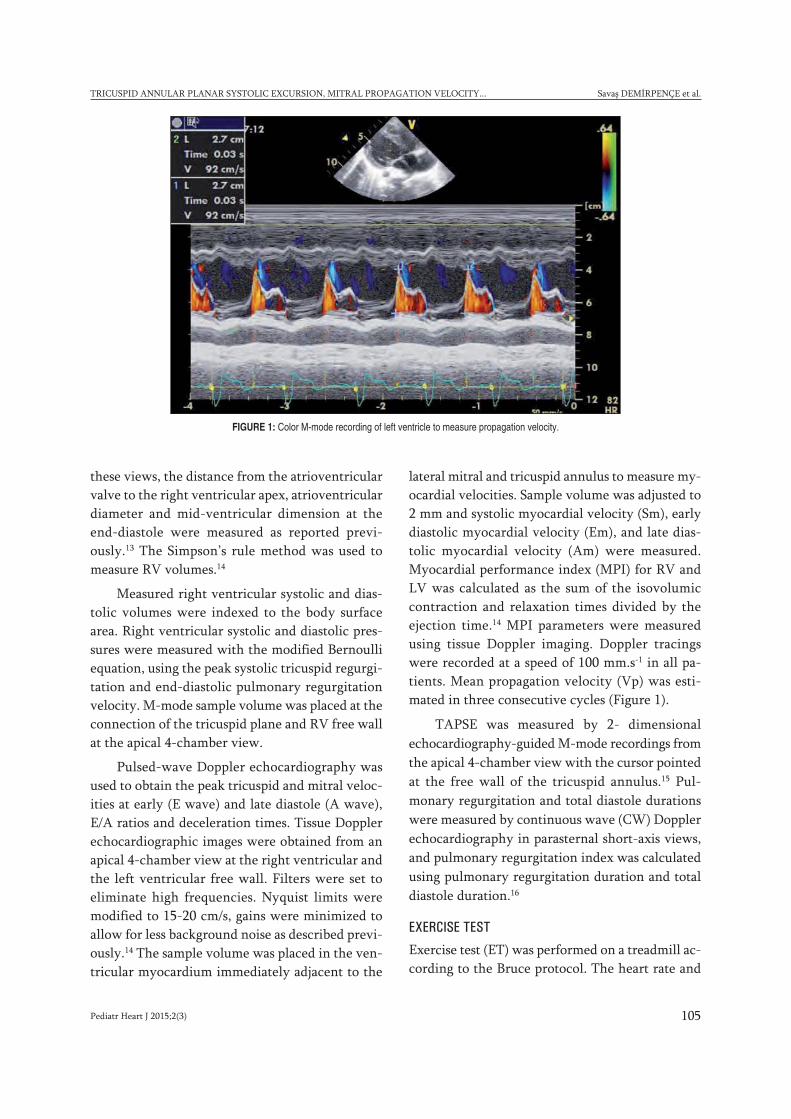
lateral mitral and tricuspid annulus to measure my-ocardial velocities. Sample volume was adjusted to2 mm and systolic myocardial velocity (Sm), earlydiastolic myocardial velocity (Em), and late dias-tolic myocardial velocity (Am) were measured.Myocardial performance index (MPI) for RV andLV was calculated as the sum of the isovolumiccontraction and relaxation times divided by theejection time.14 MPI parameters were measuredusing tissue Doppler imaging. Doppler tracingswere recorded at a speed of 100 mm.s-1 in all pa-tients. Mean propagation velocity (Vp) was esti-mated in three consecutive cycles (Figure 1).
TAPSE was measured by 2- dimensionalechocardiography-guided M-mode recordings fromthe apical 4-chamber view with the cursor pointedat the free wall of the tricuspid annulus.15 Pul-monary regurgitation and total diastole durationswere measured by continuous wave (CW) Dopplerechocardiography in parasternal short-axis views,and pulmonary regurgitation index was calculatedusing pulmonary regurgitation duration and totaldiastole duration.16
EXERCISE TEST
Exercise test (ET) was performed on a treadmill ac-cording to the Bruce protocol. The heart rate and
FIGURE 1: Color M-mode recording of left ventricle to measure propagation velocity.

Pediatr Heart J 2015;2(3)106
Savaş DEMİRPENÇE et al. TRICUSPID ANNULAR PLANAR SYSTOLIC EXCURSION, MITRAL PROPAGATION VELOCITY...
electrocardiographic changes were monitored con-tinuously; blood pressure was measured everyminute with an indirect automatic manometerthroughout the test. ST elevation or depression,negative T-waves, maximum blood pressure, heartrate, and if present, symptoms were noted. QRS du-ration and QTc were calculated during exercise.The decrease in SpO2 during exercise [ΔSpO2=SpO2(rest) - SpO
2(at maximal exercise)] was accepted
as abnormal if it was 4%, and the exercise SpO2 wasconsidered to be markedly decreased if it was 84%.
BRAIN NATRIURETIC PEPTIDE (BNP) MEASUREMENT
Venous blood samples were obtained from all sub-jects before and after the 6MWT. The plasma BNPlevel was measured using the Triage BNP im-munoassay (Biosite Diagnostics Inc., San Diego,California, USA). For the cut-off value of 100pg/ml, the assay showed 82% sensitivity and 99%specificity for differentiating heart failure fromnormal cardiac function.17
STATISTICAL ANALYSIS
Results are presented as mean ± SD unless other-wise specified. Values of P < 0.05 were consideredstatistically significant. Initially, Kolmogorov-Smirnov tests were performed for equality of dis-tribution relating to all variables. Comparison ofdemographic, clinical, and echocardiographic databetween patient and control groups were madeusing Fisher’s exact test for categorical variablesand Mann-Whitney U test and Student’s t test for
continuous variables. Pearson or Spearman corre-lation tests were used for correlation analyses. Weused SPSS 17.0 (SPSS; Chicago, IL, USA) for statis-tical analysis.
RESULTS
Baseline clinical characteristics of patients andhealthy subjects are given in Table 1. Age, distri-bution of sex and body weight was similar in bothgroups. All patients were classified as NYHA func-tional class I at the time of study enrolment. Tenpatients (40%) had had palliative shunts (modifiedBlalock-Taussig) before corrective surgery. Nine-teen patients (76%) had a transannular patch, 4 hadbovine valve (Contegra), and one patient (4%) hada pulmonary homograft. In the patient group, ageat corrective surgery was 5.1±3.5 years (range 1-14), the duration between palliative operation tocorrective surgery was 4.3 ± 2.0 years (range 2-9),and the follow-up period after corrective surgerywas 6.3 ± 3.0 years (range 2-12).
ELECTROCARDIOGRAPHIC DATA
The heart rate was significantly lower in the pa-tient group as compared to healthy controls(95±10 /min vs. 108±16 /min, respectively). TheQRS durations and corrected QT intervals weresignificantly higher in the patient group than incontrols (125±21 ms vs 89±9 ms; 438±32 ms vs408±10 ms, respectively). Eighteen patients (72%)had a complete right bundle branch block, 1 pa-tient an incomplete right bundle branch block
Variables Patients (n=25) Controls (n=25) p values
Age 11.6 ± 2.7 12.3 ± 2.3 0.41
Sex male (n(%)) 14 (%56) 11 (%55) 0.94
Body weight (kg) 38.3 ± 13.6 44 ± 13 0.24
Resting heart rate (min) 95 ± 10 (78-115) 108 ± 16 (77-130) 0.011
Average heart rate at peak exercise (min) 155 ± 25 (114 – 199) 185 ± 9 (168– 198) 0.027
Oxygen saturation at rest (percentage) 97.3 ± 1.4 98.2 ± 0.6 0.038
Oxygen saturation after exercise test (percentage) 97.2 ± 1.5 98.2 ± 0.6 0.039
QRS duration (ms) 125 ± 21 89 ± 9 0.011
QTc duration (ms) 438 ± 32 408 ± 10 0.011
TABLE 1: Clinical characteristics of patient and control groups.*
*Data are expressed as mean ± SD or number unless otherwise indicated.
and another a first degree atrioventricular block.There were no arrhythmia or block in the controlgroup.
NEUROHORMONAL VARIABLES
Only one patient had an abnormal level of BNP,which was measured before the six-minute walk-ing test (6MWT). There were no significant differ-ences between the levels of BNP before and afterthe 6MWT in the study group (39.1±33.6 ng/L,30.9±22.4 ng/L, respectively; p>0.05). Comparisonof BNP levels after the 6MWT in the study grouprevealed that patients who had modified BT shuntsbefore corrective surgery had significantly higherlevels as compared to patients without a history ofmodified Blalock-Taussig (mBT) shunt (44.6 ± 20.9ng/L, 21.8 ± 18.8 ng/L, respectively; p=0.013).
EXERCISE TEST
All subjects showed a normal exercise test. Therewere no electrocardiographic changes or symptomsduring the test. Exercise test results are summa-
rized in Table 1. When compared to patients,healthy subjects had significantly higher restingheart rates and average heart rates at peak exercise(p = 0.011, p= 0.027; respectively). Mean restingSpO2 and SpO2 at peak exercise levels were signif-icantly lower in children with TOF compared withcontrols.
ECHOCARDIOGRAPHIC DATA
Left ventricular systolic and diastolic diameters andsystolic function parameters were lower in the pa-tient group as compared to the control group, asshown in Table 2. However, LV ejection fraction(EF) and fractional shortening (FS) were withinnormal limits (EF range: 58-80%, FS range: 29-48%; respectively). Mean values of right ventricu-lar end-diastolic volume, end-systolic volume,end-diastolic area and end-systolic area were sig-nificantly higher in the patient group than in thehealthy subjects (p<0.001, Table 2).
Right ventricular ejection fraction, as calcu-lated with the modified Simpson’s rule, was lower
Pediatr Heart J 2015;2(3) 107
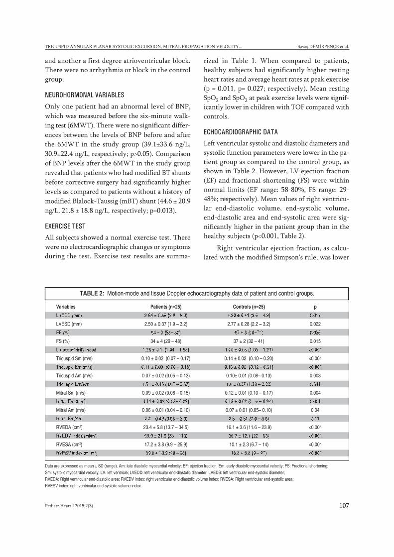
TRICUSPID ANNULAR PLANAR SYSTOLIC EXCURSION, MITRAL PROPAGATION VELOCITY... Savaş DEMİRPENÇE et al.
Data are expressed as mean ± SD (range). Am: late diastolic myocardial velocity; EF: ejection fraction; Em: early diastolic myocardial velocity; FS: Fractional shortening; Sm: systolic myocardial velocity; LV: left ventricle; LVEDD: left ventricular end-diastolic diameter; LVEDS: left ventricular end-systolic diameter; RVEDA: Right ventricular end-diastolic area; RVEDV index: right ventricular end-diastolic volume index; RVESA: Right ventricular end-systolic area; RVESV index: right ventricular end-systolic volume index.
Variables Patients (n=25) Controls (n=25) p
LVEDD (mm) 3.84 ± 0.56 (2.9 – 5.0) 4.30 ± 0.41 (3.6 – 4.8) 0.017
LVESD (mm) 2.50 ± 0.37 (1.9 – 3.2) 2.77 ± 0.28 (2.2 – 3.2) 0.022
EF (%) 64 ± 3 (58– 80) 67 ± 3 (60–71) 0.035
FS (%) 34 ± 4 (29 – 48) 37 ± 2 (32 – 41) 0.015
LV eccentricity index 1.25 ± 0.1 (1.04 – 1.53) 1.09 ± 0.06 (1.03 – 1.27) <0.001
Tricuspid Sm (m/s) 0.10 ± 0.02 (0.07 – 0.17) 0.14 ± 0.02 (0.10 – 0.20) <0.001
Tricuspid Em (m/s) 0.11 ± 0.03 (0.06 – 0.18) 0.16 ± 0.02 (0.12 – 0.21) <0.001
Tricuspid Am (m/s) 0.07 ± 0.02 (0.05 – 0.13) 0.10± 0.01 (0.08– 0.13) 0.003
Tricuspid Em/Am 1.51 ± 0.45 (0.67 – 2.57) 1.6 ± 0.27 (1.23 – 2.22) 0.541
Mitral Sm (m/s) 0.09 ± 0.02 (0.06 – 0.15) 0.12 ± 0.01 (0.10 – 0.17) 0.004
Mitral Em (m/s) 0.14 ± 0.03 (0.05– 0.22) 0.19 ± 0.02 (0.16 – 0.24) 0.001
Mitral Am (m/s) 0.06 ± 0.01 (0.04 – 0.10) 0.07 ± 0.01 (0.05– 0.10) 0.04
Mitral Em/Am 2.2 ± 0.49 (0.63 – 3.0) 2.5 ± 0.51 (2.0 – 3.6) 0.11
RVEDA (cm2) 23.4 ± 5.8 (13.7 – 34.5) 16.1 ± 3.6 (11.6 – 23.9) <0.001
RVEDV index (ml/m2) 65.9 ± 21.9 (33 – 113) 36.7 ± 12.1 (22 – 63) <0.001
RVESA (cm2) 17.2 ± 3.8 (9.9 – 25.9) 10.1 ± 2.3 (6.7 – 14) <0.001
RVESV index (ml/m2) 39.8 ± 10.9 (20 – 63) 18.3 ± 5.8 (9 – 27) <0.001
TABLE 2: Motion-mode and tissue Doppler echocardiography data of patient and control groups.

in the patient group as compared to control sub-jects (38.3 ± 7.9% vs. 49 ± 6.7%; p<0.001). Rightventricular end diastolic volume indexed to bodysurface area was significantly higher in patientsthan controls (65.9 ± 21.9 ml/m2, 36.7 ± 12.1 ml/m2,respectively; p<0.001). Compared to the controlgroup, the patient group had higher left ventricleeccentricity index (p< 0.001), higher tricuspid Awave velocity (p=0.001), lower E/A ratio (p< 0.001),lower tricuspid (p<0.001) and mitral (p=0.004) an-nular Sm velocities, lower tricuspid (p< 0.001) andmitral (p=0.001) annular Em velocities and lowertricuspid (p=0.003) and mitral (p=0.04) annular Amvelocities (Table 2).
The right ventricular and left ventricular my-ocardial performance indices were significantlyhigher in patients with tetralogy of Fallot (p<0.01)(Table 3). Patients had significantly higher rightventricular systolic pressures than in controls (15.2± 5 vs 7 ± 2 mmHg; p=0.005). None of the studysubjects had severe tricuspid regurgitation, signifi-cant pulmonary outflow obstruction or residualventricular septal defects. Pulmonary regurgitationwas mild in 5 patients, moderate in 19 patients andsevere in 5 patients. In the patient group, pul-monary regurgitation index was found to be sig-nificantly higher than those in the control group(p=0.03). There was no statistically significant dif-ference between the control and study subjects re-garding Vp levels (p=0,655).
In the patient group, there were two patientswith a TAPSE distance below 15 mm. One of themhad undergone a mBT procedure 9 years ago, a
Brock procedure 2 years ago, and total correctionwith a Contegra conduit. The other one had had amBT 4 years before total correction. In the patientgroup, TAPSE distance was significantly lowerthan that of the control group (p<0.001) (Table 3).
BNP level after exercise displayed significantcorrelation to age at operation (r=0.485, p=0.014).Duration of follow-up after surgery was positivelycorrelated with RV end-diastolic volume index(r=0.587, p=0.02) and RV end-systolic volumeindex (r=0.598, p=0.02). Right ventricular MPI hadsignificant negative correlation with 6MWT dis-tance (r=0.461, p=0.02) and BNP levels after exer-cise (r=0.426, p=0.034).
DISCUSSION
Several authors emphasized the importance of leftventricular function in the evaluation of patientswith TOF. Right and left ventricular dysfunctionas shown by decreased mitral and tricuspid systolicand diastolic velocities, and increased myocardialperformance indices in the current study is consis-tent with the findings of previous studies.18
Tei index or MPI has been accepted as a reli-able method to quantitatively determine ventricu-lar function.14 MPI reflects systolic and diastolicfunctions of both ventricles. In the current study,mean RV and LV MPI values (0.51±0.08, 0.49 ±011, respectively) in children with repaired tetral-ogy of Fallot were higher as compared to thehealthy subjects, demonstrating biventricular dys-function. Our data of biventricular MPI agree with
Pediatr Heart J 2015;2(3)108
Savaş DEMİRPENÇE et al. TRICUSPID ANNULAR PLANAR SYSTOLIC EXCURSION, MITRAL PROPAGATION VELOCITY...
Variables Patients (n=25) Controls (n=25) p
Left ventricle eccentricity index 1.25 ± 0.1 (1.04 – 1.53) 1.09 ± 0.06 (1.03 – 1.27) <0.001
Right ventricular MPI 0.51 ± 0.08 (0.36 – 0.70) 0.41 ± 0.06 (0.28 – 0.52) 0.002
Left ventricular MPI 0.49 ± 0.11 (0.3 – 0.75) 0.36 ± 0.05 (0.28 – 0.45) 0.001
PRi 0.65 ± 0.09 (0.41 – 0.83) 0.54 ± 0.21 (0.29 – 0.91) <0.001
TAPSE (mm) 18.7 ± 3.2 (12 – 24) 23.2 ± 1.9 (20 – 26) 0.03
Vp (cm/sn) 101 ± 34 (55 – 167) 97 ± 23 (58 – 153) 0.655
TABLE 3: Right and left ventricular myocardial performance indices, PRi, TAPSE and Vp data of patients and healthy subjects.
Data are expressed as mean ± SD (range). ^ measured using Simpson method. MPI, myocardial performance index; PRi, pulmonary regurgitation index; TAPSE, tricuspid annularplanar systolic excursion; Vp, propagation velocity.
those of Samman et al. who revealed an associationbetween biventricular dysfunction (higher MPIlevels) and limited exercise capacity in adults withrepaired tetralogy of Fallot.19 Samman et al. alsoshowed a direct relation between right ventricularMPI and left ventricular MPI. However, we did notobserve such an association in our study. This canbe explained by the difference in mean age and du-ration of follow-up, as mean age of our study pop-ulation was 11.6 ± 2.7 years compared to 35 ± 7years in the study of Samman et al., and mean du-ration of follow up period after corrective surgeryin our study was 6.3 ± 3.0 years.
Several studies in adults also showed that leftventricular dysfunction is one of the major factorsin late unfavorable outcomes in patients with re-paired tetralogy of Fallot. LV or RV dysfunctionhas been shown to be associated with impairedclinical condition in adults with TOF repair.20 Inour study, we did not find any association betweenright ventricular systolic-diastolic volume indexesand BNP levels. We did not observe any associa-tion between right ventricular end-diastolic vol-ume index and right ventricular MPI. Our data isconsistent with those of Geva et al.3 Moreover, weshowed a direct relation between right ventricularEF and right ventricular MPI. In the current study,we also found a correlation between right ventric-ular MPI and BNP levels after exercise.
Border et al. showed that Vp correlates signifi-cantly with the invasive indices of relaxation,namely Tau and peak negative dP/dt.21 To the best ofour knowledge, there is no report evaluating the mi-tral Vp in children after repair of TOF. In this re-port, we found that Vp for mitral valve in childrenafter repair of TOF did not change during mid-termfollow up. It is naturally evident that TAPSE must bemore related to systolic right ventricular functionsince the tricuspid valve moves toward the RV apexduring systole as lateral shortening of RV free walldevelops. Recent findings suggested that TAPSEmeasurement is better reproducible than otherechocardiographic indices of right ventricular func-tion.22 In the current study, TAPSE distance in thepatient group was significantly lower than that ofthe control group. In the patient group, we did not
find any association between TAPSE and RV ejec-tion fraction, which was measured with the Simpsonmethod. There were studies arguing that Simpson’sRV EF was not the best method to measure rightventricular ejection fraction precisely.23 Anotherlimitation of Simpson’s RV EF is high interobservervariability. The absence of correlations betweenSimpson’s RV EF and TAPSE in the current study isattributed to these factors. Consistent with this,Morcos et al. showed a weak correlation of TAPSEand RVEF determined by MRI.24 There are also cluespointing that abnormal right ventricular functioncould be observed with normal TAPSE and TDI.25
TAPSE levels below 1.5 cm and TDI levels below 10cm/s consistently predict right ventricular dysfunc-tion with a low false positive rate. Koestenberger etal. showed that TOF patients before repair had sig-nificantly increased TAPSE values compared to age-matched controls, possibly due to RV hypertrophy.26
However, they found a reduced TAPSE in TOF pa-tients which increased in postoperative years. In thecurrent study, mean duration after corrective sur-gery was 6.3 years which is consistent with the studyof Koestenberger et al. They found that TAPSE lev-els become significantly impaired after an average of7 years.26 However, we did not find any correlationbetween the duration after repair and TAPSE levels.This absence of correlation can be explained by totalright ventricle dyssynchrony that simply partiallyaffects RV longitudinal function. It is also knownthat TAPSE does not take into account segmentalRV function, and it is not able to detect abnormalwall movement, distorted RV geometry and predicttotal RV systolic function. TAPSE has been demon-strated to inversely correlate with the RV end-dias-tolic diameter and the degree of tricuspidregurgitation. There was no severe tricuspid regur-gitation in our study population.
STUDY LIMITATIONS
Several limitations deserve mentioning. As thisstudy has a cross sectional feature, we did not ob-tain serial changes in BNP levels and right ventric-ular MPI values in patients. Longitudinal studiesmay reveal the exact timing when pulmonary valvereplacement is needed.
Pediatr Heart J 2015;2(3) 109
TRICUSPID ANNULAR PLANAR SYSTOLIC EXCURSION, MITRAL PROPAGATION VELOCITY... Savaş DEMİRPENÇE et al.

Pediatr Heart J 2015;2(3)110
Savaş DEMİRPENÇE et al. TRICUSPID ANNULAR PLANAR SYSTOLIC EXCURSION, MITRAL PROPAGATION VELOCITY...
It is well established that magnetic resonanceimaging (MRI) is currently accepted as the goldstandard imaging modality for the evaluation ofright ventricular volume and right ventricularfunction. However, we did not have values of rightventricular size obtained through MRI obtained inour study.
Finally, the timing of corrective surgery wasdelayed in our study population. Mean age at cor-rective surgery was 5.2 years. It is generally ac-cepted that TOF patients are repaired between 3months and 1.5 years of age. Therefore, mean age atsurgery in this study limits extrapolation of the datato populations where surgery is performed earlier.
This discrepancy can be explained by the policy ofhealth system and perspective of surgeons in ourcountry. However, with the optimization of thehealth policy and increased experience of surgeons,mean age at corrective surgery in tetralogy of Fal-lot has been steadily decreasing.
CONCLUSION
Our study demonstrates that children with re-paired TOF had normal Vp levels at mid term fol-low-up. Indices of TAPSE, tissue Doppler and MPIfor the assessment of right and left ventricle couldbe useful in the evaluation of children after TOFrepair.
1. Hirsch JC, Mosca RS, Bove EL. Complete repair oftetralogy of Fallot in the neonate: Results in the mod-ern era. Ann Thorac Surg;232(4):508-514.
2. Helbing WA, Niezen A, Le Cessie S, et al. Right ven-tricular diastolic function in children with pulmonaryregurgitation after repair of tetralogy of Fallot: volu-metric evaluation by magnetic resonance velocitymapping. J Am Coll Cardiol;28:1827-35.
3. Geva T, Sandweiss BM, Gauvreau K, et al. Factorsassociated with impaired clinical status in long-termsurvivors of tetralogy of Fallot repair evaluated bymagnetic resonance imaging. J Am Coll Cardiol2004;43:1068-74.
4. Gatzoulis MA, Balaji S, Webber SA, et al. Risk fac-tors for arrhythmia and sudden cardiac death lateafter repair of tetralogy of Fallot: a multicentre study.Lancet;356:975–981.
5. Baspinar O, Alehan D. Dobutamine stress echocar-diography in the evaluation of cardiac haemody-namics after repair of tetralogy of Fallot in children:negative effects of pulmonary regurgitation. ActaCardiol. 2006 Jun;61(3):279-283.
6. Uebing A, Fisher G, Bethge M, et al. Influence of thepulmonary annulus diameter on pulmonary regurgi-tation and right ventricular pressure load after repairof tetralogy of Fallot. Heart 2002; 88:510–514.
7. D’Andrea A, Caso P, Sarubbi B, Russo MG et al.:Right ventricular myocardial dysfunction in adult pa-tients late after repair of tetralogy of Fallot. Int J Car-diol 2004; 94:213-220.
8. Book WM, Hott BJ, McConnell M. B-type natriureticpeptide levels in adults with congenital heart diseaseand right ventricular failure. Am J Cardiol 2005;95:545–546.
9. Brun P, Tribouilloy C, Duval AM, et al. Left ventricu-lar flow propagation during early filling is related towall relaxation: a color M-mode Doppler analysis. JAm Coll Cardiol 1992; 20: 420-432.
10. Lamia B, Teboul JL, Monnet X, et al. Relationshipbetween the tricuspid annular plane systolic excur-sion and right and left ventricular function in critically
ill patients. Intensive Care Med 2007; 33: 2143–2149.
11. Du Bois D, Du Bois EF. A formula to estimate the ap-proximate surface area if height and weight beknown Nutrition 1916; 5:303–311.
12. Bazett HC. An analysis of the time relations of elec-trocardiograms. Heart 1920; 7: 353–355.
13. Rudski LG, Lai WW, Afilalo J et al. Guidelines for theechocardiographic assessment of the right heart inadults: a report from the American Society ofEchocardiography endorsed by the European Asso-ciation of Echocardiography, a registered branch ofthe European Society of Cardiology, and the Cana-dian Society of Echocardiography. J Am SocEchocardiogr. 2010; 23:685-713.
14. Tei C, Ling LH, Hodge DO, et al. New index of com-bined systolic and diastolic myocardial performance:a simple and reproducible measure of cardiac func-tion—a study in normals and dilated cardiomyopa-thy. J Cardiol 1995; 26:357-366.
15. Ghio S, Recusani F, Klersy C, et al. Prognostic use-fulness of the tricuspid annular plane systolic excur-sion in patients with congestive heart failuresecondary to idiopathic or ischemic dilated car-diomyopathy. Am J Cardiol. 2000; 85:837–842.
16. Li W, Davlouros PA, Kilner PJ, Pennell DJ, Gibson D,Henein MY, Gatzoulis MA. Doppler-echocardio-graphic assessment of pulmonary regurgitation inadults with repaired tetralogy of Fallot: comparisonwith cardiovascular magnetic resonance imaging.Am Heart J. 2004 Jan; 147(1):165-72.
17. Wieczorek SJ, Wu AHB, Christenson R, et al. A rapidB-type natriuretic peptide assay accurately diag-noses left ventricular dysfunction and heart failure: amulticenter evaluation Am Heart J. 2002; 142: 834–839.
18. Gatzoulis MA, Elliot JT, Guru V, et al. Right and leftventricular systolic function late after repair of tetral-ogy of Fallot. Am J Cardiol 2000;86:1352–7.
19. Samman A, Schwerzmann M, Balint OH, et al. Ex-ercise capacity and biventricular function in adult pa-
tients with repaired tetralogy of Fallot Am HeartJ;156(1):100-5.
20. Davlouros PA, Kilner PJ, Hornung TS, et al. Rightventricular function in adults with repaired tetralogy ofFallot assessed with cardiovascular magnetic reso-nance imaging: detrimental role of right ventricularoutflow aneurysms or akinesia and adverse right-to-left ventricular interaction. J Am Coll Cardiol;40:2044–52.
21. Border WL, Michelfelder EC, Glascock BJ, et al.Color M-mode and doppler tissue evaluation of di-astolic function in children: simultaneous correla-tion with invasive indices. J Am Soc Echocardiogr2003;16(9):988–994.
22. Berdat PA, Immer F, Pfammatter JP, et al. Reoper-ations in adults with congenital heart disease: analy-sis of early outcome. Int J Cardiol 93:239–245.
23. Hsiao SH, Lin SK, Wang WC, et al. Severe tricuspidregurgitation shows significant impact in the rela-tionship among peak systolic tricuspid annular ve-locity, tricuspid annular plane systolic excursion, andright ventricular ejection fraction. J Am Soc Echocar-diogr. 2006 Jul;19(7):902-10.
24. Morcos P, Vick GW, Sahn DJ, et al. Correlation ofright ventricular ejection fraction and tricuspid annu-lar plane systolic excursion in tetralogy of Fallot bymagnetic resonance imaging. Int J Cardiovasc Im-aging 2009;25:263–270.
25. Miller D, Farah MG, Liner A, et al. The relation be-tween quantitative right ventricular ejection fractionand indices of tricuspid annular motion and myocar-dial performance. J Am Soc Echocardiogr.May;17(5):443-7. tetralogy of Fallot. Circulation.2004 Sep 14;110 (11 Suppl 1): II153-7.
26. Koestenberger M, Ravekes W, Everett AD, et al.Right ventricular function in infants, children and ado-lescents: reference values of the tricuspid annularplane systolic excursion (TAPSE) in 640 healthy pa-tients and calculation of z score values. J Am SocEchocardiogr. Jun;22(6):715-9. Epub 2009 May 7.
REFERENCES
alp hızı değişkenliği (KHD) ardışık kalp atışları arasındaki sürelerindeğişimi olarak tanımlanır. İlk olarak 1965’de Hon ve Lee tarafındanfetal kalpteki değişiklikler ile fark edilmesinden bu yana oldukça
fazla sayıdaki çalışmaya konu olmuştur.1 KHD analizi ile kalbin sempato-
Pediatr Heart J 2015;2(3) 111
Sporcu Çocuklarda Orta-Uzun DönemdeKalp Hızı Değişkenliği Nasıl Etkilenir?
ÖÖZET Amaç: Bu çalışmanın amacı uzun süreli spor yapan çocuklarda, sporun orta-uzun dönemdekalp hızı değişkenliği (KHD) üzerine olan etkisini araştırmaktır. Gereç ve Yöntemler: Ortalamayaşları 12,5 yıl olan, otuz sporcu (6’sı kız) ve otuz sağlıklı çocuk (15’i kız, 15’i erkek) çalışmayaalındı. Holter EKG kayıtları 24 saat süre ile hastaların günlük aktivitelerine devam etmeleri sağla-narak, sporcu çocukların da antrenmanlarından iki-üç gün sonra olacak şekilde ayarlandı. Otoma-tik olarak hesaplanan zaman bazlı KHD parametreleri (SDNN, SDANN, RMSSD, PNN50) ile frekansbazlı parametreleri (VLF, LF, HF, LF/HF) uyku, uyanıklık dönemlerinde ve 24 saatlik olarak ayrıayrı incelendi. Bulgular: KHD verilerinden 24 saatlik SDNN, SDNN index, SDANN index, RMSSD,PNN50, 24 saatlik LF, HF değerleri, uyku ve uyanıklık dönemi SDNN, RMSSD, LF, HF değerleri sporyapan grupta daha yüksek bulunmakla beraber istatistiksel olarak farksızdı. 24 saatlik, uyku ve uya-nıklık dönemi VLF değerleri çalışma grubunda kontrol grubuna göre istatistiksel olarak anlamlı şe-kilde yüksek bulundu (sırası ile p=0,008; p=0,039; p=0,019). Sonuç: Bu çalışma ile daha öncekiçalışmalarda kısa dönemde elde edilen kayıtlar neticesinde, spor ile arttığı saptanan KHD para-metrelerinden farklı olarak, orta-uzun dönemde VLF değerinin sporcu çocuklarda spor yapmayan-lara göre anlamlı olarak yüksek bulunduğu gösterildi.
Anahtar Kelimeler: Kalp hızı değişkenliği; spor; çocuk
ABSTRACT Objective: The purpose of this study is to investigate the medium-long term effect ofsport on heart rate variability in children. Material and Methods: The mean age was 12,5 years,thirty athletes (only six girl) and thirty healthy children (15 girl, 15 boy) were enrolled in study.During the ECG recording for 24 hours, the patients continued their daily activities and records setto be two-three days after the training in athletes. Time and frequency based parameters whichwere calculated automatically examined separately as 24 hours, sleep and awake period. Results: Al-though SDNN 24 hours, SDNN index, SDANN index, RMSSD, PNN50, LF and HF 24 hours, sleepand awake period of SDNN, RMSSD, LF and HF values were higher in the study group, these pa-rameters were not significantly different. VLF (24 hours, sleep and awake period) values in thestudy group were statiatically higher than the control group (p=0.008; p=0.039; p=0.019, respec-tively). Conclusion: In this report; it was differently founded that from previously short termrecords and instant effects of sport on heart rate variability analysis, the VLF values for medium-long term in athletes were significantly higher than the controls
Key Words: Heart rate variability; sports; children
Pediatr Heart J 2015;2(3):111-6
Şebnem PAYTONCUa
aMoris Şinasi Çocuk Kliniği ve Kadın Doğum Kliniği, Çocuk Kardiyoloji Birimi, Merkezefendi Devlet Hastanesi, Manisa
Ge liş Ta ri hi/Re ce i ved: 23.05.2015 Ka bul Ta ri hi/Ac cep ted: 17.08.2015
Ya zış ma Ad re si/Cor res pon den ce:Şebnem PAYTONCUMerkezefendi Devlet Hastanesi, Moris Şinasi Çocuk Kliniği ve Kadın Doğum Kliniği, Çocuk Kardiyoloji Birimi, Manisa,TÜRKİYE/[email protected]
Copyright © 2015 by Türk Pediatrik Kardiyoloji veKalp Cerrahisi Derneği
ORİJİNAL ARAŞTIRMA
Pediatr Heart J 2015;2(3)112
Şebnem PAYTONCU SPORCU ÇOCUKLARDA ORTA-UZUN DÖNEMDE KALP HIZI DEĞİŞKENLİĞİ NASIL ETKİLENİR?
vagal dengesi noninvaziv olarak değerlendirilir.Böylece kalp ritmini düzenleyen otonom sinir sis-teminin etkileri hakkında bilgi edinilir.
Egzersiz ile birlikte meydana gelen parasem-patik geri çekilme ve sempatik aktivite artışı kalphızını arttırmaktadır. Sporcularda normal sağlıklıbireylere göre atım hacmi çok daha fazla arttırıla-bildiği için, egzersiz ile kalp hızı artışı normal sağ-lıklı bireylerden çok daha düşüktür.2 KHD’ninazalması kardiyovasküler hastalıklar açısından riskfaktörü olarak kabul edilmekte ve sporun kardiyo-vasküler hastalıklar açısından risk faktörlerini or-tadan kaldırdığı bilinmektedir.3 Bu çalışmanınamacı uzun süredir spor yapan çocuklarda, en sonyapılan antrenmanlar ile orta-uzun dönemdeKHD’nin nasıl etkilendiğini araştırmaktır.
GEREÇ-YÖNTEMLER
Ekim 2012-Eylül 2013 tarihleri arasında poliklini-ğimize başvuran, anamnez, öz ve soygeçmiş bilgi-leri, fizik inceleme, laboratuvar, elektrokardiyog-rafi ve ekokardiyografi ile genetik, doğumsalve/veya edinsel herhangi bir kalp hastalığı bulun-mayan hastalar çalışmaya alındı. Holter EKG kayıtsüresi 23,5 saatin altında olan hastalar çalışma kap-samı dışında bırakıldı.
Çalışma grubu: Futbol, basketbol, hentbol,yüzme, su topu gibi sporlarla en az iki, en fazla onbir yıldır uğraşan, haftanın üç-beş günü, günde alt-mış-doksan dakika antrenman yapan, altısı kız top-lam otuz sporcu çocuk çalışma grubunu oluşturdu.Yaşları 8,5-17 (ortalama 12,23, SD: 2,01) yıl olansporcu çocuklar, merkezimize çabuk yorulma, çar-pıntı, efor dispnesi şikayetleri ile başvurmuşlardı.Çalışma grubundaki hastalardan her biri bu spor-lardan sadece biri ile ilgileniyordu.
Kontrol grubu: Çarpıntı, bayılma, kalp atışla-rında yavaşlama şikayetleri ile başvuran, yaşları9,5-16,5 (ortalama 12,63, SD: 3,12) yıl olan, sporolarak sadece okuldaki spor ve beden eğitimi ders-lerine katılan on beşi kız, toplam otuz çocuk kont-rol grubuna alındı.
Çalışma ve kontrol grubundaki tüm hastaların,Holter EKG kaydı esnasında, kalp hızı ve ritminietkileyecek ilaç kullanım öyküsü, belirgin anemi
ve enfeksiyonu bulunmamakta idi. Her bir hastayabir kez Holter EKG kaydı yapıldı, tekrarlanan kayıtolmadı.
Çalışmaya alınan tüm hastaların fizik muaye-neleri, 12- derivasyonlu standart EKG (25 mm/sn,10 mm/mV), Holter EKG kayıtları ve KHD para-metrelerinin değerlendirilmesi aynı araştırıcı tara-fından yapıldı.
Kalp hızı değişkenliği: Holter EKG kayıtlarıDMS Cardioscan Holter System version 10.0 ile 24saat süre ile hastaların günlük aktivitelerine devametmeleri sağlanarak, ev, okul ve doğal ortamlarında(sporcu çocukların ise antrenmanlarından iki-üçgün sonra olacak şekilde) ayarlandı. Holter EKG ci-hazı tüm hastalara sabah 09-10:00 sıralarında ta-kıldı. Kayıt esnasında beslenme konusundaherhangi bir kısıtlama yapılmadı. Tüm hastalarauyuma ve uyanma zamanları sorgulanarak, 24 sa-atlik, uyku ve uyanıklık dönemine ait parametrelerelde edildi.
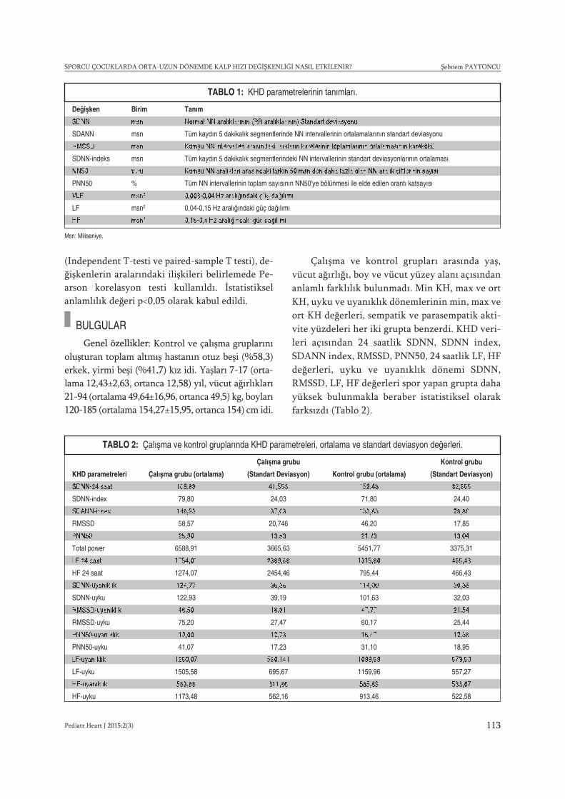
Çalışma ve kontrol grubunda herhangi bir has-tada Holter EKG sonucunda normal sınırlarda bul-guların dışında özellik saptanmadı. Cihazınotomatik olarak hesapladığı zaman bazlı kalp hızıdeğişkenliği parametreleri (SDNN, SDANN,RMSSD, PNN50) ile frekans bazlı parametreler(VLF, LF, HF, LF/HF) uyku, uyanıklık dönemle-rinde ve 24 saatlik olarak değerlendirildi. Tablo1’de KHD parametrelerinin tanımları yer almakta-dır.2
En düşük ve en yüksek kalp hızları (Min KHve max KH), ortalama kalp hızı (ort KH), uyku veuyanıklık dönemlerinde min KH, max KH ve ortKH, sempatik ve parasempatik aktivite yüzdelerikaydedildi.
Hastalara ve ailelerine gereken açıklamalar ya-pılarak, ailelere bilgilendirilmiş onam formu imza-latıldı. Merkezimiz özel bir sağlık kuruluşu olduğuiçin etik kurul onay işlemi yapılamadı.
İstatistiksel analiz: Tüm veriler SPSS 15.0programı ile değerlendirildi. Tanımlayıcı istatistik-sel yöntemler ile minimum, maksimum, ortalama,standart deviasyon değerleri elde edildi. Bağımsızgrupların verilerinin karşılaştırılmasında T-testi
Pediatr Heart J 2015;2(3) 113
SPORCU ÇOCUKLARDA ORTA-UZUN DÖNEMDE KALP HIZI DEĞİŞKENLİĞİ NASIL ETKİLENİR? Şebnem PAYTONCU
(Independent T-testi ve paired-sample T testi), de-ğişkenlerin aralarındaki ilişkileri belirlemede Pe-arson korelasyon testi kullanıldı. İstatistikselanlamlılık değeri p<0,05 olarak kabul edildi.
BULGULAR
Genel özellikler: Kontrol ve çalışma gruplarınıoluşturan toplam altmış hastanın otuz beşi (%58,3)erkek, yirmi beşi (%41,7) kız idi. Yaşları 7-17 (orta-lama 12,43±2,63, ortanca 12,58) yıl, vücut ağırlıkları21-94 (ortalama 49,64±16,96, ortanca 49,5) kg, boyları120-185 (ortalama 154,27±15,95, ortanca 154) cm idi.
Çalışma ve kontrol grupları arasında yaş,vücut ağırlığı, boy ve vücut yüzey alanı açısındananlamlı farklılık bulunmadı. Min KH, max ve ortKH, uyku ve uyanıklık dönemlerinin min, max veort KH değerleri, sempatik ve parasempatik akti-vite yüzdeleri her iki grupta benzerdi. KHD veri-leri açısından 24 saatlik SDNN, SDNN index,SDANN index, RMSSD, PNN50, 24 saatlik LF, HFdeğerleri, uyku ve uyanıklık dönemi SDNN,RMSSD, LF, HF değerleri spor yapan grupta dahayüksek bulunmakla beraber istatistiksel olarakfarksızdı (Tablo 2).
TABLO 1: KHD parametrelerinin tanımları.
Msn: Milisaniye.
Değişken Birim Tanım
SDNN msn Normal NN aralıklarının (RR aralıklarının) Standart deviasyonu
SDANN msn Tüm kaydın 5 dakikalık segmentlerinde NN intervallerinin ortalamalarının standart deviasyonu
RMSSD msn Komşu NN intervalleri arasındaki farkların karelerinin toplamlarının ortalamasının karekökü
SDNN-indeks msn Tüm kaydın 5 dakikalık segmentlerindeki NN intervallerinin standart deviasyonlarının ortalaması
NN50 vuru Komşu NN aralıkları arasındaki farkın 50 msn den daha fazla olan NN aralık çiftlerinin sayısı
PNN50 % Tüm NN intervallerinin toplam sayısının NN50'ye bölünmesi ile elde edilen orantı katsayısı
VLF msn2 0,003-0,04 Hz aralığındaki güç dağılımı
LF msn2 0,04-0,15 Hz aralığındaki güç dağılımı
HF msn2 0,15-0,4 Hz aralığındaki güç dağılımı
TABLO 2: Çalışma ve kontrol gruplarında KHD parametreleri, ortalama ve standart deviasyon değerleri.
Çalışma grubu Kontrol grubu
KHD parametreleri Çalışma grubu (ortalama) (Standart Deviasyon) Kontrol grubu (ortalama) (Standart Deviasyon)
SDNN-24 saat 169,83 41,563 152,43 32,666
SDNN-index 79,80 24,03 71,80 24,40
SDANN-index 148,93 37,03 133,83 28,80
RMSSD 58,57 20,746 46,20 17,85
PNN50 25,90 13,53 21,73 13,04
Total power 6588,91 3665,63 5451,77 3375,31
LF 24 saat 1754,01 2389,68 1315,80 466,43
HF 24 saat 1274,07 2454,46 795,44 466,43
SDNN-uyanıklık 124,77 36,36 114,00 30,35
SDNN-uyku 122,93 39,19 101,63 32,03
RMSSD-uyanıklık 48,50 18,31 47,77 21,54
RMSSD-uyku 75,20 27,47 60,17 25,44
PNN50-uyanıklık 19,00 12,73 16,47 12,38
PNN50-uyku 41,07 17,23 31,10 18,95
LF-uyanıklık 1260,07 560,141 1099,69 679,60
LF-uyku 1505,58 695,67 1159,96 557,27
HF-uyanıklık 583,88 311,86 585,89 533,07
HF-uyku 1173,48 562,16 913,46 522,58
24 saatlik, uyanıklık ve uyku dönemlerine aitVLF değerleri, sporcu çocuklarda, kontrol grubunagöre istatistiksel olarak anlamlı bir şekilde artmışbulundu (Tablo 3).
TARTIŞMA
KHD, zaman içersinde sinüs hızındaki değişikliklerile oluşan, kalpte vurudan vuruya değişikliklerin de-recesini yansıtan otonom sinir sisteminin noninva-ziv bir belirleyicisidir. Sağlıklı bir kalpte atımlar saatgibi düzenli değildir. Fiziksel ve mental stres, egzer-siz, solunum ve metabolik nedenlere bağlı olarakkalp hızında otonomik tonusla ilişkili değişikliklerolmaktadır. Sempatik ve parasempatik denge hak-kında bilgi veren KHD analizi, kardiyak otonom to-nüsün bir ölçüsü ve kardiyo-respiratuvar sisteminbir göstergesi olarak kullanılmaktadır.4
Normal bireylerde kalp hızında gün içinde de-ğişiklikler mevcuttur, gündüz sempatik indeksler;LF, gece vagal indeksler; HF komponenti daha yük-sektir. KHD’nin yüksek olması otonom sinir sistemifonksiyonlarının normal olduğunu, azalması isebazı kardiyak risk faktörlerinin beraberinde bu-lunduğunu göstermektedir. KHD düşüklüğü süre-gelen hastaların normal KHD olan hastalara göre 3kat fazla mortaliteye sahip olduğu bildirilmiştir.5-8
Fiziksel aktiviteye verilen KHD yanıtları ge-netik farklılıklara rağmen kalbin otonom düzen-lenmesinde en önemli veri kabul edilmektedir.Fizik aktivitenin KHD üzerine olan akut etkileriaraştırmalara konu olmuştur. Sporcularda KHD ileilgili yapılan sınırlı sayıdaki çalışmalarda KHD’ninarttığı gösterilmiştir.8,9
KHD çoğu çalışmada 24 saatlik uzun dönem;iki, beş ve on beş dakikalık kısa dönem, bazen deiki saatlik kayıtlar şeklinde değerlendirilmiş, iki-onbeş dakika şeklindeki kısa süreli kayıtlarda, yüksek
ve düşük frekans komponentlerinin, 24 saatlik ve-riler ile iyi korele olduğu bildirilmiştir.8-12 HF ve LFkomponentlerinin total power parametresinin%5’ini oluşturduğu, doksan derece tilt, ayaktadurma, mental stres ve sağlıklı bireylerde hafif eg-zersiz ile LF komponentinin, yüze soğuk uygulan-ması ve rotasyonel uyarılar ile HF komponentininarttığı, LF’nin sempatik, HF’nin parasempatik(vagal) aktivitenin, LF/HF oranının ise sempatova-gal dengenin bir belirleyicisi olduğu bildirilmiştir.Kısa dönem kayıtlardan elde edilen VLF kompo-nentinin ise, kuşkulu geçerliliği olduğu bildirilmiş-tir ve fizyolojik yorumu için de ileri araştırmalaryapılması gerekliliği vurgulanmıştır.3,7
Çalışmamızda uyku ve uyanıklık dönemleriniiçerecek şekilde yapılan kayıtların 24 saat olması,elde ettiğimiz verilerin güvenilirliği açısındanönem taşımaktadır. 24 saatlik SDNN, SDNN index,SDANN index, RMSSD, PNN50, 24 saatlik LF, HFdeğerleri, uyku ve uyanıklık dönemi SDNN,RMSSD, LF, HF değerleri spor yapan grupta dahayüksek bulunmakla beraber, bu yüksekliğin kont-rol grubuna göre istatistiksel olarak anlamlı birfarkı olmadığı görüldü.
Alkan ve ark.nın çalışmalarında; yaşları on birila on dört arası değişen, altmış erkek öğrenci; dü-zenli spor yapan, obez ve kontrol grubunu oluştura-cak şekilde üç gruba ayrılmış, spor yapan grupta;SDNN değerleri kontrol ve obez gruba göre anlamlıolarak yüksek saptanmıştı. Yine sporcularda RMSSDdeğerleri ortalaması kontrol ve obez gruba göre an-lamlı derecede yüksek bulunmuştu. Bizim çalışma-mızda sporcu çocuklarda yüksek saptanan VLFortalaması, bu çalışmada obez grupta spor yapanlaragöre yüksek idi. LF, HF, LF/HF değerleri açısındanher üç grubun da değerleri benzerdi. KHD’ nin yük-sek olması otonom sinir sistemi fonksiyonlarınınnormal olduğunu, azalması ise bazı kardiyak risk fak-törlerinin beraberinde bulunduğunu göstermekte-dir. Bu çalışma özellikle sporcu çocuklarda KHD’ ninanlamlı derecede yüksek olmasının, spor ile otonomfonksiyonların düzelebileceğini vurgulamaktadır.8
Aras ve ark.nın yaptıkları çalışmada yüzmesporu ile ile ilgilenen, yaşları on üç-on dört arasın-daki on sporcunun KHD parametreleri yüzme ön-
Pediatr Heart J 2015;2(3)114
Şebnem PAYTONCU SPORCU ÇOCUKLARDA ORTA-UZUN DÖNEMDE KALP HIZI DEĞİŞKENLİĞİ NASIL ETKİLENİR?
KHD parametreleri Çalışma Grubu Kontrol Grubu p değeri
VLF 24 saat 4302,93 2661,26 0,008
VLF uyanıklık 3731,80 2355,76 0,019
VLF uyku 5023,00 3209,96 0,03
TABLO 3: Çalışma ve kontrol gruplarının istatistikselolarak anlamlı bulunan ortalama KHD parametreleri.

cesi ve elli metre sprint yüzme sonrası değerlendi-rilmiş. Yüksek yoğunluklu fizik aktivite sonrasındaSDNN ve RMSSD değerlerinde anlamlı düşüş sap-tanmış ve bu durumun sempatik aktivite artışı vevagal etkinin azalması ile ilgili olduğu vurgulan-mıştır. VLF’ de ise istatistiksel olarak anlamlı deği-şiklik olmadığı bulunmuştur.9 Schuchert ve ark. daegzersiz sonrası SDNN değerlerinin sedanterleregöre %23 oranında yüksek olduğu saptamışlardır.13
Aubert ve ark.; aerobik ve/veya anaerobik egzersizyapan sporcu gençlerde RMSSD değerlerinin arttı-ğını belirlemişlerdir.14
Bizim çalışmamızda ise VLF komponentinin24 saat, gün ve gece değerlerinin spor yapan grupta,kontrol grubuna göre anlamlı olarak yüksek olduğubulundu. Elde ettiğimiz bu verinin, daha öncekibildirilerde, “VLF’ nin fizyolojik korelasyonlarınıntam olarak bilinmediği ve yorumu için ileri araş-tırmalar gerektiği, spor yapanlarda anlamlı bir de-ğişiklik olmadığı” nın belirtilmiş olması nedeni ilede bu çalışma ile saptadığımız önemli bir bulgu ol-duğunu vurgulamak gerekir.3,8,9
Vinet ve ark. yoğun bir yüzme programı uy-gulanan prepubertal erkek çocuklar ile, yaş grubuuygun spor ile ilgilenmeyen çocukları KHD açısın-dan karşılaştırdıklarında, zaman ve frekans bazlıkomponentler açısından aralarında farklılık olma-dığını bildirmişlerdir.15
ÇÇalışmanın kısıtlılıkları: Çalışmamızda enönemli kısıtlılık, futbol, basketbol, hentbol, yüzme,su topu gibi spor dalları ile ilgilenen çalışma gru-bunu oluşturan hastaların, daha homojen, tek birspor dalı ile ilgilenen, aynı takımda olan, yapılanspor yoğunluğunun eşit olduğu, yaş grubu birbirinedaha yakın, eşit sayıda erkek ve kızların olduğu şekilde düzenlenememesidir. Katılımcılara kayıtesnasında standart bir beslenme programı uygu-lanmamıştır. Çalışma grubunu oluşturan hastaları-mız çeşitli şikayetler ile merkezimize başvuran ve
tamamen asemptomatik olmayan çocuklardı. Ay-rıca; çalışmamızın yapıldığı merkezin özel bir sağ-lık kuruluşu olması nedeni ile etik kurul onayıalınamamıştır.
Çalışmamızın değiştiremediğimiz kısıtlamala-rını en aza indirebilmek için bazı uygulamalar ger-çekleştirdik. Çalışmamızın önceki çalışmalardanfarkı, Holter EKG kayıtlarının, 24 saat süre ile veher bir hastanın en son yaptığı antrenmandan iki-üç gün sonra yapılmış olmasıdır. Tüm kayıtlar aynızaman aralığında gerçekleştirildi. Önceki çalışma-ların bazılarında sporcu çocuklarda kayıtlar spor-dan hemen önce, spor esnasında ve hemensonrasında gibi kısa süreli olarak elde edilmiş, veyabazılarında sporun yapılma zamanı net olarak be-lirtilmemiştir.8,9 Bu çalışmalarda sporun KHD üze-rine akut etkileri bildirilmiş iken, çalışmamızdaKHD analizi iki-üç gün sonra yapılmıştır ve sporanındaki yapılan değerlendirme çalışmalarındanfarklı olarak daha uzun bir dönemdeki etkiler göz-den geçirilmiştir.
SONUÇ
Sonuç olarak 24 saatlik, uyanıklık ve uyku VLF de-ğerleri çalışma grubunda kontrol grubuna göre an-lamlı olarak yüksek bulunmuştur. Daha öncekibildirilerde kısa dönem kayıtlardan elde edilen VLFkomponentinin, kuşkulu geçerliliği olduğu bildi-rilmiştir ve fizyolojik yorumu için de ileri araştır-malar yapılması gerekliliği vurgulanmıştır.3,7
Çalışmamızın sonucunda çocuklarda, KHD anali-zinde uzun süredir (iki-onbir yıl) spor yapılması ile,frekans bazlı parametrelerden 0,003-0,04 Hz aralı-ğındaki güç dağılımını gösteren VLF değerinin, an-trenmanlardan orta-uzun dönem sonra (iki-üçgün), spor yapmayan çocuklara göre anlamlı bir şe-kilde artış gösterdiğini saptamış olduk. Bu bulgu-nun ileride yapılacak çalışmalara katkısı olacağınıdüşünüyoruz.
Pediatr Heart J 2015;2(3) 115
SPORCU ÇOCUKLARDA ORTA-UZUN DÖNEMDE KALP HIZI DEĞİŞKENLİĞİ NASIL ETKİLENİR? Şebnem PAYTONCU
Pediatr Heart J 2015;2(3)116
Şebnem PAYTONCU SPORCU ÇOCUKLARDA ORTA-UZUN DÖNEMDE KALP HIZI DEĞİŞKENLİĞİ NASIL ETKİLENİR?
1. Hon EH, Lee ST. Electronic evaluations offetal heart rate patterns preceding fetal death,further observations. Am J Obstet Gynec1965; 87: 814-26
2. Ferguson CM, Myers J, FroelicherVF. Over-wiev of exersise testing. In: Thompson PD et.Exerscise and sports cardiology. New York:McGraw-Hill, 2000; 71-109
3. Heart rate variability. Standarts of measure-ment, physiological interpretation, and clinicaluse. Task force of The European Society ofCardiology and The North American Societyof Pacing and Electrophysiology. Eur Heart J1996; 17; 354-81
4. Kayıkçıoğlu M, Payzın S. [Heart rate variabil-ity]. Kalp hızı değişkenliği.Türk Kardiyol DernArşivi 2001; 29: 238-45
5. Malliani A, Pagani M, Lombardi F, Cerruti S.Cardiovascular neural regulation explored in thefrequency domain. Circulation 1991;84:1482-92
6. Kleiger RE, Miller JP, Bigger JT, Moss AJ. De-crease heart rate variability and its association
with increased mortality after acute myocar-dial infarction. Am J Cardiol 1987; 59: 256-62
7. Tsuji H, Venditti FJ, Manders ES, Evans JC,Larson MG, Feldman CL, et al. Reduced heartrate variability and mortality risk in an elderlycohort. The Framingham Heart Study. Circu-lation 1994; 90: 878-93
8. Alkan A, Eker H, Hallıoğlu O, Çıtırık D, ParlakE, Demetgül H. [A comparison of obese andathletic children based on heart rate variabil-ity]. Obez ve spor yapan çocuklarda kalp hızıdeğişkenliğinin karşılaştırılması. Mersin Üniv.Sağlık Bilim Derg 2013; 6(1): 8-13
9. Aras D, Akça F, Akalan C. [The effect of 50 msprint swimming on heart rate variability in 13-14 year-old boys].50 metre sprint yüzmenin13-14 yaşlarındaki erkek yüzücülerde kalp hızıdeğişkenliğine etkisi. Spormetre Beden Eğitimive spor Bilimleri Dergisi 2013; XI (1): 13-18
10. Mehta SK, Super DM, Connuck D, Salvator A,Singer L, Fradley LG, et al. Heart rate vari-ability in healthy newborn infants. Am J Car-diol 2002; 89; 50-53
11. Bigger JT, Fleiss JL, Rolnitzky LM, SteinmanRC. The ability of several short-term measuresof RR variability to predict mortality after my-ocardial infarction. Circulation 1993; 88: 927-34
12. Tsuji H, Venditti FJ, Manders ES, Evans JC,Larson MG, Feldman CL, et al. Reduced heartrate variability and mortality risk in an elderlycohort. The Framingham Heart Study. Circu-lation 1994; 90: 878-893
13. Schuchert A, Wagner SM, Frost G, MeinertzT. Moderate exercise induces different auto-nomic modulations of sinus and AV node.Pacing Clin Elecrophysiol 2005; 28 (3): 196-99
14. Aubert AE, Beckers F, Ramaekers D. Short-term heart rate variability in young athletes. JCardiol 2001; 37 (Suppl): 85-88
15. Vinet A, Beck L, Nottin S, Obert P. Effect ofintensive training on heart rate variability inprepubertal swimmers. Eur J Clin Invest 2005;35 (10): 610-14.
KAYNAKLAR
Evaluation of Our Patients Diagnosed withBicuspid Aortic Valve
AABS TRACT Objective: Bicuspid aortic valve (BAV) is the most common congenital cardiovascularmalformation. BAV is a clinically important condition because of its association with serious poten-tial complications including aortic valve dysfunction, infective endocarditis, aortic dilation, aorticaneurysm and aortic dissection. We aimed to determine the prevalence of BAV in children bytransthoracic echocardiography in our pediatric cardiology unit and also evaluate associated factors.Material and Methods: We retrospectively reviewed data of 160 patients diagnosed as BAV. Duringthis period, 12,636 children were evaluated by transthoracic echocardiography. In all patients, thetype of cusp fusion, the presence and degree of aortic stenosis and/or regurgitation were evaluated;the diameters of the aortic annulus, sinus of Valsalva, sinotubular junction (aortic root), and theproximal ascending aorta were also measured. Results: The mean age of patients was 6.7±5.4 years(median 6 years, range: 1 day to 17 years). Fusion of the right and left coronary cusps was the mostcommon morphologic type of BAV. Moderate-to-severe aortic stenosis was observed most often inpatients with right and left cusp fusion. Right and left cusp fusions were also found to be associatedwith moderate-to-severe aortic regurgitation. 35 patients (21.8%) had dilation of the aortic root andthe ascending aorta. Dilation of the ascending aorta was significantly more pronounced in those withaortic regurgitation as compared to patients without aortic regurgitation. BAV was an isolated find-ing in 104/160 patients. Ventricular septal defect and aortic coarctation were the most frequent con-genital heart defects associated with BAV. Down syndrome and Turner syndrome were the mostfrequent genetic disorders in our cohort. Conclusion: Our study demonstrated that the prevalenceof BAV in children is 1.2% by transthoracic echocardiography, which is in accordance with resultspreviously reported in the literature. Right-left cusp fusion was more frequently associated withmoderate-to severe aortic valve stenosis and regurgitation than other morphologic phenotypes.
Key Words: Bicuspid aortic valve; echocardiography; congenital valve disease; prevalence
ÖZET Amaç: Biküspid aort kapağı (BAK) en sık doğumsal kardiyovasküler malformasyondur. Aortkapak işlev bozukluğu, infektif endokardit, aort genişlemesi, aort anevrizması ve aort diseksiyonunuiçeren ciddi potansiyel komplikasyonlara eşlik ettiğinden BAK klinik olarak önemli bir durumdur.Çocuk kardiyoloji ünitemizde çocuklarda transtorasik ekokardiyografi ile BAK prevalansını ve eşlikeden faktörleri de belirlemeyi amaçladık. Gereç ve Yöntemler: BAK tanısı konulan 160 hastanın ve-rilerini retrospektif olarak inceledik. Bu dönemde 12,636 çocuk transtorasik ekokardiyografi ile de-ğerlendirildi. Bütün hastalarda kasp füzyon tipi, aort darlığı ve/veya regürjitasyon varlığı ve derecesideğerlendirildi. Aynı zamanda aortik anülüs, sinüs Valsalva, sinotübüler bileşke (aort kökü) ve prok-simal çıkan aorta ölçüldü. Bulgular: Hastaların ortalama yaşları 6.7±5.4 yıl idi (ortanca 6 yıl, aralık 1gün-17 yıl). Sağ sol koroner kasp füzyonu, en sık morfolojik BAK tipi idi. Orta-ağır aort darlığı sağsol kasp füzyonu olan hastalarda en sık gözlendi. Sağ sol koroner kasp füzyonunun orta-ağır aort re-gürjitasyonuna da en sık eşlik ettiği bulundu. Otuz beş hastada (%21,8) aort kökünde ve çıkan aor-tada genişleme vardı. Çıkan aortada genişleme aort regürjitasyonu olanlar hastalarda aortregürjitasyonu olmayan hastalara kıyasla anlamlı olarak daha yüksek idi. BAK 104/160 hastada izolebir bulgu idi. Ventriküler septal defekt ve aort koarktasyonu BAK’a en sık eşlik eden doğumsal kalpdefektleri idi. Down sendromu ve Turner sendromu çalışmamızda en sık genetik bozukluklar idi.Sonuç: Çalışmamız literatürde daha önce bildirilen sonuçlarla uyumlu olarak çocuklarda BAK pre-valansının transtorasik ekokardiyografi ile %1,2 olduğunu göstermiştir. Sağ-sol kasp füzyonu diğermorfolojik fenotiplere göre orta-ağır aort kapak darlığı ve regürjitasyonuna daha sık eşlik ediyordu.
Anah tar Ke li me ler: Biküspid aort kapağı; ekokardiyografi; doğumsal kapak hastalığı; prevalans
Pediatr Heart J 2015;2(3):117-23
Ahmet SERT,a
Eyüp ASLAN,b
Fatih SAP,c
Ebru AYPAR,d
Dursun ODABAŞc
aClinic of Pediatric Cardiology, Konya Training and Research Hospital,KonyabClinic of Pediatric Cardiology, Denizli State Hospital, DenizlicDepartment of Pediatric Cardiology, Konya Necmettin Erbakan University Faculty of Meram Medicine, KonyadDepartment of Pediatric Cardiology, Hacettepe University Faculty of Medicine,Ankara
Ge liş Ta ri hi/Re ce i ved: 31.05.2015 Ka bul Ta ri hi/Ac cep ted: 20.10.2015
Ya zış ma Ad re si/Cor res pon den ce:Ahmet SERTKonya Training and Research Hospital, Clinic of Pediatric Cardiology, Konya,TÜRKİYE/[email protected]
Copyright © 2015 by Türk Pediatrik Kardiyoloji veKalp Cerrahisi Derneği
ORİJİNAL ARAŞTIRMA
117Pediatr Heart J 2015;2(3)
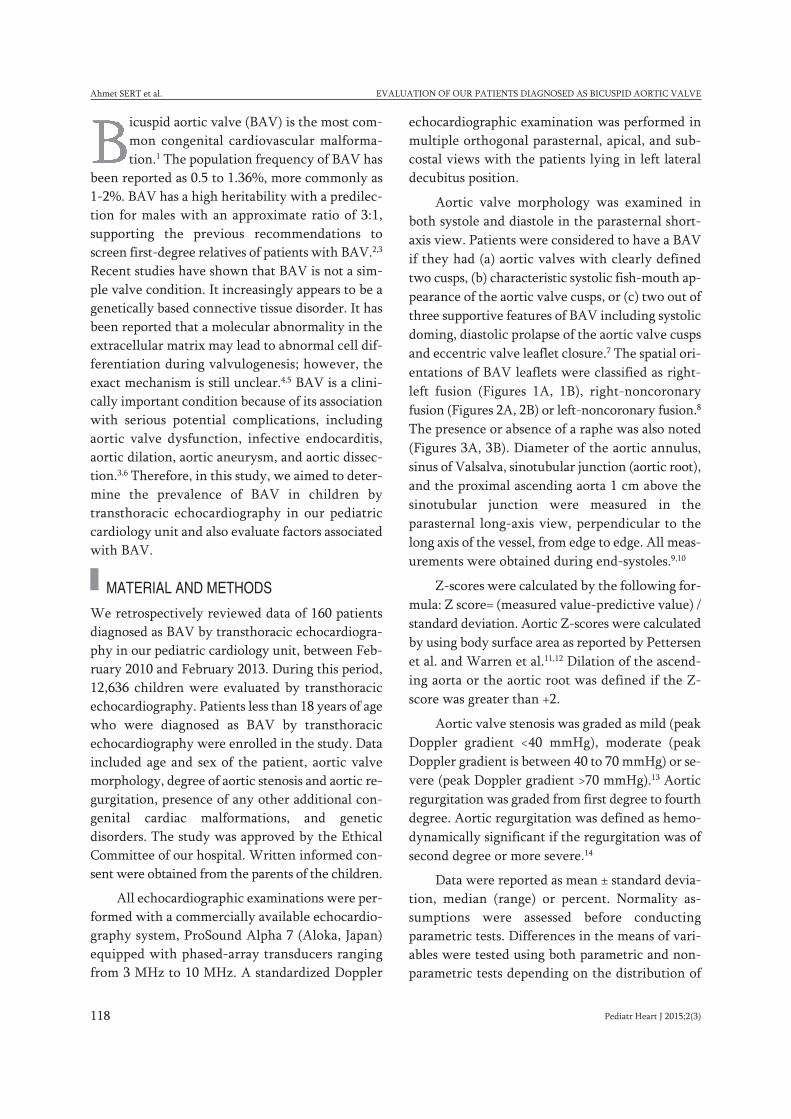
Pediatr Heart J 2015;2(3)118
Ahmet SERT et al. EVALUATION OF OUR PATIENTS DIAGNOSED AS BICUSPID AORTIC VALVE
icuspid aortic valve (BAV) is the most com-mon congenital cardiovascular malforma-tion.1 The population frequency of BAV has
been reported as 0.5 to 1.36%, more commonly as1-2%. BAV has a high heritability with a predilec-tion for males with an approximate ratio of 3:1,supporting the previous recommendations toscreen first-degree relatives of patients with BAV.2,3
Recent studies have shown that BAV is not a sim-ple valve condition. It increasingly appears to be agenetically based connective tissue disorder. It hasbeen reported that a molecular abnormality in theextracellular matrix may lead to abnormal cell dif-ferentiation during valvulogenesis; however, theexact mechanism is still unclear.4,5 BAV is a clini-cally important condition because of its associationwith serious potential complications, includingaortic valve dysfunction, infective endocarditis,aortic dilation, aortic aneurysm, and aortic dissec-tion.3,6 Therefore, in this study, we aimed to deter-mine the prevalence of BAV in children bytransthoracic echocardiography in our pediatriccardiology unit and also evaluate factors associatedwith BAV.
MATERIAL AND METHODS
We retrospectively reviewed data of 160 patientsdiagnosed as BAV by transthoracic echocardiogra-phy in our pediatric cardiology unit, between Feb-ruary 2010 and February 2013. During this period,12,636 children were evaluated by transthoracicechocardiography. Patients less than 18 years of agewho were diagnosed as BAV by transthoracicechocardiography were enrolled in the study. Dataincluded age and sex of the patient, aortic valvemorphology, degree of aortic stenosis and aortic re-gurgitation, presence of any other additional con-genital cardiac malformations, and geneticdisorders. The study was approved by the EthicalCommittee of our hospital. Written informed con-sent were obtained from the parents of the children.
All echocardiographic examinations were per-formed with a commercially available echocardio-graphy system, ProSound Alpha 7 (Aloka, Japan)equipped with phased-array transducers rangingfrom 3 MHz to 10 MHz. A standardized Doppler
echocardiographic examination was performed inmultiple orthogonal parasternal, apical, and sub-costal views with the patients lying in left lateraldecubitus position.
Aortic valve morphology was examined inboth systole and diastole in the parasternal short-axis view. Patients were considered to have a BAVif they had (a) aortic valves with clearly definedtwo cusps, (b) characteristic systolic fish-mouth ap-pearance of the aortic valve cusps, or (c) two out ofthree supportive features of BAV including systolicdoming, diastolic prolapse of the aortic valve cuspsand eccentric valve leaflet closure.7 The spatial ori-entations of BAV leaflets were classified as right-left fusion (Figures 1A, 1B), right-noncoronaryfusion (Figures 2A, 2B) or left-noncoronary fusion.8
The presence or absence of a raphe was also noted(Figures 3A, 3B). Diameter of the aortic annulus,sinus of Valsalva, sinotubular junction (aortic root),and the proximal ascending aorta 1 cm above thesinotubular junction were measured in theparasternal long-axis view, perpendicular to thelong axis of the vessel, from edge to edge. All meas-urements were obtained during end-systoles.9,10
Z-scores were calculated by the following for-mula: Z score= (measured value-predictive value) /standard deviation. Aortic Z-scores were calculatedby using body surface area as reported by Pettersenet al. and Warren et al.11,12 Dilation of the ascend-ing aorta or the aortic root was defined if the Z-score was greater than +2.
Aortic valve stenosis was graded as mild (peakDoppler gradient <40 mmHg), moderate (peakDoppler gradient is between 40 to 70 mmHg) or se-vere (peak Doppler gradient >70 mmHg).13 Aorticregurgitation was graded from first degree to fourthdegree. Aortic regurgitation was defined as hemo-dynamically significant if the regurgitation was ofsecond degree or more severe.14
Data were reported as mean ± standard devia-tion, median (range) or percent. Normality as-sumptions were assessed before conductingparametric tests. Differences in the means of vari-ables were tested using both parametric and non-parametric tests depending on the distribution of
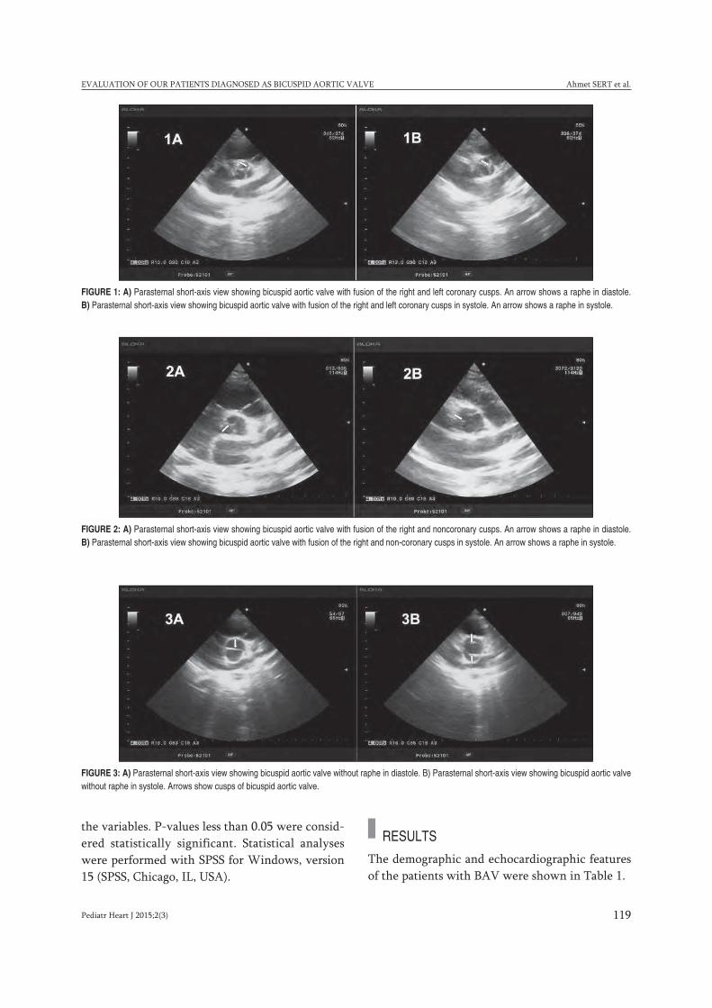
Pediatr Heart J 2015;2(3) 119
EVALUATION OF OUR PATIENTS DIAGNOSED AS BICUSPID AORTIC VALVE Ahmet SERT et al.
the variables. P-values less than 0.05 were consid-ered statistically significant. Statistical analyseswere performed with SPSS for Windows, version15 (SPSS, Chicago, IL, USA).
RESULTS
The demographic and echocardiographic featuresof the patients with BAV were shown in Table 1.
FIGURE 1: A) Parasternal short-axis view showing bicuspid aortic valve with fusion of the right and left coronary cusps. An arrow shows a raphe in diastole. B) Parasternal short-axis view showing bicuspid aortic valve with fusion of the right and left coronary cusps in systole. An arrow shows a raphe in systole.
FIGURE 2: A) Parasternal short-axis view showing bicuspid aortic valve with fusion of the right and noncoronary cusps. An arrow shows a raphe in diastole. B) Parasternal short-axis view showing bicuspid aortic valve with fusion of the right and non-coronary cusps in systole. An arrow shows a raphe in systole.
FIGURE 3: A) Parasternal short-axis view showing bicuspid aortic valve without raphe in diastole. B) Parasternal short-axis view showing bicuspid aortic valvewithout raphe in systole. Arrows show cusps of bicuspid aortic valve.

Ahmet SERT et al. EVALUATION OF OUR PATIENTS DIAGNOSED AS BICUSPID AORTIC VALVE
120 Pediatr Heart J 2015;2(3)
In our cohort of 12,636 children, 160 patientswere diagnosed as BAV by transthoracic echocar-diography, with an estimated prevalence of 1.2%.Of the 160 patients, 109 (68.1%) were males and 51(31.9%) were females. The mean age of patientswas 6.7 ± 5.4 years (median: 6 years, range: 1 day to17 years). Fusion of the right and left coronary cuspwas the most common morphologic type of BAV.Moderate-to-severe aortic stenosis was observed in
12 patients (10.9%) with the right-left fusion, in 1patient (2.4%) with the right-noncoronary fusion,and in none of the patients with the left-noncoro-nary fusion. Hemodinamically significant aortic re-gurgitation was observed in 9 patients (8.1%) withthe right-left fusion, 1 patient (2.4%) with theright-noncoronary fusion, and 1 patient (2.4%)with the left-noncoronary fusion. Thirty-five pa-tients had systolic and diastolic doming of the aor-tic valve. Forty-one out of 160 patients (25.6%) hadneither aortic stenosis nor aortic regurgitation; 43patients (26.9%) had aortic stenosis; 51 patients(31.9%) had aortic regurgitation, and 25 patients(15.6%) had both aortic stenosis and aortic regur-gitation. Twenty-nine patients (18.1%) had mildaortic stenosis, 10 (6.3%) had moderate aorticstenosis, and 4 (2.5%) had severe aortic stenosis.Eleven patients (6.9%) had hemodynamically sig-nificant aortic regurgitation. Of the 29 patientswith mild aortic stenosis, 5 had trivial or minimalaortic regurgitation, 10 had first-degree aortic re-gurgitation, 2 had second-degree aortic regurgita-tion and 1 had third-degree aortic regurgitation. Ofthe 10 patients with moderate aortic stenosis, 3 hadtrivial or first-degree aortic regurgitation, 2 hadsecond-degree aortic regurgitation, 1 had third-de-gree aortic regurgitation.
Frequency of aortic regurgitation was signifi-cantly higher in males compared to females (37.6%vs 19.6%, p=0.023). Frequency of aortic stenosiswas higher in males compared to females (31.2% vs17.6%, p>0.05) but this finding was not statisticallysignificant. Thirty-five patients (21.8%) had dila-tion of the aortic root (21/160) and the ascendingaorta (14/160). Dilation of the ascending aorta wassignificantly more pronounced in those with aorticregurgitation compared to patients without aorticregurgitation (p<0.0001). BAV was an isolated find-ing in 104 patients (65%), while 56 patients (35%)had additional congenital cardiac malformations(Table 2). Thirteen patients (8%) had genetic dis-orders including Down syndrome (n=6), Turnersyndrome (n=5), Pierre-Robin sequence (n=1), andmucopolysaccharidosis (n=1). Balloon dilation ofthe aortic valve was performed in 5 patients (3.1%).Two patients (1.2%) had surgery for aortic coarc-
Characteristics
Male/Female 109/51
Age (Year) 6.7± 5.4
6 (0-17)
Weight (kg) 22.9±18.4
16 (3-70)
Height (cm) 106±39
114 (52-175)
Diameters and z scores values
Aortic annulus (mm) 15.7±4.9
15 (9-26)
Z score 1.2 (0.06-3.01)
Sinus of Valsalva (mm) 20.3±6.9
19.7 (11-35)
Z score 0.71 (0.02-3.35)
Sinotubular junction (mm) 16.8±5.6
16 (9-29)
Z score 0.67 (0.1-3.1)
Ascending aorta (mm) 18.7±5.9
18 (11-33)
Z score 1.46 (0.06-4.24)
Bicuspid aortic valve morphology
Right/left cusp fusion 110 (68.7%)
Right/noncoronary cusp fusion 41 (25.6%)
Left/noncoronary cusp fusion 7 (4.4%)
Without Raphe 2 (1.2%)
Valvular dysfunction
Aortic stenosis 43 (26.9%)
Mild 29 (18.1%)
Moderate 10 (6.3%)
Severe 4 (2.5%)
Aortic regurgitation 51 (31.9%)
Hemodinamically non-significant aortic regurgitation 40 (25%)
Hemodinamically significant aortic regurgitation 11 (6.9%)
Both aortic stenosis and regurgitation 25 (15.6%)
TABLE 1: The demographic and echocardiographicfeatures of the patients with bicuspid aortic valve.
Data are shown as mean plus or minus standard deviation, median (range) or percent.
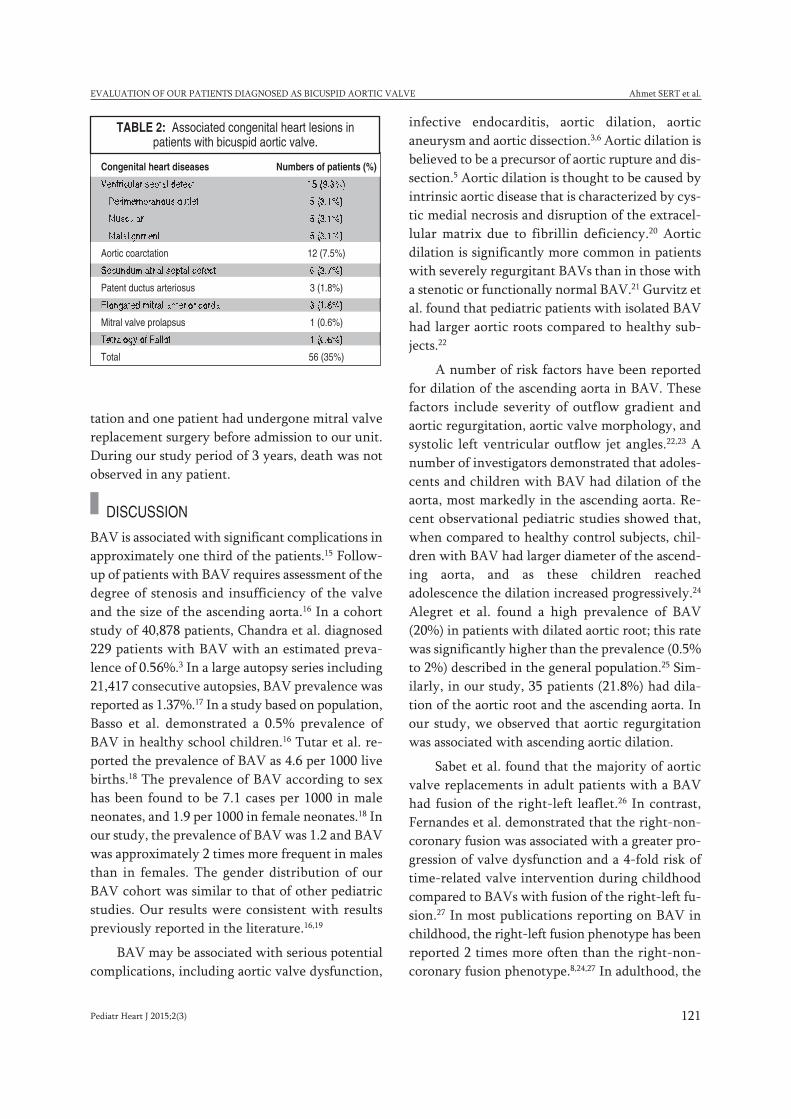
EVALUATION OF OUR PATIENTS DIAGNOSED AS BICUSPID AORTIC VALVE Ahmet SERT et al.
121Pediatr Heart J 2015;2(3)
tation and one patient had undergone mitral valvereplacement surgery before admission to our unit.During our study period of 3 years, death was notobserved in any patient.
DISCUSSION
BAV is associated with significant complications inapproximately one third of the patients.15 Follow-up of patients with BAV requires assessment of thedegree of stenosis and insufficiency of the valveand the size of the ascending aorta.16 In a cohortstudy of 40,878 patients, Chandra et al. diagnosed229 patients with BAV with an estimated preva-lence of 0.56%.3 In a large autopsy series including21,417 consecutive autopsies, BAV prevalence wasreported as 1.37%.17 In a study based on population,Basso et al. demonstrated a 0.5% prevalence ofBAV in healthy school children.16 Tutar et al. re-ported the prevalence of BAV as 4.6 per 1000 livebirths.18 The prevalence of BAV according to sexhas been found to be 7.1 cases per 1000 in maleneonates, and 1.9 per 1000 in female neonates.18 Inour study, the prevalence of BAV was 1.2 and BAVwas approximately 2 times more frequent in malesthan in females. The gender distribution of ourBAV cohort was similar to that of other pediatricstudies. Our results were consistent with resultspreviously reported in the literature.16,19
BAV may be associated with serious potentialcomplications, including aortic valve dysfunction,
infective endocarditis, aortic dilation, aorticaneurysm and aortic dissection.3,6 Aortic dilation isbelieved to be a precursor of aortic rupture and dis-section.5 Aortic dilation is thought to be caused byintrinsic aortic disease that is characterized by cys-tic medial necrosis and disruption of the extracel-lular matrix due to fibrillin deficiency.20 Aorticdilation is significantly more common in patientswith severely regurgitant BAVs than in those witha stenotic or functionally normal BAV.21 Gurvitz etal. found that pediatric patients with isolated BAVhad larger aortic roots compared to healthy sub-jects.22
A number of risk factors have been reportedfor dilation of the ascending aorta in BAV. Thesefactors include severity of outflow gradient andaortic regurgitation, aortic valve morphology, andsystolic left ventricular outflow jet angles.22,23 Anumber of investigators demonstrated that adoles-cents and children with BAV had dilation of theaorta, most markedly in the ascending aorta. Re-cent observational pediatric studies showed that,when compared to healthy control subjects, chil-dren with BAV had larger diameter of the ascend-ing aorta, and as these children reachedadolescence the dilation increased progressively.24
Alegret et al. found a high prevalence of BAV(20%) in patients with dilated aortic root; this ratewas significantly higher than the prevalence (0.5%to 2%) described in the general population.25 Sim-ilarly, in our study, 35 patients (21.8%) had dila-tion of the aortic root and the ascending aorta. Inour study, we observed that aortic regurgitationwas associated with ascending aortic dilation.
Sabet et al. found that the majority of aorticvalve replacements in adult patients with a BAVhad fusion of the right-left leaflet.26 In contrast,Fernandes et al. demonstrated that the right-non-coronary fusion was associated with a greater pro-gression of valve dysfunction and a 4-fold risk oftime-related valve intervention during childhoodcompared to BAVs with fusion of the right-left fu-sion.27 In most publications reporting on BAV inchildhood, the right-left fusion phenotype has beenreported 2 times more often than the right-non-coronary fusion phenotype.8,24,27 In adulthood, the
Congenital heart diseases Numbers of patients (%)
Ventricular septal defect 15 (9.3%)
Perimembranous outlet 5 (3.1%)
Muscular 5 (3.1%)
Malalignment 5 (3.1%)
Aortic coarctation 12 (7.5%)
Secundum atrial septal defect 6 (3.7%)
Patent ductus arteriosus 3 (1.8%)
Elongated mitral anterior corda 3 (1.8%)
Mitral valve prolapsus 1 (0.6%)
Tetralogy of Fallot 1 (0.6%)
Total 56 (35%)
TABLE 2: Associated congenital heart lesions in patients with bicuspid aortic valve.

1. Hoffman JI, Kaplan S. The incidence of con-genital heart disease. J Am Coll Cardiol2002;39(12):1890-900.
2. Girdauskas E, Borger MA, Secknus MA, Gir-dauskas G, Kuntze T. Is aortopathy in bicus-pid aortic valve disease a congenital defect ora result of abnormal hemodynamics? A criti-cal reappraisal of a one-sided argument. EurJ Cardiothorac Surg 2011;39(6):809-14.
3. Chandra S, Lang RM, Nicolarsen J, Gayat E,
Spencer KT, Mor-Avi V, Hofmannowman MA.Bicuspid aortic valve: inter-racial difference infrequency and aortic dimensions. JACC Car-diovasc Imaging 2012;5(10): 981-9.
4. Nataatmadja M, West M, West J, Summers K,Walker P, Nagata M, Watanabe T. Abnormalextracellular matrix protein transport associ-ated with increased apoptosis of vascularsmooth muscle cells in marfan syndrome andbicuspid aortic valve thoracic aortic aneurysm.
Circulation 2003;108(Suppl 1:II): 329-34.
5. Fedak PW, Verma S, David TE, Leask Rl,Weisel RD, Butany J. Clinical and pathophys-iological implications of a bicuspid aortic valve.Circulation 2002;106(8):900-4.
6. Cecconi M, Nistri S, Quarti A, Manfrin M,Colonna PL, Molini E, Perna GP. Aortic di-latation in patients with bicuspid aortic valve. JCardiovasc Med (Hagerstown) 2006;7(1): 11-20.
REFERENCES
phenotype distribution is less clear, with nearlyequal frequencies of right-left fusion and right-noncoronary morphology described by Ward, anda prevalence of the right-left fusion phenotype asgreat as 82% reported by Schaefer et al., compara-ble to the distribution described in the childhoodstudies.28,29 In our study, we found the right-left fu-sion phenotype to be the predominant BAV mor-phology (68.7%). Our finding was in accordancewith the study by Fernandes et al.
In our study, 10.9% of the patients had mod-erate-to-severe valvular stenosis in patients withright-left cusp fusion, but only 2.4% of the patientshad moderate-to-severe valvular stenosis in patientswith right-noncoronary fusion.27 Furthermore, he-modynamically significant aortic regurgitation wasobserved in 8.1% of patients with right-left cusp fu-sion compared to 2.4% in those with right-non-coronary cusp fusion.
BAV may be an isolated finding or may be as-sociated with other congenital heart lesions. Thereis a significantly higher incidence of aortic arch ob-struction (51.1%). There is a well-documented as-sociation of BAV with aortic coarctation.30
Previous studies have identified an association be-tween BAV and other congenital vascular malfor-mations, particularly ventricular septal defect.31
However, it remains unclear which type of ven-tricular septal defect may be associated with BAV.The frequency of BAV in specimens with completetransposition of great arteries has been found to beonly 1%. Hypoplastic left heart syndrome, com-plete atrioventricular canal defect, Ebstein’s anom-aly, partial or total anomalous pulmonary venous
return, and double-outlet right ventricle may oc-casionally be associated with BAV.28 In our study,ventricular septal defect and aortic coarctationwere the most frequent congenital heart defects as-sociated with BAV.
The frequency of congenital cardiovascularabnormalities was reported to be between 23% and45% in Turner syndrome.32 BAV and aortic coarc-tation have been reported as the most common car-diovascular abnormalities. BAV has been rarelyreported in children with Down syndrome.33 Thir-teen of our patients had genetic disorders. Downsyndrome (6/160) and Turner syndrome (5/160)were the most frequent genetic disorders in our co-hort.
Our study had some limitations, such as its ret-rospective design. This study represents the popu-lation of children with BAV referred to pediatriccardiology units. Patients with clinically insignifi-cant BAV may not be referred for echocardio-graphic examination because of a lack of physicalfindings.
CONCLUSION
Our study demonstrates that the prevalence ofBAV in children by transthoracic echocardiogra-phy is 1.2%, which is in accordance with previ-ously reported results in the literature. In ourstudy, the prevalence of BAV in males was approx-imately 2 times higher than that in females. Right-left cusp fusion was more frequently associatedwith moderate-to-severe aortic valve stenosis andregurgitation than the other morphologic pheno-types.
Ahmet SERT et al. EVALUATION OF OUR PATIENTS DIAGNOSED AS BICUSPID AORTIC VALVE
122 Pediatr Heart J 2015;2(3)

Pediatr Heart J 2015;2(3) 123
EVALUATION OF OUR PATIENTS DIAGNOSED AS BICUSPID AORTIC VALVE Ahmet SERT et al.
7. Brandenburg RO, Tajik AJ, Edwards WD,Reeder GS, Shub C, Seward JB. Accuracy of2-dimentional echocardiographic diagnosis ofcongenitally bicuspid aortic valve: echocar-diographic-anatomic correlation in 115 pa-tients. Am J Cardiol 1983;51(9):1469-73.
8. Mahle WT, Sutherland JL, Frias PA. Outcomeof isolated bicuspid aortic valve in childhood.J Pediatr 2010;157(3): 445-49.
9. Lang RM, Bierig M, Devereux RB, Flach-skampf FA, Foster E, Pellikka PA, Picard MH,Roman MJ, Seward J, Shanewise JS,Solomon SD, Spencer KT, Sutton MS, Stew-art WJ; Chamber Quantification WritingGroup. Recommendations for chamber quan-tification: a report from the American Societyof Echocardiography's Guidelines and Stan-dards Committee and the Chamber Quantifi-cation Writing Group, developed inconjunction with the European Association ofEchocardiography, a branch of the EuropeanSociety of Cardiology. J Am Soc Echocardiogr2005;18(12):1440-63.
10. Roman MJ, Devereux RB, Kramer-Fox R,O’Loughlin J. Two-dimensional echocardio-graphic aortic root dimensions in normal chil-dren and adults. Am J Cardiol 1989;64(8):507-12.
11. Pettersen MD, Du W, Skeens ME, Humes RA.Regression equations for calculation of zscores of cardiac structures in a large cohortof healthy infants, children, and adolescents:an echocardiographic study. J Am SocEchocardiogr 2008;21(8): 922-34.
12. Warren AE, Boyd ML, O'Connell C, Dodds L.Dilatation of the ascending aorta in paediatricpatients with bicuspid aortic valve: frequency,rate of progression and risk factors. Heart2006;92(10):1496-1500.
13. Graham TP Jr., Driscoll DJ, Gersony WM,Newburger JW, Rocchini A, Towbin JA. Taskforce 2: congenital heart disease. J Am CollCardiol 2005;45(8):1326-33.
14. Tani LY, Minich LL, Day RW, Orsmond GS,Shaddy RE. Doppler evaluation of aortic re-
gurgitation in children. Am J Cardiol1997;80(7):927-31.
15. Chan KL, Stinson WA, Veinot JP. Reliabilityof transthoracic echocardiography in the as-sessment of aortic valve morphology: patho-logical correlation in 178 patients. Can JCardiol 1999;15(1):48-52.
16. Basso C, Boschello M, Perrone C. Anechocardiographic survey of primary schoolchildren for bicuspid aortic valve. Am J Car-diol 2004;93(5):661-3.
17. Larson EW, Edwards WD. Risk factors for aor-tic dissection: a necropsy study of 161 cases.Am J Cardiol 1984;53(6):849-55.
18. Tutar E, Ekici F, Atalay S, Nacar N. The preva-lence of bicuspid aortic valve in newborns byechocardiographic screening. Am Heart J2005;150(3):513-5.
19. Beroukhim RS, Kruzick TL, Taylor AL, Gao D,Yetman AT. Progression of aortic dilation inchildren with a functionally normal bicuspidaortic valve. Am J Cardiol 2006;98(6):828-30.
20. Aboulhosn J, Child JS. Left ventricular outflowobstruction: subaortic stenosis, bicus¬pid aor-tic valve, supravalvar aortic stenosis, andcoarctation of the aorta. Circulation2006;114(22):2412-22.
21. Hahn RT, Roman MJ, Mogtader AH, Dev-ereux RB. Association of aortic dilation withregurgitant, stenotic and functionally normalbicuspid aortic valves. J Am Coll Cardiol1992;19(2):283-8.
22. Gurvitz M, Chang RK, Drant S, Allada V. Fre-quency of aortic root dilation in children with abicuspid aortic valve. Am J Cardiol 2004;94(10):1337-40.
23. den Reijer PM, Sallee D 3rd, van der VeldenP, Zaaijer ER, Parks WJ, Ramamurthy S,Robbie TQ, Donati G, Lamphier C, BeekmanRP, Brummer ME. Hemodynamic predictorsof aortic dilatation in bicuspid aortic valve byvelocity-encoded cardiovascular magnetic res-onance. J Cardiovasc Magn Reson 2010; 12:4.
24. Holmes KW, Lehmann CU, Dalal D, Nasir K,Dietz HC, Ravekes WJ, Thompson WR, Spe-vak PJ. Progressive dilation of the ascendingaorta in children with isolated bicuspid aorticvalve. Am J Cardiol 2007;99(7):978-83.
25. Alegret JM, Duran I, Palazon O, Vernis JM,Ameijide A, Rabassa A, Masana L. Preva-lence of and predictors of bicuspid aorticvalves in patients with dilated aortic roots. AmJ Cardiol 2003;91(5):619-22.
26. Sabet HY, Edwards WD, Tazelaar HD, DalyRC. Congenitally bicuspid aortic valves: a sur-gical pathology study of 542 cases (1991through 1996) and a literature review of 2,715additional cases. Mayo Clin Proc 1999;74(1):14 -26.
27. Fernandes SM, Sanders SP, Khairy P, Jenk-ins KJ, Gauvreau K, Lang P, SimondsH,Colan SD. Morphology of bicuspid aorticvalve in children and adolescents. J Am CollCardiol 2004;44(8):1648-51.
28. Ward C. Clinical significance of the bicuspidaortic valve. Heart 2000;83(1):81-5.
29. Schaefer BM, Lewin MB, Stout KK, Byers PH,Otto CM. Usefulness of bicuspid aortic valvephenotype to predict elastic properties of theascending aorta. Am J Cardiol 2007;99(5):686-90.
30. Folger GM Jr, Stein PD. Bicuspid aortic valvemorphology when associated with coartationof the aorta. Cathet Cardiovasc Diagn1984;10(1):17-25.
31. Ciotti GR, Vlahos AP, Silverman NH. Morphol-ogy and function of the bicuspid aortic valvewith and without coarctation of the aorta in theyoung. Am J Cardiol 2006;98(8):1096-102.
32. Mazzanti L, Cacciari E. Congenital heart dis-ease in patients with Turner’s syndrome: Ital-ian Study Group for Turner Syndrome(ISGTS). J Pediatr 1998;133(5):688-92.
33. Aughton DJ, Weinhouse E, Riggs TW. Iso-lated bicuspid aortic valve in a newborn withDown syndrome. Clin Pediatr (Phila) 1995;34(11):622.
Pediatr Heart J 2015;2(3)124
triyal Septal Defekt (ASD) izole anomali olarak tüm doğuştan kalphastalıklarının içinde %5-10 oranında görülür. Doğuştan kalp has-talığı olan çocukların yaklaşık %30-50’sinde kalp defektine ilave
olarak ASD bulunur.1
İzole Sekundum Atriyal Septal DefektTanısı Alan HastalarınEkokardiyografik Seyri
ÖÖZET Bu çalışmada çocukluk çağındaki izole sekundum atriyal septal defektin seyrini değerlen-dirmeyi, spontan kapanma üzerine etkili faktörleri araştırmayı amaçladık. Çalışmamızda Ocak 2000ile Haziran 2012 tarihleri arasında Eskişehir Osmangazi Üniversitesi Tıp Fakültesi Pediatri Kardi-yoloji Polikliniği’nde en az 6 ay süresince izlenmiş 746 sekundum atriyal septal defekt hastası res-trospektif olarak değerlendirildi. Atriyal septal defektli hastaların izleminde 208’inin (%27,7)defektinin spontan kapandığı saptandı. Defekti spontan kapanan hastaların yaş ortalaması 24±40ay (6-204 ay) idi. Çalışmamızda tanı yaşı ile defekt çapı arasında pozitif, defekt çapı ve tanı yaşı ilespontan kapanma arasında negatif ilişki (p<0,05) ve defekt çapı ile kapatma işlemleri yapılma ara-sında pozitif ilişki saptandı (p<0,05). Atriyal septal defektli erkek hastalarda kız hastalara göre spon-tan kapanmanın anlamlı oranda yüksek olduğu saptandı (p<0,05). Atriyal septal anevrizma, patentduktus arteriozus ve doğum zamanı ile spontan kapanma arasında istatistiksel anlamlı fark bulun-mazken, ventriküler septal defekt eşlik eden atriyal septal defekt hastalarının daha sık spontan ka-pandığı saptandı (p<0,05). Sonuç olarak; atriyal septal defektin spontan kapanması ve defekt çapınıngerilemesi üzerine diğer etkenlerden bağımsız olarak tanı anındaki defekt çapı ve tanı yaşı etkili-dir, term hastalarda ventriküler septal defekt varlığı defekt kapanma süresini kısaltmaktadır.
Anahtar Kelimeler: Atriyal septal defekt; ekokardiyografi; seyir
ABSTRACT In this study we aim to asses the progression of secundum atrial septal defect and de-termine the causes that affect the spontaneous closure. We evaluate 746 secundum atrial septal de-fect patients who have at least 6 months follow up in Eskişehir Osmangazi University PediatricCardiology department between January 2000 and June 2012. Follow-up of patients with atrial sep-tal defect in 208 (27.7%) were the spontaneous closure. The average age of patients with sponta-neous closure were 24±40 months (range 6-204 months). In our study, age of onset has been founddirectly related with diameter of defect (p<0.05). Moreover, we have found inverse relationshipbetween age of onset, diameter of defect and spontenous closure. Spontanous closure of atrial sep-tal defect in male patients is significantly more frequent than females (p<0.05). While statisticallysignificant difference was not found between atrial septal aneurysm, patent ductus arteriosus andtime of birth with spontaneous closure of atrial septal defect, there was a statistically significant cor-relation between ventricular spontaneous atrial septal defect closure of septal defects (p<0,05). Inconclusion; the diameter of defect and age of onset affect to spontanoues closure and regresion ofdefect, moreover existence of ventricular septal defect shortens the duration of closure.
Key Words: Atrial septal defect; echocardiography; follow-up
Pediatr Heart J 2015;2(3):124-9
Alperen AYDIN,a
Ali YILDIRIM,a,b
Tevfik DEMİR,a,b
Fatma AYDIN,a
Birsen UÇAR,a,b
Zübeyir KILIÇa,b
aÇocuk Sağlığı ve Hastalıkları AD, bÇocuk Kardiyoloji BD, Eskişehir Osmangazi Üniversitesi Tıp Fakültesi, Eskişehir
Ge liş Ta ri hi/Re ce i ved: 14.09.2015 Ka bul Ta ri hi/Ac cep ted: 10.11.2015
Ya zış ma Ad re si/Cor res pon den ce:Ali YILDIRIMEskişehir Osmangazi Üniversitesi Tıp Fakültesi, Çocuk Sağlığı ve Hastalıkları AD,Çocuk Kardiyoloji BD, Eskişehir,TÜRKİYE/[email protected]
Copyright © 2015 by Türk Pediatrik Kardiyoloji veKalp Cerrahisi Derneği
ORİJİNAL ARAŞTIRMA
Pediatr Heart J 2015;2(3) 125
İZOLE SEKUNDUM ATRİYAL SEPTAL DEFEKT TANISI ALAN HASTALARIN EKOKARDİYOGRAFİK SEYRİ Alperen AYDIN ve ark.
Birçok çalışmada ASD nin seyri ve spontan ka-panma sıklığı degerlendirilmiştir. Fakat sonuçlarındeğişken olduğu görülmüştür. Yapılan çalışmalardayenidoğan döneminde tanı alan ve defekt çapıküçük olan vakaların büyük çoğunluğunun spon-tan kapandığı, defekt çapı büyük olan ve tanı yaşıileri olan lezyonlarda spontan kapanma oranınınazaldığı tespit edilmiş.2-8 Nadiren ASD defekt çapı-nın yaşla birlikte arttığı tespit edilmiş.6
Bu çalışmada çocukluk çağındaki izole sekun-dum ASD seyrini değerlendirmeyi, spontan kapanmaüzerine etkili faktörleri tespit etmeyi ve klinik izlemikonusuna katkıda bulunmayı amaçladık.
GEREÇ VE YÖNTEMLER
Eskişehir Osmangazi Üniversitesi Tıp Fakültesi Pe-diatrik Kardiyoloji Polikliniğine Ocak 2000 ile Ha-ziran 2012 tarihleri arasında başvuran ve ekokar-diyografik çalışma ile izole sekundum ASD tanısıalan, yaşları 1 gün-200 ay arasında değişen 746 has-tanın dosyası geriye dönük olarak incelendi. Eski-şehir Osmangazi Üniversitesi Tıp Fakultesi EtikKurulundan çalışmamızla ilgili etik onay alındı.Çalışma grubumuza en az 6 ay izlem süresi olan veen az 2 ekokardiyografik incelemesi olan hastalardahil edildi. Hastaların tanı yaşı, vücut ağırlıkları,tekrarlayan ekokardiyografi bulguları, eşlik edenek kardiyovasküler hastalıkları, eşlik eden kardi-yovasküler sistem dışı hastalıkları, prematür olupolmamaları, hastaneye başvuru nedenleri ‘HastaBilgi Formu’na kayıt edildi.
Ekokardiyografi bulguları Radzik klasifikasyo-nuna uygun olarak defekt çaplarına göre 3 grubaayrıldı. Literatüre uygun olarak 3 mm ve altı pa-tent foramen ovale, 3 mm’den büyük defektlerASD olarak kabul edildi (2).
1. grup: >3-<5 mm olanlar (küçük defekt),
2. grup: >5-<8 mm arasında olanlar (orta de-fekt),
3. grup: >8 mm ve daha büyük olanlar (büyükdefekt),
Primum ASD, sinus venosus ASD, koronersinus ASD ve diğer major kardiyak malformasyonuolan hastalar çalışmaya dahil edilmedi. Minör kar-
diyak anomaliler (küçük ventriküler septal defekt(VSD), patent duktus arteriozus (PDA) ve mitralkapak prolapsusu, atriyal septal anevrizma (ASA))çalışmaya dahil edildi.
EKOKARDİYOGRAFİK İNCELEME
Tüm hastaların ekokardiyografik incelemesi üçfarklı çocuk kardiyoloji uzmanından en az biri ta-rafından VİVİD I portable ekokardiyografi cihazıile S3-S7 probları ve HAWLETT PACKARDSONOS 5500 cihazı ile S4-S8 VE S12 probları kul-lanılarak yapılmıştır. Tüm hastaların iki boyutlu,M mode, continuous wave (devamlı dalga-CW),pulsed wave (nabızlı dalga-PW), renkli akım eko-kardiyografi incelemeleriyapılmıştır
İSTATİSTİKSEL ANALİZLER
Çalışma verilerinin istatistiksel analizi SPSS 16.0paket programı (SPSS Inc., Chicago, Illinois, ABD)ile yapıldı. Sürekli nicel veriler; n, ortalama ve stan-dart hata (Standard error of mean; SEM) olarak;nitel veriler ise n, ortanca değer, minumum vemaksimum değerler olarak ifade edilmiştir. Bağım-sız ölçümlerden oluşan ikili grupların karşılaştırıl-masında normal dağılım gösteren veriler t testi ile,normal dağılım göstermeyen değişkenlerden olu-şan veriler ise Mann-Whitney U testi ile analizedilmiştir. Değişkenler arasındaki ilişkiyi göstere-bilmek için normal dağılım gösteren veriler içinPearson, normal dağılım göstermeyen veriler içinSpearman korelasyon testlerinden yararlanılmıştır.Kategorik yapıdaki veri setlerine ise ki-kare testiuygulanmıştır. Bazı parametreler arasındaki ilşkiyibelirlemek için ‘‘Lojistik Regresyon analizi’’ uygu-landı. Olasılık değerleri p<0,05 olanlar anlamlı ola-rak kabul edilmiştir.
BULGULAR
Yaşları 1 gün ile 200 ay arasında değişen 746 hastaretrospektif olarak değerlendirildi. Hastaların 364(48,8)’ü erkek idi. Olguların yaş ortancası 5 ay (1gün-200 ay) idi. Hastaların tanı yaşına göre grupla-ması yapıldığında 278’i (%37,2) <1 ay, 193’ü (%26)>1-<12 ay, 275’i (%36,8) >12 ay idi. Hastaların or-talama izlem süresi 17 ay (6 ay-160 ay) idi. Hasta-ların 90’ında (%12) ASA, 73’ünde (%9,7) küçükVSD, 47’sinde (%6,3) küçük PDA eşlik etmekteydi.
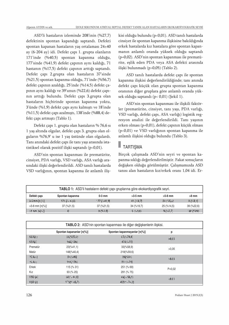
Pediatr Heart J 2015;2(3)126
Alperen AYDIN ve ark. İZOLE SEKUNDUM ATRİYAL SEPTAL DEFEKT TANISI ALAN HASTALARIN EKOKARDİYOGRAFİK SEYRİ
ASD’li hastaların izleminde 208’inin (%27,7)defektinin spontan kapandığı saptandı. Defektispontan kapanan hastaların yaş ortalaması 24±40ay (6-204 ay) idi. Defekt çapı 1. grupta olanların171’inde (%40,5) spontan kapanma olduğu,177’sinde (%41,9) defekt çapının aynı kaldığı, 71hastanın (%17,5) defekt çapının arttığı saptandı.Defekt çapı 2.grupta olan hastaların 37’sinde(%21,5) spontan kapanma olduğu, 71’inde (%56,7)defekt çapının azaldığı, 25’inde (%14,5) defekt ça-pının aynı kaldığı ve 39’unun (%22,6) defekt çapı-nın arttığı bulundu. Defekt çapı 3.grupta olanhastaların hiçbirinde spontan kapanma yoktu,3’ünde (%1,9) defekt çapı aynı kalmıştı ve 18’inde(%11,5) defekt çapı azalmıştı, 138’inde (%88,4) de-fekt çapı artmıştı (Tablo 1).
Defekt çapı 1. grupta olan hastaların % 76,6 sı1 yaş altında olgular, defekt çapı 3. grupta olan ol-guların %76,9’ u ise 1 yaş üstünde olan olgulardı.Tanı anındaki defekt çapı ile tanı yaşı arasında ista-tistiksel olarak pozitif ilişki saptandı (p<0,01).
ASD’nin spontan kapanması ile prematürite,cinsiyet, PDA varlığı, VSD varlığı, ASA varlığı ara-sındaki ilişki değerlendirildi. ASD tanılı hastalardaVSD varlığının, spontan kapanma ile anlamlı iliş-
kisi olduğu bulundu (p<0,01). ASD tanılı hastalardacinsiyet ile spontan kapanma ilişkisine bakıldığındaerkek hastalarda kız hastalara göre spontan kapan-manın anlamlı oranda yüksek olduğu saptandı(p=0,02). ASD’nin spontan kapanması ile prematü-rite, eşlik eden PDA veya ASA defekti arasındailişki bulunmadı (p>0,05) (Tablo 2).
ASD tanılı hastalarda defekt çapı ile spontankapanma ilişkisi değerlendirildiğinde; tanı anındadefekt çapı küçük olan grupta spontan kapanmaoranının diğer gruplara göre anlamlı oranda yük-sek olduğu saptandı (p< 0,01) (Şekil 1).
ASD’nin spontan kapanması ile ilişkili faktör-ler (prematürite, cinsiyet, tanı yaşı, PDA varlığı,VSD varlığı, defekt çapı, ASA varlığı) logistik reg-resyon analizi ile değerlendirildi. Tanı yaşınınerken olması (p=0,01), defekt çapının küçük olması(p<0,01) ve VSD varlığının spontan kapanma ileanlamlı ilişkisi olduğu bulundu (Tablo 3).
TARTIŞMA
Birçok çalışmada ASD’nin seyri ve spontan ka-panma sıklığı değerlendirilmiştir. Fakat sonuçlarındeğişken olduğu görülmüştür. Çalışmamızda ASDtanısı alan hastaların kız/erkek oranı 1,04 idi. Er-
Defekt çapı Spontan kapanma 0-3 mm >3-5 mm >5-8 mm >8 mm
3-5 mm [n(%)] 171 (% 40,5) 177 (%41,9) 41 (%9,7) 24 (%5,6) 9 (%2,1)
>5-8 mm [n(%)] 37 (%21,5) 37 (%21,5) 34 (%19,7) 25 (%14,5) 39 (%22,6)
>8 mm [n(%)] 0 3 (%1,9) 6 (%3,8) 9 (%5,7) 138 (%88)
TABLO 1: ASD’li hastaların defekt çapı gruplarına göre ekokardiyografik seyri.
Spontan kapananlar [n(%)] Spontan kapanmayanlar [n(%)] p
ASA(+) 23(%25,6) 67(%74,4)>0,05
ASA(-) 185(%28) 475(%72)
Prematür 23(%41,1) 33(%58,9)>0,05
Matür 148(%40,4) 218(%59,6)
PDA(+) 24(%46) 28(%54)>0,05
PDA(-) 184(%26) 514(%74)
Erkek 115 (% 31) 251 (% 69)P=0,02
Kız 93 (% 25) 291 (% 75)
VSD (+) 35(% 44,9) 43(% 55,1)<0,01
VSD (-) 173(% 25,7) 499(% 74,3)
TABLO 2: ASD’nin spontan kapanması ile diğer değişkenlerin ilişkisi.
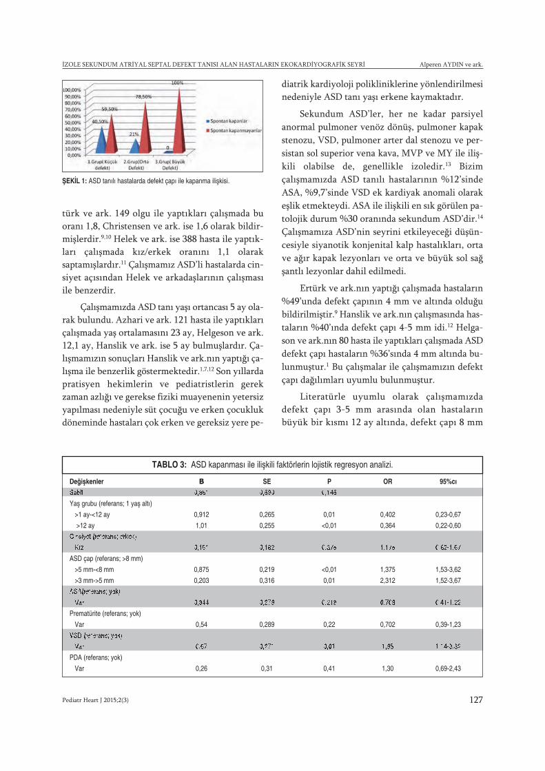
Pediatr Heart J 2015;2(3) 127
İZOLE SEKUNDUM ATRİYAL SEPTAL DEFEKT TANISI ALAN HASTALARIN EKOKARDİYOGRAFİK SEYRİ Alperen AYDIN ve ark.
türk ve ark. 149 olgu ile yaptıkları çalışmada buoranı 1,8, Christensen ve ark. ise 1,6 olarak bildir-mişlerdir.9,10 Helek ve ark. ise 388 hasta ile yaptık-ları çalışmada kız/erkek oranını 1,1 olaraksaptamışlardır.11 Çalışmamız ASD’li hastalarda cin-siyet açısından Helek ve arkadaşlarının çalışmasıile benzerdir.
Çalışmamızda ASD tanı yaşı ortancası 5 ay ola-rak bulundu. Azhari ve ark. 121 hasta ile yaptıklarıçalışmada yaş ortalamasını 23 ay, Helgeson ve ark.12,1 ay, Hanslik ve ark. ise 5 ay bulmuşlardır. Ça-lışmamızın sonuçları Hanslik ve ark.nın yaptığı ça-lışma ile benzerlik göstermektedir.1,7,12 Son yıllardapratisyen hekimlerin ve pediatristlerin gerekzaman azlığı ve gerekse fiziki muayenenin yetersizyapılması nedeniyle süt çocuğu ve erken çocuklukdöneminde hastaları çok erken ve gereksiz yere pe-
diatrik kardiyoloji polikliniklerine yönlendirilmesinedeniyle ASD tanı yaşı erkene kaymaktadır.
Sekundum ASD’ler, her ne kadar parsiyelanormal pulmoner venöz dönüş, pulmoner kapakstenozu, VSD, pulmoner arter dal stenozu ve per-sistan sol superior vena kava, MVP ve MY ile iliş-kili olabilse de, genellikle izoledir.13 Bizimçalışmamızda ASD tanılı hastalarının %12’sindeASA, %9,7’sinde VSD ek kardiyak anomali olarakeşlik etmekteydi. ASA ile ilişkili en sık görülen pa-tolojik durum %30 oranında sekundum ASD’dir.14
Çalışmamıza ASD’nin seyrini etkileyeceği düşün-cesiyle siyanotik konjenital kalp hastalıkları, ortave ağır kapak lezyonları ve orta ve büyük sol sağşantlı lezyonlar dahil edilmedi.
Ertürk ve ark.nın yaptığı çalışmada hastaların%49’unda defekt çapının 4 mm ve altında olduğubildirilmiştir.9 Hanslik ve ark.nın çalışmasında has-taların %40’ında defekt çapı 4-5 mm idi.12 Helga-son ve ark.nın 80 hasta ile yaptıkları çalışmada ASDdefekt çapı hastaların %36’sında 4 mm altında bu-lunmuştur.1 Bu çalışmalar ile çalışmamızın defektçapı dağılımları uyumlu bulunmuştur.
Literatürle uyumlu olarak çalışmamızda defekt çapı 3-5 mm arasında olan hastalarınbüyük bir kısmı 12 ay altında, defekt çapı 8 mm
ŞEKİL 1: ASD tanılı hastalarda defekt çapı ile kapanma ilişkisi.
Değişkenler Β SE P OR 95%cı
Sabit 0,861 0,590 0,145
Yaş grubu (referans; 1 yaş altı)
>1 ay-<12 ay 0,912 0,265 0,01 0,402 0,23-0,67
>12 ay 1,01 0,255 <0,01 0,364 0,22-0,60
Cinsiyet (referans; erkek)
Kız 0,161 0,182 0,375 1,175 0,82-1,67
ASD çap (referans; >8 mm)
>5 mm-<8 mm 0,875 0,219 <0,01 1,375 1,53-3,62
>3 mm->5 mm 0,203 0,316 0,01 2,312 1,52-3,67
ASA(referans; yok)
Var 0,344 0,279 0,218 0,709 0,41-1,22
Prematürite (referans; yok)
Var 0,54 0,289 0,22 0,702 0,39-1,23
VSD (referans; yok)
Var 0,67 0,271 0,01 1,95 1,14-3,32
PDA (referans; yok)
Var 0,26 0,31 0,41 1,30 0,69-2,43
TABLO 3: ASD kapanması ile ilişkili faktörlerin lojistik regresyon analizi.
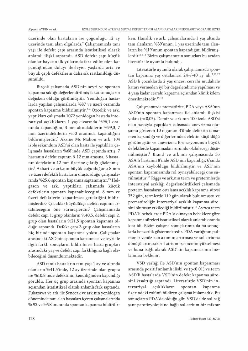
Pediatr Heart J 2015;2(3)128
Alperen AYDIN ve ark. İZOLE SEKUNDUM ATRİYAL SEPTAL DEFEKT TANISI ALAN HASTALARIN EKOKARDİYOGRAFİK SEYRİ
üzerinde olan hastaların ise çoğunluğu 12 ay üzerinde tanı alan olgulardı.7 Çalışmamızda tanıyaşı ile defekt çapı arasında istatistiksel olarakanlamlı ilişki saptandı. ASD defekt çapı küçükolanlar hayatın ilk yıllarında fark edilmeden ka-pandığından dolayı ilerleyen yaşlarda orta vebüyük çaplı defektlerin daha sık rastlanıldığı dü-şünüldü.
Birçok çalışmada ASD’nin seyri ve spontankapanma sıklığı değerlendirilmiş fakat sonuçlarındeğişken olduğu görülmüştür. Yenidoğan hasta-larda yapılan çalışmalarda %87 ve üzeri oranındaspontan kapanma bildirilmiştir.2-4 Özçelik ve ark. yaptıkları çalışmada 1072 yenidoğan hastada inte-ratriyal açıklıkların 1 yaş civarında %96,1 ora-nında kapandığını, 3 mm altındakilerin %99,3, 7mm üzerindekilerin %50 oranında kapandığınıbildirmişlerdir.5 Aksine Mc Mahon ve ark. 104izole sekundum ASD’si olan hasta ile yaptıkları ça-lışmada hastaların %68’inde ASD çapında artış, 7hastanın defekt çapının 6-12 mm arasına, 3 hasta-nın defektinin 12 mm üzerine çıktığı gözlenmiş-tir.6 Azhari ve ark.nın büyük çoğunluğunu 8 mmve üzeri defektli hastaların oluşturduğu çalışmala-rında %25,6 spontan kapanma saptanmıştır.13 Hel-gason ve ark. yaptıkları çalışmada küçükdefektlerin spontan kapanabilecegini, 8 mm veüzeri defektlerin kapatılması gerektiğini bildir-mişlerdir.7 Çocuklar büyüdükçe defekt çapının ar-tabilecegini öne sürmüşlerdir.2 Çalışmamızdadefekt çapı 1. grup olanların %40,5, defekt çapı 2.grup olan hastaların %21,5 spontan kapanma ol-duğu saptandı. Defekt çapı 3.grup olan hastalarınhiç birinde spontan kapanma yoktu. Çalışmalararasındaki ASD’nin spontan kapanması ve seyri ileilgili farklı sonuçların bildirilmesi hasta gruplarıarasındaki yaş ve defekt çapı farklılığına bağlı ola-bileceğini düşündürmektedir.
ASD tanılı hastaların tanı yaşı 1 ay ve altındaolanların %41,5’inde, 12 ay üzerinde olan gruptaise %10,8’inde defektinin kendiliğinden kapandığıgörüldü. Her üç grup arasında spontan kapanmaaçısından istatistiksel olarak anlamlı fark saptandı.Fukazawa ve ark. ile Şenocak ve ark.nın yenidoğandöneminde tanı alan hastaları içeren çalışmalarında% 92 ve %98 oranında spontan kapanma bildirilir-
ken, Hanslik ve ark. çalışmalarında 1 yaş altındatanı alanların %39’unun, 1 yaş üzerinde tanı alan-ların ise %19’unun spontan kapandığını bildirmiş-lerdir.3,4,12 Bizim çalışmamızın sonuçları bu açıdanliteratür ile uyumlu bulundu.
Literatürle uyumlu olarak çalışmamızda spon-tan kapanma yaş ortalaması 24+/-40 ay idi.7,11,12
ASD’li çocuklarda 2 yaş öncesi cerrahi müdahalekararı vermeden iyi bir değerlendirme yapılması ve4 yaşa kadar cerrahi kapatma açısından klinik izlemönerilmektedir.15-17
Çalışmamızda prematürite, PDA veya ASA’nınASD’nin spontan kapanması ile anlamlı ilişkisiyoktu (p>0,05). Demir ve ark.nın 100 izole ASD’siolan hastayla yaptıkları çalışmada anevrizma olu-şumu gösteren 10 olgunun 3’ünde defektin tama-men kapandığı ve diğerlerinde defektin küçüldüğügörülmüştür ve anevrizma formasyonunun büyükdefektlerde kapanmadan sorumlu olabilecegi düşü-nülmüştür.8 Brand ve ark.nın çalışmasında 35ASA’lı hastanın 8’inde ASD’nin kapandığı, 6’sındaASA’nın kaybolduğu bildirilmiştir ve ASD’ninspontan kapanmasında rol oynayabileceği öne sü-rülmüştür.13 Riggs ve ark.nın term ve pretermlerdeinteratriyal açıklığı değerlendirdikleri çalışmadapreterm hastaların ortalama açıklık kapanma süresi752 gün, termlerde 119 gün olarak bulunmuştu veprematüreliğin interatriyal açıklık kapanma süre-sini olumsuz etkilediği bildirilmiştir.18 Ayrıca termPDA’lı bebeklerde PDA’sı olmayan bebeklere görekapanma süreleri istatistiksel olarak anlamlı orandakısa idi. Bizim çalışma sonuçlarımız da bu sonuç-larla benzerlik göstermektedir. PDA varlığının pul-moner venöz kan akımını artırması ve sol atriumadönüşü artırarak sol atrium basıncının yükselmesive buna bağlı olarak ASD’nin kapanmasının hız-lanması beklenir.
VSD varlığı ile ASD’nin spontan kapanmasıarasında pozitif anlamlı ilişki ve (p<0,01) ve termASD’li hastalarda VSD’nin defekt kapanma süre-sini kısalttığı saptandı. Literatürde VSD’nin in-tertariyal açıklıkların spontan kapanmaüzerindeki rolünü bildiren çalışma bulamadık. Busonuçların PDA’da olduğu gibi VSD’de de sol-sağşant patofizyolojisine bağlı sol atrium bir miktar
Pediatr Heart J 2015;2(3) 129
İZOLE SEKUNDUM ATRİYAL SEPTAL DEFEKT TANISI ALAN HASTALARIN EKOKARDİYOGRAFİK SEYRİ Alperen AYDIN ve ark.
basınç artışı ile ilişkili olabilir. Bu konuyla ilgilidaha geniş serili ve daha ileri araştırmalara ihti-yaç vardır.
ASD’nin seyrine etki eden faktörlerin etkisinieş zamanlı değerlendirmek için yapılan lojistik reg-resyon analizi ile tanı yaşının erken olması (p<0,05)ve defekt çapının küçük olmasının (p<0,05) veVSD varlığının spontan kapanma ile pozitif an-lamlı ilişkisi olduğu bulundu. Hanslik ve ark.ASD’nin spontan kapanması üzerine etkili fak-törleri değerlendirdiklerinde tek önemli faktörüntanı anındaki defekt çapı olduğunu ayrıca ASD’ninspontan kapanması ve defekt çapının 3 mm ve al-
tına gerilemesi üzerine tanı anındaki çap ve tanıyaşının diğer faktörlerden bağımsız olarak etkili ol-duğunu bildirdiler.12
Sonuç olarak çalışmamızda ASD’nin tanı yaşı-nın giderek küçüldüğü, daha çok 3-5 mm arasıküçük defektlerin saptandığı ve küçük defektlerinspontan kapanma oranlarının daha yüksek olduğusaptandı. Defektin spontan kapanması üzerine tanıyaşı ve tanı anındaki defekt çaplarının en önemlifaktörler olduğu, 8 mm üzerindeki defektlerinspontan kapanmayarak sıklıkla kapatma işlemi ge-rektirdiği, VSD varlığının spontan kapanmayıolumlu etkilediği bulundu.
1. Helgason H, Jonsdottir G. Spontaneus closureof Atrial Septal Defects. Pediatric cardiology.1999; 20: 195-199.
2. Radzik D, Davignon A, Doesburg N, FournierA, Marchand T, Ducharme G. Predictive fac-tors for spontaneous closure of atrial septaldefects diagnosed in the First 3 Months of life.Pedaitric cardiology.1993:851-853.
3. Fukazawa M, Fukushige J, Ueda K. Atrial sep-tal defects in neonates with reference to spon-taneous closure. Am Heart J 1988;116:123–127.
4. Senocak F, Karademir S, Cabuk F, Onat N,Koc S, Duman A. Spontaneous closure of in-teratrial septal openings in infants: anechocardiographic study. Int J Cardiol 1996;53: 221–226.
5. Ozcelik N, Atalay S, Tutar E, Ekici F, Atasay B.The prevalence of interatrial septal openingsin newborns and predictive factors for sponta-neous closure. Int J Cardiol 2006;108:207–211.
6. McMahon C, Feltes T, Fraley J, Bricker J,Grifka R, Tortoriello T, Blake R, Bezold L. Nat-ural history of growth of secundum atrial sep-tal defects and implications for transcatheterclosure. Heart. 2002;87: 256-259.
7. Azhari N, Shihata MS, Al-Fatani A. Sponta-neous closure of atrial septal defects within theoval fossa. Cardiol Young 2004;14: 148–155.
8. Demir T, Öztunç F, Eroğlu A, Saltık L, Ahun-bay G,Kutluğ Ş, Güzeltaş A, Altun G. Outcomefor patients with isolated atrial septal defects inthe oval fossa diagnosed in infancy. CardiolYoung. 2008;18(1):75-78.
9. Ertürk L, Özyürek A.R, Ülger Z et al. Sekun-dum atriyal septal defektlerde spontan ka-panma. SSK Tepecik Hast Derg2002;12:97-100.
10. Christensen DD, Vincent RN, Campbell RM.Presentation of atrial septal defect in the pe-diatric population. Pediatric cardiology. 2005Nov-Dec; 26(6):812-814.
11. Helek D, İzole atriyal septal defektli olgulardaklinik seyir ve prognoz. Ondokuz MayısÜniversitesi Çocuk Sağlıgı Hastalıkları uz-manlık tezi. 2009.
12. Hanslik A, Pospisil U, Salzer- Muhar U, Gre-ber Platzer S, Male C. Predictors of Sponta-neous Closure of Isolated Secundum AtrialSeptal Defect in Children: A LongitudinalStudy. Pediatrics. 2006;1560.
13. Brand A, Keren A, Branski D, Abrahamov A,Stern S. Natural course of atrial septal
aneurysm in children and the potential forspontaneous closure of associated septal de-fect. The American journal of cardiology. 1989Nov 1; 64(16):996-1001.
14. Olivares RA, C.S., Lazar E, et al., Atrial sep-tal aneurysm: A new classification in two hun-dred five adults. . J Soc Echocardiography1997; 10: 644-656.
15. Porter CJ, Feldt RH, Edwards WD, SewardJB, Schaff HV. Chapter 5. Atrial Septal De-fects. Moss & Adams Heart Disease in infants,Children, and Adolescent including the Fetusand Young Adult. In: Allen HD, Gutgesell HP,Clark EB, Driscoll DJ 6. Th(eds). LippincottWilliams and Wilkins, Philadelphia 2001;687-703.
16. Glenn WW, Liebow AA, Lindskog GE. Tho-racic and cardiovascular surgery with relatedpathology: Appleton-Century-Crofts New York;1975;125-130.
17. Fyler DC. Atrial septal defect secundum. InNadas’ Pediatric Cardiology, Philadelphia:Hanley&Belfus, 1992;513-524.
18. Riggs T, Sharp S, Batton D, Hussey M, Wein-house E. Spontaneous closure of atrial septaldefects in premature vs full-term neonates.Pediatric cardiology. 2000; 21(2):129-134.
KAYNAKLAR
Pediatr Heart J 2015;2(3)130
Stenting of Ductus Arteriosus toMaintain Pulmonary Circulation: Review
AABS TRACT If early repair is not feasible in neonates and infants with pathologies involving criti-cally decreased pulmonary blood flow, conventional management consists of initial prostaglandininfusion followed by formation of a systemic-to-pulmonary shunt (SPS). Ductal stenting is pro-posed as a new alternative to these standard treatment options. It seems a reasonable solution inprostaglandin-dependent patients or patients requiring SPS in terms of shortening the hospital stay,eliminating problems associated with a thoracotomy and reducing the number of operations. Thetechnical difficulty of stenting depends mainly on ductal morphology. The anatomy of ductus inpathologies such as cyanotic Ebstein anomaly of tricuspid valve, tricuspid atresia or critical pul-monary stenosis has a short straight course and it originates from the proximal part of the de-scending aorta. This type of ductal anatomy is most favorable for stent implantation. On the otherhand, ductus in ventricular septal defect and pulmonary atresia has a more complex anatomy, whichcould potentially complicate the procedure. Nevertheless, the procedure can be performed with ahigh rate of success in selected patients. Complications such as fatal ductal spasm, acute thrombo-sis, and suboptimal position or migration of the stents may be encountered. Stent and balloon tech-nology have developed significantly in the past 20 years and these developments have dramaticallyimproved the results of this alternative treatment. With the current knowledge, ductal stent im-plantation in a subgroup of patients with ductus-dependent pulmonary circulation is feasible andeffective. The duration of palliation is still an important restriction of this alternative method.
Key Words: Ductus arteriosus; stenting; cyanosis
ÖZET Yenidoğan ve erken süt çocuğu döneminde tamiri mümkün olmayan duktusa bağımlı pul-moner dolaşımı olan patolojilerde konvansiyonel tedavi prostaglandin infüzyonunu takiben siste-mik- pulmoner s ant (SPS) operasyonudur. Duktal stent implantasyonu bu standart tedavilerealternatif yeni bir yöntem olarak ileri sürülmektedir. Duktusa bağımlı ya da SPS gerektiren olgu-larda hastanede kalım süresini kısaltan, torakotomi ile ilgili problemleri ortadan kaldıran, operas-yon sayısını azaltan ve hedeflenen bir sonraki operasyon süresine kadar zaman kazandırıcı makulbir seçenek olarak görünmektedir. Duktusa stent implantasyonunun teknik zorlugu temel olarakduktus morfolojisine bağlıdır. Triküspit atrezide yada intakt ventriküler septumlu pulmoner atrezigibi patolojilerde duktus arteriosus proksimal desendan aortadan köken alan, daha normal orijinli,kısa ve düz şekillidir. Bu duktuslar stent implantasyonu için en uygun duktuslardır. Diğer taraftanventriküler septal defekt ve pulmoner atrezi olgularındaki duktuslar daha kompleks anatomik yap-ıda olup potansiyel olarak bu islemi zorlastırabilecek yapıdadırlar. Yine de bu islem seçilmis olgu-larda oldukça basarılı bir sekilde uygulanabilir. Fatal duktal spasm, akut tromboz, stentin suboptimalpozisyonu yada migrasyonu gibi komplikasyonlarla karsılasılabilir. Son 20 yılda balon ve stent tek-nolojisindeki kayda deg er deg is ikliklerle beraber teorik olarak oldukça çekici olan bu tedavi so-nuçlarında dramatik iyilesme görülmektedir. Günümüzde duktus bagımlı pulmoner sirkülasyonuolan bir grup yenidogan ve sütçocugu olgularında duktal stent implantasyonu uygun ve güvenilirbir yöntemdir. Ancak palyasyon süresi bu alternatif yöntem için halen önemli bir kısıtlayıcı fak-tördür.
Anah tar Ke li me ler: Ductus arteriozus; stent; cyanosis
Pediatr Heart J 2015;2(3):130-5
Abdullah ERDEM,a
Hacer KAMALIa
aDepartment of Pediatric Cardiology, İstanbul Medipol UniversityFaculty of Medicine, İstanbul
Ge liş Ta ri hi/Re ce i ved: 19.02.2015Ka bul Ta ri hi/Ac cep ted: 24.05.2015
Ya zış ma Ad re si/Cor res pon den ce:Abdullah ERDEMIstanbul Medipol University Faculty of Medicine, Department of Pediatric Cardiology, İstanbul,TÜRKİYE/[email protected]
Copyright © 2015 by Türk Pediatrik Kardiyoloji veKalp Cerrahisi Derneği
DERLEME Makalenin video görüntüsü var

Pediatr Heart J 2015;2(3) 131
STENTING OF DUCTUS ARTERIOSUS TO MAINTAIN PULMONARY CIRCULATION: REVIEW Abdullah ERDEM et al.
uctus arteriosus (DA) is large channel foundin all mammalian fetuses that connects pul-monary artery to descending aorta. Func-
tional closure of the DA occurs in 12 to 24 hoursafter birth. Anatomic closure occurs with replace-ment of the muscle fiber with fibrous ligament intwo to four weeks postnatally. Contractions ofsmooth muscles, increased PO2 and decreasedprostaglandin (PGE) levels all contribute to its func-tional closure. If the pulmonary blood flow level iscritically decreased with the closure of DA or if itspatency is absolutely necessary for sufficient pul-monary blood flow, the pulmonary circulation isdefined as ductus-dependent. Conventional treat-ment consists of initial prostaglandin infusion fol-lowed by surgical formation of a systemic topulmonary shunt in these patients. Prostaglandinshave been used for many years to maintain ductalpatency, however, its use requires intravenous routeand hospitalization.1,2 Also, they have a number ofuntoward effects such as apnea, hypotension, inhi-bition of platelet aggregation, fever, diarrhea, flush-ing, bradycardia, seizures and arrhythmia.3
Systemic to pulmonary shunt or Blalock Taus-sig shunt (BTS) was a real revolution for palliationof cyanotic heart disease, first performed in 1944.4
Since then, it has been used worldwide as the stan-dard of treatment in almost every surgical centerdealing with congenital heart disease, with varyingsuccess rates. Although there is more than 70 years’experience with the BTS, when compared to BTSperformed later in childhood, those performed inthe neonates are associated with unexpectedly highmorbidity and mortality rates.5,6 The data abstractedfrom The Society of Thoracic Surgeons CongenitalHeart Surgery Database (2002 to 2009) revealed thatthe mortality rate was 7.2%, and total morbiditywas 13.1% in shunt patients. BTS has still consider-able morbidity and mortality in newborns, espe-cially in intact ventricular septum and pulmonaryatresia (IVS-PA), univentricular hearts or babiessmaller than 3 kg.7 Pleural effusion, chylothorax,phrenic and vagal nerve palsy, distortion and dif-ferential growth of the pulmonary arteries are wellknown shunt-related complications. Initial opera-tion also increases morbidity and mortality of thefollowing corrective or palliative surgeries.
Ductal stenting is proposed as a non-surgicalalternative to BTS. It seems a reasonable alterna-tive in PGE dependent patients in terms of short-ening the hospital stay, eliminating problemsassociated with a thoracotomy, reducing the num-ber of surgical interventions and delaying the timeof corrective surgery. Ductal stent implantationwas first performed by Gibbs in 1992.8 Unfortu-nately, results of early reports were discouraging.9
Considerable complications such as fatal ductalspasm, perforations of right ventricular wall andsuboptimal position or migration of the stents wereencountered. Stent and balloon technology havesignificantly improved in the past two decades. Al-though increasing experience and knowledge aboutthe procedure, developments in stent and balloontechnology and better patient selection led to im-proved results, there are still important restrictionsand the procedure is not widely applied in all clin-ics.10-13 In this article, we review the subject em-phasizing the experience in the literature.
INDICATIONS
Ductal stenting and BTS are mostly needed inneonates who have no other effective pulmonaryblood flow other than the duct. Also some newbornswho are not duct-dependent but severely cyanotic,owing to insufficient pulmonary blood flow, couldrequire these procedures for better palliation.
TECHNICAL CONSIDERATIONS
Cardiac catheterization is usually performed underdeep sedation or general anesthesia.11 Utmost caremust be exercised to prevent hypothermia. Fullmonitorization of the baby is strictly recommended.
The ductus can be reached from either side ac-cording to the pathology: retrogradely from thefemoral artery and antegradely from the femoralvein or both sides. The carotid artery, radial arteryand axillary artery are rarely used. The primary aimis to demonstrate ductus both before and duringstenting and then to develop a strategy to carry thestent to the duct. It is better not to use a 5 Frcatheter in a newborn femoral artery. In patientswho have another route to reach the aorta ante-gradely, control injections can be performed using
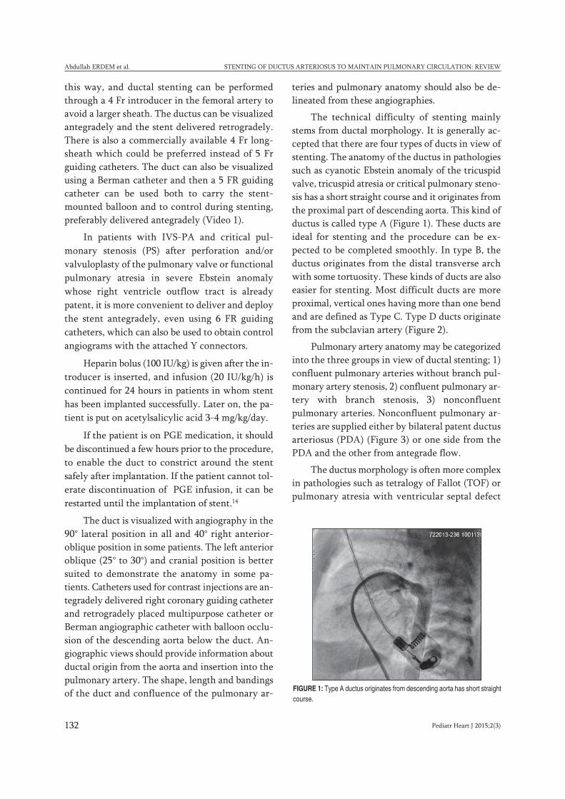
Pediatr Heart J 2015;2(3)132
Abdullah ERDEM et al. STENTING OF DUCTUS ARTERIOSUS TO MAINTAIN PULMONARY CIRCULATION: REVIEW
this way, and ductal stenting can be performedthrough a 4 Fr introducer in the femoral artery toavoid a larger sheath. The ductus can be visualizedantegradely and the stent delivered retrogradely.There is also a commercially available 4 Fr long-sheath which could be preferred instead of 5 Frguiding catheters. The duct can also be visualizedusing a Berman catheter and then a 5 FR guidingcatheter can be used both to carry the stent-mounted balloon and to control during stenting,preferably delivered antegradely (Video 1).
In patients with IVS-PA and critical pul-monary stenosis (PS) after perforation and/orvalvuloplasty of the pulmonary valve or functionalpulmonary atresia in severe Ebstein anomalywhose right ventricle outflow tract is alreadypatent, it is more convenient to deliver and deploythe stent antegradely, even using 6 FR guidingcatheters, which can also be used to obtain controlangiograms with the attached Y connectors.
Heparin bolus (100 IU/kg) is given after the in-troducer is inserted, and infusion (20 IU/kg/h) iscontinued for 24 hours in patients in whom stenthas been implanted successfully. Later on, the pa-tient is put on acetylsalicylic acid 3-4 mg/kg/day.
If the patient is on PGE medication, it shouldbe discontinued a few hours prior to the procedure,to enable the duct to constrict around the stentsafely after implantation. If the patient cannot tol-erate discontinuation of PGE infusion, it can berestarted until the implantation of stent.14
The duct is visualized with angiography in the90° lateral position in all and 40° right anterior-oblique position in some patients. The left anterioroblique (25° to 30°) and cranial position is bettersuited to demonstrate the anatomy in some pa-tients. Catheters used for contrast injections are an-tegradely delivered right coronary guiding catheterand retrogradely placed multipurpose catheter orBerman angiographic catheter with balloon occlu-sion of the descending aorta below the duct. An-giographic views should provide information aboutductal origin from the aorta and insertion into thepulmonary artery. The shape, length and bandingsof the duct and confluence of the pulmonary ar-
teries and pulmonary anatomy should also be de-lineated from these angiographies.
The technical difficulty of stenting mainlystems from ductal morphology. It is generally ac-cepted that there are four types of ducts in view ofstenting. The anatomy of the ductus in pathologiessuch as cyanotic Ebstein anomaly of the tricuspidvalve, tricuspid atresia or critical pulmonary steno-sis has a short straight course and it originates fromthe proximal part of descending aorta. This kind ofductus is called type A (Figure 1). These ducts areideal for stenting and the procedure can be ex-pected to be completed smoothly. In type B, theductus originates from the distal transverse archwith some tortuosity. These kinds of ducts are alsoeasier for stenting. Most difficult ducts are moreproximal, vertical ones having more than one bendand are defined as Type C. Type D ducts originatefrom the subclavian artery (Figure 2).
Pulmonary artery anatomy may be categorizedinto the three groups in view of ductal stenting; 1)confluent pulmonary arteries without branch pul-monary artery stenosis, 2) confluent pulmonary ar-tery with branch stenosis, 3) nonconfluentpulmonary arteries. Nonconfluent pulmonary ar-teries are supplied either by bilateral patent ductusarteriosus (PDA) (Figure 3) or one side from thePDA and the other from antegrade flow.
The ductus morphology is often more complexin pathologies such as tetralogy of Fallot (TOF) orpulmonary atresia with ventricular septal defect
FIGURE 1: Type A ductus originates from descending aorta has short straightcourse.
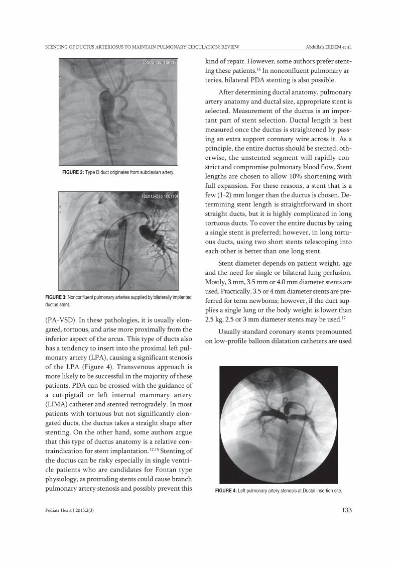
Pediatr Heart J 2015;2(3) 133
STENTING OF DUCTUS ARTERIOSUS TO MAINTAIN PULMONARY CIRCULATION: REVIEW Abdullah ERDEM et al.
(PA-VSD). In these pathologies, it is usually elon-gated, tortuous, and arise more proximally from theinferior aspect of the arcus. This type of ducts alsohas a tendency to insert into the proximal left pul-monary artery (LPA), causing a significant stenosisof the LPA (Figure 4). Transvenous approach ismore likely to be successful in the majority of thesepatients. PDA can be crossed with the guidance ofa cut-pigtail or left internal mammary artery(LIMA) catheter and stented retrogradely. In mostpatients with tortuous but not significantly elon-gated ducts, the ductus takes a straight shape afterstenting. On the other hand, some authors arguethat this type of ductus anatomy is a relative con-traindication for stent implantation.12,15 Stenting ofthe ductus can be risky especially in single ventri-cle patients who are candidates for Fontan typephysiology, as protruding stents could cause branchpulmonary artery stenosis and possibly prevent this
kind of repair. However, some authors prefer stent-ing these patients.16 In nonconfluent pulmonary ar-teries, bilateral PDA stenting is also possible.
After determining ductal anatomy, pulmonaryartery anatomy and ductal size, appropriate stent isselected. Measurement of the ductus is an impor-tant part of stent selection. Ductal length is bestmeasured once the ductus is straightened by pass-ing an extra support coronary wire across it. As aprinciple, the entire ductus should be stented; oth-erwise, the unstented segment will rapidly con-strict and compromise pulmonary blood flow. Stentlengths are chosen to allow 10% shortening withfull expansion. For these reasons, a stent that is afew (1-2) mm longer than the ductus is chosen. De-termining stent length is straightforward in shortstraight ducts, but it is highly complicated in longtortuous ducts. To cover the entire ductus by usinga single stent is preferred; however, in long tortu-ous ducts, using two short stents telescoping intoeach other is better than one long stent.
Stent diameter depends on patient weight, ageand the need for single or bilateral lung perfusion.Mostly, 3 mm, 3.5 mm or 4.0 mm diameter stents areused. Practically, 3.5 or 4 mm diameter stents are pre-ferred for term newborns; however, if the duct sup-plies a single lung or the body weight is lower than2.5 kg, 2.5 or 3 mm diameter stents may be used.17
Usually standard coronary stents premountedon low-profile balloon dilatation catheters are used
FIGURE 2: Type D duct originates from subclavian artery.
FIGURE 3: Nonconfluent pulmonary arteries supplied by bilaterally implantedductus stent.
FIGURE 4: Left pulmonary artery stenosis at Ductal insertion site.
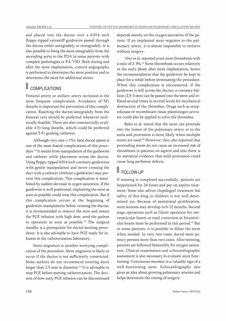
Pediatr Heart J 2015;2(3)134
Abdullah ERDEM et al. STENTING OF DUCTUS ARTERIOSUS TO MAINTAIN PULMONARY CIRCULATION: REVIEW
and placed into the ductus over a 0.014 inchfloppy-tipped extrastiff guidewire passed throughthe ductus either antegradely or retrogradely. It isalso possible to bring the stent antegradely from theascending aorta to the PDA in some patients withcomplex pathologies or PA-VSD. Both during andafter the stent implantation, control angiographyis performed to determine the stent position and todetermine the need for additional stents.
COMPLICATIONS
Femoral artery or axillary artery occlusion is themost frequent complication. Avoidance of 5Frsheaths is important for prevention of this compli-cation. Reaching the ductus antegradely from thefemoral vein should be preferred whenever tech-nically feasible. There are also commercially avail-able 4 Fr long sheaths, which could be preferredagainst 5 Fr guiding catheters.
Although very rare (<1%), fatal ductal spasm isone of the most feared complications of this proce-dure.14 It results from manipulation of the guidewireand catheter while placement across the ductus.Using floppy-tipped 0.014 inch coronary guidewireswith gentle manipulation and never crossing theduct with a catheter (without a guidewire) may pre-vent this complication. This complication is mani-fested by sudden decrease in oygen saturation. If theguidewire is well positioned, implanting the stent assoon as possible could treat this complication. But ifthis complication occurs at the beginning ofguidewire manipulation before crossing the ductus,it is recommended to remove the wire and restartthe PGE infusion with high dose, send the patientto operation as soon as possible.14 The surgicalstandby is a prerequisite for ductal stenting proce-dures. It is also advisable to have PGE ready for in-fusion in the catheterization laboratory.
Stent migration is another worrying compli-cation of the procedure. Stent migration is likely tooccur if the ductus is not sufficiently constricted.Some authors do not recommend stenting ductslarger than 2.5 mm in diameter.14 It is advisable tostop PGE before starting catheterization. The deci-sion of how early PGE infusion can be discontinued
depends mainly on the oxygen saturation of the pa-tient. If an implanted stent migrates to the pul-monary artery, it is almost impossible to retrievewithout surgery.
Alwi et al. reported acute stent thrombosis witha ratio of 2-3%.14 Stent thrombosis occurs relativelyin the early phase after stent implantation, hencethe recommendation that the guidewire be kept inplace for a while before terminating the procedure.When this complication is encountered, if theguidewire is still across the ductus, a coronary bal-loon (2,5-3 mm) can be passed into the stent and in-flated several times in several levels for mechanicaldestruction of the thrombus. Drugs such as strep-tokinase or recombinant tissue plasminogen activa-tor could also be applied to solve the thrombus.
Baho et al. stated that the stent can protrudeinto the lumen of the pulmonary artery or to theaorta and protrusion is more likely when multiplestents are used.18 However, they also reported thatprotruding stents do not cause an increased risk ofthrombosis in patients on aspirin and also there isno statistical evidence that mild protrusion couldcause lung perfusion defects.
FOLLOW-UP
If stenting is completed successfully, patients areheparinized for 24 hours and put on aspirin treat-ment. Some also advice clopidogrel treatment butsafety of this drug in children is not well deter-mined yet. Because of neointimal proliferation,stent stenosis may develop in 6-12 months. Secondstage operations such as Glenn operation for uni-ventricular hearts or total correction in biventric-ular hearts must be performed in this period.19 Butin some patients, it is possible to dilate the stentwhen needed. In very rare cases, ductal stent pa-tency persists more than two years. After stenting,patients are followed bimonthly for oxygen satura-tion. Clinical examination and echocardiographicassessment is also necessary to evaluate stent func-tioning. Continuous murmur is a valuable sign of awell-functioning stent. Echocardiography alsogives an idea about growing pulmonary arteries andhelps determine the timing of surgery.
Pediatr Heart J 2015;2(3) 135
STENTING OF DUCTUS ARTERIOSUS TO MAINTAIN PULMONARY CIRCULATION: REVIEW Abdullah ERDEM et al.
SYSTEMIC-TO-PULMONARY SHUNT VERSUS PDA STENTING
The morbidity and mortality after BTS is higher inthe neonatal period in comparison to later child-hood.4,6,7 PDA stenting is as effective as modified BTSin promoting global pulmonary artery growth incongenital heart disease with duct-dependent pul-monary circulation.14,20,21 Stenting could also reducethe hospital stay.11 Compared to PDA stenting, inmodified BTS, pleural effusion, chylothorax, phrenicand vagal nerve palsy, and distortion and differentialgrowth of the pulmonary arteries are well-knowncomplications.6,7,22 On the other hand, the stentedarterial duct tends to be less durable than a conven-tional surgical shunt owing to ductal tissue prolapsethrough the stent struts and intrastent endothelialhyperplasia, as already reported.9,23 Clinically signif-icant stenosis usually develops within six months toone year after stenting.9,23,24
When these facts are taken into consideration,is ductal stenting really a good alternative to surgi-cal shunts? At present, it is hard to answer this
question fairly. It is obvious that, when comparedwith primary surgery, this approach is less invasiveand significantly shortens hospitalization and re-duces treatment costs. However, there is no ran-domized controlled trial to compare the results ofthe two procedures and it seems difficult to carryout such a trial. There are trials that compare theeffectiveness of both methods, but they are not ran-domized controlled studies by design.25,26
CONCLUSION
Ductal stenting may be the ideal therapeutic optionin pathologies such as IVS-PA, cyanotic Ebstein inthe early postnatal period or critical PS with a shortstraight duct requiring short-term palliation. Un-fortunately, it is technically demanding in ductsthat are more proximal, vertical or tortuous and alsoit is not feasible in pathologies needing long-termpalliation. In future, advanced technologies includ-ing drug-eluting or more flexible stents could en-able us to apply this procedure in all kinds of ductalanatomy with long-term patency.
1. Olley PM, Coceani F, Bodach E. E type prostaglandins.A newemergency therapy for certain cyanotic congenital heart mal-formations. Circulation 1976;53(4):728-31.
2. Freed MD, Heymann MA, Lewis AB, Roehl SL,Kensey RC.Prostaglandin E, in infants with ductusarteriosus-dependentcongenital heart disease. Circulation 1981;64(5):899-905.
3. Lewis AB, Freed MD, Heymann MA, Roehl SL, Kensey RC.Side effects of therapy with prostaglandin E1 in infants withcritical congenital heart disease. Circulation 1981; 64(5): 893-8.
4. Blalock A, Taussig HB. The surgical treatment of malforma-tions of the heart in which there is pulmonary stenosis or pul-monary atresia. JAMA 1945;128(3):189-92.
5. Petrucci O, O'Brien SM, Jacobs ML, Jacobs JP, ManningPB,Eghtesady P. Risk factors for mortality and morbidity afterthe neonatal Blalock-Taussig shunt procedure. Ann ThoracSurg 2011;92(2):642-51.
6. Fermanis GG, Ekangaki AK, Salmon AP, Keeton BR, ShoreDF, Lamb RK, et al. Twelve year experience with the modifiedBlalock-Taussig shunt in neonates. Eur J Cardiothorac Surg1992;6(11):586-9.
7. Tamisier D, Vouhé PR, Vernant F, Lecá F, Massot C, NeveuxJY. Modified Blalock-Taussig shunts: results in infants lessthan 3 months of age. Ann Thorac Surg 1990; 49(5):797-801.
8. Gibbs JL, Rothman MT, Rees MR, Parsons JM, BlackburnME, Ruiz CE. Stenting of the arterial duct: a new approach topalliation for pulmonary atresia. Br Heart J 1992;67(3):240-5.
9. Gibbs JL, Uzun O, Blackburn ME, Wren C, Hamilton JR, Wat-terson KG. Fate of the stented arterial duct. Circulation1999;99(20): 2621-5.
10. Schneider M1, Zartner P, Sidiropoulos A, Konertz W, Haus-
dorf G. Stent implantation of the arterial duct in newborns withduct-dependent circulation. Eur Heart J 1998;19(9):1401-9.
11. Gewillig M, Boshoff DE, Dens J, Mertens L, Benson LN.Stenting the neonatal arterial duct in duct dependent pul-monary circulation: new techniques, better results. J Am CollCardiol 2004;43(1):107-12.
12. Alwi M, Choo KK, Latiff HA, Kandavello G, Samion H, MulyadiMD. Initial results and medium term follow up of stent im-plantation of patent ductus arteriosus in duct-dependent pul-monary circulation. J Am Coll Cardiol 2004; 44(2):438-45.
13. Celebi A, Yalçin Y, Erdem A, Zeybek C, Akdeniz C, Polat TB.Stent implantation into the patent ductus arteriosus in cyan-otic congenital heart disease with duct-dependent ordimin-ished pulmonary circulation. Turk J Pediatr 2007;49(4):413-7.
14. Alwi M. Stenting the ductus arteriosus: Case selection, tech-nique and possible complications. Ann Pediatr Cardiol2008;1(1):38-45.
15. Abrams SE, Walsh KP. Arterial duct morphology with refer-ence to angioplasty and stenting. Int J Cardiol 1993;40(1):27-33.
16. Garg G, Srivastava A, Radha AS. Simultaneous stenting oftightly stenosed patent ductus arteriosus and pulmonary arterybifurcation using two stents (Y stenting): an innovative technique.Catheter Cardiovasc Inter 2014;83(7):1131-4.
17. Boshoff DE, Michel-Behnke I, Schranz D, Gewillig M. Stent-ing the neonatal arterial duct. Expert Rev Cardiovasc Ther2007;5(5):893-901.
18. Baho H, Deraz S, Abouzeid H, Al-Ata J, Kouatli A. Stent pro-trusion in palliative congenital heart disease interventions:does it cause any harm? J Invasive Cardiol. 2013;25(9):460-3.
19. Erdem A, Karacı AR, Sarıtaş T, Akdeniz C, Demir H, Şaş-mazel A at all. Evaluation of the efficacy of ductus arteriosusstenting in neonates and infants with severe cyanosis untilthe later stage palliative surgery or total repair time. TurkishJ Thorac Cardiovasc Surg 2011;19(2):192-6
20. Hussain A, Al-Zharani S, Muhammed AA, Al-Ata J, Galal OM.Midterm Outcome of Stent Dilatation of Patent Ductus Arte-riosus in Ductal-dependent Pulmonary Circulation. Congeni-tal Heart Disease 2008:3;(4):241-9
21. Santoro G, Gaio G, Palladino M T, Iacono C, Carrozza M, Es-posito R at al. Stenting of the arterial duct in newborns withduct-dependent pulmonary circulation. Heart 2008;94(7);925-9
22. Sivakumar K, Shivaprakasha K, Rao SG, Kumar RK. Opera-tive outcome and intermediate term follow-up of neonatalBlalock-Taussig shunts. Indian Heart J 2001;53(1):66-70
23. Rosenthal E, Qureshi SA, Tabatabaie AH, Persaud D,Kakadekar AP, Baker EJ, et al. Medium-term results of ex-perimental stent implantation into the ductus arteriosus. AmHeart J 1996;132(3):657-63.
24. Sivakumar K, Bhagyavathy A, Coelho R, Satish R, KrishnanP. Longevity of Neonatal Ductal Stenting for Congenital HeartDiseases with Duct-dependent Pulmonary Circulation Con-genit Heart Dis. 2012;7(6):526-33.
25. Amoozgar H, Cheriki S, Borzoee M, Ajami G, Soltani M, Ah-madipour M at al. Short-Term Result of Ductus ArteriosusStent Implantation Compared With Surgically Created Shunts.Pediatr Cardiol 2012;33(8):1288-94.
26. McMullan DM, Permut LC, Jones TK, Johnston TA, Rubio AE.Modified Blalock-Taussig shunt versus ductal stenting for pal-liation of cardiac lesions with inadequate pulmonary bloodflow J Thorac Cardiovasc Surg 2014; 147(1):397-401.
REFERENCES

Video 1. Demostration of ductus byantegradely placed Berman catheter.
Pediatr Heart J 2015;2(3)136
arsiyel pulmoner venöz dönüş anomalisi (PAPVD), bir yada dahafazla pulmoner venin sağ atriyuma direk yada indirek olarak superiorvena kava (SVC) yada inferior vena kava (İVC) ile anormal bağlan-
masıdır. Otopsi serilerinde konjenital kalp hastalıklarının %0,4-0,7’sindesaptanmıştır. Sağ pulmoner venlerin tutulumu daha fazladır ve sıklıkla sinusvenosus tip atriyal septal defekt (ASD) ile birliktedir. İzole PAPVD nadir-dir ve genellikle sağ üst pulmoner venin SVC’ye drenajı saptanmaktadır.Sol akciğerin innominate vene drenajı oldukça nadir olup vertikal ven ile in-nominate vene drene olmaktadır.1
İzole Parsiyel Pulmoner Venöz DönüşAnomalisi Olan Hastaların Değerlendirilmesi
ÖÖZET İzole parsiyel pulmoner venöz dönüş anomalisi (PAPVD) çok ender bir konjenital anomaliolup kolayca saptanamayabilir. Bu nedenle, transtorasik ekokardiyografi ile dikkatli inceleme ya-pılmalı ve gerek görülürse transözefageal ekokardiyografi (TEE), bilgisayarlı tomografi (BT), man-yetik rezonans (MR) kullanılmalıdır. Hemodinamik değerlendirme ve tanıyı doğrulamada kateteranjiyografi önemlidir. Kliniğimizden on hasta başvuru tabloları, tanıları ve tedavi yaklaşımları de-ğerlendirildi. Ekokardiyografi, anjiyografi ve diğer görüntüleme yöntemleri ile altı hastada sağ ak-ciğerin ve dört hastada sol akciğerin pulmoner venlerinin anormal drenajını saptadık. Değerlen-dirilen yedi hasta ayaktan poliklinik izlemine alındı. Cerrahi tedavi önerilen diğer üç hastanın ikisiopere oldu, diğer hasta cerrahi tedaviyi kabul etmediği için ayaktan poliklinik izlemine alındı.
Anahtar Kelimeler: Akciğer venleri; ekokardiyografi; kalp kusurları, doğumsal; brakiosefalik venler; ventriküler disfonksiyon, sağ
ABSTRACT Isolated partial anomalous pulmonary venous drainage (PAPVD) is a very rare con-genital anomaly so that it can not be recognized easily. Therefore, careful examination should bedone with transthorasic echocardiography and if it is needed transoesophageal echocardiography(TEE), computerized tomography (CT), magnetic resonance (MR) should be used. Catheter an-giography is important for hemodynamic evaluation and confirmation the diagnosis. We evaluatedten patients from our clinic with their presentation, diagnosis and treatment approaches. Evaluat-ing with echocardiographic, angiographic and other visualization techniques, we demonstrated theanomalous drainage of the pulmonary veins of right lung in six patients and of left lung in four pa-tients. Evaluated seven patients were taken to outpatient follow up. Two of the three patients whowere recommended surgery, required surgical therapy, the other patient did not accept surgicaltherapy and was taken to outpatient follow up.
Key Words: Pulmonary veins; echocardiography; heart defects, congenital; brachiocephalic veins; ventricular dysfunction, right
Pediatr Heart J 2015;2(3):136-43
Ajda Mutlu MIHÇIOĞLU,a
Feyza Ayşenur PAÇ,a
Ahmet Vedat KAVURT,a
Serhat KOCAa
aÇocuk Kardiyoloji Kliniği, Türkiye Yüksek İhtisas Hastanesi,Ankara
Ge liş Ta ri hi/Re ce i ved: 13.10.2014 Ka bul Ta ri hi/Ac cep ted: 25.04.2015
Bu olgu sunumu, 11. Ulusal Pediatrik Kardiyoloji ve Kalp Damar Cerrahisi Kongresi(18-21 Nisan 2012, İzmir)’nde poster sunumu olarak yer almıştır.
Ya zış ma Ad re si/Cor res pon den ce:Feyza Ayşenur PAÇ Türkiye Yüksek İhtisas Hastanesi, Çocuk Kardiyoloji Kliniği, Ankara, TÜRKİYE/[email protected]
Copyright © 2015 by Türk Pediatrik Kardiyoloji veKalp Cerrahisi Derneği
OLGU SUNUMU
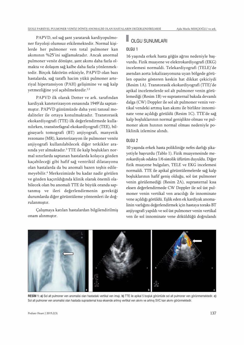
İZOLE PARSİYEL PULMONER VENÖZ DÖNÜŞ ANOMALİSİ OLAN HASTALARIN DEĞERLENDİRİLMESİ Ajda Mutlu MIHÇIOĞLU ve ark.
PAPVD, sol sağ şant yaratarak kardiyopulmo-ner fizyoloji olumsuz etkilemektedir. Normal kişi-lerde her pulmoner ven total pulmoner kanakımının %25’ini sağlamaktadır. Ancak anormalpulmoner venöz dönüşte, şant akımı daha fazla ol-makta ve dolaşım sağ kalbe daha fazla yönlenmek-tedir. Birçok faktörün etkisiyle, PAPVD olan bazıhastalarda, sağ taraflı hacim yükü pulmoner arte-riyal hipertansiyon (PAH) gelişimine ve sağ kalpyetmezliğine yol açabilmektedir.2,3
PAPVD ilk olarak Dotter ve ark. tarafındankardiyak kateterizasyon esnasında 1949’da saptan-mıştır. PAPVD günümüzde daha yeni tanısal mo-daliteler ile ortaya konulmaktadır. Transtorasikekokardiyografi (TTE) ilk değerlendirmede kulla-nılırken, transözefageal ekokardiyografi (TEE), bil-gisayarlı tomografi (BT) anjiyografi, manyetikrezonans (MR), kateterizasyon ile pulmoner venözanjiyografi kullanılabilecek diğer tetkikler ara-sında yer almaktadır.3 TTE ile kalp boşlukları nor-mal sınırlarda saptanan hastalarda kolayca gözdenkaçabileceği gibi hafif sağ ventrikül dilatasyonuolan hastalarda da bu anomali bazen teşhis edile-meyebilir.4 Merkezimizde bu kadar nadir görülenve gözden kaçırıldığında klinik olarak önemli ola-bilecek olan bu anomali TTE ile büyük oranda sap-tanmış ve ileri değerlendirmenin gerektiğidurumlarda diğer görüntüleme yöntemleri ile doğ-rulanmıştır.
Çalışmaya katılan hastalardan bilgilendirilmişonam alınmıştır.
OLGU SUNUMLARI
OLGU 1
16 yaşında erkek hasta göğüs ağrısı nedeniyle baş-vurdu. Fizik muayene ve elektrokardiyografi (EKG)incelemesi normaldi. Telekardiyografi (TELE)’deasendan aorta lokalizasyonuna uyan bölgede görü-len opasite gösteren keskin hat dikkat çekiciydi(Resim 1A). Transtorasik ekokardiyografi (TTE)’deapikal incelemelerde sol alt pulmoner venin görü-lemediği (Resim 1B) ve suprasternal bakıda devamlıdalga (CW) Doppler ile sol alt pulmoner venin ver-tikal vendeki artmış kan akımı ile birlikte innomi-nate vene açıldığı görüldü (Resim 1C). TTE’de sağkalp boşluklarının normal genişlikte olması ve pul-moner akım hızının normal olması nedeniyle po-liklinik izlemine alındı.
OLGU 2
10 yaşında erkek hasta polikliniğe nefes darlığı şika-yetiyle başvurdu (Tablo 1). Fizik muayenesinde me-zokardiyak odakta 1/6 sistolik üfürüm duyuldu. Diğerfizik muayene bulguları, TELE ve EKG incelemesinormaldi. TTE ile apikal görüntülemelerde sağ kalpboşluklarının hafif geniş olduğu, sol üst pulmonervenin görülemediği (Resim 2A), suprasternal kısaeksen değerlendirmede CW Doppler ile sol üst pul-moner venin vertikal ven aracılığı ile innominatevene açıldığı görüldü. Eşlik eden ek kardiyak anoma-linin varlığını değerlendirmek için hastaya toraks BTanjiyografi yapıldı ve sol üst pulmoner venin vertikalven ile sol innominate vene döküldüğü doğrulandı
RESİM 1: a) Sol alt pulmoner ven anomalisi olan hastadaki vertikal ven imajı. b) TTE ile apikal 5 boşluk görüntüde sol alt pulmoner ven görünmemektedir. c)Sol alt pulmoner ven anomalisi olan hastada suprasternal kısa eksende artmış vertikal ven akımı ve artmış SVC kan akımı görünmektedir.
a b c
Pediatr Heart J 2015;2(3) 137

Ajda Mutlu MIHÇIOĞLU ve ark. İZOLE PARSİYEL PULMONER VENÖZ DÖNÜŞ ANOMALİSİ OLAN HASTALARIN DEĞERLENDİRİLMESİ
Pediatr Heart J 2015;2(3)138
Anjiy
ogra
fi:Q
p/Q
s Ha
sta
No:
Yaş
(yıl)
Cins
Başv
uru
Şika
yeti
FMEK
GTE
LETT
ETE
EBT
anj
iyog
rafi
PAP(
mm
Hg)
Kara
r1
16E
Göğ
üs a
ğrıs
ıN
NO
pasi
te g
öste
ren
Sol a
lt pu
lmon
er v
en-
--
İzle
m
kesk
in h
at→
inno
min
ate
vene
Sağ
kalp
boş
lukl
arı:
N
210
EN
efes
dar
lığı
1/6
üfür
ümN
NSo
l üst
pul
mon
er v
en
--
1.23
İzle
m
→in
nom
inat
e ve
ne
12/5
/7
Sağ
kalp
boş
lukl
arı:
Haf
if d
ilate
339
KN
efes
dar
lığı,
2/6
üfür
ümN
NSo
l üst
pul
mon
er v
en
--
2 C
erra
hi
Çab
uk y
orul
ma,
→
SVC
’ye
32/2
1/25
Hal
sizl
ikSa
ğ ka
lp b
oşlu
klar
ı: D
ilate
411
K-
1/6
üfür
ümN
NSo
l üst
pul
mon
er v
enSo
l üst
pul
mon
er v
en
1.2
İzle
m
→in
nom
inat
e ve
ne→
inno
min
ate
vene
18
/10/
13
Sağ
kalp
boş
lukl
arı:
Dila
te
55
EG
öğüs
ağr
ısı,
1/6
üfür
ümN
NSa
ğ üs
t pul
mon
er v
en
-Sa
ğ üs
t pul
mon
er v
en
1.2
İzle
m
Nef
es d
arlığ
ı →
İVC
-RA
bile
şkes
ine
→İV
C-R
A bi
leşk
esin
e ve
22/9
/14
Sağ
kalp
boş
lukl
arı:
azig
oz v
ene;
sağ
alt
Nor
mal
in ü
st s
ınırı
nda
pulm
oner
ven
→İV
C’y
e
615
EG
öğüs
ağr
ısı
NN
NSa
ğ üs
t pul
mon
er v
en
--
-İz
lem
→in
nom
inat
e ve
ne
712
K-
NN
NSa
ğ üs
t pul
mon
er v
en-
--
İzle
m
→in
nom
inat
e ve
ne
89
EYo
rulm
a,
NN
NSa
ğ üs
t pul
mon
er v
en
--
-İz
lem
Göğ
üs a
ğrıs
ı→
inno
min
ate
vene
948
KG
öğüs
ağr
ısı,
1/6
üfür
ümPu
lmon
er k
onus
İn
kom
plet
sağ
Sa
ğ pu
lmon
er v
enle
r Sa
ğ p
ulm
oner
ven
ler
Sağ
pulm
oner
ven
ler
1.5
Cer
rahi
Nef
es d
arlığ
ıha
fif b
elirg
inda
l blo
ğu→
İVC
’ye
LA’y
a aç
ılmıy
or→
İVC
’ye
30/1
9/24
Sağ
kalp
boş
lukl
arı:
Haf
if di
late
1027
E-
1/6
üfür
ümPu
lmon
er k
onus
İn
kom
plet
sağ
Sa
ğ ka
lp b
oşlu
klar
ı: R
A iç
inde
SVC
Sa
ğ AC
üst
lob
venl
er
1.45
İzle
m
hafif
bel
irgin
dal b
loğu
Haf
if di
late
kom
şulu
ğund
a ya
pı→
İVC
’ye
28/1
5/20
(Cer
rahi
yi
Sağ
AC o
rta lo
b v
enle
r ka
bul e
tmed
i)
→So
l alt
pulm
oner
ven
e
Sağ
alt l
ob a
gene
zik
TABL
O 1
: H
asta
ların
klin
ik ta
blol
arın
ın d
eğer
lend
irilm
esi.
AC: A
kciğ
er; B
T: B
ilgis
ayar
lı to
mog
rafi;
E: E
rkek
; EKG
: Ele
ktro
kard
iyog
rafi;
FM
: Fiz
ik m
uaye
ne; İ
VC: İ
nfer
ior v
ena
kava
; K: K
ız/k
adın
; LA:
Sol
atri
yum
; N: N
orm
al; N
o: N
umar
a; P
A: P
ulm
oner
arte
r; Q
p/Q
s: P
ulm
oner
/sis
tem
ik a
kım
lar o
ranı
; PAP
: Pul
mon
er a
rter
bası
ncı;
RA:
Sağ
atri
yum
; SVC
: Sup
erio
r ven
a ka
va; T
EE: T
rans
özof
agea
l eko
kard
iyog
rafi;
TEL
E: T
elek
ardi
yogr
afi;
TTE:
Tra
nsto
rasi
k ek
okar
diyo
graf
i.
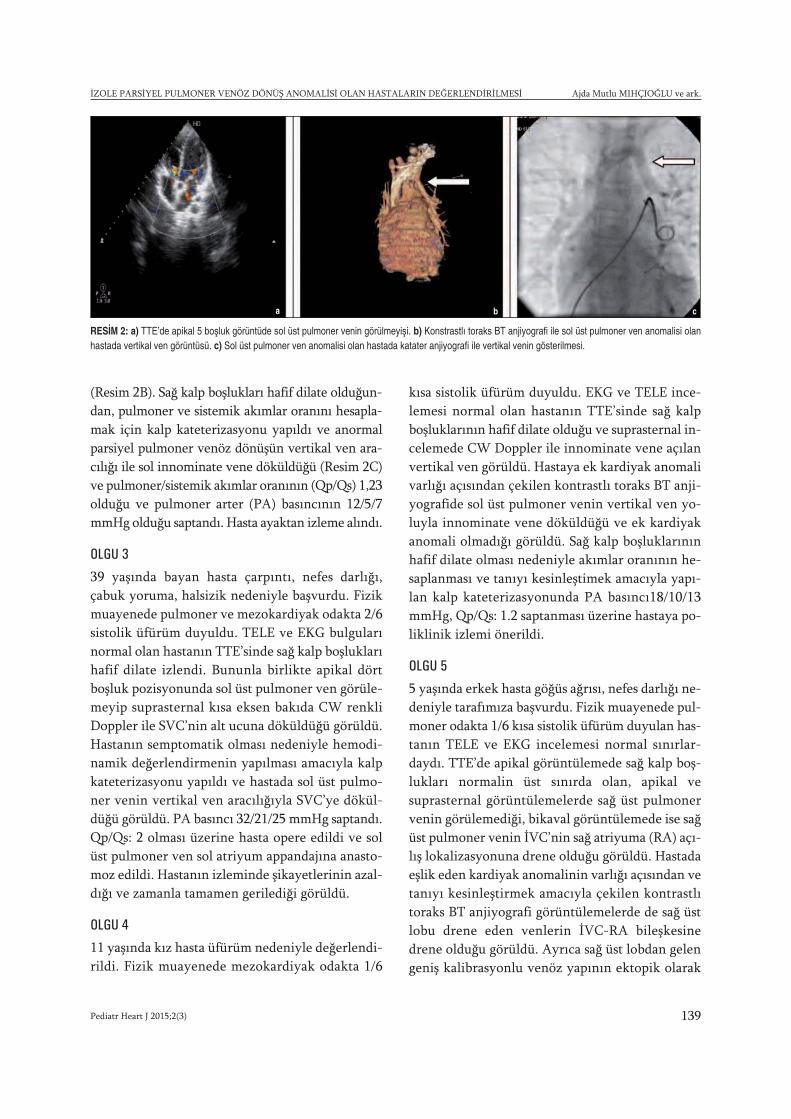
İZOLE PARSİYEL PULMONER VENÖZ DÖNÜŞ ANOMALİSİ OLAN HASTALARIN DEĞERLENDİRİLMESİ Ajda Mutlu MIHÇIOĞLU ve ark.
(Resim 2B). Sağ kalp boşlukları hafif dilate olduğun-dan, pulmoner ve sistemik akımlar oranını hesapla-mak için kalp kateterizasyonu yapıldı ve anormalparsiyel pulmoner venöz dönüşün vertikal ven ara-cılığı ile sol innominate vene döküldüğü (Resim 2C)ve pulmoner/sistemik akımlar oranının (Qp/Qs) 1,23olduğu ve pulmoner arter (PA) basıncının 12/5/7mmHg olduğu saptandı. Hasta ayaktan izleme alındı.
OLGU 3
39 yaşında bayan hasta çarpıntı, nefes darlığı,çabuk yoruma, halsizik nedeniyle başvurdu. Fizikmuayenede pulmoner ve mezokardiyak odakta 2/6sistolik üfürüm duyuldu. TELE ve EKG bulgularınormal olan hastanın TTE’sinde sağ kalp boşluklarıhafif dilate izlendi. Bununla birlikte apikal dörtboşluk pozisyonunda sol üst pulmoner ven görüle-meyip suprasternal kısa eksen bakıda CW renkliDoppler ile SVC’nin alt ucuna döküldüğü görüldü.Hastanın semptomatik olması nedeniyle hemodi-namik değerlendirmenin yapılması amacıyla kalpkateterizasyonu yapıldı ve hastada sol üst pulmo-ner venin vertikal ven aracılığıyla SVC’ye dökül-düğü görüldü. PA basıncı 32/21/25 mmHg saptandı.Qp/Qs: 2 olması üzerine hasta opere edildi ve solüst pulmoner ven sol atriyum appandajına anasto-moz edildi. Hastanın izleminde şikayetlerinin azal-dığı ve zamanla tamamen gerilediği görüldü.
OLGU 4
11 yaşında kız hasta üfürüm nedeniyle değerlendi-rildi. Fizik muayenede mezokardiyak odakta 1/6
kısa sistolik üfürüm duyuldu. EKG ve TELE ince-lemesi normal olan hastanın TTE’sinde sağ kalpboşluklarının hafif dilate olduğu ve suprasternal in-celemede CW Doppler ile innominate vene açılanvertikal ven görüldü. Hastaya ek kardiyak anomalivarlığı açısından çekilen kontrastlı toraks BT anji-yografide sol üst pulmoner venin vertikal ven yo-luyla innominate vene döküldüğü ve ek kardiyakanomali olmadığı görüldü. Sağ kalp boşluklarınınhafif dilate olması nedeniyle akımlar oranının he-saplanması ve tanıyı kesinleştimek amacıyla yapı-lan kalp kateterizasyonunda PA basıncı18/10/13mmHg, Qp/Qs: 1.2 saptanması üzerine hastaya po-liklinik izlemi önerildi.
OLGU 5
5 yaşında erkek hasta göğüs ağrısı, nefes darlığı ne-deniyle tarafımıza başvurdu. Fizik muayenede pul-moner odakta 1/6 kısa sistolik üfürüm duyulan has-tanın TELE ve EKG incelemesi normal sınırlar-daydı. TTE’de apikal görüntülemede sağ kalp boş-lukları normalin üst sınırda olan, apikal vesuprasternal görüntülemelerde sağ üst pulmonervenin görülemediği, bikaval görüntülemede ise sağüst pulmoner venin İVC’nin sağ atriyuma (RA) açı-lış lokalizasyonuna drene olduğu görüldü. Hastadaeşlik eden kardiyak anomalinin varlığı açısından vetanıyı kesinleştirmek amacıyla çekilen kontrastlıtoraks BT anjiyografi görüntülemelerde de sağ üstlobu drene eden venlerin İVC-RA bileşkesinedrene olduğu görüldü. Ayrıca sağ üst lobdan gelengeniş kalibrasyonlu venöz yapının ektopik olarak
RESİM 2: a) TTE’de apikal 5 boşluk görüntüde sol üst pulmoner venin görülmeyişi. b) Konstrastlı toraks BT anjiyografi ile sol üst pulmoner ven anomalisi olanhastada vertikal ven görüntüsü. c) Sol üst pulmoner ven anomalisi olan hastada katater anjiyografi ile vertikal venin gösterilmesi.
a b c
Pediatr Heart J 2015;2(3) 139

Ajda Mutlu MIHÇIOĞLU ve ark. İZOLE PARSİYEL PULMONER VENÖZ DÖNÜŞ ANOMALİSİ OLAN HASTALARIN DEĞERLENDİRİLMESİ
Pediatr Heart J 2015;2(3)140
azygoz vene drene olduğu görüldü. Sağ alt lob pos-terobazalden gelen 2,3 mm çapında tek pulmonervenin doğrudan İVC’ye drene olduğu görüldü. He-modinamik değerlendirme yapılması amacıyla uy-gulanan kalp kateterizasyonunda PA basıncı22/9/14 mmHg, Qp/Qs: 1,2 olarak hesaplandı. Has-tada ayaktan izlem kararı alındı.
OLGU 6
15 yaşında erkek hasta aralıklı iğne batar gibi olangöğüs ağrısı nedeniyle tarafımıza başvurdu. Fizikmuayenede üfürüm duyulmayan hastanın TELE veEKG incelemesi normal sınırlardaydı. TTE’de sağkalp boşlukları normal genişlikte olan hastanın api-kal ve suprasternal görüntülemelerde sağ üst pul-moner ven net olarak görülemedi suprasternalgörüntülemede sağ üst pumoner venin vertikal venaracılığıyla innominate vene drene olduğu görüldü.Hastada ayaktan izlem kararı alındı.
OLGU 7
12 yaşında kız hastanın hiçbir şikayeti olmayıprutin kontrol amaçlı polikliniğe başvurdu. Fizikmuayene, TELE ve EKG incelemesi normal sınır-lardaydı. TTE’de sağ kalp boşlukları normal geniş-likte olan hastanın suprasternal görüntülemelerdesağ üst pulmoner venin sol atriyuma açılışı net ola-rak görülemedi. Suprasternal görüntülemede sağüst pumoner venin vertikal ven aracılığıyla inno-minate vene drene olduğu görüldü. Hastada ayak-tan izlem kararı alındı.
OLGU 8
9 yaşında erkek hasta, yorulma ve göğüs ağrısı ne-deniyle tarafımıza başvurdu. Fizik muayene, TELEve EKG incelemesi normal sınırlardaydı. TTE’desağ kalp boşlukları normal genişlikte olan hastanınsuprasternal görüntülemelerde sağ üst pulmonervenin sağ atriyuma açılışı görülemeyip, vertikal venaracılığıyla innominate vene drene olduğu görüldü.Yorulması ve göğüs ağrısı kardiyak olmayan ne-denlerle ilişkilendirilen hasta poliklinik izleminealındı.
OLGU 9
48 yaşında kadın hasta, eforla göğüs ağrısı ve nefesdarlığı, çarpıntı şikayetleriyle başvurdu.
Fizik muayenenesinde mezokardiyak ve pul-moner odakta 1/6 sistolik üfürüm duyulan hastanınTELE’sinde pulmoner konutsa hafif belirgin-leşme mevcuttu. EKG incelemesinde sağ aks ve in-komplet sağ dal bloğu mevcuttu. TTE’de sağ kalpboşluklarında hafif dilatasyon mevcuttu. Sağ pul-moner venlerin sol atriyumla ilişkisi izlenmedi.Subkostal değerlendirmede sağ pulmoner venlerinİVC’ye döküldüğü izlendi. İVC bu bölgede dilate iz-lendi. Pulmoner venöz dönüş anomalisi ön tanısı iledaha ayrıntılı değerlendirme sağlamak için uygula-nan TEE’de sağ pulmoner venlerin sol atriyuma açı-lımı izlenmedi. Yapılan BT anjiyografide geç fazçekimlerde sağ pulmoner venlerin İVC’ye dökül-düğü görüldü. Hastaya PAPVD ön tanısı ile tanıyıdoğrulamak, hemodinamik değerlendirme yapmakamacıyla kateter anjiyografi yapıldı. Kateter anjiyo-grafide PA basıncı 30/19/24 mmHg, aort basıncı135/80/100 mmHg, Qp/Qs: 1,5 saptandı. Sağ ve solpulmoner arterlere yapılan enjeksiyonlarda sağ vesol PA dallarının dolduğu, dönüş fazında sağ pul-moner venlerin birleşerek İVC’ye açıldığı ve İVCaracılığıyla RA’yı doldurduğu izlendi (Resim 3A).İVC’den kateter ile anormal pulmoner venöz yapıyagirilerek elle yapılan kontrast madde enjeksiyo-nunda kontrast maddenin İVC’ye ve RA’ya geçtiğiizlendi (Resim 3B). Sol pulmoner venlerin ise sol at-riyuma açıldığı izlendi. Hasta opere edildi. Operas-yon ile sağ pulmoner ven sol atriyuma anastomozedildi. Ayaktan takipte semptomların ve sağ kalpboşluklarındaki dilatasyonun gerilediği izlendi.
OLGU 10
27 yaşında erkek hasta, rutin kontrolde akciğer gra-fisinde şüpheli bir görünüm nedeniyle erişkin kar-diyoloji bölümü tarafından PAPVD ön tanısı ilepolikliniğe yönlendirildi. Hastanın başvurusundaşikayeti yoktu. Fizik muayenesinde pulmonerodakta 1/6 sistolik üfürüm duyulan hastanın TE-LE’sinde pulmoner konusta hafif belirginleşmemevcuttu. EKG’sinde özellik olmayan hastanınTTE’de kalp boşlukları hafif dilate olarak saptandı.TEE’de RA içinde SVC komşuluğunda venöz dönüşanomalisi olarak düşünülen yapı izlendi. Tanıyıdesteklemek amacıyla yapılan BT anjiyografide sağakciğer üst lob venlerinin birleşerek İVC’nin sup-rahepatik segmentine drenajı saptandı. Sağ akciğer
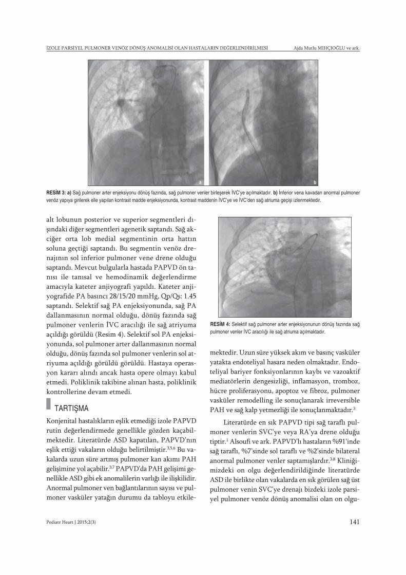
İZOLE PARSİYEL PULMONER VENÖZ DÖNÜŞ ANOMALİSİ OLAN HASTALARIN DEĞERLENDİRİLMESİ Ajda Mutlu MIHÇIOĞLU ve ark.
alt lobunun posterior ve superior segmentleri dı-şındaki diğer segmentleri agenetik saptandı. Sağ ak-ciğer orta lob medial segmentinin orta hattınsoluna geçtiği saptandı. Bu segmentin venöz dre-najının sol inferior pulmoner vene drene olduğusaptandı. Mevcut bulgularla hastada PAPVD ön ta-nısı ile tanısal ve hemodinamik değerlendirmeamacıyla kateter anjiyografi yapıldı. Kateter anji-yografide PA basıncı 28/15/20 mmHg, Qp/Qs: 1.45saptandı. Selektif sağ PA enjeksiyonunda, sağ PAdallanmasının normal olduğu, dönüş fazında sağpulmoner venlerin İVC aracılığı ile sağ atriyumaaçıldığı görüldü (Resim 4). Selektif sol PA enjeksi-yonunda, sol pulmoner arter dallanmasının normalolduğu, dönüş fazında sol pulmoner venlerin sol at-riyuma açıldığı görüldü görüldü. Hastaya operas-yon kararı alındı ancak hasta opere olmayı kabuletmedi. Poliklinik takibine alınan hasta, poliklinikkontrollerine devam etmedi.
TARTIŞMA
Konjenital hastalıkların eşlik etmediği izole PAPVDrutin değerlendirmede genellikle gözden kaçabil-mektedir. Literatürde ASD kapatılan, PAPVD’nıneşlik ettiği vakaların olduğu belirtilmiştir.3,5,6 Bu va-kalarda uzun süre artmış pulmoner kan akımı PAHgelişimine yol açabilir.3,7 PAPVD’da PAH gelişimi ge-nellikle ASD gibi ek anomalilerin varlığı ile ilişkilidir.Anormal pulmoner ven bağlantılarının sayısı ve pul-moner vasküler yatağın durumu da tabloyu etkile-
mektedir. Uzun süre yüksek akım ve basınç vasküleryatakta endoteliyal hasara neden olmaktadır. Endo-teliyal bariyer fonksiyonlarının kaybı ve vazoaktifmediatörlerin dengesizliği, inflamasyon, tromboz,hücre proliferasyonu, apoptoz ve fibroz, pulmonervasküler remodelling ile sonuçlanarak irreversiblePAH ve sağ kalp yetmezliği ile sonuçlanmaktadır.3
Literatürde en sık PAPVD tipi sağ taraflı pul-moner venlerin SVC’ye veya RA’ya drene olduğutiptir.1 Alsoufi ve ark. PAPVD’lı hastaların %91’indesağ taraflı, %7’sinde sol taraflı ve %2’sinde bilateralanormal pulmoner venler saptamışlardır.3,8 Kliniği-mizdeki on olgu değerlendirildiğinde literatürdeASD ile birlikte olan vakalarda en sık görülen sağ üstpulmoner venin SVC’ye drenajı bizdeki izole parsi-yel pulmoner venöz dönüş anomalisi olan on olgu-
RESİM 3: a) Sağ pulmoner arter enjeksiyonu dönüş fazında, sağ pulmoner venler birleşerek İVC’ye açılmaktadır. b) İnferior vena kavadan anormal pulmonervenöz yapıya girilerek elle yapılan kontrast madde enjeksiyonunda, kontrast maddenin İVC’ye ve İVC’den sağ atriuma geçişi izlenmektedir.
a b
RESİM 4: Selektif sağ pulmoner arter enjeksiyonunun dönüş fazında sağpulmoner venler İVC aracılığı ile sağ atriuma açılmaktadır.
Pediatr Heart J 2015;2(3) 141

Ajda Mutlu MIHÇIOĞLU ve ark. İZOLE PARSİYEL PULMONER VENÖZ DÖNÜŞ ANOMALİSİ OLAN HASTALARIN DEĞERLENDİRİLMESİ
Pediatr Heart J 2015;2(3)142
nun üçünde görüldü. Üç olguda da sağ pulmonervenlerin İVC’ye drene olduğu görüldü. Böylece sağakciğerden anormal pulmoner venöz dönüş, on ol-gunun altısında (%60) görüldü. Daha nadir olan solakciğerden anormal pulmoner venöz dönüş olgularıkalan dört olguda (%40) innominate vene doğru gö-rüldü. Hastalarımızın yedisinde sadece bir venin, üçhastada birden fazla venin anormal dönüşü izlendi.
PAPVD olan hastalar en sık dispne, üfürüm,aritmi nedeniyle başvurabilmektedirler.3 Çalışma-mızda yedi hasta göğüs ağrısı, nefes darlığı ve hal-sizlik nedeniyle değerlendirildi. Diğer üç hastanınşikayetleri olmayıp bir hasta üfürüm nedeniyle,diğer hasta rutin kontrol için başvurusunda, diğerhasta da akciğer grafisinde şüpheli görünüm olmasınedeniyle değerlendirildiler.
PAPVD’yı değerlendirmede transtorasik eko-kardiyografi en iyi başlangıç modalitesidir. TTE ilesağ kalp boşlukları olan RA ve sağ ventrikül (RV)dilatasyonu saptanır. İnterventriküler septumdadüzleşme, sistolde ve diyastolde RV basınç yükü ileartmış PA basıncı ve interatriyal bağlantının var-lığı yada yokluğu renkli Doppler veya kontrast ça-lışmasında değerlendirilir. Wong ve ark.nınyaptıkları izole PAPVD olan 50 çocuktan oluşanbir çalışmada hastaların %96’sında RV dilatasyonusaptanmış.9 RV dilatasyonu yaygın olarak görüle-bilse de RV yetmezliği ve PAH daha nadir saptan-maktadır.2,4,10 Bizim çalışmamızda hastalarınbeşinde RV dilatasyonu ve bir hastada yaşına göreüst sınırda çaplar mevcuttu.
TTE’de suprasternal, subkostal, sağ parasternalaçılar PAPVD tanısında önemli.4 Özellikle supras-ternal görüntünün yararı Bansal ve ark. tarafından200 erişkin hastada sösterilmiş.11 Çalışmamızda has-talarımızın yedisinde suprasternal görüntüleme ilevertikal ven ve vertikal venin innominate vene veSVC’ye drenajı izlendi. Hastaların ikisinde subkostaldeğerlendirmede İVC’ye drenaj saptandı. Ekokardi-yografi ile ayırıcı tanıda persistan sol SVC, koronersinus obstrüksiyonu, dilate aksesuar hemiazigoz ven,genişlemiş sol superior interkostal ven akla gelmeli-dir.1,12 Ayrıca RV dilatasyonu yapabilecek sinus ve-nosus defekt, ostium primum yada sekundum ASDyada aritmojenik sağ ventrikül displazisi gibi kardi-
yak hastalıklar akılda tutulmalıdır.13 TTE’nin pul-moner venleri tam olarak değerlendirmede yetersizolduğu durumlarda, TEE PAPVD varlığını doğrula-mada öne çıkmaktadır. Telekardiyografi, genelliklenormal saptanmaktadır ancak sol sağ şantın sekon-der bulguları olan kardiyomegali, pulmoner vaskü-ler çıkıntı ve PAH bulguları görülebilmektedir.Direkt grafide sol üst lobda pulmoner venleri temsileden tubuler opasiteler görülebilir.12 BT, kardiyo-pulmoner anatomiyi pulmoner venöz drenajı da içe-recek şekilde doğru değerlendirilmeyi sağlar. MR,anormal pulmoner bağlantıları gösterirken nonin-vaziv olarak Qp/Qs değerlendirilmesinde kullanı-lır.12 Radyasyon güvenliği ile birlikte değerlen-dirildiğinde ektrakardiyak şantları değerlendirmdeön planda tercih edilmektedir.1 Kateter anjiyografi,tanıyı kesinleştirmede, hemodinamik değerlendir-mede kullanılmaktadır.Literatürde izole PAPVDolan olgularda Babb ve Saalouke kateterizasyon ileyüksek PA basıncı saptamışlar ve oksimetri çalışma-ları ile de bulgularını desteklemişlerdir.2,14
Hastalarımızdan onuncu olguda akciğer grafi-sinde pulmoner konusta hafif belirginleşme nede-niyle ileri değerlendirme yapılmıştır. Diğer dokuzhastamızda tanıdan TTE ile şüphe edilmiştir. Buhastalardan birinde de direkt grafide anormal vas-küler yapıyı düşündürecek tübüler opasite mevcut-tur. Dokuz hastada tanıyı desteklemek için sadecebirinde TEE ve aynı hastaya BT yapılmış ve diğeriki hastaya ise sadece BT anjiyografi yapılmıştır. Buüç hasta ayrıca hemodinamik yönden kateterizas-yon ile de değerlendirilmişlerdir. Sadece TTE ile de-ğerlendirilen diğer altı hastanın ise dördünde tanıkonularak, ileri değerlendirme yapılmadan izlemkararı alınmıştır. Diğer iki hasta ise hemodinamikyönden ile değerlendirilmişlerdir. Göğüs röntge-ninden şüphe edilmiş olan ve ileri değerlendirmeyealınan onuncu hastada TTE ile sağ kalbe olan hacimve basınç yükünün indirek bulgularının saptanmasıüzerine ve TTE ile ayrıntılı değerlendirmenin netolarak yapılamadığından TEE, BT anjiyografi ve ka-teterizasyon ile ileri değerlendirme yapılmıştır. Ka-teterizasyon yapılan olgularda PAPVD gösterilmiş,basınç ölçümleri ve oksimetri çalışmaları ile bulgu-lar desteklenmiştir. Hiçbir olgumuzun değerlendi-rilmesinde MR kullanılmamıştır.
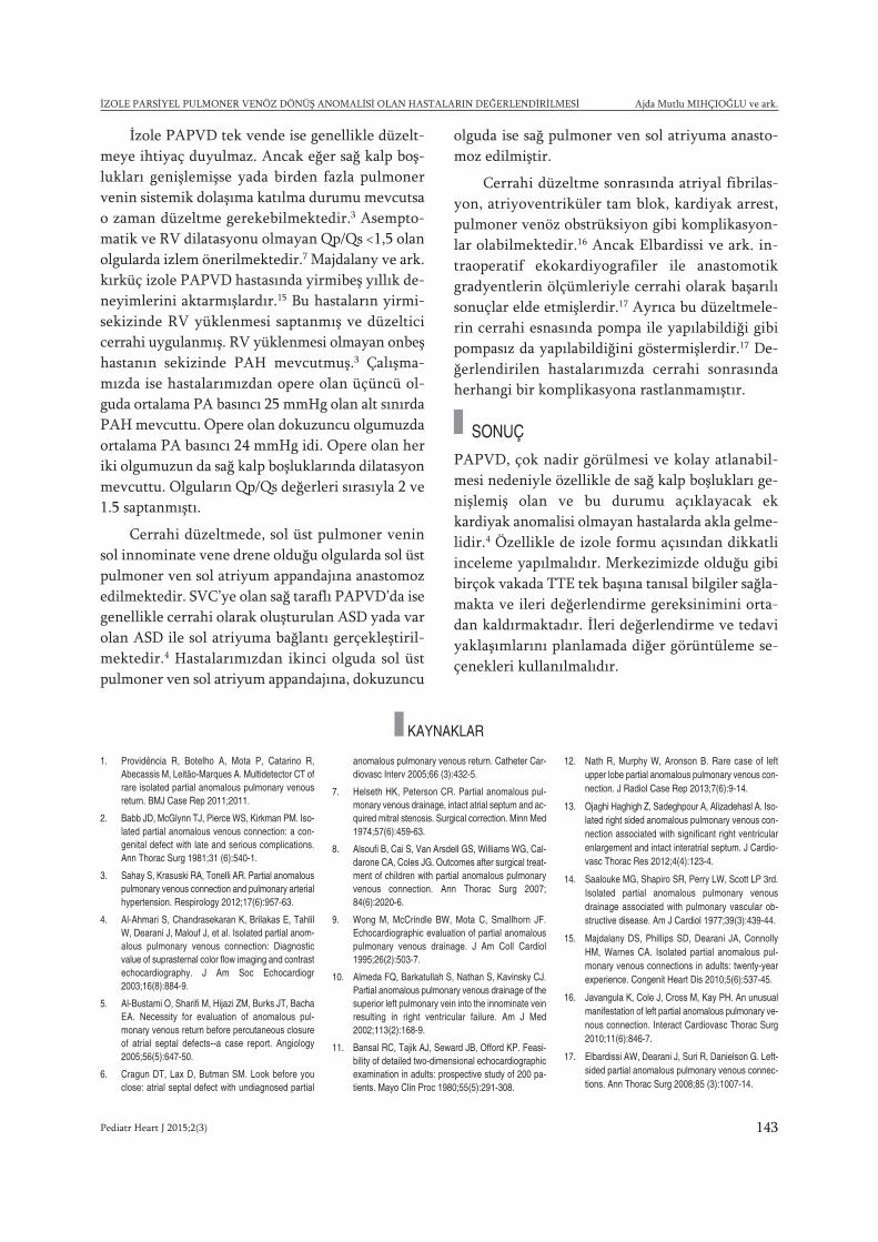
İZOLE PARSİYEL PULMONER VENÖZ DÖNÜŞ ANOMALİSİ OLAN HASTALARIN DEĞERLENDİRİLMESİ Ajda Mutlu MIHÇIOĞLU ve ark.
İzole PAPVD tek vende ise genellikle düzelt-meye ihtiyaç duyulmaz. Ancak eğer sağ kalp boş-lukları genişlemişse yada birden fazla pulmonervenin sistemik dolaşıma katılma durumu mevcutsao zaman düzeltme gerekebilmektedir.3 Asempto-matik ve RV dilatasyonu olmayan Qp/Qs <1,5 olanolgularda izlem önerilmektedir.7 Majdalany ve ark.kırküç izole PAPVD hastasında yirmibeş yıllık de-neyimlerini aktarmışlardır.15 Bu hastaların yirmi-sekizinde RV yüklenmesi saptanmış ve düzelticicerrahi uygulanmış. RV yüklenmesi olmayan onbeşhastanın sekizinde PAH mevcutmuş.3 Çalışma-mızda ise hastalarımızdan opere olan üçüncü ol-guda ortalama PA basıncı 25 mmHg olan alt sınırdaPAH mevcuttu. Opere olan dokuzuncu olgumuzdaortalama PA basıncı 24 mmHg idi. Opere olan heriki olgumuzun da sağ kalp boşluklarında dilatasyonmevcuttu. Olguların Qp/Qs değerleri sırasıyla 2 ve1.5 saptanmıştı.
Cerrahi düzeltmede, sol üst pulmoner veninsol innominate vene drene olduğu olgularda sol üstpulmoner ven sol atriyum appandajına anastomozedilmektedir. SVC’ye olan sağ taraflı PAPVD’da isegenellikle cerrahi olarak oluşturulan ASD yada varolan ASD ile sol atriyuma bağlantı gerçekleştiril-mektedir.4 Hastalarımızdan ikinci olguda sol üstpulmoner ven sol atriyum appandajına, dokuzuncu
olguda ise sağ pulmoner ven sol atriyuma anasto-moz edilmiştir.
Cerrahi düzeltme sonrasında atriyal fibrilas-yon, atriyoventriküler tam blok, kardiyak arrest,pulmoner venöz obstrüksiyon gibi komplikasyon-lar olabilmektedir.16 Ancak Elbardissi ve ark. in-traoperatif ekokardiyografiler ile anastomotikgradyentlerin ölçümleriyle cerrahi olarak başarılısonuçlar elde etmişlerdir.17 Ayrıca bu düzeltmele-rin cerrahi esnasında pompa ile yapılabildiği gibipompasız da yapılabildiğini göstermişlerdir.17 De-ğerlendirilen hastalarımızda cerrahi sonrasındaherhangi bir komplikasyona rastlanmamıştır.
SONUÇ
PAPVD, çok nadir görülmesi ve kolay atlanabil-mesi nedeniyle özellikle de sağ kalp boşlukları ge-nişlemiş olan ve bu durumu açıklayacak ekkardiyak anomalisi olmayan hastalarda akla gelme-lidir.4 Özellikle de izole formu açısından dikkatliinceleme yapılmalıdır. Merkezimizde olduğu gibibirçok vakada TTE tek başına tanısal bilgiler sağla-makta ve ileri değerlendirme gereksinimini orta-dan kaldırmaktadır. İleri değerlendirme ve tedaviyaklaşımlarını planlamada diğer görüntüleme se-çenekleri kullanılmalıdır.
1. Providência R, Botelho A, Mota P, Catarino R,Abecassis M, Leitão-Marques A. Multidetector CT ofrare isolated partial anomalous pulmonary venousreturn. BMJ Case Rep 2011;2011.
2. Babb JD, McGlynn TJ, Pierce WS, Kirkman PM. Iso-lated partial anomalous venous connection: a con-genital defect with late and serious complications.Ann Thorac Surg 1981;31 (6):540-1.
3. Sahay S, Krasuski RA, Tonelli AR. Partial anomalouspulmonary venous connection and pulmonary arterialhypertension. Respirology 2012;17(6):957-63.
4. Al-Ahmari S, Chandrasekaran K, Brilakas E, TahlilW, Dearani J, Malouf J, et al. Isolated partial anom-alous pulmonary venous connection: Diagnosticvalue of suprasternal color flow imaging and contrastechocardiography. J Am Soc Echocardiogr2003;16(8):884-9.
5. Al-Bustami O, Sharifi M, Hijazi ZM, Burks JT, BachaEA. Necessity for evaluation of anomalous pul-monary venous return before percutaneous closureof atrial septal defects--a case report. Angiology2005;56(5):647-50.
6. Cragun DT, Lax D, Butman SM. Look before youclose: atrial septal defect with undiagnosed partial
anomalous pulmonary venous return. Catheter Car-diovasc Interv 2005;66 (3):432-5.
7. Helseth HK, Peterson CR. Partial anomalous pul-monary venous drainage, intact atrial septum and ac-quired mitral stenosis. Surgical correction. Minn Med1974;57(6):459-63.
8. Alsoufi B, Cai S, Van Arsdell GS, Williams WG, Cal-darone CA, Coles JG. Outcomes after surgical treat-ment of children with partial anomalous pulmonaryvenous connection. Ann Thorac Surg 2007;84(6):2020-6.
9. Wong M, McCrindle BW, Mota C, Smallhorn JF.Echocardiographic evaluation of partial anomalouspulmonary venous drainage. J Am Coll Cardiol1995;26(2):503-7.
10. Almeda FQ, Barkatullah S, Nathan S, Kavinsky CJ.Partial anomalous pulmonary venous drainage of thesuperior left pulmonary vein into the innominate veinresulting in right ventricular failure. Am J Med2002;113(2):168-9.
11. Bansal RC, Tajik AJ, Seward JB, Offord KP. Feasi-bility of detailed two-dimensional echocardiographicexamination in adults: prospective study of 200 pa-tients. Mayo Clin Proc 1980;55(5):291-308.
12. Nath R, Murphy W, Aronson B. Rare case of leftupper lobe partial anomalous pulmonary venous con-nection. J Radiol Case Rep 2013;7(6):9-14.
13. Ojaghi Haghigh Z, Sadeghpour A, Alizadehasl A. Iso-lated right sided anomalous pulmonary venous con-nection associated with significant right ventricularenlargement and intact interatrial septum. J Cardio-vasc Thorac Res 2012;4(4):123-4.
14. Saalouke MG, Shapiro SR, Perry LW, Scott LP 3rd.Isolated partial anomalous pulmonary venousdrainage associated with pulmonary vascular ob-structive disease. Am J Cardiol 1977;39(3):439-44.
15. Majdalany DS, Phillips SD, Dearani JA, ConnollyHM, Warnes CA. Isolated partial anomalous pul-monary venous connections in adults: twenty-yearexperience. Congenit Heart Dis 2010;5(6):537-45.
16. Javangula K, Cole J, Cross M, Kay PH. An unusualmanifestation of left partial anomalous pulmonary ve-nous connection. Interact Cardiovasc Thorac Surg2010;11(6):846-7.
17. Elbardissi AW, Dearani J, Suri R, Danielson G. Left-sided partial anomalous pulmonary venous connec-tions. Ann Thorac Surg 2008;85 (3):1007-14.
KAYNAKLAR
Pediatr Heart J 2015;2(3) 143
Pediatr Heart J 2015;2(3)144
ehçet’s disease (BD) is a multisystem vasculitic syndrome of un-known etiology. Its clinical triad of aphtous ulcers, genital ulcers,and ocular manifestations was originally described by the Turkish
dermatologist Hulusi Behçet in 1937. It encompasses not only mucocuta-neous, ocular and vascular manifestations but also musculoskeletal, neuro-logic, pulmonary, gastrointestinal and genitourinary manifestations.1 Theintracardiac masses include benign and malignant primary cardiac tumors,metastatic tumors, thrombi, vegetations, iatrogenic material and normalvariants. The recognition of an inflammatory mass should prompt consid-eration of BD prior to referral for invasive procedures. An attentive historyand physical exam looking for signs and symptoms of BD is critical beforeconsidering surgical excision of unexplained intracardiac masses. Pulmonaryinvolvement in BD is rare, occurring in 7.7% of patients.2 Pulmonary artery
Right Ventricular Mass andMultiple Pulmonary Artery Aneurysms
in a Child with Behçet's Disease:Case Report
AABS TRACT Behçet’s disease is a chronic multisystemic inflammatory disorder characterized byorogenital ulcerations, uveitis and occasional cardiac involvement. This report describes an atypi-cal presentation of pediatric Behçet’s disease with pulmonary aneurysms and a cardiac mass. A 16-year-old boy presented with a 2-month history of weight loss, fever and a large tumor adhering tothe free wall of the right ventricle. Helical thoracic computed tomography (CT) angiographydemonstrated multiple pulmonary aneurysms. The discovery of an intracardiac mass in a youngpatient must evoke the diagnosis of Behçet's disease.
Key Words: Intracardiac mass; pulmonary artery aneurysm; Behçet’s disease; childhood
ÖZET Behçet Hastalıgı orogenital ülserasyonlar, üveit, kardiyak tutulum ile karakterize multisis-temik kronik inflamatuar bir hastalıktır. Bu raporda pulmoner anevrizma ve kardiyak kitle ile ati-pik prezentasyon gösteren pediatrik Behçet hastalıg ı tanımlandı. Onaltı yas ında erkek hasta 2 aydanberi devam eden kilo kaybı, ates, sag ventrikül serbest duvarında tümör görünümünde genis birkitle ile basvurdu. Helikal torasik BT anjiografide multipl pulmoner anevrizma gösterildi. Genç birhastada intrakardiyak kitle tanısı konuldug unda Behçet Hastalıg ı tanısı açısından dikkatli olun-malıdır.
Anah tar Ke li me ler: İntrakardiyak kitle; pulmoner arter anevrizması; Behçet hastalığı; çocukluk çağı
Pediatr Heart J 2015;2(3):144-7
Derya ÇİMEN,a
Mustafa KOPLAY,b
Derya ARSLAN,c
Melike EMİROĞLU,d
Osman GÜVENÇ,a
Mehmet ÖÇ,e
Bülent ORANa
Departments of aPediatric Cardiology, bRadiology, dPediatric Infectious Diseases, eHeart and Vascular Surgery, Selçuk University Faculty of Medicine, cClinic of Pediatric Cardiology, Konya Training and Research Hospital,Konya
Ge liş Ta ri hi/Re ce i ved: 01.12.2014Ka bul Ta ri hi/Ac cep ted: 11.02.2015
Ya zış ma Ad re si/Cor res pon den ce:Derya ARSLANKonya Training and Research Hospital,Clinic of Pediatric Cardiology, Konya,TÜRKİYE/[email protected]
Copyright © 2015 by Türk Pediatrik Kardiyoloji veKalp Cerrahisi Derneği
OLGU SUNUMU
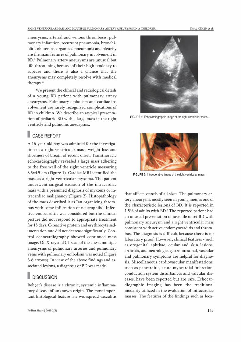
Pediatr Heart J 2015;2(3) 145
RIGHT VENTRICULAR MASS AND MULTIPLE PULMONARY ARTERY ANEURYSMS IN A CHILDREN... Derya ÇİMEN et al.
aneurysms, arterial and venous thrombosis, pul-monary infarction, recurrent pneumonia, bronchi-olitis obliterans, organized pneumonia and pleurisyare the main features of pulmonary involvement inBD.2 Pulmonary artery aneurysms are unusual butlife-threatening because of their high tendency torupture and there is also a chance that theaneurysms may completely resolve with medicaltherapy.3
We present the clinical and radiological detailsof a young BD patient with pulmonary arteryaneurysms. Pulmonary embolism and cardiac in-volvement are rarely recognized complications ofBD in children. We describe an atypical presenta-tion of pediatric BD with a large mass in the rightventricle and pulmonic aneurysms.
CASE REPORT
A 16-year-old boy was admitted for the investiga-tion of a right ventricular mass, weight loss andshortness of breath of recent onset. Transthoracicechocardiography revealed a large mass adheringto the free wall of the right ventricle measuring3.5x4.5 cm (Figure 1). Cardiac MRI identified themass as a right ventricular myxoma. The patientunderwent surgical excision of the intracardiacmass with a presumed diagnosis of myxoma or in-tracardiac malignancy (Figure 2). Histopathologyof the mass described it as “an organizing throm-bus with some infiltration of neutrophils”. Infec-tive endocarditis was considered but the clinicalpicture did not respond to appropriate treatmentfor 15 days. C-reactive protein and erythrocyte sed-imentation rate did not decrease significantly. Con-trol echocardiography showed continued massimage. On X-ray and CT scan of the chest, multipleaneurysms of pulmonary arteries and pulmonaryveins with pulmonary embolism was noted (Figure3-6 arrows). In view of the above findings and as-sociated lesions, a diagnosis of BD was made.
DISCUSSION
Behçet’s disease is a chronic, systemic inflamma-tory disease of unknown origin. The most impor-tant histological feature is a widespread vasculitis
that affects vessels of all sizes. The pulmonary ar-tery aneurysm, mostly seen in young men, is one ofthe characteristic lesions of BD. It is reported in1.5% of adults with BD.4 The reported patient hadan unusual presentation of juvenile onset BD withpulmonary aneurysm and a right ventricular massconsistent with active endomyocarditis and throm-bus. The diagnosis is difficult because there is nolaboratory proof. However, clinical features - suchas orogenital aphthae, ocular and skin lesions,arthritis, and neurologic, gastrointestinal, vascularand pulmonary symptoms are helpful for diagno-sis. Miscellaneous cardiovascular manifestations,such as pancarditis, acute myocardial infarction,conduction system disturbances and valvular dis-eases, have been reported but are rare. Echocar-diographic imaging has been the traditionalmodality utilized in the evaluation of intracardiacmasses. The features of the findings such as loca-
FIGURE 1: Echocardiographic image of the right ventricular mass.
FIGURE 2: Intraoperative image of the right ventricular mass.
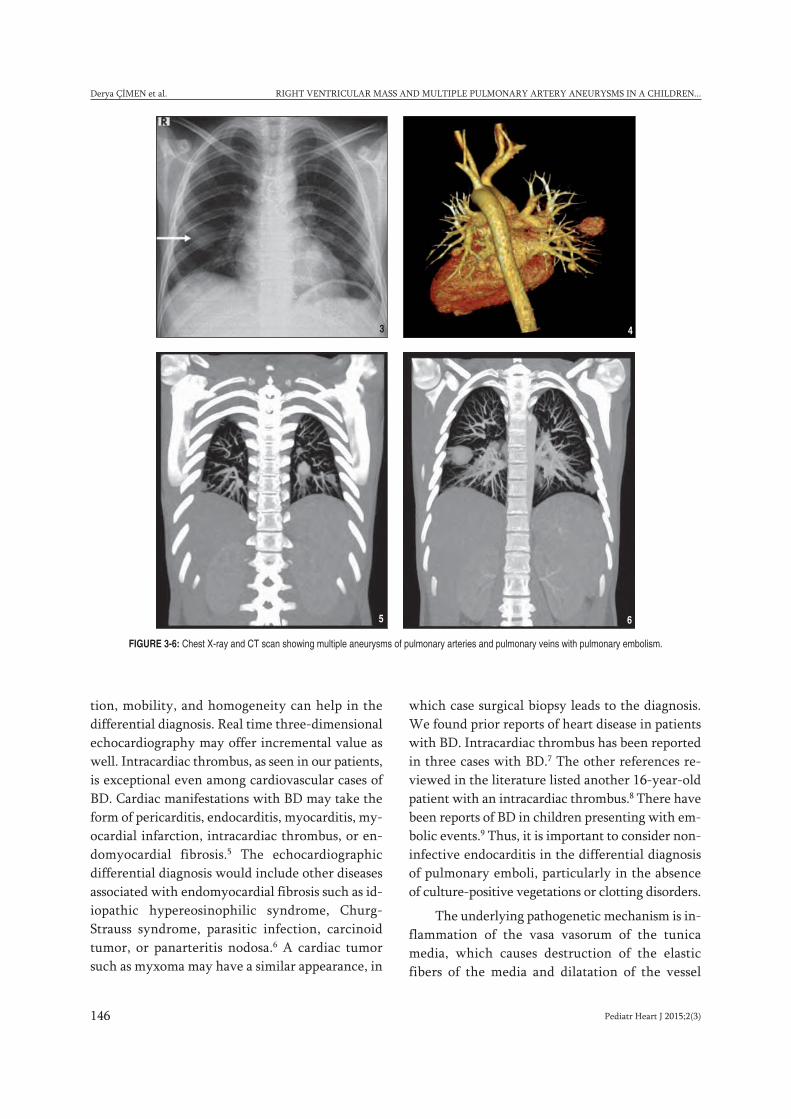
Pediatr Heart J 2015;2(3)146
Derya ÇİMEN et al. RIGHT VENTRICULAR MASS AND MULTIPLE PULMONARY ARTERY ANEURYSMS IN A CHILDREN...
tion, mobility, and homogeneity can help in thedifferential diagnosis. Real time three-dimensionalechocardiography may offer incremental value aswell. Intracardiac thrombus, as seen in our patients,is exceptional even among cardiovascular cases ofBD. Cardiac manifestations with BD may take theform of pericarditis, endocarditis, myocarditis, my-ocardial infarction, intracardiac thrombus, or en-domyocardial fibrosis.5 The echocardiographicdifferential diagnosis would include other diseasesassociated with endomyocardial fibrosis such as id-iopathic hypereosinophilic syndrome, Churg-Strauss syndrome, parasitic infection, carcinoidtumor, or panarteritis nodosa.6 A cardiac tumorsuch as myxoma may have a similar appearance, in
which case surgical biopsy leads to the diagnosis.We found prior reports of heart disease in patientswith BD. Intracardiac thrombus has been reportedin three cases with BD.7 The other references re-viewed in the literature listed another 16-year-oldpatient with an intracardiac thrombus.8 There havebeen reports of BD in children presenting with em-bolic events.9 Thus, it is important to consider non-infective endocarditis in the differential diagnosisof pulmonary emboli, particularly in the absenceof culture-positive vegetations or clotting disorders.
The underlying pathogenetic mechanism is in-flammation of the vasa vasorum of the tunicamedia, which causes destruction of the elasticfibers of the media and dilatation of the vessel
FIGURE 3-6: Chest X-ray and CT scan showing multiple aneurysms of pulmonary arteries and pulmonary veins with pulmonary embolism.
3 4
5 6
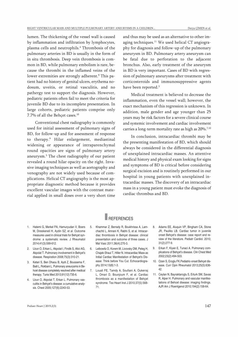
Pediatr Heart J 2015;2(3) 147
RIGHT VENTRICULAR MASS AND MULTIPLE PULMONARY ARTERY ANEURYSMS IN A CHILDREN... Derya ÇİMEN et al.
lumen. The thickening of the vessel wall is causedby inflammation and infiltration by lymphocytes,plasma cells and neutrophils.4 Thrombosis of thepulmonary arteries in BD is usually in the form ofin situ thrombosis. Deep vein thrombosis is com-mon in BD, while pulmonary embolism is rare, be-cause the thrombi in the inflamed veins of thelower extremities are strongly adherent.9 This pa-tient had no history of genital ulcers, erythema no-dosum, uveitis, or retinal vasculitis, and nopathergy test to support the diagnosis. However,pediatric patients often fail to meet the criteria forjuvenile BD due to its incomplete presentation. Inlarge cohorts, pediatric patients comprise only7.7% of all the Behçet cases.10
Conventional chest radiography is commonlyused for initial assessment of pulmonary signs ofBD, for follow-up and for assessment of responseto therapy.9 Hilar enlargement, mediastinalwidening or appearance of intraparenchymalround opacities are signs of pulmonary arteryaneurysm.3 The chest radiography of our patientrevealed a round hilar opacity on the right. Inva-sive imaging techniques as well as aortography andvenography are not widely used because of com-plications. Helical CT angiography is the most ap-propriate diagnostic method because it providesexcellent vascular images with the contrast mate-rial applied in small doses over a very short time
and thus may be used as an alternative to other im-aging techniques.11 We used helical CT angiogra-phy for diagnosis and follow-up of the pulmonaryaneurysm in BD. Pulmonary artery aneurysm canbe fatal due to perforation to the adjacentbronchus. Also, early treatment of the aneurysmin BD is very important. Cases of BD with regres-sion of pulmonary aneurysms after treatment withcorticosteroids and immunosuppressive agentshave been reported.3
Medical treatment is believed to decrease theinflammation, even the vessel wall; however, theexact mechanism of this regression is unknown. Inaddition, male gender and age younger than 25years may be risk factors for a severe clinical courseand systemic involvement and cardiac involvementcarries a long-term mortality rate as high as 20%.7,10
In conclusion, intracardiac thrombi may bethe presenting manifestation of BD, which shouldalways be considered in the differential diagnosisof unexplained intracardiac masses. An attentivemedical history and physical exam looking for signsand symptoms of BD is critical before consideringsurgical excision and is routinely performed in ourhospital in young patients with unexplained in-tracardiac masses. The discovery of an intracardiacmass in a young patient must evoke the diagnosis ofcardiac thrombus and BD.
1. Hatemi G, Merkel PA, Hamuryudan V, BoersM, Direskeneli H, Aydin SZ, et al. Outcomemeasures used in clinical trials for Behçet syn-drome: a systematic review. J Rheumatol2014;41(3):599-612.
2. Uzun O, Erkan L, Akpolat I, Findik S, Atici AG,Akpolat T. Pulmonary involvement in Behçet'sdisease. Respiration 2008;75(3):310-21.
3. Ketari S, Ben Dhaou B, Aydi Z, Boussema F,Baili L, Rokbani L. Pulmonary aneurysms in Be-hcet disease completely resolved after medicaltherapy. Tunis Med 2013;91(12):735-6.
4. Uzun O, Akpolat T, Erkan L. Pulmonary vas-culitis in Behçet’s disease: a cumulative analy-sis. Chest 2005;127(6):2243-53.
5. Khammar Z, Berrady R, Boukhrissa A, Lam-chachti L, Amrani K, Rabhi S, et al. Intracar-diac thrombosis in Behçet disease: clinicalpresentation and outcome of three cases. JMal Vasc 2011;36(4):270-3.
6. Leibowitz D, Korem M, Livovsky DM, Peleg H,Chajek-Shaul T, Hiller N. Intracardiac Mass asInitial Cardiac Manifestation of Behçet's Dis-ease: Think before You Cut. Echocardiogra-phy 2014;13(8):1-3.
7. Louali FE, Tamdy A, Soufiani A, Oukerraj L, Omari D, Bounjoum F, et al. Cardiacthrombosis as a manifestation of Behçetsyndrome. Tex Heart Inst J 2010;37(5):568-71.
8. Adams EE, Aluquin VP, Bingham CA, StoneJR, Pauliks LB. Cardiac tumor in juvenileonset Behçet's disease: case report and re-view of the literature. Pediatr Cardiol. 2010;31(2):277-9.
9. Erkan F, Kiyan E, Tunaci A. Pulmonary com-plications of Behçet’s disease. Clin Chest Med2002;23(2):494-503.
10. Ozen S, Eroglu FK.Pediatric-onset Behçet dis-ease. Curr Opin Rheumatol 2013;25(5):636-42.
11. Ceylan N, Bayraktaroglu S, Erturk SM, SavasR, Alper H. Pulmonary and vascular manifes-tations of Behcet disease: imaging findings.AJR Am J Roentgenol 2010;194(2):158-64.
REFERENCES
Pediatr Heart J 2015;2(3)148
nomalous origin of the left coronary artery from the pulmonary ar-tery (ALCAPA) is a rare entity accounting for 0.5% of congenitalheart defects.1 Typically, patients with ALCAPA present in infancy
with heart failure resulting from reduced ventricular contraction and var-ious degrees of mitral insufficiency caused by papillary muscle dysfunctiondue to myocardial ischemia. Treatment usually includes surgical repair tocreate a dual coronary artery system. We report a case of a 13-month-oldgirl with preserved left ventricular function initially diagnosed to have acoronary cameral fistula.
CASE REPORT
A 13-month-old girl was referred to the pediatric cardiology clinic for eval-uation of a cardiac murmur. Physical examination revealed a grade 2/6 con-tinuous murmur at the left precordium and mild tachypnea. The historywas unremarkable. The patient’s weight and height were 7.8 kg (3-10 per-centile for age) and 76 cm (75-90 percentile for age), respectively. The
Anomalous Origin of the Left Coronary Arteryfrom the Pulmonary Artery (ALCAPA) Presenting
as Coronary-Cameral Fistula in an Infant:Case Report
AABS TRACT Anomalous origin of the left coronary artery from the pulmonary artery (ALCAPA) isa rare entity that typically presents during infancy with heart failure. This report describes anasymptomatic pediatric patient who presented with preserved left ventricular function and a con-tinuous murmur and was initially misdiagnosed to have a coronary cameral fistula.
Key Words: Coronary artery disease; aorta-pulmonary artery fistula; child
ÖZET Pulmoner arterden anormal olarak çıkan sol koroner arteri bulunan hastalar tipik olarakkalp yetersizliği bulguları ile başvururlar. Burada normal sol ventrikül fonksiyonu bulunan ve de-vamlı üfürüm duyulan, semptomu bulunmayan, başlangıçta yanlış olarak koroner-kamaral fistültanısı alan bir hasta sunulmuştur.
Anah tar Ke li me ler: Koroner arter hastalığı; aort-pulmoner arter fistula; çocuk
Pediatr Heart J 2015;2(3):148-51
Sevcan ERDEM,a
Işıl YILDIRIM,b
Ufuk Utku GÜLLÜ,a
Orhan Kemal SALİH,c
Nazan ÖZBARLASa
Departments ofaPediatric Cardiology,cCardiovascular Surgery, Çukurova University Faculty of Medicine,bDepartment of Pediatric Cardiology,Adana Numune Teaching and Research Hospital,Adana
Ge liş Ta ri hi/Re ce i ved: 16.12.2014 Ka bul Ta ri hi/Ac cep ted: 27.06.2015
Ya zış ma Ad re si/Cor res pon den ce:Sevcan ERDEMÇukurova University Faculty of Medicine,Department of Pediatric Cardiology, Adana,TÜRKİYE/[email protected]
Copyright © 2015 by Türk Pediatrik Kardiyoloji veKalp Cerrahisi Derneği
OLGU SUNUMU
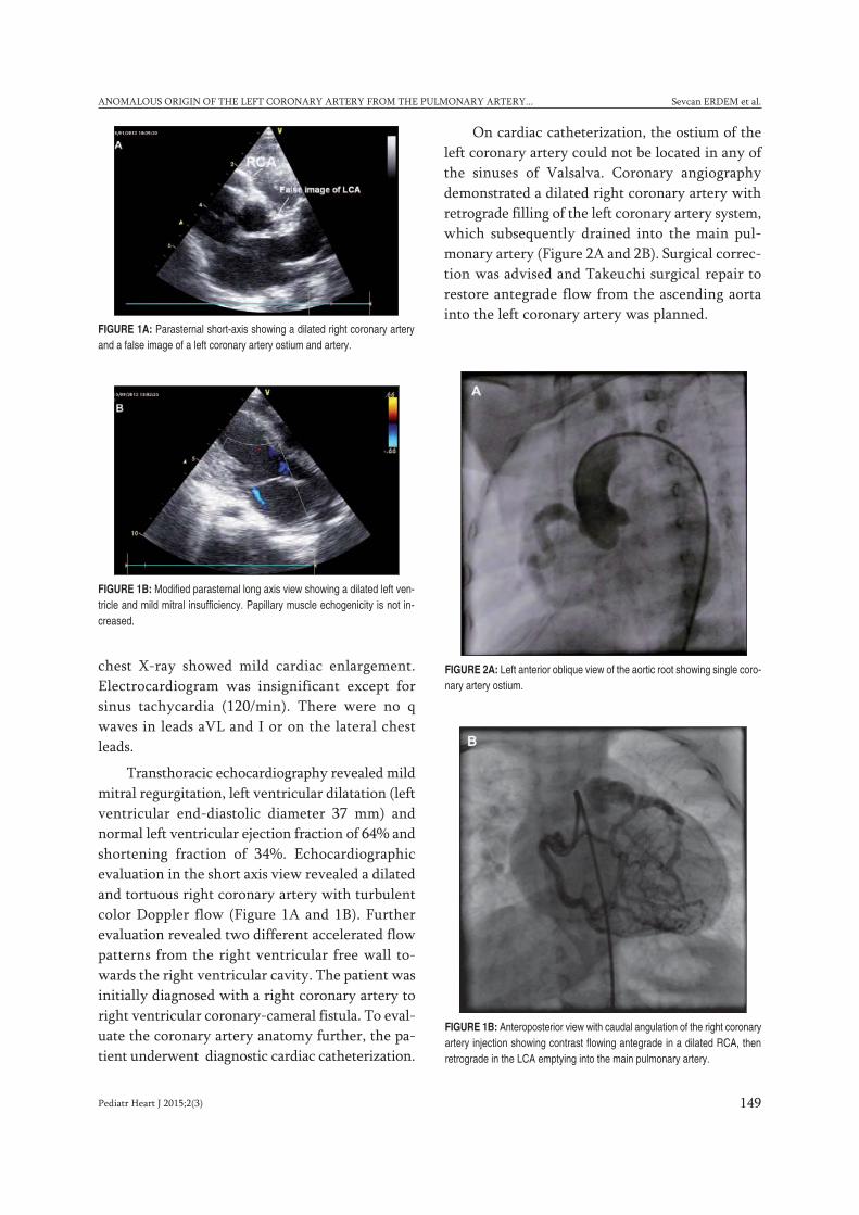
Pediatr Heart J 2015;2(3) 149
ANOMALOUS ORIGIN OF THE LEFT CORONARY ARTERY FROM THE PULMONARY ARTERY... Sevcan ERDEM et al.
chest X-ray showed mild cardiac enlargement.Electrocardiogram was insignificant except forsinus tachycardia (120/min). There were no qwaves in leads aVL and I or on the lateral chestleads.
Transthoracic echocardiography revealed mildmitral regurgitation, left ventricular dilatation (leftventricular end-diastolic diameter 37 mm) andnormal left ventricular ejection fraction of 64% andshortening fraction of 34%. Echocardiographicevaluation in the short axis view revealed a dilatedand tortuous right coronary artery with turbulentcolor Doppler flow (Figure 1A and 1B). Furtherevaluation revealed two different accelerated flowpatterns from the right ventricular free wall to-wards the right ventricular cavity. The patient wasinitially diagnosed with a right coronary artery toright ventricular coronary-cameral fistula. To eval-uate the coronary artery anatomy further, the pa-tient underwent diagnostic cardiac catheterization.
On cardiac catheterization, the ostium of theleft coronary artery could not be located in any ofthe sinuses of Valsalva. Coronary angiographydemonstrated a dilated right coronary artery withretrograde filling of the left coronary artery system,which subsequently drained into the main pul-monary artery (Figure 2A and 2B). Surgical correc-tion was advised and Takeuchi surgical repair torestore antegrade flow from the ascending aortainto the left coronary artery was planned.
FIGURE 1A: Parasternal short-axis showing a dilated right coronary arteryand a false image of a left coronary artery ostium and artery.
FIGURE 1B: Modified parasternal long axis view showing a dilated left ven-tricle and mild mitral insufficiency. Papillary muscle echogenicity is not in-creased.
FIGURE 2A: Left anterior oblique view of the aortic root showing single coro-nary artery ostium.
FIGURE 1B: Anteroposterior view with caudal angulation of the right coronaryartery injection showing contrast flowing antegrade in a dilated RCA, thenretrograde in the LCA emptying into the main pulmonary artery.
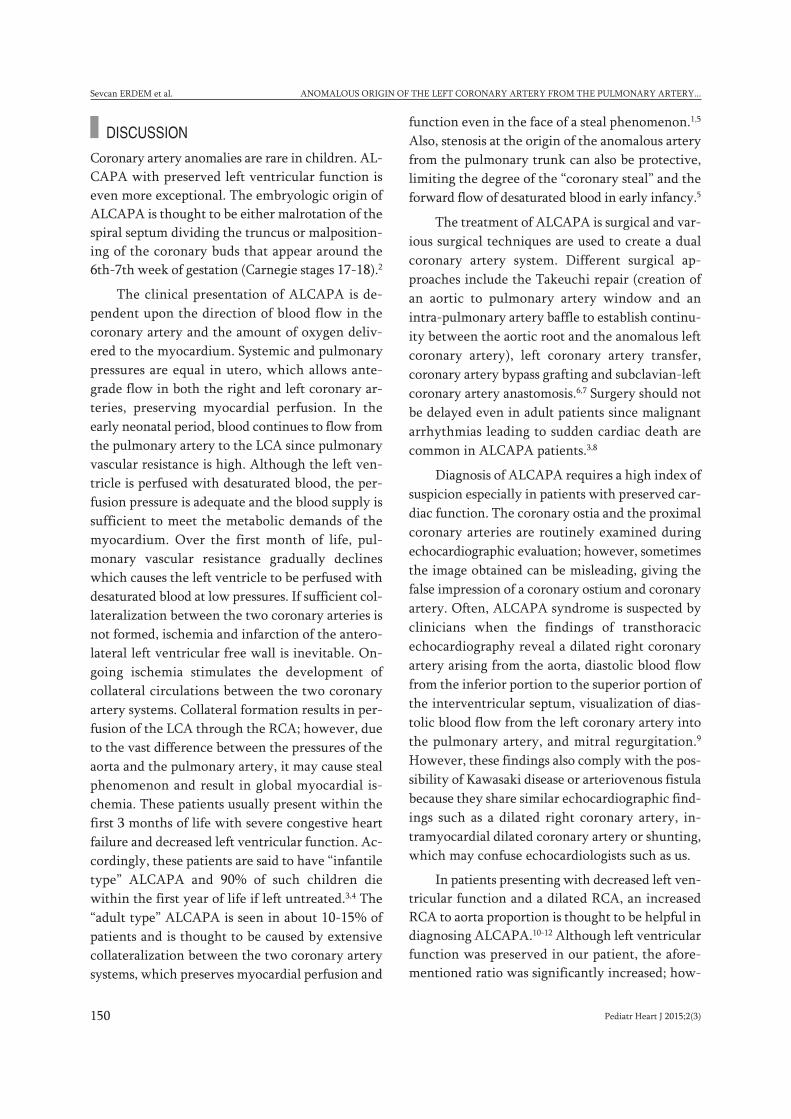
Pediatr Heart J 2015;2(3)150
Sevcan ERDEM et al. ANOMALOUS ORIGIN OF THE LEFT CORONARY ARTERY FROM THE PULMONARY ARTERY...
DISCUSSION
Coronary artery anomalies are rare in children. AL-CAPA with preserved left ventricular function iseven more exceptional. The embryologic origin ofALCAPA is thought to be either malrotation of thespiral septum dividing the truncus or malposition-ing of the coronary buds that appear around the6th-7th week of gestation (Carnegie stages 17-18).2
The clinical presentation of ALCAPA is de-pendent upon the direction of blood flow in thecoronary artery and the amount of oxygen deliv-ered to the myocardium. Systemic and pulmonarypressures are equal in utero, which allows ante-grade flow in both the right and left coronary ar-teries, preserving myocardial perfusion. In theearly neonatal period, blood continues to flow fromthe pulmonary artery to the LCA since pulmonaryvascular resistance is high. Although the left ven-tricle is perfused with desaturated blood, the per-fusion pressure is adequate and the blood supply issufficient to meet the metabolic demands of themyocardium. Over the first month of life, pul-monary vascular resistance gradually declineswhich causes the left ventricle to be perfused withdesaturated blood at low pressures. If sufficient col-lateralization between the two coronary arteries isnot formed, ischemia and infarction of the antero-lateral left ventricular free wall is inevitable. On-going ischemia stimulates the development ofcollateral circulations between the two coronaryartery systems. Collateral formation results in per-fusion of the LCA through the RCA; however, dueto the vast difference between the pressures of theaorta and the pulmonary artery, it may cause stealphenomenon and result in global myocardial is-chemia. These patients usually present within thefirst 3 months of life with severe congestive heartfailure and decreased left ventricular function. Ac-cordingly, these patients are said to have “infantiletype” ALCAPA and 90% of such children diewithin the first year of life if left untreated.3,4 The“adult type” ALCAPA is seen in about 10-15% ofpatients and is thought to be caused by extensivecollateralization between the two coronary arterysystems, which preserves myocardial perfusion and
function even in the face of a steal phenomenon.1,5
Also, stenosis at the origin of the anomalous arteryfrom the pulmonary trunk can also be protective,limiting the degree of the “coronary steal” and theforward flow of desaturated blood in early infancy.5
The treatment of ALCAPA is surgical and var-ious surgical techniques are used to create a dualcoronary artery system. Different surgical ap-proaches include the Takeuchi repair (creation ofan aortic to pulmonary artery window and anintra-pulmonary artery baffle to establish continu-ity between the aortic root and the anomalous leftcoronary artery), left coronary artery transfer,coronary artery bypass grafting and subclavian-leftcoronary artery anastomosis.6,7 Surgery should notbe delayed even in adult patients since malignantarrhythmias leading to sudden cardiac death arecommon in ALCAPA patients.3,8
Diagnosis of ALCAPA requires a high index ofsuspicion especially in patients with preserved car-diac function. The coronary ostia and the proximalcoronary arteries are routinely examined duringechocardiographic evaluation; however, sometimesthe image obtained can be misleading, giving thefalse impression of a coronary ostium and coronaryartery. Often, ALCAPA syndrome is suspected byclinicians when the findings of transthoracicechocardiography reveal a dilated right coronaryartery arising from the aorta, diastolic blood flowfrom the inferior portion to the superior portion ofthe interventricular septum, visualization of dias-tolic blood flow from the left coronary artery intothe pulmonary artery, and mitral regurgitation.9
However, these findings also comply with the pos-sibility of Kawasaki disease or arteriovenous fistulabecause they share similar echocardiographic find-ings such as a dilated right coronary artery, in-tramyocardial dilated coronary artery or shunting,which may confuse echocardiologists such as us.
In patients presenting with decreased left ven-tricular function and a dilated RCA, an increasedRCA to aorta proportion is thought to be helpful indiagnosing ALCAPA.10-12 Although left ventricularfunction was preserved in our patient, the afore-mentioned ratio was significantly increased; how-
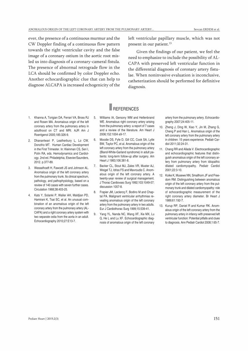
Pediatr Heart J 2015;2(3) 151
ANOMALOUS ORIGIN OF THE LEFT CORONARY ARTERY FROM THE PULMONARY ARTERY... Sevcan ERDEM et al.
ever, the presence of a continuous murmur and theCW Doppler finding of a continuous flow patterntowards the right ventricular cavity and the falseimage of a coronary ostium in the aortic root mis-led us into diagnosis of a coronary-cameral fistula.The presence of abnormal retrograde flow in theLCA should be confirmed by color Doppler echo.Another echocardiographic clue that can help todiagnose ALCAPA is increased echogenicity of the
left ventricular papillary muscle, which was notpresent in our patient.13
Given the findings of our patient, we feel theneed to emphasize to include the possibility of AL-CAPA with preserved left ventricular function inthe differential diagnosis of coronary artery fistu-lae. When noninvasive evaluation is inconclusive,catheterization should be performed for definitivediagnosis.
1. Khanna A, Torigian DA, Ferrari VA, Bross RJand Rosen MA. Anomalous origin of the leftcoronary artery from the pulmonary artery inadulthood on CT and MRI. AJR Am JRoentgenol 2005;185:326-9.
2. Dhanantwari P, Leatherbury L, Lo CW,Donofrio MT. Human Cardiac Developmentin the First Trimester. In: Kleinman CS, Seri I,Polin RA, eds. Hemodynamics and Cardiol-ogy. 2nd ed. Philadelphia, Elsevier/Saunders,2012. p.377-89.
3. Wesselhoeft H, Fawcett JS and Johnson AL.Anomalous origin of the left coronary arteryfrom the pulmonary trunk. Its clinical spectrum,pathology, and pathophysiology, based on areview of 140 cases with seven further cases.Circulation 1968;38:403-25.
4. Kats Y, Solanki P, Waller AH, Maldjian PD,Hamirani K, Tsai SC, et al. An unusual com-bination of an anomalous origin of the leftcoronary artery from the pulmonary artery (AL-CAPA) and a right coronary artery system withtwo separate ostia from the aorta in an adult.Echocardiography 2010;27:E13-7.
5. Williams IA, Gersony WM and HellenbrandWE. Anomalous right coronary artery arisingfrom the pulmonary artery: a report of 7 casesand a review of the literature. Am Heart J2006;152:1004 e9-17.
6. Moodie DS, Fyfe D, Gill CC, Cook SA, LytleBW, Taylor PC, et al. Anomalous origin of theleft coronary artery from the pulmonary artery(Bland-White-Garland syndrome) in adult pa-tients: long-term follow-up after surgery. AmHeart J 1983;106:381-8.
7. Backer CL, Stout MJ, Zales VR, Muster AJ,Weigel TJ, Idriss FS and Mavroudis C. Anom-alous origin of the left coronary artery. Atwenty-year review of surgical management.J Thorac Cardiovasc Surg 1992;103:1049-57;discussion 1057-8.
8. Frapier JM, Leclercq F, Bodino M and Chap-tal PA. Malignant ventricular arrhythmias re-vealing anomalous origin of the left coronaryartery from the pulmonary artery in two adults.Eur J Cardiothorac Surg 1999;15:539-41.
9. Yang YL, Nanda NC, Wang XF, Xie MX, LuQ, He L and Lu XF. Echocardiographic diag-nosis of anomalous origin of the left coronary
artery from the pulmonary artery. Echocardio-graphy 2007;24:405-11.
10. Zheng J, Ding W, Xiao Y, Jin M, Zhang G,Cheng P and Han L. Anomalous origin of theleft coronary artery from the pulmonary arteryin children: 15 years experience. Pediatr Car-diol 2011;32:24-31.
11. Chang RR and Allada V. Electrocardiographicand echocardiographic features that distin-guish anomalous origin of the left coronary ar-tery from pulmonary artery from idiopathicdilated cardiomyopathy. Pediatr Cardiol2001;22:3-10.
12. Koike K, Musewe NN, Smallhorn JF and Free-dom RM. Distinguishing between anomalousorigin of the left coronary artery from the pul-monary trunk and dilated cardiomyopathy: roleof echocardiographic measurement of theright coronary artery diameter. Br Heart J1989;61:192-7.
13. Kurup RP, Daniel R and Kumar RK. Anom-alous origin of the left coronary artery from thepulmonary artery in infancy with preserved leftventricular function: Potential pitfalls and cluesto diagnosis. Ann Pediatr Cardiol 2008;1:65-7.
REFERENCES
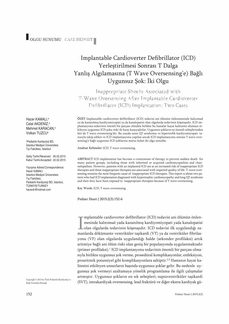
Pediatr Heart J 2015;2(3)152
Implantable cardioverter defibrillator (ICD) tedavisi ani ölümün önlen-mesinde kalıtımsal yada kazanılmış kardiyomiyopati yada kanalopatisiolan olgularda tedavinin köşetaşıdır. ICD tedavisi ilk uygulandığı za-
manlarda dökümante ventriküler taşikardi (VT) ya da ventriküler fibrilas-yonu (VF) olan olgularda uygulandığı halde (sekonder profilaksi) artıkaritmiye bağlı ani ölüm riski olan geniş bir populasyonda uygulanmaktadır(primer profilaksi).1 ICD implantasyonu tedavinin önemli bir parçası olma-sıyla birlikte uygunsuz şok verme, prosedüral komplikasyonlar, enfeksiyon,proaritmik potansiyel gibi komplikasyonlara sahiptir.2,3 Hastanın hayat ka-litesini etkileyen unsurların başında uygunsuz şoklar gelir. Bu nedenle uy-gunsuz şok vermeyi azaltamaya yönelik programlama ile ilgili çalışmalarartmıştır. Uygunsuz şokların en sık sebepleri; supraventriküler taşikardi(SVT), intrakardiyak oversensing, lead fraktürü ve diğer ekstra kardiyak gü-
Implantable Cardioverter Defibrillator (ICD)Yerleştirilmesi Sonrası T Dalga
Yanlış Algılamasına (T Wave Oversensing’e) BağlıUygunsuz Şok: İki Olgu
ÖÖZET Implantable cardioverter defibrillator (ICD) tedavisi ani ölümün önlenmesinde kalıtımsalya da kazanılmış kardiyomiyopati ya da kanalopatisi olan olgularda tedavinin köşetaşıdır. ICD im-plantasyonu tedavinin önemli bir parçası olmakla birlikte bu hastalar hayat kalitesini olumsuz et-kileyen uygunsuz ICD şoku riski ile karşı karşıyadırlar. Uygunsuz şokların en önemli sebeplerindenbiri de T wave oversensing’dir. Bu yazıda uzun QT sendromu ve hipertrofik kardiyomiyopati ta-nısıyla takip edilen ve ICD implantasyonu yapılan ancak ICD implantasyonu sonrası T wave over-sensing’e bağlı uygunsuz ICD şoklarına maruz kalan iki olgu sunuldu.
Anah tar Ke li me ler: ICD; T wave oversensing
ABS TRACT ICD implantation has become a cornerstone of therapy to prevent sudden death formany patient groups, including those with inherited or acquired cardiomyopathies and chan-nelopathies. However, patients with an implanted ICD are at an increased risk of inappropriate ICDtherapies and these inappropriate therapies are associated with impaired quality of life. T-wave over-sensing remains the most frequent cause of inappropriate ICD therapies. Thıs report ıs about two pa-tient who had ICD implantation diagnosed with hypertrophic cardiomyopathy and long QT syndromeand then they have been exposed to inappropriate therapies because of T wave oversensing.
Key Words: ICD; T wave oversensing
Pediatr Heart J 2015;2(3):152-6
Hacer KAMALI,a
Celal AKDENİZ,a
Mehmet KARACAN,a
Volkan TUZCUa
aPediatrik Kardiyoloji BD, İstanbul Medipol Üniversitesi Tıp Fakültesi, İstanbul
Ge liş Ta ri hi/Re ce i ved: 06.02.2015Ka bul Ta ri hi/Ac cep ted: 22.02.2015
Ya zış ma Ad re si/Cor res pon den ce:Hacer KAMALIİstanbul Medipol Üniversitesi Tıp Fakültesi,Pediatrik Kardiyoloji BD, İstanbul,TÜRKİYE/[email protected]
Copyright © 2015 by Türk Pediatrik Kardiyoloji veKalp Cerrahisi Derneği
OLGU SUNUMU
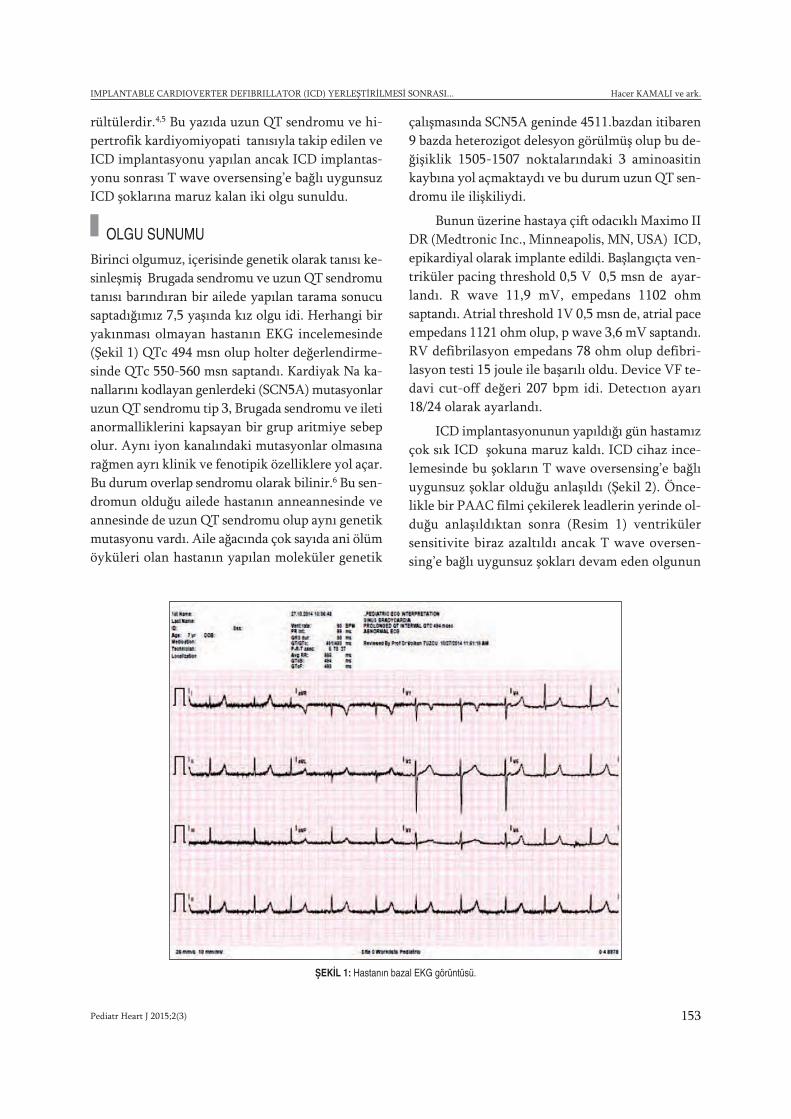
Pediatr Heart J 2015;2(3) 153
IMPLANTABLE CARDIOVERTER DEFIBRILLATOR (ICD) YERLEŞTİRİLMESİ SONRASI... Hacer KAMALI ve ark.
rültülerdir.4,5 Bu yazıda uzun QT sendromu ve hi-pertrofik kardiyomiyopati tanısıyla takip edilen veICD implantasyonu yapılan ancak ICD implantas-yonu sonrası T wave oversensing’e bağlı uygunsuzICD şoklarına maruz kalan iki olgu sunuldu.
OLGU SUNUMU
Birinci olgumuz, içerisinde genetik olarak tanısı ke-sinleşmiş Brugada sendromu ve uzun QT sendromutanısı barındıran bir ailede yapılan tarama sonucusaptadığımız 7,5 yaşında kız olgu idi. Herhangi biryakınması olmayan hastanın EKG incelemesinde(Şekil 1) QTc 494 msn olup holter değerlendirme-sinde QTc 550-560 msn saptandı. Kardiyak Na ka-nallarını kodlayan genlerdeki (SCN5A) mutasyonlaruzun QT sendromu tip 3, Brugada sendromu ve iletianormalliklerini kapsayan bir grup aritmiye sebepolur. Aynı iyon kanalındaki mutasyonlar olmasınarağmen ayrı klinik ve fenotipik özelliklere yol açar.Bu durum overlap sendromu olarak bilinir.6 Bu sen-dromun olduğu ailede hastanın anneannesinde veannesinde de uzun QT sendromu olup aynı genetikmutasyonu vardı. Aile ağacında çok sayıda ani ölümöyküleri olan hastanın yapılan moleküler genetik
çalışmasında SCN5A geninde 4511.bazdan itibaren9 bazda heterozigot delesyon görülmüş olup bu de-ğişiklik 1505-1507 noktalarındaki 3 aminoasitinkaybına yol açmaktaydı ve bu durum uzun QT sen-dromu ile ilişkiliydi.
Bunun üzerine hastaya çift odacıklı Maximo IIDR (Medtronic Inc., Minneapolis, MN, USA) ICD,epikardiyal olarak implante edildi. Başlangıçta ven-triküler pacing threshold 0,5 V 0,5 msn de ayar-landı. R wave 11,9 mV, empedans 1102 ohmsaptandı. Atrial threshold 1V 0,5 msn de, atrial paceempedans 1121 ohm olup, p wave 3,6 mV saptandı.RV defibrilasyon empedans 78 ohm olup defibri-lasyon testi 15 joule ile başarılı oldu. Device VF te-davi cut-off değeri 207 bpm idi. Detectıon ayarı18/24 olarak ayarlandı.
ICD implantasyonunun yapıldığı gün hastamızçok sık ICD şokuna maruz kaldı. ICD cihaz ince-lemesinde bu şokların T wave oversensing’e bağlıuygunsuz şoklar olduğu anlaşıldı (Şekil 2). Önce-likle bir PAAC filmi çekilerek leadlerin yerinde ol-duğu anlaşıldıktan sonra (Resim 1) ventrikülersensitivite biraz azaltıldı ancak T wave oversen-sing’e bağlı uygunsuz şokları devam eden olgunun
ŞEKİL 1: Hastanın bazal EKG görüntüsü.

Pediatr Heart J 2015;2(3)154
Hacer KAMALI ve ark. IMPLANTABLE CARDIOVERTER DEFIBRILLATOR (ICD) YERLEŞTİRİLMESİ SONRASI...
cihazının jeneratörünün değiştirilmesine karar ve-rildi.
İkinci olgumuz hipertrofik kardiyomiyopatitanısı ile izlenen ve dış merkezde 10 yaşında ikenBiotronic marka ICD takılan şu anda 20 yaşındaerkek olgu idi. ICD takıldıktan 6 yıl sonra hastamızşok yemeye başlayınca incelenen ICD cihazı ince-lemesinde şokların yine T wave oversensing’e bağlıuygunsuz şoklar olduğu görüldü. Bu hastanın daICD jeneratörünün değiştirilmesine karar verildi.
Sonrasında her iki hastaya yine dual chamberlifarklı bir jeneratörü olan ve T wave oversensingengelleme özelliği olan Medtronic Protecta DRimplante edildi. Her iki hastada da yeni jeneratörledaha yüksek sensitivite ayarlarında bile T waveoversense olmadı (Şekil 3) ve her iki hasta herhangibir uygunsuz şoka maruz kalmadan taburcu edildi.
ŞEKİL 2: Olgunun T wave oversensing’e bağlı uygunsuz şok görünümü.
RESİM 1: Uygunsuz şoklar sonrası PAAC ile leadlerin yerinin kontroledilmesi.
ŞEKİL 3: Medtronic Protecta cihazı takıldıktan sonra cihazın T wave oversensing’i tanıması.

Pediatr Heart J 2015;2(3) 155
IMPLANTABLE CARDIOVERTER DEFIBRILLATOR (ICD) YERLEŞTİRİLMESİ SONRASI... Hacer KAMALI ve ark.
TARTIŞMA
ICD implantasyonu hayati risk taşıyan aritmisi olanolgularda tedavinin önemli bir parçası olmakla bir-likte bu hastalar uygunsuz şok tehlikesi ile karşıkarşıyadırlar. Bu uygunsuz şoklar psikolojik bo-zukluklara, hayat kalitesinin düşmesine ve daha daönemlisi klinik sonuçların olumsuz olmasına yolaçabilir.7-10 Ayrıca uygunsuz ICD şoku ile karşılaşanolgularda mortalite riskinin artmış olduğunu gös-teren çalışmalar mevcuttur.11-13
Asıl istenen hastanın güvenliğini tehlikeye at-madan hem uygun hem uygunsuz şok tedavisiniazaltmaktır. Bu da uygun medikal tedavi, gelişmişdevice programlaması ve ablasyon stratejileri ile ya-pılabilir. Beta bloker tedavisi hem supraventriküleraritmileri hem ventriküler aritmileri azaltan dola-yısı ile şok sayısını azaltan uygun medikal tedavidir.
Yeni detection algoritmalarına rağmen primerya da sekonder profilaktik ICD takılan olguların %2si ilk yıl uygunsuz ICD şoklarına maruz kalırlar.14
Bu uygunsuz şokları önlemek için uygulana-bilecek teknolojik olarak çeşitli uygulamalar var-dır; anti taşikardi pacing, VT detection zamanınınuzun ve gecikmiş tutulması ve cihazın yeni QRSvektör algoritmalarına göre ayırım yapabilme ye-teneği artırılması gibi (Wavelet algoritması).15,16
Yapılan çeşitli çalışmalarda uygunsuz şokların ensık sebebinin atrial fibrilasyon sonrası supraventrikü-ler taşikardi olduğu görüldü.17 Uygunsuz şokların se-bepleri içinde diğer bir sebep ventriküler oversen-sing’dir. Ventriküler oversensing oluşum mekaniz-malarından biri de T wave oversensing’dir (TWOS).
T wave oversensing T dalgasının R dalgası gibialgılanması sonucu kalp hızının olduğunun 2 katıgibi gözükmesi sonrasında uygunsuz ICD şokunayol açabilir. T wave oversensing ventriküler over-sensingin en sık sebebidir. ICD hastalarda %14 ora-nında görülebilir18 ve bunun önceden öngörülmesiolası değildir.
T wave oversensing integre bipolar lead olanolgularda, kardiyomiyopatili olgularda, uzun QTsendromlu yada kısa QT sendromlu hastalarda,Brugada sendromlu olgularda görülme riski artanbir durumdur.19-22Ayrıca hiperkalemi gibi elektro-
lit imbalansında, iskemi, travma ve lead implantas-yonu sonrası hasarlanmaya bağlı geçici T yüksekli-ğine bağlı meydana gelebilir.23,24
Bizim vakamızda olduğu gibi özellikle uzunQT tip 3 sendromu olan olgularda yayvan ST seg-menti ile beraber dar ve peak yapmış T dalgalarıgörülebilir.
Ventriküler sensitivitenin azaltılması, post-ventriküler sensing reftaktör peryodun uzun ayar-lanması, taşikardi zon detectıon intervalininyükseltilmesi yada taşikardi detection hızının tah-mini sinüs hızının 2 katının üzerine ayarlanmasıgibi noninvaziv yöntemlerle T wave oversensing’inüstesinden gelmeye çalışılır. Ancak bu yöntemlerleçoğu zaman başarılı olunması güçtür. Ayrıca buyöntemlerle cihazın ventriküler taşikardi yada ven-triküler fibrilasyona olan duyarlılığını da azaltmışolacağımızdan bu durumlara uygun zamanda mü-dahale etme olasılığını da azaltmış oluruz.
Bazı ICD programlarının dar QRS i geniş QRSten ayırabilme özelliği kazandıran algoritmalarıvardır.25,26 Bu da ritmin sinüs yada supraventrikülertaşikardiyi tanımlamasına dolayısı ile T wave over-sensing’in üstesinden gelmesine yardımcı olur.
Wavelet algoritması da >250 hızın üzerindeçalışmayacağından çözüm değildir.26
Nadiren bazı vakalarda lead yerinin değiştiril-mesi TWOS için çözüm olabilir.27
Akıllı şoklama özelliği olan ve bizim uygula-mamızda kullandığımız Medtronic Protecta gibi ci-hazlar QRS ile T dalgasını çok hassas bir şekildeayırt edebilecek yazılıma sahiptir. Bunu yaparkenventriküler sensitiviteyi bozmadan yani VF sap-tama duyarlılığını azaltmadan bu problemi önler.Bu özellikler cihaza spesifiktir ve programlanamaz.Cihazları birbirinden ayıran detayların, özel tescilliolduğundan başka bir cihaza uygulanması mümkünolmamaktadır. Bu özellikle filtreleme setleri ve sis-temi için de geçerlidir.28
Biz de daha önce yayımlanmış yazılardakinebenzer şekilde28 T wave oversensing’in ICD cihaz-larına spesifik olduğunu ve ICD cihazı değiştirildiğitakdirde uygunsuz şokların önlenebileceğini bu ya-zıda vurgulamak istedik.

Pediatr Heart J 2015;2(3)156
Hacer KAMALI ve ark. IMPLANTABLE CARDIOVERTER DEFIBRILLATOR (ICD) YERLEŞTİRİLMESİ SONRASI...
1. Ezekowitz JA, Armstrong PW, McAlister FA.Implantable cardioverter defibrillators in pri-mary and secondary prevention: a systematicreview of randomized, controlled trials. Ann In-tern Med 2003;138(6):445-52.
2. Kraaier K, Starrenburg AH, Verheggen RM,van der Palen J, Scholten MF. Incidence and-predictors of phantom shocks in implantablecardioverter defibrillator recipients. Neth HeartJ 2013;21(4):191-5.
3. Wijers SC, van der Kolk BY, Tuinenburg AE,Doevendans PA, Vos MA, Meine M. Imple-mentationof guidelines for implantable car-dioverter-defibrillatortherapy in clinicalpractice: which patients do benefit? NethHeart J 2013;21(6):274-83.
4. Poole JE, Johnson GW, Hellkamp AS, Ander-son J, Callans DJ, Raitt MH, et al. Prognosticimportance of defibrillator shocks in patientswith heart failure. N Engl J Med 2008;359(10):1009-17.
5. Koneru JN, Swerdlow CD, Wood MA, Ellen-bogen KA. Minimizing inappropriate or “un-necessary” implantable cardioverter-defibrillator shocks: appropriate programming.Circ Arrhythm Electrophysiol 2011;4(5):778-90.
6. Remme CA, Wilde AAM, Bezzina CR. Cardiacsodium channel overlap syndromes: differentfaces of SCN5A mutations. Trends Cardio-vasc Med 2008;18(3):78-87.
7. Opic P, Utens EM, Moons P, Theuns DA, vanDijk AP, Hoendermis ES, et al. Psychosocialimpact of implantable cardioverter defibrilla-tors (icd) in young adults with tetralogy of fal-lot. Clin Res Cardiol Clin Res Cardiol 2012;101(7):509-19.
8. Sweeney MO, Wathen MS, Volosin K, AbdallaI, DeGroot PJ, Otterness MF, et al. Appropri-ate and inappropriate ventricular therapies,quality of life, and mortality among primaryand secondary prevention implantable car-dioverter defibrillator patients: results from thepacing fast vt reduces shock therapies(painfree rx ii) trial. Circulation 2005;111(22):2898-905.
9. Daubert JP, Zareba W, Cannom DS, McNittS, Rosero SZ, Wang P, et al. Inappropriate im-plantable cardioverter-defibrillator shocks inmadit ii: frequency, mechanisms, predictors,and survival impact. J Am Coll Cardiol 2008;51(14):1357-65.
10. Van Rees JB, Borleffs CJ, de Bie MK, StijnenT, van Erven L, Bax JJ, et al. Inappropriate im-plantable cardioverter-defibrillator shocks: in-cidence, predictors, and impact on mortality.J Am Coll Cardiol 2011;57(5):556-62.
11. Poole JE, Johnson GW, Hellkamp AS, Ander-son J, Callans DJ, Raitt MH, et al. Prognosticimportance of defibrillator shocks in patientswith heart failure. N Engl J Med 2008;359(10):1009-17.
12. Pacifico A, Ferlic LL, Cedillo-Salazar FR, NasirJr N, Doyle TK, Henry PD. Shocks as predic-tors of survival in patients with implantable car-dioverter-defibrillators. J Am Coll Cardiol 1999;34(1):204-10.
13. Moss AJ, Greenberg H, Case RB, Zareba W,Hall WJ, Brown MW, et al; Multicenter Auto-matic Defibrillator Implantation Trial-II (MADIT-II) Research Group.Long-term clinical courseof patients after termination of ventricular tach-yarrhythmia by an implanted defibrillator. Cir-culation 2004;110(25):3760-5.
14. Schloss E, Auricchio A, Kurita T, Meijer A,Zweibel S, Alsmadi F, et al. PainFree SST trialprimary results: low shock rates in patientswith dual and triple chamber ICDs using noveldetection algorithms. Europace 2013;15:ii116-ii7.
15. Wilkoff BL, Williamson BD, Stern RS, MooreSL, Lu F, Lee SW, et al; PREPARE Study In-vestigators.Strategic programming of detec-tion and therapy parameters in implantablecardioverter-defibrillators reduces shocks inprimary prevention patients: results from theprepare (primary prevention parameters eval-uation) study. J Am Coll Cardiol 2008;52(7):541-50.
16. Shome S, Li D, Thompson J, McCabe A. Im-proving contemporary algorithms for im-plantable cardioverter-defibrillator function. JElectrocardiol 2010;43(6):503-8.
17. Daubert JP, Zareba W, Cannom DS, McNittS, Rosero SZ, Wang P, et al; MADIT II Inves-tigators.Inappropriate implantable car-dioverter-defibrillator shocks in MADIT II:frequency, mechanisms, predictors, and sur-vival impact. J Am Coll Cardiol 2008; 51(14):1357-6.
18. Weretka S1, Michaelsen J, Becker R, KarleCA, Voss F, Hilbel T, et al. Ventricular over-sensing: A study of 101 patients implantedwith dual chamber defibrillators and two dif-
ferent lead systems. Pacing Clin Electrophys-iol 2003;26(1 Pt 1):65-70.
19. Alizadeh A, Haghjoo M, Arya A, Fazelifar AF,Alasti M, Bagherzadeh AA, et al. Inappropri-ate ICD discharge due to T-wave oversensingin a patient with the Brugada syndrome. J In-terv Card Electrophysiol 2006;15(1):65-8.
20. Washizuka T, Chinushi M, Tagawa M, KasaiH, Watanabe H, Hosaka Y, et al. Inappropriatedischarges by fourth generationimplantablecardioverter defibrillators in patients with ven-tricular arrhythmias. Jpn Circ J 2001;65(11):927-30.
21. Stix G, Bella PD, Carbucicchio C, SchmidingerH. Spatial and temporal heterogeneity of de-polarization and repolarization may complicateimplantable cardioverter defibrillator therapyin Brugada syndrome. J Cardiovasc Electro-physiol 2000;11(5):516-21.
22. Schimpf R, Wolpert C, Bianchi F, Giustetto C,Gaita F, Bauersfeld U, et al. Congenital shortQT syndrome and implantable cardioverterdefibrillator treatment: Inherent risk for inap-propriate shock delivery. J Cardiovasc Elec-trophysiol 2003;14(12):1273-7.
23. Barold SS, Falkoff MD, Ong LS, Heinle RA.Oversensing by singlechamber pacemakers:Mechanisms, diagnosis, and treatment. Car-diol Clin 1985;3(4):565-85.
24. Yokoyama M, Wada J, Barold SS. Transientearly T wave sensing by implanted program-mable demand pulse generator. Pacing ClinElectrophysiol 1981;4(1):68-74.
25. Kaup HJ, Hexamer M,Werner J. Morphologi-cal detection algorithms for the automatic im-plantable cardioverter/defibrillator (AICD).Biomed Tech (Berl) 2004;49(11):306-10.
26. Klein GJ, Gillberg JM, Tang A, Inbar S,Sharma A, Unterberg-Buchwald C, et al;Worldwide Wave Investigators. ImprovingSVT discrimination in single-chamber ICDs: Anew electrogram morphology-based algo-rithm. J Cardiovasc Electrophysiol 2006;17(12):1310-9.
27. Hsu SS, Mohib S, Schroeder A, Deger FT. Twave oversensing in implantable cardioverterdefibrillators. J Interv Card Electrophysiol2004; 11(1):67-72.
28. Gilliam FR, 3rd. T-wave oversensing in im-plantable cardiac defibrillators is due to tech-nical failure of device sensing. J CardiovascElectrophysiol 2006;17(5):553-6.
KAYNAKLAR
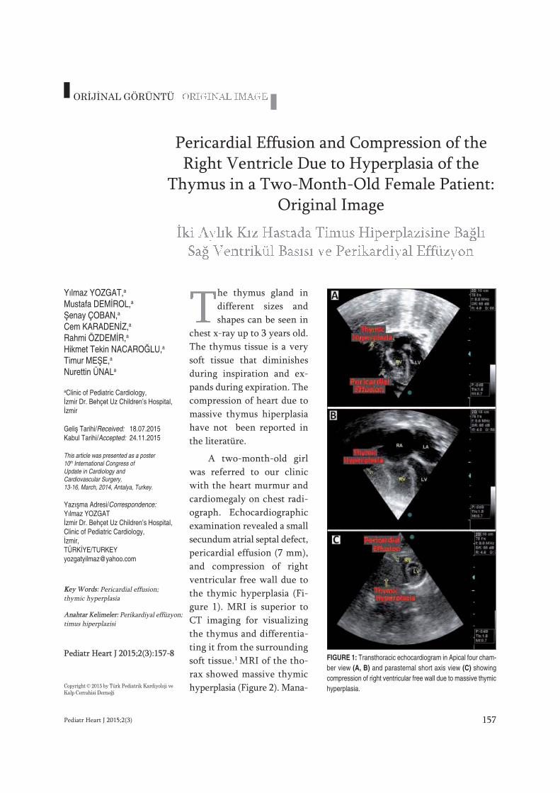
he thymus gland indifferent sizes andshapes can be seen in
chest x-ray up to 3 years old.The thymus tissue is a verysoft tissue that diminishesduring inspiration and ex-pands during expiration. Thecompression of heart due tomassive thymus hiperplasiahave not been reported inthe literatüre.
A two-month-old girlwas referred to our clinicwith the heart murmur andcardiomegaly on chest radi-ograph. Echocardiographicexamination revealed a smallsecundum atrial septal defect,pericardial effusion (7 mm),and compression of rightventricular free wall due tothe thymic hyperplasia (Fi-gure 1). MRI is superior toCT imaging for visualizingthe thymus and differentia-ting it from the surroundingsoft tissue.1 MRI of the tho-rax showed massive thymichyperplasia (Figure 2). Mana-
Pediatr Heart J 2015;2(3) 157
Yılmaz YOZGAT,a
Mustafa DEMİROL,a
Şenay ÇOBAN,a
Cem KARADENİZ,a
Rahmi ÖZDEMİR,a
Hikmet Tekin NACAROĞLU,a
Timur MEŞE,a
Nurettin ÜNALa
aClinic of Pediatric Cardiology,İzmir Dr. Behçet Uz Children’s Hospital,İzmir
Geliş Tarihi/Received: 18.07.2015Kabul Tarihi/Accepted: 24.11.2015
This article was presented as a poster10th International Congress ofUpdate in Cardiology andCardiovascular Surgery,13-16, March, 2014, Antalya, Turkey.
Yazışma Adresi/Correspondence:Yılmaz YOZGATİzmir Dr. Behçet Uz Children’s Hospital,Clinic of Pediatric Cardiology,İzmir,TÜRKİYE/[email protected]
KKey Words: Pericardial effusion;thymic hyperplasia
Anahtar Kelimeler: Perikardiyal effüzyon;timus hiperplazisi
Pediatr Heart J 2015;2(3):157-8
Copyright © 2015 by Türk Pediatrik Kardiyoloji veKalp Cerrahisi Derneği
ORİJİNAL GÖRÜNTÜ
FIGURE 1: Transthoracic echocardiogram in Apical four cham-ber view (A, B) and parasternal short axis view (C) showingcompression of right ventricular free wall due to massive thymichyperplasia.
Pericardial Effusion and Compression of the Right Ventricle Due to Hyperplasia of the
Thymus in a Two-Month-Old Female Patient: Original Image

Pediatr Heart J 2015;2(3)158
gement of thymic hyperplasia is controversial.When the patient is asymptomatic and is less thantwo years of age, thymic hyperplasia generally reg-resses.2
On follow up at 6 months of age, thymichyperplasia was still present and thymus was com-pressing the right ventricular free wall; however,we did not observe right ventricular outflow tractobstruction or pericardial effusion.
Yılmaz YOZGAT et al. PERICARDIAL EFFUSION AND COMPRESSION OF THE RIGHT VENTRICLE DUE TO HYPERPLASIA...
FIGURE 2: MRI of thorax showing pericardial effusion and compression of theright ventricular free wall due to the thymic hiperplasia.
1. Siegel MJ,Glazer HS, Wiener JI. et al. Normal and abnormal thymus in childhood: MR imaging. Radiology 1989: 172: 367-371.
2. Altar Z, Muraji T, Matsumoto Y, et al . Malignant mesenchymoma of the mediastinum initially presenting as bening thymic hyperplasia.Pediatr Surgical Int.1988:4:56-8.
REFERENCES
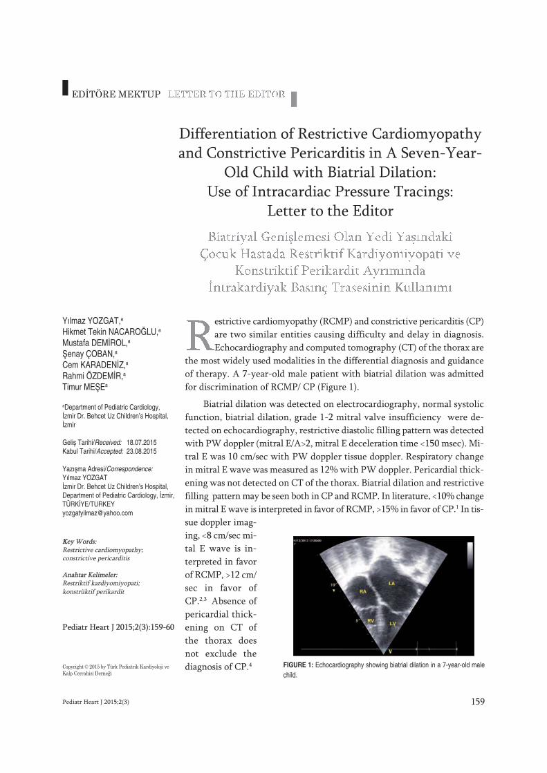
estrictive cardiomyopathy (RCMP) and constrictive pericarditis (CP)are two similar entities causing difficulty and delay in diagnosis.Echocardiography and computed tomography (CT) of the thorax are
the most widely used modalities in the differential diagnosis and guidanceof therapy. A 7-year-old male patient with biatrial dilation was admittedfor discrimination of RCMP/ CP (Figure 1).
Biatrial dilation was detected on electrocardiography, normal systolicfunction, biatrial dilation, grade 1-2 mitral valve insufficiency were de-tected on echocardiography, restrictive diastolic filling pattern was detectedwith PW doppler (mitral E/A>2, mitral E deceleration time <150 msec). Mi-tral E was 10 cm/sec with PW doppler tissue doppler. Respiratory changein mitral E wave was measured as 12% with PW doppler. Pericardial thick-ening was not detected on CT of the thorax. Biatrial dilation and restrictivefilling pattern may be seen both in CP and RCMP. In literature, <10% changein mitral E wave is interpreted in favor of RCMP, >15% in favor of CP.1 In tis-sue doppler imag-ing, <8 cm/sec mi-tal E wave is in-terpreted in favorof RCMP, >12 cm/sec in favor ofCP.2,3 Absence ofpericardial thick-ening on CT ofthe thorax doesnot exclude thediagnosis of CP.4
Pediatr Heart J 2015;2(3) 159
Differentiation of Restrictive Cardiomyopathyand Constrictive Pericarditis in A Seven-Year-
Old Child with Biatrial Dilation:Use of Intracardiac Pressure Tracings:
Letter to the Editor
Yılmaz YOZGAT,a
Hikmet Tekin NACAROĞLU,a
Mustafa DEMİROL,a
Şenay ÇOBAN,a
Cem KARADENİZ,a
Rahmi ÖZDEMİR,a
Timur MEŞEa
aDepartment of Pediatric Cardiology,İzmir Dr. Behcet Uz Children’s Hospital,İzmir
Geliş Tarihi/Received: 18.07.2015Kabul Tarihi/Accepted: 23.08.2015
Yazışma Adresi/Correspondence:Yılmaz YOZGATİzmir Dr. Behcet Uz Children’s Hospital,Department of Pediatric Cardiology, İzmir,TÜRKİYE/[email protected]
KKey Words: Restrictive cardiomyopathy; constrictive pericarditis
Anahtar Kelimeler: Restriktif kardiyomiyopati; konstrüktif perikardit
Pediatr Heart J 2015;2(3):159-60
Copyright © 2015 by Türk Pediatrik Kardiyoloji veKalp Cerrahisi Derneği
EDİTÖRE MEKTUP
FIGURE 1: Echocardiography showing biatrial dilation in a 7-year-old malechild.

Pediatr Heart J 2015;2(3)160
Echocardiography and CT of the thorax find-ings were not beneficial and cardiac catheteriza-tion was planned. A parallel pressure recording setwas arranged and both left and right heart cham-bers were recorded simultaneously. Simultaneoussystolic pressure tracings from left and right ven-tricles showed a parallel pattern in the form of a re-duction during inspirium and increase duringexpirium (respiratory concordance) (Figure 2).End-diastolic pressures of left heart chambers werefound to be 5 mmHg greater than that of rightheart chambers. With these findings, the patientwas diagnosed with RCMP and managed accord-ingly. He was lost on month 5 of the follow-up dueto sudden cardiac arrest at home.
Death is the most feared outcome in childrenwith RCMP and CP. Children diagnosed withRCMP are directed for cardiac transplantation asthe only solution, whereas appropriately timed sur-gery provides a chance for cure for patients withCP. Therefore, when confronted with a differential
diagnosis between RCMP and CP, every effortshould be made for early establishment of the de-finitive diagnosis. Besides echocardiography andother imaging methods, cardiac catheterization andpressure recording techniques can provide signifi-cant benefits.
Yılmaz YOZGAT et al. DIFFERENTIATION OF RESTRICTIVE CARDIOMYOPATHY AND CONSTRICTIVE PERICARDITIS...
1. Rajagopalan, N, Garcia, MJ, Rodriguez, L, etal. Comparison of new Doppler echocardoi-graphic methods to differentiate constrictivepericardial heart disease and restrictive car-diomyopathy. Am J Cardiol 2001; 87:86.
2. Ha, JW, Oh, JK, Ling, LH, et al. Annulus para-doxus: transmitral flow velocity to mitral annu-
lar velocity ratio is inversely proportional topulmonary capillary wedge pressure in pa-tients with constrictive pericarditis. Circulation2001; 104:976.
3. Talreja, DR, Edwards, WD, Danielson, GK, etal. Constrictive pericarditis in 26 patients withhistologically normal pericardial thickness. Cir-
culation 2003; 108:1852.
4. Choi JH, Choi JO, Ryu DR, et al. Mitral andtricuspid annular velocities in constrictive peri-carditis and restrictive cardiomyopathy: corre-lation with pericardial thickness on computedtomography. JACC Cardiovasc Imaging 2011;4:567.
REFERENCES
FIGURE 2: Cardiac catheterization showing a parallel pressure pattern as areduction in inspirium and an increase in expirium on systolic pressure trac-ings obtained simultaneously from both ventricles during cardiac catheteri-zation(respiratory concordance).
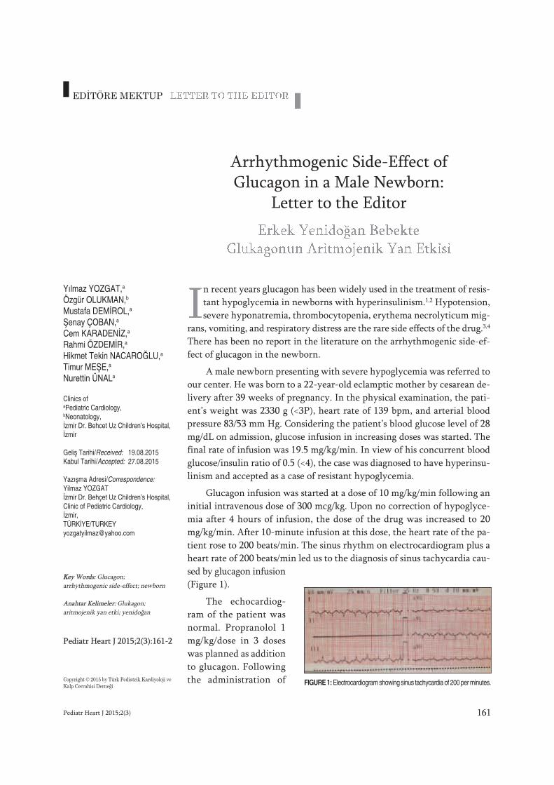
n recent years glucagon has been widely used in the treatment of resis-tant hypoglycemia in newborns with hyperinsulinism.1,2 Hypotension,severe hyponatremia, thrombocytopenia, erythema necrolyticum mig-
rans, vomiting, and respiratory distress are the rare side effects of the drug.3,4
There has been no report in the literature on the arrhythmogenic side-ef-fect of glucagon in the newborn.
A male newborn presenting with severe hypoglycemia was referred toour center. He was born to a 22-year-old eclamptic mother by cesarean de-livery after 39 weeks of pregnancy. In the physical examination, the pati-ent’s weight was 2330 g (<3P), heart rate of 139 bpm, and arterial bloodpressure 83/53 mm Hg. Considering the patient’s blood glucose level of 28mg/dL on admission, glucose infusion in increasing doses was started. Thefinal rate of infusion was 19.5 mg/kg/min. In view of his concurrent bloodglucose/insulin ratio of 0.5 (<4), the case was diagnosed to have hyperinsu-linism and accepted as a case of resistant hypoglycemia.
Glucagon infusion was started at a dose of 10 mg/kg/min following aninitial intravenous dose of 300 mcg/kg. Upon no correction of hypoglyce-mia after 4 hours of infusion, the dose of the drug was increased to 20mg/kg/min. After 10-minute infusion at this dose, the heart rate of the pa-tient rose to 200 beats/min. The sinus rhythm on electrocardiogram plus aheart rate of 200 beats/min led us to the diagnosis of sinus tachycardia cau-sed by glucagon infusion(Figure 1).
The echocardiog-ram of the patient wasnormal. Propranolol 1mg/kg/dose in 3 doseswas planned as additionto glucagon. Followingthe administration of
Pediatr Heart J 2015;2(3) 161
Arrhythmogenic Side-Effect ofGlucagon in a Male Newborn:
Letter to the Editor
Yılmaz YOZGAT,a
Özgür OLUKMAN,b
Mustafa DEMİROL,a
Şenay ÇOBAN,a
Cem KARADENİZ,a
Rahmi ÖZDEMİR,a
Hikmet Tekin NACAROĞLU,a
Timur MEŞE,a
Nurettin ÜNALa
Clinics ofaPediatric Cardiology,bNeonatology,İzmir Dr. Behcet Uz Children’s Hospital,İzmir
Geliş Tarihi/Received: 19.08.2015Kabul Tarihi/Accepted: 27.08.2015
Yazışma Adresi/Correspondence:Yilmaz YOZGATİzmir Dr. Behçet Uz Children’s Hospital,Clinic of Pediatric Cardiology,İzmir,TÜRKİYE/[email protected]
KKey Words: Glucagon; arrhythmogenic side-effect; newborn
Anahtar Kelimeler: Glukagon; aritmojenik yan etki; yenidoğan
Pediatr Heart J 2015;2(3):161-2
Copyright © 2015 by Türk Pediatrik Kardiyoloji veKalp Cerrahisi Derneği
EDİTÖRE MEKTUP
FIGURE 1: Electrocardiogram showing sinus tachycardia of 200 per minutes.

Pediatr Heart J 2015;2(3)162
Yılmaz YOZGAT et al. ARRHYTHMOGENIC SIDE-EFFECT OF GLUCAGON IN A MALE NEWBORN: LETTER TO THE EDITOR
the first dose of propranonol, the heart rate felldown to 120 beats/min at hour 2, to 110 beats/minat hour 4, and to 80 beats/min at hour 6. Upon de-velopment of sinus bradycardia in the patient, thesecond dose of propranolol was not given, and thedrug was cut off. The sinus bradycardia lasted 4hours (70-80 beats/min). Twelve hours after thefirst dose of propranolol the heart rate returnedto normal with 130 beats/min. The glucagon in-fusion was given one more day as the heart rate
ranged between 120 and 160 beats per minute.When the blood glucose started to continue above40 mg/dL, glucagon was gradually decreased anddiscontinued. The patient was discharged withcure after one week.
There are reports in the literature on tach-yarrhythmia in adults caused by glucagon.5 To thebest of our knowledge, our patient is the first new-born case showing sinus tachycardia secondary toglucagon treatment.
1. Sweet CB, Grayson S, Polak M. ManagementStrategies for Neonatal Hypoglycemia.J Pedi-atr Pharmacol Ther. 2013 Jul;18(3):199-208.
2. Miralles RE, Lodha A, Perlman M, Moore AM.Experience with intravenous glucagon infu-sions as a treatment for resistant neonatal hy-poglycemia. Arch Pediatr Adolesc Med.2002;156(10):999-1004.
3. Belik J, Musey J, Trussell RA. Continuousinfusions of glucagon induces severe hy-ponatremia and thrombocytopenia in pre-mature infants. Pediatrics. 2001;107(3):595-597.
4. Wald M, Lawrenz K, Luckner D, etal.Glucagon therapy as a possible cause oferythema necrolyticum migrans in two
neonates with persistent hyperinsulinaemichypoglycemia.Eur J Pediatr. 2002;161(11):600-603.
5. IJ Jaca. Glucagon. First report of paroxysmalsupraventricular tachycardia in an elderly pa-tient: case report. A. 76-year-old woman. Re-actions Weekly, 2002, Volume: 921, Issue 1,8-8
REFERENCES
1
AAEPC Teaching Course: Fetal Cardiology
18-20 Şubat 2016, Varşova - Polonya
12. Uluslararası Kardiyoloji ve Kardiyovasküler Cerrahide Yenilikler Kongresi
10-13 Mart 2016, Sueno Belek Kongre Merkezi, Antalya - Türkiye
15. Ulusal Pediatrik Kardiyoloji ve Kalp Cerrahisi Kongresi
13-16 Nisan 2016, Titanic Lara Otel - Belek/Antalya - Türkiye
AEPC 2016 - 50th Annual Meeting of the Association for European Paediatric and Congenital Cardiology
1-4 Haziran 2015, Roma - İtalya
BİLİMSEL TOPLANTILAR

MAKALE GÖNDERMEK İÇİN
Pediatric Heart Journal’e makale göndermek için; www.turkpedkar.org.tr adresindeki“Online Makale” linkini tıklayınız (Yalnızca bu yolla gönderilen makaleler işleme alın-maktadır). Makalelerinizle ilgili tüm işlemleri de bu adresten takip edebilirsiniz.
GENEL BİLGİLER
Pediatric Heart Journal, retrospektif, prospektif veya deneysel araştırmalar, derlemeler,olgu sunumları, editöryal yorum/tartışmalar, editöre mektuplar, tıbbi eğitimler, bilimsel mek-tuplar, cerrahi teknikler, ayırıcı tanılar, orijinal görüntüler, tanınız nedir? ler, tıbbi kitap de-ğerlendirmeleri, soru-cevaplar ve tıp gündemini belirleyen güncel konuları yayımlayan,ulusal ve uluslararası tüm tıbbi kurum ve personele ulaşmayı hedefleyen bilimsel bir der-gidir.
Dergiler; yayımladıkları makalelerde, konu ile ilgili en yüksek etik ve bilimsel standartlardaolması ve ticari kaygılarda olmaması şartını gözetmektedir.
Editörler ve yayınevi, reklam amacı ile verilen ticari ürünlerin özellikleri ve açıklamaları ko-nusunda hiçbir garanti vermemekte ve sorumluluk kabul etmemektedir.
Yayımlanmak için gönderilen makalelerin daha önce başka bir yerde yayımlanmamış veyayayımlanmak üzere gönderilmemiş olması gerekir. Eğer makalede daha önce yayımlan-mış; alıntı yazı, tablo, resim vs. mevcut ise makale yazarı, yayın hakkı sahibi ve yazarla-rından yazılı izin almak ve bunu makalede belirtmek zorundadır. Bilimsel toplantılardasunulan özetler, makalede belirtilmesi koşulu ile kabul edilir.
Dergiye gönderilen makale biçimsel esaslara uygun ise, editör ve en az yurt içi-yurt dışıiki danışmanın incelemesinden geçip, gerek görüldüğü takdirde, istenen değişiklikler ya-zarlarca yapıldıktan sonra yayımlanır.
Makale işleme alındıktan sonra, yayın hakları devir formunda belirtilmiş olan yazar isim-leri ve sıralaması esas alınır. Bu aşamadan sonra;
� Makaleye hiçbir aşamada, yayın hakları devir formunda imzası bulunan yazarlar dı-şında yazar ismi eklenemez ve yazar sırası değiştirilemez.
YAYIN KURALLARI
BİLİMSEL SORUMLULUK
Tüm yazarların gönderilen makalede akademik-bilimsel olarak doğrudan katkısı olmalıdır.Yazar olarak belirlenen isim aşağıdaki özelliklerin tamamına sahip olmalıdır:
- Makaledeki çalışmayı planlamalı veya yapmalı,
- Makaleyi yazmalı veya revize etmeli,
- Son halini kabul etmelidir.
� Makalelerin bilimsel kurallara uygunluğu yazarların sorumluluğundadır.
ETİK SORUMLULUK
� Dergi, “İnsan” öğesinin içinde bulunduğu tüm çalışmalarda Helsinki Deklerasyonu Pren-sipleri’ne uygunluk (http://www.wma.net/en/30publications/10policies/b3/ index.html) il-kesini kabul eder. Bu tip çalışmaların varlığında yazarlar, makalenin GEREÇ VEYÖNTEMLER bölümünde bu prensiplere uygun olarak çalışmayı yaptıklarını, kurumla-rının etik kurullarından ve çalışmaya katılmış insanlardan “Bilgilendirilmiş olur” (informedconsent) aldıklarını belirtmek zorundadır.
� Çalışmada “Hayvan” öğesi kullanılmış ise yazarlar, makalenin GEREÇ VE YÖNTEM-LER bölümünde Guide for the Care and Use of Laboratory Animals (www.nap.edu/catalog/5140.html) prensipleri doğrultusunda çalışmalarında hayvan haklarını ko-ruduklarını ve kurumlarının etik kurullarından onay aldıklarını belirtmek zorundadır.
� Olgu sunumlarında hastanın kimliğinin ortaya çıkmasına bakılmaksızın hastalardan“Bilgilendirilmiş olur” (informed consent) alınmalıdır.
� Eğer makalede direkt-indirekt ticari bağlantı veya çalışma için maddi destek verenkurum mevcut ise yazarlar; kullanılan ticari ürün, ilaç, firma... ile ticari hiçbir ilişkisininolmadığını ve varsa nasıl bir ilişkisinin olduğunu (konsültan, diğer anlaşmalar), edi-töre sunum sayfasında bildirmek zorundadır.
� Makalede “Etik Kurul Onayı” alınması gerekli ise; alınan belge online olarak,www.turkiyeklinikleri.com adresinde yer alan “Makale Gönderim” linkindeki bölüm-den, makale ile birlikte gönderilmelidir.
� Makalelerin etik kurallara uygunluğu yazarların sorumluluğundadır.� Makalenin değerlendirilmesi aşamasında, yayın kurulunun gerek görmesi halinde,
makale ile ilgili araştırma verilerinin ve/veya etik kurul onayı belgesinin sunulması ya-zarlardan talep edilebilir.
BİYOİSTATİSTİKSEL DEĞERLENDİRMETüm retrospektif, prospektif ve deneysel araştırma makaleleri biyoistatistiksel olarak de-ğerlendirilmeli ve uygun plan, analiz ve raporlama ile belirtilmelidir.Makalelerde p değerleri açık olarak verilmelidir (p= 0.025; p= 0.524 gibi).Araştırma makaleleri dergiye gönderilmeden önce, biyoistatistik uzmanı tarafından değer-lendirilmeli ve uzmanın ismi yazarlar arasında yer almalıdır.Biyomedikal dergilere gönderilen yazıların biyoistatistiki uygunluğunun kontrolü için ek bilgiwww.icmje.org adresinden temin edilebilir.� Makalelerin biyoistatistiksel kurallara uygunluğu yazarların sorumluluğundadır.
YAZIM DİLİ YÖNÜNDEN DEĞERLENDİRME
Derginin yayın dili Türkçe ve İngilizce’dir.Türkçe makalelerde Türk Dil Kurumu’nun Türkçe sözlüğü veya www.tdk.gov.tr adresi esasalınmalıdır.İngilizce makaleler ve İngilizce özetler, dergiye gönderilmeden önce dil uzmanı tarafındandeğerlendirilmeli ve uzman onayı editöre sunum sayfasında özellikle belirtilmelidir. Maka-leyi, İngilizce yönünden değerlendiren, yazarlardan biri değil ise bu kişinin ismi makaleninsonunda bulunan TEŞEKKÜR (Acknowledgement) bölümünde belirtilmelidir.Ayrıca gönderilmiş olan makalelerdeki yazım ve dilbilgisi hataları, makalenin içeriğine do-kunmadan, redaksiyon komitemiz tarafından düzeltilmektedir.� Makalelerin yazım ve dil bilgisi kurallarına uygunluğu yazarların sorumluluğundadır.
YAYIN HAKKI
1976 Copyright Act’e göre, yayımlanmak üzere kabul edilen yazıların her türlü yayın hakkıdergiyi yayımlayan kuruma aittir. Yazılardaki düşünce ve öneriler tümüyle yazarların so-rumluluğundadır. Makale yazarlarına, yazıları karşılığında herhangi bir ücret ödenmez.
Yazarlar, http://turkishclinics.com/Log/phj internet adresinden ulaşacakları “Yayın Hakları DevirFormu”nu doldurup, online olarak, www.turkpedkar.org.tr adresinde yer alan “Online Ma-kale” linkindeki bölümden, makale ile birlikte göndermelidirler.
YAZI ÇEŞİTLERİ
Dergiye yayımlanmak üzere gönderilecek yazı çeşitleri şu şekildedir;
Orijinal Araştırma: Kliniklerde yapılan prospektif-retrospektif ve her türlü deneysel çalış-malar yayımlanabilmektedir.
Yapısı:
- Özet (Ortalama 200-250 kelime; amaç, gereç ve yöntemler, bulgular ve sonuç bölüm-lerinden oluşan, Türkçe ve İngilizce)
- Giriş
- Gereç ve Yöntemler
- Bulgular
- Tartışma
- Sonuç
- Teşekkür
- Kaynaklar
Derleme: Doğrudan veya davet edilen yazarlar tarafından hazırlanır. Tıbbi özellik göste-ren her türlü konu için son tıp literatürünü de içine alacak şekilde hazırlanabilir. Yazarın okonu ile ilgili basılmış yayınlarının olması özellikle tercih nedenidir.
Yapısı:
- Özet (Ortalama 200-250 kelime, bölümsüz, Türkçe ve İngilizce)
- Konu ile ilgili başlıklar
- Kaynaklar
YAZARLARA
1 Scientific Style and Format: The CBE Manuel for Authors, Editors, and Publishers, 6th ed. NewYork: Cambridge University Press, 1994.
Olgu Sunumu: Nadir görülen, tanı ve tedavide farklılık gösteren makalelerdir. Yeterli sa-yıda fotoğraflarla ve şemalarla desteklenmiş olmalıdır.
Yapısı:
- Özet (ortalama 100-150 kelime; bölümsüz; Türkçe ve İngilizce)
- Giriş
- Olgu Sunumu
- Tartışma
- Kaynaklar
Editöryel Yorum/Tartışma: Yayımlanan orijinal araştırma makalelerinın, araştırmanın ya-
zarları dışındaki, o konunun uzmanı tarafından değerlendirilmesidir. İlgili makalenin so-
nunda yayımlanır.
Editöre Mektup: Son bir yıl içinde dergide yayımlanan makaleler ile ilgili okuyucuların de-
ğişik görüş, tecrübe ve sorularını içeren en fazla 500 kelimelik yazılardır.
Yapısı:
- Başlık ve özet bölümleri yoktur.
- Kaynak sayısı 5 ile sınırlıdır.
- Hangi makaleye (sayı, tarih verilerek) ithaf olunduğu belirtilmeli ve sonunda yazarın
ismi, kurumu, adresi bulunmalıdır. Mektuba cevap, editör veya makalenin yazar(lar)ı
tarafından, yine dergide yayımlanarak verilir.
Bilimsel Mektup: Genel tıbbi konularda okuyucuyu bilgilendiren, basılmış bilimsel maka-
lelere de atıfta bulunarak o konuyu tartışan makalelerdir.
Yapısı:
- Özet (ortalama 100-150 kelime; bölümsüz, Türkçe ve İngilizce)
- Konu ile ilgili başlıklar
- Kaynaklar
Cerrahi Teknik: Ameliyat tekniklerinin ayrıntılı işlendiği makalelerdir.
Yapısı:
- Özet (ortalama 100-150 kelime; bölümsüz, Türkçe ve İngilizce)
- Cerrahi teknik
- Kaynaklar
Ayırıcı Tanı: Güncel değeri olan olgu sunumlarıdır. Benzer hastalıklarla ilgili yorumu içer-
mektedir.
Yapısı:
- Özet (ortalama 100-150 kelime; bölümsüz, Türkçe ve İngilizce)
- Konu ile ilgili başlıklar
- Kaynaklar (3-5 arası)
Orijinal Görüntüler: Literatürde nadir gözlenen açıklamalı tıbbi resim ve fotoğraflardır.
Yapısı:
- 300 kelimelik metin, orjinal resimler, kaynaklar
Tanınız Nedir?: Nadir görülen, tanı ve tedavide farklılık gösteren hastalıklar hakkında,
soru-cevap şeklinde hazırlanmış makalelerdir.
Yapısı:
- Konu ile ilgili başlıklar
- Kaynaklar (3-5 arası)
Tıbbi Kitap Değerlendirmeleri: Güncel değeri olan ulusal veya uluslararası kabul görmüş
kitapların değerlendirmeleridir.
Soru Cevaplar: Tıbbi konularda bilimsel eğitici-öğreticiliği olan soru ve cevaplar.
YAZIM KURALLARI
Dergiye yayımlanması için gönderilen makalelerde aşağıdaki biçimsel esaslara uyulmalıdır:
- Makale, PC uyumlu bilgisayarlarda Microsoft Word programı ile yazılmalıdır.
KISALTMALAR: Kelimenin ilk geçtiği yerde parantez içinde verilir ve tüm metin boyunca
o kısaltma kullanılır. Uluslararası kullanılan kısaltmalar için “Bilimsel Yazım Kuralları”1 kay-
nağına başvurulabilir.
ŞEKİL, RESİM, TABLO VE GRAFİKLER:
-Şekil, resim, tablo ve grafiklerin metin içinde geçtiği yerler ilgili cümlenin sonunda belirtilme-
lidir. Şekil, resim, tablo ve grafiklerin açıklamaları makale sonuna eklenmelidir.
-Şekil, resim/fotoğraflar ayrı birer .jpg veya .gif dosyası olarak (pixel boyutu yaklaşık
500x400, 8 cm eninde ve 300 çözünürlükte taranarak), sisteme eklenmelidir.
- Kullanılan kısaltmalar şekil, resim, tablo ve grafiklerin altındaki açıklamada belirtilmelidir.
- Daha önce basılmış şekil, resim, tablo ve grafik kullanılmış ise yazılı izin alınmalıdır ve
bu izin açıklama olarak şekil, resim, tablo ve grafik açıklamasında belirtilmelidir.
- Resimler/fotoğraflar renkli, ayrıntıları görülecek derecede kontrast ve net olmalıdır.
EDİTÖRE SUNUM SAYFASI: Gönderilen makalenin kategorisi, daha önce başka bir der-
giye gönderilmemiş olduğu, varsa çalışmayı maddi olarak destekleyen kişi ve kuruluşlar
ve varsa bu kuruluşların yazarlarla olan ilişkileri belirtilmelidir.
KAPAK SAYFASI: Makalenin başlığı (Türkçe ve İngilizce), tüm yazarların ad-soyadları,
akademik ünvanları, kurumları, iş telefonu-GSM, e-posta ve yazışma adresleri belirtilme-
lidir. Makale daha önce tebliğ olarak sunulmuş ise tebliğ yeri ve tarihi belirtilmelidir.
ÖZETLER: YAZI ÇEŞİTLERİ bölümünde belirtilen şekilde hazırlanarak, makale metni içe-
risine yerleştirilmelidir.
ANAHTAR KELİMELER:
- En az 2 adet, Türkçe ve İngilizce yazılmalıdır.
- Kelimeler birbirlerinden noktalı virgül (;) ile ayrılmalıdır.
- İngilizce anahtar kelimeler “Medical Subject Headings (MESH)”e uygun olarak verilme-
lidir (Bkz: www.nlm.nih.gov/mesh/MBrowser.html).
- Türkçe anahtar kelimeler Türkiye Bilim Terimleri (TBT)’ne uygun olarak verilmelidir (Bkz:
www.bilimterimleri.com).
TEŞEKKÜR: Eğer çıkar çatışması, finansal destek, bağış ve diğer bütün editöryal (ista-
tistiksel analiz, İngilizce/Türkçe değerlendirme) ve/veya teknik yardım varsa, metnin so-
nunda sunulmalıdır.
KAYNAKLAR: İngilizce yazılmalıdır. Orijinal basımı Türkçe olan kaynakların başlığı [ ]
içinde ve İngilizce olarak yazılmalıdır. Kaynaklar makalede geliş sırasına göre yazılmalı ve
metinde cümle sonunda noktalama işaretlerinden hemen sonra “Üst Simge” olarak belir-
tilmelidir. Makalede bulunan yazar sayısı 6 veya daha az ise tüm yazarlar belirtilmeli, 7 veya
daha fazla ise ilk 6 isim yazılıp “et al.” eklenmelidir. Kaynak yazımı için kullanılan format
Index Medicus’ta belirtilen şekilde olmalıdır (Bkz: www.icmje.org). Kongre bildirileri, ki-şisel deneyimler, basılmamış yayınlar, tezler ve internet adresleri kaynak olarak gös-terilemez.
Kaynakların yazımı için örnekler (Noktalama işaretlerine lütfen dikkat ediniz):
Makale için; Yazar(lar)ın soyad(lar)ı ve isim(ler)inin başharf(ler)i, makale ismi, dergi ismi,
yıl, cilt, sayı, sayfa no’su belirtilmelidir. Örnek:
Arıcı C, Oğuz V. [Surgical Treatment Options According to Inferior Oblique Hyper-
function in Superior Oblique Palsy]. Turkiye Klinikleri J Med Sci 2011;31(5):1160-6.
Kitap için; Yazar(lar)ın soyad(lar)ı ve isim(ler)inin başharf(ler)i, bölüm başlığı, editö-
rün(lerin) ismi, kitap ismi, kaçıncı baskı olduğu, şehir, yayınevi, yıl ve sayfalar belirtilmeli-
dir. Örnek:
Yabancı dilde yayımlanan kitaplar için;
Underwood LE, Van Wyk JJ. Normal and aberrant growth. In: Wilson JD, Foster DW,
eds. Wiliams’ Textbook of Endocrinology. 1st ed. Philadelphia: WB Saunders; 1992.p.1079-138.
Türkçe kitaplar için;
Tür A. [Emergency airway management and endotracheal intubation]. Şahinoğlu AH,
editör. Yoğun Bakım Sorunları ve Tedavileri. 2. Baskı. Ankara: Türkiye Klinikleri; 2003.p.9-16.
Yazar ve editörün aynı olduğu kitaplar için; Yazar(lar)ın/editörün soyad(lar)ı ve
isim(ler)inin başharf(ler)i, bölüm başlığı, kitap ismi, kaçıncı baskı olduğu, şehir, yayınevi,
yıl ve sayfalar belirtilmelidir. Örnek:
Yabancı dilde yayımlanan kitaplar için;
Solcia E, Capella C, Kloppel G. Tumors of the exocrine pancreas. Tumors of the Pan-
creas. 2nd ed. Washington: Armed Forces Institute of Pathology; 1997. p.145-210.
Türkçe kitaplar için;
Eken A. [Cosmeceutical ingredients: drugs to cosmetics products]. Kozmesötik Etken
Maddeler. 1. Baskı. Ankara: Türkiye Klinikleri; 2006. p.1-7.
Sadece on-line yayınlar için;
DOI tek kabul edilebilir on-line referanstır.
İletişim:Adres : Türk Pediatrik Kardiyoloji ve Kalp Cerrahisi Derneği
Hoşdere Caddesi No: 180/4 Çankaya, Ankara/TürkiyeTel : (0312) 212 02 00Faks : (0312) 212 02 00e-posta : [email protected] : www.turkpedkar.org.tr

SUBMITTING AN ARTICLE
In order to submit an article for the Pediatric Heart Journal, you click “Online Article”link in www.turkpedkar.org.tr address (Only internet submitting will be considered).You also may follow up all the procedures related with your articles from this website.
GENERAL INFORMATION
Pediatric Heart Journal is a scientific journal that aims to reach all national/international me-dical institutions&personnel and to publish retrospective, prospective or experimental re-searchs, reviews, case reports, editorial comment/discussions, letters to the editor, medicaleducation, scientific letters, surgical techniques, distinctive diagnosis, original images,"what is your diagnosis?", medical book reviews, questions-answers and recent issuesthat determine medical agenda.
The Journals commits to rigorous peer review, and stipulates freedom from commercial in-fluence, and promotion of the highest ethical and scientific standards in published articles.
Neither the Editor(s) nor the publisher guarantees, warrants or endorses any product orservice advertised in this publication.
Articles are accepted for publication on the condition that they are original, are not underconsideration by another journal, or have not been previously published. Direct quotations,tables, or illustrations that have appeared in copyrighted material must be accompaniedby written permission for their use from the copyright owner and authors.
All articles are subject to review by the editors and referees. Acceptance is based on sig-nificance, and originality of the material submitted. If the article is accepted for publication,it may be subject to editorial revisions to aid clarity and understanding without changingthe data presented.
The writers' names and placement that are indicated in the copyright transfer form will beconsidered after the process of the manuscript is started. After this stage;
� A writer name cannot be added to the manuscript in any time except the writers whosigned the copyright transfer form, and the order of the writers' names cannot be changed.
EDITORIAL POLICIES
SCIENTIFIC RESPONSIBILITY
All authors should have contributed to the article directly either academically or scientifi-cally. All persons designated as authors should meet all of the following criterias:
- Planned or performed the study,
- Wrote the paper or reviewed the versions,
- Approved the final version.
� It is the authors’ responsibility to prepare a manuscript that meets scientific criterias.
ETHICAL RESPONSIBILITY
� The Journal adheres to the principles set forth in the Helsinki Declaration(http://www.wma.net/en/30publications/10policies/b3/ index.html) and holds that all re-ported research involving “Human beings” conducted in accordance with such prin-ciples. Reports describing data obtained from research conducted in humanparticipants must contain a statement in the MATERIAL AND METHODS section in-dicating approval by the institutional ethical review board and affirmation that IN-FORMED CONSENT was obtained from each participant.
� All papers reporting experiments using animals must include a statement in the MA-TERIAL AND METHODS section giving assurance that all animals have received hu-mane care in compliance with the Guide for the Care and Use of Laboratory Animals(www.nap.edu/catalog/5140.html) and indicating approval by the institutional ethicalreview board.
� Case reports should be accompanied by INFORMED CONSENT whether the iden-tity of the patient is disclosed or not.
� If the proposed publication concerns any commercial product, the author must in-clude in the cover letter a statement indicating that the author(s) has (have) no fi-nancial or other interest in the product or explaining the nature of any relation(including consultancies) between the author(s) and the manufacturer or distributor ofthe product.
� If “Ethics Commitee Approval” must be obtained for an article, document obtainedfrom ethics commitee should be sent to “Online Article Processing” link onwww.turkiyeklinikleri.com
� It is the authors’ responsibility to prepare a manuscript that meets ethical criteria.� During the evaluation of the manuscript, the research data and/or ethics committee ap-
proval form can be requested from the authors if it’s required by the editorial board.
STATISTICAL EVALUATION
All retrospective, prospective and experimental research articles must be evaluated in termsof biostatics and it must be stated together with appropriate plan, analysis and report.
p values must be given clearly in the manuscripts (e.g. p= 0.025; p= 0.524).
Research articles must be evaluated by a biostatistician prior to submission and the nameof the biostatistician should be placed among the authors’ names.
Additional information in order to control the biostatistical convenience of the papers thatare submitted to biomedical journals can be obtained from the web page www.icmje.org.
� It is the authors’ responsibility to prepare a manuscript that meets biostatistical rules.
LANGUAGE
The official languages of the Journals are Turkish and English.
Manuscripts and abstracts in English must be checked for language by an expert and thisshould be mentioned particularly in the cover letter. The expert responsible for English Lan-guage editing should be listed in the ACKNOWLEDGEMENTS if he/she is not in the authorlist.
All spelling and grammar mistakes in the submitted articles, are corrected by our redac-tion commitee without changing the data presented.
� It is the authors’ responsibility to prepare a manuscript that meets spelling and grammarrules.
COPYRIGHT STATEMENT
In accordance with the Copyright Act of 1976, the publisher owns the copyright of all pub-lished articles. Statements and opinions expressed in the published material herein arethose of the author(s). Manuscript writers are not paid by any means for their manuscripts.
All manuscripts submitted must be accompanied by the “Copyright Transfer and Author Dec-laration Statement form” that is available in http://turkishclinics.com/Log/phj.
CATEGORIES OF ARTICLES
The Journal publishes the following types of articles:Original Research Articles: Original prospective or retrospective studies of basic or clin-ical investigations in areas relevant to medicine.Content:- Abstract (200-250 words; the structured abstract contain the following sections:
objective, material and methods, results, conclusion; English)- Introduction- Material and Methods- Results- Discussion- Conclusion- Acknowledgements- ReferencesReview Articles: The authors may be invited to write or may submit a review article. Re-views including the latest medical literature may be prepared on all medical topics. Authorswho have published materials on the topic are preferred.
INFORMATION FOR AUTHORS
Content:
- Abstract (200-250 words; without structural divisions; English)
- Titles on related topics
- References
Case Reports: Brief descriptions of a previously undocumented disease process, a uniqueunreported manifestation or treatment of a known disease process, or unique unreportedcomplications of treatment regimens. They should include an adequate number of photosand figures.
Content:
- Abstract (average 100-150 words; without structural divisions; English)
- Introduction
- Case report
- Discussion
- References
Editorial Commentary/Discussion: Evaluation of the original research article is done by thespecialists of the field (except the authors of the research article) and it is published at theend of the related article.
Letters to the Editor: These are the letters that include different views, experiments andquestions of the readers about the manuscripts that were published in this journal in therecent year and should be no more that 500 words.
Content:
- There’s no title and abstract.
- The number of references should not exceed 5.
- Submitted letters should include a note indicating the attribution to an article(with the number and date) and the name, affiliation and address of the author(s) atthe end.
- The answer to the letter is given by the editor or the author(s) of the manuscript and ispublished in the journal.
Scientific Letter: Presentations of the current cardiovascular topics with comments onpublished articles in related fields.
Content:
- Abstract (100-150 words; without structural division; English)
- Titles on related topics
- References
Surgical Technique: These are articles in which surgical techniques are explained.
Content:
- Abstracts (100-150 words; without structural division; English)
- Surgical techniques
- References
Differential Diagnosis: These are case reports which have topical importance. They in-clude commentaries related with similar diseases.
Content:
- Abstract (100-150 words; without structural divisions; English)
- Titles related with subject
- References
Original Images: Self-explanotory figures or pictures on rare issues in literature.
Content:
- Text with 300 words, original images, references
What is Your Diagnosis?: These articles are related with diseases that are seen rarely andshow differences in diagnosis and treatment, and they are prepared as questions-answers.
Content:
- Titles related with subject
- References (between 3 and 5)
Medical Book Reviews: Reviews and comments on current national and internationalmedical books.
Questions and Answers: Scientific educational questions and answers on medical topics.
MANUSCRIPT PREPARATION
Authors are encouraged to follow the following principles before submitting their material.
-The article should be written in IBM compatible computers with Microsoft Word.
ABBREVATIONS: Abbrevations that are used should be defined in parenthesis where
the full word is first mentioned. For commonly accepted abbreviations and usage, please
refer to Scientific Style and Format.1
FIGURES, PICTURES, TABLE S AND GRAPHICS:
-All figures, pictures, tables and graphics should be cited at the end of the relevant sen-
tence. Explanations about figures, pictures, tables and graphics must be placed at the end
of the article.
-Figures, pictures/photographs must be added to the system as separate .jpg or .gif files(approximately 500x400 pixels, 8 cm in width and scanned at 300 resolution).
- All abbrevations used, must be listed in explanation which will be placed at the bottomof each figure, picture, table and graphic.
- For figures, pictures, tables and graphics to be reproduced relevant permissions need tobe provided. This permission must be mentioned in the explanation.
- Pictures/photographs must be in color, clear and with appropriate contrast to separate de-tails.
COVER LETTER: Cover letter should include statements about manuscript category des-ignation, single-journal submission affirmation, conflict of interest statement, sources ofoutside funding, equipments (if so), approval for language for articles in English and ap-proval for statistical analysis for original research articles.
TITLE PAGE: A concise, informative title (English), should be provided. All authors shouldbe listed with academic degrees, affiliations, addresses, office and mobile telephone andfax numbers, and e-mail and postal addresses. If the study was presented in a congress,the author(s) should identify the date/place of the congress of the study presented.
ABSTRACT: The abstracts should be prepared in accordance with the instructions in the“Categories of Articles” and placed in the article file.
KEY WORDS:
-They should be minimally two, and should written English.
-The words should be separated by semicolon (;), from each other.
- Key words should be appropriate to “Medical Subject Headings (MESH)”(Look: www.nlm.nih.gov/mesh/MBrowser.html).
ACKNOWLEDGEMENTS: Conflict of interest, financial support, grants, and all other edi-torial (statistical analysis, language editing) and/or technical asistance if present, must bepresented at the end of the text.
REFERENCES: References in the text should be numbered as superscript numbers andlisted serially according to the order of mentioning on a separate page, double-spaced, atthe end of the paper in numerical order. All authors should be listed if six or fewer, other-wise list the first six and add the et al. Journal abbreviations should conform to the styleused in the Cumulated Index Medicus (please look at: www.icmje.org). Declarations, per-sonal experiments, unpublished papers, thesis can not be given as reference.
Examples for writing references (please give attention to punctuation):
Format for journal articles; initials of author’s names and surnames, titles of article, jour-nal name, date, volume, number, and inclusive pages, must be indicated. Example:
Stephane A. Management of Congenital Cholesteatoma with Otoendoscopic Surgery:Case Report. Turkiye Klinikleri J Med Sci 2010;30(2):803-7.
Format for books; initials of author’s names and surnames, chapter title, editor’s name,book title, edition, city, publisher, date and pages. Example:
Underwood LE, Van Wyk JJ. Normal and aberrant growth. In: Wilson JD, Foster DW,eds. Wiliams’ Textbook of Endocrinology. 1st ed. Philadelphia: WB Saunders; 1992.p.1079-138.
Format for books of which the editor and author are the same person; initials of aut-hor(s)’ editor(s)’ names and surnames chapter title, book title, edition, city, publisher, dateand pages. Example:
Solcia E, Capella C, Kloppel G. Tumors of the exocrine pancreas. Tumors of the Pan-creas. 2nd ed. Washington: Armed Forces Institute of Pathology; 1997. p.145-210.
Format for on-line-only publications; DOI is the only acceptable on-line reference.
Communication:
Address : Turkish Pediatric Cardiology and Cardiac Surgery Society
Hoşdere Caddesi No: 180/4 Çankaya, Ankara/Turkey
Phone : +90 0312 212 02 00
Fax : +90 0312 212 02 00
E-mail : [email protected]
Web : www.turkpedkar.org.tr
1 Scientific Style and Format: The CBE Manuel for Authors, Editors, and Publishers, 6th ed. NewYork: Cambridge University Press, 1994.
Related Documents