14434 Biochemistry 1994,33, 14434- 14442 Assignment and Calcium Dependence of Methionyl EC and EH Resonances in Cardiac Troponin CT Xin Lin, George A. Krudy,* Jack Howarth, Rui M. M. Brito,§ Paul R. Rosevear, and John A. Putkey* Department of Biochemistry and Molecular Biology, The University of Texas Medical School, 6431 Fannin Street, Houston, Texas 77030 Received July 15, 1994; Revised Manuscript Received September 23, 1994@ ABSTRACT: The 10 Met methyl groups in recombinant cardiac troponin (cTnC) were metabolically labeled with [ 13C-methyl]Met and detected as 10 individual cross-peaks using two-dimensional heteronuclear single- and multiple-quantum coherence (HSMQC) spectroscopy. The EC and EH chemical shifts for all 10 Met residues were sequence-specifically assigned using a combination of HSMQC and systematic conversion of the Met residues to Leu. The only negative functional consequence of these changes was seen when both Met 45 and 81 were mutated. Binding of Ca2+ to the high affinity C-terminal sites I11 and IV induced relatively large changes in the EH and EC chemical shifts of all Met residues in the C-terminal domain as well as small but significant changes in the chemical shifts of EH Met 47 and Met 81 in the N-terminal half of cTnC. Binding of Ca2+ to the low affinity N-terminal site I1 induced large changes in the EH and EC chemical shifts of Met 45, Met 80, and Met 81. Binding of Ca2+ to site I1 had no effect on the chemical shifts of Met residues located in the C-terminal domain. The nature of the chemical shift changes of Met residues in the N- versus the C-terminal halves of cTnC were consistent with different Ca2+-induced conformational changes in these domains. Thus, the assigned methyl Met chemical shifts can serve as useful structural markers to study conformational transitions in free cTnC and potentially after association with small ligands, peptides, and other troponin subunits. The contractionirelaxation cycle in vertebrate striated muscle is regulated by the association and release of Ca2+ from the N-terminal Ca2+-binding sites in troponin C (TnC).' Ca2+-dependent conformational changes in TnC alter its interaction with the other troponin subunits, TnI and TnT, and ultimately lead to an increase or decrease of myosin cross-bridge cycling (Zot & Potter, 1987). There are two isoforms of TnC. One is found in fast skeletal muscle (sTnC) while the other is found in both slow skeletal and cardiac muscle (cTnC). The crystallographic structures of both sTnC and calmodulin (CaM) revealed elongated structures in which the N-terminal and C-terminai globular domains are con- nected by a long central helix (Herzberg & James, 1985; Sundaralingam et al., 1985). The crystal structure of cTnC has not been resolved, but it is likely to be similar to sTnC and CaM on the basis of their high degree of similarity in primary sequence (Collins et al., 1977; Van Eerd & Taka- hashi, 1976; Dedman et al., 1978). This work was supported in part by grants from the National Institutes of Health (HL45724) and from the Robert Welch Foundation to J.A.P (AU-1144), by Dr. Frederick Rudolph (C-1041) of Rice University for the support of R.M.M.B., and by a grant from the American Heart Association to P.R.R. (AHA92015340). J.A.P. is the recipient of a Research Career Development Award from the National Institutes of Health. * To whom correspondence should be addressed. * Current address: Texas Biotechnology Corporation, 7000 Fannin Current address: Departamento de Bioquimica, Universidade de @ Abstract published in Advance ACS Abstracts, November 1, 1994. Abbreviations: TnC, both isoforms of troponin C; cTnC, cardiac troponin C; sTnC, skeletal troponin C; CaM, calmodulin; HMQC, heteronuclear multiple-quantum coherence; HSMQC, heteronuclear single- and multiple-quantum coherence; TnI, troponin I; TqT; troponin T. Suite 1920, Houston, TX 77030. Coimbra, Coimbra, Portugal. 0006-2960/94/0433-14434$04.50/0 Vertebrate sTnC binds four Ca2+ions per mole of protein (Potter & Gergely, 1975). Cardiac TnC has four potential Ca*+-binding sites, but site I is inactive due to several amino acid substitutions relative to sTnC that eliminate two of the seven Ca2+ coordination positions (Collins et al., 1977; Van Eerd & Takahashi, 1976). The N-terminal Ca2+-binding sites in both cTnC and sTnC bind Ca2+with lower affinity than the C-terminal sites. Calcium-binding sites I and I1 in sTnC and site I1 in cTnC have association constants of about 3 x lo5 M-l, while sites I11 and IV of both cTnC and sTnC have association constants of about 2 x lo7 M-' (Johnson & Potter, 1978; Holroyde et al., 1980; Potter & Gergely, 1975). Kinetic and biochemical (Johnson et al., 1979; Robertson et al., 1981; Zot & Potter, 1982) as well as mutagenesis studies (Putkey et al., 1989; Sheng et al., 1990) studies have demonstrated that the N-terminal sites I and I1 in sTnC and site I1 in cTnC are responsible for regulating muscle contraction. Metal binding to sites I11 and IV performs primarily a structural role in allowing TnC to properly associate with TnI and TnT (Cox et al., 1981; Zot & Potter, 1982; Negele et al., 1992; Dotson & Putkey, 1993). Efforts to identify and characterize sites of protein-protein interactions between TnC and TnI have focused primarily on the fast skeletal isoforms and have utilized a variety of techniques including proteolysis, chemical modification, NMR, fluorescence spectroscopy, and synthetic peptides. It is clear that there are multiple sites of interactions between sTnC and sTnI that involve discrete sequences in distal domains of each protein (Syska et al., 1976; Grabarek et al., 1981; Zot & Potter, 1982; Talbot & Hodges, 1981; Sheng et al., 1992; Ngai et al., 1994; Ngai & Hodges, 1992; Farah et al., 1994). These interactive sites are either Ca2+- dependent or independent and rely on both electrostatic and hydrophobic bonds. Concerted interactions between TnI/ 0 1994 American Chemical Society

Welcome message from author
This document is posted to help you gain knowledge. Please leave a comment to let me know what you think about it! Share it to your friends and learn new things together.
Transcript
14434 Biochemistry 1994,33, 14434- 14442
Assignment and Calcium Dependence of Methionyl EC and EH Resonances in Cardiac Troponin CT
Xin Lin, George A. Krudy,* Jack Howarth, Rui M. M. Brito,§ Paul R. Rosevear, and John A. Putkey*
Department of Biochemistry and Molecular Biology, The University of Texas Medical School, 6431 Fannin Street, Houston, Texas 77030
Received July 15, 1994; Revised Manuscript Received September 23, 1994@
ABSTRACT: The 10 Met methyl groups in recombinant cardiac troponin (cTnC) were metabolically labeled with [ 13C-methyl]Met and detected as 10 individual cross-peaks using two-dimensional heteronuclear single- and multiple-quantum coherence (HSMQC) spectroscopy. The EC and EH chemical shifts for all 10 Met residues were sequence-specifically assigned using a combination of HSMQC and systematic conversion of the Met residues to Leu. The only negative functional consequence of these changes was seen when both Met 45 and 81 were mutated. Binding of Ca2+ to the high affinity C-terminal sites I11 and IV induced relatively large changes in the EH and EC chemical shifts of all Met residues in the C-terminal domain as well as small but significant changes in the chemical shifts of EH Met 47 and Met 81 in the N-terminal half of cTnC. Binding of Ca2+ to the low affinity N-terminal site I1 induced large changes in the EH and EC chemical shifts of Met 45, Met 80, and Met 81. Binding of Ca2+ to site I1 had no effect on the chemical shifts of Met residues located in the C-terminal domain. The nature of the chemical shift changes of Met residues in the N- versus the C-terminal halves of cTnC were consistent with different Ca2+-induced conformational changes in these domains. Thus, the assigned methyl Met chemical shifts can serve as useful structural markers to study conformational transitions in free cTnC and potentially after association with small ligands, peptides, and other troponin subunits.
The contractionirelaxation cycle in vertebrate striated muscle is regulated by the association and release of Ca2+ from the N-terminal Ca2+-binding sites in troponin C (TnC).' Ca2+-dependent conformational changes in TnC alter its interaction with the other troponin subunits, TnI and TnT, and ultimately lead to an increase or decrease of myosin cross-bridge cycling (Zot & Potter, 1987). There are two isoforms of TnC. One is found in fast skeletal muscle (sTnC) while the other is found in both slow skeletal and cardiac muscle (cTnC). The crystallographic structures of both sTnC and calmodulin (CaM) revealed elongated structures in which the N-terminal and C-terminai globular domains are con- nected by a long central helix (Herzberg & James, 1985; Sundaralingam et al., 1985). The crystal structure of cTnC has not been resolved, but it is likely to be similar to sTnC and CaM on the basis of their high degree of similarity in primary sequence (Collins et al., 1977; Van Eerd & Taka- hashi, 1976; Dedman et al., 1978).
This work was supported in part by grants from the National Institutes of Health (HL45724) and from the Robert Welch Foundation to J.A.P (AU-1144), by Dr. Frederick Rudolph (C-1041) of Rice University for the support of R.M.M.B., and by a grant from the American Heart Association to P.R.R. (AHA92015340). J.A.P. is the recipient of a Research Career Development Award from the National Institutes of Health.
* To whom correspondence should be addressed. * Current address: Texas Biotechnology Corporation, 7000 Fannin
Current address: Departamento de Bioquimica, Universidade de
@ Abstract published in Advance ACS Abstracts, November 1, 1994. Abbreviations: TnC, both isoforms of troponin C; cTnC, cardiac
troponin C; sTnC, skeletal troponin C; CaM, calmodulin; HMQC, heteronuclear multiple-quantum coherence; HSMQC, heteronuclear single- and multiple-quantum coherence; TnI, troponin I; TqT; troponin T.
Suite 1920, Houston, TX 77030.
Coimbra, Coimbra, Portugal.
0006-2960/94/0433- 14434$04.50/0
Vertebrate sTnC binds four Ca2+ ions per mole of protein (Potter & Gergely, 1975). Cardiac TnC has four potential Ca*+-binding sites, but site I is inactive due to several amino acid substitutions relative to sTnC that eliminate two of the seven Ca2+ coordination positions (Collins et al., 1977; Van Eerd & Takahashi, 1976). The N-terminal Ca2+-binding sites in both cTnC and sTnC bind Ca2+ with lower affinity than the C-terminal sites. Calcium-binding sites I and I1 in sTnC and site I1 in cTnC have association constants of about 3 x lo5 M-l, while sites I11 and IV of both cTnC and sTnC have association constants of about 2 x lo7 M-' (Johnson & Potter, 1978; Holroyde et al., 1980; Potter & Gergely, 1975). Kinetic and biochemical (Johnson et al., 1979; Robertson et al., 1981; Zot & Potter, 1982) as well as mutagenesis studies (Putkey et al., 1989; Sheng et al., 1990) studies have demonstrated that the N-terminal sites I and I1 in sTnC and site I1 in cTnC are responsible for regulating muscle contraction. Metal binding to sites I11 and IV performs primarily a structural role in allowing TnC to properly associate with TnI and TnT (Cox et al., 1981; Zot & Potter, 1982; Negele et al., 1992; Dotson & Putkey, 1993).
Efforts to identify and characterize sites of protein-protein interactions between TnC and TnI have focused primarily on the fast skeletal isoforms and have utilized a variety of techniques including proteolysis, chemical modification, NMR, fluorescence spectroscopy, and synthetic peptides. It is clear that there are multiple sites of interactions between sTnC and sTnI that involve discrete sequences in distal domains of each protein (Syska et al., 1976; Grabarek et al., 1981; Zot & Potter, 1982; Talbot & Hodges, 1981; Sheng et al., 1992; Ngai et al., 1994; Ngai & Hodges, 1992; Farah et al., 1994). These interactive sites are either Ca2+- dependent or independent and rely on both electrostatic and hydrophobic bonds. Concerted interactions between TnI/
0 1994 American Chemical Society
13C-Methyl-Labeled Cardiac Troponin C
TnT and both the N- and C-terminal domains of TnC are important since the isolated N- and C-terminal halves of sTnC or cTnC are incapable of regulating myofibril ATPase activity either individually or when added together (Grabarek et al., 1981; Dotson & Putkey, 1993).
The complexity and importance of the multiple Ca2+- dependent and independent interactions between the troponin subunits emphasizes the need to obtain structural information on intact TnC and intact TnI. Toward this goal, we have chosen to use the methionyl methyl groups in cTnC as structural markers for NMR studies. Cardiac TnC has a relatively high content of Met with 10 residues which are distributed in both the N- and C-terminal domains. The Met residues in sTnC are located in hydrophobic regions that may be exposed upon binding Ca2+ (Herzberg et al., 1986). The Met residues in CaM play an important structural role in the binding of peptides from myosin light chain kinase and CaM-dependent kinase I1 (Ikura et al., 1992; Meador et al., 1992, 1993). Recombinant protein can be easily and economically metabolically labeled with [ 13C-methyl]Met. Two-dimensional isotope edited experiments have the po- tential to selectively observe individual 'H- 13C correlations for I3C-methyl groups of cTnC even in the presence of the other troponin subunits. It has previously been shown that individual Met methyl groups could be detected and used to monitor ligand-induced conformational changes in proteins having an approximate molecular mass of 66 kDa (Rosevear, 1988). This characteristic holds the promise for meaningful NMR studies of TnC/TnI. In this report we have sequence- specifically assigned the 10 Met residues in cTnC and have characterized changes in chemical shifts in response to Ca2+ binding to the low and high affinity Ca2+-binding sites.
Biochemistry, Vol. 33, No. 48, 1994 14435
with 30 mL of an overnight culture, prepared in the same media, and incubated at 30 "C until the absorbance at 550 nm reached 0.4-0.5. The cultures were then supplemented with [ 13C-methyl]Met (Cambridge Isotope Laboratories) at a concentration of 50 pg/mL and diluted with l/2 volume of growth media that was prewarmed to 65 "C. The cultures were incubated at 42 "C for an additional 2-3 h. All isotope-enriched proteins were purified essentially as de- scribed previously (Putkey et al., 1989) by sequential chromatography using Macro-Prep 50 Q (Bio-Rad) and phenyl-Sepharose fast flow high-sub (LKBPhmacia) chro- matography supports. Typically, 25-30 mg of isotope enriched cTnC3 were recovered from 2 L of bacterial culture.
Proteins to be used in the myofibril ATPase assay were dialyzed against 50 mM MOPS, pH 7.0, and frozen until needed. Proteins to be used for NMR experiments were extensively dialyzed against 50 mM NfiHC03, pH 7.5, and then lyophilized. For preparation of apoproteins, the samples were decalcified by addition of 10 mM of both CDTA and EDTA, and approximately 1 x lo6 cpm of 45Ca2+, followed by desalting on a Bio-Gel P-6DG (Bio-Rad) column equili- brated in Chelex-treated 50 mM (NH4)HC03 buffer, pH 7.5. Protein fractions devoid of 45Ca2+ were pooled and lyoph- ilized. Lyophilized protein samples for NMR experiments were dissolved in 20 mM Tris-dll, 200 mM KC1, and 5 mM DTT in 2H20 at pH 7.5 at a final protein concentration of 0.5-2.0 mM.
NMR Methods. Heteronuclear single- and multiple- quantum coherence (HSMQC) (Zuidenveg, 1990) spectra of the [ l3C-methyl]Met-1abeled proteins were typically collected with 512 complex data points in the t 2 domain and 150 increments in tl. The 'H and 13C spectral widths were 5556 and 2000 Hz, respectively. The water resonance was suppressed by continuous irradiation during the relaxation delay. HSMQC spectra were processed with a 60"-shifted sine-bell squared function and zero-filled to 1024 points in both tl and tz. 'H and 13C chemical shifts were reported relative to the HDO signal at 4.563 ppm and [13C-methyl]- Met in HDO at 14.86 ppm, respectively. The accuracy of the chemical shift measurements were +0.01 for 'H and +O. 1 for 13C. All protein samples were between 0.5 and 2.0 mM. Spectra were usually collected ovemight for convenience but could be completed in approximately 4 h. The data were collected on a GE 500 MHz instrument with TecMag pulse programmer and data acquisition. All spectra were processed using the FELIX software package (Biosym Technologies, Inc.). For Ca2+ titrations, additions of CaZf were made from a 2 M stock solution of CaC12 The final total Ca2+ concentration in the sample was determined by atomic absorption spectroscopy.
RESULTS
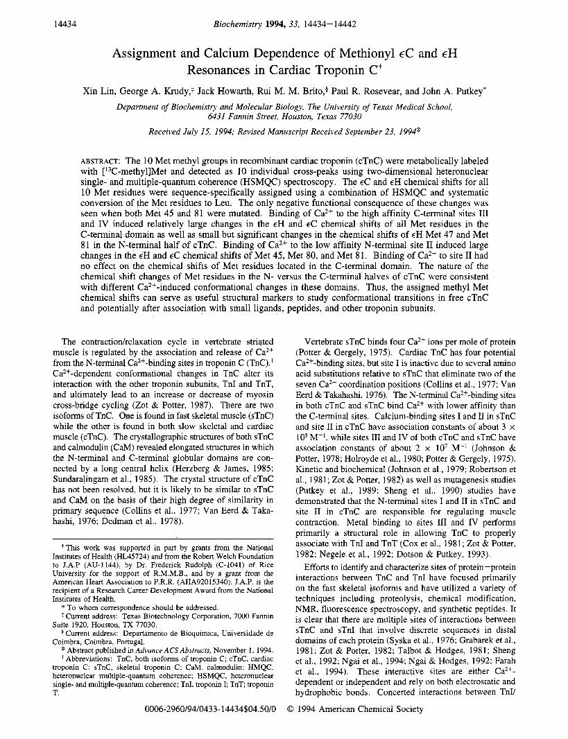
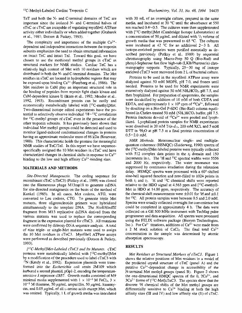
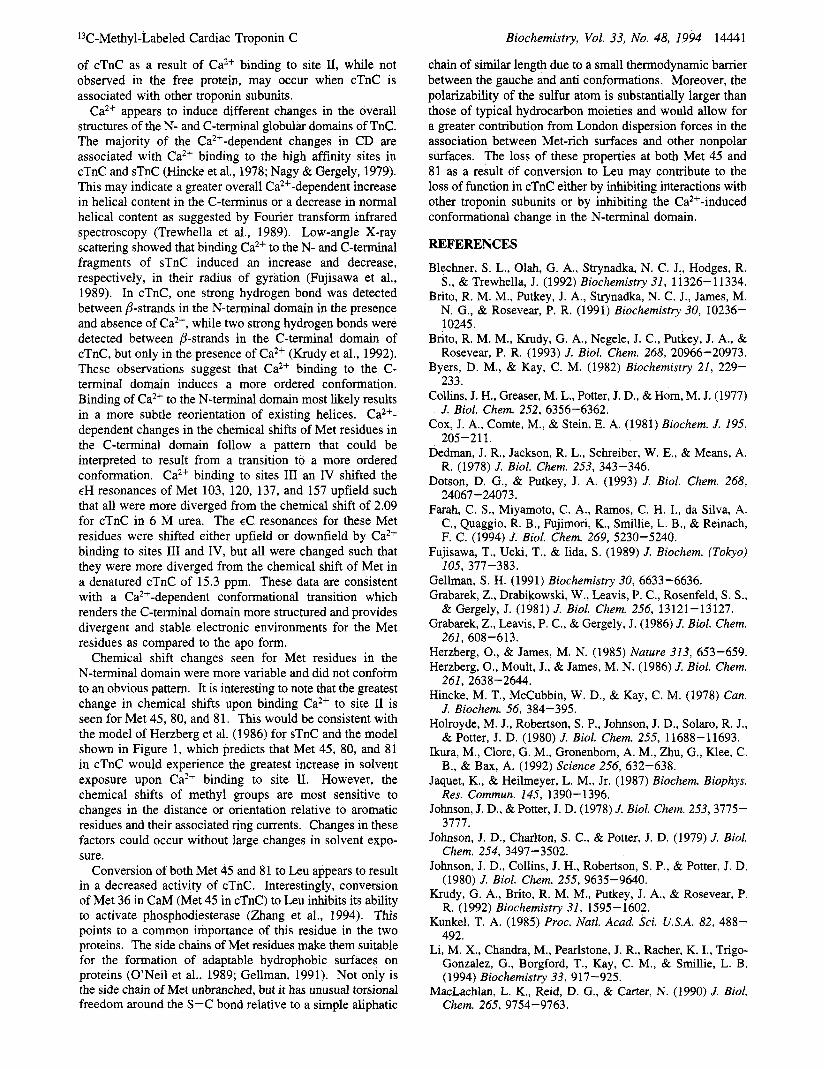
Met Residues as Structural Markers of cTnC3. Figure 1 shows the relative positions of Met residues in a model of the predicted crystal structure of cTnC (panel A) and the putative Ca2+-dependent change in accessibility of the N-terminal Met methyl groups (panel B). Figure 2 shows the one-dimensional HMQC spectra of the 0, 2Ca2+, and 3Ca2+ forms of [13C-Met]cTnC3. The spectra show that the discrete 'H chemical shifts of the Met methyl groups are differentially sensitive to Ca2+ binding at both the high affinity sites (I11 and IV) and low affinity site (11) of cTnC.
MATERIALS AND METHODS
Site-Directed Mutagenesis. The coding sequence for recombinant cTnC (cTnC3) (Putkey et al., 1989) was cloned into the filamentous phage M13mpl8 to generate ssDNA for site-directed mutagenesis on the basis of the method of Kunkel (1985). In all cases, Met codons, ATG, were converted to Leu codons, CTG. To generate triple Met mutants, three oligonucleotide primers were hybridized simultaneously to the template DNA. The NcoIIHindIII fragment from M13 replicative dsDNA derived from the various mutants was used to replace the corresponding fragment in the expression plasmid for cTnC3. All mutations were confirmed by dideoxy-DNA sequence analysis. A total of nine triple- or single-Met mutants were used to assign the 10 Met residues in cTnC3. Myofibril ATPase assays were performed as described previously (Dotson & Putkey, 1993).
[13C-Methyl]Met-Labeled cTnC3 and Its Mutants. cTnC proteins were metabolically labeled with [13C-methylJMet by a modification of the procedure used to label cTnC3 with 15N (Krudy et al., 1992). Expression plasmids were trans- formed into the Escherichia coli strain JM109 which harbored a second plasmid, pGpl-2, encoding the temperature- sensitive 1 repressor cI857. Growth media consisted of M9 minimal media supplemented with 1 x M FeC13, 3 x
M thiamine, 50 pg/mL ampicillin, 50 pglmL kanamy- cin, and 0.05 pglmL of all L-amino acids except Met, which was omitted. Typically, 1 L of growth media was inoculated
14436 Biochemistry, Vol. 33, No. 48, 1994 Lin et al.
A n
3Ca2*-cTnC3
B 47 45
2 C a*+-cTn C 3Ca2+-cTnC FIGURE 1: Relative positions of the Met residues in cTnC3. Space- filling representations of the model for cTnC3 (Brito et al., 1991) are shown in which the shaded spheres are the methyl carbons of the indicated Met residues. (Panel A) Model of the whole Ca2+- saturated protein in which only the a-carbon atoms and the Met residues included. (Panel B) Space-filling representation of N- terminal domain of cTnC3 in which all atoms of amino acids 1-90 are included. Models are compared under conditions in which Ca2+- binding site I1 is unoccupied (2Ca2+-cTnC) or occupied (3Ca2+- cTnC). The protein is oriented such that the central helix extends out of the plane of the paper.
The spectrum of Ca2+-saturated [13C-Met]CBM-IIA, a mutant in which site I1 is inactive (Putkey et al., 1989), was essentially identical to that of the 2Ca2+ form of cTnC3 (data not shown). Thus, the Met methyl resonances provide sensitive markers for conformational changes induced by the binding of Ca2+ to the high and low affinity sites in recombinant TnC.
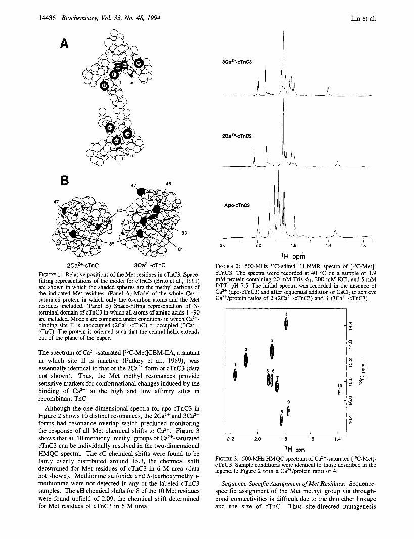
Although the one-dimensional spectra for apo-cTnC3 in Figure 2 shows 10 distinct resonances, the 2Ca2+ and 3Ca2+ forms had resonance overlap which precluded monitoring the response of all Met chemical shifts to Ca2+. Figure 3 shows that all 10 methionyl methyl groups of Ca2+-saturated cTnC3 can be individually resolved in the two-dimensional HMQC spectra. The EC chemical shifts were found to be fairly evenly distributed around 15.3, the chemical shift determined for Met residues of cTnC3 in 6 M urea (data not shown). Methionine sulfoxide and S-(carboxymethyl)- methionine were not detected in any of the labeled cTnC3 samples. The EH chemical shifts for 8 of the 10 Met residues were found upfield of 2.09, the chemical shift determined for Met residues of cTnC3 in 6 M urea.
- I I I 1 I I I I I 26 2 2 1.8 1 4 1 0
'H PPm FIGURE 2: 500-MHz I3C-edited 'H NMR spectra of [13C-Met]- cTnC3. The spectra were recorded at 40 "C on a sample of 1.9 mM protein containing 20 mM Tris-d~ I , 200 mM KC1, and 5 mM DTT, pH 7.5. The initial spectra was recorded in the absence of Ca2+ (apo-cTnC3) and after sequential addition of CaC12 to achieve Ca2+/protein ratios of 2 (2Ca2+-cTnC3) and 4 (3Ca2+-cTnC3).
i 3
1 i 5 6
i 10
I
2.2 2.0 1.0 1.6 1.4
'H ppm
FIGURE 3: 500-MHz HMQC spectrum of Caz+-saturated [I3C-Met]- cTnC3. Sample conditions were identical to those described in the legend to Figure 2 with a Ca*+/protein ratio of 4.
Sequence-Specijic Assignment of Met Residues. Sequence- specific assignment of the Met methyl group via through- bond connectivities is difficult due to the thio ether linkage and the size of cTnC. Thus site-directed mutagenesis
13C-Methyl-Labeled Cardiac Troponin C Biochemistry, Vol. 33, No. 48, 1994 14437
A. cTnC3 8 157
103
80 45 8
0 l , I , I I I I l I
8 C. M45,81,120L
I 8)
I "0 1 2.2 2.0 1.8 1.6 1.4
B. M47,81,120L B
@ 47 n U
% 120 01
0 Q) 6
I , I , I < I , I I
0 D. M45,85,120L
85 0 0
0 0 120
a 45
0
8
1 1 1 1 1 1 1 1 1 1
2.2 2.0 1.8 1.6 1.4
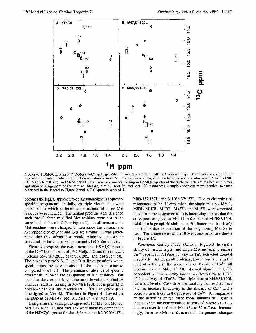
FIGURE 4: HSMQC spectra of [13C-Met]cTnC3 and triple-Met mutants. Spectra were collected from wild type cTnC3 (A) and a set of three triple-Met mutants, in which different combination of three Met residues were changed to Leu by site-directed mutagenesis, M47/81/120L (B), M45/81/120L (C), and M45/85/120L (D). Those resonances missing in HSMQC spectra of the triple mutants are marked with boxes and allowed assignment of the Met 45, Met 47, Met 81, Met 85, and Met 120 resonances. Sample conditions were identical to those described in the legend to Figure 2 with a Ca*+/protein ratio of 4.
becomes the logical approach to obtain unambiguous sequence- specific assignments. Initially, six triple-Met mutants were generated in which different combinations of three Met residues were mutated. The mutant proteins were designed such that all three modified Met residues were not in the same half of the cTnC (see Figure 1). In all mutants, the Met residues were changed to Leu since the volume and hydrophobicity of Met and Leu are similar. It was antici- pated that this substitution would minimize undesirable structural perturbations in the mutant cTnC3 derivatives.
Figure 4 compares the two-dimensional HSMQC spectra of the Ca2+-bound forms of [13C-Met]cTnC and three mutant proteins M47/8 1/120L, M45/8 1/120L, and M45/85/120L. The boxes in panels B, C, and D indicate positions where specific cross-peaks were absent in the mutant proteins as compared to cTnC3. The presence or absence of specific cross-peaks allowed the assignment of Met residues. For example, the cross-peak with the most downfield-shifted 'H chemical shift is missing in M47/81/120L but is present in both M45/81/120L and M45/85/120L. Thus, this cross-peak is assigned to Met 45. The data in Figure 4 allowed the assignment of Met 47, Met 81, Met 85, and Met 120.
Using a similar strategy, assignments for Met 60, Met 80, Met 103, Met 137, and Met 157 were made by comparison of the HSMQC spectra for the triple mutants M60/103/137L,
M80/137/157L, and M103/137/157L. Due to clustering of resonances in the 'H dimension, the single mutants M60L, M80L, M103L, M120L, M137L, and M157L were generated to confirm the assignments. It is interesting to note that the cross-peak assigned to Met 81 in the mutant M45/85/120L exhibits a large upfield shift in the 13C dimension. It is likely that this is due to mutation of the neighboring Met 85 to Leu. The assignments of all 10 Met cross-peaks are shown in Figure 4A.
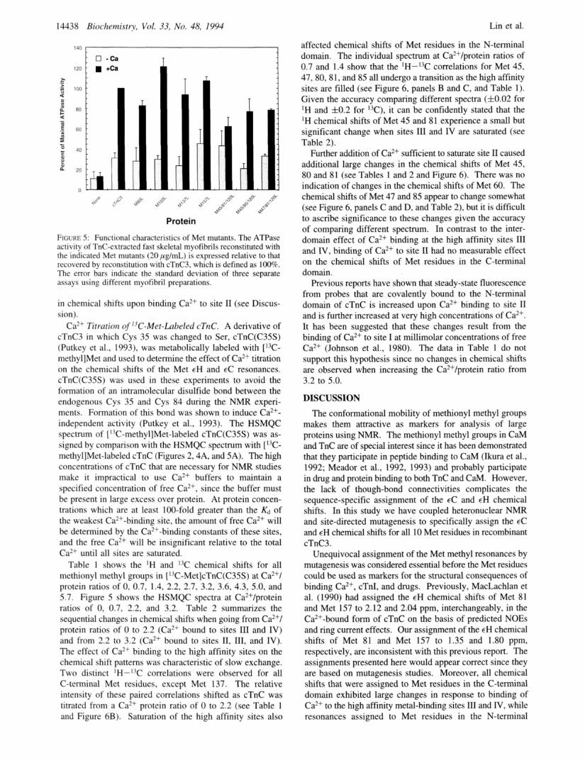
Functional Activity of Met Mutants. Figure 5 shows the ability of various triple- and single-Met mutants to restore Ca2+-dependent ATPase activity in TnC-extracted skeletal myofibrils. Although all proteins showed variations in the level of activity in the presence and absence of Ca2+, all proteins, except M45/8 1/120L, showed significant Ca2+- dependent ATPase activity that ranged from 65% to 130% of the activity of cTnC3. The triple mutant M45/81/120L had a low level of Ca2+-dependent activity that resulted from both an increase in activity in the absence of Ca2+ and a decrease in activity in the presence of Ca2+. A comparison of the activities of the three triple mutants in Figure 5 indicates that the compromised activity of M45/8 1/120L is due to conversion of both Met 45 and 81 to Leu. Interest- ingly, these two Met residues exhibit the greatest changes
14438 Biochernistry, Vol. 33, No. 48, 1994 Lin et al.
0 -Ca 120 1 +Ca
L
L .b
Protein
FIGURE 5: Functional characteristics of Met mutants. The ATPase activity of TnC-extracted fast skeletal myofibrils reconstituted with the indicated Met mutants (20 pg/mL) is expressed relative to that recovered by reconstitution with cTnC3, which is defined as 100%. The error bars indicate the standard deviation of three separate assays using different myofibril preparations.
in chemical shifts upon binding Ca2+ to site I1 (see Discus- sion).
Ca2+ Titration qf 13C-Met-Laheled cTnC. A derivative of cTnC3 in which Cys 35 was changed to Ser, cTnC(C35S) (Putkey et al., 1993), was metabolically labeled with [I3C- methyl]Met and used to determine the effect of Ca2+ titration on the chemical shifts of the Met EH and EC resonances. cTnC(C35S) was used in these experiments to avoid the formation of an intramolecular disulfide bond between the endogenous Cys 35 and Cys 84 during the NMR experi- ments. Formation of this bond was shown to induce Ca2+- independent activity (Putkey et al., 1993). The HSMQC spectrum of [ '3C-methyl]Met-labeled cTnC(C35S) was as- signed by comparison with the HSMQC spectrum with [13C- methyl]Met-labeled cTnC (Figures 2,4A, and 5A). The high concentrations of cTnC that are necessary for NMR studies make it impractical to use Ca2+ buffers to maintain a specified concentration of free Ca2+, since the buffer must be present in large excess over protein. At protein concen- trations which are at least 100-fold greater than the & of the weakest Ca2+-binding site, the amount of free Ca2+ will be determined by the Ca2+-binding constants of these sites, and the free Ca2+ will be insignificant relative to the total Ca2+ until all sites are saturated.
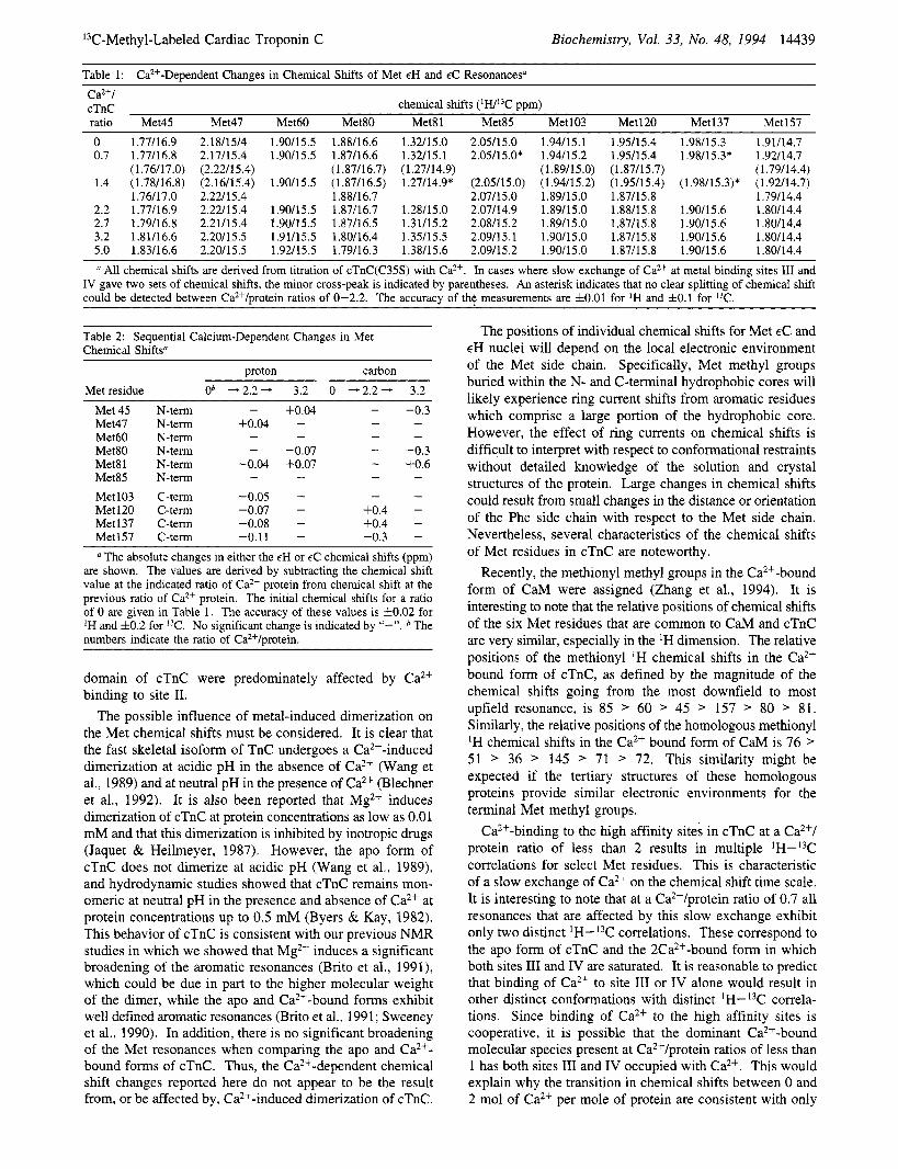
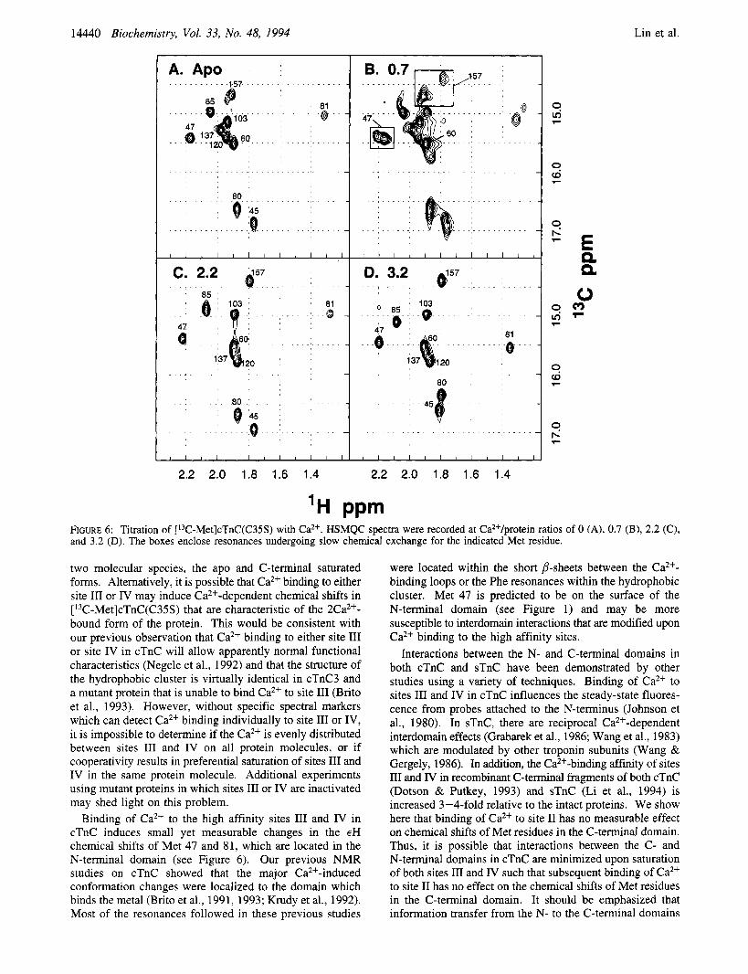
Table I shows the 'H and chemical shifts for all methionyl methyl groups in [ 13C-Met]cTnC(C35S) at Ca2+/ protein ratios of 0, 0.7, 1.4, 2.2, 2.7, 3.2, 3.6, 4.3, 5.0, and 5.7. Figure 5 shows the HSMQC spectra at Ca2+/protein ratios of 0, 0.7, 2.2, and 3.2. Table 2 summarizes the sequential changes in chemical shifts when going from Ca2+/ protein ratios of 0 to 2.2 (Ca2+ bound to sites 111 and IV) and from 2.2 to 3.2 (Ca2+ bound to sites 11, 111, and IV). The effect of Ca2+ binding to the high affinity sites on the chemical shift patterns was characteristic of slow exchange. Two distinct IH-13C correlations were observed for all C-terminal Met residues, except Met 137. The relative intensity of these paired correlations shifted as cTnC was titrated from a Ca2+ protein ratio of 0 to 2.2 (see Table 1 and Figure 6B). Saturation of the high affinity sites also
affected chemical shifts of Met residues in the N-terminal domain. The individual spectrum at Ca2+/protein ratios of 0.7 and 1.4 show that the lH-I3C correlations for Met 45, 47,80,8 1, and 85 all undergo a transition as the high affinity sites are filled (see Figure 6, panels B and C, and Table I). Given the accuracy comparing different spectra (f0.02 for 'H and f0.2 for 13C), it can be confidently stated that the 'H chemical shifts of Met 45 and 81 experience a small but significant change when sites 111 and IV are saturated (see Table 2).
Further addition of Ca2+ sufficient to saturate site I1 caused additional large changes in the chemical shifts of Met 45, 80 and 81 (see Tables 1 and 2 and Figure 6). There was no indication of changes in the chemical shifts of Met 60. The chemical shifts of Met 47 and 85 appear to change somewhat (see Figure 6, panels C and D, and Table 2), but it is difficult to ascribe significance to these changes given the accuracy of comparing different spectrum. In contrast to the inter- domain effect of Ca2+ binding at the high affinity sites 111 and IV, binding of Ca2+ to site I1 had no measurable effect on the chemical shifts of Met residues in the C-terminal domain.
Previous reports have shown that steady-state fluorescence from probes that are covalently bound to the N-terminal domain of cTnC is increased upon Ca2+ binding to site I1 and is further increased at very high concentrations of Ca2+. It has been suggested that these changes result from the binding of Ca2+ to site I at millimolar concentrations of free Ca2+ (Johnson et al., 1980). The data in Table 1 do not support this hypothesis since no changes in chemical shifts are observed when increasing the Ca2+/protein ratio from 3.2 to 5.0.
DISCUSSION The conformational mobility of methionyl methyl groups
makes them attractive as markers for analysis of large proteins using NMR. The methionyl methyl groups in CaM and TnC are of special interest since it has been demonstrated that they participate in peptide binding to CaM (Ikura et al., 1992; Meador et al., 1992, 1993) and probably participate in drug and protein binding to both TnC and CaM. However, the lack of though-bond connectivities complicates the sequence-specific assignment of the EC and EH chemical shifts. In this study we have coupled heteronuclear NMR and site-directed mutagenesis to specifically assign the EC and EH chemical shifts for all 10 Met residues in recombinant cTnC3.
Unequivocal assignment of the Met methyl resonances by mutagenesis was considered essential before the Met residues could be used as markers for the structural consequences of binding Ca2+, cTnI, and drugs. Previously, MacLachlan et al. (1990) had assigned the EH chemical shifts of Met 81 and Met 157 to 2.12 and 2.04 ppm, interchangeably, in the Ca2+-bound form of cTnC on the basis of predicted NOES and ring current effects. Our assignment of the EH chemical shifts of Met 81 and Met 157 to 1.35 and 1.80 ppm, respectively, are inconsistent with this previous report. The assignments presented here would appear correct since they are based on mutagenesis studies. Moreover, all chemical shifts that were assigned to Met residues in the C-terminal domain exhibited large changes in response to binding of Ca2+ to the high affinity metal-binding sites 111 and IV, while resonances assigned to Met residues in the N-terminal
l3C-Methy1-Labeled Cardiac Troponin C Biochemistry, Vol. 33, No. 48, 1994 14439
Table 1: Ca*+-Dependent Changes in Chemical Shifts of Met EH and EC Resonances" Ca2+/ cTnC chemical shifts ( W 3 C ppm) ratio Met45 Met47 Met60 Met80 Met81 Met85 Met103 Met120 Met137 Met157 0 1.77A6.9 0.7 1.77/16.8
(1.76/17.0) 1.4 (1.7U16.8)
1.76/17.0 2.2 1.77/16.9 2.7 1.79A6.8 3.2 1.8M6.6 5.0 1.83/16.6
2.18/15/4 2.17/15.4 (2.22/15.4) (2.16/15.4) 2.22h5.4 2.22/15.4 2.2U15.4 2.20/15.5 2.20A5.5
1.90/15.5 1.88A6.6 1.90/15.5 1.87h6.6
(1.87/16.7) 1.90/15.5 (1.87/16.5)
1.88h6.7 1.90/15.5 1.87/16.7 1.90/15.5 1.87/16.5 1.91/15.5 1.80/16.4 1.92L5.5 1.79A6.3
1.32/15 .O 1.32/15.1 (1.27/14.9) 1.27/14.9*
1.28/15.0 1.3U15.2 1.3Y15.5 1.3W15.6
2.05/15.0 2.05/15.0*
(2.0Y15.0) 2.07/15.0 2.07/14.9 2.08A5.2 2.09/15.1 2.09/15.2
1.94/15.1 1.94115.2 (1.89/15.0) (1.94/15.2) 1.89/15.0 1.89/15.0 1.89/15.0 1.90/15.0 1.90/15.0
1.9Y15.4 1.9Y15.4 ( 1.87/15.7) (1.95/15.4) 1.87/15.8 1.88/15.8 1.87/15.8 1.87/15.8 1.87h5.8
1.98/15.3 1.9U14.7 1.98/15.3* 1.92/14.7
(1.79/14.4) (1.98/15.3)* (1.92/14.7)
1.79h4.4 1.90/15.6 1.80/14.4 1.90/15.6 1.80h4.4 1.90/15.6 1.80/14.4 1.90/15.6 1.80/14.4
All chemical shifts are derived from titration of cTnC(C35S) with CaZ+. In cases where slow exchange of CaZ+ at metal binding sites 111 and IV gave two sets of chemical shifts, the minor cross-peak is indicated by parentheses. An asterisk indicates that no clear splitting of chemical shift could be detected between Ca*+/protein ratios of 0-2.2. The accuracy of the measurements are 10.01 for IH and fO.l for I3C.
Table 2: Sequential Calcium-Dependent Changes in Met Chemical Shifts"
proton carbon Met residue ob -2.2- 3.2 0 -2.2- 3.2
Met 45 Met47 Met60 Met80 Met8 1 Met85 Met103 Met120 Met137 Met157
N-term N-term N-term N-term N-term N-term C-term C-term C-term C-term
- +0.04 - -
-0.04 -
-0.05 -0.07 -0.08 -0.11
+0.04 - -
-0.07 $0.07 - - - - -
The absolute changes in either the EH or EC chemical shifts (ppm) are shown. The values are derived by subtracting the chemical shift value at the indicated ratio of Ca2+ protein from chemical shift at the previous ratio of Ca2+ protein. The initial chemical shifts for a ratio of 0 are given in Table 1. The accuracy of these values is 10.02 for 'H and 10.2 for I3C. No significant change is indicated by "-". The numbers indicate the ratio of Ca2+/protein.
domain of cTnC were predominately affected by Ca2+ binding to site 11.
The possible influence of metal-induced dimerization on the Met chemical shifts must be considered. It is clear that the fast skeletal isoform of TnC undergoes a Ca2+-induced dimerization at acidic pH in the absence of Ca2+ (Wang et al., 1989) and at neutral pH in the presence of Ca2+ (Blechner et al., 1992). It is also been reported that Mg2+ induces dimerization of cTnC at protein concentrations as low as 0.01 mM and that this dimerization is inhibited by inotropic drugs (Jaquet & Heilmeyer, 1987). However, the apo form of cTnC does not dimerize at acidic pH (Wang et al., 1989), and hydrodynamic studies showed that cTnC remains mon- omeric at neutral pH in the presence and absence of Ca2+ at protein concentrations up to 0.5 mM (Byers & Kay, 1982). This behavior of cTnC is consistent with our previous NMR studies in which we showed that Mg2+ induces a significant broadening of the aromatic resonances (Brito et al., 1991), which could be due in part to the higher molecular weight of the dimer, while the apo and Ca2+-bound forms exhibit well defined aromatic resonances (Brito et al., 1991; Sweeney et al., 1990). In addition, there is no significant broadening of the Met resonances when comparing the apo and Ca2+- bound forms of cTnC. Thus, the Ca2+-dependent chemical shift changes reported here do not appear to be the result from, or be affected by, Ca2+-induced dimerization of cTnC.
The positions of individual chemical shifts for Met EC and EH nuclei will depend on the local electronic environment of the Met side chain. Specifically, Met methyl groups buried within the N- and C-terminal hydrophobic cores will likely experience ring current shifts from aromatic residues which comprise a large portion of the hydrophobic core. However, the effect of ring currents on chemical shifts is difficult to interpret with respect to conformational restraints without detailed knowledge of the solution and crystal structures of the protein. Large changes in chemical shifts could result from small changes in the distance or orientation of the Phe side chain with respect to the Met side chain. Nevertheless, several characteristics of the chemical shifts of Met residues in cTnC are noteworthy.
Recently, the methionyl methyl groups in the Ca2+-bound form of CaM were assigned (Zhang et al., 1994). It is interesting to note that the relative positions of chemical shifts of the six Met residues that are common to CaM and cTnC are very similar, especially in the 'H dimension. The relative positions of the methionyl 'H chemical shifts in the Ca2+ bound form of cTnC, as defined by the magnitude of the chemical shifts going from the most downfield to most upfield resonance, is 85 > 60 > 45 > 157 > 80 > 81. Similarly, the relative positions of the homologous methionyl 'H chemical shifts in the Ca2+ bound form of CaM is 76 > 51 > 36 145 > 71 > 72. This similarity might be expected if the tertiary structures of these homologous proteins provide similar electronic environments for the terminal Met methyl groups.
Ca2+-binding to the high affinity sites in cTnC at a Ca2+/ protein ratio of less than 2 results in multiple lH-13C correlations for select Met residues. This is characteristic of a slow exchange of Ca2+ on the chemical shift time scale. It is interesting to note that at a Ca2+/protein ratio of 0.7 all resonances that are affected by this slow exchange exhibit only two distinct 'H-13C correlations. These correspond to the apo form of cTnC and the 2Ca2+-bound form in which both sites I11 and IV are saturated. It is reasonable to predict that binding of Ca2+ to site I11 or IV alone would result in other distinct conformations with distinct 'H- 13C correla- tions. Since binding of Ca2+ to the high affinity sites is cooperative, it is possible that the dominant Ca2+-bound molecular species present at Ca2+/protein ratios of less than 1 has both sites I11 and IV occupied with Ca2+. This would explain why the transition in chemical shifts between 0 and 2 mol of Ca2+ per mole of protein are consistent with only
14440 Biochemistry, Vol. 33, No. 48, 1994 Lin et al.
A. Apo 57
81
@ - @@
-
57
81
8
80
45 i 45
0
2.2 2.0 1.8 1.6 1.4 2.2 2.0 1.8 1.6 1.4
'H PPm FIGURE 6: Titration of ['3C-Met]cTnC(C35S) with Ca2+. HSMQC spectra were recorded at Ca2+/protein ratios of 0 (A), 0.7 (B), 2.2 (C), and 3.2 (D). The boxes enclose resonances undergoing slow chemical exchange for the indicated Met residue.
two molecular species, the apo and C-terminal saturated forms. Alternatively, it is possible that Ca2+ binding to either site I11 or IV may induce Ca2+-dependent chemical shifts in [ 13C-Met]cTnC(C35S) that are characteristic of the 2Ca2+- bound form of the protein. This would be consistent with our previous observation that Caz+ binding to either site I11 or site IV in cTnC will allow apparently normal functional characteristics (Negele et al., 1992) and that the structure of the hydrophobic cluster is virtually identical in cTnC3 and a mutant protein that is unable to bind Ca2+ to site 111 (Brito et al., 1993). However, without specific spectral markers which can detect Ca2+ binding individually to site I11 or IV, it is impossible to determine if the Ca2+ is evenly distributed between sites 111 and IV on all protein molecules, or if cooperativity results in preferential saturation of sites I11 and IV in the same protein molecule. Additional experiments using mutant proteins in which sites I11 or IV are inactivated may shed light on this problem.
Binding of Ca2+ to the high affinity sites I11 and IV in cTnC induces small yet measurable changes in the cH chemical shifts of Met 47 and 81, which are located in the N-terminal domain (see Figure 6). Our previous NMR studies on cTnC showed that the major Ca*+-induced conformation changes were localized to the domain which binds the metal (Brito et al., 1991, 1993; Krudy et al., 1992). Most of the resonances followed in these previous studies
were located within the short P-sheets between the Ca2+- binding loops or the Phe resonances within the hydrophobic cluster. Met 47 is predicted to be on the surface of the N-terminal domain (see Figure 1) and may be more susceptible to interdomain interactions that are modified upon Ca2+ binding to the high affinity sites.
Interactions between the N- and C-terminal domains in both cTnC and sTnC have been demonstrated by other studies using a variety of techniques. Binding of Ca2+ to sites I11 and IV in cTnC influences the steady-state fluores- cence from probes attached to the N-terminus (Johnson et al., 1980). In sTnC, there are reciprocal Ca2+-dependent interdomain effects (Grabarek et al., 1986; Wang et al., 1983) which are modulated by other troponin subunits (Wang & Gergely, 1986). In addition, the Ca2+-binding affinity of sites I11 and IV in recombinant C-terminal fragments of both cTnC (Dotson & Putkey, 1993) and sTnC (Li et al., 1994) is increased 3-4-fold relative to the intact proteins. We show here that binding of Ca2+ to site I1 has no measurable effect on chemical shifts of Met residues in the C-terminal domain. Thus, it is possible that interactions between the C- and N-terminal domains in cTnC are minimized upon saturation of both sites I11 and IV such that subsequent binding of Ca2+ to site I1 has no effect on the chemical shifts of Met residues in the C-terminal domain. It should be emphasized that information transfer from the N- to the C-terminal domains
13C-Methyl-Labeled Cardiac Troponin C
of cTnC as a result of Ca2+ binding to site 11, while not observed in the free protein, may occur when cTnC is associated with other troponin subunits.
Ca2+ appears to induce different changes in the overall structures of the N- and C-terminal globular domains of TnC. The majority of the Ca2+-dependent changes in CD are associated with Ca2+ binding to the high affinity sites in cTnC and sTnC (Hincke et al., 1978; Nagy & Gergely, 1979). This may indicate a greater overall Ca2+-dependent increase in helical content in the C-terminus or a decrease in normal helical content as suggested by Fourier transform infrared spectroscopy (Trewhella et al., 1989). Low-angle X-ray scattering showed that binding Ca2+ to the N- and C-terminal fragments of sTnC induced an increase and decrease, respectively, in their radius of gyration (Fujisawa et al., 1989). In cTnC, one strong hydrogen bond was detected between ,&strands in the N-terminal domain in the presence and absence of Ca2+, while two strong hydrogen bonds were detected between P-strands in the C-terminal domain of cTnC, but only in the presence of Ca2+ (Krudy et al., 1992). These observations suggest that CaZ+ binding to the C- terminal domain induces a more ordered conformation. Binding of Ca2+ to the N-terminal domain most likely results in a more subtle reorientation of existing helices. CaZ+- dependent changes in the chemical shifts of Met residues in the C-terminal domain follow a pattern that could be interpreted to result from a transition to a more ordered conformation. Ca2+ binding to sites I11 an IV shifted the EH resonances of Met 103, 120, 137, and 157 upfield such that all were more diverged from the chemical shift of 2.09 for cTnC in 6 M urea. The EC resonances for these Met residues were shifted either upfield or downfield by Ca2+ binding to sites I11 and IV, but all were changed such that they were more diverged from the chemical shift of Met in a denatured cTnC of 15.3 ppm. These data are consistent with a Ca2+-dependent conformational transition which renders the C-terminal domain more structured and provides divergent and stable electronic environments for the Met residues as compared to the apo form.
Chemical shift changes seen for Met residues in the N-terminal domain were more variable and did not conform to an obvious pattem. It is interesting to note that the greatest change in chemical shifts upon binding Ca2+ to site I1 is seen for Met 45, 80, and 8 1. This would be consistent with the model of Herzberg et al. (1986) for sTnC and the model shown in Figure I, which predicts that Met 45, 80, and 81 in cTnC would experience the greatest increase in solvent exposure upon Ca2+ binding to site 11. However, the chemical shifts of methyl groups are most sensitive to changes in the distance or orientation relative to aromatic residues and their associated ring currents. Changes in these factors could occur without large changes in solvent expo- sure.
Conversion of both Met 45 and 8 1 to Leu appears to result in a decreased activity of cTnC. Interestingly, conversion of Met 36 in Cah4 (Met 45 in cTnC) to Leu inhibits its ability to activate phosphodiesterase (Zhang et al., 1994). This points to a common importance of this residue in the two proteins. The side chains of Met residues make them suitable for the formation of adaptable hydrophobic surfaces on proteins (O'Neil et al., 1989; Gellman, 1991). Not only is the side chain of Met unbranched, but it has unusual torsional freedom around the S-C bond relative to a simple aliphatic
Biochemistry, Vol. 33, No. 48, 1994 14441
chain of similar length due to a small thermodynamic barrier between the gauche and anti conformations. Moreover, the polarizability of the sulfur atom is substantially larger than those of typical hydrocarbon moieties and would allow for a greater contribution from London dispersion forces in the association between Met-rich surfaces and other nonpolar surfaces. The loss of these properties at both Met 45 and 81 as a result of conversion to Leu may contribute to the loss of function in cTnC either by inhibiting interactions with other troponin subunits or by inhibiting the Ca2+-induced conformational change in the N-terminal domain.
REFERENCES
Blechner, S. L., Olah, G. A., Strynadka, N. C. J., Hodges, R. S., & Trewhella, J. (1992) Biochemistry 31, 11326-11334.
Brito, R. M. M., Putkey, J. A., Strynadka, N. C. J., James, M. N. G., & Rosevear, P. R. (1991) Biochemistry 30, 10236- 10245.
Brito, R. M. M., Krudy, G. A., Negele, J. C., Putkey, J. A., & Rosevear, P. R. (1993) J. Bid. Chem. 268, 20966-20973.
Byers, D. M., & Kay, C. M. (1982) Biochemistry 21, 229- 233.
Collins, J. H., Greaser, M. L., Potter, J. D., & Horn, M. J. (1977) J. Biol. Chem. 252, 6356-6362.
Cox, J. A,, Comte, M., & Stein, E. A. (1981) Biochem. J. 195, 205-21 1.
Dedman, J. R., Jackson, R. L., Schreiber, W. E., & Means, A. R. (1978) J. Biol. Chem. 253, 343-346.
Dotson, D. G., & Putkey, J. A. (1993) J. Biol. Chem. 268, 24067 -24073.
Farah, C. S., Miyamoto, C. A., Ramos, C. H. I., da Silva, A. C., Quaggio, R. B., Fujimori, K., Smillie, L. B., & Reinach, F. C. (1994) J. Biol. Chem. 269, 5230-5240.
Fujisawa, T., Ueki, T., & Iida, S. (1989) J. Biochem. (Tokyo) 105, 377-383.
Gellman, S. H. (1991) Biochemistry 30, 6633-6636. Grabarek, Z., Drabikowski, W., Leavis, P. C., Rosenfeld, S. S.,
& Gergely, J. (1981) J. Bid. Chem. 256, 13121-13127. Grabarek, Z., Leavis, P. C., & Gergely, J. (1986) J. Biol. Chem.
261, 608-613. Herzberg, O., & James, M. N. (1985) Nature 313, 653-659. Herzberg, O., Moult, J., & James, M. N. (1986) J. Biol. Chem.
261, 2638-2644. Hincke, M. T., McCubbin, W. D., & Kay, C. M. (1978) Can.
J. Biochem. 56, 384-395. Holroyde, M. J., Robertson, S. P., Johnson, J. D., Solaro, R. J.,
& Potter, J. D. (1980) J. Biol. Chem. 255, 11688- 11693. Ikura, M., Clore, G. M., Gronenborn, A. M., Zhu, G., Klee, C.
B., & Bax, A. (1992) Science 256, 632-638. Jaquet, K., & Heilmeyer, L. M., Jr. (1987) Biochem. Biophys.
Res. Commun. 145, 1390-1396. Johnson, J. D., & Potter, J. D. (1978) J. Biol. Chem. 253,3775-
3777. Johnson, J. D., Charlton, S. C., & Potter, J. D. (1979) J. Bid.
Chem. 254, 3497-3502. Johnson, J. D., Collins, J. H., Robertson, S. P., & Potter, J. D.
(1980) J. Biol. Chem. 255, 9635-9640. Krudy, G. A., Brito, R. M. M., Putkey, J. A., & Rosevear, P.
R. (1992) Biochemistry 31, 1595-1602. Kunkel, T. A. (1985) Proc. Natl. Acad. Sci. U.S.A. 82, 488-
492. Li, M. X., Chandra, M., Pearlstone, J. R., Racher, K. I., Trigo-
Gonzalez, G., Borgford, T., Kay, C. M., & Smillie, L. B. (1994) Biochemistry 33, 917-925.
MacLachlan, L. K., Reid, D. G., & Carter, N. (1990) J. Biol. Chem. 265, 9754-9763.
14442 Biochemistry, Vol. 33, No. 48, 1994
Meador, W. E., Means, A. R., & Quiocho, F. A. (1992) Science
Meador, W. E., Means, A. R., & Quiocho, F. A. (1993) Science
Nagy, B., & Gergely, J. (1979) J. Biol. Chem. 254, 12732-
Negele, J. C., Dotson, D. G., Liu, W., Sweeney, H. L., & Putkey,
Ngai, S.-M., & Hodges, R. S. (1992) J. Biol. Chem. 267,
Ngai, S.-M., Sonnichsen, F. D., & Hodges, R. S. (1994) J. Biol.
O’Neil, K. T., Erickson-Viitanen, S., & DeGrado, W. F. (1989)
Potter, J. D., & Gergely, J. (1975) J. Biol. Chem. 250, 4628-
Putkey, J. A., Sweeney, H. L., & Campbell, S. T. (1989) J.
Putkey, J. A., Dotson, D. G., & Mouawad, P. (1993) J. Biol.
Robertson, S . P., Johnson, J. D., & Potter, J. D. (1981) Biophys.
Rosevear, P. R. (1988) Biochemistry 27, 7931-7939. Sheng, Z., Strauss, W. L., Francois, J. M., & Potter, J. D. (1990)
Sheng, Z., Pan, B.-S., Miller, T. E., & Potter, J. D. (1992) J.
257, 1251-1255.
262, 1718-1721.
12737.
J. A. (1992) J. Biol. Chem. 267, 825-831.
157 15- 15720.
Chem. 269, 2165-2172.
J. Biol. Chem. 264, 14571-14578.
4633.
Biol. Chem. 264, 12370-12378.
Chem. 268, 6827-6830.
J. 34, 559-569.
J. Biol. Chem. 265, 21554-21560.
Biol. Chem. 267, 25407-25413.
Lin et al.
Sundaralingam, M., Bergstrom, R., Strasburg, G., Rao, S. T., Roychowdhury, P., Greaser, M., & Wang, B. C. (1985) Science 227, 945-948.
Sweeney, H. L., Brito, R. M. M., Rosevear, P. R., & Putkey, J. A. (1990) Proc. Natl. Acad. Sci. U.S.A. 87, 9538-9542.
Syska, H., Wilkinson, J. M., Grand, R. J. M., & Perry, S. V. (1976) Biochem. J. 153, 375-387.
Talbot, J. A., & Hodges, R. S. (1981) J. Biol. Chem. 256,2798- 2702.
Trewhella, J., Liddle, W. K., Heidom, D. B., & Strynadka, N. (1989) Biochemistry 28, 1294-1301.
Van Eerd, J. P., & Takahashi, K. (1976) Biochemistry 15, 1171- 1180.
Wang, C. L., & Gergely, J. (1986) Eur. J . Biochem. 154,225- 228.
Wang, C. L., Leavis, P. C., & Gergely, J. (1983) J. Biol. Chem. 258, 9175-9177.
Wang, C. K., Lebowitz, J., & Cheung, H. C. (1989) Proteins 6,424-430.
Zhang, M., Li, M., Wang, J. H., & Vogel, H. J. (1994) J. Biol. Chem. 269, 15546-15552.
Zot, H. G., & Potter, J. D. (1982) J. Biol. Chem. 257, 7678- 7683.
Zot, A. S., & Potter, J. D. (1987) Annu. Rev. Biophys. Biophys. Chem. 16, 535-559.
Zuiderweg, E. R. P. (1990) J. Magn. Reson. 86, 346-351.
Related Documents


