This article appeared in a journal published by Elsevier. The attached copy is furnished to the author for internal non-commercial research and education use, including for instruction at the authors institution and sharing with colleagues. Other uses, including reproduction and distribution, or selling or licensing copies, or posting to personal, institutional or third party websites are prohibited. In most cases authors are permitted to post their version of the article (e.g. in Word or Tex form) to their personal website or institutional repository. Authors requiring further information regarding Elsevier’s archiving and manuscript policies are encouraged to visit: http://www.elsevier.com/authorsrights

Welcome message from author
This document is posted to help you gain knowledge. Please leave a comment to let me know what you think about it! Share it to your friends and learn new things together.
Transcript
This article appeared in a journal published by Elsevier. The attachedcopy is furnished to the author for internal non-commercial researchand education use, including for instruction at the authors institution
and sharing with colleagues.
Other uses, including reproduction and distribution, or selling orlicensing copies, or posting to personal, institutional or third party
websites are prohibited.
In most cases authors are permitted to post their version of thearticle (e.g. in Word or Tex form) to their personal website orinstitutional repository. Authors requiring further information
regarding Elsevier’s archiving and manuscript policies areencouraged to visit:
http://www.elsevier.com/authorsrights

Author's personal copy
Animal Reproduction Science 139 (2013) 121– 126
Contents lists available at SciVerse ScienceDirect
Animal Reproduction Science
jou rn al hom epage : w ww.elsev ier .com/ locate /an i r eprosc i
Assessment of uterine involution in bitches using B-modeand Doppler ultrasonography
Claudia da Cunha Barbosaa,∗, Mírley Barbosa de Souzaa,Luana Azevedo de Freitasa, Ticiana Franco Pereira da Silvaa,Sheyla Farhayldes Souza Dominguesb, Lúcia Daniel Machado da Silvaa
a Laboratory of Carnivore Reproduction, Faculty of Veterinary Medicine, State University of Ceará, Fortaleza, Ceará, Brazilb Laboratory of Biology and Medicine of Wild Mammals from Amazônia, Federal University of Pará, Belém, Pará, Brazil
a r t i c l e i n f o
Article history:Received 22 August 2012Received in revised form 18 February 2013Accepted 28 February 2013Available online 16 March 2013
Keywords:Doppler ultrasonographyPuerperiumUterine involutionDog
a b s t r a c t
The aim of this study was to measure the uterine diameter by B-mode and Dopplervelocimetry patterns of uterine arteries in postpartum female dogs after normal deliveryor cesarean section (c-section). Ten female dogs were assessed on weeks 0, 1, 2, 3 and 4postpartum. Only at week 0, bitches submitted to c-section presented higher body diame-ters and uterine horns when compared to normal delivery. It was observed a reduction inuterine diameters over the weeks in both groups. In general, bitches submitted to c-sectionpresented lower uterine perfusion. Each group presented distinct Doppler velocimetriccharacteristics. It was concluded that the B-mode ultrasound and Doppler are importanttools for assessing puerperal uterine with distinct characteristics influenced by the type ofdelivery.
© 2013 Elsevier B.V. All rights reserved.
1. Introduction
Ultrasonography is an important tool in clinical repro-ductive studies in dogs for assessing ovulation, pregnancy,embryo loss and puerperium (England and Russo, 2006;Davidson and Baker, 2009). Puerperium is the phaseimmediately following delivery, which extends from theexpulsion of the last pup’s placenta to the moment whenthe uterus returns to its normal non-pregnant state, includ-ing the uterine involution and endometrial regeneration.Dogs exhibit serous-bloody vaginal discharge during puer-perium, which lasts 1–6 weeks, when they are more
∗ Corresponding author at: 1700, Paranjana Avenue, Campus do Itaperi,CEP 60740-903, Fortaleza, CE, Brazil. Tel.: +55 85 3101 9851;fax: +55 85 3101 9840.
E-mail addresses: clauvet [email protected] (C.d.C. Barbosa),[email protected], [email protected](L.D.M.d. Silva).
susceptible to uterine diseases such as metritis and subin-volution of the placental site (Feldman and Nelson, 1986).
Despite its importance in veterinary medicine, thenumber of studies that have used B-mode ultrasonogra-phy for puerperium monitoring is small. Nevertheless,this approach has been reported for cows (Okano andTomizuka, 1987), ewes (Hauser and Bostedt, 2002), sows(Cortez, 2003), queens (Ferretti et al., 2000) and bitches(Yeager and Concannon, 1990; Ferri and Vicente, 2002;Ferri et al., 2003). In addition to B-mode ultrasound,Doppler is also used in women to diagnose placental reten-tion (Nijman et al., 2002), persistence of trophoblast cells(Alcázar, 1998), arteriovenous malformations and hemor-rhage (Kelly et al., 2003; Teo et al., 2008) and, also, to assessuterine blood flow during puerperium (Schoubroeck et al.,2004).
In dogs, Doppler use has only been reported to assessdifferent phases of the estrous cycle of ovarian (Köster et al.,2001) and uterine arteries (Alvarez-Clau and Liste, 2005)and to monitor pregnancy by assessing maternal and fetal
0378-4320/$ – see front matter © 2013 Elsevier B.V. All rights reserved.http://dx.doi.org/10.1016/j.anireprosci.2013.02.027

Author's personal copy
122 C.d.C. Barbosa et al. / Animal Reproduction Science 139 (2013) 121– 126
arteries (Nautrup, 1998; Di Salvo et al., 2006; Miranda andDomingues, 2010). Use of Doppler ultrasonography to eval-uate the uterus during the puerperium was described incows (Herzog and Bollwein, 2007; Krueger et al., 2009) andmares (Mortensen et al., 2011) but to our knowledge thereis only one report in bitches and observations were limitedto the first 14 days postpartum (Serin and Tarimcilar, 2011)and an abstract report that describe Doppler ultrasono-graphic changes of uterine arteries during normal caninepuerperium (Batista et al., 2012). The aim of this study wasto use B-mode and Doppler ultrasonography to measurethe diameter of the uterus and velocimetry patterns of theuterine arteries in postpartum female dogs after normaldelivery or c-section.
2. Materials and methods
2.1. Setting and experimental animals
This study was performed at the Laboratory of Carni-vore Reproduction at the State University of Ceará andthe experimental protocol was approved by the AnimalEthics Committee of the same university (protocol number08517453-0), in accordance with the guidelines of care anduse of laboratory animals established by Brazilian Collegeof Animal Experimentation.
Ten bitches were used. Among them, six (2 Pugs, 3Schnauzers and 1 French Bulldog), ranging from 10 to31 months, weighing between 6 and 10 kg, with a pro-lificacy from 2 to 7 puppies after normal delivery wereused. Among them, four were primiparous and 2 were mul-tiparous. Four bitches (4 French Bulldogs) ranging from18 to 36 months, weighing between 8 and 10 kg, witha prolificacy from 5 to 7 puppies after to cesarean sec-tion (c-section). Among them, 3 were primiparous and 1was multiparous. The dogs were the property of commer-cial kennels, and after parturition were housed with theirpups in individual kennels and fed commercial rations forpuppies and water ad libitum. All dogs had good overallclinical status and known reproductive history. The sta-tus of health of the bitches was checked at each day of theultrasound examination. Body temperature was measured,visible mucous membranes were inspected and cardio-pulmonary auscultation was done. All these parameterswere always within normal limits. Pups from all litterswere weaned at 45 days postpartum. The bitches sub-mitted to a c-section were above 60 days of pregnancyand did not present any sign of parturition. The proce-dure was recommended after fetal maturity confirmationby ultrasonography evaluation. For c-section it was used aspre anesthetic medication atropine (0.02 mg/kg), tramadol(2 mg/kg) and diazepam (0.25 mg/kg). Anesthesia was per-formed with propofol (2–5 mg/kg) and maintained withisoflurane. For the postoperative treatment of females sub-mitted to c-section was instituted cephalexin (30 mg/kg)every 12 h for 5 days and ketoprofen (1 mg/kg) once a dayfor 5 days.
2.2. Ultrasonography assessment
Ultrasonography assessment was performed on weeks0, 1, 2, 3 and 4 postpartum, in an acclimatized room
at approximately 25 ◦C. Week 0 corresponded to 2 dayspostpartum and the subsequent weeks corresponded toeach 7 days postpartum. Before assessment, bitches werefasted for a minimum of 12 h, the abdomen shaved,and placed in dorsal recumbency on a foam cushion. B-mode and Doppler velocimetry analyses were performedusing a SonoAce PICO (Medison Co., Ltd., Daechi-Dong,Kangnam-ku, Cuseoul, Korea) model ultrasonographydevice with 5–9 MHz multifrequency linear transducer.Ultrasonography-specific gel was used in all tests.
Using the B-mode, the uterine body was located at theentrance to the pelvis and assessed for ultrasound charac-teristics. The uterine body diameter was measured cranialto the cervix, in the longitudinal plane, using electroniccalipers. The uterine horns were identified accompanyingthe uterine body and were assessed for ultrasound char-acteristics of echogenicity of the serosa, myometrium andendometrium, and for the presence and features of intra-luminal content. The diameters of the uterine horns (leftand right) were measured on the longitudinal plane, in theplacental sites, near to the uterine bifurcation, using theelectronic caliper.
The uterine artery was evaluated parallel to the uter-ine body and located by means of longitudinal scans withB-mode and color Doppler (Alvarez-Clau and Liste, 2005).The caliper for pulsed Doppler was placed on the uterineartery lumen image with an insonation angle of 60◦ andgraphic representation of the artery flow was performedwith visualization of consecutive waves of equal veloc-ity and amplitude. At least three waves were measured ateach side of the uterine artery to analyze the results. Peaksystolic velocity (PSV), end diastolic velocity (EDV), resis-tance index (RI) and pulsatility index (PI) were recorded.All parameters were automatically calculated by the device(Szatmári et al., 2001).
2.3. Statistical analysis
Repeated measures ANOVA were used to determine themain effect of days and kind of delivery on uterine hornand body diameter, as well as PSV, EDV, RI and PI fromuterine arteries. Differences were located with a Fisher’sprotected least significant difference post hoc test. Further-more, for these end points, a paired Student’s t-test wasused to compare (within specific days) left versus right,uterine diameter and arteries indices. Stat View software(SAS Institute Inc., Cary, NC, USA) was used for all analysesand all data were reported as mean ± SD. For all analyses,P < 0.05 was considered significant.
3. Results
Ultrasonographic assessment lasted for up to a max-imum of 45 min, and the uterine body and horns couldbe visualized throughout the 4 weeks of evaluation. Thetype of birth did not influence the ecographic appear-ance evaluated by B-mode. Using B-mode ultrasound, theuterine body and horns exhibited a tubular format withmixed echogenicity, and the serous layer could be iden-tified in the uterine wall as a hyperechoic outer layer.Three parts of the myometrium could be visualized, the

Author's personal copy
C.d.C. Barbosa et al. / Animal Reproduction Science 139 (2013) 121– 126 123
Fig. 1. B-mode ultrasonography of canine on week 1 postpartum uterinehorn in longitudinal section. Visualization of uterus layers: hyperechoicserous membrane (arrow), myometrium formed by 3 layers (full line):hypo- (+), hyper- (−), and hypoechoic (x) and endometrium (dotted line).
hypo-, hyper- and hypoechoic layers which coincided withthe outer longitudinal, middle circular and outer longitudi-nal layers, respectively. The endometrium and the uterinelumen appeared as a layer with mixed echogenicity (Fig. 1).
Stratification of the myometrium was best observedduring the first 2 weeks, after which differentiation amonglayers gradually became more difficult until the layerswere no longer distinguishable by week 4. At this time,the endometrium was visible as a more echogenic layerthan the myometrium (Fig. 2). Moderate amounts of ane-choic contents were seen within the uterine lumen onlyon week 0 postpartum (Fig. 2A), which decreased in subse-quent assessments and were no longer visible from week1 onwards.
The type of birth influenced the diameters of the uter-ine body and horns at week 0 postpartum (P < 0.05), withno difference between the diameters of the left and right
horns (P > 0.05). The uterine horn diameter was defined asthe average diameter of both horns in each kind of birth.The largest diameters of the uterine body and horns wereregistered at week 0 for both groups and at this moment,the uterine diameters of the bitches which underwentc-section were higher than those from normal deliverybitches (P < 0.05), but this difference was not observedbetween the groups on other weeks (P > 0.05). The diame-ter of the uterine body presented a reduction between theweek 0 and week 1, in both groups, and between week 1and week 4 only in bitches with normal delivery. The uter-ine horns also presented a reduction in diameter betweenthe week 0 and week 1, followed by a further reduction inweek 2 and 4 for both groups (P < 0.05) (Table 1).
Using color Doppler, uterine arteries were visualizedparallel to the uterine body as linear vessels. Their calibersdecreased slightly over time, and they could be identi-fied throughout the experimental time course. On spectralDoppler, the uterine artery exhibited a biphasic wave witha high systolic peak, the presence of a diastolic notch and aslight diastolic peak in both groups (Fig. 3).
On pulsed Doppler, uterine artery RI and PI were signif-icantly lower in normal delivery bitches than in c-sectionbitches until week 2 and over the weeks, respectively. Ingeneral, RI increased only at week 4 in both groups and PIhad similar profile in normal delivery while for c-sectionbitches it remained almost constant. When comparing thesides, the left uterine artery RI and PI, in normal deliverygroup, was significantly higher than right uterine artery atweeks 0 and 4, with no differences between the sides inbitches submitted to c-section (Table 2).
There were no differences in the VPS regardless thetype of delivery, wherever EDV from left and right uter-ine artery in bitches with normal delivery was higher thanc-section bitches until week 2 and week 1 respectively. Forboth groups, uterine artery VPS decreased from week 1and EDV decreased from week 2 only in normal deliverybitches, while in c-section bitches, it remains constant. Inboth groups, left uterine artery VPS was higher than rightside only on week 3 and in bitches submitted to c-section,
Fig. 2. Ultrasonographic images of canine postpartum uterine horn in longitudinal section. (A) Week 0, (B) week 1, (C) week 2, (D) week 3 and (E) week 4.Uterine diameter (dotted line). Lumen (arrow).

Author's personal copy
124 C.d.C. Barbosa et al. / Animal Reproduction Science 139 (2013) 121– 126
Table 1Uterine measurements (mean ± SD) evaluated by B-mode ultrasonography in bitches during postpartum after normal delivery (n = 6) or c-section (n = 4).
Week Uterine body (cm) Uterine horn (cm)
Delivery C-section Delivery C-section
0 1.44 ± 0.41Aa 1.79 ± 0.31Ba 1.76 ± 0.35Aa 2.02 ± 0.49Ba
1 1.11 ± 0.16Ab 1.24 ± 0.31Ab 1.41 ± 0.30Ab 1.51 ± 0.28Ab
2 1.00 ± 0.14Abc 0.96 ± 0.12Ab 1.15 ± 0.19Ac 1.14 ± 0.32Ac
3 0.95 ± 0.20Abc 1.00 ± 0.04Ab 1.05 ± 0.13Ac 1.01 ± 0.25Acd
4 0.80 ± 0.06Ac 0.99 ± 0.28Ab 0.86 ± 0.18Ad 0.89 ± 0.25Ad
(A, B) Comparison between types of birth in the same week, in the same location (between columns). (a, b, c, d) Comparison between weeks in the same typeof birth (between lines). (P < 0.05).
EDV from left uterine artery was lower than right side atweek 2, and in normal delivery bitches this difference wasseen at week 4 (Table 3).
4. Discussion
The uterine echogenicity and appearance were simi-lar in all investigated dogs, regardless the delivery type.It was possible to identify the uterine wall layers andlumen when visible and detail the myometrium stratifi-cation in 3 echogenicity different layers, as reported byYeager and Concannon (1990). That study reported no dif-ferences in the uterine ultrasonographic appearance duringpuerperium in primiparous and multiparous dogs. Thestratification of myometrium by ultrasonography duringthe first two weeks postpartum is possible because his-tologically in this period the myometrium is thick andinterlacing bundles of collagen fibers appear between themuscle bundles of the outer longitudinal and circular mus-cle layers. In the fourth week the myometrium is thin, themuscle fibers are smaller and only a few fibers are observedbetween muscle bundles, making in this period the defi-nition of layers of myometrium by ultrasonography quiteimpossible (Al-Bassam et al., 1981).
Fig. 3. Triplex Doppler of the canine uterine artery on week 0 postpar-tum with the presence of a diastolic notch (narrow arrow). Peak systolicvelocity (wide arrow) and end diastolic velocity (dotted arrow).
Visualization of the contents of the uterine lumen inbitches from different breeds and mixed breeds with nor-mal delivery remained until 3 days postpartum (Ferriand Vicente, 2002) and in bitches submitted to c-sectionremained about 25 days postpartum (Ferri et al., 2003). Inour study, the uterine lumen was only visualized duringthe first week postpartum. Thus, it is evident that the dogsin this study exhibited normal lochia expulsion, and patho-logical processes during the evaluated period can be ruledout.
Even though histologically, the uterus takes 12 weeks tocomplete its involution (Al-Bassam et al., 1981), we chooseto perform the ultrasound until the fourth week postpar-tum at which time the uterus is reported to return to itsnormal diameter during anoestrus between 4 and 6 weeksafter delivery (England et al., 2003). This is also the mostcritical period for the onset of puerperal disorders. Thediameters of the uterine horns in this study were similarto previously reported values of 1.5–2 cm during the firstweek postpartum and 0.8–1.5 cm between weeks 5 and6 using macroscopic measurement of anatomical samples(Al-Bassam et al., 1981).
The type of delivery influenced the body and uterinehorns diameter at week 0, as well as the Doppler indexesalong the weeks being observed, in general, larger uterinediameter and higher Doppler indexes of the uterine arteryin dogs submitted to c-section. It is believed that this differ-ence is due to the fact that dogs undergoing c-section havenot gone through hormonal and physiological changes ofnormal delivery, suffering not so effective action of PGF2�,which is produced during childbirth and has a vasodilationfunction and stimulation for oxytocin produce uterine con-tractions (Hoffmann et al., 1996). Another factor that mustbe taken into account is the breed since, coincidentally;all of the bitches that had c-sections were from the samebreed.
As a function of our Doppler velocimetry results, overweeks, in both groups, we noticed a decrease in PSV andEDV and concomitant increases in RI and PI. Our findingsare according to Batista et al. (2012) in bitches, which foundan increase in RI from uterine artery located laterally tothe uterine body throughout puerperium. These findingsare also similar to those observed in cows (Herzog andBollwein, 2007; Krueger et al., 2009). These results are con-firmed by histology in bitches as the myometrium vesselswere dilated with thickened walls during the first weekpostpartum and subsequently contracted and showed thin-ner walls at week 4 (Al-Bassam et al., 1981). On the other
Author's personal copyC.d.C.
Barbosa
et
al.
/
Anim
al
Reproduction
Science
139 (2013) 121– 126125
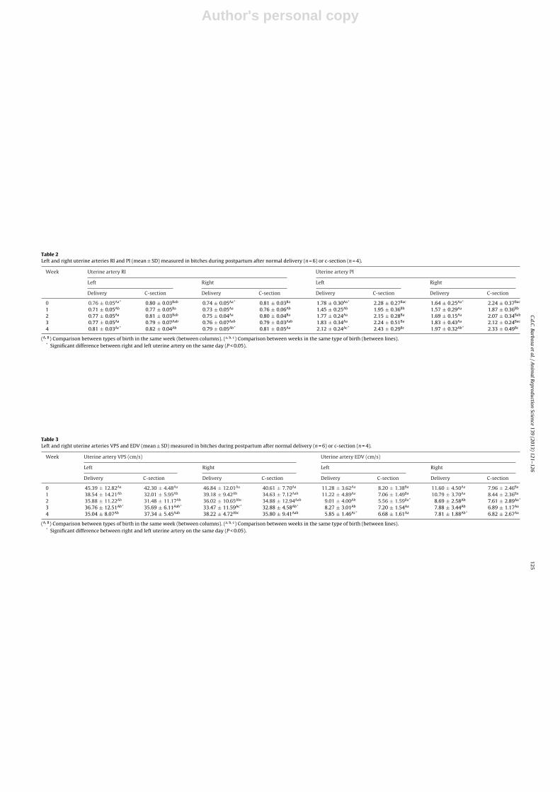
Table 2Left and right uterine arteries RI and PI (mean ± SD) measured in bitches during postpartum after normal delivery (n = 6) or c-section (n = 4).
Week Uterine artery RI Uterine artery PI
Left Right Left Right
Delivery C-section Delivery C-section Delivery C-section Delivery C-section
0 0.76 ± 0.05Aa* 0.80 ± 0.03Bab 0.74 ± 0.05Aa* 0.81 ± 0.03Ba 1.78 ± 0.30Aa* 2.28 ± 0.27Bac 1.64 ± 0.25Aa* 2.24 ± 0.37Bac
1 0.71 ± 0.05Ab 0.77 ± 0.05Ba 0.73 ± 0.05Aa 0.76 ± 0.06Ab 1.45 ± 0.25Ab 1.95 ± 0.36Bb 1.57 ± 0.29Aa 1.87 ± 0.36Bb
2 0.77 ± 0.05Aa 0.81 ± 0.03Bab 0.75 ± 0.04Aa 0.80 ± 0.04Ba 1.77 ± 0.24Aa 2.15 ± 0.28Ba 1.69 ± 0.15Aa 2.07 ± 0.34Bab
3 0.77 ± 0.05Aa 0.79 ± 0.07Aab 0.76 ± 0.07Aab 0.79 ± 0.03Aab 1.83 ± 0.34Aa 2.24 ± 0.51Ba 1.83 ± 0.43Aa 2.12 ± 0.24Bac
4 0.81 ± 0.03Ac* 0.82 ± 0.04Ab 0.79 ± 0.05Ab* 0.81 ± 0.05Aa 2.12 ± 0.24Ac* 2.43 ± 0.29Bc 1.97 ± 0.32Ab* 2.33 ± 0.49Bc
(A, B) Comparison between types of birth in the same week (between columns). (a, b, c) Comparison between weeks in the same type of birth (between lines).* Significant difference between right and left uterine artery on the same day (P < 0.05).
Table 3Left and right uterine arteries VPS and EDV (mean ± SD) measured in bitches during postpartum after normal delivery (n = 6) or c-section (n = 4).
Week Uterine artery VPS (cm/s) Uterine artery EDV (cm/s)
Left Right Left Right
Delivery C-section Delivery C-section Delivery C-section Delivery C-section
0 45.39 ± 12.82Aa 42.30 ± 4.48Aa 46.84 ± 12.01Aa 40.61 ± 7.70Aa 11.28 ± 3.62Aa 8.20 ± 1.38Ba 11.60 ± 4.50Aa 7.96 ± 2.46Ba
1 38.54 ± 14.21Ab 32.01 ± 5.95Ab 39.18 ± 9.42Ab 34.63 ± 7.12Aab 11.22 ± 4.89Aa 7.06 ± 1.49Ba 10.79 ± 3.70Aa 8.44 ± 2.36Ba
2 35.88 ± 11.22Ab 31.48 ± 11.17Ab 36.02 ± 10.65Abc 34.88 ± 12.94Aab 9.01 ± 4.00Ab 5.56 ± 1.59Ba* 8.69 ± 2.58Ab 7.61 ± 2.89Aa*
3 36.76 ± 12.51Ab* 35.69 ± 6.11Aab* 33.47 ± 11.59Ac* 32.88 ± 4.58Ab* 8.27 ± 3.01Ab 7.20 ± 1.54Aa 7.88 ± 3.44Ab 6.89 ± 1.17Aa
4 35.04 ± 8.07Ab 37.34 ± 5.45Aab 38.22 ± 4.72Abc 35.80 ± 9.41Aab 5.85 ± 1.46Ac* 6.68 ± 1.61Aa 7.81 ± 1.88Ab* 6.82 ± 2.67Aa
(A, B) Comparison between types of birth in the same week (between columns). (a, b, c) Comparison between weeks in the same type of birth (between lines).* Significant difference between right and left uterine artery on the same day (P < 0.05).

Author's personal copy
126 C.d.C. Barbosa et al. / Animal Reproduction Science 139 (2013) 121– 126
hand, Serin and Tarimcilar (2011), evaluating the uter-ine artery in bitches at the uteroplacental site, observeda decrease in RI and PI between day 4 and 14 after delivery,however only one bitch was evaluated in this period.
The indexes and velocities of the uterine artery wereinfluenced by the side that was evaluated in some daysof the week in both types of delivery. This fact may berelated to the number of pups per uterine horn, as Pereiraet al. (2012) evaluating the uterine artery in cyclic andpregnant cats, found that the number of kittens per uterinehorn influence the outcome of the pulsatility and resistanceindexes.
This work is of fundamental importance because itallowed evaluation of the uterus and uterine artery dur-ing normal puerperium, showing a reduction of diameterand blood flow in the evaluated period, with occasionaldifferences between types of delivery in the process of uter-ine involution. This tool can help during ultrasonographicexamination to identify animals with normal puerperium.However more studies are needed to verify the Dopplerchanges according to the breed and the number of puppiesper uterine horn, as well as in pathological puerperium.
Acknowledgments
We are grateful to Grande Canafístula, Difirresus, andVivenda dos Pinheiros kennels, which supplied the dogsused in this study; to the Laboratory of CarnivorousReproduction for supplying the corresponding infrastruc-ture; and to CAPES, FUNCAP and CNPQ for funding thisstudy.
References
Al-Bassam, M.A., Thomson, R.G., O’Donnell, L., 1981. Normal postpartuminvolution of the uterus in the dog. Can. J. Comp. Med. 45, 217–232.
Alcázar, J.L., 1998. Transvaginal ultrasonography combined with colorvelocity imaging and pulsed Doppler to detect residual trophoblastictissue. Ultrasound Obstet. Gynecol. 11, 54–58.
Alvarez-Clau, A., Liste, F., 2005. Ultrasonographic characterization ofthe uterine artery in the nonestrus bitch. Ultrasound Med. Biol. 31,1583–1587.
Batista, P.R., Blanco, P.G., Tórtora, M., Arias, D.O., Corrada, Y.A., Gobello, C.,2012. Doppler ultrasonographic assessment of uterine arteries duringnormal canine puerperium. In: Proceeding of International Sympo-sium on Canine and Feline Reproduction, Canada.
Cortez, A.A., 2003. Acompanhamento reprodutivo de fêmeas suínas porultra-sonografia (Reproductive follow-up of swine by means of ultra-sonography). Dissertation (Master in Veterinary Sciences), Faculty ofVeterinary Sciences, Ceará State University.
Davidson, A.P., Baker, T.W., 2009. Reproductive ultrasound of the bitchand queen. Top. Comp. Anim. Med. 24, 55–63.
Di Salvo, P., Bocci, F., Zelli, R., Polisca, A., 2006. Doppler evaluation ofmaternal and fetal vessels during normal gestation in the bitch. Res.Vet. Sci. 81, 382–388.
England, G.C.W., Russo, M., 2006. Ultrasonographic characteristics of earlypregnancy failure in bitches. Theriogenology 66, 1694–1698.
England, G.C.W., Yeager, A.E., Concannon, P.W., 2003. Ultrasound imagingof the reproductive tract of the bitch. In: Concannon, P.W., England,G.C.W. (Eds.), Recent Advances in Small Animal Reproduction. Interna-tional Veterinary Information Service, Ithaca, NY http://www.ivis.org
Feldman, E.C., Nelson, R.W., 1986. Canine and Feline Endocrinology andReproduction. W.B. Saunders, Philadelphia.
Ferretti, L.M., Newell, S.M., Graham, J.P., Roberts, G.D., 2000. Radiographicand ultrasonographic evaluation of the normal feline postpartumuterus. Vet. Radiol. Ultrasound 41, 287–291.
Ferri, S.T.S., Vicente, W.R.R., 2002. Estudo ultra-sonográfico da involuc ãouterina pós-parto em cadelas (Ultrasonographic study of postpartumuterine involution in dogs). Arq. Bras. Med. Vet. Zootec. 54, 19–23.
Ferri, S.T.S., Vicente, W.R.R., Toniollo, G.H., 2003. Ultrasonographic studyof the postpartum uterine involution in bitches after cesarean section.Arq. Bras. Med. Vet. Zootec. 55, 167–172.
Hauser, B., Bostedt, H., 2002. Ultrasonographic observations of uterineregression in the ewe under different obstetrical conditions. J. Vet.Med. 49, 511–516.
Herzog, K., Bollwein, H., 2007. Application of Doppler ultrasonography incattle reproduction. Reprod. Dom. Anim. 42, 51–58.
Hoffmann, B., Riesenbech, A., Klein, R., 1996. Reproductive endocrinologyof bitches. Anim. Reprod. Sci. 42, 257–288.
Kelly, S.M., Belli, A.M., Campbell, S., 2003. Arteriovenous malformation ofthe uterus associated with secondary postpartum hemorrhage. Repro-duction 21, 602–605.
Köster, K., Nautrup, P.C., Günzel-Apel, A.R., 2001. A Doppler ultrasono-graphic study of cyclic changes of ovarian perfusion in the Beaglebitch. Reproduction 122, 453–461.
Krueger, L., Koerte, J., Tousis, G., Herzog, K., Flachowsky, G., Bollwein, H.,2009. Transrectal Doppler sonography of uterine blood flow duringthe first 12 weeks after parturition in health dairy cows. Anim. Reprod.Sci. 114, 23–31.
Miranda, S.A., Domingues, S.F.S., 2010. Conceptus ecobiometry and triplexDoppler ultrasonography of uterine and umbilical arteries for assess-ment of fetal viability in dogs. Theriogenology 74, 608–617.
Mortensen, C.J., Kelly, D.E., Warren, L.K., 2011. Supplemental l-arginineshortens gestation length and increases mare uterine blood flowbefore and after parturition. J. Equine Vet. Sci. 31, 514–520.
Nautrup, C.P., 1998. Doppler ultrasonography of canine maternal and fetalarteries during normal gestation. J. Reprod. Fertil. 112, 301–314.
Nijman, R.G.W., Martingh, A., Aarnoudse, J.G., 2002. Persistent retainedplacenta percreta: methotrexate treatment and Doppler flow charac-teristics. Int. J. Gynaecol. Obstet. 109, 587–588.
Okano, A., Tomizuka, T., 1987. Ultrasonic observation of postpartum uter-ine involution in the cow. Theriogenology 27, 369–372.
Pereira, B.S., Freire, L.M.P., Pinto, J.N., Domingues, S.F.S., Silva, L.D.M., 2012.Triplex Doppler evaluation of uterine arteries in cyclic and pregnantdomestic cats. Anim. Reprod. Sci. 130, 99–104.
Schoubroeck, D.V., Bosch, T.V.D., Scharpe, K., et al., 2004. Prospective eval-uation of blood flow in the myometrium and uterine arteries in thepuerperium. Ultrasound Obstet. Gynecol. 23, 378–381.
Serin, G., Tarimcilar, T., 2011. Obstetric Doppler ultrasound findings in aGerman shepherd bitch at pregnancy and puerperium. Maced. J. Anim.Sci. 1, 239–244.
Szatmári, V., Sótonyi, P., Vörös, K., 2001. Normal duplex Doppler wave-forms of major abdominal blood vessels in dogs: a review. Vet. Radiol.Ultrasound 42, 93–107.
Teo, S.B.L., Kanagalingam, D., Tan, H.-K., Tan, L.-K., 2008. Massive post-partum haemorrhage after uterus-conserving surgery in placentapercreta: the danger of the partial placenta percreta. Int. J. Gynaecol.Obstet. 115, 789–792.
Yeager, A.E., Concannon, P.W., 1990. Serial ultrasonographic appearanceof postpartum uterine involution in beagle dogs. Theriogenology 38,523–535.
Related Documents


![MILES BITCHES · 2006. 2. 10. · JAZZ DOOR (G) JD 1284/5 ANOTHER BITCHES BREW JAZZ DOOR (G) JD 1284/5 MILES DAVIS ANOTHER BITCHES BREW Disc 1 [67:23 m] Medley [incomplete] Nov 3,](https://static.cupdf.com/doc/110x72/612732d1df21a4630d67d7e6/miles-2006-2-10-jazz-door-g-jd-12845-another-bitches-brew-jazz-door-g.jpg)