Chapter 3 Assessment of Safety Standards of Magnetic Resonance Imaging at the Korle Bu Teaching Hospital (KBTH) in Accra, Ghana Samuel Opoku, William Antwi and Stephanie Ruby Sarblah Additional information is available at the end of the chapter http://dx.doi.org/10.5772/52699 1. Introduction The various equipment and chemicals used in the radiology departments can be a source of hazards and hence result in an adverse effect to affected individuals (Johnston and Killion, 2005). Interdisciplinary approach to monitor the activities at radiology departments to en‐ sure compliance in safety standards may help avoid or reduce hazards in the working envi‐ ronment (Byrnset al., 2000). Magnetic Resonance Imaging (MRI) unit in a radiology department is one particular place where safety precautions should be directed due to the ferromagnetic nature of the equipment and the strong magnetic field used in its operations (Joseph, 2006). MRI is a painless, non-invasive and one of the most advanced imaging modalities current‐ ly available in radiology departments (Kusumasuganda, 2010). Research and awareness of safety issues concerning MRI has received much attention (Ordridgeet al., 2000). Accord‐ ing to Westbrook et al (2009), recent occurrences in the operation of MRI have led to ques‐ tions being raised on the safety of the modality. Phin (2001) has suggested that adequate policies and procedures should be developed and adhered to in order to ensure safe, effi‐ cient and operating conditions of MRI. Several potential problems and hazards are associ‐ ated with the performance of patient monitoring and support in the MRI environment (Kanal and Shellock, 1992). According to Henner and Servomaa (2010), the main factors that affect safety practice in the MRI unit is management style and attitude of staff. Vari‐ ous reports found in the literature have indicated that MRI accidents are mostly caused by human errors rather than scanner malfunction. These have led to several calls for regu‐ © 2013 Opoku et al.; licensee InTech. This is an open access article distributed under the terms of the Creative Commons Attribution License (http://creativecommons.org/licenses/by/3.0), which permits unrestricted use, distribution, and reproduction in any medium, provided the original work is properly cited.

Welcome message from author
This document is posted to help you gain knowledge. Please leave a comment to let me know what you think about it! Share it to your friends and learn new things together.
Transcript
Chapter 3
Assessment of Safety Standards of MagneticResonance Imaging at the Korle Bu Teaching Hospital(KBTH) in Accra, Ghana
Samuel Opoku, William Antwi andStephanie Ruby Sarblah
Additional information is available at the end of the chapter
http://dx.doi.org/10.5772/52699
1. Introduction
The various equipment and chemicals used in the radiology departments can be a source ofhazards and hence result in an adverse effect to affected individuals (Johnston and Killion,2005). Interdisciplinary approach to monitor the activities at radiology departments to en‐sure compliance in safety standards may help avoid or reduce hazards in the working envi‐ronment (Byrnset al., 2000). Magnetic Resonance Imaging (MRI) unit in a radiologydepartment is one particular place where safety precautions should be directed due to theferromagnetic nature of the equipment and the strong magnetic field used in its operations(Joseph, 2006).
MRI is a painless, non-invasive and one of the most advanced imaging modalities current‐ly available in radiology departments (Kusumasuganda, 2010). Research and awareness ofsafety issues concerning MRI has received much attention (Ordridgeet al., 2000). Accord‐ing to Westbrook et al (2009), recent occurrences in the operation of MRI have led to ques‐tions being raised on the safety of the modality. Phin (2001) has suggested that adequatepolicies and procedures should be developed and adhered to in order to ensure safe, effi‐cient and operating conditions of MRI. Several potential problems and hazards are associ‐ated with the performance of patient monitoring and support in the MRI environment(Kanal and Shellock, 1992). According to Henner and Servomaa (2010), the main factorsthat affect safety practice in the MRI unit is management style and attitude of staff. Vari‐ous reports found in the literature have indicated that MRI accidents are mostly causedby human errors rather than scanner malfunction. These have led to several calls for regu‐
© 2013 Opoku et al.; licensee InTech. This is an open access article distributed under the terms of the CreativeCommons Attribution License (http://creativecommons.org/licenses/by/3.0), which permits unrestricted use,distribution, and reproduction in any medium, provided the original work is properly cited.
lations and policies to guide the operations of MRI (New York Times, 2010; HealthcarePurchasing News, 2010). This has become necessary because the risk in the MRI environ‐ment does not only affect the patient, but also affects the health professionals and thosewho find themselves in the magnetic field (Kanal et al., 2007). There is therefore the needfor maximum safety to be ensured in the MRI unit. Moreover Chaljub (2003) and Joseph(2006) have both emphasized the need to keep training health personnel on safety issuesrelating to MRI. In particular, Joseph reiterated that the MRI’s magnet which is over100,000 times the earth’s natural magnetic pull is always on mode; hence those who ap‐proach it should have training due to the special safety risk it poses. In addition to therisks to people, it is also important to put in precautionary measures to protect the equip‐ment from damage and breakdowns. The need to assess the staff of the radiology depart‐ment and hospital’s management on their attitude and adherence towards maintainingsafety at the MRI can therefore not be overemphased.
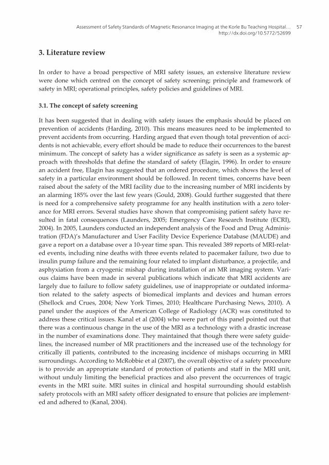
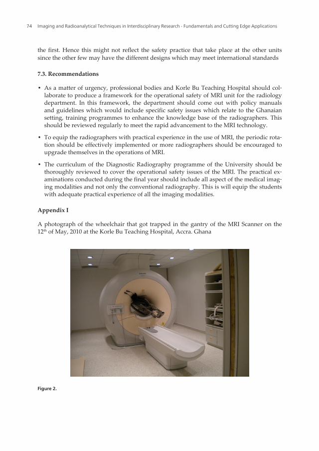
In recent times, Magnetic Resonance Imaging (MRI) unit of the Korle - Bu Teaching Hospital(KBTH) in Accra, Ghana has witnessed various degrees of accidents. In particular, there wasa fire outbreak in 2007 which brought the operation of the MRI facility down for a wholeyear. Again in 2010, a wheelchair was pulled into the gantry of the MRI scanner by the mis‐sile effect when a patient was lifted off the wheelchair onto the MRI table as shown in ap‐pendix I on page 28. This incident resulted in three weeks down time of the facility. Asecond incident in the same year occurred where a Radiographer Intern at the MRI unitwrongly switched off the safety button, resulting in three weeks shut-down of the entireunit. These incidences have been documented in the Incidence Reporting Book at the MRIUnit and are reproduced here with the permission from the Radiology Department of thehospital. These incidences at MRI Unit at the Korle Bu Teaching Hospital are very worrying,suggesting that the safety aspects might have been compromised. Thus it is imperative thatthe existence of policy guidelines and manuals regarding the operational safety of the MRIin the hospital and their compliance and adherence by staff needed to be evaluated. Similarincidences occurring in other hospitals around the world are documented in the literatureand some of which are reproduced in Appendix II on page 29.
2. Materials and methodology
This study was undertaken at the MRI Unit at Korle Bu Teaching Hospital in Accra, Ghana.The specific objectives of study were to identify safety policies regarding the operations ofthe MRI unit and whether they conform to international standards. Additionally, it soughtto ascertain adherence and compliance of the policy guidelines and to evaluate the designfeatures of the MRI suite for its safety compatibility as well as to determine the safety train‐ing needs of radiographers who operate the MRI.
The study focused on the safe use of MRI as an imaging modality and involved radiogra‐phers of the Radiology Department. A member of the Hospital Management Team also par‐ticipated in the study since the management members are responsible for the safety policiesformulation and ensuring their implementation at the MRI unit.
Imaging and Radioanalytical Techniques in Interdisciplinary Research - Fundamentals and Cutting Edge Applications56
3. Literature review
In order to have a broad perspective of MRI safety issues, an extensive literature reviewwere done which centred on the concept of safety screening; principle and framework ofsafety in MRI; operational principles, safety policies and guidelines of MRI.
3.1. The concept of safety screening
It has been suggested that in dealing with safety issues the emphasis should be placed onprevention of accidents (Harding, 2010). This means measures need to be implemented toprevent accidents from occurring. Harding argued that even though total prevention of acci‐dents is not achievable, every effort should be made to reduce their occurrences to the barestminimum. The concept of safety has a wider significance as safety is seen as a systemic ap‐proach with thresholds that define the standard of safety (Elagin, 1996). In order to ensurean accident free, Elagin has suggested that an ordered procedure, which shows the level ofsafety in a particular environment should be followed. In recent times, concerns have beenraised about the safety of the MRI facility due to the increasing number of MRI incidents byan alarming 185% over the last few years (Gould, 2008). Gould further suggested that thereis need for a comprehensive safety programme for any health institution with a zero toler‐ance for MRI errors. Several studies have shown that compromising patient safety have re‐sulted in fatal consequences (Launders, 2005; Emergency Care Research Institute (ECRI),2004). In 2005, Launders conducted an independent analysis of the Food and Drug Adminis‐tration (FDA)’s Manufacturer and User Facility Device Experience Database (MAUDE) andgave a report on a database over a 10-year time span. This revealed 389 reports of MRI-relat‐ed events, including nine deaths with three events related to pacemaker failure, two due toinsulin pump failure and the remaining four related to implant disturbance, a projectile, andasphyxiation from a cryogenic mishap during installation of an MR imaging system. Vari‐ous claims have been made in several publications which indicate that MRI accidents arelargely due to failure to follow safety guidelines, use of inappropriate or outdated informa‐tion related to the safety aspects of biomedical implants and devices and human errors(Shellock and Crues, 2004; New York Times, 2010; Healthcare Purchasing News, 2010). Apanel under the auspices of the American College of Radiology (ACR) was constituted toaddress these critical issues. Kanal et al (2004) who were part of this panel pointed out thatthere was a continuous change in the use of the MRI as a technology with a drastic increasein the number of examinations done. They maintained that though there were safety guide‐lines, the increased number of MR practitioners and the increased use of the technology forcritically ill patients, contributed to the increasing incidence of mishaps occurring in MRIsurroundings. According to McRobbie et al (2007), the overall objective of a safety procedureis to provide an appropriate standard of protection of patients and staff in the MRI unit,without unduly limiting the beneficial practices and also prevent the occurrences of tragicevents in the MRI suite. MRI suites in clinical and hospital surrounding should establishsafety protocols with an MRI safety officer designated to ensure that policies are implement‐ed and adhered to (Kanal, 2004).
Assessment of Safety Standards of Magnetic Resonance Imaging at the Korle Bu Teaching Hospital…http://dx.doi.org/10.5772/52699
57
3.2. MRI suite design and zones
An MRI suite should be designed to restrict access and limit exposure to static magneticfields. Various publications have provided different designs to the MRI suite to ensure max‐imum safety and they all showed that an MRI suite should be built to restrict access byzones (Gould 2008; Kanal et al., 2007; Junk and Gilk, 2005; Shellock and Crues, 2004). Thezones suggested by the various articles are as follows;
Zone I: - Opens to the general public and presents the least exposure to the patients, staffand visitors. Usually it is the reception and waiting room for the MRI suite purposed tochannel patients and medical staff to the pre-screening area (zone II) and limit entry to theMRI suite.
Zone II: - This is the first interaction site for patients, visitors and staff in the MRI suite. Thepurpose of this zone is to restrict further public access to the suite and provide direct super‐vision of patients and visitors by the MRI staff. Pre-screening of all patients, staff and visi‐tors also takes place here. If ambulatory, the patient is screened through a ferrous metaldetector installed into the zone II. Non ambulatory patients in walkers, wheelchairs or pa‐tient support need the transport equipment to be verified as MRI- safe or exchanged forMRI- safe equipment. The zone II generally has a metal detector and a 1000 gauss magnet tohelp screen medical equipment for ambulatory patients. MRI staff including the MRI tech‐nologist is directly responsible for enforcing strict adherence to the MRI safety protocols forthe MRI suite and patient safety.
Zone III: - This is the entry zone to the MRI machine room which is zone IV. Entrance to thiszone is restricted physically and by protocol. Being the last barrier against an incident or in‐jury due to an interaction of a static or active magnetic field and any unscreened personnel,patient or equipment, only MRI technologist, certified staff and pre-screening attendingphysician accompany the patient into the MRI machine room. The portal or entrance to theMRI machine room must be monitored by a second ferromagnetic- sensitive detector anddoor must be locked. Sounding of detector will require verification of either an MRI- safe orcompatible event or the discovery of an MRI–unsafe condition in the patient, transporting ormedical equipment or the attending medical staff. The standard access method is a card ac‐cess system which should allow access to only certified MRI staff between zone III and zoneIV. All medical staff must be pre-screened prior to entry into zone III to make sure nounscreened individuals will be allowed access to zone IV. Ideally, the personnel in zone IIImust be uniformed in MRI compatible scrubs which will avoid the use of identificationbadges in the suite, MRI-safe shoes and undergarments. Personnel must avoid all jewellery,watches metallic writing instruments, and wire-framed glass which may raise a false alarmfrom the detector (Shellock and Crues, 2004).
Finally in zone IV, the MRI room should have a clear demarcation of the five gauss line tap‐ed or painted on the MRI suite floor to indicate the area beyond which requires MRI- safe orMRI – conditional equipment or instrumentation. This should be in line with the distanceand tesla rating of the MRI. Zone IV should be clearly marked with a red light and lightedsign stating, “The Magnet is on.” In situations where an alarm goes off for a code red, there
Imaging and Radioanalytical Techniques in Interdisciplinary Research - Fundamentals and Cutting Edge Applications58
is the need to use MRI- safe equipments to address the situation with restrictions of publicfirst responders from zone IV of the MRI environment until safe conditions are establishedor responders are verified to be safe (Junk and Gilk, 2005). Access to zone IV should be ena‐bled by a programmed key and the key kept in a restricted access box in the MRI controlroom.
Architectural and design engineering for a MRI suite have been established in standardspublished by the Joint Commission on the Accreditation of Health Organisations (JCAHO),the American College of Radiology guidelines, the International Building Code (IBC) andOccupational Safety and Health Administration (OSHA).
3.3. Pre- screening and screening forms
Shellock and Crues (2004) emphasized that the establishment of thorough and effectivescreening procedures for patients and other individuals is one of the most critical compo‐nents of a programme to guard the safety of all those preparing to undergo MR proce‐dures or to enter the MR environment. All preliminary patient history, MRI safetyscreening and documentation must be completed and signed by the patient, guardian orclinician before procedures are undertaken on patients (Shellock and Crues, 2004; Ferris etal., 2007). Various screening forms are used for different categories of people who come tothe MRI suite. In general, screening forms are developed with patients in mind (Sawyer-Glover and Shellock, 2002).
3.4. Colour and symbol coding
Various means have been adopted to help with ensuring safety in an MRI unit. This is usu‐ally meant to provide on the spot recognition of MR- safe equipment and surroundings,likewise unsafe and MRI-conditional equipments and locations. At the University of Califor‐nia San Francisco (2011), yellow is used to signify caution and is painted around the en‐trance of the door. Gas tanks that have been painted green signifies ferrous cylinder andhence make it easy for identification as MRI unsafe equipment. For safe MRI tanks, the cyl‐inders are coloured silver. Symbols have also been used as a new classification system forimplants and ancillary clinical devices.
An MRI safe symbol signifies that the device or implant is completely non-magnetic, non-electrically conductive, and non-RF reactive, eliminating all of the primary potential threatsduring an MRI procedure. An MRI Conditional sign is used to identify a device or implantthat may contain magnetic, electrically conductive or RF-reactive components that is safe foroperations in proximity to the MRI, provided the conditions for safe operation are definedand observed (such as 'tested safe to 1.5 teslas' or 'safe in magnetic fields below 500 gauss instrength). Finally, an MRI unsafe symbol is reserved for objects that are significantly ferro‐magnetic and pose a clear and direct threat to persons and equipment within the magnetroom. An appropriate coding system is thus necessary to be adopted by every MRI unit tofacilitate easy identification of safe items.
Assessment of Safety Standards of Magnetic Resonance Imaging at the Korle Bu Teaching Hospital…http://dx.doi.org/10.5772/52699
59
3.5. Operational principles of MRI
As opposed to conventional x-rays and computed tomography (CT) scans, there is no ioniz‐ing radiation used in MRI. However, MRI uses an extremely powerful static magnetic field,rapidly changing gradient magnetic fields and radiofrequency electromagnetic impulses toobtain detailed anatomic or functional images of any part of the body (Faulker, 2002; Berger,2002). Currently, there is no evidence of a short or long term adverse effect due to exposureto field strengths of MRI and durations that is clinically used (Schenck, 2000).
Despite the relative safety of MRI, there are potential hazards associated with its operations.Some of these are related to the physical properties of the MRI equipment and also to thechallenges of maintaining physiologic stability of the individual undergoing the examina‐tion. In a reported incident in 2001,a small boy undergoing an MRI following surgery to re‐move a benign tumour was struck and killed by an oxygen tank inadvertently taken into theMRI suite (Emergency Care Research Institute, 2001). In most situations the MR systemscause the disaster due to it interactions with other properties around it.
3.6. Magnetic fields and the missile effect
The static magnetic field generated by a powerful magnet is tens of thousands timesstronger than the earth’s magnetic field which can attract objects containing ferrous mate‐rials, transforming them into dangerous airborne projectiles (Dempsey et al., 2002). Thereare two features of the magnetic field that are the source of most MRI incidents; the pro‐jectile or missile effect which is the ability of the magnet to attract ferromagnetic objectsand draw them rapidly into the bore with considerable force (Centre for Devices and Ra‐diological Health, 1997). Ferromagnetic objects include metallic objects containing ironsuch as scissors, laryngoscopes, nail clippers, pocket knives and steel buckets. Largeritems like wheelchairs, gurneys, intravenous poles have also become MR-system- inducedmissiles (Centre for Devices and Radiological Health, 1997). The other source of most MRIincidents is the translational attraction which occurs when one point of an object in amagnetic field is attracted to a great extent than the object’s furthest point from the at‐tracting source (Gould, 2008).
3.7. Magnetic field interactions
The static magnetic field of an MR system is always on. No sound, sight, smells alerts per‐sonnel to the presence or the extent of the invisible field surrounding the magnet in all direc‐tions. The magnetic pull is strongest at the centre of the MR system and weakens withincreased distance from the magnet, creating a spatial magnetic field gradient (Price, 1999).The distribution of the magnetic field outside the main magnet called fringe field is impossi‐ble to see, but it is critical to safety in the MR environment because it can determine whethera ferromagnetic object could become a projectile. MR systems with large fringe field general‐ly create the greatest hazards (Price, 1999). If the fringe strength decreases more graduallywith distance from the magnet, the object’s attraction to the magnet progressively strength‐ens as it becomes closer to the magnet. Personnel within the MR room may notice an in‐
Imaging and Radioanalytical Techniques in Interdisciplinary Research - Fundamentals and Cutting Edge Applications60
creasingly stronger pull on objects they are wearing or carrying as they walk closer to theMR system, permitting them to retreat from the MR system before an accident occurs (Kanalet al., 2002).
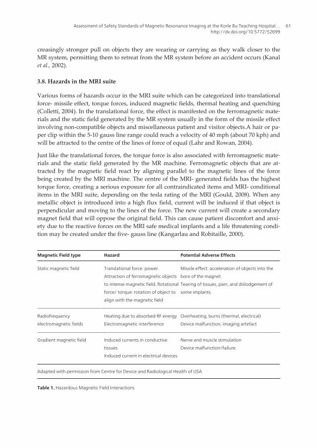
3.8. Hazards in the MRI suite
Various forms of hazards occur in the MRI suite which can be categorized into translationalforce- missile effect, torque forces, induced magnetic fields, thermal heating and quenching(Colletti, 2004). In the translational force, the effect is manifested on the ferromagnetic mate‐rials and the static field generated by the MR system usually in the form of the missile effectinvolving non-compatible objects and miscellaneous patient and visitor objects.A hair or pa‐per clip within the 5-10 gauss line range could reach a velocity of 40 mph (about 70 kph) andwill be attracted to the centre of the lines of force of equal (Lahr and Rowan, 2004).
Just like the translational forces, the torque force is also associated with ferromagnetic mate‐rials and the static field generated by the MR machine. Ferromagnetic objects that are at‐tracted by the magnetic field react by aligning parallel to the magnetic lines of the forcebeing created by the MRI machine. The centre of the MRI- generated fields has the highesttorque force, creating a serious exposure for all contraindicated items and MRI- conditionalitems in the MRI suite, depending on the tesla rating of the MRI (Gould, 2008). When anymetallic object is introduced into a high flux field, current will be induced if that object isperpendicular and moving to the lines of the force. The new current will create a secondarymagnet field that will oppose the original field. This can cause patient discomfort and anxi‐ety due to the reactive forces on the MRI safe medical implants and a life threatening condi‐tion may be created under the five- gauss line (Kangarlau and Robitaille, 2000).
Magnetic Field type Hazard Potential Adverse Effects
Static magnetic field Translational force: power.
Attraction of ferromagnetic objects
to intense magnetic field. Rotational
force/ torque: rotation of object to
align with the magnetic field
Missile effect: acceleration of objects into the
bore of the magnet.
Tearing of tissues, pain, and dislodgement of
some implants.
Radiofrequency
electromagnetic fields
Heating due to absorbed RF energy
Electromagnetic interference
Overheating, burns (thermal, electrical)
Device malfunction; imaging artefact
Gradient magnetic field Induced currents in conductive
tissues
Induced current in electrical devices
Nerve and muscle stimulation
Device malfunction/failure
Adapted with permission from Centre for Device and Radiological Health of USA
Table 1. Hazardous Magnetic Field Interactions
Assessment of Safety Standards of Magnetic Resonance Imaging at the Korle Bu Teaching Hospital…http://dx.doi.org/10.5772/52699
61
The most common source of thermal exposure tends to be looped or un-looped medicalequipment leads, MRI accessories and sensors. The most serious exposure is located inthe bore of the MRI machine and in the axis points, as they possess the highest potentialtorque forces. Extremity coils could increase the risk but this can be avoided by the useof MRI safe polymeric foam padding (Gilk, 2006). MRI machines are cooled by a supercooling fluid (liquid helium). The release of the super cooling fluid into the atmosphereis called quenching. Most clinical machines have about 700 to 1000 litre volume of thiscryogenic. In the event that there is venting, it may cause the oxygen in the MRI roomto condense around the vent pipe and accumulate in the MRI machine causing a red firehazard. Another risk is a quench vent pipe breech which could flood the room withcryogenic fluids creating an asphyxiation hazard for the patient and the staff (Clark,2007).
3.9. Radiofrequency electromagnetic fields effects
The MRI system has electromagnetic coils in a transmitter within it that delivers the radio‐frequency (RF) pulses during imaging. When tissues absorb the RF energy, tissue heatingcan occur, mostly in patients with poor thermoregulatory control (Dempsey et al., 2002). Therate at which RF energy is deposited in tissue is known as the specific absorption rate (SAR),measured in units of watts per kilogram (w/kg) (Centre for Devices and RadiologicalHealth, 1997). The maximum allowed SAR is 3W/kg which is averaged over ten minutes forhead imaging and 4W/kg for whole body imaging, averaged over fifteen minutes (Centrefor Devices and Radiological Health, 2003).
Radiofrequency fields can cause skin burns if monitor cables or wires are permitted toform conductive loops with themselves or with other body parts (Kanal et al., 2002).Temporary metallic intra cardiac pacing wires will behave like antennae and conductelectromagnetic waves, also resulting in thermal tissue injury (Dempsey et al., 2002). Ra‐diofrequency signals emitted during the MR examination can affect non- MR-compatibleprogrammed infusion pumps, resulting in erratic performance. Affected pumps could de‐liver higher or lower than desired volumes of pressor agent, analgesics, sedative or dex‐trose and electrolytic solutions, all of which cause serious physiological consequencesparticularly, infants (Cornetteet al., 2002).
3.10. Gradient magnetic field effects
When an infant is subjected to sudden, rapidly changing gradient magnetic fields duringimaging, the magnetic field can induce circulating currents in conductive tissues of the body(Schaefer et al., 2000). These currents have been found to be large enough to producechanges in nerves and muscles function theoretically.Where safety standard limits are prac‐ticed, it limits the maximum rate of change of magnetic field strength that can be used thusreducing the likelihood of its observation during a clinical MRI (Center for Devices and Ra‐diological Health, 2003).
Imaging and Radioanalytical Techniques in Interdisciplinary Research - Fundamentals and Cutting Edge Applications62
3.11. Safety policies and guidelines of MRI
The American College of Radiology (ACR) Guidance Document for Safe MRI Practices-2007recommends that all MRI sites should maintain MR safety policies (Kanal et al., 2007). Thesepolicies, it claims should be reviewed concurrently with the introduction of any significantchanges in the safety parameters of the MR environment and updated as needed. It also stat‐ed that Site Administration is responsible to ensure that the policies and procedures are im‐plemented and adhered to by all site personnel. Any adverse events, MR safety incidents ornear incidents are to be reported and used in continuous quality improvement efforts. Toaugment the recommendations made by the ACR, the 2008 Joint Commission Sentinel Alertissued by the Medical College of Wisconsin’s (2009) accreditation organisation suggestedthat actions consistent with the ACR recommendations should be used to prevent accidentsand injuries in the MRI suite. In other works, the Device Bulletin (2007) produced a docu‐ment to serve as guidelines covering important aspects of MRI equipment in clinical usewith specific reference to safety. They were intended to bring to the attention of those in‐volved with the clinical use of such equipment, important matters requiring careful consid‐eration before purchase and after installation of the equipment. It was also to be used as anorientation for those who are not familiar with the type of equipment and act as a reminderfor those who are familiar with the equipment (Buxton and Lui, 2007). It was further intend‐ed to act as a reminder of the legislation and published guidance relating to MRI, draw theattention of the users to the guidance published by the National Radiological ProtectionBoard (NRPB), its successor the Health Protection Agency (HPA), the International Electro‐chemical Commission (IEC) and the International Commission on Non –Ionizing RadiationProtection (ICNIRP)
4. Materials and method
The study employed both qualitative and quantitative design using a structured interviewand descriptive survey. A structured interview involves guiding the interview in a particu‐lar pattern such that the information received falls in line with the objective of the studywithout it being altered by the interviewer (Brink and Wood, 1994; Pontin, 2000). A descrip‐tive survey provides a better means of investigating and assessing the attitude and practicesof people when they are involved in a particular situation (Carter, 2000; Gray, 2004).
The study was carried out MRI Suite of the Radiology Department of the Korle Bu TeachingHospital. (KBTH), Accra. Ghana. KBTH is the leading referral hospital in Ghana, with theradiology department being one of the busiest departments in the hospital. Currently, thehospital has a bed capacity of about 2000, with an average 1,500 outpatient attendances dai‐ly, an admission rate of 250 per day and 65% of the daily attendance visiting the radiologydepartment (www.korlebuhospital.org).
The Radiology Department of the hospital has a staff population of forty-six. These includethirty-one radiographers, nine radiology residents and six consultant radiologists. Of the
Assessment of Safety Standards of Magnetic Resonance Imaging at the Korle Bu Teaching Hospital…http://dx.doi.org/10.5772/52699
63
thirty one radiographers, twelve are degree holders, fifteen are diploma holders and the restare certificate holders.
The entire population of radiographers was used for the study. This gave a population sizethat was easy to handle and ensure an effective statistical analysis to be done (Burns andGrove, 2001). Using a small data set makes it possible to overcome the inconveniences creat‐ed by lack of time, ensures homogeneity, improves the accuracy and quality of the data (At‐kinson, 2000; Aderet al., 2008). In Korle Bu Teaching Hospital, there are no specialized orpermanent MRI radiographers and all of them rotate periodically to the MRI unit; hence thereason for using the entire population of radiographers for the study.
Polgar and Thomas (2000) emphasized that in any scientific research the primary considera‐tion is the protection of the rights and welfare of participants. Thus, ethical approval wassought from the Ethical Review Committee of the School of Allied Health Sciences, Collegeof Health Sciences, University of Ghana. Permission was also sought from the Dr. Frank G.Shellock (2002) to reproduce content in his work and from the Institute for Magnetic Reso‐nance Safety, Education and Research as well as the Radiology Department of the Korle BuTeaching Hospital for the use of facility for the study. Informed consent was sought fromparticipants in the form of written consent forms after the objectives of the study had beenexplained to them. They were assured of their anonymity, confidentiality of identity and in‐formation provided.
A self- administered open and closed ended survey questionnaire was used to obtain datafrom the participants. Before the main study, a pilot study involving three radiographerswas conducted to assess the validity and reliability of the questionnaires. A modified check‐list designed by Gillies (2002) was attached to the pilot phase for respondents to make sug‐gestions that helped to modify the questionnaire as required. Ambiguity was thus removed;clarity of the format and design adopted was also ensured (Bailey, 1997).
A structured interview also was conducted with a member of the hospital management.This was to obtain additional data, validate and verify results obtained from the survey (Pol‐gar and Thomas, 2000). Policy formulation and supervision of implementation is the respon‐sibility of management of the hospital and the department (Beddoe et al., 2004). Thusinterviewing members of the management was considered the best way to obtain detailedand comprehensive information about safety management.
Questions on the framework of operational safety of the MRI unit, training programmes andpractical safety problems faced by the MRI unit were among other things asked during theinterview. The interview was electronically recorded, transcribed and data grouped intothemes and analyzed.
The data obtained from the questionnaires was rearranged in an ordered manner to enhanceits processing by the Statistical Package for Social Sciences (SPSS) version 16.0. Nominal andordinal levels of measurement were used because the study design was a descriptive survey(Burns and Grove, 2001). Results were presented using descriptive statistics in the form ofcharts, frequency tables and percentages.
Imaging and Radioanalytical Techniques in Interdisciplinary Research - Fundamentals and Cutting Edge Applications64
5. Results
This study investigated the availability of safety policies and guidelines and adherence tothem by staff at the MRI suite of the Korle-Bu Teaching Hospital. It also investigated the de‐sign features of the MRI suite as to whether it meets the acceptable safety standards. A totalof thirty-one closed ended questionnaires (31) were distributed to all the practicing radiog‐raphers twenty eight (28) questionnaires were completed and returned completed giving aresponse rate of 90.3% (n=28/31).
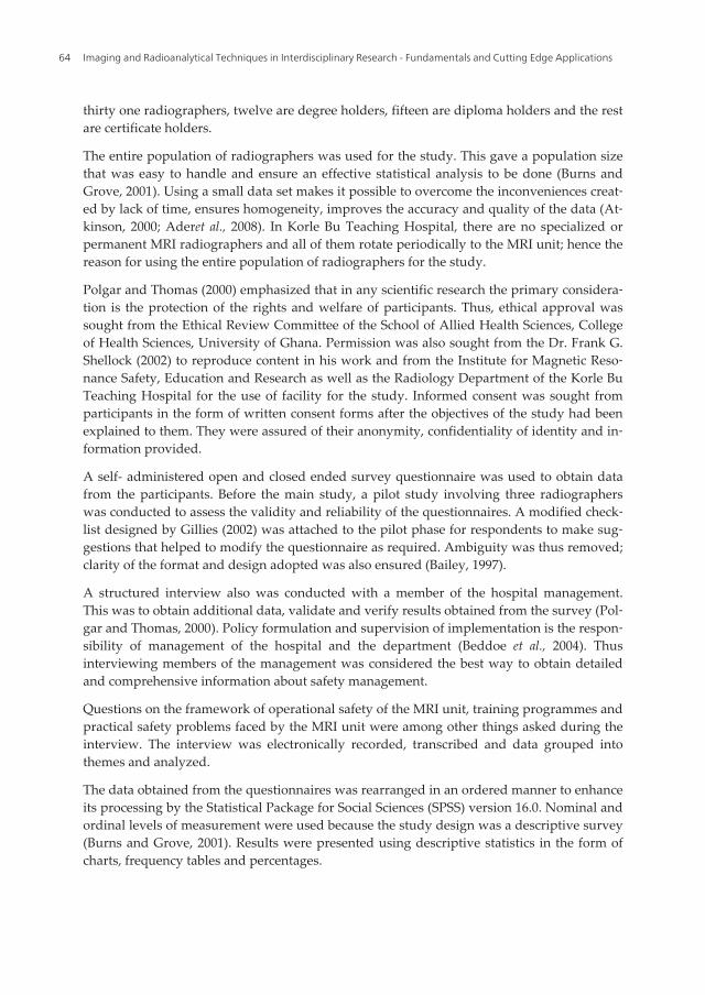
Professional qualification heldTotal
Gender of Respondents Certificate Diploma Bachelor's degree
Male2 10 9 21
7.1% 35.7% 32.1% 75.0%
Female2 2 3 7
7.1% 7.1% 10.7% 25.0%
Total4 12 12 28
14.3% 42.9% 42.9% 100.0%
Table 2. Demographic Data of the Respondents
The profile above shows that the ratio of male to female respondents was 3:1. Nearly half ofthe respondents (42.9%) were both diploma and degree holders respectively
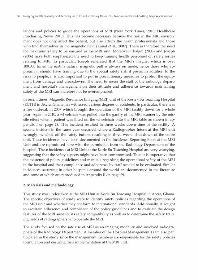
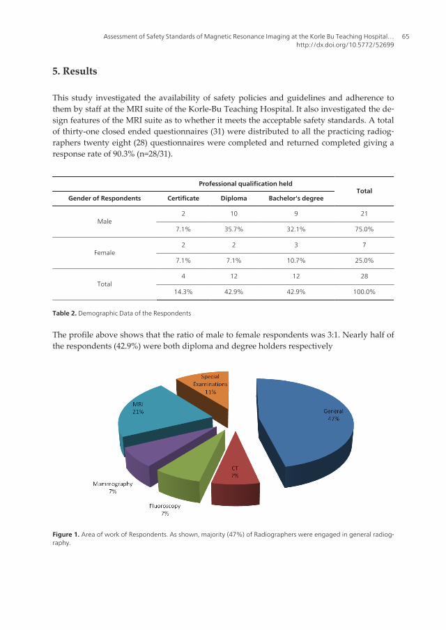
Figure 1. Area of work of Respondents. As shown, majority (47%) of Radiographers were engaged in general radiog‐raphy.
Assessment of Safety Standards of Magnetic Resonance Imaging at the Korle Bu Teaching Hospital…http://dx.doi.org/10.5772/52699
65
Question
Code QUESTIONSResponses to MRI Safety issues
TotalYes No
Q1 Is there a restricted access to everyone who
comes to the MRI Suite26 (92.9%) 2 (7.1 %) 28 (100.0%)
Q2 Do you undertake screening of patients who
enter the MRI Suite?21 (75.0%) 7 (25.0%) 28 (100.0%)
Q3 Do you undertake screening of staff who
enters the MRI Suite?10 (35.7 %) 18 (64.3 %) 28 (100.0%)
Q5 Are there lockers to store personal
belongings that may be ferrous in nature or
has a magnetic stripes in the MRI Unit
16 (57.1%) 12 (42.9%) 28 (100.0%)
Q6 Is the equipment used in the MRI
environment checked by any authority,
deemed MRI safe and labelled as such prior
to implementation?
21 (75.0 %) 7 (25.0%) 28 (100.0%)
Q7 Does the equipment used in the MRI unit
have colour codes to identify ferrous material
and MRI safety material?
5 (17.9%) 23 (82.1) 28 (100.0%)
Q9 Do patients complete any MRI history and
assessmentform that addresses possible
contraindications prior to any MRI
procedure?
26 (92.9% ) 2 (7.1%) 28 (100.0%)
Q11 Are there proximity access doors and
emergency exits to MRI suite?15 (53.6%) 13 (46.4%) 28 (100.0%)
Q12 Do you face any problems in your bid to
ensure the safety of patients and staff in the
MRI unit?
14 (50.0%) 14 (50.0 %) 28 (100.0%)
Q14 Is there an assigned anaesthetist to the MRI
unit to undertake procedures that need
patients to be anesthetized?
7 (25.0%) 21 (75.0%) 28 (100.0%)
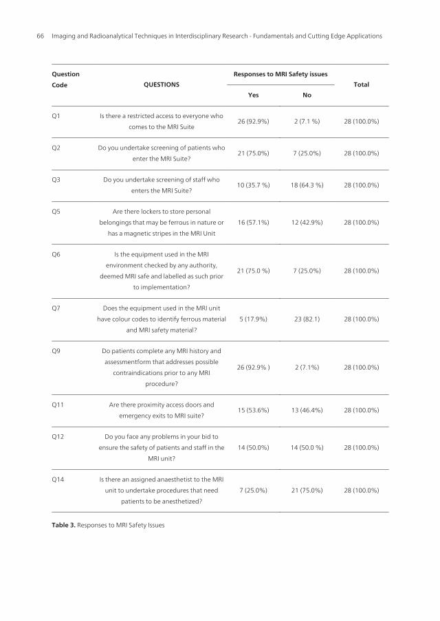
Table 3. Responses to MRI Safety Issues
Imaging and Radioanalytical Techniques in Interdisciplinary Research - Fundamentals and Cutting Edge Applications66
In Table 3, majority of 92.9 % (n=26/28) of the respondents stressed the need to restrict accessto the MRI suite. Additionally, 75.0 % (n= 21/28) of the respondents were of the view thatpatients should be screened before allowed to enter MRI suites. However, only ten out oftwenty eight (n= 10/28= 35.7%) suggested screening for radiographers’ (workers), beforethey enter the MRI suite.
Table 3 also shows that 57.1% (16/28) of the respondents mentioned the presence of lockersin the MRI suite to store personal belongings that may be ferrous in nature or has magneticstripes. Furthermore, 75% (n=21/28) reported that equipment used in the MRI environmentundergo regular quality check. However, 82.0% (23/28) of the respondents disclosed that theequipment in the MRI unit did not have colour codes to identify ferrous material and MRIsafety material.
Majority of the respondents (92.9%) reported that prior to procedure or examination, pa‐tients are asked to complete questionnaires to determine any contraindications they mayhave. A significant majority (53.6%) reported the presence of emergency exits to the MRIsuite. Finally 75.0% of the respondents were unaware of availability of anaesthesia servicesat MRI unit for patients who would require anaesthesia as part of the procedure.
Question
Code QUESTIONS
Responses to Safety MRI training
and unit design features Total
Yes No
Q15 Have you had any training programme(s) on
MRI safety issues?6 (21.4%) 22 (78.6%) 28 (100.0%)
Q16 Have you attended any of such training
programmes, if yes to question 18?4 (14.3%) 24 (85.7%) 28 (100.0%)
Q17 Do you know the MRI zones? 8 (28.6%) 20 (71.4 %) 28 (100.0%)
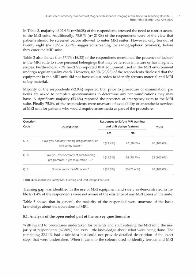
Table 4. Responses to Safety MRI Training and Unit Design Features
Training gap was identified in the use of MRI equipment and safety as demonstrated in Ta‐ble 4 71.4% of the respondents were not aware of the existence of any MRI zones in the suite.
Table 5 shows that in general, the majority of the responded were unaware of the basicknowledge about the operations of MRI.
5.1. Analysis of the open ended part of the survey questionnaire
With regard to procedures undertaken for patients and staff entering the MRI unit, the ma‐jority of respondents (67.86%) had very little knowledge about what were being done. Theremaining 32.14% had a fair idea but could not provide detailed description of the exactsteps that were undertaken. When it came to the colours used to identify ferrous and MRI
Assessment of Safety Standards of Magnetic Resonance Imaging at the Korle Bu Teaching Hospital…http://dx.doi.org/10.5772/52699
67
safe material, there was a poor appreciation, evident by the fact all the 28 respondents didnot know the existence of the colour identification of ferrous and MRI safe material.
QUESTIONS
Responses to MRI General Knowledge
Strongly
disagreeDisagree Undecided Agree
Strongly
agreeTotal
Q21.The magnet is only on
during the working day
14
(50.0%)
2
(7.1%)
3
(10.7%4 (14.3%) 5 (17.9%) 28 (100.0%)
Q22.A strong magnetic field
produces X-ray used for
imaging
13
(46.4%)
2
(7.1 %)
1
(3.6%)4 (14.3%) 8 (28.6%) 28 (100.0%)
Q23.A static magnetic field
strength may be up to 100,000
times the magnetic field
strength of the earth
1
(3.6%)
6
(21.4%)13 (46.4%) 4 (14.3%) 4 (14.3%) 28 (100.0%)
Q24.5 Gauss line is the
parameter around the MRI
system where field strength is
over 5 Gauss
0
(0.0%)
2
(7.1 %)
22
(78.6 %)
4
(14.3%)0 (0.0 %) 28 (100.0 %)
Q25.At 5Gausspacemakers may
be affected, ferrous items
become potential flying
projectiles and magnetic stripes
are erased
1
(3.6 %)
1
(3.6 %)
19
(67.9%)4 (14.3 %) 3 (10.7 %) 28 (100.0 %)
Q26.Below 5 Gaus is considered
to be a safe level of magnetic
field exposure to the public
0
(0.0%)
2
(7.1 %)20 (71.4 %) 2 (7.1 %) 4 (14.3 %) 28 (100.0%)
Q27.Magnetic field strength is
measured in Tesla (T) or Gauss
(G)
2
(7.1 %)
2
(7.1 %)3 (10.7 %) 8 (28.6 %) 13 (46.4 %) 28 (100.0 %)
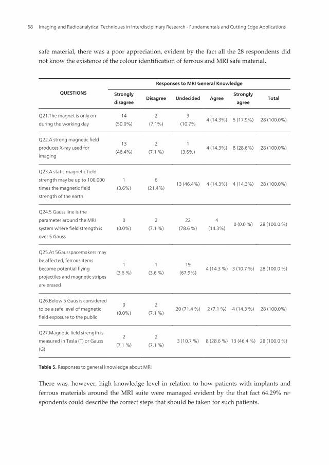
Table 5. Responses to general knowledge about MRI
There was, however, high knowledge level in relation to how patients with implants andferrous materials around the MRI suite were managed evident by the that fact 64.29% re‐spondents could describe the correct steps that should be taken for such patients.
Imaging and Radioanalytical Techniques in Interdisciplinary Research - Fundamentals and Cutting Edge Applications68
All the respondents cited the following as challenges in ensuring safety of patients and staffat the MRI unit;
• A communication gap between patients and health professionals
• Unwillingness of co-workers to comply with protocol used at the unit
• Small waiting area which is shared by the CT-scanning unit and the MRI unit
• The lack of knowledge on continuity of procedure that has been performed for patientsby referring clinicians.
On the issue of zoning in an MRI suite, 14.29% were aware of the different zones that areneeded in a standard MRI unit as against 96.4% of the respondents which were not aware ofthe colour used to indicate the different zones. According to 21. 43% of the respondents,zoning was completely absent at the MRI unit of the Korle Bu Teaching Hospital. The gener‐al overview of the results suggests a huge knowledge gap on the safety issues of MRI bymajority of the respondents.
5.2. The interview data
Thematic analysis was used to analyse the qualitative interview data. The predominantthemes that emerged were the context for framework for operational safety at MRI unit,availability of departmental policy manual and training programmes for MRI. The areasidentified included a maintenance programme for the MRI unit, structures in place for acci‐dents and breakdown of the MRI unit and practical problems faced in ensuring safety at theMRI unit.
The context for framework for operational safety at MRI unit in this study represents a com‐bination of organisational and operational methods that from the radiographers perspective,significantly affect the achievement of operational safety at MRI unit. It was noted that MRIwas a relatively new modality in the country with the suite at the Korle Bu Teaching Hospi‐tal which was in 2006 being the first in the country. It was further observed that there wasno documented formal framework by either the hospital or the department and that prepa‐rations were underway to produce one in accordance with best international standards. Thisobservation was consistent with the findings from the survey questionnaire indicating theabsence of a policy manual at the radiology department.
On the issue of continuous education and training of radiographers on MRI, it was notedthat this was non-existent. As stated earlier, there was no MRI specialized radiographer inthe department and any qualified radiographers who has basic knowledge in MRI could beassigned to the unit. The need to provide a platform to training and educate the practicingradiographers on MRI was identified.
On maintenance programme for the MRI unit, it was mentioned that the supplier of theequipment has a maintenance contract with the hospital to undertake routine maintenanceof the MRI.
Assessment of Safety Standards of Magnetic Resonance Imaging at the Korle Bu Teaching Hospital…http://dx.doi.org/10.5772/52699
69
On measures that were being taken to prevent further accidents, it was indicated that the staffwere required to report any incident or missed-incident to the appropriate authority. The ab‐sence of zoning in the suite and the adjoining CT scan suite made it difficult to undertake anyeffective screening because both MRI and CT scan patients have to enter through the same en‐trance. The absence of access codes for entrance into the MRI unit was also identified as a chal‐lenge to restricting access and this was attributed to defect in MRI – suite.
Other challenges identified include the attitude of some hospital personnel who were not will‐ing to comply with safety and security measures in place. The need to undertake some structur‐al adjustment to the unit was being considered to detach the CT suit from the MRI. Ensuringtotal commitment from both the management and staff of the hospital to safety and security is‐sues was identified as one of the main means to prevent accidents at the MRI unit.
6. Discussion
This research sought to investigate the availability of safety policies and guidelines and ad‐herence to them by staff at the MRI suite of the Korle-Bu Teaching Hospital. It also investi‐gated the design features of the MRI suite to ascertain whether it meets the acceptableinternational safety standards as these inevitably, affect patient care during MRI procedures.In this chapter, the findings are discussed and key issues which require immediate attentionare identified.
6.1. Response rate
Out of the 31 questionnaires administered, 28 were returned providing an appreciable re‐sponse rate of 90.3% (n=28/31). The high response rate received could be due to the smallpopulation of radiographers in the department and their easy accessibility. It could also bedue to the time and period that the data was collected; just after close of work. A responserate above 50% is an important part of a survey because it enables findings to be generalized(Burns and Grove, 2003). The survey undertaken can thus be generalized to the populationthat was studied.
6.2. Demographic profile of respondent: gender, professional qualification and workingarea
The demographic profile in table 2 on page 13 shows that the ratio of male to female respond‐ents was 3:1 (75% - 25%). This observation may be associated to the general perception individ‐uals have of radiation. As espoused by Maiorova et al (2008) most females prefer to work inother professions than to be in the radiography profession which is consistent with the Ghana‐ian situation where high numbers of females are found in other professions, particularly, nurs‐ing. As a result of the misconception people have about radiation in Ghana, some nurses evenrefuse to stay in the duty room at the radiography department to assist patients that they haveaccompanied. However, in other parts of the world, especially Australia, the radiography pro‐fession is dominated by the female population (Merchant et al., 2011).
Imaging and Radioanalytical Techniques in Interdisciplinary Research - Fundamentals and Cutting Edge Applications70
Table 2 on page 13 also shows that equal numbers of the respondents were either diplomaor degree holders (42.9% each) with the certificate holders being the least (14.3%). This isdue to the fact that the certificate programme had been phased out long time ago and re‐cently the diploma programme has also been stopped. The only radiography educationalprogramme currently being offered in Ghana is the bachelor’s programme. It was howeverobserved that there was no respondent with a postgraduate degree hence the highest educa‐tional qualification in the study setting was first degree holders. As a result the absence ofpost-graduate education in the country, only few radiographers have managed to acquirepost graduation abroad and they are mostly in the academia.
Figure 1 on page 13 shows that a good number of the respondents (47%) were into generalradiography. The increased requests for general radiography examinations and the in‐creased number of duty rooms may be responsible for this trend. Facilities for specialisedimaging modalities are very limited and as has been stated earlier, there is only one MRI, aCT-scan and one mammogram in the department, hence the majority of the respondents ingeneral radiography.
6.3. MRI safety issues
In this study, majority of the respondent were of the view that access to the MRI suiteshould be restricted to everybody who enters the unit. Whilst majority of the respondentsclaimed that patients were screened before entry, the same could not be said about the staffmembers as only ten respondents reported to be screening staff members. However, this as‐sertion could not be entirely true because close observation during the study revealed thatnot a single staff was made to undergo mandatory screening apart from taking out theirmetallic possessions on their own volition. This also goes to confirm the assertion that thepersonnel were unwilling to comply with safety and security protocols at the unit. This as‐sertion was corroborated during the interview about the absence of coded access keys forstaff in accordance with international best practices found in the literature (Kanal et al., 2007;Junk and Gilk, 2005; Shellock and Crues, 2004).It was also observed that patients were onlymade to change into gowns placed in the changing room of the MRI unit and all metallicopacities removed from them before entering the scanning room. A metal detector screeningcoupled with visual observation was the only form of screening that was done at the unitaside patients filling out an MRI screening form. There were no in - built detectors in thebuilding to give off any alarm as an indication of the presence of a metallic substance(Gould, 2008). The study also showed that there were no lockers for both staff and patientsto keep their valuables that may be ferrous in nature even though 57.1% of the respondentsclaimed that there were such facilities. The only available option for the staff was to keeptheir items in the rest room or bring them to the control panel area for safekeeping; which isnot completely safety - assured. With regards to patients, their valuables were either kept inthe changing rooms or brought to the control panel area. A positive observation made wasthe availability and use of an MR-compatible wheelchairs and trolleys. This developmentmay be described as the reactive response by the management to the wheelchair incidentthat occurred at the unit as captured in the problem statement.The staff were also more vigi‐
Assessment of Safety Standards of Magnetic Resonance Imaging at the Korle Bu Teaching Hospital…http://dx.doi.org/10.5772/52699
71
lant and non ambulatory patients were thus transferred onto MRI safe wheelchairs and trol‐ley before being sent to the scanning room.
It was reported by 57.1% of the respondents that the MRI equipment was regularly checkedby the hospital authorities. However, it was established that these checks were not regularaccording to the standardized quarterly quality checks and maintenance scheme. The irreg‐ularity of the quality assurance checks could be a contributing factor to the frequent break‐downs of the equipment a view held by the respondents as contained in Table 3 on page 14
It was observed that patients were asked to complete MRI history and assessment forms todetermine if they have conditions that were contraindicated to MRI procedure. However,non- patients including referring clinicians entering the unit did not complete this form.With the exception of the screening forms, no other safety and security documentation forboth patients and staff were available in contravention of standardised policies and guide‐lines (Ferris et al., 2007).
It was found that occasionally anaesthetists were assigned to the MRI unit for required pro‐cedures. This could explain why majority of the respondents were unaware of the presenceof anaesthetic services at the radiography department.
6.4. MRI training and unit design features
The study revealed (as shown in table 4 on page 15) that there was a huge training gap inthe use of MRI equipment. This was evident from the low general knowledge in MRI exhib‐ited by the respondents, which was collaborated during the interview. The knowledge de‐fect was also demonstrated by the fact that most of the respondent did not provide accurateresponses to the questionnaires, a situation which may be attributable to the lack of policiesand guidelines.
The study also found out that the design of the MRI suite did not conform to the basic de‐sign feature of a well laid out MRI unit as described by various organisations including theJoint Commission on the Accreditation of Health Organisations (JCAHO); InternationalBuilding Code (IBC); Occupational Safety and Health Administration (OSHA)}. The defectin the design of the unit may be as a result of its mergence with the Computed Tomography(CT-scan) unit and other imaging modality units. The old CT- scan unit was collapsed andexpanded to make room for the MRI unit and other imaging units thus preventing the idealdesign of an MRI unit to be built out.
6.5. General knowledge about MRI
The responses on the general knowledge on MRI confirmed the training defect. It is possiblethat the few radiographers who had some knowledge about MRI acquired it through per‐sonal effort and on the job observations. Thus the absence of a framework for operationalsafety of the MRI could be a major issue that militates against the effective practice of safetyat the MRI unit in the radiology department of the hospital.
Imaging and Radioanalytical Techniques in Interdisciplinary Research - Fundamentals and Cutting Edge Applications72
6.6. Summary
Safety of patients and staff around the MRI unit is a critical issue in the practice of diagnos‐tic radiology due to the high magnetic fields and radiofrequencies associated with the oper‐ations of the MRI scanner. Magnetic field associated with the MRI scanner is 10,000 timeshigher than the earth’s magnetic field; therefore a detection of the smallest amount of fer‐rous in any material is essential. It is therefore essential that radiographers take practicalsteps to identify any unknown material in or on any patient or staff that may be ferrous innature or magnetic-sensitive.
The creation of an attitude of safety screening, however, requires a firm commitment of bothsenior management and staff of the hospital, which must be communicated through policiesand local rules.
7. Conclusions
7.1. Based on the findings of the study, the following conclusions are drawn;
• Poor documentation of safety issues at the department was noted.
• Safety screening was practiced to some extent but there were no written local rules or pol‐icies that actually specify what a radiographer should do routinely. There was thereforeno standard of practice in the department.
• The safety screening undertaken in the MRI unit was done primarily on patients, over‐looking the risks posed by other individuals and co - workers who come to the MRI unit
• There was lack of an effective and efficient policy and guidelines in the hospital in generaland the radiography department in particular.
• The inappropriate design feature of the MRI suite was also seen to be a hindrance to effec‐tive safety screening practices.
• This research is the first of its kind to be conducted at the MRI unit of KBTH. It is ourconsidered view that further work needs to be carried out to validate the assumption thatthe frequent accidents and breakdowns at the MRI unit is as a result of the lack of safetypolicies and operating guidelines at the unit. It would also me necessary to extend such astudy to the other MRI units in the country to determine their safety and security levels,
7.2. Limitations of the study
This study was conducted exclusively in the Korle Bu Teaching Hospital with a study popu‐lation of thirty one radiographers. Although KBTH is the leading referral hospital in Ghana,making generalizations about radiographers nationwide has to be done with caution sincethe sample may not be truly representative of the entire population. There are a very limitednumber of MRI scanners in the country with the one at Korle – Bu Teaching Hospital being
Assessment of Safety Standards of Magnetic Resonance Imaging at the Korle Bu Teaching Hospital…http://dx.doi.org/10.5772/52699
73
the first. Hence this might not reflect the safety practice that take place at the other unitssince the other few may have the different designs which may meet international standards
7.3. Recommendations
• As a matter of urgency, professional bodies and Korle Bu Teaching Hospital should col‐laborate to produce a framework for the operational safety of MRI unit for the radiologydepartment. In this framework, the department should come out with policy manualsand guidelines which would include specific safety issues which relate to the Ghanaiansetting, training programmes to enhance the knowledge base of the radiographers. Thisshould be reviewed regularly to meet the rapid advancement to the MRI technology.
• To equip the radiographers with practical experience in the use of MRI, the periodic rota‐tion should be effectively implemented or more radiographers should be encouraged toupgrade themselves in the operations of MRI.
• The curriculum of the Diagnostic Radiography programme of the University should bethoroughly reviewed to cover the operational safety issues of the MRI. The practical ex‐aminations conducted during the final year should include all aspect of the medical imag‐ing modalities and not only the conventional radiography. This is will equip the studentswith adequate practical experience of all the imaging modalities.
Appendix I
A photograph of the wheelchair that got trapped in the gantry of the MRI Scanner on the12th of May, 2010 at the Korle Bu Teaching Hospital, Accra. Ghana
Figure 2.
Imaging and Radioanalytical Techniques in Interdisciplinary Research - Fundamentals and Cutting Edge Applications74
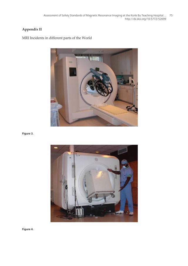
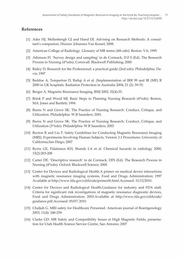
Appendix II
MRI Incidents in different parts of the World
Figure 3.
Figure 4.
Assessment of Safety Standards of Magnetic Resonance Imaging at the Korle Bu Teaching Hospital…http://dx.doi.org/10.5772/52699
75

Figure 5.
Figure 6.
Author details
Samuel Opoku, William Antwi and Stephanie Ruby Sarblah
*Address all correspondence to: [email protected]
Department of Radiography, College of Health Sciences, SAHS, University of Ghana, Legon,Accra, Ghana
Imaging and Radioanalytical Techniques in Interdisciplinary Research - Fundamentals and Cutting Edge Applications76
References
[1] Ader HJ, Mellenbergh GJ and Hand DJ. Advising on Research Methods: A consul‐tant’s companion, Huizen: Johannes Van Kessel; 2008.
[2] American College of Radiology. Glossary of MR terms (4th edn). Reston: VA; 1995
[3] Atkinson FI. ‘Survey design and sampling’ in de Cormack, D.F.S (Ed). The ResearchProcess in Nursing (4thedn). Cornwall: Blackwell Publishing; 2000
[4] Bailey D, Research for the Professional: a practical guide (2nd edn). Philadelphia: Da‐vis; 1997
[5] Beddoe A, Temperton D, Rafiqi A et al. (Implementation of IRR 99 and IR (ME) R2000 in UK hospitals. Radiation Protection in Australia 2004; 21 (2): 59-70.
[6] Berger A. Magnetic Resonance Imaging, BMJ 2002; 32(4):35.
[7] Brink P and Wood MJ. Basic Steps in Planning Nursing Research (4thedn). Boston,MA: Jones and Bartlett; 1994
[8] Burns N and Grove SK. The Practice of Nursing Research: Conduct, Critique, andUtilization. Philadelphia: W.B Saunders; 2001.
[9] Burns N and Grove SK. The Practice of Nursing Research: Conduct, Critique, andUtilization (3rdedn). Philadelphia: W.B Saunders; 2003
[10] Buxton R and Liu T. Safety Guidelines for Conducting Magnetic Resonance Imaging(MRI). Experiments Involving Human Subjects, Version 3.1 Procedures: University ofCalifornia,San Diego; 2007
[11] Byrns GE, Palatianos KH, Shands LA et al. Chemical hazards in radiology 2000;15(2):203-208
[12] Carter DE. ‘Descriptive research’ in de Cormack, DFS (Ed). The Research Process inNursing (4thedn). Oxford: Blackwell Science; 2000
[13] Center for Devices and Radiological Health.A primer on medical device interactionswith magnetic resonance imaging systems, Food and Drugs Administration; 1997Available at http://www.fda.gov/cdrh/ode/primerf6.html.Accessed: 31/12/2010.
[14] Center for Devices and Radiological Health.Guidance for industry and FDA staff,Criteria for significant risk investigations of magnetic resonance diagnostic devices,Food and Drugs Administration; 2003.Available at http://www.fda.gov/cdrh/ode/guidance.pdf.Accessed: 05/07/ 2010.
[15] Chaljub G. MRI safety for Healthcare Personnel. American journal of Roentgenology2003; 11(4): 248-259.
[16] Clarke GD. MR Safety and Compatibility Issues at High Magnetic Fields, presenta‐tion for Utah Health Science Service Centre, San Antonio; 2007
Assessment of Safety Standards of Magnetic Resonance Imaging at the Korle Bu Teaching Hospital…http://dx.doi.org/10.5772/52699
77
[17] Colletti PM. Size H Oxygen Cylinder: Accidental MR Projectile at 1.5 Tesla. Journalof Magnetic Resonance Imaging 2004; 19: 141-143.
[18] Cornette LG, Tanner SF, Ramenghi LA et al. Magnetic Resonance Imaging, Fetal andNeonatal Education, 86:F171-F191; 2002. Available at www.archdischild.com.Ac‐cessed: 03/01/11.
[19] Dempsey MF, Condon B and Hadley DM. Ultrasound CT MRI. MRI Safety review2002;. 23:392-401.
[20] Device Bulletin. Safety Guidelines for Magnetic Resonance Imaging Equipment inClinical Use, Safeguarding Public Health: 03; 2007
[21] Emergency Care Research Institute. Patient death illustrates the importance of adher‐ing to safety precautions in the magnetic resonance environments, Hazard report;2001 Available at http//www.ecri.org/document/hazrd_MR1080601. Accessed:15/10/2010.
[22] Elagin YP. The concept of safety, ensuring safety for infants undergoing MagneticResonance Imaging. Atomic Energy 1996; 80(6):389-393. Available at http//www.medscape.com/article/499273. Accessed 05/11/2010.
[23] Faulkner W. MRI: Basic Physics, Instrumentation and Quality Control. Maiden Mass:Blackwell Science; 2002
[24] Ferris JN, Kavnoudias H, Theil C et al. ACR Guidance Document for Safe MRPracti‐ces, American Journal of Roentgenology 2007; 188 (5): 1388-1394. Available at http://www.acr.org/SecondaryMainMenuCategories/quality_safety/MRSafety/safe_mr07.aspx.Accessed: 20/12/2010.
[25] Gilk T. MRI Suites: Safety outside the Bore. Patient Safety and Quality Healthcare,1-8; 2006
[26] Gillies A. Using research in Nursing: a workbook for practitioners. Oxford: RadcliffeMedical Press; 2002
[27] Gould TA, How MRI works. Health, Willis HRH 08/09:1-8; 2008
[28] Gray DE, Doing Research in the real World. London: Sage; 2004
[29] Harding W. Concept of safety: Good Fellow. Texas: AFB; 2010
[30] Health Purchasing News. MRI accidents on the rise 2010; Available at http://findarti‐cles.com/p/articles/mi m0BPC/is 10 29/ai n15685953.Accessed: 29/06/2010.
[31] Henner A and Servomaa A. The Safety Culture as a part of radiation protection inmedical imaging, Proceedings of Third European IRPA Congress, Helsinki, Finland;2010. Available at http://www.fda.gov/cdrh/ode/primerf6.html.Accessed: 31/12/2010.
[32] Johnson JN and Killion JB, Hazards in the radiology department. Entrepreneur: July-August; 2005; Available at http://www.entrepreneur.com/tradeljournals/article/134676835.html. Accessed: 20/01/11.
Imaging and Radioanalytical Techniques in Interdisciplinary Research - Fundamentals and Cutting Edge Applications78
[33] Joseph NJ. MRI Safety for Healthcare Personnel.Online Radiology Continuing Edu‐cation for Professional; 2006.Available at https://www.ceessentials.net/article7.html.Accessed: 09/08/2010.
[34] Junk R and Gilk T.Emergency Preparedness for Imaging Service Providers.Radiolog‐ical Management 2005; 27(5): 16- 24.
[35] Kanal E, Barkovich AJ, Bell C et al. ACR Guidance Document for Safe MR Practices:American Journal of Roentgenology 2007; 188: 1447-1474.
[36] Kanal E and Shellock FG.Policies, Guidelines and Recommendations for MR Imagingsafety and patient management and monitoring during MR examination.J MagnRe‐son Imaging 1992; 2:247.
[37] Kanal E, Barkovich AJ, Gilk T et al. ACR White Paper on Magnetic Resonance (MR)Safety, presented as a combined paper to the American College of Radiology 2004;1-24.
[38] Kanal E. Magnetic Resonance Safe Practice Guidelines of the University of PittsburghMedical Centre, summary from the Department of Radiology, University of Pitts‐burgh Medical Centre 2002; 155-163.
[39] Kanal E, Barkovich AJ, Gilk T et al. ACR Guidance Document for Safe MR Practices.American Journal of Radiology 2007; 188: 1-27.
[40] Kanal E, Borgstede JP, Barkovich AJ et al. White Paper on MRI Safety. AmericanJournal of Roentgenology, Am J Roentgenol 2002; 178.1335-1347.
[41] Kangarlau A and Robitaille PM. Biological Effects and Health Implications in Mag‐netic Resonance Imaging.Concepts in Magnetic Resonance 2000;12 (5): 312-359.
[42] Kusumasuganda IGK. Introduction to MR Safety: Part I 2010; Available at http//mriforyou.bogspot.com.Accessed: 08/08/2010.
[43] Lahr W and Rowan R. How Metal Detectors Work. White’s Electronics; 2004.
[44] Launders J, Preventing accidents and injuries in the MRI suite. The Joint Commission2008; Issue 38: 1-3.
[45] Maiorova T, Stevens F, Van der zee J et al. Shortage in general practice deposit withfeminisation of the medical workforce: A seeming paradox? A Cohort Study. BMCHealth Service Research 2008, 8:262.
[46] Merchant SP, Halkett GK and Sale C. Australian radiation therapy: an overview. TheRadiographer 2011; 58 (1):1-2.
[47] McRobbie DW, Moore EA, Graves M J and Prince MR. MRI, New York: CambridgeUniversity Press; 2007
[48] Medical College of Wisconsin, MRI Safety Policies and procedures, Milwaukee, Wis‐consin, 53226; 2009.
Assessment of Safety Standards of Magnetic Resonance Imaging at the Korle Bu Teaching Hospital…http://dx.doi.org/10.5772/52699
79
[49] New York Times, MRI accidents, 29 June 2010; Available at http://findarticles.com/p/articles/mi_m0BPC/is_10_29/ai_n15685953/ Accessed: 05/12/2010.
[50] Ordridge RJ, Fullerton G and Norris DG. MRI Safety limits: Is MRI safe or not? TheBritish Journal of Radiology 2000; 73: 1-2.
[51] Phin D. Workplace Safety.General Safety Policies 2001; Available at http://www.inc.com/tools/2000/12/21572.html.Accessed: 07/01/2011.
[52] Polgar S and Thomas SA, Introduction to research in the Health Sciences (4thedn).London: Churchill Livingstone; 2000.
[53] Pontin D. ‘Descriptive research’ in de Cormack, DFS (Ed). The Research Process inNursing (4thedn), Oxford: Blackwell Science; 2000.
[54] Price RP. The AAPM/RSNA physics tutorial for residents, MR imaging safety consid‐erations 1999; 19:1641-1651.
[55] Sawyer-Glover A and Shellock FG. Pre-MRI Procedures Screening: Recommenda‐tions and Safety Considerations for Biomedical Implants and Devices. Journal ofMagnetic Resonance Imaging 2002; 12: 92-106.
[56] Schaefer DJ, Bourland JD and Nyenhuis JA. Review of patient safety in time-varyinggradient fields. J. MagnReson Imaging 2000; 12:20-29.
[57] Schenck JF. Safety of strong, static magnetic fields. J MagnReson Imaging 2000;12:2-19.
[58] Shellock FG and Crues JV. MR Procedures: Biologic Effects, Safety, and Patient Care.Radiology 2004; 232 (3): 635 – 652.
[59] Shellock FG. MRI safety, bio effects and patient management 2002; Available atwww.MRIsafety.com.Accessed: 29/08/2010.
[60] The Joint Commission, Preventing accidents and injuries in the MRI suite. Risk re‐duction strategies 2008; Issue 38. Available at www.jointcommission.org/sentinele‐vents/sentineleventalert/sea_38.htm Accessed: 29/06/10.
[61] University of California at San Francisco.MRI Safety Primer, Author, California;2011.
[62] Westbrook C, Talbot J and Roth CK. What do MRI radiographers really know? Mag‐netic Resonance Imaging Journal 2009; 1(2): 52 -60.
Imaging and Radioanalytical Techniques in Interdisciplinary Research - Fundamentals and Cutting Edge Applications80
Related Documents