HAL Id: tel-00265610 https://tel.archives-ouvertes.fr/tel-00265610 Submitted on 19 Mar 2008 HAL is a multi-disciplinary open access archive for the deposit and dissemination of sci- entific research documents, whether they are pub- lished or not. The documents may come from teaching and research institutions in France or abroad, or from public or private research centers. L’archive ouverte pluridisciplinaire HAL, est destinée au dépôt et à la diffusion de documents scientifiques de niveau recherche, publiés ou non, émanant des établissements d’enseignement et de recherche français ou étrangers, des laboratoires publics ou privés. Assemblage et fonction de complexes ARN-protéines Christine Allmang To cite this version: Christine Allmang. Assemblage et fonction de complexes ARN-protéines. Sciences du Vivant [q-bio]. Université Louis Pasteur - Strasbourg I, 2007. tel-00265610

Welcome message from author
This document is posted to help you gain knowledge. Please leave a comment to let me know what you think about it! Share it to your friends and learn new things together.
Transcript
HAL Id: tel-00265610https://tel.archives-ouvertes.fr/tel-00265610
Submitted on 19 Mar 2008
HAL is a multi-disciplinary open accessarchive for the deposit and dissemination of sci-entific research documents, whether they are pub-lished or not. The documents may come fromteaching and research institutions in France orabroad, or from public or private research centers.
L’archive ouverte pluridisciplinaire HAL, estdestinée au dépôt et à la diffusion de documentsscientifiques de niveau recherche, publiés ou non,émanant des établissements d’enseignement et derecherche français ou étrangers, des laboratoirespublics ou privés.
Assemblage et fonction de complexes ARN-protéinesChristine Allmang
To cite this version:Christine Allmang. Assemblage et fonction de complexes ARN-protéines. Sciences du Vivant [q-bio].Université Louis Pasteur - Strasbourg I, 2007. �tel-00265610�
Université Louis Pasteur Faculté des Sciences de la vie
STRASBOURG
Habilitation à diriger des Recherches
Assemblage et fonction de complexes ARN-protéines
Présentée par
Christine Allmang-Cura
Chargée de Recherche au CNRS Architecture et Réactivité de l’ARN, UPR 9002 du CNRS
Soutenue le 26 novembre 2007
Membres du Jury : Dr. Michèle Caizergues-Ferrer rapporteur externe Pr. Jean-Pierre Rousset rapporteur externe Dr. James Stevenin rapporteur interne Pr. David Tollervey examinateur Pr. Eric Westhof examinateur Dr. Alain Krol garant d’habilitation
1
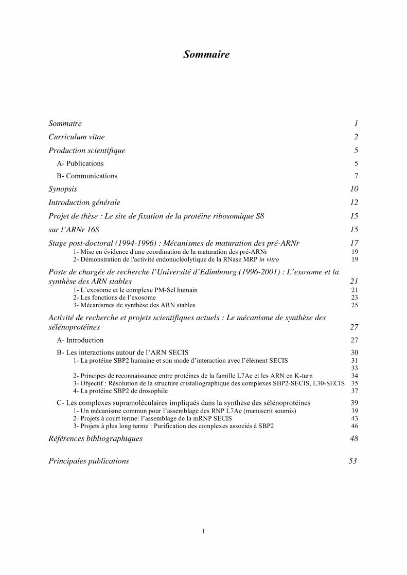
Sommaire
Sommaire 1 Curriculum vitae 2
Production scientifique 5 A- Publications 5 B- Communications 7
Synopsis 10
Introduction générale 12
Projet de thèse : Le site de fixation de la protéine ribosomique S8 15
sur l’ARNr 16S 15 Stage post-doctoral (1994-1996) : Mécanismes de maturation des pré-ARNr 17
1- Mise en évidence d'une coordination de la maturation des pré-ARNr 19 2- Démonstration de l'activité endonucléolytique de la RNase MRP in vitro 19
Poste de chargée de recherche l’Université d’Edimbourg (1996-2001) : L’exosome et la synthèse des ARN stables 21
1- L’exosome et le complexe PM-Scl humain 21 2- Les fonctions de l’exosome 23 3- Mécanismes de synthèse des ARN stables 25
Activité de recherche et projets scientifiques actuels : Le mécanisme de synthèse des sélénoprotéines 27
A- Introduction 27 B- Les interactions autour de l’ARN SECIS 30
1- La protéine SBP2 humaine et son mode d’interaction avec l’élément SECIS 31 33
2- Principes de reconnaissance entre protéines de la famille L7Ae et les ARN en K-turn 34 3- Objectif : Résolution de la structure cristallographique des complexes SBP2-SECIS, L30-SECIS 35 4- La protéine SBP2 de drosophile 37
C- Les complexes supramoléculaires impliqués dans la synthèse des sélénoprotéines 39 1- Un mécanisme commun pour l’assemblage des RNP L7Ae (manuscrit soumis) 39 2- Projets à court terme: l’assemblage de la mRNP SECIS 43 3- Projets à plus long terme : Purification des complexes associés à SBP2 46
Références bibliographiques 48
Principales publications 53
2
Curriculum vitae Dr. ALLMANG-CURA Christine UPR 9002 du CNRS Institut de Biologie Moléculaire et Cellulaire 15, rue René Descartes 67084 Strasbourg Cedex, FRANCE Tel: + 33 3 88 41 70 80 Fax: + 33 3 88 60 22 18 e-mail: [email protected] Née le 26 Août 1967 à Sarreguemines (Moselle) Mariée, 1 enfant Nationalité française Formation universitaire à l'Université Louis Pasteur de Strasbourg : - Mars 1994 : Doctorat de l'Université Louis Pasteur de Strasbourg en Biologie
Moléculaire (mention Très Honorable) - Juin 1990 : DEA de Biologie Cellulaire et Moléculaire (mention B) - 1988-1990 : Magistère de Chimie Biologie (mention B) Position actuelle : Depuis Octobre 2001 : Chargée de Recherche CR1 au CNRS. UPR 9002 du CNRS. IBMC, Strasbourg. Directeur : Professeur Eric Westhof. Equipe du Docteur Alain Krol. Poste à l’Université d’Edimbourg : 0ctobre 1996 – mars 2001 : “Research Fellow". Wellcome Trust Centre for Cell Biology, Université d’Edimbourg, Grande Bretagne. Equipe du Professeur David Tollervey. Stage Post-doctoral : Mai 1994- septembre 1996 : Département d'Expression Génétique. EMBL, Heidelberg, Allemagne. Directeur : Iain Mattaj. Equipe du Docteur David Tollervey. Thèse : 1990-1994 : DEA et thèse en Biologie Cellulaire et Moléculaire à l'Université Louis Pasteur de Strasbourg I. IBMC, Strasbourg. Equipe du Professeur Bernard Ehresmann. Directeur de thèse: Docteur Chantal Ehresmann. Thèse soutenue le 23 mars 1994. Titre : Ingénierie et études structurales d'ARN en solution. Application à trois systèmes: Le site de fixation de la protéine S8 sur l'ARN ribosomique 16S d'Escherichia coli, l'ARN ribosomique 5S de Xenopus laevis et de l'ARN 3 du virus des nervures jaunes et nécrotiques de la betterave.
3
Séjours scientifiques : - Université libre d’Amsterdam, Pays-Bas. Septembre 1996. Equipe du Professeur R.J. Planta. Analyse de protéines ribosomiques sur gel bidimensionnels. - Université de Bayreuth, Allemagne. Mai 1992. Equipe du Professeur M. Sprinzl Synthèse chimique d’ARN par la voie des H- phosphonates. - Université de Victoria, Colombie Britannique, Canada. Mai - juin 1991. Equipe du Professeur P. Romaniuk. Synthèse chimique d’ARN par la voie des phosphoramidates. Encadrement et responsabilités collectives : Encadrement, formation : Akiko Takeuchi, Doctorante (Aspects moléculaires et cellulaires de la Biologie) depuis septembre 2006. Directeur de thèse : Docteur Alain Krol. Sujet : Caractérisation, purification et cristallisation de la protéine SBP2 de Drosophila melanogaster Laurence Wurth, étudiante en DEA de Biologie Moléculaire et Cellulaire puis Doctorante (allocataire de recherche MREST), depuis septembre 2005. Directeur de thèse : Docteur Alain Krol. Sujet : Identification de nouveaux facteurs moléculaires impliqués dans la synthèse des sélénoprotéines Vincent Olieric, étudiant en DEA puis Doctorant en cristallographie biologique, de septembre 2003 à novembre 2006. Directeur de thèse : Docteur Philippe Dumas (UPR 9002 du CNRS). Sujet: Cristallisation du complexe SBP2/ARN SECIS au cœur du mécanisme de synthèse des sélénoprotéines David Schmitt, étudiant en DEA de Biologie Moléculaire et Cellulaire de septembre 2002-juin 2003. Sujet : Dissection fonctionnelle de SBP2, protéine impliquée dans la synthèse des sélénoprotéines. Emmanuelle Kiefer, étudiante en BTS Biotechnologie de janvier- février 2002. Sujet : Clonage de l’ADNc de la protéine hSBP2 entière et tronquée dans des vecteurs d’expression en vue d’études cristallographiques. Thomas Khalish, étudiant en Licence L3. Stage de Biologie Moléculaire juillet 2007. Sujet : Clonage de l’ADNc de facteurs d’assemblages impliqués dans le mécanisme de synthèse des sélénoprotéines.
4
Participation à des contrats de recherche (Partenariat et valorisation) : J’ai participé à la rédaction des demandes de financement du laboratoire ci-dessous, relatives aux sujets de ma thématique. J’ai plus particulièrement pris en charge la rédaction de la demande de contrat ANR concernant notre équipe. ANR blanc (depuis octobre 2006) Titre du projet : Nufip/Rsa1: a common assembly machine for snoRNPs, telomerase, and selenoprotein mRNPs. Rôle : Responsable de thématique et rédaction du projet strasbourgeois. Obtention d’un financement pour un post-doctorant qui sera recruté en 2008 et que je superviserai. Coordinateur du projet : Edouard Bertrand (UMR 5535 CNRS-IGM, Montpellier) Partenaires : Bruno Charpentier et Christiane Branlant (UMR 7567 CNRS-UHP, Nancy), Alain Krol (UPR 9002 du CNRS, Strasbourg), Barbara Bardoni (Université Nice, Sophia Antipolis). Action Concertée Incitative Biologie Cellulaire Moléculaire et Structurale-Ministère de la Recherche (Octobre 2004-Septembre 2007) Titre du projet : Comment les structures formées entre ARN en K-turn et protéines de la famille L7Ae initient-elles la formation de particules ribonucléoprotéiques ayant des fonctions cellulaires variées ? Coordinateur du projet : Alain Krol Partenaires du projet : Christiane Branlant (UMR 7567 CNRS-UHP, Nancy), André Aubry (UMR 7086 CNRS-UHP, Nancy), Philippe Dumas (UPR 9002 du CNRS, Strasbourg), Alain Krol (UPR 9002 du CNRS). Programme Toxicologie Nucléaire Environnementale CEA-CNRS-INSERM-INRA-Ministère de la Recherche Projet Signalisation, Détection, Détoxication & Contrôle Redox en Réponse aux Métaux Lourds (SIDDERE) (Octobre 2004-Septembre 2007) Titre du projet : Fonction biologique du sélénium. Incorporation ciblée dans les protéines à sélénocystéine chez les eucaryotes supérieurs Coordinateurs du projet : Michel Toledano, Jean-Luc Montillet et Alain Vavasseur Animation et administration de la recherche : Membre élu du conseil de laboratoire de l’UPR 9002 du CNRS depuis septembre 2004 Membre de la Société Française de Biochimie et Biologie Moléculaire
5
Production scientifique
A- Publications
1- Cléry A, Bourguignon-Igel V, Allmang C, Krol A, and Branlant C. (2007) An improved definition of the RNA binding specificity of SECIS binding protein 2, an essential component of the selenocysteine incorporation machinery. Nucleic Acids Res. 35,1868-84.
2- Allmang C and Krol A. (2006) SECIS RNAs and K-turn binding proteins. A survey of
evolutionary conserved RNA and protein motifs. In Selenium, its molecular Biology and role in human Health 2nd edition. DL Hatfield (ed) Kluwer Academic Publishers. 5, 51-61.
3- Allmang C and Krol A. (2006). Selenoprotein synthesis: UGA does not end the story.
Biochimie. 88, 1561-1571. 4- Milligan L, Torchet C, Allmang C, Shipman T and Tollervey D. (2005) A nuclear
surveillance pathway for mRNAs with defective polyadenylation. Mol. Cell. Biol. 25, 9996-10004.
5- Kufel J, Allmang C, Verdone L, Beggs J and Tollervey D. (2003). A complex
pathway for 3’-processing of the yeast U3 snoRNA. Nucleic Acids Res. 31, 6788-6797.
6- Kufel J, Allmang C, Petfalski E, Beggs J and Tollervey D. (2003). Lsm Proteins Are
Required for Normal Processing and Stability of Ribosomal RNAs. J. Biol. Chem. 278, 2147-2156.
7- Allmang C, Carbon P and Krol A. (2002). The SBP2 and 15.5 kD/Snu13p proteins
share the same RNA binding domain: identification of SBP2 amino acids important to SECIS RNA binding RNA 8, 1308-1318.
8- Lescure A, Allmang C, Yamada K, Carbon P and Krol A. (2002). cDNA cloning,
expression pattern and RNA binding analysis of human SECIS binding protein 2. Gene 291, 279-285.
9- Kufel J, Allmang C, Verdone L, Beggs JD and Tollervey D. (2002). Lsm proteins are
required for normal processing of pre-tRNAs and their efficient association with La-homologous protein Lhp1p. Mol. Cell. Biol. 22, 5248-5256.
10- Brouwer R, Allmang C, Raijmakers R, van Aarssen Y, Vree Egberts W, Petfalski E,
van Venrooij WJ, Tollervey D and Pruijn GJM. (2001). Three novel components of the human exosome. J. Biol. Chem. 276, 6177-6184.
11- Kufel J, Allmang C, Chanfreau G, Petfalski E, Lafontaine DL and Tollervey D.
(2000). Precursors to the U3 snoRNA lack snoRNP proteins but are stabilized by La binding. Mol. Cell. Biol. 20, 5415-5424.
12- Allmang C, Mitchell P, Petfalski E and Tollervey D. (2000) Degradation of ribosomal
RNA precursors by the exosome. Nucleic Acids Res. 28, 1684-91.
6
13- Allmang C, Kufel J, Chanfreau G, Mitchell P, Petfalski E and Tollervey D. (1999)
Functions of the exosome in rRNA, snoRNA and snRNA synthesis. EMBO J. 18, 5399-5410.
14- Allmang C, Petfalski E, Podtelejnikov A, Mann M, Tollervey D and Mitchell P.
(1999) The yeast exosome and human PM-Scl are related complexes of 3’ -> 5’ exonucleases. Genes and Dev. 13, 2148-2158.
15- Allmang C and Tollervey D. (1998) The role of the 3’ external transcribed spacer in
yeast pre-rRNA processing. J. Mol. Biol. 278, 67-78.
16- Lygerou Z, Allmang C, Tollervey D and Séraphin B. (1996) Accurate processing of a eukaryotic precursor ribosomal RNA by Ribonuclease MRP in vitro. Science 272, 268-270.
17- Allmang C, Henry Y, Morrissey J.P, Wood H, Petfalski E and Tollervey D. (1996)
Processing of the yeast pre-rRNA at sites A2 and A3 is linked. RNA 2, 63-73.
18- Allmang C, Henry Y, Wood H, Morrissey J.P, Petfalski E and Tollervey D. (1996) Recognition of cleavage site A2 in the yeast pre-rRNA. RNA 2, 51-62.
19- Allmang C, Mougel M, Westhof E, Ehresmann B and Ehresmann C. (1994) Role of
conserved nucleotides in building the 16S rRNA binding site of E. coli ribosomal protein S8. Nucleic Acids Res. 22, 3708-3714.
20- Mougel M, Allmang C, Eyermann F, Cachia C, Ehresmann B and Ehresmann C.
(1993) Minimal 16S rRNA binding site and role of conserved nucleotides in E. coli ribosomal protein recognition. Eur. J. Biochem. 215, 787-792.
21- Gilmer D, Allmang C, Ehresmann C, Guilley H, Richards K, Jonard G and Ehresmann
B. (1993) The secondary structure of the 5'-noncoding region of beet necrotic yellow vein virus RNA 3: evidence for a role in viral RNA replication. Nucleic Acids Res. 21, 1389-1395.
7
B- Communications
Présentations orales : 2ième rencontre entre l’Unité ‘Architecture et Réactivité des ARN’ (IBMC) et le département de Biologie et Génomique Structurale (IGBMC) (Mont Sainte Odile, France, 25-26 janvier 2007). A common mechanism for the assembly of nuclear and SECIS RNPs. Allmang C. Rencontre de l’Unité ‘Architecture et Réactivité des ARN’ (IBMC) (CIARUS, Strasbourg, 14 décembre 2006). A common mechanism for the assembly of sno, sn and SECIS mRNPs. Allmang C, Wurth L et Krol A. Séminaire ToxNuc-E (Dijon, France, juin 2005). Un mécanisme original pour la synthèse des sélénoprotéines. Allmang C et Krol A. 4ième Rencontre SIFRARN.. Structure, intégration, fonction et réactivité des ARN. (Nancy, France, 14-17 octobre 2002). Un même domaine de liaison à l’ARN pour les protéines SBP2 et 15.5 kD/Snu13p. Allmang C et Krol A. 3ième Rencontre SIFRARN. Le monde des ARN et ses nouvelles frontières. (Toulouse, France, 19-21 janvier 2000). L’Exosome: un complexe multifonctionnel d’exonucléases 3’ ->5’. Allmang C, Mitchell P, Kufel J, Brouwer R, Petfalski L, van Venrooij W et Tollervey D. RNA' 99 (The fourth annual meeting of the RNA Society, University of Edinburgh 23-27 juin 1999). The yeast exosome and human PM-Scl are related complexes of 3’->5’ exonucleases. Allmang C, Mitchell P, Brouwer R, Petfalski E. van Venrooij W et Tollervey D. RNA' 98 (The third annual meeting of the RNA Society, University of Wisconsin-Madison, 26-31 mai 1998). The exosome: A surprisingly large complex of 3’->5’ exonucleases. Allmang C, Mitchell P, Petfalski E et Tollervey D. 2ième Rencontre SIFRARN. Le monde des ARN et leurs nouvelles fonctions: réalités et perspectives. (Strasbourg, France, 28-30 avril 1998). Coordination de la maturation des pré-ARN. Allmang C et Tollervey D. 5 th UK RNA Processing Workshop (Rydal Hall, Ambleside, Cumbria, 23-25 janvier 1998). The coordination of pre-rRNA processing. Allmang C et Tollervey D. Young Scientist's View of Molecular Biotechnology (Mt. Ste. Odile, France, 28 février- 6 mars 1993). The binding site of E. coli ribosomal protein S8 on 16S rRNA: Minimal RNA structural requirement and role of conserved nucleotides in protein S8 recognition. Allmang C, Mougel M, Eyermann F, Ehresmann B et Ehresmann C. XVIIIe Forum des Jeunes Chercheurs (Tours, France 3-6 septembre 1991). Le site de reconnaissance de la protéine ribosomique S8 sur l'ARN ribosomique 16S d'E. coli : Relations structure/reconnaisssance par mutagénèse dirigée. Allmang C, Mougel M, Ehresmann B et Ehresmann C.
8
Posters : 6ième rencontre SifrARN. Structure, integration, fonction et réactivité des ARN (Rennes, France, 3-6 juillet 2006). Interactions ARN/protéines au niveau de l’ARN SECIS: variations autour du motif L7A/L30 et des ARN en K-turn. Wurth L, Cléry A, Branlant C, Krol A et Allmang C. Séminaire de Toxicologie Nucléaire Environnementale.(Auteuil, France, 5-7 décembre 2005). Un mécanisme original pour la synthèse des protéines à selenium. Allmang C., Beniaminov A., Wurth L., et Krol A.
EMBO Conference on Protein Synthesis and Translational Control (EMBL, Heidelberg, Allemagne, 14-18 Septembre 2005). Principles of RNA-protein recognition between proteins of the L7A/L30 family and K-turn RNA motifs. Cléry A, Schmitt D, Wurth L., Bourguignon-Igel V, Branlant C, Krol A et Allmang C. 5ième rencontre SifrARN. ARN, le nouveau monde (Arcachon, France, 10-13 Octobre 2004). Dimérisation de SBP2, une protéine de liaison à l’ARN impliquée dans le mécanisme de synthèse des sélénoprotéines. Schmitt D, Krol A et Allmang C. Symposium. Structure, function and dynamics of RNA-protein complexes. (Göttingen, Allemagne, 17-20 septembre 2003). SBP2, a multifunctional protein involved in selenoprotein synthesis. Schmitt D, Krol A et Allmang C. 1ier congrès de Traduction Francophone (Institut Pasteur, Paris, 12-13 décembre 2002). Un même domaine de liaison à l’ARN pour les protéines SBP2 et 15.5 kD/Snu13p. Allmang C et Krol A. RNA 2002 (The Seventh Annual meeting of the RNA Society, University of Wisconsin-Madison, 28 mai- 2 juin 2002). Similar protein contacts for SBP2/SECIS RNA and 15.5 kD/U4 snRNA complexes. Allmang C et Krol A. The ribosome: Its (nucleolar) synthesis and structure (Amsterdam, 16-20 Aôut 1997). Interactions between yeast pre-rRNA processing complexes. Allmang C et Tollervey D. RNA' 96 (The first annual meeting of the RNA Society, University of Wisconsin-Madison, 28 mai - 02 juin 1996). Interactions between yeast pre-rRNA processing complexes. Allmang C, Henry Y, Morrissey JP, Wood H, Petfalski E et Tollervey D. Molecular Biology of RNA: Splicing and 3'-end formation of RNA (Mont Ste Odile, France, 13-17 september 1995). Pre-rRNA processing in ITS1 of Saccharomyces cerevisiae. Allmang C, Lygerou Z, Henry Y, Morrissey JP, Wood H, Séraphin B et Tollervey D. Ribosome synthesis and nucleolar function (Cold Spring Harbor, New York, 28 septembre- 02 mars 1994). Role of the conserved nucleotides of S8 16S rRNA binding site in E. coli. Moine H, Allmang C, Mougel M, Westhof E, Ehresmann B et Ehresmann C. Symposium on structural Tools for the Analysis of Protein- Nucleic Acid Complexes (Wildbad Kreuth, Allemagne, 3-7 mai 1992). Three-dimensional structure of the binding site of E. coli
9
ribosomal protein S8 on 16S rRNA by structure probing, site directed mutagenesis and graphic modelling. Allmang C, Mougel M, Ehresmann B, Eyermann F, Westhof E et Ehresmann C. Autre participations : 8th Symposium on Selenium in Biology and Medecine (Madison, Etats-Unis, 25-30 juillet 2006). RNA-recognition at the SECIS RNA. Allmang C, Cléry A, Bourguignon V, Allamand V, Richard P, Lescure A, Guicheney P, Branlant C and Krol A. Second JSPS (Japan Society for the Promotion of Science) Forum in France (Strasbourg, 28 novembre 2003). International Conference on the Translational Apparatus, (Berlin, Allemagne, 31 octobre- 5 novembre 1992). E.coli ribosomal protein S8 recognizes specific three-dimensional features of 16S RNA. Mougel M, Allmang C, Westhof E, Ehresmann B et Ehresmann C.
10
Synopsis
Les particules ribonucléoprotéiques (ou RNP) sont à la base de nombreuses fonctions cellulaires fondamentales. La formation de ces particules RNP est un processus très complexe qui nécessite de nombreuses étapes de maturation et de multiples facteurs d'assemblage. Par ailleurs, une structure correcte des particules RNP est essentielle à leur fonction. Il est donc critique de comprendre comment ces particules sont formées dans la cellule. Au cours de ma carrière, je me suis intéressée à plusieurs aspects de ces mécanismes. Au cours de ma thèse dirigée par Chantal Ehresmann (1990-1994), dans l’équipe de Bernard Ehresmann (UPR 9002 du CNRS) j’ai étudié le mode d’interaction de la protéine ribosomique S8 sur l’ARNr 16S d’E. coli. Mon travail post-doctoral dans l’équipe de David Tollervey (EMBL, Heidelberg (1994-1996) et Université d’Edimbourg (1996-2001) a porté sur l’étude des mécanismes de maturation, d’assemblage et de dégradation de diverses RNP. J’ai notamment contribué à la caractérisation de l’exosome, un complexe d’exonucléases 3’-> 5’ impliqué dans la maturation et la dégradation de divers ARN chez la levure. J’ai également étudié le rôle de protéines chaperons dans la biogenèse des snoARN (biogenèse des ribosomes), des ARN ribosomiques et des ARNt.
En 2001, j’ai été recrutée au grade de chargée de recherche au CNRS dans l’équipe d’Alain Krol où nous étudions les mécanismes de synthèse des sélénoprotéines. L’incorporation de sélénocystéine dans les sélénoprotéines fait appel au recodage co-traductionnel d’un codon UGASec en phase. Chez les eucaryotes, ce mécanisme implique l’assemblage d’un complexe ARN-protéine au niveau d’une structure en tige-boucle ou ARN SECIS (Selenocysteine Insertion Sequence) située dans la région 3’non codante de l’ARNm des sélénoprotéines. La protéine SBP2 se fixe spécifiquement à l’ARN SECIS et recrute les facteurs de la machinerie de biosynthèse. Elle fait également partie de complexes supramoléculaires dans le cytoplasme et le noyau, suggérant un possible assemblage nucléaire de la mRNP SECIS. Nous avons montré que la protéine SBP2 présentait une origine évolutive commune avec des protéines de la famille L7Ae. Ces protéines partagent un domaine de liaison à l'ARN similaire et participent à la construction de plusieurs RNP essentielles telles les sous-unités ribosomiques, les snoRNP (biogenèse des ribosomes), les snRNP (épissage), et les mRNP codant pour les sélénoprotéines. Nos objectifs sont d’élucider les principes d’interaction SBP2/SECIS, d’identifier les composants moléculaires des complexes qui se forment autour du SECIS et de comprendre leur assemblage.
En collaboration avec Edouard Bertrand (Montpellier) et Bruno Charpentier et Christiane Branlant (Nancy) nous avons identifié une machinerie d’assemblage des RNP L7Ae conservée de la levure à l’homme et d’importance fondamentale pour la cellule. Elle est constituée d’une protéine adaptatrice et d’un complexe de protéines chaperons. Notre objectif est de comprendre son rôle dans l’assemblage des mRNP de sélénoprotéines.

11
12
Introduction générale
Les particules ribonucléoprotéiques (ou RNP) sont à la base de nombreuses fonctions
cellulaires fondamentales chez les eucaryotes. Au sein de ces particules, des ARN non
codants participent à des mécanismes aussi variés que la traduction (ARNr, ARNt), l'épissage
des ARN pré-messagers (UsnARN), la biogenèse des ribosomes et d’ARN non codants, la
modification de bases des ARN (snARN, snoARN, scaARN), la réplication des télomères
(télomérase) et la sécrétion des protéines (SRP). Enfin, ces dernières années ont vu
l’émergence de microRNPs, contenant des ARN non codants (ARNsi, ARNmi) capables de
moduler l’efficacité de la transcription, la stabilité des ARNm et vraisemblablement la
structure de la chromatine.
La plupart des petits ARN non codants accomplissent leurs fonctions en association
avec des protéines sous la forme de ribonucléoparticules (ou RNP). La formation des
particules RNP est un processus très complexe qui nécessite de nombreuses étapes de
maturation et de multiples facteurs d'assemblage (Fatica & Tollervey, 2002; Matera et al.,
2007 ; Yong et al., 2004). En effet, le nombre de facteurs requis pour l’assemblage de RNP
fonctionnelles dépasse souvent le nombre de protéines présentes au sein de la particule
mature. Ainsi, les ribosomes sont constitués d’environ 80 protéines, mais n’utilisent pas
moins de 140 facteurs pour leur assemblage (Fatica & Tollervey, 2002). Ces facteurs
semblent non seulement importants pour faciliter l'assemblage de la particule, mais aussi pour
exercer un contrôle strict sur la qualité des particules produites. Au niveau cellulaire,
l’assemblage des RNP est également synonyme de mécanismes complexes de trafic
intracellulaire car il peut avoir lieu dans des compartiments cellulaires différents du site
fonctionnel. C’est le cas des particules UsnRNP impliquées dans les mécanismes d’épissage
(Bertrand E & R., 2004; Carmo-Fonseca et al., 2002; Yong et al., 2004) : elles sont tout
d’abord exportées dans le cytoplasme, où leur assemblage fait appel au complexe SMN (Yong
et al., 2004) ; puis réimportées dans le noyau vers leur site final de maturation et enfin leur
site fonctionnel. En plus des RNP non codantes qui agissent en trans, la régulation de
l’expression des gènes chez les eucaryotes est souvent dépendante de la formation de
complexes RNP directement sur l’ARNm au niveau d’éléments structuraux régulateurs
capables d’agir en cis.
Dans chacun des cas, la structure correcte des particules RNP est essentielle à leur
fonction. Il est donc critique de comprendre comment ces particules sont formées dans la
cellule. Au cours de ma carrière, je me suis intéressée à plusieurs aspects de ces mécanismes.
13
Durant ma thèse dirigée par Chantal Ehresmann (1990-1994), j’ai étudié le mode
d’interaction de la protéine ribosomique S8 sur l’ARNr 16S d’E. coli. Cette protéine primaire
joue un rôle central dans l’assemblage coordonné de la petite sous-unité ribosomique ainsi
que dans la régulation de son propre opéron chez les procaryotes. Nous avons proposé un
modèle de repliement tridimensionnel de son site ARN par analyse structurale en solution et
modélisation.
Mon travail dans l’équipe de David Tollervey (1994-2001) a plus directement porté
sur l’étude des mécanismes de maturation, d’assemblage et de dégradation de diverses RNP.
J’ai notamment contribué à la caractérisation de l’exosome, un complexe d’exonucléases 3’->
5’ impliqué dans la maturation et la dégradation de divers ARN chez la levure. Notre travail a
permis d’élucider les fonctions de l’exosome, notamment dans le noyau où ce complexe
participe à la synthèse des ARN ribosomiques, des petits ARN nucléolaires (snoARN) et
nucléaires (snARN) mais joue également un rôle dans les mécanismes de dégradation et de
surveillance des ARN. La majorité des ARN stables qui constituent les RNP non codantes
sont maturés à partir de précurseurs. Leur maturation ou dégradation implique en plus des
exonucléases et endonucléases, une série de cofacteurs, d’hélicases et de protéines chaperons.
Nous avons montré qu’un jeu limité de ces facteurs pouvait être recruté sous différentes
combinaisons pour la synthèse de presque tous les ARN cellulaires. Deux types de protéines
chaperons (Lhp1 et Lsm) ont été plus particulièrement analysées pour leur rôle dans la
biogenèse du snoARN U3, des ARN ribosomiques et des ARNt. Ces protéines contribuent à
faciliter les interactions ARN/protéines ainsi que les réarrangements structuraux lors de
l’assemblage des particules ribonucléoprotéiques.
En 2001, j’ai été recrutée au grade de chargée de recherche au CNRS dans l’équipe
d’Alain Krol qui étudie les mécanismes de synthèse des sélénoprotéines. L’incorporation de
sélénocystéine dans les sélénoprotéines fait appel au recodage co-traductionnel d’un codon
UGASec en phase. Chez les eucaryotes, ce mécanisme implique l’assemblage d’un complexe
ARN-protéine au niveau d’une structure en tige-boucle ou ARN SECIS (Selenocysteine
Insertion Sequence) située dans la région 3’UTR de l’ARNm des sélénoprotéines (Allmang &
Krol, 2006b). La protéine SBP2 se fixe spécifiquement à l’ARN SECIS et recrute les facteurs
de la machinerie de biosynthèse. Elle fait également partie d’un complexe supramoléculaire et
est soumise au transport nucléocytoplasmique suggérant un possible assemblage nucléaire de
la mRNP SECIS (de Jesus et al., 2006 ; Small-Howard et al., 2006). Nous avons montré que
la protéine SBP2 présentait une origine évolutive commune avec des protéines de la famille
14
L7Ae. Ces protéines partagent un domaine de liaison à l'ARN similaire et participent à la
construction de plusieurs RNP essentielles telles les sous-unités ribosomiques, les snoRNP à
boites C/D et H/ACA (biogenèse des ribosomes), la snRNP U4 (épissage), et comme nous
l’avons montré dans les mRNP codant pour les sélénoprotéines. Nous étudions les principes
d’interaction SBP2/SECIS et tentons d’identifier les composants moléculaires du complexe
qui se forme autour du SECIS afin de comprendre quelle est l’origine de la diversité de
fonctions apparues au cours de l’évolution pour les RNP L7Ae. Des déterminants de
spécificité pour le SECIS et les ARN cibles des protéines L7Ae ont ainsi été identifiés en
collaboration avec l’équipe de Christiane Branlant (Nancy).
Enfin, notre objectif est d’élucider le mécanisme d’assemblage de la mRNP SECIS et
de comprendre comment il s’intègre dans le schéma d’assemblage général des RNP de la
famille L7Ae mais aussi dans le mécanisme traductionnel des sélénoprotéines. En
collaboration avec Edouard Bertrand (Montpellier) et Bruno Charpentier (Nancy) nous avons
identifié une machinerie conservée destinée à l’assemblage des particules RNP stables de la
famille L7Ae ainsi que de la RNP SECIS que nous caractérisons.
15
Projet de thèse : Le site de fixation de la protéine ribosomique S8 sur l’ARNr 16S
Equipe du Pr. Bernard et du Dr. Chantal Ehresmann. IBMC, UPR 9002 du CNRS, Université Louis Pasteur, Strasbourg
Mon travail de thèse a été effectué avec Marylène Mougel dans l’équipe de Bernard et
Chantal Ehresmann. Il a consisté en l’ingénierie et l’étude structurale d’ARN en solution. En
particulier, nous avons étudié le site de fixation de la protéine ribosomique S8 sur l’ARNr
16S d’E. coli. Cette protéine primaire se fixe dans le domaine central de l’ARNr 16S et
permet la fixation coopérative de protéines secondaires par induction d’une modification
conformationnelle de leur site ARN. La région de fixation de S8 représente l’un des sites de
nucléation lors de l’assemblage de la sous-unité ribosomique 30S (Held et al., 1974;
Mizushima et al., 1970). La protéine S8 intervient également dans la régulation de son propre
opéron (spc). En se liant sur son ARNm, elle en inhibe la traduction et régule ainsi sa propre
synthèse et celle des autres protéines ribosomiques de son opéron. En 1990, lorsque j’ai
débuté ma thèse, il était admis que le site de fixation de S8 situé au sein d’une structure en
tige boucle de l’ARNr 16S, était centré sur une région hélicoïdale irrégulière à la base de
l’hélice et dont un nombre limité de nucléotides dictait la conformation. Le repliement de
cette région était cependant très controversé et plusieurs modèles étaient en vigueur, dont l’un
proposé par notre laboratoire (Mougel et al., 1987). Nos travaux ont contribué à affiner la
connaissance de ce site et à définir le site minimum d’ARN reconnu par S8 (Mougel et al., 1993). Par la combinaison de techniques de mutagenèse dirigée, d’analyses structurales en
solution et de modélisation graphique nous avons construit un nouveau modèle
tridimensionnel de ce site avec Eric Westhof (Allmang et al., 1994). Cette étude a révélé la
présence de contraintes structurales importantes, conférant une géométrie et accessibilité
particulière à certains résidus spécifiques du site ainsi qu’au squelette sucre-phosphate de
l’ARN. Nous avions notamment proposé l’existence d’interactions non canoniques et d’un
réseau important de liaisons hydrogènes. Depuis, ce modèle s’est avéré incorrect quant à la
nature exacte des connections prédites, mais il avait mis en valeur la complexité du site.
Prédire de telles interactions reste un défi dans l’étude du repliement des ARN et ce système
modèle nous a mené aux limites des méthodologies employées. Après ma thèse, l’étude du
site s’est poursuivie à l’aide de techniques plus adaptées à la détection d’interactions non
canoniques multiples telle que le SELEX (Moine et al., 1997) et enfin la résolution de la
structure cristallographique du complexe S8-ARNr chez Methanococcus jannashii (Tishchenko et al., 2001). La résolution de la structure du ribosome de Thermus thermophilus
16
a quant à elle permis de replacer ces interactions dans le contexte de la particule RNP du
ribosome (Brodersen et al., 2002 ; Yusupov et al., 2001).
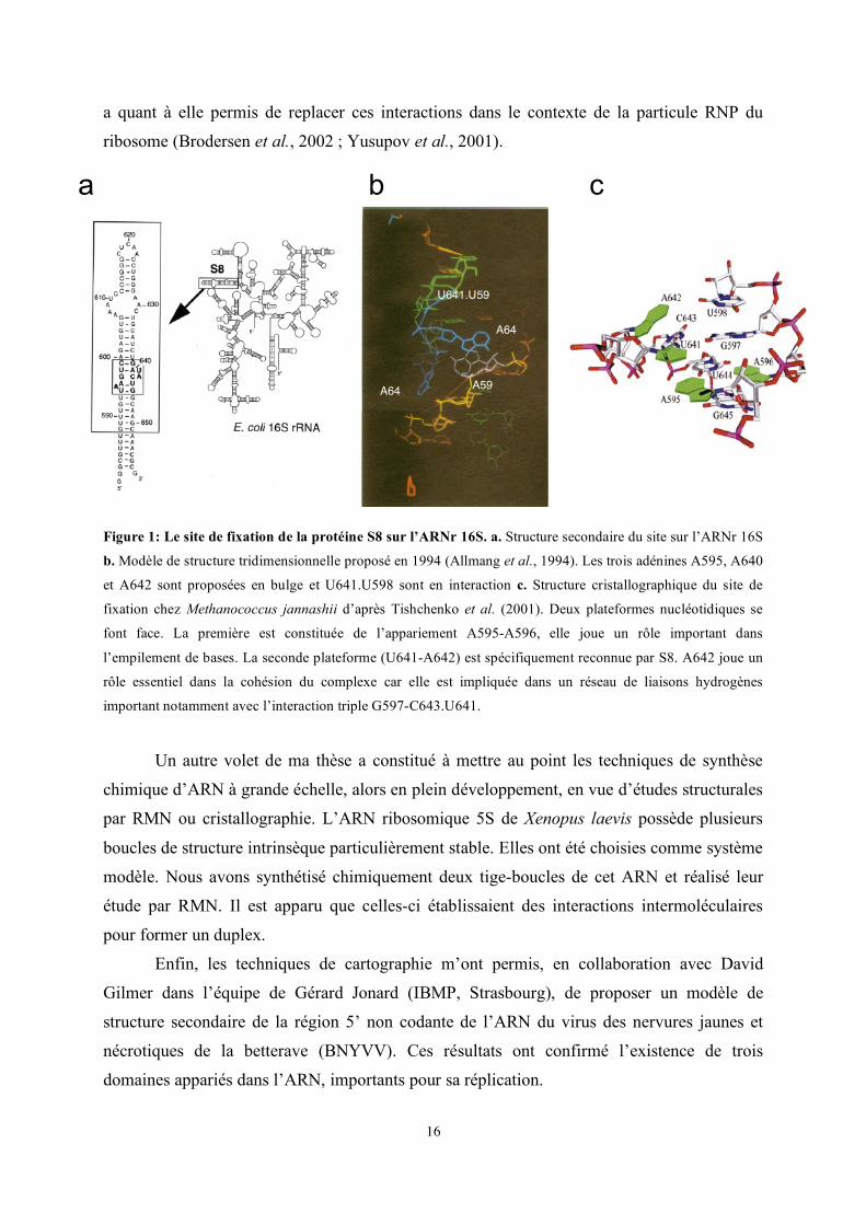
Figure 1: Le site de fixation de la protéine S8 sur l’ARNr 16S. a. Structure secondaire du site sur l’ARNr 16S
b. Modèle de structure tridimensionnelle proposé en 1994 (Allmang et al., 1994). Les trois adénines A595, A640
et A642 sont proposées en bulge et U641.U598 sont en interaction c. Structure cristallographique du site de
fixation chez Methanococcus jannashii d’après Tishchenko et al. (2001). Deux plateformes nucléotidiques se
font face. La première est constituée de l’appariement A595-A596, elle joue un rôle important dans
l’empilement de bases. La seconde plateforme (U641-A642) est spécifiquement reconnue par S8. A642 joue un
rôle essentiel dans la cohésion du complexe car elle est impliquée dans un réseau de liaisons hydrogènes
important notamment avec l’interaction triple G597-C643.U641.
Un autre volet de ma thèse a constitué à mettre au point les techniques de synthèse
chimique d’ARN à grande échelle, alors en plein développement, en vue d’études structurales
par RMN ou cristallographie. L’ARN ribosomique 5S de Xenopus laevis possède plusieurs
boucles de structure intrinsèque particulièrement stable. Elles ont été choisies comme système
modèle. Nous avons synthétisé chimiquement deux tige-boucles de cet ARN et réalisé leur
étude par RMN. Il est apparu que celles-ci établissaient des interactions intermoléculaires
pour former un duplex.
Enfin, les techniques de cartographie m’ont permis, en collaboration avec David
Gilmer dans l’équipe de Gérard Jonard (IBMP, Strasbourg), de proposer un modèle de
structure secondaire de la région 5’ non codante de l’ARN du virus des nervures jaunes et
nécrotiques de la betterave (BNYVV). Ces résultats ont confirmé l’existence de trois
domaines appariés dans l’ARN, importants pour sa réplication.
a b c
A640
A642
A595
U641.U598
17
Stage post-doctoral (1994-1996) : Mécanismes de maturation des pré-ARNr
Equipe de David Tollervey. Département d’Expression Génétique, EMBL, Heidelberg, Allemagne
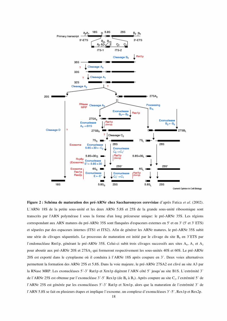
La biogenèse des ribosomes eucaryotes est un mécanisme complexe et dynamique qui a lieu successivement dans le nucléole, le nucléoplasme et le cytoplasme. Ce processus englobe les étapes de transcription, maturation, modifications des précurseurs d'ARN ribosomiques (pré-ARNr), leur assemblage en sous-unités ribosomiques et enfin leur transport. Plus d’une centaine de petits ARN nucléolaires (snoARN) servent de guides pour les modifications de l’ARN et environ 140 protéines non-ribosomiques sont impliquées dans les étapes de maturation (Fatica & Tollervey, 2002).
L’ARNr 18S de la petite sous-unité et les deux ARNr 5,8S et 25S de la grande sous-unité ribosomique sont transcrits par l'ARN polymérase I sous la forme d'un long précurseur unique: le pré-ARNr 35S. Les régions correspondant aux ARN matures du pré-ARNr 35S sont flanquées d'espaceurs externes en 5' et en 3' (5' et 3' ETS) et séparées par des espaceurs internes (ITS1 et ITS2). Les différentes étapes de maturation des pré-ARNr ainsi que les enzymes impliquées sont représentées Figure 2 (pour une revue voir Venema & Tollervey, 1999). Il existe une séparation entre la voie de synthèse de l’ARN 18S, qui implique quatre coupures successives par des endonucléases, et la voie de synthèse des ARN 5,8S et 25S, plus complexe et qui fait appel à une coupure endonucléolitique suivie par des étapes multiples de digestion par des exonucléases. Une fois modifiés les ARNr matures s’assemblent avec les 80 protéines ribosomiques et l’ARNr 5S qui est transcrit indépendamment.
Le laboratoire du Pr. David Tollervey, dans lequel j'ai effectué mon stage post-doctoral, a largement contribué à l’élucidation des étapes de maturation des pré-ARNr chez la levure et à l’identification d’un nombre important de facteurs impliqués dans le mécanisme. Mon travail post-doctoral dans cette équipe a contribué à identifier certains des signaux de coupures sur le pré-ARNr mais aussi à caractériser plusieurs facteurs de maturation et à analyser leur fonction.
18
Figure 2 : Schéma de maturation des pré-ARNr chez Saccharomyces cerevisiae d’après Fatica et al. (2002).
L’ARNr 18S de la petite sous-unité et les deux ARNr 5,8S et 25S de la grande sous-unité ribosomique sont
transcrits par l'ARN polymérase I sous la forme d'un long précurseur unique: le pré-ARNr 35S. Les régions
correspondant aux ARN matures du pré-ARNr 35S sont flanquées d'espaceurs externes en 5' et en 3' (5' et 3' ETS)
et séparées par des espaceurs internes (ITS1 et ITS2). Afin de générer les ARNr matures, le pré-ARNr 35S subit
une série de clivages séquentiels. Le processus de maturation est initié par le clivage du site B0 en 3’ETS par
l’endonucléase Rnt1p, générant le pré-ARNr 35S. Celui-ci subit trois clivages successifs aux sites A0, A1 et A2
pour aboutir aux pré-ARNr 20S et 27SA2 qui formeront respectivement les sous-unités 40S et 60S. Le pré-ARNr
20S est exporté dans le cytoplasme où il conduira à l’ARNr 18S après coupure en 3’. Deux voies alternatives
permettent la formation des ARNr 25S et 5,8S. Dans la voie majeure, le pré-ARNr 27SA2 est clivé au site A3 par
la RNase MRP. Les exonucléases 5’-3’ Rat1p et Xrn1p digèrent l’ARN côté 5’ jusqu’au site B1S. L’extrémité 3’
de l’ARNr 25S est obtenue par l’exonucléase 3’-5’ Rex1p (de B0 à B2). Après coupure au site C2, l’extrémité 5’ de
l’ARNr 25S est générée par les exonucléases 5’-3’ Rat1p et Xrn1p, alors que la maturation de l’extrémité 3’ de
l’ARN 5,8S se fait en plusieurs étapes et implique l’exosome, un complexe d’exonucléases 3’-5’, Rex1p et Rex2p.
19
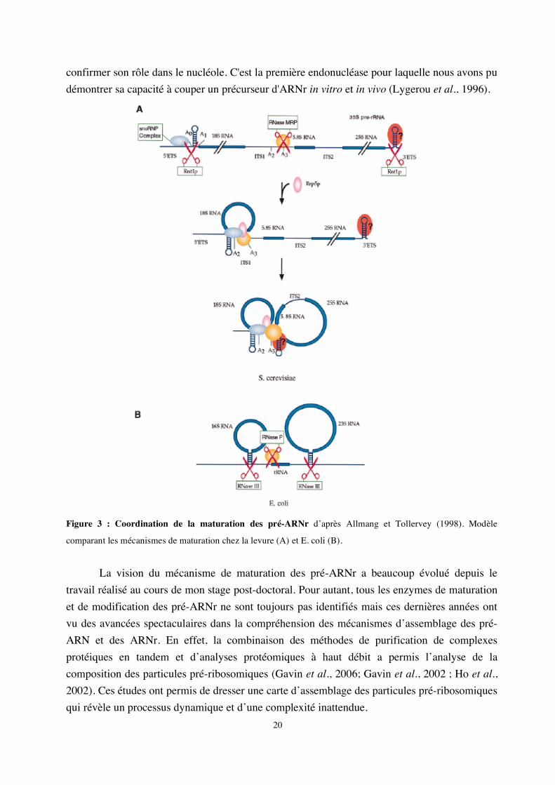
1- Mise en évidence d'une coordination de la maturation des pré-ARNr
Je me suis particulièrement intéressée aux mécanismes de maturation au sein de deux espaceurs (ITS1 et 3’ETS). ITS1 contient deux sites de clivage importants : A2 et A3. Le clivage au niveau du site A2 est une étape cruciale dans la maturation du pré-ARNr 35S qui permet de scinder le pré-ARNr en deux fragments 5' et 3' destinés respectivement à former les ARN de la petite et de la grande sous-unité ribosomique. Il est dépendant de signaux en cis à proximité de A2 mais également dans la région 5’ETS, tel que le site de fixation du snoARN U3 (Beltrame et al., 1994; Venema et al., 1995). Le clivage du site A2 dépend d’un complexe de maturation composé de snoRNP. Le clivage au niveau du site A3, assuré par la RNase MRP, permet quant à lui de générer l'extrémité 5' de la forme majeure de l'ARN 5,8S. La région 3’ETS est coupée par Rnt1 (homologue de la RNase III), ce clivage initie l’ensemble du mécanisme de maturation du pré-ARNr 35S. Mon travail a contribué à identifier les signaux requis en cis pour le clivage des régions ITS1 et 3’ETS (Allmang et al., 1996b; Allmang & Tollervey, 1998). Cette analyse a permis de révéler un lien tout à fait inattendu entre les sites de clivage A2 et A3 (Allmang et al., 1996a), suggérant que le complexe de maturation du site A2 et 5’ETS (snoRNP) interagissait avec celui du site A3 (RNase MRP). Par ailleurs, nous avons montré qu’une structure en tige-boucle en 3’ETS est nécessaire et suffisante pour la coupure par Rnt1p, mais également pour le clivage à distance du site A3 par la RNase MRP. La maturation des régions 3’ETS et ITS1 apparaît donc, elle aussi, couplée (Allmang & Tollervey, 1998). L’ensemble de nos résultats a suggéré une coordination de la maturation des pré-ARN qui peut être mise en parallèle avec les mécanismes de maturation bactériens (voir Figure 3). Chez les eubactéries, les extrémités non codantes situées en 5' et 3' des ARNr matures s’apparient et sont coupées par la RNase III. Un ARNt présent dans l’espaceur fournit un site de clivage pour la RNase P. Chez la levure, des tiges-boucles en 5’ et 3’ETS constituent les sites de clivage de Rnt1p. Des complexes de snoRNP remplacent les appariements intramoléculaires afin d’assurer la coordination de la maturation en 5’ et 3’. La RNase MRP assure un rôle comparable à celui de la RNase P dans l’espaceur ITS1, mais interagit également avec les complexes de maturation en 5’ et 3’ ETS.
2- Démonstration de l'activité endonucléolytique de la RNase MRP in vitro
En accord avec sa localisation nucléolaire, de nombreux arguments génétiques suggéraient que la RNase MRP intervenait dans la maturation du pré-ARNr in vivo. Le rôle de la RNase MRP était cependant controversé car elle fut initialement identifiée comme une endonucléase mitochondriale (Chang & Clayton, 1987). En collaboration avec Bertrand Séraphin (alors à l’EMBL), un système capable de reproduire avec précision la coupure au site A3 par la RNase MRP in vitro a été mis au point. Ces expériences nous ont permis de démontrer que la RNase MRP était bien directement responsable de la coupure du site A3 et de
20
confirmer son rôle dans le nucléole. C'est la première endonucléase pour laquelle nous avons pu démontrer sa capacité à couper un précurseur d'ARNr in vitro et in vivo (Lygerou et al., 1996).
Figure 3 : Coordination de la maturation des pré-ARNr d’après Allmang et Tollervey (1998). Modèle
comparant les mécanismes de maturation chez la levure (A) et E. coli (B).
La vision du mécanisme de maturation des pré-ARNr a beaucoup évolué depuis le
travail réalisé au cours de mon stage post-doctoral. Pour autant, tous les enzymes de maturation et de modification des pré-ARNr ne sont toujours pas identifiés mais ces dernières années ont vu des avancées spectaculaires dans la compréhension des mécanismes d’assemblage des pré-ARN et des ARNr. En effet, la combinaison des méthodes de purification de complexes protéiques en tandem et d’analyses protéomiques à haut débit a permis l’analyse de la composition des particules pré-ribosomiques (Gavin et al., 2006; Gavin et al., 2002 ; Ho et al., 2002). Ces études ont permis de dresser une carte d’assemblage des particules pré-ribosomiques qui révèle un processus dynamique et d’une complexité inattendue.
21
Poste de chargée de recherche l’Université d’Edimbourg (1996-
2001) : L’exosome et la synthèse des ARN stables
Research Fellow dans l'équipe du Pr. David Tollervey. Wellcome Trust Centre for Cell Biology, Université d’Edimbourg. Ecosse.
Dans la continuité du travail amorcé à l’EMBL (Heidelberg), notre équipe, une fois
installée au «Wellcome Trust Center for Cell Biology» de l’Université d’Edimbourg (Ecosse) a continué à s’intéresser aux mécanismes de maturation des ARN. La recherche d’enzymes impliquées dans la maturation des pré-ARNr a conduit à l’identification d'un complexe d’exonucléases multifonctionnel : l’exosome. J’ai participé à sa caractérisation et à son analyse fonctionnelle.
1- L’exosome et le complexe PM-Scl humain
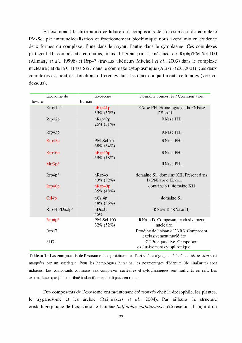
L’exosome est un complexe d’exonucléases 3’->5’ impliqué dans la maturation et la dégradation de divers ARN chez la levure. Il fut initialement identifié par Phil Mitchell dans notre équipe comme un complexe de 5 exonucléases essentielles, toutes impliquées dans la maturation de l’extrémité 3’ de l’ARNr 5,8S (Mitchell et al., 1997). La combinaison d’analyses biochimiques et génétiques nous a conduit à identifier six nouveaux composants du complexe décrits Tableau I (Allmang et al., 1999b). A notre grande surprise, à l’exception de Rrp6p, tous étaient essentiels et participaient à la maturation de l’ARN 5,8S. La majorité des composants identifiés sont des homologues d’exonucléases 3’->5’ bactériennes. L’activité d’un certain nombre d’entre eux a été démontrée in vitro (voir Tableau I).
Nous avons par ailleurs identifié les homologues humains de 9 des composants de l’exosome. Deux des exonucléases identifiées chez la levure sont homologues de protéines du complexe PM-Scl humain (voir Tableau I). Le complexe PM-Scl comporte onze à seize polypeptides reconnus par les anticorps de malades souffrant de la maladie auto-immune de polymyosite (Polymyositis-scleroderma overlap syndrome). Des sérums de patients atteints de polymyosite, fournis par le Pr. van Venrooij (Université de Nimègue, Pays-Bas), m’ont permis d’établir le lien entre le complexe PM-Scl humain et l’exosome. Nous avons montré que l’homologue du composant Rrp4p de l’exosome faisait partie du complexe PM-Scl humain et démontré que ce dernier était bien l’homologue fonctionnel de l’exosome (Allmang et al., 1999b). Nous l’avons confirmé en clonant et identifiant trois autres composants du complexe humain (Brouwer et al., 2001). Ces expériences ont, pour la première fois, conduit à l’identification de la cible des auto-anticorps de patients atteints de polymyosite dont la nature était inconnue.
22
En examinant la distribution cellulaire des composants de l’exosome et du complexe PM-Scl par immunolocalisation et fractionnement biochimique nous avons mis en évidence deux formes du complexe, l’une dans le noyau, l’autre dans le cytoplasme. Ces complexes partagent 10 composants communs, mais diffèrent par la présence de Rrp6p/PM-Scl-100 (Allmang et al., 1999b) et Rrp47 (travaux ultérieurs Mitchell et al., 2003) dans le complexe nucléaire ; et de la GTPase Ski7 dans le complexe cytoplasmique (Araki et al., 2001). Ces deux complexes assurent des fonctions différentes dans les deux compartiments cellulaires (voir ci-dessous).
Exosome de
levure Exosome
humain Domaine conservés / Commentaires
Rrp41p* hRrp41p 35% (55%)
RNase PH. Homologue de la PNPase d’E. coli
Rrp42p hRrp42p 25% (51%)
RNase PH.
Rrp43p RNase PH.
Rrp45p PM-Scl 75 38% (64%)
RNase PH.
Rrp46p hRrp46p 35% (48%)
RNase PH.
Mtr3p* RNase PH.
Rrp4p* hRrp4p 43% (52%)
domaine S1; domaine KH. Présent dans la PNPase d’E. coli
Rrp40p hRrp40p 35% (48%)
domaine S1: domaine KH
Csl4p hCsl4p 48% (56%)
domaine S1
Rrp44p/Dis3p* hDis3p 45%
RNase R (RNase II)
Rrp6p* PM-Scl 100 32% (52%)
RNase D. Composant exclusivement nucléaire.
Rrp47 Protéine de liaison à l’ARN Composant exclusivement nucléaire
Ski7 GTPase putative. Composant exclusivement cytoplasmique.
Tableau 1 : Les composants de l’exosome. Les protéines dont l’activité catalytique a été démontrée in vitro sont
marquées par un astérisque. Pour les homologues humains, les pourcentages d’identité (de similarité) sont
indiqués. Les composants communs aux complexes nucléaires et cytoplasmiques sont surlignés en gris. Les
exonucléases que j’ai contribué à identifier sont indiquées en rouge.
Des composants de l’exosome ont maintenant été trouvés chez la drosophile, les plantes,
le trypanosome et les archae (Raijmakers et al., 2004). Par ailleurs, la structure cristallographique de l’exosome de l’archae Sulfolobus solfataricus a été résolue. Il s’agit d’un
23
anneau hexamérique composé de 3 RNases PH actives et de trois RNases PH inactives surmonté d’un trimère de protéines de liaison à l’ARN (Buttner et al., 2005; Lorentzen et al., 2007; Lorentzen et al., 2005). Les importantes similitudes de séquence avec les composants de l’exosome eucaryote et les similarités structurales avec les exonucléases bactériennes permettent de proposer une origine commune pour les machineries de dégradation des ARN dans les trois domaines du vivant.
2- Les fonctions de l’exosome
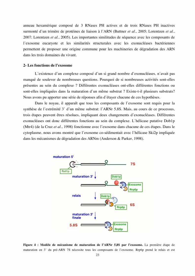
L’existence d’un complexe composé d’un si grand nombre d’exonucléases, n’avait pas manqué de soulever de nombreuses questions. Pourquoi de si nombreuses activités sont-elles présentes au sein du complexe ? Différentes exonucléases ont-elles différentes fonctions ou sont-elles impliquées dans la maturation d’un même substrat ? Existe-t-il plusieurs substrats? Nous avons pu apporter une série de réponses afin d’étayer chacune de ces hypothèses.
Dans le noyau, il apparaît que tous les composants de l’exosome sont requis pour la synthèse de l’extrémité 3’ d’un même substrat: l’ARNr 5,8S. Mais, au cours de ce processus, trois étapes peuvent êtres résolues, impliquant deux changements d’exonucléases. Différentes exonucléases ont donc différentes fonctions au sein du complexe. L’hélicase putative Dob1p (Mtr4) (de la Cruz et al., 1998) fonctionne avec l’exosome dans chacune de ces étapes. Dans le cytoplasme, nous avons montré que l’exosome co-sédimentait avec l’hélicase Ski2p impliquée dans les mécanismes de dégradation des ARNm (Anderson & Parker, 1998).
Figure 4 : Modèle de mécanisme de maturation de l’ARNr 5,8S par l’exosome. La première étape de maturation en 3’ du pré-ARN 7S nécessite tous les composants de l’exosome. Rrp6p prend le relais et est

24
spécifiquement requise pour l’obtention du pré-ARNr 6S. Enfin, la maturation finale en ARNr 5,8S implique à nouveau l’ensemble des exonucléases. Chacune des étapes est dépendante de l’hélicase putative Dob1p/Mtr4p.
Nous avons également identifié de nouveaux substrats nucléaires de l’exosome, en démontrant qu’il était impliqué dans la synthèse des snoARN et des snARN (Allmang et al., 1999a) ainsi que dans diverses étapes de la maturation du pré-ARNr (Allmang et al., 2000). Chez les eucaryotes, les petits ARN nucléolaires (ou snoARN) jouent un rôle majeur dans la maturation et la modification des pré-ARNr. La plupart des snoARN sont codés par des introns ou synthétisés sous la forme d’un précurseur polycistronique. Dans chacun des cas, leur excision requiert des mécanismes de maturation par des endonucléases et exonucléases. Rnt1p clive les précurseurs polycistroniques; l’épissage initie la synthèse des snoARN introniques. J’ai montré que la synthèse de leur extrémité 3’ était alors dépendante de l’action de l’exosome. L’extrémité 5’ est générée par l’exonucléase 5’ -> 3’ Rat1p (Petfalski et al., 1998). Ce processus est multiphasique et plusieurs exonucléases du complexe ont des fonctions distinctes. Le composant Rrp6p de l’exosome est spécifiquement impliqué dans l’étape finale de maturation en 3’ (Allmang et al., 1999a). Le pré-snoARN U3 et les pré-snARN U1, U4 et U5, bien que synthétisés à partir de leurs propres promoteurs, sont maturés en 3’ par les mêmes enzymes.
Un équilibre entre maturation et dégradation a pu être mis en évidence pour tous les substrats de l’exosome. L’exosome joue en effet un rôle important dans les mécanismes de dégradation des ARN, comme celle des précurseurs d’ARNr aberrants (Allmang et al., 2000).
D’autres travaux ont révélé que l’exosome fonctionnait également dans la dégradation nucléaire de pré-ARNm (Bousquet-Antonelli et al., 2000) suggérant un rôle potentiel dans la régulation de l’expression des gènes. Plus récemment, une partie du travail que j’avais initié a été poursuivie et complétée par Laura Milligan et Claire Torchet. Elle a conduit à démontrer que le composant exclusivement nucléaire de l’exosome (Rrp6) était impliqué dans les mécanismes de surveillance des ARN. En effet, dans une souche mutée au niveau la poly(A) polymérase et qui conduit à un ralentissement de polyadénylation des ARNm, les ARNm sont détectés par l’activité de surveillance de Rrp6p, déadénylés et rapidement dégradés par l’exosome (Milligan et al., 2005).
De nombreux autres travaux réalisés par la suite dans l’équipe de David Tollervey et ailleurs ont imposé l’exosome comme un acteur clé de la machinerie de surveillance des ARN. L’exosome intervient dans tous les types mécanismes de surveillance cytoplasmique des ARNm, tels la dégradation des ARNm sans codon de terminaison (non-stop decay), des ARNm à codon non-sens (non-sense-mediated decay) et des ARNm sujets à des arrêts prématurés de traduction (no-go decay) (pour une revue voir Houseley et al., 2006).
25
3- Mécanismes de synthèse des ARN stables
La quasi-totalité des ARN de la cellule est synthétisée à partir de précurseurs. L’une des observations les plus frappantes de notre étude est que la maturation ou dégradation de ces ARN implique un jeu bien défini mais limité d’enzymes et de cofacteurs. Les enzymes majeures sont le complexe de l’exosome, les exonucléases 5’->3’ Rat1p, Xrn1p et les endonucléases RNaseP/MRP et Rnt1p. Les cofacteurs incluent les hélicases Dob1p et Ski2p ainsi que des protéines chaperons telles que Lhp1p et le complexe Lsm. Nous avons analysé systématiquement le rôle de chacun de ces facteurs dans la synthèse des ARN stables (snoARN, snARN, ARNr et ARNt). Notre objectif était de comprendre le rôle relatif de ces facteurs, notamment dans la détermination de l’équilibre entre maturation et dégradation des ARN par l’exosome.
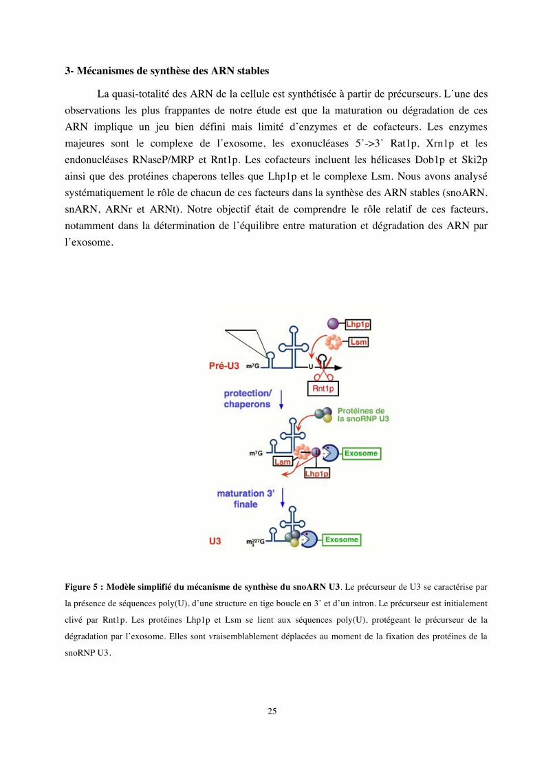
Figure 5 : Modèle simplifié du mécanisme de synthèse du snoARN U3. Le précurseur de U3 se caractérise par
la présence de séquences poly(U), d’une structure en tige boucle en 3’ et d’un intron. Le précurseur est initialement
clivé par Rnt1p. Les protéines Lhp1p et Lsm se lient aux séquences poly(U), protégeant le précurseur de la
dégradation par l’exosome. Elles sont vraisemblablement déplacées au moment de la fixation des protéines de la
snoRNP U3.
26
Avec Joanna Kufel, nous nous sommes notamment intéressés à la synthèse du snoARN U3. Le snoARN U3 est transcrit par l’ARN polymérase II; son précurseur est alors clivé par Rnt1p et maturé en 3’ par l’exosome (Kufel et al., 2000). Nous avons établi que d’autres facteurs étaient impliqués, notamment Lhp1p et le complexe Lsm. Lhp1p est une protéine chaperon qui se fixe sur la région poly (U) en 3’ des transcrits de l’ARN polymérase III (Pannone et al., 1998; Rinke & Steitz, 1982; Yoo & Wolin, 1997). Nous avons montré que Lhp1p stabilisait l’extrémité 3’ du pré-ARN U3 en s’y fixant. Le complexe Lsm, composé d’un anneau de 7 protéines (Achsel et al., 1999; Bouveret et al., 2000; Mayes et al., 1999) joue lui aussi ce rôle en coordination avec Lhp1 (Kufel et al., 2003b). Ces deux facteurs favorisent la maturation de l’extrémité 3’ en protégeant le précurseur de la dégradation par l’exosome tant que les protéines de la snoRNP mature ne sont pas fixées. Les protéines de la particule U3 déplaceraient les protéines chaperons, permettant la maturation finale de l’ARN (Kufel et al., 2000). Ce type de mécanisme est vraisemblablement ubiquitaire, car nous avons montré que les protéines chaperons Lhp1p et Lsm fonctionnaient également dans la synthèse de nombreux autres ARN, tels les ARNt et pré-ARNr (Kufel et al., 2003a; Kufel et al., 2002). Il en va de même pour l’exosome, les exonucleases 5’->3’ et les hélicases. Ces différents facteurs semblent pouvoir être recrutés sous différentes combinaisons et vers différents substrats pour en assurer la maturation et la dégradation.
Une question restait en suspens, à savoir comment se fait la discrimination des différents substrats et leur orientation vers les voies de maturation ou de dégradation. Ces dernières années ont vu d’importants développements dans la compréhension de ces mécanismes, en particulier en ce qui concerne la connaissance des signaux et mécanismes d’activation et de régulation de l’exosome. Plusieurs cofacteurs ont été identifiés, le plus impressionnant est certainement le complexe TRAMP dont la fonction semble être de diriger les précurseurs d’ARN défectueux vers l’exosome en les polyadénylant (Houseley et al., 2006; LaCava et al., 2005).
27
Activité de recherche et projets scientifiques actuels : Le mécanisme de synthèse des sélénoprotéines
Equipe du Dr. Alain Krol. Unité Architecture et Réactivité de l’ARN, CNRS, Université Louis Pasteur, IBMC, Strasbourg. A- Introduction
Le sélénium est un oligo-élément essentiel. Son importance physiologique n’a été appréciée à sa juste valeur que depuis les années 1970 avec l’identification de la forme biologique majeure du sélénium, l’acide aminé sélénocystéine qui est incorporé dans les sélénoprotéines par une machinerie de traduction spécialisée (Flohe et al., 2000).
Les premières sélénoprotéines identifiées étaient essentiellement des enzymes utilisant le potentiel d’oxydoréduction du sélénium dans leur site actif pour la lutte contre les radicaux libres oxygénés, telles les glutathion peroxydases (Flohe et al., 1973 ; Rotruck et al., 1973). L’identification récente de nouvelles sélénoprotéines montre qu’elles sont impliquées dans une grande variété de fonctions telles que le transport, la signalisation, la structure ou le développement musculaire (pour une revue voir Moghadaszadeh & Beggs, 2006). Celles-ci peuvent être intracellulaires, transmembranaires ou sécrétées et leur expression est tantôt ubiquitaire, tantôt tissu spécifique. On trouve des sélénoprotéines chez les archae, les bactéries et les eucaryotes mais elles ne sont pas représentées de façon égale dans ces trois règnes (Castellano et al., 2004; Kryukov & Gladyshev, 2004). Des mécanismes de biosynthèse différents sont mis en jeu chez les bactéries, les archae et les eucaryotes (pour une revue voir Allmang & Krol, 2006b).
L’équipe d’Alain Krol, que j’ai rejointe en 2001, a contribué à l’identification de nouvelles sélénoprotéines mais également à l’élucidation du mécanisme de synthèse des sélénoprotéines chez les eucaryotes. J’ai intégré cette dernière thématique et développé un nouveau sujet en étudiant le rôle de facteurs d’assemblage dans le mécanisme.
La sélénocystéine (Sec) est considérée comme le 21e acide aminé. Cet analogue de la
cystéine dont le groupement thiol est remplacé par un groupement sélénol est incorporé dans la chaîne peptidique de façon co-traductionnelle en réponse à un codon UGA habituellement reconnu comme l’un des trois codons de terminaison. Deux étapes majeures peuvent êtres résolues : la biosynthèse de la sélénocystéine et son incorporation par recodage du codon UGASec. Chez les eucaryotes, ces mécanismes sont particulièrement complexes et coordonnés par des facteurs capables de s’organiser en complexes supramoléculaires (Figures 6 et 7).
La biosynthèse de la sélénocystéine
La sélénocystéine n’existe pas en tant qu’acide aminé libre et c’est la sérine qui est, dans un premier temps, chargée sur l’ARNtSec par la sérine-ARNt synthétase conventionnelle avant
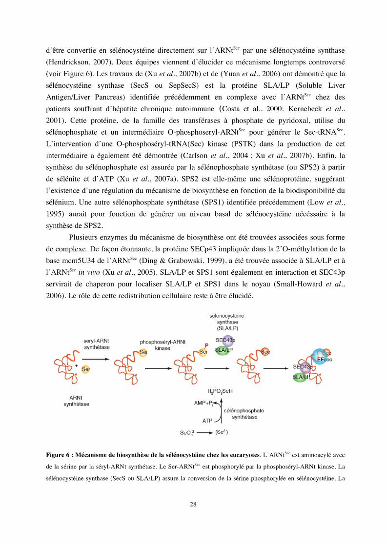
28
d’être convertie en sélénocystéine directement sur l’ARNtSec par une sélénocystéine synthase (Hendrickson, 2007). Deux équipes viennent d’élucider ce mécanisme longtemps controversé (voir Figure 6). Les travaux de (Xu et al., 2007b) et de (Yuan et al., 2006) ont démontré que la sélénocystéine synthase (SecS ou SepSecS) est la protéine SLA/LP (Soluble Liver Antigen/Liver Pancreas) identifiée précédemment en complexe avec l’ARNtSec chez des patients souffrant d’hépatite chronique autoimmune (Costa et al., 2000; Kernebeck et al., 2001). Cette protéine, de la famille des transférases à phosphate de pyridoxal, utilise du sélénophosphate et un intermédiaire O-phosphoseryl-ARNtSec pour générer le Sec-tRNASec. L’intervention d’une O-phosphoséryl-tRNA(Sec) kinase (PSTK) dans la production de cet intermédiaire a également été démontrée (Carlson et al., 2004 ; Xu et al., 2007b). Enfin, la synthèse du sélénophosphate est assurée par la sélénophosphate synthétase (ou SPS2) à partir de sélénite et d’ATP (Xu et al., 2007a). SPS2 est elle-même une sélénoprotéine, suggérant l’existence d’une régulation du mécanisme de biosynthèse en fonction de la biodisponibilité du sélénium. Une autre sélénophosphate synthétase (SPS1) identifiée précédemment (Low et al., 1995) aurait pour fonction de générer un niveau basal de sélénocystéine nécéssaire à la synthèse de SPS2. Plusieurs enzymes du mécanisme de biosynthèse ont été trouvées associées sous forme de complexe. De façon étonnante, la protéine SECp43 impliquée dans la 2’O-méthylation de la base mcm5U34 de l’ARNtSec (Ding & Grabowski, 1999), a été trouvée associée à SLA/LP et à l’ARNtSec in vivo (Xu et al., 2005). SLA/LP et SPS1 sont également en interaction et SEC43p servirait de chaperon pour localiser SLA/LP et SPS1 dans le noyau (Small-Howard et al., 2006). Le rôle de cette redistribution cellulaire reste à être élucidé.
Figure 6 : Mécanisme de biosynthèse de la sélénocystéine chez les eucaryotes. L’ARNtSec est aminoacylé avec
de la sérine par la séryl-ARNt synthétase. Le Ser-ARNtSec est phosphorylé par la phosphoséryl-ARNt kinase. La
sélénocystéine synthase (SecS ou SLA/LP) assure la conversion de la sérine phosphorylée en sélénocystéine. La
29
synthèse du sélénophosphate nécessaire à cette étape est catalysée par une sélénophosphate synthétase. L’ARNtSec
est pris en charge par le facteur d’élongation spécialisé eEFsec.
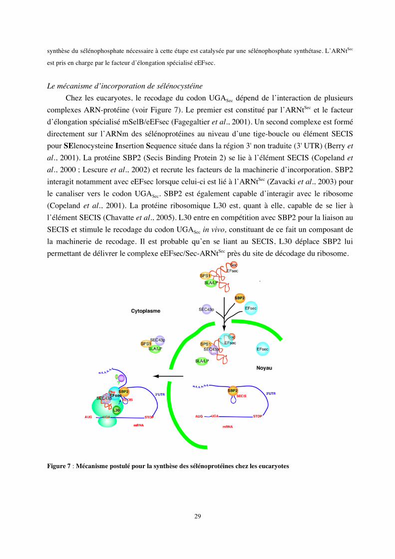
Le mécanisme d’incorporation de sélénocystéine
Chez les eucaryotes, le recodage du codon UGASec dépend de l’interaction de plusieurs complexes ARN-protéine (voir Figure 7). Le premier est constitué par l’ARNtSec et le facteur d’élongation spécialisé mSelB/eEFsec (Fagegaltier et al., 2001). Un second complexe est formé directement sur l’ARNm des sélénoprotéines au niveau d’une tige-boucle ou élément SECIS pour SElenocysteine Insertion Sequence située dans la région 3' non traduite (3' UTR) (Berry et al., 2001). La protéine SBP2 (Secis Binding Protein 2) se lie à l’élément SECIS (Copeland et al., 2000 ; Lescure et al., 2002) et recrute les facteurs de la machinerie d’incorporation. SBP2 interagit notamment avec eEFsec lorsque celui-ci est lié à l’ARNtSec (Zavacki et al., 2003) pour le canaliser vers le codon UGASec. SBP2 est également capable d’interagir avec le ribosome (Copeland et al., 2001). La protéine ribosomique L30 est, quant à elle, capable de se lier à l’élément SECIS (Chavatte et al., 2005). L30 entre en compétition avec SBP2 pour la liaison au SECIS et stimule le recodage du codon UGASec in vivo, constituant de ce fait un composant de la machinerie de recodage. Il est probable qu’en se liant au SECIS, L30 déplace SBP2 lui permettant de délivrer le complexe eEFsec/Sec-ARNtSec près du site de décodage du ribosome.
Figure 7 : Mécanisme postulé pour la synthèse des sélénoprotéines chez les eucaryotes
30
Plusieurs modèles ont été proposés pour expliquer ce mécanisme selon que SBP2 est initialement associée au ribosome ou à l’ARN SECIS (Chavatte et al., 2005; Kinzy et al., 2005).
Des résultats récents ont montré que SBP2 faisait partie de complexes supramoléculaires et est présente à la fois dans le cytoplasme et le noyau. En effet, il apparaît que la protéine SECp43 trouvée associée à l’ARNtSec et aux facteurs de la biogenèse de la sélénocystéine (voir paragraphe précédent) est également capable de promouvoir l’interaction entre eEFsec et SBP2 in vivo (Small-Howard et al., 2006). SECp43 influence par ailleurs la localisation nucléaire de ces protéines. Des signaux de localisation et d’export nucléaire ont pu être identifiés pour eEFsec et SBP2 et un assemblage nucléaire précoce des facteurs du mécanisme d’incorporation de sélénocystéine sur l’ARN SECIS a été proposé (de Jesus et al., 2006). La séquestration nucléaire de SBP2 peut être induite par un stress oxydatif et l’oxydation de cystéines essentielles qui la rendent incapable d’interagir avec le facteur d’export nucléaire CRM1 (Papp et al., 2006). Ceci a pour conséquence une diminution de l’incorporation de sélénocystéine et pourrait également représenter un moyen de régulation de l’expression des sélénoprotéines en fonction du statut redox de la cellule. Il est également vraisemblable que l’assemblage du complexe dans le noyau permet d’éviter que les ARNm de sélénoprotéines ne soient soumis aux mécanismes de dégradation des ARN à codon non-sens ou nonsense-mediated decay (NMD) (de Jesus et al., 2006).
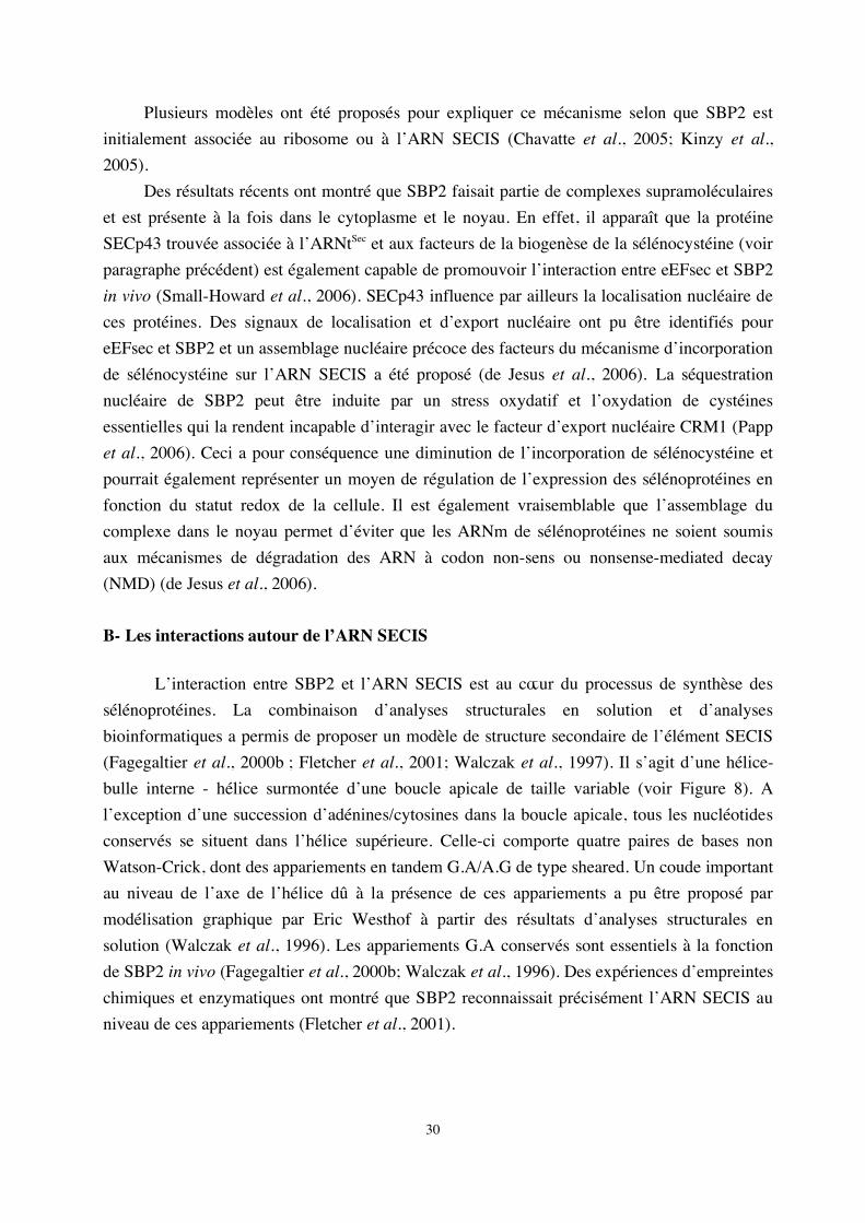
B- Les interactions autour de l’ARN SECIS L’interaction entre SBP2 et l’ARN SECIS est au cœur du processus de synthèse des sélénoprotéines. La combinaison d’analyses structurales en solution et d’analyses bioinformatiques a permis de proposer un modèle de structure secondaire de l’élément SECIS (Fagegaltier et al., 2000b ; Fletcher et al., 2001; Walczak et al., 1997). Il s’agit d’une hélice-bulle interne - hélice surmontée d’une boucle apicale de taille variable (voir Figure 8). A l’exception d’une succession d’adénines/cytosines dans la boucle apicale, tous les nucléotides conservés se situent dans l’hélice supérieure. Celle-ci comporte quatre paires de bases non Watson-Crick, dont des appariements en tandem G.A/A.G de type sheared. Un coude important au niveau de l’axe de l’hélice dû à la présence de ces appariements a pu être proposé par modélisation graphique par Eric Westhof à partir des résultats d’analyses structurales en solution (Walczak et al., 1996). Les appariements G.A conservés sont essentiels à la fonction de SBP2 in vivo (Fagegaltier et al., 2000b; Walczak et al., 1996). Des expériences d’empreintes chimiques et enzymatiques ont montré que SBP2 reconnaissait précisément l’ARN SECIS au niveau de ces appariements (Fletcher et al., 2001).
31
Figure 8 : L’ARN SECIS d’après Allmang et Krol (2006). A. Modèles de structure secondaire des ARN SECIS
eucaryotes de forme 1 et 2. Les séquences et caractéristiques structurales conservées sont indiquées. N, n’importe
quel nucléotide ; A/g et A/c indique que A est le nucléotide majoritaire. B. Représentation de la structure en K-turn
potentielle de l’ARN SECIS de l’iodotyronine désiodase de rat de type 1 d’après la nomenclature de Leontis et
Westhof (2001).
Notre objectif a été d’analyser plus précisément les détails de cette interaction en identifiant les acides aminés de SBP2 importants pour la liaison à l’ARN SECIS par dissection fonctionnelle, sélection d’ARN in vitro et résolution de la structure cristallographique du complexe SBP2/SECIS. De façon plus générale, nous avons également tenté de dégager les principes d’interaction des protéines de la famille L7Ae avec leurs ARN cibles. 1- La protéine SBP2 humaine et son mode d’interaction avec l’élément SECIS
Lorsque j’ai rejoint l’équipe d’Alain Krol en octobre 2001, l’ADNc de la protéine SBP2 humaine venait d’être cloné au laboratoire par Alain Lescure (Lescure et al., 2002). J’ai été associée à ce travail en montrant que la liaison de SBP2 à l’ARN SECIS était stimulée par le facteur d’élongation spécialisé eEFsec sans qu’il ne s’associe pour autant au complexe. Ces résultats suggéraient que eEFsec était capable d’induire une conformation de SBP2 plus propice à la reconnaissance de l’ARN SECIS. Ceci nous a conduit à entamer une dissection fonctionnelle de SBP2 afin d’affiner la compréhension de ses différents domaines et plus particulièrement son domaine de liaison à l’ARN.
Le domaine de liaison à l’ARN de SBP2 se situe entre les acides aminés 500 et 750 (Allmang et al., 2002 ; Copeland et al., 2000 ; Lescure et al., 2002). Nous avons montré qu’il s’agissait d’un domaine bipartite constitué de séquences spécifiques à SBP2 et d’un module structural conservé. Par alignement de séquences, nous avons découvert au sein du domaine de liaison à l’ARN un module appartenant à la famille des protéines ribosomiques L7Ae (Allmang
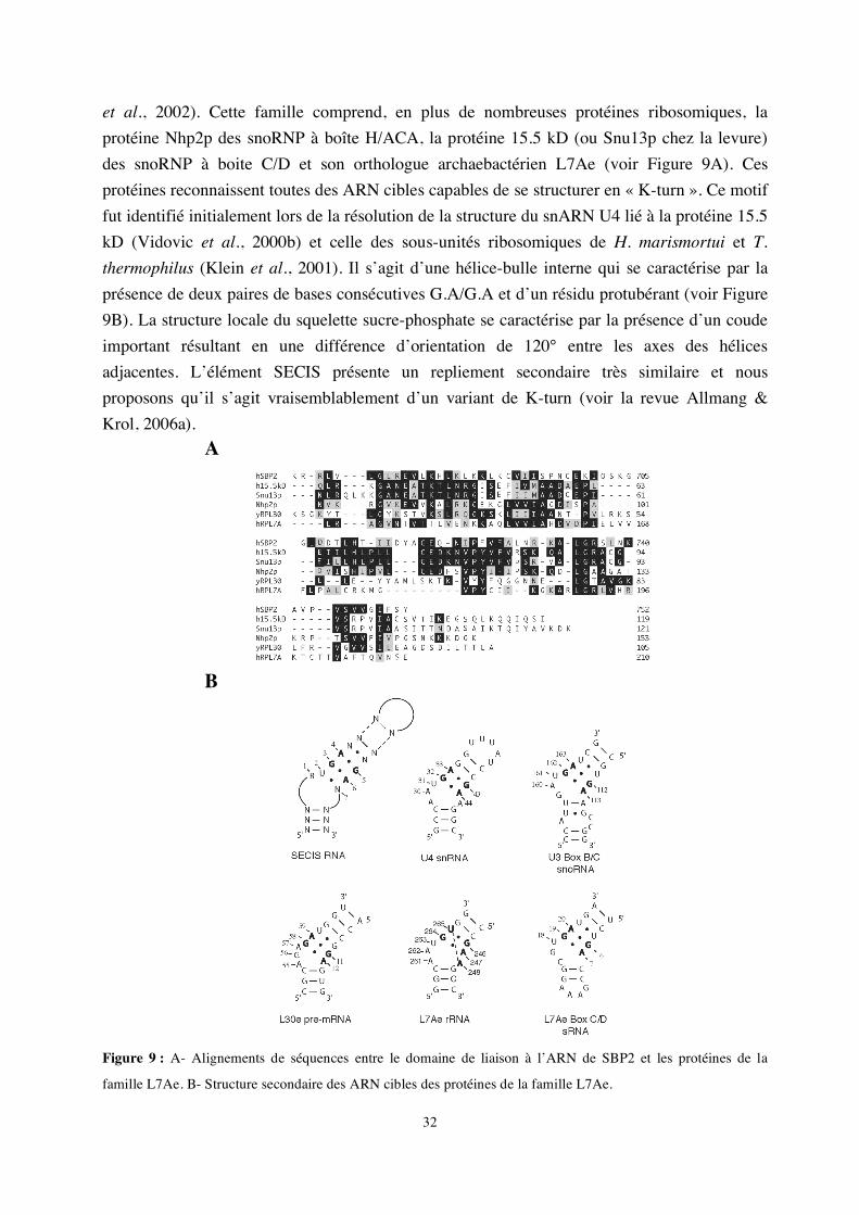
32
et al., 2002). Cette famille comprend, en plus de nombreuses protéines ribosomiques, la protéine Nhp2p des snoRNP à boîte H/ACA, la protéine 15.5 kD (ou Snu13p chez la levure) des snoRNP à boite C/D et son orthologue archaebactérien L7Ae (voir Figure 9A). Ces protéines reconnaissent toutes des ARN cibles capables de se structurer en « K-turn ». Ce motif fut identifié initialement lors de la résolution de la structure du snARN U4 lié à la protéine 15.5 kD (Vidovic et al., 2000b) et celle des sous-unités ribosomiques de H. marismortui et T. thermophilus (Klein et al., 2001). Il s’agit d’une hélice-bulle interne qui se caractérise par la présence de deux paires de bases consécutives G.A/G.A et d’un résidu protubérant (voir Figure 9B). La structure locale du squelette sucre-phosphate se caractérise par la présence d’un coude important résultant en une différence d’orientation de 120° entre les axes des hélices adjacentes. L’élément SECIS présente un repliement secondaire très similaire et nous proposons qu’il s’agit vraisemblablement d’un variant de K-turn (voir la revue Allmang & Krol, 2006a).
A
B
Figure 9 : A- Alignements de séquences entre le domaine de liaison à l’ARN de SBP2 et les protéines de la
famille L7Ae. B- Structure secondaire des ARN cibles des protéines de la famille L7Ae.
33
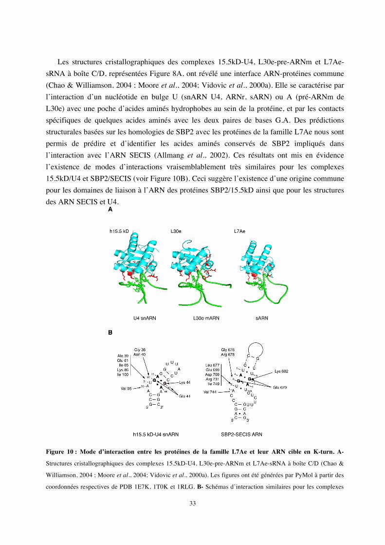
Les structures cristallographiques des complexes 15.5kD-U4, L30e-pre-ARNm et L7Ae-
sRNA à boîte C/D, représentées Figure 8A, ont révélé une interface ARN-protéines commune (Chao & Williamson, 2004 ; Moore et al., 2004; Vidovic et al., 2000a). Elle se caractérise par l’interaction d’un nucléotide en bulge U (snARN U4, ARNr, sARN) ou A (pré-ARNm de L30e) avec une poche d’acides aminés hydrophobes au sein de la protéine, et par les contacts spécifiques de quelques acides aminés avec les deux paires de bases G.A. Des prédictions structurales basées sur les homologies de SBP2 avec les protéines de la famille L7Ae nous sont permis de prédire et d’identifier les acides aminés conservés de SBP2 impliqués dans l’interaction avec l’ARN SECIS (Allmang et al., 2002). Ces résultats ont mis en évidence l’existence de modes d’interactions vraisemblablement très similaires pour les complexes 15.5kD/U4 et SBP2/SECIS (voir Figure 10B). Ceci suggère l’existence d’une origine commune pour les domaines de liaison à l’ARN des protéines SBP2/15.5kD ainsi que pour les structures des ARN SECIS et U4.
Figure 10 : Mode d’interaction entre les protéines de la famille L7Ae et leur ARN cible en K-turn. A-
Structures cristallographiques des complexes 15.5kD-U4, L30e-pre-ARNm et L7Ae-sRNA à boîte C/D (Chao &
Williamson, 2004 ; Moore et al., 2004; Vidovic et al., 2000a). Les figures ont été générées par PyMol à partir des
coordonnées respectives de PDB 1E7K, 1T0K et 1RLG. B- Schémas d’interaction similaires pour les complexes
34
15.5kD-snARN U4 et SBP2-SECIS. 4 acides aminés de SBP2 sont essentiels à l’interaction : Gly676 et Glu679
postulés en contact avec les guanines des paires G.A ; Glu699 et Arg731 postulés en contact avec le U en bulge.
Par ailleurs, avec David Schmitt (étudiant en DEA de Biologie Moléculaire en 2002) nous
avons délimité plus précisément le domaine de liaison à l’ARN et montré qu’il s’étendait bien
au-delà du module conservé L7Ae et comprenait une séquence conservée riche en lysines. Ces
résultats suggèrent que des contacts additionnels existent entre SBP2 et l’ARN SECIS par
rapport aux autres protéines de la famille L7Ae.
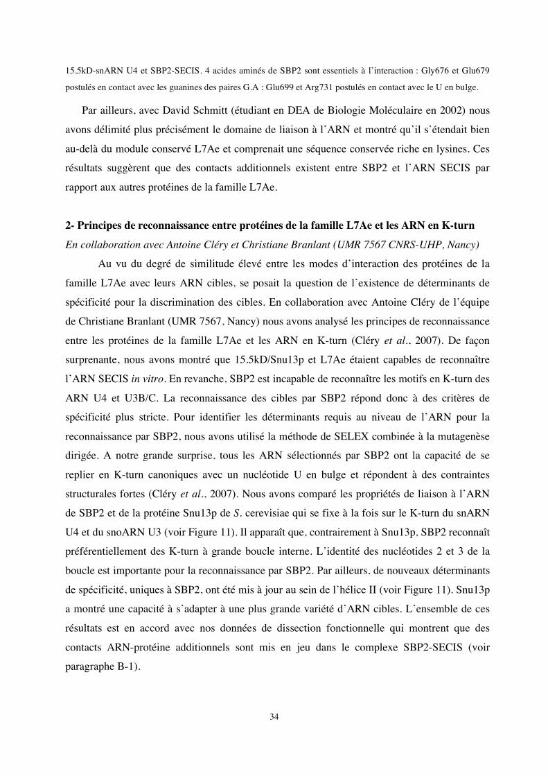
2- Principes de reconnaissance entre protéines de la famille L7Ae et les ARN en K-turn
En collaboration avec Antoine Cléry et Christiane Branlant (UMR 7567 CNRS-UHP, Nancy)
Au vu du degré de similitude élevé entre les modes d’interaction des protéines de la
famille L7Ae avec leurs ARN cibles, se posait la question de l’existence de déterminants de
spécificité pour la discrimination des cibles. En collaboration avec Antoine Cléry de l’équipe
de Christiane Branlant (UMR 7567, Nancy) nous avons analysé les principes de reconnaissance
entre les protéines de la famille L7Ae et les ARN en K-turn (Cléry et al., 2007). De façon
surprenante, nous avons montré que 15.5kD/Snu13p et L7Ae étaient capables de reconnaître
l’ARN SECIS in vitro. En revanche, SBP2 est incapable de reconnaître les motifs en K-turn des
ARN U4 et U3B/C. La reconnaissance des cibles par SBP2 répond donc à des critères de
spécificité plus stricte. Pour identifier les déterminants requis au niveau de l’ARN pour la
reconnaissance par SBP2, nous avons utilisé la méthode de SELEX combinée à la mutagenèse
dirigée. A notre grande surprise, tous les ARN sélectionnés par SBP2 ont la capacité de se
replier en K-turn canoniques avec un nucléotide U en bulge et répondent à des contraintes
structurales fortes (Cléry et al., 2007). Nous avons comparé les propriétés de liaison à l’ARN
de SBP2 et de la protéine Snu13p de S. cerevisiae qui se fixe à la fois sur le K-turn du snARN
U4 et du snoARN U3 (voir Figure 11). Il apparaît que, contrairement à Snu13p, SBP2 reconnaît
préférentiellement des K-turn à grande boucle interne. L’identité des nucléotides 2 et 3 de la
boucle est importante pour la reconnaissance par SBP2. Par ailleurs, de nouveaux déterminants
de spécificité, uniques à SBP2, ont été mis à jour au sein de l’hélice II (voir Figure 11). Snu13p
a montré une capacité à s’adapter à une plus grande variété d’ARN cibles. L’ensemble de ces
résultats est en accord avec nos données de dissection fonctionnelle qui montrent que des
contacts ARN-protéine additionnels sont mis en jeu dans le complexe SBP2-SECIS (voir
paragraphe B-1).
35
Figure 11 : Déterminants de spécificité reconnus par SBP2 et Snu13p/15.5kD au niveau des ARN en K-turn.
A gauche : Structure secondaire de l’ARN obtenu par SELEX et reconnu par SBP2 avec la meilleure affinité. Des
variants de cet ARN (mutations au sein des hélices I, II et de la boucle interne) ont été testées pour leur capacité à
être reconnus par SBP2 et Snu13p. Les déterminants de spécificité identifiés pour chacune des protéines sont
représentés en rouge.
L’assemblage de la machinerie de synthèse des sélénoprotéines est vraisemblablement initiée par la fixation de SBP2 aux éléments SECIS dans le noyau (voire le nucléole) où la protéine 15.5kD/Snu13p est très abondante. Les différences importantes au niveau des déterminants de spécificité des ARN cibles de SBP2 et 15.5kD/Snu13p contribuent vraisemblablement à la spécificité d’association des complexes SBP2-SECIS dans ce compartiment cellulaire. 3- Objectif : Résolution de la structure cristallographique des complexes SBP2-SECIS, L30-
SECIS
Travail de thèse de Vincent Olieric dirigé par Philippe Dumas (UPR 9002 du CNRS) et d’Akiko Takeuchi (Doctorante au laboratoire depuis 2006).
Afin d’établir définitivement si l’ARN SECIS possède un repliement en K-turn nous avons entrepris de résoudre la structure aux rayons X du complexe SBP2/ARN SECIS en collaboration avec l’équipe de P. Dumas (UPR 9002 du CNRS). La résolution de cette structure permettrait également de valider nos prédictions structurales (Allmang et al., 2002) ainsi que de comprendre le rôle du motif de liaison à l’ARN additionnel par rapport aux autres protéines de la famille L7Ae.
36
Ce travail a fait l’objet de la thèse de Vincent Olieric dans l’équipe de Philippe Dumas. Des protocoles d’expression et de purification de SBP2 ont été optimisés et une grande variété d’ARN SECIS a été synthétisée. Il n’a pas été possible d’obtenir de cristaux du complexe, ni de la protéine isolée. En revanche, la caractérisation biophysique de SBP2 par RMN et ultracentrifugation analytique a révélé une absence de structuration. Ceci est en accord avec des analyses bioinformatiques prédisant une prédominance de zones non repliées, en dehors du module L7Ae. SBP2 semble répondre à plusieurs critères caractéristiques des protéines intrinsèquement non structurées ou « IUP » (Intrinsically Unstructured Proteins) (Dosztanyi et al., 2005). Chez les eucaryotes supérieurs, bon nombre de protéines impliquées dans des mécanismes de régulation ou de transduction des signaux ne se replient de façon stable qu’en présence de leurs partenaires moléculaires (pour des revues voir Dunker et al., 2005; Tompa, 2005). SBP2 sert de plateforme pour le recrutement des autres partenaires de la machinerie de biosynthèse des sélénoprotéines. Des résultats récents obtenus au laboratoire montrent que SBP2 interagit avec un complexe de protéines chaperons lié à la protéine HSP90, et que cette association joue un rôle fonctionnel important dans le repliement de SBP2 et son interaction avec ses cibles (voir paragraphe C). La possibilité que SBP2 soit partiellement non structurée est donc compatible avec les interactions multiples qu’elle doit assurer et nos nouvelles données. Il n’est cependant pas possible d’exclure que l’absence de structuration résulte de l’expression de SBP2 dans E.coli qui ne permet pas d’assurer les modifications post-traductionnelles. Afin de vérifier cette hypothèse, la protéine SBP2 sera produite dans des cellules eucaryotes à partir de vecteurs de type baculovirus. Ce travail a été initié par Akiko Takeuchi étudiante en thèse dans notre laboratoire depuis septembre 2006 avec l’aide de la plateforme de biologie génomique et structurales (CEBGS-Illkirch) et du service baculovirus de l’IGBMC (Illkirch). Nous tenterons également de co-cristalliser SBP2 en présence de HSP90, car il est vraisemblable que l’interaction SBP2-HSP90 facilite la structuration de SBP2 afin de la rendre apte à interagir avec ses partenaires finaux.
Enfin, nous tenterons de surproduire et de cristalliser la protéine SBP2 issue d’un autre organisme, en particulier celle de Drosophila melanogaster qui présente la particularité d’être dépourvue du domaine N-terminal présent dans la protéine humaine (voir paragraphe B-4). Nous évaluerons sa capacité à se lier spécifiquement à l’élément SECIS et à se replier de façon stable.
La fixation de la protéine ribosomique L30 sur l’ARN SECIS a également été identifiée comme l’une des étapes du mécanisme de recodage des sélénoprotéines (Chavatte et al., 2005). Nous tenterons de cristalliser le complexe L30/SECIS. La comparaison des structures des deux complexes SBP2/SECIS, L30/SECIS devrait nous donner de précieux renseignements quant au rôle central de l’ARN SECIS dans l’étape de translecture, c'est-à-dire s’il existe une compétition des protéines pour les mêmes sites au niveau du SECIS, ou des changements allostériques permettant interaction avec le ribosome.
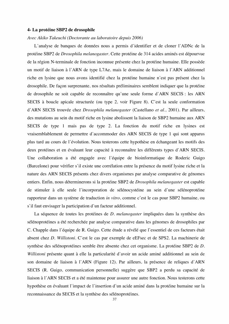
37
4- La protéine SBP2 de drosophile
Avec Akiko Takeuchi (Doctorante au laboratoire depuis 2006)
L’analyse de banques de données nous a permis d’identifier et de cloner l’ADNc de la
protéine SBP2 de Drosophila melanogaster. Cette protéine de 314 acides aminés est dépourvue
de la région N-terminale de fonction inconnue présente chez la protéine humaine. Elle possède
un motif de liaison à l’ARN de type L7Ae, mais le domaine de liaison à l’ARN additionnel
riche en lysine que nous avons identifié chez la protéine humaine n’est pas présent chez la
drosophile. De façon surprenante, nos résultats préliminaires semblent indiquer que la protéine
de drosophile ne soit capable de reconnaître qu’une seule forme d’ARN SECIS : les ARN
SECIS à boucle apicale structurée (ou type 2, voir Figure 8). C’est la seule conformation
d’ARN SECIS trouvée chez Drosophila melanogaster (Castellano et al., 2001). Par ailleurs,
des mutations au sein du motif riche en lysine abolissent la liaison de SBP2 humaine aux ARN
SECIS de type 1 mais pas de type 2. La fonction du motif riche en lysines est
vraisemblablement de permettre d’accommoder des ARN SECIS de type 1 qui sont apparus
plus tard au cours de l’évolution. Nous testerons cette hypothèse en échangeant les motifs des
deux protéines et en évaluant leur capacité à reconnaître les différents types d’ARN SECIS.
Une collaboration a été engagée avec l’équipe de bioinformatique de Roderic Guigo
(Barcelone) pour vérifier s’il existe une corrélation entre la présence du motif lysine riche et la
nature des ARN SECIS présents chez divers organismes par analyse comparative de génomes
entiers. Enfin, nous déterminerons si la protéine SBP2 de Drosophila melanogaster est capable
de stimuler à elle seule l’incorporation de sélénocystéine au sein d’une sélénoprotéine
rapporteur dans un système de traduction in vitro, comme c’est le cas pour SBP2 humaine, ou
s’il faut envisager la participation d’un facteur additionnel.
La séquence de toutes les protéines de D. melanogaster impliquées dans la synthèse des
sélénoprotéines a été recherchée par analyse comparative dans les génomes de drosophiles par
C. Chapple dans l’équipe de R. Guigo. Cette étude a révélé que l’essentiel de ces facteurs était
absent chez D. Willistoni. C’est le cas par exemple de eEFsec et de SPS2. La machinerie de
synthèse des sélénoprotéines semble être absente chez cet organisme. La protéine SBP2 de D.
Willistoni présente quant à elle la particularité d’avoir un acide aminé additionnel au sein de
son domaine de liaison à l’ARN (Figure 12). Par ailleurs, la présence de reliques d’ARN
SECIS (R. Guigo, communication personnelle) suggère que SBP2 a perdu sa capacité de
liaison à l’ARN SECIS et a été maintenue pour assurer une autre fonction. Nous testerons cette
hypothèse en évaluant l’impact de l’insertion d’un acide aminé dans la protéine humaine sur la
reconnaissance du SECIS et la synthèse des sélénoprotéines.
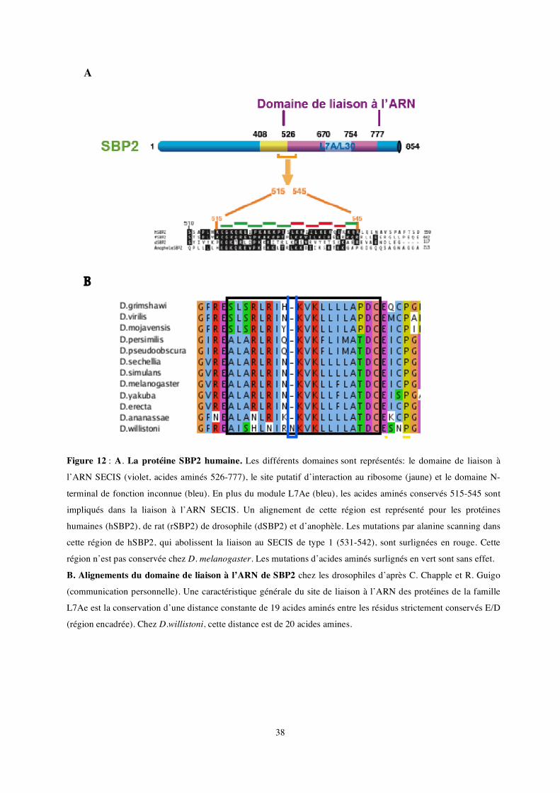
38
A
B
Figure 12 : A. La protéine SBP2 humaine. Les différents domaines sont représentés: le domaine de liaison à
l’ARN SECIS (violet, acides aminés 526-777), le site putatif d’interaction au ribosome (jaune) et le domaine N-
terminal de fonction inconnue (bleu). En plus du module L7Ae (bleu), les acides aminés conservés 515-545 sont
impliqués dans la liaison à l’ARN SECIS. Un alignement de cette région est représenté pour les protéines
humaines (hSBP2), de rat (rSBP2) de drosophile (dSBP2) et d’anophèle. Les mutations par alanine scanning dans
cette région de hSBP2, qui abolissent la liaison au SECIS de type 1 (531-542), sont surlignées en rouge. Cette
région n’est pas conservée chez D. melanogaster. Les mutations d’acides aminés surlignés en vert sont sans effet.
B. Alignements du domaine de liaison à l’ARN de SBP2 chez les drosophiles d’après C. Chapple et R. Guigo
(communication personnelle). Une caractéristique générale du site de liaison à l’ARN des protéines de la famille
L7Ae est la conservation d’une distance constante de 19 acides aminés entre les résidus strictement conservés E/D
(région encadrée). Chez D.willistoni, cette distance est de 20 acides amines.
39
C- Les complexes supramoléculaires impliqués dans la synthèse des sélénoprotéines Avec Laurence Wurth (Doctorante depuis octobre 2005).
La famille des protéines L7Ae, à laquelle appartient SBP2, participe à la construction de
plusieurs RNP essentielles et leur liaison à l’ARN conditionne le recrutement des autres
protéines. Nos objectifs sont, d’une part de comprendre les mécanismes d’assemblages
généraux mis en jeu pour la construction de ces RNP et d’autre part, d’identifier les composants
encore inconnus du complexe moléculaire qui est recruté spécifiquement par SBP2 sur l’ARN
SECIS.
1- Un mécanisme commun pour l’assemblage des RNP L7Ae (manuscrit soumis)
En collaboration avec Edouard Bertrand (CNRS UMR 5535, Montpellier), Bruno Charpentier
et Christiane Branlant (UMR 7567 CNRS-UHP, Nancy), Tamas Kiss (LBME, Toulouse) et
Barbara Bardoni (Université de Nice-Sophia antipolis) dans le cadre d’un contrat de l’Agence
Nationale pour la Recherche.
Nous avons identifié une machinerie d’assemblage des RNP L7Ae conservée de la
levure à l’homme et d’importance fondamentale pour la cellule. Elle est constituée d’une
protéine adaptatrice Nufip (Rsa1 chez la levure) et d’un complexe de protéines chaperons.
Nufip, un facteur d'assemblage des particules de la famille L7A
Nous avons utilisé des cribles double et triple hybrides pour caractériser de nouveaux
facteurs impliqués dans la biogenèse des snoARN à boîte C/D. Chez Saccharomyces
cerevisiae, ceux-ci ont permis de détecter la protéine Rsa1 (Kressler et al., 1999) et de montrer
qu’elle interagissait avec Snu13p in vitro (Bruno Charpentier, Nancy). Parallèlement, la
protéine humaine Nufip a été trouvée en interaction avec 15.5kD (Edouard Bertrand,
Montpellier). Cette interaction a été confirmée in vivo. Nufip est une protéine nucléaire qui se
lie à l’ARN. Elle fut initialement identifiée en interaction avec la protéine FRMP qui est
impliquée dans le transport et la localisation d’ARNm (Bardoni et al., 1999). Des comparaisons
de séquences ont révélé que Nufip et Rsa1 présentent un motif conservé de 32 acides aminés
(ou motif PEP). Nous avons démontré que Nufip et Rsa1 étaient capables d’interagir
respectivement avec 15.5kD et Snu13p, par l’intermédiaire du motif PEP et que Nufip était
l’homologue fonctionnel de Rsa1.
De façon intéressante, Nufip reconnaît deux autres membres de la famille L7Ae,
hNhp2p qui fait partie des snoRNP à boîte H/ACA et SBP2. Nous avons notamment pu
confirmer l’interaction entre Nufip et SBP2 in vivo en co-purifiant les protéines endogènes à
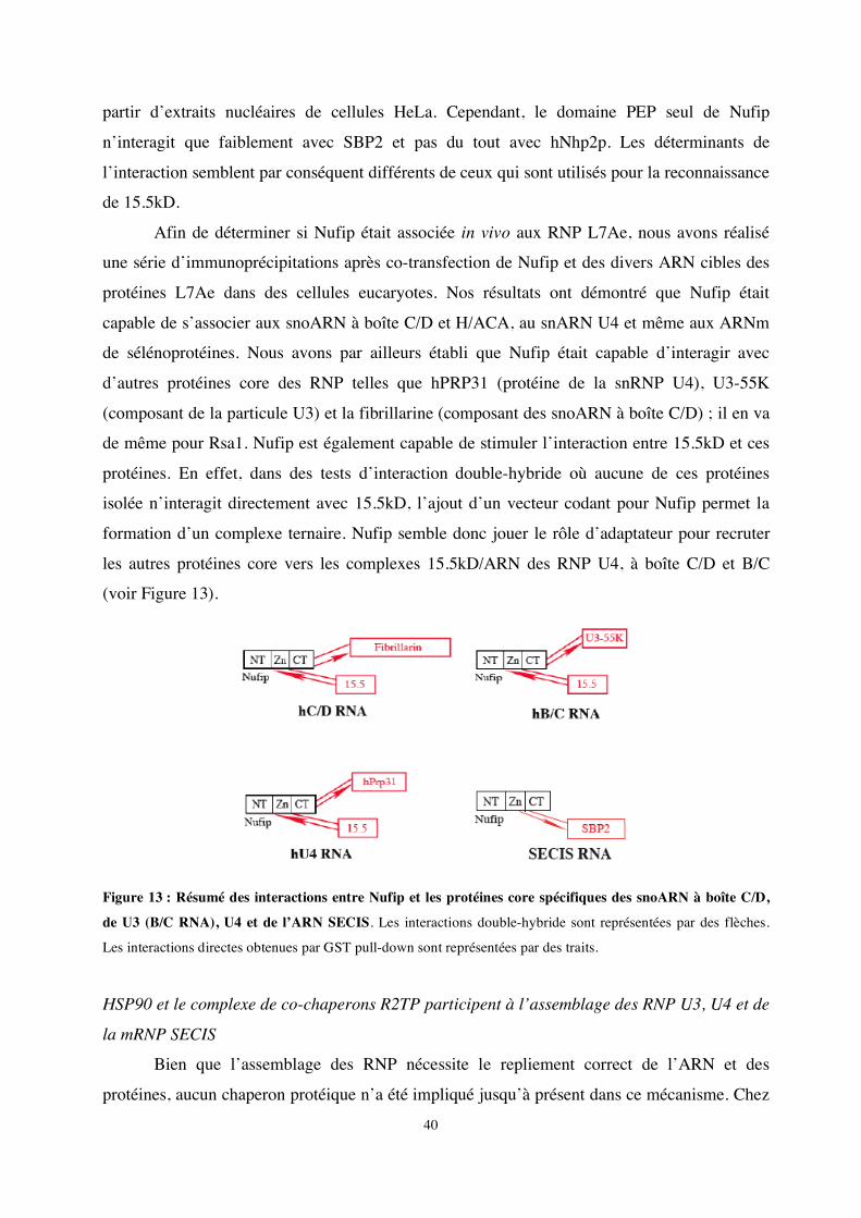
40
partir d’extraits nucléaires de cellules HeLa. Cependant, le domaine PEP seul de Nufip
n’interagit que faiblement avec SBP2 et pas du tout avec hNhp2p. Les déterminants de
l’interaction semblent par conséquent différents de ceux qui sont utilisés pour la reconnaissance
de 15.5kD.
Afin de déterminer si Nufip était associée in vivo aux RNP L7Ae, nous avons réalisé
une série d’immunoprécipitations après co-transfection de Nufip et des divers ARN cibles des
protéines L7Ae dans des cellules eucaryotes. Nos résultats ont démontré que Nufip était
capable de s’associer aux snoARN à boîte C/D et H/ACA, au snARN U4 et même aux ARNm
de sélénoprotéines. Nous avons par ailleurs établi que Nufip était capable d’interagir avec
d’autres protéines core des RNP telles que hPRP31 (protéine de la snRNP U4), U3-55K
(composant de la particule U3) et la fibrillarine (composant des snoARN à boîte C/D) ; il en va
de même pour Rsa1. Nufip est également capable de stimuler l’interaction entre 15.5kD et ces
protéines. En effet, dans des tests d’interaction double-hybride où aucune de ces protéines
isolée n’interagit directement avec 15.5kD, l’ajout d’un vecteur codant pour Nufip permet la
formation d’un complexe ternaire. Nufip semble donc jouer le rôle d’adaptateur pour recruter
les autres protéines core vers les complexes 15.5kD/ARN des RNP U4, à boîte C/D et B/C
(voir Figure 13).
Figure 13 : Résumé des interactions entre Nufip et les protéines core spécifiques des snoARN à boîte C/D,
de U3 (B/C RNA), U4 et de l’ARN SECIS. Les interactions double-hybride sont représentées par des flèches.
Les interactions directes obtenues par GST pull-down sont représentées par des traits.
HSP90 et le complexe de co-chaperons R2TP participent à l’assemblage des RNP U3, U4 et de
la mRNP SECIS
Bien que l’assemblage des RNP nécessite le repliement correct de l’ARN et des
protéines, aucun chaperon protéique n’a été impliqué jusqu’à présent dans ce mécanisme. Chez
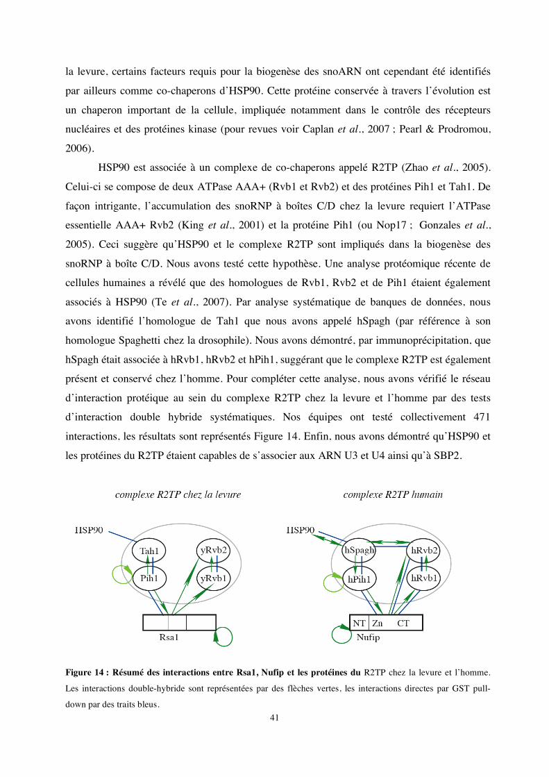
41
la levure, certains facteurs requis pour la biogenèse des snoARN ont cependant été identifiés
par ailleurs comme co-chaperons d’HSP90. Cette protéine conservée à travers l’évolution est
un chaperon important de la cellule, impliquée notamment dans le contrôle des récepteurs
nucléaires et des protéines kinase (pour revues voir Caplan et al., 2007 ; Pearl & Prodromou,
2006).
HSP90 est associée à un complexe de co-chaperons appelé R2TP (Zhao et al., 2005).
Celui-ci se compose de deux ATPase AAA+ (Rvb1 et Rvb2) et des protéines Pih1 et Tah1. De
façon intrigante, l’accumulation des snoRNP à boîtes C/D chez la levure requiert l’ATPase
essentielle AAA+ Rvb2 (King et al., 2001) et la protéine Pih1 (ou Nop17 ; Gonzales et al.,
2005). Ceci suggère qu’HSP90 et le complexe R2TP sont impliqués dans la biogenèse des
snoRNP à boîte C/D. Nous avons testé cette hypothèse. Une analyse protéomique récente de
cellules humaines a révélé que des homologues de Rvb1, Rvb2 et de Pih1 étaient également
associés à HSP90 (Te et al., 2007). Par analyse systématique de banques de données, nous
avons identifié l’homologue de Tah1 que nous avons appelé hSpagh (par référence à son
homologue Spaghetti chez la drosophile). Nous avons démontré, par immunoprécipitation, que
hSpagh était associée à hRvb1, hRvb2 et hPih1, suggérant que le complexe R2TP est également
présent et conservé chez l’homme. Pour compléter cette analyse, nous avons vérifié le réseau
d’interaction protéique au sein du complexe R2TP chez la levure et l’homme par des tests
d’interaction double hybride systématiques. Nos équipes ont testé collectivement 471
interactions, les résultats sont représentés Figure 14. Enfin, nous avons démontré qu’HSP90 et
les protéines du R2TP étaient capables de s’associer aux ARN U3 et U4 ainsi qu’à SBP2.
Figure 14 : Résumé des interactions entre Rsa1, Nufip et les protéines du R2TP chez la levure et l’homme.
Les interactions double-hybride sont représentées par des flèches vertes, les interactions directes par GST pull-
down par des traits bleus.
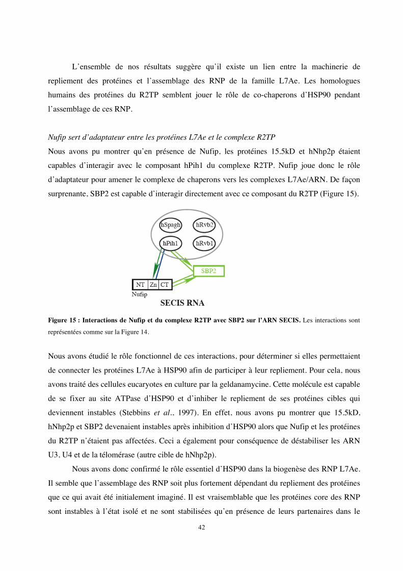
42
L’ensemble de nos résultats suggère qu’il existe un lien entre la machinerie de
repliement des protéines et l’assemblage des RNP de la famille L7Ae. Les homologues
humains des protéines du R2TP semblent jouer le rôle de co-chaperons d’HSP90 pendant
l’assemblage de ces RNP.
Nufip sert d’adaptateur entre les protéines L7Ae et le complexe R2TP
Nous avons pu montrer qu’en présence de Nufip, les protéines 15.5kD et hNhp2p étaient
capables d’interagir avec le composant hPih1 du complexe R2TP. Nufip joue donc le rôle
d’adaptateur pour amener le complexe de chaperons vers les complexes L7Ae/ARN. De façon
surprenante, SBP2 est capable d’interagir directement avec ce composant du R2TP (Figure 15).
Figure 15 : Interactions de Nufip et du complexe R2TP avec SBP2 sur l’ARN SECIS. Les interactions sont
représentées comme sur la Figure 14.
Nous avons étudié le rôle fonctionnel de ces interactions, pour déterminer si elles permettaient
de connecter les protéines L7Ae à HSP90 afin de participer à leur repliement. Pour cela, nous
avons traité des cellules eucaryotes en culture par la geldanamycine. Cette molécule est capable
de se fixer au site ATPase d’HSP90 et d’inhiber le repliement de ses protéines cibles qui
deviennent instables (Stebbins et al., 1997). En effet, nous avons pu montrer que 15.5kD,
hNhp2p et SBP2 devenaient instables après inhibition d’HSP90 alors que Nufip et les protéines
du R2TP n’étaient pas affectées. Ceci a également pour conséquence de déstabiliser les ARN
U3, U4 et de la télomérase (autre cible de hNhp2p).
Nous avons donc confirmé le rôle essentiel d’HSP90 dans la biogenèse des RNP L7Ae.
Il semble que l’assemblage des RNP soit plus fortement dépendant du repliement des protéines
que ce qui avait été initialement imaginé. Il est vraisemblable que les protéines core des RNP
sont instables à l’état isolé et ne sont stabilisées qu’en présence de leurs partenaires dans le

43
complexe final. Dans le cas de SBP2, nos résultats d’analyse structurale prédisent en effet
qu’elle présente toutes les caractéristiques d’une protéine intrinsèquement non repliée (voir
paragraphe B- 3).
HSP90 est directement impliqué dans les mécanismes du cancer chez l’homme et
apparaît comme un acteur clé de la régulation de la prolifération cellulaire car elle contrôle
plusieurs cascades de signalisation cellulaire. Nos travaux enrichissent cette vision, puisqu’ils
établissent qu’HSP90 peut également contrôler la croissance cellulaire en influant sur la
biosynthèse des ribosomes, la réplication via la production de l’ARN de la télomérase et la lutte
contre les radicaux libres via la synthèse des sélénoprotéines.
2- Projets à court terme: l’assemblage de la mRNP SECIS
Notre objectif est maintenant de mieux comprendre comment est assuré le contrôle de
l’assemblage des RNP L7Ae par HSP90, le complexe R2TP et l’adaptateur Nufip en disséquant
ces mécanismes in vivo et in vitro. Les équipes d’Edouard Bertrand, de Bruno Charpentier et
Christiane Branlant étudieront ces mécanismes dans le cas des sn et snoRNP. Notre équipe
s’attachera à élucider ces aspects dans le cas de la mRNP SECIS.
Localisation subcellulaire de Nufip et SBP2
Bien que majoritairement cytoplasmique, SBP2 est capable de transiter entre le noyau et
le cytoplasme et sa localisation conditionne celle du facteur d’élongation eEFsec (Small-
Howard et al., 2006). L’existence d’une interaction entre SBP2 et Nufip renforce l’hypothèse
d’un mécanisme d’assemblage nucléaire des facteurs sur l’ARN SECIS. Il est de ce fait crucial
d’examiner les distributions cellulaires respectives de Nufip et SBP2 par immunolocalisation et
celle d’un ARNm de sélénoprotéine tel que celui de la glutathion peroxydase (GPx) par
hybridation in situ. Ces expériences seront également réalisées dans différentes conditions
notamment en présence de geldanamycine (inhibiteur d’HSP90) qui devrait affecter la
formation du complexe, ou de stress oxydant qui conduit à l’accumulation de SBP2 dans le
noyau (Papp et al., 2006).
Un assemblage nucléaire précoce pourrait également servir à diriger les ARNm de
sélénoprotéines vers une voie d’export spécialisée. L’export nucléaire de SBP2 semble être
dépendant du facteur CRM1 (Papp et al., 2006). De façon intéressante, CRM1 et une ATPase
du complexe R2TP (Rvb2) ont été trouvés associés au snoARN U3 mature (Watkins et al.,
2004). CRM1 est impliqué dans le transport nucléolaire d’U3, mais il a été suggéré qu’il
44
pourrait également jouer un rôle dans l’assemblage de la RNP U3 (Boulon et al., 2004). Au vu
de similitudes entre les mécanismes d’assemblage des RNP U3 et des mRNP SECIS, nous
vérifierons le rôle exact de CRM1 dans le mécanisme de synthèse des sélénoprotéines. Nous
testerons si CRM1 est associé aux mRNP de sélénoprotéines et à SBP2 par des expériences de
co-transfections et immunoprécipitations en présence ou en absence de leptomycine B
(inhibiteur de CRM1).
Influence de Nufip et HSP90 sur stabilité des ARNm de sélénoprotéines et la synthèse des
sélénoprotéines
Nous avons montré que Nufip et HSP90 étaient associés aux mRNP SECIS. En
favorisant l’assemblage de protéines sur l’ARN SECIS, il est vraisemblable que ces facteurs
permettent d’éviter la dégradation des ARNm de sélénoprotéines par les mécanismes du NMD.
Nous testerons l’effet de l’invalidation du gène de Nufip par RNA interférence, et celui de
l’inhibition HSP90 par la geldanamycine, sur la stabilité des ARNm de sélénoprotéines
endogènes in vivo par RT-PCR quantitative. Par transfections transitoires d’ARNm de
sélénoprotéines rapporteurs, nous évaluerons l’impact de l’inhibition de Nufip et HSP90 sur la
synthèse de la sélénoprotéine correspondante. Un défaut d’association de SBP2 devrait inhiber
la synthèse de sélénoprotéines et conduire à l’arrêt prématuré de la traduction au niveau du
codon UGASec et la production d’une protéine tronquée. Ceci devrait nous aider à mieux
comprendre le rôle fonctionnel de Nufip et HSP90 dans le mécanisme de synthèse des
sélénoprotéines.
Etude in vitro de l’interaction Nufip/SBP2
L’interaction de Nufip avec la protéine 15.5kD se fait par l’intermédiaire du motif PEP
mais nous avons montré que celui-ci ne semblait pas suffisant pour interagir efficacement avec
SBP2. Nous affinerons l’analyse du domaine d’interaction de Nufip par mutagenèse. Nous
tenterons également d’identifier le domaine de SBP2 impliqué dans l’interaction avec Nufip. Il
est vraisemblable que cette interaction ait lieu par l’intermédiaire du domaine conservé L7Ae.
Nous le vérifierons à l’aide de protéine SBP2 tronquées, puis déterminerons la nature des
acides aminés impliqués par mutagenèse, GST-pull down et co-immunoprécipitations. Des
études similaires seront réalisées par nos collaborateurs en ce qui concerne Rsa1 et Snu13p
(Bruno Charpentier, Nancy), Nufip et 15.5kD (Montpellier). L’ensemble de ces travaux devrait
nous permettre d’établir si les modes d’interaction entre Nufip (Rsa1) et de ses différentes
45
cibles font appel aux mêmes surfaces d’interactions protéine-protéine. A plus long terme, nous
pourrons également envisager la cristallisation de ces complexes.
Quelles protéines core pour l’ARN SECIS ?
Si l’assemblage des facteurs sur l’ARN SECIS est au cœur du mécanisme de synthèse
des sélénoprotéines, la composition de la mRNP SECIS reste mal connue. Certaines pistes
directes s’offrent cependant à nous. En effet, des expériences d’immunopurification de SBP2
nous ont permis d’isoler la protéine NSEP1 et de démontrer qu’elle interagissait in vivo avec
SBP2. NSEP1 est une protéine capable de se lier à l’ARN SECIS in vitro (Fagegaltier et al.,
2000a). Son rôle dans la synthèse des sélénoprotéines, longtemps controversé, vient récemment
d’être démontré (Qichang Shen, 2006). Nos résultats préliminaires montrent que NSEP1
interagit également avec Nufip in vivo. Par des tests double-hybride et GST-pull down in vitro,
nous vérifierons si ces interactions sont directes ou si Nufip sert d’adaptateur entre SBP2 et
NSEP1.
De façon surprenante, nous avons détecté une interaction entre SBP2, Nop58 et Nop56
par co-transfection et co-immunoprécipitation. Ceci pose la question de savoir si certaines des
protéines core des snoRNP à boîte C/D sont également recrutées vers l’ARN SECIS. Nous
examinerons cette possibilité.
Assemblage in vitro des mRNP de sélénoprotéines
Notre étude in vitro sera complétée par des essais de reconstitution in vitro de la RNP
SECIS. Dans un premier temps, nous déterminerons si Nufip est capable de se lier directement
à l’ARN SECIS par des expériences de retard sur gel, ou s’il stimule l’assemblage des autres
facteurs sur l’ARN SECIS comme dans le cas des autres RNP (voir paragraphe C-1). Nous
testerons la capacité de Nufip à favoriser la reconstitution d'un complexe entre les protéines
recombinantes SBP2, NSEP1 et l’ARN SECIS. Le facteur d’élongation eEFsec sera également
inclus dans les expériences de reconstitution ; nous avions montré qu’il est capable de stimuler
la liaison de SBP2 au SECIS sans pour autant s’associer au complexe (Lescure et al., 2002). Il
sera intéressant de vérifier si Nufip stabilise ou non ce complexe ou s’il s’agit d’une interaction
transitoire qui n’est stimulée que par la présence de l’ARNtSec (Zavacki et al., 2003).
Le rôle des protéines du complexe R2TP sur l’assemblage, et notamment des deux
ATPases Rvb1 et Rvb2, sera également testé en présence ou en absence d’ATP. Il est
vraisemblable que ces protéines participent au remodelage de la RNP, voire au transfert des
protéines core recrutées par Nufip vers l’ARN, et à la dissociation de Nufip du complexe. Des
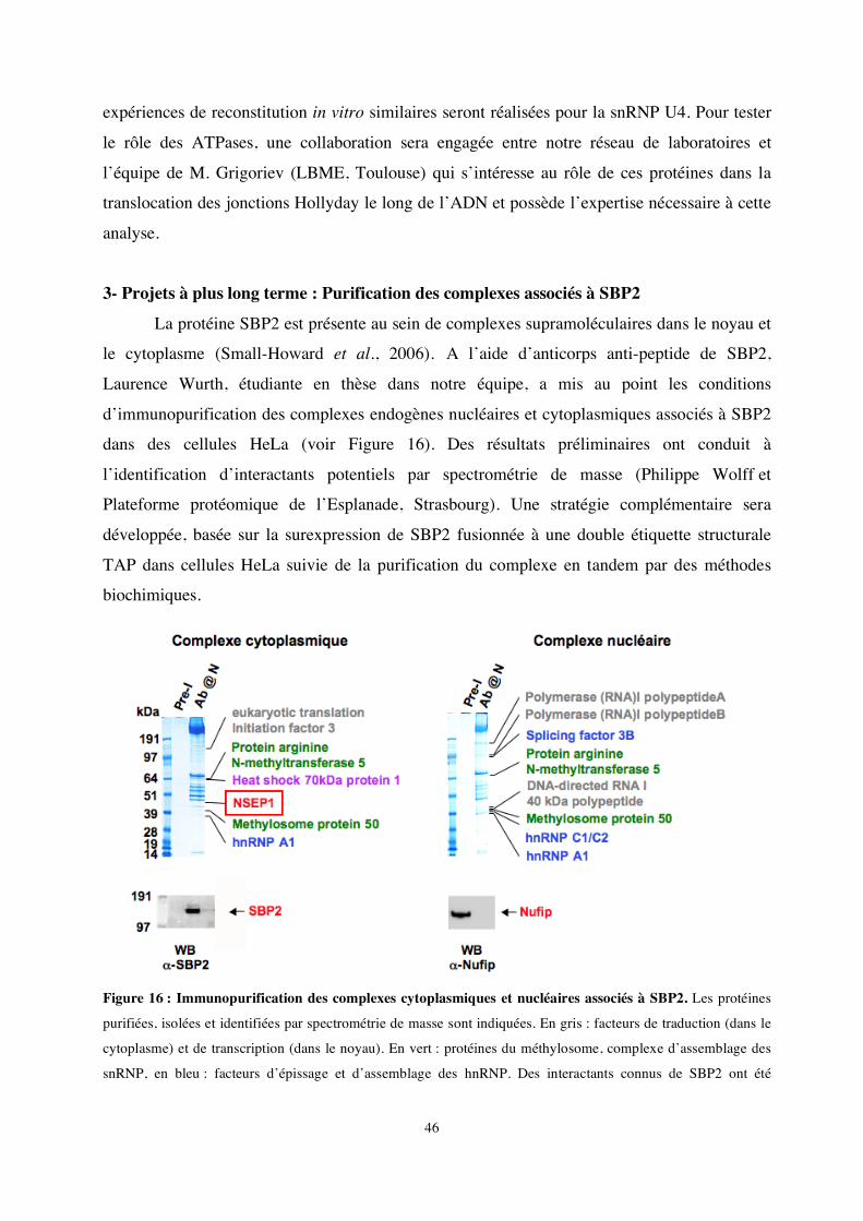
46
expériences de reconstitution in vitro similaires seront réalisées pour la snRNP U4. Pour tester
le rôle des ATPases, une collaboration sera engagée entre notre réseau de laboratoires et
l’équipe de M. Grigoriev (LBME, Toulouse) qui s’intéresse au rôle de ces protéines dans la
translocation des jonctions Hollyday le long de l’ADN et possède l’expertise nécessaire à cette
analyse.
3- Projets à plus long terme : Purification des complexes associés à SBP2
La protéine SBP2 est présente au sein de complexes supramoléculaires dans le noyau et
le cytoplasme (Small-Howard et al., 2006). A l’aide d’anticorps anti-peptide de SBP2,
Laurence Wurth, étudiante en thèse dans notre équipe, a mis au point les conditions
d’immunopurification des complexes endogènes nucléaires et cytoplasmiques associés à SBP2
dans des cellules HeLa (voir Figure 16). Des résultats préliminaires ont conduit à
l’identification d’interactants potentiels par spectrométrie de masse (Philippe Wolff et
Plateforme protéomique de l’Esplanade, Strasbourg). Une stratégie complémentaire sera
développée, basée sur la surexpression de SBP2 fusionnée à une double étiquette structurale
TAP dans cellules HeLa suivie de la purification du complexe en tandem par des méthodes
biochimiques.
Figure 16 : Immunopurification des complexes cytoplasmiques et nucléaires associés à SBP2. Les protéines
purifiées, isolées et identifiées par spectrométrie de masse sont indiquées. En gris : facteurs de traduction (dans le
cytoplasme) et de transcription (dans le noyau). En vert : protéines du méthylosome, complexe d’assemblage des
snRNP, en bleu : facteurs d’épissage et d’assemblage des hnRNP. Des interactants connus de SBP2 ont été
47
détectés, notamment la protéine NSEP1, le chaperon HSP70 (voir paragraphes C-1 et C-2). SBP2 et Nufip ont été
détectées par Western blot (panneaux inférieurs).
Notre objectif sera d’évaluer la validité des interactions mises au jour et d’établir si ces
protéines font partie de façon stable ou transitoire des complexes supramoléculaires de la
machinerie de synthèse des sélénoprotéines. Si tel est le cas, ces facteurs seront caractérisés
fonctionnellement, et leur mode de liaison avec l'ARN SECIS et leurs protéines cibles seront
analysés. Notre objectif sera de comprendre comment ceux-ci s’insèrent dans le contexte global
du mécanisme traductionnel des sélénoprotéines.
Plusieurs complexes d’assemblage pour la RNP SECIS?
De façon surprenante, deux protéines du complexe du méthylosome ont été co-purifiées
avec SBP2 (voir Figure 16). Le méthylosome est impliqué avec le complexe SMN dans les
mécanismes d’assemblage des snRNP (Yong et al., 2004). En effet, il permet la méthylation de
protéines Sm qui sont prises en charge par le complexe SMN et fonctionnent comme chaperon
d’assemblage des snARN. Une hypothèse possible stipule qu’il existerait un lien entre le
complexe d’assemblage que nous avons caractérisé (Nufip, HSP90 et le complexe R2TP) et
l’autre grande machinerie d’assemblage de la cellule, le complexe SMN. Nous examinerons
dans un premier temps si les protéines du méthylosome et du complexe SMN forment un
complexe avec SBP2 ou Nufip mais également si les protéines Sm sont capables de se lier à
l’ARN SECIS et participent à son assemblage. En fonction des résultats de ces expériences
préliminaires, nous vérifierons si le complexe R2TP et le complexe SMN collaborent pour
l’assemblage de la RNP SECIS.
L’ensemble de ces expériences devrait nous apporter une vision globale du mécanisme
de synthèse des sélénoprotéines et nous aider à comprendre la composition mais également la
dynamique d’assemblage et de désassemblage des complexes multi protéiques impliqués dans
le processus.
48
Références bibliographiques
Achsel, T., Brahms, H., Kastner, B., Bachi, A., Wilm, M. and Luhrmann, R. (1999) A doughnut-shaped heteromer
of human Sm-like proteins binds to the 3'-end of U6 snRNA, thereby facilitating U4/U6 duplex formation in vitro. EMBO J., 18, 5789-5802.
Allmang, C., Carbon, P. and Krol, A. (2002) The SBP2 and 15.5 kD/Snu13p proteins share the same RNA binding domain: identification of SBP2 amino acids important to SECIS RNA binding. RNA, 8, 1308-1318.
Allmang, C., Henry, Y., Morrissey, J.P., Wood, H., Petfalski, E. and Tollervey, D. (1996a) Processing of the yeast pre-rRNA at sites A(2) and A(3) is linked. RNA, 2, 63-73.
Allmang, C., Henry, Y., Wood, H., Morrissey, J.P., Petfalski, E. and Tollervey, D. (1996b) Recognition of cleavage site A(2) in the yeast pre-rRNA. RNA, 2, 51-62.
Allmang, C. and Krol, A. (2006a) SECIS RNAs and K-turn binding proteins. A survey of evolutionary conserved RNA and protein motifs. Selenium, its molecular Biology and role in human Health 2nd edition. DL Hatfield (ed) Kluwer Academic Publishers. , 5, 51-61.
Allmang, C. and Krol, A. (2006b) Selenoprotein synthesis: UGA does not end the story. Biochimie. Allmang, C., Kufel, J., Chanfreau, G., Mitchell, P., Petfalski, E. and Tollervey, D. (1999a) Functions of the
exosome in rRNA, snoRNA and snRNA synthesis. EMBO J, 18, 5399-5410. Allmang, C., Mitchell, P., Petfalski, E. and Tollervey, D. (2000) Degradation of ribosomal RNA precursors by the
exosome. Nucleic Acids Res, 28, 1684-1691. Allmang, C., Mougel, M., Westhof, E., Ehresmann, B. and Ehresmann, C. (1994) Role of conserved nucleotides in
building the 16S rRNA binding site of E. coli ribosomal protein S8. Nucleic Acids Res, 22, 3708-3714. Allmang, C., Petfalski, E., Podtelejnikov, A., Mann, M., Tollervey, D. and Mitchell, P. (1999b) The yeast exosome
and human PM-Scl are related complexes of 3' --> 5' exonucleases. Genes Dev, 13, 2148-2158. Allmang, C. and Tollervey, D. (1998) The role of the 3' external transcribed spacer in yeast pre-rRNA processing.
J Mol Biol, 278, 67-78. Anderson, J. and Parker, R. (1998) The 3' to 5' degradation of yeast mRNAs is a general mechanism for mRNA
turnover that requires the SKI2 DEVH box protein and 3' to 5' exonucleases of the exosome complex. EMBO J.,, 17, 1497-1506.
Araki, Y., Takahashi, S., Kobayashi, T., Kajiho, H., Hoshino, S.-i. and Katada, T. (2001) Ski7p G protein interacts with the exosome and the Ski complex for 3'-to-5' mRNA decay in yeast. EMBO J., 20, 4684-4693.
Bardoni, B., Schenck, A. and Mandel, J.L. (1999) A novel RNA-binding nuclear protein that interacts with the fragile X mental retardation (FMR1) protein. Hum Mol Genet, 8, 2557-2566.
Beltrame, M., Henry, Y. and Tollervey, D. (1994) Mutational analysis of an essential binding site for the U3 snoRNA in the 5' external transcribed spacer of yeast pre-rRNA. Nucl. Acids Res., 22, 5139-5147.
Berry, M.J., Tujebajeva, R.M., Copeland, P.R., Xu, X.M., Carlson, B.A., Martin, G.W., 3rd, Low, S.C., Mansell, J.B., Grundner-Culemann, E., Harney, J.W., Driscoll, D.M. and Hatfield, D.L. (2001) Selenocysteine incorporation directed from the 3'UTR: characterization of eukaryotic EFsec and mechanistic implications. Biofactors, 14, 17-24.
Bertrand E and R., B. (2004) Assembly and traffic of small nuclear RNPs. Prog Mol Subcell Biol. , 35, 79-97. Boulon, S., Verheggen, C., Jady, B.E., Girard, C., Pescia, C., Paul, C., Ospina, J.K., Kiss, T., Matera, A.G.,
Bordonne, R. and Bertrand, E. (2004) PHAX and CRM1 Are Required Sequentially to Transport U3 snoRNA to Nucleoli. Molecular Cell, 16, 777-787.
Bousquet-Antonelli, C., Presutti, C. and Tollervey, D. (2000) Identification of a Regulated Pathway for Nuclear Pre-mRNA Turnover. Cell, 102, 765-775.
Bouveret, E., Rigaut, G., Shevchenko, A., M, W. and B, S. (2000) A Sm-like protein complex that participates in mRNA degradation. EMBO J, 19, 1661-1671.
Brodersen, D.E., Clemons, J.W.M., Carter, A.P., Wimberly, B.T. and Ramakrishnan, V. (2002) Crystal structure of the 30 s ribosomal subunit from Thermus thermophilus: structure of the proteins and their interactions with 16 s RNA. Journal of Molecular Biology, 316, 725-768.
Brouwer, R., Allmang, C., Raijmakers, R., van Aarssen, Y., Egberts, W.V., Petfalski, E., van Venrooij, W.J., Tollervey, D. and Pruijn, G.J. (2001) Three novel components of the human exosome. J Biol Chem, 276, 6177-6184.
Buttner, K., Wenig, K. and Hopfner, K.-P. (2005) Structural Framework for the Mechanism of Archaeal Exosomes in RNA Processing. Molecular Cell, 20, 461-471.
Caplan, A.J., Mandal, A.K. and Theodoraki, M.A. (2007) Molecular chaperones and protein kinase quality control. Trends in Cell Biology, 17, 87-92.
49
Carlson, B.A., Xu, X.-M., Kryukov, G.V., Rao, M., Berry, M.J., Gladyshev, V.N. and Hatfield, D.L. (2004) Identification and characterization of phosphoseryl-tRNA[Ser]Sec kinase. PNAS 101, 12848-12853.
Carmo-Fonseca, M., Platani, M. and Swedlow, J.R. (2002) Macromolecular mobility inside the cell nucleus. Trends in Cell Biology, 12, 491-495.
Castellano, S., Morozova, N., Morey, M., Berry, M.J., Serras, F., Corominas, M. and Guigó, R. (2001) In silico identification of novel selenoproteins in the Drosophila melanogaster genome EMBO reports, 2, 697–702.
Castellano, S., Novoselov, S., Kryukov, G., Lescure, A., Blanco, E., Krol, A., Gladyshev, V. and Guigó, R. (2004) Reconsidering the evolution of eukaryotic selenoproteins: a novel nonmammalian family with scattered phylogenetic distribution. EMBO Rep. , 5, 71-77.
Chang, D.D. and Clayton, D.A. (1987) A novel endoribonuclease cleaves at a priming site of mouse mitochondrial DNA replication. EMBO J., 6, 409-417.
Chao, J.A. and Williamson, J.R. (2004) Joint X-ray and NMR refinement of the yeast L30e-mRNA complex. Structure (Camb), 12, 1165-1176.
Chavatte, L., Brown, B.A. and Driscoll, D.M. (2005) Ribosomal protein L30 is a component of the UGA-selenocysteine recoding machinery in eukaryotes. Nat Struct Mol Biol.
Cléry, A., Bourguignon-Igel, V., Allmang, C., Krol, A. and Branlant, C. (2007) An improved definition of the RNA-binding specificity of SECIS-binding protein 2, an essential component of the selenocysteine incorporation machinery. Nucleic Acids Res. , 35, 1868–1884.
Costa, M., Rodríguez-Sánchez, J., Czaja, A. and Gelpí, C. (2000) Isolation and characterization of cDNA encoding the antigenic protein of the human tRNP(Ser)Sec complex recognized by autoantibodies from patients withtype-1 autoimmune hepatitis. Clin Exp Immunol. , 121, 364-374.
Copeland, P.R., Fletcher, J.E., Carlson, B.A., Hatfield, D.L. and Driscoll, D.M. (2000) A novel RNA binding protein, SBP2, is required for the translation of mammalian selenoprotein mRNAs. EMBO J, 19, 306-314.
Copeland, P.R., Stepanik, V.A. and Driscoll, D.M. (2001) Insight into mammalian selenocysteine insertion: domain structure and ribosome binding properties of Sec insertion sequence binding protein 2. Mol Cell Biol, 21, 1491-1498.
de Jesus, L.A., Hoffmann, P.R., Michaud, T., Forry, E.P., Small-Howard, A., Stillwell, R.J., Morozova, N., Harney, J.W. and Berry, M.J. (2006) Nuclear Assembly of UGA Decoding Complexes on Selenoprotein mRNAs: a Mechanism for Eluding Nonsense-Mediated Decay? Mol Cell Biol, 26, 1795-1805.
de la Cruz, J., Kressler, D., Tollervey, D. and Linder, P. (1998) Dob1p (Mtr4p) is a putative ATP-dependent RNA helicase required for the 3' end formation of 5.8S rRNA in Saccharomyces cerevisiae. EMBO J. , 17, 1128-1140.
Ding, F. and Grabowski, P.J. (1999) Identification of a protein component of a mammalian tRNA(Sec) complex implicated in the decoding of UGA as selenocysteine. RNA, 5, 1561-1569.
Dosztanyi, Z., Csizmok, V., Tompa, P. and Simon, I. (2005) IUPred: web server for the prediction of intrinsically unstructured regions of proteins based on estimated energy content. Bioinformatics 21, 3433-3434.
Dunker, A.K., Cortese, M.S., Romero, P., Iakoucheva, L.M. and Uversky, V.N. (2005) Flexible nets. The roles of intrinsic disorder in protein interaction networks. FEBS Journal, 272, 5129-5148.
Fagegaltier, D., Carbon, P. and Krol, A. (2001) Distinctive features in the SelB family of elongation factors for selenoprotein synthesis. A glimpse of an evolutionary complexified translation apparatus. Biofactors, 14, 5-10.
Fagegaltier, D., Hubert, N., Carbon, P. and Krol, A. (2000a) The selenocysteine insertion sequence binding protein SBP is different from the Y-box protein dbpB. Biochimie, 82, 117-122.
Fagegaltier, D., Lescure, A., Walczak, R., Carbon, P. and Krol, A. (2000b) Structural analysis of new local features in SECIS RNA hairpins. Nucleic Acids Res, 28, 2679-2689.
Fatica, A. and Tollervey, D. (2002) Making ribosomes. Current Opinion in Cell Biology, 14, 313-318. Fletcher, J.E., Copeland, P.R., Driscoll, D.M. and Krol, A. (2001) The selenocysteine incorporation machinery:
interactions between the SECIS RNA and the SECIS-binding protein SBP2. RNA, 7, 1442-1453. Flohe, L., Andreesen, J.R., Brigelius-Flohe, R., Maiorino, M. and Ursini, F. (2000) Selenium, the element of the
moon, in life on earth. IUBMB Life, 49, 411-420. Flohe, L., Günzler, W. and Schock, H. (1973) Glutathione peroxidase: a selenoenzyme. FEBS Lett., 32, 132-134. Gavin, A.-C., Aloy, P., Grandi, P., Krause, R., Boesche, M., Marzioch, M., Rau, C., Jensen, L.J., Bastuck, S.,
Dumpelfeld, B., Edelmann, A., Heurtier, M.-A., Hoffman, V., Hoefert, C., Klein, K., Hudak, M., Michon, A.-M., Schelder, M., Schirle, M., Remor, M., Rudi, T., Hooper, S., Bauer, A., Bouwmeester, T., Casari, G., Drewes, G., Neubauer, G., Rick, J.M., Kuster, B., Bork, P., Russell, R.B. and Superti-Furga, G. (2006) Proteome survey reveals modularity of the yeast cell machinery. Nature, 440, 631-636.
Gavin, A.-C., Bosche, M., Krause, R., Grandi, P., Marzioch, M., Bauer, A., Schultz, J., Rick, J.M., Michon, A.-M., Cruciat, C.-M., Remor, M., Hofert, C., Schelder, M., Brajenovic, M., Ruffner, H., Merino, A., Klein, K.,
50
Hudak, M., Dickson, D., Rudi, T., Gnau, V., Bauch, A., Bastuck, S., Huhse, B., Leutwein, C., Heurtier, M.-A., Copley, R.R., Edelmann, A., Querfurth, E., Rybin, V., Drewes, G., Raida, M., Bouwmeester, T., Bork, P., Seraphin, B., Kuster, B., Neubauer, G. and Superti-Furga, G. (2002) Functional organization of the yeast proteome by systematic analysis of protein complexes. Nature, 415, 141-147.
Gonzales, F.A., Zanchin, N.I.T., Luz, J.S. and Oliveira, C.C. (2005) Characterization of Saccharomyces cerevisiae Nop17p, a Novel Nop58p-Interacting Protein that is Involved in Pre-rRNA Processing. Journal of Molecular Biology, 346, 437-455.
Held, W.A., Ballou, B., Mizushima, S. and Nomura, M. (1974) Assembly Mapping of 30 S Ribosomal Proteins from Escherichia coli. Further studies. J. Biol. Chem., 249, 3103-3111.
Hendrickson, T.L. (2007) Easing selenocysteine into proteins. Nat Struct Mol Biol, 14, 100-101. Ho, Y., Gruhler, A., Heilbut, A., Bader, G.D., Moore, L., Adams, S.-L., Millar, A., Taylor, P., Bennett, K.,
Boutilier, K., Yang, L., Wolting, C., Donaldson, I., Schandorff, S., Shewnarane, J., Vo, M., Taggart, J., Goudreault, M., Muskat, B., Alfarano, C., Dewar, D., Lin, Z., Michalickova, K., Willems, A.R., Sassi, H., Nielsen, P.A., Rasmussen, K.J., Andersen, J.R., Johansen, L.E., Hansen, L.H., Jespersen, H., Podtelejnikov, A., Nielsen, E., Crawford, J., Poulsen, V., Sorensen, B.D., Matthiesen, J., Hendrickson, R.C., Gleeson, F., Pawson, T., Moran, M.F., Durocher, D., Mann, M., Hogue, C.W.V., Figeys, D. and Tyers, M. (2002) Systematic identification of protein complexes in Saccharomyces cerevisiae by mass spectrometry. Nature, 415, 180-183.
Houseley, J., LaCava, J. and Tollervey, D. (2006) RNA-quality control by the exosome. Nat Rev Mol Cell Biol, 7, 529-539.
Kernebeck, T., Lohse, A. and Grötzinger, J. (2001) A bioinformatical approach suggests the function of the autoimmune hepatitis target antigen soluble liver antigen/liver pancreas. Hepatology, 34, 230-233.
King, T.H., Decatur, W.A., Bertrand, E., Maxwell, E.S. and Fournier, M.J. (2001) A Well-Connected and Conserved Nucleoplasmic Helicase Is Required for Production of Box C/D and H/ACA snoRNAs and Localization of snoRNP Proteins. Mol. Cell. Biol., 21, 7731-7746.
Kinzy, S.A., Caban, K. and Copeland, P.R. (2005) Characterization of the SECIS binding protein 2 complex required for the co-translational insertion of selenocysteine in mammals. Nucleic Acids Res, 33, 5172-5180.
Klein, D.J., Schmeing, T.M., Moore, P.B. and Steitz, T.A. (2001) The kink-turn: a new RNA secondary structure motif. EMBO J, 20, 4214-4221.
Kressler, D., Doere, M., Rojo, M. and Linder, P. (1999) Synthetic Lethality with Conditional dbp6 Alleles Identifies Rsa1p, a Nucleoplasmic Protein Involved in the Assembly of 60S Ribosomal Subunits. Mol. Cell. Biol., 19, 8633-8645.
Kryukov, G. and Gladyshev, V. (2004) The prokaryotic selenoproteome. EMBO Rep., 5, 538-543. Kufel, J., Allmang, C., Chanfreau, G., Petfalski, E., Lafontaine, D.L. and Tollervey, D. (2000) Precursors to the U3
small nucleolar RNA lack small nucleolar RNP proteins but are stabilized by La binding. Mol Cell Biol, 20, 5415-5424.
Kufel, J., Allmang, C., Petfalski, E., Beggs, J. and Tollervey, D. (2003a) Lsm Proteins are required for normal processing and stability of ribosomal RNAs. J Biol Chem, 278, 2147-2156.
Kufel, J., Allmang, C., Verdone, L., Beggs, J. and Tollervey, D. (2003b) A complex pathway for 3' processing of the yeast U3 snoRNA. Nucleic Acids Res, 31, 6788-6797.
Kufel, J., Allmang, C., Verdone, L., Beggs, J.D. and Tollervey, D. (2002) Lsm proteins are required for normal processing of pre-tRNAs and their efficient association with La-homologous protein Lhp1p. Mol Cell Biol, 22, 5248-5256.
LaCava, J., Houseley, J., Saveanu, C., Petfalski, E., Thompson, E., Jacquier, A. and Tollervey, D. (2005) RNA Degradation by the Exosome Is Promoted by a Nuclear Polyadenylation Complex. Cell, 121, 713-724.
Leontis, N.B. and Westhof, E. (2001) Geometric nomenclature and classification of RNA base pairs. RNA, 7, 499-512.
Lescure, A., Allmang, C., Yamada, K., Carbon, P. and Krol, A. (2002) cDNA cloning, expression pattern and RNA binding analysis of human selenocysteine insertion sequence (SECIS) binding protein 2. Gene, 291, 279-285.
Lorentzen, E., Dziembowski, A., Lindner, D., Seraphin, B. and Conti, E. (2007) RNA channelling by the archaeal exosome. EMBO reports, 8, 470–476.
Lorentzen, E., Walter, P., Fribourg, S., Evguenieva-Hackenberg, E., Klug, G. and Conti, E. (2005) The archaeal exosome core is a hexameric ring structure with three catalytic subunits. Nat Struct Mol Biol, 12, 575-581.
Low, S.C., Harney, J.W. and Berry, M.J. (1995) Cloning and Functional Characterization of Human Selenophosphate Synthetase, an Essential Component of Selenoprotein Synthesis. J. Biol. Chem., 270, 21659-21664.
51
Lygerou, Z., Allmang, C., Tollervey, D. and Seraphin, B. (1996) Accurate processing of a eukaryotic precursor ribosomal RNA by ribonuclease MRP in vitro. Science, 272, 268-270.
Matera, A.G., Terns, R.M. and Terns, M.P. (2007) Non-coding RNAs: lessons from the small nuclear and small nucleolar RNAs. Nat Rev Mol Cell Biol, 8, 209-220.
Mayes, A., Verdone, L., Legrain, P. and Beggs, J. (1999) Characterization of Sm-like proteins in yeast and their association with U6 snRNA. EMBO J, 18, 4321-4331.
Milligan, L., Torchet, C., Allmang, C., Shipman, T. and Tollervey, D. (2005) A nuclear surveillance pathway for mRNAs with defective polyadenylation. Mol Cell Biol, 25, 9996-10004.
Mitchell, P., Petfalski, E., Houalla, R., Podtelejnikov, A., Mann, M. and Tollervey, D. (2003) Rrp47p Is an Exosome-Associated Protein Required for the 3' Processing of Stable RNAs. Mol. Cell. Biol., 23, 6982-6992.
Mitchell, P., Petfalski, E., Shevchenko, A., Mann, M. and Tollervey, D. (1997) The exosome: a conserved eukaryotic RNA processing complex containing multiple 3'-->5' exoribonucleases. Cell, 91, 457-466.
Mizushima S and M, N. (1970) Assembly mapping of 30S ribosomal proteins from E. coli. Nature, 226, 1214. Moghadaszadeh, B. and Beggs, A.H. (2006) Selenoproteins and Their Impact on Human Health Through Diverse
Physiological Pathways. Physiology 2006, 21, 307-315. Moine, H., Cachia, C., Westhof, E., Ehresmann, B. and Ehresmann, C. (1997) The RNA binding site of S8
ribosomal protein of Escherichia coli: Selex and hydroxyl radical probing studies. RNA, 3, 255-268. Moore, T., Zhang, Y., Fenley, M.O. and Li, H. (2004) Molecular basis of box C/D RNA-protein interactions;
cocrystal structure of archaeal L7Ae and a box C/D RNA. Structure (Camb), 12, 807-818. Mougel, M., Allmang, C., Eyermann, F., Cachia, C., Ehresmann, B. and Ehresmann, C. (1993) Minimal 16S rRNA
binding site and role of conserved nucleotides in Escherichia coli ribosomal protein S8 recognition. Eur J Biochem, 215, 787-792.
Mougel, M., Eyermann, F., Westhof, E., Romby, P., Expert-Bezancon, A., Ebel, J.P., Ehresmann, B. and Ehresmann, C. (1987) Binding of Escherichia coli ribosomal protein S8 to 16 S rRNA. A model for the interaction and the tertiary structure of the RNA binding site. J Mol Biol. , 198, 91-107.
Pannone, B., Xue, D. and Wolin, S. (1998) A role for the yeast La protein in U6 snRNP assembly: evidence that the La protein is a molecular chaperone for RNA polymerase III transcripts. EMBO J. , 17, 7442-7453.
Papp, L.V., Lu, J., Striebel, F., Kennedy, D., Holmgren, A. and Khanna, K.K. (2006) The redox state of SECIS binding protein 2 controls its localization and selenocysteine incorporation function. Mol Cell Biol, 26, 4895-4910.
Pearl, L.H. and Prodromou, C. (2006) Structure and mechanism of the HSP90 molecular chaperone machinery. Annual Review of Biochemistry, 75, 271-294.
Petfalski, E., Dandekar, T., Henry, Y. and Tollervey, D. (1998) Processing of the Precursors to Small Nucleolar RNAs and rRNAs Requires Common Components. Mol. Cell. Biol., 18, 1181-1189.
Qichang Shen, L.F.P.E.N. (2006) Nuclease sensitive element binding protein 1 associates with the selenocysteine insertion sequence and functions in mammalian selenoprotein translation. Journal of Cellular Physiology, 207, 775-783.
Raijmakers, R., Schilders, G. and Pruijn, G. (2004) The exosome, a molecular machine for controlled RNA degradation in both nucleus and cytoplasm. Eur J Cell Biol., 83, 75-83.
Rinke, J. and Steitz, J.A. (1982) Precursor molecules of both human 5S ribosomal RNA and transfer RNAs are bound by a cellular protein reactive with anti-La Lupus antibodies. Cell, 29, 149-159.
Rotruck, J., Pope, A., Ganther, H., Swanson, A., Hafeman, D. and Hoekstra, W. (1973) Selenium: biochemical role as a component of glutathione peroxidase. Science, 179, 588-590.
Small-Howard, A., Morozova, N., Stoytcheva, Z., Forry, E.P., Mansell, J.B., Harney, J.W., Carlson, B.A., Xu, X.M., Hatfield, D.L. and Berry, M.J. (2006) Supramolecular complexes mediate selenocysteine incorporation in vivo. Mol Cell Biol, 26, 2337-2346.
Stebbins, C.E., Russo, A.A., Schneider, C., Rosen, N., Hartl, F.U. and Pavletich, N.P. (1997) Crystal Structure of an Hsp90-Geldanamycin Complex: Targeting of a Protein Chaperone by an Antitumor Agent. Cell, 89, 239-250.
Te, J., Jia, L., Rogers, J., Miller, A. and Hartson, S.D. (2007) Novel Subunits of the Mammalian Hsp90 Signal Transduction Chaperone. J. Proteome Res., 6, 1963-1973.
Tishchenko, S., Nikulin, A., Fomenkova, N., Nevskaya, N., Nikonov, O., Dumas, P., Moine, H., Ehresmann, B., Ehresmann, C., Piendl, W., Lamzin, V., Garber, M. and Nikonov, S. (2001) Detailed analysis of RNA-protein interactions within the ribosomal protein S8-rRNA complex from the archaeon Methanococcus jannaschii. Journal of Molecular Biology, 311, 311-324.
Tompa, P. (2005) The interplay between structure and function in intrinsically unstructured proteins. FEBS Letters, 579, 3346-3354.
52
Venema, J., Henry, Y. and Tollervey, D. (1995) Two distinct recognition signals define the site of endonucleolytic cleavage at the 5'-end of yeast 18S rRNA. EMBO J. , 14, 1995
Venema, J. and Tollervey, D. (1999) Ribosome synthesis in Saccharomyces cerevisiae. Annu Rev Genet. , 33, 261-311.
Vidovic, I., Nottrott, S., Hartmuth, K., Luhrmann, R. and Ficner, R. (2000a) Crystal structure of the spliceosomal 15.5kD protein bound to a U4 snRNA fragment. Mol Cell, 6, 1331-1342.
Vidovic, I., Nottrott, S., Hartmuth, K., Luhrmann, R. and Ficner, R. (2000b) Crystal structure of the spliceosomal 15.5kD protein bound to a U4 snRNA fragment. Mol Cell, 6, 1331-1342.
Walczak, R., Hubert, N., Carbon, P. and Krol, A. (1997) Solution structure of SECIS, the mRNA element required for eukaryotic selenocysteine insertion-interaction studies with the SECIS-Binding Protein SBP. Biomed. and Environ. Sci., 10, 177-181.
Walczak, R., Westhof, E., Carbon, P. and Krol, A. (1996) A novel RNA structural motif in the selenocysteine insertion element of eukaryotic selenoprotein mRNAs. RNA, 2, 367-379.
Watkins, N.J., Lemm, I., Ingelfinger, D., Schneider, C., Hobach, M., Urlaub, H. and Luhrmann, R. (2004) Assembly and Maturation of the U3 snoRNP in the Nucleoplasm in a Large Dynamic Multiprotein Complex. Mol Cell, 16, 789-798.
Xu, X., Carlson, B., Irons, R., Mix, H., Zhong, N., Gladyshev, V. and DL, H. (2007a) Selenophosphate synthetase 2 is essential for selenoprotein biosynthesis. Biochem J. , 404, 115-120.
Xu, X.-M., Carlson, B.A., Mix, H., Zhang, Y., Saira, K., Glass, R.S., Berry, M.J., Gladyshev, V.N. and Hatfield, D.L. (2007b) Biosynthesis of Selenocysteine on Its tRNA in Eukaryotes. PLoS Biology, 5, e4.
Xu, X.-M., Mix, H., Carlson, B.A., Grabowski, P.J., Gladyshev, V.N., Berry, M.J. and Hatfield, D.L. (2005) Evidence for Direct Roles of Two Additional Factors, SECp43 and Soluble Liver Antigen, in the Selenoprotein Synthesis Machinery. J. Biol. Chem., 280, 41568-41575.
Yong, J., Wan, L. and Dreyfuss, G. (2004) Why do cells need an assembly machine for RNA-protein complexes? Trends in Cell Biology, 14, 226-232.
Yoo, C.J. and Wolin, S.L. (1997) The Yeast La Protein Is Required for the 3' Endonucleolytic Cleavage That Matures tRNA Precursors. Cell, 89, 393-402.
Yuan, J., Palioura, S., Salazar, J.C., Su, D., O'Donoghue, P., Hohn, M.J., Cardoso, A.M., Whitman, W.B. and Soll, D. (2006) RNA-dependent conversion of phosphoserine forms selenocysteine in eukaryotes and archaea. PNAS, 103, 18923-18927.
Yusupov, M.M., Yusupova, G.Z., Baucom, A., Lieberman, K., Earnest, T.N., Cate, J.H.D. and Noller, H.F. (2001) Crystal Structure of the Ribosome at 5.5 A Resolution. Science, 292, 883-896.
Zavacki, A.M., Mansell, J.B., Chung, M., Klimovitsky, B., Harney, J.W. and Berry, M.J. (2003) Coupled tRNA(Sec)-dependent assembly of the selenocysteine decoding apparatus. Mol Cell, 11, 773-781.
Zhao, R., Davey, M., Hsu, Y.-C., Kaplanek, P., Tong, A., Parsons, A.B., Krogan, N., Cagney, G., Mai, D., Greenblatt, J., Boone, C., Emili, A. and Houry, W.A. (2005) Navigating the Chaperone Network: An Integrative Map of Physical and Genetic Interactions Mediated by the Hsp90 Chaperone. Cell, 120, 715-727.


Principales publications

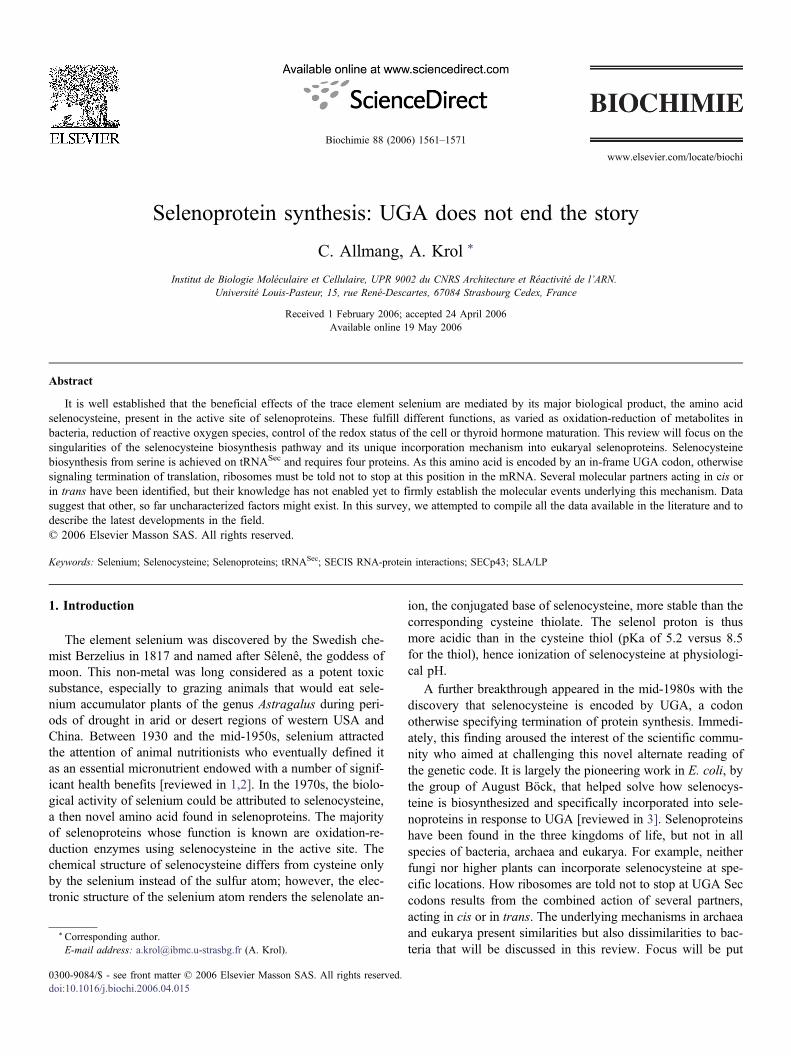
www.elsevier.com/locate/biochi
Biochimie 88 (2006) 1561–1571
Selenoprotein synthesis: UGA does not end the story
C. Allmang, A. Krol *
* Corresponding author.E-mail address: a.krol
0300-9084/$ - see front mdoi:10.1016/j.biochi.2006.
Institut de Biologie Moléculaire et Cellulaire, UPR 9002 du CNRS Architecture et Réactivité de l’ARN.
Université Louis-Pasteur, 15, rue René-Descartes, 67084 Strasbourg Cedex, FranceReceived 1 February 2006; accepted 24 April 2006Available online 19 May 2006
Abstract
It is well established that the beneficial effects of the trace element selenium are mediated by its major biological product, the amino acidselenocysteine, present in the active site of selenoproteins. These fulfill different functions, as varied as oxidation-reduction of metabolites inbacteria, reduction of reactive oxygen species, control of the redox status of the cell or thyroid hormone maturation. This review will focus on thesingularities of the selenocysteine biosynthesis pathway and its unique incorporation mechanism into eukaryal selenoproteins. Selenocysteinebiosynthesis from serine is achieved on tRNASec and requires four proteins. As this amino acid is encoded by an in-frame UGA codon, otherwisesignaling termination of translation, ribosomes must be told not to stop at this position in the mRNA. Several molecular partners acting in cis orin trans have been identified, but their knowledge has not enabled yet to firmly establish the molecular events underlying this mechanism. Datasuggest that other, so far uncharacterized factors might exist. In this survey, we attempted to compile all the data available in the literature and todescribe the latest developments in the field.© 2006 Elsevier Masson SAS. All rights reserved.
Keywords: Selenium; Selenocysteine; Selenoproteins; tRNASec; SECIS RNA-protein interactions; SECp43; SLA/LP
1. Introduction
The element selenium was discovered by the Swedish che-mist Berzelius in 1817 and named after Sêlenê, the goddess ofmoon. This non-metal was long considered as a potent toxicsubstance, especially to grazing animals that would eat sele-nium accumulator plants of the genus Astragalus during peri-ods of drought in arid or desert regions of western USA andChina. Between 1930 and the mid-1950s, selenium attractedthe attention of animal nutritionists who eventually defined itas an essential micronutrient endowed with a number of signif-icant health benefits [reviewed in 1,2]. In the 1970s, the biolo-gical activity of selenium could be attributed to selenocysteine,a then novel amino acid found in selenoproteins. The majorityof selenoproteins whose function is known are oxidation-re-duction enzymes using selenocysteine in the active site. Thechemical structure of selenocysteine differs from cysteine onlyby the selenium instead of the sulfur atom; however, the elec-tronic structure of the selenium atom renders the selenolate an-
@ibmc.u-strasbg.fr (A. Krol).
atter © 2006 Elsevier Masson SAS. All rights reserved.04.015
ion, the conjugated base of selenocysteine, more stable than thecorresponding cysteine thiolate. The selenol proton is thusmore acidic than in the cysteine thiol (pKa of 5.2 versus 8.5for the thiol), hence ionization of selenocysteine at physiologi-cal pH.
A further breakthrough appeared in the mid-1980s with thediscovery that selenocysteine is encoded by UGA, a codonotherwise specifying termination of protein synthesis. Immedi-ately, this finding aroused the interest of the scientific commu-nity who aimed at challenging this novel alternate reading ofthe genetic code. It is largely the pioneering work in E. coli, bythe group of August Böck, that helped solve how selenocys-teine is biosynthesized and specifically incorporated into sele-noproteins in response to UGA [reviewed in 3]. Selenoproteinshave been found in the three kingdoms of life, but not in allspecies of bacteria, archaea and eukarya. For example, neitherfungi nor higher plants can incorporate selenocysteine at spe-cific locations. How ribosomes are told not to stop at UGA Seccodons results from the combined action of several partners,acting in cis or in trans. The underlying mechanisms in archaeaand eukarya present similarities but also dissimilarities to bac-teria that will be discussed in this review. Focus will be put

C. Allmang, A. Krol / Biochimie 88 (2006) 1561–15711562
primarily on eukarya with comparisons to the bacterial and ar-chaeal systems wherever needed. Two aspects will be ad-dressed, biosynthesis of selenocysteine in the first place, fol-lowed by its co-translational incorporation into selenoproteins.
2. Biosynthesis of selenocysteine
Selenocysteine does not occur as a free amino acid. Thus,the first step of its biosynthesis consists in the charge of serineon the specific tRNASec by the conventional seryl-tRNAsynthetase. The Ser-tRNASec is next converted into Sec-tRNASec by selenocysteine synthase that utilizes monoseleno-phosphate as the substrate. This compound is produced fromsodium selenite or more likely selenide by a reaction catalyzedby selenophosphate synthetase. We will describe in this para-graph the characteristic features of tRNASec and the selenocys-teine biosynthesis pathway.
2.1. Structure-function of the tRNASec
Secondary structure models for tRNAsSec are shown inFig. 1, arising from experimental determination in bacteriaand eukarya [4–6], or structure-based sequence alignments inarchaea [6]. Two main characteristic features distinguishtRNAsSec from canonical tRNAs. First, they share the hallmarkof having a 6 bp D-stem, instead of 3–4 bp in other tRNAs.This extended D-stem was shown to be a major identity deter-minant for serine phosphorylation [7], a likely intermediate inselenocysteine biosynthesis in eukarya (see below). Second,the amino acid acceptor arm (A-T), resulting from coaxialstacking of the A and T-stems, is longer in tRNAsSec (13 bp)than in canonical tRNAs where it is 12 bp long (7 + 5 bp). Inbacteria, the 13 bp A-T arm is formed by coaxial stacking ofthe 8 bp A-stem and 5 bp T-stem whereas the same length isobtained in archaea and eukarya by stacking of the longer A-stem (9 bp) and shorter T-stem (4 bp) [5,6,8–14]. This evolu-tionary conservation is obviously a signal for one or more li-
Fig. 1. Secondary structure comparisons of canonical tRNAs versus selenocysteine tRT stand for the amino acid, D, anticodon and T stems, respectively. 7/5, 8/5, 9/4 indiDashes in the canonical tRNA structure signify that the extra arm is of variable lebacterial and eukaryal tRNAsSec. They were omitted in the canonical tRNA. The a
gand(s). In bacteria, the extra length of the A-T arm is a deter-minant for binding to the specialized translation elongationfactor SelB whereas it is required for serine to selenocysteineconversion in eukarya [15,16].
The position and nature of post-transcriptional modifica-tions have been investigated in the vertebrate tRNASec [17,18]. It contains only four modified bases, thus fewer than ca-nonical tRNAs. Apart from pseudo-U55 and m1A58 in the T-loop, mass spectrometry identified 6-isopentenyl-A37 (i6A37)and mcm5Um34, the 5-methylcarboxymethyl-2′-O-methyluri-dine modification, in the anticodon loop. The 2′ O-ribose mod-ification, associated to mcm5U, has been found so far intRNASec only and its yield is a function of the dietary seleniumstatus [17]. Formation of mcm5U34 depends on the tRNASec
tertiary structure and completion of all the other base modifica-tions [19]. Interestingly, protein SECp43 identified earlier in acomplex with the tRNASec [20], might be involved directly orindirectly in the 2′-O-methylation of mcm5U34 [21]. Modifica-tion of i6A37 has also a great importance as its absence pro-duced a severe down effect on selenoprotein synthesis [22].However, as conversion of A37 to i6A37 occurs before U34is modified to mcm5Um and is indeed required for obtainingthe latter, it was difficult to assign the observed effect to thelack of one or the other modification. To address the issue,knock-out transgenic mice were obtained wherein thetRNASec was replaced by the wt or a mutant transgene produ-cing a tRNA that lacked both the U34 and A37 modified bases[23]. This study concluded that U34 modification has a greaterinfluence than i6A37 in regulating the expression of variousmammalian selenoproteins.
2.2. The Ser-tRNASec to Sec-tRNASec conversion step
Neither in eukarya nor in archaea has been isolated mono-selenophosphate, the biological donor of selenium in bacteria.Two enzymes catalyzing formation of this compound havehowever been described. A human cDNA of selenophosphate
NAsSec. The various secondary structure elements are indicated: A, D, AC, andcate the number of base pairs forming the coaxial A-T arm in the tRNAs shown.ngth in different tRNAs. Modified bases are indicated where identified in therchaeal tRNASec was not investigated for its base modification content.
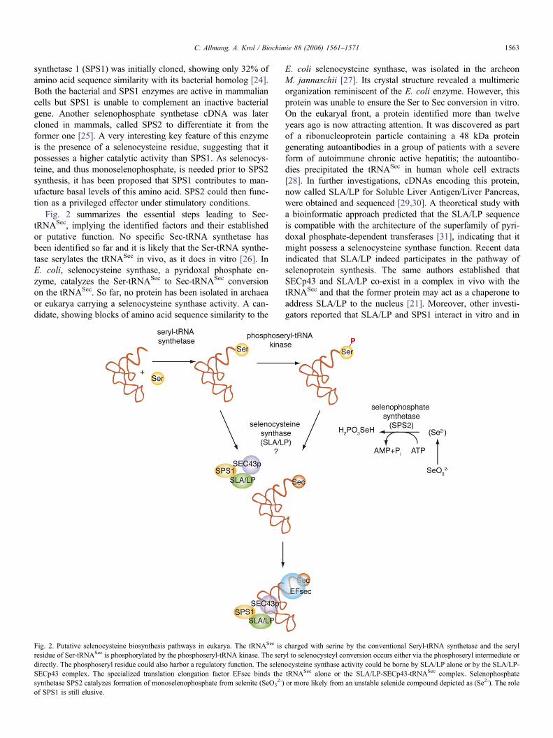
C. Allmang, A. Krol / Biochimie 88 (2006) 1561–1571 1563
synthetase 1 (SPS1) was initially cloned, showing only 32% ofamino acid sequence similarity with its bacterial homolog [24].Both the bacterial and SPS1 enzymes are active in mammaliancells but SPS1 is unable to complement an inactive bacterialgene. Another selenophosphate synthetase cDNA was latercloned in mammals, called SPS2 to differentiate it from theformer one [25]. A very interesting key feature of this enzymeis the presence of a selenocysteine residue, suggesting that itpossesses a higher catalytic activity than SPS1. As selenocys-teine, and thus monoselenophosphate, is needed prior to SPS2synthesis, it has been proposed that SPS1 contributes to man-ufacture basal levels of this amino acid. SPS2 could then func-tion as a privileged effector under stimulatory conditions.
Fig. 2 summarizes the essential steps leading to Sec-tRNASec, implying the identified factors and their establishedor putative function. No specific Sec-tRNA synthetase hasbeen identified so far and it is likely that the Ser-tRNA synthe-tase serylates the tRNASec in vivo, as it does in vitro [26]. InE. coli, selenocysteine synthase, a pyridoxal phosphate en-zyme, catalyzes the Ser-tRNASec to Sec-tRNASec conversionon the tRNASec. So far, no protein has been isolated in archaeaor eukarya carrying a selenocysteine synthase activity. A can-didate, showing blocks of amino acid sequence similarity to the
Fig. 2. Putative selenocysteine biosynthesis pathways in eukarya. The tRNASec isresidue of Ser-tRNASec is phosphorylated by the phosphoseryl-tRNA kinase. The serdirectly. The phosphoseryl residue could also harbor a regulatory function. The selenSECp43 complex. The specialized translation elongation factor EFsec binds thesynthetase SPS2 catalyzes formation of monoselenophosphate from selenite (SeO3
2-)of SPS1 is still elusive.
E. coli selenocysteine synthase, was isolated in the archeonM. jannaschii [27]. Its crystal structure revealed a multimericorganization reminiscent of the E. coli enzyme. However, thisprotein was unable to ensure the Ser to Sec conversion in vitro.On the eukaryal front, a protein identified more than twelveyears ago is now attracting attention. It was discovered as partof a ribonucleoprotein particle containing a 48 kDa proteingenerating autoantibodies in a group of patients with a severeform of autoimmune chronic active hepatitis; the autoantibo-dies precipitated the tRNASec in human whole cell extracts[28]. In further investigations, cDNAs encoding this protein,now called SLA/LP for Soluble Liver Antigen/Liver Pancreas,were obtained and sequenced [29,30]. A theoretical study witha bioinformatic approach predicted that the SLA/LP sequenceis compatible with the architecture of the superfamily of pyri-doxal phosphate-dependent transferases [31], indicating that itmight possess a selenocysteine synthase function. Recent dataindicated that SLA/LP indeed participates in the pathway ofselenoprotein synthesis. The same authors established thatSECp43 and SLA/LP co-exist in a complex in vivo with thetRNASec and that the former protein may act as a chaperone toaddress SLA/LP to the nucleus [21]. Moreover, other investi-gators reported that SLA/LP and SPS1 interact in vitro and in
charged with serine by the conventional Seryl-tRNA synthetase and the serylyl to selenocysteyl conversion occurs either via the phosphoseryl intermediate orocysteine synthase activity could be borne by SLA/LP alone or by the SLA/LP-tRNASec alone or the SLA/LP-SECp43-tRNASec complex. Selenophosphateor more likely from an unstable selenide compound depicted as (Se2-). The role
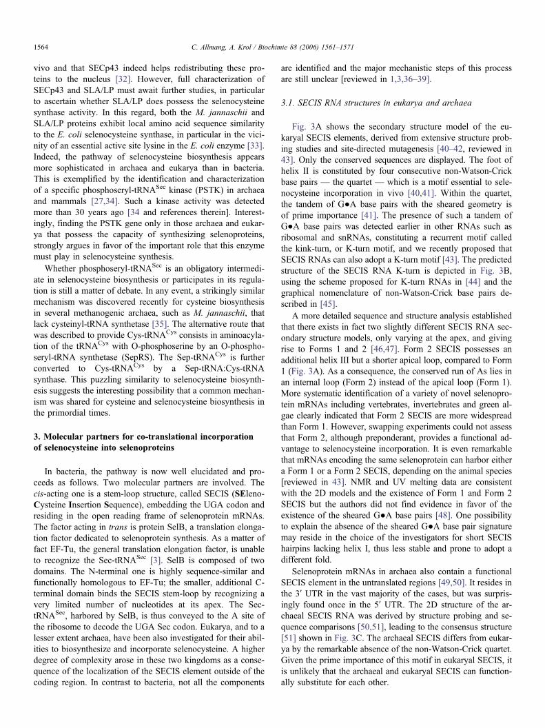
C. Allmang, A. Krol / Biochimie 88 (2006) 1561–15711564
vivo and that SECp43 indeed helps redistributing these pro-teins to the nucleus [32]. However, full characterization ofSECp43 and SLA/LP must await further studies, in particularto ascertain whether SLA/LP does possess the selenocysteinesynthase activity. In this regard, both the M. jannaschii andSLA/LP proteins exhibit local amino acid sequence similarityto the E. coli selenocysteine synthase, in particular in the vici-nity of an essential active site lysine in the E. coli enzyme [33].Indeed, the pathway of selenocysteine biosynthesis appearsmore sophisticated in archaea and eukarya than in bacteria.This is exemplified by the identification and characterizationof a specific phosphoseryl-tRNASec kinase (PSTK) in archaeaand mammals [27,34]. Such a kinase activity was detectedmore than 30 years ago [34 and references therein]. Interest-ingly, finding the PSTK gene only in those archaea and eukar-ya that possess the capacity of synthesizing selenoproteins,strongly argues in favor of the important role that this enzymemust play in selenocysteine synthesis.
Whether phosphoseryl-tRNASec is an obligatory intermedi-ate in selenocysteine biosynthesis or participates in its regula-tion is still a matter of debate. In any event, a strikingly similarmechanism was discovered recently for cysteine biosynthesisin several methanogenic archaea, such as M. jannaschii, thatlack cysteinyl-tRNA synthetase [35]. The alternative route thatwas described to provide Cys-tRNACys consists in aminoacyla-tion of the tRNACys with O-phosphoserine by an O-phospho-seryl-tRNA synthetase (SepRS). The Sep-tRNACys is furtherconverted to Cys-tRNACys by a Sep-tRNA:Cys-tRNAsynthase. This puzzling similarity to selenocysteine biosynth-esis suggests the interesting possibility that a common mechan-ism was shared for cysteine and selenocysteine biosynthesis inthe primordial times.
3. Molecular partners for co-translational incorporationof selenocysteine into selenoproteins
In bacteria, the pathway is now well elucidated and pro-ceeds as follows. Two molecular partners are involved. Thecis-acting one is a stem-loop structure, called SECIS (SEleno-Cysteine Insertion Sequence), embedding the UGA codon andresiding in the open reading frame of selenoprotein mRNAs.The factor acting in trans is protein SelB, a translation elonga-tion factor dedicated to selenoprotein synthesis. As a matter offact EF-Tu, the general translation elongation factor, is unableto recognize the Sec-tRNASec [3]. SelB is composed of twodomains. The N-terminal one is highly sequence-similar andfunctionally homologous to EF-Tu; the smaller, additional C-terminal domain binds the SECIS stem-loop by recognizing avery limited number of nucleotides at its apex. The Sec-tRNASec, harbored by SelB, is thus conveyed to the A site ofthe ribosome to decode the UGA Sec codon. Eukarya, and to alesser extent archaea, have been also investigated for their abil-ities to biosynthesize and incorporate selenocysteine. A higherdegree of complexity arose in these two kingdoms as a conse-quence of the localization of the SECIS element outside of thecoding region. In contrast to bacteria, not all the components
are identified and the major mechanistic steps of this processare still unclear [reviewed in 1,3,36–39].
3.1. SECIS RNA structures in eukarya and archaea
Fig. 3A shows the secondary structure model of the eu-karyal SECIS elements, derived from extensive structure prob-ing studies and site-directed mutagenesis [40–42, reviewed in43]. Only the conserved sequences are displayed. The foot ofhelix II is constituted by four consecutive non-Watson-Crickbase pairs — the quartet — which is a motif essential to sele-nocysteine incorporation in vivo [40,41]. Within the quartet,the tandem of G●A base pairs with the sheared geometry isof prime importance [41]. The presence of such a tandem ofG●A base pairs was detected earlier in other RNAs such asribosomal and snRNAs, constituting a recurrent motif calledthe kink-turn, or K-turn motif, and we recently proposed thatSECIS RNAs can also adopt a K-turn motif [43]. The predictedstructure of the SECIS RNA K-turn is depicted in Fig. 3B,using the scheme proposed for K-turn RNAs in [44] and thegraphical nomenclature of non-Watson-Crick base pairs de-scribed in [45].
A more detailed sequence and structure analysis establishedthat there exists in fact two slightly different SECIS RNA sec-ondary structure models, only varying at the apex, and givingrise to Forms 1 and 2 [46,47]. Form 2 SECIS possesses anadditional helix III but a shorter apical loop, compared to Form1 (Fig. 3A). As a consequence, the conserved run of As lies inan internal loop (Form 2) instead of the apical loop (Form 1).More systematic identification of a variety of novel selenopro-tein mRNAs including vertebrates, invertebrates and green al-gae clearly indicated that Form 2 SECIS are more widespreadthan Form 1. However, swapping experiments could not assessthat Form 2, although preponderant, provides a functional ad-vantage to selenocysteine incorporation. It is even remarkablethat mRNAs encoding the same selenoprotein can harbor eithera Form 1 or a Form 2 SECIS, depending on the animal species[reviewed in 43]. NMR and UV melting data are consistentwith the 2D models and the existence of Form 1 and Form 2SECIS but the authors did not find evidence in favor of theexistence of the sheared G●A base pairs [48]. One possibilityto explain the absence of the sheared G●A base pair signaturemay reside in the choice of the investigators for short SECIShairpins lacking helix I, thus less stable and prone to adopt adifferent fold.
Selenoprotein mRNAs in archaea also contain a functionalSECIS element in the untranslated regions [49,50]. It resides inthe 3′ UTR in the vast majority of the cases, but was surpris-ingly found once in the 5′ UTR. The 2D structure of the ar-chaeal SECIS RNA was derived by structure probing and se-quence comparisons [50,51], leading to the consensus structure[51] shown in Fig. 3C. The archaeal SECIS differs from eukar-ya by the remarkable absence of the non-Watson-Crick quartet.Given the prime importance of this motif in eukaryal SECIS, itis unlikely that the archaeal and eukaryal SECIS can function-ally substitute for each other.

Fig. 3. Structure models for SECIS RNAs. (A). Secondary structures of eukaryal Forms 1 and 2 SECIS. The conserved sequence and structural features are indicated.N, any nucleotide; A/g and A/c indicate that A is the prevalent base. (B). Representation of the putative eukaryal SECIS K-turn with the sequence of the rat type 1iodothyronine deiodinase SECIS RNA. The 5′ and 3′ strands are depicted in green and red, respectively. The model is from [43]; the geometric nomenclature,classification and graphical conventions for displaying non-Watson-Crick base pairs were described in [45]: black arrows ending with a square indicate TransHoogsteen/Sugar Edge G●A sheared base pairs, the open gray arrow heads depict Trans Sugar Edge/Sugar Edge and the black circles are Cis non-Watson-Crick basepairs. (C). Consensus secondary structure model of the archaeal SECIS RNA, adapted from [51]. S stands for G or C.
C. Allmang, A. Krol / Biochimie 88 (2006) 1561–1571 1565
3.2. SECIS-binding proteins
Two major proteins have been described to bind the eu-karyal SECIS RNA specifically, the SECIS Binding Protein 2and ribosomal protein L30. Interestingly, as described below,they both share the same RNA binding domain.
3.2.1. The SECIS Binding Protein 2SBP2 proteins have been characterized so far only in rat and
humans [52,53]. They are about 850 amino acid long. Thefunctional importance of this protein in humans has been rein-forced by the recent description that patients, carrying muta-tions in the SBP2 gene, display a specific thyroid phenotype
associated with reduction in type 2 iodothyronine deiodinaseactivity, a selenoenzyme involved in thyroxine maturation[54]. The SBP2 amino acid sequence can be grossly dividedinto two equal parts (Fig. 4A). The N-terminal domain couldnot be assigned a well-defined function yet. The lack of se-quence similarity to proteins in databases is obviously a strongimpediment towards elucidating its role. Recent data, however,identified a predicted nuclear localization signal and demon-strated that SBP2 undergoes nuclear shuttling, suggesting amechanism for the nuclear assembly of the selenocysteine in-corporation machinery [55]. The C-terminal section containsthe ribosomal and SECIS RNA binding domains, and a regionidentified as important to selenocysteine incorporation in vitro
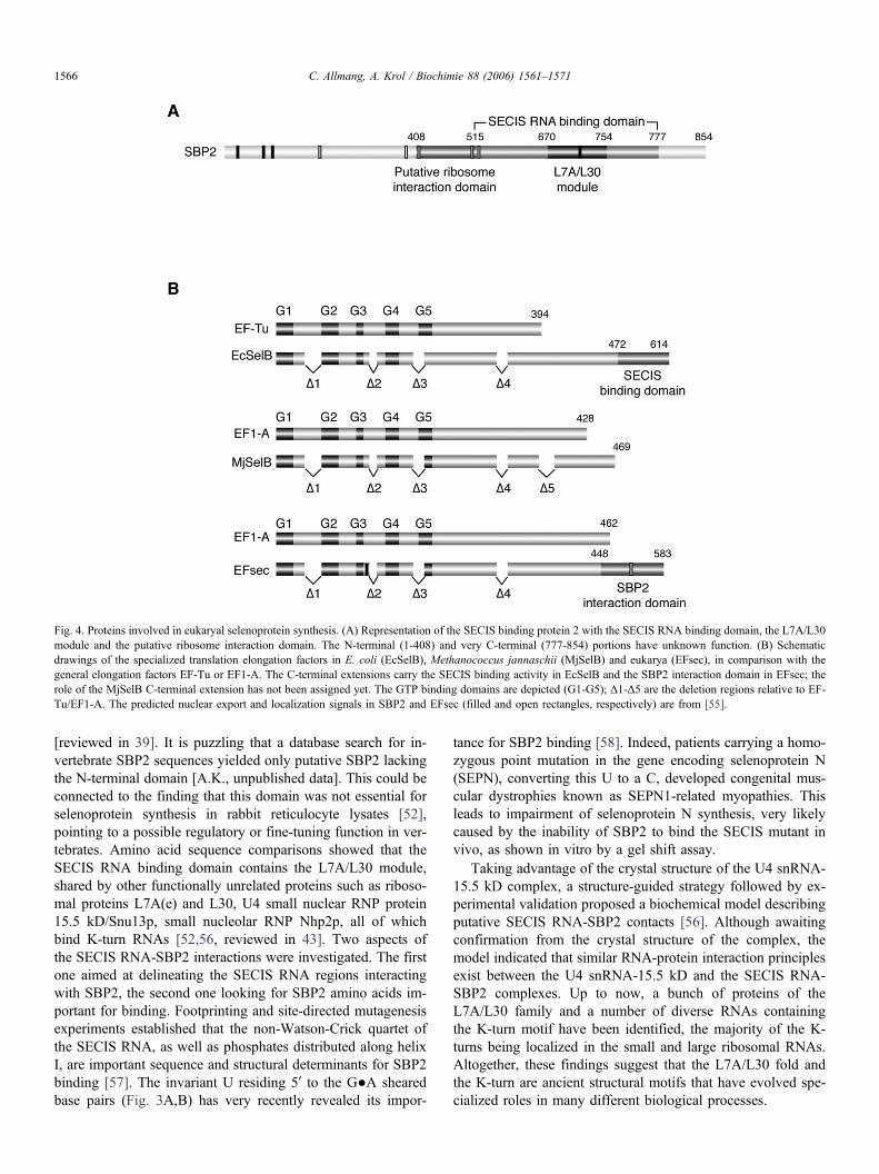
Fig. 4. Proteins involved in eukaryal selenoprotein synthesis. (A) Representation of the SECIS binding protein 2 with the SECIS RNA binding domain, the L7A/L30module and the putative ribosome interaction domain. The N-terminal (1-408) and very C-terminal (777-854) portions have unknown function. (B) Schematicdrawings of the specialized translation elongation factors in E. coli (EcSelB), Methanococcus jannaschii (MjSelB) and eukarya (EFsec), in comparison with thegeneral elongation factors EF-Tu or EF1-A. The C-terminal extensions carry the SECIS binding activity in EcSelB and the SBP2 interaction domain in EFsec; therole of the MjSelB C-terminal extension has not been assigned yet. The GTP binding domains are depicted (G1-G5); Δ1-Δ5 are the deletion regions relative to EF-Tu/EF1-A. The predicted nuclear export and localization signals in SBP2 and EFsec (filled and open rectangles, respectively) are from [55].
C. Allmang, A. Krol / Biochimie 88 (2006) 1561–15711566
[reviewed in 39]. It is puzzling that a database search for in-vertebrate SBP2 sequences yielded only putative SBP2 lackingthe N-terminal domain [A.K., unpublished data]. This could beconnected to the finding that this domain was not essential forselenoprotein synthesis in rabbit reticulocyte lysates [52],pointing to a possible regulatory or fine-tuning function in ver-tebrates. Amino acid sequence comparisons showed that theSECIS RNA binding domain contains the L7A/L30 module,shared by other functionally unrelated proteins such as riboso-mal proteins L7A(e) and L30, U4 small nuclear RNP protein15.5 kD/Snu13p, small nucleolar RNP Nhp2p, all of whichbind K-turn RNAs [52,56, reviewed in 43]. Two aspects ofthe SECIS RNA-SBP2 interactions were investigated. The firstone aimed at delineating the SECIS RNA regions interactingwith SBP2, the second one looking for SBP2 amino acids im-portant for binding. Footprinting and site-directed mutagenesisexperiments established that the non-Watson-Crick quartet ofthe SECIS RNA, as well as phosphates distributed along helixI, are important sequence and structural determinants for SBP2binding [57]. The invariant U residing 5′ to the G●A shearedbase pairs (Fig. 3A,B) has very recently revealed its impor-
tance for SBP2 binding [58]. Indeed, patients carrying a homo-zygous point mutation in the gene encoding selenoprotein N(SEPN), converting this U to a C, developed congenital mus-cular dystrophies known as SEPN1-related myopathies. Thisleads to impairment of selenoprotein N synthesis, very likelycaused by the inability of SBP2 to bind the SECIS mutant invivo, as shown in vitro by a gel shift assay.
Taking advantage of the crystal structure of the U4 snRNA-15.5 kD complex, a structure-guided strategy followed by ex-perimental validation proposed a biochemical model describingputative SECIS RNA-SBP2 contacts [56]. Although awaitingconfirmation from the crystal structure of the complex, themodel indicated that similar RNA-protein interaction principlesexist between the U4 snRNA-15.5 kD and the SECIS RNA-SBP2 complexes. Up to now, a bunch of proteins of theL7A/L30 family and a number of diverse RNAs containingthe K-turn motif have been identified, the majority of the K-turns being localized in the small and large ribosomal RNAs.Altogether, these findings suggest that the L7A/L30 fold andthe K-turn are ancient structural motifs that have evolved spe-cialized roles in many different biological processes.
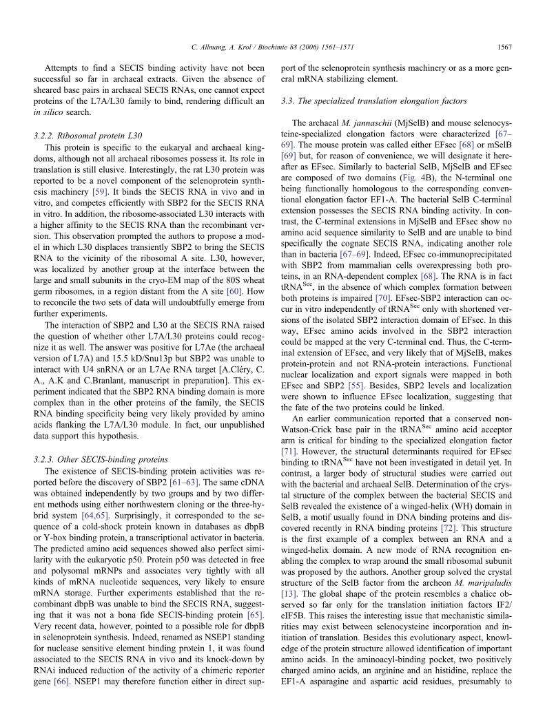
C. Allmang, A. Krol / Biochimie 88 (2006) 1561–1571 1567
Attempts to find a SECIS binding activity have not beensuccessful so far in archaeal extracts. Given the absence ofsheared base pairs in archaeal SECIS RNAs, one cannot expectproteins of the L7A/L30 family to bind, rendering difficult anin silico search.
3.2.2. Ribosomal protein L30This protein is specific to the eukaryal and archaeal king-
doms, although not all archaeal ribosomes possess it. Its role intranslation is still elusive. Interestingly, the rat L30 protein wasreported to be a novel component of the selenoprotein synth-esis machinery [59]. It binds the SECIS RNA in vivo and invitro, and competes efficiently with SBP2 for the SECIS RNAin vitro. In addition, the ribosome-associated L30 interacts witha higher affinity to the SECIS RNA than the recombinant ver-sion. This observation prompted the authors to propose a mod-el in which L30 displaces transiently SBP2 to bring the SECISRNA to the vicinity of the ribosomal A site. L30, however,was localized by another group at the interface between thelarge and small subunits in the cryo-EM map of the 80S wheatgerm ribosomes, in a region distant from the A site [60]. Howto reconcile the two sets of data will undoubtfully emerge fromfurther experiments.
The interaction of SBP2 and L30 at the SECIS RNA raisedthe question of whether other L7A/L30 proteins could recog-nize it as well. The answer was positive for L7Ae (the archaealversion of L7A) and 15.5 kD/Snu13p but SBP2 was unable tointeract with U4 snRNA or an L7Ae RNA target [A.Cléry, C.A., A.K and C.Branlant, manuscript in preparation]. This ex-periment indicated that the SBP2 RNA binding domain is morecomplex than in the other proteins of the family, the SECISRNA binding specificity being very likely provided by aminoacids flanking the L7A/L30 module. In fact, our unpublisheddata support this hypothesis.
3.2.3. Other SECIS-binding proteinsThe existence of SECIS-binding protein activities was re-
ported before the discovery of SBP2 [61–63]. The same cDNAwas obtained independently by two groups and by two differ-ent methods using either northwestern cloning or the three-hy-brid system [64,65]. Surprisingly, it corresponded to the se-quence of a cold-shock protein known in databases as dbpBor Y-box binding protein, a transcriptional activator in bacteria.The predicted amino acid sequences showed also perfect simi-larity with the eukaryotic p50. Protein p50 was detected in freeand polysomal mRNPs and associates very tightly with allkinds of mRNA nucleotide sequences, very likely to ensuremRNA storage. Further experiments established that the re-combinant dbpB was unable to bind the SECIS RNA, suggest-ing that it was not a bona fide SECIS-binding protein [65].Very recent data, however, pointed to a possible role for dbpBin selenoprotein synthesis. Indeed, renamed as NSEP1 standingfor nuclease sensitive element binding protein 1, it was foundassociated to the SECIS RNA in vivo and its knock-down byRNAi induced reduction of the activity of a chimeric reportergene [66]. NSEP1 may therefore function either in direct sup-
port of the selenoprotein synthesis machinery or as a more gen-eral mRNA stabilizing element.
3.3. The specialized translation elongation factors
The archaeal M. jannaschii (MjSelB) and mouse selenocys-teine-specialized elongation factors were characterized [67–69]. The mouse protein was called either EFsec [68] or mSelB[69] but, for reason of convenience, we will designate it here-after as EFsec. Similarly to bacterial SelB, MjSelB and EFsecare composed of two domains (Fig. 4B), the N-terminal onebeing functionally homologous to the corresponding conven-tional elongation factor EF1-A. The bacterial SelB C-terminalextension possesses the SECIS RNA binding activity. In con-trast, the C-terminal extensions in MjSelB and EFsec show noamino acid sequence similarity to SelB and are unable to bindspecifically the cognate SECIS RNA, indicating another rolethan in bacteria [67–69]. Indeed, EFsec co-immunoprecipitatedwith SBP2 from mammalian cells overexpressing both pro-teins, in an RNA-dependent complex [68]. The RNA is in facttRNASec, in the absence of which complex formation betweenboth proteins is impaired [70]. EFsec-SBP2 interaction can oc-cur in vitro independently of tRNASec only with shortened ver-sions of the isolated SBP2 interaction domain of EFsec. In thisway, EFsec amino acids involved in the SBP2 interactioncould be mapped at the very C-terminal end. Thus, the C-term-inal extension of EFsec, and very likely that of MjSelB, makesprotein-protein and not RNA-protein interactions. Functionalnuclear localization and export signals were mapped in bothEFsec and SBP2 [55]. Besides, SBP2 levels and localizationwere shown to influence EFsec localization, suggesting thatthe fate of the two proteins could be linked.
An earlier communication reported that a conserved non-Watson-Crick base pair in the tRNASec amino acid acceptorarm is critical for binding to the specialized elongation factor[71]. However, the structural determinants required for EFsecbinding to tRNASec have not been investigated in detail yet. Incontrast, a larger body of structural studies were carried outwith the bacterial and archaeal SelB. Determination of the crys-tal structure of the complex between the bacterial SECIS andSelB revealed the existence of a winged-helix (WH) domain inSelB, a motif usually found in DNA binding proteins and dis-covered recently in RNA binding proteins [72]. This structureis the first example of a complex between an RNA and awinged-helix domain. A new mode of RNA recognition en-abling the complex to wrap around the small ribosomal subunitwas proposed by the authors. Another group solved the crystalstructure of the SelB factor from the archeon M. maripaludis[13]. The global shape of the protein resembles a chalice ob-served so far only for the translation initiation factors IF2/eIF5B. This raises the interesting issue that mechanistic simila-rities may exist between selenocysteine incorporation and in-itiation of translation. Besides this evolutionary aspect, knowl-edge of the protein structure allowed identification of importantamino acids. In the aminoacyl-binding pocket, two positivelycharged amino acids, an arginine and an histidine, replace theEF1-A asparagine and aspartic acid residues, presumably to
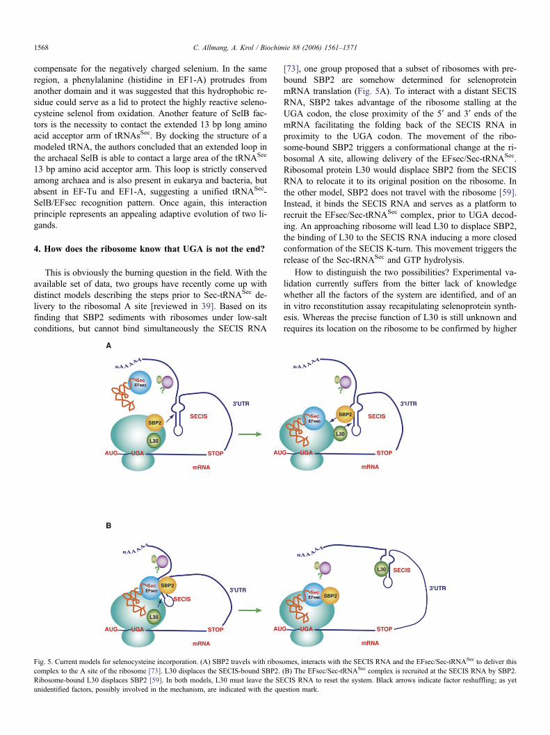
C. Allmang, A. Krol / Biochimie 88 (2006) 1561–15711568
compensate for the negatively charged selenium. In the sameregion, a phenylalanine (histidine in EF1-A) protrudes fromanother domain and it was suggested that this hydrophobic re-sidue could serve as a lid to protect the highly reactive seleno-cysteine selenol from oxidation. Another feature of SelB fac-tors is the necessity to contact the extended 13 bp long aminoacid acceptor arm of tRNAsSec. By docking the structure of amodeled tRNA, the authors concluded that an extended loop inthe archaeal SelB is able to contact a large area of the tRNASec
13 bp amino acid acceptor arm. This loop is strictly conservedamong archaea and is also present in eukarya and bacteria, butabsent in EF-Tu and EF1-A, suggesting a unified tRNASec-SelB/EFsec recognition pattern. Once again, this interactionprinciple represents an appealing adaptive evolution of two li-gands.
4. How does the ribosome know that UGA is not the end?
This is obviously the burning question in the field. With theavailable set of data, two groups have recently come up withdistinct models describing the steps prior to Sec-tRNASec de-livery to the ribosomal A site [reviewed in 39]. Based on itsfinding that SBP2 sediments with ribosomes under low-saltconditions, but cannot bind simultaneously the SECIS RNA
Fig. 5. Current models for selenocysteine incorporation. (A) SBP2 travels with riboscomplex to the A site of the ribosome [73]. L30 displaces the SECIS-bound SBP2.Ribosome-bound L30 displaces SBP2 [59]. In both models, L30 must leave the SEunidentified factors, possibly involved in the mechanism, are indicated with the qu
[73], one group proposed that a subset of ribosomes with pre-bound SBP2 are somehow determined for selenoproteinmRNA translation (Fig. 5A). To interact with a distant SECISRNA, SBP2 takes advantage of the ribosome stalling at theUGA codon, the close proximity of the 5′ and 3′ ends of themRNA facilitating the folding back of the SECIS RNA inproximity to the UGA codon. The movement of the ribo-some-bound SBP2 triggers a conformational change at the ri-bosomal A site, allowing delivery of the EFsec/Sec-tRNASec.Ribosomal protein L30 would displace SBP2 from the SECISRNA to relocate it to its original position on the ribosome. Inthe other model, SBP2 does not travel with the ribosome [59].Instead, it binds the SECIS RNA and serves as a platform torecruit the EFsec/Sec-tRNASec complex, prior to UGA decod-ing. An approaching ribosome will lead L30 to displace SBP2,the binding of L30 to the SECIS RNA inducing a more closedconformation of the SECIS K-turn. This movement triggers therelease of the Sec-tRNASec and GTP hydrolysis.
How to distinguish the two possibilities? Experimental va-lidation currently suffers from the bitter lack of knowledgewhether all the factors of the system are identified, and of anin vitro reconstitution assay recapitulating selenoprotein synth-esis. Whereas the precise function of L30 is still unknown andrequires its location on the ribosome to be confirmed by higher
omes, interacts with the SECIS RNA and the EFsec/Sec-tRNASec to deliver this(B) The EFsec/Sec-tRNASec complex is recruited at the SECIS RNA by SBP2.CIS RNA to reset the system. Black arrows indicate factor reshuffling; as yetestion mark.
[3]
[4]
[5]
[6]
[7]
[8]
[9]
[10
[11
[12
[13
[14
[15
[16
[17
[18
[19
[20
[21
[22
C. Allmang, A. Krol / Biochimie 88 (2006) 1561–1571 1569
resolution data, both models converge to propose that this pro-tein displaces SBP2 from the SECIS RNA. The L30-SBP2 re-shuffling hypothesis, however, does not consider that SBP2was found to dissociate very slowly or not at all in vivo, oncebinding to SECIS RNA has taken place [74]. Another centralquestion asks how the SECIS RNA-bound complex folds backat an approaching ribosome and competes with the release fac-tor: no need to say that mechanistic issues are here of primeimportance. In this regard, the recent discovery of stem-loopstructures different from SECIS RNAs, lying in the open read-ing frame at the 3′ vicinity of the UGA selenocysteine codon insome eukaryal selenoprotein mRNAs, added a missing piece toour knowledge [75]. Such stem-loops were found in some butnot all selenoprotein mRNAs and they do not share a con-served secondary structure in the different selenoproteinmRNAs. This observation may legitimately lead to ask whetherthe function of these stem-loops is pivotal to selenoproteinsynthesis. One possibility is their requirement under certaincircumstances to favor ribosome pausing in much the sameway as stem-loops or pseudoknots contribute to frameshifting.
We are left with the take-home message that a clearer pic-ture of selenoprotein synthesis is popping up but, at the sametime, a number of questions remain unsolved. It looks as if asingle supramolecular complex could achieve sequentially (orsimultaneously) selenocysteine biosynthesis and its incorpora-tion into selenoproteins [32; reviewed in 38]. The cornerstonein this complex is protein SECp43 that establishes RNA-pro-tein and protein-protein contacts with several partners, notablyfacilitating the interaction between tRNASec, EFsec and SBP2in vivo. SECp43 thus appears as a key player in orchestratingmultiple interactions and redistributing the nucleocytoplasmiclocalization of other components involved. Taking into accountthat EFsec and SBP2 undergo nucleocytoplasmic shuttling[55], assembly of this supramolecular complex in the nucleusis an appealing model that could help circumvent nonsense-mediated decay at UGA Sec codons [55]. Another advantagewould be to provide ribosomes with SBP2-pre-bound seleno-protein mRNAs, fueling the hypothesis of the existence of afraction of ribosomes pre-determined for selenoprotein synth-esis [37]. Extremely dynamic contacts must exist to enablemultiple conformational changes to occur. There is obviouslyroom for future directions of research in this actively evolvingfield.
Acknowledgments
Philippe Carbon and Alain Lescure are thanked for carefulreading of the manuscript.
References
[1] D.L. Hatfield, V.N. Gladyshev, How selenium has altered our under-standing of the genetic code, Mol. Cell. Biol. 22 (2002) 3565–3576.
[2] M. Rederstorff, A. Krol, A. Lescure, Understanding the importance ofselenium and selenoproteins in muscle function, Cell. Mol. Life Sci. 63(2006) 52–59.
A. Böck, Selenium metabolism in bacteria, in: D.L. Hatfield (Ed.), Sele-nium, its molecular biology and role in human health, Kluwer AcademicPublishers, Norwood, MA, 2001, pp. 7–22.C. Baron, E. Westhof, A. Böck, R. Giege, Solution structure of seleno-cysteine-inserting tRNASec from E. coli. Comparison with canonicaltRNASer, J. Mol. Biol. 231 (1993) 274–292.C. Sturchler, E. Westhof, P. Carbon, A. Krol, Unique secondary and ter-tiary structural features of the eukaryotic selenocysteine tRNASec, Nu-cleic Acids Res. 21 (1993) 1073–1079.N. Hubert, C. Sturchler, E. Westhof, P. Carbon, A. Krol, The 9/4 second-ary structure of eukaryotic selenocysteine tRNA: more pieces of evi-dence, RNA 4 (1998) 1029–1033.X.-Q. Wu, H.J. Gross, The length and the secondary structure of the D-stem of human selenocysteine tRNA are the major identity determinantsfor serine phosphorylation, EMBO J. 13 (1994) 241–248.A. Böck, K. Forchhammer, J. Heider, C. Baron, Selenoprotein synthesis:an expansion of the genetic code, Trends Biochem. 16 (1991) 463–467.M. Rao, B.A. Carlson, S.V. Novoselov, D.P. Weeks, V.N. Gladyshev,D.L. Hatfield, Chlamydomonas reinhardtii selenocysteine tRNA[Ser]Sec,RNA 9 (2003) 923–930.
] A.G. Russell, M.N. Schnare, M.W. Gray, Pseudouridine-guide RNAsand other Cbf5p-associated RNAs in Euglena gracilis, RNA 10 (2004)1034–1046.
] R.K. Shrimali, A.V. Lobanov, X.-M. Xu, M. Rao, B.A. Carlson, D.C.Mahadeo, C.A. Parent, V.N. Gladyshev, D.L. Hatfield, SelenocysteinetRNA identification in the model organisms Dictyostelium discoideumand Tetrahymena thermophila, Biochem. Biophys. Res. Com. 329(2005) 147–151.
] T. Mourier, A. Pain, B. Barrell, S. Griffiths-Jones, A selenocysteinetRNA and SECIS element in Plasmodium falciparum, RNA 11 (2005)119–122.
] M. Leibundgut, C. Frick, M. Thanbichler, A. Böck, N. Ban, Selenocys-teine tRNA-specific elongator factor SelB is a structural chimaera ofelongation and initiation factors, EMBO J. 24 (2005) 11–22.
] A.V. Lobanov, C. Delgado, S. Rahlfs, S.V. Novoselov, G.V. Kryukov,S. Gromer, D.L. Hatfield, K. Becker, V.N. Gladyshev, The Plasmodiumselenoproteome, Nucleic Acids Res. 34 (2006) 496–505.
] C. Baron, A. Böck, The length of the aminoacyl-acceptor stem of theselenocysteine-specific tRNASec of E.coli is the determinant for bindingto elongation factors SelB or Tu, J. Biol. Chem. 266 (1991) 20375–20379.
] C. Sturchler-Pierrat, N. Hubert, T. Totsuka, T. Mizutani, P. Carbon, A.Krol, Selenocysteylation in eukaryotes necessitates the uniquely longaminoacyl acceptor stem of selenocysteine tRNASec, J. Biol. Chem. 270(1995) 18570–18574.
] A.M. Diamond, I.-S. Choi, P.F. Crain, T. Hashizume, S.C. Pomerantz, R.Cruz, C.J. Steer, K.E. Hill, R.F. Burk, J.A. McCloskey, D.L. Hatfield,Dietary selenium affects methylation of the wobble nucleoside in the an-ticodon of selenocysteine tRNA[Ser]Sec, J. Biol. Chem. 268 (1993)14215–14223.
] C. Sturchler, A. Lescure, G. Keith, P. Carbon, A. Krol, Base modifica-tion pattern at the wobble position of Xenopus selenocysteine tRNASec,Nucleic Acids Res. 22 (1994) 1354–1358.
] L.K. Kim, T. Matsufuji, S. Matsufuji, B.A. Carlson, S.S. Kim, D.L. Hat-field, B.J. Lee, Methylation of the ribosyl moiety at position 34 of sele-nocysteine tRNA[Ser]Sec is governed by both primary and tertiary struc-ture, RNA 6 (2000) 1306–1315.
] F. Ding, P.J. Grabowski, Identification of a protein component of a mam-malian tRNASec complex implicated in the decoding of UGA as seleno-cysteine, RNA 5 (1999) 1561–1569.
] X.-M. Xu, H. Mix, B.A. Carlson, P.J. Grabowski, V.N. Gladyshev, M.J.Berry, D.L. Hatfield, Evidence for direct roles of two additional factors,SECp43 and Soluble Liver Antigen, in the selenoprotein synthesis ma-chinery, J. Biol. Chem. 280 (2005) 41568–41575.
] G.J. Warner, M.J. Berry, M.E. Moustafa, B.A. Carlson, D.L. Hatfield,J.R. Faust, Inhibition of selenoprotein synthesis by selenocysteinetRNA[Ser]Sec lacking isopentenyladenosine, J. Biol. Chem. 275 (2000)28110–28119.
[23
[24
[25
[26
[27
[28
[29
[30
[31
[32
[33
[34
[35
[36
[37
[38
[39
[40
[41
[42
[43
[44
[45
[46
[47
[48
[49
[50
[51
[52
[53
[54
[55
[56
[57
[58
[59
[60
[61
[62
C. Allmang, A. Krol / Biochimie 88 (2006) 1561–15711570
] B.A. Carlson, X.-M. Xu, V.N. Gladyshev, D.L. Hatfield, Selective rescueof selenoprotein expression in mice lacking a highly specialized methylgroup in selenocysteine tRNA, J. Biol. Chem. 280 (2005) 5542–5548.
] S.C. Low, J.W. Harney, M.J. Berry, Cloning and functional characteriza-tion of human selenophosphate synthetase, an essential component of se-lenoprotein synthesis, J. Biol. Chem. 270 (1995) 21659–21664.
] M.J. Guimaraes, D. Peterson, A. Vicari, B.G. Cocks, N.G. Copeland,D.J. Gilbert, N.A. Jenkins, D.A. Ferrick, R.A. Kastelein, J.F. Bazan, A.Zlotnik, Identification of a novel selD homolog from eukaryotes, bacter-ia and archaea: is there an autoregulatory mechanism in selenocysteinemetabolism?, Proc. Natl. Acad. Sci. USA 93 (1996) 15086–15091.
] T. Ohama, D.C.H. Yang, D.L. Hatfield, Selenocysteine tRNA and serinetRNA are aminoacylated by the same synthetase, but manifest differentidentities with respect to the long extra arm, Arch. Biochem. Biophys.315 (1994) 293–301.
] J.T. Kaiser, K. Gromadski, M. Rother, H. Engelhardt, M.V. Rodnina,M.C. Wahl, Structural and functional investigation of a putative archaealselenocysteine synthase, Biochem. 44 (2005) 13315–13327.
] C. Gelpi, E.J. Sontheimer, J.L. Rodriguez-Sanchez, Autoantibodiesagainst a serine tRNA-protein complex implicated in cotranslational sele-nocysteine insertion, Proc. Natl. Acad. Sci. USA 89 (1992) 9739–9743.
] I. Wies, S. Brunner, J. Henninger, J. Herkel, S. Kanzler, K.-H. Meyerzum Büschenfelde, A.W. Lohse, Identification of target antigen forSLA/LP autoantibodies in autoimmune hepatitis, Lancet 355 (2000)1510–1515.
] M. Costa, J.L. Rodriguez-Sanchez, A.J. Czaja, C. Gelpi, Isolation andcharacterization of cDNA encoding the antigenic protein of the humantRNP[Ser]Sec complex recognized by autoantibodies from patients withtype-1 autoimmune hepatitis, Clin. Exp. Immunol. 121 (2000) 364–374.
] T. Kernebeck, A.W. Lohse, J. Grötzinger, A bioinformatical approachsuggests the function of the autoimmune hepatitis target antigen SolubleLiver Antigen/Liver Pancreas, Hepatology 34 (2001) 230–233.
] A. Small-Howard, N. Morozova, Z. Stoytcheva, E.P. Forry, J.B. Mansell,J.W. Harney, B.A. Carlson, X.-M. Xu, D.L. Hatfield, M.J. Berry, Supra-molecular complexes mediate selenocysteine incorporation in vivo, Mol.Cell. Biol. 26 (2006) 2337–2346.
] P. Tormay, R. Wilting, F. Lottspeich, P.K. Mehta, P. Christen, A. Böck,Bacterial selenocysteine synthase, structural and functional properties,Eur. J. Biochem. 254 (1998) 655–661.
] B.A. Carlson, X.-M. Xu, G.V. Kryukov, M. Rao, M.J. Berry, V.N. Gla-dyshev, D.L. Hatfield, Identification and characterization of phosphoser-yl-tRNA[Ser]Sec kinase, Proc. Natl. Acad. Sci. USA 101 (2004) 12848–12853.
] A. Sauerwald, W. Zhu, T.A. Major, H. Roy, S. Palioura, D. Jahn, W.B.Whitman, J.R. Yates 3rd, M. Ibba, D. Söll, RNA-dependent cysteine bio-synthesis in archaea, Science 307 (2005) 1969–1972.
] A. Krol, Evolutionary different RNA motifs and RNA-protein complexesto achieve selenoprotein synthesis, Biochimie 84 (2002) 765–774.
] D.M. Driscoll, P.R. Copeland, Mechanism and regulation of selenopro-tein synthesis, Annu. Rev. Nutr. 23 (2003) 17–40.
] A.L. Small-Howard, M.J. Berry, Unique features of selenocysteine incor-poration function within the context of general eukaryotic translationalprocesses, Biochem. Soc. 33 (2005) 1493–1497.
] K. Caban, P.R. Copeland, Size matters: a view of selenocysteine incor-poration from the ribosome, Cell. Mol. Life Sci. 63 (2006) 73–81.
] R. Walczak, E. Westhof, P. Carbon, A. Krol, A novel RNA structuralmotif in the selenocysteine insertion element of eukaryotic selenoproteinmRNAs, RNA 2 (1996) 367–379.
] R. Walczak, P. Carbon, A. Krol, An essential non-Watson-Crick basepair motif in 3’UTR to mediate selenoprotein translation, RNA 4 (1998)74–84.
] G.W. Martin IV, J.W. Harney, M.J. Berry, Functionality of mutations atconserved nucleotides in eukaryotic SECIS elements is determined by theidentity of a single nonconserved nucleotide, RNA 4 (1998) 65–73.
] C. Allmang, A. Krol, SECIS RNAs and K-turn binding proteins. A sur-vey of evolutionary conserved RNA and protein motifs, in Selenium, itsmolecular biology and role in human health 2nd edition, M.J.Berry,
V.N.Gladyshev, D.L.Hatfield (eds), Kluwer Academic Publishers, 2006,in press.
] D.J. Klein, T.M. Schmeing, P.B. Moore, T.A. Steitz, The kink-turn, anew RNA secondary structure motif, EMBO J. 20 (2001) 4214–4221.
] N.B. Leontis, E. Westhof, Geometric nomenclature and classification ofRNA base pairs, RNA 7 (2001) 499–512.
] E. Grundner-Culemann, G.W. Martin III, J.W. Harney, M.J. Berry, Twodistinct SECIS RNA structures capable of directing selenocysteine incor-poration in eukaryotes, RNA 5 (1999) 625–635.
] D. Fagegaltier, A. Lescure, R. Walczak, P. Carbon, A. Krol, Structuralanalysis of new local features in SECIS RNA hairpins, Nucleic AcidsRes. 28 (2000) 2679–2689.
] A. Ramos, A.N. Lane, D. Hollingworth, T.W.-N. Fan, Secondary struc-ture and stability of the selenocysteine insertion sequences (SECIS) forhuman thioredoxin reductase and glutathione peroxidase, Nucleic AcidsRes. 32 (2004) 1746–1755.
] R. Wilting, S. Schorling, B.C. Persson, A. Böck, Selenoprotein synthesisin archaea: identification of an mRNA element of Methanococcus jan-naschii probably directing selenocysteine insertion, J. Mol. Biol. 266(1997) 637–641.
] M. Rother, A. Resch, W.L. Gardner, W.B. Whitman, A. Böck, Heterolo-gous expression of archaeal selenoprotein genes directed by the SECISelement located in the 3’ non-translated region, Mol. Microbiol. 40(2001) 900–908.
] G.V. Kryukov, V.N. Gladyshev, The prokaryotic selenoproteome,EMBO Rep. 5 (2004) 538–543.
] P.R. Copeland, J.E. Fletcher, B.A. Carlson, D.L. Hatfield, D.M. Driscoll,A novel RNA binding protein, SBP2, is required for the translation ofmammalian selenoprotein mRNAs, EMBO J. 19 (2000) 306–314.
] A. Lescure, C. Allmang, K. Yamada, P. Carbon, A. Krol, cDNA cloning,expression pattern and RNA binding analysis of human selenocysteineinsertion sequence (SECIS) binding protein 2, Gene 291 (2002) 279–285.
] A.M. Dumitrescu, X.-H. Liao, M.S.Y. Abdullah, J. Lado-Abeal, F. Ab-dul Majed, L.C. Moeller, G. Boran, L. Schomburg, R.E. Weiss, S. Refet-off, Mutations in SECISBP2 result in abnormal thyroid hormone metabo-lism, Nat. Genet. 37 (2005) 1247–1252.
] L.A. de Jesus, P.R. Hoffmann, T. Michaud, E.P. Forry, A. Small-Ho-ward, R.J. Stillwell, N. Morozova, J.W. Harney, M.J. Berry, Nuclear as-sembly of UGA decoding complexes on selenoprotein mRNAs: a me-chanism for eluding nonsense-mediated decay?, Mol. Cell. Biol. 26(2006) 1795–1805.
] C. Allmang, P. Carbon, A. Krol, The SBP2 and 15.5 kD/Snu13p proteinsshare the same RNA binding domain: identification of SBP2 amino acidsimportant to SECIS RNA binding, RNA 8 (2002) 1308–1318.
] J.E. Fletcher, P.R. Copeland, D.M. Driscoll, A. Krol, The selenocysteineincorporation machinery: interactions between the SECIS RNA and theSECIS-binding protein SBP2, RNA 7 (2001) 1442–1453.
] V. Allamand, P. Richard, A. Lescure, C. Ledeuil, D. Desjardin, N. Petit,C. Gartioux, A. Ferreiro, A. Krol, N. Pellegrini, J. Andoni Urtizberea, P.Guicheney, A single homozygous point mutation in a 3’ untranslated re-gion motif of selenoprotein N mRNA causes SEPN1-related myopathy,EMBO Rep. 7 (2006) 450–454.
] L. Chavatte, B.A. Brown, D.M. Driscoll, Ribosomal protein L30 is acomponent of the UGA-selenocysteine recoding machinery in eukar-yotes, Nat. Struct. Mol. Biol. 12 (2005) 408–416.
] M. Halic, T. Becker, J. Frank, C.M.T. Spahn, R. Beckmann, Localizationand dynamic behavior of ribosomal protein L30e, Nat. Struct. Mol. Biol.12 (2005) 467–468.
] Q. Shen, P.A. McQuikin, P.E. Newburger, RNA-binding proteins thatspecifically recognize the selenocysteine insertion sequence of humancellular glutathione peroxidase mRNA, J. Biol. Chem. 270 (1995)30448–30452.
] N. Hubert, R. Walczak, P. Carbon, A. Krol, A protein binds the seleno-cysteine insertion element in the 3’UTR of mammalian selenoproteinmRNAs, Nucleic Acids Res. 24 (1996) 464–469.
[63
[64
[65
[66
[67
[68
[69
[70
[71
[72
[73
[74
[75
C. Allmang, A. Krol / Biochimie 88 (2006) 1561–1571 1571
] T. Fujiwara, K. Busch, H.J. Gross, T. Mizutani, A SECIS binding protein(SBP) is distinct from selenocysteyl-tRNA protecting factor (SePF), Bio-chimie 81 (2000) 213–218.
] Q. Shen, R. Wu, J.L. Leonard, P.E. Newburger, Identification and mole-cular cloning of a human selenocysteine insertion sequence-binding pro-tein. A bifunctional role for DNA-binding protein B, J. Biol. Chem. 273(1998) 5443–5446.
] D. Fagegaltier, N. Hubert, P. Carbon, A. Krol, The selenocysteine inser-tion sequence binding protein SBP is different from the Y-box proteindbpB, Biochimie 82 (2000) 117–122.
] Q. Shen, L. Fan, P.E. Newburger, Nuclease sensitive element bindingprotein 1 associates with the selenocysteine insertion sequence and func-tions in mammalian selenoprotein translation, J. Cell. Physiol. 207(2006) 775–783.
] M. Rother, R. Wilting, S. Commans, A. Böck, Identification and charac-terization of the selenocysteine-specific translation factor SelB from thearcheon Methanococcus jannaschii, J. Mol. Biol. 299 (2000) 351–358.
] R.M. Tujebajeva, P.R. Copeland, X.-M. Xu, B.A. Carlson, J.W. Harney,D.M. Driscoll, D.L. Hatfield, M.J. Berry, Decoding apparatus for eukar-yotic selenocysteine incorporation, EMBO Rep. 2 (2000) 158–163.
] D. Fagegaltier, N. Hubert, K. Yamada, T. Mizutani, P. Carbon, A. Krol,Characterization of mSelB, a novel mammalian elongation factor for se-lenoprotein translation, EMBO J. 19 (2000) 4796–4805.
] A.M. Zavacki, J.B. Mansell, M. Chung, B. Klimovitsky, J.W. Harney,M.J. Berry, Coupled tRNASec-dependent assembly of the selenocysteinedecoding apparatus, Mol. Cell 11 (2003) 773–781.
] T. Mizutani, K. Tanabe, K. Yamada, A G.U base pair in the eukaryoticselenocysteine tRNA is important for interaction with SePF, the putativeselenocysteine-specific elongation factor, FEBS Lett. 429 (1998) 189–193.
] S. Yoshizawa, L. Rasubala, T. Ose, D. Kohda, D. Fourmy, K. Maenaka,Structural basis for mRNA recognition by elongation factor SelB, Nat.Struct. Mol. Biol. 12 (2005) 198–203.
] S.A. Kinzy, K. Caban, P.R. Copeland, Characterization of the SECISbinding protein 2 complex required for the co-translational insertion ofselenocysteine in mammals, Nucleic Acids Res. 33 (2005) 5172–5180.
] S.C. Low, E. Grundner-Culemann, J.W. Harney, M.J. Berry, SECIS-SBP2 interactions dictate selenocysteine incorporation efficiency and se-lenoprotein hierarchy, EMBO J. 19 (2000) 6882–6890.
] M.T. Howard, G. Aggarwal, C.B. Anderson, S. Khatri, K.M. Flanigan,J.F. Atkins, Recoding elements located adjacent to a subset of eukaryalselenocysteine-specifying UGA codons, EMBO J. 24 (2005) 1596–1607.

1
Chapter 5. SECIS RNAs and K-turn binding proteins. A survey of evolutionary conserved RNA and protein motifs Christine Allmang and Alain Krol Architecture et Réactivité de l'arN. UPR 9002 du CNRS-Université Louis Pasteur. Institut de Biologie Moléculaire et Cellulaire. 67084 Strasbourg, France Summary: The SelenoCysteine Insertion Sequence (SECIS) is a stem-loop structure residing in the 3' untranslated region of all selenoprotein mRNAs. Its presence is mandatory to allow the ribosome to readthrough the UGA selenocysteine codon. The SECIS RNA possesses a well-defined secondary structure. Four consecutive non-Watson-Crick base pairs, with a central tandem of sheared G.A/A.G base pairs, constitute the functional motif of the SECIS RNA which is recognized by the SECIS binding protein SBP2. The tandem of sheared base pairs is part of a recurrent motif, the kink-turn (K-turn), occurring in a variety of different RNAs. The K-turn is a helix-internal loop-helix composed of a non-Watson-Crick stem containing the G.A base pairs and a canonical stem. The internal loop between the stems is always asymmetrical and usually contains three unpaired nucleotides on one strand and none on the other. We propose here that the SECIS RNA must represent a K-turn variant with regard to the limited structural differences that distinguish it from consensus K-turns. Work by others showed that ribosomal protein L30 also binds the SECIS RNA in a specific manner. L30 and SBP2 are members of a family of proteins sharing the same RNA-binding domain called L7A/L30. All proteins possessing this fold recognize K-turn RNAs. Three structures of RNA-protein complexes containing the L7A/L30 protein fold and cognate K-turn RNAs have been solved. In light of the interaction principles governing these RNA-protein complexes, we discuss how L30 can recognize the SECIS RNA. Collectively, all the findings suggest that the L7A/L30 protein fold and the K-turn are ancient structural motifs that have evolved various functions, from pre-mRNA splicing to protein synthesis. Introduction The field of eukaryotic selenoprotein research is fascinating in several respects. First, the existence of taxa-specific selenoproteins altered the initial perception that mammals recapitulate the eukaryotic selenoproteome. Second, it becomes increasingly apparent that the number of molecular
2
partners involved in selenoprotein synthesis is larger than previously thought. Relocation of the SECIS element, from the coding frame in bacteria to the 3'-untranslated region (3’UTR) of selenoprotein mRNAs in eukaryotes, may be responsible only in part for this complexification. Indeed, even selenocysteine biosynthesis itself seems to take a more sophisticated pathway in eukaryotes. The SECIS stem-loop contains a well defined structural motif composed of four consecutive non-Watson-Crick base pairs, with a central tandem of sheared G.A base pairs [1]. This motif (Figure 1A) also ensures a functional role as it is essential to selenocysteine incorporation in vivo [2,3] and constitutes the binding site of SBP2, a protein binding specifically to the SECIS RNA [4,5]. The SBP2 RNA-binding domain contains a region sharing a high degree of amino acid sequence similarity to the L7A/L30 protein family containing ribosomal proteins L7Ae and L30, the 15.5kD/Snu13p spliceosomal protein and other functionally unrelated proteins [6,7]. Cocrystal structures of the L7Ae, L30 and 15.5 kD proteins in complex with their cognate RNAs revealed that the proteins fold into a highly conserved compact globular domain, the L7A/L30 domain, that binds specifically to RNAs possessing a kink-turn (K-turn) motif. The canonical K-turn is a recurrent element, occurring notably in ribosomal RNAs, U4 snRNA, and box C/D regions of snoRNAs and archaeal sRNAs. It contains a tandem of sheared G.A/A.G base pairs that have an important structural role in forming and stabilizing the turn [8]. In earlier studies, we proposed a 3D model for the SECIS RNA where the phosphodiester backbone is bent at the non-Watson-Crick base pairs [1]. Combined with the presence of sheared base pairs, the proposed folding of the SECIS RNA suggests that it could be a canonical K-turn or a K-turn related RNA. From all these findings emerges the important issue of how different RNAs harboring K-turn motifs can selectively discriminate proteins sharing the same RNA-binding domain. This is a particularly burning question in light of the finding that ribosomal protein L30 is another SECIS-binding protein [9]. This chapter will describe the SECIS RNA structure with comparison to canonical K-turn RNAs, then highlight the similarities/differences between protein-RNA complexes formed with proteins of the L7A/L30 family and K-turn RNAs. The SECIS RNA: a K-turn variant An experimental secondary structure model for SECIS RNAs (Figure 1A) was proposed about ten years ago based on structure probing in solution [1]. It was next discovered that certain SECIS RNAs can adopt a slightly different 2D structure at their apex [10]. Called Form 2 SECIS (Figure 1A), they possess an additional helix III but a shorter apical loop, compared to Form 1 SECIS. Besides the non-Watson-Crick quartet, Form 2 shares with Form 1 the other conserved features characterizing SECIS RNAs, i.e. the run
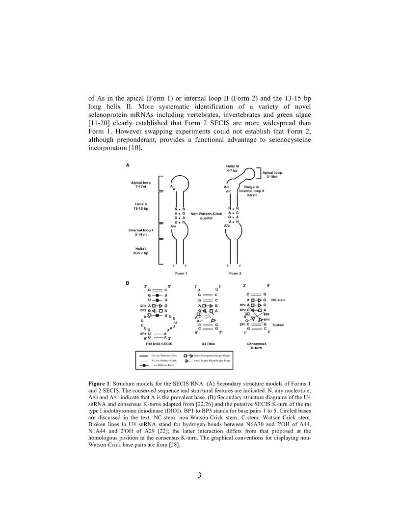
3
of As in the apical (Form 1) or internal loop II (Form 2) and the 13-15 bp long helix II. More systematic identification of a variety of novel selenoprotein mRNAs including vertebrates, invertebrates and green algae [11-20] clearly established that Form 2 SECIS are more widespread than Form 1. However swapping experiments could not establish that Form 2, although preponderant, provides a functional advantage to selenocysteine incorporation [10].
Figure 1. Structure models for the SECIS RNA. (A) Secondary structure models of Forms 1 and 2 SECIS. The conserved sequence and structural features are indicated. N, any nucleotide; A/G and A/C indicate that A is the prevalent base. (B) Secondary structure diagrams of the U4 snRNA and consensus K-turns adapted from [22,26] and the putative SECIS K-turn of the rat type I iodothyronine deiodinase (DIOI). BP1 to BP5 stands for base pairs 1 to 5. Circled bases are discussed in the text. NC-stem: non-Watson-Crick stem; C-stem: Watson-Crick stem. Broken lines in U4 snRNA stand for hydrogen bonds between N6A30 and 2'OH of A44, N1A44 and 2'OH of A29 [22]; the latter interaction differs from that proposed at the homologous position in the consensus K-turn. The graphical conventions for displaying non-Watson-Crick base pairs are from [28].
4
It is remarkable that mRNAs encoding the same selenoprotein can harbor either a Form 1 or a Form 2 SECIS, depending on the animal species. This is well exemplified by the SelM mRNA where Form 2 occurs in mammals while zebrafish harbors Form 1 SECIS [14]. Remarkably, chemical probing experiments indicated that the conserved As are single stranded and well accessible whatever the SECIS form [21]. In a few cases, especially in the mammalian SelM and some Chlamydomonas Form 2 SECIS, Cs are found instead of As without apparently altering the SECIS function [14,15]. Thus the universal conservation of the As, which was taken for granted at the time when the number of available SECIS sequences was too little to make statistically valid comparisons, is called into question. The mechanistic role of these unpaired A/Cs is still unknown, but their functional importance has been experimentally proven in vivo by site-directed mutagenesis. Along the same lines, the nucleotide 5' to the non-Watson-Crick quartet is A in the vast majority of SECIS RNAs. However, compilation of selenoprotein mRNA sequences in other organisms led to the conclusion that G can sometimes be found instead, an interesting example being provided by the single selenoprotein mRNA in nematodes [11,20,21]. This correlates with in vivo experiments and mobility shift assays with SBP2 and SECIS RNAs concluding that an A is preferred but not mandatory [5,21]. In conclusion, it emerges from phylogenetic studies that SECIS RNAs exhibit a remarkable conservation of the 2D structures but few invariant nucleotides. Clearly, elucidation of the function of the single stranded A/C and conserved length of helix II is a necessary step toward an in-depth understanding of the function of the SECIS RNA. The non-Watson-Crick quartet at the foot of helix II is a characteristic feature recognized by SBP2 (Figure 1B). The central G.A tandem was shown by structure probing experiments and computer modeling to adopt the sheared geometry [1]. Tandem sheared base pairs were initially discovered in the crystal structure of the 5' stem-loop of U4 snRNA in complex with the 15.5 kD protein [22]. The prevalence of this RNA motif was in fact revealed by the analysis of the atomic structures of the large and small ribosomal subunits where it occurs six times in H.marismortui 23S rRNA and twice in T.thermophilus 16S rRNA [8]. Its presence was further identified in the crystal structures of three other RNA-protein complexes: the yeast ribosomal protein L30e with its pre-mRNA, and the archaeal ribosomal protein L7Ae in complex with box C/D or box H/ACA sRNAs [23-25]. A two-dimensional representation of the tertiary structure of a consensus K-turn is diagrammed in Figure 1B, which was adapted from [26]. In this publication, the authors derived the consensus from examination of K-turns in crystal structures and compared them with the sequence alignments of rRNAs from the three kingdoms of life. The K-turn is a two-stranded, helix-internal loop-helix

5
motif comprising about 15 nucleotides. The first stem (canonical or C-stem) ends at the internal loop with two Watson-Crick base pairs, mostly G-C. The non-canonical stem (NC-stem) starts typically with the sheared G.A base pairs. The internal loop is always asymmetrical with three unpaired nucleotides on one strand and none on the other. Because of the cross-strand stacking of the sheared base pairs, a sharp bend of the sugar-phosphate backbone occurs between the C and NC-stems. Five base pairs characterize the K-turn motif (Figure 1B): the Watson-Crick C-G base pair 1, the sheared G.A base pairs 2 and 3, the triple A.C-G base pair 4, and G.A base pair 5. The adenine of base pair 4 mediates the minor groove interaction with the C-stem (A-minor motif; see reference 27) and is crucial for K-turn folding. Figure 1B shows the 2D structure model of the non-Watson-Crick quartet of the rat type I iodothyronine deiodinase (DIOI) SECIS RNA [1] compared to the structure of the U4 snRNA K-turn motif adapted from [22]. Visual inspection of the SECIS 2D structure identified an important K-turn characteristic feature: the C-stem separated by an internal loop from the NC-stem comprising the invariant sheared base pairs. Despite the similarity, a few SECIS specific structural features led us to ask whether they form genuine K-turns. The non-Watson-Crick U.U base pair 3' to the sheared base pairs will not be discussed further because it displays sequence variation in SECIS and other RNAs and does not participate directly in the K-turn interactions [26]. The first question concerns the U residue (circled in Figure 1B) 5' to the sheared base pair 2. Chemical probing experiments detected that it forms a non-Watson-Crick U.U base pair in the naked SECIS RNA [1]. In contrast, the homologous position is unpaired in U4 snRNA and in the consensus K-turn (Figure 1B; see also Figure 2B). Moreover, data from crystal structures of RNA-protein complexes showed that the base at this position protrudes away from the RNA chain and is tightly bound in a pocket of the protein [22-25]. However, one cannot exclude the possibility that an SBP2-promoted induced fit leads to unpairing and flipping out of the U residue. It could thus be the positional analog of the protruding base in the other K-turns (Figures 1B and 2B). The second question asks whether the counterparts to base pairs 4 and 5 of the consensus K-turn also exist in the SECIS RNA as chemical probing cannot detect them. Formation of base pair 4 in the SECIS RNA will only depend on the sequence of base pair 1 since A is invariant in base pair 3 (Figure 1B). Base pair 1 is U-A in the SECIS RNA shown, very often C-G and G-C but rarely A-U or G.U in others [1,11-21]. Interestingly, tables of sequence variation in [26] show the prevalence of C-G, G-C or U-A at base pair 1 in K-turns, indicating that formation of base pair 4 is theoretically possible in SECIS RNAs. Likewise, base pair 5 could form in SECIS RNAs as tables in [26] established that base pairing is permitted in canonical K-turns between the invariant A of base pair 2 and

6
any nucleotide. Lastly, one could argue that the size of internal loop I of SECIS RNAs is larger than in canonical K-turns.
Figure 2. K-turn motifs and amino acid sequence alignments of L7A/L30 RNA-binding domains. (A) The secondary structures of the U4 snRNA, L30e pre-mRNA, L7Ae rRNA, L7Ae box C/D sRNA were taken from the crystal structures of the corresponding RNA-protein complexes, those of the SECIS RNA and U3 snoRNA result from structure probing (see text). In bold are the sheared G.A base pairs. Numbering is from the original publications, except that of the SECIS RNA which is arbitrary. The dotted line between A248 and U265 in L7Ae rRNA represents the hydrogen bond giving rise to the base triple A.U.G [30]. (B) Amino acid sequence alignment of L7A/L30 proteins. hSBP2, human SBP2; h15.5 kD, human 15.5 kD; Snu13p, the yeast 15.5 kD ortholog; Nhp2p, the yeast core protein of box H/ACA snoRNPs; yRPL30, yeast ribosomal protein L30; hRPL7A, human ribosomal protein L7A. Identical and similar amino acids are displayed in black and gray backgrounds, respectively.
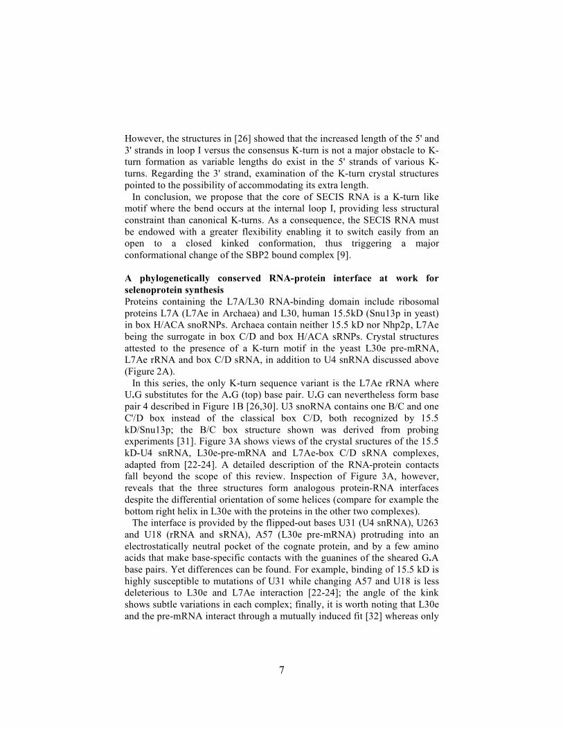
7
However, the structures in [26] showed that the increased length of the 5' and 3' strands in loop I versus the consensus K-turn is not a major obstacle to K-turn formation as variable lengths do exist in the 5' strands of various K-turns. Regarding the 3' strand, examination of the K-turn crystal structures pointed to the possibility of accommodating its extra length. In conclusion, we propose that the core of SECIS RNA is a K-turn like motif where the bend occurs at the internal loop I, providing less structural constraint than canonical K-turns. As a consequence, the SECIS RNA must be endowed with a greater flexibility enabling it to switch easily from an open to a closed kinked conformation, thus triggering a major conformational change of the SBP2 bound complex [9]. A phylogenetically conserved RNA-protein interface at work for selenoprotein synthesis Proteins containing the L7A/L30 RNA-binding domain include ribosomal proteins L7A (L7Ae in Archaea) and L30, human 15.5kD (Snu13p in yeast) in box H/ACA snoRNPs. Archaea contain neither 15.5 kD nor Nhp2p, L7Ae being the surrogate in box C/D and box H/ACA sRNPs. Crystal structures attested to the presence of a K-turn motif in the yeast L30e pre-mRNA, L7Ae rRNA and box C/D sRNA, in addition to U4 snRNA discussed above (Figure 2A). In this series, the only K-turn sequence variant is the L7Ae rRNA where U.G substitutes for the A.G (top) base pair. U.G can nevertheless form base pair 4 described in Figure 1B [26,30]. U3 snoRNA contains one B/C and one C'/D box instead of the classical box C/D, both recognized by 15.5 kD/Snu13p; the B/C box structure shown was derived from probing experiments [31]. Figure 3A shows views of the crystal sructures of the 15.5 kD-U4 snRNA, L30e-pre-mRNA and L7Ae-box C/D sRNA complexes, adapted from [22-24]. A detailed description of the RNA-protein contacts fall beyond the scope of this review. Inspection of Figure 3A, however, reveals that the three structures form analogous protein-RNA interfaces despite the differential orientation of some helices (compare for example the bottom right helix in L30e with the proteins in the other two complexes). The interface is provided by the flipped-out bases U31 (U4 snRNA), U263 and U18 (rRNA and sRNA), A57 (L30e pre-mRNA) protruding into an electrostatically neutral pocket of the cognate protein, and by a few amino acids that make base-specific contacts with the guanines of the sheared G.A base pairs. Yet differences can be found. For example, binding of 15.5 kD is highly susceptible to mutations of U31 while changing A57 and U18 is less deleterious to L30e and L7Ae interaction [22-24]; the angle of the kink shows subtle variations in each complex; finally, it is worth noting that L30e and the pre-mRNA interact through a mutually induced fit [32] whereas only
8
the RNA component (U4 snRNA or sRNA) undergoes an induced fit upon binding to 15.5 kD and L7Ae, respectively [31, 33-37]. Earlier work localized the SBP2 RNA-binding domain in a region lying approximately between positions 500 and 750 [4,7,29, and our unpublished results]. Within this area, database searches [4,6,7] identified a subdomain homologous to the L7A/L30 RNA-binding domain (Figure 2B), SBP2 and 15.5 kD/Snu13p sharing the highest amino acid sequence similarity [7]. The RNA-binding domain of SBP2 is thus bipartite, composed of the conserved L7A/L30 module flanked by SBP2-specific sequences. A structure-guided strategy, based on the similarities between SBP2/15.5 kD and SECIS RNA/U4 snRNA, and the crystal structure of the 15.5 kD-U4 snRNA complex, predicted SBP2 amino acids that should contact the SECIS RNA [7]. Changing them to alanines led to the identification of eight amino acids critical for SECIS binding, four of them being crucial: Gly676 and Glu679 are postulated to contact the guanines of the sheared base pairs, Glu699 and Arg731 being very likely part of the pocket accommodating the SECIS RNA U2 (Figure 3B). These findings established that the recognition principles governing the 15.5 kD-U4 snRNA interaction must be similar in the SBP2-SECIS RNA complex especially at the guanines of the G.A base pairs and at U2 (SECIS RNA) and U31 (U4 snRNA). Another member of the L7A/L30 family, the rat ribosomal protein L30, was recently shown to be a novel SECIS-binding protein [9]. As a follow-up, determination of the molecular basis underlying this interaction would be instructive in particular to understand how L30 can recognize the SECIS K-turn and compete with SBP2. In the absence of a structural model though, comparison of the structures of the L30e, 15.5 kD, L7Ae and SBP2 RNA-protein complexes [7, 22-25, 37] provided some clues that may explain the L30 versatility. In all of the complexes, mutations of the bases comprising the sheared G.A base pairs resulted in the complete loss of protein binding in vitro. Together with the high amino acid sequence similarity between yeast L30e and rat L30, these findings strongly suggest that rat L30 in complex with the SECIS RNA should also interact at the G.A tandem of the SECIS RNA, most likely at the guanines. An interesting difference between 15.5 kD and SBP2 on the one hand, and L7Ae and L30e on the other, occurs at the flipped-out base. In the 15.5 kD and SBP2 complexes, mutations of U31 and U2 to any nucleotide is detrimental to binding in vitro and function in vivo (for the SECIS RNA). In contrast, L7Ae can accommodate a C instead of U18 and there is little sequence preference at A57 for the binding in vitro of L30e which can tolerate G or even C [38]. Remarkably, a correlation can be made at the protein level. In the 15.5kD-U4 snRNA and SBP2-SECIS RNA complexes, five (almost) identical amino acids contact U31 and probably U2, respectively (Figure 3B). Instead, only two L7Ae amino acids make base specific contacts with U18, L30e showing the simplest interaction scheme
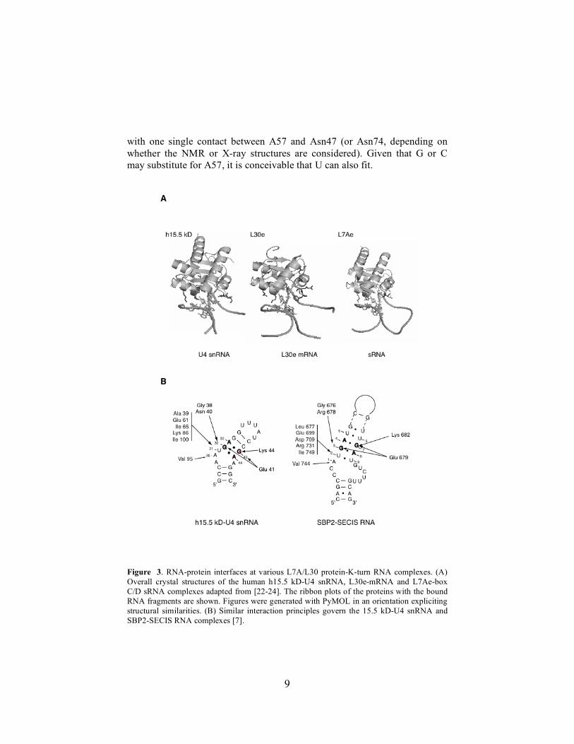
9
with one single contact between A57 and Asn47 (or Asn74, depending on whether the NMR or X-ray structures are considered). Given that G or C may substitute for A57, it is conceivable that U can also fit.
Figure 3. RNA-protein interfaces at various L7A/L30 protein-K-turn RNA complexes. (A) Overall crystal structures of the human h15.5 kD-U4 snRNA, L30e-mRNA and L7Ae-box C/D sRNA complexes adapted from [22-24]. The ribbon plots of the proteins with the bound RNA fragments are shown. Figures were generated with PyMOL in an orientation expliciting structural similarities. (B) Similar interaction principles govern the 15.5 kD-U4 snRNA and SBP2-SECIS RNA complexes [7].
10
Taking into account that a single contact forms between L30e and A57, we propose that the SECIS RNA U2 could also hydrogen bond with L30e Asn47 or Asn74 upon repositioning of the base to offer the appropriate hydrogen bond donor and acceptor groups. As rat L30 binds the SECIS RNA, we assayed other L7A/L30 proteins for their abilities to recognize the SECIS RNA. Snu13p and L7Ae indeed bound the SECIS RNA but the reverse did not happen since SBP2 was unable to interact with U4 snRNA or an L7Ae RNA target (A.Cléry, C.A, A.K and C. Branlant, manuscript in preparation). We concluded from this experiment that the SBP2 RNA-binding domain is more complex than in the other proteins of the family, the SECIS RNA binding specificity being very likely provided by amino acids flanking the L7A/L30 subdomain. In fact, our unpublished data support this hypothesis. The analogous protein-RNA interface formed between L7A/L30 proteins and various K-turn RNAs suggests a conformational adaptability of the RNA upon binding to its cognate protein. Such a dynamic process could potentially confer the binding specificity for different K-turns, as exemplified by rat L30. This adaptability could be facilitated by the dimorphism of K-turn RNAs that are in dynamic equilibrium between a tightly kinked-turn and a more open structure [39]. A bunch of proteins of the L7A/L30 family and a large number of diverse RNAs containing the K-turn motif have been identified, with the majority of the K-turns residing in the small and large ribosomal RNAs [8]. Altogether, these findings suggest that the L7A/L30 fold and the K-turn are ancient structural motifs that have evolved specialized roles in many different biological processes. References 1. R Walczak, E Westhof, P Carbon, A Krol 1996 RNA 2:367 2. R Walczak, P Carbon, A Krol 1998 RNA 4:74 3. GW Martin III, JW Harney, MJ Berry 1998 RNA 4:65 4. PR Copeland, JE Fletcher, BA Carlson, DL Hatfield, DM Driscoll 2000 EMBO J 19:306 5. JE Fletcher, PR Copeland, DM Driscoll, A Krol 2001 RNA 7:1442 6. PR Copeland, DM Driscoll 2001 Selenium: Its Molecular Biology and Role in Human
Health DL Hatfield (Ed) Kluwer Academic Publishers Boston/Dordrecht/London pp 55 7. C Allmang, P Carbon, A Krol 2002 RNA 8:1308 8. DJ Klein, TM Schmeing, PB Moore, TA Steitz 2001 EMBO J 20:4214 9. L Chavatte, BA Brown, DM Driscoll 2005 Nature Struct & Mol Biol 12:408 10. E Grundner-Culemann, GW Martin III, JW Harney, MJ Berry 1999 RNA 5:625 11. C Buettner, JW Harney, MJ Berry 1999 J Biol Chem 274:21598 12. M Hirosawa-Takamori, H Jäckle, G Vorbrüggen 2000 EMBO Rep 1:441 13. S Castellano, N Morozova, M Morey, MJ Berry, F Serras, M Corominas, R Guigo 2001
EMBO Rep 2:697 14. KV Korotkov, SV Novoselov, DL Hatfield, VN Gladyshev 2002 Mol Cell Biol 22:1402 15. SV Novoselov, M Rao, NV Onoshko, H Zhi, GV Kryukov, Y Xiang, DP Weeks, DL
Hatfield, VN Gladyshev 2002 EMBO J 21:3681
11
16. GV Kryukov, S Castellano, SV Novoselov, AV Lobanov, O Zehtab, R Guigo, VN Gladyshev 2003 Science 300:1439
17. C Thisse, A Degrave, GV Kryukov, VN Gladyshev, S Obrecht-Pflumio, A Krol, B Thisse, A Lescure 2003 Gene Expression Patterns 3:525
18. S Castellano, SV Novoselov, GV Kryukov, A Lescure, E Blanco, A Krol, VN Gladyshev, R Guigo 2004 EMBO Rep 5:71
19. T Mourier, A Pain, B Barrell, S Griffiths-Jones 2005 RNA 11:119 20. K Taskov, C Chapple, GV Kryukov, S Castellano, AV Lobanov, KV Korotkov, R
Guigo, VN Gladyshev 2005 Nucleic Acids Res 33:2227 21. D Fagegaltier, A Lescure, R Walczak, P Carbon, A Krol 2000 Nucleic Acids Res
28:2679 22. I Vidovic, S Nottrott, K Hartmuth, R Lührmann, R Ficner 2000 Mol Cell 6:1331 23. JA Chao, JR Williamson 2004 Structure 12:1165 24. T Moore, Y Zhang, MO Fenley, H Li 2004 Structure 12:807 25. T Hamma, AR Ferré-D'Amaré 2004 Structure 12:893 26. A Lescoute, NB Leontis, C Massire, E Westhof 2005 Nucleic Acids Res 33:2395 27. P Nissen, JA Ippolito, N Ban, PB Moore, TA Steitz 2001 Proc Natl Acad Sci USA
98:4899 28. NB Leontis, E Westhof 2001 RNA 7:499 29. A Lescure, C Allmang, K Yamada, P Carbon, A Krol 2002 Gene 291:279 30. N Ban, P Nissen, J Hansen, PB Moore, TA Steitz 2000 Science 289:905 31. N Marmier-Gourrier, A Cléry, V Senty-Ségault, B Charpentier, F Schlotter, F Leclerc,
R Fournier, C Branlant 2003 RNA 9:821 32. JA Chao, GS Prasad, SA White, C David Stout, JR Williamson 2003 J Mol Biol
326:999 33. V Cojocaru, S Nottrott, R Klement, TM Jovin 2005 RNA 11:197 34. B Turner, SE Melcher, TJ Wilson, DG Norman, DMJ Lilley 2005 RNA 11:1192 35. AK Wozniak, S Nottrott, E Kühn-Hölsken, GF Schröder, H Grubmüller, R Lührmann,
CAM Seidel, F Oesterhelt 2005 RNA 11:1545 36. C Charron, X Manival, A Cléry, V Senty-Ségault, B Charpentier, N Marmier-Gourrier,
C Branlant, A Aubry 2004 J Mol Biol 342:757 37. J Suryadi, EJ Tran, E Stuart Maxwell, BA Brown 2005 Biochem 44:9657 38. SA White, M Hoeger, JJ Schweppe, A Shillingford, V Shipilov, J Zarutskie 2004 RNA
10:369 39. TA Goody, SE Melcher, DG Norman, DMJ Lilley 2004 RNA 10:254

The SBP2 and 15.5 kD/Snu13p proteins share thesame RNA binding domain: Identification of SBP2amino acids important to SECIS RNA binding
CHRISTINE ALLMANG, PHILIPPE CARBON, and ALAIN KROLStructure des Macromolécules Biologiques et Mécanismes de Reconnaissance,Unité Propre de Recherche 9002 du Centre National de la Recherche Scientifique–Université Louis Pasteur,Institut de Biologie Moléculaire et Cellulaire, 67084 Strasbourg Cedex, France
ABSTRACT
Selenoprotein synthesis in eukaryotes requires the selenocysteine insertion sequence (SECIS) RNA, a hairpin in the39 untranslated region of selenoprotein mRNAs. The SECIS RNA is recognized by the SECIS-binding protein 2 (SBP2),which is a key player in this specialized translation machinery. The objective of this work was to obtain structuralinsight into the SBP2-SECIS RNA complex. Multiple sequence alignment revealed that SBP2 and the U4 snRNA-bindingprotein 15.5 kD/Snu13p share the same RNA binding domain of the L7A/L30 family, also found in the box H/ACAsnoRNP protein Nhp2p and several ribosomal proteins. In corollary, we have detected a similar secondary structuremotif in the SECIS and U4 RNAs. Combining the data of the crystal structure of the 15.5 kD-U4 snRNA complex, andthe SBP2/15.5 kD sequence similarities, we designed a structure-guided strategy predicting 12 SBP2 amino acids thatshould be critical for SECIS RNA binding. Alanine substitution of these amino acids followed by gel shift assays ofthe SBP2 mutant proteins identified four residues whose mutation severely diminished or abolished SECIS RNAbinding, the other eight provoking intermediate down effects. In addition to identifying key amino acids for SECISrecognition by SBP2, our findings led to the proposal that some of the recognition principles governing the 15.5 kD-U4snRNA interaction must be similar in the SBP2-SECIS RNA complex.
Keywords: L7A/L30 RNA binding domain; RNA–protein interactions; SECIS-binding protein 2; selenocysteine;U4 snRNA
INTRODUCTION
Selenium is mostly found in the active site of seleno-proteins, in the form of the amino acid selenocysteine+In mammals, selenoproteins participate in several gluta-thione- or thioredoxin-dependent oxidation–reductionreactions, or in the maturation of the thyroid hormone(reviewed in Köhrle et al+, 2000; Gladyshev & Kryukov,2001)+ The importance of selenium and selenoproteinswas further underscored by two recent discoveries+ Thefirst one refers to the capital roles for sperm maturationof the phospholipid hydroperoxide glutathione peroxi-dase (Ursini et al+, 1999) and protamine thiol crosslink-ing glutathione peroxidase (Pfeifer et al+, 2001), twosplice variants of the same pre-mRNA+ It is remarkable
that these findings provided the molecular basis forearlier observations linking selenium deficiencies andmale infertility+ The second discovery is that patientsdeveloping a form of congenital muscular dystrophycarry mutations in the gene encoding selenoproteinSePN1 (Moghadaszadeh et al+, 2001)+ This finding con-stituted the first report establishing a direct correlationbetween the occurrence of a genetic disease and mu-tations in a selenoprotein gene+ Eukaryotic selenocys-teine biosynthesis and cotranslational incorporation inresponse to a redefined UGA Sec codon are achievedby a complex molecular machinery containing RNAand protein partners (reviewed in Fagegaltier et al+,2001; Lescure et al+, 2002b)+ This amino acid is syn-thesized from the seryl residue of the Ser-tRNASec,generating the Sec-tRNASec that is loaded onto theselenocysteine-specialized translation elongation fac-tor mSelB/eEFsec (Fagegaltier et al+, 2000a; Tuje-bajeva et al+, 2000)+ Decoding of UGA Sec codonsnecessitates not only the presence of this elongationfactor but also the SECIS element, an RNA hairpin in
Reprint requests to: Alain Krol, Structure des Macromolécules Bi-ologiques et Mécanismes de Reconnaissance, Unité Propre de Re-cherche 9002 du Centre National de la Recherche Scientifique-Université Louis Pasteur, Institut de Biologie Moléculaire et Cellulaire,15 Rue René Descartes, 67084 Strasbourg Cedex, France; e-mail:A+Krol@ibmc+u-strasbg+fr+
RNA (2002), 8:1308–1318+ Cambridge University Press+ Printed in the USA+Copyright © 2002 RNA Society+DOI: 10+1017/S1355838202020034
1308
the 39 UTR of selenoprotein mRNAs (Berry et al+, 1991)+Structure–function studies proposed secondary andthree-dimensional structure models for the SECIS ele-ment (Walczak et al+, 1996, 1998; Martin et al+, 1998;Grundner-Culemann et al+, 1999; Fagegaltier et al+,2000b)+ The core of the hairpin consists of a quartet ofnon-Watson–Crick base pairs containing a tandem ofsheared G-A base pairs that are pivotal for mediatingUGA Sec decoding (Walczak et al+, 1996, 1998)+ SBP2,the SECIS binding protein 2, interacts with the SECISelement (Copeland et al+, 2000; Lescure et al+, 2002a)and most likely with mSelB/eEFsec (Fagegaltier et al+,2000a; Tujebajeva et al+, 2000)+ From these and otherfunctional data (Low et al+, 2000), it is obvious thatSBP2 is a key player in the machinery+ Two major stud-ies were previously undertaken to delineate the SECISRNA and SBP2 domains important for the interaction+In the first one, structural investigations of the SECISRNA-SBP2 complex revealed that the phosphate back-bone and the non-Watson–Crick base pairs at the coreof the SECIS RNA are important features governingthe interaction (Fletcher et al+, 2001)+ The other studydealt with the functional dissection of SBP2+ It wasdiscovered that it belongs to the family of proteins con-taining the L7A/L30 RNA-binding domain (Copelandet al+, 2001)+ This domain comprises several ribosomalproteins of the large and small subunits, Nhp2p that isthe core component of the yeast H/ACA family of smallnucleolar ribonucleoprotein particles (Henras et al+,1998), and the eRF1 subunit of the translation termi-nation release factor+ Interestingly, the existence of suchan RNA-binding domain was hypothesized several yearsago, based on amino acid sequence comparisons ofthe limited number of proteins available at the time(Koonin et al+, 1994)+
An extensive study of the amino acids required forthe binding of SBP2 to the SECIS RNA has not beenpublished yet+ The issue is especially crucial becausethe various proteins of the L7A/L30 family can specif-ically recognize their cognate RNA yet share identicalor similar sequences in their homologous RNA-bindingdomains+ The objective of the work reported here wasprecisely to identify amino acids in the RNA-bindingdomain of SBP2 that are important for recognition ofthe SECIS RNA+ The strategy that was taken stemmedfrom our two initial findings described in this report:(1) the RNA-binding domain of SBP2 displays aminoacid sequence identity to another member of the L7A/L30 family, the human 15+5 kD protein (ortholog ofthe yeast Snu13p) that binds the 59 stem-loop ofspliceosomal U4 snRNAs but also box C/D snoRNAs(Nottrott et al+, 1999; Gottschalk et al+, 1999; Stevens& Abelson, 1999; Watkins et al+, 2000); (2) the SECISRNA and the 59 stem-loop of U4 snRNA possess com-mon structural features+Combining the data of the crys-tal structure of the 15+5 kD-U4 snRNA complex (Vidovicet al+, 2000) and the sequence alignment between the
15+5 kD and SBP2 proteins, we designed a structure-guided strategy to identify SBP2 amino acids that shouldbe important for the SECIS RNA interaction+ The pre-diction was tested in the human SBP2 by alanine sub-stitution of the relevant amino acids followed by RNAbinding assays of the SBP2 mutant proteins+ This en-abled the identification of amino acids critical for theSBP2-SECIS RNA interaction+
RESULTS
The RNA-binding domain of SBP2 andspliceosomal 15.5 kD/Snu13p proteinsexhibits striking sequence similarities
To identify amino acids conserved in the RNA-bindingdomain (RBD) of various SBP2 and that could be in-volved in SECIS RNA interaction, databases weresearched for SBP2 sequences from distantly relatedspecies+ Various attempts were carried out to minimizethe many hits engendered by ribosomal proteins pos-sessing the L7A/L30 RBD+ The best procedure for dis-carding ribosomal protein sequences was to performBlastp searches of the nonredundant database with a84-amino-acid-long subdomain of the human SBP2 RBDencompassing residues 673–756, and not with the en-tire domain+ This 84-amino-acid sequence was ob-tained after proceeding by trial and error with severaloverlapping sequences of the hSBP2 RBD, seekingthe largest sequence that did not match ribosomal pro-teins+ Two hits, which were not included in a previouslyreported sequence alignment (Copeland et al+, 2001),drew our attention: They corresponded to the humanspliceosomal 15+5 kD protein (Nottrott et al+, 1999) andits Snu13p ortholog in yeast (Gottschalk et al+, 1999;Stevens & Abelson, 1999)+ This incited us to obtainmore information on the degree of sequence similaritybetween SBP2, the 15+5 kD protein, and other membersof the L7A/L30 family+ A multiple sequence alignmentwas performed between the human SBP2 (hSBP2),15+5 kD, Snu13p, Nhp2p, yeast ribosomal protein L30(yRPL30), and human ribosomal protein L7A (hRPL7A)+Figure 1 shows the region of maximum homology thatwas obtained between 79 amino acids of the hSBP2RBD (positions 672–750) and the RBDs of the otherproteins+ From the alignment, we found that hSBP2and 15+5 kD/Snu13p possess 47% amino acid similar-ity (26% identity) over the homologous sections+ Thesimilarity between hSBP2 and Nhp2p is 43% (20% iden-tity), the value dropping to 30% (16% identity) withyRPL30 and hRPL7A+ Identical results were obtainedwhen the sequence of the rat SBP2 RBD was used inthe alignment (data not shown)+ Thus, the RBD se-quences in the mammalian SBP2 and 15+5 kD/Snu13pare closer to each other than to other members of theL7A/L30 family+
SBP2 amino acids important to SECIS RNA binding 1309
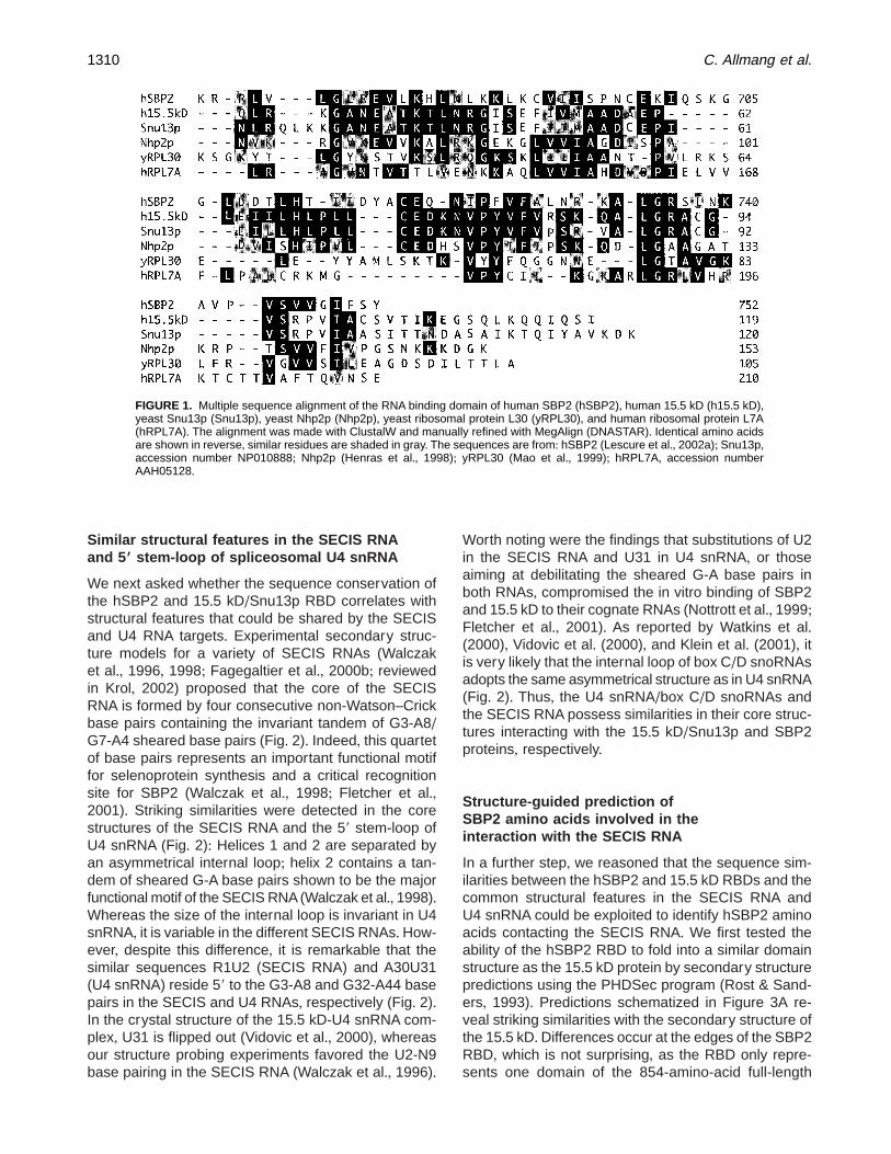
Similar structural features in the SECIS RNAand 59 stem-loop of spliceosomal U4 snRNA
We next asked whether the sequence conservation ofthe hSBP2 and 15+5 kD/Snu13p RBD correlates withstructural features that could be shared by the SECISand U4 RNA targets+ Experimental secondary struc-ture models for a variety of SECIS RNAs (Walczaket al+, 1996, 1998; Fagegaltier et al+, 2000b; reviewedin Krol, 2002) proposed that the core of the SECISRNA is formed by four consecutive non-Watson–Crickbase pairs containing the invariant tandem of G3-A8/G7-A4 sheared base pairs (Fig+ 2)+ Indeed, this quartetof base pairs represents an important functional motiffor selenoprotein synthesis and a critical recognitionsite for SBP2 (Walczak et al+, 1998; Fletcher et al+,2001)+ Striking similarities were detected in the corestructures of the SECIS RNA and the 59 stem-loop ofU4 snRNA (Fig+ 2): Helices 1 and 2 are separated byan asymmetrical internal loop; helix 2 contains a tan-dem of sheared G-A base pairs shown to be the majorfunctional motif of the SECIS RNA (Walczak et al+, 1998)+Whereas the size of the internal loop is invariant in U4snRNA, it is variable in the different SECIS RNAs+ How-ever, despite this difference, it is remarkable that thesimilar sequences R1U2 (SECIS RNA) and A30U31(U4 snRNA) reside 59 to the G3-A8 and G32-A44 basepairs in the SECIS and U4 RNAs, respectively (Fig+ 2)+In the crystal structure of the 15+5 kD-U4 snRNA com-plex, U31 is flipped out (Vidovic et al+, 2000), whereasour structure probing experiments favored the U2-N9base pairing in the SECIS RNA (Walczak et al+, 1996)+
Worth noting were the findings that substitutions of U2in the SECIS RNA and U31 in U4 snRNA, or thoseaiming at debilitating the sheared G-A base pairs inboth RNAs, compromised the in vitro binding of SBP2and 15+5 kD to their cognate RNAs (Nottrott et al+, 1999;Fletcher et al+, 2001)+ As reported by Watkins et al+(2000), Vidovic et al+ (2000), and Klein et al+ (2001), itis very likely that the internal loop of box C/D snoRNAsadopts the same asymmetrical structure as in U4 snRNA(Fig+ 2)+ Thus, the U4 snRNA/box C/D snoRNAs andthe SECIS RNA possess similarities in their core struc-tures interacting with the 15+5 kD/Snu13p and SBP2proteins, respectively+
Structure-guided prediction ofSBP2 amino acids involved in theinteraction with the SECIS RNA
In a further step, we reasoned that the sequence sim-ilarities between the hSBP2 and 15+5 kD RBDs and thecommon structural features in the SECIS RNA andU4 snRNA could be exploited to identify hSBP2 aminoacids contacting the SECIS RNA+ We first tested theability of the hSBP2 RBD to fold into a similar domainstructure as the 15+5 kD protein by secondary structurepredictions using the PHDSec program (Rost & Sand-ers, 1993)+ Predictions schematized in Figure 3A re-veal striking similarities with the secondary structure ofthe 15+5 kD+Differences occur at the edges of the SBP2RBD, which is not surprising, as the RBD only repre-sents one domain of the 854-amino-acid full-length
FIGURE 1. Multiple sequence alignment of the RNA binding domain of human SBP2 (hSBP2), human 15+5 kD (h15+5 kD),yeast Snu13p (Snu13p), yeast Nhp2p (Nhp2p), yeast ribosomal protein L30 (yRPL30), and human ribosomal protein L7A(hRPL7A)+ The alignment was made with ClustalW and manually refined with MegAlign (DNASTAR)+ Identical amino acidsare shown in reverse, similar residues are shaded in gray+ The sequences are from: hSBP2 (Lescure et al+, 2002a); Snu13p,accession number NP010888; Nhp2p (Henras et al+, 1998); yRPL30 (Mao et al+, 1999); hRPL7A, accession numberAAH05128+
1310 C. Allmang et al.
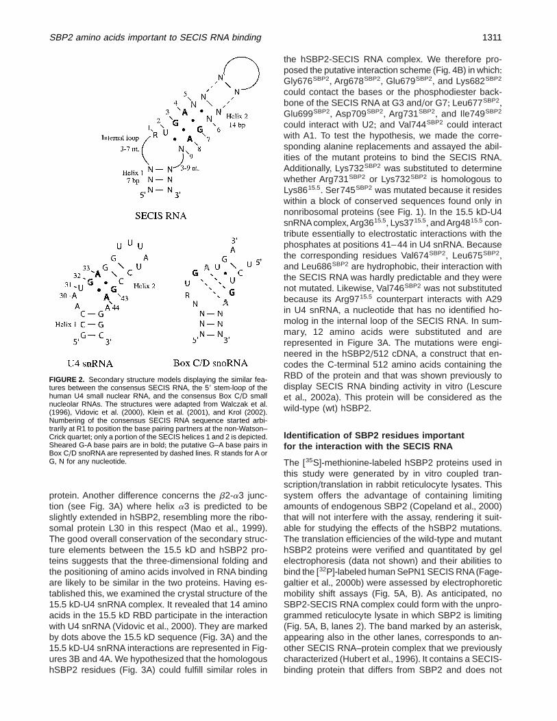
protein+ Another difference concerns the b2-a3 junc-tion (see Fig+ 3A) where helix a3 is predicted to beslightly extended in hSBP2, resembling more the ribo-somal protein L30 in this respect (Mao et al+, 1999)+The good overall conservation of the secondary struc-ture elements between the 15+5 kD and hSBP2 pro-teins suggests that the three-dimensional folding andthe positioning of amino acids involved in RNA bindingare likely to be similar in the two proteins+ Having es-tablished this, we examined the crystal structure of the15+5 kD-U4 snRNA complex+ It revealed that 14 aminoacids in the 15+5 kD RBD participate in the interactionwith U4 snRNA (Vidovic et al+, 2000)+ They are markedby dots above the 15+5 kD sequence (Fig+ 3A) and the15+5 kD-U4 snRNA interactions are represented in Fig-ures 3B and 4A+We hypothesized that the homologoushSBP2 residues (Fig+ 3A) could fulfill similar roles in
the hSBP2-SECIS RNA complex+ We therefore pro-posed the putative interaction scheme (Fig+ 4B) in which:Gly676SBP2, Arg678SBP2, Glu679SBP2, and Lys682SBP2
could contact the bases or the phosphodiester back-bone of the SECIS RNA at G3 and/or G7; Leu677SBP2,Glu699SBP2, Asp709SBP2, Arg731SBP2, and Ile749SBP2
could interact with U2; and Val744SBP2 could interactwith A1+ To test the hypothesis, we made the corre-sponding alanine replacements and assayed the abil-ities of the mutant proteins to bind the SECIS RNA+Additionally, Lys732SBP2 was substituted to determinewhether Arg731SBP2 or Lys732SBP2 is homologous toLys8615+5+ Ser745SBP2 was mutated because it resideswithin a block of conserved sequences found only innonribosomal proteins (see Fig+ 1)+ In the 15+5 kD-U4snRNA complex,Arg3615+5, Lys3715+5, and Arg4815+5 con-tribute essentially to electrostatic interactions with thephosphates at positions 41–44 in U4 snRNA+ Becausethe corresponding residues Val674SBP2, Leu675SBP2,and Leu686SBP2 are hydrophobic, their interaction withthe SECIS RNA was hardly predictable and they werenot mutated+ Likewise, Val746SBP2 was not substitutedbecause its Arg9715+5 counterpart interacts with A29in U4 snRNA, a nucleotide that has no identified ho-molog in the internal loop of the SECIS RNA+ In sum-mary, 12 amino acids were substituted and arerepresented in Figure 3A+ The mutations were engi-neered in the hSBP2/512 cDNA, a construct that en-codes the C-terminal 512 amino acids containing theRBD of the protein and that was shown previously todisplay SECIS RNA binding activity in vitro (Lescureet al+, 2002a)+ This protein will be considered as thewild-type (wt) hSBP2+
Identification of SBP2 residues importantfor the interaction with the SECIS RNA
The [35S]-methionine-labeled hSBP2 proteins used inthis study were generated by in vitro coupled tran-scription/translation in rabbit reticulocyte lysates+ Thissystem offers the advantage of containing limitingamounts of endogenous SBP2 (Copeland et al+, 2000)that will not interfere with the assay, rendering it suit-able for studying the effects of the hSBP2 mutations+The translation efficiencies of the wild-type and mutanthSBP2 proteins were verified and quantitated by gelelectrophoresis (data not shown) and their abilities tobind the [32P]-labeled human SePN1 SECIS RNA(Fage-galtier et al+, 2000b) were assessed by electrophoreticmobility shift assays (Fig+ 5A, B)+ As anticipated, noSBP2-SECIS RNA complex could form with the unpro-grammed reticulocyte lysate in which SBP2 is limiting(Fig+ 5A, B, lanes 2)+ The band marked by an asterisk,appearing also in the other lanes, corresponds to an-other SECIS RNA–protein complex that we previouslycharacterized (Hubert et al+, 1996)+ It contains a SECIS-binding protein that differs from SBP2 and does not
FIGURE 2. Secondary structure models displaying the similar fea-tures between the consensus SECIS RNA, the 59 stem-loop of thehuman U4 small nuclear RNA, and the consensus Box C/D smallnucleolar RNAs+ The structures were adapted from Walczak et al+(1996), Vidovic et al+ (2000), Klein et al+ (2001), and Krol (2002)+Numbering of the consensus SECIS RNA sequence started arbi-trarily at R1 to position the base pairing partners at the non-Watson–Crick quartet; only a portion of the SECIS helices 1 and 2 is depicted+Sheared G-A base pairs are in bold; the putative G–A base pairs inBox C/D snoRNA are represented by dashed lines+ R stands for A orG, N for any nucleotide+
SBP2 amino acids important to SECIS RNA binding 1311
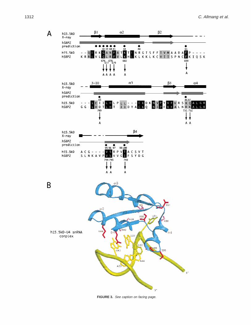
FIGURE 3. See caption on facing page.
1312 C. Allmang et al.
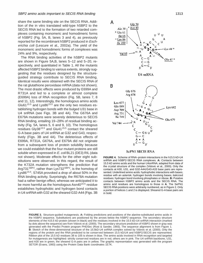
share the same binding site on the SECIS RNA+ Addi-tion of the in vitro translated wild-type hSBP2 to theSECIS RNA led to the formation of two retarded com-plexes containing monomeric and homodimeric formsof hSBP2 (Fig+ 5A, B, lanes 3 and 4), as previouslyreported for the recombinant hSBP2 produced in Esch-erichia coli (Lescure et al+, 2002a)+ The yield of themonomeric and homodimeric forms of complexes was24% and 9%, respectively+
The RNA binding activities of the hSBP2 mutantsare shown in Figure 5A,B, lanes 5–12 and 5–20, re-spectively, and quantitated in Table 1+ All the mutantsaffected hSBP2 binding to various extents, strongly sug-gesting that the residues designed by the structure-guided strategy contribute to SECIS RNA binding+Identical results were obtained with the SECIS RNA ofthe rat glutathione peroxidase mRNA (data not shown)+The most drastic effects were produced by E699A andR731A and led to a complete or almost complete(E699A) loss of RNA recognition (Fig+ 5B, lanes 7, 8and 11, 12)+ Interestingly, the homologous amino acidsGlu6115+5 and Lys8615+5 are the only two residues es-tablishing hydrogen bonds with the bulged U31 base inU4 snRNA (see Figs+ 3B and 4A)+ The G676A andE679A mutations were severely deleterious to SECISRNA binding, entailing 19–28% of residual binding ac-tivity (Fig+ 5A, lanes 5, 6 and 9, 10)+ The homologousresidues Gly3815+5 and Glu4115+5 contact the shearedG-A base pairs of U4 snRNA at G32 and G43, respec-tively (Figs+ 3B and 4A)+ The deleterious effects ofE699A, R731A, G676A, and E679A did not originatefrom a subsequent loss of protein solubility becausewe could establish that the four mutant proteins are stillsoluble when expressed in E. coli BL21 (DE3) RIL (datanot shown)+ Moderate effects for the other eight sub-stitutions were observed+ In this regard, the result ofthe K732A mutation strengthens the prediction thatArg731SBP2, rather than Lys732SBP2, is the homolog ofLys8615+5+ S745A provoked a drop of about 50% in theRNA binding activity+ Surprisingly, the R678A mutationhad a rather benign effect, whereas we anticipated it tobe more harmful as the homologous Asn4015+5 residueestablishes hydrophobic and hydrogen bond contactsin U4 snRNA with G32 at the sheared G32-A44 (Figs+ 3B
FIGURE 3. Structure-guided mutagenesis+ A: Folding predictions and positions of the alanine-substituted amino acids inthe hSBP2 sequence+ Substitutions are positioned by the arrows below the hSBP2 sequence+ The secondary structureelements of the h15+5 kD protein (shown in black) and the residues involved in the 15+5 kD-U4 snRNA interaction (markedby dots above the sequence) are from Vidovic et al+ (2000)+ The secondary structure prediction of hSBP2 shown in gray wasgenerated with the Predict Protein program PHDSec (Rost & Sander, 1993)+ The sequence alignment is from Figure 1+B: Sketch of the three-dimensional structure of the 15+5kD-U4 snRNA complex solved by Vidovic et al+ (2000)+ Only theregions of the protein and the RNA predicted to be conserved between 15+5 kD/U4 and hSBP2/SECIS are represented+Ribbon plot of the 15+5 kD residues 38 to 105 is shown in blue+ The amino acids involved in RNA recognition and targetedfor mutagenesis are highlighted: strictly conserved residues are in red, others are in pink+ The U4 snRNA backbone, U31,and A30 are in green, the sheared G-A pairs are in yellow+ The graphic representation was generated with the programSETOR (Evans, 1993) using the Protein Data Bank coordinates 1E7K+
FIGURE 4. Scheme of RNA–protein interactions in the h15+5 kD-U4snRNA and hSBP2-SECIS RNA complexes+ A: Contacts between15+5 kD amino acids and the human U4snRNA, adapted from data ofthe crystal structure of the complex (Vidovic et al+, 2000)+ Only thecontacts at A30, U31, and G32-A44/G43-A33 base pairs are repre-sented+Underlined amino acids: hydrophobic interactions with bases;residue with an asterisk: hydrogen bonds involving bases; italicizedresidues: hydrogen bond involving phosphates or ribose+ B: Putativecontacts between hSBP2 amino acids and the SECIS RNA+ Theamino acid residues are homologous to those in A+ The SePN1SECIS RNA positions were arbitrarily numbered, as in Figure 2+ Onlya portion of helices 1 and 2 is displayed+ Sheared G-A base pairs arein bold+
SBP2 amino acids important to SECIS RNA binding 1313
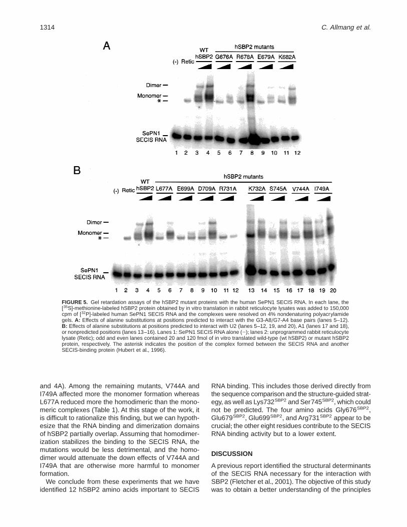
and 4A)+ Among the remaining mutants, V744A andI749A affected more the monomer formation whereasL677A reduced more the homodimeric than the mono-meric complexes (Table 1)+ At this stage of the work, itis difficult to rationalize this finding, but we can hypoth-esize that the RNA binding and dimerization domainsof hSBP2 partially overlap+ Assuming that homodimer-ization stabilizes the binding to the SECIS RNA, themutations would be less detrimental, and the homo-dimer would attenuate the down effects of V744A andI749A that are otherwise more harmful to monomerformation+
We conclude from these experiments that we haveidentified 12 hSBP2 amino acids important to SECIS
RNA binding+ This includes those derived directly fromthe sequence comparison and the structure-guided strat-egy, as well as Lys732SBP2 and Ser745SBP2,which couldnot be predicted+ The four amino acids Gly676SBP2,Glu679SBP2, Glu699SBP2, and Arg731SBP2 appear to becrucial; the other eight residues contribute to the SECISRNA binding activity but to a lower extent+
DISCUSSION
A previous report identified the structural determinantsof the SECIS RNA necessary for the interaction withSBP2 (Fletcher et al+, 2001)+ The objective of this studywas to obtain a better understanding of the principles
FIGURE 5. Gel retardation assays of the hSBP2 mutant proteins with the human SePN1 SECIS RNA+ In each lane, the[35S]-methionine-labeled hSBP2 protein obtained by in vitro translation in rabbit reticulocyte lysates was added to 150,000cpm of [32P]-labeled human SePN1 SECIS RNA and the complexes were resolved on 4% nondenaturing polyacrylamidegels+ A: Effects of alanine substitutions at positions predicted to interact with the G3-A8/G7-A4 base pairs (lanes 5–12)+B: Effects of alanine substitutions at positions predicted to interact with U2 (lanes 5–12, 19, and 20), A1 (lanes 17 and 18),or nonpredicted positions (lanes 13–16)+ Lanes 1: SePN1 SECIS RNA alone (2); lanes 2: unprogrammed rabbit reticulocytelysate (Retic); odd and even lanes contained 20 and 120 fmol of in vitro translated wild-type (wt hSBP2) or mutant hSBP2protein, respectively+ The asterisk indicates the position of the complex formed between the SECIS RNA and anotherSECIS-binding protein (Hubert et al+, 1996)+
1314 C. Allmang et al.

governing the SBP2-SECIS RNA interaction, in partic-ular the identification of the amino acids important forSECIS RNA recognition+ Three main findings emergedfrom our investigations+ The first one was the discoverythat SBP2 shares the same RBD as the mammalian15+5 kD protein (or the Snu13p ortholog in yeast) thatbinds the 59 stem-loop of the spliceosomal U4 snRNA+In corollary, the second finding concerned the similar-ities detected in the core structures of the SECIS RNAand U4 snRNA bound by the SBP2 and 15+5 kD pro-teins, respectively+ It is precisely these protein and RNAsimilar features, combined with secondary structure pre-diction of hSBP2 and the information from the crystalstructure of the U4 snRNA-15+5 kD complex (Vidovicet al+, 2000), that enabled the prediction of amino acidsin the human SBP2 protein that should be critical toSECIS RNA binding+ In the absence of a structuralmodel for the SBP2-SECIS RNA complex, the structure-guided strategy offers the advantage of targeting aminoacids that contribute to the interaction with the RNA,rather than those participating in the overall folding ofthe protein+ This was verified by in silico investigation ofthe 15+5 kD three-dimensional structure; indeed theside chains of the amino acids corresponding to thosemutated in hSBP2 do not establish intramolecular con-tacts important to the overall folding of the 15+5kD+Gly3815+5 (corresponding to Gly676SBP2) is particular inthat it adopts a conformation that is not allowed to anyother amino acid at the b1-a2 junction+ This invariantamino acid is thus important for folding the RBD, but isalso in close contact to the G-A pairs+ Assays of theRNA-binding activities of the hSBP2 mutants allowedthe identification of 12 amino acids whose substitu-tion led to a complete or partial loss of RNA binding+From this data, we inferred that the four amino acidsGly676SBP2, Glu679SBP2, Glu699SBP2, Arg731SBP2 are
primordial to the interaction and that the other eightparticipate in SECIS RNA binding, constituting the thirdfinding of this report+
Our structure-guided strategy allowed the identifica-tion of hSBP2 amino acids important for recognition ofthe SECIS RNA (Fig+ 4B)+Obviously, the detailed RNA–protein contacts cannot be predicted by this type ofstudy+However, solution structure probing of the SECISRNA and SECIS RNA-SBP2 complex (Walczak et al+,1996; Fletcher et al+, 2001), combined with the workpresented here, suggest the putative interaction scheme+Gly676SBP2 and Arg678SBP2 could interact with G3;Glu679SBP2 with G3 and G7; Lys682SBP2 with G7;Leu677SBP2,Glu699SBP2,Asp709SBP2,Arg731SBP2, andIle749SBP2 with U2; and Val744SBP2 with A1+ Interest-ingly, the solution structure of the complex between theyeast ribosomal protein L30 and its autoregulatory sitein the L30 mRNA was solved by NMR spectroscopy(Mao et al+, 1999)+ L30 binds to an internal loop con-stituted by a complex array of non-Watson–Crick basepairs whose three-dimensional structure differs fromthat of the U4 snRNA+ In this RNA–protein complex,it is remarkable that Gly26L30, which corresponds toGly3815+5 and Gly676SBP2 (see Fig+ 1), is central tothe interaction with the mRNA+ Substitution of theGly676SBP2 homologs in the rat SBP2 and 15+5 kDproteins led to detrimental effects as well (Nottrott et al+,1999; Copeland et al+, 2001), in good agreement withthe important role of this amino acid in the L7A/L30family for both the structure of the RNA binding domainand recognition of the sheared G-A base pairs+ Addi-tionally, important roles were established in the L30-mRNA complex for Tyr27L30 (corresponding to Ala3915+5
and Leu677SBP2) and Lys28L30 (corresponding toAsn4015+5 and Arg678SBP2)+ This is consistent with ourresults for Leu677SBP2+ However, our data did not sug-gest a major role for Arg678SBP2, highlighting subtlevariations in the RNA–protein recognition schemes+Wefound that substitution of Lys732SBP2 and Ser745SBP2
affected the SECIS RNA binding, although the 15+5 kDhomologous residues do not establish contacts withthe U4 snRNA (Vidovic et al+, 2000)+ It is unlikely thatthe mutations led to misfolding of hSBP2 because wecould verify in silico that the corresponding amino acidsGln8715+5 and Ser9615+5 do not establish intramolecularcontacts (Fig+ 3B)+ One explanation could arise fromthe intimate RNA structure of the core and its vicinitythat may not be strictly identical in both RNAs, partic-ularly at the asymmetrical loop+ Lys732SBP2 andSer745SBP2 could thus be involved in the specializationfor the SECIS RNA interaction+
The results of our mutagenesis study are in goodagreement with a previous work assaying mutant SECISRNAs for their ability to bind SBP2 (Fletcher et al+,2001)+ It was found that the U2C mutation, or the G3-A8/A4-G7 changes to A3-G8/G4-A7 or A3-A8/G4-A7,impaired formation of the SBP2-SECIS RNA complex+
TABLE 1 + Quantitation of the binding activities of the hSBP2 mutantproteins from the gels shown in Figure 5+a
hSBP2 Mutants Monomer Dimer
Wild-type 100 100E699A 13+5 0R731A 0 0G676A 19 16E679A 19 28L677A 57 27R678A 64 67K682A 56 78D709A 46 76K732A 57 46S745A 56 51V744A 51 100I749A 47 100
aThe percentage of monomeric and dimeric complexes was cal-culated as the ratio of the values obtained with the highest amount ofhSBP2 (even lanes in Fig+ 5A, B) to those of the wild-type monomericand dimeric complexes taken as 100% (lanes 4)+
SBP2 amino acids important to SECIS RNA binding 1315
This underscored the importance of U2 as well as ofG3 and G7 in the sheared G3-A8/G7-A4 base pairs+Altogether, the data presented may suggest that SBP2recognizes the SECIS RNA sheared G-A base pairs ina manner similar to the 15+5 kD protein in the 15+5kD-U4 snRNA complex+ It is worth mentioning that U31is flipped out in the crystal structure (Vidovic et al+,2000; Fig+ 3B) whereas structure probing of the SECISRNA proposed that U2 is not bulged out but ratherbase paired with U9 (Walczak et al+, 1996; see alsoFig+ 2)+ Indeed, one could envisage the unpairing of U2by an induced fit of the SECIS RNA upon SBP2 binding+
It was recently reported that U4 snRNA, RNase MRPRNA, human SRP 7SL RNA, and several ribosomalRNA regions contain a new secondary structure motifcalled the kink-turn, or K-turn motif (Klein et al+, 2001)+These authors proposed that the L30 mRNA bindingsite can also adopt the K-turn motif+ It is characterizedby an asymmetrical internal loop flanked by a regularhelix on one side and an irregular helix containingsheared G-A base pairs on the other (Fig+ 2)+ A kinkoccurs at the internal loop, causing a sharp turn in theRNA helix+ Interestingly, the K-turn in U4 snRNA andL30 mRNA is the binding site for the 15+5 kD/Snu13pand yeast L30 proteins, respectively+ It was proposedthat box C/D snoRNAs also contain a K-turn motif (Kleinet al+, 2001), in line with the binding of 15+5 kD/Snu13pto this type of snoRNA (Watkins et al+, 2000)+ Structureprobing data combined with computer modeling led toa three-dimensional structure model for the SECIS RNAwhere a kink occurs at the internal loop, showing theG-A base pairs well accessible to the solvent (Walczaket al+, 1996)+ Considering this particular structural fea-ture of the SECIS RNA and the binding of SBP2 at theG-A base pairs (Fletcher et al+, 2001), we speculatethat the SECIS RNA is another member of the RNAfamily containing a K-turn motif+
We observed the formation of hSBP2-SECIS RNAcomplexes containing monomeric and homodimericforms of hSBP2+ This observation is in line with ourearlier report using the recombinant hSBP2 protein pro-duced in E. coli (Lescure et al+, 2002a)+ Using glycerolgradient centrifugation, Copeland et al+ (2001) also ob-served homodimerization of the rat SBP2 protein+ Tak-ing into account that SBP2 binds not only the SECISRNA but also the 28S ribosomal RNA via a ribosome-binding domain located N-ter to the RBD, these au-thors hypothesized that homodimers could representthe functional form of SBP2+ It has not been reportedyet that other members of the L7A/L30 family possessthe capacity to homodimerize, but SBP2 could be uniquein this respect: it is a rather large protein (854 aminoacids for the full-length protein and 512 amino acids inthe hSBP2 fragment used in this study) compared tothe relatively small size of the 15+5 kD (128 aminoacids) and other proteins of the L7A/L30 family+ Actu-ally, homodimerization of RNA-binding proteins is not
unprecedented, and was already reported for RRM-containing proteins such as U1A, hnRNP A1, the Laautoantigen and eIF4B (reviewed in Méthot et al+, 1996;Craig et al+, 1997; Puglisi, 2000)+
There is a growing importance of functionally diverseeukaryotic proteins containing the L7A/L30 RBD+ Infact, this domain was recently renamed Pelota(Anantharaman et al+, 2002) after the name of a locusthat encodes a protein required for meiotic cell divisionin Drosophila (Eberhart & Wasserman, 1995)+ How-ever, our amino acid sequence alignment of the L7A/L30 family of proteins with Pelota orthologs indicatedthat the latter contain sequence similarity to only thefirst 28 amino acids (with respect to hSBP2) at theN-terminus of the L7A/L30 RBD (data not shown)+Therefore, the blocks of homology in the C-terminalhalf of the L7A/L30 domain (Fig+ 1), lacking in Pelotaproteins, may provide different binding opportunities+
Some of the members of the L7A/L30 protein familybind, or potentially bind, RNAs with K-turn motifs+ Wehave shown here that the 15+5 kD/Snu13p-U4 snRNAand SBP2-SECIS RNA complexes exhibit structural sim-ilarities, raising the question of how each protein canspecifically identify its cognate RNA+ This is especiallycrucial in light of the following recent reports addingevolutionary aspects to the issue+ It was found that thearchaeal ribosomal protein L7A possesses the otherfunction of binding the archaeal box C/D small RNAand mammalian box C/D snoRNAs+ The archaeal L7Aprotein is therefore the functional homolog to the eu-karyotic 15+5 kD/Snu13p (Kuhn et al+, 2002; Tang et al+,2002)+ Additionally, the Nhp2p protein that contains anL7A/L30 RBD and is a constituent of box H/ACAsnoRNAs can also bind box C/D snoRNAs in vitro (Hen-ras et al+, 2001)+ Distinct features in the structures ofeach K-turn-containing RNA can account for the spec-ificity of binding+ Another straightforward and not mu-tually exclusive possibility is that every L7A/L30 RBDcontains nonconserved amino acids, specifically dedi-cated to recognizing each individual RNA target+ Elu-cidation of this question represents the route for futureinvestigations+
MATERIALS AND METHODS
cDNA constructs and site-directedmutagenesis
To allow in vitro transcription/translation of hSBP2 wild-typeand mutant constructs, the hSBP2 cDNA was subcloned down-stream of the T7 promoter of pBluescript II KS (2)+ To do this,the 2+1 kb XbaI-HindIII fragment arising from phSBP2/512(Lescure et al+, 2002a), containing an N-terminal Strep-tag II(IBA, Germany) fused to the 512 C-terminal amino acids ofhSBP2, was inserted into pBluescript II KS (2)+ The resultingplasmid was termed pKS-hSBP2/512+ Alanine substitution
1316 C. Allmang et al.
mutants were generated in pKS-hSBP2/512 by site-directedmutagenesis+ Mutant constructs were entirely sequenced byautomated DNA sequencing+
Oligonucleotides used for mutagenesis were as follows:
G676A: 59-GAGAACCTCCCTCAAGGCCAACACAAGTCGACG-39;
L677A: 59-TTTGAGAACCTCCCTGGCCCCCAACACAAGTCG-39;
R678A: 59-GTGTTTGAGAACCTCGGCCAACCCCAACACAAG-39;
E679A: 59-CAGGTGTTTGAGAACGGCCCTCAACCCCAACAC-39;
K682A: 59-TTTGAGCTTCAGGTGGGCGAGAACCTCCCTCAA-39;
E699A: 59-TTTTGACTGTATCTTGGCACAGTTGGGAGAAAT-39;
D709A: 59-TAATTGTGTGCAAAGTGGCATCCAGCCCACCTTT-39;
R731A: 59-GCGCCCCAGAGCTTTGGCGTTGAGAGCAAACAC-39;
K732A: 59-ACTGCGCCCCAGAGCGGCGCGGTTGAGAGCAAA-39;
V744A: 59-GATCCCCACCACACTGGCAGGAACTGCCTTATT-39;
S745A: 59-GAAGATCCCCACCACGGCGACAGGAACTGCCTT-39;
I749A: 59-CCCATCATAGCTGAAGGCCCCCACCACACTGAC-39+
In vitro translation
Wild-type and mutant hSBP2 proteins were generated in vitrousing TNT coupled Reticulocyte Lysate Systems (Promega)+One microgram of each of the pKS-hSBP2/512 wild-type ormutant plasmid DNAs was used as the template in 50 mL invitro transcription/translation reactions in the presence of 25 mLrabbit reticulocyte lysate and 20 mCi of [35S]-methionine (1,175Ci/mmol)+ The yield of [35S]-methionine incorporation wasdetermined by 5% TCA precipitation of 2-mL aliquots of thereactions, followed by scintillation counting and calculationwith respect to the [35S]-methionine input+ Obtaining of thetranslation products was verified by electrophoresis on 12%SDS-PAGE+ The amount of each hSBP2 protein was quan-titated with a Fuji BioImage BAS2000 analyzer+ To assay thesolubility of the hSBP2 mutant proteins that affected SECISRNA binding, constructs were transformed into E. coli BL21(DE3) RIL+ After induction of protein synthesis, the solubleand insoluble fractions were loaded on SDS-PAGE and an-alyzed by western blotting using an anti-Strep-tag II antibody(IBA, Germany)+
Electrophoretic mobility shift assays
For in vitro transcription of the human SePN1 and rat GPxSECIS RNAs, plasmids pT7BcKSelN and pRGPxBcK werelinearized by EcoRI (Walczak et al+, 1998; Fagegaltier et al+,2000b)+ Internally labeled SePN1 and GPx SECIS RNAs wereobtained by T7 transcription with [a-32P]-ATP (3,000 Ci/mmol) according to Hubert et al+ (1996)+ Formation of theSePN1 SECIS RNA-hSBP2 and GPx SECIS RNA-hSBP2
complexes were conducted as described in Copeland et al+(2001) and Fletcher et al+ (2001)+ Routinely, 150,000 cpm(2+4 fmol) of 32P-labeled SECIS RNAwere incubated for 30 minat 30 8C with 20 or 120 fmol of in vitro translated wild-type ormutant hSBP2 protein, in 20 mL of phosphate buffer salinepH 7+4, 10 mM DTT+ RNA–protein complexes were sepa-rated by 4% nondenaturing polyacrylamide gel electropho-resis in Tris-glycine, pH 8 (Fletcher et al+, 2001)+ The intensitiesof free and bound RNAs were quantitated with a Fuji Bio-Image BAS2000 analyzer+Two independent experiments wereperformed for both SePN1 and GPx SECIS RNAs+ Quanti-tation of the results varied within 15%+
ACKNOWLEDGMENTS
We are grateful to V+ Cura for the graphical representationand help with in silico analysis+ A+ Lescure and E+ Myslinskiare thanked for helpful comments on the manuscript and C+Loegler for excellent technical assistance+ C+A+ was awardeda fellowship of the Fondation pour la Recherche Médicale+This work was supported by the Fondation pour la Recher-che Médicale, the Ligue Régionale contre le Cancer and theAssociation pour la Recherche contre le Cancer+
Received June 14, 2002; returned for revisionJune 28, 2002; revised manuscript received July 19, 2002
REFERENCES
Anantharaman V, Koonin EV, Aravind L+ 2002+ Comparative genom-ics and evolution of proteins involved in RNA metabolism+ NucleicAcids Res 30:1427–1464+
Berry MJ, Banu L, Chen YY, Mandel SJ, Kieffer JD, Harney JW,Larsen PR+ 1991+ Recognition of UGA as a selenocysteine codonin type 1 deiodinase requires sequences in the 39 terminal un-translated region+ Nature 353:273–276+
Copeland PR, Fletcher JE, Carlson BA, Hatfield DL, Driscoll DM+2000+ A novel RNA binding protein, SBP2, is required for thetranslation of mammalian selenoprotein mRNAs+ EMBO J 19:306–314+
Copeland PR, Stepanik VA, Driscoll DM+ 2001+ Insight into mamma-lian selenocysteine insertion:Domain structure and ribosome bind-ing properties of Sec insertion sequence Binding protein 2+ MolCell Biol 21:1491–1498+
Craig AWB, Svitkin YV, Lee HS, Belsham GJ, Sonenberg N+ 1997+The La autoantigen contains a dimerization domain that is es-sential for enhancing translation. Mol Cell Biol 17:163–169+
Eberhart CG, Wasserman SA+ 1995+ The pelota locus encodes aprotein required for meiotic cell division: An analysis of G2/Marrest in Drosophila spermatogenesis+ Development 121:3477–3486+
Evans SV+ 1993+ SETOR: Hardware-lighted three-dimensional solidmodel representations of macromolecules+ J Mol Graph 134:127–128+
Fagegaltier D, Carbon P, Krol A+ 2001+ Distinctive features in theSelB family of elongation factors for selenoprotein synthesis+ Aglimpse of an evolutionary complexified translation apparatus+BioFactors 14:5–10+
Fagegaltier D, Hubert N, Yamada K, Mizutani T, Carbon P, Krol A+2000a+ Characterization of mSelB, a novel mammalian elonga-tion factor for selenoprotein translation+ EMBO J 19:4796–4805+
Fagegaltier D, Lescure A,Walczak R, Carbon P, Krol A+ 2000b+ Struc-tural analysis of new local features in SECIS RNA hairpins+ Nu-cleic Acids Res 28:2679–2689+
Fletcher JE, Copeland PR, Driscoll DM, Krol A+ 2001+ The seleno-cysteine incorporation machinery: Interactions between theSECIS RNA and the SECIS-binding protein SBP2+ RNA 7:1442–1453+
SBP2 amino acids important to SECIS RNA binding 1317
Gladyshev VN, Kryukov GV+ 2001+ Evolution of selenocysteine-containing proteins: Significance of identification and functionalcharacterization of selenoproteins+ BioFactors 14:87–92+
Gottschalk A, Neubauer G, Banroques J, Mann M, Lührmann R,Fabrizio P+ 1999+ Identification by mass spectrometry and func-tional analysis of novel proteins of the yeast [U4/U6+U5] tri-snRNP+ EMBO J 18:4535–4548+
Grundner-Culemann E, Martin GW III, Harney JW, Berry MJ+ 1999+Two distinct SECIS structures capable of directing selenocys-teine incorporation in eukaryotes+ RNA 5:625–635+
Henras A, Dez C, Noaillac-Depeyre J, Henry Y, Caizergues-Ferrer M+2001+ Accumulation of H/ACA snoRNPs depends on the integrityof the conserved central domain of the RNA-binding protein Nhp2p+Nucleic Acids Res 29:2733–2746+
HenrasA,Henry Y,Bousquet-Antonelli C,Noaillac-Depeyre J,GélugneJ-P, Caizergues-Ferrer M+ 1998+ Nhp2p and Nop10p are essen-tial for the function of H/ACA snoRNPs+ EMBO J 17:7078–7090+
Hubert N, Walczak R, Carbon P, Krol A+ 1996+ A protein binds theselenocysteine insertion element in the 39 UTR of mammalianselenoprotein mRNAs+ Nucleic Acids Res 24:464–469+
Klein DJ, Schmeing TM, Moore PB, Steitz TA+ 2001+ The kink-turn:A new RNA secondary structure motif+ EMBO J 20:4214–4221+
Köhrle J, Brigelius-Flohé R, Böck A, Gärtner R, Meyer O, Flohé L+2000+ Selenium in biology: Facts and medical perspectives+ BiolChem 381:849–864+
Koonin EV, Bork P, Sander C+ 1994+ A novel RNA-binding motif inomnipotent suppressors of translation termination, ribosomal pro-teins and a ribosome modification enzyme? Nucleic Acids Res22:2166–2167+
Krol A+ 2002+ Evolutionarily different RNA motifs and RNA–proteincomplexes to achieve selenoprotein synthesis+Biochimie+ In press+
Kuhn JF, Tran EJ, Maxwell ES+ 2002+ Archaeal ribosomal protein L7is a functional homolog of the eukaryotic 15+5 kD/Snu13p snoRNPcore protein+ Nucleic Acids Res 30:931–941+
Lescure A, Allmang C, Yamada K, Carbon P, Krol A+ 2002a+ cDNAcloning, expression pattern and RNA binding analysis of humanselenocysteine insertion sequence (SECIS) binding protein 2+Gene 291:279–285+
Lescure A, Fagegaltier D, Carbon P, Krol A+ 2002b+ Protein factorsmediating selenoprotein synthesis+Curr Prot Pept Sci 3:143–151+
Low SC,Grundner-Culemann E,Harney JW, Berry MJ+ 2000+ SECIS-SBP2 interactions dictate selenocysteine incorporation efficiencyand selenoprotein hierarchy+ EMBO J 19:6882–6890+
Mao H,White SA,Williamson JR+ 1999+A novel loop-loop recognitionmotif in the yeast ribosomal protein L30 autoregulatory RNA com-plex+ Nature Struct Biol 6:1139–1147+
Martin GW III, Harney JW, Berry MJ+ 1998+ Functionality of muta-tions at conserved nucleotides in eukaryotic SECIS elements isdetermined by the identity of a single nonconserved nucleotide+RNA 4:65–73+
Méthot N, Song MS, Sonenberg N+ 1996+ A region rich in asparticacid, arginine, tyrosine, and glycine (DRYG) mediates eukaryoticInitiation Factor 4B (eIF4B) self-association and interaction witheIF3+ Mol Cell Biol 16:5328–5334+
Moghadaszadeh B, Petit N, Jaillard C, Brockington M, Quijano-RoyS, Merlini L, Romero N, Estournet B, Desguerre I, Chaigne D,Muntoni F, Topaloglu H, Guicheney P+ 2001+ Mutations in SEPN1cause congenital muscular dystrophy with spinal rigidity and re-strictive respiratory syndrome+ Nature Genet 29:17–18+
Nottrott S, Hartmuth K, Fabrizio P, Urlaub H, Vidovic I, Ficner R,Lührmann R+ 1999+ Functional interaction of a novel 15+5 kD[U4/U6+U5] tri-snRNP protein with the 59 stem-loop of U4 snRNA+EMBO J 18:6119–6133+
Pfeifer H, Conrad M, Roethlein D, Kyriakopoulos A, Brielmeier M,Bornkamm GW, Behne D+ 2001+ Identification of a specific spermnuclei selenoenzyme necessary for protamine thiol cross-linkingduring sperm maturation+ FASEB J 15:1236–1238+
Puglisi JD+ 2000+ mRNA processing: The 39 end justifies the means+Nature Struct Biol 7:263–264+
Rost B, Sander C+ 1993+ Prediction of protein secondary structure atbetter than 70% accuracy+ J Mol Biol 232:584–599+
Stevens SW, Abelson J+ 1999+ Purification of the yeast U4/U6+U5small nuclear ribonucleoprotein particle and identification of itsproteins+ Proc Natl Acad Sci USA 96:7226–7231+
Tang TH, Rozhdestvensky TS, Clouet d’Orval B, Bortolin M-L, HuberH, Charpentier B, Branlant C, Bachellerie J-P, Brosius J, Hütten-hofer A+ 2002+ RNomics in Archaea reveals a further link betweensplicing of archaeal introns and rRNA processing+ Nucleic AcidsRes 30:921–930+
Tujebajeva RM, Copeland PR, Xu XM, Carlson BA, Harney JW,Driscoll DM, Hatfield DL, Berry MJ+ 2000+ Decoding apparatus foreukaryotic selenocysteine insertion+ EMBO Rep 1:158–163+
Ursini F, Heim S, Kiess M, Maiorino M, Roveri A, Wissing J, Flohé L+1999+ Dual function of the selenoprotein PHGPx during spermmaturation+ Science 285:1393–1396+
Vidovic I, Nottrott S, Hartmuth K, Lührmann R, Ficner R+ 2000+ Crys-tal structure of the spliceosomal 15+5 kD protein bound to a U4snRNA fragment+ Mol Cell 6:1331–1342+
Walczak R, Carbon P, Krol A+ 1998+ An essential non-Watson–Crickbase pair motif in 39 UTR to mediate selenoprotein translation+RNA 4:74–84+
Walczak R, Westhof E, Carbon P, Krol A+ 1996+ A novel RNA struc-tural motif in the selenocysteine insertion element of eukaryoticselenoprotein mRNAs+ RNA 2:367–379+
Watkins NJ, Ségault V, Charpentier B, Nottrott S, Fabrizio P, Bachi A,Wilm M, Rosbash M, Branlant C, Lührmann R+ 2000+ A commoncore RNP structure shared between the small nucleolar box C/DRNPs and the spliceosomal U4 snRNP+ Cell 103:457–466+
1318 C. Allmang et al.

1868–1884 Nucleic Acids Research, 2007, Vol. 35, No. 6 Published online 1 March 2007doi:10.1093/nar/gkm066
An improved definition of the RNA-bindingspecificity of SECIS-binding protein 2,an essential component of theselenocysteine incorporation machineryA. Clery1, V. Bourguignon-Igel1, C. Allmang2, A. Krol2 and C. Branlant1,*
1Laboratoire de Maturation des ARN et Enzymologie Moleculaire – UMR 7567 CNRS-UHP, Nancy Universite,Faculte des Sciences et Techniques – BP 239, 54506 Vandoeuvre-les-Nancy Cedex, France and 2Architecture etReactivite de l’arN – CNRS-Universite Louis Pasteur, Institut de Biologie Moleculaire et Cellulaire 15 Rue ReneDescartes, 67084 Strasbourg Cedex, France
Received September 27, 2006; Revised January 20, 2007; Accepted January 22, 2007
ABSTRACT
By binding to SECIS elements located in the 30-UTRof selenoprotein mRNAs, the protein SBP2 playsa key role in the assembly of the selenocysteineincorporation machinery. SBP2 contains anL7Ae/L30 RNA-binding domain similar to that ofprotein 15.5K/Snu13p, which binds K-turn motifswith a 3-nt bulge loop closed by a tandem ofG.A and A.G pairs. Here, by SELEX experiments,we demonstrate the capacity of SBP2 to bind suchK-turn motifs with a protruding U residue. However,we show that conversion of the bulge loop intoan internal loop reinforces SBP2 affinity and toa greater extent RNP stability. Opposite variationswere found for Snu13p. Accordingly, footprintingassays revealed strong contacts of SBP2 withhelices I and II and the 50-strand of the internalloop, as opposed to the loose interaction of Snu13p.Our data also identifies new determinants for SBP2binding which are located in helix II. Among theL7Ae/L30 family members, these determinantsare unique to SBP2. Finally, in accordance withfunctional data on SECIS elements, the identity ofresidues at positions 2 and 3 in the loop influencesSBP2 affinity. Altogether, the data provide a veryprecise definition of the SBP2 RNA specificity.
INTRODUCTION
Based on ribosomal subunit 3D-structure analysis,K-turn motifs were found to be frequent protein-recognition motifs in ribosomal RNAs (1). A total of
8 K-turn motifs were detected in the 23S rRNA fromHaloarcula marismortui and the 16S rRNA from Thermusthermophilus (1–4). K-turn motifs are all characterized bya helix I-loop-helix II structure, and the formation of twonon-Watson–Crick base pairs (most frequently G.A andA.G) within the internal loop extends helix II (1,5). Due tothe stacking onto helix I or helix II of residues in theinternal loop, one of the RNA strand forms a sharp angle(1,5). Only one of the residues in the loop is projected outof the K-turn structure and is located in a pocket ofthe protein in RNA–protein complexes. In addition totheir presence in rRNAs, K-turn motifs are also found inthe U4 and U4atac spliceosomal snRNAs (5,6) andin the numerous C/D box snoRNAs (7), that guide20-O-methylation and cleavages in pre-ribosomal RNA(for review, 8). K-turn motifs were also recently foundin both C/D and H/ACA sRNAs, that guide rRNAmodifications in archaea (9–11). They are thus veryancient RNA-binding motifs. Both in eukarya and inarchaea, small RNAs containing K-turn motifs assembleinto RNP particles and the K-turn motifs play a centralrole in protein assembly (7,9–15). More specifically,the ribosomal L7Ae protein in archaea or its eukaryalhomolog, the Snu13p (yeast)/15.5K (human) protein,first recognizes the K-turn structure and the complexformed then serves as a platform for assembly of the otherproteins (9,10,12–19).
The Snu13p/15.5K and L7Ae proteins belong tothe L7Ae/L30 protein family, which is characterized bythe presence of an L7Ae/L30 RNA-binding domain(6,20). The founding member of this protein family, theyeast L30 ribosomal protein recognizes a peculiar K-turnmotif in its own pre-mRNA (21–23). One differencebetween the yeast L30 RNA–protein complex, and theSnu13p/15.5K or L7Ae RNA–protein complexes is the
*To whom the correspondence should be addressed. Tel: 33 383684303; Fax: 33 383684307; Email: [email protected]
� 2007 The Author(s)
This is an Open Access article distributed under the terms of the Creative Commons Attribution Non-Commercial License (http://creativecommons.org/licenses/
by-nc/2.0/uk/) which permits unrestricted non-commercial use, distribution, and reproduction in any medium, provided the original work is properly cited.
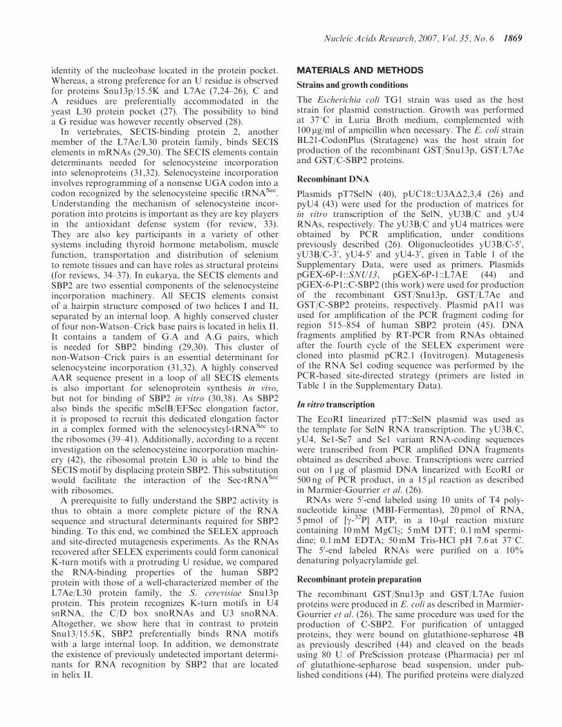
identity of the nucleobase located in the protein pocket.Whereas, a strong preference for an U residue is observedfor proteins Snu13p/15.5K and L7Ae (7,24–26), C andA residues are preferentially accommodated in theyeast L30 protein pocket (27). The possibility to binda G residue was however recently observed (28).
In vertebrates, SECIS-binding protein 2, anothermember of the L7Ae/L30 protein family, binds SECISelements in mRNAs (29,30). The SECIS elements containdeterminants needed for selenocysteine incorporationinto selenoproteins (31,32). Selenocysteine incorporationinvolves reprogramming of a nonsense UGA codon into acodon recognized by the selenocysteine specific tRNASec.Understanding the mechanism of selenocysteine incor-poration into proteins is important as they are key playersin the antioxidant defense system (for review, 33).They are also key participants in a variety of othersystems including thyroid hormone metabolism, musclefunction, transportation and distribution of seleniumto remote tissues and can have roles as structural proteins(for reviews, 34–37). In eukarya, the SECIS elements andSBP2 are two essential components of the selenocysteineincorporation machinery. All SECIS elements consistof a hairpin structure composed of two helices I and II,separated by an internal loop. A highly conserved clusterof four non-Watson–Crick base pairs is located in helix II.It contains a tandem of G.A and A.G pairs, whichis needed for SBP2 binding (29,30). This cluster ofnon-Watson–Crick pairs is an essential determinant forselenocysteine incorporation (31,32). A highly conservedAAR sequence present in a loop of all SECIS elementsis also important for selenoprotein synthesis in vivo,but not for binding of SBP2 in vitro (30,38). As SBP2also binds the specific mSelB/EFSec elongation factor,it is proposed to recruit this dedicated elongation factorin a complex formed with the selenocysteyl-tRNASec tothe ribosomes (39–41). Additionally, according to a recentinvestigation on the selenocysteine incorporation machin-ery (42), the ribosomal protein L30 is able to bind theSECIS motif by displacing protein SBP2. This substitutionwould facilitate the interaction of the Sec-tRNASec
with ribosomes.A prerequisite to fully understand the SBP2 activity is
thus to obtain a more complete picture of the RNAsequence and structural determinants required for SBP2binding. To this end, we combined the SELEX approachand site-directed mutagenesis experiments. As the RNAsrecovered after SELEX experiments could form canonicalK-turn motifs with a protruding U residue, we comparedthe RNA-binding properties of the human SBP2protein with those of a well-characterized member of theL7Ae/L30 protein family, the S. cerevisiae Snu13pprotein. This protein recognizes K-turn motifs in U4snRNA, the C/D box snoRNAs and U3 snoRNA.Altogether, we show here that in contrast to proteinSnu13/15.5K, SBP2 preferentially binds RNA motifswith a large internal loop. In addition, we demonstratethe existence of previously undetected important determi-nants for RNA recognition by SBP2 that are locatedin helix II.
MATERIALS AND METHODS
Strains and growth conditions
The Escherichia coli TG1 strain was used as the hoststrain for plasmid construction. Growth was performedat 378C in Luria Broth medium, complemented with100 mg/ml of ampicillin when necessary. The E. coli strainBL21-CodonPlus (Stratagene) was the host strain forproduction of the recombinant GST/Snu13p, GST/L7Aeand GST/C-SBP2 proteins.
Recombinant DNA
Plasmids pT7SelN (40), pUC18::U3A�2,3,4 (26) andpyU4 (43) were used for the production of matrices forin vitro transcription of the SelN, yU3B/C and yU4RNAs, respectively. The yU3B/C and yU4 matrices wereobtained by PCR amplification, under conditionspreviously described (26). Oligonucleotides yU3B/C-50,yU3B/C-30, yU4-50 and yU4-30, given in Table 1 of theSupplementary Data, were used as primers. PlasmidspGEX-6P-1::SNU13, pGEX-6P-1::L7AE (44) andpGEX-6-P1::C-SBP2 (this work) were used for productionof the recombinant GST/Snu13p, GST/L7Ae andGST/C-SBP2 proteins, respectively. Plasmid pA11 wasused for amplification of the PCR fragment coding forregion 515–854 of human SBP2 protein (45). DNAfragments amplified by RT-PCR from RNAs obtainedafter the fourth cycle of the SELEX experiment werecloned into plasmid pCR2.1 (Invitrogen). Mutagenesisof the RNA Se1 coding sequence was performed by thePCR-based site-directed strategy (primers are listed inTable 1 in the Supplementary Data).
In vitro transcription
The EcoRI linearized pT7::SelN plasmid was used asthe template for SelN RNA transcription. The yU3B/C,yU4, Se1-Se7 and Se1 variant RNA-coding sequenceswere transcribed from PCR amplified DNA fragmentsobtained as described above. Transcriptions were carriedout on 1 mg of plasmid DNA linearized with EcoRI or500 ng of PCR product, in a 15 ml reaction as describedin Marmier-Gourrier et al. (26).RNAs were 50-end labeled using 10 units of T4 poly-
nucleotide kinase (MBI-Fermentas), 20 pmol of RNA,5 pmol of [g-32P] ATP, in a 10-ml reaction mixturecontaining 10mM MgCl2; 5mM DTT; 0.1mM spermi-dine; 0.1mM EDTA; 50mM Tris-HCl pH 7.6 at 378C.The 50-end labeled RNAs were purified on a 10%denaturing polyacrylamide gel.
Recombinant protein preparation
The recombinant GST/Snu13p and GST/L7Ae fusionproteins were produced in E. coli as described in Marmier-Gourrier et al. (26). The same procedure was used for theproduction of C-SBP2. For purification of untaggedproteins, they were bound on glutathione-sepharose 4Bas previously described (44) and cleaved on the beadsusing 80 U of PreScission protease (Pharmacia) per mlof glutathione-sepharose bead suspension, under pub-lished conditions (44). The purified proteins were dialyzed
Nucleic Acids Research, 2007, Vol. 35, No. 6 1869
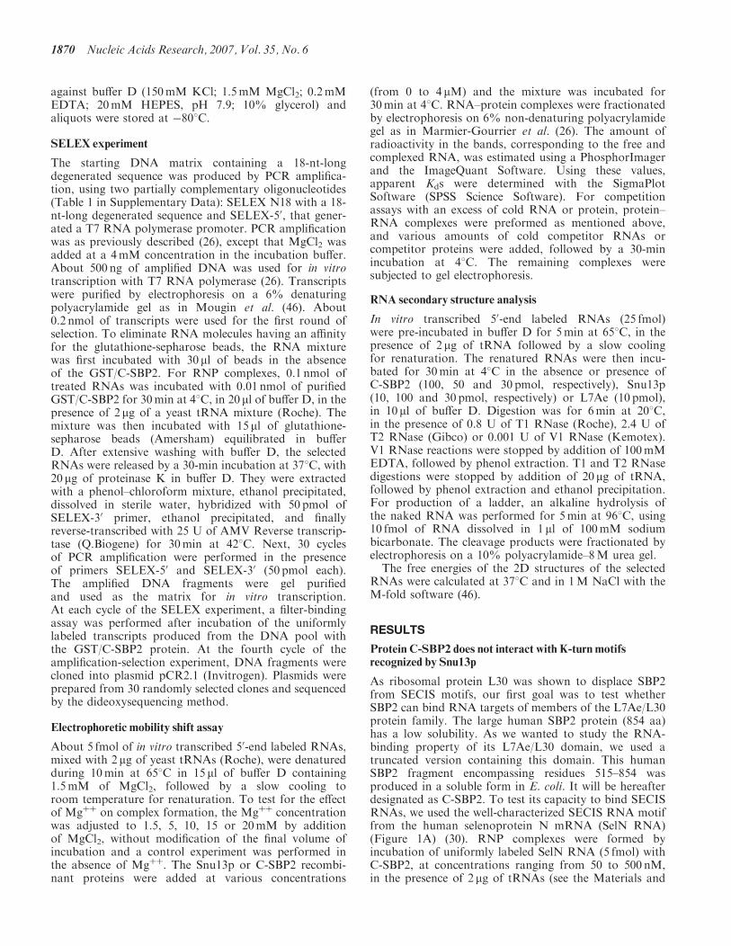
against buffer D (150mM KCl; 1.5mM MgCl2; 0.2mMEDTA; 20mM HEPES, pH 7.9; 10% glycerol) andaliquots were stored at �808C.
SELEX experiment
The starting DNA matrix containing a 18-nt-longdegenerated sequence was produced by PCR amplifica-tion, using two partially complementary oligonucleotides(Table 1 in Supplementary Data): SELEX N18 with a 18-nt-long degenerated sequence and SELEX-50, that gener-ated a T7 RNA polymerase promoter. PCR amplificationwas as previously described (26), except that MgCl2 wasadded at a 4mM concentration in the incubation buffer.About 500 ng of amplified DNA was used for in vitrotranscription with T7 RNA polymerase (26). Transcriptswere purified by electrophoresis on a 6% denaturingpolyacrylamide gel as in Mougin et al. (46). About0.2 nmol of transcripts were used for the first round ofselection. To eliminate RNA molecules having an affinityfor the glutathione-sepharose beads, the RNA mixturewas first incubated with 30 ml of beads in the absenceof the GST/C-SBP2. For RNP complexes, 0.1 nmol oftreated RNAs was incubated with 0.01 nmol of purifiedGST/C-SBP2 for 30min at 48C, in 20 ml of buffer D, in thepresence of 2 mg of a yeast tRNA mixture (Roche). Themixture was then incubated with 15 ml of glutathione-sepharose beads (Amersham) equilibrated in bufferD. After extensive washing with buffer D, the selectedRNAs were released by a 30-min incubation at 378C, with20 mg of proteinase K in buffer D. They were extractedwith a phenol–chloroform mixture, ethanol precipitated,dissolved in sterile water, hybridized with 50 pmol ofSELEX-30 primer, ethanol precipitated, and finallyreverse-transcribed with 25 U of AMV Reverse transcrip-tase (Q.Biogene) for 30min at 428C. Next, 30 cyclesof PCR amplification were performed in the presenceof primers SELEX-50 and SELEX-30 (50 pmol each).The amplified DNA fragments were gel purifiedand used as the matrix for in vitro transcription.At each cycle of the SELEX experiment, a filter-bindingassay was performed after incubation of the uniformlylabeled transcripts produced from the DNA pool withthe GST/C-SBP2 protein. At the fourth cycle of theamplification-selection experiment, DNA fragments werecloned into plasmid pCR2.1 (Invitrogen). Plasmids wereprepared from 30 randomly selected clones and sequencedby the dideoxysequencing method.
Electrophoretic mobility shift assay
About 5 fmol of in vitro transcribed 50-end labeled RNAs,mixed with 2 mg of yeast tRNAs (Roche), were denaturedduring 10min at 658C in 15 ml of buffer D containing1.5mM of MgCl2, followed by a slow cooling toroom temperature for renaturation. To test for the effectof Mgþþ on complex formation, the Mgþþ concentrationwas adjusted to 1.5, 5, 10, 15 or 20mM by additionof MgCl2, without modification of the final volume ofincubation and a control experiment was performed inthe absence of Mgþþ. The Snu13p or C-SBP2 recombi-nant proteins were added at various concentrations
(from 0 to 4 mM) and the mixture was incubated for30min at 48C. RNA–protein complexes were fractionatedby electrophoresis on 6% non-denaturing polyacrylamidegel as in Marmier-Gourrier et al. (26). The amount ofradioactivity in the bands, corresponding to the free andcomplexed RNA, was estimated using a PhosphorImagerand the ImageQuant Software. Using these values,apparent Kds were determined with the SigmaPlotSoftware (SPSS Science Software). For competitionassays with an excess of cold RNA or protein, protein–RNA complexes were preformed as mentioned above,and various amounts of cold competitor RNAs orcompetitor proteins were added, followed by a 30-minincubation at 48C. The remaining complexes weresubjected to gel electrophoresis.
RNA secondary structure analysis
In vitro transcribed 50-end labeled RNAs (25 fmol)were pre-incubated in buffer D for 5min at 658C, in thepresence of 2 mg of tRNA followed by a slow coolingfor renaturation. The renatured RNAs were then incu-bated for 30min at 48C in the absence or presence ofC-SBP2 (100, 50 and 30 pmol, respectively), Snu13p(10, 100 and 30 pmol, respectively) or L7Ae (10 pmol),in 10 ml of buffer D. Digestion was for 6min at 208C,in the presence of 0.8 U of T1 RNase (Roche), 2.4 U ofT2 RNase (Gibco) or 0.001 U of V1 RNase (Kemotex).V1 RNase reactions were stopped by addition of 100mMEDTA, followed by phenol extraction. T1 and T2 RNasedigestions were stopped by addition of 20 mg of tRNA,followed by phenol extraction and ethanol precipitation.For production of a ladder, an alkaline hydrolysis ofthe naked RNA was performed for 5min at 968C, using10 fmol of RNA dissolved in 1 ml of 100mM sodiumbicarbonate. The cleavage products were fractionated byelectrophoresis on a 10% polyacrylamide–8M urea gel.
The free energies of the 2D structures of the selectedRNAs were calculated at 378C and in 1M NaCl with theM-fold software (46).
RESULTS
Protein C-SBP2 does not interact with K-turn motifsrecognized by Snu13p
As ribosomal protein L30 was shown to displace SBP2from SECIS motifs, our first goal was to test whetherSBP2 can bind RNA targets of members of the L7Ae/L30protein family. The large human SBP2 protein (854 aa)has a low solubility. As we wanted to study the RNA-binding property of its L7Ae/L30 domain, we used atruncated version containing this domain. This humanSBP2 fragment encompassing residues 515–854 wasproduced in a soluble form in E. coli. It will be hereafterdesignated as C-SBP2. To test its capacity to bind SECISRNAs, we used the well-characterized SECIS RNA motiffrom the human selenoprotein N mRNA (SelN RNA)(Figure 1A) (30). RNP complexes were formed byincubation of uniformly labeled SelN RNA (5 fmol) withC-SBP2, at concentrations ranging from 50 to 500 nM,in the presence of 2 mg of tRNAs (see the Materials and
1870 Nucleic Acids Research, 2007, Vol. 35, No. 6
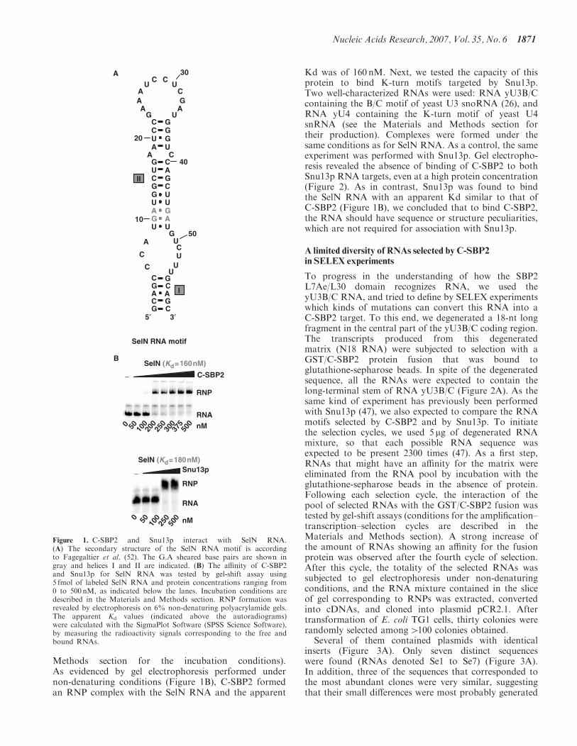
Methods section for the incubation conditions).As evidenced by gel electrophoresis performed undernon-denaturing conditions (Figure 1B), C-SBP2 formedan RNP complex with the SelN RNA and the apparent
Kd was of 160 nM. Next, we tested the capacity of thisprotein to bind K-turn motifs targeted by Snu13p.Two well-characterized RNAs were used: RNA yU3B/Ccontaining the B/C motif of yeast U3 snoRNA (26), andRNA yU4 containing the K-turn motif of yeast U4snRNA (see the Materials and Methods section fortheir production). Complexes were formed under thesame conditions as for SelN RNA. As a control, the sameexperiment was performed with Snu13p. Gel electropho-resis revealed the absence of binding of C-SBP2 to bothSnu13p RNA targets, even at a high protein concentration(Figure 2). As in contrast, Snu13p was found to bindthe SelN RNA with an apparent Kd similar to that ofC-SBP2 (Figure 1B), we concluded that to bind C-SBP2,the RNA should have sequence or structure peculiarities,which are not required for association with Snu13p.
A limited diversity of RNAs selected by C-SBP2in SELEX experiments
To progress in the understanding of how the SBP2L7Ae/L30 domain recognizes RNA, we used theyU3B/C RNA, and tried to define by SELEX experimentswhich kinds of mutations can convert this RNA into aC-SBP2 target. To this end, we degenerated a 18-nt longfragment in the central part of the yU3B/C coding region.The transcripts produced from this degeneratedmatrix (N18 RNA) were subjected to selection with aGST/C-SBP2 protein fusion that was bound toglutathione-sepharose beads. In spite of the degeneratedsequence, all the RNAs were expected to contain thelong-terminal stem of RNA yU3B/C (Figure 2A). As thesame kind of experiment has previously been performedwith Snu13p (47), we also expected to compare the RNAmotifs selected by C-SBP2 and by Snu13p. To initiatethe selection cycles, we used 5 mg of degenerated RNAmixture, so that each possible RNA sequence wasexpected to be present 2300 times (47). As a first step,RNAs that might have an affinity for the matrix wereeliminated from the RNA pool by incubation with theglutathione-sepharose beads in the absence of protein.Following each selection cycle, the interaction of thepool of selected RNAs with the GST/C-SBP2 fusion wastested by gel-shift assays (conditions for the amplification–transcription–selection cycles are described in theMaterials and Methods section). A strong increase ofthe amount of RNAs showing an affinity for the fusionprotein was observed after the fourth cycle of selection.After this cycle, the totality of the selected RNAs wassubjected to gel electrophoresis under non-denaturingconditions, and the RNA mixture contained in the sliceof gel corresponding to RNPs was extracted, convertedinto cDNAs, and cloned into plasmid pCR2.1. Aftertransformation of E. coli TG1 cells, thirty colonies wererandomly selected among4100 colonies obtained.Several of them contained plasmids with identical
inserts (Figure 3A). Only seven distinct sequenceswere found (RNAs denoted Se1 to Se7) (Figure 3A).In addition, three of the sequences that corresponded tothe most abundant clones were very similar, suggestingthat their small differences were most probably generated
RNP
RNA
C-SBP2 −
0 50 100
200
250
300
375
500 nM
SelN (Kd=160nM)
SelN RNA motif
I
II
C
C
5′
C
C
C
C
C
C
CC
C
A
C
G
G
G
G
G
G
G
G
GG
G
G
A
A
A
A
AA
A
AA
U
U
U
U
U
U
U
UU
U
U
U
U
U
U
C
C
C
CG
G
GG
A A
3′
10
20
30
40
50
A
B
RNP
RNA
Snu13p−
0 50 100
250
500 nM
SelN (Kd=180nM)
Figure 1. C-SBP2 and Snu13p interact with SelN RNA.(A) The secondary structure of the SelN RNA motif is accordingto Fagegaltier et al. (52). The G.A sheared base pairs are shown ingray and helices I and II are indicated. (B) The affinity of C-SBP2and Snu13p for SelN RNA was tested by gel-shift assay using5 fmol of labeled SelN RNA and protein concentrations ranging from0 to 500 nM, as indicated below the lanes. Incubation conditions aredescribed in the Materials and Methods section. RNP formation wasrevealed by electrophoresis on 6% non-denaturing polyacrylamide gels.The apparent Kd values (indicated above the autoradiograms)were calculated with the SigmaPlot Software (SPSS Science Software),by measuring the radioactivity signals corresponding to the free andbound RNAs.
Nucleic Acids Research, 2007, Vol. 35, No. 6 1871
by RT-PCR errors in the course of the amplification.Therefore, only four main classes of Se RNAs wereselected in the experiment (Figure 3A). This limiteddiversity of the selected sequences was one major differ-ence, compared to the SELEX experiment performed withSnu13p (31 very different sequences were obtained startingfrom the same initial RNA mixture). This suggested theexistence of strong sequence and/or structural constraintsfor SBP2 recognition. Three of the less frequent Se RNAshad a different length as compared to the initial RNAs:RNA Se2 (Figure 3A) contained an additional residue inthe degenerated sequence, whereas RNA Se1 and Se3lacked two residues compared to the initial RNAs.In addition, all the Se RNAs had a G instead of aU residue at position 38 in the conserved sequence.The same U38G base substitution was also found inseveral of the RNAs selected by the GST/Snu13p protein(47). By using gel-shift experiments, we verified thatthe seven distinct Se RNAs, that were selected, showedan affinity for the untagged C-SBP2 protein (Figure 3C).A wide range of apparent Kd values was observed(from 500 nM to42000 nM) (Figure 3C).
The selected RNAs all form canonical K-turn motifs
In order to understand the structural reasons for thesedifferent affinities, the possible folding of the sevenselected RNAs was investigated. Remarkably, each ofthem could form a canonical K-turn structure withtandem G.A and A.G base pairs and a 3-nt bulgeincluding a U residue at position 3. Most of the proposedstructures were verified by enzymatic probing (Figure 4A).
They are represented in Figure 4B, where they areclassified according to the values of the establishedapparent Kds. The free energies of the proposed 2Dstructures at 378C in 1M NaCl were also calculated byusing the M-Fold software. Based on these structures,nts 17–21 and 38–39 correspond to residues 1–5 and 6-7of the K-turn motif. Hence, residues 1 and 2 in the bulge,the A residue of the first G.A pair in stem II and oneU residue of the third pair in this stem correspondedto invariant residues in the starting RNA mixture.The G residue of the A.G pair corresponded to theabove-mentioned U to G mutation at position 38 inthe constant region. This G residue might have beengenerated by misincorporation in the course of theamplification cycles. Its selection in all the RNAs isin agreement with the high functional importance ofthe A.G pair in K-turn formation. All the selected RNAshad an identical UGAU sequence from position 19 to 22in the randomized segment, which demonstrated a strongpressure for the selection of a perfectly canonical K-turnstructure with two A.G and G.A pairs, and a U residue atposition 3 in the bulge. In all the selected RNAs, exceptRNA Se3 which contains a G.U pair, the constant U37
residue was always facing a U or a C residue in helix II.Interestingly, a U.U pair was almost always selected atthis position of helix II in the SELEX experimentperformed with Snu13p (47). Requirement of a non-Watson–Crick pair on top of the G.A and A.G pairsfor binding of C-SBP2 may explain the absence of bindingof C-SBP2 to yU4 RNA which has a G–C pair at thisposition in helix II (Figure 2B). Finally, in all the selectedSe RNAs, helix II contained at least two Watson–Crick
1000
1250
1500
1750
2000
RNA
C-SBP2−
025
050
075
0 nM25
00
yU3B/C + C-SBP2
RNA
C-SBP2
nM0
250
500
75010
0012
5015
0017
5020
00
−yU4 + C-SBP2A
RNA
Snu13p−
0 50 100
250
500
750
1000
1500
2000 nM
RNP
yU3B/C + Snu13p (Kd=230nM)
RNA
Snu13p−
0 50 100
250
500
750
1000 nM
RNP
yU4 + Snu13p (Kd=230nM)
AAC
G
G
A
AG
UG
A C
U C
G
G
G
U
ACU
U
A
AU
10
40C
C
AU
GG
CC
UG
G
A
A U
30
A
BC
12
543
67
I
II
yU3B/C
3′
U
C CG
AA
U
CA G
U
GAG
20
50
5′
A U
CCG
GG
10
20
U
A
UG
AU
UG
GU
C
C
A
C
I
U
CUA
AG
AG
UG
A
GC
G
A GU
U
ACU
C
12
54
3 6
7
II
G
3′U GUG UG A UUA UA A
50
30
40
5′
B
yU4
G
Figure 2. C-SBP2 does not interact with yU3B/C and yU4 RNAs. The binding of C-SBP2 and Snu13p on yU3B/C or yU4 RNAs was tested bygel-shift assays. The secondary structures of yU3B/C (Panel A) and yU4 (Panel B) are according to Marmier-Gourrier et al. (26) and Mougin et al.(43), respectively. The G.A sheared pairs are in gray and helices I and II are indicated. Nucleotides involved in the K-turn folding are numbered from1 to 7. Complexes were formed between 5 fmol of uniformly labeled yU3B/C (Panel A) or yU4 (Panel B) RNAs, and C-SBP2 or Snu13p atconcentrations ranging from 0 to 2500 nM, as indicated below the lanes. Incubation conditions were as described in the Materials and Methodssection. The autoradiograms obtained after electrophoresis on 6% polyacrylamide gels are shown. For the Snu13p–RNP complexes shown ascontrols, the apparent Kd values were calculated as described in the Materials and Methods section.
1872 Nucleic Acids Research, 2007, Vol. 35, No. 6
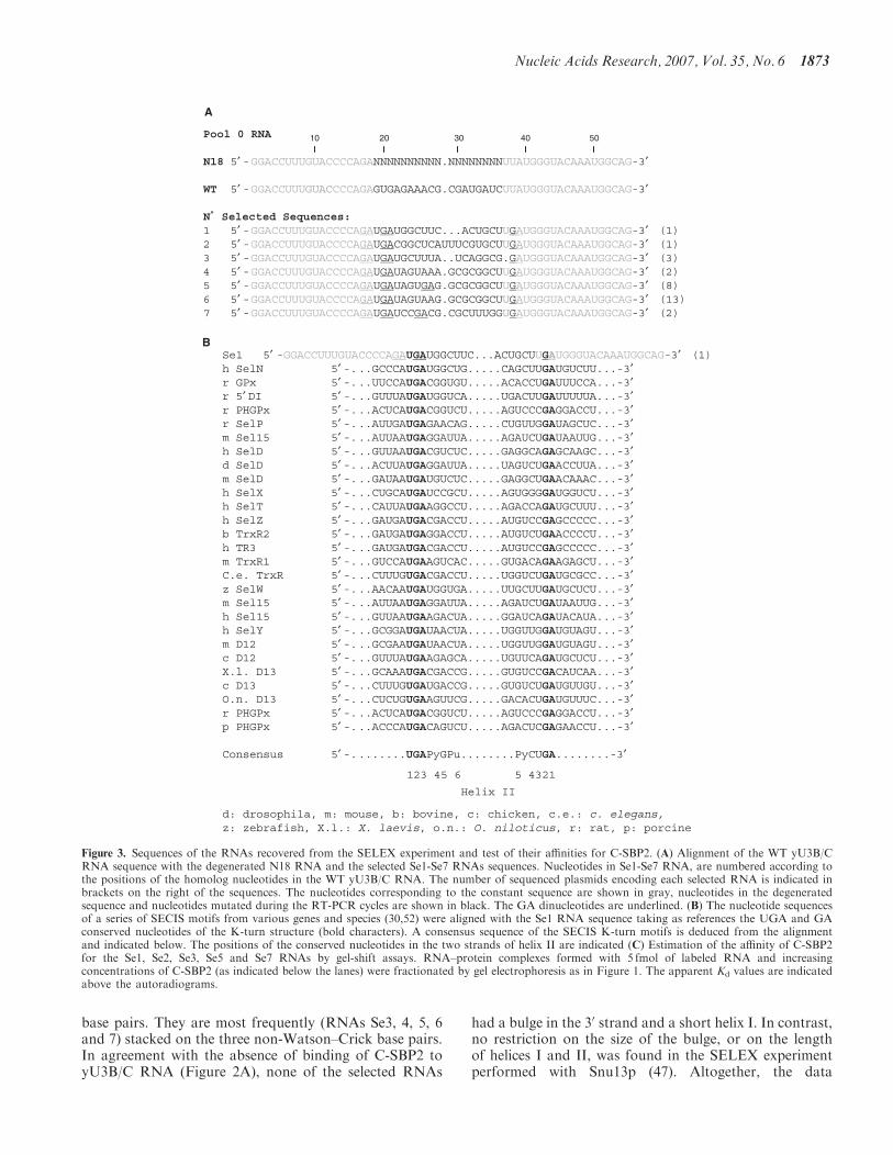
base pairs. They are most frequently (RNAs Se3, 4, 5, 6and 7) stacked on the three non-Watson–Crick base pairs.In agreement with the absence of binding of C-SBP2 toyU3B/C RNA (Figure 2A), none of the selected RNAs
had a bulge in the 30 strand and a short helix I. In contrast,no restriction on the size of the bulge, or on the lengthof helices I and II, was found in the SELEX experimentperformed with Snu13p (47). Altogether, the data
Pool 0 RNA
N18 5′ -GGACCUUUGUACCCCAGANNNNNNNNNN.NNNNNNNNUUAUGGGUACAAAUGGCAG-3′
WT 5′ -GGACCUUUGUACCCCAGAGUGAGAAACG.CGAUGAUCUUAUGGGUACAAAUGGCAG-3′
N˚ Selected Sequences:1 5′ -GGACCUUUGUACCCCAGAUGAUGGCUUC...ACUGCUUGAUGGGUACAAAUGGCAG-3′ (1)2 5′ -GGACCUUUGUACCCCAGAUGACGGCUCAUUUCGUGCUUGAUGGGUACAAAUGGCAG-3′ (1)3 5′ -GGACCUUUGUACCCCAGAUGAUGCUUUA..UCAGGCG.GAUGGGUACAAAUGGCAG-3′ (3)4 5′ -GGACCUUUGUACCCCAGAUGAUAGUAAA.GCGCGGCUUGAUGGGUACAAAUGGCAG-3′ (2)5 5′ -GGACCUUUGUACCCCAGAUGAUAGUGAG.GCGCGGCUUGAUGGGUACAAAUGGCAG-3′ (8)6 5′ -GGACCUUUGUACCCCAGAUGAUAGUAAG.GCGCGGCUUGAUGGGUACAAAUGGCAG-3′ (13)7 5′ -GGACCUUUGUACCCCAGAUGAUCCGACG.CGCUUUGGUGAUGGGUACAAAUGGCAG-3′ (2)
10 20 30 40 50
Se1 5′ -GGACCUUUGUACCCCAGAUGAUGGCUUC...ACUGCUUGAUGGGUACAAAUGGCAG-3′ (1)h SelN 5′ -...GCCCAUGAUGGCUG.....CAGCUUGAUGUCUU...-3′ r GPx 5′ -...UUCCAUGACGGUGU.....ACACCUGAUUUCCA...-3′ r 5′ DI 5′ -...GUUUAUGAUGGUCA.....UGACUUGAUUUUUA...-3′ r PHGPx 5′ -...ACUCAUGACGGUCU.....AGUCCCGAGGACCU...-3′ r SelP 5′ -...AUUGAUGAGAACAG.....CUGUUGGAUAGCUC...-3′ m Sel15 5′ -...AUUAAUGAGGAUUA.....AGAUCUGAUAAUUG...-3′ h SelD 5′ -...GUUAAUGACGUCUC.....GAGGCAGAGCAAGC...-3′ d SelD 5′ -...ACUUAUGAGGAUUA.....UAGUCUGAACCUUA...-3′ m SelD 5′ -...GAUAAUGAUGUCUC.....GAGGCUGAACAAAC...-3′ h SelX 5′ -...CUGCAUGAUCCGCU.....AGUGGGGAUGGUCU...-3′ h SelT 5′ -...CAUUAUGAAGGCCU.....AGACCAGAUGCUUU...-3′ h SelZ 5′ -...GAUGAUGACGACCU.....AUGUCCGAGCCCCC...-3′ b TrxR2 5′ -...GAUGAUGAGGACCU.....AUGUCUGAACCCCU...-3′ h TR3 5′ -...GAUGAUGACGACCU.....AUGUCCGAGCCCCC...-3′ m TrxR1 5′ -...GUCCAUGAAGUCAC.....GUGACAGAAGAGCU...-3′ C.e. TrxR 5′ -...CUUUGUGACGACCU.....UGGUCUGAUGCGCC...-3′ z SelW 5′ -...AACAAUGAUGGUGA.....UUGCUUGAUGCUCU...-3′ m Sel15 5′ -...AUUAAUGAGGAUUA.....AGAUCUGAUAAUUG...-3′ h Sel15 5′ -...GUUAAUGAAGACUA.....GGAUCAGAUACAUA...-3′ h SelY 5′ -...GCGGAUGAUAACUA.....UGGUUGGAUGUAGU...-3′ m D12 5′ -...GCGAAUGAUAACUA.....UGGUUGGAUGUAGU...-3′ c D12 5′ -...GUUUAUGAAGAGCA.....UGUUCAGAUGCUCU...-3′ X.l. D13 5′ -...GCAAAUGACGACCG.....GUGUCCGACAUCAA...-3′ c D13 5′ -...CUUUGUGAUGACCG.....GUGUCUGAUGUUGU...-3′ O.n. D13 5′ -...CUCUGUGAAGUUCG.....GACACUGAUGUUUC...-3′ r PHGPx 5′ -...ACUCAUGACGGUCU.....AGUCCCGAGGACCU...-3′ p PHGPx 5′ -...ACCCAUGACAGUCU.....AGACUCGAGAACCU...-3′
Consensus 5′ -........UGAPyGPu........PyCUGA........-3′
d: drosophila, m: mouse, b: bovine, c: chicken, c.e.: c. elegans, z: zebrafish, X.l.: X. laevis, o.n.: O. niloticus, r: rat, p: porcine
A
B
Helix II
123 45 6 5 4321
Figure 3. Sequences of the RNAs recovered from the SELEX experiment and test of their affinities for C-SBP2. (A) Alignment of the WT yU3B/CRNA sequence with the degenerated N18 RNA and the selected Se1-Se7 RNAs sequences. Nucleotides in Se1-Se7 RNA, are numbered according tothe positions of the homolog nucleotides in the WT yU3B/C RNA. The number of sequenced plasmids encoding each selected RNA is indicated inbrackets on the right of the sequences. The nucleotides corresponding to the constant sequence are shown in gray, nucleotides in the degeneratedsequence and nucleotides mutated during the RT-PCR cycles are shown in black. The GA dinucleotides are underlined. (B) The nucleotide sequencesof a series of SECIS motifs from various genes and species (30,52) were aligned with the Se1 RNA sequence taking as references the UGA and GAconserved nucleotides of the K-turn structure (bold characters). A consensus sequence of the SECIS K-turn motifs is deduced from the alignmentand indicated below. The positions of the conserved nucleotides in the two strands of helix II are indicated (C) Estimation of the affinity of C-SBP2for the Se1, Se2, Se3, Se5 and Se7 RNAs by gel-shift assays. RNA–protein complexes formed with 5 fmol of labeled RNA and increasingconcentrations of C-SBP2 (as indicated below the lanes) were fractionated by gel electrophoresis as in Figure 1. The apparent Kd values are indicatedabove the autoradiograms.
Nucleic Acids Research, 2007, Vol. 35, No. 6 1873
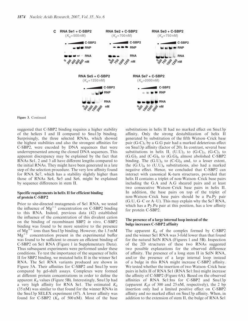
suggested that C-SBP2 binding requires a higher stabilityof the helices I and II compared to Snu13p binding.Surprisingly, the three selected RNAs, which showedthe highest stabilities and also the strongest affinities forC-SBP2, were encoded by DNA sequences that wereunderrepresented among the cloned DNA sequences. Thisapparent discrepancy may be explained by the fact thatRNAs Se1, 2 and 3 all have different lengths compared tothe initial RNAs. They might have been generated in a latestep of the selection procedure. The very low affinity foundfor RNA Se7, which has a stability slightly higher thanthose of RNAs Se4, Se5 and Se6, might be explainedby sequence differences in stem II.
Specific requirements in helix II for efficient bindingof protein C-SBP2
Prior to site-directed mutagenesis of Se1 RNA, we testedthe influence of Mgþþ concentration on C-SBP2 bindingto this RNA. Indeed, previous data (42) establishedthe influence of the concentration of this divalent cationon the binding of recombinant SBP2 in vitro. C-SBP2binding was found to be more sensitive to the presenceof Mgþþ ions than Snu13p binding. However, the 1.5mMMgþþ concentration present in the experimental bufferwas found to be sufficient to ensure an efficient binding ofC-SBP2 on Se1 RNA (Figure 1 in Supplementary Data).Thus subsequent experiments were performed under theseconditions. To test the importance of the sequence of helixII for SBP2 binding, we mutated helix II in the winner Se1RNA. The Se1 RNA variants produced are shown inFigure 5A. Their affinities for C-SBP2 and Snu13p werecompared by gel-shift assays. Complexes were formedat different protein concentrations in order to define theapparent Kd values (Figure 5B). Interestingly, Snu13p hada very high affinity for RNA Se1. The estimated Kd
(35 nM) was similar to that found for the winner RNAs inthe Snu13p SELEX experiment (47). A lower affinity wasfound for C-SBP2 (Kd of 500 nM). Most of the base
substitutions in helix II had no marked effect on Snu13paffinity. Only the strong destabilization of helix IIgenerated by substitution of the fifth Watson–Crick basepair (G-C)5 by a G.G pair had a marked deleterious effecton Snu13p affinity (factor of 20). In contrast, several basesubstitutions in helix II, (U.U)3 to (G-C)3, (G-C)5 to(G.G)5 and (C-G)6 to (G.G)6 almost abolished C-SBP2binding. The (G.U)4 to (C-G)4 and, to a lesser extent,the (G.U)4 to (U.U)4 substitutions, also had a markednegative effect. Hence, we concluded that C-SBP2 caninteract with canonical K-turn structures, provided thathelix II contains a triplet of non-Watson–Crick base pairsincluding the G.A and A.G sheared pairs and at leasttwo consecutive Watson–Crick base pairs in helix II.In addition, the base pairs on top of the triplet ofnon-Watson–Crick base pairs should be a Pu.Py pair(G.U, G–C or A–U). This may explain why the Se7 RNA,which has a Py.Pu pair at this position, has a low affinityfor protein C-SBP2.
The presence of a large internal loop instead of thebulge increases C-SBP2 affinity
The apparent Kd of the complex formed by C-SBP2and the winner Se1 RNA was 3-fold lower than that foundfor the natural SelN RNA (Figures 1 and 5B). Inspectionof the 2D structures of these two RNAs suggestedtwo possible explanations for the observed differenceof affinity. The presence of a long stem II in SelN RNA,and/or the presence of a large internal loop insteadof a bulge in this RNA might increase C-SBP2 affinity.We tested whether the insertion of two Watson–Crick basepairs in helix II of RNA Se1 (RNA Se1:Ins) might increasethe affinity of C-SBP2 (Figure 6A). Based on the observedaffinities of RNA Se1:Ins for C-SBP2 and Snu13p(apparent Kds of 300 and 25 nM, respectively), the 2 bpinsertion only had a limited positive effect on C-SBP2affinity and no marked effect on Snu13p affinity. When, inaddition to the extension of stem II, the bulge of RNA Se1
C RNA Se1 + C-SBP2(Kd=500nM)
RNP
RNA
C-SBP2
020
040
060
0 nM800
RNA Se3 + C-SBP2(Kd=750nM)
RNP
RNA
C-SBP2
050
075
010
00 nM
1250
RNA Se7 + C-SBP2(Kd>2000nM)
RNP
RNA
C-SBP2
050
010
0015
00 nM
2000
RNA Se5 + C-SBP2(Kd=1250nM)
RNP
RNA
C-SBP2
050
010
0015
00 nM
2000
RNA Se2 + C-SBP2(Kd=700nM)
RNP
RNA
C-SBP2
050
075
010
00 nM
1250
Figure 3. Continued
1874 Nucleic Acids Research, 2007, Vol. 35, No. 6
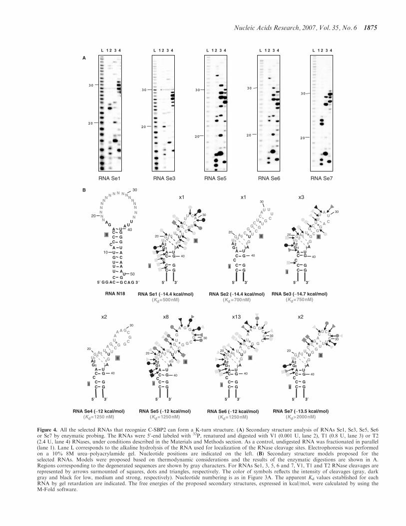
A
B
RNA Se3 (−14.7 kcal/mol)(Kd=750nM)
AU
G
AU
C
C
U
C
AG
AGU G
A
UG
C
C
GG
GA U
3′5′
UGC
G20
30
40
123
45
67
I
II
C
30
20C
C
CG G
G
G
GA
G
A G
U
U
C
AG
AGU
GA
UU
C
C
GG
GA U
3′5′
40
123
45
67
I
II
C
RNA Se1 (−14.4 kcal/mol)(Kd=500nM)
20
G
C
CU
U
U
C
AG
AGU G
A
UU
C
C
GG
GA U
3′5′
CGG
C
U A
40
123
45
6
7
I
II
30
C
C
CG
G
U
A
AC
G
U
A
C
AG
AGU G
A
UU
C
C
GG
GA U
3′5′
G
GG
20
30
40
123
45
6
7
I
II
C
UUA
C
G CG
G
C
C
AG
AGU G
A
UU
C
C
GG
GA U
3′5′
GUC
G
C
20
30
40
123
45
6
7
I
II
C
N
N
N
NN
NN
U
N
N N N
N
N
C
AG
NNN U
A
N
N
CC
GGG
A U
35′
40
20
30
UGU
ACA
A U
CC
U
GG
AU A
C
U
AG G C G ′A
10
50
II
RNA N18 RNA Se2 (−14.4 kcal/mol)(Kd =700nM)
UU
G
UU
C
U
A
C
AG
AGU G
A
CU
C
C
GG
GA U
CGG
C
G
U
3′5′
C
20
30
40
123
45
67
I
II
C
C
C
CG G
G
G
GA
AA A
U
U
C
AG
AGU G
A
UU
C
C
GG
GA U
3′5′
40
20
30
123
45
6
7
I
II
C
x1 x1 x3
x2 x8 x13 x2
L 1 2 3 4
20
30
RNA Se1
20
30
RNA Se3
20
30
RNA Se5
20
30
RNA Se6
20
30
RNA Se7
L 1 2 3 4 L 1 2 3 4 L 1 2 3 4 L 1 2 3 4
RNA Se4 (−12 kcal/mol)(Kd=1250 nM)
RNA Se5 (−12 kcal/mol)(Kd=1250nM)
RNA Se6 (−12 kcal/mol)(Kd=1250nM)
RNA Se7 (−13.5 kcal/mol)(Kd>2000nM)
Figure 4. All the selected RNAs that recognize C-SBP2 can form a K-turn structure. (A) Secondary structure analysis of RNAs Se1, Se3, Se5, Se6or Se7 by enzymatic probing. The RNAs were 50-end labeled with 32P, renatured and digested with V1 (0.001 U, lane 2), T1 (0.8 U, lane 3) or T2(2.4 U, lane 4) RNases, under conditions described in the Materials and Methods section. As a control, undigested RNA was fractionated in parallel(lane 1). Lane L corresponds to the alkaline hydrolysis of the RNA used for localization of the RNase cleavage sites. Electrophoresis was performedon a 10% 8M urea–polyacrylamide gel. Nucleotide positions are indicated on the left. (B) Secondary structure models proposed for theselected RNAs. Models were proposed based on thermodynamic considerations and the results of the enzymatic digestions are shown in A.Regions corresponding to the degenerated sequences are shown by gray characters. For RNAs Se1, 3, 5, 6 and 7, V1, T1 and T2 RNase cleavages arerepresented by arrows surmounted of squares, dots and triangles, respectively. The color of symbols reflects the intensity of cleavages (gray, darkgray and black for low, medium and strong, respectively). Nucleotide numbering is as in Figure 3A. The apparent Kd values established for eachRNA by gel retardation are indicated. The free energies of the proposed secondary structures, expressed in kcal/mol, were calculated by using theM-Fold software.
Nucleic Acids Research, 2007, Vol. 35, No. 6 1875
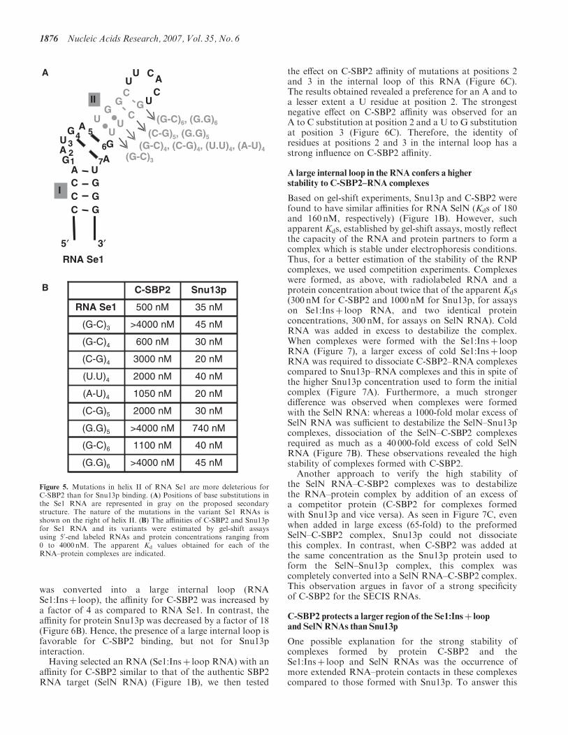
was converted into a large internal loop (RNASe1:Insþ loop), the affinity for C-SBP2 was increased bya factor of 4 as compared to RNA Se1. In contrast, theaffinity for protein Snu13p was decreased by a factor of 18(Figure 6B). Hence, the presence of a large internal loop isfavorable for C-SBP2 binding, but not for Snu13pinteraction.Having selected an RNA (Se1:Insþ loop RNA) with an
affinity for C-SBP2 similar to that of the authentic SBP2RNA target (SelN RNA) (Figure 1B), we then tested
the effect on C-SBP2 affinity of mutations at positions 2and 3 in the internal loop of this RNA (Figure 6C).The results obtained revealed a preference for an A and toa lesser extent a U residue at position 2. The strongestnegative effect on C-SBP2 affinity was observed for anA to C substitution at position 2 and a U to G substitutionat position 3 (Figure 6C). Therefore, the identity ofresidues at positions 2 and 3 in the internal loop has astrong influence on C-SBP2 affinity.
A large internal loop in the RNA confers a higherstability to C-SBP2–RNA complexes
Based on gel-shift experiments, Snu13p and C-SBP2 werefound to have similar affinities for RNA SelN (Kds of 180and 160 nM, respectively) (Figure 1B). However, suchapparent Kds, established by gel-shift assays, mostly reflectthe capacity of the RNA and protein partners to form acomplex which is stable under electrophoresis conditions.Thus, for a better estimation of the stability of the RNPcomplexes, we used competition experiments. Complexeswere formed, as above, with radiolabeled RNA and aprotein concentration about twice that of the apparent Kds(300 nM for C-SBP2 and 1000 nM for Snu13p, for assayson Se1:Insþ loop RNA, and two identical proteinconcentrations, 300 nM, for assays on SelN RNA). ColdRNA was added in excess to destabilize the complex.When complexes were formed with the Se1:Insþ loopRNA (Figure 7), a larger excess of cold Se1:Insþ loopRNA was required to dissociate C-SBP2–RNA complexescompared to Snu13p–RNA complexes and this in spite ofthe higher Snu13p concentration used to form the initialcomplex (Figure 7A). Furthermore, a much strongerdifference was observed when complexes were formedwith the SelN RNA: whereas a 1000-fold molar excess ofSelN RNA was sufficient to destabilize the SelN–Snu13pcomplexes, dissociation of the SelN–C-SBP2 complexesrequired as much as a 40 000-fold excess of cold SelNRNA (Figure 7B). These observations revealed the highstability of complexes formed with C-SBP2.
Another approach to verify the high stability ofthe SelN RNA–C-SBP2 complexes was to destabilizethe RNA–protein complex by addition of an excess ofa competitor protein (C-SBP2 for complexes formedwith Snu13p and vice versa). As seen in Figure 7C, evenwhen added in large excess (65-fold) to the preformedSelN–C-SBP2 complex, Snu13p could not dissociatethis complex. In contrast, when C-SBP2 was added atthe same concentration as the Snu13p protein used toform the SelN–Snu13p complex, this complex wascompletely converted into a SelN RNA–C-SBP2 complex.This observation argues in favor of a strong specificityof C-SBP2 for the SECIS RNAs.
C-SBP2 protects a larger region of the Se1:Insþ loopand SelN RNAs than Snu13p
One possible explanation for the strong stability ofcomplexes formed by protein C-SBP2 and theSe1:Insþ loop and SelN RNAs was the occurrence ofmore extended RNA–protein contacts in these complexescompared to those formed with Snu13p. To answer this
(G-C)3
(G-C)6, (G.G)6
(C-G)5, (G.G)5
I
IIG
C
CU
U
U
C
AG
AGU G
A
UU
CC
GGG
A U
CGG
C
U A
3′5′
(G-C)4, (C-G)4, (U.U)4, (A-U)4
123
4 5
6
7
RNA Se1
A
B C-SBP2 Snu13p
RNA Se1 500 nM 35 nM
(G-C)3 >4000 nM 45 nM
(G-C)4 600 nM 30 nM
(C-G)4 3000 nM 20 nM
(U.U)4 2000 nM 40 nM
(A-U)4 1050 nM 20 nM
(C-G)5 2000 nM 30 nM
(G.G)5 >4000 nM 740 nM
(G-C)6 1100 nM 40 nM
(G.G)6 >4000 nM 45 nM
Figure 5. Mutations in helix II of RNA Se1 are more deleterious forC-SBP2 than for Snu13p binding. (A) Positions of base substitutions inthe Se1 RNA are represented in gray on the proposed secondarystructure. The nature of the mutations in the variant Se1 RNAs isshown on the right of helix II. (B) The affinities of C-SBP2 and Snu13pfor Se1 RNA and its variants were estimated by gel-shift assaysusing 50-end labeled RNAs and protein concentrations ranging from0 to 4000 nM. The apparent Kd values obtained for each of theRNA–protein complexes are indicated.
1876 Nucleic Acids Research, 2007, Vol. 35, No. 6
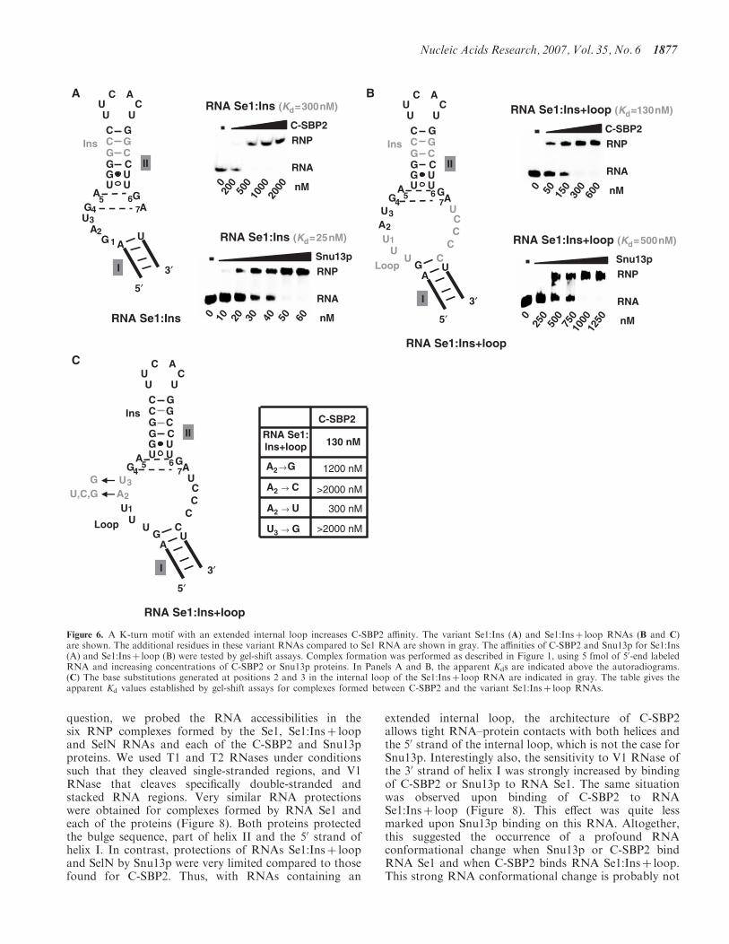
question, we probed the RNA accessibilities in thesix RNP complexes formed by the Se1, Se1:Insþ loopand SelN RNAs and each of the C-SBP2 and Snu13pproteins. We used T1 and T2 RNases under conditionssuch that they cleaved single-stranded regions, and V1RNase that cleaves specifically double-stranded andstacked RNA regions. Very similar RNA protectionswere obtained for complexes formed by RNA Se1 andeach of the proteins (Figure 8). Both proteins protectedthe bulge sequence, part of helix II and the 50 strand ofhelix I. In contrast, protections of RNAs Se1:Insþ loopand SelN by Snu13p were very limited compared to thosefound for C-SBP2. Thus, with RNAs containing an
extended internal loop, the architecture of C-SBP2allows tight RNA–protein contacts with both helices andthe 50 strand of the internal loop, which is not the case forSnu13p. Interestingly also, the sensitivity to V1 RNase ofthe 30 strand of helix I was strongly increased by bindingof C-SBP2 or Snu13p to RNA Se1. The same situationwas observed upon binding of C-SBP2 to RNASe1:Insþ loop (Figure 8). This effect was quite lessmarked upon Snu13p binding on this RNA. Altogether,this suggested the occurrence of a profound RNAconformational change when Snu13p or C-SBP2 bindRNA Se1 and when C-SBP2 binds RNA Se1:Insþ loop.This strong RNA conformational change is probably not
BA
RNA Se1:Ins
U
C ACU
U
GCGCCGCG
UUGA
UG
A
A
UG
G
Ins
234
5 67
1
II
AU
3′
5′
I
C
C-SBP2
RNA Se1:Ins+loop
A2 →G
A2 → C
A2 → U
U3 → G
130 nM
1200 nM
>2000 nM
300 nM
>2000 nM
RNA Se1:Ins (Kd=300nM)
RNP
RNA
C-SBP2
020
050
010
00 nM
2000
RNA Se1:Ins+loop (Kd=130nM)
RNP
RNA
C-SBP2
0 50 150
300 nM600
RNA Se1:Ins (Kd=25nM)
RNP
RNA
Snu13p
0 10 20 30 nM40 50 60
RNA Se1:Ins+loop (Kd=500nM)
RNP
RNA
Snu13p
025
050
075
0 nM
1000
1250
RNA Se1:Ins+loop
U
C ACU
U
GCGCCGCG
UUGA
UG
A
A
U
A
3′
5′
GC
UG
CCC
U
UU
U
Ins
Loop
2
34
5 67
1
I
II
RNA Se1:Ins+loop
U
C ACU
U
GCGCCGCG
UUGA
UG
A
A
U
A
3′
5′
GC
UG
CCC
U
UU
U
Ins
Loop
U,C,GG
2
34
5 67
1
I
II
Figure 6. A K-turn motif with an extended internal loop increases C-SBP2 affinity. The variant Se1:Ins (A) and Se1:Insþ loop RNAs (B and C)are shown. The additional residues in these variant RNAs compared to Se1 RNA are shown in gray. The affinities of C-SBP2 and Snu13p for Se1:Ins(A) and Se1:Insþ loop (B) were tested by gel-shift assays. Complex formation was performed as described in Figure 1, using 5 fmol of 50-end labeledRNA and increasing concentrations of C-SBP2 or Snu13p proteins. In Panels A and B, the apparent Kds are indicated above the autoradiograms.(C) The base substitutions generated at positions 2 and 3 in the internal loop of the Se1:Insþ loop RNA are indicated in gray. The table gives theapparent Kd values established by gel-shift assays for complexes formed between C-SBP2 and the variant Se1:Insþ loop RNAs.
Nucleic Acids Research, 2007, Vol. 35, No. 6 1877
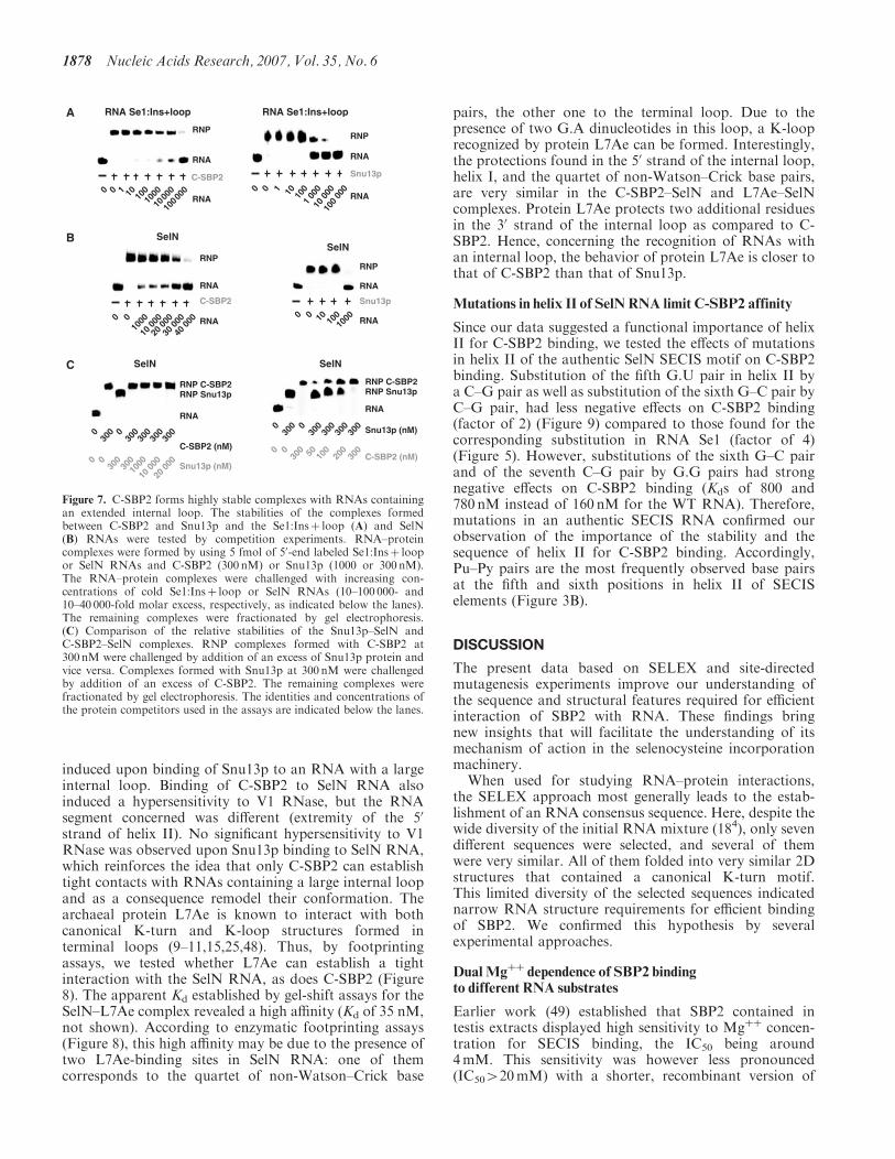
induced upon binding of Snu13p to an RNA with a largeinternal loop. Binding of C-SBP2 to SelN RNA alsoinduced a hypersensitivity to V1 RNase, but the RNAsegment concerned was different (extremity of the 50
strand of helix II). No significant hypersensitivity to V1RNase was observed upon Snu13p binding to SelN RNA,which reinforces the idea that only C-SBP2 can establishtight contacts with RNAs containing a large internal loopand as a consequence remodel their conformation. Thearchaeal protein L7Ae is known to interact with bothcanonical K-turn and K-loop structures formed interminal loops (9–11,15,25,48). Thus, by footprintingassays, we tested whether L7Ae can establish a tightinteraction with the SelN RNA, as does C-SBP2 (Figure8). The apparent Kd established by gel-shift assays for theSelN–L7Ae complex revealed a high affinity (Kd of 35 nM,not shown). According to enzymatic footprinting assays(Figure 8), this high affinity may be due to the presence oftwo L7Ae-binding sites in SelN RNA: one of themcorresponds to the quartet of non-Watson–Crick base
pairs, the other one to the terminal loop. Due to thepresence of two G.A dinucleotides in this loop, a K-looprecognized by protein L7Ae can be formed. Interestingly,the protections found in the 50 strand of the internal loop,helix I, and the quartet of non-Watson–Crick base pairs,are very similar in the C-SBP2–SelN and L7Ae–SelNcomplexes. Protein L7Ae protects two additional residuesin the 30 strand of the internal loop as compared to C-SBP2. Hence, concerning the recognition of RNAs withan internal loop, the behavior of protein L7Ae is closer tothat of C-SBP2 than that of Snu13p.
Mutations in helix II of SelN RNA limit C-SBP2 affinity
Since our data suggested a functional importance of helixII for C-SBP2 binding, we tested the effects of mutationsin helix II of the authentic SelN SECIS motif on C-SBP2binding. Substitution of the fifth G.U pair in helix II bya C–G pair as well as substitution of the sixth G–C pair byC–G pair, had less negative effects on C-SBP2 binding(factor of 2) (Figure 9) compared to those found for thecorresponding substitution in RNA Se1 (factor of 4)(Figure 5). However, substitutions of the sixth G–C pairand of the seventh C–G pair by G.G pairs had strongnegative effects on C-SBP2 binding (Kds of 800 and780 nM instead of 160 nM for the WT RNA). Therefore,mutations in an authentic SECIS RNA confirmed ourobservation of the importance of the stability and thesequence of helix II for C-SBP2 binding. Accordingly,Pu–Py pairs are the most frequently observed base pairsat the fifth and sixth positions in helix II of SECISelements (Figure 3B).
DISCUSSION
The present data based on SELEX and site-directedmutagenesis experiments improve our understanding ofthe sequence and structural features required for efficientinteraction of SBP2 with RNA. These findings bringnew insights that will facilitate the understanding of itsmechanism of action in the selenocysteine incorporationmachinery.
When used for studying RNA–protein interactions,the SELEX approach most generally leads to the estab-lishment of an RNA consensus sequence. Here, despite thewide diversity of the initial RNA mixture (184), only sevendifferent sequences were selected, and several of themwere very similar. All of them folded into very similar 2Dstructures that contained a canonical K-turn motif.This limited diversity of the selected sequences indicatednarrow RNA structure requirements for efficient bindingof SBP2. We confirmed this hypothesis by severalexperimental approaches.
DualMgþþ
dependence of SBP2 bindingto different RNA substrates
Earlier work (49) established that SBP2 contained intestis extracts displayed high sensitivity to Mgþþ concen-tration for SECIS binding, the IC50 being around4mM. This sensitivity was however less pronounced(IC50420mM) with a shorter, recombinant version of
A RNA Se1:Ins+loop1
RNP
C-SBP2
0 10 100
10000
1000
0 10
0000
RNA
RNA
RNA Se1:Ins+loop
1
RNP
Snu13p
0 10 100
1 00
00
10 0
0010
0 00
0
RNA
RNA
B
0
RNP
RNA
C-SBP2
RNA0
1000
10 0
0020
000
30 0
0040
000
SelNSelN
RNP
RNA
Snu13p
RNA10010
010
000
C
300
RNP C-SBP2
RNA
Snu13p (nM)
C-SBP2 (nM)
0 030
030
030
030
0
SelN
00
300
300
1000
10 0
0020
000
RNP Snu13p
300
RNA
Snu13p (nM)
C-SBP2 (nM)
0 030
030
030
030
0SelN
00
300 50 100
200
300
RNP C-SBP2 RNP Snu13p
Figure 7. C-SBP2 forms highly stable complexes with RNAs containingan extended internal loop. The stabilities of the complexes formedbetween C-SBP2 and Snu13p and the Se1:Insþ loop (A) and SelN(B) RNAs were tested by competition experiments. RNA–proteincomplexes were formed by using 5 fmol of 50-end labeled Se1:Insþ loopor SelN RNAs and C-SBP2 (300 nM) or Snu13p (1000 or 300 nM).The RNA–protein complexes were challenged with increasing con-centrations of cold Se1:Insþ loop or SelN RNAs (10–100 000- and10–40 000-fold molar excess, respectively, as indicated below the lanes).The remaining complexes were fractionated by gel electrophoresis.(C) Comparison of the relative stabilities of the Snu13p–SelN andC-SBP2–SelN complexes. RNP complexes formed with C-SBP2 at300 nM were challenged by addition of an excess of Snu13p protein andvice versa. Complexes formed with Snu13p at 300 nM were challengedby addition of an excess of C-SBP2. The remaining complexes werefractionated by gel electrophoresis. The identities and concentrations ofthe protein competitors used in the assays are indicated below the lanes.
1878 Nucleic Acids Research, 2007, Vol. 35, No. 6
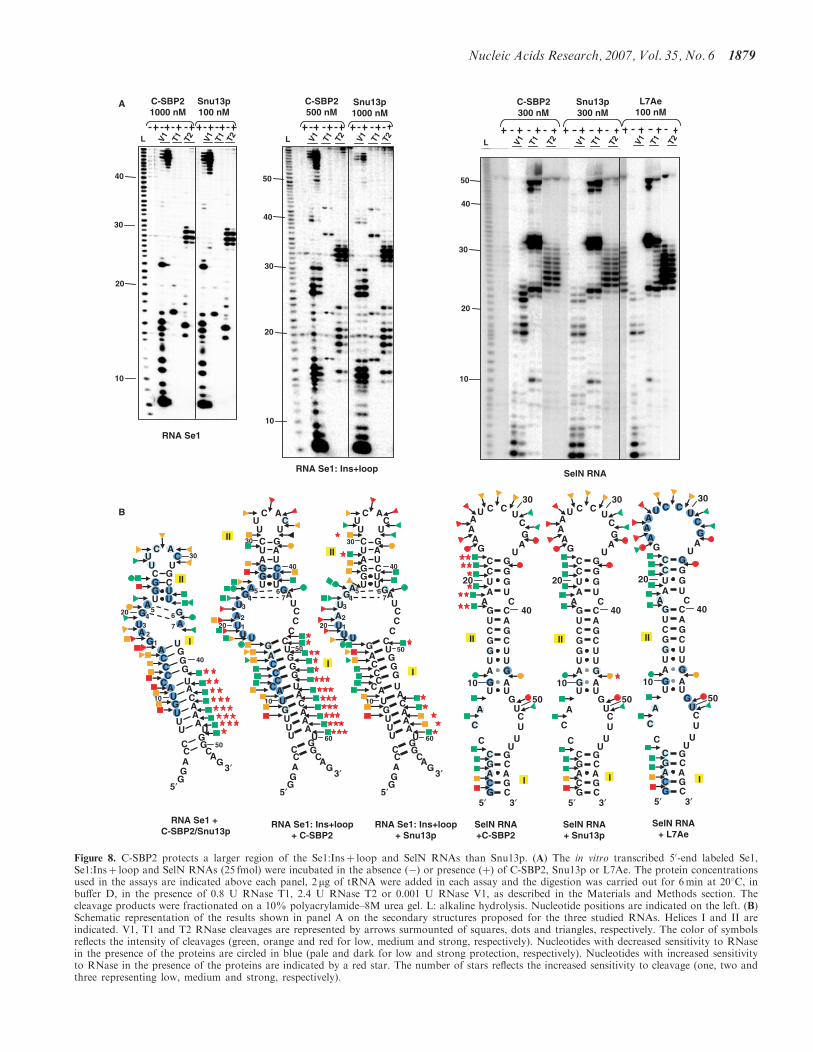
A
20
10
30
40
V1 T1 T2 V1 T1 T2
C-SBP21000 nM
Snu13p100 nM
L
RNA Se1
20
10
30
40
50
V1 T1 T2 V1 T1 T2
C-SBP2500 nM
Snu13p1000 nM
L
RNA Se1: Ins+loop
20
10
30
40
50
V1 T1 T2
C-SBP2300 nM
Snu13p300 nM
L V1 T1 T2 V1 T1 T2
L7Ae100 nM
SelN RNA
U
C ACU
UGCAUUACG
UUGA
UG
A
A
U
A
CG
CG
CG
C
AG
G
A U
UA
GC
UA
UA
UA
UC
G
CG
CAG
3′
5′
G C
UG
CCC
U
UUU
10
20
30
40
50
60
2
34
5 67
1
I
II
B
RNA Se1: Ins+loop+ C-SBP2
RNA Se1: Ins+loop+ Snu13p
U
C ACU
UGCAUUACG
UUGA
UG
A
A
U
A
CG
CG
CG
C
AG
G
A U
UA
GC
UA
UA
UA
UC
G
CG
CAG
3′
5′
G C
UG
CCC
U
UUU
10
20
30
40
50
60
2
34
5 67
1
I
II
RNA Se1 + C-SBP2/Snu13p
I
II
U
C ACU
UGCCG
UUG
A
UG
A
AU
A
CG
CG
CG
C
AG
G
AU
UA
GC
UA
UA
UA
U
CG
CG
CAG
3′
5′
G
UG
10
20
30
40
50
12
34
56
7
SelN RNA+ Snu13p
C
C
5′
C
C
C
C
C
C
CC
C
A
C
G
G
G
G
G
G
G
G
GG
G
G
A
A
A
A
AA
A
AA
U
U
U
U
U
U
U
UU
U
U
U
U
U
U
C
C
C
CG
G
GG
A A
3′
10
20
30
40
50
I
II
SelN RNA+C-SBP2
C
C
5′
C
C
C
C
C
C
CC
C
A
C
G
G
G
G
G
G
G
G
GG
G
G
A
A
A
A
AA
A
AA
U
U
U
U
U
U
U
UU
U
U
U
U
U
U
C
C
C
CG
G
GG
A A
3′
10
20
30
40
50
II
I
SelN RNA+ L7Ae
C
C
5′
C
C
C
C
C
C
CC
C
A
C
G
G
G
G
G
G
G
G
GG
G
G
A
A
A
A
AA
A
AA
U
U
U
U
U
U
U
UU
U
U
U
U
U
U
C
C
C
CG
G
GG
A A
3′
10
20
30
40
50
II
I
Figure 8. C-SBP2 protects a larger region of the Se1:Insþ loop and SelN RNAs than Snu13p. (A) The in vitro transcribed 50-end labeled Se1,Se1:Insþ loop and SelN RNAs (25 fmol) were incubated in the absence (�) or presence (þ) of C-SBP2, Snu13p or L7Ae. The protein concentrationsused in the assays are indicated above each panel, 2 mg of tRNA were added in each assay and the digestion was carried out for 6min at 208C, inbuffer D, in the presence of 0.8 U RNase T1, 2.4 U RNase T2 or 0.001 U RNase V1, as described in the Materials and Methods section. Thecleavage products were fractionated on a 10% polyacrylamide–8M urea gel. L: alkaline hydrolysis. Nucleotide positions are indicated on the left. (B)Schematic representation of the results shown in panel A on the secondary structures proposed for the three studied RNAs. Helices I and II areindicated. V1, T1 and T2 RNase cleavages are represented by arrows surmounted of squares, dots and triangles, respectively. The color of symbolsreflects the intensity of cleavages (green, orange and red for low, medium and strong, respectively). Nucleotides with decreased sensitivity to RNasein the presence of the proteins are circled in blue (pale and dark for low and strong protection, respectively). Nucleotides with increased sensitivityto RNase in the presence of the proteins are indicated by a red star. The number of stars reflects the increased sensitivity to cleavage (one, two andthree representing low, medium and strong, respectively).
Nucleic Acids Research, 2007, Vol. 35, No. 6 1879
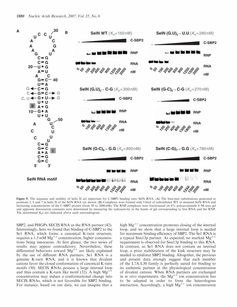
SBP2, and PHGPx SECIS RNA as the RNA partner (42).Interestingly, here we found that binding of C-SBP2 to theSe1 RNA, which forms a canonical K-turn structure,requires a 1.5mM Mgþþ concentration, higher concentra-tions being innocuous. At first glance, the two series ofresults may appear contradictory. Nevertheless, thesedifferential behaviors toward Mgþþ are likely explainedby the use of different RNA partners. Se1 RNA is agenuine K-turn RNA, and it is known that divalentcations favor the closed conformation of canonical K-turnmotifs (50). SECIS RNAs possess a large internal loopand thus contain a K-turn like motif (32). A high Mgþþ
concentration may induce a conformational change intoSECIS RNAs, which is not favorable for SBP2 binding.For instance, based on our data, we can imagine that a
high Mgþþ concentration promotes closing of the internalloop, and we show that a large internal loop is neededfor maximum binding efficiency of SBP2. The Se1 RNA isa typical Snu13p partner. As expected, no marked Mgþþ
requirement is observed for Snu13p binding to this RNA.In contrast, as Se1 RNA does not contain an internalloop, a prior stabilization of the kink structure may beneeded to reinforce SBP2 binding. Altogether, the previousand present data strongly suggest that each memberof the L7A/L30 family is perfectly suited for binding toits authentic partner at the physiological concentrationof divalent cations. When RNA partners are exchangedin in vitro experiments, the Mgþþ ion concentration hasto be adapted in order to form the heterologousinteraction. Accordingly, a high Mgþþ ion concentration
A B
I
II
C
C
5′
C
C
C
C
C
C
CC
C
A
C
G
G
G
G
G
G
G
G
GG
G
G
A
A
A
A
AA
A
AA
U
U
U
U
U
U
U
UU
U
U
U
U
U
U
C
C
C
CG
G
GG
A A
3′
10
20
30
40
50
RNA
C-SBP2−
0 50 100
200
400
650
900
1250
1500
nM
RNP
SelN (G.U)5→ U.U (Kd=280nM)
2000
RNA
C-SBP2−
0 50 100
200
400
650
900
1250
1500 nM
RNP
SelN WT (Kd=160nM)
2000
C, UG
G SelN (G.U)5→ C-G (Kd=300nM)
RNA
C-SBP2−
010
020
040
080
013
0018
00
nM
RNP
SelN (G-C)6→ C-G (Kd=270nM)
RNA
C-SBP2−
010
020
040
080
013
0018
00
nM
RNP
GC
RNA
C-SBP2−
0 50 100
200
400
650
900
1250
1500 nM
RNP
SelN (G-C)6→ G.G (Kd=800nM)
2000 0 50 100
200
400
650
900
1250
1500
2000
RNA
C-SBP2 −
nM
RNP
SelN (C-G)7→ G.G (Kd=780nM)
SelN RNA motif
Figure 9. The sequence and stability of helix II are important for C-SBP2 binding onto SelN RNA. (A) The base-pair substitutions generated atpositions 5, 6 and 7 in helix II of the SelN RNA are shown. (B) Complexes were formed with 5 fmol of radiolabeled WT or mutated SelN RNA andincreasing concentrations of the C-SBP2 protein (from 50 to 2000 nM). The RNP complexes were fractionated on 6% polyacrylamide 8–M urea geland apparent dissociation constants were determined by measuring the radioactivity in the bands of gel corresponding to free RNA and the RNP.The determined Kds are indicated above each autoradiogram.
1880 Nucleic Acids Research, 2007, Vol. 35, No. 6
was found to be required for efficient in vitro bindingof protein L30 to a SECIS element, in the presence ofSBP2 (42).
Specific sequence requirements in helix II
Site-directed mutagenesis, performed on the winner Se1RNA obtained by SELEX experiments, demonstratedthat binding of C-SBP2 requires the presence of a stablehelix II containing at least two Watson–Crick base pairs.In agreement with this observation, all the SECISelements identified so far contain a series of Watson–Crick base pairs on top of the non-Watson–Crick base-pair quartet (30,51–53; A.K., unpublished data).Accordingly, we showed that their individual disruptionin SelN RNA decreases C-SBP2 affinity. Not only helix IIstability but also its sequence has an influence on C-SBP2affinity. The presence of a Pu–Py pair at the fourthposition in helix II was found to be of high importancefor C-SBP2 binding to Se1 RNA, a Pu–Py pair at thisposition being also more favorable for C-SBP2 binding toSelN RNA. This is in contrast with the absence ofsequence requirement in helix II, except for the G.A andA.G base pairs and the adjacent U.U pair found forproteins Snu13p/15.5K and L7Ae (6,7,16,24,47,54–56).Up to now, little attention was given to the importanceof the identity of base pairs in the upper part of helix II ofSECIS elements. However, at position 4 of helix II, a Puresidue (most frequently a G residue) is almost alwaysfound in the 50 strand and a Py residue (most frequently aC residue) is observed in the 30 strand (30,51–53; A.K.,unpublished data). Although less strictly conserved, thefifth base pair in helix II is predominantly a Pu–Py pair(Figure 3B). Together with our experimental data, thesephylogenetic observations strongly suggest a functionalimportance of these conserved Pu.Py base pairs atpositions 4 and 5 in helix II. In accordance with thishypothesis, the G–C pair at the fourth position in RNASe1 was protected against V1 RNase digestion in thecomplex formed with C-SBP2, but not in the complexformed with Snu13p (Figure 8). Accordingly, the verylimited V1 RNase cleavage, which is located betweenresidues G13 and G14 in free SelN RNA, disappeared inthe presence of C-SBP2, but not with Snu13p.Remarkably, this V1 RNase cleavage was also abolishedin the presence of protein L7Ae.
Comparison of the Se1 to Se7 RNAs and site-directedmutagenesis of the Se1 RNA also show the importancefor a non-Watson–Crick base pair on top of the A.Gand G.A pair tandem (Figures 4 and 5). Accordingly,U.U pairs are frequently encountered pairs at this positionin SECIS elements (30,51–53; A.K., unpublished data)and a U.U pair was also preferentially selected at thisposition of helix II, in the SELEX experiment performedwith Snu13p. The presence of a U.U base pair atthis position, with a C10–C10 distance of the ribose ringclose to that in G.A pairs, is very likely required tofavor the smooth transition from the non-Watson–Crickto the Watson–Crick section of helix II. Noticeably also,in K-turn structures found in ribosomal RNAs, thenucleobase of one of this unpaired couple of nucleotides
was proposed to interact with one nucleobase in helix I,and thus to reinforce the inter-helical angle between helix Iand helix II (57).
Importance of a large SECIS internal loop
Increasing the size of both helix II and the internal loopof the winner Se1 RNA obtained by SELEX, yieldedan RNA with an affinity similar to that of SelN RNA(Figure 6B). Such an RNA could not be obtained in theSELEX experiment, because of limitation in size of thedegenerated sequence that can be used (18 nt) in theseexperiments. Indeed, due to the necessity to cover allthe possible sequences during the screening, one cannotuse largely extended degenerated sequences (58,59).In agreement with the importance of the size of helix II,all the identified SECIS elements contain a long helix II.Based on our footprinting data, the high affinity ofC-SBP2 for RNAs with an internal loop, as well as thestability of the complexes formed, are due to its capacityto contact helices I and II and the 50 strand of the internalloop in these RNAs (Figure 8). Interestingly, Martin et al.(38) showed that closing of the internal loop of the ratD1 SECIS element almost completely abolished seleno-cysteine incorporation in vivo. In agreement with theobserved requirement of at least one base pair closingthe 3-nt bulge loop of K-turn motifs for efficient bindingof Snu13p (44), Snu13p establishes very loose contactswith RNAs containing an internal loop. The presence ofa closing base pair is not required for L7Ae and thisprotein is able to bind open K-loop structures (9,44,48).The presence of an arginine at position 95 in the 15.5K/Snu13p protein, that forms a salt bridge with the50 phosphate of the residue at position 1 in the bulge,and its replacement by a valine in L7Ae, were proposed toexplain this difference between proteins 15.5K/Snu13pand L7Ae (60). Interestingly, like L7Ae, SBP2 containsa valine at the corresponding position in the L7A/L30domain (29). This may explain our observation of similarbinding properties of proteins SBP2 and L7Ae on RNAscontaining a large internal loop.In free RNAs containing a canonical K-turn structure
with a bulge loop, helices I and II form a 768 angle. UponSnu13p/15.5K binding, the RNA undergoes furtherfolding, so that the helix I–helix II angle is reduced to488 (56,61). This folding likely explains the tight contact ofSnu13p with both helices of RNA Se1 that we detected byfootprinting assay. As very similar footprinting resultswere obtained with C-SBP2, it probably also induces afolding of this RNA. However, C-SBP2 but not Snu13pmay induce a similar folding in both the Se1:Insþ loopand SelN RNAs.
Sequence requirement in the internal loop
Our site-directed mutagenesis experiments on theSe1:Insþ loop RNA revealed the importance of theidentity of residues at positions 2 and 3 in the internalloop for C-SBP2 binding. The most deleterious basesubstitution at position 2 was the A to C replacement.Interestingly, a 66% decrease of selenocysteine incorpora-tion was observed when the same A to C substitution
Nucleic Acids Research, 2007, Vol. 35, No. 6 1881
was generated in the SECIS element of the rat GPxmRNA while A to G and A to U changes only led to a lossof 30 and 22% of the incorporation, respectively (52). Inaccordance with the decrease of the C-SBP2 affinity uponU to G substitution at position 3 in RNA Se1:Insþ loop,selenocysteine incorporation was decreased by 88%when this base substitution was generated in the SECISelement of the rat GPx mRNA (38). In addition, a U toC substitution at this position abolished the binding ofSBP2 to SelN RNA and is responsible for a human geneticdisease, the rigid spine muscular dystrophy (62). As theresidue at position 3 in canonical K-turns is located inthe protein pocket, its mutation also has a deleteriouseffect on 15.5K/Snu13p and L7Ae binding (6,24–26).Residues E61 and K86 in 15.5K, and D54 and K79 inArchaeoglobus fulgidus L7Ae, are involved in the interac-tion with the nucleobase at position 3. Their counterpartsin SBP2 (E699 and R730) probably play a similar role,since they are crucial for binding to SECIS RNAs(5,29,56). The specificity of L7Ae/L30 protein memberstowards the residue at position 2 in the K-turn motifis variable. Whereas an A or G residue at position 2increases 15.5K/Snu13p affinity, substitutions at position2 have no marked effect on L7Ae affinity (25). Concerningposition 2, SBP2 exhibits a behavior closer to that of15.5K/Snu13p than to L7Ae.The ribosomal protein L30 was recently shown
to compete with SBP2 for binding to SECIS RNA (42).L30 was found to recognize a K-turn structure of itspre-mRNA that contains a protruding A residue in a smallinternal loop (21–23). SELEX experiments performedwith L30 revealed its preference for K-turn motifs withprotruding C or A residues (27). Later, it was shown thatL30 also has the ability to accommodate K-turn structureswith a protruding G (28). However, its interaction withK-turn motifs containing a protruding U residue hasnot been demonstrated yet. Consequently, the bindingof L30 to SECIS elements, that all contain a U residue atposition 3, raises the question of how it can achieve this.
CONCLUSION
Assembly of the selenocysteine incorporation machineryis proposed to be initiated by SBP2 association toSECIS elements in the nucleus, and more likely in thenucleolus (63). Protein 15.5K/Snu13p is abundant inthe nucleolus and SBP2 shares several common featureswith Snu13/15.5K. However, our data reveal importantdifferences in RNA specificities that may ensure thespecific association of SBP2 to SECIS elements inthe nuclear compartment.
ACKNOWLEDGEMENTS
V. Senty-Segault and S. Massenet are acknowledgedfor helpful discussions. S. Sonkaria is thanked for carefulreading of the manuscript. A. Clery was a fellow fromthe French ‘Ministere de la Recherche et des NouvellesTechnologies’. The work was supported by the CentreNational de la Recherche Scientifique, the French
‘Ministere de la Recherche et des NouvellesTechnologies’, the ACI ‘Biologie Cellulaire, Moleculaireet Structurale’ n8BCMS226, the PRST ‘Bioingenierie’of the ‘Conseil Regional Lorrain’ and the ToxNuc-EProgramme ‘Toxicologie Nucleaire Environnementale’.Funding to pay the Open Access publication charge wasprovided by CNRS-Sciences de la vie.
Conflict of interest statement. None declared.
REFERENCES
1. Klein,D.J., Schmeing,T.M., Moore,P.B. and Steitz,T.A. (2001)The kink-turn: a new RNA secondary structure motif. EMBO J.,20, 4214–4221.
2. Wimberly,B.T., Brodersen,D.E., Clemons,W.M.Jr, Morgan-Warren,R.J., Carter,A.P., Vonrhein,C., Hartsch,T. andRamakrishnan,V. (2000) Structure of the 30S ribosomal subunit.Nature, 407, 327–339.
3. Schluenzen,F., Tocilj,A., Zarivach,R., Harms,J., Gluehmann,M.,Janell,D., Bashan,A., Bartels,H., Agmon,I. et al. (2000) Structureof functionally activated small ribosomal subunit at 3.3 angstromsresolution. Cell, 102, 615–623.
4. Ban,N., Nissen,P., Hansen,J., Moore,P.B. and Steitz,T.A. (2000)The complete atomic structure of the large ribosomal subunit at2.4 A resolution. Science, 289, 905–920.
5. Vidovic,I., Nottrott,S., Hartmuth,K., Luhrmann,R. and Ficner,R.(2000) Crystal structure of the spliceosomal 15.5kD protein boundto a U4 snRNA fragment. Mol. Cell, 6, 1331–1342.
6. Nottrott,S., Hartmuth,K., Fabrizio,P., Urlaub,H., Vidovic,I.,Ficner,R. and Luhrmann,R. (1999) Functional interaction of anovel 15.5kD [U4/U6.U5] tri-snRNP protein with the 50 stem-loopof U4 snRNA. EMBO J., 18, 6119–6133.
7. Watkins,N.J., Segault,V., Charpentier,B., Nottrott,S., Fabrizio,P.,Bachi,A., Wilm,M., Rosbash,M., Branlant,C. et al. (2000) Acommon core RNP structure shared between the small nucleoar boxC/D RNPs and the spliceosomal U4 snRNP. Cell, 103, 457–466.
8. Terns,M.P. and Terns,R.M. (2002) Small nucleolar RNAs: versatiletrans-acting molecules of ancient evolutionary origin. Gene Expr.,10, 17–39.
9. Charpentier,B., Muller,S. and Branlant,C. (2005) Reconstitutionof archaeal H/ACA small ribonucleoprotein complexes active inpseudouridylation. Nucleic Acids Res., 33, 3133–3144.
10. Omer,A.D., Ziesche,S., Ebhardt,H. and Dennis,P.P. (2002) In vitroreconstitution and activity of a C/D box methylation guideribonucleoprotein complex. Proc. Natl. Acad. Sci. U.S.A., 99,5289–5294.
11. Rozhdestvensky,T.S., Tang,T.H., Tchirkova,I.V., Brosius,J.,Bachellerie,J.P. and Huttenhofer,A. (2003) Binding of L7Ae proteinto the K-turn of archaeal snoRNAs: a shared RNA binding motiffor C/D and H/ACA box snoRNAs in Archaea. Nucleic Acids Res.,31, 869–877.
12. Granneman,S., Pruijn,G.J., Horstman,W., van Venrooij,W.J.,Luhrmann,R. and Watkins,N.J. (2002) The hU3-55K proteinrequires 15.5K binding to the box B/C motif as well as flankingRNA elements for its association with the U3 small nucleolar RNAin Vitro. J. Biol. Chem., 277, 48490–48500.
13. Nottrott,S., Urlaub,H. and Luhrmann,R. (2002) Hierarchical,clustered protein interactions with U4/U6 snRNA: a biochemicalrole for U4/U6 proteins. EMBO J., 21, 5527–5538.
14. Watkins,N.J., Dickmanns,A. and Luhrmann,R. (2002) Conservedstem II of the box C/D motif is essential for nucleolar localizationand is required, along with the 15.5K protein, for the hierarchicalassembly of the box C/D snoRNP. Mol. Cell. Biol., 22, 8342–8352.
15. Baker,D.L., Youssef,O.A., Chastkofsky,M.I., Dy,D.A., Terns,R.M.and Terns,M.P. (2005) RNA-guided RNA modification: functionalorganization of the archaeal H/ACA RNP. Genes Dev., 19,1238–1248.
16. Tran,E.J., Zhang,X. and Maxwell,E.S. (2003) Efficient RNA20-O-methylation requires juxtaposed and symmetrically assembledarchaeal box C/D and C0/D0 RNPs. EMBO J., 22, 3930–3940.
1882 Nucleic Acids Research, 2007, Vol. 35, No. 6
17. Rashid,R., Aittaleb,M., Chen,Q., Spiegel,K., Demeler,B. and Li,H.(2003) Functional requirement for symmetric assembly of archaealbox C/D small ribonucleoprotein particles. J. Mol. Biol., 333,295–306.
18. Bortolin,M.L., Bachellerie,J.P. and Clouet-d0Orval,B. (2003)In vitro RNP assembly and methylation guide activity of anunusual box C/D RNA, cis-acting archaeal pre-tRNA(Trp).Nucleic Acids Res., 31, 6524–6535.
19. Schultz,A., Nottrott,S., Hartmuth,K. and Luhrmann,R. (2006)RNA structural requirements for the association of the spliceosomalhPrp31 protein with the U4 and U4atac small nuclear ribonucleo-proteins. J. Biol. Chem., 281, 28278–28286.
20. Koonin,E.V., Bork,P. and Sander,C. (1994) A novel RNA-bindingmotif in omnipotent suppressors of translation termination,ribosomal proteins and a ribosome modification enzyme?Nucleic Acids Res., 22, 2166–2167.
21. Vilardell,J. and Warner,J.R. (1994) Regulation of splicing at anintermediate step in the formation of the spliceosome. Genes Dev.,8, 211–220.
22. Chao,J.A. and Williamson,J.R. (2004) Joint X-ray and NMRrefinement of the yeast L30e-mRNA complex. Structure, 12,1165–1176.
23. Mao,H., White,S.A. and Williamson,J.R. (1999) A novel loop-looprecognition motif in the yeast ribosomal protein L30 autoregulatoryRNA complex. Nat. Struct. Biol., 6, 1139–1147.
24. Kuhn,J.F., Tran,E.J. and Maxwell,E.S. (2002) Archaeal ribosomalprotein L7 is a functional homolog of the eukaryotic 15.5kD/Snu13p snoRNP core protein. Nucleic Acids Res., 30, 931–941.
25. Charron,C., Manival,X., Clery,A., Senty-Segault,V., Charpentier,B.,Marmier-Gourrier,N., Branlant,C. and Aubry,A. (2004) Thearchaeal sRNA binding protein L7Ae has a 3D structure verysimilar to that of its eukaryal counterpart while having a broaderRNA-binding specificity. J. Mol. Biol., 342, 757–773.
26. Marmier-Gourrier,N., Clery,A., Senty-Segault,V., Charpentier,B.,Schlotter,F., Leclerc,F., Fournier,R. and Branlant,C. (2003) Astructural, phylogenetic, and functional study of 15.5-kD/Snu13protein binding on U3 small nucleolar RNA. RNA, 9, 821–838.
27. Li,H. and White,S.A. (1997) RNA apatamers for yeast ribosomalprotein L32 have a conserved purine-rich internal loop. RNA, 3,245–254.
28. White,S.A., Hoeger,M., Schweppe,J.J., Shillingford,A., Shipilov,V.and Zarutskie,J. (2004) Internal loop mutations in the ribosomalprotein L30 binding site of the yeast L30 RNA transcript. RNA, 10,369–377.
29. Allmang,C., Carbon,P. and Krol,A. (2002) The SBP2 and 15.5kD/Snu13p proteins share the same RNA binding domain:identification of SBP2 amino acids important to SECIS RNAbinding. RNA, 8, 1308–1318.
30. Fletcher,J.E., Copeland,P.R., Driscoll,D.M. and Krol,A. (2001)The selenocysteine incorporation machinery: interactions betweenthe SECIS RNA and the SECIS-binding protein SBP2. RNA, 7,1442–1453.
31. Walczak,R., Carbon,P. and Krol,A. (1998) An essential non-Watson–Crick base pair motif in 30UTR to mediate selenoproteintranslation. RNA, 4, 74–84.
32. Allmang,C. and Krol,A. (2006) Selenoprotein synthesis: UGA doesnot end the story. Biochimie, 88, 1561–1571.
33. Krol,A. (2002) Evolutionarily different RNA motifs andRNA-protein complexes to achieve selenoprotein synthesis.Biochimie, 84, 765–774.
34. Gladyshev,V.N. (2001). Selenium in biology and human health:controversies and perspectives. In Hatfield,D.L. (ed), Selenium:Its Molecular Biology and Role in Human Health. Kluwer, Boston,pp. 313–317.
35. Flohe,L., Andreesen,J.R., Brigelius-Flohe,R., Maiorino,M. andUrsini,F. (2000) Selenium, the element of the moon, in life on earth.IUBMB Life, 49, 411–420.
36. Rayman,M.P. (2000) The importance of selenium to human health.Lancet, 356, 233–241.
37. Rederstorff,M., Krol,A. and Lescure,A. (2006) Understandingthe importance of selenium and selenoproteins in muscle function.Cell Mol. Life Sci., 63, 52–59.
38. Martin,G.W.III, Harney,J.W. and Berry,M.J. (1998) Functionalityof mutations at conserved nucleotides in eukaryotic SECIS elements
is determined by the identity of a single nonconserved nucleotide.RNA, 4, 65–73.
39. Tujebajeva,R.M., Copeland,P.R., Xu,X.M., Carlson,B.A.,Harney,J.W., Driscoll,D.M., Hatfield,D.L. and Berry,M.J. (2000)Decoding apparatus for eukaryotic selenocysteine insertion.EMBO Rep., 1, 158–163.
40. Fagegaltier,D., Hubert,N., Yamada,K., Mizutani,T., Carbon,P. andKrol,A. (2000) Characterization of mSelB, a novel mammalianelongation factor for selenoprotein translation. EMBO J., 19,4796–4805.
41. Zavacki,A.M., Mansell,J.B., Chung,M., Klimovitsky,B.,Harney,J.W. and Berry,M.J. (2003) Coupled tRNA(Sec)-dependentassembly of the selenocysteine decoding apparatus. Mol. Cell, 11,773–781.
42. Chavatte,L., Brown,B.A. and Driscoll,D.M. (2005) Ribosomalprotein L30 is a component of the UGA-selenocysteinerecoding machinery in eukaryotes. Nat. Struct. Mol. Biol., 12,408–416.
43. Mougin,A., Gottschalk,A., Fabrizio,P., Luhrmann,R. andBranlant,C. (2002) Direct probing of RNA structure andRNA-protein interactions in purified HeLa cells and yeastspliceosomal U4/U6.U5 tri-snRNP particles. J. Mol. Biol., 317,631–649.
44. Charron,C., Manival,X., Charpentier,B., Branlant,C. and Aubry,A.(2004) Purification, crystallization and preliminary X-ray diffractiondata of L7Ae sRNP core protein from Pyrococcus abyssii. ActaCrystallogr. D Biol. Crystallogr., 60, 122–124.
45. Lescure,A., Allmang,C., Yamada,K., Carbon,P. and Krol,A. (2002)cDNA cloning, expression pattern and RNA binding analysis ofhuman selenocysteine insertion sequence (SECIS) binding protein 2.Gene, 291, 279–285.
46. Mathews,D.H., Sabina,J., Zuker,M. and Turner,D.H. (1999)Expanded sequence dependence of thermodynamic parametersimproves prediction of RNA secondary structure. J. Mol. Biol., 288,911–940.
47. Clery,A., Senty-Segault,V., Leclerc,F., Raue,H.A. and Branlant,C.(2007) Analysis of sequence and structural features that identify theB/C motif of U3 small nucleolar RNA as the recognition site forthe Snu13p-Rrp9p protein pair. Mol. Cell. Biol., 27, 1191–1206.
48. Nolivos,S., Carpousis,A.J. and Clouet-d’Orval,B. (2005)The K-loop, a general feature of the Pyrococcus C/D guide RNAs,is an RNA structural motif related to the K-turn. Nucleic AcidsRes., 33, 6507–6514.
49. Copeland,P.R. and Driscoll,D.M. (1999) Purification, redoxsensitivity, and RNA binding properties of SECIS-binding protein2, a protein involved in selenoprotein biosynthesis. J. Biol. Chem.,274, 25447–25454.
50. Matsumura,S., Ikawa,Y. and Inoue,T. (2003) Biochemical char-acterization of the kink-turn RNA motif. Nucleic Acids Res., 31,5544–5551.
51. Kryukov,G.V., Castellano,S., Novoselov,S.V., Lobanov,A.V.,Zehtab,O., Guigo,R. and Gladyshev,V.N. (2003) Characterizationof mammalian selenoproteomes. Science, 300, 1439–1443.
52. Fagegaltier,D., Lescure,A., Walczak,R., Carbon,P. and Krol,A.(2000) Structural analysis of new local features in SECIS RNAhairpins. Nucleic Acids Res., 28, 2679–2689.
53. Lescure,A., Gautheret,D., Carbon,P. and Krol,A. (1999) Novelselenoproteins identified in silico and in vivo by using a conservedRNA structural motif. J. Biol. Chem., 274, 38147–38154.
54. Szewczak,L.B., DeGregorio,S.J., Strobel,S.A. and Steitz,J.A. (2002)Exclusive interaction of the 15.5 kD protein with the terminal boxC/D motif of a methylation guide snoRNP. Chem. Biol., 9,1095–1107.
55. Szewczak,L.B., Gabrielsen,J.S., Degregorio,S.J., Strobel,S.A. andSteitz,J.A. (2005) Molecular basis for RNA kink-turn recognitionby the h15.5K small RNP protein. RNA, 11, 1407–1419.
56. Moore,T., Zhang,Y., Fenley,M.O. and Li,H. (2004) Molecularbasis of box C/D RNA-protein interactions; cocrystalstructure of archaeal L7Ae and a box C/D RNA. Structure, 12,807–818.
57. Razga,F., Spackova,N., Reblova,K., Koca,J., Leontis,N.B. andSponer,J. (2004) Ribosomal RNA kink-turn motif – a flexiblemolecular hinge. J. Biomol. Struct. Dyn., 22, 183–194.
Nucleic Acids Research, 2007, Vol. 35, No. 6 1883
58. Tuerk,C. and Gold,L. (1990) Systematic evolution of ligands byexponential enrichment: RNA ligands to bacteriophage T4 DNApolymerase. Science, 249, 505–510.
59. Klug,S.J. and Famulok,M. (1994) All you wanted to know aboutSELEX. Mol. Biol. Rep., 20, 97–107.
60. Hamma,T. and Ferre-D’Amare,A.R. (2004) Structure of proteinL7Ae bound to a K-turn derived from an archaeal box H/ACAsRNA at 1.8A resolution. Structure, 12, 893–903.
61. Wozniak,A.K., Nottrott,S., Kuhn-Holsken,E., Schroder,G.F.,Grubmuller,H., Luhrmann,R., Seidel,C.A. and Oesterhelt,F. (2005)Detecting protein-induced folding of the U4 snRNA kink-turn by
single-molecule multiparameter FRET measurements. RNA, 11,1545–1554.
62. Allamand,V., Richard,P., Lescure,A., Ledeuil,C., Desjardin,D., Petit,N.,Gartioux,C., Ferreiro,A., Krol,A. et al. (2006) A single homozygouspoint mutation in a 30untranslated region motif of selenoprotein NmRNA causes SEPN1-related myopathy. EMBO Rep., 7, 450–454.
63. de Jesus,L.A., Hoffmann,P.R., Michaud,T., Forry,E.P.,Small-Howard,A., Stillwell,R.J., Morozova,N., Harney,J.W. andBerry,M.J. (2006) Nuclear assembly of UGA decodingcomplexes on selenoprotein mRNAs: a mechanism for eludingnonsense-mediated decay? Mol. Cell. Biol., 26, 1795–1805.
1884 Nucleic Acids Research, 2007, Vol. 35, No. 6


The yeast exosome and humanPM–Scl are related complexesof 3* → 5* exonucleasesChristine Allmang,1 Elisabeth Petfalski,1 Alexandre Podtelejnikov,2 Matthias Mann,2
David Tollervey,1,3 and Philip Mitchell1
1Institute of Cell and Molecular Biology, University of Edinburgh, Edinburgh EH9 3JR UK; 2CEBI Odense University,DK-5230 Odense M, Denmark
We previously identified a complex of 3* → 5* exoribonucleases, designated the exosome, that is expected toplay a major role in diverse RNA processing and degradation pathways. Further biochemical and geneticanalyses have revealed six novel components of the complex. Therefore, the complex contains 11 components,10 of which are predicted to be 3* → 5* exoribonucleases on the basis of sequence homology. Humanhomologs were identified for 9 of the 11 yeast exosome components, three of which complement mutations inthe respective yeast genes. Two of the newly identified exosome components are homologous to knowncomponents of the PM–Scl particle, a multisubunit complex recognized by autoimmune sera of patientssuffering from polymyositis–scleroderma overlap syndrome. We demonstrate that the homolog of the Rrp4pexosome subunit is also a component of the PM–Scl complex, thereby providing compelling evidence that theyeast exosome and human PM–Scl complexes are functionally equivalent. The two complexes are similar insize, and biochemical fractionation and indirect immunofluorescence experiments show that, in both yeastand humans, nuclear and cytoplasmic forms of the complex exist that differ only by the presence of theRrp6p/PM–Scl100 subunit exclusively in the nuclear complex.
[Key Words: Exoribonucleases; exosome; polymyositis-scleroderma; RRP4]
Received April 21, 1999; revised version accepted July 2, 1999.
The RRP4 gene was identified initially in the yeast Sac-charomyces cerevisiae, via a mutation that resulted indefective pre-rRNA processing (Mitchell et al. 1996). Bio-chemical analyses revealed that Rrp4p is a component ofa protein complex that was designated the exosome(Mitchell et al. 1997). Initial characterization identifiedfive components of the exosome; Rrp4p, Rrp41p (Ski6p),Rrp42p, Rrp43p, and Rrp44p (Dis3p). Of these, recombi-nant Rrp4p, Rrp41p, and Rrp44p were each demonstratedto have 38 → 58 exonuclease activity in vitro (Mitchell etal. 1997). The in vitro activities shown by the recombi-nant proteins were not, however, identical. Rrp4p is adistributive, hydrolytic enzyme, Rrp44p is a processive,hydrolytic enzyme, and Rrp41p is a processive, phospho-rolytic enzyme. Consistent with this activity, Rrp44p ishomologous to Escherichia coli RNase R (vacB), a mem-ber of the RNase II family of processive, hydrolytic exo-nucleases (Cheng et al. 1998), whereas Rrp41p is ho-mologous to E. coli RNase PH, a phosphorolytic exo-nuclease (Mian 1997; Mitchell et al. 1997). Rrp42p andRrp43p are also homologous to RNase PH (Mian 1997;Mitchell et al. 1997), and, therefore, the five initial mem-
bers of the complex were all known or strongly predictedto be 38 → 58 exonucleases. It was, however, notable thatthe purified exosome complex exhibited only a distribu-tive, hydrolytic activity in vitro; no processive or phos-phorolytic activities were observed (Mitchell et al. 1996,1997). This observation suggested that a reason for theassembly of multiple activities into one complex mightbe to allow their coordinate repression in the absence ofactivation by specific cofactors.
In all eukaryotes, the mature 5.8S, 18S, 25S/28SrRNAs are generated from a single large pre-rRNA bypost-transcriptional processing. The five components ofthe exosome that were identified initially were allshown to be required for the 38 processing of the 7S pre-rRNA to the mature 5.8S rRNA; genetic depletion ofeach gave a very similar processing defect, which closelyresembled that seen in the original rrp4-1 mutation(Mitchell et al. 1996, 1997). Subsequent analyses re-vealed that the exosome functions not only as an RNAprocessing complex but is also required for specific RNAturnover pathways. The degradation of the excisedspacer fragment extending from the 58 end of the 35Sprimary transcript to cleavage site A0 within the 58 ex-ternal transcribed spacer (58 ETS) region is defective inthe rrp4-1 strain and in strains depleted of Rrp4p,
3Corresponding author.E-MAIL [email protected]; FAX 131 650 7040.
2148 GENES & DEVELOPMENT 13:2148–2158 © 1999 by Cold Spring Harbor Laboratory Press ISSN 0890-9369/99 $5.00; www.genesdev.org
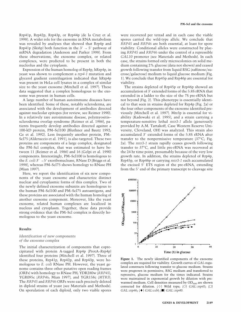
Rrp41p, Rrp42p, Rrp43p, or Rrp44p (de la Cruz et al.1998). A wider role for the exosome in RNA metabolismwas revealed by analyses that showed that Rrp4p andRrp41p (Ski6p) both function in the 38 → 58 pathway ofmRNA degradation (Anderson and Parker 1998). Fromthese observations, the exosome complex, or relatedcomplexes, were predicted to be present in both thenucleolus and the cytoplasm.
Expression of the human homolog of Rrp4p, hRrp4p, inyeast was shown to complement a rrp4-1 mutation andglycerol gradient centrifugation indicated that hRrp4pwas present in HeLa cell lysates in a complex of similarsize to the yeast exosome (Mitchell et al. 1997). Thesedata suggested that a complex homologous to the exo-some was present in human cells.
A large number of human autoimmune diseases havebeen identified. Some of these, notably scleroderma, areassociated with the development of antibodies directedagainst nucleolar epitopes (for review, see Reimer 1990).In a relatively rare autoimmune disease, polymyositis–scleroderma overlap syndrome (Reimer et al. 1986), pa-tients frequently develop antibodies directed against a100-kD protein, PM–Scl100 (Bluthner and Bautz 1992;Ge et al. 1992). Less frequently another protein, PM–Scl75 (Alderuccio et al. 1991), is also targeted. These twoproteins are components of a large complex, designatedthe PM–Scl complex, that was estimated to have be-tween 11 (Reimer et al. 1986) and 16 (Gelpi et al. 1990)components. Interestingly, PM–Scl100 is homologous tothe E. coli 38 →58 exoribonuclease, RNase D (Briggs et al.1998), whereas PM–Scl75 shows homology to RNase PH(Mian 1997).
Here, we report the identification of six new compo-nents of the yeast exosome and characterize distinctnuclear and cytoplasmic forms of this complex. Two ofthe newly defined exosome subunits are homologous tothe human PM–Scl100 and PM–Scl75 autoantigens, andthese proteins are associated with the human homolog ofanother exosome component. Moreover, like the yeastexosome, related human complexes are localized innucleus and cytoplasm. Together, these data providestrong evidence that the PM–Scl complex is directly ho-mologous to the yeast exosome.
Results
Identification of new componentsof the exosome complex
The initial characterization of components that copre-cipitated with protein A-tagged Rrp4p (ProtA–Rrp4p)identified four proteins (Mitchell et al. 1997). Three ofthese proteins, Rrp41p, Rrp42p, and Rrp43p, were ho-mologous to E. coli RNase PH. However, the yeast ge-nome contains three other putative open reading frames(ORFs) with homology to RNase PH; YDR280w (RRP45),YGR095c (RRP46; Mian 1997), and YGR158c (MTR3).The RRP45 and RRP46 ORFs were each precisely deletedin diploid strains of yeast (see Materials and Methods).On sporulation of each diploid, only two viable spores
were recovered per tetrad and in each case the viablespores carried the wild-type allele. We conclude thatRRP45 and RRP46 are both essential, at least for sporeviability. Conditional alleles were constructed by plac-ing RRP45 and RRP46 under the control of a repressibleGAL10 promoter (see Materials and Methods). In eachcase, the strains formed only microcolonies on solid me-dium containing 2% glucose (data not shown) and ceasedgrowth following transfer from liquid RSG (raffinose/su-crose/galactose) medium to liquid glucose medium (Fig.1). We conclude that Rrp45p and Rrp46p are essential forviability.
The strains depleted of Rrp45p or Rrp46p showed anaccumulation of 38 extended forms of the 5.8S rRNA thatextended in a ladder to the size of the 7S pre-rRNA butnot beyond (Fig. 2). This phenotype is essentially identi-cal to that seen in strains depleted for Rrp4p (Fig. 2a) orthe four other components of the exosome identified pre-viously (Mitchell et al. 1997). Mtr3p is essential for vi-ability (Kadowaki et al. 1995), and a strain carrying atemperature-sensitive lethal mtr3-1 allele (generouslyprovided by A.M. Tartakoff, Case Western Reserve Uni-versity, Cleveland, OH) was analyzed. This strain alsoaccumulated 38 extended forms of the 5.8S rRNA aftertransfer to the nonpermissive temperature (37°C; Fig.2a). The mtr3-1 strain rapidly ceases growth followingtransfer to 37°C, and little pre-rRNA was recovered atthe 24 hr time point, presumably because of the very lowgrowth rate. In addition, the strains depleted of Rrp4p,Rrp45p, or Rrp46p or carrying mtr3-1 each accumulatedthe excised 58 ETS region of the pre-rRNA, extendingfrom the 58 end of the primary transcript to cleavage site
Figure 1. The newly identified components of the exosomecomplex are required for viability. Growth curves of GAL-regu-lated constructs following transfer to glucose medium. Strainswere pregrown in permissive, RSG medium and transferred torepressive, glucose medium for the times indicated. Strainswere maintained in exponential growth by dilution with pre-warmed medium. Cell densities measured by OD600 are showncorrected for dilution. (L) Wild type; (s) GAL::rrp45; (h)GAL::rrp46; (l) GAL:csl4; (j) GAL::rrp40.
PM–Scl and the exosome
GENES & DEVELOPMENT 2149
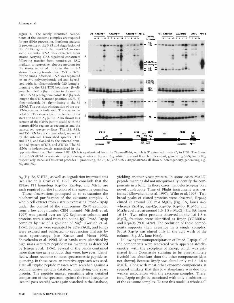
A0 (Fig. 2c; 58 ETS), as well as degradation intermediates(see also de la Cruz et al. 1998). We conclude that theRNase PH homologs Rrp45p, Rrp46p, and Mtr3p areeach required for the function of the exosome complex.
These observations prompted us to re-examine thebiochemical purification of the exosome complex. Awhole-cell extract from a strain expressing ProtA–Rrp4punder the control of the endogenous RRP4 promoterfrom a low-copy-number CEN plasmid (Mitchell et al.1997) was passed over an IgG–Sepharose column, andproteins were eluted from the bound IgG–ProtA–Rrp4pcomplex by use of a gradient of Mg2+ (Gorlich et al.1996). Proteins were separated by SDS-PAGE, and bandswere excised and subjected to sequencing analysis bymass spectroscopy (see Kuster and Mann 1998;Shevchenko et al. 1996). Most bands were identified byhigh mass accuracy peptide mass mapping as describedby Jensen et al. (1996). Several of the bands containedmore than one gene product that were, however, identi-fied without recourse to mass spectrometric peptide se-quencing. In these cases, an iterative approach was used.First all tryptic peptide masses were searched against acomprehensive protein database, identifying one yeastprotein. The peptide masses remaining after detailedcomparison of the spectrum against the found sequence(second pass search), were again searched in the database,
yielding another yeast protein. In some cases MALDIpeptide mapping did not unequivocally identify the com-ponents in a band. In these cases, nanoelectrospray on anovel quadrupole Time of Flight instrument was per-formed (Shevchenko et al. 1997a; Wilm et al. 1996). Twobroad peaks of eluted proteins were observed; Rrp44peluted at around 500 mM MgCl2 (Fig. 3A, lanes 4–6)whereas Rrp41p, Rrp42p, Rrp43p, Rrp45p, Rrp46p, andMtr3p coeluted at around 1.6–1.8 M MgCl2 (Fig. 3A, lanes16–18). Two other proteins observed in the 1.6–1.8 M
MgCl2 fractions were identified as Rrp6p (YOR001w)and Rrp40p (YOL142w). The coelution of these compo-nents supports their presence in a single complex.ProtA–Rrp4p was eluted only in the acid wash of thecolumn (Fig. 3A, lane HAc).
Following immunoprecipitation of ProtA–Rrp4p, all ofthe components were recovered with apparent stoichi-ometry, with the exception of Rrp6p, which was esti-mated from Coomassie staining to be approximatelyfivefold less abundant than the other components (datanot shown). Because Rrp6p was eluted only at 1.6–1.8 M
MgCl2, along with most other exosome components, itseemed unlikely that this low abundance was due to aweaker association with the exosome complex. There-fore, Rrp6p might be associated with only a subfractionof the exosome complex. To test this model, a whole-cell
Figure 2. The newly identified compo-nents of the exosome complex are requiredfor pre-rRNA processing. Northern analysisof processing of the 5.8S and degradation ofthe 58ETS region of the pre-rRNA in exo-some mutants. RNA was extracted fromstrains carrying GAL-regulated constructsfollowing transfer from permissive, RSGmedium to repressive, glucose medium forthe times indicated, or from the mtr3-1strain following transfer from 25°C to 37°Cfor the times indicated. RNA was separatedon an 6% polyacrylamide gel and hybrid-ized with: (a) oligonucleotide 020 (comple-mentary to the 5.8S/ITS2 boundary), (b) oli-gonucleotide 017 (hybridizing to the mature5.8S rRNA), (c) oligonucleotide 033 (hybrid-izing to the 58ETS around position +278). (d)oligonucleotide 041 (hybridizing to the 5SrRNA). The position of migration of the pre-rRNA species is indicated. The species la-beled 58 ETS extends from the transcriptionstart site to site A0 (+610). Also shown is acartoon of the rDNA (not to scale) with themature rRNA regions as rectangles and thetranscribed spacers as lines. The 18S, 5.8S,and 25S rRNAs are cotranscribed, separatedby the internal transcribed spacers (ITS1and ITS2) and flanked by the external tran-scribed spacers (58ETS and 38ETS). The 5SrRNA is independently transcribed in theopposite direction. The mature 5.8S rRNA is synthesized from the 7S pre-rRNA, which is 38 extended to site C2 in ITS2. The 58 endof the 5.8S rRNA is generated by processing at sites at B1L and B1S, which lie about 8 nucleotides apart, generating 5.8SL and 5.8SS,respectively. Because this event precedes 38 processing, the 7S, 6S, and 5.8S + 30 pre-rRNAs all show 58 heterogeneity, generating, e.g.,6SL and 6SS.
Allmang et al.
2150 GENES & DEVELOPMENT

extract from the ProtA–Rrp4p strain was fractionated bycolumn chromatography (see Fig. 3B). Three fractionscontaining ProtA–Rrp4p were recovered (Fig. 3C). The
most abundant complex was recovered in fraction 1; thiscomplex probably corresponds to the major complex pu-rified previously by glycerol gradient centrifugation andimmunoprecipitation (Mitchell et al. 1997). In additionto the previously characterized components of the exo-some, fraction 1 contained Rrp40p, Rrp46p, and Mtr3p.Other protein bands in fraction 1 were identified asCsl4p (YNL232w) and the cytoplasmic Hsp70-like pro-tein Ssa1p (YAL005c), but Rrp6p was not present. Frac-tion 3 contained the same exosome components as frac-tion 1, but lacked Ssa1p and contained Rrp6p. Rrp43p,which comigrates with ProtA–Rrp4p and the IgG heavychain (Mitchell et al. 1997), was identified in fraction 3but not in fraction 1 (Fig. 3C). Csl4p and Rrp45p alsoappear to comigrate in SDS-PAGE; from the bandmarked Csl4p + Rrp45p, only Rrp45p was identifiedfrom the preparation shown in Figure 3A, whereas onlyCsl4p was identified from the preparations shown in Fig-ure 3C. It is, however, very likely that Rrp43p andRrp45p are components of both complexes (see also be-low). Consistent with the recovery of Rrp6p in the totalimmunoprecipitate (Fig. 3A), approximately threefoldless ProtA–Rrp4p was recovered in fraction 3 than infraction 1 (twofold less of the material recovered in frac-tion 1 was loaded onto the gel in Fig. 3C than of thematerial in fractions 2 and 3). Fraction 2 comprises onlyProtA–Rrp4p with Ssa1p, and was approximately four-fold less abundant than fraction 1. Consistent with glyc-erol gradient centrifugation (Mitchell et al. 1997), no freeProtA–Rrp4p was recovered. The ProtA–Rrp4p–Ssa1pcomplex was detected in variable yield on glycerol gra-dients (typically 5%–10% of total ProtA–Rrp4p; Mitchellet al. 1997) and may be due to dissociation of ProtA–Rrp4p from the complex during purification, possibly re-lated to the presence of the protein A tag.
CSL4 was identified previously in a screen for syn-thetic lethality with the chromatin protein Cep1p and isessential for viability (Baker et al. 1998). Conditional al-leles of CSL4 and RRP40 were constructed by placingtheir expression under the control of a GAL10 promoter(see Materials and Methods). In each case, the strainsformed only microcolonies on solid medium containing2% glucose (data not shown). Following transfer fromliquid RSG medium to liquid glucose medium (Fig. 1) thestrains ceased growth and 38 extended forms of the 5.8SrRNA accumulated (Fig. 2a), showing a pattern of inter-mediates similar to other exosome mutants. Depletionof Rrp40p or Csl4p also led to the accumulation of the 58ETS pre-rRNA spacer fragment (Fig. 2c). Therefore, ge-netic depletion of any of the 10 essential componentsidentified by copurification results in very similar de-fects in the processing of the 5.8S rRNA, showing thatthey form a single complex.
RRP6 is not essential for viability (Briggs et al. 1998),and a strain carrying a precise deletion of RRP6 was con-structed (see Materials and Methods). This strain wasimpaired in growth at all temperatures and was nonvi-able at 37°C (temperature-sensitive lethal; data notshown). The rrp6-D strain was defective in the 38 pro-cessing of the 5.8S rRNA, but differed from the other
Figure 3. Fractionation of the exosome complex and identifi-cation of new components. (A) Proteins associated with IgG–Sepharose via binding to ProtA–Rrp4p were eluted using a gra-dient of MgCl2 and analyzed by SDS-PAGE. (Lanes 1–20) Mate-rial eluted with a 100 mM step gradient of MgCl2 concentrationfrom 100 mM (lane 1) to 2 M (lane 20). (HAc) Proteins eluted bythe acid wash. Proteins are visualized by silver staining. Thestrong bands specifically seen in lane 8 were not observed inother experiments. (B) Purification scheme. A whole-cell ex-tract (CXT) was batch-bound to DEAE–Sepharose FF. Boundmaterial was eluted (E300) with TMN buffer containing 300 mM
NaCl/10% glycerol (TMN-300). The eluate, in TMN-100, waspassed through a Mono Q column and bound material waseluted stepwise with TMN-150, TMN-200 (E200), TMN-320(E320), and TMN-500. Material that failed to bind to DEAE–Sepharose FF (FT) was passed through a Mono S column andbound material was eluted with TMN-500 (E500). Each samplewas immunoprecipitated on IgG–Sepharose. (C) Proteins pre-sent in fractions 1, 2, and 3, obtained as outlined in B, wereseparated by SDS-PAGE. Approximately twofold more of thematerial recovered in fractions 2 and 3 was loaded onto the gel,as compared with fraction 1. Proteins positively identified bymass spectroscopy are indicated. Species in brackets were notidentified in the preparations shown but are predicted to bepresent from other analyses. Molecular weight markers are alsoshown. Proteins are visualized by Coomassie staining.
PM–Scl and the exosome
GENES & DEVELOPMENT 2151
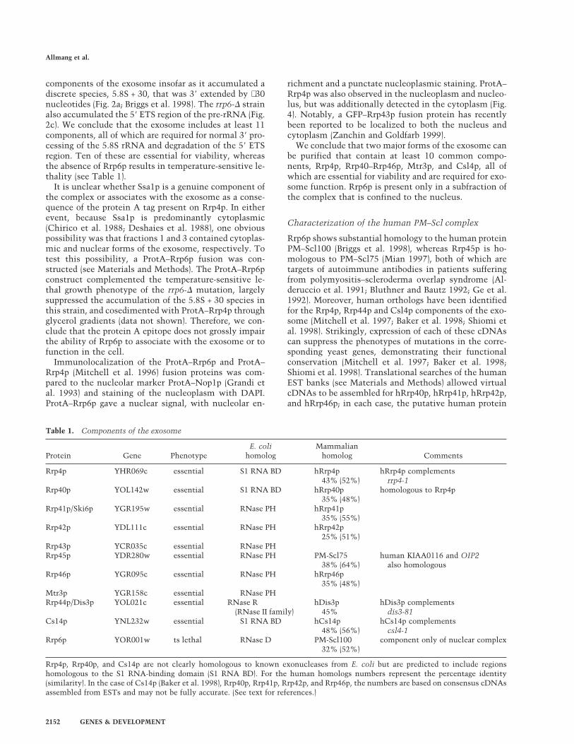
components of the exosome insofar as it accumulated adiscrete species, 5.8S + 30, that was 38 extended by ∼30nucleotides (Fig. 2a; Briggs et al. 1998). The rrp6-D strainalso accumulated the 58 ETS region of the pre-rRNA (Fig.2c). We conclude that the exosome includes at least 11components, all of which are required for normal 38 pro-cessing of the 5.8S rRNA and degradation of the 58 ETSregion. Ten of these are essential for viability, whereasthe absence of Rrp6p results in temperature-sensitive le-thality (see Table 1).
It is unclear whether Ssa1p is a genuine component ofthe complex or associates with the exosome as a conse-quence of the protein A tag present on Rrp4p. In eitherevent, because Ssa1p is predominantly cytoplasmic(Chirico et al. 1988; Deshaies et al. 1988), one obviouspossibility was that fractions 1 and 3 contained cytoplas-mic and nuclear forms of the exosome, respectively. Totest this possibility, a ProtA–Rrp6p fusion was con-structed (see Materials and Methods). The ProtA–Rrp6pconstruct complemented the temperature-sensitive le-thal growth phenotype of the rrp6-D mutation, largelysuppressed the accumulation of the 5.8S + 30 species inthis strain, and cosedimented with ProtA–Rrp4p throughglycerol gradients (data not shown). Therefore, we con-clude that the protein A epitope does not grossly impairthe ability of Rrp6p to associate with the exosome or tofunction in the cell.
Immunolocalization of the ProtA–Rrp6p and ProtA–Rrp4p (Mitchell et al. 1996) fusion proteins was com-pared to the nucleolar marker ProtA–Nop1p (Grandi etal. 1993) and staining of the nucleoplasm with DAPI.ProtA–Rrp6p gave a nuclear signal, with nucleolar en-
richment and a punctate nucleoplasmic staining. ProtA–Rrp4p was also observed in the nucleoplasm and nucleo-lus, but was additionally detected in the cytoplasm (Fig.4). Notably, a GFP–Rrp43p fusion protein has recentlybeen reported to be localized to both the nucleus andcytoplasm (Zanchin and Goldfarb 1999).
We conclude that two major forms of the exosome canbe purified that contain at least 10 common compo-nents, Rrp4p, Rrp40–Rrp46p, Mtr3p, and Csl4p, all ofwhich are essential for viability and are required for exo-some function. Rrp6p is present only in a subfraction ofthe complex that is confined to the nucleus.
Characterization of the human PM–Scl complex
Rrp6p shows substantial homology to the human proteinPM–Scl100 (Briggs et al. 1998), whereas Rrp45p is ho-mologous to PM–Scl75 (Mian 1997), both of which aretargets of autoimmune antibodies in patients sufferingfrom polymyositis–scleroderma overlap syndrome (Al-deruccio et al. 1991; Bluthner and Bautz 1992; Ge et al.1992). Moreover, human orthologs have been identifiedfor the Rrp4p, Rrp44p and Csl4p components of the exo-some (Mitchell et al. 1997; Baker et al. 1998; Shiomi etal. 1998). Strikingly, expression of each of these cDNAscan suppress the phenotypes of mutations in the corre-sponding yeast genes, demonstrating their functionalconservation (Mitchell et al. 1997; Baker et al. 1998;Shiomi et al. 1998). Translational searches of the humanEST banks (see Materials and Methods) allowed virtualcDNAs to be assembled for hRrp40p, hRrp41p, hRrp42p,and hRrp46p; in each case, the putative human protein
Table 1. Components of the exosome
Protein Gene PhenotypeE. coli
homologMammalian
homolog Comments
Rrp4p YHR069c essential S1 RNA BD hRrp4p43% (52%)
hRrp4p complementsrrp4-1
Rrp40p YOL142w essential S1 RNA BD hRrp40p35% (48%)
homologous to Rrp4p
Rrp41p/Ski6p YGR195w essential RNase PH hRrp41p35% (55%)
Rrp42p YDL111c essential RNase PH hRrp42p25% (51%)
Rrp43p YCR035c essential RNase PHRrp45p YDR280w essential RNase PH PM-Scl75
38% (64%)human KIAA0116 and OIP2
also homologousRrp46p YGR095c essential RNase PH hRrp46p
35% (48%)Mtr3p YGR158c essential RNase PHRrp44p/Dis3p YOL021c essential RNase R hDis3p hDis3p complements
(RNase II family) 45% dis3-81Cs14p YNL232w essential S1 RNA BD hCs14p
48% (56%)hCs14p complements
csl4-1Rrp6p YOR001w ts lethal RNase D PM-Scl100
32% (52%)component only of nuclear complex
Rrp4p, Rrp40p, and Cs14p are not clearly homologous to known exonucleases from E. coli but are predicted to include regionshomologous to the S1 RNA-binding domain (S1 RNA BD). For the human homologs numbers represent the percentage identity(similarity). In the case of Cs14p (Baker et al. 1998), Rrp40p, Rrp41p, Rrp42p, and Rrp46p, the numbers are based on consensus cDNAsassembled from ESTs and may not be fully accurate. (See text for references.)
Allmang et al.
2152 GENES & DEVELOPMENT

showed high homology to the yeast protein (Table 1). Inaddition, two other genes, KIAA0116 and OIP2, werefound to be homologous to Rrp45p, although less so thanPM–Scl75. For hRrp40p and hRrp46p, apparent productsof alternative splicing were evident when the EST se-quences were assembled into contigs (data not shown).In some cases, these alternative forms may have led to anoverestimation of the number of discrete protein speciesin the PM–Scl complex.
Previous analyses showed that hRrp4p is present in alarge complex (Mitchell et al. 1997). To determinewhether the human homologs of other exosome compo-nents are present in the same complex, HeLa cell nuclearand cytoplasmic extracts (generously provided by JuanValcarcel, EMBL, or prepared as described in Materialsand Methods) were fractionated by glycerol gradient cen-trifugation. Fractions were analyzed by Western blottingwith human autoimmune serum (generously providedby Walter van Venrooij, University of Nijmegen, The
Netherlands) or antibodies raised against recombinanthRrp4p (Mitchell et al. 1997; Fig. 5). In the nuclear ex-tract, PM–Scl75 and an uncharacterized protein of ∼25kD that is also a target of the autoimmune serum (PM–Scl25) cosedimented with hRrp4p, with a peak in frac-tions 13 and 14. PM–Scl100 also showed substantialcosedimentation (Fig. 5A). The band at 45 kD is likely tobe the species previously reported to cross-react withanti-PM–Scl75 antibodies (Alderuccio et al. 1991). PM–Scl100 was not detected in the cytoplasmic extract, butPM–Scl75 and hRrp4p cosedimented (Fig. 5B), as didPM–Scl25 (data not shown), with a peak in fractions 13and 14.
To confirm the association between PM–Scl100 andhRrp4p in the HeLa cell nuclear extract, immunoprecipi-tation was performed (Fig. 6). Three different autoim-mune sera (sera 1–3) were used, each of which reacted
Figure 4. Rrp4p and Rrp6p differ in their nuclear-cytoplasmicdistribution. (A) Strains expressing ProtA–Rrp4p, ProtA–Rrp6p,or ProtA–Nop1p were examined by indirect immunofluores-cence using an anti-protein A antibody coupled to Texas Red.Also shown is the position of the DNA, visualized by DAPIstaining. The combined image is pseudocolored with DAPI ingreen and Texas Red in red. For each tagged strain an otherwiseisogenic wild-type control strain was also analyzed. The wild-type strain shown (P51) is isogenic with the ProtA–Rrp4p strain(see Table 2). (B) Higher resolution images are shown for theProtA–Rrp4p and ProtA–Rrp6p to show the punctate stainingpattern.
Figure 5. Cosedimentation of hRrp4p and the PM–Scl com-plex. (A) HeLa cell nuclear extract. (B) HeLa cell cytoplasmicextract. Cell extracts were fractionated by glycerol gradient cen-trifugation. Samples were analyzed by Western blotting deco-rated with human autoimmune antisera reactive against PM–Scl100, PM–Scl75, and a previously uncharacterized humanprotein (PM–Scl25) or with rabbit antiserum raised against re-combinant hRrp4p. The serum also cross-reacts with an unre-lated 45-kD protein. Also shown is the sedimentation of mo-lecular weight markers on a gradient run in parallel with thenuclear extract. Markers: (A) alcohol deyhdrogenase from yeast(7.4S); (B) bovine serum albumin (4.3S); (C) bovine catalase(11.3S; Siegel and Monty 1966).
PM–Scl and the exosome
GENES & DEVELOPMENT 2153
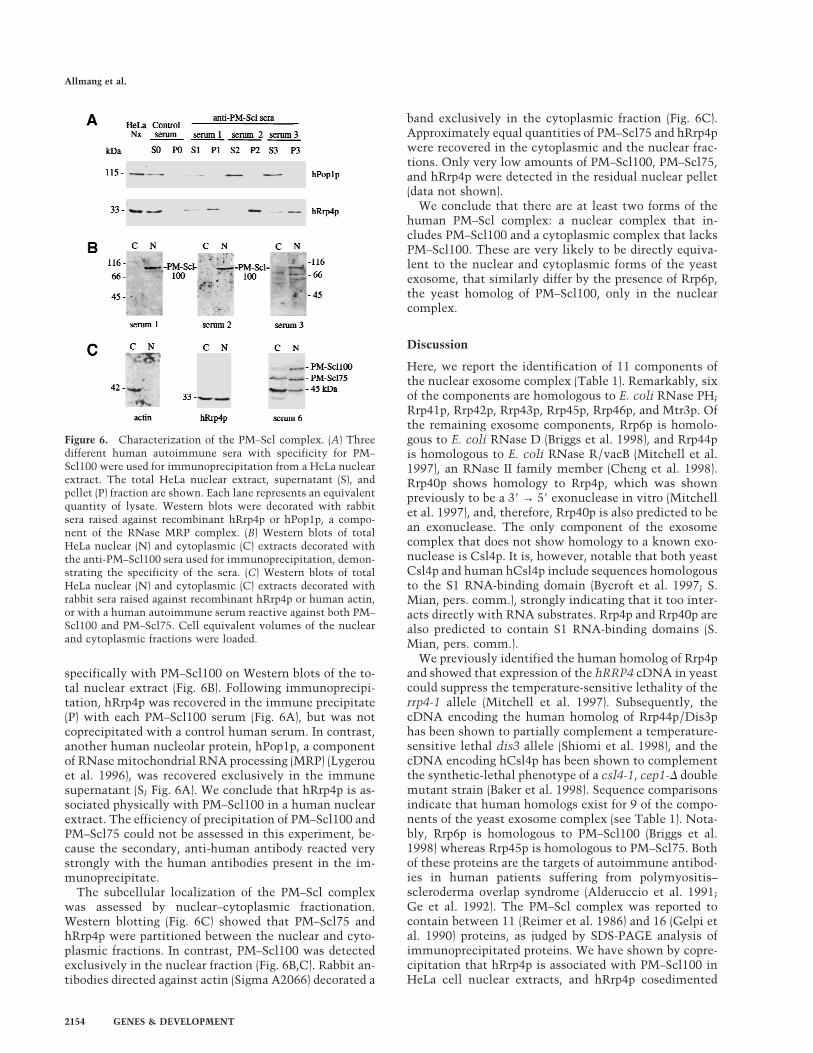
specifically with PM–Scl100 on Western blots of the to-tal nuclear extract (Fig. 6B). Following immunoprecipi-tation, hRrp4p was recovered in the immune precipitate(P) with each PM–Scl100 serum (Fig. 6A), but was notcoprecipitated with a control human serum. In contrast,another human nucleolar protein, hPop1p, a componentof RNase mitochondrial RNA processing (MRP) (Lygerouet al. 1996), was recovered exclusively in the immunesupernatant (S; Fig. 6A). We conclude that hRrp4p is as-sociated physically with PM–Scl100 in a human nuclearextract. The efficiency of precipitation of PM–Scl100 andPM–Scl75 could not be assessed in this experiment, be-cause the secondary, anti-human antibody reacted verystrongly with the human antibodies present in the im-munoprecipitate.
The subcellular localization of the PM–Scl complexwas assessed by nuclear–cytoplasmic fractionation.Western blotting (Fig. 6C) showed that PM–Scl75 andhRrp4p were partitioned between the nuclear and cyto-plasmic fractions. In contrast, PM–Scl100 was detectedexclusively in the nuclear fraction (Fig. 6B,C). Rabbit an-tibodies directed against actin (Sigma A2066) decorated a
band exclusively in the cytoplasmic fraction (Fig. 6C).Approximately equal quantities of PM–Scl75 and hRrp4pwere recovered in the cytoplasmic and the nuclear frac-tions. Only very low amounts of PM–Scl100, PM–Scl75,and hRrp4p were detected in the residual nuclear pellet(data not shown).
We conclude that there are at least two forms of thehuman PM–Scl complex: a nuclear complex that in-cludes PM–Scl100 and a cytoplasmic complex that lacksPM–Scl100. These are very likely to be directly equiva-lent to the nuclear and cytoplasmic forms of the yeastexosome, that similarly differ by the presence of Rrp6p,the yeast homolog of PM–Scl100, only in the nuclearcomplex.
Discussion
Here, we report the identification of 11 components ofthe nuclear exosome complex (Table 1). Remarkably, sixof the components are homologous to E. coli RNase PH;Rrp41p, Rrp42p, Rrp43p, Rrp45p, Rrp46p, and Mtr3p. Ofthe remaining exosome components, Rrp6p is homolo-gous to E. coli RNase D (Briggs et al. 1998), and Rrp44pis homologous to E. coli RNase R/vacB (Mitchell et al.1997), an RNase II family member (Cheng et al. 1998).Rrp40p shows homology to Rrp4p, which was shownpreviously to be a 38 → 58 exonuclease in vitro (Mitchellet al. 1997), and, therefore, Rrp40p is also predicted to bean exonuclease. The only component of the exosomecomplex that does not show homology to a known exo-nuclease is Csl4p. It is, however, notable that both yeastCsl4p and human hCsl4p include sequences homologousto the S1 RNA-binding domain (Bycroft et al. 1997; S.Mian, pers. comm.), strongly indicating that it too inter-acts directly with RNA substrates. Rrp4p and Rrp40p arealso predicted to contain S1 RNA-binding domains (S.Mian, pers. comm.).
We previously identified the human homolog of Rrp4pand showed that expression of the hRRP4 cDNA in yeastcould suppress the temperature-sensitive lethality of therrp4-1 allele (Mitchell et al. 1997). Subsequently, thecDNA encoding the human homolog of Rrp44p/Dis3phas been shown to partially complement a temperature-sensitive lethal dis3 allele (Shiomi et al. 1998), and thecDNA encoding hCsl4p has been shown to complementthe synthetic-lethal phenotype of a csl4-1, cep1-D doublemutant strain (Baker et al. 1998). Sequence comparisonsindicate that human homologs exist for 9 of the compo-nents of the yeast exosome complex (see Table 1). Nota-bly, Rrp6p is homologous to PM–Scl100 (Briggs et al.1998) whereas Rrp45p is homologous to PM–Scl75. Bothof these proteins are the targets of autoimmune antibod-ies in human patients suffering from polymyositis–scleroderma overlap syndrome (Alderuccio et al. 1991;Ge et al. 1992). The PM–Scl complex was reported tocontain between 11 (Reimer et al. 1986) and 16 (Gelpi etal. 1990) proteins, as judged by SDS-PAGE analysis ofimmunoprecipitated proteins. We have shown by copre-cipitation that hRrp4p is associated with PM–Scl100 inHeLa cell nuclear extracts, and hRrp4p cosedimented
Figure 6. Characterization of the PM–Scl complex. (A) Threedifferent human autoimmune sera with specificity for PM–Scl100 were used for immunoprecipitation from a HeLa nuclearextract. The total HeLa nuclear extract, supernatant (S), andpellet (P) fraction are shown. Each lane represents an equivalentquantity of lysate. Western blots were decorated with rabbitsera raised against recombinant hRrp4p or hPop1p, a compo-nent of the RNase MRP complex. (B) Western blots of totalHeLa nuclear (N) and cytoplasmic (C) extracts decorated withthe anti-PM–Scl100 sera used for immunoprecipitation, demon-strating the specificity of the sera. (C) Western blots of totalHeLa nuclear (N) and cytoplasmic (C) extracts decorated withrabbit sera raised against recombinant hRrp4p or human actin,or with a human autoimmune serum reactive against both PM–Scl100 and PM–Scl75. Cell equivalent volumes of the nuclearand cytoplasmic fractions were loaded.
Allmang et al.
2154 GENES & DEVELOPMENT
with PM–Scl75 and PM–Scl25 in both nuclear and cyto-plasmic extracts. Homologs of at least three componentsof the exosome are present in the PM–Scl complex, pro-viding strong evidence that these complexes are directlyhomologous.
Six human homologs of RNase PH were identified.These do not, however, have a 1:1 relationship with thesix RNase PH homologs in the exosome. No clear hu-man homologs were identified for Rrp43p or Mtr3p.Searches of the EST banks with these proteins identifiedESTs related to KIAA0116 and OIP2; these sequencesare, however, more homologous to Rrp45p than to theother yeast PH homologs (although less so than PM–Scl75). A probable interpretation is that yeast and hu-mans have the same number of RNase PH homologs, butthat some drift has occurred with duplicates of theRRP45/PM–Scl75 gene replacing other species.
Mutations in individual components of the yeast exo-some inhibited both nucleolar pre-rRNA processing andcytoplasmic mRNA turnover (Anderson and Parker1998), indicating that related complexes are present inthe nucleus and the cytoplasm. Moreover, a mutation inMtr3p leads to nuclear accumulation of poly(A)+ RNA(Kadowaki et al. 1995), as does a mutation in Dob1p/Mtr4p (de la Cruz et al. 1998; Liang et al. 1996), a puta-tive RNA helicase required in addition to the exosomefor 5.8S rRNA 38 end maturation and degradation of the58 ETS fragment (de la Cruz et al. 1998). These observa-tions suggest that the exosome may also play some rolein nucleoplasmic RNA turnover or processing. Consis-tent with this hypothesis, GFP–Rrp43p (Zanchin andGoldfarb 1999) and ProtA–Rrp4p were detected in thenucleolus, nucleoplasm, and cytoplasm. In contrast,ProtA–Rrp6p was found to be exclusively nuclear, with anucleolar enrichment. Two complexes could also beseparated biochemically; these include 10 common com-ponents and differ in the presence of either Ssa1p, a cy-toplasmic Hsp70-like protein (Chirico et al. 1988; De-shaies et al. 1988), or Rrp6p. The form lacking Rrp6p ispresumed to be the cytoplasmic exosome complex, a pro-posal supported by the presence of Ssa1p. Approximatelythreefold more of this complex was recovered than theputative nuclear exosome that includes Rrp6p. HumanPM–Scl100 was also restricted to the nucleus, while PM–Scl75, PM–Scl25, and hRrp4p partition between thenucleus and cytoplasm. The reported nucleolar enrich-ment of the human PM–Scl complex is probably a con-sequence of the immunodominance of PM–Scl100 in au-toimmune sera (Ge et al. 1992; Gelpi et al. 1990). In fact,approximately equal amounts of the human nuclear andcytoplasmic complexes were recovered following subcel-lular fractionation.
We conclude that there are two forms of the exosome/PM–Scl complex in the nucleus and the cytoplasm thatcan be distinguished by the presence of Rrp6p/PM–Scl100 specifically in the nuclear form.
Rrp6p is not essential for viability, in contrast to theother 10 components of the exosome complex, althoughrrp6-D strains are severely impaired in growth and aretemperature sensitive. Therefore, the exosome is there-
fore predicted to retain at least partial function in theabsence of Rrp6p, a view supported by the observationthat the major form of the complex lacks this protein.Conversely, all of the PM–Scl100 present in Hela celllysates appeared to be associated with the PM–Scl com-plex, suggesting that Rrp6p/PM–Scl100 may not func-tion independently of the complex in vivo.
In E. coli, the homologs of the exosome componentsare not present in a related complex. However, the de-gradosome complex includes another 38 → 58 exonucle-ase, PNPase, together with the endonuclease and exo-nuclease RNase E and the putative RNA helicase RhlB(Carpousis et al. 1994; Py et al. 1996; Mackie 1998;Vanzo et al. 1998). It appears that throughout evolution,major activities involved in RNA processing and degra-dation have been assembled into large complexes, possi-bly to allow their coordinate regulation. The composi-tion of these complexes are, however, very different inbacteria and eukaryotes.
Materials and methods
Strains and media
Except where stated, strains were grown in liquid or on solidminimal medium containing 0.67% yeast nitrogen base(DIFCO) and 2% glucose with appropriate supplements. Fordepletion, strains carrying GAL-regulated constructs werepregrown in RSG (2% peptone, 1% yeast extract, 2% raffinose,2% sucrose, 2% galactose) and transferred to YPD (2% peptone,1% yeast extract, 2% glucose).
Yeast strains used and constructed in this study are listed inTable 2. Gene disruptions of RRP45 and RRP46 were generatedby a PCR strategy in the diploid strain BMA38 (Baudin et al.1993) resulting in the replacement of the complete ORF by anauxotrophic marker (see Table 2). Successful disruption wasconfirmed by Southern hybridization. Chromosomal DNA fromthe RRP45/rrp45::TRP1 and RRP46/rrp46::HIS3 strains was di-gested by EcoRI–HindIII or KpnI–EcoRI, respectively, and hy-bridized with a probe derived from the PCR products that wereused for transformation. Twelve tetrads from the RRP45/rrp45::TRP1 strain and eight tetrads from RRP46/rrp46::HIS3strain were dissected on YPD plates and incubated for 6 days at23°C. Each showed 2:2 segregation for spore viability. All viablespores were auxotrophic for tryptophan or histidine, respec-tively, indicating that the disrupted alleles were lethal. Thenonessential RRP6 gene was disrupted in the haploid strainYBD38 (see Table 2) by use of the Kluyveromyces lactis TRPmarker, obtained by PCR amplification from plasmid pBS1408(generously provided by Bertrand Seraphin, EMBL, Heidelberg,Germany).
The oligonucleotides used to construct and test the gene dis-ruptions were 58RRP45::TRP1 (807); 38RRP45::TRP (808); 58
RRP46::HIS3 (809); 38RRP46::HIS3 (810); 58RRP6::Kl TRP (811);38RRP6::Kl TRP (812). Test oligonucleotides were 38RRP45(813); 38RRP6 (815); Sc TRP (816); HIS (817); Kl TRP (818) (fullsequences are available from the authors).
Conditional mutants under the control of the inducibleGAL10 promoter were generated for the RRP40, RRP45, RRP46,and CSL4 genes by a one-step PCR strategy in the YDL401strain (Lafontaine and Tollervey 1996). Transformants were se-lected for His+ prototrophy and screened by PCR.
The oligonucleotides used to construct the conditional mu-tants were 58GAL-RRP45 (819); 38GAL-RRP45 (820); 58GAL-
PM–Scl and the exosome
GENES & DEVELOPMENT 2155
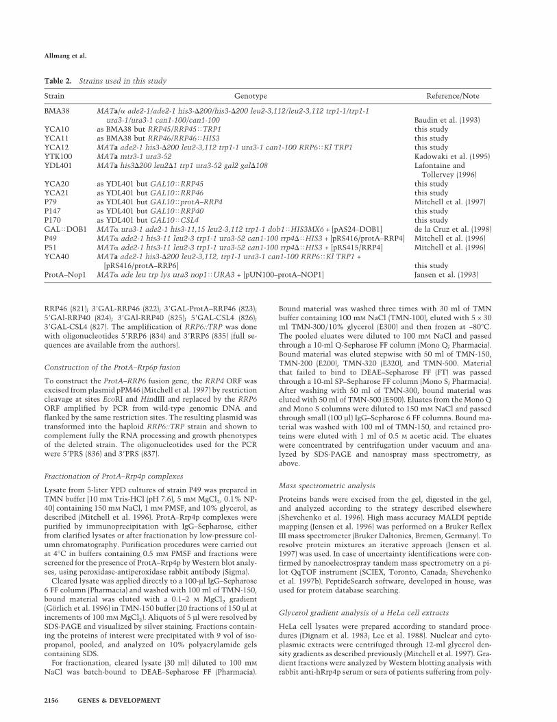
RRP46 (821); 38GAL-RRP46 (822); 38GAL-ProtA–RRP46 (823);58GAl-RRP40 (824); 38GAl-RRP40 (825); 58GAL-CSL4 (826);38GAL-CSL4 (827). The amplification of RRP6::TRP was donewith oligonucleotides 58RRP6 (834) and 38RRP6 (835) (full se-quences are available from the authors).
Construction of the ProtA–Rrp6p fusion
To construct the ProtA–RRP6 fusion gene, the RRP4 ORF wasexcised from plasmid pPM46 (Mitchell et al. 1997) by restrictioncleavage at sites EcoRI and HindIII and replaced by the RRP6ORF amplified by PCR from wild-type genomic DNA andflanked by the same restriction sites. The resulting plasmid wastransformed into the haploid RRP6::TRP strain and shown tocomplement fully the RNA processing and growth phenotypesof the deleted strain. The oligonucleotides used for the PCRwere 58PRS (836) and 38PRS (837).
Fractionation of ProtA–Rrp4p complexes
Lysate from 5-liter YPD cultures of strain P49 was prepared inTMN buffer [10 mM Tris-HCl (pH 7.6), 5 mM MgCl2, 0.1% NP-40] containing 150 mM NaCl, 1 mM PMSF, and 10% glycerol, asdescribed (Mitchell et al. 1996). ProtA–Rrp4p complexes werepurified by immunoprecipitation with IgG–Sepharose, eitherfrom clarified lysates or after fractionation by low-pressure col-umn chromatography. Purification procedures were carried outat 4°C in buffers containing 0.5 mM PMSF and fractions werescreened for the presence of ProtA–Rrp4p by Western blot analy-ses, using peroxidase-antiperoxidase rabbit antibody (Sigma).
Cleared lysate was applied directly to a 100-µl IgG–Sepharose6 FF column (Pharmacia) and washed with 100 ml of TMN-150,bound material was eluted with a 0.1–2 M MgCl2 gradient(Gorlich et al. 1996) in TMN-150 buffer (20 fractions of 150 µl atincrements of 100 mM MgCl2). Aliquots of 5 µl were resolved bySDS-PAGE and visualized by silver staining. Fractions contain-ing the proteins of interest were precipitated with 9 vol of iso-propanol, pooled, and analyzed on 10% polyacrylamide gelscontaining SDS.
For fractionation, cleared lysate (30 ml) diluted to 100 mM
NaCl was batch-bound to DEAE–Sepharose FF (Pharmacia).
Bound material was washed three times with 30 ml of TMNbuffer containing 100 mM NaCl (TMN-100), eluted with 5 × 30ml TMN-300/10% glycerol (E300) and then frozen at −80°C.The pooled eluates were diluted to 100 mM NaCl and passedthrough a 10-ml Q-Sepharose FF column (Mono Q; Pharmacia).Bound material was eluted stepwise with 50 ml of TMN-150,TMN-200 (E200), TMN-320 (E320), and TMN-500. Materialthat failed to bind to DEAE–Sepharose FF (FT) was passedthrough a 10-ml SP–Sepharose FF column (Mono S; Pharmacia).After washing with 50 ml of TMN-300, bound material waseluted with 50 ml of TMN-500 (E500). Eluates from the Mono Qand Mono S columns were diluted to 150 mM NaCl and passedthrough small (100 µl) IgG–Sepharose 6 FF columns. Bound ma-terial was washed with 100 ml of TMN-150, and retained pro-teins were eluted with 1 ml of 0.5 M acetic acid. The eluateswere concentrated by centrifugation under vacuum and ana-lyzed by SDS-PAGE and nanospray mass spectrometry, asabove.
Mass spectrometric analysis
Proteins bands were excised from the gel, digested in the gel,and analyzed according to the strategy described elsewhere(Shevchenko et al. 1996). High mass accuracy MALDI peptidemapping (Jensen et al. 1996) was performed on a Bruker ReflexIII mass spectrometer (Bruker Daltonics, Bremen, Germany). Toresolve protein mixtures an iterative approach (Jensen et al.1997) was used. In case of uncertainty identifications were con-firmed by nanoelectrospray tandem mass spectrometry on a pi-lot QqTOF instrument (SCIEX, Toronto, Canada; Shevchenkoet al. 1997b). PeptideSearch software, developed in house, wasused for protein database searching.
Glycerol gradient analysis of a HeLa cell extracts
HeLa cell lysates were prepared according to standard proce-dures (Dignam et al. 1983; Lee et al. 1988). Nuclear and cyto-plasmic extracts were centrifuged through 12-ml glycerol den-sity gradients as described previously (Mitchell et al. 1997). Gra-dient fractions were analyzed by Western blotting analysis withrabbit anti-hRrp4p serum or sera of patients suffering from poly-
Table 2. Strains used in this study
Strain Genotype Reference/Note
BMA38 MATa/a ade2-1/ade2-1 his3-D200/his3-D200 leu2-3,112/leu2-3,112 trp1-1/trp1-1ura3-1/ura3-1 can1-100/can1-100 Baudin et al. (1993)
YCA10 as BMA38 but RRP45/RRP45<TRP1 this studyYCA11 as BMA38 but RRP46/RRP46<HIS3 this studyYCA12 MATa ade2-1 his3-D200 leu2-3,112 trp1-1 ura3-1 can1-100 RRP6<Kl TRP1 this studyYTK100 MATa mtr3-1 ura3-52 Kadowaki et al. (1995)YDL401 MATa his3D200 leu2D1 trp1 ura3-52 gal2 galD108 Lafontaine and
Tollervey (1996)YCA20 as YDL401 but GAL10<RRP45 this studyYCA21 as YDL401 but GAL10<RRP46 this studyP79 as YDL401 but GAL10<protA–RRP4 Mitchell et al. (1997)P147 as YDL401 but GAL10<RRP40 this studyP170 as YDL401 but GAL10<CSL4 this studyGAL<DOB1 MATa ura3-1 ade2-1 his3-11,15 leu2-3,112 trp1-1 dob1<HIS3MX6 + [pAS24–DOB1] de la Cruz et al. (1998)P49 MATa ade2-1 his3-11 leu2-3 trp1-1 ura3-52 can1-100 rrp4D<HIS3 + [pRS416/protA–RRP4] Mitchell et al. (1996)P51 MATa ade2-1 his3-11 leu2-3 trp1-1 ura3-52 can1-100 rrp4D<HIS3 + [pRS415/RRP4] Mitchell et al. (1996)YCA40 MATa ade2-1 his3-D200 leu2-3,112, trp1-1 ura3-1 can1-100 RRP6<Kl TRP1 +
[pRS416/protA–RRP6] this studyProtA–Nop1 MATa ade leu trp lys ura3 nop1<URA3 + [pUN100–protA–NOP1] Jansen et al. (1993)
Allmang et al.
2156 GENES & DEVELOPMENT
myositis–scleroderma overlap syndrome [kindly provided byDr. W. van Venrooij and obtained from the University Hospital(St. Radboud) of Nijmegen].
Immunofluorescence
Cells were grown in selective medium to mid-exponentialphase, fixed by incubation in 4% (vol/vol) formaldehyde for 1 hrat room temperature, and spheroplasted. Then immunofluores-cence was then performed as described previously (Berges et al.1994; Grandi et al. 1993). Protein A fusions were detected witha rabbit anti-protein A antibody (Sigma) and a secondary goatanti-rabbit antibody coupled to Texas Red (Dianova) at a 1:100and 1:200 dilution, respectively. To stain nuclear DNA, DAPIwas included in the mounting medium (Vectashield, VectorLaboratories).
Immunoprecipitation of the PM–Scl complex withpatient sera
Patient sera directed specifically against PM–Scl100 (kindly pro-vided by Dr. W. van Venrooij) were used for the immunopre-cipitation experiments. HeLa cell lysates were prepared as de-scribed above. A 50% solution of protein A–Sepharose beads(10µl; Pharmacia) was washed three times in IPP 500 [500 mM
NaCl, 10 mM Tris-HCl (pH 8), 0.1% NP-40, 0.5 mM PMSF] andincubated for 1 hr at room temperature with 5 µl of humanautoimmune sera. Beads were washed three times with IPP500,transferred in 10 µl of IPP150 ([50 mM NaCl, 10 mM Tris-HCl(pH 8), 0.1% NP-40, 0.5 mM PMSF] and then added to 10 µl ofHeLa cell nuclear extract. After incubation for 2 hr at 4°C, thesupernatant was recovered and beads were washed four timeswith IPP150. Bound proteins were eluted from the beads by a 5min boiling in protein gel loading buffer. Total, supernatant,and pellet proteins were analyzed by SDS-PAGE and Westernblotting analysis with anti-hRrp4p serum or affinity-purifiedanti-hPop1 antibodies (Lygerou et al. 1996; Mitchell et al. 1997).
RNA analysis
RNA isolation and Northern blot hybridization were performedas described previously (Beltrame and Tollervey 1992; Tollervey1987). Oligonucleotides used for rRNA and pre-rRNA analysiswere 58-TGAGAAGGAAATGACGCT (oligonucleotide 020),58-GCGTTGTTCATCGATGC (oligonucleotide 017), 58-CGC-TGCTCACAATGG (oligonucleotide 033), and 58-CTACTCG-GTCAGGCTC (oligonucleotide 014).
Database searches
The human EST banks were searched using the EFEAME p2nprogram for translational frame-shifting, on the Bioacceleratorof the European Molecular Biology Laboratory (http://ww-w.embl-heidelberg.de). Contigs were assembled from the re-trieved ESTs by use of the Gene JockeyII program. Homologywas calculated by use of using the Bestfit program [WisconsinPackage Version 9.1, Genetics Computer Group (GCG), Madi-son, WI.].
The ESTs used for the alignments were hRRP40: HS103148,AA916866, AA715297, AA909843, AA829746, AA760696,AA748308, AA747303, HSA01383, HS479237, HS417169,HS1213865, HS1191331, HS1186630, AA937191, AA741488,HSA57832, HSA01383, HS620247, HS617138, AA736510,HS1300540, HS1273716, HS1269362, HS1229711, HS1198690,HS1191331, and HS1174014; hRRP41: HS0229, HSZZ84720,HS462881, HS1210855, HS060127, and HSAA29848; hRRP42:
AA654791, HS599371, HSZZ85135, HS20834, AA581010,HS414162, HS979316, and HSZZ84357; hRRP46: HS078341,HS84856, HS1255212, HS1226957, HSZZ41259, HS1256223,HS1225454, HS1249336, and HS1172072.
Acknowledgments
We thank Walter van Venrooij for providing the PM–Scl sera,Juan Balcacel for providing HeLa cell nuclear and cytoplasmicextract, Alan Tartakoff for the mtr3-1 strain and BertrandSeraphin for pBS1408. We thank Roy Parker for pointing out thehomology between Rrp41p and Mtr3p and Saira Mian for point-ing out the homology between Rrp4p and Rrp40p, as well as theputative S1 DNA binding domains in Rrp4p, Rrp40p, and Csl4p.This work was supported by the Wellcome Trust.
The publication costs of this article were defrayed in part bypayment of page charges. This article must therefore be herebymarked ‘advertisement’ in accordance with 18 USC section1734 solely to indicate this fact.
References
Alderuccio, F., E.K. Chan, and E.M. Tan. 1991. Molecular char-acterization of an autoantigen of PM–Scl in the polymyosi-tis/scleroderma overlap syndrome: A unique and completehuman cDNA encoding an apparent 75-kD acidic protein ofthe nucleolar complex. J. Exp. Med. 173: 941–952.
Anderson, J.S.J. and R.P. Parker. 1998. The 38 to 58 degradationof yeast mRNAs is a general mechanism for mRNA turnoverthat requires the SKI2 DEVH box protein and 38 to 58 exo-nucleases of the exosome complex. EMBO J. 17: 1497–1506.
Baker, R.E., K. Harris, and K. Zhang. 1998. Mutations syntheti-cally lethal with cep1 target S. cerevisiae kinetochore com-ponents. Genetics 149: 73–85.
Baudin, A., O. Ozier-Kalogeropoulos, A. Denouel, F. Lacroute,and C. Cullin. 1993. A simple and efficient method for directgene deletion in Saccharomyces cerevisiae. Nucleic AcidsRes. 21: 3329–3330.
Beltrame, M. and D. Tollervey. 1992. Identification and func-tional analysis of two U3 binding sites on yeast pre-ribo-somal RNA. EMBO J. 11: 1531–1542.
Berges, T., E. Petfalski, D. Tollervey, and E.C. Hurt. 1994. Syn-thetic lethality with fibrillarin identifies NOP77p, a nucleo-lar protein required for pre-rRNA processing and modifica-tion. EMBO J. 13: 3136–3148.
Bluthner, M. and F.A. Bautz. 1992. Cloning and characterizationof the cDNA coding for a polymyositis-scleroderma overlapsyndrome-related nucleolar 100-kD protein. J. Exp. Med.176: 973–980.
Briggs, M.W., K.T. Burkard, and J.S. Butler. 1998. Rrp6p, theyeast homologue of the human PM-Scl 100-kDa autoanti-gen, is essential for efficient 5.8 S rRNA 38 end formation. J.Biol. Chem. 273: 13255–13263.
Bycroft, M., T.J. Hubbard, M. Proctor, S.M. Freund, and A.G.Murzin. 1997. The solution structure of the S1 RNA bindingdomain: A member of an ancient nucleic acid-binding fold.Cell 88: 235–342.
Carpousis, A.J., G. Van Houwe, C. Ehretsmann, and H.M.Krisch. 1994. Copurification of E. coli RNAase E and PN-Pase: Evidence for a specific association between two en-zymes important in RNA processing and degradation. Cell76: 889–900.
Cheng, Z.F., Y. Zuo, Z. Li, K.E. Rudd, and M.P. Deutscher. 1998.The vacB gene required for virulence in Shigella flexneri and
PM–Scl and the exosome
GENES & DEVELOPMENT 2157

Escherichia coli encodes the exoribonuclease RNase R. J.Biol. Chem. 273: 14077–14080.
Chirico, W.J., M.G. Waters, and G. Blobel. 1988. 70K heat shockrelated proteins stimulate protein translocation into micro-somes. Nature 332: 805–810.
de la Cruz, J., D. Kressler, D. Tollervey, and P. Linder. 1998.Dob1p (Mtr4p) is a putative ATP-dependent RNA helicaserequired for the 38 end formation of 5.8S rRNA in Saccharo-myces cerevisiae. EMBO J 17: 1128–1140.
Deshaies, R.J., B.D. Koch, M. Werner-Washburne, E.A. Craig,and R. Schekman. 1988. A subfamily of stress proteins fa-cilitates translocation of secretory and mitochondrial pre-cursor polypeptides. Nature 332: 800–805.
Dignam, J.D., P.L. Martin, B.S. Shastry, and R.G. Roeder. 1983.Eukaryotic gene transcription with purified components.Methods Enzymol. 101: 582–598.
Ge, Q., M.B. Frank, C. O’Brien, and I.N. Targoff. 1992. Cloningof a complementary DNA coding for the 100-kD antigenicprotein of the PM-Scl autoantigen. J. Clin. Invest. 90: 559–570.
Gelpi, C., A. Alguero, M. Angeles Martinez, S. Vidal, C. Juarez,and J.L. Rodriguez-Sanchez. 1990. Identification of proteincomponents reactive with anti-PM/Scl autoantibodies. Clin.Exp. Immunol. 81: 59–64.
Gorlich, D., R. Kraft, S. Kostka, F. Vogel, E. Hartmann, R.A.Laskey, I.W. Mattaj, and E. Izaurraide. 1996. Importin pro-vides a link between nuclear protein import and U snRNAexport. Cell 87: 21–32.
Grandi, P., V. Doyl, and E.C. Hurt. 1993. Purification of NSP1reveals complex formation with ‘GLFG’ nucleporins and anovel nuclear pore protein NIC96. EMBO J. 12: 3061–3071.
Jansen, R., D. Tollervey, and E.C. Hurt. 1993. A U3 snoRNPprotein with homology to splicing factor PRP4 and Gb do-mains is required for ribosomal RNA processing. EMBO J.12: 2549–2558.
Jensen, O.N., A. Podtelejnikov, and M. Mann. 1996. Delayedextraction improves specificity in database searches by ma-trix-assisted laser desorption/ionization peptide maps.Rapid Commun. Mass Spectrom. 10: 1371–1378.
———. 1997. Identification of the components of simple proteinmixtures by high-accuracy peptide mass mapping and data-base searching. Anal. Chem. 69: 4741–4750.
Kadowaki, T., R. Schneiter, M. Hitomi, and A.M. Tartakoff.1995. Mutations in nucleolar proteins lead to nucleolar ac-cumulation of polyA+ RNA in Saccharomyces cerevisiae.Mol. Biol. Cell 6: 1103–1110.
Kuster, B. and M. Mann. 1998. Identifying proteins and post-translational modifications by mass spectrometry. Curr.Opin. Struct. Biol. 8: 393–400.
Lafontaine, D. and D. Tollervey. 1996. One-step PCR mediatedstrategy for the construction of conditionally expressed andepitope tagged yeast proteins. Nucleic Acids Res. 24: 3469–3472.
Lee, K.A., A. Bindereif, and M.R. Green. 1988. A small-scaleprocedure for preparation of nuclear extracts that supportefficient transcription and pre-mRNA splicing. Gene Anal.Tech. 5: 22–31.
Liang, S., M. Hitomi, Y.H. Hu, Y. Liu, and A.M. Tartakoff. 1996.A DEAD-box-family protein is required for nucleocytoplas-mic transport of yeast mRNA. Mol. Cell. Biol. 16: 5139–5146.
Lygerou, Z., H. Pluk, W.J. van Venrooij, and B. Seraphin. 1996.hPop1: An autoantigenic protein subunit shared by the hu-man RNase P and RNase MRP ribonucleoproteins. EMBO J.15: 5936–5948.
Mackie, G.A. 1998. Ribonuclease E is a 58-end-dependent endo-
nuclease. Nature 395: 720–723.Mian, I.S. 1997. Comparative sequence analysis of ribonucle-
ases HII, III, II PH and D. Nucleic Acids Res. 25: 3187–3195.Mitchell, P., E. Petfalski, and D. Tollervey. 1996. The 38-end of
yeast 5.8S rRNA is generated by an exonuclease processingmechanism. Genes & Dev. 10: 502–513.
Mitchell, P., E. Petfalski, A. Shevchenko, M. Mann, and D. Tol-lervey. 1997. The exosome: A conserved eukaryotic RNAprocessing complex containing multiple 38 → 58 exoribo-nuclease activities. Cell 91: 457–466.
Py, B., C.F. Higgins, H.M. Krisch, and A.J. Carpousis. 1996. ADEAD-box RNA helicase in the Escherichia coli RNA de-gradosome. Nature 381: 169–172.
Reimer, G. 1990. Autoantibodies against nuclear, nucleolar, andmitochondrial antigens in systemic sclerosis (scleroderma).Rheum. Dis. Clin. North Am. 16: 169–183.
Reimer, G., U. Scheer, J.M. Peters, and E.M. Tan. 1986. Immu-nolocalization and partial characterization of a nucleolar au-toantigen (PM-Scl) associated with polymyositis/scleroder-ma overlap syndromes. J. Immunol. 137: 3802–3808.
Shevchenko, A., O.N. Jensen, A.V. Podtelejnikov, F. Sagliocco,M. Wilm, O. Vorm, P. Mortensen, A. Shevchenko, H.Boucherie, and M. Mann. 1996. Linking genome and pro-teome by mass spectrometry: Large-scale identification ofyeast proteins from two dimensional gels. Proc. Natl. Acad.Sci. 93: 14440–14445.
Shevchenko, A., I. Chernushevich, W. Ens, K.G. Standing, B.Thomson, M. Wilm, and M. Mann. 1997a. Rapid ‘de novo’peptide sequencing by a combination of nanoelectrospray,isotopic labeling and a quadrupole/time-of-flight mass spec-trometer. Rapid Commun. Mass Spectrom. 11: 1015–1024.
Shevchenko, A., M. Wilm, and M. Mann. 1997b. Peptide se-quencing by mass spectrometry for homology searches andcloning of genes. J. Protein Chem. 16: 481–490.
Shiomi, T., K. Fukushima, N. Suzuki, N. Nakashima, E. Nogu-chi, and T. Nishimoto. 1998. Human Dis3p, which binds toeither GTP- or GDP-Ran, complements Saccharomyces cer-evisiae dis3. J. Biochem. 123: 883–890.
Siegel, L.M. and K.J. Monty. 1966. Determination of molecularweights and frictional ratios of proteins in impure systemsby use of gel filtration and density gradient centrifugation.Application to crude preparations of sulfite and hydroxyl-amine reductases. Biochim. Biophys. Acta. 112: 346–362.
Tollervey, D. 1987. A yeast small nuclear RNA is required fornormal processing of pre-ribosomal RNA. EMBO J. 6: 4169–4175.
Vanzo, N.F., Y.S. Li, B. Py, E. Blum, C.F. Higgins, L.C. Raynal,H.M. Krisch, and A.J. Carpousis. 1998. Ribonuclease E orga-nizes the protein interactions in the Escherichia coli RNAdegradosome. Genes & Dev. 12: 2770–2781.
Wilm, M., A. Shevchenko, T. Houthaeve, S. Breit, L. Schweig-erer, T. Fotsis, and M. Mann. 1996. Femtomole sequencingof proteins from polyacrylamide gels by nanoelectrospraymass spectrometry. Nature 379: 466–469.
Zanchin, N.I. and D.S. Goldfarb. 1999. Nip7p interacts withNop8p, an essential nucleolar protein required for 60S ribo-some biogenesis, and the exosome subunit Rrp43p. Mol.Cell. Biol. 19: 1518–1525.
Allmang et al.
2158 GENES & DEVELOPMENT

The EMBO Journal Vol.18 No.19 pp.5399–5410, 1999
Functions of the exosome in rRNA, snoRNA andsnRNA synthesis
Christine Allmang, Joanna Kufel,Guillaume Chanfreau1,2, Philip Mitchell,Elisabeth Petfalski and David Tollervey3
Institute of Cell and Molecular Biology, University of Edinburgh,Swann Building, King’s Buildings, Edinburgh EH9 3JR, UK and1GIM-Biotechnologies, Institut Pasteur, 25 rue du Dr Roux,75724 Paris Cedex 15, France2Present address: Department of Chemistry and Biochemistry, UCLA,Los Angeles CA 90095-1569, USA3Corresponding authore-mail: [email protected]
C.Allmang and J.Kufel contributed equally to this work
The yeast nuclear exosome contains multiple 39→59exoribonucleases, raising the question of why so manyactivities are present in the complex. All componentsare required during the 39 processing of the 5.8S rRNA,together with the putative RNA helicase Dob1p/Mtr4p.During this processing three distinct steps can beresolved, and hand-over between different exonucleasesappears to occur at least twice. 39 processing ofsnoRNAs (small nucleolar RNAs) that are excised frompolycistronic precursors or from mRNA introns is alsoa multi-step process that involves the exosome, withfinal trimming specifically dependent on the Rrp6pcomponent. The spliceosomal U4 snRNA (small nuclearRNA) is synthesized from a 39 extended precursor thatis cleaved by Rnt1p at sites 135 and 169 nt downstreamof the mature 39 end. This cleavage is followed by39→59 processing of the pre-snRNA involving theexosome complex and Dob1p. The exosome, togetherwith Rnt1p, also participates in the 39 processing ofthe U1 and U5 snRNAs. We conclude that the exosomeis involved in the processing of many RNA substratesand that different components can have distinctfunctions.Keywords: pre-rRNA/RNA processing/Saccharomycescerevisiae/snoRNA/snRNA
Introduction
Eukaryotic cells contain a large number of stable RNAspecies, nearly all of which are synthesized by post-transcriptional processing from larger precursors. This haslong been known for the highly abundant cytoplasmicRNAs, tRNAs and rRNAs, but more recently it hasbecome clear that this is also the case for the small nuclearRNAs (snRNAs), which participate in pre-mRNA splicing,and the small nucleolar RNAs (snoRNAs), which particip-ate in rRNA processing and modification.
Analyses of 39 processing of the 5.8S rRNA inSaccharo-myces cerevisiaeled to the identification of the exosome
© European Molecular Biology Organization 5399
complex of 39→59 exonucleases (Mitchellet al., 1996,1997; Allmanget al., 1999). Originally reported to containfive different 39→59 exonucleases, it is now likely thatthe exosome contains at least 10 exonucleases (Allmanget al., 1999). These include six homologues ofEscherichiacoli RNase PH (Rrp41p, Rrp42p, Rrp43p, Rrp45p, Rrp46pand Mtr3p), a homologue ofE.coli RNase R and RNaseII (Rrp44p/Dis3p), and a homologue ofE.coli RNase D(Rrp6p). Two other components, Rrp4p and Rrp40p, arehomologous to each other, and Rrp4p has been shown tobe a 39→59 exonuclease (Mitchellet al., 1997). Theremaining component is Csl4p, which has not beenreported to have nuclease activity but does contain apotential S1 RNA binding domain (S.Mian, personalcommunication), indicating that it is also likely to bindRNA directly. All components of the exosome are essentialfor viability (Mitchell et al., 1996, 1997; Noguchiet al.,1996; Bakeret al., 1998; Allmanget al., 1999) with theexception of Rrp6p, the absence of which results intemperature-sensitive (ts) lethality and impaired growthat all temperatures (Briggset al., 1998). Normal processingof the 7S pre-rRNA to the mature 5.8S rRNA requires allcomponents of the exosome, but the phenotype of therrp6-∆ strain differs substantially from that of the othermutants, making it unclear whether these function in thesame or parallel pathways. In addition to the componentsof the exosome, the yeast genome contains at least sixother open reading frames that are predicted to encode39→59 exonucleases, based on sequence comparisons withE.coli enzymes (Mian, 1997; Moseret al., 1997).
59 processing of the 5.8S rRNA requires the activity oftwo homologous 59→39 exonucleases, Rat1p and Xrn1p,with the major role probably being played by Rat1p(Henry et al., 1994). The same exonucleases are requiredfor the 59 processing of several snoRNA species, manyof which are either synthesized from polycistronic pre-snoRNA transcripts, or are excised from the introns ofpre-mRNAs following intron lariat debranching (Ooiet al.,1998; Petfalskiet al., 1998). All characterized yeastpolycistronic snoRNAs are initially processed by endo-nuclease cleavage by Rnt1p (Chanfreauet al., 1998a,b;Qu et al., 1999), the yeast homologue ofE.coli RNase III(Abou Elelaet al., 1996), which separates the individualpre-snoRNAs. Rnt1p also processes the pre-rRNA in the39 external transcribed spacer (39-ETS) (Abou Elelaet al.,1996; Kufel et al., 1999) and cleaves 39 extended pre-cursors to the U1, U2 and U5 snRNAs (Chanfreauet al.,1997; Abou Elela and Ares, 1998; Seipeltet al., 1999).In the absence of Rnt1p cleavage, polyadenylated formsof U1 and U2 are synthesized (Abou Elela and Ares,1998; Seipeltet al., 1999). Inrnt1-∆ strains the processingof the 39-ETS and polycistronic pre-snoRNAs is almostcompletely inhibited, with severe effects on rRNA andsnoRNA synthesis. However, 39 processing of the snRNAs
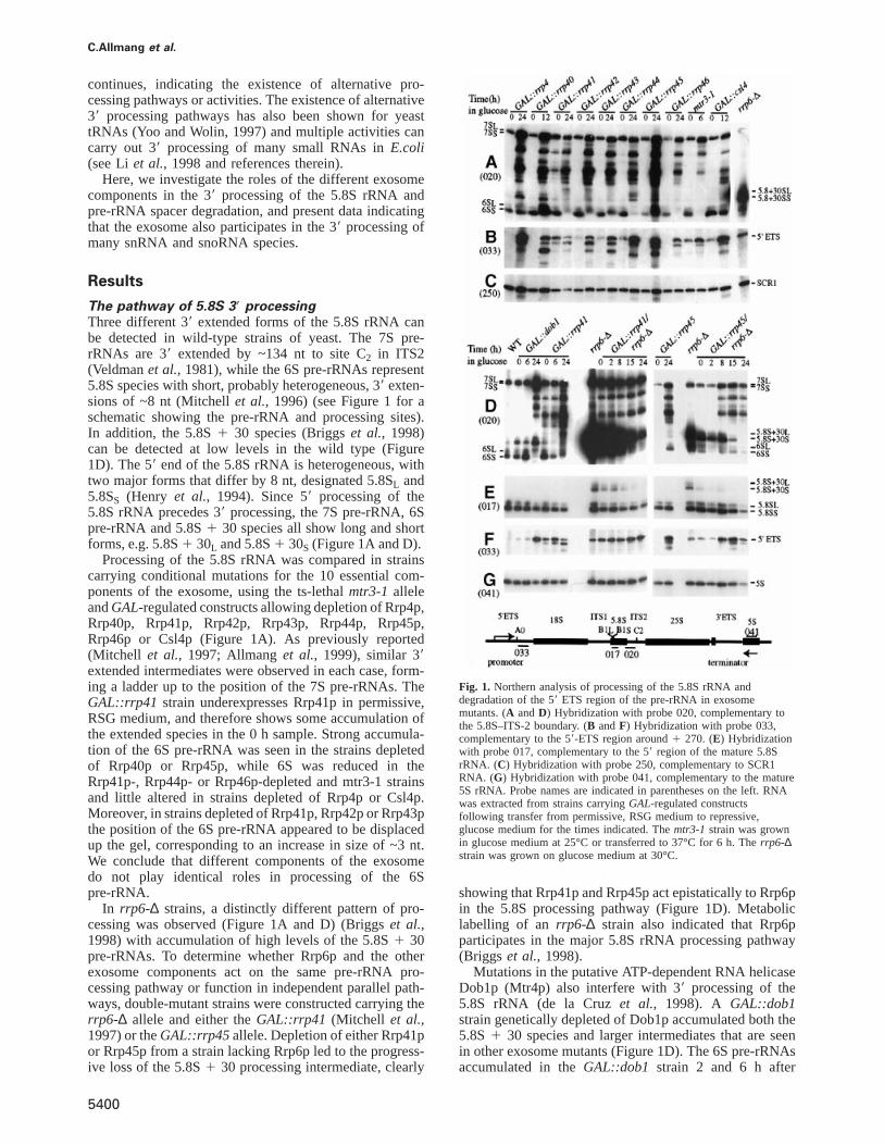
C.Allmang et al.
continues, indicating the existence of alternative pro-cessing pathways or activities. The existence of alternative39 processing pathways has also been shown for yeasttRNAs (Yoo and Wolin, 1997) and multiple activities cancarry out 39 processing of many small RNAs inE.coli(see Liet al., 1998 and references therein).
Here, we investigate the roles of the different exosomecomponents in the 39 processing of the 5.8S rRNA andpre-rRNA spacer degradation, and present data indicatingthat the exosome also participates in the 39 processing ofmany snRNA and snoRNA species.
Results
The pathway of 5.8S 39 processingThree different 39 extended forms of the 5.8S rRNA canbe detected in wild-type strains of yeast. The 7S pre-rRNAs are 39 extended by ~134 nt to site C2 in ITS2(Veldmanet al., 1981), while the 6S pre-rRNAs represent5.8S species with short, probably heterogeneous, 39 exten-sions of ~8 nt (Mitchellet al., 1996) (see Figure 1 for aschematic showing the pre-rRNA and processing sites).In addition, the 5.8S1 30 species (Briggset al., 1998)can be detected at low levels in the wild type (Figure1D). The 59 end of the 5.8S rRNA is heterogeneous, withtwo major forms that differ by 8 nt, designated 5.8SL and5.8SS (Henry et al., 1994). Since 59 processing of the5.8S rRNA precedes 39 processing, the 7S pre-rRNA, 6Spre-rRNA and 5.8S1 30 species all show long and shortforms, e.g. 5.8S1 30L and 5.8S1 30S (Figure 1A and D).
Processing of the 5.8S rRNA was compared in strainscarrying conditional mutations for the 10 essential com-ponents of the exosome, using the ts-lethalmtr3-1 alleleandGAL-regulated constructs allowing depletion of Rrp4p,Rrp40p, Rrp41p, Rrp42p, Rrp43p, Rrp44p, Rrp45p,Rrp46p or Csl4p (Figure 1A). As previously reported(Mitchell et al., 1997; Allmanget al., 1999), similar 39extended intermediates were observed in each case, form-ing a ladder up to the position of the 7S pre-rRNAs. TheGAL::rrp41 strain underexpresses Rrp41p in permissive,RSG medium, and therefore shows some accumulation ofthe extended species in the 0 h sample. Strong accumula-tion of the 6S pre-rRNA was seen in the strains depletedof Rrp40p or Rrp45p, while 6S was reduced in theRrp41p-, Rrp44p- or Rrp46p-depleted and mtr3-1 strainsand little altered in strains depleted of Rrp4p or Csl4p.Moreover, in strains depleted of Rrp41p, Rrp42p or Rrp43pthe position of the 6S pre-rRNA appeared to be displacedup the gel, corresponding to an increase in size of ~3 nt.We conclude that different components of the exosomedo not play identical roles in processing of the 6Spre-rRNA.
In rrp6-∆ strains, a distinctly different pattern of pro-cessing was observed (Figure 1A and D) (Briggset al.,1998) with accumulation of high levels of the 5.8S1 30pre-rRNAs. To determine whether Rrp6p and the otherexosome components act on the same pre-rRNA pro-cessing pathway or function in independent parallel path-ways, double-mutant strains were constructed carrying therrp6-∆ allele and either theGAL::rrp41 (Mitchell et al.,1997) or theGAL::rrp45 allele. Depletion of either Rrp41por Rrp45p from a strain lacking Rrp6p led to the progress-ive loss of the 5.8S1 30 processing intermediate, clearly
5400
Fig. 1. Northern analysis of processing of the 5.8S rRNA anddegradation of the 59 ETS region of the pre-rRNA in exosomemutants. (A andD) Hybridization with probe 020, complementary tothe 5.8S–ITS-2 boundary. (B andF) Hybridization with probe 033,complementary to the 59-ETS region around1 270. (E) Hybridizationwith probe 017, complementary to the 59 region of the mature 5.8SrRNA. (C) Hybridization with probe 250, complementary to SCR1RNA. (G) Hybridization with probe 041, complementary to the mature5S rRNA. Probe names are indicated in parentheses on the left. RNAwas extracted from strains carryingGAL-regulated constructsfollowing transfer from permissive, RSG medium to repressive,glucose medium for the times indicated. Themtr3-1 strain was grownin glucose medium at 25°C or transferred to 37°C for 6 h. Therrp6-∆strain was grown on glucose medium at 30°C.
showing that Rrp41p and Rrp45p act epistatically to Rrp6pin the 5.8S processing pathway (Figure 1D). Metaboliclabelling of an rrp6-∆ strain also indicated that Rrp6pparticipates in the major 5.8S rRNA processing pathway(Briggs et al., 1998).
Mutations in the putative ATP-dependent RNA helicaseDob1p (Mtr4p) also interfere with 39 processing of the5.8S rRNA (de la Cruzet al., 1998). A GAL::dob1strain genetically depleted of Dob1p accumulated both the5.8S1 30 species and larger intermediates that are seenin other exosome mutants (Figure 1D). The 6S pre-rRNAsaccumulated in theGAL::dob1 strain 2 and 6 h after
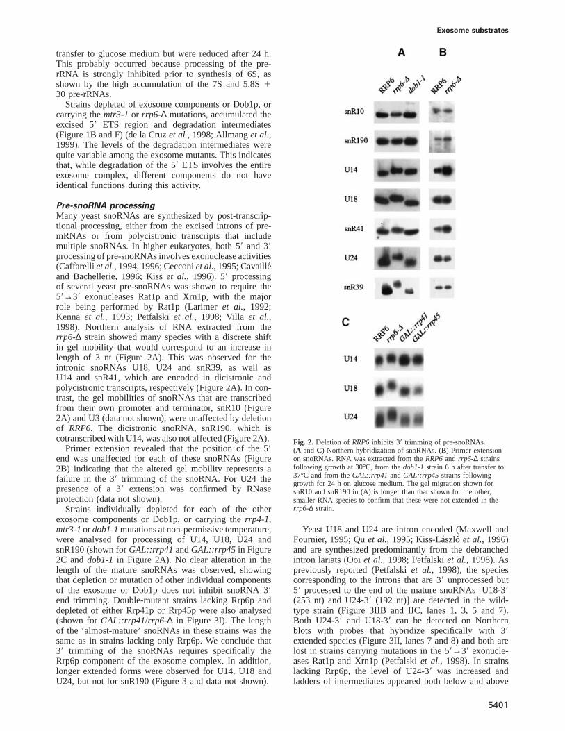
Exosome substrates
transfer to glucose medium but were reduced after 24 h.This probably occurred because processing of the pre-rRNA is strongly inhibited prior to synthesis of 6S, asshown by the high accumulation of the 7S and 5.8S130 pre-rRNAs.
Strains depleted of exosome components or Dob1p, orcarrying themtr3-1or rrp6-∆ mutations, accumulated theexcised 59 ETS region and degradation intermediates(Figure 1B and F) (de la Cruzet al., 1998; Allmanget al.,1999). The levels of the degradation intermediates werequite variable among the exosome mutants. This indicatesthat, while degradation of the 59 ETS involves the entireexosome complex, different components do not haveidentical functions during this activity.
Pre-snoRNA processingMany yeast snoRNAs are synthesized by post-transcrip-tional processing, either from the excised introns of pre-mRNAs or from polycistronic transcripts that includemultiple snoRNAs. In higher eukaryotes, both 59 and 39processing of pre-snoRNAs involves exonuclease activities(Caffarelliet al., 1994, 1996; Cecconiet al., 1995; Cavaille´and Bachellerie, 1996; Kisset al., 1996). 59 processingof several yeast pre-snoRNAs was shown to require the59→39 exonucleases Rat1p and Xrn1p, with the majorrole being performed by Rat1p (Larimeret al., 1992;Kenna et al., 1993; Petfalskiet al., 1998; Villa et al.,1998). Northern analysis of RNA extracted from therrp6-∆ strain showed many species with a discrete shiftin gel mobility that would correspond to an increase inlength of 3 nt (Figure 2A). This was observed for theintronic snoRNAs U18, U24 and snR39, as well asU14 and snR41, which are encoded in dicistronic andpolycistronic transcripts, respectively (Figure 2A). In con-trast, the gel mobilities of snoRNAs that are transcribedfrom their own promoter and terminator, snR10 (Figure2A) and U3 (data not shown), were unaffected by deletionof RRP6. The dicistronic snoRNA, snR190, which iscotranscribed with U14, was also not affected (Figure 2A).
Primer extension revealed that the position of the 59end was unaffected for each of these snoRNAs (Figure2B) indicating that the altered gel mobility represents afailure in the 39 trimming of the snoRNA. For U24 thepresence of a 39 extension was confirmed by RNaseprotection (data not shown).
Strains individually depleted for each of the otherexosome components or Dob1p, or carrying therrp4-1,mtr3-1ordob1-1mutations at non-permissive temperature,were analysed for processing of U14, U18, U24 andsnR190 (shown forGAL::rrp41 andGAL::rrp45 in Figure2C anddob1-1 in Figure 2A). No clear alteration in thelength of the mature snoRNAs was observed, showingthat depletion or mutation of other individual componentsof the exosome or Dob1p does not inhibit snoRNA 39end trimming. Double-mutant strains lacking Rrp6p anddepleted of either Rrp41p or Rrp45p were also analysed(shown forGAL::rrp41/rrp6-∆ in Figure 3I). The lengthof the ‘almost-mature’ snoRNAs in these strains was thesame as in strains lacking only Rrp6p. We conclude that39 trimming of the snoRNAs requires specifically theRrp6p component of the exosome complex. In addition,longer extended forms were observed for U14, U18 andU24, but not for snR190 (Figure 3 and data not shown).
5401
Fig. 2. Deletion ofRRP6inhibits 39 trimming of pre-snoRNAs.(A andC) Northern hybridization of snoRNAs. (B) Primer extensionon snoRNAs. RNA was extracted from theRRP6and rrp6-∆ strainsfollowing growth at 30°C, from thedob1-1strain 6 h after transfer to37°C and from theGAL::rrp41 andGAL::rrp45 strains followinggrowth for 24 h on glucose medium. The gel migration shown forsnR10 and snR190 in (A) is longer than that shown for the other,smaller RNA species to confirm that these were not extended in therrp6-∆ strain.
Yeast U18 and U24 are intron encoded (Maxwell andFournier, 1995; Quet al., 1995; Kiss-La´szlo et al., 1996)and are synthesized predominantly from the debranchedintron lariats (Ooiet al., 1998; Petfalskiet al., 1998). Aspreviously reported (Petfalskiet al., 1998), the speciescorresponding to the introns that are 39 unprocessed but59 processed to the end of the mature snoRNAs [U18-39(253 nt) and U24-39 (192 nt)] are detected in the wild-type strain (Figure 3IIB and IIC, lanes 1, 3, 5 and 7).Both U24-39 and U18-39 can be detected on Northernblots with probes that hybridize specifically with 39extended species (Figure 3II, lanes 7 and 8) and both arelost in strains carrying mutations in the 59→39 exonucle-ases Rat1p and Xrn1p (Petfalskiet al., 1998). In strainslacking Rrp6p, the level of U24-39 was increased andladders of intermediates appeared both below and above
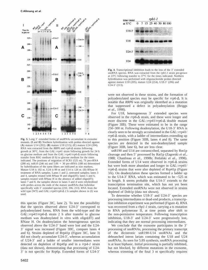
C.Allmang et al.
Fig. 3. Long 39 extended forms of snoRNAs accumulate in exosomemutants. (I and II ) Northern hybridization with probes directed against(A) mature U14 (202); (B) mature U18 (215); (C) mature U24 (204).RNA was extracted from theRRP6and rrp6-∆ strains followinggrowth at 30°C, from theGAL::rrp41 strain following growth for 24 hon glucose medium and from theGAL::rrp41/rrp6-∆ strain followingtransfer from RSG medium (0 h) to glucose medium for the timesindicated. The positions of migration of SCR1 (525 nt), 7S pre-rRNA(288 nt), snR10 (246 nt) and 5.8S1 30 pre-rRNA (188 nt) determinedby hybridization of the same filters are indicated as size markers.Mature U18 is 102 nt, U24 is 89 nt and U14 is 126 nt. (II) RNase Htreatment of RNA samples. Lanes 1 and 2, untreated samples; lanes 3and 4, samples treated with RNase H and oligo(dT); lanes 5 and 6,samples treated with RNase H in the absence of added oligo(dT);lanes 7 and 8, the samples shown in lanes 3 and 4 were rehybridizedwith probes across the ends of the mature snoRNAs that hybridizespecifically with 39 extended species (210, 206, 213). RNA from thewild type (WT) andGAL::rrp41rrp6-∆ 2 h samples shown in (I) wasused.
this species (Figure 3IC, lane 2). To test the possibilitythat the species observed above U24-39 correspond topolyadenylated forms, RNA from the wild-type and theGAL::rrp41/rrp6-∆ strain 2 h after transfer to glucosemedium was deadenylatedin vitro with oligo(dT) andRNase H. On deadenylation the heterogeneous speciesobserved above the U24-39 band were lost and the U24-39 signal was increased (Figure 3IIC, compare lanes 4and 6). Strains depleted of Rrp41p (Figure 3IC, lane 3)did not clearly accumulate U24-39, whereas accumulationof U24-39 and a ladder of smaller intermediates weredetected on depletion of Rrp45p and in arrp4-1 strain(data not shown), demonstrating that processing of U24-39 is not specific for Rrp6p. Extended forms of U24-39
5402
Fig. 4. Transcriptional inhibition leads to the loss of the 39 extendedsnoRNA species. RNA was extracted from therpb1-1 strain pre-grownat 23°C following transfer to 37°C for the times indicated. Northernhybridization was performed with oligonucleotide probes directedagainst mature U18 (205), mature U24 (214), U18-39 (206) andU24-39 (213).
were not observed in these strains, and the formation ofpolyadenylated species may be specific forrrp6-∆. It isnotable thatRRP6was originally identified as a mutationthat suppressed a defect in polyadenylation (Briggset al., 1998).
For U18, heterogeneous 39 extended species wereobserved in therrp6-∆ strain, and these were longer andmore discrete in theGAL::rrp41/rrp6-∆ double mutant(Figure 3IB). These were estimated to be in the range250–300 nt. Following deadenylation, the U18-39 RNA isclearly seen to be strongly accumulated in theGAL::rrp41/rrp6-∆ strain, with a ladder of intermediates extending upto this position (Figure 3IIB, lanes 4 and 8). The samespecies are detected in the non-deadenylated sample(Figure 3IIB, lane 6), but are less clear.
snR190 and U14 are cotranscribed, separated by Rnt1pcleavage and then 59 processed by Rat1p (Zagorskiet al.,1988; Chanfreauet al., 1998b; Petfalskiet al., 1998).Extended forms of U14 were observed inrrp6-∆ strainsbut were both more abundant and longer inGAL::rrp41/rrp6-∆ strains that were also depleted of Rrp41p (Figure3A). On deadenylation these species formed a ladder upto the U14-39 RNA, which was estimated to be ~525 ntin length. It seems probable that U14-39 extends to thetranscription termination site, which has not yet beenlocated. Extended snoRNAs were not observed in strainsdepleted of Dob1p (data not shown).
To determine whether U18-39 and U24-39 species areprocessing intermediates or dead-end products, a transcrip-tion inhibition experiment was performed (Figure 4). RNAwas recovered from arbp1-1strain, carrying a ts mutationin RNA polymerase II, at time points after shift tothe non-permissive temperature. Following transcriptioninhibition, U18-39 and U24-39 were progressively lost,indicating that they are normal processing intermediates.
We conclude that the exosome participates in the 39processing of snoRNAs, processing the primary transcriptof the dicistronic snR190-U14 snoRNAs and thedebranched intron lariats containing the U18 and U24snoRNAs. As seen for 5.8S synthesis, snoRNA processingis at least biphasic. Initial processing is partially inhibited,but not blocked, by different mutations in the exosome,whereas trimming of the final 3 nt specifically requires
Exosome substrates
Rrp6p. The additive effect of therrp41 andrrp6 mutationson U18 and U14 synthesis is, however, in contrast to theepistatic interactions observed for 5.8S processing. In theabsence of Rrp6p some polyadenylation of processingintermediates also occurs.
snRNA synthesisSince a number of snRNAs have been shown to undergo39 processing we analysed snRNA species for alterationsin exosome mutants.
Analysis of the U4 snRNA showed the existence oflow levels of two longer forms (U4-39I and U4-39II ) inthe wild type (Figure 5, lanes 1 and 12). Both specieswere detected with either the internal U4 probe (oligo243) (Figure 5B) or a probe complementary to the sequenceacross the 39 end of the mature U4 (oligo 246), whichhybridizes only to 39 extended species (Figure 5A). U4-39I is a set of heterogeneous species carrying short 39extensions similar in length to those detected in the 6Spre-rRNA. Oligo 246 detects only the longer forms ofU4-39I. The U4-39II species is ~140 nt larger than matureU4 and is presumably a normal processing intermediatesince it is detected in wild-type cells. In strains depletedof the exosome components Rrp41p (Figure 5, lanes 2and 3) or Rrp45p (Figure 5, lanes 7 and 8), or lackingRrp6p (Figure 5, lane 15), the levels of U4-39I and U4-39II were increased and a ladder of intermediates wasobserved that extended from the size of U4-39I to that ofU4-39II . TheGAL::rrp41 strain shows some accumulationof the extended species in the 0 h sample due to under-expression of Rrp41p (Figure 5, lane 2). The same specieswere accumulated in a strain depleted of Dob1p (data notshown). The accumulation of these intermediates was notstronger in therrp6-∆ strain that was also depleted ofRrp41p or Rrp45p than in therrp6-∆ single-mutant strain(data not shown). In strains lacking Rnt1p, U4-39II RNAwas absent whilst the levels of the U4-39I species wereincreased. These observations suggested that the U4-39II
is generated by Rnt1p cleavage and acts as an entry sitefor the exosome complex.
Strains lacking Rnt1p also accumulated longer 39extended forms of U4 (Figure 5, lanes 13 and 14; 4-foldmore RNA was loaded in lane 14 to allow visualizationof the longer 39 extended species). These were moreabundant inrnt1-∆ strains that were also depleted ofRrp41p (Figure 5, lanes 4–6), Rrp45p (Figure 5, lanes 9–11) or Rrp6p (Figure 5, lane 16). Particularly in thernt1-∆/rrp6-∆ strain (Figure 5B, lane 16) these were seen toextend to a distinct species (U4-39III ) of ~590 nt. Itseems likely that these represent intermediates in the 39processing of U4 from U4-39III , which most probablyextends to the transcription termination site. Little effecton the 39 extended forms of U4 was seen on treatmentwith RNase H and oligo(dT) (data not shown).
Rnt1p cleaves on both sides of extended, imperfectstems with closing AGNN tetraloops (Chanfreauet al.,1998a). Inspection of the U4 39 flanking region identifiedgood consensus Rnt1p cleavage sites (Figure 6D). Todetermine whether this represented a genuine substratefor Rnt1p, cleavage was assayedin vitro. The region ofthe U4 39 flanking sequence from positions149 to1235,which includes the putative cleavage sites, was transcribedin vitro (see Materials and methods). The sites cleaved
5403
Fig. 5. Northern analysis of processing of U4 snRNA inrnt1-∆ andexosome mutants. RNA was extracted from strains carryingGAL-regulated constructs following transfer from permissive, RSG mediumto repressive, glucose medium at 30°C for the times indicated, or fromthe wild-type (WT),rnt1-∆ and rrp6-∆ strains grown on glucosemedium at 30°C. RNA was separated on a 6% polyacrylamide gel andhybridized with oligonucleotide probes. (A) Oligo 246 complementaryto the region across the 39 end of the mature U4 snRNA.(B andC) Oligo 243 complementary to the mature U4 snRNA.(D) Oligo 250 complementary to the mature SCR1 RNA; the panelsshow successive hybridizations of the same filter. Probe names areindicated in parentheses on the left and the positions of detected RNAspecies are indicated on the right. (C) presents a weaker exposure ofthe same gel as (B). (C and D) present only relevant regions of theNorthern blots. The species marked with * in (A) probably resultsfrom a cross-hybridization with another RNA since it was not detectedin (B). The amount of total RNA loaded in lane 14 is 4-fold higherthan in lane 13 and other lanes. The positions of migration of SCR1(525 nt), MRP RNA (340 nt), U5L (215 nt) and U5S (180 nt)determined by hybridization of the same filter are indicated as sizemarkers. Mature U4 is 160 nt.
in vitro by recombinant His6–Rnt1p were identified byprimer extension with oligo 249 (Figure 6A). Twomajor stops, corresponding to sites of cleavage between
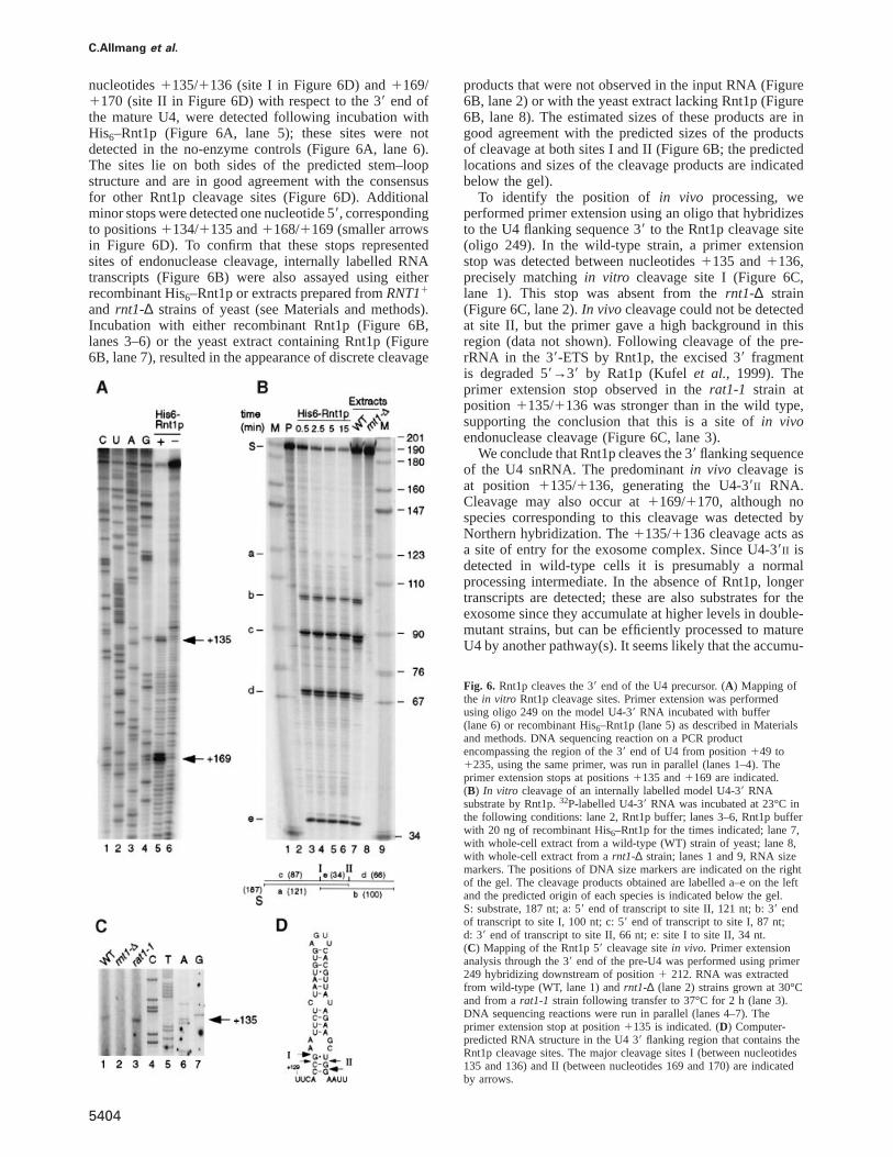
C.Allmang et al.
nucleotides1135/1136 (site I in Figure 6D) and1169/1170 (site II in Figure 6D) with respect to the 39 end ofthe mature U4, were detected following incubation withHis6–Rnt1p (Figure 6A, lane 5); these sites were notdetected in the no-enzyme controls (Figure 6A, lane 6).The sites lie on both sides of the predicted stem–loopstructure and are in good agreement with the consensusfor other Rnt1p cleavage sites (Figure 6D). Additionalminor stops were detected one nucleotide 59, correspondingto positions1134/1135 and1168/1169 (smaller arrowsin Figure 6D). To confirm that these stops representedsites of endonuclease cleavage, internally labelled RNAtranscripts (Figure 6B) were also assayed using eitherrecombinant His6–Rnt1p or extracts prepared fromRNT11
and rnt1-∆ strains of yeast (see Materials and methods).Incubation with either recombinant Rnt1p (Figure 6B,lanes 3–6) or the yeast extract containing Rnt1p (Figure6B, lane 7), resulted in the appearance of discrete cleavage
5404
products that were not observed in the input RNA (Figure6B, lane 2) or with the yeast extract lacking Rnt1p (Figure6B, lane 8). The estimated sizes of these products are ingood agreement with the predicted sizes of the productsof cleavage at both sites I and II (Figure 6B; the predictedlocations and sizes of the cleavage products are indicatedbelow the gel).
To identify the position of in vivo processing, weperformed primer extension using an oligo that hybridizesto the U4 flanking sequence 39 to the Rnt1p cleavage site(oligo 249). In the wild-type strain, a primer extensionstop was detected between nucleotides1135 and1136,precisely matchingin vitro cleavage site I (Figure 6C,lane 1). This stop was absent from thernt1-∆ strain(Figure 6C, lane 2).In vivocleavage could not be detectedat site II, but the primer gave a high background in thisregion (data not shown). Following cleavage of the pre-rRNA in the 39-ETS by Rnt1p, the excised 39 fragmentis degraded 59→39 by Rat1p (Kufelet al., 1999). Theprimer extension stop observed in therat1-1 strain atposition 1135/1136 was stronger than in the wild type,supporting the conclusion that this is a site ofin vivoendonuclease cleavage (Figure 6C, lane 3).
We conclude that Rnt1p cleaves the 39 flanking sequenceof the U4 snRNA. The predominantin vivo cleavage isat position 1135/1136, generating the U4-39II RNA.Cleavage may also occur at1169/1170, although nospecies corresponding to this cleavage was detected byNorthern hybridization. The1135/1136 cleavage acts asa site of entry for the exosome complex. Since U4-39II isdetected in wild-type cells it is presumably a normalprocessing intermediate. In the absence of Rnt1p, longertranscripts are detected; these are also substrates for theexosome since they accumulate at higher levels in double-mutant strains, but can be efficiently processed to matureU4 by another pathway(s). It seems likely that the accumu-
Fig. 6. Rnt1p cleaves the 39 end of the U4 precursor. (A) Mapping ofthe in vitro Rnt1p cleavage sites. Primer extension was performedusing oligo 249 on the model U4-39 RNA incubated with buffer(lane 6) or recombinant His6–Rnt1p (lane 5) as described in Materialsand methods. DNA sequencing reaction on a PCR productencompassing the region of the 39 end of U4 from position149 to1235, using the same primer, was run in parallel (lanes 1–4). Theprimer extension stops at positions1135 and1169 are indicated.(B) In vitro cleavage of an internally labelled model U4-39 RNAsubstrate by Rnt1p.32P-labelled U4-39 RNA was incubated at 23°C inthe following conditions: lane 2, Rnt1p buffer; lanes 3–6, Rnt1p bufferwith 20 ng of recombinant His6–Rnt1p for the times indicated; lane 7,with whole-cell extract from a wild-type (WT) strain of yeast; lane 8,with whole-cell extract from arnt1-∆ strain; lanes 1 and 9, RNA sizemarkers. The positions of DNA size markers are indicated on the rightof the gel. The cleavage products obtained are labelled a–e on the leftand the predicted origin of each species is indicated below the gel.S: substrate, 187 nt; a: 59 end of transcript to site II, 121 nt; b: 39 endof transcript to site I, 100 nt; c: 59 end of transcript to site I, 87 nt;d: 39 end of transcript to site II, 66 nt; e: site I to site II, 34 nt.(C) Mapping of the Rnt1p 59 cleavage sitein vivo. Primer extensionanalysis through the 39 end of the pre-U4 was performed using primer249 hybridizing downstream of position1 212. RNA was extractedfrom wild-type (WT, lane 1) andrnt1-∆ (lane 2) strains grown at 30°Cand from arat1-1 strain following transfer to 37°C for 2 h (lane 3).DNA sequencing reactions were run in parallel (lanes 4–7). Theprimer extension stop at position1135 is indicated. (D) Computer-predicted RNA structure in the U4 39 flanking region that contains theRnt1p cleavage sites. The major cleavage sites I (between nucleotides135 and 136) and II (between nucleotides 169 and 170) are indicatedby arrows.
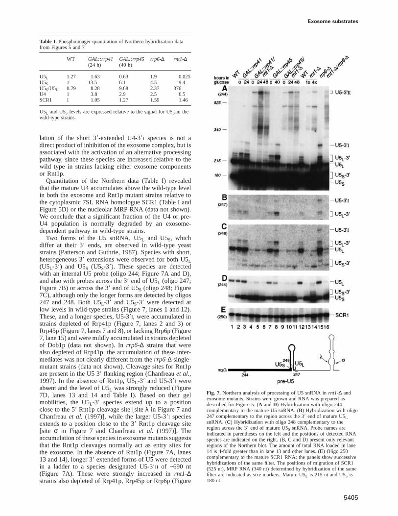
Exosome substrates
Table I. Phosphoimager quantitation of Northern hybridization datafrom Figures 5 and 7
WT GAL::rrp41 GAL::rrp45 rrp6-∆ rnt1-∆(24 h) (40 h)
U5L 1.27 1.63 0.63 1.9 0.025U5S 1 13.5 6.1 4.5 9.4U5S/U5L 0.79 8.28 9.68 2.37 376U4 1 3.8 2.9 2.5 6.5SCR1 1 1.05 1.27 1.59 1.46
U5L and U5S levels are expressed relative to the signal for U5S in thewild-type strains.
lation of the short 39-extended U4-39I species is not adirect product of inhibition of the exosome complex, but isassociated with the activation of an alternative processingpathway, since these species are increased relative to thewild type in strains lacking either exosome componentsor Rnt1p.
Quantitation of the Northern data (Table I) revealedthat the mature U4 accumulates above the wild-type levelin both the exosome and Rnt1p mutant strains relative tothe cytoplasmic 7SL RNA homologue SCR1 (Table I andFigure 5D) or the nucleolar MRP RNA (data not shown).We conclude that a significant fraction of the U4 or pre-U4 population is normally degraded by an exosome-dependent pathway in wild-type strains.
Two forms of the U5 snRNA, U5L and U5S, whichdiffer at their 39 ends, are observed in wild-type yeaststrains (Patterson and Guthrie, 1987). Species with short,heterogeneous 39 extensions were observed for both U5L(U5L-39) and U5S (U5S-39). These species are detectedwith an internal U5 probe (oligo 244; Figure 7A and D),and also with probes across the 39 end of U5L (oligo 247;Figure 7B) or across the 39 end of U5S (oligo 248; Figure7C), although only the longer forms are detected by oligos247 and 248. Both U5L-39 and U5S-39 were detected atlow levels in wild-type strains (Figure 7, lanes 1 and 12).These, and a longer species, U5-39I, were accumulated instrains depleted of Rrp41p (Figure 7, lanes 2 and 3) orRrp45p (Figure 7, lanes 7 and 8), or lacking Rrp6p (Figure7, lane 15) and were mildly accumulated in strains depletedof Dob1p (data not shown). Inrrp6-∆ strains that werealso depleted of Rrp41p, the accumulation of these inter-mediates was not clearly different from therrp6-∆ single-mutant strains (data not shown). Cleavage sites for Rnt1pare present in the U5 39 flanking region (Chanfreauet al.,1997). In the absence of Rnt1p, U5L-39 and U5-39I wereabsent and the level of U5L was strongly reduced (Figure7D, lanes 13 and 14 and Table I). Based on their gelmobilities, the U5L-39 species extend up to a positionclose to the 59 Rnt1p cleavage site [siteλ in Figure 7 andChanfreauet al. (1997)], while the larger U5-39I speciesextends to a position close to the 39 Rnt1p cleavage site[site σ in Figure 7 and Chanfreauet al. (1997)]. Theaccumulation of these species in exosome mutants suggeststhat the Rnt1p cleavages normally act as entry sites forthe exosome. In the absence of Rnt1p (Figure 7A, lanes13 and 14), longer 39 extended forms of U5 were detectedin a ladder to a species designated U5-39II of ~690 nt(Figure 7A). These were strongly increased inrnt1-∆strains also depleted of Rrp41p, Rrp45p or Rrp6p (Figure
5405
Fig. 7. Northern analysis of processing of U5 snRNA inrnt1-∆ andexosome mutants. Strains were grown and RNA was prepared asdescribed for Figure 5. (A andD) Hybridization with oligo 244complementary to the mature U5 snRNA. (B) Hybridization with oligo247 complementary to the region across the 39 end of mature U5LsnRNA. (C) Hybridization with oligo 248 complementary to theregion across the 39 end of mature U5S snRNA. Probe names areindicated in parentheses on the left and the positions of detected RNAspecies are indicated on the right. (B, C and D) present only relevantregions of the Northern blot. The amount of total RNA loaded in lane14 is 4-fold greater than in lane 13 and other lanes. (E) Oligo 250complementary to the mature SCR1 RNA; the panels show successivehybridizations of the same filter. The positions of migration of SCR1(525 nt), MRP RNA (340 nt) determined by hybridization of the samefilter are indicated as size markers. Mature U5L is 215 nt and U5S is180 nt.
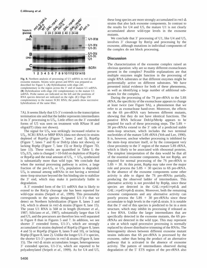
C.Allmang et al.
Fig. 8. Northern analysis of processing of U1 snRNA inrnt1-∆ andexosome mutants. Strains were grown and RNA was prepared asdescribed for Figure 5. (A) Hybridization with oligo 245complementary to the region across the 39 end of mature U1 snRNA.(B) Hybridization with oligo 242 complementary to the mature U1snRNA. Probe names are indicated on the left and the positions ofRNA species detected are indicated on the right. (C) Oligo 250complementary to the mature SCR1 RNA; the panels show successivehybridizations of the same filter.
7A). It seems likely that U5-39II extends to the transcriptiontermination site and that the ladder represents intermediatesin its 39 processing to U5S. Little effect on the 39 extendedforms of U5 was seen on treatment with RNase H andoligo(dT) (data not shown).
The signal for U5S was strikingly increased relative toU5L, SCR1 RNA or MRP RNA (data not shown) in strainsdepleted of Rrp41p (Figure 7, lanes 2 and 3), Rrp45p(Figure 7, lanes 7 and 8) or Dob1p (data not shown), orlacking Rrp6p (Figure 7, lane 15) or Rnt1p (Figure 7D,lane 13). These results are quantified in Table I; theU5S:U5L ratio is changed.10-fold on depletion of Rrp41por Rrp45p and the total amount of U5L 1 U5S synthesizedis substantially more than wild type. We conclude thatwhen the normal processing pathway is active, a largefraction of the pre-U5S or U5S population is degraded.U5S is unusual among snRNAs in not having a terminalstem–loop structure beyond the Sm binding site to stabilizethe 39 end, which may make it particularly liable todegradation.
A 39 extended form of the U1 snRNA that is likely toextend to the Rnt1p cleavage site has been reported forwild-type strains (Seipeltet al., 1999). This presumablycorresponds to the species designated U1-39II that wedetect on Northern hybridization (Figure 8, lanes 3 and14), which is absent inrnt1-∆ strains (Figure 8, lane 15).The yeast U1 RNA is 568 nt in length (Kretzneret al.,1987; Silicianoet al., 1987), substantially larger than U4and U5, and the precursors are therefore less well separatedin Figure 8 than in Figures 5 and 7. In addition, we seeshorter 39 extended forms, U1-39I, which are stronglyaccumulated in strains depleted of Rrp41p (Figure 8, lanes4 and 5) or Rrp45p (Figure 8, lanes 9 and 10), or lackingRrp6p (Figure 8, lane 2). Unlike the longer U1-39II species,the shorter U1-39I persists inrnt1-∆ strains (Figure 8, lane15). Thernt1-∆ strain accumulates longer, heterogeneous39 extended species, U1-39III , which are reported to bepolyadenylated (Seipeltet al., 1999). As for U4 and U5,
5406
these long species are more strongly accumulated inrnt1-∆strains that also lack exosome components. In contrast tothe results for U4 and U5, the mature U1 is not clearlyaccumulated above wild-type levels in the exosomemutants.
We conclude that 39 processing of U1, like U4 and U5,involves 39 cleavage by Rnt1p and processing by theexosome, although mutations in individual components ofthe complex do not block processing.
Discussion
The characterization of the exosome complex raised anobvious question: why are so many different exonucleasespresent in the complex? Possible explanations are thatmultiple enzymes might function in the processing ofsingle RNA substrates or that different enzymes might bepreferentially active on different substrates. We havepresented initial evidence for both of these phenomena,as well as identifying a large number of additional sub-strates for the complex.
During the processing of the 7S pre-rRNA to the 5.8SrRNA, the specificity of the exonuclease appears to changeat least twice (see Figure 9A), a phenomenon that werefer to as exonuclease hand-over. Moreover, the effectson the 6S pre-rRNA vary between different mutants,showing that they do not have identical functions. Theputative RNA helicase Dob1p/Mtr4p appears to berequired for each of these processing steps. The 5.8S130 pre-rRNAs extend to the 39 side of a predicted stablestem–loop structure, which includes the two terminalnucleotides of the mature 5.8S rRNA (Yeh and Lee, 1990).It is, however, unclear whether processing is inhibited bythe stem–loop structure itself, or by the consequent veryclose proximity to the 39 region of the mature 5.8S rRNA,which is likely to be associated with ribosomal proteins.The simplest interpretation of the data would be that allof the essential exosome components, but not Rrp6p, arerequired for normal processing of the 7S pre-rRNA to5.8S1 30. At this point, Rrp6p may take over the majorrole and process the 5.8S1 30 species to 6S pre-rRNA.In the absence of the exosome components some otheractivity is able to digest the 7S pre-rRNAs partially,producing the observed ladder of intermediates. Thisalternative activity is not provided by Rrp6p, since thesespecies are detected in theGAL::rrp41/rrp6-∆ andGAL::rrp45/rrp6-∆ strains. Moreover, both the remainingexosome components and any alternative activity canpoorly process the 5.8S1 30 pre-rRNAs since theseaccumulate to high levels in therrp6-∆ strain. It is notablethat the 39 end of this species is predicted to lie in a stemstructure, which may inhibit its processing if released asa free RNA. Unlike the larger intermediates that arespecifically detected in the exosome mutants, the 6S pre-rRNAs are detected in the wild type. This may representa site at which rapid processive processing is normallyreplaced by slower distributive trimming of the RNAs. Theheterogeneity shown between different exosome mutantstrains indicates that 6S pre-rRNA is processed by theexosome, rather than being the product of an alternativepathway that is activated in the absence of exosomeactivity. The pattern of intermediates observed duringdegradation of the 59 ETS region of the pre-rRNA also
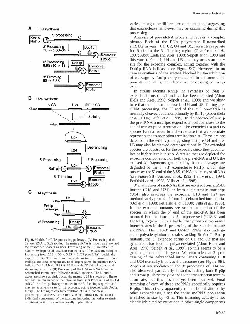
Exosome substrates
Fig. 9. Models for RNA processing pathways. (A) Processing of the7S pre-rRNA to 5.8S rRNA. The mature rRNA is shown as a box andthe transcribed spacers as lines. Processing of the 7S pre-rRNA to5.8S1 30 requires all essential components of the exosome complex.Processing from 5.8S1 30 to 5.8S1 8 (6S pre-rRNA) specificallyrequires Rrp6p. The final trimming to the mature 5.8S again requiresmultiple exosome components. Each step requires the putative RNAhelicase Dob1p/Mtr4p. 5.8S1 30 lies at the 39 side of a predictedstem–loop structure. (B) Processing of the U24 snoRNA from thedebranched intron lariat following mRNA splicing. The 59 and 39exons are shown as dark boxes, the mature U24 is shown as a lighterbox and the remainder of the intron as lines. (C) Processing of the U4snRNA. An Rnt1p cleavage site lies in the 39 flanking sequence andmay act as an entry site for the exosome, acting together with Dob1p/Mtr4p. The timing of cap trimethylation of U4 is not clear. 39processing of snoRNAs and snRNAs is not blocked by mutation ofindividual components of the exosome indicating that other extrinsicor intrinsic activities can functionally replace these.
5407
varies amongst the different exosome mutants, suggestingthat exonuclease hand-over may be occurring during thisprocessing.
Analysis of pre-snRNA processing reveals a complexpicture. Each of the RNA polymerase II-transcribedsnRNAs in yeast, U1, U2, U4 and U5, has a cleavage sitefor Rnt1p in the 39 flanking region (Chanfreauet al.,1997; Abou Elela and Ares, 1998; Seipeltet al., 1999 andthis work). For U1, U4 and U5 this may act as an entrysite for the exosome complex, acting together with theDob1p RNA helicase (see Figure 9C). However, in nocase is synthesis of the snRNA blocked by the inhibitionof cleavage by Rnt1p or by mutations in exosome com-ponents, indicating that alternative processing pathwaysexist.
In strains lacking Rnt1p the synthesis of long 39extended forms of U1 and U2 has been reported (AbouElela and Ares, 1998; Seipeltet al., 1999) and we showhere that this is also the case for U4 and U5. During pre-rRNA processing, the 39 end of the 35S pre-rRNA isnormally cleaved cotranscriptionally by Rnt1p (Abou Elelaet al., 1996; Kufelet al., 1999). In the absence of Rnt1pthe pre-rRNA transcripts extend to a position close to thesite of transcription termination. The extended U4 and U5species form a ladder to a discrete size that we speculaterepresents the transcription termination site. These are notdetected in the wild type, suggesting that pre-U4 and pre-U5 may also be cleaved cotranscriptionally. The extendedspecies are substrates for the exosome since they accumu-late at higher levels inrnt1-∆ strains that are depleted forexosome components. For both the pre-rRNA and U4, theexcised 39 fragments generated by Rnt1p cleavage aredegraded by the 59→39 exonuclease Rat1p, which alsoprocesses the 59 end of the 5.8SsrRNA and many snoRNAs(see Figure 9B) (Amberget al., 1992; Henryet al., 1994;Petfalskiet al., 1998; Villa et al., 1998).
39 maturation of snoRNAs that are excised from mRNAintrons (U18 and U24) or from a dicistronic transcript(U14) also involves the exosome. U18 and U24 arepredominately processed from the debranched intron lariat(Ooi et al., 1998; Petfalskiet al., 1998; Villaet al., 1998).In the exosome mutants we see accumulation of thespecies in which the 59 end of the snoRNA has beenmatured but the intron is 39 unprocessed (U18-39 andU24-39), together with a ladder that probably representsintermediates in the 39 processing of these to the maturesnoRNAs. The U18-39 and U24-39 RNAs also undergosome polyadenylation in strains lacking Rrp6p. In Rnt1pmutants, the 39 extended forms of U1 and U2 that aregenerated also become polyadenylated (Abou Elela andAres, 1998; Seipeltet al., 1999), so this seems to be ageneral phenomenon in yeast. We conclude that 39 pro-cessing of the debranched intron lariats containing U18and U24 normally involves the exosome (see Figure 9B).Apparent intermediates in the 39 processing of U14 arealso observed, particularly in strains lacking both Rrp6pand Rrp41p. These may extend to the transcription termin-ation site, but this has not yet been localized. Finaltrimming of each of these snoRNAs specifically requiresRrp6p. This activity apparently cannot be substituted byother exonucleases, since the entire snoRNA populationis shifted in size by ~3 nt. This trimming activity is notclearly inhibited by mutations in other single components
C.Allmang et al.
Table II. Yeast strains used in this work
Strain Genotype Reference
BMA 38 MATa/α ade2-1/ade2-1 his3-∆200/his3-∆200 leu2-3, 112/leu2-3, 112 trp1-1/trp1-1 ura3-1/ura3-1 Baudinet al. (1993)can1-100/can1-100
YCA12 MATa ade2-1 his3-∆200 leu2-3, 112 trp1-1 ura3-1 can1-100 RRP6::Kl TRP1 Allmang et al. (1999)YDL401 MATa his3-∆200 leu2-∆1 trp1 ura3-52 gal2 gal∆108 Lafontaine and Tollervey (1996)P118 as YDL401 butGAL10::prot.A-RRP41 Lafontaine and Tollervey (1996)YCA20 as YDL401 butGAL10::RRP45 Allmang et al. (1999)YCA30 as YCA20 butRRP6::Kl TRP1 this studyYCA31 as P118 butRRP6::Kl TRP1 this studyYJK10 as YDL401 butRNT1::TRP1 this studyYJK11 as P118 butRNT1::TRP1 this studyYJK12 as YCA20 butRNT1::TRP1 this studyYJK13 as YCA12 butRNT1::TRP1 this studyGAL::DOB1 MATα ura3-1 ade2-1 his3-11,15 leu2-3, 112 trp1-1 dob1::HIS3MX61 [pAS24-DOB1] de la Cruzet al. (1998)rat1-1 MATα, his3-∆200, leu2-∆1, ura3-52, rat1-1 Amberget al. (1992)RP582 MATa leu2-3, 112 ura3-52 rpb1-1 Decker and Parker (1993)
of the exosome. It is notable that each of the enzymaticactivities shown to be involved in pre-snoRNA processing,the exosome, the 59→39 exonuclease Rat1p and theendonuclease Rnt1p, also participate in pre-rRNA pro-cessing.
There are apparent similarities between 39 processingof the 5.8S rRNA, snoRNAs and snRNAs. In each casethere is a downstream cleavage, by endonuclease orsplicing, which acts as a site of entry for exonucleases.Processing is at least biphasic with short 39 extendedforms accumulating, and each appears to involve theactivities of more than one component of the exosome.In each case, other activities can partially substitute forthe mutant exosome components. For the 7S pre-rRNA,and particularly the 5.8S1 30 pre-RNA, this is inefficient,and synthesis of mature 5.8S is strongly inhibited. Thefinal trimming of snoRNAs is apparently completelydependent on the activity of Rrp6p, but the processing offurther 39 extended pre-snoRNAs and pre-snRNAs can becarried out by other activities with good efficiency, asshown by the wild-type levels of the mature RNAs.Indeed, U4, and particularly U5S, are synthesized atsubstantially elevated levels in exosome mutants, indicat-ing competition between synthesis of mature snRNA anddegradation of the precursors in the wild type.
It is unclear whether residual processing in the exosomemutant strains is carried out by other components of thecomplex. It is notable that double-mutant strains lackingboth Rrp6p and Rrp41p show stronger phenotypes forsome activities (e.g. 39 processing of U14 and U18) thandoes either single mutant, indicating that the absence ofone component does not necessarily inactivate the entirecomplex. Alternatively, the yeast genome contains severalother predicted 39→59 exonucleases (Mian, 1997; Moseret al., 1997) that may be able to partially substitute forthe exosome. The combination of mutations in these geneswith mutations in exosome components will now beneeded to analyse their interactions and substrates.
Materials and methods
StrainsGrowth and handling ofS.cerevisiaewere by standard techniques. Thetransformation procedure was according to Gietzet al. (1992). Exceptwhere stated, strains were grown in liquid and solid minimal mediumcontaining 0.67% yeast nitrogen base (Difco) and 2% glucose.GAL-
5408
regulated strains were pre-grown in RSG medium (2% raffinose, 2%sucrose, 2% galactose, 0.67% yeast nitrogen base) and harvested atintervals following a shift to 2% glucose.
Yeast strains used and constructed in this study are listed in Table II.To construct the double mutantsGAL10::rrp45/rrp6-∆ (YCA30) and
GAL10::rrp41/rrp6-∆ (YCA31), the RRP6::TRPdisrupted allele wasamplified by PCR from genomic DNA extracted fromrrp6-∆ (YCA12).The PCR product was transformed into the corresponding conditionalmutant strains P118 and YCA20, respectively. The amplification ofRRP6::TRPwas done with oligos: 59RRP6, CAGTAATGAATATTAAT-GTTCATCTGAAGATAGACG; 39RRP6, ATGGTGTGCATGGGGG-AGCCATAACTCCATGACACA. Strains YDL401, P118 and YCA20were used to construct the mutantsrnt1-∆ (YJK10), GAL10::rrp41/rnt1-∆ (YJK11) andGAL10::rrp45/rnt1-∆ (YJK12), using the same PCRstrategy. Oligonucleotides 59RNT1, 59-GAAGACATATCCGAAGTG-ACA and 39RNT1, 59-GGATTTCTATACCCTCGAGGAG, complement-ary to sequences beyond theRNT1gene, were used for the amplificationwith genomic DNA extracted fromrnt1-∆ strain generously providedby G.Chanfreau (Chanfreauet al., 1998b). Transformants were selectedfor Trp1 prototrophy and were screened by PCR. The phenotypes ofrespective constructs were confirmed by Northern hybridization. Strainrrp6-∆/rnt1-∆ (YJK13) was constructed by crossing YJK10 and YCA12strains. The double-mutant strain was selected from dissected full tetradsby testing for the pre-rRNA and snoRNA processing phenotypes byNorthern hybridization. Wild-typeRNT1 and rnt1-∆ sister strains(Chanfreauet al., 1998b) were used to prepare whole-cell extract. Strainrat1-1 was kindly provided by C.Cole (Amberget al., 1992).
RNA extraction, Northern hybridization and primerextensionRNA was extracted as described previously (Tollervey and Mattaj,1987). Northern hybridization (Tollervey, 1987) and primer extension(Beltrame and Tollervey, 1992) were as described previously. Standard6 or 8% acrylamide gels were used to analyse low molecular weightrRNA species and primer extension reactions.
For pre-rRNA and rRNA analysis the following oligonucleotides wereused: 017 59-GCGTTGTTCATCGATGC; 020 59-TGAGAAGGAAATG-ACGCT; 033 59-CGCTGCTCACCAATGG; 041 59-CTACTCGGTCA-GGCTC.
The oligonucleotides used for Northern blot hybridization and primerextensions on other small RNAs were as follows: 031 (MRP) 59-AATAGAGGTACCAGGTCAAGAAGC; 201 (snR190) 59-CGTCAT-GGTCGAATCGG; 202 (U14) 59-TCACTCAGACATCCTAGG; 205(U18) 59-GTCAGATACTGTGATAGTC; 206 (U18-39) 59-GCTCTG-TGTGCTATCGTC; 210 (U14-39) 59-GTATACGATCACTCAGAC; 213(U24-39) 59-AAACCATTCATCAGAG; 214 (U24) 59-TCAGAGAT-CTTGGTGATAAT; 218 (snR10, 29-O-methyl RNA) 59-CUIUUAAAU-UUICIUU; 236 (snR39) 59-GGTGATAAGTTACGACAGC; 238 (snR41)59-GGGTTGTCGACATGTAGTTA; 242 (U1) 59-CACGCCTTCCGCG-CCGT; 243 (U4) 59-CCGTGCATAAGGAT; 244 (U5) 59-AATATG-GCAAGCCC; 245 (39Ex-U1) 59-TGTTCCATTTATTTCTGAAA; 246(39Ex-U4) 59-AAAGAATGAATATCGGTAATG; 247 (39Ex-U5S) 59-GAGAAAAAGGGCAGAAAAG; 248 (39Ex-U5L) 59-TAGAAAAGAT-AAACGCCCT; 249 (U4DS) 59-GACACACAAGAAGGAGAACACTC;250 (SCR1) 59-AAGGACCCAGAACTACCTTG.
Exosome substrates
RNase H treatmentDeadenylation was performed essentially as described (Decker andParker, 1993). Samples of 30µg of RNA were annealed with 750 ngoligo(dT) at 65°C for 1 h and digested with 6 U RNase H at 30°C for1 h. The control samples were treated identically, except that theoligo(dT) was omitted.
In vitro processing reactionsSynthetic U4-39 RNA was obtained byin vitro transcription as described(Chanfreauet al., 1998b) using PCR product as template. PCR productwas generated from genomic DNA using a forward primer carrying aT7 promoter (T7U4DS, 59-GCGAATTCTAATACGACTCACTATAGG-AAGTAATATCAAAAAATAGG) and a reverse primer U4DS.
Whole-cell extracts were prepared from wild-type andrnt1∆ sisterstrains as described (Chanfreauet al., 1998b).
Recombinant His6–Rnt1p was prepared by cloning a PCR-amplifiedRNT1gene into pET16B (Novagen), usingNdeI and BamHI restrictionsites added into the primers (NdeI–RntI, 59-GGGAATTCCATATGGGCT-CAAAAGTAGCAGG; Bam–RntI, 39-CGGGATCCTCAGCTTGTAT-CTGAGAATTTTCTTTTCTTATTC). Expression of His6–Rnt1p inE.colistrain BL21 was induced by addition of isopropyl-β-D-galactopyranosideat OD0.5 (final concentration 0.5 mM). After 3 h of expression at 30°C,cells were harvested and pellets were kept at –80°C. Pellets wereresuspended in 40 ml of Start buffer (20 mM sodium phosphate pH 7.0,10 mM imidazole) and cells were further lysed by passing through aFrench press. Cell debris were pelleted and the supernatant was loadedinto a Hi-Trap Chelating column (Pharmacia) pre-equilibrated with theStart buffer. The column was washed with 10 ml Start buffer andproteins were eluted with sodium phosphate buffer with increasingimidazole concentration (20, 40, 60, 100, 300 and 500 mM) or a lineargradient of imidazole (10–500 mM). Peak fractions were pooled and theprotein was dialysed twice against the storage buffer (50% glycerol,50 mM Tris–HCl pH 7.6, 200 mM KCl, 0.5 mM dithiothreitol, 0.5 mMEDTA pH 8.5). The protein was stored at a concentration of 2 mg/mlat –20°C and remained active for several months after storage.
In vitro processing of U4-39 RNA in cell extracts or with recombinantHis6–Rnt1p, and mapping of the cleavage sites using primer extension,was performed as described (Chanfreauet al., 1998a,b). Prior to thereaction, gel-purified RNA substrates (2 nM) were denatured for 2 minat 85°C in the Rnt1p buffer (50 mM Tris–HCl pH 7.6, 200 mM KCl,0.1 mg/ml wheat-germ tRNA, 5 mM MgCl2) and cooled to 23°C. Thecleavage reaction was performed either at 23°C using from 50 to200 fmol of recombinant His6–Rnt1 or by incubation in the whole-cell extracts.
Acknowledgements
J.K. was the recipient of a long-term EMBO fellowship. This work wassupported by the Wellcome Trust.
References
Abou Elela,S. and Ares,M.,Jr (1998) Depletion of yeast RNase III blockscorrect U2 39 end formation and results in polyadenylated butfunctional U2 snRNA.EMBO J., 17, 3738–3746.
Abou Elela,S., Igel,H. and Ares,M.,Jr (1996) RNase III cleaves eukaryoticpreribosomal RNA at a U3 snoRNP-dependent site. Cell, 85, 115–124.
Allmang,C., Petfalski,E., Podtelejnikov,A., Mann,M., Tollervey,D. andMitchell,P. (1999) The yeast exosome and human PM-Scl are relatedcomplexes of 39→59 exonucleases.Genes Dev., 13, 2148–2158.
Amberg,D.C., Goldstein,A.L. and Cole,C.N. (1992) Isolation andcharacterization ofRAT1: an essential gene ofSaccharomycescerevisiaerequired for the efficient nucleocytoplasmic trafficking ofmRNA. Genes Dev., 6, 1173–1189.
Baker,R.E., Harris,K. and Zhang,K. (1998) Mutations synthetically lethalwith cep1targetS.cerevisiaekinetochore components.Genetics, 149,73–85.
Baudin,A., Ozier-Kalogeropoulos,O., Denouel,A., Lacroute,F. andCullin,C. (1993) A simple and efficient method for direct gene deletionin Saccharomyces cerevisiae. Nucleic Acids Res., 21, 3329–3330.
Beltrame,M. and Tollervey,D. (1992) Identification and functionalanalysis of two U3 binding sites on yeast pre-ribosomal RNA.EMBO J., 11, 1531–1542.
Briggs,M.W., Burkard,K.T. and Butler,J.S. (1998) Rrp6p, the yeasthomologue of the human PM-Scl 100-kDa autoantigen, is essential
5409
for efficient 5.8 S rRNA 39 end formation.J. Biol. Chem., 273,13255–13263.
Caffarelli,E., Arese,M., Santoro,B., Fragapane,P. and Bozzoni,I. (1994)In vitro study of processing of the intron-encoded U16 small nucleolarRNA in Xenopus laevis. Mol. Cell. Biol., 14, 2966–2974.
Caffarelli,E., De Gregorio,E., Fatica,A., Prislei,S., Fragapane,P. andBozzoni,I. (1996) Processing of the intron-encoded U16 and U18snoRNAs: the conserved C and D boxes control both the processingreactions and the stability of the mature snoRNAs.EMBO J., 15,1121–1131.
Cavaille,J. and Bachellerie,J.-P. (1996) Processing of fibrillarin-associated snoRNAs from pre-mRNA introns: an exonucleolyticprocess exclusively directed by the common stem-box terminalstructure.Biochimie, 78, 443–456.
Cecconi,F., Mariottini,P. and Amaldi,F. (1995) TheXenopusintron-encoded U17 snoRNA is produced by exonucleolytic processing ofits precursor in oocytes.Nucleic Acids Res., 23, 4670–4676.
Chanfreau,G., Elela,S.A., Ares,M.,Jr and Guthrie,C. (1997) Alternative39-end processing of U5 snRNA by RNase III.Genes Dev., 11,2741–2751.
Chanfreau,G., Legrain,P. and Jacquier,A. (1998a) Yeast RNase III as akey processing enzyme in small nucleolar RNAs metabolism.J. Mol.Biol., 284, 975–988.
Chanfreau,G., Rotondo,G., Legrain,P. and Jacquier,A. (1998b) Processingof a dicistronic small nucleolar RNA precursor by the RNAendonuclease Rnt1.EMBO J., 17, 3726–3737.
de la Cruz,J., Kressler,D., Tollervey,D. and Linder,P. (1998) Dob1p(Mtr4p) is a putative ATP-dependent RNA helicase required for the39 end formation of 5.8S rRNA inSaccharomyces cerevisiae. EMBO J.,17, 1128–1140.
Decker,C.J. and Parker,R. (1993) A turnover pathway for both stableand unstable mRNAs in yeast: evidence for a requirement fordeadenylation.Genes Dev., 7, 1632–1643.
Gietz,D., St Jean,A., Woods,R.A. and Schiestl,R.H. (1992) Improvedmethod for high efficiency transformation of intact yeast cells.NucleicAcids Res., 20, 1425.
Henry,Y., Wood,H., Morrissey,J.P., Petfalski,E., Kearsey,S. andTollervey,D. (1994) The 59 end of yeast 5.8S rRNA is generatedby exonucleases from an upstream cleavage site.EMBO J., 13,2452–2463.
Kenna,M., Stevens,A., McCammon,M. and Douglas,M.G. (1993) Anessential yeast gene with homology to the exonuclease-encodingXRN1/KEM1gene also encodes a protein with exoribonuclease activity.Mol. Cell. Biol., 13, 341–350.
Kiss,T., Bortolini,M.-L. and Filipowicz,W. (1996) Characterization ofthe intron-encoded U19 RNA, a new mammalian small nucleolarRNA that is not associated with fibrillarin.Mol. Cell. Biol., 16,1391–1400.
Kiss-Laszlo,Z., Henry,Y., Bachellerie,J.-P., Caizergues-Ferrer,M. andKiss,T. (1996) Site-specific ribose methylation of preribosomal RNA:a novel function for small nucleolar RNAs.Cell, 85, 1077–1088.
Kretzner,L., Rymond,B.C. and Rosbash,M. (1987)S.cerevisiaeU1 RNAis large and has limited primary sequence homology to metazoan U1snRNA.Cell, 50, 593–602.
Kufel,J., Dichtl,B. and Tollervey,D. (1999) Yeast Rnt1p is required forcleavage of the pre-ribosomal RNA in the 39 ETS but not the 59 ETS.RNA, 5, 909–917.
Lafontaine,D. and Tollervey,D. (1996) One-step PCR mediated strategyfor the construction of conditionally expressed and epitope taggedyeast proteins.Nucleic Acids Res., 24, 3469–3472.
Larimer,F.W., Hsu,C.L., Maupin,M.K. and Stevens,A. (1992)Characterization of theXRN1gene encoding a 59→39 exoribonuclease:sequence data and analysis of disparate protein and mRNA levels ofgene-disrupted yeast cells.Gene, 120, 51–57.
Li,Z., Pandit,S. and Deutscher,M.P. (1998) 39 exoribonucleolytictrimming is a common feature of the maturation of small, stableRNAs in Escherichia coli. Proc. Natl Acad. Sci. USA, 95, 2856–2861.
Maxwell,E.S. and Fournier,M.J. (1995) The small nucleolar RNAs.Annu. Rev. Biochem., 35, 897–934.
Mian,I.S. (1997) Comparative sequence analysis of ribonucleases HII,III, II PH and D. Nucleic Acids Res., 25, 3187–3195.
Mitchell,P., Petfalski,E. and Tollervey,D. (1996) The 39-end of yeast5.8S rRNA is generated by an exonuclease processing mechanism.Genes Dev., 10, 502–513.
Mitchell,P., Petfalski,E., Shevchenko,A., Mann,M. and Tollervey,D.(1997) The exosome; a conserved eukaryotic RNA processing complex
C.Allmang et al.
containing multiple 39→59 exoribonuclease activities. Cell, 91, 457–466.
Moser,M.J., Holley,W.R., Chatterjee,A. and Mian,I.S. (1997) Theproofreading domain ofEscherichia coliDNA polymerase I and otherDNA and/or RNA exonuclease domains.Nucleic Acids Res., 25,5110–5118.
Noguchi,E. et al. (1996) Dis3, implicated in mitotic control, bindsdirectly to Ran and enhances the GEF activity of RCC1.EMBO J.,15, 5595–5605.
Ooi,S.L., Samarsky,D.A., Fournier,M.J. and Boeke,J.D. (1998) IntronicsnoRNA biosynthesis inSaccharomyces cerevisiaedepends on thelariat-debranching enzyme: intron length effects and activity of aprecursor snoRNA.RNA, 4, 1096–1110.
Patterson,B. and Guthrie,C. (1987) An essential yeast snRNA with aU5-like domain is required for splicingin vivo. Cell, 49, 613–624.
Petfalski,E., Dandekar,T., Henry,Y. and Tollervey,D. (1998) Processingof the precursors to small nucleolar RNAs and rRNAs requirescommon components.Mol. Cell. Biol., 18, 1181–1189.
Qu,L.H., Henry,Y., Nicoloso,M., Michot,B., Azum,M.C., Renalier,M.H.,Caizergues-Ferrer,M. and Bachellerie,J.P. (1995) U24, a novel intron-encoded small nucleolar RNA with two 12 nt long, phylogeneticallyconserved complementarities to 28S rRNA.Nucleic Acids Res., 23,2669–2676.
Qu,L.H. et al. (1999) Seven novel methylation guide small nucleolarRNAs are processed from a common polycistronic transcript by Rat1pand RNase III in yeast.Mol. Cell. Biol., 19, 1144–1158.
Seipelt,R.L., Zheng,B., Asuru,A. and Rymond,B.C. (1999) U1 snRNAis cleaved by RNase III and processed through an Sm site-dependentpathway.Nucleic Acids Res., 27, 587–595.
Siliciano,P.G., Jones,M.H. and Guthrie,C. (1987)Saccharomycescerevisiae has a U1-like small nuclear RNA with unexpectedproperties. Science, 237, 1484–1487.
Tollervey,D. (1987) A yeast small nuclear RNA is required for normalprocessing of pre-ribosomal RNA.EMBO J., 6, 4169–4175.
Tollervey,D. and Mattaj,I.W. (1987) Fungal small nuclear ribo-nucleoproteins share properties with plant and vertebrate U-snRNPs.EMBO J., 6, 469–476.
Veldman,G.M., Klootwijk,J., van Heerihuizen,H. and Planta,R.J. (1981)The nucleotide sequence of the intergenic region between the 5.8Sand 26S rRNA genes of the yeast ribosomal RNA operon. Possibleimplications for the interaction between 5.8S and 26S rRNA and theprocessing of the primary transcript.Nucleic Acids Res., 9, 4847–4862.
Villa,T., Ceradini,F., Presutti,C. and Bozzoni,I. (1998) Processing of theintron-encoded U18 small nucleolar RNA in the yeastSaccharomycescerevisiaerelies on both exo- and endonucleolytic activities.Mol.Cell. Biol., 18, 3376–3383.
Yeh,L.C. and Lee,J.C. (1990) Structural analysis of the internaltranscribed spacer 2 of the precursor ribosomal RNA fromSaccharomyces cerevisiae. J. Mol. Biol., 211, 699–712.
Yoo,C.J. and Wolin,S.L. (1997) The yeast La protein is required for the39 endonucleolytic cleavage that matures tRNA precursors.Cell, 89,393–402.
Zagorski,J., Tollervey,D. and Fournier,M.J. (1988) Characterization ofan SNRgene locus inSaccharomyces cerevisiaethat specifies bothdispensable and essential small nuclear RNAs.Mol. Cell. Biol., 8,3282–3290.
Received June 24, 1999; revised and accepted August 10, 1999
5410
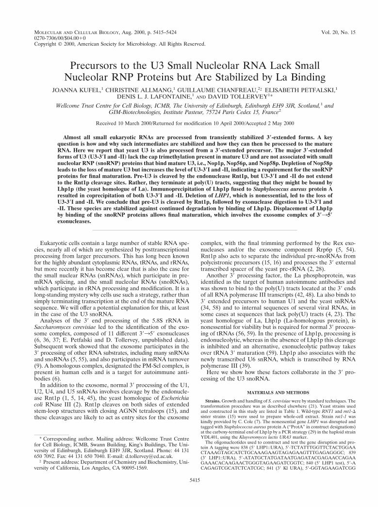
MOLECULAR AND CELLULAR BIOLOGY,0270-7306/00/$04.0010
Aug. 2000, p. 5415–5424 Vol. 20, No. 15
Copyright © 2000, American Society for Microbiology. All Rights Reserved.
Precursors to the U3 Small Nucleolar RNA Lack SmallNucleolar RNP Proteins but Are Stabilized by La Binding
JOANNA KUFEL,1 CHRISTINE ALLMANG,1 GUILLAUME CHANFREAU,2† ELISABETH PETFALSKI,1
DENIS L. J. LAFONTAINE,1 AND DAVID TOLLERVEY1*
Wellcome Trust Centre for Cell Biology, ICMB, The University of Edinburgh, Edinburgh EH9 3JR, Scotland,1 andGIM-Biotechnologies, Institute Pasteur, 75724 Paris Cedex 15, France2
Received 10 March 2000/Returned for modification 10 April 2000/Accepted 2 May 2000
Almost all small eukaryotic RNAs are processed from transiently stabilized 3*-extended forms. A keyquestion is how and why such intermediates are stabilized and how they can then be processed to the matureRNA. Here we report that yeast U3 is also processed from a 3*-extended precursor. The major 3*-extendedforms of U3 (U3-3*I and -II) lack the cap trimethylation present in mature U3 and are not associated with smallnucleolar RNP (snoRNP) proteins that bind mature U3, i.e., Nop1p, Nop56p, and Nop58p. Depletion of Nop58pleads to the loss of mature U3 but increases the level of U3-3*I and -II, indicating a requirement for the snoRNPproteins for final maturation. Pre-U3 is cleaved by the endonuclease Rnt1p, but U3-3*I and -II do not extendto the Rnt1p cleavage sites. Rather, they terminate at poly(U) tracts, suggesting that they might be bound byLhp1p (the yeast homologue of La). Immunoprecipitation of Lhp1p fused to Staphylococcus aureus protein Aresulted in coprecipitation of both U3-3*I and -II. Deletion of LHP1, which is nonessential, led to the loss ofU3-3*I and -II. We conclude that pre-U3 is cleaved by Rnt1p, followed by exonuclease digestion to U3-3*I and-II. These species are stabilized against continued degradation by binding of Lhp1p. Displacement of Lhp1pby binding of the snoRNP proteins allows final maturation, which involves the exosome complex of 3*35*exonucleases.
Eukaryotic cells contain a large number of stable RNA spe-cies, nearly all of which are synthesized by posttranscriptionalprocessing from larger precursors. This has long been knownfor the highly abundant cytoplasmic RNAs, tRNAs, and rRNAs,but more recently it has become clear that is also the case forthe small nuclear RNAs (snRNAs), which participate in pre-mRNA splicing, and the small nucleolar RNAs (snoRNAs),which participate in rRNA processing and modification. It is along-standing mystery why cells use such a strategy, rather thansimply terminating transcription at the end of the mature RNAsequence. We will offer a potential explanation for this, at leastin the case of the U3 snoRNA.
Analyses of the 39 end processing of the 5.8S rRNA inSaccharomyces cerevisiae led to the identification of the exo-some complex, composed of 11 different 39359 exonucleases(6, 36, 37; E. Petfalski and D. Tollervey, unpublished data).Subsequent work showed that the exosome participates in the39 processing of other RNA substrates, including many snRNAsand snoRNAs (5, 55), and also participates in mRNA turnover(9). A homologous complex, designated the PM-Scl complex, ispresent in human cells and is a target for autoimmune anti-bodies (6).
In addition to the exosome, normal 39 processing of the U1,U2, U4, and U5 snRNAs involves cleavage by the endonucle-ase Rnt1p (1, 5, 14, 45), the yeast homologue of Escherichiacoli RNase III (2). Rnt1p cleaves on both sides of extendedstem-loop structures with closing AGNN tetraloops (15), andthese cleavages are likely to act as entry sites for the exosome
complex, with the final trimming performed by the Rex exo-nucleases and/or the exosome component Rrp6p (5, 54).Rnt1p also acts to separate the individual pre-snoRNAs frompolycistronic precursors (15, 16) and processes the 39 externaltranscribed spacer of the yeast pre-rRNA (2, 28).
Another 39 processing factor, the La phosphoprotein, wasidentified as the target of human autoimmune antibodies andwas shown to bind to the poly(U) tracts located at the 39 endsof all RNA polymerase III transcripts (42, 48). La also binds to39 extended precursors to human U1 and the yeast snRNAs(34, 58) and to internal sequences of several viral RNAs, insome cases at sequences that lack poly(U) tracts (4, 23). Theyeast homologue of La, Lhp1p (La-homologous protein), isnonessential for viability but is required for normal 39 process-ing of tRNAs (56, 59). In the presence of Lhp1p, processing isendonucleolytic, whereas in the absence of Lhp1p this cleavageis inhibited and an alternative, exonucleolytic pathway takesover tRNA 39 maturation (59). Lhp1p also associates with thenewly transcribed U6 snRNA, which is transcribed by RNApolymerase III (39).
Here we show how these factors collaborate in the 39 pro-cessing of the U3 snoRNA.
MATERIALS AND METHODS
Strains. Growth and handling of S. cerevisiae were by standard techniques. Thetransformation procedure was as described elsewhere (21). Yeast strains usedand constructed in this study are listed in Table 1. Wild-type RNT1 and rnt1-Dsister strains (15) were used to prepare whole-cell extract. Strain rat1-1 waskindly provided by C. Cole (7). The nonessential gene LHP1 was disrupted andtagged with Staphylococcus aureus protein A (“ProtA” in construct designations)at the carboxy-terminal end of Lhp1p by a PCR strategy (29) in the haploid strainYDL401, using the Kluyveromyces lactis URA3 marker.
The oligonucleotides used to construct and test the gene disruption and pro-tein A tagging were 838 (59 LHP1::URA), 59-TCTATTTGGTTCTACTGGAACTAAAGTAGCATCTGCAAAGAAGTAGAGAAGTTTGAGAGGGC; 839(39 LHP1::URA), 59-ATATGCTATGATAATGAGATACGAGAACCAGAAGAAACACAAGAACTGGGTAGAAGATCGGTC; 840 (59 LHP1 test), 59-ACAGAGTCGCATCTCATCGC; 841 (39 Kl URA), 59-GGTAGAAGATCGG
* Corresponding author. Mailing address: Wellcome Trust Centrefor Cell Biology, ICMB, Swann Building, King’s Buildings, The Uni-versity of Edinburgh, Edinburgh EH9 3JR, Scotland. Phone: 44 131650 7092. Fax: 44 131 650 7040. E-mail: [email protected].
† Present address: Department of Chemistry and Biochemistry, Uni-versity of California, Los Angeles, CA 90095-1569.
5415
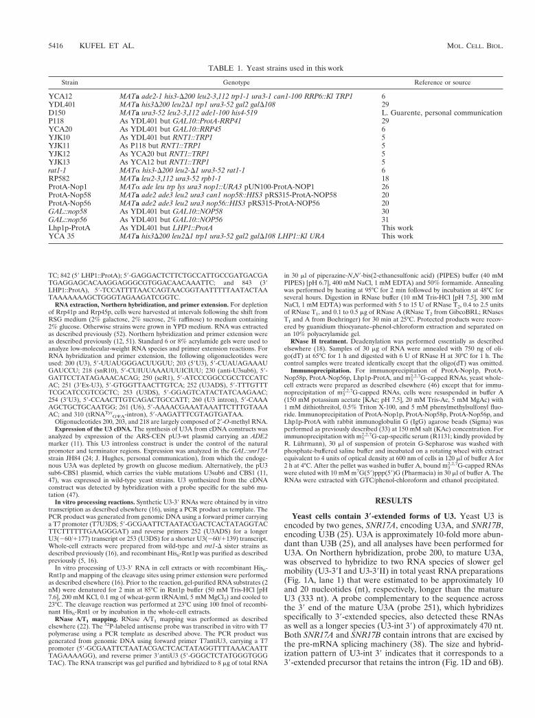
TC; 842 (59 LHP1::ProtA); 59-GAGGACTCTTCTGCCATTGCCGATGACGATGAGGAGCACAAGGAGGGCGTGGACAACAAATTC; and 843 (39LHP1::ProtA), 59-TCCATTTTAACCAGTAACGGTAATTTTTAATACTAATAAAAAAAGCTGGGTAGAAGATCGGTC.
RNA extraction, Northern hybridization, and primer extension. For depletionof Rrp41p and Rrp45p, cells were harvested at intervals following the shift fromRSG medium (2% galactose, 2% sucrose, 2% raffinose) to medium containing2% glucose. Otherwise strains were grown in YPD medium. RNA was extractedas described previously (52). Northern hybridization and primer extension wereas described previously (12, 51). Standard 6 or 8% acrylamide gels were used toanalyze low-molecular-weight RNA species and primer extension reactions. ForRNA hybridization and primer extension, the following oligonucleotides wereused: 200 (U3), 59-UUAUGGGACUUGUU; 203 (59U3), 59-CUAUAGAAAUGAUCCU; 218 (snR10), 59-CUIUUAAAUUUICIUU; 230 (anti-U3sub6), 59-GATTCCTATAGAAACACAG; 250 (scR1), 59-ATCCCGGCCGCCTCCATCAC; 251 (39Ex-U3), 59-GTGGTTAACTTGTCA; 252 (U3ADS), 59-TTTGTTTTCGCATCCGTCGCTC; 253 (U3DS), 59-GGAGTCATACTATCAAGAAC;254 (39U3), 59-CCAACTTGTCAGACTGCCATT; 260 (U3 intron), 59-CAAAAGCTGCTGCAATGG; 261 (U6), 59-AAAACGAAATAAATTCTTTGTAAAAC; and 310 (tRNATyr
GCA-intron), 59-AAGATTTCGTAGTGATAA.Oligonucleotides 200, 203, and 218 are largely composed of 29-O-methyl RNA.Expression of the U3 cDNA. The synthesis of U3A from cDNA constructs was
analyzed by expression of the ARS-CEN pU3-wt plasmid carrying an ADE2marker (11). This U3 intronless construct is under the control of the naturalpromoter and terminator regions. Expression was analyzed in the GAL::snr17Astrain JH84 (24; J. Hughes, personal communication), from which the endoge-nous U3A was depleted by growth on glucose medium. Alternatively, the pU3sub6-CBS1 plasmid, which carries the viable mutations U3sub6 and CBS1 (11,47), was expressed in wild-type yeast strains. U3 synthesized from the cDNAconstruct was detected by hybridization with a probe specific for the sub6 mu-tation (47).
In vitro processing reactions. Synthetic U3-39 RNAs were obtained by in vitrotranscription as described elsewhere (16), using a PCR product as template. ThePCR product was generated from genomic DNA using a forward primer carryinga T7 promoter (T7U3DS; 59-GCGAATTCTAATACGACTCACTATAGGTACTTCTTTTTTGAAGGGAT) and reverse primers 252 (U3ADS) for a longerU3(260/1177) transcript or 253 (U3DS) for a shorter U3(260/1139) transcript.Whole-cell extracts were prepared from wild-type and rnt1-D sister strains asdescribed previously (16), and recombinant His6-Rnt1p was purified as describedpreviously (5, 16).
In vitro processing of U3-39 RNA in cell extracts or with recombinant His6-Rnt1p and mapping of the cleavage sites using primer extension were performedas described elsewhere (16). Prior to the reaction, gel-purified RNA substrates (2nM) were denatured for 2 min at 85°C in Rnt1p buffer (50 mM Tris-HCl [pH7.6], 200 mM KCl, 0.1 mg of wheat-germ tRNA/ml, 5 mM MgCl2) and cooled to23°C. The cleavage reaction was performed at 23°C using 100 fmol of recombi-nant His6-Rnt1 or by incubation in the whole-cell extracts.
RNase A/T1 mapping. RNase A/T1 mapping was performed as describedelsewhere (22). The 32P-labeled antisense probe was transcribed in vitro with T7polymerase using a PCR template as described above. The PCR product wasgenerated from genomic DNA using forward primer T7antiU3, carrying a T7promoter (59-GCGAATTCTAATACGACTCACTATAGGTTTTAAACAATTTAGAAAAGG), and reverse primer 39antiU3 (59-GGGCTCTATGGGTGGGTAC). The RNA transcript was gel purified and hybridized to 8 mg of total RNA
in 30 ml of piperazine-N,N9-bis(2-ethanesulfonic acid) (PIPES) buffer (40 mMPIPES) [pH 6.7], 400 mM NaCl, 1 mM EDTA) and 50% formamide. Annealingwas performed by heating at 95°C for 2 min followed by incubation at 48°C forseveral hours. Digestion in RNase buffer (10 mM Tris-HCl [pH 7.5], 300 mMNaCl, 1 mM EDTA) was performed with 5 to 15 U of RNase T2, 0.4 to 2.5 unitsof RNase T1, and 0.1 to 0.5 mg of RNase A (RNase T2 from GibcoBRL; RNasesT1 and A from Boehringer) for 30 min at 25°C. Protected products were recov-ered by guanidium thiocyanate–phenol-chloroform extraction and separated onan 10% polyacrylamide gel.
RNase H treatment. Deadenylation was performed essentially as describedelsewhere (18). Samples of 30 mg of RNA were annealed with 750 ng of oli-go(dT) at 65°C for 1 h and digested with 6 U of RNase H at 30°C for 1 h. Thecontrol samples were treated identically except that the oligo(dT) was omitted.
Immunoprecipitation. For immunoprecipitation of ProtA-Nop1p, ProtA-Nop58p, ProtA-Nop56p, Lhp1p-ProtA, and m3
2,2,7G-capped RNAs, yeast whole-cell extracts were prepared as described elsewhere (46) except that for immu-noprecipitation of m3
2,2,7G-capped RNAs, cells were resuspended in buffer A(150 mM potassium acetate [KAc; pH 7.5], 20 mM Tris-Ac, 5 mM MgAc) with1 mM dithiothreitol, 0.5% Triton X-100, and 5 mM phenylmethylsulfonyl fluo-ride. Immunoprecipitation of ProtA-Nop1p, ProtA-Nop58p, ProtA-Nop56p, andLhp1p-ProtA with rabbit immunoglobulin G (IgG) agarose beads (Sigma) wasperformed as previously described (33) at 150 mM salt (KAc) concentration. Forimmunoprecipitation with m3
2,2,7G-cap-specific serum (R1131; kindly provided byR. Luhrmann), 30 ml of suspension of protein G-Sepharose was washed withphosphate-buffered saline buffer and incubated on a rotating wheel with extractequivalent to 4 units of optical density at 600 nm of cells in 120 ml of buffer A for2 h at 4°C. After the pellet was washed in buffer A, bound m3
2,2,7G-capped RNAswere eluted with 10 mM m7G(59)ppp(59)G (Pharmacia) in 30 ml of buffer A. TheRNAs were extracted with GTC/phenol-chloroform and ethanol precipitated.
RESULTS
Yeast cells contain 3*-extended forms of U3. Yeast U3 isencoded by two genes, SNR17A, encoding U3A, and SNR17B,encoding U3B (25). U3A is approximately 10-fold more abun-dant than U3B (25), and all analyses have been performed forU3A. On Northern hybridization, probe 200, to mature U3A,was observed to hybridize to two RNA species of slower gelmobility (U3-39I and U3-39II) in total yeast RNA preparations(Fig. 1A, lane 1) that were estimated to be approximately 10and 20 nucleotides (nt), respectively, longer than the matureU3 (333 nt). A probe complementary to the sequence acrossthe 39 end of the mature U3A (probe 251), which hybridizesspecifically to 39-extended species, also detected these RNAsas well as a longer species (U3-int 39) of approximately 470 nt.Both SNR17A and SNR17B contain introns that are excised bythe pre-mRNA splicing machinery (38). The size and hybrid-ization pattern of U3-int 39 indicates that it corresponds to a39-extended precursor that retains the intron (Fig. 1D and 6B).
TABLE 1. Yeast strains used in this work
Strain Genotype Reference or source
YCA12 MATa ade2-1 his3-D200 leu2-3,112 trp1-1 ura3-1 can1-100 RRP6::Kl TRP1 6YDL401 MATa his3D200 leu2D1 trp1 ura3-52 gal2 galD108 29D150 MATa ura3-52 leu2-3,112 ade1-100 his4-519 L. Guarente, personal communicationP118 As YDL401 but GAL10::ProtA-RRP41 29YCA20 As YDL401 but GAL10::RRP45 6YJK10 As YDL401 but RNT1::TRP1 5YJK11 As P118 but RNT1::TRP1 5YJK12 As YCA20 but RNT1::TRP1 5YJK13 As YCA12 but RNT1::TRP1 5rat1-1 MATa his3-D200 leu2-D1 ura3-52 rat1-1 6RP582 MATa leu2-3,112 ura3-52 rpb1-1 18ProtA-Nop1 MATa ade leu trp lys ura3 nop1::URA3 pUN100-ProtA-NOP1 26ProtA-Nop58 MATa ade2 ade3 leu2 ura3 can1 nop58::HIS3 pRS315-ProtA-NOP58 20ProtA-Nop56 MATa ade2 ade3 leu2 ura3 nop56::HIS3 pRS315-ProtA-NOP56 20GAL::nop58 As YDL401 but GAL10::NOP58 30GAL::nop56 As YDL401 but GAL10::NOP56 31Lhp1p-ProtA As YDL401 but LHP1::ProtA This workYCA 35 MATa his3D200 leu2D1 trp1 ura3-52 gal2 galD108 LHP1::Kl URA This work
5416 KUFEL ET AL. MOL. CELL. BIOL.
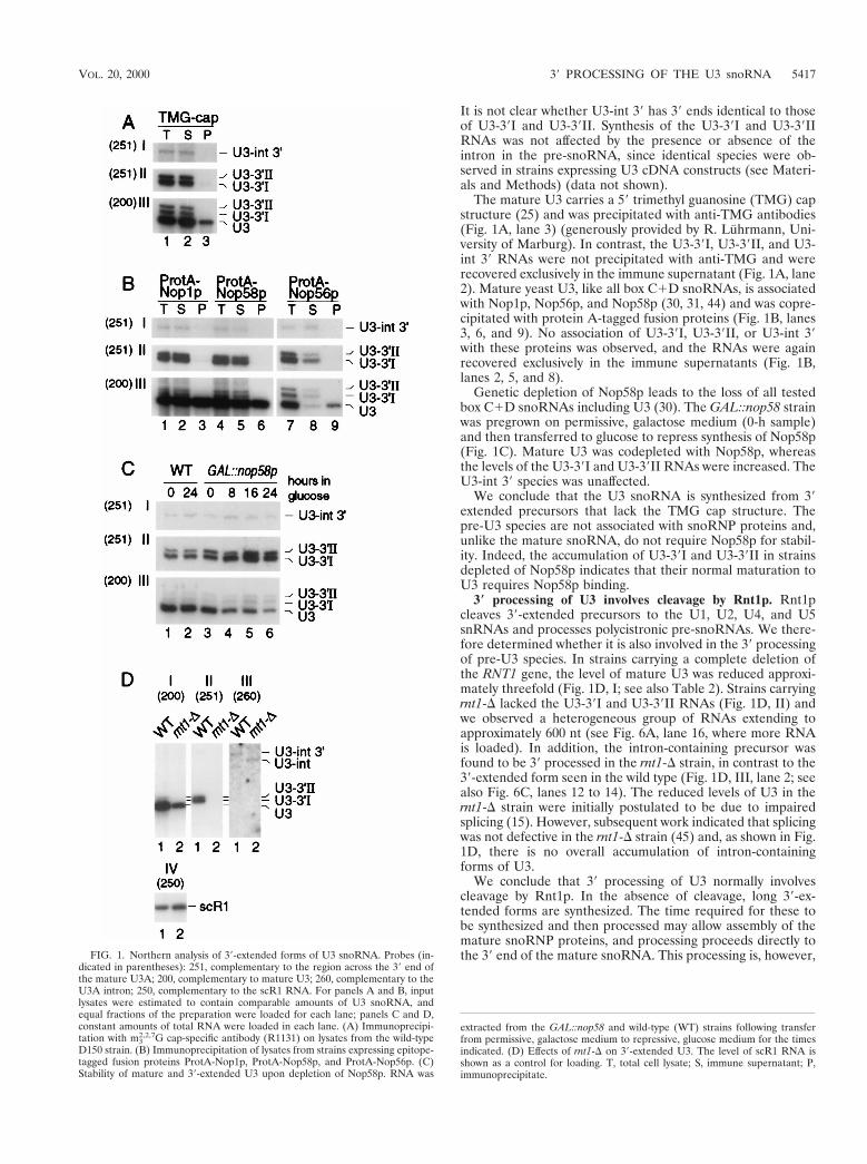
It is not clear whether U3-int 39 has 39 ends identical to thoseof U3-39I and U3-39II. Synthesis of the U3-39I and U3-39IIRNAs was not affected by the presence or absence of theintron in the pre-snoRNA, since identical species were ob-served in strains expressing U3 cDNA constructs (see Materi-als and Methods) (data not shown).
The mature U3 carries a 59 trimethyl guanosine (TMG) capstructure (25) and was precipitated with anti-TMG antibodies(Fig. 1A, lane 3) (generously provided by R. Luhrmann, Uni-versity of Marburg). In contrast, the U3-39I, U3-39II, and U3-int 39 RNAs were not precipitated with anti-TMG and wererecovered exclusively in the immune supernatant (Fig. 1A, lane2). Mature yeast U3, like all box C1D snoRNAs, is associatedwith Nop1p, Nop56p, and Nop58p (30, 31, 44) and was copre-cipitated with protein A-tagged fusion proteins (Fig. 1B, lanes3, 6, and 9). No association of U3-39I, U3-39II, or U3-int 39with these proteins was observed, and the RNAs were againrecovered exclusively in the immune supernatants (Fig. 1B,lanes 2, 5, and 8).
Genetic depletion of Nop58p leads to the loss of all testedbox C1D snoRNAs including U3 (30). The GAL::nop58 strainwas pregrown on permissive, galactose medium (0-h sample)and then transferred to glucose to repress synthesis of Nop58p(Fig. 1C). Mature U3 was codepleted with Nop58p, whereasthe levels of the U3-39I and U3-39II RNAs were increased. TheU3-int 39 species was unaffected.
We conclude that the U3 snoRNA is synthesized from 39extended precursors that lack the TMG cap structure. Thepre-U3 species are not associated with snoRNP proteins and,unlike the mature snoRNA, do not require Nop58p for stabil-ity. Indeed, the accumulation of U3-39I and U3-39II in strainsdepleted of Nop58p indicates that their normal maturation toU3 requires Nop58p binding.
3* processing of U3 involves cleavage by Rnt1p. Rnt1pcleaves 39-extended precursors to the U1, U2, U4, and U5snRNAs and processes polycistronic pre-snoRNAs. We there-fore determined whether it is also involved in the 39 processingof pre-U3 species. In strains carrying a complete deletion ofthe RNT1 gene, the level of mature U3 was reduced approxi-mately threefold (Fig. 1D, I; see also Table 2). Strains carryingrnt1-D lacked the U3-39I and U3-39II RNAs (Fig. 1D, II) andwe observed a heterogeneous group of RNAs extending toapproximately 600 nt (see Fig. 6A, lane 16, where more RNAis loaded). In addition, the intron-containing precursor wasfound to be 39 processed in the rnt1-D strain, in contrast to the39-extended form seen in the wild type (Fig. 1D, III, lane 2; seealso Fig. 6C, lanes 12 to 14). The reduced levels of U3 in thernt1-D strain were initially postulated to be due to impairedsplicing (15). However, subsequent work indicated that splicingwas not defective in the rnt1-D strain (45) and, as shown in Fig.1D, there is no overall accumulation of intron-containingforms of U3.
We conclude that 39 processing of U3 normally involvescleavage by Rnt1p. In the absence of cleavage, long 39-ex-tended forms are synthesized. The time required for these tobe synthesized and then processed may allow assembly of themature snoRNP proteins, and processing proceeds directly tothe 39 end of the mature snoRNA. This processing is, however,FIG. 1. Northern analysis of 39-extended forms of U3 snoRNA. Probes (in-
dicated in parentheses): 251, complementary to the region across the 39 end ofthe mature U3A; 200, complementary to mature U3; 260, complementary to theU3A intron; 250, complementary to the scR1 RNA. For panels A and B, inputlysates were estimated to contain comparable amounts of U3 snoRNA, andequal fractions of the preparation were loaded for each lane; panels C and D,constant amounts of total RNA were loaded in each lane. (A) Immunoprecipi-tation with m3
2,2,7G cap-specific antibody (R1131) on lysates from the wild-typeD150 strain. (B) Immunoprecipitation of lysates from strains expressing epitope-tagged fusion proteins ProtA-Nop1p, ProtA-Nop58p, and ProtA-Nop56p. (C)Stability of mature and 39-extended U3 upon depletion of Nop58p. RNA was
extracted from the GAL::nop58 and wild-type (WT) strains following transferfrom permissive, galactose medium to repressive, glucose medium for the timesindicated. (D) Effects of rnt1-D on 39-extended U3. The level of scR1 RNA isshown as a control for loading. T, total cell lysate; S, immune supernatant; P,immunoprecipitate.
VOL. 20, 2000 39 PROCESSING OF THE U3 snoRNA 5417
inefficient since mature U3 levels are strongly reduced (Fig.1D; see also Table 2).
Rnt1p cleaves on both sides of extended stem-loop struc-tures with a closing AGNN tetraloop (15). Inspection of the 39flanking sequences revealed the presence of good matches toconsensus Rnt1p cleavage sites 39 to both SNR17A andSNR17B, the genes encoding U3A and U3B, respectively(shown for SNR17A in Fig. 2D). To confirm that these areauthentic cleavage sites, the cleavage of the SNR17A site wastested in vitro. The U3(260/1139) in vitro transcript, whichspans the region between positions 260 and 1139 with respectto the mature U3 39 end including the predicted stem-loopstructure, was used to map the cleavage site by primer exten-sion (Fig. 2A). Incubation with recombinant His6-Rnt1p (Fig.2A, lane 5) resulted in the appearance of two primer extensionstops that were not detected after incubation in the absence ofRnt1p (Fig. 2A, lane 6). The primer extension stops were at nt122 and 159, corresponding to cleavage between nt 121/22and 158/159, and are in good agreement with the consensussites of Rnt1p cleavage (Fig. 2D). To demonstrate that in vitroprocessing is by endonuclease cleavage, a longer transcript waslabeled internally; U3(260/1177) spans the 39 region of theU3A precursor between positions 260 and 1177. Incubationwith either recombinant His6-Rnt1p (Fig. 2B, lanes 3 to 5) oran extract from a wild-type (RNT11) strain of yeast (Fig. 2B,lane 6) led to the appearance of a set of discrete cleavageproducts that were not observed with the no-enzyme controlreaction (Fig. 2B, lane 2) or with an extract from an rnt1-Dstrain of yeast (Fig. 2B, lane 7). The substrate is 237 nt, andcomparison to size markers (Fig. 2B, lanes 1 and 8) indicatedthat the sizes of the three smaller species were in good agree-ment with the predicted cleavage products: from 159 to the 39end of the transcript (predicted size, 119 nt) (band a), from the59 end to 121 (predicted size, 81 nt) (band b), and from 122to 158 (predicted size, 37 nt) (band c).
The 39 fragments generated by Rnt1p cleavage of the pre-U4snRNA and the pre-rRNA are strongly stabilized by mutationof the nuclear 59339 exonuclease Rat1p (5, 28), indicating thatit normally degrades these regions. The sites of in vivo cleavageof pre-U3 were identified by primer extension using probe 252,which hybridizes in the SNR17A flanking sequence 39 to thestem-loop structure. In the rat1-1 strain (Fig. 2C, lane 5),primer extension stops were observed at 122 and 159, iden-tical to the in vitro cleavage sites. These were absent fromRNA extracted from the rnt1-D strain (Fig. 2C, lane 6) butwere also not detectable in the wild-type strain (Fig. 2C, lane7). The stop corresponding to the position of the 39 end ofmature U3 may be a consequence of the stem structure at thisposition. The level of this stop is unaltered in the rat1-1 strain,suggesting that it is not a cleavage site. We cannot, however,exclude the possibility that a fraction of U3 is processed byendonucleolytic cleavage at the mature 39 end. RNase MRPwas shown not to be involved in this process (data not shown).
We conclude that Rnt1p cleaves the 39 extended pre-U3 at121/122 and 158/159. Following cleavage the 39 fragment is
FIG. 2. Rnt1p cleaves the 39 end of the U3 precursor. (A) Mapping of the invitro Rnt1p cleavage sites. Primer extension was performed with probe 253 onthe model U3(260/1139) RNA incubated with buffer (lane 6) or recombinantHis6-Rnt1p (lane 5) as described in Materials and Methods. DNA sequencingreaction on a PCR product encompassing the 39 end of U3 from positions 260to 1139, using the same primer, was run in parallel (lanes 1 to 4). The primerextension stops at positions 122 and 159 are indicated. (B) In vitro cleavage ofan internally labeled model U3(260/1177) RNA substrate by Rnt1p. 32P-labeledU3(260/1177) RNA was incubated at 23°C in the following conditions: lane 2,Rnt1p buffer; lanes 3 to 5, Rnt1p buffer with 10 ng of recombinant His6-Rnt1pfor the times indicated; lane 6, with whole-cell extract from a wild-type (WT)strain of yeast; lane 7, with whole-cell extract from an rnt1-D strain. Lanes 1 and8, RNA size markers. The positions of DNA size markers are indicated on theright in nucleotides. The obtained cleavage products are labeled a to c on the left,and the predicted origins of these species are as follows: S, substrate (237 nt); a,39 end of transcript to position 121/122 (119 nt); b, 59 end of transcript toposition 158/159 (81 nt); c, positions 121/122 to 158/159 (37 nt). Since invitro cleavages of U3(260/1177) are complete (100%), no intermediate cleav-
age products are visible. (C) Mapping of the Rnt1p 59 cleavage site in vitro.Primer extension analysis through the 39 end of the pre-U3 was performed withprimer 252, hybridizing downstream of position 1177. RNA was extracted fromwild-type (lane 7) and rnt1-D (lane 6) strains grown at 30°C and from a rat1-1strain following transfer to 37°C for 2 h (lane 5). DNA sequencing reactions wererun in parallel (lanes 1 to 4). The primer extension stops at positions 159, 122,and 11 (39 end of U3) are indicated. (D) Computer-predicted RNA structure inthe U3 39 flanking region that contains the Rnt1p cleavage sites. The cleavagesites between nt 121 and 122 and between nt 158 and 159 are indicated byarrows. The 39 end of mature U3 is underlined.
5418 KUFEL ET AL. MOL. CELL. BIOL.
degraded by Rat1p. The level of the mature U3 is reduced instrains lacking Rnt1p, indicating that this is normally the majorsynthesis pathway.
The major 3*-extended forms of U3 do not extend to theRnt1p cleavage sites. High-resolution Northern hybridizationshowed that the U3-39I band was too small to extend to theRnt1p cleavage sites, and even the larger U3-39 II speciesappeared to be slightly smaller than expected. The 39 ends ofthese species were therefore determined by RNase protection.For this, the region of SNR17A from 295 to 136 was amplifiedby PCR using a primer that incorporated a T7 promoter (seeMaterials and Methods). In addition to the band correspond-ing to the mature 39 end of U3, two major protected fragmentswere detected in RNA from the wild-type strain (Fig. 3A, lane3) but were absent from the rnt1-D strain (Fig. 3A, lane 4). Thesizes to these bands correspond to species that extend toU3112 and U3118, in good agreement with the gel mobilitiesof the U3-39I and U3-39II RNAs, respectively.
We conclude that following Rnt1p cleavage, the pre-U3undergoes rapid trimming back to 112 and 118.
The major 3*-extended forms of U3 are stabilized by Lhp1p.It seemed very likely that some RNA binding factor was re-sponsible for stabilizing the 39 ends of the U3-39I and -IIspecies. Inspection of the sequence showed that these RNAspossessed 39 poly(U) tracts (Fig. 3B). The 39 poly(U) tracts ofRNAs transcribed by RNA polymerase III are bound by the Laprotein (42, 48), as are the 39 extended precursors to humanU1 (34) and yeast (58) snRNAs. We therefore tested whether
the U3-39I and -II RNAs were being stabilized by binding toLhp1p, the yeast homologue of La (39, 59).
The LHP1 gene is nonessential (59), and a gene disruptionwas performed by a one-step PCR approach (10) using the K.lactis URA3 marker (see Materials and Methods). RNase pro-tection analysis of RNA from the lhp1-D strain showed the lossof the major 39-extended ends at 118 and 112 and the ap-pearance of shorter, heterogeneous protected fragments cor-responding to RNAs from U318 to U3111 (Fig. 3A, lane 7).This result was confirmed by Northern hybridization (Fig. 4).The U3-39II and U3-39I species were absent from the lhp1-Dstrain (Fig. 4A), and a species slightly shorter than U3-39I wasdetected. The level of mature U3 was unaffected in the lhp1-Dstrain (Figs. 3A and 4B), as were the levels of the truncated U3degradation intermediates seen in wild-type cells (see Fig. 6;data not shown). These data suggested that both U3-39I andU3-39II were stabilized by binding Lhp1p.
To confirm this, a C-terminal fusion between Lhp1p and twocopies of the Z domain of S. aureus protein A was constructedand integrated at the chromosomal LHP1 locus by a one-stepPCR approach (29) (see Materials and Methods). Westernblotting confirmed that the fusion protein was expressed andcould be efficiently immunoprecipitated with IgG agarose(data not shown). Immunoprecipitation was performed on twoindependently isolated Lhp1p-ProtA strains; data are pre-sented for only one strain in Fig. 5. Processing of pre-tRNATyr
appeared to be the same in the strain expressing only Lhp1p-ProtA and the wild type (Fig. 5D); however, some accumula-tion of the shorter 39-extended pre-U3 species was visible (Fig.5A), suggesting that the Lhp1p-ProtA fusion protein is under-expressed or otherwise not fully functional.
As expected, the tRNATyr primary transcript (Fig. 5D) andthe U6 snRNA (Fig. 5E) were immunoprecipitated on IgGagarose from the strain expressing Lhp1p-ProtA (lane 6) butnot from the wild type (lane 3). Both U3-39I and U3-39II werecoprecipitated with Lhp1p-ProtA (Fig. 5A), as were U3-int 39and a species of approximately 800 nt designated U3-39III (Fig.5B). The species shorter than U3-39I seen in the Lhp1p-ProtAstrain was not coprecipitated and remained in the immune
FIG. 3. Mapping of the 39-extended forms of U3 by RNase protection. (A)RNA was extracted from wild-type (WT), rnt1-D, and lhp1-D strains grown at30°C and from GAL::rrp41 and GAL::rrp41/rnt1-D strains following transfer frompermissive, RSG medium to repressive, glucose medium at 30°C for 24 and 48 h,respectively. Total E. coli tRNA was used as a control RNA. Positions of theRnt1p-dependent protected species at 112 and 118 are indicated. (B) Sche-matic of the U3 39 flanking region showing the ends of the protected regions andthe Rnt1p cleavage sites.
FIG. 4. 39-extended forms of U3 are stabilized by Lhp1p. Lane 1, LHP1strain; lane 2, lhp1-D strain. Total RNA was analyzed by Northern hybridizationwith probe 251, specific for the 39-extended U3 (A), probe 200, which hybridizesto the mature U3 (B), and probe 250, which hybridizes to scR1 RNA (C).
VOL. 20, 2000 39 PROCESSING OF THE U3 snoRNA 5419
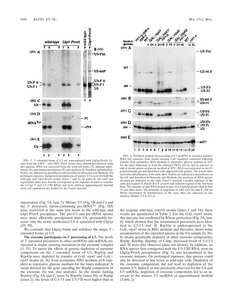
supernatant (Fig. 5A, lane 5). Mature U3 (Fig. 5B and C) andthe 39 processed, intron-containing pre-tRNATyr (Fig. 5D)were recovered at the same low levels in the wild-type andLhp1-ProtA precipitates. The pre-U3 and pre-tRNA specieswere more efficiently precipitated than U6, presumably be-cause only the newly synthesized U6 is associated with Lhp1p(35, 39).
We conclude that Lhp1p binds and stabilizes the major 39-extended forms of U3.
The exosome participates in 3* processing of U3. The levelsof 39-extended precursors to other snoRNAs and snRNAs areelevated in strains carrying mutations in the exosome complex(5, 55). To assess the effects of genetic depletion of exosomecomponents on the 39-extended forms of U3, Rrp41p andRrp45p were depleted by transfer of GAL::rrp41 and GAL::rrp45 strains (6, 36) from permissive RSG medium (0-h sam-ples) to repressive, glucose medium for the times indicated. Astrain deleted for the gene encoding the Rrp6p component ofthe exosome (6) was also analyzed. In the strains lackingRrp41p (Fig. 6A and C, lanes 5), Rrp45p (lanes 10), or Rrp6p(lanes 2), the levels of U3-39I and U3-39II were higher than in
the isogenic wild-type control strains (lanes 3 and 14); theseresults are quantitated in Table 2. For the GAL::rrp41 strain,this increase was confirmed by RNase protection (Fig. 3A, lane4), which showed that the accumulated precursors were iden-tical to U3-39I and -II. Rrp41p is underexpressed in theGAL::rrp41 strain in RSG medium and therefore shows someaccumulation of the extended species in the 0-h sample (6, 36).In strains genetically depleted of other exosome components,Rrp4p, Rrp40p, Rrp46p, or Csl4p, increased levels of U3-39Iand -II were also observed (data not shown). In addition, anRNA species that comigrated with the U3-39III RNA, seen onLhp1p-ProtA precipitation (Fig. 5), was accumulated in theexosome mutants. On prolonged exposure, this species couldalso be detected at low levels in wild-type cells. Depletion ofthe exosome components did not lead to depletion of themature U3. Indeed, as was previously observed for the U4 andU5 snRNAs, depletion of exosome components led to an in-crease in the mature U3 snoRNA of approximately twofold(Table 2).
FIG. 5. 39-extended forms of U3 are coprecipitated with Lhp1p-ProtA. Ly-sates from the LHP11 and LHP1::ProtA strains were immunoprecipitated usingIgG agarose. RNA was recovered from the total cell lysate (T), immune super-natant (S), and immunoprecipitate (P) and analyzed by Northern hybridization.Probes are indicated in parentheses and described in Materials and Methods. Onprolonged exposure, background precipitation of mature U3 is seen for both thewild-type and Lhp1-ProtA strains (lanes 7 and 8). In panel B, the total andsupernatant lanes were heavily overexposed at the exposure needed to visualizethe U3-int 39 and U3-39III RNAs and were omitted. Approximately fourfoldmore cell equivalents are loaded for the bound material.
FIG. 6. Northern analysis of processing of U3 snoRNA in exosome mutants.RNA was extracted from strains carrying GAL-regulated constructs followingtransfer from permissive, RSG medium to repressive, glucose medium at 30°Cfor the times indicated, or from the wild-type (WT), rnt1-D, rrp6-D, and rnt1-D/rrp6-D strains grown on glucose medium at 30°C. RNA was separated on an 6%polyacrylamide gel and hybridized with oligonucleotide probes. The panels showsuccessive hybridization of the same filter. Probes are indicated in parentheses onthe left and described in Materials and Methods; the positions of RNA speciesdetected are indicated on the right. Panel C presents a weaker exposure of thesame gel as panel A. Panels B to E present only relevant regions of the Northernblots. The amount of total RNA loaded in lane 16 is fourfold greater than in lane15 and other lanes. The positions of migration of scR1 (525 nt) and P (369 nt)RNAs determined by hybridization of the same filter are indicated as sizemarkers. Mature U3 is 333 nt.
5420 KUFEL ET AL. MOL. CELL. BIOL.
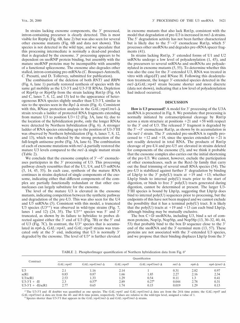
In strains lacking exosome components, the 39 processed,intron-containing precursor is clearly detected. This is mostvisible for Rrp6p (Fig. 6B, lane 2) but was also seen for severalother exosome mutants (Fig. 6B and data not shown). Thisspecies is not detected in the wild type, and we speculate thatthis processing intermediate is normally a dead-end productthat is degraded by the exosome. 39 processing appears to bedependent on snoRNP protein binding, but assembly with themature snoRNP proteins may be incompatible with assemblyof a functional spliceosome. The exosome also degrades otherstalled, intron-containing pre-mRNAs (C. Bousquet-Antonelli,C. Presutti, and D. Tollervey, submitted for publication).
The combination of the deletion of both RNT1 and RRP6(Fig. 6, lane 1) partially restored synthesis of species with thesame gel mobility as the U3-39I and U3-39II RNAs. Depletionof Rrp41p or Rrp45p from the strain lacking Rnt1p (Fig. 6Aand C, lanes 7, 8, 12, and 13) led to the appearance of heter-ogeneous RNA species slightly smaller than U3-39I, similar insize to the species seen in the lhp1-D strain (Fig. 4). Consistentwith this, RNase protection analysis in the GAL::rrp41/rnt1-Dstrain reveals a ladder of protected RNA fragments extendingfrom mature U3 to position U3112 (Fig. 3A, lane 6); due tothe location of the hybridization probe, only the longer RNAswere detected by Northern hybridization (Fig. 6). A strongerladder of RNA species extending up to the position of U3-39IIIwas observed by Northern hybridization (Fig. 6, lanes 7, 8, 12,and 13), which was reflected by the strong protection of thefull-length antisense probe (Fig. 3A, lane 6). The combinationof each of exosome mutations with rnt1-D partially restored themature U3 levels compared to the rnt1-D single mutant strain(Table 2).
We conclude that the exosome complex of 39359 exonucle-ases participates in the 39 processing of U3. This processingpathway closely resembles that of the U1, U4, and U5 snRNAs(5, 14, 45, 55). In each case, synthesis of the mature RNAcontinues in strains depleted of single components of the exo-some, indicating either that different components of the com-plex are partially functionally redundant or that other exo-nucleases can largely substitute for the exosome.
The level of the mature U3 is elevated in the exosomemutants, indicating competition between the synthetic pathwayand degradation of the pre-U3. This was also seen for the U4and U5 snRNAs (5). Consistent with this model, a truncatedU3 species (U3**) was observed in wild-type strains (Fig. 7,lanes 1 and 12) (24, 35). The U3** species was 59 and 39truncated, as shown by its failure to hybridize to probes di-rected against either the 39 end of U3 (Fig. 7B) or the 59 endof U3 (Fig. 7C). In contrast, the U3* species that is accumu-lated in rrp6-D, GAL::rrp41, and GAL::rrp45 strains was trun-cated only at the 59 end, indicating that U3 is normally 39degraded by the exosome. The level of U3* is further elevated
in exosome mutants that also lack Rnt1p, consistent with themodel that degradation of pre-U3 is increased in rnt1-D strains.The 59 degradation activity has not been further characterizedbut is likely due to the 59339 exonuclease Rat1p, which 59processes other snoRNAs and degrades pre-rRNA spacer frag-ments (41).
In strains lacking Rnt1p, 39 extended forms of U1 and U2snRNAs undergo a low level of polyadenylation (1, 45), andthe precursors to several snRNAs and snoRNAs are polyade-nylated in exosome mutants (5, 55). To determine whether thiswas also the case for the 39-extended U3, RNA was treated invitro with oligo(dT) and RNase H. Following this deadenyla-tion treatment, the longer 39-extended species detected in thernt1-D/GAL::rrp41 strain became shorter and more discrete(data not shown), indicating that a low level of polyadenylationhad indeed occurred.
DISCUSSION
How is U3 processed? A model for 39 processing of the U3AsnoRNA is presented in Fig. 8. We postulate that processing isnormally initiated by cotranscriptional cleavage by Rnt1pacross a stem structure at positions 121 and 158 with respectto the 39 end of U3. The released 39 fragment is degraded bythe 59339 exonuclease Rat1p, as shown by its accumulation inthe rat1-1 strain. The 39 extended pre-snoRNA is rapidly pro-cessed to 112 and 118, since the species extended to 121 isnot readily detected in total RNA. The products of Rnt1pcleavage of pre-U4 and pre-U5 are elevated in strains deletedfor components of the exosome (5), and we think it probablethat the exosome complex also carries out the initial shorteningof the pre-U3. We cannot, however, exclude the participationof other exonucleases, such as the Rex1-3p family that carryout the final trimming of several small RNA species (54). Thepre-U3 is stabilized against further 39 degradation by bindingof Lhp1p to the 39 poly(U) tracts at 119 and 113; whetherLhp1p binds to internal poly(U) tracts prior to the start ofdigestion, or binds to free 39 poly(U) tracts generated duringdigestion, cannot be determined at present. The larger U3-39III species is bound by Lhp1p, suggesting that Lhp1p doesbind to internal poly(U) sequences prior to processing, but theendpoints of this have not been mapped and we cannot excludethe possibility that it has a terminal poly(U) tract. It is likelythat the poly(U) tracts at 119 and 113 can each bind Lhp1p,although binding may be mutually exclusive.
The box C1D snoRNAs, including U3, bind a set of com-mon proteins, Nop1p, Nop56p, and Nop58p (13, 30–32, 40, 44,53) that probably bind to the box D sequence close to the 39end of the snoRNA and the 39-terminal stem (13, 57). Theseproteins are not associated with the 39-extended U3 species,and we propose that their binding displaces Lhp1p from the 39
TABLE 2. PhosphorImager quantification of Northern hybridization data from Fig. 6a
ConstructQuantification
GAL::rrp41 GAL::rrp41/rnt1-D GAL::rrp45 GAL::rrp45/rnt1-D rnt1-D rrp6-D rrp6-D/rnt1-D
U3 2.1 1.16 2.14 1 0.31 2.82 0.97scR1 0.83 0.87 1.66 1.85 2.27 2.16 2.34U3/scR1 2.53 1.33 1.29 0.54 0.11 1.3 0.41U3-39I 1 -II 2.3 0.57b 2.89 0.27b 0.044 2.78 0.31U3-39I 1 -II/scR1 2.77 0.65 1.74 0.15 0.019 1.29 0.13
a The U3-39I and -II doublet was quantified as one species. The GAL::rrp41 and GAL::rrp41/rnt1-D data are from the 24-h time points; the GAL::rrp45 andGAL::rrp45/rnt1-D data are from the 40- and 48-h time points, respectively. Values are relative to the wild-type level, assigned a value of 1.
b Species shorter than U3-39I that appears in the GAL::rrp41/rnt1-D and GAL::rrp45/rnt1-D strains.
VOL. 20, 2000 39 PROCESSING OF THE U3 snoRNA 5421
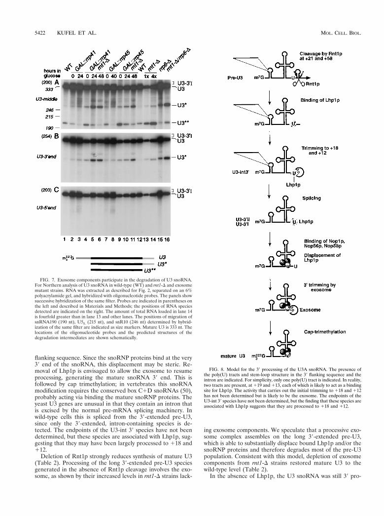
flanking sequence. Since the snoRNP proteins bind at the very39 end of the snoRNA, this displacement may be steric. Re-moval of Lhp1p is envisaged to allow the exosome to resumeprocessing, generating the mature snoRNA 39 end. This isfollowed by cap trimethylation; in vertebrates this snoRNAmodification requires the conserved box C1D snoRNAs (50),probably acting via binding the mature snoRNP proteins. Theyeast U3 genes are unusual in that they contain an intron thatis excised by the normal pre-mRNA splicing machinery. Inwild-type cells this is spliced from the 39-extended pre-U3,since only the 39-extended, intron-containing species is de-tected. The endpoints of the U3-int 39 species have not beendetermined, but these species are associated with Lhp1p, sug-gesting that they may have been largely processed to 118 and112.
Deletion of Rnt1p strongly reduces synthesis of mature U3(Table 2). Processing of the long 39-extended pre-U3 speciesgenerated in the absence of Rnt1p cleavage involves the exo-some, as shown by their increased levels in rnt1-D strains lack-
ing exosome components. We speculate that a processive exo-some complex assembles on the long 39-extended pre-U3,which is able to substantially displace bound Lhp1p and/or thesnoRNP proteins and therefore degrades most of the pre-U3population. Consistent with this model, depletion of exosomecomponents from rnt1-D strains restored mature U3 to thewild-type level (Table 2).
In the absence of Lhp1p, the U3 snoRNA was still 39 pro-
FIG. 7. Exosome components participate in the degradation of U3 snoRNA.For Northern analysis of U3 snoRNA in wild-type (WT) and rnt1-D and exosomemutant strains. RNA was extracted as described for Fig. 2, separated on an 6%polyacrylamide gel, and hybridized with oligonucleotide probes. The panels showsuccessive hybridization of the same filter. Probes are indicated in parentheses onthe left and described in Materials and Methods; the positions of RNA speciesdetected are indicated on the right. The amount of total RNA loaded in lane 14is fourfold greater than in lane 13 and other lanes. The positions of migration ofsnRNA190 (190 nt), U5L (215 nt), and snR10 (246 nt) determined by hybrid-ization of the same filter are indicated as size markers. Mature U3 is 333 nt. Thelocations of the oligonucleotide probes and the predicted structures of thedegradation intermediates are shown schematically.
FIG. 8. Model for the 39 processing of the U3A snoRNA. The presence ofthe poly(U) tracts and stem-loop structure in the 39 flanking sequence and theintron are indicated. For simplicity, only one poly(U) tract is indicated. In reality,two tracts are present, at 119 and 113, each of which is likely to act as a bindingsite for Lhp1p. The activity that carries out the initial trimming to 118 and 112has not been determined but is likely to be the exosome. The endpoints of theU3-int 39 species have not been determined, but the finding that these species areassociated with Lhp1p suggests that they are processed to 118 and 112.
5422 KUFEL ET AL. MOL. CELL. BIOL.

cessed. The U3-39I and U3-39II species were absent but slightlysmaller, heterogeneous species were observed, indicating thatsome other factor(s) can also bind the 39 poly(U) tract. Anobvious candidate is the Lsm complex, which binds to the 39poly(U) tract of the U6 snRNA and is required for normal 39processing of the RNase P RNA (3, 17, 35, 43). Consistent withthis model, mutations in Lsm8p were lethal in combinationwith the deletion of LHP1 (39).
In otherwise wild-type strains, depletion of exosome compo-nents increased the mature U3 level by inhibiting a 39 degra-dation pathway that generates the truncated U3** intermedi-ate, indicating competition between the synthetic anddegradative pathways during normal U3 synthesis. Similar ob-servations have been reported for the U4 and U5 snRNAs (5).
The processing pathway deduced here for yeast U3 showssimilarities to the processing pathways proposed for the U1,U2, U4, and U5 spliceosomal snRNAs. In each case, down-stream cleavage by Rnt1p is thought to act as an entry for theexonucleases (1, 5, 14, 45). For U1, U4, and U5, this processingwas also shown to involve the exosome complex (5, 55); this hasnot been addressed for U2. Also in each case, shorter 39 ex-tended species normally accumulate as transient intermedi-ates, although their 39 ends have not yet been accuratelymapped. In the case of pre-U4 and pre-U5, the Rnt1p cleavageproducts are 39 processed by the exosome complex and thentrimmed to the mature RNAs by the Rex1-3p family of exo-nucleases together with the Rrp6p exosome component (54).Other box C1D snoRNAs are 39 trimmed by Rrp6p (5), butthis is not the case for U3.
Inspection of the 39 flanking sequences reveals that poly(U)tracts are present in the 39 flanking sequences of the U1, U2,U4, and U5 snRNA genes (Fig. 9). In each case, the Rnt1pcleavage site is adjacent to a poly(U) tract (Fig. 9A). For theU2, U3, and U5L RNAs, the mature RNA regions (uppercasein Fig. 9A) are located relatively close to the Rnt1p cleavagesite, with additional poly(U) tracts between the Rnt1p cleavagesite and the mature 39 end. The mature regions of U1, U4, andU5S are more distant, and their 39 ends are located within a
further poly(U) tract (Fig. 9B). Lhp1p is associated with yeastpre-U1, U2, U4, and U5 (58). However, in contrast to themodel presented here for U3, Lhp1p is proposed to function asa cofactor for the assembly of the spliceosomal snRNAs withthe Sm proteins. The human and plant U3 snoRNAs also have39 flanking poly(U) tracts, suggesting that this feature may beconserved throughout eukaryotes (27, 49).
Why is U3 processed? The 39 ends of almost all RNAs fromall organisms are generated by 39 processing rather than tran-scription termination, but the reasons for this have largelyremained obscure. The data presented here provide a possibleexplanation, at least for U3. The binding sites for the commonsnoRNP proteins, the box D element and the terminal stemstructure, define the 39 end of the mature U3 snoRNA. Tran-scription termination at this site would generate an RNA witha monomethylguanosine cap structure and lacking the snoRNPproteins. This could not readily be distinguished from theproducts of premature termination or failed pre-mRNA splic-ing. It is likely that these are normally very rapidly degraded bythe exosome complex and Rat1p (C. Bousquet-Antonelli, C.Presutti, and D. Tollervey, unpublished data). Delaying orreducing these degradative activities might allow sufficient timefor snoRNP assembly and cap trimethylation, but at the ex-pense of allowing greater accumulation of aberrant RNAs.Such a strategy might also allow a greater level of accidentalprotection of inappropriate RNA species by RNA-binding pro-teins. Instead, the cell has adopted a mechanism to specificallydelay 39 processing of the snoRNA. Transcription continuesbeyond the 39 end of the mature snoRNA, with the transcriptnormally being cleaved by Rnt1p and protected by binding ofLhp1p. This leaves the mature 39 end free for binding of thesnoRNP proteins. Such a system has the additional advantageof acting as a quality control system. We envisage that thesnoRNP proteins, or at least Nop58p, must displace Lhp1p toallow final maturation of the snoRNA. In the absence ofNop58p binding, the 39 extended pre-U3 accumulates to lowlevels and is then degraded. Binding of La to pre-tRNAs hasalso been proposed to function as a quality control system (19),and binding of Lhp1p to the U6 snRNA and pre-tRNAi
Met isalso likely to antagonize rapid 39 degradation (8, 39).
We propose that 39 processing acts as a quality controlsystem in the synthesis of many RNA species and that thisunderlies its ubiquitous occurrence.
ACKNOWLEDGMENTS
We thank Bertrand Seraphin (EMBL) for the cloned K. lactis URA3gene and R. Luhrmann for the kind gift of R1131 antibodies. J.K. wasthe recipient of a long-term EMBO fellowship. This work was sup-ported by the Wellcome Trust.
REFERENCES
1. About Elela, S., and M. Ares, Jr. 1998. Depletion of yeast RNase III blockscorrect U2 39 end formation and results in polyadenylated but functional U2snRNA. EMBO J. 17:3738–3746.
2. Abou Elela, S., H. Igel, and M. Ares, Jr. 1996. RNase III cleaves eukaryoticpreribosomal RNA at a U3 snoRNP-dependent site. Cell 85:115–124.
3. Achsel, T., H. Brahms, B. Kastner, A. Bachi, M. Wilm, and R. Luhrmann.1999. A doughnut-shaped heteromer of human Sm-like proteins binds to the39-end of U6 snRNA, thereby facilitating U4/U6 duplex formation in vitro.EMBO J. 18:5789–5802.
4. Ali, N., and A. Siddiqui. 1997. The La antigen binds 59 noncoding region ofthe hepatitis C virus RNA in the context of the initiator AUG codon andstimulates internal ribosome entry site-mediated translation. Proc. Natl.Acad. Sci. USA 94:2249–2254.
5. Allmang, C., J. Kufel, G. Chanfreau, P. Mitchell, E. Petfalski, and D. Toll-ervey. 1999. Functions of the exosome in rRNA, snoRNA and snRNAsynthesis. EMBO J. 18:5399–5410.
6. Allmang, C., E. Petfalski, A. Podtelejnikov, M. Mann, D. Tollervey, and P.Mitchell. 1999. The yeast exosome and human PM-Scl are related complexes
FIG. 9. Comparison of the 39 flanking sequence of U3A to those of the U1,U2, U4, and U5 snRNAs. In panel A, the Rnt1p cleavage sites (\) have beenaligned. The mature regions of U3, U2, and U5L are in uppercase. For U1, U4,and U5S, the mature regions are further from the Rnt1p cleavage site. These arealigned in the panel B. Poly(U) sequences of four or more residues are under-lined.
VOL. 20, 2000 39 PROCESSING OF THE U3 snoRNA 5423
of 39-.59 exonucleases. Genes Dev. 13:2148–2158.7. Amberg, D. C., A. L. Goldstein, and C. N. Cole. 1992. Isolation and charac-
terization of RAT1: an essential gene of Saccharomyces cerevisiae requiredfor the efficient nucleocytoplasmic trafficking of mRNA. Genes Dev. 6:1173–1189.
8. Anderson, J., L. Phan, R. Cuesta, B. A. Carlson, M. Pak, K. Asano, G. R.Bjork, M. Tamame, and A. G. Hinnebusch. 1998. The essential Gcd10p-Gcd14p nuclear complex is required for 1-methyladenosine modification andmaturation of initiator methionyl-tRNA. Genes Dev. 12:3650–3662.
9. Anderson, J. S. J., and R. P. Parker. 1998. The 39 to 59 degradation of yeastmRNAs is a general mechanism for mRNA turnover that requires the SK12DEVH box protein and 39 to 59 exonucleases of the exosome complex.EMBO J. 17:1497–1506.
10. Baudin, A., O. Ozier-Kalogeropoulos, A. Denouel, F. Lacroute, and C. Cul-lin. 1993. A simple and efficient method for direct gene deletion in Saccha-romyces cerevisiae. Nucleic Acids Res. 21:3329–3330.
11. Beltrame, M., and D. Tollervey. 1995. Base-pairing between U3 and thepre-ribosomal RNA is required for 18S rRNA synthesis. EMBO J. 14:4350–4356.
12. Beltrame, M., and D. Tollervey. 1992. Identification and functional analysisof two U3 binding sites on yeast pre-ribosomal RNA. EMBO J. 11:1531–1542.
13. Caffarelli, E., M. Losito, C. Giorgi, A. Fatica, and I. Bozzoni. 1998. In vivoidentification of nuclear factors interacting with the conserved elements ofbox C/D small nucleolar RNAs. Mol. Cell. Biol. 18:1023–1028.
14. Chanfreau, G., S. A. Elela, M. Ares, Jr., and C. Guthrie. 1997. Alternative39-end processing of U5 snRNA by RNase III. Genes Dev. 11:2741–2751.
15. Chanfreau, G., P. Legrain, and A. Jacquier. 1998. Yeast RNase III as a keyprocessing enzyme in small nucleolar RNAs metabolism. J. Mol. Biol. 284:975–988.
16. Chanfreau, G., G. Rotondo, P. Legrain, and A. Jacquier. 1998. Processing ofa dicistronic small nucleolar RNA precursor by the RNA endonucleaseRnt1. EMBO J. 17:3726–3737.
17. Cooper, M., L. H. Johnston, and J. D. Beggs. 1995. Identification and char-acterization of Uss1p (Sdb23p): a novel U6 snRNA-associated protein withsignificant similarity to core proteins of small nuclear ribonucleoproteins.EMBO J. 14:2066–2075.
18. Decker, C. J., and R. Parker. 1993. A turnover pathway for both stable andunstable mRNAs in yeast: evidence for a requirement for deadenylation.Genes Dev. 7:1632–1643.
19. Fan, H., J. L. Goodier, J. R. Chamberlain, D. R. Engelke, and R. J. Maraia.1998. 59 processing of tRNA precursors can be modulated by the human Laantigen phosphoprotein. Mol. Cell. Biol. 18:3201–3211.
20. Gautier, T., T. Berges, D. Tollervey, and E. Hurt. 1997. The nucleolarKKE/D repeat proteins Nop56p and Nop58p interact with Nop1p and arerequired for ribosome biogenesis. Mol. Cell. Biol. 17:7088–7098.
21. Gietz, D., A. St. Jean, R. A. Woods, and R. H. Schiestl. 1992. Improvedmethod for high efficiency transformation of intact yeast cells. Nucleic AcidsRes. 20:1425.
22. Goodall, G. J., K. Wiebauer, and W. Filipowicz. 1990. Analysis of pre-mRNAprocessing in transfected plant protoplasts. Methods Enzymol. 181:148–161.
23. Heise, T., L. G. Guidotti, and F. V. Chisari. 1999. La autoantigen specificallyrecognizes a predicted stem-loop in hepatitis B virus RNA. J. Virol. 73:5767–5776.
24. Hughes, J. M. X., and M. J. Ares. 1991. Depletion of U3 small nucleolarRNA inhibits cleavage in the 59 external transcribed spacer of yeast pre-ribosomal RNA and impairs formation of 18S ribosomal RNA. EMBO J.10:4231–4239.
25. Hughes, J. M. X., D. A. M. Konings, and G. Cesareni. 1987. The yeasthomologue of U3 snRNA. EMBO J. 6:2145–2155.
26. Jansen, R., D. Tollervey, and E. C. Hurt. 1993. A U3 snoRNP protein withhomology to splicing factor PRP4 and Gb domains is required for ribosomalRNA processing. EMBO J. 12:2549–2558.
27. Kiss, T., C. Marshallsay, and W. Filipowicz. 1991. Alteration of the RNApolymerase specificity of U3 snRNA genes during evolution and in vitro. Cell65:517–526.
28. Kufel, J., B. Dichtl, and D. Tollervey. 1999. Yeast Rnt1p is required forcleavage of the pre-ribosomal RNA in the 39 ETS but not the 59 ETS. RNA5:909–917.
29. Lafontaine, D., and D. Tollervey. 1996. One-step PCR mediated strategy forthe construction of conditionally expressed and epitope tagged yeast pro-teins. Nucleic Acids Res. 24:3469–3472.
30. Lafontaine, D. L. J., and D. Tollervey. 1999. Nop58p is a common compo-nent of the box C1D snoRNPs that is required for stability of the snoRNAs.RNA 5:455–467.
31. Lafontaine, D. L. J., and D. Tollervey. 2000. Synthesis and assembly of thebox C1D small nucleolar RNPs. Mol. Cell. Biol. 20:2650–2659.
32. Lischwe, M. A., R. L. Ochs, R. Reddy, R. G. Cook, L. C. Yeoman, E. M. Tan,M. Reichlin, and H. Busch. 1985. Purification and partial characterization of
a nucleolar scleroderma antigen (Mr534,000; pI, 8.5) rich in NG, NG-dimethylarginine. J. Biol. Chem. 260:14304–14310.
33. Lygerou, Z., P. Mitchell, E. Petfalski, B. Seraphin, and D. Tollervey. 1994.The POP1 gene encodes a protein component common to the RNase MRPand RNase P ribonucleoproteins. Genes Dev. 8:1423–1433.
34. Madore, S. J., E. D. Wieben, and T. Pederson. 1984. Eukaryotic smallribonucleoproteins. Anti-La human autoantibodies react with U1 RNA-protein complexes. J. Biol. Chem. 259:1929–1933.
35. Mayes, A. E., L. Verdone, P. Legrain, and J. D. Beggs. 1999. Characterizationof Sm-like proteins in yeast and their association with U6 snRNA. EMBO J.18:4321–4331.
36. Mitchell, P., E. Petfalski, A. Shevchenko, M. Mann, and D. Tollervey. 1997.The exosome; a conserved eukaryotic RNA processing complex containingmultiple 39359 exoribonuclease activities. Cell 91:457–466.
37. Mitchell, P., E. Petfalski, and D. Tollervey. 1996. The 39-end of yeast 5.8SrRNA is generated by an exonuclease processing mechanism. Genes Dev.10:502–513.
38. Myslinski, E., V. Segault, and C. Branlant. 1990. An intron in the genes forU3 small nucleolar RNAs of the yeast Saccharomyces cerevisiae. Science247:1213–1216.
39. Pannone, B. K., D. Xue, and S. L. Wolin. 1998. A role for the yeast Laprotein in U6 snRNP assembly: evidence that the La protein is a molecularchaperone for RNA polymerase III transcripts. EMBO J. 17:7442–7453.
40. Parker, K. A., and J. A. Steitz. 1987. Structural analysis of the human U3ribonucleoprotein particle reveal a conserved sequence available for basepairing with pre-rRNA. Mol. Cell. Biol. 7:2899–2913.
41. Petfalski, E., T. Dandekar, Y. Henry, and D. Tollervey. 1998. Processing ofthe precursors to small nucleolar RNAs and rRNAs requires common com-ponents. Mol. Cell. Biol. 18:1181–1189.
42. Rinke, J., and J. A. Steitz. 1982. Precursor molecules of both human 5Sribosomal RNA and transfer RNAs are bound by a cellular protein reactivewith anti-La lupus antibodies. Cell 29:149–159.
43. Salgado-Garrido, J., E. Bragado-Nilsson, S. Kandels-Lewis, and B.Seraphin. 1999. Sm and Sm-like proteins assemble in two related complexesof deep evolutionary origin. EMBO J. 18:3451–3462.
44. Schimmang, T., D. Tollervey, H. Kern, R. Frank, and E. C. Hurt. 1989. Ayeast nucleolar protein related to mammalian fibrillarin is associated withsmall nucleolar RNA and is essential for viability. EMBO J. 8:4015–4024.
45. Seipelt, R. L., B. Zheng, A. Asuru, and B. C. Rymond. 1999. U1 snRNA iscleaved by RNase III and processed through an Sm site-dependent pathway.Nucleic Acids Res. 27:587–595.
46. Seraphin, B., and M. Rosbash. 1989. Identification of functional U1 snRNA-pre-mRNA complexes committed to spliceosome assembly and splicing. Cell59:349–358.
47. Sharma, K., and D. Tollervey. 1999. Base pairing between U3 small nucle-olar RNA and the 59 end of 18S rRNA is required for pre-rRNA processing.Mol. Cell. Biol. 19:6012–6019.
48. Stefano, J. E. 1984. Purified lupus antigen La recognizes an oligouridylatestretch common to the 39 termini of RNA polymerase III transcripts. Cell36:145–154.
49. Suh, D., H. Busch, and R. Reddy. 1986. Isolation and characterization of ahuman U3 small nucleolar RNA gene. Biochem. Biophys. Res. Commun.137:1133–1140.
50. Terns, M. P., C. Grimm, E. Lund, and J. E. Dahlberg. 1995. A commonmaturation pathway for small nucleolar RNAs. EMBO J. 14:4860–4871.
51. Tollervey, D. 1987. A yeast small nuclear RNA is required for normalprocessing of pre-ribosomal RNA. EMBO J. 6:4169–4175.
52. Tollervey, D., and I. W. Mattaj. 1987. Fungal small nuclear ribonucleopro-teins share properties with plant and vertebrate U-snRNPs. EMBO J. 6:469–476.
53. Tyc, K., and J. A. Steitz. 1989. U3, U8 and U13 comprise a new class ofmammalian snRNPs localized in the cell nucleolus. EMBO J. 8:3113–3119.
54. van Hoof, A., P. Lennertz, and R. Parker. 2000. Three conserved members ofthe RNAse D family have unique and overlapping functions in the process-ing of 5S, 5.8S, U4, U5, RNase MRP and RNase P RNAs in yeast. EMBOJ. 19:1357–1365.
55. van Hoof, A., P. Lennertz, and R. Parker. 2000. Yeast exosome mutantsaccumulate 39-extended polyadenylated forms of U4 small nuclear RNA andsmall nucleolar RNAs. Mol. Cell. Biol. 20:441–452.
56. Van Horn, D. J., C. J. Yoo, D. Xue, H. Shi, and S. L. Wolin. 1997. The Laprotein in Schizosaccharomyces pombe: a conserved yet dispensable phos-phoprotein that functions in tRNA maturation. RNA 3:1434–1443.
57. Watkins, N. J., D. R. Newman, J. F. Kuhn, and E. S. Maxwell. 1998. In vitroassembly of the mouse U14 snoRNP core complex and identification of a65-kDa box C/D-binding protein. RNA 4:582–593.
58. Xue, D., D. Rubinson, B. K. Pannone, C. J. Yoo, and S. L. Wolin. 2000. UsnRNP assembly in yeast involves the La protein. EMBO J. 19:1650–1660.
59. Yoo, C. J., and S. L. Wolin. 1997. The yeast La protein is required for the 39endonucleolytic cleavage that matures tRNA precursors. Cell 89:393–402.
5424 KUFEL ET AL. MOL. CELL. BIOL.
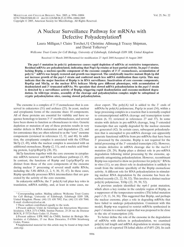
MOLECULAR AND CELLULAR BIOLOGY, Nov. 2005, p. 9996–10004 Vol. 25, No. 220270-7306/05/$08.00�0 doi:10.1128/MCB.25.22.9996–10004.2005Copyright © 2005, American Society for Microbiology. All Rights Reserved.
A Nuclear Surveillance Pathway for mRNAs withDefective Polyadenylation¶
Laura Milligan,† Claire Torchet,†‡ Christine Allmang,§ Tracey Shipman,and David Tollervey*
Wellcome Trust Centre for Cell Biology, University of Edinburgh, Edinburgh EH9 3JR, United Kingdom
Received 11 March 2005/Returned for modification 27 April 2005/Accepted 10 August 2005
The pap1-5 mutation in poly(A) polymerase causes rapid depletion of mRNAs at restrictive temperatures.Residual mRNAs are polyadenylated, indicating that Pap1-5p retains at least partial activity. In pap1-5 strainslacking Rrp6p, a nucleus-specific component of the exosome complex of 3�-5� exonucleases, accumulation ofpoly(A)� mRNA was largely restored and growth was improved. The catalytically inactive mutant Rrp6-1p didnot increase growth of the pap1-5 strain and conferred much less mRNA stabilization than rrp6�. This mayindicate that the major function of Rrp6p is in RNA surveillance. Inactivation of core exosome components,Rrp41p and Mtr3p, or the nuclear RNA helicase Mtr4p gave different phenotypes, with accumulation ofdeadenylated and 3�-truncated mRNAs. We speculate that slowed mRNA polyadenylation in the pap1-5 strainis detected by a surveillance activity of Rrp6p, triggering rapid deadenylation and exosome-mediated degra-dation. In wild-type strains, assembly of the cleavage and polyadenylation complex might be suboptimal atcryptic polyadenylation sites, causing slowed polyadenylation.
The exosome is a complex of 3�-5�exonucleases that is con-served in eukaryotes (31) and archaea (25). In yeast, nuclearand cytoplasmic forms of the exosome share 10 components.All of these proteins are essential for viability and have se-quence homology to known 3�-5� exoribonucleases, and severalhave been shown to function as ribonucleases in vitro. Geneticdepletion or mutation of any of these proteins results in verysimilar defects in RNA maturation and degradation (2), andfor convenience they are often referred to as the “core” exosomecomponents (reviewed in references 12, 32, and 48). In addi-tion, the cytoplasmic complex is associated with the GTPaseSki7p (3, 49), while the nuclear complex is associated with anadditional exonuclease, Rrp6p (2, 11), and a nucleic acid bind-ing protein, Lrp1p/Rrp47p (30, 35).
Ski7p functions together with the core exosome in cytoplas-mic mRNA turnover and RNA surveillance pathways (3, 49).In contrast, the functions of Rrp6p and Lrp1p/Rrp47p aredistinct from those of the core components of the exosomeduring nuclear 3� processing of several small stable RNAs,including the 5.8S rRNA (1, 2, 9, 30, 35, 47). In these cases,Rrp6p specifically processes RNA intermediates that are gen-erated by the activity of the core exosome.
In eukaryotic mRNAs, the 3� poly(A) tail plays key roles intranslation, mRNA stability, and, at least in some cases, nu-
clear export. The poly(A) tail is added to the 3� ends ofmRNAs by poly(A) polymerase, Pap1p in yeast (34), within alarge processing complex in a reaction that is normally coupledto cotranscriptional mRNA cleavage and transcription termi-nation (6, 53; reviewed in references 27 and 37). In somestrains with defects in pre-mRNA cleavage, long 3�-extendedtranscripts that are rapidly degraded by the nuclear exosomeare generated (42). In certain cases, subsequent polyadenyla-tion that is uncoupled to pre-mRNA cleavage can apparentlygenerate functional mRNAs from pre-mRNAs that have been3� processed by the exosome. Rrp6p is not required for theinitial processing of the 3�-extended transcripts (42). However,in strains defective in mRNA cleavage due to the rna14-1mutation (28, 29), Rrp6p plays a distinct role in pre-mRNAdegradation following initial processing by the exosome, ap-parently antagonizing polyadenylation. However, recombinantRrp6p was reported to show no preference for poly(A)� RNAsin vitro (11), so any direct role in deadenylation is unlikely toresult from the intrinsic specificity of the Rrp6p exonucleaseactivity. A different role for RNA polyadenylation in stimulat-ing nuclear RNA degradation by the exosome has been de-scribed recently (21, 22, 50, 52). This involves a distinct nuclearpoly(A) polymerase, Trf4p (22, 39, 50, 52).
A previous analysis identified the rrp6-1 point mutation,which alters a key residue in the catalytic region of Rrp6p, asa suppressor of the temperature-sensitive (TS) lethal mutationpap1-1 (9, 11, 34, 36). This suggested that Rrp6p, and perhapsthe nuclear exosome, plays a role in degrading mRNAs thathave failed to undergo polyadenylation. Consistent with thismodel, Rrp6p was required to restrict mRNAs synthesized inpap1-1 strains to a nuclear region that was proposed to lie closeto the site of transcription (18).
To better define the role of the exosome in the degradationof mRNAs with defects in polyadenylation, we examinedpoly(A) tail length and mRNA degradation in strains carryinga collection of reported TS lethal alleles of PAP1 (28). In each
* Corresponding author. Mailing address: Wellcome Trust Centrefor Cell Biology, University of Edinburgh, Edinburgh EH9 3JR,United Kingdom. Phone: (44) 131 650 7092. Fax: (44) 131 650 7040.E-mail: [email protected].
† These authors contributed equally to the work.‡ Present address: Unite de Genetique des Interactions Macromo-
leculaires, URA 2171-CNRS, Institut Pasteur, 25-28 rue du DocteurROUX, F-75724 Paris Cedex 15, France.
§ Present address: UPR 9002 du CNRS, Institut de Biologie Mo-leculaire et Cellulaire, 15 rue Rene Descartes, F-67084 StrasbourgCedex, France.
¶ Supplemental material for this article may be found at http://mcb.asm.org/.
9996
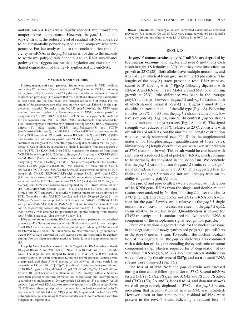
mutant, mRNA levels were rapidly reduced after transfer tononpermissive temperature. However, in pap1-5, but notpap1-2, strains, the reduced level of residual mRNAs appearedto be substantially polyadenylated at the nonpermissive tem-perature. Further analyses led to the conclusion that the defi-ciency in mRNAs in the pap1-5 strain is not due to the inabilityto synthesize poly(A) tails per se but to an RNA surveillancepathway that triggers nuclear deadenylation and exosome-me-diated degradation of the newly synthesized pre-mRNAs.
MATERIALS AND METHODS
Strains, media, and yeast genetics. Strains were grown in YPD medium,containing 2% peptone, 1% yeast extract, and 2% glucose, or YPGal, containing2% peptone, 1% yeast extract, and 2% galactose. Transformation was performedas described previously (17), except that 6% dimethyl sulfoxide was added priorto heat shock and the final pellet was resuspended in 0.15 M NaCl. For thestrains of Saccharomyces cerevisiae used in this study, see Table S1 in the sup-plemental material. To make strain YCA42 (pap1-5/rrp6�), the RRP6 openreading frame was replaced by Kluyveromyces lactis URA3 in strain pap1-5 byusing primers 5�RRP6::URA (849) (see Table S2 in the supplemental materialfor the sequence) and 3�RRP6::URA (850). Transformants were selected forUra� prototrophy and analyzed by Northern blotting for 5.8S rRNA processingdefects. To make strains YCT56 (pap1-5/GAL::rrp41) and YCT59(pap1-5/rrp6�/GAL::rrp41), the HIS3-GAL10-ProtA-RRP41 cassette was ampli-fied by PCR from strain P118 with primers RRP41-1 (842) and RRP41-2 (843)and transformed into strains pap1-5 and YCA42. Correct gene deletion wasconfirmed by analysis of the 5.8S rRNA processing defect. Strain YCT83 (pap1-5/mtr3-1) was obtained by sporulation of diploids resulting from crossing pap1-5with YCT73. The KAN-GAL-3HA-MTR4 construct was generated from strainYCBA81 by one-step PCR (22a) in strain pap1-5 with primers MTR4-F4 (991)and MTR4-R3 (992). Transformants were selected for kanamycin resistance andanalyzed by Northern blotting for 5.8S rRNA processing defects. One transfor-mant, YCT109 (pap1-5/GAL::MTR4), was selected. To make strains YCT68(ski7�) and YCT71 (pap1-5/ski7�), the KAN::ski7 cassette was amplified by PCRfrom strain Y01852 (EUROSCARF) with primers SKI7-1 (993) and SKI7-2(994) and transformed into D270 and pap1-5, respectively. Correct integrationwas confirmed by PCR. To make strains YLM122 (ccr4�) and YLM124 (pap1-5/ccr4�), the KAN::ccr4 cassette was amplified by PCR from strain Y00387(EUROSCARF) with primers CCR4-1 (1101) and CCR4-2 (1102) and trans-formed into D270 and pap1-5, respectively; correct integration was confirmed byPCR. To make strains YLM127 (pan2�) and YLM129 (pap1-5/pan2�), theKAN::pan2 cassette was amplified by PCR from strain Y04461 (EUROSCARF)with primers PAN2-1 (1104) and PAN2-2 (1105) and transformed into D270 andpap1-5, respectively; correct integration was confirmed by PCR. Strain YLM121(pap1-5/rrp6-1) was obtained by sporulation of diploids resulting from crossingpap1-5 with a strain carrying the rrp6-1 allele (11).
RNA extraction and analysis. RNA extractions were performed as describedpreviously (41). Seven micrograms of total RNA was analyzed for each sample.Small RNAs were separated on a 6% acrylamide gel containing 8.3 M urea andtransferred to a Hybond N� membrane by electrotransfer. High-molecular-weight RNAs were analyzed on 1.2% agarose gels and transferred by capillaryelution. For the oligonucleotides used, see Table S2 in the supplemental mate-rial.
For poly(A) tail length analysis of mRNA, 7 �g of total RNA was digested with10 �g of RNase A and 250 units of RNase T1 in 10 mM Tris, pH 8, 300 mMNaCl. The digestion was stopped by adding 10 mM EDTA, 0.25% sodiumdodecyl sulfate, 25 �g/ml proteinase K, and 0.5 mg/ml glycogen. Samples wereprecipitated, and then 3� end labeling of the poly(A) tails was carried outovernight at 4°C with 10 �Ci [32P]pCp (cytidine-3�,5�-bisphosphate) and 40 unitsof T4 RNA ligase in 50 mM Tris-HCl, pH 7.9, 10 mM MgCl2, 3.3 mM dithio-threitol, 10 �g/ml bovine serum albumin, and 10% dimethyl sulfoxide. Sampleswere then phenol-chloroform extracted and precipitated, and electrophoreticseparation was analyzed on a 12% acrylamide-8 M urea gel. For total poly(A) tailanalysis, 7 �g of total RNA was extensively hydrolyzed with RNase A and RNaseT1. Following ethanol precipitation to remove free nucleotides, residual poly(A)tracts were 3� end labeled with [32P]pCp and RNA ligase and resolved on a 12%polyacrylamide gel containing 8 M urea. Similar results were obtained with twoindependent experiments.
RNase H treatment. Deadenylation was performed essentially as describedpreviously (33). Samples (20 �g) of RNA were annealed with 400 ng oligo(dT)at 68°C for 10 min and digested with 1.5 U RNase H at 30°C for 1 h.
RESULTS
In pap1-5 mutant strains, poly(A)� mRNAs are degraded bythe nuclear exosome. The pap1-2 and pap1-5 mutations eachresult in tight TS lethality at 37°C, but they have little effect ongrowth at 23°C (28). Both alleles have multiple mutations, andit is not clear which of them give rise to the TS phenotype. Thelengths of the poly(A) tracts present in total RNA were as-sessed by 3� labeling with [32P]pCp following digestion withRNase A and RNase T1 (see Materials and Methods). Duringgrowth at 23°C, little difference was seen in the averagepoly(A) tail length between the pap1-2 and pap1-5 strains, bothof which showed maximal poly(A) tail lengths around 20 nu-cleotides shorter than that of the wild type (Fig. 1A). Followingtransfer to 37°C for 30 min, the pap1-2 strain retained only lowlevels of poly(A) (Fig. 1A, lane 5). In contrast, pap1-5 strainsretained substantial poly(A) tracts (Fig. 1A, lane 6). The signalstrength was reduced at 37°C relative to 23°C, consistent withoverall loss of mRNAs, but the maximal tail length distributionwas not greatly shortened (see Fig. S1 in the supplementalmaterial for PhosphorImager quantification of these data).Similar poly(A) length distribution was seen even after 90 minat 37°C (data not shown). This result would be consistent withsynthesis of a reduced level of poly(A)� RNAs, which continueto be normally deadenylated in the cytoplasm. We concludethat the pap1-5 strain, but not the pap1-2 strain, retains signif-icant polyadenylation activity at 37°C. This suggested that le-thality in the pap1-5 strain did not result simply from an in-ability to generate poly(A) tails.
The pap1-2 and pap1-5 alleles were combined with deletionof the RRP6 gene. RNAs from the single- and double-mutantstrains were analyzed by Northern blotting 2 h after transfer to37°C (Fig. 1B). Increased CYH2 and ACT1 mRNA levels wereseen for the pap1-5 rrp6� strain relative to the pap1-5 singlemutant. In contrast, no increases were seen in the pap1-2 rrp6�strain relative to pap1-2 alone. Quantification is shown forCYH2 transcript and is standardized relative to scR1 RNA, acomponent of the cytoplasmic signal recognition particle.
These observations suggested a role for the nuclear exosomein the degradation of newly synthesized poly(A)� pre-mRNAsin the pap1-5 mutant strain. To confirm the nuclear localiza-tion of this degradation, the pap1-5 allele was also combinedwith a deletion of the gene encoding the cytoplasmic exosomecomponent Ski7p, which is required for 3� degradation of cy-toplasmic mRNAs (3, 5, 45, 49). No clear mRNA stabilizationwas conferred by the absence of Ski7p, and no truncated RNAspecies were observed (Fig. 1C).
The loss of mRNA from the pap1-5 strain was assessedduring a time course following transfer to 37°C. Several mRNAstested (ACT1, CYH2, RPL25, and MFA2 and RPL30, RPS26a,and CYC1) (Fig. 2A and B, lanes 8 to 14, and data not shown)were all progressively depleted at 37°C in the pap1-5 strain,indicating that accumulation of new mRNA was inhibited.However, even at late time points, residual mRNAs werepresent in the pap1-5 strain, indicating a reduced level of
VOL. 25, 2005 RNA SURVEILLANCE BY THE EXOSOME 9997
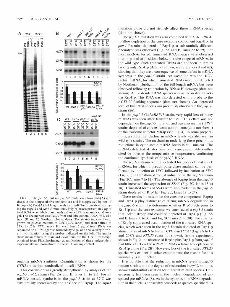
ongoing mRNA synthesis. Quantification is shown for theCYH2 transcript, standardized to scR1 RNA.
This conclusion was greatly strengthened by analysis of thepap1-5 rrp6� strain (Fig. 2A and B, lanes 15 to 21). For allmRNAs tested, synthesis in the pap1-5 strain at 37°C wassubstantially increased by the absence of Rrp6p. The rrp6�
mutation alone did not strongly affect these mRNA species(data not shown).
The pap1-5 mutation was also combined with GAL::RRP41to allow depletion of the core exosome component Rrp41p. Inpap-1-5 strains depleted of Rrp41p, a substantially differentphenotype was observed (Fig. 2A and B, lanes 22 to 29). Formost mRNAs tested, truncated RNA species were observedthat migrated at positions below the size range of mRNAs inthe wild type. Such truncated RNAs are not seen in strainslacking only Rrp41p (data not shown; see references 8 and 42),showing that they are a consequence of some defect in mRNAsynthesis in the pap1-5 strain. An exception was the ACT1(actin) mRNA, for which truncated RNAs were not detectedby Northern hybridization of the full-length mRNA but wereobserved following truncation by RNase H cleavage (data notshown). A 3�-extended RNA species was visible in strains lack-ing Rrp41p. This RNA was also detected with a probe to theACT1 3� flanking sequence (data not shown). An increasedlevel of this RNA species was previously observed in the pap1-1strain (26).
In the pap1-5 GAL::RRP41 strain, very rapid loss of manymRNAs was seen after transfer to 37°C. This effect was notdependent on the pap1-5 mutation and was also seen in PAP1�
strains depleted of core exosome components (data not shown)or the exosome cofactor Mtr4p (see Fig. 4). In some prepara-tions, a substantial decline in mRNA levels was also seen inwild-type strains. The mechanism underlying these precipitousreductions in cytoplasmic mRNA levels is still unclear. ThemRNAs detected at later time points are presumably synthe-sized de novo at the nonpermissive temperature, confirmingthe continued synthesis of poly(A)� RNAs.
The pap1-5 strains were also tested for decay of heat shockmRNAs, for which a pseudo-pulse-chase analysis can be per-formed by induction at 42°C, followed by incubation at 37°C(Fig. 2C). SSA3 showed robust induction in the pap1-5 strain(Fig. 2C, lanes 7 to 12). The absence of Rrp6p from the pap1-5strain increased the expression of SSA3 (Fig. 2C, lanes 13 to18). Truncated forms of SSA3 were also evident in the pap1-5strain depleted of Rrp41p (Fig. 2C, lanes 19 to 24).
These results indicated that the exosome components Rrp6pand Rrp41p play distinct roles during mRNA degradation inthe pap1-5 strain. To determine whether Rrp6p acts prior toRrp41p and the core exosome, we constructed a pap1-5 strainthat lacked Rrp6p and could be depleted of Rrp41p (Fig. 2Aand B, lanes 30 to 37, and Fig. 2C, lanes 25 to 30). The absenceof Rrp6p suppressed accumulation of the truncated RNA spe-cies, which were seen in the pap1-5 strain depleted of Rrp41palone, for most mRNAs tested, CYH2 and SSA3 (Fig. 2A to C)and CYC1 and RPL30 (data not shown). In the experimentshown in Fig. 2, the absence of Rrp6p plus Rrp41p from pap1-5had little effect on the RPL25 mRNAs relative to depletion ofRrp41p alone (Fig. 2B). However, loss of the truncated RPL25species was evident in other experiments; the reason for thisvariability is still unclear.
It is notable that the reduction in mRNA levels in pap1-5mutant strains, and the degree of restoration in rrp6� mutants,showed substantial variation for different mRNA species. Het-erogeneity has been seen in the nuclear degradation of un-spliced pre-mRNAs (8). As in the cytoplasm, mRNA degrada-tion in the nucleus apparently proceeds at species-specific rates
FIG. 1. The pap1-5, but not pap1-2, mutation allows poly(A) syn-thesis at the nonpermissive temperature and is suppressed by loss ofRrp6p. (A) Poly(A) tail length analysis of mRNAs from strains carry-ing the pap1-2 and pap1-5 mutations. Poly(A) tracts present in 7 �g oftotal RNA were labeled and analyzed on a 12% acrylamide-8 M ureagel. The size marker was tRNA from end-labeled total RNA. WT, wildtype. (B and C) Northern blot analyses. The strains indicated weregrown on glucose medium at 23°C (23°C lanes) and then shifted to37°C for 2 h (37°C lanes). For each lane, 7 �g of total RNA wasseparated on a 1.2% agarose-formaldehyde gel and analyzed by North-ern hybridization using the probes indicated on the left. The graphsshow mean values � standard deviations for the CYH2 transcript,obtained from PhosphorImager quantification of three independentexperiments and normalized to the scR1 loading control.
9998 MILLIGAN ET AL. MOL. CELL. BIOL.
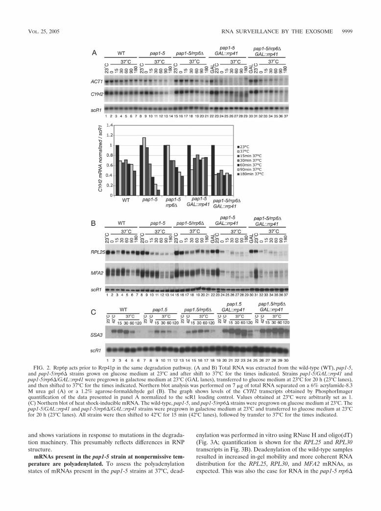
and shows variations in response to mutations in the degrada-tion machinery. This presumably reflects differences in RNPstructure.
mRNAs present in the pap1-5 strain at nonpermissive tem-perature are polyadenylated. To assess the polyadenylationstates of mRNAs present in the pap1-5 strains at 37°C, dead-
enylation was performed in vitro using RNase H and oligo(dT)(Fig. 3A; quantification is shown for the RPL25 and RPL30transcripts in Fig. 3B). Deadenylation of the wild-type samplesresulted in increased in-gel mobility and more coherent RNAdistribution for the RPL25, RPL30, and MFA2 mRNAs, asexpected. This was also the case for RNA in the pap1-5 rrp6�
FIG. 2. Rrp6p acts prior to Rrp41p in the same degradation pathway. (A and B) Total RNA was extracted from the wild-type (WT), pap1-5,and pap1-5/rrp6� strains grown on glucose medium at 23°C and after shift to 37°C for the times indicated. Strains pap1-5/GAL::rrp41 andpap1-5/rrp6�/GAL::rrp41 were pregrown in galactose medium at 23°C (GAL lanes), transferred to glucose medium at 23°C for 20 h (23°C lanes),and then shifted to 37°C for the times indicated. Northern blot analysis was performed on 7 �g of total RNA separated on a 6% acrylamide-8.3M urea gel (A) or a 1.2% agarose-formaldehyde gel (B). The graph shows levels of the CYH2 transcripts obtained by PhosphorImagerquantification of the data presented in panel A normalized to the scR1 loading control. Values obtained at 23°C were arbitrarily set as 1.(C) Northern blot of heat shock-inducible mRNA. The wild-type, pap1-5, and pap1-5/rrp6� strains were pregrown on glucose medium at 23°C. Thepap1-5/GAL::rrp41 and pap1-5/rrp6�/GAL::rrp41 strains were pregrown in galactose medium at 23°C and transferred to glucose medium at 23°Cfor 20 h (23°C lanes). All strains were then shifted to 42°C for 15 min (42°C lanes), followed by transfer to 37°C for the times indicated.
VOL. 25, 2005 RNA SURVEILLANCE BY THE EXOSOME 9999
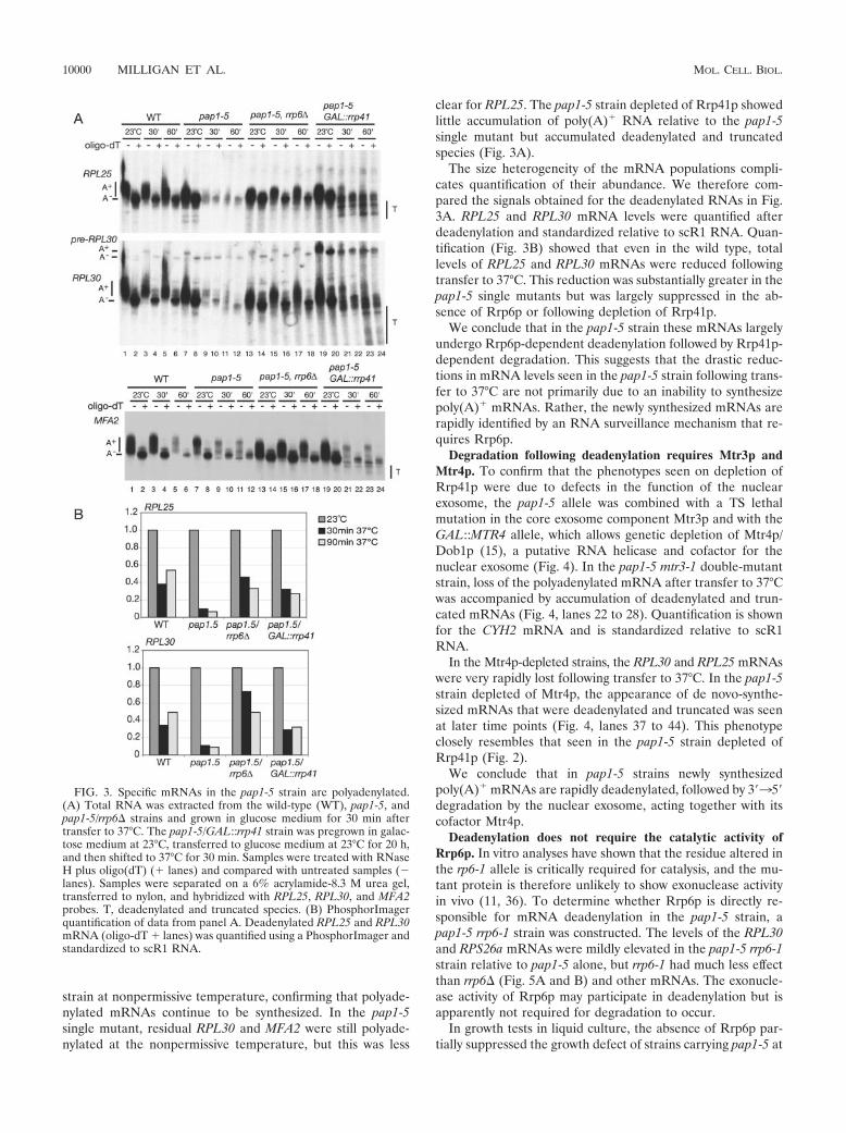
strain at nonpermissive temperature, confirming that polyade-nylated mRNAs continue to be synthesized. In the pap1-5single mutant, residual RPL30 and MFA2 were still polyade-nylated at the nonpermissive temperature, but this was less
clear for RPL25. The pap1-5 strain depleted of Rrp41p showedlittle accumulation of poly(A)� RNA relative to the pap1-5single mutant but accumulated deadenylated and truncatedspecies (Fig. 3A).
The size heterogeneity of the mRNA populations compli-cates quantification of their abundance. We therefore com-pared the signals obtained for the deadenylated RNAs in Fig.3A. RPL25 and RPL30 mRNA levels were quantified afterdeadenylation and standardized relative to scR1 RNA. Quan-tification (Fig. 3B) showed that even in the wild type, totallevels of RPL25 and RPL30 mRNAs were reduced followingtransfer to 37°C. This reduction was substantially greater in thepap1-5 single mutants but was largely suppressed in the ab-sence of Rrp6p or following depletion of Rrp41p.
We conclude that in the pap1-5 strain these mRNAs largelyundergo Rrp6p-dependent deadenylation followed by Rrp41p-dependent degradation. This suggests that the drastic reduc-tions in mRNA levels seen in the pap1-5 strain following trans-fer to 37°C are not primarily due to an inability to synthesizepoly(A)� mRNAs. Rather, the newly synthesized mRNAs arerapidly identified by an RNA surveillance mechanism that re-quires Rrp6p.
Degradation following deadenylation requires Mtr3p andMtr4p. To confirm that the phenotypes seen on depletion ofRrp41p were due to defects in the function of the nuclearexosome, the pap1-5 allele was combined with a TS lethalmutation in the core exosome component Mtr3p and with theGAL::MTR4 allele, which allows genetic depletion of Mtr4p/Dob1p (15), a putative RNA helicase and cofactor for thenuclear exosome (Fig. 4). In the pap1-5 mtr3-1 double-mutantstrain, loss of the polyadenylated mRNA after transfer to 37°Cwas accompanied by accumulation of deadenylated and trun-cated mRNAs (Fig. 4, lanes 22 to 28). Quantification is shownfor the CYH2 mRNA and is standardized relative to scR1RNA.
In the Mtr4p-depleted strains, the RPL30 and RPL25 mRNAswere very rapidly lost following transfer to 37°C. In the pap1-5strain depleted of Mtr4p, the appearance of de novo-synthe-sized mRNAs that were deadenylated and truncated was seenat later time points (Fig. 4, lanes 37 to 44). This phenotypeclosely resembles that seen in the pap1-5 strain depleted ofRrp41p (Fig. 2).
We conclude that in pap1-5 strains newly synthesizedpoly(A)� mRNAs are rapidly deadenylated, followed by 3�35�degradation by the nuclear exosome, acting together with itscofactor Mtr4p.
Deadenylation does not require the catalytic activity ofRrp6p. In vitro analyses have shown that the residue altered inthe rp6-1 allele is critically required for catalysis, and the mu-tant protein is therefore unlikely to show exonuclease activityin vivo (11, 36). To determine whether Rrp6p is directly re-sponsible for mRNA deadenylation in the pap1-5 strain, apap1-5 rrp6-1 strain was constructed. The levels of the RPL30and RPS26a mRNAs were mildly elevated in the pap1-5 rrp6-1strain relative to pap1-5 alone, but rrp6-1 had much less effectthan rrp6� (Fig. 5A and B) and other mRNAs. The exonucle-ase activity of Rrp6p may participate in deadenylation but isapparently not required for degradation to occur.
In growth tests in liquid culture, the absence of Rrp6p par-tially suppressed the growth defect of strains carrying pap1-5 at
FIG. 3. Specific mRNAs in the pap1-5 strain are polyadenylated.(A) Total RNA was extracted from the wild-type (WT), pap1-5, andpap1-5/rrp6� strains and grown in glucose medium for 30 min aftertransfer to 37°C. The pap1-5/GAL::rrp41 strain was pregrown in galac-tose medium at 23°C, transferred to glucose medium at 23°C for 20 h,and then shifted to 37°C for 30 min. Samples were treated with RNaseH plus oligo(dT) (� lanes) and compared with untreated samples (�lanes). Samples were separated on a 6% acrylamide-8.3 M urea gel,transferred to nylon, and hybridized with RPL25, RPL30, and MFA2probes. T, deadenylated and truncated species. (B) PhosphorImagerquantification of data from panel A. Deadenylated RPL25 and RPL30mRNA (oligo-dT � lanes) was quantified using a PhosphorImager andstandardized to scR1 RNA.
10000 MILLIGAN ET AL. MOL. CELL. BIOL.
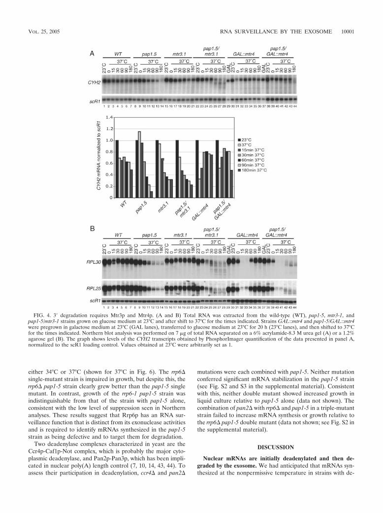
either 34°C or 37°C (shown for 37°C in Fig. 6). The rrp6�single-mutant strain is impaired in growth, but despite this, therrp6� pap1-5 strain clearly grew better than the pap1-5 singlemutant. In contrast, growth of the rrp6-1 pap1-5 strain wasindistinguishable from that of the strain with pap1-5 alone,consistent with the low level of suppression seen in Northernanalyses. These results suggest that Rrp6p has an RNA sur-veillance function that is distinct from its exonuclease activitiesand is required to identify mRNAs synthesized in the pap1-5strain as being defective and to target them for degradation.
Two deadenylase complexes characterized in yeast are theCcr4p-Caf1p-Not complex, which is probably the major cyto-plasmic deadenylase, and Pan2p-Pan3p, which has been impli-cated in nuclear poly(A) length control (7, 10, 14, 43, 44). Toassess their participation in deadenylation, ccr4� and pan2�
mutations were each combined with pap1-5. Neither mutationconferred significant mRNA stabilization in the pap1-5 strain(see Fig. S2 and S3 in the supplemental material). Consistentwith this, neither double mutant showed increased growth inliquid culture relative to pap1-5 alone (data not shown). Thecombination of pan2� with rrp6� and pap1-5 in a triple-mutantstrain failed to increase mRNA synthesis or growth relative tothe rrp6� pap1-5 double mutant (data not shown; see Fig. S2 inthe supplemental material).
DISCUSSION
Nuclear mRNAs are initially deadenylated and then de-graded by the exosome. We had anticipated that mRNAs syn-thesized at the nonpermissive temperature in strains with de-
FIG. 4. 3� degradation requires Mtr3p and Mtr4p. (A and B) Total RNA was extracted from the wild-type (WT), pap1-5, mtr3-1, andpap1-5/mtr3-1 strains grown on glucose medium at 23°C and after shift to 37°C for the times indicated. Strains GAL::mtr4 and pap1-5/GAL::mtr4were pregrown in galactose medium at 23°C (GAL lanes), transferred to glucose medium at 23°C for 20 h (23°C lanes), and then shifted to 37°Cfor the times indicated. Northern blot analysis was performed on 7 �g of total RNA separated on a 6% acrylamide-8.3 M urea gel (A) or a 1.2%agarose gel (B). The graph shows levels of the CYH2 transcripts obtained by PhosphorImager quantification of the data presented in panel A,normalized to the scR1 loading control. Values obtained at 23°C were arbitrarily set as 1.
VOL. 25, 2005 RNA SURVEILLANCE BY THE EXOSOME 10001
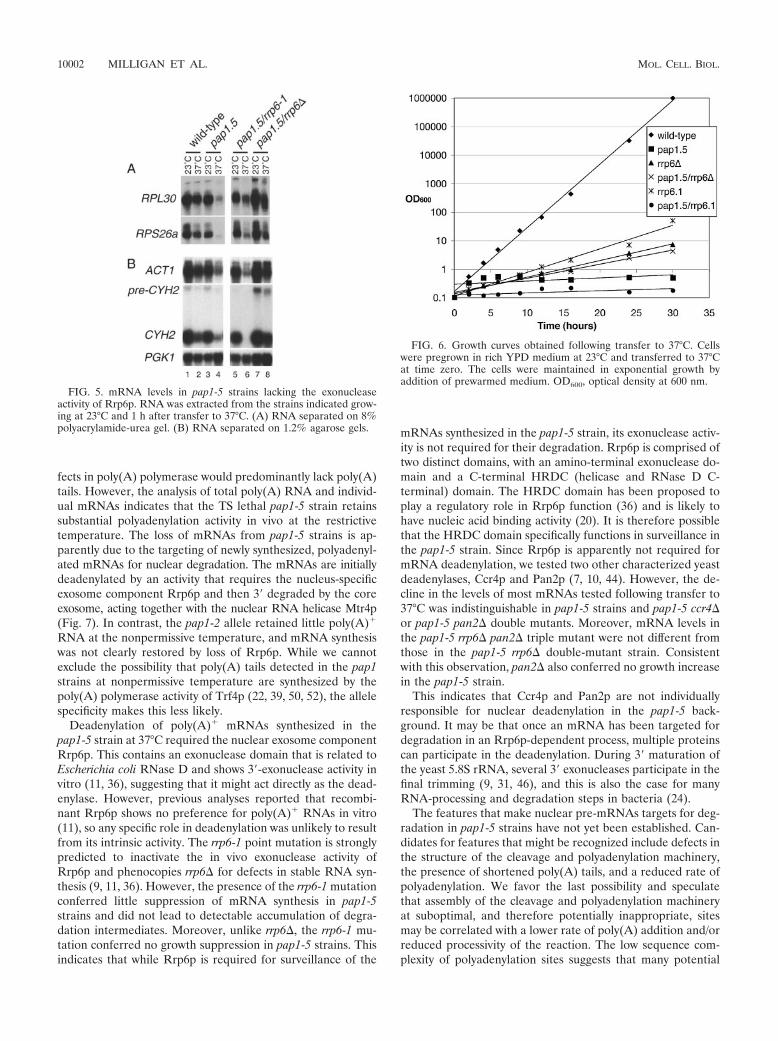
fects in poly(A) polymerase would predominantly lack poly(A)tails. However, the analysis of total poly(A) RNA and individ-ual mRNAs indicates that the TS lethal pap1-5 strain retainssubstantial polyadenylation activity in vivo at the restrictivetemperature. The loss of mRNAs from pap1-5 strains is ap-parently due to the targeting of newly synthesized, polyadenyl-ated mRNAs for nuclear degradation. The mRNAs are initiallydeadenylated by an activity that requires the nucleus-specificexosome component Rrp6p and then 3� degraded by the coreexosome, acting together with the nuclear RNA helicase Mtr4p(Fig. 7). In contrast, the pap1-2 allele retained little poly(A)�
RNA at the nonpermissive temperature, and mRNA synthesiswas not clearly restored by loss of Rrp6p. While we cannotexclude the possibility that poly(A) tails detected in the pap1strains at nonpermissive temperature are synthesized by thepoly(A) polymerase activity of Trf4p (22, 39, 50, 52), the allelespecificity makes this less likely.
Deadenylation of poly(A)� mRNAs synthesized in thepap1-5 strain at 37°C required the nuclear exosome componentRrp6p. This contains an exonuclease domain that is related toEscherichia coli RNase D and shows 3�-exonuclease activity invitro (11, 36), suggesting that it might act directly as the dead-enylase. However, previous analyses reported that recombi-nant Rrp6p shows no preference for poly(A)� RNAs in vitro(11), so any specific role in deadenylation was unlikely to resultfrom its intrinsic activity. The rrp6-1 point mutation is stronglypredicted to inactivate the in vivo exonuclease activity ofRrp6p and phenocopies rrp6� for defects in stable RNA syn-thesis (9, 11, 36). However, the presence of the rrp6-1 mutationconferred little suppression of mRNA synthesis in pap1-5strains and did not lead to detectable accumulation of degra-dation intermediates. Moreover, unlike rrp6�, the rrp6-1 mu-tation conferred no growth suppression in pap1-5 strains. Thisindicates that while Rrp6p is required for surveillance of the
mRNAs synthesized in the pap1-5 strain, its exonuclease activ-ity is not required for their degradation. Rrp6p is comprised oftwo distinct domains, with an amino-terminal exonuclease do-main and a C-terminal HRDC (helicase and RNase D C-terminal) domain. The HRDC domain has been proposed toplay a regulatory role in Rrp6p function (36) and is likely tohave nucleic acid binding activity (20). It is therefore possiblethat the HRDC domain specifically functions in surveillance inthe pap1-5 strain. Since Rrp6p is apparently not required formRNA deadenylation, we tested two other characterized yeastdeadenylases, Ccr4p and Pan2p (7, 10, 44). However, the de-cline in the levels of most mRNAs tested following transfer to37°C was indistinguishable in pap1-5 strains and pap1-5 ccr4�or pap1-5 pan2� double mutants. Moreover, mRNA levels inthe pap1-5 rrp6� pan2� triple mutant were not different fromthose in the pap1-5 rrp6� double-mutant strain. Consistentwith this observation, pan2� also conferred no growth increasein the pap1-5 strain.
This indicates that Ccr4p and Pan2p are not individuallyresponsible for nuclear deadenylation in the pap1-5 back-ground. It may be that once an mRNA has been targeted fordegradation in an Rrp6p-dependent process, multiple proteinscan participate in the deadenylation. During 3� maturation ofthe yeast 5.8S rRNA, several 3� exonucleases participate in thefinal trimming (9, 31, 46), and this is also the case for manyRNA-processing and degradation steps in bacteria (24).
The features that make nuclear pre-mRNAs targets for deg-radation in pap1-5 strains have not yet been established. Can-didates for features that might be recognized include defects inthe structure of the cleavage and polyadenylation machinery,the presence of shortened poly(A) tails, and a reduced rate ofpolyadenylation. We favor the last possibility and speculatethat assembly of the cleavage and polyadenylation machineryat suboptimal, and therefore potentially inappropriate, sitesmay be correlated with a lower rate of poly(A) addition and/orreduced processivity of the reaction. The low sequence com-plexity of polyadenylation sites suggests that many potential
FIG. 5. mRNA levels in pap1-5 strains lacking the exonucleaseactivity of Rrp6p. RNA was extracted from the strains indicated grow-ing at 23°C and 1 h after transfer to 37°C. (A) RNA separated on 8%polyacrylamide-urea gel. (B) RNA separated on 1.2% agarose gels.
FIG. 6. Growth curves obtained following transfer to 37°C. Cellswere pregrown in rich YPD medium at 23°C and transferred to 37°Cat time zero. The cells were maintained in exponential growth byaddition of prewarmed medium. OD600, optical density at 600 nm.
10002 MILLIGAN ET AL. MOL. CELL. BIOL.
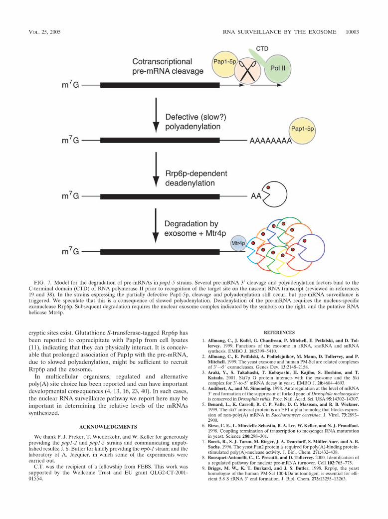
cryptic sites exist. Glutathione S-transferase-tagged Rrp6p hasbeen reported to coprecipitate with Pap1p from cell lysates(11), indicating that they can physically interact. It is conceiv-able that prolonged association of Pap1p with the pre-mRNA,due to slowed polyadenylation, might be sufficient to recruitRrp6p and the exosome.
In multicellular organisms, regulated and alternativepoly(A) site choice has been reported and can have importantdevelopmental consequences (4, 13, 16, 23, 40). In such cases,the nuclear RNA surveillance pathway we report here may beimportant in determining the relative levels of the mRNAssynthesized.
ACKNOWLEDGMENTS
We thank P. J. Preker, T. Wiederkehr, and W. Keller for generouslyproviding the pap1-2 and pap1-5 strains and communicating unpub-lished results; J. S. Butler for kindly providing the rrp6-1 strain; and thelaboratory of A. Jacquier, in which some of the experiments werecarried out.
C.T. was the recipient of a fellowship from FEBS. This work wassupported by the Wellcome Trust and EU grant QLG2-CT-2001-01554.
REFERENCES
1. Allmang, C., J. Kufel, G. Chanfreau, P. Mitchell, E. Petfalski, and D. Tol-lervey. 1999. Functions of the exosome in rRNA, snoRNA and snRNAsynthesis. EMBO J. 18:5399–5410.
2. Allmang, C., E. Petfalski, A. Podtelejnikov, M. Mann, D. Tollervey, and P.Mitchell. 1999. The yeast exosome and human PM-Scl are related complexesof 3�35� exonucleases. Genes Dev. 13:2148–2158.
3. Araki, Y., S. Takahashi, T. Kobayashi, H. Kajiho, S. Hoshino, and T.Katada. 2001. Ski7p G protein interacts with the exosome and the Skicomplex for 3�-to-5� mRNA decay in yeast. EMBO J. 20:4684–4693.
4. Audibert, A., and M. Simonelig. 1998. Autoregulation at the level of mRNA3� end formation of the suppressor of forked gene of Drosophila melanogasteris conserved in Drosophila virilis. Proc. Natl. Acad. Sci. USA 95:14302–14307.
5. Benard, L., K. Carroll, R. C. P. Valle, D. C. Masison, and R. B. Wickner.1999. The ski7 antiviral protein is an EF1-alpha homolog that blocks expres-sion of non-poly(A) mRNA in Saccharomyces cerevisiae. J. Virol. 73:2893–2900.
6. Birse, C. E., L. Minvielle-Sebastia, B. A. Lee, W. Keller, and N. J. Proudfoot.1998. Coupling termination of transcription to messenger RNA maturationin yeast. Science 280:298–301.
7. Boeck, R., S. J. Tarun, M. Rieger, J. A. Deardorff, S. Muller-Auer, and A. B.Sachs. 1996. The yeast Pan2 protein is required for poly(A)-binding protein-stimulated poly(A)-nuclease activity. J. Biol. Chem. 271:432–438.
8. Bousquet-Antonelli, C., C. Presutti, and D. Tollervey. 2000. Identification ofa regulated pathway for nuclear pre-mRNA turnover. Cell 102:765–775.
9. Briggs, M. W., K. T. Burkard, and J. S. Butler. 1998. Rrp6p, the yeasthomologue of the human PM-Scl 100-kDa autoantigen, is essential for effi-cient 5.8 S rRNA 3� end formation. J. Biol. Chem. 273:13255–13263.
FIG. 7. Model for the degradation of pre-mRNAs in pap1-5 strains. Several pre-mRNA 3� cleavage and polyadenylation factors bind to theC-terminal domain (CTD) of RNA polymerase II prior to recognition of the target site on the nascent RNA transcript (reviewed in references19 and 38). In the strains expressing the partially defective Pap1-5p, cleavage and polyadenylation still occur, but pre-mRNA surveillance istriggered. We speculate that this is a consequence of slowed polyadenylation. Deadenylation of the pre-mRNA requires the nucleus-specificexonuclease Rrp6p. Subsequent degradation requires the nuclear exosome complex indicated by the symbols on the right, and the putative RNAhelicase Mtr4p.
VOL. 25, 2005 RNA SURVEILLANCE BY THE EXOSOME 10003

10. Brown, C. E., S. Z. Tarun, Jr., R. Boeck, and A. B. Sachs. 1996. PAN3encodes a subunit of the Pab1p-dependent poly(A) nuclease in Saccharomy-ces cerevisiae. Mol. Cell. Biol. 16:5744–5753.
11. Burkard, K. T., and J. S. Butler. 2000. A nuclear 3�-5� exonuclease involvedin mRNA degradation interacts with poly(A) polymerase and the hnRNAprotein Npl3p. Mol. Cell. Biol. 20:604–616.
12. Butler, S. 2002. The ying and yang of the exosome. Trends Biochem. Sci.12:90–96.
13. Castelo-Branco, P., A. Furger, M. Wollerton, C. Smith, A. Moreira, and N.Proudfoot. 2004. Polypyrimidine tract binding protein modulates efficiencyof polyadenylation. Mol. Cell. Biol. 24:4174–4183.
14. Daugeron, M. C., F. Mauxion, and B. Seraphin. 2001. The yeast POP2 geneencodes a nuclease involved in mRNA deadenylation. Nucleic Acids Res.29:2448–2455.
15. de la Cruz, J., D. Kressler, D. Tollervey, and P. Linder. 1998. Dob1p (Mtr4p)is a putative ATP-dependent RNA helicase required for the 3� end forma-tion of 5.8S rRNA in Saccharomyces cerevisiae. EMBO J. 17:1128–1140.
16. Edwalds-Gilbert, G., K. L. Veraldi, and C. Milcarek. 1997. Alternativepoly(A) site selection in complex transcription units: means to an end?Nucleic Acids Res. 25:2547–2561.
17. Gietz, R. D., R. H. Schiestl, A. R. Willems, and R. A. Woods. 1995. Studieson the transformation of intact yeast cells by the LiAc/SS-DNA/PEG pro-cedure. Yeast 11:355–360.
18. Hilleren, P., T. McCarthy, M. Rosbash, R. Parker, and T. H. Jensen. 2001.Quality control of mRNA 3�-end processing is linked to the nuclear exo-some. Nature 413:538–542.
19. Hirose, Y., and J. L. Manley. 2000. RNA polymerase II and the integrationof nuclear events. Genes Dev. 14:1415–1429.
20. Janscak, P., P. L. Garcia, F. Hamburger, Y. Makuta, K. Shiraishi, Y. Imai,H. Ikeda, and T. A. Bickle. 2003. Characterization and mutational analysis ofthe RecQ core of the bloom syndrome protein. J. Mol. Biol. 330:29–42.
21. Kadaba, S., A. Krueger, T. Trice, A. M. Krecic, A. G. Hinnebusch, and J.Anderson. 2004. Nuclear surveillance and degradation of hypomodified ini-tiator tRNAMet in S. cerevisiae. Genes Dev. 18:1227–1240.
22. LaCava, J., J. Houseley, C. Saveanu, E. Petfalski, E. Thompson, A. Jacquier,and D. Tollervey. 2005. RNA degradation by the exosome is promoted by anuclear polyadenylation complex. Cell 121:713–724.
22a.LaFontaine, D., and D. Tollervey. 1996. One-step PCR mediated strategy forthe construction of conditionally expressed and epitope tagged yeast pro-teins. Nucleic Acids Res. 24:3469–3472.
23. Legendre, M., and D. Gautheret. 2003. Sequence determinants in humanpolyadenylation site selection. BMC Genomics 4:7.
24. Li, Z., S. Pandit, and M. P. Deutscher. 1998. 3� Exoribonucleolytic trimmingis a common feature of the maturation of small, stable RNAs in Escherichiacoli. Proc. Natl. Acad. Sci. USA 95:2856–2861.
25. Lorentzen, E., P. Walter, S. Fribourg, E. Evguenieva-Hackenberg, G. Klug,and E. Conti. 2005. The archaeal exosome core is a hexameric ring structurewith three catalytic subunits. Nat. Struct. Mol. Biol. 12:575–581.
26. Mandart, E., and R. Parker. 1995. Effects of mutations in the Saccharomycescerevisiae RNA14, RNA15, and PAP1 genes on polyadenylation in vivo. Mol.Cell. Biol. 15:6979–6986.
27. Minvielle-Sebastia, L., and W. Keller. 1999. mRNA polyadenylation and itscoupling to other RNA processing reactions and to transcription. Curr.Opin. Cell Biol. 11:352–357.
28. Minvielle-Sebastia, L., P. J. Preker, and W. Keller. 1994. RNA14 andRNA15 proteins as components of a yeast pre-mRNA 3�-end processingfactor. Science 266:1702–1705.
29. Minvielle-Sebastia, L., B. Winsor, N. Bonneaud, and F. Lacroute. 1991.Mutations in the yeast RNA14 and RNA15 genes result in an abnormalmRNA decay rate; sequence analysis reveals an RNA-binding domain in theRNA15 protein. Mol. Cell. Biol. 11:3075–3087.
30. Mitchell, P., E. Petfalski, R. Houalla, A. Podtelejnikov, M. Mann, and D.Tollervey. 2003. Rrp47p is an exosome-associated protein required for the 3�processing of stable RNAs. Mol. Cell. Biol. 23:6982–6992.
31. Mitchell, P., E. Petfalski, A. Shevchenko, M. Mann, and D. Tollervey. 1997.
The exosome; a conserved eukaryotic RNA processing complex containingmultiple 3�35� exoribonuclease activities. Cell 91:457–466.
32. Mitchell, P., and D. Tollervey. 2000. Musing on the structural organization ofthe exosome complex. Nat. Struct. Biol. 7:843–846.
33. Muhlrad, D., and R. Parker. 1992. Mutations affecting stability and dead-enylation of the yeast MFA2 transcript. Genes Dev. 6:2100–2111.
34. Patel, D., and J. S. Butler. 1992. Conditional defect in mRNA 3� endprocessing caused by a mutation in the gene for poly(A) polymerase. Mol.Cell. Biol. 12:3297–3304.
35. Peng, W. T., M. D. Robinson, S. Mnaimneh, N. J. Krogan, G. Cagney, Q.Morris, A. P. Davierwala, J. Grigull, X. Yang, W. Zhang, N. Mitsakakis,O. W. Ryan, N. Datta, V. Jojic, C. Pal, V. Canadien, D. Richards, B. Beattie,L. F. Wu, S. J. Altschuler, S. Roweis, B. J. Frey, A. Emili, J. F. Greenblatt,and T. R. Hughes. 2003. A panoramic view of yeast noncoding RNA pro-cessing. Cell 113:919–933.
36. Phillips, S., and J. S. Butler. 2003. Contribution of domain structure to theRNA 3� end processing and degradation functions of the nuclear exosomesubunit Rrp6p. RNA 9:1098–1107.
37. Proudfoot, N. 2000. Connecting transcription to messenger RNA processing.Trends Biochem. Sci. 25:290–293.
38. Proudfoot, N. J., A. Furger, and M. J. Dye. 2002. Integrating mRNA pro-cessing with transcription. Cell 108:501–512.
39. Saitoh, S., A. Chabes, W. H. McDonald, L. Thelander, J. R. Yates, and P.Russell. 2002. Cid13 is a cytoplasmic poly(A) polymerase that regulatesribonucleotide reductase mRNA. Cell 109:563–573.
40. Simpson, G. G., P. P. Dijkwel, V. Quesada, I. Henderson, and C. Dean. 2003.FY is an RNA 3� end-processing factor that interacts with FCA to controlthe Arabidopsis floral transition. Cell 113:777–787.
41. Tollervey, D., and I. W. Mattaj. 1987. Fungal small nuclear ribonucleopro-teins share properties with plant and vertebrate U-snRNPs. EMBO J. 6:469–476.
42. Torchet, C., C. Bousquet-Antonelli, L. Milligan, E. Thompson, J. Kufel, andD. Tollervey. 2002. Processing of 3� extended read-through transcripts by theexosome can generate functional mRNAs. Mol. Cell 9:1285–1296.
43. Tucker, M., R. R. Staples, M. A. Valencia-Sanchez, D. Muhlrad, and R.Parker. 2002. Ccr4p is the catalytic subunit of a Ccr4p/Pop2p/Notp mRNAdeadenylase complex in Saccharomyces cerevisiae. EMBO J. 21:1427–1436.
44. Tucker, M., M. A. Valencia-Sanchez, R. R. Staples, J. Chen, C. L. Denis, andR. Parker. 2001. The transcription factor associated Ccr4 and Caf1 proteinsare components of the major cytoplasmic mRNA deadenylase in Saccharo-myces cerevisiae. Cell 104:377–386.
45. van Hoof, A., P. A. Frischmeyer, H. C. Dietz, and R. Parker. 2002. Exosome-mediated recognition and degradation of mRNAs lacking a terminationcodon. Science 295:2262–2264.
46. van Hoof, A., P. Lennertz, and R. Parker. 2000. Three conserved members ofthe RNase D family have unique and overlapping functions in the processingof 5S, 5.8S, U4, U5, RNase MRP and RNase P RNAs in yeast. EMBO J.19:1357–1365.
47. van Hoof, A., P. Lennertz, and R. Parker. 2000. Yeast exosome mutantsaccumulate 3�-extended polyadenylated forms of U4 small nuclear RNA andsmall nucleolar RNAs. Mol. Cell. Biol. 20:441–452.
48. van Hoof, A., and R. Parker. 1999. The exosome: a proteasome for RNA?Cell 99:347–350.
49. van Hoof, A., R. R. Staples, R. E. Baker, and R. Parker. 2000. Function of theski4p (Csl4p) and Ski7p proteins in 3�-to-5� degradation of mRNA. Mol.Cell. Biol. 20:8230–8243.
50. Vanacova, S., J. Wolf, G. Martin, D. Blank, S. Dettwiler, A. Friedlein, H.Langen, G. Keith, and W. Keller. 2005. A new yeast poly(A) polymerasecomplex involved in RNA quality control. PLoS Biol. 3:e189.
51. Reference deleted.52. Wyers, F., M. Rougemaille, G. Badis, J.-C. Rousselle, M.-E. Dufour, J.
Boulay, B. Regnault, F. Devaux, A. Namane, B. Seraphin, D. Libri, and A.Jacquier. 2005. Cryptic Pol II transcripts are degraded by a nuclear qualitycontrol pathway involving a new poly(A) polymerase. Cell 121:725–737.
53. Yonaha, M., and N. J. Proudfoot. 2000. Transcriptional termination andcoupled polyadenylation in vitro. EMBO J. 19:3770–3777.
10004 MILLIGAN ET AL. MOL. CELL. BIOL.

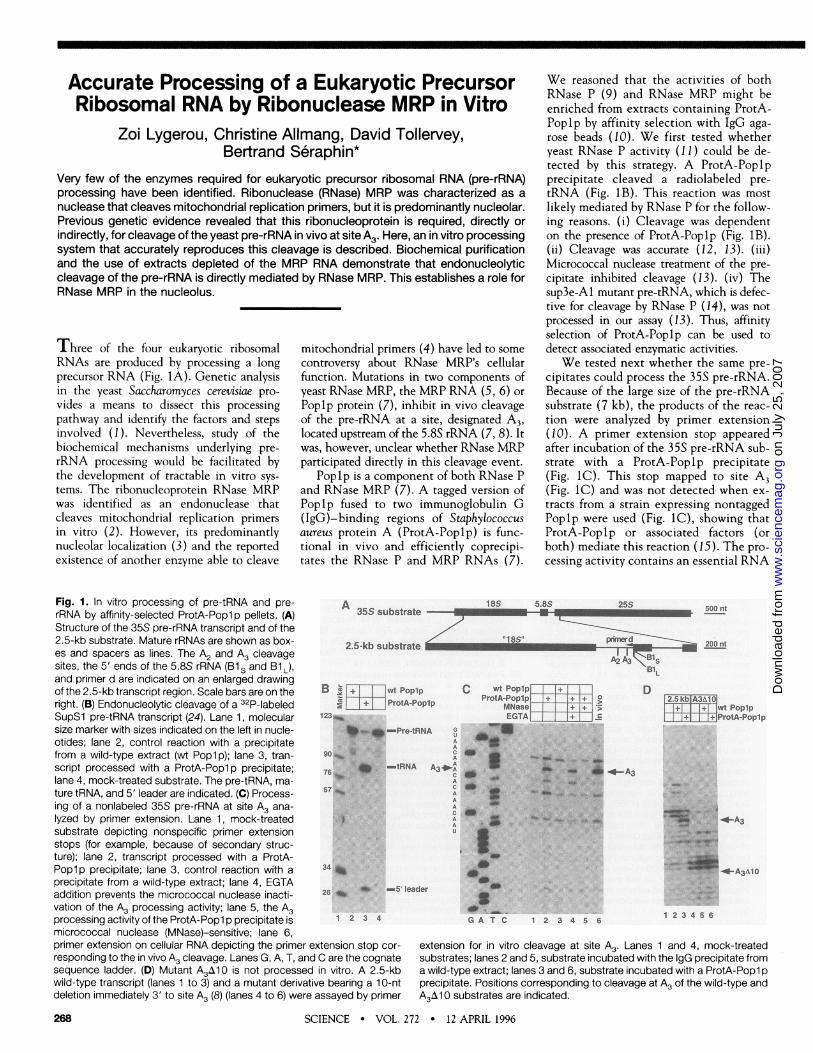
Accurate Processing of a Eukaryotic PrecursorRibosomal RNA by Ribonuclease MRP in Vitro
Zoi Lygerou, Christine Allmang, David Tollervey,Bertrand Seraphin*
Very few of the enzymes required for eukaryotic precursor ribosomal RNA (pre-rRNA)processing have been identified. Ribonuclease (RNase) MRP was characterized as anuclease that cleaves mitochondrial replication primers, but it is predominantly nucleolar.Previous genetic evidence revealed that this ribonucleoprotein is required, directly orindirectly, for cleavage of the yeast pre-rRNA in vivo at site A3. Here, an in vitro processingsystem that accurately reproduces this cleavage is described. Biochemical purificationand the use of extracts depleted of the MRP RNA demonstrate that endonucleolyticcleavage of the pre-rRNA is directly mediated by RNase MRP. This establishes a role forRNase MRP in the nucleolus.
Three of the four eukaryotic ribosomalRNAs are produced by processing a longprecursor RNA (Fig. IA). Genetic analysisin the yeast Saccharomyces cerevisiae pro-
vides a means to dissect this processingpathway and identify the factors and stepsinvolved (1). Nevertheless, study of thebiochemical mechanisms underlying pre-
rRNA processing would be facilitated bythe development of tractable in vitro sys-
tems. The ribonucleoprotein RNase MRPwas identified as an endonuclease thatcleaves mitochondrial replication primersin vitro (2). However, its predominantlynucleolar localization (3) and the reportedexistence of another enzyme able to cleave
mitochondrial primers (4) have led to somecontroversy about RNase MRP's cellularfunction. Mutations in two components ofyeast RNase MRP, the MRP RNA (5, 6) or
Poplp protein (7), inhibit in vivo cleavageof the pre-rRNA at a site, designated A3,located upstream of the 5.8S rRNA (7, 8). Itwas, however, unclear whether RNase MRPparticipated directly in this cleavage event.
Poplp is a component of both RNase Pand RNase MRP (7). A tagged version ofPoplp fused to two immunoglobulin G(IgG)-binding regions of Staphylococcusaureus protein A (ProtA-Poplp) is func-tional in vivo and efficiently coprecipi-tates the RNase P and MRP RNAs (7).
We reasoned that the activities of bothRNase P (9) and RNase MRP might beenriched from extracts containing ProtA-Poplp by affinity selection with IgG aga-rose beads (10). We first tested whetheryeast RNase P activity (11) could be de-tected by this strategy. A ProtA-Poplpprecipitate cleaved a radiolabeled pre-tRNA (Fig. iB). This reaction was mostlikely mediated by RNase P for the follow-ing reasons. (i) Cleavage was dependenton the presence of ProtA-Poplp (Fig. iB).(ii) Cleavage was accurate (12, 13). (iii)Micrococcal nuclease treatment of the pre-cipitate inhibited cleavage (13). (iv) Thesup3e-A1 mutant pre-tRNA, which is defec-tive for cleavage by RNase P (14), was notprocessed in our assay (13). Thus, affinityselection of ProtA-Poplp can be used todetect associated enzymatic activities.We tested next whether the same pre-
cipitates could process the 35S pre-rRNA.Because of the large size of the pre-rRNAsubstrate (7 kb), the products of the reac-
tion were analyzed by primer extension(10). A primer extension stop appearedafter incubation of the 35S pre-rRNA sub-strate with a ProtA-Poplp precipitate(Fig. 1C). This stop mapped to site A3(Fig. 1C) and was not detected when ex-
tracts from a strain expressing nontaggedPoplp were used (Fig. 1C), showing thatProtA-Poplp or associated factors (orboth) mediate this reaction (15). The pro-cessing activity contains an essential RNA
Fig. 1. In vitro processing of pre-tRNA and pre- A
rRNA by affinity-selected ProtA-Popl p pellets. (A) 35S substrStructure of the 35S pre-rRNA transcript and of the2.5-kb substrate. Mature rRNAs are shown as box- 2.5-kb substr
es and spacers as lines. The A2 and A3 cleavagesites, the 5' ends of the 5.8S rRNA (B1s and B1L),and primer d are indicated on an enlarged drawingof the 2.5-kb transcript region. Scale bars are on the B ijII2wt P
right. (B) Endonucleolytic cleavage of a 32P-labeled aLjlLjProUSupSl pre-tRNA transcript (24). Lane 1, molecular 123size marker with sizes indicated on the left in nucle- -Protides; lane 2, control reaction with a precipitatefrom a wild-type extract (wt Popl p); lane 3, tran- 90
script processed with a ProtA-Popl p precipitate; 76 l tf|lane 4, mock-treated substrate. The pre-tRNA, ma-ture tRNA, and 5' leader are indicated. (C) Process- 67
ing of a nonlabeled 35S pre-rRNA at site A3 ana-lyzed by primer extension. Lane 1, mock-treatedsubstrate depicting nonspecific primer extensionstops (for example, because of secondary struc-ture); lane 2, transcript processed with a ProtA-Poplp precipitate; lane 3, control reaction with a 34precipitate from a wild-type extract; lane 4, EGTAaddition prevents the micrococcal nuclease inacti- 26vation of the A processing activity; lane 5, the Aprocessing activity of the ProtA-Popl p precipitate is 1 2 3 4micrococcal nuclease (MNase)-sensitive; lane 6,primer extension on cellular RNA depicting the primer extension stop cor-responding to the in vivo A3 cleavage. Lanes G, A, T, and C are the cognatesequence ladder. (D) Mutant A3A1O is not processed in vitro. A 2.5-kbwild-type transcript (lanes 1 to 3) and a mutant derivative bearing a 1 0-ntdeletion immediately 3' to site A3 (8) (lanes 4 to 6) were assayed by primer
rate
rate
'op1p C wt PopipA-Pop1p ProtA-PoplpLS| >T
EGTAI I +|I
D
500 nt
200 nt
2.5kb A311+1 I I wt Pop1p
1+1 11jProtA-Pop1p
-4-A3
-4-A3
-A3A10
G A T C 1 2 3 4 5 6 1 23456
extension for in vitro cleavage at site A3. Lanes 1 and 4, mock-treatedsubstrates; lanes 2 and 5, substrate incubated with the IgG precipitate froma wild-type extract; lanes 3 and 6, substrate incubated with a ProtA-Popl pprecipitate. Positions corresponding to cleavage at A3 of the wild-type andA31M 0 substrates are indicated.
SCIENCE * VOL. 272 * 12 APRIL 1996
18S 5.8S 25S
J
..l
I IA2 A3 Bls7
BlL
re-tRNA
INA A3 00
leader
268
on
July
25,
200
7 w
ww
.sci
ence
mag
.org
Dow
nloa
ded
from
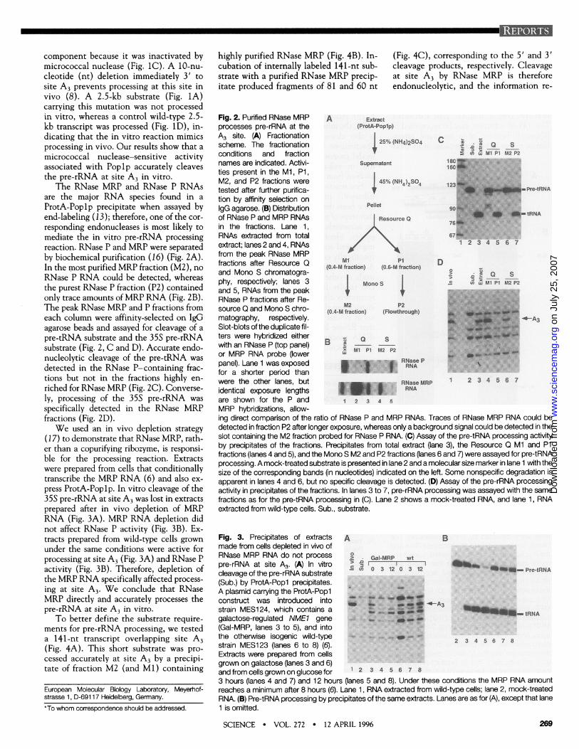
component because it was inactivated bymicrococcal nuclease (Fig. 1C). A 10-nu-cleotide (nt) deletion immediately 3' tosite A3 prevents processing at this site invivo (8). A 2.5-kb substrate (Fig. 1A)carrying this mutation was not processedin vitro, whereas a control wild-type 2.5-kb transcript was processed (Fig. 1D), in-dicating that the in vitro reaction mimicsprocessing in vivo. Our results show that a
micrococcal nuclease-sensitive activityassociated with Poplp accurately cleavesthe pre-rRNA at site A3 in vitro.
The RNase MRP and RNase P RNAsare the major RNA species found in a
ProtA-Poplp precipitate when assayed byend-labeling (13); therefore, one of the cor-
responding endonucleases is most likely to
mediate the in vitro pre-rRNA processingreaction. RNase P and MRP were separatedby biochemical purification (16) (Fig. 2A).In the most purified MRP fraction (M2), no
RNase P RNA could be detected, whereasthe purest RNase P fraction (P2) containedonly trace amounts of MRP RNA (Fig. 2B).The peak RNase MRP and P fractions fromeach column were affinity-selected on IgGagarose beads and assayed for cleavage of a
pre-tRNA substrate and the 35S pre-rRNAsubstrate (Fig. 2, C and D). Accurate endo-nucleolytic cleavage of the pre-tRNA was
detected in the RNase P-containing frac-tions but not in the fractions highly en-
riched for RNase MRP (Fig. 2C). Converse-ly, processing of the 35S pre-rRNA was
specifically detected in the RNase MRPfractions (Fig. 2D).We used an in vivo depletion strategy
(17) to demonstrate that RNase MRP, rath-er than a copurifying ribozyme, is responsi-ble for the processing reaction. Extractswere prepared from cells that conditionallytranscribe the MRP RNA (6) and also ex-
press ProtA-Poplp. In vitro cleavage of the35S pre-rRNA at site A3 was lost in extractsprepared after in vivo depletion of MRPRNA (Fig. 3A). MRP RNA depletion didnot affect RNase P activity (Fig. 3B). Ex-tracts prepared from wild-type cells grownunder the same conditions were active forprocessing at site A3 (Fig. 3A) and RNase Pactivity (Fig. 3B). Therefore, depletion ofthe MRP RNA specifically affected process-
ing at site A3. We conclude that RNaseMRP directly and accurately processes thepre-rRNA at site A3 in vitro.
To better define the substrate require-ments for pre-rRNA processing, we testeda 141-nt transcript overlapping site A3(Fig. 4A). This short substrate was pro-
cessed accurately at site A3 by a precipi-tate of fraction M2 (and MI) containing
highly purified RNase MRP (Fig. 4B). In-cubation of internally labeled 141-nt sub-strate with a purified RNase MRP precip-itate produced fragments of 81 and 60 nt
Fig. 2. Purified RNase MRPprocesses pre-rRNA at theA3 site. (A) Fractionationscheme. The fractionationconditions and fractionnames are indicated. Activi-ties present in the Ml, P1,M2, and P2 fractions weretested after further purifica-tion by affinity selection onIgG agarose. (B) Distributionof RNase P and MRP RNAsin the fractions. Lane 1,RNAs extracted from totalextract; lanes 2 and 4, RNAsfrom the peak RNase MRPfractions after Resource Qand Mono S chromatogra-phy, respectively; lanes 3and 5, RNAs from the peakRNase P fractions after Re-source Q and Mono S chro-matography, respectively.Slot-blots of the duplicate fil-ters were hybridized eitherwith an RNase P (top panel)or MRP RNA probe (lowerpanel). Lane 1 was exposedfor a shorter period thanwere the other lanes, butidentical exposure lengthsare shown for the P andMRP hybridizations, allow-
(Fig. 4C), corresponding to the 5' and 3'cleavage products, respectively. Cleavageat site A3 by RNase MRP is thereforeendonucleolytic, and the information re-
A Extract(ProtA-Popip)
25% (NH4)2SO4
Supernatant
45% (NH4)2SO4
Pellet
Resource 0
M P1(0.4-M fraction) (0.6-M fraction)
Mono S
a ctL MI Pl M2 P218C160
123
D
M2 P2(0.4-M fraction) (Flowthrough)
B t a S' Ml P1 M2 P2
RNase PRNA
RNase MRPRNA
1 2 3 4
E n LE Q S
3 X M1 P1 M2P2
m Pre-tRNA
-tRNA
4-A3
1 2 3 4 5 6 7
ing direct comparison of the ratio of RNase P and MRP RNAs. Traces of RNase MRP RNA could bedetected in fraction P2 after longer exposure, whereas only a background signal could be detected in theslot containing the M2 fraction probed for RNase P RNA. (C) Assay of the pre-tRNA processing activityby precipitates of the fractions. Precipitates from total extract (lane 3), the Resource Q Ml and P1fractions (lanes 4 and 5), and the Mono S M2 and P2 fractions (lanes 6 and 7) were assayed for pre-tRNAprocessing. A mock-treated substrate is presented in lane 2 and a molecular size marker in lane 1 with thesize of the corresponding bands (in nucleotides) indicated on the left. Some nonspecific degradation isapparent in lanes 4 and 6, but no specific cleavage is detected. (D) Assay of the pre-rRNA processingactivity in precipitates of the fractions. In lanes 3 to 7, pre-rRNA processing was assayed with the samefractions as for the pre-tRNA processing in (C). Lane 2 shows a mock-treated RNA, and lane 1, RNAextracted from wild-type cells. Sub., substrate.
Fig. 3. Precipitates of extracts A Bmade from cells depleted in vivo ofRNase MRP RNA do not process ° Gal-MRP wt
pre-rRNA at site A3. (A) In vitrocleavage of the pre-rRNA substrate ' 8 03120312 Pr--tRNA(Sub.) by ProtA-Popl precipitates.
A plasmid carrying the ProtA-Poplconstruct was introduced intostrain MES1 24, which contains a
galactose-regulated NME1 gene E(Gal-MRP, lanes 3 to 5), and intothe otherwise isogenic wild-type
strain MES1 23 (lanes 6 to 8) (6).
Extracts were prepared from cellsgrown on galactose (lanes 3 and 6)and from cells grown on glucose for 1 2 3 4 5 6 7 8
3 hours (lanes 4 and 7) and 12 hours (lanes 5 and 8). Under these conditions the MRP RNA amountreaches a minimum after 8 hours (6). Lane 1, RNA extracted from wild-type cells; lane 2, mock-treatedRNA. (B) Pre-tRNA processing by precipitates of the same extracts. Lanes are as for (A), except that lane1 is omitted.
SCIENCE * VOL. 272 * 12 APRIL 1996
European Molecular Biology Laboratory, Meyerhof-strasse 1, D-69117 Heidelberg, Germany.*To whom correspondence should be addressed.
269
on
July
25,
200
7 w
ww
.sci
ence
mag
.org
Dow
nloa
ded
from
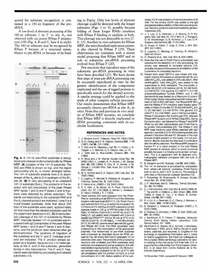
quired for substrate recognition is con-tained in a 141-nt fragment of the pre-rRNA.A low level of aberrant processing of the
141-nt substrate 1 nt 3' to site A3 wasobserved with our purest RNase P prepara-tion (18) (Fig. 4, B and C, lane 4 in each).The 141-nt substrate may be recognized byRNase P because of a structural resem-blance to pre-tRNA or because of its bind-
A- ~~~~~~~~~35
-141-nt substrate
B a s 0Ca SUl Pi "sos a s "eA
Sub. Sub.
5' products I
3' products I
Fig. 4. A 141-nt pre-rRNA substrate is recog-nized and cleaved endonucleolytically by RNaseMRP. (A) Location of the 141-nt substrate. The35S pre-rRNA is shown on top, and the regionsurrounding site A3 is shown enlarged below.The 141-nt substrate extends from 3 nt down-stream of the A2 site to 9 nt upstream of the B1 Lsite (8). (B) In vitro processing of an unlabeled141-nt substrate (Sub.). The products of the re-action with IgG precipitates of the peak RNaseMRP (lanes 1 and 3) and P (lanes 2 and 4) frac-tions were detected by primer extension. Thebands corresponding to the substrate RNA andthe A3 cleaved product are indicated. Lane 5 is amock-treated substrate. Note that about 250fmol of the substrate were used, approximately50-fold more than the labeled substrate used forthe experiment depicted in (C). (C) Endonucleo-lytic cleavage of the 141-nt substrate by RNaseMRP. Internally labeled 141-nt substrate was in-cubated with IgG precipitates of the peak RNaseMRP (lanes 1 and 3) and P (lanes 2 and 4) frac-tions, and the products were detected after gelelectrophoresis. The positions of migration ofthe substrate and the 5' and 3' cleavage prod-ucts are indicated. The 3' product always ap-pears as a doublet, because of a 1-nt heteroge-neity at the 3' end of the substrate, generatedduring in vitro transcription. The 3' and 5' frag-ments were identified by processing end-labeledsubstrates (13).
270
ing to Poplp. Only low levels of aberrantcleavage could be detected with the longersubstrates (Figs. 1 to 3), possibly becausefolding of these longer RNAs interfereswith RNase P binding or catalysis or both.This cleavage was not detectable in vivo (7,8, 13). Another in vitro substrate for RNaseMRP, the mitochondrial replication primer,is also cleaved by RNase P (19). Theseobservations are consistent with a recentmodel proposing that RNase MRP and itsrole in eukaryotic pre-rRNA processingevolved from RNase P (20).
Few reactions that reproduce steps of theeukaryotic pre-rRNA processing in vitrohave been described (21). We have shownthat steps of yeast pre-rRNA processing canbe accurately reproduced in vitro by thegenetic identification of the componentsimplicated and the use of tagged proteins tospecifically enrich for the desired activity.A similar strategy could be applied to thestudy of other complex cellular processes.Our results demonstrate that RNase MRPaccurately cleaves pre-rRNA at site A3 invitro. From this and previous in vivo stud-ies of RNase MRP mutants, we concludethat RNase MRP is directly implicated inrRNA processing, consistent with its nu-cleolar localization.
REFERENCES AND NOTES
1 J. Venema and D. Tollervey, Yeast 11, 1629 (1995).2. D. D. Chang and D. A. Clayton, Science 235, 1178
(1987); Cell 56, 131 (1989).3. T. Kiss and W. Filipowicz, Cell 70, 11 (1992); J. N.
Topper, J. L. Bennett, D. A. Clayton, ibid., p. 16.4. J. C6t6 and A. Ruiz-Carrillo, Science 261, 765
(1993).5. K. Shuai and J. W. Warner, Nucleic Acids Res. 19,
5059 (1991); L. Lindahl, R. H. Archer, J. M. Zengal,ibid. 20, 295 (1992); S. Chu, R. H. Archer, J. M.Zengel, L. Lindahl, Proc. Natl. Acad. Sci. U.S.A. 91,659 (1994).
6. M. E. Schmitt and D. A. Clayton, Mol. Cell. Biol. 13,7935 (1993).
7. Z. Lygerou, P. Mitchell, E. Peffalski, B. Seraphin, D.Tollervey, Genes Dev. 8,1423 (1994).
8. Y. Henry et al., EMBO J. 13, 2452 (1994).9. S. C. Darr, J. W. Brown, N. R. Pace, Trends Bio-
chem. Sci. 17,178 (1992); S. Altman, L. Kirsebom,S. Talbot, FASEB J. 7, 7 (1993).
10. Yeast extracts (22) were prepared from the ProtA-Popl p-expressing strain BSY414 and the otherwiseisogenic wild-type strain BSY17 (7, 23). ProtA-Popl pwas selected from 0.6 p.1 of extract by incubation for2.5 hours at 4°C with 6 p.1 of a 50% suspension of IgGagarose beads in 120 p.1 of IPP150 buffer [150 mMNaCI, 10 mM tris-CI (pH 8.0), 0.1 % NP-40, and 0.1 %NaN3] (7). IgG pellets were incubated with 5 fmol oflabeled pre-tRNASuPsl (24) for 30 min at 37°C in 20mM tris-CI (pH 8), 10 mM MgCI2, 1 mM dithiothreitol(DTT), 50 mM KCI, bovine serum albumin (50 p.g/ml),and RNasin (60 U/mI). Pre-rRNA substrates wereproduced by in vitro transcription of the appropriateplasmids. Five femtomoles of pre-rRNA substratewere processed as described above for tRNA, ex-cept for Fig. 4B where 250 fmol of unlabeled 141-ntsubstrate were used. The products of the processingreactions with unlabeled pre-rRNA substrates wereextracted and analyzed by primer extension (23) withradiolabeled oligonucleotide d (8) (Fig. 1 A). Micrococ-cal nuclease was incubated for 20 min at 37°C withIgG precipitates after the addition of CaCI2 to a finalconcentration of 4 mM. Before addition of the sub-
strate, EGTA was added to a final concentration of 32mM. For the control, EGTA was added to the IgGprecipitates before addition of the micrococcal nucle-ase. Reaction products were fractionated by gel elec-trophoresis (25).
11. J. Y. Lee, C. E. Rohlman, L. A. Molony, D. R. En-gelke, Mol. Cell. Biol. 11, 721 (1991); A. J. Tranguch,D. W. Kindelberger, C. E. Rohiman, J. Y. Lee, D. R.Engelke, Biochemistry 33,1778 (1994).
12. G. Krupp, D. Kahle, T. Vogt, S. Char, J. Mol. Biol.217, 637 (1991).
13. Z. Lygerou, C. Allmang, D. Tollervey, B. Seraphin,data not shown.
14. D. Pearson et al., Mol. Cell. Biol. 5, 808 (1985).15. Note that the use of ProtA-Popl p precipitates was
essential for the detection of in vitro processing. Noactivity was detected in complete extracts (13),probably because of the presence of inhibitors or theoccurrence of competing reactions.
16. Extract from strain BSY414 was mixed with one-eighth volume 2M potassium phosphate (pH 8), andsolid (NH4)2SO4 was added to 25% saturation. Aftercentrifugation, solid (NH4)2SO4 was added to thesupernatant to 45% saturation. The precipitate, inbuffer Q6-50 [20 mM histidine (pH 6), 50 mM NaCI,0.2 mM EDTA, 10% glycerol, 0.5 mM DTT, 0.5 mMphenylmethylsulfonyl fluoride (PMSF), and 2 mMbenzamidine] was loaded on a Resource 0 column(Pharmacia). Elution was performed with a linear gra-dient of 300 to 800 mM NaCI. The RNase MRP (Ml)and the RNase P (P1) fractions were loaded sepa-rately on Mono S in buffer S7-50 [50 mM NaPO4 (pH7), 50 mM NaCI, 0.2 mM EDTA, 10% glycerol, 0.5mM DTT, 0.5 mM PMSF, and 2 mM benzamidine].RNase P remained in the flowthrough (P2), whereasRNase MRP eluted in a 0.4-M NaCI step (M2). Duringthe fractionation, we followed the RNase P and MRPRNAs by slot blot hybridizations and ProtA-Popl pby protein immunoblofting (25). The RNase P andMRP peak fractions were affinity-selected with IgGagarose. Less than 0.01 % of the starting protein waspresent in fractions M2 and P2. Because of the highamount of IgGs present on the beads, it is not pos-sible to directly evaluate the final level of purificationafter the affinity selection. The RNase MRP present infraction P1 is a minor amount of the total RNaseMRP present in extracts, which is poorly functionalpossibly because it is missing some protein compo-nent. This is in agreement with its aberrant chro-matographic behavior compared with the bulk ofRNase MRP.
17. B. Seraphin and M. Rosbash, Cell 59, 349 (1989).18. The 141-nt transcript is processed by precipitates
from whole-cell extracts containing ProtA-Popl pboth at site A3 and 1 nt 3' to site A3. Processing atboth sites is micrococcal nuclease-sensitive (13).
19. T. Potuschak, W. Rossmanith, R. Karwan, NucleicAcids Res. 21, 3239 (1993).
20. J. P. Morrissey and D. Tollervey, Trends Biochem.Sci. 20, 78 (1995).
21. G. J. Hannon etal., Mol. Cell. Biol. 9,4422 (1989); M.T. Yip and M. J. Holland, J. Biol. Chem. 264, 4045(1989); S. Kass, K. Tyc, J. A. Steitz, B. Sollner-Webb, Cell 60, 897 (1990).
22. R.-J. Lin, A. J. Newman, S.-C. Cheng, J. Abelson, J.Biol. Chem. 260,14780 (1985).
23. B. Seraphin, L. Kretzner, M. Rosbash, EMBO J. 7,2533 (1988).
24. D. Drainas, S. Zimmerly, l. Willis, D. Soll, FEBS Lett.251, 84 (1989).
25. J. Sambrook, E. F. Fritsch, T. Maniatis, MolecularCloning (Cold Spring Harbor Laboratory, ColdSpring Harbor, NY, 1989).
26. We thank D. A. Clayton, M. E. Schmitt, J. Morrissey,J. Mermoud, l. Willis, and D. Soll for the gift of yeaststrains, plasmids, and enzymes, D. Engelke for theexchange of unpublished information, and J. Lewisfor helpful suggestions. We thank our colleagues atthe European Molecular Biology Laboratory for care-ful reading of the manuscript and their help. C.A. issupported by a fellowship from the European Union.B.S. is on leave from Centre National de la Re-cherche Scientifique.
13 November 1995; accepted 8 February 1996
SCIENCE * VOL. 272 * 12 APRIL 1996
on
July
25,
200
7 w
ww
.sci
ence
mag
.org
Dow
nloa
ded
from

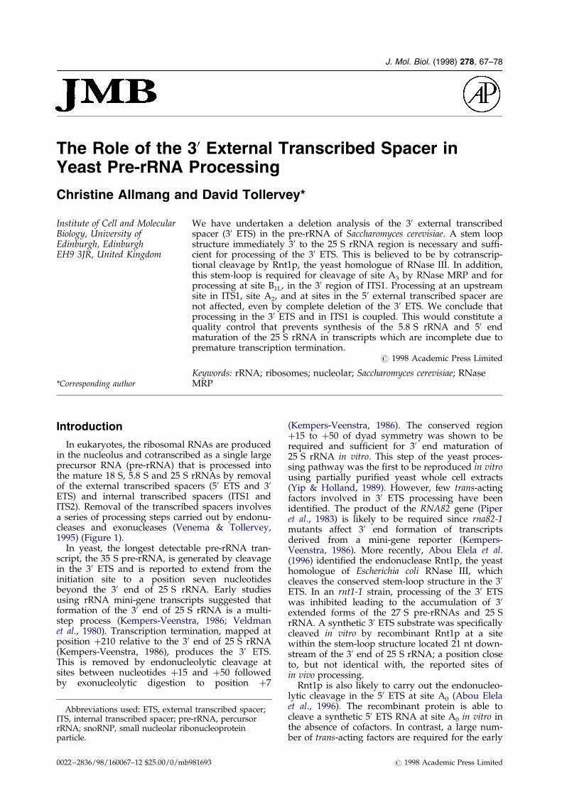
J. Mol. Biol. (1998) 278, 67±78
The Role of the 30 External Transcribed Spacer inYeast Pre-rRNA Processing
Christine Allmang and David Tollervey*
Institute of Cell and MolecularBiology, University ofEdinburgh, EdinburghEH9 3JR, United Kingdom
*Corresponding author
Abbreviations used: ETS, externaITS, internal transcribed spacer; prerRNA; snoRNP, small nucleolar ribparticle.
0022±2836/98/160067±12 $25.00/0/mb
We have undertaken a deletion analysis of the 30 external transcribedspacer (30 ETS) in the pre-rRNA of Saccharomyces cerevisiae. A stem loopstructure immediately 30 to the 25 S rRNA region is necessary and suf®-cient for processing of the 30 ETS. This is believed to be by cotranscrip-tional cleavage by Rnt1p, the yeast homologue of RNase III. In addition,this stem-loop is required for cleavage of site A3 by RNase MRP and forprocessing at site B1L, in the 30 region of ITS1. Processing at an upstreamsite in ITS1, site A2, and at sites in the 50 external transcribed spacer arenot affected, even by complete deletion of the 30 ETS. We conclude thatprocessing in the 30 ETS and in ITS1 is coupled. This would constitute aquality control that prevents synthesis of the 5.8 S rRNA and 50 endmaturation of the 25 S rRNA in transcripts which are incomplete due topremature transcription termination.
# 1998 Academic Press Limited
Keywords: rRNA; ribosomes; nucleolar; Saccharomyces cerevisiae; RNaseMRP
Introduction
In eukaryotes, the ribosomal RNAs are producedin the nucleolus and cotranscribed as a single largeprecursor RNA (pre-rRNA) that is processed intothe mature 18 S, 5.8 S and 25 S rRNAs by removalof the external transcribed spacers (50 ETS and 30ETS) and internal transcribed spacers (ITS1 andITS2). Removal of the transcribed spacers involvesa series of processing steps carried out by endonu-cleases and exonucleases (Venema & Tollervey,1995) (Figure 1).
In yeast, the longest detectable pre-rRNA tran-script, the 35 S pre-rRNA, is generated by cleavagein the 30 ETS and is reported to extend from theinitiation site to a position seven nucleotidesbeyond the 30 end of 25 S rRNA. Early studiesusing rRNA mini-gene transcripts suggested thatformation of the 30 end of 25 S rRNA is a multi-step process (Kempers-Veenstra, 1986; Veldmanet al., 1980). Transcription termination, mapped atposition �210 relative to the 30 end of 25 S rRNA(Kempers-Veenstra, 1986), produces the 30 ETS.This is removed by endonucleolytic cleavage atsites between nucleotides �15 and �50 followedby exonucleolytic digestion to position �7
l transcribed spacer;-rRNA, percursoronucleoprotein
981693
(Kempers-Veenstra, 1986). The conserved region�15 to �50 of dyad symmetry was shown to berequired and suf®cient for 30 end maturation of25 S rRNA in vitro. This step of the yeast proces-sing pathway was the ®rst to be reproduced in vitrousing partially puri®ed yeast whole cell extracts(Yip & Holland, 1989). However, few trans-actingfactors involved in 30 ETS processing have beenidenti®ed. The product of the RNA82 gene (Piperet al., 1983) is likely to be required since rna82-1mutants affect 30 end formation of transcriptsderived from a mini-gene reporter (Kempers-Veenstra, 1986). More recently, Abou Elela et al.(1996) identi®ed the endonuclease Rnt1p, the yeasthomologue of Escherichia coli RNase III, whichcleaves the conserved stem-loop structure in the 30ETS. In an rnt1-1 strain, processing of the 30 ETSwas inhibited leading to the accumulation of 30extended forms of the 27 S pre-rRNAs and 25 SrRNA. A synthetic 30 ETS substrate was speci®callycleaved in vitro by recombinant Rnt1p at a sitewithin the stem-loop structure located 21 nt down-stream of the 30 end of 25 S rRNA; a position closeto, but not identical with, the reported sites ofin vivo processing.
Rnt1p is also likely to carry out the endonucleo-lytic cleavage in the 50 ETS at site A0 (Abou Elelaet al., 1996). The recombinant protein is able tocleave a synthetic 50 ETS RNA at site A0 in vitro inthe absence of cofactors. In contrast, a large num-ber of trans-acting factors are required for the early
# 1998 Academic Press Limited
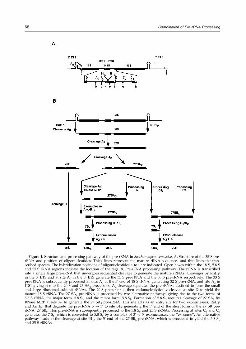
Figure 1. Structure and processing pathway of the pre-rRNA in Saccharomyces cerevisiae. A, Structure of the 35 S pre-rRNA and position of oligonucleotides. Thick lines represent the mature rRNA sequences and thin lines the tran-scribed spacers. The hybridization positions of oligonucleotides a to i are indicated. Open boxes within the 18 S, 5.8 Sand 25 S rRNA regions indicate the location of the tags. B, Pre-rRNA processing pathway. The rDNA is transcribedinto a single large pre-rRNA that undergoes sequential cleavage to generate the mature rRNAs. Cleavages by Rnt1pin the 30 ETS and at site A0 in the 50 ETS generate the 35 S pre-rRNA and the 33 S pre-rRNA respectively. The 33 Spre-rRNA is subsequently processed at sites A1 at the 50 end of 18 S rRNA, generating 32 S pre-rRNA, and site A2 inITS1 giving rise to the 20 S and 27 SA2 precursors. A2 cleavage separates the pre-rRNAs destined to form the smalland large ribosomal subunit rRNAs. The 20 S precursor is then endonucleolytically cleaved at site D to yield themature 18 S rRNA. The 27 SA2 pre-rRNA is processed by two alternative pathways giving rise to the two forms of5.8 S rRNA, the major form, 5.8 SS, and the minor form, 5.8 SL. Formation of 5.8 SS requires cleavage of 27 SA2 byRNase MRP at site A3 to generate the 27 SA3 pre-rRNA. This site acts as an entry site for two exonucleases, Rat1pand Xrn1p, that degrade the pre-rRNA 50 ! 30 to site B1S, generating the 50 end of the short form of the 27 SB pre-rRNA, 27 SBS. This pre-rRNA is subsequently processed to the 5.8 SS and 25 S rRNAs. Processing at sites C1 and C2
generates the 7 SS, which is converted to 5.8 SS by a complex of 30 ! 50 exonucleases, the ``exosome''. An alternativepathway leads to the cleavage at site B1L, the 50 end of the 27 SBL pre-rRNA, which is processed to yield the 5.8 SL
and 25 S rRNAs.
68 Coordination of Pre-rRNA Processing
Coordination of Pre-rRNA Processing 69
cleavages of the pre-rRNA in vivo. The major classcomprises a number of small nucleolar ribonucleo-protein particles (snoRNPs; for reviews seeMaxwell & Fournier, 1995; Tollervey & Kiss, 1997).Four snoRNPs, U3, U14, snR10 and snR30 havebeen shown to be required for the early cleavagesin the 50 region of the pre-rRNA at sites A0, A1 andA2 (see Figure 1). Genetic depletion of any of theRNA or protein components of these snoRNP hassimilar effects: the inhibition of cleavage at site A0,A1 and A2, resulting in the inhibition of thesynthesis of mature 18 S rRNA. These snoRNPcomponents may participate in a multi-snoRNPprocessing complex, probably assembling on the 50ETS, that brings together the sequences surround-ing sites A0, A1 and A2 in order to coordinate theircleavage (reviewed by Morrissey & Tollervey,1995).
Subsequent cleavages further 30 in the pre-rRNAgenerate the 5.8 S and 25 S rRNAs. Another RNPparticle, RNase MRP plays a role in these proces-sing reactions (Chu et al., 1994; Lygerou et al., 1994,1996; Schmitt & Clayton, 1993). RNase MRPdirectly cleaves site A3 located in ITS1 (Lygerouet al., 1996), providing an entry site for 50 ! 30 exo-nuclease degradation to site B1S, the 50 end of themajor form of 5.8 SS rRNA (Henry et al., 1994).This trimming requires two proteins, Xrn1p andRat1p, that exhibit a 50 ! 30 exonuclease activityin vitro (Amberg et al., 1992; Kenna et al., 1993;Larimer et al., 1992; Stevens & Poole, 1995). Analternative, less understood, pathway processessite B1L the 50 end of the minor 5.8 SL rRNA.Although capable of functioning independently,the RNase MRP complex assembled at site A3 andthe snoRNP complex assembled in the 50 ETS andsite A2 are believed to interact to bring about ef®-cient A2 and A3 cleavage (Allmang et al., 1996).This interaction may occur via a bridging factor,Rrp5p (Venema & Tollervey, 1996).
Strikingly, it appears that none of the pre-rRNAprocessing activities function on the nascent pre-rRNA; yeast pre-rRNA processing is initiated onlyon the fully transcribed pre-rRNA. Following com-pletion of transcription, however, the 35 S pre-rRNA undergoes very rapid processing. We there-fore speculated that a signal present in the 30 ETSmight be required to initiate the processing path-way.
Various cis-acting signals are predicted to bepresent in the 30 ETS and in search of these signalswe have undertaken an analysis of the effects ofdeletions in the 30 ETS on pre-rRNA processingin vivo.
Results
Nested deletions in the 30 ETS
To test the effects of deletions in the 30 ETS onribosome synthesis we have used a system allow-ing the conditional expression of mutant and wild-type pre-rRNA (Henry et al., 1994). Previous ana-
lyses have shown that yeast RNA polymerase Itranscription terminates 210 nt beyond the mature30 end of 25 S rRNA (Veldman et al., 1980). In theGAL::rDNA construct, the GAL7 terminator regionis inserted 284 nt 30 to the 25 S coding sequenceand introduces an SalI site directly adjacent to therDNA coding sequence (Nogi et al., 1991). Nesteddeletions were constructed that encompassed thewhole 30 ETS region, including the stem-loop struc-ture cleaved by Rnt1p, and analyzed in vivo.Unidirectional deletions were generated by Exonu-clease III from a synthetic linker inserted into theSalI site (see Materials and Methods) and enter the30 ETS progressively from 30 to 50. The extents ofthese deletions are shown Figure 2. The clearestpredicted structure in this region is a strong stem-loop located immediately 30 to the 25 S rRNA cod-ing sequence (nt �8 to �56; see Figure 2B).Mutations 30 ETS �1 to �3 leave the stem-loopstructure intact, whereas 30 ETS �4 and �5 enterthe stem-loop. 30 ETS �6 deletes the entire stem-loop structure and in addition deletes six nucleo-tides from the 30 end of the 25 S rRNA. In the 30ETS �6 � H mutant, the stem-loop structure wasrecreated, with the last six nucleotides of 25 SrRNA substituted by the SalI restriction sitesequence (see Material and Methods). Three of thelast six nucleotides of the 25 S rRNA are predictedto be base-paired at the base of a helical stemwithin the 25 S rRNA (Gutell & Fox, 1988). The 30ETS �6 � H mutation may therefore alter thestructure of the 30 helix in the mature 25 S rRNA.
The mutations were functionally analysed byexpression in the pGAL::rDNA construct.Expression of the pre-rRNA carrying the deletionsthat leave the hairpin structure intact (30 ETS �1,�2 and �3) supported the growth of an rpa12strain at the non-permissive temperature (37�C) ongalactose medium (Figure 3) at rates similar to thewild-type pre-rRNA. In contrast, expression ofthe pre-rRNA carrying the mutations that enter thestem-loop structure (30 ETS �4, �5 and �6) didnot support the growth of the rpa12 strain. Restor-ation of the hairpin structure in 30 ETS �6 � H pre-rRNA did not restore the ability to support growthin an rpa12 strain (Figure 3).
Deletions in the 30 ETS affect 30 endprocessing of the pre-rRNA
The effect of the 30 ETS deletions on pre-rRNAprocessing were examined by Northern hybridiz-ation six hours after transfer to 37�C (Figure 4).The steady state levels of the mature rRNAs weredetermined using oligonucleotides complementaryto neutral tags that are present in the 25 S and 18 SrRNA (Beltrame & Tollervey, 1992), while thelevels of the pre-rRNAs were determined using theoligonucleotides indicated in ®gure 1A. None ofthe 30 ETS deletions affected the accumulationof the 18 S rRNA (Figure 4VII) or altered the levelsof the 20 S pre-rRNA (Figure 4VI). This showedthat global processing of the pre-rRNA was not
70 Coordination of Pre-rRNA Processing
affected by the deletions. We conclude that, con-trary to our expectations, there is no signal in the30 ETS that is required to initiate the pre-rRNA pro-cessing pathway.
The 30 ETS �1, �2 and �3 mutant pre-rRNAsgenerated normal levels of 25 S rRNA (Figure 4V,
Figure 2. Deletions in the 30 ETS. A, Schematic representatstructure and the Sal1 site located into the GAL7 terminatoretions in mutants 30 ETS �1 to 30 ETS �6 is represented belhatched box indicates the altered region in the re-introducemissive temperature is indicated in each case; (�) represemutation. B, Predicted structure of the 50 region of the 30 EElela et al., 1996) and the end points of the 30 ETS �3, �4, �is that re-introduced in the �6 � H mutation.
lanes 3 to 5) and pre-rRNA processing was unaf-fected with normal levels of all the major inter-mediates. Therefore, no sequence 30 to the stem-loop structure is required for normal function ofthe 30 ETS. In contrast, synthesis of the 25 S rRNAfrom pre-rRNAs carrying the 30 ETS �4 and �5
ion of the 30 ETS region (thin line) showing the stem-loop(Nogi et al., 1991). The extent and size of the 30 ! 50 del-
ow. In the schematic representation of 30 ETS �6 � H thed sequence. The viability of the rpa12 strain at non-per-nting a fully viable mutation, (ÿ) representing a lethalTS. The major site of in vitro cleavage by Rnt1p (Abou
5 and �6 mutations are indicated. The sequence in italics
Figure 3. Growth analysis of 30 ETSdeletion mutants. Growth curves ofNOY504 strains transformed bypTA1 plasmids containing the 30ETS deletions. Cells were grown inminimal galactose medium at 23�Cuntil reaching an A600 nm of 0.07and then shifted to 37�C, the non-permissive temperature, to represschromosomal rDNA transcription.Growth curves were measuredafter the temperature shift. Thesymbols used for each mutant arerepresented beside the curves.Strains were transformed with thefollowing vectors: (WT 30 ETS) vec-tor containing the wild-type 30 ETSsequence; (ÿrDNA) vector lackingthe rDNA sequence; (30 ETS �1-�6and 30 ETS �6 � H) vectors con-taining the 30 ETS deletionsreported in Figure 2.
Coordination of Pre-rRNA Processing 71
mutations was strongly reduced (Figure 4V, lanes6 and 7), and no detectable 25 S rRNA was syn-thesized from the 30 ETS �6 pre-rRNA (Figure 4V,lane 8). Restoration of the stem-loop structure inthe 30 ETS �6 � H pre-rRNA only partiallyrestored 25 S rRNA synthesis (Figure 4V, lane 9),to levels similar to those observed for the 30 ETS�4 and �5 constructs. Transformants carrying aplasmid without the rDNA unit did not show anybackground signal (Figure 4, lane 2). The 30 ETS�4, �5 and �6 mutations resulted in the pro-duction of aberrant extended forms of 27 SA2 pre-rRNA (Figure 4II, lanes 6 to 8) and 27 SB(Figure 4III, lanes 6 to 8). Furthermore, anextended form of 25 S rRNA was synthesized fromthe 30 ETS �4 and �5 pre-rRNAs. These aberrantintermediates, 27 SA2*, 27 SB* and 25 S*, representforms of the corresponding RNAs that are 30extended up to the GAL7 terminator since they canbe detected by oligonucleotide i (complementary tothe GAL7 terminator sequence; Figure. 4IV, lanes 6and 7). The levels of 27 SA2* synthesized from the30 ETS �4 and �5 are comparable to those of the27SA2 synthesized from the wild-type pre-rRNA(Figure 4II), again indicating that processing at siteA2 is unaffected by even the complete deletion ofthe 30 ETS. The 27 SA2* pre-rRNA, however,slightly accumulates in �6 pre-rRNAs (Figure 4II).In contrast, levels of 27 SB* synthesized from the 30ETS �4 and �5 pre-rRNAs are strongly reduced ascompared to the wild-type 27 SB (Figure 4III).Moreover, no 27 SB* and 25 S* rRNA are syn-thesized from the 30 ETS �6 mutant, in markedcontrast to the 30 ETS �4 and �5 pre-rRNAs fromwhich 25 S* rRNA is synthesized. The ratio of25 S* : 27 SA2* in the �4 and �5 pre-rRNAs is sub-stantially lower than the ratio of 25 S : 27 SA2 syn-thesized from the wild-type pre-rRNA (data not
shown). This indicates that the processing of27 SA2* to 25 S* is relatively inef®cient, consistentwith the reduced level of 27 SB*.
When the 30 ETS hairpin structure is restored inmutant 30 ETS �6 � H, the aberrant 30 extendedintermediates are lost, and only normal pre-rRNAspecies are detected (Figure 4II, III, IV, lane 9).However, the 27 SA2 pre-rRNA slightly accumu-lates (Figure 4II) and the level of 27 SB is stronglydecreased (Figure 4III).
We conclude that an intact stem-loop structure isrequired for processing of the 30 ETS. Mutationswhich enter (30 ETS �4 and �5) or remove (30 ETS�6) this structure prevent processing, leading tothe accumulation of pre-rRNA species that are 30extended to the transcription termination site. Pro-cessing of the 30 ETS is restored by re-insertion ofthe hairpin structure in the 30 ETS �6 � H pre-rRNA. In addition, both the six nucleotides at the30 end of the 25 S rRNA and the hairpin structureare important for the processing in ITS1 that con-verts the 27 SA2 pre-rRNA to 27 SB. Since themajor processing pathway for 27 SA2 is cleavage atsite A3 by RNase MRP, these data indicate that A3
cleavage was inhibited by the mutations in the 30ETS.
Deletions in the 30 ETS affect processingin ITS1
The effects of the deletions on the ef®ciency andaccuracy of the pre-rRNA cleavages in ITS1 wereanalysed by primer extension using oligonucleo-tide g that hybridizes to the pre-rRNA in ITS2 (seeFigure 1). The levels of primer extension productsterminating at site A2 re¯ect the abundance of the27 SA2 and 27 SA2* pre-rRNAs; these are unaf-fected by any of the deletions in the 30 ETS
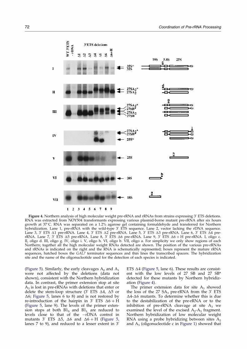
Figure 4. Northern analysis of high molecular weight pre-rRNA and rRNAs from strains expressing 30 ETS deletions.RNA was extracted from NOY504 transformants expressing various plasmid-borne mutant pre-rRNA after six hoursgrowth at 37�C. RNA was separated on a 1.2% agarose gel containing formaldehyde and transferred for Northernhybridization. Lane 1, pre-rRNA with the wild-type 30 ETS sequence. Lane 2, vector lacking the rDNA sequence.Lane 3, 30 ETS �1 pre-rRNA. Lane 4, 30 ETS �2 pre-rRNA. Lane 5, 30 ETS �3 pre-rRNA. Lane 6, 30 ETS �4 pre-rRNA. Lane 7, 30 ETS �5 pre-rRNA. Lane 8, 30 ETS �6 pre-rRNA. Lane 9, 30 ETS �6 � H pre-rRNA. I, oligo c.II, oligo d. III, oligo g. IV, oligo i. V, oligo h. VI, oligo b. VII, oligo a. For simplicity we only show regions of eachNorthern; together all the high molecular weight RNAs detected are shown. The position of the various pre-rRNAsand rRNAs is indicated on the right and the RNA is schematically represented; boxes represent the mature rRNAsequences, hatched boxes the GAL7 terminator sequences and thin lines the transcribed spacers. The hybridizationsite and the name of the oligonucleotide used for the detection of each species is indicated.
72 Coordination of Pre-rRNA Processing
(Figure 5). Similarly, the early cleavages A0 and A1
were not affected by the deletions (data notshown), consistent with the Northern hybridizationdata. In contrast, the primer extension stop at siteA3 is lost in pre-rRNAs with deletions that enter ordelete the stem-loop structure (30 ETS �4, �5 or�6; Figure 5, lanes 6 to 8) and is not restored byre-introduction of the hairpin in 30 ETS �6 � H(Figure 5, lane 9). The levels of the primer exten-sion stops at both B1S and B1L are reduced tolevels close to that of the ÿrDNA control inmutants 30 ETS �5, �6 and �6 � H (Figure 5,lanes 7 to 9), and reduced to a lesser extent in 30
ETS �4 (Figure 5, lane 6). These results are consist-ent with the low levels of 27 SB and 27 SB*detected for these mutants by Northern hybridiz-ation (Figure 4).
The primer extension data for site A3 showedthe loss of the 27 SA3 pre-rRNA from the 30 ETS�4-�6 mutants. To determine whether this is dueto the destabilization of the pre-rRNA or to theinhibition of pre-rRNA cleavage at site A3 weexamined the level of the excised A2-A3 fragment.Northern hybridization of low molecular weightRNA using a probe hybridizing between sites A2
and A3 (oligonucleotide c in Figure 1) showed that
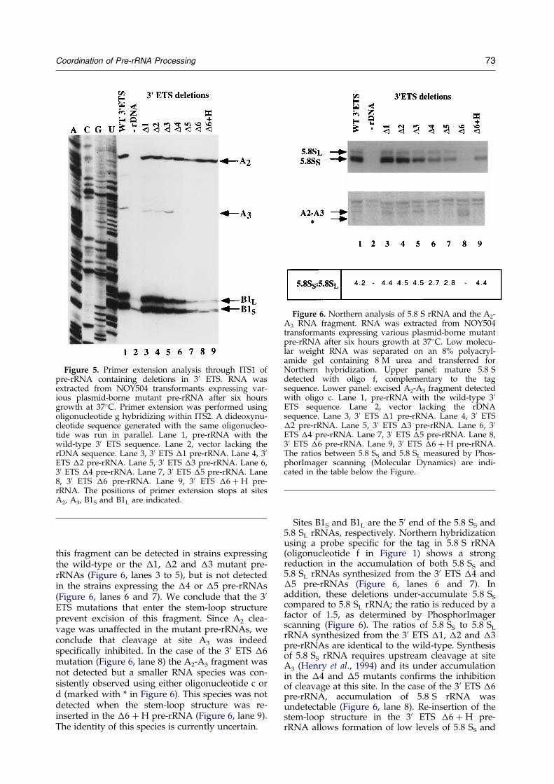
Figure 5. Primer extension analysis through ITS1 ofpre-rRNA containing deletions in 30 ETS. RNA wasextracted from NOY504 transformants expressing var-ious plasmid-borne mutant pre-rRNA after six hoursgrowth at 37�C. Primer extension was performed usingoligonucleotide g hybridizing within ITS2. A dideoxynu-cleotide sequence generated with the same oligonucleo-tide was run in parallel. Lane 1, pre-rRNA with thewild-type 30 ETS sequence. Lane 2, vector lacking therDNA sequence. Lane 3, 30 ETS �1 pre-rRNA. Lane 4, 30ETS �2 pre-rRNA. Lane 5, 30 ETS �3 pre-rRNA. Lane 6,30 ETS �4 pre-rRNA. Lane 7, 30 ETS �5 pre-rRNA. Lane8, 30 ETS �6 pre-rRNA. Lane 9, 30 ETS �6 � H pre-rRNA. The positions of primer extension stops at sitesA2, A3, B1S and B1L are indicated.
Figure 6. Northern analysis of 5.8 S rRNA and the A2-A3 RNA fragment. RNA was extracted from NOY504transformants expressing various plasmid-borne mutantpre-rRNA after six hours growth at 37�C. Low molecu-lar weight RNA was separated on an 8% polyacryl-amide gel containing 8 M urea and transferred forNorthern hybridization. Upper panel: mature 5.8 Sdetected with oligo f, complementary to the tagsequence. Lower panel: excised A2-A3 fragment detectedwith oligo c. Lane 1, pre-rRNA with the wild-type 30ETS sequence. Lane 2, vector lacking the rDNAsequence. Lane 3, 30 ETS �1 pre-rRNA. Lane 4, 30 ETS�2 pre-rRNA. Lane 5, 30 ETS �3 pre-rRNA. Lane 6, 30ETS �4 pre-rRNA. Lane 7, 30 ETS �5 pre-rRNA. Lane 8,30 ETS �6 pre-rRNA. Lane 9, 30 ETS �6 � H pre-rRNA.The ratios between 5.8 SS and 5.8 SL measured by Phos-phorImager scanning (Molecular Dynamics) are indi-cated in the table below the Figure.
Coordination of Pre-rRNA Processing 73
this fragment can be detected in strains expressingthe wild-type or the �1, �2 and �3 mutant pre-rRNAs (Figure 6, lanes 3 to 5), but is not detectedin the strains expressing the �4 or �5 pre-rRNAs(Figure 6, lanes 6 and 7). We conclude that the 30
ETS mutations that enter the stem-loop structureprevent excision of this fragment. Since A2 clea-vage was unaffected in the mutant pre-rRNAs, weconclude that cleavage at site A3 was indeedspeci®cally inhibited. In the case of the 30 ETS �6mutation (Figure 6, lane 8) the A2-A3 fragment wasnot detected but a smaller RNA species was con-sistently observed using either oligonucleotide c ord (marked with * in Figure 6). This species was notdetected when the stem-loop structure was re-inserted in the �6 � H pre-rRNA (Figure 6, lane 9).The identity of this species is currently uncertain.
Sites B1S and B1L are the 50 end of the 5.8 SS and5.8 SL rRNAs, respectively. Northern hybridizationusing a probe speci®c for the tag in 5.8 S rRNA(oligonucleotide f in Figure 1) shows a strongreduction in the accumulation of both 5.8 SS and5.8 SL rRNAs synthesized from the 30 ETS �4 and�5 pre-rRNAs (Figure 6, lanes 6 and 7). Inaddition, these deletions under-accumulate 5.8 SS
compared to 5.8 SL rRNA; the ratio is reduced by afactor of 1.5, as determined by PhosphorImagerscanning (Figure 6). The ratios of 5.8 SS to 5.8 SL
rRNA synthesized from the 30 ETS �1, �2 and �3pre-rRNAs are identical to the wild-type. Synthesisof 5.8 SS rRNA requires upstream cleavage at siteA3 (Henry et al., 1994) and its under accumulationin the �4 and �5 mutants con®rms the inhibitionof cleavage at this site. In the case of the 30 ETS �6pre-rRNA, accumulation of 5.8 S rRNA wasundetectable (Figure 6, lane 8). Re-insertion of thestem-loop structure in the 30 ETS �6 � H pre-rRNA allows formation of low levels of 5.8 SS and
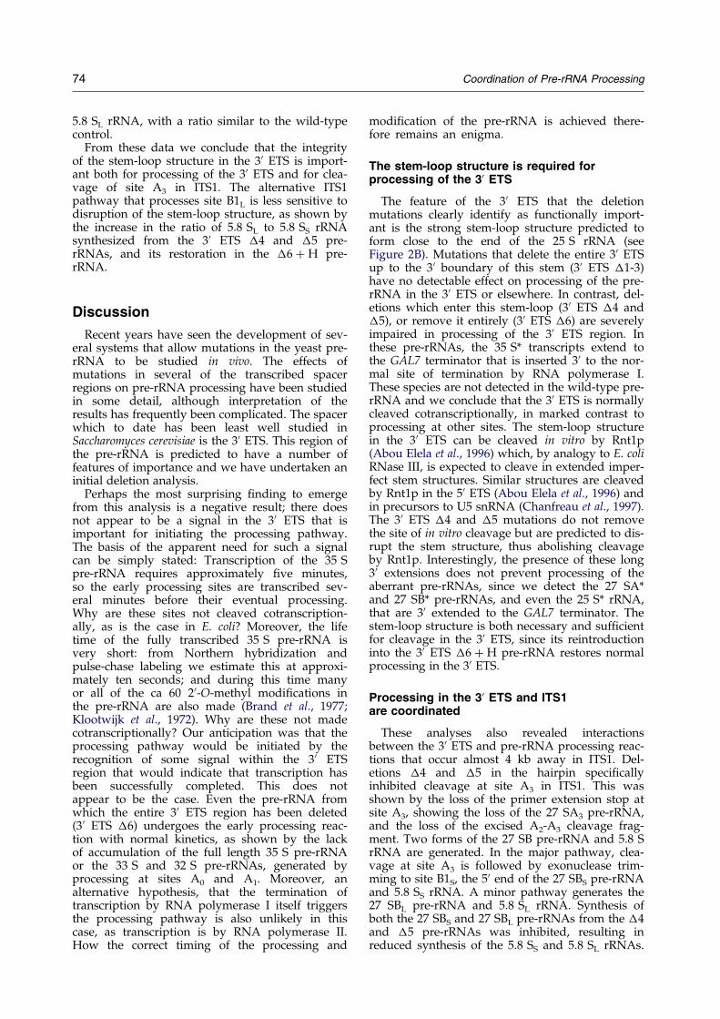
74 Coordination of Pre-rRNA Processing
5.8 SL rRNA, with a ratio similar to the wild-typecontrol.
From these data we conclude that the integrityof the stem-loop structure in the 30 ETS is import-ant both for processing of the 30 ETS and for clea-vage of site A3 in ITS1. The alternative ITS1pathway that processes site B1L is less sensitive todisruption of the stem-loop structure, as shown bythe increase in the ratio of 5.8 SL to 5.8 SS rRNAsynthesized from the 30 ETS �4 and �5 pre-rRNAs, and its restoration in the �6 � H pre-rRNA.
Discussion
Recent years have seen the development of sev-eral systems that allow mutations in the yeast pre-rRNA to be studied in vivo. The effects ofmutations in several of the transcribed spacerregions on pre-rRNA processing have been studiedin some detail, although interpretation of theresults has frequently been complicated. The spacerwhich to date has been least well studied inSaccharomyces cerevisiae is the 30 ETS. This region ofthe pre-rRNA is predicted to have a number offeatures of importance and we have undertaken aninitial deletion analysis.
Perhaps the most surprising ®nding to emergefrom this analysis is a negative result; there doesnot appear to be a signal in the 30 ETS that isimportant for initiating the processing pathway.The basis of the apparent need for such a signalcan be simply stated: Transcription of the 35 Spre-rRNA requires approximately ®ve minutes,so the early processing sites are transcribed sev-eral minutes before their eventual processing.Why are these sites not cleaved cotranscription-ally, as is the case in E. coli? Moreover, the lifetime of the fully transcribed 35 S pre-rRNA isvery short: from Northern hybridization andpulse-chase labeling we estimate this at approxi-mately ten seconds; and during this time manyor all of the ca 60 20-O-methyl modi®cations inthe pre-rRNA are also made (Brand et al., 1977;Klootwijk et al., 1972). Why are these not madecotranscriptionally? Our anticipation was that theprocessing pathway would be initiated by therecognition of some signal within the 30 ETSregion that would indicate that transcription hasbeen successfully completed. This does notappear to be the case. Even the pre-rRNA fromwhich the entire 30 ETS region has been deleted(30 ETS �6) undergoes the early processing reac-tion with normal kinetics, as shown by the lackof accumulation of the full length 35 S pre-rRNAor the 33 S and 32 S pre-rRNAs, generated byprocessing at sites A0 and A1. Moreover, analternative hypothesis, that the termination oftranscription by RNA polymerase I itself triggersthe processing pathway is also unlikely in thiscase, as transcription is by RNA polymerase II.How the correct timing of the processing and
modi®cation of the pre-rRNA is achieved there-fore remains an enigma.
The stem-loop structure is required forprocessing of the 30 ETS
The feature of the 30 ETS that the deletionmutations clearly identify as functionally import-ant is the strong stem-loop structure predicted toform close to the end of the 25 S rRNA (seeFigure 2B). Mutations that delete the entire 30 ETSup to the 30 boundary of this stem (30 ETS �1-3)have no detectable effect on processing of the pre-rRNA in the 30 ETS or elsewhere. In contrast, del-etions which enter this stem-loop (30 ETS �4 and�5), or remove it entirely (30 ETS �6) are severelyimpaired in processing of the 30 ETS region. Inthese pre-rRNAs, the 35 S* transcripts extend tothe GAL7 terminator that is inserted 30 to the nor-mal site of termination by RNA polymerase I.These species are not detected in the wild-type pre-rRNA and we conclude that the 30 ETS is normallycleaved cotranscriptionally, in marked contrast toprocessing at other sites. The stem-loop structurein the 30 ETS can be cleaved in vitro by Rnt1p(Abou Elela et al., 1996) which, by analogy to E. coliRNase III, is expected to cleave in extended imper-fect stem structures. Similar structures are cleavedby Rnt1p in the 50 ETS (Abou Elela et al., 1996) andin precursors to U5 snRNA (Chanfreau et al., 1997).The 30 ETS �4 and �5 mutations do not removethe site of in vitro cleavage but are predicted to dis-rupt the stem structure, thus abolishing cleavageby Rnt1p. Interestingly, the presence of these long30 extensions does not prevent processing of theaberrant pre-rRNAs, since we detect the 27 SA*and 27 SB* pre-rRNAs, and even the 25 S* rRNA,that are 30 extended to the GAL7 terminator. Thestem-loop structure is both necessary and suf®cientfor cleavage in the 30 ETS, since its reintroductioninto the 30 ETS �6 � H pre-rRNA restores normalprocessing in the 30 ETS.
Processing in the 30 ETS and ITS1are coordinated
These analyses also revealed interactionsbetween the 30 ETS and pre-rRNA processing reac-tions that occur almost 4 kb away in ITS1. Del-etions �4 and �5 in the hairpin speci®callyinhibited cleavage at site A3 in ITS1. This wasshown by the loss of the primer extension stop atsite A3, showing the loss of the 27 SA3 pre-rRNA,and the loss of the excised A2-A3 cleavage frag-ment. Two forms of the 27 SB pre-rRNA and 5.8 SrRNA are generated. In the major pathway, clea-vage at site A3 is followed by exonuclease trim-ming to site B1S, the 50 end of the 27 SBS pre-rRNAand 5.8 SS rRNA. A minor pathway generates the27 SBL pre-rRNA and 5.8 SL rRNA. Synthesis ofboth the 27 SBS and 27 SBL pre-rRNAs from the �4and �5 pre-rRNAs was inhibited, resulting inreduced synthesis of the 5.8 SS and 5.8 SL rRNAs.
Coordination of Pre-rRNA Processing 75
However, synthesis of 5.8 SS was more severelyinhibited than was synthesis of 5.8 SL, indicatingthat A3 cleavage by RNase MRP was more sensi-tive to mutations in the 30 ETS stem-loop than wasprocessing at B1L.
The effects of mutations in the 30 ETS are distinctfrom mutations in Rnt1p. In particular, the rnt1-1strain accumulates the 23 S RNA, a product ofcleavage at site A3, and is not clearly defective inthe processing of 27 SA2* to 27 SB* (Abou Elelaet al., 1996). These observations indicate that thedefects in processing of site A3 in the 30 ETS �4and �5 pre-rRNAs are not a direct consequence ofthe inhibition of 30 ETS processing. We concludethat the stem-loop structure is recognized by a fac-tor other than Rnt1p, and this recognition isimportant for processing in ITS1.
The obvious rationale for the existence of such asystem is to prevent synthesis of the 5.8 S rRNAand maturation of the 50 end of 25 S rRNA fromtranscripts which are incomplete due to prematuretranscription termination. A similar situation hasbeen reported in Schizosaccharomyces pombe.Mutations that delete, or interfere with the struc-ture of, a hairpin stem in the 30 ETS inhibited boththe removal of the 30 ETS region and production ofthe 5.8 S and 25 S rRNAs (Hitchen et al., 1997;Melekhovets et al., 1994), suggesting a similar inhi-bition of steps in ITS1 processing. Indeed, Goodet al. (1997a, b) have proposed that the potentialfor such quality control systems is a reason for theexistence of pre-rRNA processing in general.
A model for the coordination of pre-rRNA processing
In both Bacteria and Archaea the 16 S and 23 SrRNA sequences are ¯anked by spacer sequenceswhich form extensive helices that generate the rec-ognition sites for endonucleases; RNase III in bac-teria (King et al., 1984; Robertson & Dunn, 1975;Young & Steitz, 1978) and the bulge-helix-bulgeendonuclease in Archaea (reviewed by Dennis,1997; Garrett et al., 1991). These interactions withinthe pre-rRNAs ensure the coordination of proces-sing at the 50 and 30 ends of the rRNAs. In S. cerevi-siae, the hairpin structures in the 50 ETS and 30 ETSeach provide cleavage sites for Rnt1p (RNase III)but different mechanisms exist to ensure thecoordination of the processing reactions at theopposite ends of the 18 S and 5.8 S/25 S rRNAs.The eukaryotic 5.8 S rRNA is clearly homologousto the 50 end of the bacterial and archaeal 23 SrRNA, indicating that the ITS2 region arose by aninsertion event in an early eukaryote. The couplingreported here between processing in ITS1 and pro-cessing of the 30 ETS would therefore be function-ally analogous to the coupling between processingat the 50 and 30 ends of the bacterial and archaeal23 S provided by the requirement for base-pairingof the ¯anking sequences.
In Xenopus laevis the snoRNA U8 is required forthe synthesis of 5.8 S and 25 S rRNA; depletion of
U8 leads to a phenotype similar to deletions of the30 ETS stem-loop structure with the inhibition ofprocessing both of the 30 ETS and in ITS1 (Peculis& Steitz, 1993). No yeast homologue of U8 has yetbeen identi®ed, but we predict that a functionalhomologue will play a role in the processing ana-lysed here. It is possible that this RNP binds to thehairpin structure in the 30 ETS, promoting cleavageby Rnt1p and also interacts with the RNase MRPcomplex bound to the pre-rRNA around site A3 inITS1.
This model resembles that proposed for thecoupling of pre-rRNA processing in the 50 ETS andin ITS1. Cleavage of site A0 in the 50 ETS by Rnt1pin vivo absolutely requires the binding of the U3snoRNA to the pre-rRNA at a site some 140nucleotides 50 to the cleavage site. Moreover,mutations in U3 or other snoRNAs (U14, snR10and snR30) inhibit processing both at the 50 end ofthe 18 S rRNA and in ITS1 at site A2. This has ledto the suggestion that a multi-snoRNP complexforms and is required for these coordinated proces-sing reactions. Furthermore, processing at sites A1
and A2 is coupled to processing by RNase MRP atsite A3. This coupling may involve the large Rrp5pprotein functioning as a bridging factor (Venema &Tollervey, 1996).
This suggests a model (Figure 7) in which RNaseMRP interacts with multiple components of theprocessing machinery: an snoRNP complex andRrp5p bound to the 50 ETS and at site A2 andanother complex bound to the pre-rRNA in the 30ETS, making 35 S pre-rRNA processing a highlycoordinated process.
Materials and Methods
Strains and media
Growth and handling of S. cerevisiae used standardtechniques. The strain used was NOY504: a, rpa12::LEU2,leu2-3, 112, ura3-1, trp1-1, his3-11, can1-100 (Nogi et al.,1993; generously provided by M. Nomura).
Construction of nested deletions in the 30 ETS
The wild-type plasmid pTA1 used to generate nesteddeletions in the 30 ETS was derived from pGAL::rDNA(Henry et al., 1994). This plasmid, contains the entireyeast rDNA unit fused to the GAL7 promoter (Nogi et al.,1993) in YEp24 (2mm-URA3). In addition, small oligonu-cleotide tags have been inserted in the 18 S, 5.8 S and25 S rRNA genes. pTA1 was obtained by insertion of thepolylinker NotI, BstEII, BstXI (KpnI), SalI at the SalI sitelocated nine nucleotides downstream of the rDNA unitin the GAL7 terminator sequence. 30 ETS nested deletionsof the 30 ETS were produced by exonuclease III digestion(Erase-a-Base, Promega). The pTA1 plasmid wasdigested with KpnI and NotI and progressive uni-directional deletions were generated from the NotI site.The plasmid sequences were protected from digestion bythe 30 overhang at the KpnI restriction site. The 30 SalIsite was left intact and now directly ¯anked the rDNAsequence. The positions of the mutations are indicated inFigure 2, numbering is relative to the 30 end of 25 S
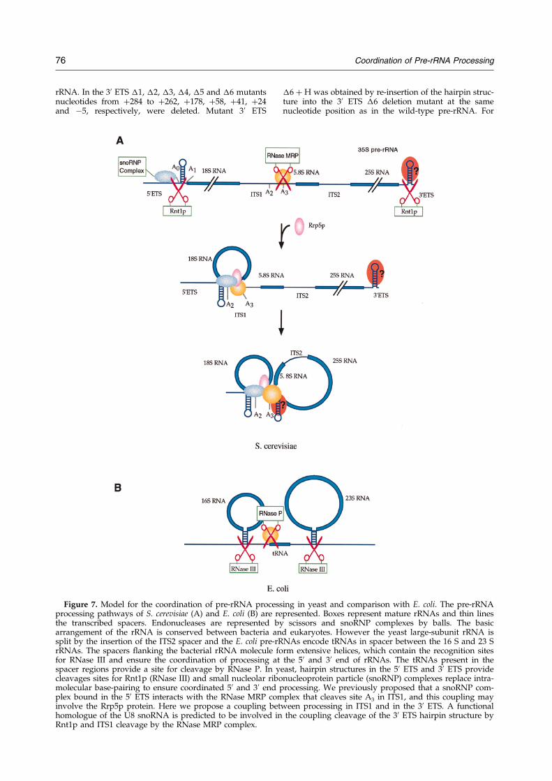
76 Coordination of Pre-rRNA Processing
rRNA. In the 30 ETS �1, �2, �3, �4, �5 and �6 mutantsnucleotides from �284 to �262, �178, �58, �41, �24and ÿ5, respectively, were deleted. Mutant 30 ETS
Figure 7. Model for the coordination of pre-rRNA procesprocessing pathways of S. cerevisiae (A) and E. coli (B) are rethe transcribed spacers. Endonucleases are represented barrangement of the rRNA is conserved between bacteria ansplit by the insertion of the ITS2 spacer and the E. coli pre-rRrRNAs. The spacers ¯anking the bacterial rRNA molecule ffor RNase III and ensure the coordination of processing atspacer regions provide a site for cleavage by RNase P. In ycleavages sites for Rnt1p (RNase III) and small nucleolar ribomolecular base-pairing to ensure coordinated 50 and 30 endplex bound in the 50 ETS interacts with the RNase MRP cominvolve the Rrp5p protein. Here we propose a coupling bethomologue of the U8 snoRNA is predicted to be involved inRnt1p and ITS1 cleavage by the RNase MRP complex.
�6 � H was obtained by re-insertion of the hairpin struc-ture into the 30 ETS �6 deletion mutant at the samenucleotide position as in the wild-type pre-rRNA. For
sing in yeast and comparison with E. coli. The pre-rRNApresented. Boxes represent mature rRNAs and thin lines
y scissors and snoRNP complexes by balls. The basicd eukaryotes. However the yeast large-subunit rRNA isNAs encode tRNAs in spacer between the 16 S and 23 S
orm extensive helices, which contain the recognition sitesthe 50 and 30 end of rRNAs. The tRNAs present in the
east, hairpin structures in the 50 ETS and 30 ETS providenucleoprotein particle (snoRNP) complexes replace intra-
processing. We previously proposed that a snoRNP com-plex that cleaves site A3 in ITS1, and this coupling may
ween processing in ITS1 and in the 30 ETS. A functionalthe coupling cleavage of the 30 ETS hairpin structure by
Coordination of Pre-rRNA Processing 77
this, the 30 ETS �6 � H plasmid was linearized at theSalI site and nucleotides 50-TCGATTTTTATTTCTTTCT-AAGTGGGTACTGGCAGGAGTCGGGGCCTAGTTTA-GAGAGAG-30 were inserted, resulting in the mutationof the last six nucleotides 3387AUUUGU3392 of 25 S rRNAto 3387GGUCGA3392 and restoration of the wild-type 30ETS sequence and hairpin structure up to nucleotides�58 (Figure 2B).
Growth rate measurement
For growth rate measurements cells were ®rst grownin minimal galactose medium at 23�C (the permissivetemperature for chromosomal rDNA transcription inrpa12 strains) to an A600 nm of 0.07. Cells were thenshifted to the non-permissive temperature (37�C) torepress chromosomal rDNA transcription and selectivelyexpress the plasmid-encoded, mutant rDNA. Regulardilution of cells with pre-warmed medium was per-formed in order to maintain exponential growth and theA600 nm was followed for 50 hours.
RNA extraction
Prior to RNA extraction for Northern analysis or pri-mer extension, NOY504 strains were transformed withpGAL::rDNA, the 30 ETS mutant plasmids or a negativecontrol plasmid (ÿrDNA) YEplac 195 (2mm-URA3; Gietz& Sugino, 1988); and grown at 23�C in minimal galactosemedium until they reached mid log phase. Cells werediluted to A600 nm 0.09 and shifted for six hours to 37�C(Henry et al., 1994). Total RNA was extracted as pre-viously described (Tollervey & Mattaj, 1987).
Northern Hybridization
For each sample 8 mg of total RNA was separated on1.2% (w/v) agarose-formaldehyde gels and transferredto Hybond N� membranes (Amersham) for Northernhybridization as described by Tollervey & Mattaj (1987).Northern hybridization was performed as previouslydescribed using the following oligonucleotides. (a) 50-CGAGGATCCAGGCTTT-30; (b) 50-GCTCTTTGCTCTTGCC-30; (c) 50-TGTTACCTCTGGGCCC-30; (d) 50-CCAGTTACGAAAATTCTTG-30; (g) 50-GGCCAG-CAATTTCAAGT-30; (h) 50-ACTCGAGAGCTTCAGTAC-30; (i) 50-AAGAATCAGATTTACAGATAATGATGT-CATT-30. Oligonucleotides a, and h hybridize to the tagsin 18 S and 25 S rRNAs, respectively. Oligo i hybridizesto the GAL7 terminator sequence, 47 nucleotides down-stream of the SalI site (see Figure 1).
Low molecular weight RNAs were separated on 8%(w/v) polyacrylamide gels containing 8 M urea in1 � TBE and electrobloted onto Hybond N� membranes(Amersham). Hybridization to the tag in 5.8 S rRNA wasperformed using an 20-O-allyl oligonucleotide (f)(Lamond & Sproat, 1993). The sequence is 50-DGDDUD-CUGGCGDdGdC-30 (Henry et al., 1994), where D is 2,6-diamino-purine, which can form three hydrogen bondsto U residues (Lamm et al., 1991).
Primer extension
Primer extension was performed as described pre-viously (Beltrame & Tollervey, 1992) on 4 mg of totalRNA using primer (d) 50-CCAGTTACGAAAATTCTTG-30. To identify the position of primer extension stops,
DNA sequencing reactions performed with the same oli-gonucleotide were run in parallel.
Acknowledgements
We thank Dr M. Nomura (university of California,Irvine) for the rpa12 strain and Phil Mitchell andEmanuelle Pascolo for critical reading of the manuscript.C.A. was the recipient of a grant from the E.U. Thiswork was partly supported by the Wellcome Trust.
References
Abou, Elela S., Igel, H. & Ares, M. J. (1996). RNase IIIcleaves eukaryotic preribosomal RNA at a U3snoRNP-dependent site. Cell, 85, 115±124.
Allmang, C., Henry, Y., Morrissey, J. P., Wood, H.,Petfalski, E. & Tollervey, D. (1996). Processing ofyeast pre-rRNA at sites A2 and A3 is linked. RNA,2, 63±73.
Amberg, D. C., Goldstein, A. L. & Cole, C. N. (1992).Isolation and characterization of RAT1: an essentialgene of Saccharomyces cerevisiae required for the ef®-cient nucleocytoplasmic traf®cking of mRNA. GenesDev. 6, 1173±1189.
Beltrame, M. & Tollervey, D. (1992). Identi®cation andfunctional analysis of two U3 binding sites on yeastpre-ribosomal RNA. EMBO J. 11, 1531±1542.
Brand, R. C., Klootwijk, J., van Steenbergen, T. J. M., deKok, A. J. & Planta, R. J. (1977). Secondary methyl-ation of yeast ribosomal precursor RNA. Eur. J. Bio-chem. 75, 311±318.
Chanfreau, G., Abou, Elela S., Ares, M., Jr & Guthrie, C.(1997). Alternative 30-end processing of U5 snRNAby RNase III. Genes Dev. 11 (27), 2741±2751.
Chu, S., Archer, R. H., Zengel, J. M. & Lindahl, L.(1994). The RNA of RNase MRP is required for nor-mal processing of ribosomal RNA. Proc. Natl Acad.Sci. USA, 91, 659±663.
Dennis, P. P. (1997). Ancient ciphers: translation inArchaea. Cell, 89 (7), 1007±10.
Garrett, R. A., Dalgaard, J., Larsen, N., Kjems, J. &Mankin, A. S. (1991). Archaeal rRNA operons.Trends Biochem. Sci. 16 (1), 22±6.
Gietz, R. D. & Sugino, A. (1988). New yeast-Escherichiacoli shuttle vectors constructed with in vitro muta-genized yeast genes lacking six-base pair restrictionsites. Gene, 74, 527±534.
Good, L., Intine, R. V. & Nazar, R. N. (1997a).Interdependence in the processing of ribosomalRNAs in Schizosaccharomyces pombe. J. Mol. Biol. 273(4), 782±8.
Good, L., Intine, R. V. & Nazar, R. N. (1997b). The ribo-somal-RNA-processing pathway in Schizosaccharo-myces pombe. Eur. J. Biochem. 247, 314±321.
Gutell, R. R. & Fox, G. E. (1988). A compilation of largesubunit RNA sequences presented in a structuralformat. Nucl. Acids Res. 16(suppl), r175±r269.
Henry, Y., Wood, H., Morrissey, J. P., Petfalski, E.,Kearsey, S. & Tollervey, D. (1994). The 50 end ofyeast 5.8 S rRNA is generated by exonucleases froman upstream cleavage site. EMBO J. 13, 2452±2463.
Hitchen, J., Ivakine, E., Melekhovets, Y. F., Lalev, A. &Nazar, R. N. (1997). Structural features in the 30external transcribed spacer affecting intragenic pro-cessing of yeast rRNA. J. Mol. Biol. 274 (4), 481±490.
Kempers-Veenstra, A. E., Oliemans, J., Offenberg, H.,Dekker, A. F., Piper, P. W., Planta, R. J. &
78 Coordination of Pre-rRNA Processing
Klootwijk, J. (1986). 30-End formation of transcriptsfrom the yeast rRNA operon. EMBO J. 5, 2703±2710.
Kenna, M., Stevens, A., McCammon, M. & Douglas,M. G. (1993). An essential yeast gene with hom-ology to the exonuclease-encoding XRN1/KEM1gene also encodes a protein with exoribonucleaseactivity. Mol. Cell. Biol. 13, 341±350.
King, T. C., Sirdeshmukh, R. & Schlessinger, D. (1984).RNase III cleavage is obligate for maturation butnot for function of Escherichia coli pre-23 S rRNA.Proc. Natl Acad. Sci. U S A, 81 (1), 185±188.
Klootwijk, J., van der Bos, R. C. & Planta, R. J. (1972).Secondary methylation of yeast ribosomal RNA.FEBS letters, 27, 102±106.
Lamm, G. M., Blencowe, B. J., Sproat, B. S., Iribarren,A. M., Ryder, U. & Lamond, A. I. (1991). Antisenseprobes containing 2±aminoadenosine allow ef®cientdepletion of U5 snRNP from HeLa splicing extracts.Nucl. Acids Res. 19, 3193±3198.
Lamond, A. I. & Sproat, B. S. (1993). Antisense oligonu-cleotides made of 20-O-alkyl RNA: their propertiesand applications in RNA biochemistry. FEBS Letters,325, 123±127.
Larimer, F. W., Hsu, C. L., Maupin, M. K. & Stevens, A.(1992). Characterization of the XRN1 gene encodinga 50 ! 30 exoribonuclease: sequence data and anal-ysis of disparate protein and mRNA levels of gene-disrupted yeast cells. Gene, 120, 51±57.
Lygerou, Z., Mitchell, P., Petfalski, E., SeÂraphin, B. &Tollervey, D. (1994). The POP1 gene encodes a pro-tein component common to the RNase MRP andRNase P ribonucleoproteins. Genes Dev. 8, 1423±1433.
Lygerou, Z., Allmang, C., Tollervey, D. & SeÂraphin, B.(1996). Accurate processing of a eukaryotic precur-sor ribosomal RNA by ribonuclease MRP in vitro.Science, 222, 268±270.
Maxwell, E. S. & Fournier, M. J. (1995). The smallnucleolar RNAs. Annu. Rev. Biochem. 35, 897±934.
Melekhovets, Y. F., Good, L., Abou, Elela S. & Nazar,R. N. (1994). Intragenic processing in yeast rRNA isdependent on the 30 external transcribed spacer.J. Mol. Biol. 239, 170±180.
Morrissey, J. P. & Tollervey, D. (1995). Birth of thesnoRNPs - the evolution of RNase MRP and theeukaryotic pre-rRNA processing system. Trends Bio-chem. Sci. 20, 78±82.
Nogi, Y., Yano, R. & Nomura, M. (1991). Synthesis oflarge rRNAs by RNA polymerase II in mutants
defective in RNA polymerase I. Proc. Natl Acad. Sci.(USA), 88, 3962±3966.
Nogi, Y., Yano, R., Dodd, J., Carles, C. & Nomura, M.(1993). Gene RRN4 in Saccharomyces cerevisiaeencodes the A12. 2 subunit of RNA polymerase Iand is essential only at high temperatures. Mol. Cell.Biol. 13, 114±122.
Peculis, B. A. & Steitz, J. A. (1993). Disruption of U8nucleolar snRNA inhibits 5. 8 S and 28 S rRNA pro-cessing in the Xenopus oocyte. Cell, 73, 1233±1245.
Piper, P. W., Bellatin, J. A. & Lockheart, A. (1983).Altered maturation of sequences at the 30 terminusof 5 S gene transcripts in Saccharomyces cerevisiae.EMBO J. 2, 353±359.
Robertson, H. D. & Dunn, J. J. (1975). Ribonucleic acidprocessing activity of Escherichia coli ribonucleaseIII. J Biol Chem, 250 (8), 3050±3056.
Schmitt, M. E. & Clayton, D. A. (1993). Nuclear RNaseMRP is required for correct processing of pre-5. 8 SrRNA in Saccharomyces cerevisiae. Mol. Cell. Biol. 13,7935±7941.
Stevens, A. & Poole, T. L. (1995). 50-exonuclease-2 of Sac-charomyces cerevisiae. Puri®cation and features ofribonuclease activity with comparison to 50-exonu-clease-1. J. Biol. Chem. 270 (27), 16063±16069.
Tollervey, D. & Kiss, T. (1997). Function and synthesisof small nucleolar RNAs. Curr. Opin. Cell Biol. 9,337±342.
Tollervey, D. & Mattaj, I. W. (1987). Fungal smallnuclear ribonucleoproteins share properties withplant and vertebrate U-snRNPs. EMBO J. 6, 469±476.
Veldman, G. M., Klootwijk, J., De Jong, P., Leer, R. J. &Planta, R. J. (1980). The transcription terminationsite of the ribosomal RNA operon in yeast. Nucl.Acids Res. 8, 5179±5192.
Venema, J. & Tollervey, D. (1995). Processing of pre-ribosomal RNA in Saccharomyces cerevisiae. Yeast, 11,1629±1650.
Venema, J. & Tollervey, D. (1996). RRP5 is required forthe formation of both 18 S and 5. 8 S rRNA inyeast. EMBO J. 15, 5701±5714.
Yip, M. T. & Holland, M. J. (1989). In vitro RNA proces-sing generates mature 30 termini of yeast 35 S and25 S ribosomal RNAs. J. Biol. Chem. 264, 4045±4051.
Young, R. A. & Steitz, J. A. (1978). Complementarysequences 1700 nucleotides apart form a ribonu-clease III cleavage site in Escherichia coli ribosomalprecursor RNA. Proc. Natl. Acad. Sci. U S A, 75 (8),3593±3597.
Edited by M. Yaniv
(Received 7 November 1997; received in revised form 2 February 1998; accepted 4 February 1998)
3708-3714 Nucleic Acids Research, 1994, Vol. 22, No. 18
Role of conserved nucleotides in building the 16S rRNAbinding site of E.coli ribosomal protein S8
Christine Allmang, Marylene Mougel, Eric Westhof, Bernard Ehresmann andChantal Ehresmann*UPR 9002 du CNRS, Institut de Biologie Moleculaire et Cellulaire, 15 rue Rene Descartes,67084 Strasbourg cedex, France
Received June 6, 1994; Revised and Accepted July 29, 1994
ABSTRACT
Ribosomal protein S8 specifically recognizes a helicaland irregular region of 16S rRNA that is highlyevolutionary constrained. Despite its restricted size, theprecise conformation of this region remains a questionof debate. Here, we used chemical probing to analyzethe structural consequences of mutations in this RNAregion. These data, combined with computer modellingand previously published data on protein binding wereused to investigate the conformation of the RNAbinding site. The experimental data confirm the modelin which adenines A595, A640 and A642 bulge out inthe deep groove. In addition to the already proposednon canonical U598 - U641 interaction, the structure isstabilized by stacking interactions (between A595 andA640) and an array of hydrogen bonds involving basesand the sugar phosphate backbone. Mutations that alterthe ability to form these interdependent interactionsresult in a local destabilization or reorganization. Thespecificity of recognition by protein S8 is provided bythe irregular and distorted backbone and the twobulged adenines 640 and 642 in the deep groove. Thethird adenine (A595) is not a direct recognition site butmust adopt a bulged position. The U598 - U641 pairshould not be directly in contact with the protein.
INTRODUCTIONThe interaction of E.coli ribosomal protein S8 with its 16S rRNAbinding site represents an interesting model for studying themolecular mechanism of specific RNA -protein recognition.Protein S8 is capable of binding individually to the central domainof 16S rRNA and plays an important role in the early stage ofribosomal 30S subunit assembly (1-2). It participates to theformation of one early nucleation site (3), and interacts co-operatively with other ribosomal proteins (4-5). It is thereforea crucial element for the sequential assembly of RNA and proteinsconstituting the small ribosomal subunit. It is also able to regulatethe translation of its own operon (6-8) by a feed-backmechanism.
A considerable amount of work was already devoted to theinteractions between S8 and its 16S rRNA target site and to thefine structure of this site (4-5, 9-14). It was recently shownthat the rRNA can be restricted to a short helical stem (nucleotides588 -605/633 -651), without significantly altering the apparentaffinity constant (15). The central part of this helical region (called'region C') is highly evolutionary constrained and the conservedelements are also found in the target regulatory site of S8 on itsmRNA (8,16). We previously proposed a three-dimensionalmodel of region C, derived from structure probing and computermodeling (14). This model displays characteristic features: A595,A640 and A642 bulge out in the deep groove of the helix, andU598 and U641 form a non-canonical base pair. However, theconformation of this region is disputed and three other foldingmodels have been proposed in the literature. These modelsessentially differ in the pairing ofU598 which is either with A640(17- 18), U641 (14) or A642 (5). We favoured a U595-U641base pair (14), since it accounts for the non reactivity of U598and U641 and for the reactivity of A640, A642 and A595. Thepair U598-A640 was recently proposed on the basis of sequencecomparison (17- 18). In order to agree with the reactivity data,such a U598 -A640 pair should involve Hoogsteen hydrogenbonding and not Watson-Crick interactions. In addition, the nonreactivity of the unpaired U641 could only be explained byadditional tertiary interaction or stacking.
Recently, we investigated the role of conserved nucleotides inregion C as potential determinants for S8 recognition by studyingthe effect of 14 single and double mutations on S8 recognition(15). Of the 14 mutants tested, only three are still efficientlyrecognized by S8. In order to discriminate whether the loss ofrecognition is due to the loss of a specific contact or toconformational rearrangement, we now report the structuralconsequences of the mutations, using chemical probing on the14 RNA variants mentioned above and of two new RNA mutants(A598/U640 and A598/U640/G64 1). In addition, footprintingexperiments were conducted on those mutants that still retain S8binding capacity. Our results emphasize the subtleties of RNAconformation and an unexpected versatility in the structuralconsequences of single base mutations. An improved three-
*To whom correspondence should be addressed
k.) 1994 Oxford University Press

Nucleic Acids Research, 1994, Vol. 22, No. 18 3709
dimensional model is derived from the present experimental dataand the results are discussed in terms of RNA folding and S8recognition.
MATERIALS AND METHODSPreparation of the biological materialPlasmids construction, RNA synthesis and purification of wild-type and mutant 16S rRNA fragments (nucleotides 584-756)are described in (15). Two additional mutants were constructed(A598/U640 and A598/U640/G641) following the same protocol.Their relative binding affinity was determined as in (15).Ribosomal protein S8 was prepared under non-denaturingconditions according to Cachia et al. (19).
Chemical probing and footprintingA standard assay contains 16 pmol RNA and 2 jg carrier tRNAin 20 IL of appropriate buffer. RNA was first pre-incubated for15 min at 40°C in buffer Ni [50 mM sodium cacodylate (pH7.5), 20 mM magnesium acetate, 250 mM potassium acetate]or N2 [50 mM sodium borate (pH 8.0); 20 mM magnesiumacetate, 250 mM potassium acetate]. For each reaction, a controlwas treated in parallel, omitting the reagent. Modification withDMS: incubation was for 5 and 10 min in buffer Ni or for 2and 5 min in buffer Dl [50 mM sodium cacodylate (pH 7.5),1 mM EDTA] for semi-denaturing conditions. Modifications withCMCT: incubation was for 15 and 30 min in buffer N2 or for2 and 5 min in buffer D2 [50 mM sodium borate (pH 8.0), 1mM EDTA] for semi-denaturing conditions. Modifications withDEPC: incubation was for 15, 30 and 60 min in buffer NI orfor 15 and 30 min in buffer DI (semi-denaturing conditions).All modifications were at 37°C. Footprinting experiments usingCMCT and DMS were conducted on wild-type RNA and mutantsallowing S8 binding. Complexes were formed in the presenceof 0.4 ,M S8 for wild-type RNA, mutants U595 and A641, or2 ,tM for mutant A598 -U640. Footprinting gels were scannedusing the Bio-Imager Analyzer BAS 2000 (Fuji). Synthesis ofprimer, labeling, hybridization, reverse transcription and analysisof generated cDNA fragments were described by Mougel et al.(14).
Computer modelingThe modeled molecule integrating stereochemical constraints andexperimental data was constructed with the help of severalcomputer programs and tested by comparing the theoreticalaccessibility of atoms with the observed experimental reactivity,as described earlier (20).
RESULTSBinding strength of the new mutantsPrevious results showed that both mutants A598 and U642 failto recognize S8 (15). Here, we tested the possibility to restoreS8 binding by the double mutation A598/U642. The results (notshown) show that this double mutation restores only partially S8recognition (with a 5-fold reduced binding strength). Sequencecomparison indicates that U598 is highly conserved. However,in Rcy purpur, nucleotide 598 is an adenine, and nucleotides 640and 641 are simultaneously replaced by U and G, respectively.Therefore, we constructed a new mutant containing these threemutations (A598/U640/G641). This triple mutant is notrecognized by S8 (results not shown).
Conformational studies of the RNA variantsThe four bases were tested for their chemical reactivity at oneof their Watson-Crick positions with DMS, at A(Nl) andC(N3), and with CMCT, at G(N1) and U(N3). For somemutants, position N7 of adenines was also probed with DEPC.In addition, footprinting experiments were conducted using DMSand CMCT with those RNAs that still retain S8 binding ability.A typical experiment is shown in Fig. 1. Experiments wererepeated several times (from 2 to 4 times) and the degree ofreactivity was evaluated from 1 to 4 by visual inspection. In thecase of footprinting experiments, reactivity changes induced byS8 binding were quantified.The reactivity changes induced by the mutations are exclusively
localized in region C (nucleotides 594-599/639-645). Resultsare summarized in Table 1 and in Figs 2-4 which show thededuced secondary foldings of region C. One strikingconsequence of all the mutations tested is that U641, which isnot reactive in the wild-type RNA, becomes reactive at variousdegrees in all mutated RNAs, with the single exception of mutantG643 (Table 1). By contrast, U598 remains unreactive in allmutants, suggesting that its N3 position is involved in H-bondingor that the residue is stacked inside the helix, preventingmodification.
DISCUSSIONMutations affecting adenines 595, 640 and 642The deletion of any of these three adenines results in a completeloss of binding (15). The deletion of either A640 or A642 inducesreactivity at U641 and decreases the reactivity of A642(N1) orA640(N 1), respectively (Table 1). These results suggest thatnucleotide U641 is bulging out in these two mutants and that U598pairs with either A642 or A640, respectively (Fig. 2). Moreover,
DEPC DMS CMCT
0 1 2 3a12012 A2012Al2ACGU
_ ~~~~~~~~~~~~~~~~~. _ . .... ... .I!
DMS CMCT
0 1 2 Al 2 0 1 2 A1A2 A C
....wA595 -U598 -
U619
U632- ....a
U619- _w-rw
4 m
,.F
A641 -
*,o
L:-I. :-J
A640\U641 -
A642'
A641 C597/G643
Figure 1. Probing on RNAs A641 and C597/G643. Modification with DMS andCMCT: Incubation control (lane 0). Native conditions: incubation with DMSor CMCT was for 5 min Oane 1) and 10 min (lane 2); semi-denaturing conditions:incubation with CMCT was for 5 min (lane Al) and 10 min Oane A2). Modificationwith DEPC: incubation control (lane 0); native conditions: incubation for 15 min(lane 1) and 30 min (lane 2); semi-denaturing conditions: incubation for 5 min(lane A1) and 10 min (lane A2).
A595-
:4,
.N,-
f
.: .;sf:
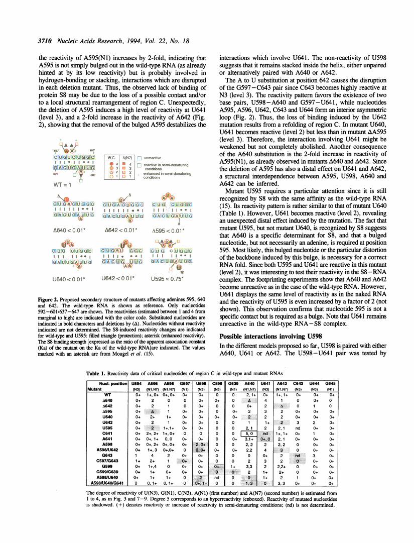
3710 Nucleic Acids Research, 1994, Vol. 22, No. 18
the reactivity of A595(N1) increases by 2-fold, indicating thatA595 is not simply bulged out in the wild-type RNA (as alreadyhinted at by its low reactivity) but is probably involved inhydrogen-bonding or stacking, interactions which are disruptedin each deletion mutant. Thus, the observed lack of binding ofprotein S8 may be due to the loss of a possible contact and/orto a local structural rearrangement of region C. Unexpectedly,the deletion of A595 induces a high level of reactivity at U641(level 3), and a 2-fold increase in the reactivity of A642 (Fig.2), showing that the removal of the bulged A595 destabilizes the
A A,
6_-\> AX 547
CUGUC UGGC
I.IGA C UG A U UG
592
WT= 1
N
C U G A C U G G C
I I
G A C U G A.U U G
A
A640< 0.01'
CUG CUGGC
GA C U GA:U U GA
W. c. A(N7)~jl nreactive
4 4 reactive in sernl-denaturing3 3 conditions
2 enhanced in semi-deiaturieq1 conditions
Af,,,,f A A
CUGACUGGC CU G C U G G C
I I * * * *GACUGA UUG G A CUG A U U G
Ah
A642 <0.01* A595 c 0.01*
AfuCUG AU GGC
* * a
GAC UG U UGA.:A
\ts A "-
C iG C U G GC
GA C UG A U UG
f
U640<0.01* U642<0.01* U595=0.75*
Figure 2. Proposed secondary structure of mutants affecting adenines 595, 640and 642. The wild-type RNA is shown as reference. Only nucleotides592-601/637-647 are shown. The reactivities (estimated between 1 and 4 frommarginal to high) are indicated with the color code. Substituted nucleotides areindicated in bold characters and deletions by (A). Nucleotides without reactivityindicated are not determined. The S8-induced reactivity changes are indicatedfor wild-type and U595: filled triangle (protection); asterisk (enhanced reactivity).The S8 binding strength [expressed as the ratio of the apparent association constant(Ka) of the mutant on the Ka of the wild-type RNA]are indicated. The valuesmarked with an asterisk are from Mougel et al. (15).
interactions which involve U641. The non-reactivity of U598suggests that it remains stacked inside the helix, either unpairedor alternatively paired with A640 or A642.The A to U substitution at position 642 causes the disruption
of the G597 -C643 pair since C643 becomes highly reactive atN3 (level 3). The reactivity pattern favors the existence of twobase pairs, U598 -A640 and G597 -U641, while nucleotidesA595, A596, U642, C643 and U644 form an interior asymmetricloop (Fig. 2). Thus, the loss of binding induced by the U642mutation results from a refolding of region C. In mutant U640,U641 becomes reactive (level 2) but less than in mutant AA595(level 3). Therefore, the interaction involving U641 might beweakened but not completely abolished. Another consequenceof the A640 substitution is the 2-fold increase in reactivity ofA595(N1), as already observed in mutants A640 and A642. Sincethe deletion of A595 has also a distal effect on U641 and A642,a structural interdependence between A595, U598, A640 andA642 can be inferred.Mutant U595 requires a particular attention since it is still
recognized by S8 with the same affinity as the wild-type RNA(15). Its reactivity pattern is rather similar to that of mutant U640(Table 1). However, U641 becomes reactive (level 2), revealingan unexpected distal effect induced by the mutation. The fact thatmutant U595, but not mutant U640, is recognized by S8 suggeststhat A640 is a specific determinant for S8, and that a bulgednucleotide, but not necessarily an adenine, is required at position595. Most likely, this bulged nucleotide or the particular distortionof the backbone induced by this bulge, is necessary for a correctRNA fold. Since both U595 and U641 are reactive in this mutant(level 2), it was interesting to test their reactivity in the S8-RNAcomplex. The footprinting experiments show that A640 and A642become unreactive as in the case of the wild-type RNA. However,U641 displays the same level of reactivity as in the naked RNAand the reactivity of U595 is even increased by a factor of 2 (notshown). This observation confirms that nucleotide 595 is not a
specific contact but is required as a bulge. Note that U641 remainsunreactive in the wild-type RNA-S8 complex.
Possible interactions involving U598In the different models proposed so far, U598 is paired with eitherA640, U641 or A642. The U598-U641 pair was tested by
Table 1. Reactivity data of critical nucleotides of region C in wild-type and mutant RNAs
Nucl. position U594 A595 A596 G597 U598 C599 G639 A640 U641 A642 C643 U644 G645Mutant (N3) (Ni,N7) (Nl,N7) (N1) (N3) (N3) (N1) (N1,N7) (N3) (N1,N7) (N3) (N3) (N1)
WIT 0+ 1+, 0+ 0+,0+ 0+ 0+ 0 0 2,1+ 0+ 1+, 1+ 0+ 0+ 0+A640 0+ 2 0 0 0+ 0+ 0 A 4 1 0 0+ 0A642 0+ 2 1 0 0+ 0 0 0+ 2 .: 0 1 0A595 0+ A 1 0+ 0+ 0 0+ 2 3 2 0+ 0+ 0+U640 0+ 2+ 1+ 0+ 0+ 0+ 0+ 2 2 2 0+ 0+ 0+U642 0+ 2 1 0+ 0+ 0 0 1 1+ 2 3 2 0+U595 0+ 2 1+1+ 0+ 0+ 0 0 2,1 2 2,1 nd 0+ 0+C641 0+ 2+,2+ 1+,0+ 0 0 0 0 5,0. .d 1+1+ 0+ 1 0+A641 0+ 0+,1+ 0,0 0+ 0+ 0 0+ 3,1+ .+,0 2,1 0+ 0+ 0+A598 0+ 0+,2+ 0+,0+ 0+ 2,0+ 0 0 2,2 2 2,2 0 0+ 0+
A5981U642 0+ 1+,3 0+,0+ 0 2,0 0+ 0+ 2,2 4 0 0+ 0+G643 1 4 2 0+ 0+ 0 0 0 0+ 2 i 3 0+
C597/G643 1+ 2+ 1 0+ 0+ 0 0 2 3 2 0+ 0+G5990+ 1+,4 0 0+ 0~~~~~ ~~~~~~+.:::0. 1..+ 3,3 2 2,2+ 0 0+ 0+
G599/C639 0+ 1+ 0+ 0+ 0+ . 0 2 1+ 2+ 0 0+ 0+A598/U640 0+ 1+ 1+ 0 2 nd 0 0 1+ 2 1 0+ 0+
A598AU6401G641 0 0, 1+ 0, 1+ 0 0+,1+ 0 0 1,3 0 3,3 0+ 0+ 0+
The degree of reactivity of U(N3), G(Nl), C(N3), A(Nl) (first number) and A(N7) (second number) is estimated from1 to 4, as in Fig. 3 and 7-9. Degree 5 corresponds to an hyperreactivity (enboxed). Reactivity of mutated nucleotidesis shadowed. (+) denotes reactivity or increase of reactivity in semi-denaturing conditions; (nd) is not determined.
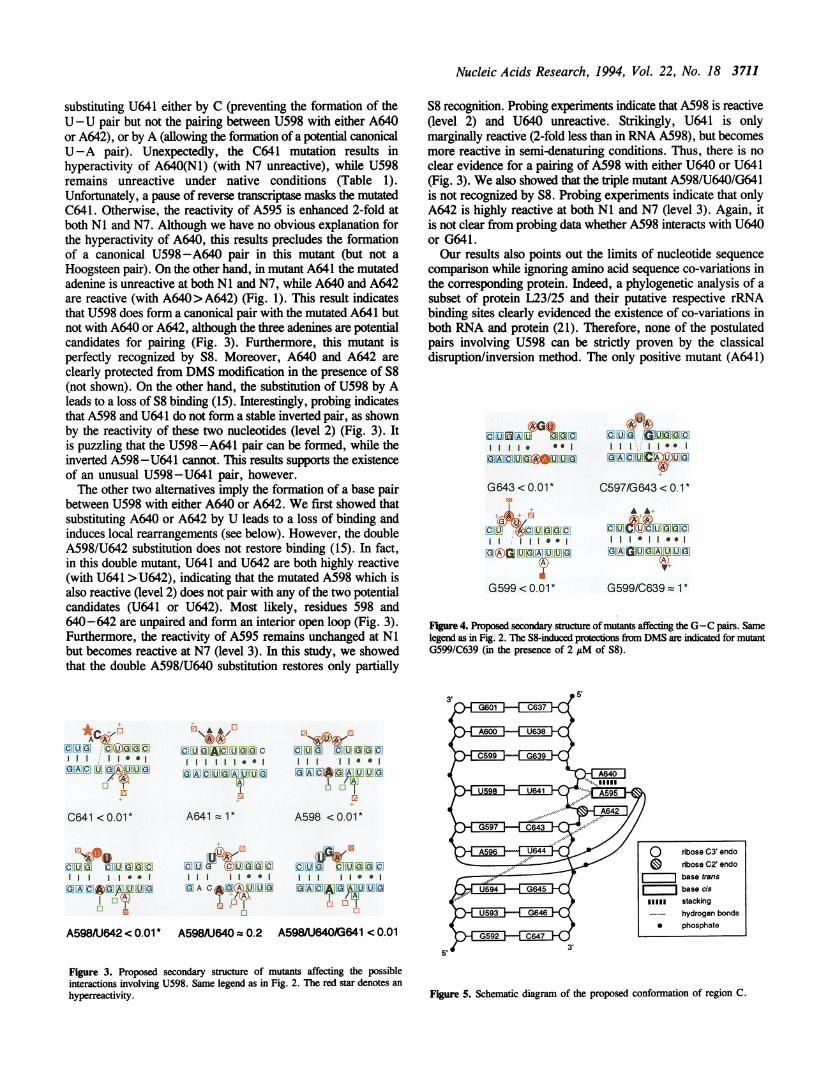
Nucleic Acids Research, 1994, Vol. 22, No. 18 3711
substituting U641 either by C (preventing the formation of theU-U pair but not the pairing between U598 with either A640or A642), or by A (allowing the formation of a potential canonicalU-A pair). Unexpectedly, the C641 mutation results inhyperactivity of A640(Nl) (with N7 unreactive), while U598remains unreactive under native conditions (Table 1).Unfortunately, a pause of reverse trianscptase masks the mutatedC641. Otherwise, the reactivity of A595 is enhanced 2-fold atboth Nl and N7. Although we have no obvious explanation forthe hyperactivity of A640, this results precludes the formationof a canonical U598-A640 pair in this mutant (but not aHoogsteen pair). On the other hand, in mutant A641 the mutatedadenine is unreactive at both NI and N7, while A640 and A642are reactive (with A640>A642) (Fig. 1). This result indicatesthat U598 does form a canonical pair with the mutated A641 butnot with A640 or A642, although the three adenines are potentialcandidates for pairing (Fig. 3). Furthermore, this mutant isperfectly recognized by S8. Moreover, A640 and A642 areclearly protected from DMS modification in the presence of S8(not shown). On the other hand, the substitution of U598 by Aleads to a loss of S8 binding (15). Interestingly, probing indicatesthat A598 and U641 do not form a stable inverted pair, as shownby the reactivity of these two nucleotides (level 2) (Fig. 3). Itis puzzling that the U598-A641 pair can be formed, while theinverted A598 -U641 cannot. This results supports the existenceof an unusual U598-U641 pair, however.The other two alternatives imply the formation of a base pair
between U598 with either A640 or A642. We first showed thatsubstituting A640 or A642 by U leads to a loss of binding andinduces local rearrangements (see below). However, the doubleA598/U642 substitution does not restore binding (15). In fact,in this double mutant, U641 and U642 are both highly reactive(with U641 > U642), indicating that the mutated A598 which isalso reactive (level 2) does not pair with any of the two potentialcandidates (U641 or U642). Most likely, residues 598 and640-642 are unpaired and form an interior open loop (Fig. 3).Furthermore, the reactivity of A595 remains unchanged at Nlbut becomes reactive at N7 (level 3). In this study, we showedthat the double A598/U640 substitution restores only partially
S8 recognition. Probing experiments indicate that A598 is reactive(level 2) and U640 unreactive. Strikingly, U641 is onlymarginally reactive (2-fold less than in RNA A598), but becomesmore reactive in semi-denaturing conditions. Thus, there is noclear evidence for a pairing of A598 with either U640 or U641(Fig. 3). We also showed that the triple mutant A598/U640/G641is not recognized by S8. Probing experiments indicate that onlyA642 is highly reactive at both Ni and N7 (level 3). Again, itis not clear from probing data whether A598 interacts with U640or G641.Our results also points out the limits of nucleotide sequence
comparison while ignoring amino acid sequence co-variations inthe corresponding protein. Indeed, a phylogenetic analysis of asubset of protein L23/25 and their putative respective rRNAbinding sites clearly evidenced the existence of co-variations inboth RNA and protein (21). Therefore, none of the postulatedpairs involving U598 can be strictly proven by the classicaldisruption/inversion method. The only positive mutant (A641)
CUA)U GOCC. U El A a
O A C UiG:U 3J 0
G643 < 0.01*
CU~CUGGcII I i ..
G GQ U G A U U G
G599 < 0.01*
C U G- G -UOCG
0G A C U C@:u
C597/G643 < 0.1*
C U ('tCl G. C
l * l *@l
G A A U 'U G
G599/C639 1 *
Figure 4. Proposed secondary structure of mutants affecting the G-C pairs. Samelegend as in Fig. 2. The S8-induced protections from DMS are indicated for mutantG599/C639 (in the presence of 2 j&M of S8).
*.C r
ACt(<CUG CUGOGcIIII I1* 1G A C U G®U U G
C641 <0.01*
0CUG CUGGCI III Iii..G A C &G A,U U G
0.01
A598/UJ642 < 0.01 *
.-I\A A,-
C UGAc UGGCI I I I I I 0 0
G ACUGA U UGA
A641 = 1*
C U G_ U G G C
I I **G A C &GUUG
& Dsl&
C U G C U G G CI I I I * * I0 A C$*0 A U U G
/A
A598 < 0.01 *
CUG CUGGCI I II ** IOACAGA UUG
1
A598AJ640 = 0.2 A598/U640/G641 < 0.01
Figure 3. Proposed secondary structure of mutants affecting the possibleinteractions involving U598. Same legend as in Fig. 2. The red star denotes an
hyperreactivity. Figure 5. Schematic diagram of the proposed conformation of region C.
o ribose C3 endo
drbose C2' endo
I______ base trans
r i,base cismlill stacking...... hydrogen bonds* phosphate
5'
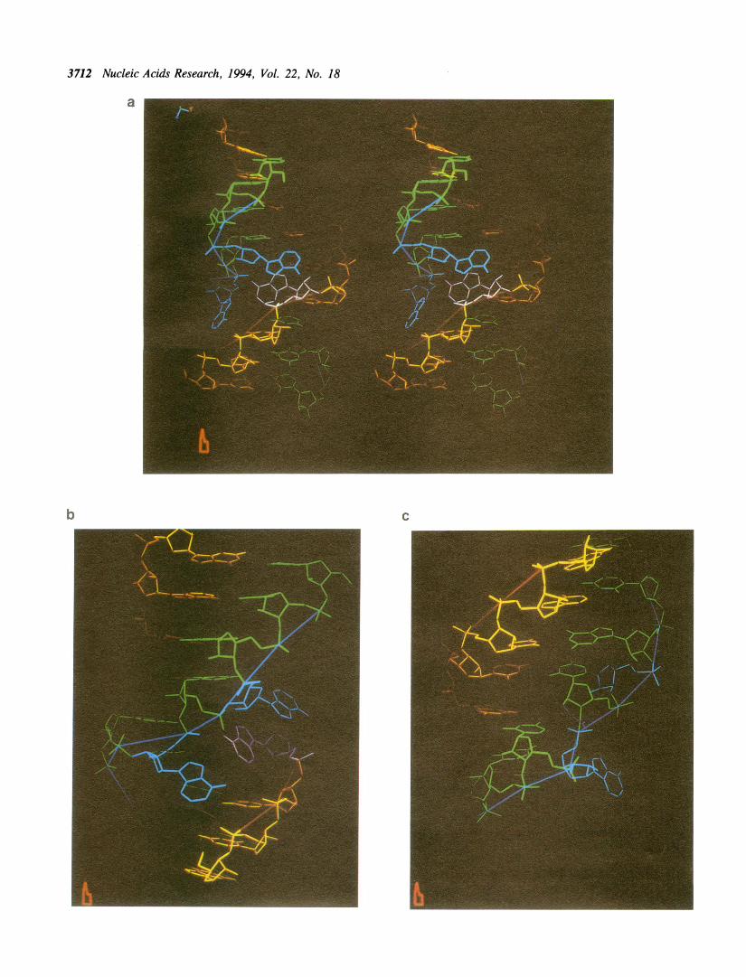
3712 Nucleic Acids Research, 1994, Vol. 22, No. 18
a
b C
Nucleic Acids Research, 1994, Vol. 22, No. 18 3713
favors the existence of the U598 -U641 pair, since the formationof a U598 -A641 pair was clearly evidenced. One should notethat U641 is frequently substituted by A in 16S-like rRNAs.
Mutations affecting the G-C pairsFrom our model, the G599 and G643 substitutions, which bothlead to a complete loss of S8 binding (15), are expected to disruptthe two G- C pairs surrounding the U-U pair and most likelyto have a destabilizing effect. Indeed, reactivity data indicate thatthese mutations induce conformational rearrangements. As aresult of the G643 mutation, A640(N1) becomes unreactive whilethe reactivity of A595(N1) is enhanced by a factor of 4 (Table1). In addition, U644 and A596(N1) become reactive (level 3and 2, respectively). These data suggest the formation of pairsU598-A640 and G597-U641 (as in mutant U642).Unexpectedly, the potential A595 -U644 pair is not formed andnucleotides 595, 596 and 642-644 form a five-base internal loop(Fig. 4). The G599 mutation induces a 2-fold increase ofreactivity at A640 (both NI and N7 positions). It also inducesnew reactivity at U641 (level 2) and at A595(N7) (level 4). Mostlikely, helix III is extended by the two meta-stable U598 -A642and G599-U641 pairs (Fig. 4).The double mutation G599-C639 restores S8 binding (15) and
gives a reactivity pattern similar to that of the wild-type RNA(Table 1). The only difference with the wild-type RNA is amarginal reactivity of U641 (level 1) in native conditions, anda 2-fold increase of the reactivity of A640(N1). Therefore, theC599-G639 base pair can be inverted without significantfunctional and structural effect (Fig. 4). On the contrary, thedouble mutation C597/G643 does not restore binding (15).Probing experiments show that the mutations cause a strongreactivity of U641 (level 3) and a 2-fold increased reactivity ofA595 and A640 at NI (Fig. 1). The high reactivity at U641 couldbe explained by the possible loss of interactions with U598 asa consequence of the mutations or to the alteration of a networkof interactions involving other nucleotides like A595. Thus, aninverted C597/G643 pair is formed but it is not structurallyequivalent to the wild-type one. However, specific contactsbetween S8 and this G-C pair cannot be excluded. Interestingly,the G597-C643 pair is strictly conserved. Note that a C to Utransition and a single deletion at position 643, both produce over50-fold reduction in S8 affinity and confer slow growth in E. colicells in vivo (16).
A possible three-dimensional modelThe present results show that the fold of region C is functionallyand structurally highly constrained. The effect of mutations couldnot be predicted by a simple secondary structure model. Themutations can be classified in 3 classes: (i) mutations that displaya wild-type like folding and affinity for protein S8 (A641 andG599/C639); (ii) mutations that induce a substantial refolding(A640, A642, U642, C641, G643 and G599) and are notrecognized by protein S8; (iii) mutations that induce a localopening of region C (A595, U595, U640, A598, A598/U642,A598/U640, A598/U640/G641 and C597/G643) with variableeffect on S8 binding. These latter mutations seem to beresponsible for the disruption of a network of interactions in
region C resulting in a destabilization of the postulatedU598-U641 pair. Furthermore, there is a clear structuralinterdependence between nucleotides A595, U598, A640, A642and G597 and/or C643. The new model we propose does notbasically differ from the previous one, as far as the base-pairingscheme is concerned, however the conformation of the sugar-phosphate backbone is more irregular and tertiary interactionsaccount for the present observations (Figs 5-6).
In this model, the U598(N3, 04) -U641(02, N3) alreadyproposed in the previous model (14) has been maintained. Thethree bulged adenines are still bulging out on the same side ofthe helix, facing the major groove, but their orientation has beenmodified. Both A595 and A640 adopt a C2' endo sugar pucker.Adenine 595, which is in a syn conformation, is stacked on A640and both residues can be involved in an array of hydrogen bonds(Figs 5-6). Thus, hydrogen bonding between A595(N6) and thephosphate groups of both U641 and A642, between A595(N7)and the 2'OH of A640, as well as between A642(N6) and thephosphate group of U594 can occur. There is a very goodcorrelation between the reactivity of A640 and A595 at both NIand N7 and their accessibility in the model. Moreover, thepostulated hydrogen bonds involving A595 and the ribose-phosphate backbone most likely stabilize its particularconformation. Thus, according to the model, deleting orsubstituting A595 results in the loss of these interactions and tothe destabilization of the U598-U641 pair. The free hydrogenof C643(N4) can also form a bond with the phosphate group ofA642. This should account for the observed increased reactivityof both A642 and U641 when inverting the G597-C643 pair.This model offers a rather satisfying solution for the observedinterdependence between the three bulged adenines, the U-Upair and C643. Other hydrogen bonding possibilities cannot beexcluded. Overall, the postulated structure is characterized by:(i) the known tendency of R-Y-R sequences for conformationsin which the two purine residues stack on a side opposite to thatof the pyrimidine (22); (ii) the added stabilization brought aboutby the third adenine 'intercalating' between the two bulgedadenines.
What is recognized by protein S8?One characteristic feature of the model is the irregularity of thesugar-phosphate backbone (with one kink on the 5' strand andtwo kinks on 3' strand). The reason why the U598 -U641 paircan be replaced by U598-A641 but not by A598-U641 isprobably correlated with this particular geometry. Anotherconsequence of the proposed conformation is the widening ofthe deep groove, allowing to position the three bulged adenines.Protein S8 may sit in the distorted deep groove of the RNA andprobably recognizes the irregular backbone conformation. Themodel also fits with the idea that A640(N 1), which is accessiblein the naked RNA and protected in the bound form, is a specificcontact. The invariant A642 is also a good candidate for specificinteraction, in particular positions N6 and Ni which are bothaccessible in the model. It should be reminded that S8 bindingis strongly affected by protonation of (a) residue(s) with a pKaround 5-6 (13) and that an adenine was considered to be thebest candidate. On the opposite, A595 which is buried and poorly
Figure 6. Proposed three-dimensional model of region C. (a) Stereoscopic view down the deep groove, with strand 637-647 in green, strand 592-601 in yellow,A595 in pink, A640 and A642 in blue. (b) Detailed view showing the coaxial stacking between A640 and A595 and possible hydrogen bonds (A595(N6)-OP641and -OP642; A595(N7) -A640(2'OH); A642(N6) -OP594). (c) Detailed view after a rotation of 180° about the vertical axis, showing the U598-U641 and thepossible hydrogen bond between C643(N4) and OP642.

3714 Nucleic Acids Research, 1994, Vol. 22, No. 18
accessible does not appear to be involved in direct interaction.The evidence that A595 is not a recognition site is provided bymutant U595. In this case, the reactivity of U595(N3) is evenincreased in the presence of S8, suggesting that it is tilted outsidethe helix. The fact that U641(N3) remains reactive in this mutantin the presence of S8 also indicates that U641 is probably notdirectly recognized. This can be explained by the particularlocation of the U598-U641 pair: in the proposed model, itsaccess from the distorted deep groove is partially shielded bythe bulged adenines.The S8 binding site constitutes a typical example of RNA
structural complexity used as a source of protein specificrecognition. Our results highlight subtleties in the RNAconformation which cannot be explained by a simple secondarystructure. In addition, they clearly show that the classicaldisruption/replacement method used to prove standardWatson-Crick base-pairing is inadequate for identifying noncanonical interactions.
21. Metzenberg, S., Joblet, C., Verspieren, P. and Agabian, N. (1993) NucleicAcids Res. 21, 4936-4940.
22. van de Hoogen, Y.T., Treumiet, S.J., Roelen, H.C.P.F., de Vroom, E.,van der Marel, G.A., van Boom, J.H. and Altona, C. (1988) Eur J. Biochem.171, 155-162.
ACKNOWLEDGEMENTSWe are indebted to F.Eyermann for skilful technical assistance,to C.Cachia for providing purified ribosomal protein S8, toJ.Reinbolt for analysis of S8 and to B.Masquida for sequencealignments. P.Romby is thanked for critical reading of themanuscript.
REFERENCES1. Nomura, M. and Held, W.A. (1990) in 'Ribosomes' (Nomura, M., Tissieres,
A. and Lengyel. P. eds) Cold Spring Harbor Laboratory Press, Cold SpringHarbor, NY., pp 1933-223.
2. Held, W.A., Ballou, B., Mizushima, S. and Nomura, M. (1974) J. Biol.Chem. 249, 3103-3111.
3. Moore, P.B. (1987) Cold spring Harbor Symp Quant Biol. 52, 721-728.4. Gregory, R.J., Zeller, M.L., Thurlow, D.L., Gourse, R.L., Stark, M.J.R.,
Dahlberg, A.E. and Zimmermann, R.A. (1984) J. Mol. Biol. 178, 287-302.5. Svensson, P., Changchien, L.M., Craven, G.R. and Noller, H.F. (1988)
J. Mol. Biol. 200, 301-308.6. Yates, J.L. Arfsten, A.E. and Nomura, M. (1980) Proc. Natl. Acad. Sci.
USA. 77, 1837-1841.7. Dean, D., Yates, J.L. and Nomura, M. (1981) Nature 289, 89-91.8. Cerretti, D.P., Mattheakis, L.C., Kearney, K.R., Vu, L.V. and Nomura,
M. (1988) J. Mol. Biol. 204, 309-329.9. Ungewickell, E., Garrett, R.A., Ehresmann, C., Stiegler, P. and Fellner,
P. (1975) Eur. J. Biochem. 51, 165-180.10. Zimmermann, R.A., Mackie, G.A., Muto, A., Garrett, R.A., Ungewickell,
E., Ehresmann, C., Stiegler, P., Ebel, J.P. and Fellner, P. (1975) NucleicAcids Res. 2, 279-302.
11. Zimmermiann, R.A. and Singh-Bergmann, K. (1979) Biochim. Biophys. Acta563. 422-431.
12. Wower, I. and Brimacombe, R. (1983) Nucleic Acids Res. 11, 1419-1437.13. Mougel, M., Ehresmann, B. and Ehresmann, C. (1986) Biochemistry 25,
2756-2765.14. Mougel, M., Eyermann, F., Westhof, E., Romby, P., Expert-Bezancon,
A., Ebel, J.P., Ehresmann, B. and Ehresmann, C. (1987) J. Mol. Biol. 198,91-107.
15. Mougel, M., Allmang, C., Eyerman, F., Cachia, C., Ehresmann, B. andEhresmann, C. (1993) Eur. J. Biochem. 215, 787-792.
16. Gregory, R.J., Cahill, P.B.F., Thurlow, D.L. and Zimmermann, R.A. (1988)J. Mol. Biol. 204, 295 -307.
17. Gutell, R. R. (1993) Nucleic Acids Res. 21, 3051-3054.18. Neefs, J.M., Van der Peer, Y., de Rijk, P., Chapelle, S. and de Wachter,
R. (1993) Nucleic Acids Res. 21, 3025-3049.19. Cachia, C., Flamion, P.J. and Schreiber, J.P. (1991) Biochinie 73, 607-610.20. Westhof, E., Romby, P., Romaniuk, P., Ebel, J.P., Ehresmann, C. &
Ehresmann, B. (1989) J. Mol. Biol. 207, 417-431.

Related Documents


