Aspiration Cytology of Ewing ’s Sarcoma Light and Electron Microscopic Correlations MOHAMMED AKHTAR, MD,’ MUHAMMAD ASHRAF ALI, MD, FRCP(C),t AND RAJEH SABBAH, MDS Fine-needle aspiration biopsy specimens from 15 histologically proven cases of Ewing’s sarcoma of the bone were studied by both light and electron microscopy. Large glycogen deposits in the Ewing’s sarcoma cells could be easily recognized in smears stained routinely with Diff-Quik (American Scientific Product, McCraw Park, IL). These deposits appeared as punched-out clear spaces in the cytoplasm. Ultrastructurally the tumor cells could be divided into chief cells and dark cells. The chief cells were further categorized based on their nuclear morphologic features into cleaved, noncleaved, and intermediate cells. The dominant cell was noncleaved in eight, cleaved in three, and intermediate in four cases. All five tumors with abundant glycogen were composed predominantly of noncleaved cells. The tumor cells in aspiration material appeared to be irregular in shape, and some had cytoplasmic processes. In one tumor these processes were quite prominent; this caused some difficulty in differentiation from neuroblastoma. Comer 56:2051-2060, 1985 INE-NEEDLE ASPIRATION BIOPSY (FNAB) is now being F used increasingly in the diagnosis of a variety of neo- plasms.’ This method, however, has been employed only sparingly for diagnosis of small round cell malignant tu- mors such as Ewing’s sarcoma, neuroblastoma, and rhab- domyosarcoma. This is because the cells in these tumors are usually undifferentiated, and these tumors are there- fore difficult to distinguish from one another. Recently it has been shown that round cell malignant tumors may be diagnosed effectively when the aspiration biopsy material from these tumors is studied by both light and electron microscopy.2This combined approach with light and electron microscopic correlations is extremely helpful in the recognition and understanding of some of the subtle morphologic details that cannot be interpreted by light microscopy alone. In this study we present our observations of a series of FNAB specimens from patients with histologically proven Ewing’s sarcoma; we studied the specimens by both light and electron microscopy. From the Departments of Pathology and Oncology, King Faisal Spe- Director, Electron Microscopy Section, Department of Pathology t Staff Pathologist. $ Chairman, Department of Oncology. Address for reprints: Mohammed Akhtar, MD, Department of Pa- thology and Laboratory Medicine, King Faisal Specialist Hospital and Research Centre, Riyadh I I2 1 I, Saudi Arabia. The authors thank Mohamed Bakry and Eleanor Joan Nash for valu- able technical help in electron microscopy and Pamela Keown for typing the manuscript. cialist Hospital and Research Centre, Riyadh, Saudi Arabia. and Laboratory Medicine. Accepted for publication December 8, 1984. Materials and Methods All cases of Ewing’s sarcoma of the bone in which as- piration biopsies were performed between January 198 1 and June 1984 were reviewed. Of these, a group of 21 cases was identified in which a histologic confirmation of the FNAB diagnosis was made during the clinical course. Adequate material for electron microscopy was available in 1 5 of the 2 1 cases. These 15 cases form the basis of this report. In all these cases the clinical and radiologic ap- pearance was considered highly consistent with Ewing’s sarcoma. The salient clinicopathologic features of these cases are given in Table 1. For light microscopy the aspirate was squirted onto glass slides, and smears were made in the usual manner and stained by Diff-Quik (American Scientific Product, McGraw Park, IL) and periodic acid-Schiff (PAS). For electron microscopy, the aspirated material was placed into a vial containing 3% buffered gluteraldehyde (pH 7.3) and fixed for at least 1 hour. The material was then pal- letized by centrifugation at 1500 rpm for 5 minutes. The pellet was then postfixed in 2% osmium tetroxide, dehy- drated in graded acetones, and embedded in a mixture of Epon and Araldite (Polyscience Inc., Wamngton, PA). One-micron sections were stained with toluidine blue for orientation. Appropriate clusters of cells were identified, and the blocks were trimmed accordingly (Fig. I). Ultra- thin sections were stained with uranyl acetate and lead citrate before examination by electron microscopy. Tissues from surgical biopsies were fixed in 10% buff- ered formalin and embedded in paraffin. Four-micron sectionswere stained wih hematoxylin and eosin (H & E) stain, as well as by PAS stain with and without diastase. 205 1

Welcome message from author
This document is posted to help you gain knowledge. Please leave a comment to let me know what you think about it! Share it to your friends and learn new things together.
Transcript
Aspiration Cytology of Ewing ’s Sarcoma Light and Electron Microscopic Correlations
MOHAMMED AKHTAR, MD,’ MUHAMMAD ASHRAF ALI, MD, FRCP(C),t AND RAJEH SABBAH, MDS
Fine-needle aspiration biopsy specimens from 15 histologically proven cases of Ewing’s sarcoma of the bone were studied by both light and electron microscopy. Large glycogen deposits in the Ewing’s sarcoma cells could be easily recognized in smears stained routinely with Diff-Quik (American Scientific Product, McCraw Park, IL). These deposits appeared as punched-out clear spaces in the cytoplasm. Ultrastructurally the tumor cells could be divided into chief cells and dark cells. The chief cells were further categorized based on their nuclear morphologic features into cleaved, noncleaved, and intermediate cells. The dominant cell was noncleaved in eight, cleaved in three, and intermediate in four cases. All five tumors with abundant glycogen were composed predominantly of noncleaved cells. The tumor cells in aspiration material appeared to be irregular in shape, and some had cytoplasmic processes. In one tumor these processes were quite prominent; this caused some difficulty in differentiation from neuroblastoma.
Comer 56:2051-2060, 1985
INE-NEEDLE ASPIRATION BIOPSY (FNAB) is now being F used increasingly in the diagnosis of a variety of neo- plasms.’ This method, however, has been employed only sparingly for diagnosis of small round cell malignant tu- mors such as Ewing’s sarcoma, neuroblastoma, and rhab- domyosarcoma. This is because the cells in these tumors are usually undifferentiated, and these tumors are there- fore difficult to distinguish from one another.
Recently it has been shown that round cell malignant tumors may be diagnosed effectively when the aspiration biopsy material from these tumors is studied by both light and electron microscopy.2 This combined approach with light and electron microscopic correlations is extremely helpful in the recognition and understanding of some of the subtle morphologic details that cannot be interpreted by light microscopy alone.
In this study we present our observations of a series of FNAB specimens from patients with histologically proven Ewing’s sarcoma; we studied the specimens by both light and electron microscopy.
From the Departments of Pathology and Oncology, King Faisal Spe-
Director, Electron Microscopy Section, Department of Pathology
t Staff Pathologist. $ Chairman, Department of Oncology. Address for reprints: Mohammed Akhtar, MD, Department of Pa-
thology and Laboratory Medicine, King Faisal Specialist Hospital and Research Centre, Riyadh I I2 1 I , Saudi Arabia.
The authors thank Mohamed Bakry and Eleanor Joan Nash for valu- able technical help in electron microscopy and Pamela Keown for typing the manuscript.
cialist Hospital and Research Centre, Riyadh, Saudi Arabia.
and Laboratory Medicine.
Accepted for publication December 8, 1984.
Materials and Methods All cases of Ewing’s sarcoma of the bone in which as-
piration biopsies were performed between January 198 1 and June 1984 were reviewed. Of these, a group of 21 cases was identified in which a histologic confirmation of the FNAB diagnosis was made during the clinical course. Adequate material for electron microscopy was available in 1 5 of the 2 1 cases. These 15 cases form the basis of this report. In all these cases the clinical and radiologic ap- pearance was considered highly consistent with Ewing’s sarcoma. The salient clinicopathologic features of these cases are given in Table 1.
For light microscopy the aspirate was squirted onto glass slides, and smears were made in the usual manner and stained by Diff-Quik (American Scientific Product, McGraw Park, IL) and periodic acid-Schiff (PAS). For electron microscopy, the aspirated material was placed into a vial containing 3% buffered gluteraldehyde (pH 7.3) and fixed for at least 1 hour. The material was then pal- letized by centrifugation at 1500 rpm for 5 minutes. The pellet was then postfixed in 2% osmium tetroxide, dehy- drated in graded acetones, and embedded in a mixture of Epon and Araldite (Polyscience Inc., Wamngton, PA). One-micron sections were stained with toluidine blue for orientation. Appropriate clusters of cells were identified, and the blocks were trimmed accordingly (Fig. I ) . Ultra- thin sections were stained with uranyl acetate and lead citrate before examination by electron microscopy.
Tissues from surgical biopsies were fixed in 10% buff- ered formalin and embedded in paraffin. Four-micron sections were stained wih hematoxylin and eosin (H & E) stain, as well as by PAS stain with and without diastase.
205 1
2052 CANCER October 15 1985 Vol. 56
TABLE 1. Salient Clinicopathologic Features of I5 Cases of Ewing’s Sarcoma
Glycogen Predominant Glycogen No. of case ARe (Wan) Sex Location LM* EM cell type
I 2 3 4 5 6 7 8 9
08 11 12 13
~
9 26 10 30 6
14 9
10 22
8 18 5
14
~
F F M F F F M F F F M M F
Scapula Lumbar vertebra Pelvic bone Rib Rib Femur Pubis Tibia Ilium Pelvic bone Humerus Temporal bone Scapula
It 3+ 2+ 3+ 2+ 2+ 2+ 2+ 2+ I + 3+ I + I +
2+ 3+ 2+ 3+ 2+ 2+ 2+ 2+ 3+ 2+ 3+ 2+ 2+
Cleaved Noncleaved Intermediate Noncleaved Intermediate Noncleaved Noncleaved Noncleaved Noncleaved Cleaved Noncleaved Intermediate Intermediate
14 5 F Sacroiliac bone Absent I + Cleaved IS 18 M Facial bone 2+ 3+ Noncleaved
* Based on the frequency of clear spaces in the cytoplasm. LM: light microscopy; EM: electron microscopy.
Results 2). The cells appeared uniform in size and shape, and their nuclei were mostly round with occasional nucleoli. The cytoplasm stained light blue and cell borders appeared well defined. In a variable number of tumor cells, rather large punched-out clear spaces were recognized within the cytoplasm. These tended to be located in the pennuclear region. Occasionally, however, similar spaces were seen
Light Microscopy
The tumor cells in the aspirate smears from all cases were arranged in clusters of variable sizes. In addition, large numbers of tumor cells were dispersed singly (Fig.
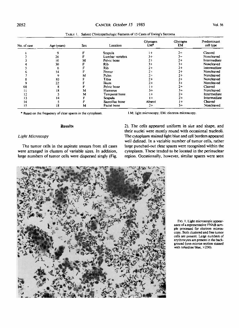
FIG. I . Light microscopic appear- ance of a representative FNAB sam- ple processed for electron micros- copy. Both clustered and free tumor cells are present. Large numbers of erythrocytes are present in the back- ground (one-micron section stained with toluidine blue, X250).
No. 8 ASPIRATION CYTOLOGY OF EWING’S SARCOMA * Akhtur et uf. 2053
in broad cytoplasmic processes extending from the cells (Figs. 3 and 4).
In one case (Case l ) , occasional pseudorosettes were present. In addition, some of the tumor cells had slender cytoplasmic processes that appeared to intertwine with those of adjacent cells (Figs. 5 and 6).
Electron Microscopy
Although the cellular morphologic features within the tumors varied considerably from case to case, the tumor
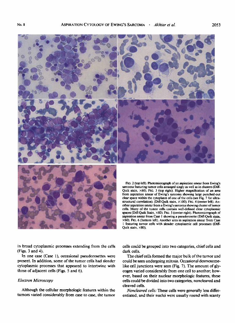
Rc. 2 (top left). Photomicrograph of an aspiration smear from Ewing’s sarcoma featuring tumor cells arranged singly as well as in clusters (Diff- Quik stain, X60). FIG. 3 (top right). Higher magnification of an area from aspiration smear of Ewing’s sarcoma showing large punched-out clear space within the cytoplasm of one of the cells (see Fig. 7 for ultra- structural correlation). (Diff-Quik stain, X 100). FIG. 4 (center left). An- other aspiration smear from a Ewing’s sarcoma showing cluster of tumor cells. Many of the tumor cells contain welldefined clear cytoplasmic spaces(Diff-Quik Stain, X80). FIG. 5 (center right). Photomicrograph of aspiration smear from Case I showing a pseudorosette (Diff-Quik stain, X60). FIG. 6 (bottom left). Another area in aspiration smear from Case I featuring tumor cells with slender cytoplasmic cell processes (Diff- Quik stain, X80).
cells could be grouped into two categories, chief cells and dark cells.
The chief cells formed the major bulk of the tumor and could be seen undergoing mitosis. Occasional desmosome- like cell junctions were seen (Fig. 7). The amount of gly- cogen varied considerably from one cell to another; how- ever, based on their nuclear morphologic features, these cells could be divided into two categories, noncleaved and cleaved cells.
Noncleuved cells: These cells were generally less differ- entiated, and their nuclei were usually round with scanty
2054 CANCER October 15 1985 Vol. 56
heterochromatin arranged as a thin rim along the nuclear membrane (Fig. 7). Nucleoli, if present, were small and inconspicious.
Cleuved cells: These cells were usually more differen- tiated and revealed more abundant cytoplasm with greater numbers of mitochondria and Golgi complexes and fre- quent profiles of rough endoplasmic reticulum. Nuclei were more irregular in configuration and frequently con- tained one or more deep identations. Heterochromatin was somewhat more abundant, although it was still ar- ranged along the periphery of the nuclei. Nucleoli were larger and more frequent (Fig. 8).
In addition to the above-described extremes, several cells with morphologic features intermediate between the two cell types were also present. The dark cells were smaller in size, compared with the chief cells, and their cytoplasm was electron-dense (Fig. 9). Variable amounts
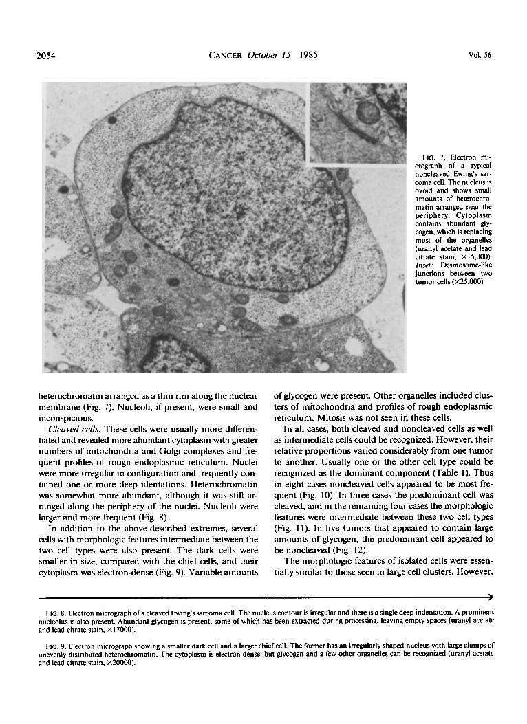
FIG. 7. Electron mi- crograph of a typical noncleaved Ewing's sar- coma cell. The nucleus is ovoid and shows small amounts of heterochro- matin arranged near the periphery. Cytoplasm contains abundant gly- cogen, which is replacing most of the organelles (uranyl acetate and lead citrate stain, X 15,000). Inset: Desmosome-like junctions between two tumor cells (X25,OOO).
of glycogen were present. Other organelles included clus- ters of mitochondria and profiles of rough endoplasmic reticulum. Mitosis was not seen in these cells.
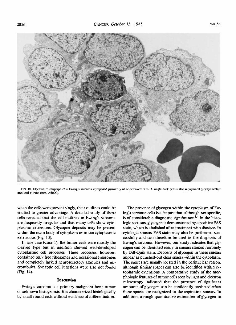
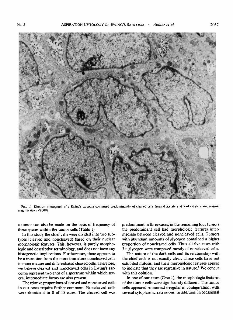
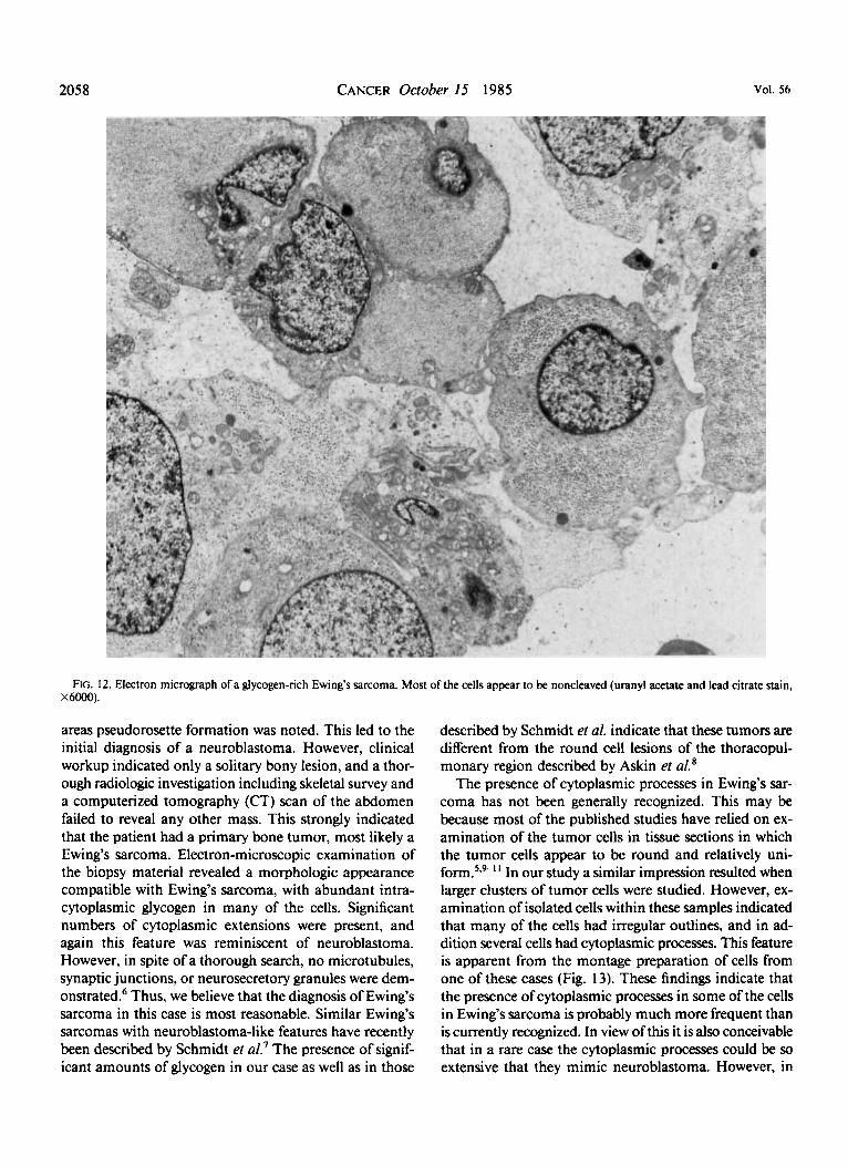
In all cases, both cleaved and noncleaved cells as well as intermediate cells could be recognized. However, their relative proportions varied considerably from one tumor to another. Usually one or the other cell type could be recognized as the dominant component (Table I). Thus in eight cases noncleaved cells appeared to be most fre- quent (Fig. 10). In three cases the predominant cell was cleaved, and in the remaining four cases the morphologic features were intermediate between these two cell types (Fig. 1 I ) . In five tumors that appeared to contain large amounts of glycogen, the predominant cell appeared to be noncleaved (Fig. 12).
The morphologic features of isolated cells were essen- tially similar to those seen in large cell clusters. However,
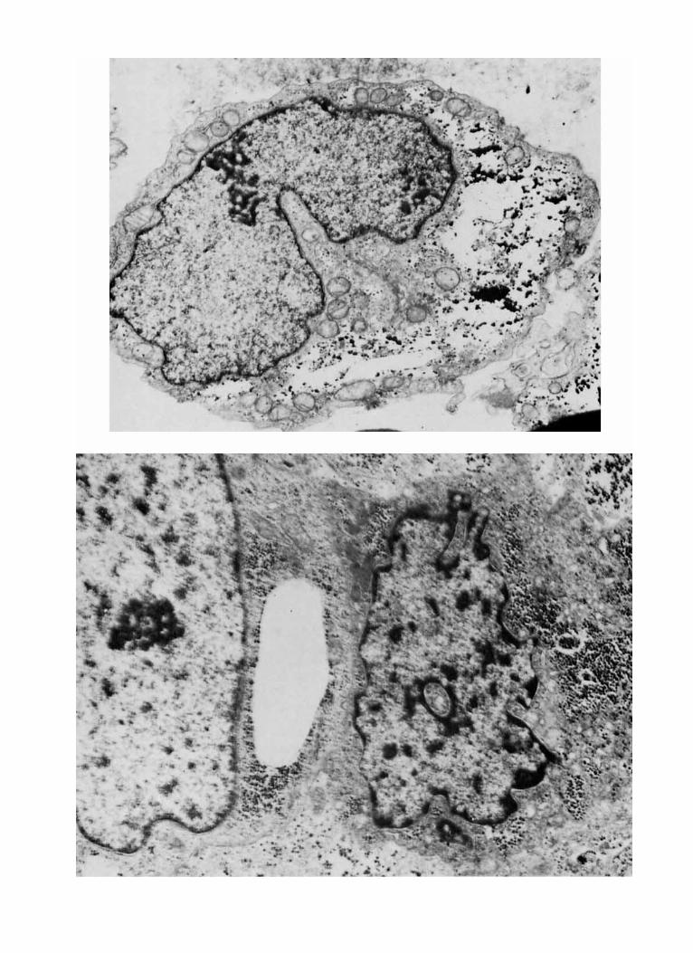
> FIG. 8. Electron micrograph of a cleaved Ewing's sarcoma cell. The nucleus contour is irregular and there is a single deep indentation. A prominent
nucleolus is also present. Abundant glycogen is present. some of which has been extracted during processing, leaving empty spaces (uranyl acetate and lead citrate stain, X 17000).
FIG. 9. Electron micrograph showing a smaller dark cell and a larger chief cell. The former has an irregularly shaped nucleus with large clumps of unevenly distributed heterochromatin. The cytoplasm is electrondense. but glycogen and a few other organelles can be recognized (uranyl acetate and lead citrate stain, X20000).
2056 CANCER October 15 1985 Vol. 56
FIG. 10. Electron micrograph of a Ewing’s sarcoma composed primarily of noncleaved cells. A single dark cell is also recognized (uranyl acetate and lead citrate stain, X6000).
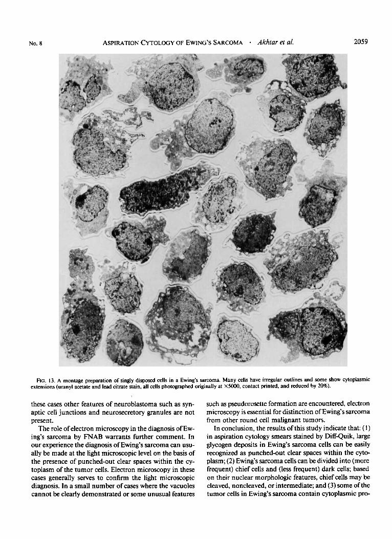
when the cells were present singly, their outlines could be studied to greater advantage. A detailed study of these cells revealed that the cell outlines in Ewing’s sarcoma are frequently irregular and that many cells show cyto- plasmic extensions. Glycogen deposits may be present within the main body of cytoplasm or in the cytoplasmic extensions (Fig. 13).
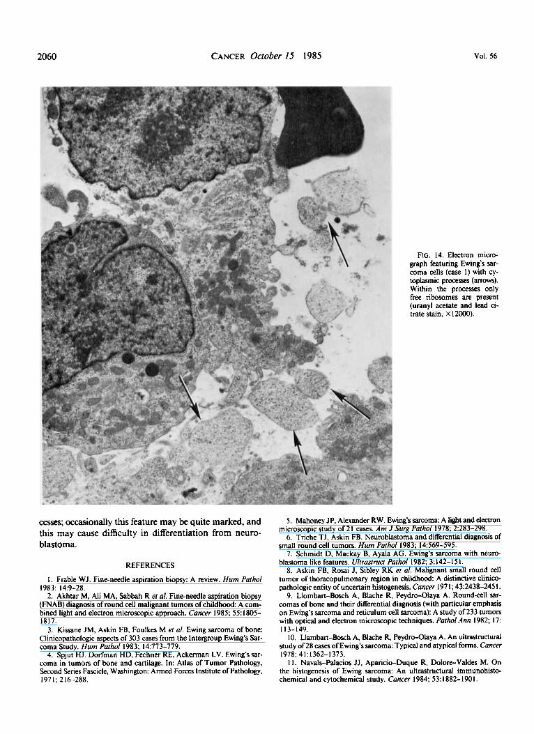
In one case (Case I), the tumor cells were mostly the cleaved type but in addition showed welldeveloped cytoplasmic cell processes. These processes, however, contained only free ribosomes and occasional lysosomes and completely lacked neurosecretory granules and mi- crotubules. Synaptic cell junctions were also not found (Fig. 14).
Discussion Ewing’s sarcoma is a primary malignant bone tumor
of unknown histogenesis. It is characterized histologically by small round cells without evidence of differentiation.
The presence of glycogen within the cytoplasm of Ew- ing’s sarcoma cells is a feature that, although not specific, is of considerable diagnostic ~ignificance.’.~ In the histo- logic sections, glycogen is demonstrated by a positive PAS stain, which is abolished after treatment with diastase. In cytologic smears PAS stain may also be performed suc- cessfully and can therefore be used in the diagnosis of Ewing’s sarcoma. However, our study indicates that gly- cogen can be identified easily in smears stained routinely by Diff-Quik stain. Deposits of glycogen in these smears appear as punched-out clear spaces within the cytoplasm. The spaces are usually located in the perinuclear region, although similar spaces can also be identified within cy- toplasmic extensions. A comparative study of the mor- phologic features of tumor cells seen by light and electron microscopy indicated that the presence of significant amounts of glycogen can be confidently predicted when these spaces are recognized in the aspiration smears. In addition, a rough quantitative estimation of glycogen in
No. 8 ASPIRATION CYTOLOGY OF EWINC’S SARCOMA - Akhtar et al. 2057
FIG. 1 1 . Electron micrograph of a Ewing’s sarcoma composed predominantly of cleaved cells (uranyl acetate and lead citrate stain, original magnification X9000) .
a tumor can also be made on the basis of frequency of these spaces within the tumor cells (Table 1).
In this study the chief cells were divided into two sub- types (cleaved and noncleaved) based on their nuclear morphologic features. This, however, is purely morpho- logic and descriptive terminology, and does not have any histogenetic implications. Furthermore, there appears to be a transition from the more immature noncleaved cells to more mature and differentiated cleaved cells. Therefore, we believe cleaved and noncleaved cells in Ewing’s sar- coma represent two ends of a spectrum within which sev- eral intermediate forms are also present.
The relative proportions of cleaved and noncleaved cells in our cases require further comment. Noncleaved cells were dominant in 8 of 15 cases. The cleaved cell was
predominant in three cases; in the remaining four tumors the predominant cell had morphologic features inter- mediate between cleaved and noncleaved cells. Tumors with abundant amounts of glycogen contained a higher proportion of noncleaved cells. Thus all five cases with 3+ glycogen were composed mostly of noncleaved cells.
The nature of the dark cells and its relationship with the chief cells is not exactly clear. These cells have not exhibited mitosis, and their morphologic features appear to indicate that they are regressive in nature.’ We concur with this opinion.
In one of our cases (Case I ) , the morphologic features of the tumor cells were significantly different. The tumor cells appeared somewhat irregular in configuration, with several cytoplasmic extensions. In addition, in occasional
2058 CANCER Oclober 15 1985 Vol. 56
FIG. 12. Electron micrograph of a glycogen-rich Ewing's sarcoma. Most of the cells appear to be noncleaved (uranyl acetate and lead citrate stain, X6000).
areas pseudorosette formation was noted. This led to the initial diagnosis of a neuroblastoma. However, clinical workup indicated only a solitary bony lesion, and a thor- ough radiologic investigation including skeletal survey and a computerized tomography (CT) scan of the abdomen failed to reveal any other mass. This strongly indicated that the patient had a primary bone tumor, most likely a Ewing's sarcoma. Electron-microscopic examination of the biopsy material revealed a morphologic appearance compatible with Ewing's sarcoma, with abundant intra- cytoplasmic glycogen in many of the cells. Significant numbers of cytoplasmic extensions were present, and again this feature was reminiscent of neuroblastoma. However, in spite of a thorough search, no microtubules, synaptic junctions, or neurosecretory granules were dem- onstrated.6 Thus, we believe that the diagnosis of Ewing's sarcoma in this case is most reasonable. Similar Ewing's sarcomas with neuroblastoma-like features have recently been described by Schmidt et a/.' The presence of signif- icant amounts of glycogen in our case as well as in those
described by Schmidt et al. indicate that these tumors are different from the round cell lesions of the thoracopul- monary region described by Askin et
The presence of cytoplasmic processes in Ewing's sar- coma has not been generally recognized. This may be because most of the published studies have relied on ex- amination of the tumor cells in tissue sections in which the tumor cells appear to be round and relatively uni- f ~ r m . ~ * ~ - ' ' In our study a similar impression resulted when larger clusters of tumor cells were studied. However, ex- amination of isolated cells within these samples indicated that many of the cells had irregular outlines, and in ad- dition several cells had cytoplasmic processes. This feature is apparent from the montage preparation of cells from one of these cases (Fig. 13). These findings indicate that the presence of cytoplasmic processes in some of the cells in Ewing's sarcoma is probably much more frequent than is currently recognized. In view of this it is also conceivable that in a rare case the cytoplasmic processes could be so extensive that they mimic neuroblastoma. However, in
No. 8 ASPIRATION CYTOLOGY OF €WING’S SARCOMA Akhtar et a/. 2059
FIG. 13. A montage preparation of singly disposed cells in a Ewing’s sarcoma. Many cells have irregular outlines and some show cytoplasmic extensions (uranyl acetate and lead citrate stain, all cells photographed originally at X5000, contact printed, and reduced by 20%).
these cases other features of neuroblastoma such as syn- aptic cell junctions and neurosecretory granules are not present.
The role of electron microscopy in the diagnosis of Ew- ing’s sarcoma by FNAB warrants further comment. In our experience the diagnosis of Ewing’s sarcoma can usu- ally be made at the light microscopic level on the basis of the presence of punched-out clear spaces within the cy- toplasm of the tumor cells. Electron microscopy in these cases generally serves to confirm the light microscopic diagnosis. In a small number of cases where the vacuoles cannot be clearly demonstrated or some unusual features
such as pseudorosette formation are encountered, electron microscopy is essential for distinction of Ewing’s sarcoma from other round cell malignant tumors.
In conclusion, the results of this study indicate that: ( 1) in aspiration cytology smears stained by Diff-Quik, large glycogen deposits in Ewing’s sarcoma cells can be easily recognized as punched-out clear spaces within the cyto- plasm; (2) €wing’s sarcoma cells can be divided into (more frequent) chief cells and (less frequent) dark cells; based on their nuclear morphologic features, chief cells may be cleaved, noncleaved, or intermediate; and (3) some of the tumor cells in Ewing’s sarcoma contain cytoplasmic pro-
2060 CANCER October 15 1985 Vol. 56
cesses; occasionally this feature may be quite marked, and this may cause difficulty in differentiation from neuro- blastoma.
REFERENCES
I . Frable WJ. Fine-needle aspiration biopsy: A review. Hum Pafhol
2. Akhtar M, Ali MA, Sabbah R ef a/. Fine-needle aspiration biopsy (FNAB) diagnosis of round cell malignant tumors of childhood: A com- bined light and electron microscopic approach. Cancer 1985; 55: 1805- 1817.
3. Kissane JM, Askin FB, Foulkes M et a/. Ewing sarcoma of bone: Clinicopathologic aspects of 303 cases from the Intergroup Ewing's Sar- coma Study. Hum Pafhol 1983; 14:773-779.
4. Spjut HJ, Dorfman HD, Fechner RE, Ackerman LV. Ewing's sar- coma in tumors of bone and cartilage. In: Atlas of Tumor Pathology, Second Series Fascicle, Washington: Armed Forces Institute of Pathology,
1983: 14:9-28.
1971; 216-288.
RG. 14. Electron micro- graph featuring Ewing's sar- coma cells (case I ) with cy- toplasmic processes (arrows). Within the processes only free ribosomes are present (uranyl acetate and lead ci- trate stain. X 12000).
5 . Mahoney JP, Alexander RW. Ewing's sarcoma: A light and electron microscopic study of 21 cases. Am J Surg Patho/ 1978; 2:283-298.
6. Triche TJ, Askin FB. Neuroblastoma and differential diagnosis of small round cell tumors. Hum Patho/ 1983; 14569-595.
7. Schmidt D, Mackay B, Ayala AG. Ewing's sarcoma with neuro- blastoma like features. Ulfrusfrucr Patho/ 1982; 3: 142-15 I .
8. Askin FB, Rosai J. Sibley RK ef a/. Malignant small round cell tumor of thoracopulmonary region in childhood: A distinctive clinico- pathologic entity of uncertain histogenesis. Cancer 197 I ; 43:2438-245 1.
9. Llombart-Boxh A, Blache R, Peydro-Olaya A. Roundcell sar- comas of bone and their differential diagnosis (with particular emphasis on Ewing's sarcoma and reticulum cell sarcoma): A study of 233 tumors with optical and electron microscopic techniques. Patho/ Ann 1982; 17:
10. Llambart-Bosch A, Blache R, Peydro-Olaya A. An ultrastructural study of 28 cases of Ewing'ssarcoma: Typical and atypical forms. Cancer
1 I . Navals-Palacios JJ, Aparicio-Duque R, Dolore-Valdes M. On the histogenesis of Ewing sarcoma: An ultrastructural immunohisto- chemical and cytochemical study. Cancer 1984 53:1882-1901.
113-149.
1978: 41: 1362-1 373.
Related Documents