ASPicDB: a database of annotated transcript and protein variants generated by alternative splicing Pier L. Martelli 1 , Mattia D’Antonio 2 , Paola Bonizzoni 3 , Tiziana Castrignano ` 2 , Anna M. D’Erchia 4 , Paolo D’Onorio De Meo 2 , Piero Fariselli 1 , Michele Finelli 5 , Flavio Licciulli 6 , Marina Mangiulli 4 , Flavio Mignone 7 , Giulio Pavesi 8 , Ernesto Picardi 3 , Raffaella Rizzi 3 , Ivan Rossi 5 , Alessio Valletti 4 , Andrea Zauli 5 , Federico Zambelli 8 , Rita Casadio 1, * and Graziano Pesole 4,9, * 1 Biocomputing Group, University of Bologna, Bologna 40126, 2 Consorzio Interuniversitario per le Applicazioni di Supercalcolo per Universita ` e Ricerca (CASPUR), Rome 00185, 3 DISCo, University of Milan-Bicocca, Milan, 20135, 4 Dipartimento di Biochimica e Biologia Molecolare, University of Bari, Bari 70126, 5 BioDec srl, Casalecchio di Reno, Bologna 40033, 6 Istituto Tecnologie Biomediche, Consiglio Nazionale delle Ricerche, Bari 70126, 7 Dipartimento di Chimica Strutturale e Stereochimica Inorganica, University of Milan, 8 Dipartimento di Scienze Biomolecolari e Biotecnologie, University of Milan, Milan 20133 and 9 Istituto Biomembrane e Bioenergetica, Consiglio Nazionale delle Ricerche, Bari 70125, Italy Received August 9, 2010; Revised and Accepted October 14, 2010 ABSTRACT Alternative splicing is emerging as a major mech- anism for the expansion of the transcriptome and proteome diversity, particularly in human and other vertebrates. However, the proportion of alterna- tive transcripts and proteins actually endowed with functional activity is currently highly debated. We present here a new release of ASPicDB which now provides a unique annotation resource of human protein variants generated by alternative splicing. A total of 256 939 protein variants from 17 191 multi-exon genes have been extensively annotated through state of the art machine learn- ing tools providing information of the protein type (globular and transmembrane), localization, presence of PFAM domains, signal peptides, GPI- anchor propeptides, transmembrane and coiled- coil segments. Furthermore, full-length variants can be now specifically selected based on the annotation of CAGE-tags and polyA signal and/or polyA sites, marking transcription initiation and termination sites, respectively. The retrieval can be carried out at gene, transcript, exon, protein or splice site level allowing the selection of data sets fulfilling one or more features settled by the user. The retrieval inter- face also enables the selection of protein vari- ants showing specific differences in the annotated features. ASPicDB is available at http://www .caspur.it/ASPicDB/. INTRODUCTION Alternative splicing is a well characterized mechanism which, coupled with alternative initiation and termination of transcription (1), may expand the transcriptome and proteome complexity in human and other organisms by over one order of magnitude with respect to the number of annotated genes (2,3). In particular, it is now widely demonstrated that virtually all multi-exon genes may generate multiple transcripts and protein variants (3,4) and that the splicing process is tightly regulated in differ- ent physiological conditions, tissues or developmental stages (5). Furthermore, alterations of the splicing process can be observed in several genetic diseases and in cancer (6–10). The huge amount of EST sequences (11) together with the relevant reference genome sequence has been used to carry out an extensive analysis of alternative splicing in *To whom correspondence should be addressed. Tel: +39 080 5443588; Fax: +39 080 5443317; Email: [email protected] Correspondence may also be addressed to Rita Casadio. Tel: +39 0512094005; Fax: +39 051242576; Email: [email protected] The authors wish it to be known that in their opinion, the first two authors are the joint First Authors. D80–D85 Nucleic Acids Research, 2011, Vol. 39, Database issue Published online 4 November 2010 doi:10.1093/nar/gkq1073 ß The Author(s) 2010. Published by Oxford University Press. This is an Open Access article distributed under the terms of the Creative Commons Attribution Non-Commercial License (http://creativecommons.org/licenses/ by-nc/2.5), which permits unrestricted non-commercial use, distribution, and reproduction in any medium, provided the original work is properly cited.

Welcome message from author
This document is posted to help you gain knowledge. Please leave a comment to let me know what you think about it! Share it to your friends and learn new things together.
Transcript
ASPicDB: a database of annotated transcript andprotein variants generated by alternative splicingPier L. Martelli1, Mattia D’Antonio2, Paola Bonizzoni3, Tiziana Castrignano2,
Anna M. D’Erchia4, Paolo D’Onorio De Meo2, Piero Fariselli1, Michele Finelli5,
Flavio Licciulli6, Marina Mangiulli4, Flavio Mignone7, Giulio Pavesi8, Ernesto Picardi3,
Raffaella Rizzi3, Ivan Rossi5, Alessio Valletti4, Andrea Zauli5, Federico Zambelli8,
Rita Casadio1,* and Graziano Pesole4,9,*
1Biocomputing Group, University of Bologna, Bologna 40126, 2Consorzio Interuniversitario per le Applicazioni diSupercalcolo per Universita e Ricerca (CASPUR), Rome 00185, 3DISCo, University of Milan-Bicocca, Milan,20135, 4Dipartimento di Biochimica e Biologia Molecolare, University of Bari, Bari 70126, 5BioDec srl,Casalecchio di Reno, Bologna 40033, 6Istituto Tecnologie Biomediche, Consiglio Nazionale delle Ricerche, Bari70126, 7Dipartimento di Chimica Strutturale e Stereochimica Inorganica, University of Milan, 8Dipartimento diScienze Biomolecolari e Biotecnologie, University of Milan, Milan 20133 and 9Istituto Biomembrane eBioenergetica, Consiglio Nazionale delle Ricerche, Bari 70125, Italy
Received August 9, 2010; Revised and Accepted October 14, 2010
ABSTRACT
Alternative splicing is emerging as a major mech-anism for the expansion of the transcriptome andproteome diversity, particularly in human and othervertebrates. However, the proportion of alterna-tive transcripts and proteins actually endowedwith functional activity is currently highly debated.We present here a new release of ASPicDB whichnow provides a unique annotation resource ofhuman protein variants generated by alternativesplicing. A total of 256 939 protein variants from17 191 multi-exon genes have been extensivelyannotated through state of the art machine learn-ing tools providing information of the protein type(globular and transmembrane), localization,presence of PFAM domains, signal peptides, GPI-anchor propeptides, transmembrane and coiled-coil segments. Furthermore, full-length variants canbe now specifically selected based on the annotationof CAGE-tags and polyA signal and/or polyA sites,marking transcription initiation and terminationsites, respectively. The retrieval can be carried outat gene, transcript, exon, protein or splice site level
allowing the selection of data sets fulfilling one ormore features settled by the user. The retrieval inter-face also enables the selection of protein vari-ants showing specific differences in the annotatedfeatures. ASPicDB is available at http://www.caspur.it/ASPicDB/.
INTRODUCTION
Alternative splicing is a well characterized mechanismwhich, coupled with alternative initiation and terminationof transcription (1), may expand the transcriptome andproteome complexity in human and other organisms byover one order of magnitude with respect to the number ofannotated genes (2,3). In particular, it is now widelydemonstrated that virtually all multi-exon genes maygenerate multiple transcripts and protein variants (3,4)and that the splicing process is tightly regulated in differ-ent physiological conditions, tissues or developmentalstages (5). Furthermore, alterations of the splicingprocess can be observed in several genetic diseases andin cancer (6–10).
The huge amount of EST sequences (11) together withthe relevant reference genome sequence has been used tocarry out an extensive analysis of alternative splicing in
*To whom correspondence should be addressed. Tel: +39 080 5443588; Fax: +39 080 5443317; Email: [email protected] may also be addressed to Rita Casadio. Tel: +39 0512094005; Fax: +39 051242576; Email: [email protected]
The authors wish it to be known that in their opinion, the first two authors are the joint First Authors.
D80–D85 Nucleic Acids Research, 2011, Vol. 39, Database issue Published online 4 November 2010doi:10.1093/nar/gkq1073
� The Author(s) 2010. Published by Oxford University Press.This is an Open Access article distributed under the terms of the Creative Commons Attribution Non-Commercial License (http://creativecommons.org/licenses/by-nc/2.5), which permits unrestricted non-commercial use, distribution, and reproduction in any medium, provided the original work is properly cited.
human through the ASPIC algorithm (12–14). The alter-native splicing pattern of human multi-exon genes,determined by ASPIC, has been collected in ASPicDB, adatabase resource which presents some unique featureswith respect to other similar databases (15). The ASPICalgorithm implements an optimization strategy that, per-forming a multiple alignment of all available transcriptdata (including full-length cDNA and EST sequences) tothe relevant genome sequence, detects the set of intronsthat minimizes the number of splicing sites. It also gener-ates through a directed-acyclic graph combinatorialprocedure the minimal set of non-mergeable transcriptisoforms compatible with the detected splicing events(14). The reliability of splicing isoforms detectedby ASPIC has been recently established through acomparative assessment (16).
The advent of massive transcriptome sequence datagenerated by RNA-Seq (17) is steadily increasing thenumber of validated splicing sites and isoforms inhuman and other organisms thus suggesting that afraction of alternative splicing events are the result ofbackground noise in the splicing process (18) which gen-erates non-functional isoforms expressed at low level.Therefore, extensive research efforts are required to dis-tinguish functional species-specific variants fromnon-functional ones originated from neutral drift in thesplicing process, as well as to asses the biological role offunctional isoforms.
The annotation of the protein variants predicted withASPIC is an essential step for exploring the functionaland structural diversity of the proteins originatingfrom the same gene by means of alternative splicing andtherefore for unraveling the complex physiological effectsof alternative splicing events (19). Indeed, currently avail-able databases, such as ASD (20), ASAP II (21),ASTALAVISTA (22) and H-DBAS (23), mostly collectinformation on alternative transcripts at the mRNA level,without considering the effect of alternative splicing on theprotein structure and function. The ProSAS (24) databasecontains structural information as derived from compara-tive modeling procedure, but due to the limitations of themodeling techniques, only �15% of the human transcriptsare endowed with a reliable protein structure prediction.
ASPICdb aims at filling the gap of structural and func-tional annotation of protein splicing variants, by adoptinga set of analysis and prediction tools that do not rely onlyon annotation transfer by sequence similarity. It providesa thorough computational annotation of predicted humanprotein variants including PFAM domains (25),N-terminal signal peptides, GPI-anchor propeptides,transmembrane domains, subcellular localization andother features, also reporting the relevant crosslinks toUniprotKB/Swissprot (26) and PDB databases (27). Acomprehensive annotation of the domain architectureand other structural features could also be extremelyuseful to critically assess the reliability of the functionalclassification provided the GO System (25), which stillneglects much of the relevant information for alternativesplicing products.
In addition, in consideration of the fragmented natureof the available transcript data, the new version of
ASPicDB include the annotation of CAGE tags (28) inorder to identify truly transcription initiation sites anddiscriminate between full-length isoforms using alternativetranscription initiations and 50-partial transcripts forwhich a full-length CDS and the encoded protein cannotbe reliably predicted.
ANNOTATION PIPELINE OF HUMAN PROTEINVARIANTS
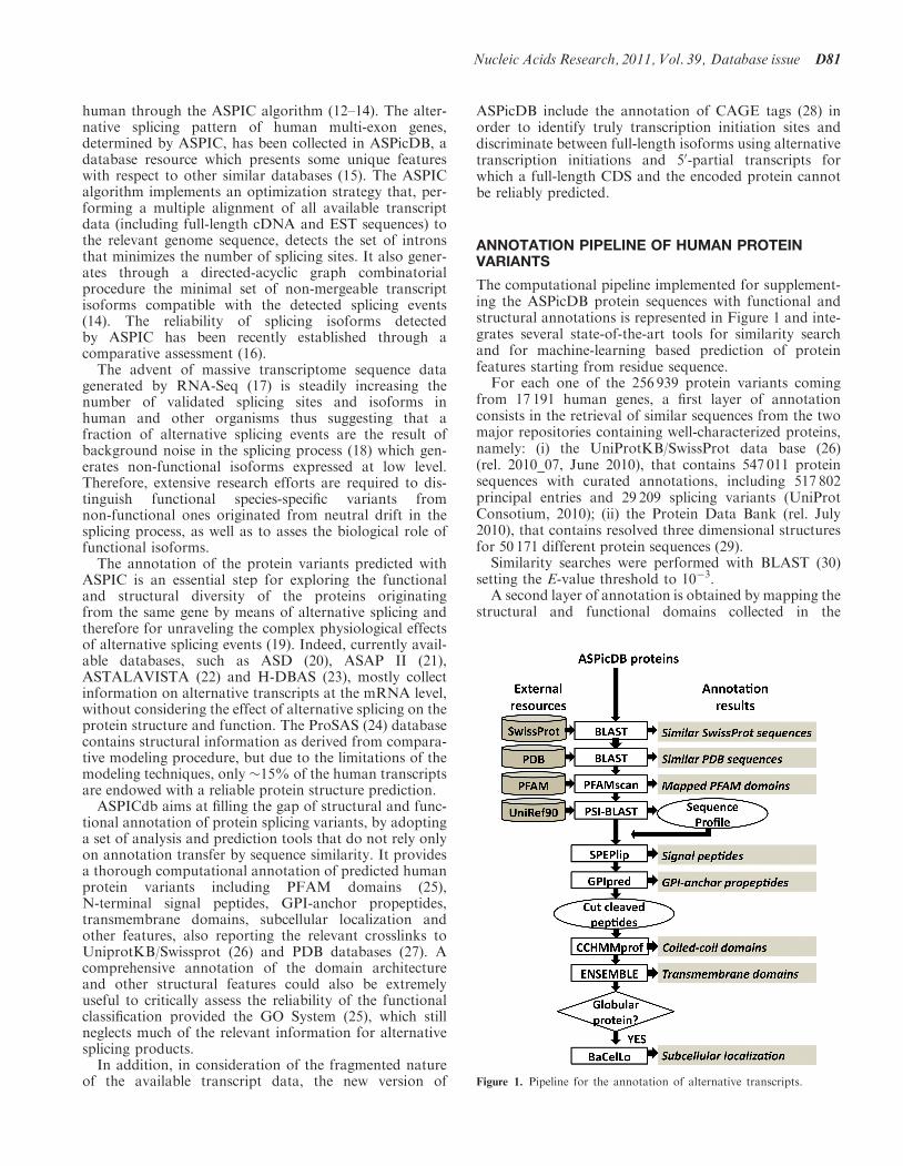
The computational pipeline implemented for supplement-ing the ASPicDB protein sequences with functional andstructural annotations is represented in Figure 1 and inte-grates several state-of-the-art tools for similarity searchand for machine-learning based prediction of proteinfeatures starting from residue sequence.For each one of the 256 939 protein variants coming
from 17 191 human genes, a first layer of annotationconsists in the retrieval of similar sequences from the twomajor repositories containing well-characterized proteins,namely: (i) the UniProtKB/SwissProt data base (26)(rel. 2010_07, June 2010), that contains 547 011 proteinsequences with curated annotations, including 517 802principal entries and 29 209 splicing variants (UniProtConsotium, 2010); (ii) the Protein Data Bank (rel. July2010), that contains resolved three dimensional structuresfor 50 171 different protein sequences (29).Similarity searches were performed with BLAST (30)
setting the E-value threshold to 10�3.A second layer of annotation is obtained by mapping the
structural and functional domains collected in the
Figure 1. Pipeline for the annotation of alternative transcripts.
Nucleic Acids Research, 2011, Vol. 39, Database issue D81
PFAM-A database (rel. 24.0, October 2009) that containscurated multiple sequence alignments based on hiddenMarkov models (HMM) for 8691 families, 2985 domains,162 repeats and 74 motifs (25). The PFAM models weremapped on the ASPicDB protein sequences by means ofthe pfam_scan.pl program (ftp://ftp.sanger.ac.uk/pub/databases/Pfam/Tools/), based on HMMER3.0 (31).The third layer of annotation results from the integra-
tion of several predictors based on machine learning tools,such as neural networks, hidden Markov models, supportvector machines and conditional random fields. Sincemost of the methods take advantage of the evolutionaryinformation encoded in sequence profiles, we compiledthem starting from the similar sequences retrieved withtwo PSI-BLAST iterations (setting the E-value thresholdto 10�3) from the UniRef90 data set consisting of6 955 504 sequences (July 2010). The first predictedfeatures are the presence of N-terminal signal peptideand of C-terminal GPI-anchor propeptides, withSPEPlip (32) and PredGPI (33), respectively. Both themethods are among the best available predictors, scoringwith accuracy as high as 95% the former and 88% thelatter. When present, the signal peptide and the propeptideare cleaved from the protein sequence. The presence ofcoiled-coil domains is predicted with CCHMM-PROFthat is able to locate coiled-coil segments in protein se-quences with 80% accuracy (34). a-Helical transmem-brane domains are then predicted with ENSEMBLE(35), that discriminates transmembrane from globularproteins with false positive and false negative rates bothequal to 3%. The same tool is adopted for predicting thenumber and the position of transmembrane segmentsalong the sequence, with an accuracy of 90% on theprotein base. The subcellular localization of globularproteins is predicted with BaCelLo (36), which discrimin-ates four localizations in animals (secretory pathway,cytoplasm, nucleus and mitochondrion) with 74%accuracy.
ASPicDB CONTENT AND ANNOTATION OFPROTEIN VARIANTS
Table 1 reports some statistics on the data contained in thecurrent version of ASPicDB (version 2.0, August 2010)which refers only to human multi-exon genes annotatedin NCBI Entrez Gene (37) with at least one RefSeq
transcript (38) and the relevant Unigene cluster (39)collecting all available gene-specific cDNA and ESTsequences.
In the current version of ASPicDB some more featuresare available including the annotation of the CAGE tags(28) which define truly transcription initiation sites and acomprehensive protein annotation. A total of 12 789 394CAGE tags have been mapped thus supporting constitu-tive or alternative transcription start sites. To each tran-script variant a ‘unique identifier’ (16) has been associatedin order to make possible the unambiguous comparisonwith alternative transcripts collected in other databases.
All alternative proteins collected in ASPicDB have beencompared with UniprotKB/SwissProt (26) and PDB (29)databases. The results of similarity searches are reportedin Table 2. Only 17% of the ASPicDB protein sequencesare identical to proteins deposited in UniProtKB/SwissProt database. However, 94% of the sequencesshare significant similarity with proteins annotated in thesame database, prompting the possibility of a reliable an-notation transfer. Moreover, 54% of ASPicDB sequencesare similar to proteins deposited in the PDB suggestingthat their structures can be modeled, at least partially.
A considerable amount of PFAM models map on theASPicDB sequences (Table 2). On the overall, 71% ofsequences match with at least one model. This result isin agreement with the reported sequence coverage on thehuman proteome of the current PFAM release, which isequal to 72.5% (25). It is worth noticing that, although allthe models map with an E-value < 10�5, only 20% of thematches are complete (that is, involve the whole model). Anote of caution is necessary when inferring features frompartial matches and the actual extent of the match has tobe evaluated for each instance.
Table 3 summarizes the results of the annotationprocess performed with machine learning based predict-ors. Two percent of proteins were not predicted since theyare shorter than 50 residues, 16% of proteins are predictedas transmembrane and 82% are predicted as globular.Among the globular proteins, 12% are predicted assecreted, 35% as cytoplasmic, 27% as globular and 8%as mitochondrial. Signal peptides and GPI-anchorpropeptides are predicted in the 12 and 0.7% of the
Table 2. Annotation of human variants upon similarity and PFAM
searches
Sequence repository No of proteinsa No of genesa
UniProtKB/SwissProt, %E-value< 10�3, % 239 814 (93) 17 054 (99)Identical, % 42 601 (17) 13 043 (76)
PDBE-value< 10�3, % 137 528 (54) 11 062 (64)Identical, % 1079 (0.4) 316 (2)
PFAMAll matches, E-value< 10�5, % 183 483 (71) 14 205 (83)Complete matches,E-value< 10�5, %
46 630 (18) 5621 (33)
aThe percentages are computed with respect to 256 939 protein variantsand 17 191 genes.
Table 1. Statistics of the ASPicDB content (v2.0, August 2010)
ASPicDB v2.0
Genes 17 191Transcripts 319 092Proteins 256 939Exons 390 886Splicing sites 351 345U2 302 164U12 1712
Splicing events 233 717
The number of splicing sites belonging to the U2 or U12 class and ofsplicing events is also reported.
D82 Nucleic Acids Research, 2011, Vol. 39, Database issue

sequences, respectively. Coiled-coil domains are predictedin 1.3% of the proteins. At the gene level, 30 and 92% ofgenes encode for transmembrane and globular proteins,respectively. Since the sum exceed 100%, it follows that22% of the genes encode for both globular and transmem-brane variants. The same consideration holds for the otherannotations as reported in Table 4. The amount of genespredicted to encode for proteins with different subcellularlocalization achieves 56%. This is partially explained bythe fact that BaCelLo scores with an accuracy equal to74%, which is the lowest among the methods included inthe pipeline. Indeed the discrimination between the ‘cyto-plasmic’ and the ‘nuclear’ classes is still a difficult task forall subcellular localization predictors (40). When the twoclasses are merged together, the BaCelLo accuracy in-creases up to 91%, but the rate of genes encoding forproteins with different localizations is still as high as44%, suggesting that localization diversity is inherent inthe ASPicDB protein variants. The structure of PFAMannotations is also highly variable: 38% of genes encodefor variants matching with different number and/or typeof PFAM models. Altogether, results listed in Table 4suggest that alternative transcripts can encode forproteins endowed with different structural and functionalfeatures. ASPicDB provides a unique resource reportingthe annotation of alternative splicing variants at theprotein level and an interface enabling the discovery ofsuch differences.
ASPicDB RETRIEVAL INTERFACE
ASPicDB can be accessed though simple or advancedquery forms. The simple query form allows the user toobtain the splicing pattern of one or more genes selectedaccording to several criteria (e.g. HGNC name, RefSeq orUnigene accession IDs, etc.). The advanced query formallows the user to search for (i) genes, (ii) transcripts;(iii) exons; (iv) splicing sites; and (v) proteins, fulfillingdifferent criteria (e.g. exons in a given length range,etc.). Depending on the choice separate query formsappear. The ‘gene’, ‘transcript’ and ‘splicing sites’ queryforms have been described previously (15) whereas the‘exon’ and ‘protein’ query forms are novel features ofthis version of ASPicDB. The exon query form allowsthe user to select exons in a given length range, belongingto a specific type (initial, internal or teminal), flanked byspecific splicing sites or associated to one or moreAffimetrix ExonArray probeset IDs.The ‘protein’ query form allows the retrieval of tran-
scripts encoding proteins isoforms of a specific class (e.g.globular or transmembrane), subcellular localization (e.g.mitochondrion, nucleus, secretory, cytoplasm) or contain-ing one or more features, including occurrence andnumber of PFAM or transmembrane domains, GPI-anchor propeptides, signal peptides. Finally, it is alsopossible to retrieve genes encoding for alternativeproteins that show differences in the above mentionedfeatures.
ASPicDB OUTPUT
After a simple or advanced query has been submitted theoutput for each selected gene is shown which is organizedin eight panels.
(1) Gene information reports a summary of the genomicand transcript data used by ASPIC to generate theprediction, downloadable by the user and links toother popular prediction programs such as ASAP2(21), ASD (20) and ACEVIEW (41) as well as toASPIC results for orthologous genes in other species.
(2) Gene structure view provides a schematic graphicalview of the gene structure including all predictedexons/introns.
(3) Predicted transcripts show a graphical representationof the assembled transcripts with predicted annota-tions of 50-UTR, CDS and 30-UTR, CAGE tagmapping, Premature Termination Codons (PTC)and polyA sites.
(4) Transcript table lists the details of all predicted alter-native transcripts including their length, number ofexons and presence of a protein coding sequence.The ‘variant type’ column lists all the alternativesplicing events using a RefSeq mRNA as the refer-ence transcript. The transcript signature is alsoreported which consists in a unique ID for alterna-tively spliced variants generated according to (16).
(5) Predicted proteins show a graphical representationof the encoded proteins with matching domains(Figure 2). For each mapped domain the sequence
Table 3. Machine learning-based prediction of the human proteins
deposited ASPicDB
Annotation No. of proteinsa No. of genesa
TypeGlobular, % 210 608 (82) 15 513 (90)Transmembrane, % 41 561 (16) 5439 (32)
Localization (globular proteins)Secretory pathway, % 31 917 (12) 7348 (43)Cytoplasm, % 90 046 (35) 10 327 (60)Nucleus, % 69 167 (27) 8183 (48)Mitochondrion, % 19 478 (8) 4698 (27)
DomainsSignal peptide, % 30 508 (12) 5153 (30)GPI-anchor propeptide, % 1673 (0.7) 629 (4)Coiled-coil segments, % 3423 (1.3) 497 (2.8)
aThe percentages are computed with respect to 256 939 protein variantsand 17 191 genes.
Table 4. Differences among alternative proteins encoded by the same
human gene
Annotation No. of genesa, %
Type (globular/transmembrane) 3817 (22)Subcellular localization (globular proteins) 9593 (56)Presence of signal peptide 3939 (23)Presence of GPI-anchor propeptide 591 (3.4)Presence of coiled-coil domains 464 (2.7)Number of transmembrane helices 2140 (12)PFAM models (all matches) 6575 (38)
aThe percentages are computed with respect to 17 191 genes.
Nucleic Acids Research, 2011, Vol. 39, Database issue D83
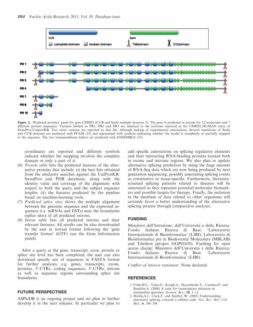
coordinates are reported and different symbolsindicate whether the mapping involves the completedomain or only a part of it.
(6) Protein table lists the predicted features of the alter-native proteins that include: (i) the best hits obtainedfrom the similarity searches against the UniProtKB/SwissProt and PDB databases, along with theidentity value and coverage of the alignment withrespect to both the query and the subject sequencelengths; (ii) the features predicted by the pipelinebased on machine-learning tools.
(7) Predicted splice sites shows the multiple alignmentbetween the genomic sequence and the expressed se-quences (i.e. mRNAs and ESTs) near the boundaries(splice sites) of all predicted introns.
(8) Intron table lists all predicted introns and theirrelevant features; All results can be also downloadedby the user in textual format following the ‘genetransfer format’ (GTF) (see the Gene Informationpanel).
After a query at the gene, transcript, exon, protein orsplice site level has been completed, the user can alsodownload specific sets of sequences in FASTA formatfor further analyses, e.g. genes, transcripts, exons,proteins, 50-UTRs, coding sequences, 30-UTRs, intronsas well as sequence regions surrounding splice siteboundaries.
FUTURE PERSPECTIVES
ASPicDB is an ongoing project and we plan to furtherdevelop it in the next releases. In particular we plan to
add specific annotations on splicing regulatory elementsand their interacting RNA-binding proteins located bothin exonic and intronic regions. We also plan to updatealternative splicing prediction by using the huge amountof RNA-Seq data which are now being produced by nextgeneration sequencing, possibly annotating splicing eventsas constitutive or tissue-specific. Furthermore, literature-screened splicing patterns related to diseases will beannotated as they represent potential molecular biomark-ers and possible targets for therapy. Finally, the inclusionin the database of data related to other organisms willcertainly favor a better understanding of the alternativesplicing process through comparative analyses.
FUNDING
Ministero dell’Istruzione, dell’Universita e della Ricerca:Fondo Italiano Ricerca di Base: ‘LaboratorioInternazionale di Bioinformatica’ (LIBI); Laboratorio diBioinformatica per la Biodiversita Molecolare (MBLAB)and Telethon (project GGP01658). Funding for openaccess charge: Ministero dell’Universita e della Ricerca:Fondo Italiano Ricerca di Base: ‘LaboratorioInternazionale di Bioinformatica’ (LIBI).
Conflict of interest statement. None declared.
REFERENCES
1. Frith,M.C., Valen,E., Krogh,A., Hayashizaki,Y., Carninci,P. andSandelin,A. (2008) A code for transcription initiation inmammalian genomes. Genome Res., 18, 1–12.
2. Matlin,A.J., Clark,F. and Smith,C.W. (2005) Understandingalternative splicing: towards a cellular code. Nat. Rev. Mol. CellBiol., 6, 386–398.
Figure 2. ‘Predicted proteins’ panel for gene CSMD3 (CUB and Sushi multiple domains 3). The gene is predicted to encode for 12 transcripts and 7different protein sequences. Variants labeled as PR1, PR2 and PR3 are identical to the isoforms reported in the CSMD3_HUMAN entry ofSwissProt/UniprotKB. Two more variants are reported in that file, although lacking of experimental annotations. Several repetitions of Sushiand CUB domains are predicted with PFAM (25) and represented with symbols indicating whether the model is completely or partially mappedto the sequence. The two transmembrane helices are predicted with ENSEMBLE (35).
D84 Nucleic Acids Research, 2011, Vol. 39, Database issue
3. Wang,E.T., Sandberg,R., Luo,S., Khrebtukova,I., Zhang,L.,Mayr,C., Kingsmore,S.F., Schroth,G.P. and Burge,C.B. (2008)Alternative isoform regulation in human tissue transcriptomes.Nature, 456, 470–476.
4. Pan,Q., Shai,O., Lee,L.J., Frey,B.J. and Blencowe,B.J. (2008)Deep surveying of alternative splicing complexity in the humantranscriptome by high-throughput sequencing. Nat. Genet., 40,1413–1415.
5. Barash,Y., Calarco,J.A., Gao,W., Pan,Q., Wang,X., Shai,O.,Blencowe,B.J. and Frey,B.J. (2010) Deciphering the splicing code.Nature, 465, 53–59.
6. Faustino,N.A. and Cooper,T.A. (2003) Pre-mRNA splicing andhuman disease. Genes Dev., 17, 419–437.
7. Wang,G.S. and Cooper,T.A. (2007) Splicing in disease: disruptionof the splicing code and the decoding machinery. Nat. Rev.Genet., 8, 749–761.
8. Pettigrew,C.A. and Brown,M.A. (2008) Pre-mRNA splicingaberrations and cancer. Front. Biosci., 13, 1090–1105.
9. Srebrow,A. and Kornblihtt,A.R. (2006) The connection betweensplicing and cancer. J. Cell Sci., 119, 2635–2641.
10. Venables,J.P. (2004) Aberrant and alternative splicing in cancer.Cancer Res., 64, 7647–7654.
11. Boguski,M.S., Lowe,T.M. and Tolstoshev,C.M. (1993)dbEST–database for ‘‘expressed sequence tags’’. Nat. Genet., 4,332–333.
12. Bonizzoni,P., Rizzi,R. and Pesole,G. (2005) ASPIC: a novelmethod to predict the exon-intron structure of a gene that isoptimally compatible to a set of transcript sequences.BMC Bioinformatics, 6, 244.
13. Castrignano,T., Rizzi,R., Talamo,I.G., De Meo,P.D., Anselmo,A.,Bonizzoni,P. and Pesole,G. (2006) ASPIC: a web resource foralternative splicing prediction and transcript isoformscharacterization. Nucleic Acids Res., 34, W440–W443.
14. Bonizzoni,P., Mauri,G., Pesole,G., Picardi,E., Pirola,Y. andRizzi,R. (2009) Detecting alternative gene structures fromspliced ESTs: a computational approach. J. Comput. Biol., 16,43–66.
15. Castrignano,T., D’Antonio,M., Anselmo,A., Carrabino,D.,D’Onorio De Meo,A., D’Erchia,A.M., Licciulli,F., Mangiulli,M.,Mignone,F., Pavesi,G. et al. (2008) ASPicDB: a databaseresource for alternative splicing analysis. Bioinformatics, 24,1300–1304.
16. Riva,A. and Pesole,G. (2009) A unique, consistent identifier foralternatively spliced transcript variants. PLoS ONE, 4, e7631.
17. Wang,Z., Gerstein,M. and Snyder,M. (2009) RNA-Seq: arevolutionary tool for transcriptomics. Nat. Rev. Genet., 10,57–63.
18. Melamud,E. and Moult,J. (2009) Stochastic noise in splicingmachinery. Nucleic Acids Res., 37, 4873–4886.
19. Tress,M.L., Martelli,P.L., Frankish,A., Reeves,G.A.,Wesselink,J.J., Yeats,C., Olason,P.L., Albrecht,M., Hegyi,H.,Giorgetti,A. et al. (2007) The implications of alternative splicingin the ENCODE protein complement. Proc. Natl Acad. Sci. USA,104, 5495–5500.
20. Stamm,S., Riethoven,J.J., Le Texier,V., Gopalakrishnan,C.,Kumanduri,V., Tang,Y., Barbosa-Morais,N.L. and Thanaraj,T.A.(2006) ASD: a bioinformatics resource on alternative splicing.Nucleic Acids Res., 34, D46–D55.
21. Kim,N., Alekseyenko,A.V., Roy,M. and Lee,C. (2007) TheASAP II database: analysis and comparative genomics ofalternative splicing in 15 animal species. Nucleic Acids Res., 35,D93–D98.
22. Foissac,S. and Sammeth,M. (2007) ASTALAVISTA: dynamic andflexible analysis of alternative splicing events in custom genedatasets. Nucleic Acids Res., 35, W297–W299.
23. Takeda,J., Suzuki,Y., Sakate,R., Sato,Y., Gojobori,T., Imanishi,T.and Sugano,S. (2010) H-DBAS: human-transcriptome database
for alternative splicing: update 2010. Nucleic Acids Res., 38,D86–D90.
24. Birzele,F., Kuffner,R., Meier,F., Oefinger,F., Potthast,C. andZimmer,R. (2008) ProSAS: a database for analyzing alternativesplicing in the context of protein structures. Nucleic Acids Res.,36, D63–D68.
25. Finn,R.D., Mistry,J., Tate,J., Coggill,P., Heger,A., Pollington,J.E.,Gavin,O.L., Gunasekaran,P., Ceric,G., Forslund,K. et al. (2010)The Pfam protein families database. Nucleic Acids Res., 38,D211–D222.
26. Boutet,E., Lieberherr,D., Tognolli,M., Schneider,M. andBairoch,A. (2007) UniProtKB/Swiss-Prot. Methods Mol. Biol.,406, 89–112.
27. Berman,H.M., Westbrook,J., Feng,Z., Gilliland,G., Bhat,T.N.,Weissig,H., Shindyalov,I.N. and Bourne,P.E. (2000) The ProteinData Bank. Nucleic Acids Res., 28, 235–242.
28. Kodzius,R., Kojima,M., Nishiyori,H., Nakamura,M., Fukuda,S.,Tagami,M., Sasaki,D., Imamura,K., Kai,C., Harbers,M. et al.(2006) CAGE: cap analysis of gene expression. Nat. Methods, 3,211–222.
29. Dutta,S., Burkhardt,K., Swaminathan,G.J., Kosada,T.,Henrick,K., Nakamura,H. and Berman,H.M. (2008) Datadeposition and annotation at the worldwide protein data bank.Methods Mol. Biol., 426, 81–101.
30. Altschul,S.F., Madden,T.L., Schaffer,A.A., Zhang,J., Zhang,Z.,Miller,W. and Lipman,D.J. (1997) Gapped BLAST andPSI-BLAST: a new generation of protein database searchprograms. Nucleic Acids Res., 25, 3389–3402.
31. Eddy,S.R. (2009) A new generation of homology searchtools based on probabilistic inference. Genome Inform., 23,205–211.
32. Fariselli,P., Finocchiaro,G. and Casadio,R. (2003) SPEPlip: thedetection of signal peptide and lipoprotein cleavage sites.Bioinformatics, 19, 2498–2499.
33. Pierleoni,A., Martelli,P.L. and Casadio,R. (2008) PredGPI: aGPI-anchor predictor. BMC Bioinformatics, 9, 392.
34. Bartoli,L., Fariselli,P., Krogh,A. and Casadio,R. (2009)CCHMM_PROF: a HMM-based coiled-coil predictor withevolutionary information. Bioinformatics, 25, 2757–2763.
35. Martelli,P.L., Fariselli,P. and Casadio,R. (2003) An ENSEMBLEmachine learning approach for the prediction of all-alphamembrane proteins. Bioinformatics, 19(Suppl. 1), i205–i211.
36. Pierleoni,A., Martelli,P.L., Fariselli,P. and Casadio,R. (2006)BaCelLo: a balanced subcellular localization predictor.Bioinformatics, 22, e408–e416.
37. Sayers,E.W., Barrett,T., Benson,D.A., Bolton,E., Bryant,S.H.,Canese,K., Chetvernin,V., Church,D.M., Dicuccio,M., Federhen,S.et al. (2010) Database resources of the National Center forBiotechnology Information. Nucleic Acids Res., 38, D5–D16.
38. Pruitt,K.D., Tatusova,T. and Maglott,D.R. (2007) NCBIreference sequences (RefSeq): a curated non-redundant sequencedatabase of genomes, transcripts and proteins. Nucleic Acids Res.,35, D61–D65.
39. Zhuo,D., Zhao,W.D., Wright,F.A., Yang,H.Y., Wang,J.P.,Sears,R., Baer,T., Kwon,D.H., Gordon,D., Gibbs,S. et al. (2001)Assembly, annotation, and integration of UNIGENE clusters intothe human genome draft. Genome Res., 11, 904–918.
40. Casadio,R., Martelli,P.L. and Pierleoni,A. (2008) The predictionof protein subcellular localization from sequence: a shortcut tofunctional genome annotation. Brief. Funct. Genomic Proteomic,7, 63–73.
41. Thierry-Mieg,D. and Thierry-Mieg,J. (2006) AceView: acomprehensive cDNA-supported gene and transcripts annotation.Genome Biol., 7(Suppl. 1), S12, 1–14.
Nucleic Acids Research, 2011, Vol. 39, Database issue D85
Related Documents