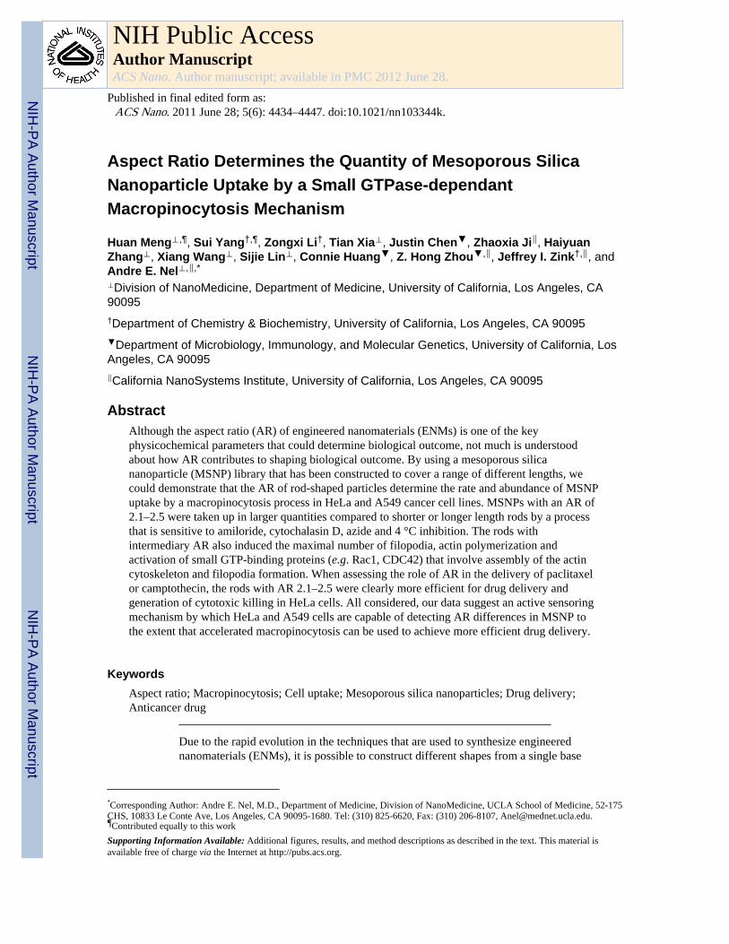
Aspect Ratio Determines the Quantity of Mesoporous Silica Nanoparticle Uptake by a Small GTPase-dependant Macropinocytosis Mechanism Huan Meng ⊥,¶ , Sui Yang †,¶ , Zongxi Li † , Tian Xia ⊥ , Justin Chen ▼ , Zhaoxia Ji ‖ , Haiyuan Zhang ⊥ , Xiang Wang ⊥ , Sijie Lin ⊥ , Connie Huang ▼ , Z. Hong Zhou ▼,‖ , Jeffrey I. Zink †,‖ , and Andre E. Nel ⊥,‖,* ⊥ Division of NanoMedicine, Department of Medicine, University of California, Los Angeles, CA 90095 † Department of Chemistry & Biochemistry, University of California, Los Angeles, CA 90095 ▼ Department of Microbiology, Immunology, and Molecular Genetics, University of California, Los Angeles, CA 90095 ‖ California NanoSystems Institute, University of California, Los Angeles, CA 90095 Abstract Although the aspect ratio (AR) of engineered nanomaterials (ENMs) is one of the key physicochemical parameters that could determine biological outcome, not much is understood about how AR contributes to shaping biological outcome. By using a mesoporous silica nanoparticle (MSNP) library that has been constructed to cover a range of different lengths, we could demonstrate that the AR of rod-shaped particles determine the rate and abundance of MSNP uptake by a macropinocytosis process in HeLa and A549 cancer cell lines. MSNPs with an AR of 2.1–2.5 were taken up in larger quantities compared to shorter or longer length rods by a process that is sensitive to amiloride, cytochalasin D, azide and 4 °C inhibition. The rods with intermediary AR also induced the maximal number of filopodia, actin polymerization and activation of small GTP-binding proteins (e.g. Rac1, CDC42) that involve assembly of the actin cytoskeleton and filopodia formation. When assessing the role of AR in the delivery of paclitaxel or camptothecin, the rods with AR 2.1–2.5 were clearly more efficient for drug delivery and generation of cytotoxic killing in HeLa cells. All considered, our data suggest an active sensoring mechanism by which HeLa and A549 cells are capable of detecting AR differences in MSNP to the extent that accelerated macropinocytosis can be used to achieve more efficient drug delivery. Keywords Aspect ratio; Macropinocytosis; Cell uptake; Mesoporous silica nanoparticles; Drug delivery; Anticancer drug Due to the rapid evolution in the techniques that are used to synthesize engineered nanomaterials (ENMs), it is possible to construct different shapes from a single base * Corresponding Author: Andre E. Nel, M.D., Department of Medicine, Division of NanoMedicine, UCLA School of Medicine, 52-175 CHS, 10833 Le Conte Ave, Los Angeles, CA 90095-1680. Tel: (310) 825-6620, Fax: (310) 206-8107, [email protected]. ¶ Contributed equally to this work Supporting Information Available: Additional figures, results, and method descriptions as described in the text. This material is available free of charge via the Internet at http://pubs.acs.org. NIH Public Access Author Manuscript ACS Nano. Author manuscript; available in PMC 2012 June 28. Published in final edited form as: ACS Nano. 2011 June 28; 5(6): 4434–4447. doi:10.1021/nn103344k. NIH-PA Author Manuscript NIH-PA Author Manuscript NIH-PA Author Manuscript

Welcome message from author
This document is posted to help you gain knowledge. Please leave a comment to let me know what you think about it! Share it to your friends and learn new things together.
Transcript
Aspect Ratio Determines the Quantity of Mesoporous SilicaNanoparticle Uptake by a Small GTPase-dependantMacropinocytosis Mechanism
Huan Meng⊥,¶, Sui Yang†,¶, Zongxi Li†, Tian Xia⊥, Justin Chen▼, Zhaoxia Ji‖, HaiyuanZhang⊥, Xiang Wang⊥, Sijie Lin⊥, Connie Huang▼, Z. Hong Zhou▼,‖, Jeffrey I. Zink†,‖, andAndre E. Nel⊥,‖,*
⊥Division of NanoMedicine, Department of Medicine, University of California, Los Angeles, CA90095†Department of Chemistry & Biochemistry, University of California, Los Angeles, CA 90095▼Department of Microbiology, Immunology, and Molecular Genetics, University of California, LosAngeles, CA 90095‖California NanoSystems Institute, University of California, Los Angeles, CA 90095
AbstractAlthough the aspect ratio (AR) of engineered nanomaterials (ENMs) is one of the keyphysicochemical parameters that could determine biological outcome, not much is understoodabout how AR contributes to shaping biological outcome. By using a mesoporous silicananoparticle (MSNP) library that has been constructed to cover a range of different lengths, wecould demonstrate that the AR of rod-shaped particles determine the rate and abundance of MSNPuptake by a macropinocytosis process in HeLa and A549 cancer cell lines. MSNPs with an AR of2.1–2.5 were taken up in larger quantities compared to shorter or longer length rods by a processthat is sensitive to amiloride, cytochalasin D, azide and 4 °C inhibition. The rods withintermediary AR also induced the maximal number of filopodia, actin polymerization andactivation of small GTP-binding proteins (e.g. Rac1, CDC42) that involve assembly of the actincytoskeleton and filopodia formation. When assessing the role of AR in the delivery of paclitaxelor camptothecin, the rods with AR 2.1–2.5 were clearly more efficient for drug delivery andgeneration of cytotoxic killing in HeLa cells. All considered, our data suggest an active sensoringmechanism by which HeLa and A549 cells are capable of detecting AR differences in MSNP tothe extent that accelerated macropinocytosis can be used to achieve more efficient drug delivery.
KeywordsAspect ratio; Macropinocytosis; Cell uptake; Mesoporous silica nanoparticles; Drug delivery;Anticancer drug
Due to the rapid evolution in the techniques that are used to synthesize engineerednanomaterials (ENMs), it is possible to construct different shapes from a single base
*Corresponding Author: Andre E. Nel, M.D., Department of Medicine, Division of NanoMedicine, UCLA School of Medicine, 52-175CHS, 10833 Le Conte Ave, Los Angeles, CA 90095-1680. Tel: (310) 825-6620, Fax: (310) 206-8107, [email protected].¶Contributed equally to this workSupporting Information Available: Additional figures, results, and method descriptions as described in the text. This material isavailable free of charge via the Internet at http://pubs.acs.org.
NIH Public AccessAuthor ManuscriptACS Nano. Author manuscript; available in PMC 2012 June 28.
Published in final edited form as:ACS Nano. 2011 June 28; 5(6): 4434–4447. doi:10.1021/nn103344k.
NIH
-PA Author Manuscript
NIH
-PA Author Manuscript
NIH
-PA Author Manuscript

material, including spheres, rods, disks, ellipsoids and cylinders.1–5 Besides sphericalnanoparticles that have been intensively investigated, non-spherical ENMs with a highaspect ratio (AR) are of great interest since this physicochemical feature has been shown tohave a disproportionate impact on biological outcome,1, 3 including determining the rate ofcellular uptake,1 the mechanism of uptake,1, 6 particle transportation,7, 8 biodistribution,2and biocompatibility.9, 10 How exactly AR impacts cellular function is unknown but thisinformation is of considerable importance in understanding how to improve nanomaterialsafety and uptake. In order to understand the impact of AR on cellular function, it isnecessary to construct an ENM library in which the same base material is used forconstructing a range of AR’s. One example is synthesis of triacrylate or monomethacrylatenanoparticles through the “particle replication in non-breaking templates” (PRINT)fabrication technique, allowing these investigators to look at the uptake of cylindricalparticles that exhibit an AR of 1–3 in HeLa cells.1 The authors demonstrated that theparticles with an AR of 3 (450 nm×150 nm) were taken up four times more rapidly than theparticles with an AR of 1. Another example is the use of a set of colloidal goldnanoparticles, composed of spheres (14 nm and 74 nm) and rods (14 nm × 74 nm) to studycellular uptake in HeLa cells.4 This demonstrated a 2–4 times higher uptake of sphericalgold nanoparticles compared to rod-shaped particles with an AR of 5.
We constructed a mesoporous silica nanoparticle (MSNP) library composed of spherical androd-shaped particles to study the impact of AR variation on cellular uptake in HeLa andA549 cancer cells. The advantage of using mesoporous silica is that this material is usefulfor drug delivery as well as studying ENM safety.11–13 AR effects on MSNP cellular uptakeadds another design feature that could improve the utility of this multifunctional platformthat is being used for drug delivery by controlling pore opening with nanovalves, surfaceligation, surface charge variation, etc.12–18 Our data demonstrate that rod-shaped MSNPwith an AR of 2.1–2.5 is preferentially endocytosed by an active uptake mechanism that iscapable of distinguishing intermediary length from longer and shorter rods. This implies acellular mechanism capable of discerning and responding to rod length. Furtherinvestigation of the molecular pathway that involves filopodia formation andmacropinocytosis demonstrated that intermediary length rods that are capable of smallGTPase activation (e.g. Rac1, CDC42), leading to cytoskeletal activation and formation offilopodia. We also observed that the intermediary length rods are more effective for thedelivery of chemotherapeutic agents to HeLa cells.
RESULTSSynthesis and physicochemical characterization of MSNPs
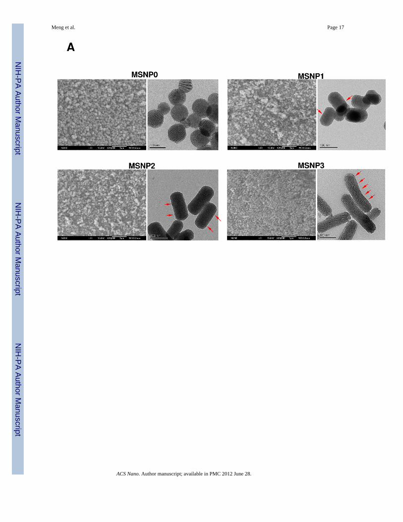
MSNPs were synthesized via a modified procedure to yield rod-shaped structures.19 Themorphology of the rods, in comparison to the spherical particles, is shown in the SEM andTEM images in Fig. 1A. MSNP0 are spheres of ~110 nm diameter, exhibiting uniform poresizes of 2.5 nm and an AR = 1 (Fig. 1A). By increasing the perfluorooctanoic acid (PFOA)/cetyltrimethylammonium bromide (CTAB) doping ratio during the synthesis, it was possibleto obtain rod-shaped cylinders with dimensions of 110–130/60–80 nm (MSNP1), 160–190/60–90 nm (MSNP2), and 260–300/50–70 nm (MSNP3), respectively (Table 1). Thesevalues were used to calculate the AR, which varied from 1.5–1.7 (MSNP1), 2.1–2.5(MSNP2) and 4–4.5 (MSNP3). In spite of differences in the AR, the pore structure and poresizes of the rods are similar (Fig. 1 and Table 1). However, with an increase in the AR of theMSNP from 1 to 4.5, the surface area of these particles decreased from 1077.9 m2/g to 760.2m2/g (Table 1). Small angle powder XRD analysis showed 3 well-resolved peaks that can beindexed as the 100, 110 and 200 reflections and confirms the hexagonal symmetry (p6m) ofthe pore structure.20 The rod-shaped particles showed periodical “fringes” along their length
Meng et al. Page 2
ACS Nano. Author manuscript; available in PMC 2012 June 28.
NIH
-PA Author Manuscript
NIH
-PA Author Manuscript
NIH
-PA Author Manuscript

(indicated by arrows in Fig. 1A), which represent the ordered helical hexagonal porearrangements as reported in the literature.19, 21, 22
To optimize the particle dispersal for purposes of biological experimentation, the MSNPwere suspended in distilled water containing 5% fetal bovine serum (FBS) and ultimatelytransferred into complete RPMI containing 10% FBS.13 Not only does this suspensionsequence prevents particle agglomeration due to culture medium ionic effects but alsoadjusts the particles’ zeta potentials from positive to negative as a result of proteinadsorption (Table 1).
AR influences cellular uptake of MSNPs in HeLa cells and A549 cellsPrevious publications have shown that a non-spherical shape enhances the abundance andrate of cellular uptake of nanoparticles.1, 3, 6, 23 In order to assess cellular uptake of FITC-labeled MSNP in HeLa cells and A549 cell line, we used a combination of flow cytometryand confocal microscopy. The fluorescence labeling efficiency is similar for all particlesurfaces, allowing us to compare the uptake abundance directly (Fig. S1A). Moreover,comparison of the sedimentation rate during a dynamic assessment of the suspensionstability index showed that the spheres and rods behave similarly and do not lead to differentrates of settling that could affect cellular uptake (Fig. S1B). However, compared to sphericalparticles (MSNP0), there was a significant increase in the mean fluorescence intensity (MFI)of cells exposed to rod-shaped particles (Fig. 2A). This amounted to an 18, 40 and 8-foldincrease of the MFI for MSNP1, MSNP2 and MSNP3, respectively. Interestingly, thecellular association was consistently higher in MSNP2 than MSNP1 or MSNP3, suggestingthat the intermediary AR is preferred for cellular uptake. The preference for a rod vs. aspherical shape was further confirmed by comparing FITC-labeled spheres and rods withRITC-labeled spheres (Fig. 2A). Although there is a slight increase in the uptake of theRITC-labeled spheres in cells that were subsequently treated with FITC-labeled rods, thisuptake was small (<3 fold) compared to rods. The small increase likely represents RITC-labeled spheres that are not removed by washing and is internalized during themacropinocytosis of the rods.
The flow data was confirmed by confocal images showing higher uptake of FITC-labeledrods compared to the FITC-labeled spheres (Fig. 2B). Moreover, among the rods, theparticles with an AR of 2.1–2.5 were endocytosed in greater abundance (Fig. 2B) and at amore rapid rate compared to MSNP1 and MSNP3 (Fig. 2C). MSNP2 appeared to localizepredominantly in the perinuclear region of HeLa cells whereas MSNP1 and MSNP3 weremore randomly distributed (Fig. 2B).
To further confirm that the impact of AR is not unique to HeLa cells, the experiments wererepeated in the A549 lung cancer cell line (Fig. S2). Consistent with the HeLa data, MSNP2yielded the highest cellular uptake compared to spheres or longer/shorter rods (Fig. S2).
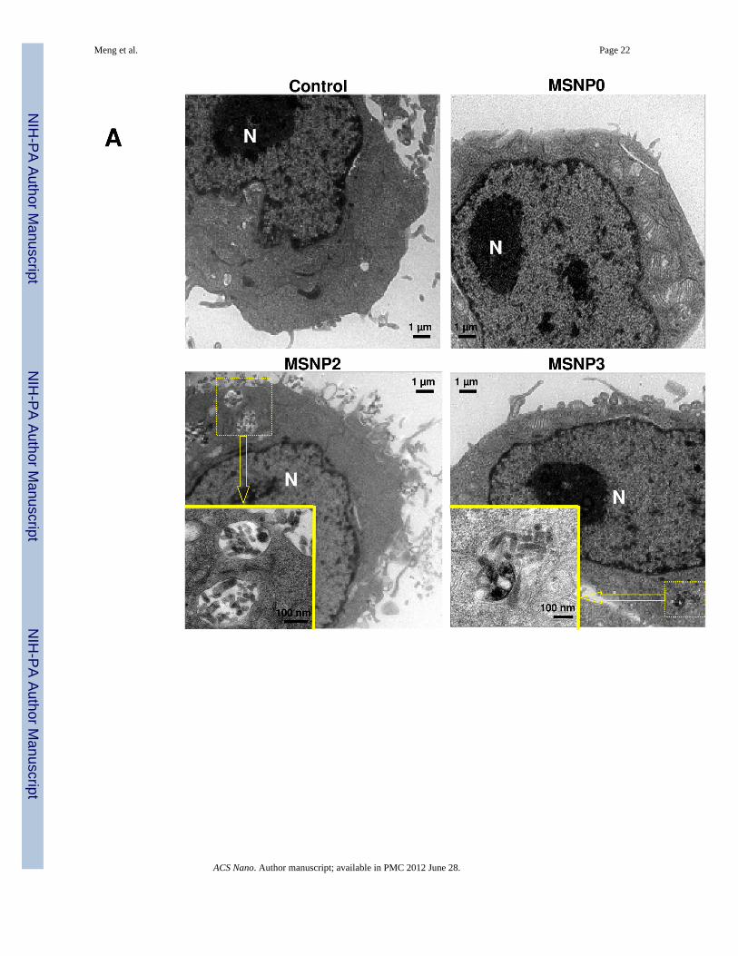
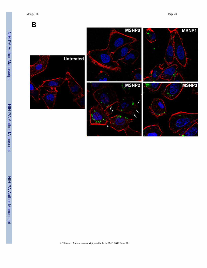
MSNP2 uptake requires cytoskeletal activation and filopodia formationElectron microscopy (EM) was used for ultrastructural resolution of MSNP uptake in HeLacells (Fig. 3). Although HeLa cells are capable of endocytosing nanoparticles by variousroutes,1, 24 MSNPs were taken up by a process of macropinocytosis as evidenced by thepresence of filopodia and formation of macropinocytotic vesicles (Fig. 3A).25 Noteworthy,the number of filopodia and extent of membrane ruffling were dramatically enhanced incells exposed to MSNP2 compared to cells exposed to MSNP3 (Fig. 3A) or MSNP1 (notshown). More detailed TEM images of the macropinocytosis process are shown online (Fig.S3) together with electron tomography and 3D image reconstruction (Fig. S4 and Video V1,V2). Electron tomography was powerful enough to capture the porous structure of the
Meng et al. Page 3
ACS Nano. Author manuscript; available in PMC 2012 June 28.
NIH
-PA Author Manuscript
NIH
-PA Author Manuscript
NIH
-PA Author Manuscript
particles inside the cells, an ultrastructural feature that has not previously beenaccomplished.
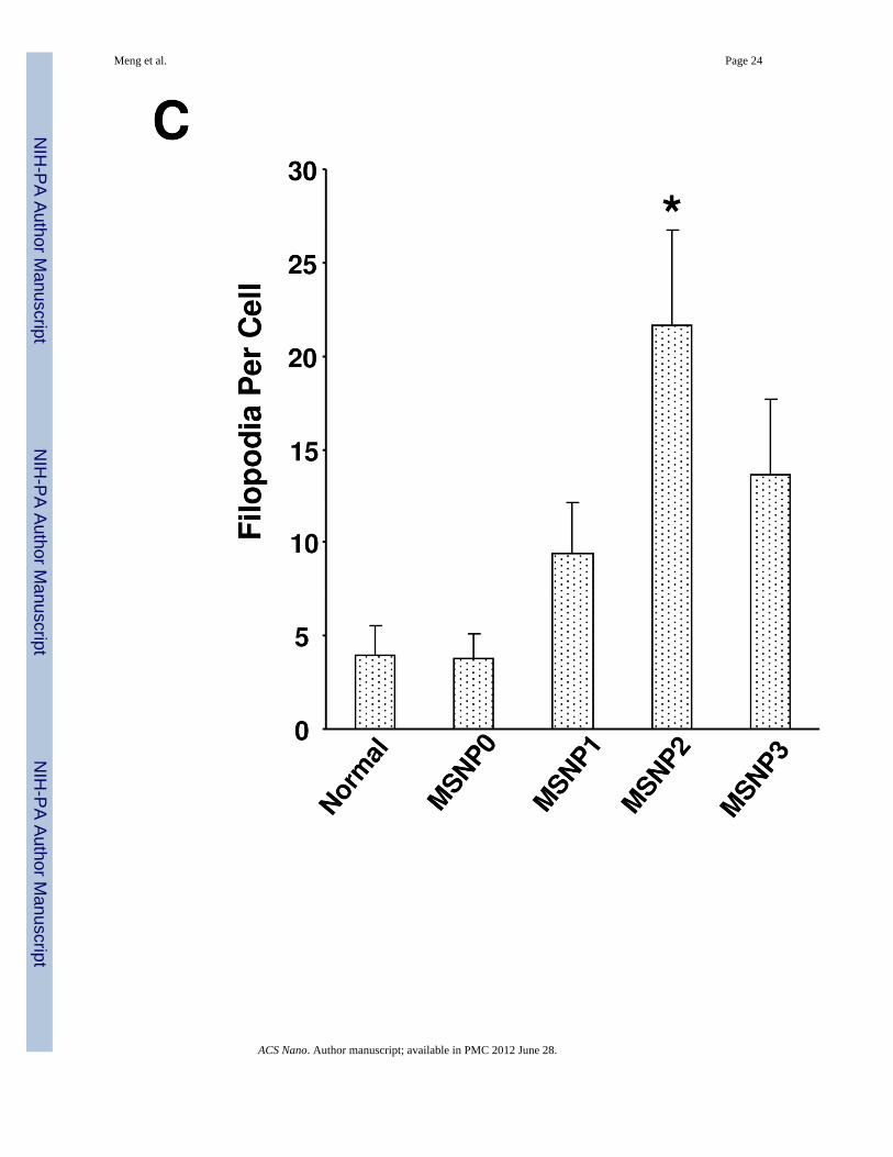
Since macropinocytosis and filopodia formation is dependent on actin assembly at the cellmembrane,26–28 we performed Alexa 594-phalloidin staining to visualize these cytoskeletalchanges. The confocal images demonstrate that the actin polymerization accompanyingfilopodia formation is more prominent in cells treated with MSNP2 than the other particletypes; this takes on the form of radially distributed spikes that stand away from the ring-likecortical cytoskeleton (Fig. 3B, arrow). Scoring of the number of filopodia (spike) by atechnique previously published in the literature29 (and explained in the legend of Fig. 3C),demonstrated a 5, 2.5 and 1.8-fold increase in filopodia number in cells treated with MSNP2compared to MSNP0, MSNP1 and MSNP3 (Fig. 3C). In addition, the actin assembly inresponse to MSNP2 was sustained for at least 6 h compared to the transient effects seen withstimuli such as epidermal growth factor.26
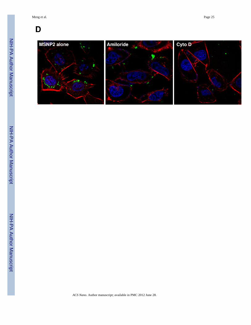
Chemical inhibitors are widely used to confirm the occurrence of macropinocytosis and as ameans of distinguishing this uptake mechanism from clathrin- and caveolae-mediateduptake.26, 30 Prior cellular treatment with amiloride26 as well as the cytoskeletal inhibitorcytochalasin D (Cyto D)1, 31 decreased MSNP2 uptake by >50% (Fig. 3D). These agentsalso interfered in MSNP2-induced filopodia formation (Fig. 3E). Moreover, during the useof filipin to inhibit caveolar uptake through cholesterol sequestration32 as well as employingmonodansylcadaverine to inhibit formation of clathrin-coated pits33, we did not observe anyinterference in MSNP2 uptake in HeLa cells (Fig. S5A). The energy dependence of themacropinocytosis pathway was confirmed by culturing the cells at 4 °C 34 or pretreatingthem with sodium azide (NaN3)/2-deoxyglucose (2-DG) for 3 h before the addition of theparticles (Fig. S5B).1 The latter inhibitors also decreased filopodia formation and MSNPuptake by >60% (Fig. 3E).
To study the subcellular fate of endocytosed MSNP2, confocal microscopy was used tolocalize FITC-labeled MSNP2 in relation to clathrin-coated vesicles, caveolae andlysosomes in HeLa cells (Fig. S6).35 These endocytotic compartments were localized byRITC-labeled secondary antibodies capable of binding to primary antibodies recognizingclathrin, caveolin-1 and LAMP-1, respectively. Image J software analysis to quantify FITC-labeled MSNP2 colocalization with labeled compartments showed that while <1% of theserods were localized in clathrin or caveolin-1 stained compartments (Fig. S6, upper panel),more MSNP2 entered a LAMP-1 positive compartment (Fig. S6, lower panel). Thus, while<5% of the particles entered the LAMP-1 positive compartment by 6 h, this fractionincreased to 75% by 36 h. All considered, these data indicate that MSNP2 are primarilytaken up by macropinocytosis and are ultimately transferred to an acidifying endosomalcompartment that specializes in particle degradation. Interestingly, the MSNP-ladenlysosomes could be seen to change their localization from a random to a clusteredarrangement in the cell (Fig. S6, bottom right panel). The significance of this redistributionis uncertain.
MSNP2 promotes active macropinocytosis through the activation of small GTPasesMacropinocytosis is frequently triggered by external stimuli, such as viruses25 or growthfactors36 capable of activating membrane-associated signaling cascades that play a role inactin polymerization and plasma membrane ruffling.26, 30 One of the signaling mechanismsinvolves the activation of the GTP-binding protein, Rac1, which plays a role in membraneruffling, cytoskeletal changes and closure of macropinosome.26, 30 In order to see if Rac1 isactivated during MSNP macropinocytosis, we used of an immunochemical technique thatdistinguishes activated, GTP-bound Rac1 from total cellular Rac1 protein.37–39 This wasaccomplished by using a secondary Alexa 594-conjugated antibody that recognizes anti-
Meng et al. Page 4
ACS Nano. Author manuscript; available in PMC 2012 June 28.
NIH
-PA Author Manuscript
NIH
-PA Author Manuscript
NIH
-PA Author Manuscript
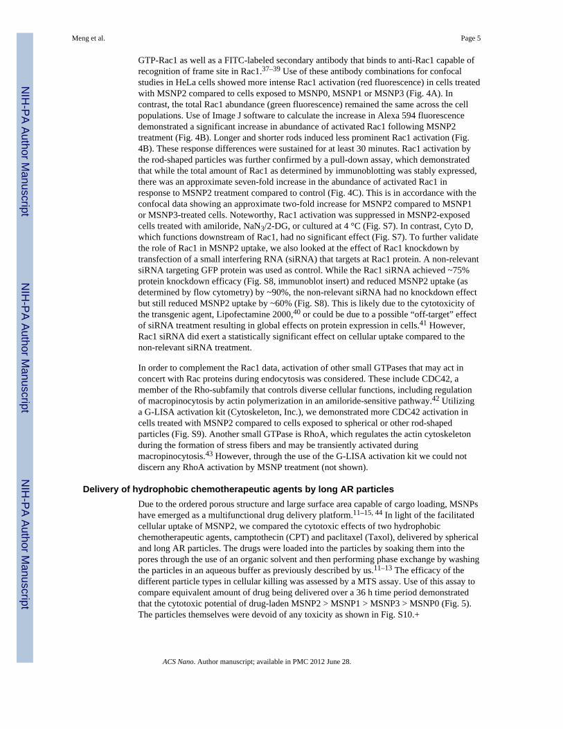
GTP-Rac1 as well as a FITC-labeled secondary antibody that binds to anti-Rac1 capable ofrecognition of frame site in Rac1.37–39 Use of these antibody combinations for confocalstudies in HeLa cells showed more intense Rac1 activation (red fluorescence) in cells treatedwith MSNP2 compared to cells exposed to MSNP0, MSNP1 or MSNP3 (Fig. 4A). Incontrast, the total Rac1 abundance (green fluorescence) remained the same across the cellpopulations. Use of Image J software to calculate the increase in Alexa 594 fluorescencedemonstrated a significant increase in abundance of activated Rac1 following MSNP2treatment (Fig. 4B). Longer and shorter rods induced less prominent Rac1 activation (Fig.4B). These response differences were sustained for at least 30 minutes. Rac1 activation bythe rod-shaped particles was further confirmed by a pull-down assay, which demonstratedthat while the total amount of Rac1 as determined by immunoblotting was stably expressed,there was an approximate seven-fold increase in the abundance of activated Rac1 inresponse to MSNP2 treatment compared to control (Fig. 4C). This is in accordance with theconfocal data showing an approximate two-fold increase for MSNP2 compared to MSNP1or MSNP3-treated cells. Noteworthy, Rac1 activation was suppressed in MSNP2-exposedcells treated with amiloride, NaN3/2-DG, or cultured at 4 °C (Fig. S7). In contrast, Cyto D,which functions downstream of Rac1, had no significant effect (Fig. S7). To further validatethe role of Rac1 in MSNP2 uptake, we also looked at the effect of Rac1 knockdown bytransfection of a small interfering RNA (siRNA) that targets at Rac1 protein. A non-relevantsiRNA targeting GFP protein was used as control. While the Rac1 siRNA achieved ~75%protein knockdown efficacy (Fig. S8, immunoblot insert) and reduced MSNP2 uptake (asdetermined by flow cytometry) by ~90%, the non-relevant siRNA had no knockdown effectbut still reduced MSNP2 uptake by ~60% (Fig. S8). This is likely due to the cytotoxicity ofthe transgenic agent, Lipofectamine 2000,40 or could be due to a possible “off-target” effectof siRNA treatment resulting in global effects on protein expression in cells.41 However,Rac1 siRNA did exert a statistically significant effect on cellular uptake compared to thenon-relevant siRNA treatment.
In order to complement the Rac1 data, activation of other small GTPases that may act inconcert with Rac proteins during endocytosis was considered. These include CDC42, amember of the Rho-subfamily that controls diverse cellular functions, including regulationof macropinocytosis by actin polymerization in an amiloride-sensitive pathway.42 Utilizinga G-LISA activation kit (Cytoskeleton, Inc.), we demonstrated more CDC42 activation incells treated with MSNP2 compared to cells exposed to spherical or other rod-shapedparticles (Fig. S9). Another small GTPase is RhoA, which regulates the actin cytoskeletonduring the formation of stress fibers and may be transiently activated duringmacropinocytosis.43 However, through the use of the G-LISA activation kit we could notdiscern any RhoA activation by MSNP treatment (not shown).
Delivery of hydrophobic chemotherapeutic agents by long AR particlesDue to the ordered porous structure and large surface area capable of cargo loading, MSNPshave emerged as a multifunctional drug delivery platform.11–15, 44 In light of the facilitatedcellular uptake of MSNP2, we compared the cytotoxic effects of two hydrophobicchemotherapeutic agents, camptothecin (CPT) and paclitaxel (Taxol), delivered by sphericaland long AR particles. The drugs were loaded into the particles by soaking them into thepores through the use of an organic solvent and then performing phase exchange by washingthe particles in an aqueous buffer as previously described by us.11–13 The efficacy of thedifferent particle types in cellular killing was assessed by a MTS assay. Use of this assay tocompare equivalent amount of drug being delivered over a 36 h time period demonstratedthat the cytotoxic potential of drug-laden MSNP2 > MSNP1 > MSNP3 > MSNP0 (Fig. 5).The particles themselves were devoid of any toxicity as shown in Fig. S10.+
Meng et al. Page 5
ACS Nano. Author manuscript; available in PMC 2012 June 28.
NIH
-PA Author Manuscript
NIH
-PA Author Manuscript
NIH
-PA Author Manuscript
DISCUSSIONIn this paper, we demonstrate that HeLa and A549 cells endocytose rod-shaped MSNPthrough a macropinocytosis process that is capable of discerning the AR and adapting thecellular response so that uptake is maximal for particles with an AR of 2.1–2.5. Thisdifferential effect involves filopodia formation that is linked to activation of the actincytoskeleton by a small GTP-binding protein. These findings suggest a mechanosensitiveprocess that is capable of translating variations in AR into small GTPase activation,including Rac1 and CDC42. Particles taken up into the pinocytotic vesicles are shuttled intoan acidifying lysosomal compartment and can be used to deliver hydrophobicchemotherapeutic agents. This utility was demonstrated by the delivery of Taxol and CPT inHeLa cells, with optimal cytotoxicity being achieved when the drugs are loaded into rodswith an AR of 2.1–2.5.
Current research in the field of biomaterials is witnessing the emergence of a powerful set ofnew design parameters, including the use of physical shape at micron and nanoscale levelsto control biological responses.3, 8, 10, 45, 46 Shape is an important physical characteristic andhas an important role in modeling cellular responsiveness and associated applications inbiotechnology.1 Theoretical models and experimental studies have confirmed the benefits ofusing non-spherical particles for drug delivery based on their effects on cellularinternalization and vascular dynamics.1, 2, 7, 47 This includes the observation that for micron-size particles the local geometry at the point of contact with the cell membrane rather thanthe overall particle shape dictates whether macrophages initiate internalization.45 In thisregard, it has been demonstrated that when a macrophage encounters an elliptical disc at itspointed edge or side, the particle is rapidly phagocytosed within minutes whereas attachmentto the flat side of the disc failed to initiate phagocytosis for an extensive timeperiod.6, 8, 45, 48 The effect of geometry in phagocytosis could be quantified by measuringthe angle between the membrane at the point of initial contact and the line defining theparticle curvature at this point. If this angle exceeds a critical value of >45°, the cells lost theability to entrap the particles.3, 45 The authors proposed that the particle shape at the point ofattachment determines the type of actin rearrangements that are required for cellularuptake.3, 45 According to this view, the requisite actin structures can not form if the angle is>45°, leading the macrophages to switch to an alternative spreading behavior.45
At a much smaller length scale, Gratton et al. demonstrated that internalization of cylindricalparticles depends on AR.1 In this elegant study, the authors evaluated internalizationpathways of three different series of micro- and nano-sized particles made from the cross-linked PEG-based hydrogels produced by the PRINT technique.1 The synthesized materialsincluded cubic microparticles, cylindrical microparticles and cylindrical nanoparticles(200×200 nm; 100×300 nm; 150×450 nm). While all the particles were internalized by HeLacells, the nanoparticles entered more rapidly, with the longer cylinders (150×450 nm) beingcaptured more rapidly than cubic particles of nearly similar volume (200×200 nm) or shortercylinders of lower volume (100×300 nm). Particles with an AR of 3 were internalized aboutfour times more rapidly than spheres of the same volume.1 In contrast to the results forhydrogel particles, other studies have found that receptor-mediated endocytosis of Au nano-objects was significantly decreased with increased AR.4, 49, 50 For instance, Au nanosphereswith diameters of 14 nm or 74 nm were endocytosed three times more readily by HeLa cellscompared to 74×14 nm rods.4 Muro et al. compared targeted accumulation of spheres ofvarious diameters (ranging from 100 nm to 10 µm) or elliptical discs of microscaledimensions (1×3 µm) in tissues and found that the targeting efficiency of micron-scale discsis better than spheres, even those with nanoscale dimensions.51
Meng et al. Page 6
ACS Nano. Author manuscript; available in PMC 2012 June 28.
NIH
-PA Author Manuscript
NIH
-PA Author Manuscript
NIH
-PA Author Manuscript
This paper enhances our understanding of the role of AR by demonstrating that for MSNP ofthe same chemical composition there is differential macropinocytosis of particles with anAR 2.1–2.5. Moreover, this effect is mediated through a pathway that involves the activationof small GTP-binding proteins (e.g. Rac1), the actin cytoskeleton and filopodia formation.52
Not only is the demonstration that Rac1 and CDC42 activation by rod-shaped MSNP anovel finding, but also provides a platform for understanding how specific shape variationscan be used to modulate cellular function. Macropinocytosis refers to the formation of largeendocytic vesicles of irregular shape and size that is generated by actin-driven evaginationsof the plasma membrane.26 Macropinocytosis is an efficient route for the non-selectiveuptake of soluble macromolecules, and is either constitutive or stimulated by growth factorssuch as epidermal growth factor, platelet derived growth factor, macrophage colony-stimulating factor, interleukin-4 or phorbol esters.26, 30, 53 It has been reported that antibodyconjugates directed against intercellular adhesion molecule (ICAM-1) or platelet-endothelialcell adhesion molecule (PECAM-1) can promote drug delivery by vehicles that arerecognized and internalized by endothelial cells through macropinocytosis.54 Interestingly,these agents are all capable of initiating signaling pathways that activate small GTPases,which have also been shown to play a role in regulating macropinocytosis.36 The occurrenceof macropinocytosis in a variety of different cell types suggests that it contributes to cellularfunctions such as nutrient uptake,55 host-pathogen interactions,56 antigen processing57 anddirected cell movement.55 Macropinocytosis differs from other endocytic mechanisms thatare involved in the uptake of individual nanoparticles by smaller vesicles.26 Different fromclathrin-dependent, caveolae-mediated or caveolae- and clathrin-independent pathways,58
macropinocytosis is dependent on signaling to the actin cytoskeleton and actin-drivenmembrane movement.26, 35 This results in membrane ruffling and the formation of filopodiathat are necessary for the closure of the macropinocytotic vesicles.26 The intracellular fate ofmacropinosomes varies depending on the cell type but in most cases end up fusing withlysosomes and shrinking.55
A key question that remains is how the differences in AR are being discerned and translatedinto small GTPase activation and actin assembly in HeLa and A549 cells? While we do nothave an exact explanation, it is worthwhile considering the role of the integrated-adhesomenetwork that responds to complex chemosensitive and mechanosensitive environmental cuesin the extracellular matrix (ECM).59 Cells demonstrate an extraordinary ability to respond toa wide range of physical signals, either locally or globally and are capable of reacting toECM topography, including its rigidity,60 spatial organization and anisotropy.59, 61 Thecrucial scaffolding interactions involved in the link of the ECM to the actin cytoskeletoninvolve actin-polymerizing and actin-linking modules associated with the integrin-receptorsystem.59 This system as a whole is mechanoresponsive and numerous studies havedemonstrated that the biochemical characteristics of the ECM, including its spatialorganization, are recognized by cells as a result of differential signaling from integrin-basedmolecular complexes.59, 62 This molecular machinery can be viewed as a network of tightlyinterconnected modules that contains ~700 links, most involving binding interactions withthe rest consisting of pathways that modify those interactions.59 The biological activities ofthe adhesome components are quite diverse and include several actin regulators that affectthe organization of the attached cytoskeleton. These include adapter proteins that link actinto integrins (directly or indirectly) as well as a wide range of signaling molecules, includingkinases, phosphatases, GTP-binding proteins and their regulators.59
How could this model apply to the sensing and response to AR variations of a nanomaterial?While it is unlikely that integrins are directly involved in this process, MSNPs may bedecorated with serum proteins that could act as ligands on the particle surface. However, weregard this as an unlikely explanation because macropinocytosis differs from receptor-mediated endocytosis that classically proceeds via caveolae or clathrin-mediated uptake.
Meng et al. Page 7
ACS Nano. Author manuscript; available in PMC 2012 June 28.
NIH
-PA Author Manuscript
NIH
-PA Author Manuscript
NIH
-PA Author Manuscript
Another possibility is that AR variation could be detected at the contact site of the nanoscalespheres and rods with the cell surface membrane or the lipid bilayer lining themacropinocytotic vesicles. In this regard, it has been demonstrated that silanol groups onsilica nanoparticles are capable of interacting with membrane lipid compounds and possiblyalso with electrostatically charged membrane proteins.63, 64 It is possible that the numberand spatial distribution of these contact sites may be capable of relaying information tosmall GTPases by a transduction process that either involves GTP loading via a guaninenucleotide exchange factor or inhibiting the activity of the GTPase. The exquisite sensitivityof cells to variations in adhesive patch spacing at the nanoscale level has been demonstratedthrough the use of nano-lithography approaches, e.g., positioning of nanoscale gold particles(1–15 nm) in spatial arrangements or formation of spacing gradients that can be tuned at 10–200 nm scales.59, 65, 66 Examination of cellular spreading on these surfaces havedemonstrated that the weakest gradient to which cells can respond corresponds to a strengthof ~15 nm per mm, provided that the interparticle spacings remained at 58–73 nm.59, 67, 68
Given a typical spreading length of 60 µm, the implication is that cells can respond to 1 nmdifferences in average ligand patch spacing between the front and rear end of the cell.59
Thus, the sensitivity to small variations in interparticle spacing is quite remarkable andprobably achievable in a time-integrated manner.59 Interestingly, these variations are muchsmaller than the typical variations in the inter-ligand spacing that governs integrininteractions with the ECM. Thus, given this exquisite sensitivity at the nanoscale level, wepropose that a related mechanism may be involved in detecting AR differences in rod-shaped MSNP.
Not only do these results advance our understanding of the mechanism by which ARdifferences change cellular uptake, but also allows us to add a shape design feature to the listof tunable MSNP properties that can be exploited to improve drug delivery.11, 15, 69, 70
Accumulated evidence indicates that spherical nano carriers may not be optimal drugdelivery devices due to the poor cellular uptake and unsatisfactory biodistribution comparedto disc-shaped objects or particles exhibiting a long AR.1, 2, 7 To improve cellular uptake ofMSNP, we have previously shown that better uptake can be achieved by non-covalentattachment of the cationic polymer, polyethyleneimine, to the particle surface.12, 13
However, cationic functionalization could lead to cytotoxicity.12, 13 We now demonstrateimproved delivery and killing in HeLa cells by using an optimal AR of 2.1–2.5 (Fig. 5). Tofurther understand the bio-behavior of AR particles in vivo, we are currently performinganimal experiments to test the efficacy of a non-spherical MSNP delivery system.
CONCLUSIONWe demonstrate through the use of AR variation in a designed MSNP library that cellsactively respond to the shape change by changing particle uptake through amacropinocytosis process. Not only can HeLa and A549 cells distinguish between specificARs, but are also capable of translating this recognition into activation of small GTPasesuch as Rac1, control actin assembly and optimal filopodia formation that leads to maximaluptake of intermediary length rods. This active uptake process can be inhibited by a list ofinhibitors (amiloride, Cyto D and NaN3/2-DG) or dropping the culture temperature to 4 °C.We also demonstrate that intermediary length MSNP with an AR of 2.1–2.5 are moreefficient at delivery hydrophobic chemotherapeutic agents to HeLa cells.
MATERIALS AND METHODSExperiment Reagents
Commercial reagents and analytical materials used in this study include: tetraethoxysilane(TEOS, 98%, Aldrich), cetyltrimethylammonium bromide (CTAB, 98%, Aldrich),
Meng et al. Page 8
ACS Nano. Author manuscript; available in PMC 2012 June 28.
NIH
-PA Author Manuscript
NIH
-PA Author Manuscript
NIH
-PA Author Manuscript
perfluorooctanoic acid (PFOA, 96%, Aldrich), methanol (99.9%, Fisher), triethylamine(99.5%, Aldrich), ethanol (200 proof, Pharmaco-AAPER), 3-aminopropyltriethoxysilane(APTES, 99%, Gelest), rhodamine-B-isothiocyanate (RITC, Aldrich), fluoresceinisothiocynate (FITC, >90%, Aldrich), amiloride (Aldrich), cytochalasin D (Aldrich), filipin(Aldrich), monodansylcadaverine (Aldrich), sodium azide (NaN3, Aldrich), and 2-deoxyglucose (2-DG, Aldrich), camptothecin (CPT, 99%, Aldrich), Taxol (99%, Aldrich).RPMI 1640 cell culture medium, penicillin/streptomycin, L-glutamine, Alexa 594-phalloidin, and Hoechst 33342 were purchased from Invitrogen (Carlsbad, CA). Fetalbovine serum (FBS) was from Atlanta Biologicals, Inc (Lawrenceville, GA). Anti-LAMP-1and anti-GTP-Rac1 antibody were obtained from Abcam (Cambridge, MA) and Cellsignaling (Danvers, MA). Anti-total-Rac1, anti-clathrin and anti-caveolin antibodies werepurchased from Santa Cruz Biotechnology (Santa Cruz, CA). Deionized H2O was obtainedfrom a Millipore water purification system. Toluene (99.5%) refluxed before use.
MSNP SynthesisMSNP with different AR were chemically synthesized by a sol-gel approach using asurfactant/co-structure direct agent (CSDA) mixture as template. Different shaped MSNPand AR were synthesized by varying the PFOA/CTAB molar ratio as follows: MSNP0 = 0;MSNP1 = 0.015; MSNP2 = 0.03; MSNP3 = 0.06. The PFOA/CTAB mixture was stirred at600 rpm for 1 h at room temperature before the addition of 2.1 mL 2 M NaOH.Subsequently, the solution temperature was raised to 80 °C before the addition of 4.1 mL ofTEOS. This mixture was stirred for an additional 2 h and the precipitate was carefullycollected by filtration. After washing 3 times with methanol and deionized water, the solidswere air dried overnight at room temperature. The MSNP templates were removed byextracting the CTAB with HCl containing methanol under nitrogen protection. The resultingMSNP were collected by filtration, washed by methanol, and dried overnight in air.Depending on the shape and AR, the particles were designated MSNP0, MSNP1, MSNP2and MSNP3 as explained in Table 1. To visualize the particles under a confocal microscopeor perform flow cytometry, FITC- or RITC-labeled MSNP were prepared by suspending 200mg of MSNP in 10 mL of dry toluene with 3 µL of APTES, followed by the attachment ofFITC or RITC in ethanol. The labeled MSNP were collected by centrifugation and washedby methanol and water before use.
Physicochemical Characterization of MSNPMSNP were characterized for shape (aspect ratio), surface area, hydrodynamic size, and zetapotential. The shape and morphology of the MSNP were characterized by scanning electronmicroscopy (SEM, JSM-6700F) at 15 kV. The pore structure and aspect ratio were studiedby using a transmission electron microscope, JEM 1200-EX operated at 80 kV. Microfilmsfor TEM imaging were obtained by placing a drop of the respective MSNP suspensions ontoa 200-mesh copper TEM grid and drying at room-temperature overnight. The AR valueswere determined by measuring the length and diameter of at least 30 randomly selectedparticles. X-ray diffraction (XRD) patterns were recorded on a Panalytical X'Pert ProPowder Diffractometer with Ni-filtered Cu Kα radiation. UV-vis spectra were collectedusing a Cary 5000 UV-vis-NIR spectrophotometer. For surface area measurement, N2adsorption-desorption isotherms were obtained on a Quadrasorb SI surface area analyzer andpore size analyzer (Quantachrome instruments, Florida, USA). The BET model was appliedto evaluate the specific surface areas and the Barret-Joyner-Halenda (BJH) method was usedto calculate the pore size. Particle hydrodynamic size and zeta potential were measured inpure water or cell culture medium using a ZetaSizer Nano (Malvern Instruments Ltd.,Worcestershire, UK).
Meng et al. Page 9
ACS Nano. Author manuscript; available in PMC 2012 June 28.
NIH
-PA Author Manuscript
NIH
-PA Author Manuscript
NIH
-PA Author Manuscript
MSNP Dispersion and Use to Perform Tissue CultureAll cell cultures were maintained in 75 cm2 cell culture flasks in which the cells werepassaged at 70–80% confluency every 2–3 days. The human cervical (HeLa) and lungcancer (A549) cells were cultured in RPMI 1640 containing 10% FBS, 100 U/mL penicillin,100 µg/mL streptomycin, and 2 mM L-glutamine (complete RPMI medium). To disperseMSNP in cell culture medium, the water stock solution was sonicated (Tekmar SonicDisruptor probe) for 10 s before use. In order to coat the surface of MSNP with FBS, 19 µLMSNP suspension was mixed with 1 µL FBS. Complete cell culture media was then addedto the serum-coated MSNP suspension.
Assessment of cellular MSNP uptake by flow cytometry and confocal microscopyFor the performance of flow cytometry, aliquots of 1 × 10⁵ cells (HeLa & A549 cells) werecultured in 12-well plates in 1 mL complete RPMI medium. RITC-labeled spherical MSNPwere added to the above cultures at 20 µg/mL for 1 h, washed carefully in PBS and thenincubated with different FITC-labeled MSNP at 20 µg/mL for 6 h in complete RPMImedium. The purpose of prior incubation with the RITC-labeled spheres is to demonstratethat cells can distinguish between a spherical shape and a rod-shaped particle. All cell typeswere trypsinized and washed in PBS for 3 times. Cells were analyzed in a SCAN flowcytometer using mean FL-2 and FL-1 to assess RITC and FITC fluorescence, respectively.Data are reported as fold increase in mean fluorescence intensity (MFI), using MSNP0uptake as reference.
For the confocal studies, cellular uptake of the spheres and different AR particles wasperformed by adding 20 µg/mL FITC-labeled particles to 8-well chamber slides. Each wellcontained 5 × 10⁴ cells in 0.5 mL complete culture medium. Cell membranes were co-stained with 5 µg/mL Alexa Fluor 633-conjugated wheat germ agglutinin (WGA) in PBS for30 min. Slides were mounted with Hoechst 33342 and visualized under a confocalmicroscope (Leica Confocal 1P/FCS) in the UCLA/CNSI Advanced Light Microscopy/Spectroscopy Shared Facility. High magnification images were obtained with the 100×objective.
Transmission electron microscopy of MSNP treated cellsHeLa cells were treated with each of the MSNP variants at 20 µg/mL for 3 h in completeRPMI. The cells were washed in PBS and immediately fixed with 2.5% glutaraldehyde inPBS. After secondary fixation in 1% OsO4 in PBS, the cells were dehydrated in a gradedethanol series, treated with propylene oxide, and embedded in resin. Approximately 60–70nm thick sections were cut on a Leica ultramicrotome and picked up on formvar-coatedcopper grids. The sections were examined in a CM120 electron microscope (Philips).
Electron tomographyTEM grids were used for the tomography imaging on an FEI Tecnai F20 microscopeoperated at 200 kV. Tilt series were acquired by tilting the specimen from −70˚ to 70˚ at 1˚increments and with a TIETZ F415MP 16 megapixel CCD camera at a magnification of26,600× and then aligned using the ETOMO program in the IMOD package.71 The alignedtilt series were then further processed using the reconstruction Inspect3D program from FEI,with the SIRT option, to improve reconstruction accuracy and contrast. 3D movies of thealigned tilt series were generated using both Inspect3d and Windows movie maker.Visualization of 3D tomographic reconstructions were performed using 3DMOD ofIMOD 71 and Amira (http://www.amira.com/).
Meng et al. Page 10
ACS Nano. Author manuscript; available in PMC 2012 June 28.
NIH
-PA Author Manuscript
NIH
-PA Author Manuscript
NIH
-PA Author Manuscript
F-actin staining and filopodia countingHeLa cells were seeded into 8-well chamber slides and incubated with or without MSNP at aconcentration of 20 µg/mL in complete RPMI for 6 h. Cells were washed 3 times in PBS,fixed with 3.7% formaldehyde for 30 min, and permeabilized with 0.25% Triton X-100 for10 min. For F-actin staining, cells were incubated with Alex 594 phalloidin in the dark for30 min at room temperature. The slides were viewed using a confocal microscope equippedwith a 100× oil-immersion objective. The average number of filopodia per cell asdetermined by actin fiber staining was calculated by counting at least 20 randomly selectedcells in each exposure category.29 After filopodia counting, two-sided Student’s t test wasperformed to determine whether there was a statistically significant difference.29
Drug and temperature inhibition studiesHeLa cells were seeded in a 12-wells plate at the density of 1×10⁵ per well. The cells werepre-cultured in serum free RPMI 1640 medium containing amiloride (75 µM), Cyto D (2.5µg/mL), or 0.1% NaN3/50 mM 2-DG for 3 h. Alternatively, cells were placed at 4 °C. After3 h pretreatment, the media was exchanged into fresh complete RPMI 1640 that contained20 µg/mL FITC-labeled MSNP2, one of the chemical inhibitors (amiloride, Cyto D orNaN3/2-DG), and 10% FBS for a further 6 h culture time. To observe a temperature effect,the cellular incubation with particles was carried out at 4 °C for 6 h. Following a 6 hincubation period, the cells were washed with PBS and then processed for flow cytometry asdescribed above. The filopodia counting was performed by confocal microscopy asdescribed above. We also conducted experiments with the caveolar and clathrin coatedinhibitors. The cells were pre-cultured in complete RPMI 1640 medium containing filipin (5µM), or monodansylcadaverine (50 µM) for 3 h. At this time point, the medium wasreplaced by fresh complete RPMI containing 20 µg/mL FITC-labeled MSNP2 and filipin (5µM) or monodansylcadaverine (50 µM) and cells incubated for a further 6 h time period.The cells were washed with PBS and then processed for flow cytometry analysis.
Assessment of cellular Rac1 activationTo study Rac1 activation, HeLa cells were serum-starved for 4 h before introduction of theparticles that were dispersed in RPMI containing 10% FBS. Cells were incubated with theparticles for 30 min at a concentration of 20 µg/mL. Because FBS may also contribute toRac1 activation due to the presence of growth factors, it was necessary to include a controlwhere serum starved cells were exposed to complete RPMI for 30 min to allow adjustmentfor any possible Rac1 activation due to the complete medium. After fixation andpermeabilization, cells were stained with antibodies recognizing the activated, GTP-boundform of Rac1 or total Rac1 protein.37–39 The primary antibodies were detected by secondaryantibodies conjugated to Alexa 594 or FITC, respectively. Nuclei were stained by Hoechst33342. The Alexa 594 fluorescence intensity as a measure of Rac1 activation wasdetermined by Image J software. The fluorescence intensity of control cells incubated inserum-containing medium for 30 min was chosen as reference with fluorescent intensity = 1.The fluorescence intensity of cells treated with the various MSNPs was then expressed asthe fold-increase compared to this control value. The experiment was repeated in presenceof amiloride, Cyto D, NaN3/2-DG as well as 4 °C as described above.
In order to further confirm the confocal data, particle induced Rac1 activation was detectedby a pull-down assay using configuration-specific monoclonal antibody Rac activation assaykit (NewEast Biosciences, PA) according to the manufacture’s instruction. Briefly, HeLacells were seeded into 10 cm tissue culture dish (~2×10⁷ cells/dish) and received the variousparticle treatments as described above. After cold PBS washing for 3 times, 1 mL ice-coldlysis buffer was added to the cells. Aliquots of each cell lysate were added to twomicrocentrifuge tubes, one for analysis of the active and the other for the analysis of total
Meng et al. Page 11
ACS Nano. Author manuscript; available in PMC 2012 June 28.
NIH
-PA Author Manuscript
NIH
-PA Author Manuscript
NIH
-PA Author Manuscript
Rac1 content. The protein content of the supernatants was determined by the Bradfordmethod. After the addition of 1 µL of the monoclonal antibody recognizing active Rac1 thecell lysates received the addition of 20 µL protein A/G agarose beads. After incubation at 4°C for 1 h with gentle agitation, the beads were collected and washed by lysis buffer for 3times. Following the resuspension of the bead pellet in 20 µL SDS-PAGE sample buffer, thesamples were electrophoresed by 12% SDS-PAGE and transferred to a PVDF membrane.After blocking, the membranes were incubated with 1:1000 dilution of primary polyclonalantibody to Rac1 (Santa Cruz Biotechnology, CA). The membranes were overlayed withgoat anti-mouse secondary antibody (1:1000 dilution) before the addition of the HRP-conjugated streptavidin-biotin complex. The proteins were detected by using the ECLreagent according to the manufacturer’s instructions. The total Rac1 protein was detected bywestern blotting using anti-Rac1 antibody (Santa Cruz Biotechnology, CA).
Drug loading and loading capacity measurementTwo hydrophobic anticancer drugs, Taxol and camptothecin (CPT), were loaded intodifferent MSNP using 20 mg of each type of particles suspended into a solution containing0.5 mg of each drug in 1mL DMSO. After 24 h, the particles were collected bycentrifugation, and the trace amounts of DMSO were removed by drying under vacuum. Thedrug-laden particles were washed and stored as an aqueous stock solution at 20 mg/mL. Tomeasure the loading capacity of the particles, they were resuspended in methanol andthoroughly sonicated to remove the drug from the pores. After centrifugation and removal ofthe supernatant, the process was repeated 3 times to elute all drug. The supernatants werecombined and the amount of free drug calculated using Taxol absorbance at 229 nm and theCPT fluorescence at excitation and emission wavelengths of 370 and 448 nm, respectively,in a microplate reader (SpectraMax M5 Microplate Reader, Molecular Device, USA).
Assessment of HeLa cytotoxicity in response to treatment with drug loaded particlesHeLa cells were plated at 1 × 10⁴ cells per well in 96 well plates. MSNP, loaded with CPTor Taxol, were incubated with the cells to deliver CPT concentrations of 0.1–8.0 µg/mL andTaxol concentrations of 0.1–2.0 µg/mL for 36 h. The effect of the MSNP-bound drug wascompared to similar amounts of free drug delivered in DMSO/PBS. A MTS assay wasperformed 36 h after drug and particle exposure to assess cellular viability as previous.described.13
Statistical analysisData represent the mean ± SD for duplicate or triplicate measurements in each experiment.Differences between the mean values were analyzed by two-sided Student’s t test or oneway ANVOA. All experiments were repeated at least twice.
Supplementary MaterialRefer to Web version on PubMed Central for supplementary material.
AcknowledgmentsThis study was funded by the U.S. Public Health Service Grants, U19 ES019528 (UCLA Center for NanoBiologyand Predictive Toxicology) as well as RO1 CA133697, RO1 ES016746, and RC2 ES018766. Support for this studywas also provided the National Science Foundation and the Environmental Protection Agency under CooperativeAgreement Number EF 0830117. The work was also supported by the NSF USDOD HDTRA 1-08-1-0041 grantsand grant 53292 from the Bill & Melinda Gates Foundation through the Grand Challenges Exploration Initiative.We acknowledge the use of electron microscope facilities in the UCLA Electron Imaging Center for NanoMachinessupported by NIH (1S10RR23057 to ZHZ).
Meng et al. Page 12
ACS Nano. Author manuscript; available in PMC 2012 June 28.
NIH
-PA Author Manuscript
NIH
-PA Author Manuscript
NIH
-PA Author Manuscript
REFERENCES AND NOTES1. Gratton SEA, Ropp PA, Pohlhaus PD, Luft JC, Madden VJ, Napier ME, DeSimone JM. The Effect
of Particle Design on Cellular Internalization Pathways. Proc. Natl. Acad. Sci. U.S.A. 2008;105:11613–11618. [PubMed: 18697944]
2. Decuzzi P, Godin B, Tanaka T, Lee SY, Chiappini C, Liu X, Ferrari M. Size and Shape Effects inthe Biodistribution of Intravascularly Injected Particles. J. Control. Release. 2010; 141:320–327.[PubMed: 19874859]
3. Mitragotri S, Lahann J. Physical Approaches to Biomaterial Design. Nat. Mater. 2009; 8:15–23.[PubMed: 19096389]
4. Chithrani BD, Ghazani AA, Chan WCW. Determining the Size and Shape Dependence of GoldNanoparticle Uptake into Mammalian Cells. Nano Lett. 2006; 6:662–668. [PubMed: 16608261]
5. Yang S, Zhou X, Yuan P, Yu M, Xie S, Zou J, Lu G, Yu C. Siliceous Nanopods from aCompromised Dual-Templating Approach. Angew. Chem. Int. Ed. Engl. 2007; 46:8579–8582.[PubMed: 17924601]
6. Doshi N, Mitragotri S. Macrophages Recognize Size and Shape of Their Targets. PLoS ONE. 2010;5:e10051. [PubMed: 20386614]
7. Lee S-Y, Ferrari M, Decuzzi P. Shaping Nano-/Micro-particles for Enhanced Vascular Interaction inLaminar Flows. Nanotechnology. 2009; 20:495101. [PubMed: 19904027]
8. Champion JA, Katare YK, Mitragotri S. Particle shape: A New Design Parameter for Micro- andNanoscale Drug Delivery Carriers. J. Control. Release. 2007; 121:3–9. [PubMed: 17544538]
9. Meng H, Xia T, George S, Nel AE. A Predictive Toxicological Paradigm for the Safety Assessmentof Nanomaterials. ACS Nano. 2009; 3:1620–1627. [PubMed: 21452863]
10. Nel AE, Madler L, Velegol D, Xia T, Hoek EMV, Somasundaran P, Klaessig F, Castranova V,Thompson M. Understanding Biophysicochemical Interactions at the Nano-Bio Interface. Nat.Mater. 2009; 8:543–557. [PubMed: 19525947]
11. Lu J, Liong M, Zink J. I., Tamanoi, F., Mesoporous Silica Nanoparticles as a Delivery System forHydrophobic Anticancer Drugs. Small. 2007; 3:1341–1346. [PubMed: 17566138]
12. Meng H, Liong M, Xia T, Li Z, Ji Z, Zink JI, Nel AE. Engineered Design of Mesoporous SilicaNanoparticles to Deliver Doxorubicin and P-Glycoprotein siRNA to Overcome Drug Resistance ina Cancer Cell Line. ACS Nano. 2010; 4:4539–4550. [PubMed: 20731437]
13. Xia T, Kovochich M, Liong M, Meng H, Kabehie S, George S, Zink JI, Nel AE.Polyethyleneimine Coating Enhances the Cellular Uptake of Mesoporous Silica Nanoparticles andAllows Safe Delivery of siRNA and DNA Constructs. ACS Nano. 2009; 3:3273–3286. [PubMed:19739605]
14. Liong M, Lu J, Kovochich M, Xia T, Ruehm SG, Nel AE, Tamanoi F, Zink JI. MultifunctionalInorganic Nanoparticles for Imaging, Targeting, and Drug Delivery. ACS Nano. 2008; 2:889–896.[PubMed: 19206485]
15. Meng H, Xue M, Xia T, Zhao Y-L, Tamanoi F, Stoddart JF, Zink JI, Nel AE. Autonomous in VitroAnticancer Drug Release from Mesoporous Silica Nanoparticles by pH-Sensitive Nanovalves. J.Am. Chem. Soc. 2010; 132:12690–12697. [PubMed: 20718462]
16. Liong M, Angelos S, Choi E, Patel K, Stoddart JF, Zink JI. Mesostructured MultifunctionalNanoparticles for Imaging and Drug Delivery. J. Mater. Chem. 2009; 19:6251–6257.
17. Ferris DP, Zhao Y-L, Khashab NM, Khatib HA, Stoddart JF, Zink JI. Light-Operated MechanizedNanoparticles. J. Am. Chem. Soc. 2009; 131:1686–1688. [PubMed: 19159224]
18. Coti KK, Belowich ME, Liong M, Ambrogio MW, Lau YA, Khatib HA, Zink JI, Khashab NM,Stoddart JF. Mechanised Nanoparticles for Drug Delivery. Nanoscale. 2009; 1:16–39. [PubMed:20644858]
19. Yang S, Zhao L, Yu C, Zhou X, Tang J, Yuan P, Chen D, Zhao D. On the Origin of HelicalMesostructures. J. Am. Chem. Soc. 2006; 128:10460–10466. [PubMed: 16895411]
20. Kresge CT, Leonowicz ME, Roth WJ, Vartuli JC, Beck JS. Ordered Mesoporous Molecular SievesSynthesized by a Liquid-Crystal Template Mechanism. Nature. 1992; 359:710–712.
21. Zhang L, Qiao SZ, Cheng L, Yan Z, Lu GQM. Fabrication of a Magnetic Helical MesostructuredSilica Rod. Nanotechnology. 2008; 19:435608.
Meng et al. Page 13
ACS Nano. Author manuscript; available in PMC 2012 June 28.
NIH
-PA Author Manuscript
NIH
-PA Author Manuscript
NIH
-PA Author Manuscript
22. Ohsuna T, Liu Z, Che S, Terasaki O. Characterization of Chiral Mesoporous Materials byTransmission Electron Microscopy. Small. 2005; 1:233–237. [PubMed: 17193437]
23. Huang X, Teng X, Chen D, Tang F, He J. The Effect of the Shape of Mesoporous SilicaNanoparticles on Cellular Uptake and Cell Function. Biomaterials. 2010; 31:438–448. [PubMed:19800115]
24. Dausend J, Musyanovych A, Dass M, Walther P, Schrezenmeier H, Landfester K, Mailänder V.Uptake Mechanism of Oppositely Charged Fluorescent Nanoparticles in HeLa Cells. Macromol.Biosci. 2008; 8:1135–1143. [PubMed: 18698581]
25. Kalin S, Amstutz B, Gastaldelli M, Wolfrum N, Boucke K, Havenga M, DiGennaro F, Liska N,Hemmi S, Greber UF. Macropinocytotic Uptake and Infection of Human Epithelial Cells withSpecies B2 Adenovirus Type 35. J. Virol. 2010; 84:5336–5350. [PubMed: 20237079]
26. Mercer J, Helenius A. Virus Entry by Macropinocytosis. Nat. Cell Biol. 2009; 11:510–520.[PubMed: 19404330]
27. Nakase I, Niwa M, Takeuchi T, Sonomura K, Kawabata N, Koike Y, Takehashi M, Tanaka S,Ueda K, Simpson JC, et al. Cellular Uptake of Arginine-Rich Peptides: Roles forMacropinocytosis and Actin Rearrangement. Mol. Ther. 2004; 10:1011–1022. [PubMed:15564133]
28. Hillaireau H, Couvreur P. Nanocarriers’ Entry into the Cell: Relevance to Drug Delivery. Cell.Mol. Life Sci. 2009; 66:2873–2896. [PubMed: 19499185]
29. Sheldon H, Andre M, Legg JA, Heal P, Herbert JM, Sainson R, Sharma AS, Kitajewski JK, HeathVL, Bicknell R. Active Involvement of Robo1 and Robo4 in Filopodia Formation and EndothelialCell Motility Mediated via WASP and Other Actin Nucleation-Promoting Factors. FASEB J.2009; 23:513–522. [PubMed: 18948384]
30. West MA, Bretscher MS, C W. Distinct Endocytotic Pathways in Epidermal Growth Factor-Stimulated Human Carcinoma A431 Cells. J. Cell Biol. 1989; 109:2731–2739. [PubMed:2556406]
31. Schrijvers DM, Martinet W, De Meyer GRY, Andries L, Herman AG, Kockx MM. FlowCytometric Evaluation of a Model for Phagocytosis of Cells Undergoing Apoptosis. J. Immunol.Methods. 2004; 287:101–108. [PubMed: 15099759]
32. Greyner HJ, Wiraszka T, Zhang L-S, Petroll WM, Mummert M. E., Inducible Macropinocytosis ofHyaluronan in B16-F10 Melanoma Cells. Matrix Biol. 2010; 29:503–510. [PubMed: 20600893]
33. von Delwig A, Hilkens C, Altmann D, Holmdahl R, Isaacs J, Harding C, Robertson H, McKie N,Robinson J. Inhibition of Macropinocytosis Blocks Antigen Presentation of Type II Collagen InVitro and In Vivo In HLA-DR1 Transgenic Mice. Arthritis Res. Ther. 2006; 8:1–11.
34. Li W, Chen C, Ye C, Wei T, Zhao YZ, Lao FL, Chen Z, Meng H, Gao Y, Yuan H, et al. TheTranslocation of Fullerenic Nanoparticles into Lysosome via the Pathway of Clathrin-MediatedEndocytosis. Nanotechnology. 2008; 19:145102.
35. Conner SD, Schmid SL. Regulated Portals of Entry into the Cell. Nature. 2003; 422:37–44.[PubMed: 12621426]
36. Ridley AJ, Paterson HF, Johnston CL, Diekmann D, Hall A. The Small GTP-binding Protein RacRegulates Growth Factor-Induced Membrane Ruffling. Cell. 1992; 70:401–410. [PubMed:1643658]
37. Li Q, Ho CS, Marinescu V, Bhatti H, Bokoch GM, Ernst SA, Holz RW, Stuenkel EL. Facilitationof Ca2+-Dependent Exocytosis by Rac1-GTPase in Bovine Chromaffin Cells. J. Physiol. 2003;550:431–445. [PubMed: 12754309]
38. Osada T, Watanabe S, Tanaka H, Hirose M, Miyazaki A, Sato N. Effect of Mechanical Strain onGastric Cellular Migration and Proliferation During Mucosal Healing: Role of Rho Dependent andRac Dependent Cytoskeletal Reorganisation. Gut. 1999; 45:508–515. [PubMed: 10486357]
39. Ueda S, Kataoka T, Satoh T. Activation of the Small GTPase Rac1 by a Specific Guanine-Nucleotide-Exchange Factor Suffices to Induce Glucose Uptake into Skeletal-Muscle Cells. Biol.Cell. 2008; 100:645–657. [PubMed: 18482007]
40. Clements BA, Incani V, Kucharski C, Lavasanifar A, Ritchie B, Uludag H. A ComparativeEvaluation of Poly-l-lysine-palmitic Acid and Lipofectamine (TM) 2000 for Plasmid Delivery toBone Marrow Stromal Cells. Biomaterials. 2007; 28:4693–4704. [PubMed: 17686514]
Meng et al. Page 14
ACS Nano. Author manuscript; available in PMC 2012 June 28.
NIH
-PA Author Manuscript
NIH
-PA Author Manuscript
NIH
-PA Author Manuscript
41. Jackson AL, Linsley PS. Recognizing and Avoiding siRna Off-Target Effects For TargetIdentification and Therapeutic Application. Nat. Rev. Drug Discov. 2010; 9:57–67. [PubMed:20043028]
42. Koivusalo M, Welch C, Hayashi H, Scott CC, Kim M, Alexander T, Touret N, Hahn KM,Grinstein S. Amiloride Inhibits Macropinocytosis by Lowering Submembranous pH andPreventing Rac1 and Cdc42 Signaling. J. Cell Biol. 2010; 188:547–563. [PubMed: 20156964]
43. Mercer J, Knebel S, Schmidt FI, Crouse J, Burkard C, Helenius A. Vaccinia Virus Strains UseDistinct Forms Of Macropinocytosis for Host-Cell Entry. Proc. Natl. Acad. Sci. U.S.A. 2010;107:9346–9351. [PubMed: 20439710]
44. Slowing II, Trewyn BG, Lin VSY. Mesoporous Silica Nanoparticles for Intracellular Delivery ofMembrane-Impermeable Proteins. J. Am. Chem. Soc. 2007; 129:8845–8849. [PubMed: 17589996]
45. Champion JA, Mitragotri S. Role of Target Geometry in Phagocytosis. Proc. Natl. Acad. Sci.U.S.A. 2006; 103:4930–4934. [PubMed: 16549762]
46. Simone EA, Dziubla TD, Muzykantov VR. Polymeric Carriers: Role of Geometry in DrugDelivery. Expert Opin. Drug Deliv. 2008; 5:1283–1300. [PubMed: 19040392]
47. Decuzzi P, Ferrari M. The Receptor-Mediated Endocytosis of Nonspherical Particles. Biophys. J.2008; 94:3790–3797. [PubMed: 18234813]
48. Champion JA, Katare YK, Mitragotri S. Making Polymeric Micro- and Nanoparticles of ComplexShapes. Proc. Natl. Acad. Sci. U.S.A. 2007; 104:11901–11904. [PubMed: 17620615]
49. Arnida, Malugin A, Ghandehari H. Cellular Uptake and Toxicity of Gold Nanoparticles in ProstateCancer Cells: A Comparative Study of Rods and Spheres. J. Appl. Toxicol. 2010; 30:212–217.[PubMed: 19902477]
50. Chithrani BD, Chan WCW. Elucidating the Mechanism of Cellular Uptake and Removal ofProtein-Coated Gold Nanoparticles of Different Sizes and Shapes. Nano Lett. 2007; 7:1542–1550.[PubMed: 17465586]
51. Muro S, Garnacho C, Champion JA, Leferovich J, Gajewski C, Schuchman EH, Mitragotri S,Muzykantov VR. Control of Endothelial Targeting and Intracellular Delivery of TherapeuticEnzymes by Modulating the Size and Shape of ICAM-1-targeted Carriers. Mol. Ther. 2008;16:1450–1458. [PubMed: 18560419]
52. Hayakawa K, Tatsumi H, Sokabe M. Actin Stress Fibers Transmit and Focus Force to ActivateMechanosensitive Channels. J. Cell Sci. 2008; 121:496–503. [PubMed: 18230647]
53. Fiorentini C, Falzano L, Fabbri A, Stringaro A, Logozzi M, Travaglione S, Contamin S, AranciaG, Malorni W, Fais S. Activation of Rho GTPases by Cytotoxic Necrotizing Factor 1 InducesMacropinocytosis and Scavenging Activity in Epithelial Cells. Mol. Biol. Cell. 2001; 12:2061–2073. [PubMed: 11452003]
54. Muro S, Wiewrodt R, Thomas A, Koniaris L, Albelda SM, Muzykantov VR, Koval M. A NovelEndocytic Pathway Induced by Clustering Endothelial ICAM-1 or PECAM-1. J. Cell. Sci. 2003;116:1599–1609. [PubMed: 12640043]
55. Dharmawardhane S, Schurmann A, Sells MA, Chernoff J, Schmid SL, Bokoch GM. Regulation ofMacropinocytosis by p21-activated Kinase-1. Mol. Biol. Cell. 2000; 11:3341–3352. [PubMed:11029040]
56. Chen L-M, Hobbie S, Galan JE. Requirement of CDC42 for Salmonella-Induced Cytoskeletal andNuclear Responses. Science. 1996; 274:2115–2118. [PubMed: 8953049]
57. Sandgren KJ, Wilkinson J, Miranda-Saksena M, McInerney GM, Byth-Wilson K, Robinson PJ,Cunningham AL. A Differential Role for Macropinocytosis in Mediating Entry of the Two Formsof Vaccinia Virus into Dendritic Cells. PLoS Pathog. 2010; 6:e1000866. [PubMed: 20421949]
58. Sahay G, Alakhova DY, Kabanov AV. Endocytosis of Nanomedicines. J. Control. Release. 2010;145:182–195. [PubMed: 20226220]
59. Geiger B, Spatz JP, Bershadsky AD. Environmental Sensing Through Focal Adhesions. Nat. Rev.Mol. Cell Biol. 2009; 10:21–33. [PubMed: 19197329]
60. Discher DE, Janmey P, Wang Y. Tissue Cells Feel and Respond to the Stiffness of Their Substrate.Science. 2005; 310:1139–1143. [PubMed: 16293750]
Meng et al. Page 15
ACS Nano. Author manuscript; available in PMC 2012 June 28.
NIH
-PA Author Manuscript
NIH
-PA Author Manuscript
NIH
-PA Author Manuscript
61. Thery M, Racine V, Piel M, Pepin A, Dimitrov A, Chen Y, Sibarita J-B, Bornens M. Anisotropy ofCell Adhesive Microenvironment Governs Cell Internal Organization and Orientation of Polarity.Proc. Natl. Acad. Sci. U.S.A. 2006; 103:19771–19776. [PubMed: 17179050]
62. Butler B, Gao C, Mersich AT, Blystone SD. Purified Integrin Adhesion Complexes Exhibit Actin-Polymerization Activity. Curr. Biol. 2006; 16:242–251. [PubMed: 16461277]
63. Roiter Y, Ornatska M, Rammohan AR, Balakrishnan J, Heine DR, Minko S. Interaction ofNanoparticles with Lipid Membrane. Nano Lett. 2008; 8:941–944. [PubMed: 18254602]
64. Slowing II, Wu CW, Vivero-Escoto JL, Lin VSY. Mesoporous Silica Nanoparticles for ReducingHemolytic Activity Towards Mammalian Red Blood Cells. Small. 2009; 5:57–62. [PubMed:19051185]
65. Glass R, Arnold M, Blümmel J, Küller A, Möller M, Spatz JP. Micro-Nanostructured InterfacesFabricated by the Use of Inorganic Block Copolymer Micellar Monolayers as Negative Resist forElectron-Beam Lithography. Adv. Funct. Mater. 2003; 13:569–575.
66. Glass R, Arnold M, Cavalcanti-Adam EA, Blümmel J, Haferkemper C, Dodd C, Spatz JP. BlockCopolymer Micelle Nanolithography on Non-Conductive Substrates. New J. of Phys. 2004; 6:101.
67. Arnold M, Cavalcanti-Adam EA, Glass R, Blümmel J, Eck W, Kantlehner M, Kessler H, Spatz J.P., Activation of Integrin Function by Nanopatterned Adhesive Interfaces. ChemPhysChem. 2004;5:383–388. [PubMed: 15067875]
68. Cavalcanti-Adam EA, Volberg T, Micoulet A, Kessler H, Geiger B, Spatz JP. Cell Spreading andFocal Adhesion Dynamics Are Regulated by Spacing of Integrin Ligands. Biophys. J. 2007;92:2964–2974. [PubMed: 17277192]
69. Karmali PP, Kotamraju VR, Kastantin M, Black M, Missirlis D, Tirrell M, Ruoslahti E. Targetingof albumin-embedded paclitaxel nanoparticles to tumors. Nanomedicine. 2009; 5:73–82.[PubMed: 18829396]
70. Lobo C, Lopes G, Silva O, Gluck S. Paclitaxel Albumin-Bound Particles (Abraxane(TM)) inCombination with Bevacizumab with or without Gemcitabine: Early Experience at the Universityof Miami/Braman Family Breast Cancer Institute. Biomed. Pharmacother. 2007; 61:531–533.[PubMed: 17913443]
71. Kremer JR, Mastronarde DN, McIntosh JR. Computer Visualization of Three-dimensional ImageData Using IMOD. J. Struct. Biol. 1996; 116:71–76. [PubMed: 8742726]
Meng et al. Page 16
ACS Nano. Author manuscript; available in PMC 2012 June 28.
NIH
-PA Author Manuscript
NIH
-PA Author Manuscript
NIH
-PA Author Manuscript
Meng et al. Page 17
ACS Nano. Author manuscript; available in PMC 2012 June 28.
NIH
-PA Author Manuscript
NIH
-PA Author Manuscript
NIH
-PA Author Manuscript
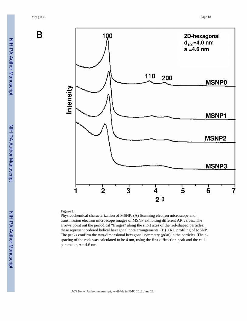
Figure 1.Physicochemical characterization of MSNP. (A) Scanning electron microscope andtransmission electron microscope images of MSNP exhibiting different AR values. Thearrows point out the periodical “fringes” along the short axes of the rod-shaped particles;these represent ordered helical hexagonal pore arrangements. (B) XRD profiling of MSNP.The peaks confirm the two-dimensional hexagonal symmetry (p6m) in the particles. The d-spacing of the rods was calculated to be 4 nm, using the first diffraction peak and the cellparameter, a = 4.6 nm.
Meng et al. Page 18
ACS Nano. Author manuscript; available in PMC 2012 June 28.
NIH
-PA Author Manuscript
NIH
-PA Author Manuscript
NIH
-PA Author Manuscript

Meng et al. Page 19
ACS Nano. Author manuscript; available in PMC 2012 June 28.
NIH
-PA Author Manuscript
NIH
-PA Author Manuscript
NIH
-PA Author Manuscript
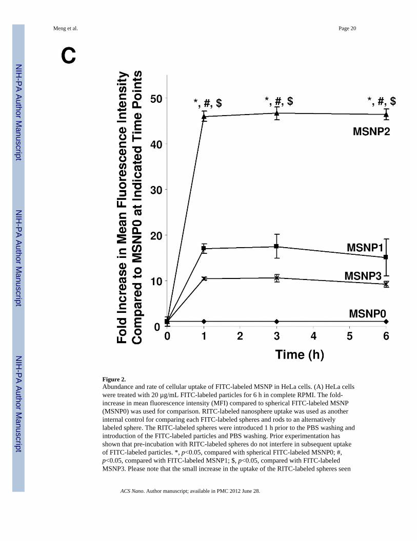
Figure 2.Abundance and rate of cellular uptake of FITC-labeled MSNP in HeLa cells. (A) HeLa cellswere treated with 20 µg/mL FITC-labeled particles for 6 h in complete RPMI. The fold-increase in mean fluorescence intensity (MFI) compared to spherical FITC-labeled MSNP(MSNP0) was used for comparison. RITC-labeled nanosphere uptake was used as anotherinternal control for comparing each FITC-labeled spheres and rods to an alternativelylabeled sphere. The RITC-labeled spheres were introduced 1 h prior to the PBS washing andintroduction of the FITC-labeled particles and PBS washing. Prior experimentation hasshown that pre-incubation with RITC-labeled spheres do not interfere in subsequent uptakeof FITC-labeled particles. *, p<0.05, compared with spherical FITC-labeled MSNP0; #,p<0.05, compared with FITC-labeled MSNP1; $, p<0.05, compared with FITC-labeledMSNP3. Please note that the small increase in the uptake of the RITC-labeled spheres seen
Meng et al. Page 20
ACS Nano. Author manuscript; available in PMC 2012 June 28.
NIH
-PA Author Manuscript
NIH
-PA Author Manuscript
NIH
-PA Author Manuscript

in cells subsequently treated with rods likely reflect a small number of red spheres stickingto the cell surface after washing and then being internalized when the rods activatemacropinocytosis. (B) HeLa cells were seeded into 8-well chamber slides before addition ofthe FITC-labeled particles at 20 µg/mL for 6 h in complete RPMI. After fixation andpermeabilization, cells were stained with 5 µg/mL wheat germ agglutinin 633 and Hoechst33342 dye, following by visualization under a confocal 1P/FCS inverted microscope. (C)Fold-increase in MFI of FITC-labeled rods compared to sphere at 0 to 6 h. HeLa cells wereexposed to different FITC-labeled MSNP at 20 µg/mL, and flow cytometry were conductedat the indicated time points. *, p<0.05 compared with spherical FITC-labeled particle(MSNP0); #, p<0.05 compared with FITC-labeled MSNP1; $, p<0.05 compared with FITC-labeled MSNP3.
Meng et al. Page 21
ACS Nano. Author manuscript; available in PMC 2012 June 28.
NIH
-PA Author Manuscript
NIH
-PA Author Manuscript
NIH
-PA Author Manuscript
Meng et al. Page 22
ACS Nano. Author manuscript; available in PMC 2012 June 28.
NIH
-PA Author Manuscript
NIH
-PA Author Manuscript
NIH
-PA Author Manuscript
Meng et al. Page 23
ACS Nano. Author manuscript; available in PMC 2012 June 28.
NIH
-PA Author Manuscript
NIH
-PA Author Manuscript
NIH
-PA Author Manuscript
Meng et al. Page 24
ACS Nano. Author manuscript; available in PMC 2012 June 28.
NIH
-PA Author Manuscript
NIH
-PA Author Manuscript
NIH
-PA Author Manuscript
Meng et al. Page 25
ACS Nano. Author manuscript; available in PMC 2012 June 28.
NIH
-PA Author Manuscript
NIH
-PA Author Manuscript
NIH
-PA Author Manuscript
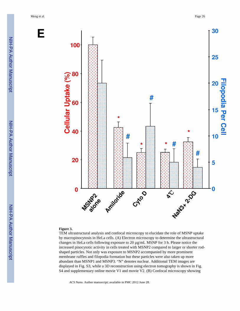
Figure 3.TEM ultrastructural analysis and confocal microscopy to elucidate the role of MSNP uptakeby macropinocytosis in HeLa cells. (A) Electron microscopy to determine the ultrastructuralchanges in HeLa cells following exposure to 20 µg/mL MSNP for 3 h. Please notice theincreased pinocytotic activity in cells treated with MSNP2 compared to larger or shorter rod-shaped particles. Not only was exposure to MSNP2 accompanied by more prominentmembrane ruffles and filopodia formation but these particles were also taken up moreabundant than MSNP1 and MSNP3. “N” denotes nuclear. Additional TEM images aredisplayed in Fig. S3, while a 3D reconstruction using electron tomography is shown in Fig.S4 and supplementary online movie V1 and movie V2. (B) Confocal microscopy showing
Meng et al. Page 26
ACS Nano. Author manuscript; available in PMC 2012 June 28.
NIH
-PA Author Manuscript
NIH
-PA Author Manuscript
NIH
-PA Author Manuscript
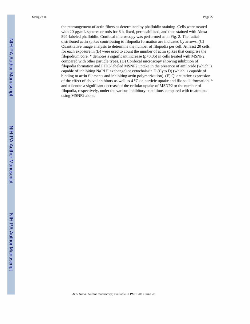
the rearrangement of actin fibers as determined by phalloidin staining. Cells were treatedwith 20 µg/mL spheres or rods for 6 h, fixed, permeabilized, and then stained with Alexa594-labeled phalloidin. Confocal microscopy was performed as in Fig. 2. The radial-distributed actin spikes contributing to filopodia formation are indicated by arrows. (C)Quantitative image analysis to determine the number of filopodia per cell. At least 20 cellsfor each exposure in (B) were used to count the number of actin spikes that comprise thefilopodium core. * demotes a significant increase (p<0.05) in cells treated with MSNP2compared with other particle types. (D) Confocal microscopy showing inhibition offilopodia formation and FITC-labeled MSNP2 uptake in the presence of amiloride (which iscapable of inhibiting Na+/H+ exchange) or cytochalasin D (Cyto D) (which is capable ofbinding to actin filaments and inhibiting actin polymerization). (E) Quantitative expressionof the effect of above inhibitors as well as 4 °C on particle uptake and filopodia formation. *and # denote a significant decrease of the cellular uptake of MSNP2 or the number offilopodia, respectively, under the various inhibitory conditions compared with treatmentsusing MSNP2 alone.
Meng et al. Page 27
ACS Nano. Author manuscript; available in PMC 2012 June 28.
NIH
-PA Author Manuscript
NIH
-PA Author Manuscript
NIH
-PA Author Manuscript
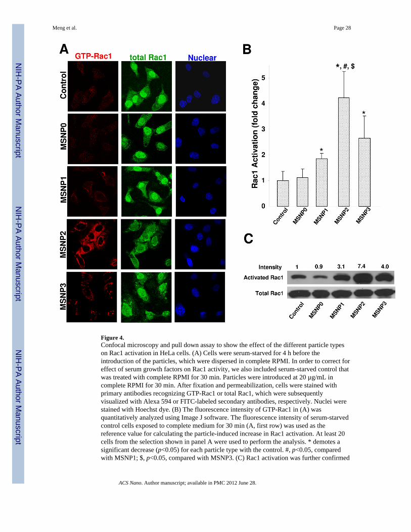
Figure 4.Confocal microscopy and pull down assay to show the effect of the different particle typeson Rac1 activation in HeLa cells. (A) Cells were serum-starved for 4 h before theintroduction of the particles, which were dispersed in complete RPMI. In order to correct foreffect of serum growth factors on Rac1 activity, we also included serum-starved control thatwas treated with complete RPMI for 30 min. Particles were introduced at 20 µg/mL incomplete RPMI for 30 min. After fixation and permeabilization, cells were stained withprimary antibodies recognizing GTP-Rac1 or total Rac1, which were subsequentlyvisualized with Alexa 594 or FITC-labeled secondary antibodies, respectively. Nuclei werestained with Hoechst dye. (B) The fluorescence intensity of GTP-Rac1 in (A) wasquantitatively analyzed using Image J software. The fluorescence intensity of serum-starvedcontrol cells exposed to complete medium for 30 min (A, first row) was used as thereference value for calculating the particle-induced increase in Rac1 activation. At least 20cells from the selection shown in panel A were used to perform the analysis. * demotes asignificant decrease (p<0.05) for each particle type with the control. #, p<0.05, comparedwith MSNP1; $, p<0.05, compared with MSNP3. (C) Rac1 activation was further confirmed
Meng et al. Page 28
ACS Nano. Author manuscript; available in PMC 2012 June 28.
NIH
-PA Author Manuscript
NIH
-PA Author Manuscript
NIH
-PA Author Manuscript

by a pull-down assay using that Rac activation assay kit from NewEast Biosciences(Malvern, PA). After similar particle treatments as in (A), HeLa cells were instantly lysed inice-cold lysis buffer and anti-GTP bound Rac1 monoclonal antibody was added to the celllysates. The bound, active Rac1 protein was then captured by protein A/G agarose andsubsequently detected by western blotting using a second anti-Rac1 antibody. The total Rac1protein was detected by western blotting using anti-Rac1 antibody (Santa CruzBiotechnology, CA).
Meng et al. Page 29
ACS Nano. Author manuscript; available in PMC 2012 June 28.
NIH
-PA Author Manuscript
NIH
-PA Author Manuscript
NIH
-PA Author Manuscript
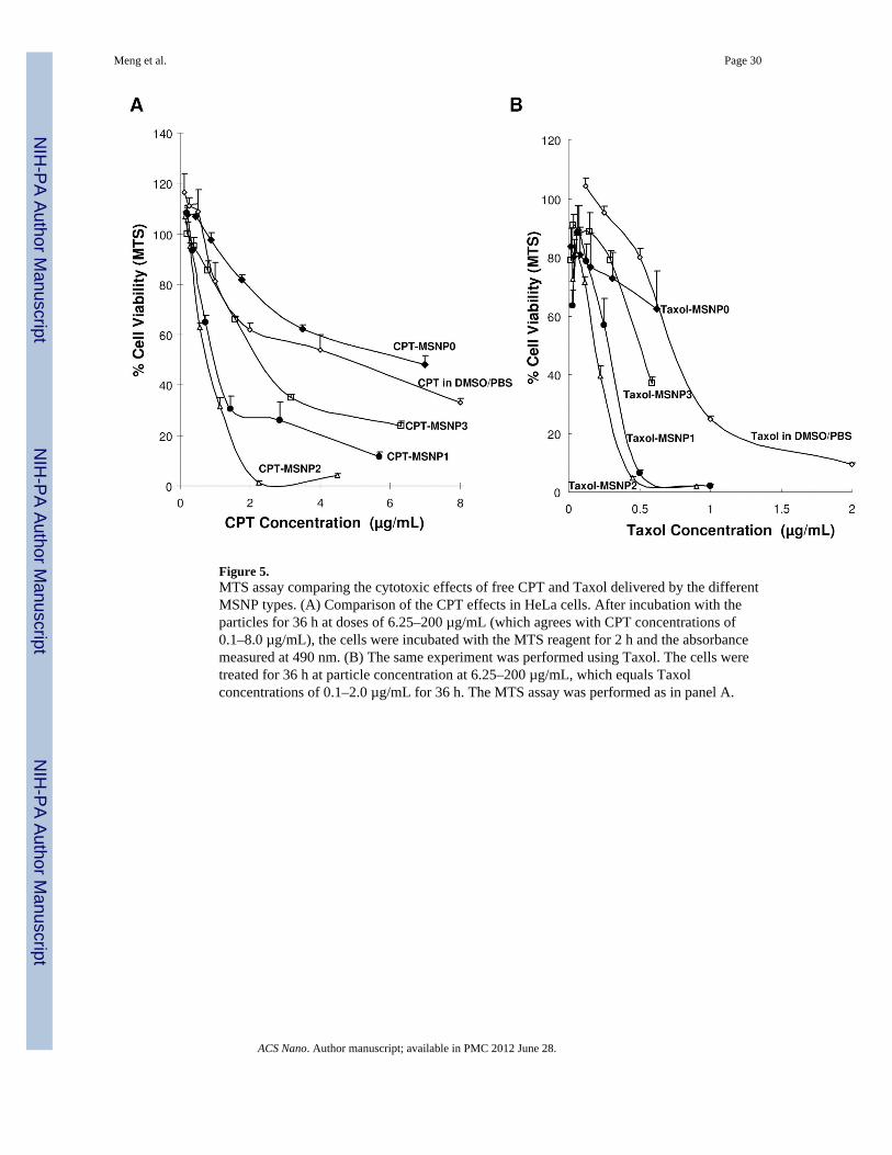
Figure 5.MTS assay comparing the cytotoxic effects of free CPT and Taxol delivered by the differentMSNP types. (A) Comparison of the CPT effects in HeLa cells. After incubation with theparticles for 36 h at doses of 6.25–200 µg/mL (which agrees with CPT concentrations of0.1–8.0 µg/mL), the cells were incubated with the MTS reagent for 2 h and the absorbancemeasured at 490 nm. (B) The same experiment was performed using Taxol. The cells weretreated for 36 h at particle concentration at 6.25–200 µg/mL, which equals Taxolconcentrations of 0.1–2.0 µg/mL for 36 h. The MTS assay was performed as in panel A.
Meng et al. Page 30
ACS Nano. Author manuscript; available in PMC 2012 June 28.
NIH
-PA Author Manuscript
NIH
-PA Author Manuscript
NIH
-PA Author Manuscript

NIH
-PA Author Manuscript
NIH
-PA Author Manuscript
NIH
-PA Author Manuscript
Meng et al. Page 31
Table 1
Physicochemical characterization of MSNP
Samples MSNP0 MSNP1 MSNP2 MSNP3
Aspect ratio 1~1.2 1.5~1.7 2.1~2.5 4.0~4.5
d100 (nm) 4 4 4 4
Pore diameter (nm) 2.5 2.5 2.5 2.5
Surface area (m2/g) 1077.9 926.1 896.9 760.2
Size in H2O/RPMI/CRPMI (nm)a 219/1036/239 207/723/229 198/902/337 185/1023/249
Zeta potential in H2O/CRPMI (mV) 15.7/–5.7 17.1/–6.9 13.0/–5.7 13.5/–6.2
aParticle size measurements were performed using dilute suspensions (100 µg/mL) in water or RPMI at pH 7.4 in a ZetaSizer Nano (Malvern
Instruments Ltd., Worcestershire, UK). To disperse the particles, the stock solutions in water (20 mg/mL) were sonicated (Tekmar Sonic Disruptorprobe) for 15 s before use. In order to coat the surface of MSNP with FBS, 19 µL of particle suspension was mixed with 1 µL FBS before additionto complete medium and sonication for 15 s. RPMI denotes the particle size in RPMI cell culture medium without serum; CRPMI reflects the sizeof MSNP that pre-coated using FBS in complete RPMI containing 10% FBS.
ACS Nano. Author manuscript; available in PMC 2012 June 28.
Related Documents