Ascites in Ventriculoperitoneal Shunt Raj Kumar, Surbhi Sahay 1, Bandana Gaur 2 and Vinita Singh 1 Departments of Neurosurgery, 1Neuroanaesthesiologyand 2Radiology, Sanjay Gandhi Postgraduate Institute of Medical Sciences & King George'sMedical College, Lucknow, India. Abstract. Objective : To fetch out the factors responsible for ascites, following shunt CSF diversion in cases of intracarnial lesions. Four children developing ascites/abdominal psuedocyst following ventriculoperitoneal shunt were analyzed to see the factors responsible for such complication. Methods: Records of 4 cases developing ascites were studied retrospectively. These children developed ascites at 8 months, 6 months, 1 year and 1 year 2 months interval following their shunt installation. Resut$ : The primary etiology of hydrocephalus was demonstrated as thalamic glioblastoma, choroid plexus papillomas of third ventricle, post tubercular meningitis hydrocephalus and suprasellar craniopharyngioma. Conclusion : The proposed etiology of ascites in these cases was peritoneal metastasis from thalamic glioblastoma through ventriculoperitoneal shunt in first case, excessive production of CSF by choroid plexus papilloma in second, infection in the third case and craniopharyngioma causing excessive production of CSF in the fourth child. All the children were treated by reasonable laparotomy and fenestration of cyst along with the repositioning of shunt tip at another site. [Indian a Pediatr 2003; 70 (11) : 859-864] E-maih [email protected] Key words : Ventriculoperitoneal shunt; Ascites; Shunt complications Ascites has been defined as "accumulation of excess fluid within the peritoneal cavity", The common causes being, cirrhosis of liver or other serious hepatic diseases, nephrotic syndrome, anemia with hypoproteinaemia, tuberculosis and malignancies. But, ascites following ventriculoperitoneal shunt (VP shunt) is a rare phenomenon; literature offers no or minimal definite explanation and reports of CSF ascites. Several intra- abdominal complications of VP shunt are known, and many, like infections and obstruction are quite common, but an unloculated and loculated collection of CSF in the abdomen following ventriculoperitoneal shunt procedure needs discussion. Here, we present a series of 4 children who presented with free or encysted fluid in the peritoneal cavity following ventriculoperitoneal shunt procedures; shunts CSF diversion in whom, was carried out, due to different intracranial pathologies preceeding hydrocephalus. MATERIALS AND METHODS The clinical records of 4 children who developed ascites or encysted peritoneal fluid post ventriculo-peritoneal shunt procedure were studied retrospectively. The causes of hydrocephalus, number of shunt revisions, time interval between shunt procedures and appearance of symptoms, management and outcome of these cases were analyzed. All the 4 children were males. The age of presentation with abdominal ascites post shunt procedure were 2, 4, 8 Reprintrequests : Dr. RajKumar, Associate Professor, Department of Neurosurgery, SanjayGandhi Post Graduate Institute of Medical Sciences,Luckn0w-226014,U.P.India. and 9 years respectively. Duration of developing ascites following shunt was 8 months, 6 months, 1 year and 1 year 2 months respectively. Etiology of abdominal ascites and large encysted fluid collection following shunt was : 1. Extra cranial metas- tasis of thalamic glioblastoma through ventriculo- peritoneal shunt into abdomen. 2. Ventriculoperitoneal shunt for hydrocephalus secondary to third ventricular choroid plexus papilloma. 3. Shunt infection in post tubercular meningitis (TBM) hydrocephalus. 4. Craniopharyngioma with hydrocephalus undergone biventricular ventriculoperitoneal shunt. CASE REPORTS Case 1 A 9-year-old male child presented with recurrent headache, vomiting and progressively increasing left sided hemiparesis. Right VP shunt had already been done for hydrocephalus 11,5 months back (at some other center) following cranial CT scan. Examination revealed a conscious, alert and co-operative child with normal higher mental functions. There was bilateral papilloedema, left VI nerve paresis and left sided hemiparesis with power grade 4/5. Planters were bilateral extensor. Contrast enhanced cranial CT showed a large heterogeneous, rim enhancing ill - defined, irregular right thalamic mass with moderate amount of peritumoural oedema, extending across midline and involving opposite thalamus. Mass was causing significant compression of right lateral and third ventricle. Operatively, the left frontal ventricular catheter was introduced and connected to right ventriculoperitoneal shunt by Y connector to decompress Indian Journal of Pediatrics, Volume 70--November, 2003 859

Welcome message from author
This document is posted to help you gain knowledge. Please leave a comment to let me know what you think about it! Share it to your friends and learn new things together.
Transcript

Ascites in Ventriculoperitoneal Shunt
Raj Kumar, Surbhi Sahay 1, Bandana Gaur 2 and Vinita Singh 1
Departments of Neurosurgery, 1Neuroanaesthesiology and 2Radiology, Sanjay Gandhi Postgraduate Institute of Medical Sciences & King George's Medical College, Lucknow, India.
Abstract. Objective : To fetch out the factors responsible for ascites, following shunt CSF diversion in cases of intracarnial lesions. Four children developing ascites/abdominal psuedocyst following ventriculoperitoneal shunt were analyzed to see the factors responsible for such complication. Methods: Records of 4 cases developing ascites were studied retrospectively. These children developed ascites at 8 months, 6 months, 1 year and 1 year 2 months interval following their shunt installation. Resut$ : The primary etiology of hydrocephalus was demonstrated as thalamic glioblastoma, choroid plexus papillomas of third ventricle, post tubercular meningitis hydrocephalus and suprasellar craniopharyngioma. Conclusion : The proposed etiology of ascites in these cases was peritoneal metastasis from thalamic glioblastoma through ventriculoperitoneal shunt in first case, excessive production of CSF by choroid plexus papilloma in second, infection in the third case and craniopharyngioma causing excessive production of CSF in the fourth child. All the children were treated by reasonable laparotomy and fenestration of cyst along with the repositioning of shunt tip at another site. [Indian a Pediatr 2003; 70 (11) : 859-864] E-maih [email protected]
Key words : Ventriculoperitoneal shunt; Ascites; Shunt complications
Ascites has been defined as "accumulation of excess fluid within the peritoneal cavity", The common causes being, cirrhosis of liver or other serious hepatic diseases, nephrotic syndrome, anemia with hypoproteinaemia, tuberculosis and malignancies. But, ascites following ventriculoperi toneal shunt (VP shunt) is a rare phenomenon; literature offers no or minimal definite explanation and reports of CSF ascites. Several intra- abdominal complications of VP shunt are known, and many, like infections and obstruction are quite common, but an unloculated and loculated collection of CSF in the abdomen following ventriculoperitoneal shunt procedure needs discussion.
Here, we present a series of 4 children who presented with free or encysted fluid in the peritoneal cavity following ventriculoperitoneal shunt procedures; shunts CSF diversion in whom, was carried out, due to different intracranial pathologies preceeding hydrocephalus.
MATERIALS AND METHODS
The clinical records of 4 children who developed ascites or encysted peritoneal fluid post ventriculo-peritoneal shunt procedure were studied retrospectively. The causes of hydrocephalus, number of shunt revisions, time interval between shunt procedures and appearance of symptoms, management and outcome of these cases were analyzed. All the 4 children were males. The age of presentation with abdominal ascites post shunt procedure were 2, 4, 8
Reprint requests : Dr. Raj Kumar, Associate Professor, Department of Neurosurgery, Sanjay Gandhi Post Graduate Institute of Medical Sciences, Luckn0w-226014, U.P. India.
and 9 years respectively. Duration of developing ascites following shunt was 8 months, 6 months, 1 year and 1 year 2 months respectively.
Etiology of abdominal ascites and large encysted fluid collection following shunt was : 1. Extra cranial metas- tasis of thalamic glioblastoma through ventriculo- peritoneal shunt into abdomen. 2. Ventriculoperitoneal shunt for hydrocephalus secondary to third ventricular choroid plexus papilloma. 3. Shunt infection in post tubercular meningitis (TBM) hydrocephalus . 4. Craniopharyngioma with hydrocephalus undergone biventricular ventriculoperitoneal shunt.
CASE REPORTS
Case 1 A 9-year-old male child presented with recurrent headache, vomiting and progressively increasing left sided hemiparesis. Right VP shunt had already been done for hydrocephalus 11,5 months back (at some other center) following cranial CT scan. Examination revealed a conscious, alert and co-operative child with normal higher mental functions. There was bilateral papilloedema, left VI nerve paresis and left sided hemiparesis with power grade 4/5. Planters were bilateral extensor. Contrast enhanced cranial CT showed a large heterogeneous, rim enhancing ill - defined, irregular right thalamic mass with moderate amount of peritumoural oedema, extending across midline and involving opposite thalamus. Mass was causing significant compression of right lateral and third ventricle. Operatively, the left frontal ventricular catheter was introduced and connected to right ventriculoperitoneal shunt by Y connector to decompress
Indian Journal of Pediatrics, Volume 70--November, 2003 859
Raj Kumar et al
the enlarged non-communicating ventricle. Patient underwent right parietal osteoplastic craniotomy with near total micro-surgical excision of tumour through superior parietal lobule. Biopsy of tumour revealed glioblastoma. The child received appropriate postoperative radiotherapy and recovered to walk independently within 6 weeks.
At a follow-up of 8 months, he presented with mild ascites and remit tent fever. The ascites rapidly progressed to become incapacitating over a 2 week period (Fig. 1). The peritoneal cavity was tapped to drain 1.2 litres of ascitic fluid, which, on cytological examination revealed malignant cells of glial origin. The child kept on deteriorating, despite all possible treatment and finally, unfortunately, succumbed to his disease with in 3 weeks of drainage of ascites.
Total excision of tumor was achieved through inter- hemispheric, transcallosal route. There was a large, lobulated, soft mass like choroid plexus present with in the cavity of third ventricle, attached to the roof of third ventricle. Post operative recovery of child was smooth and uneventful. Patient remained asymptomatic at follow-up of 3 years with digitally and radiologically functioning ventriculo-atrial shunt. Histopathology revealed papillary tumor composed of multiple fronds covered with well differentiated cuboidal to columnar epithelium and delicate fibrovascular connective stroma.
Case 3
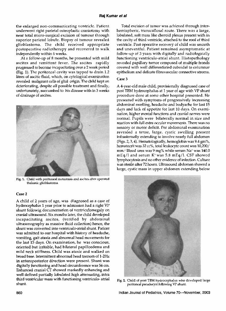
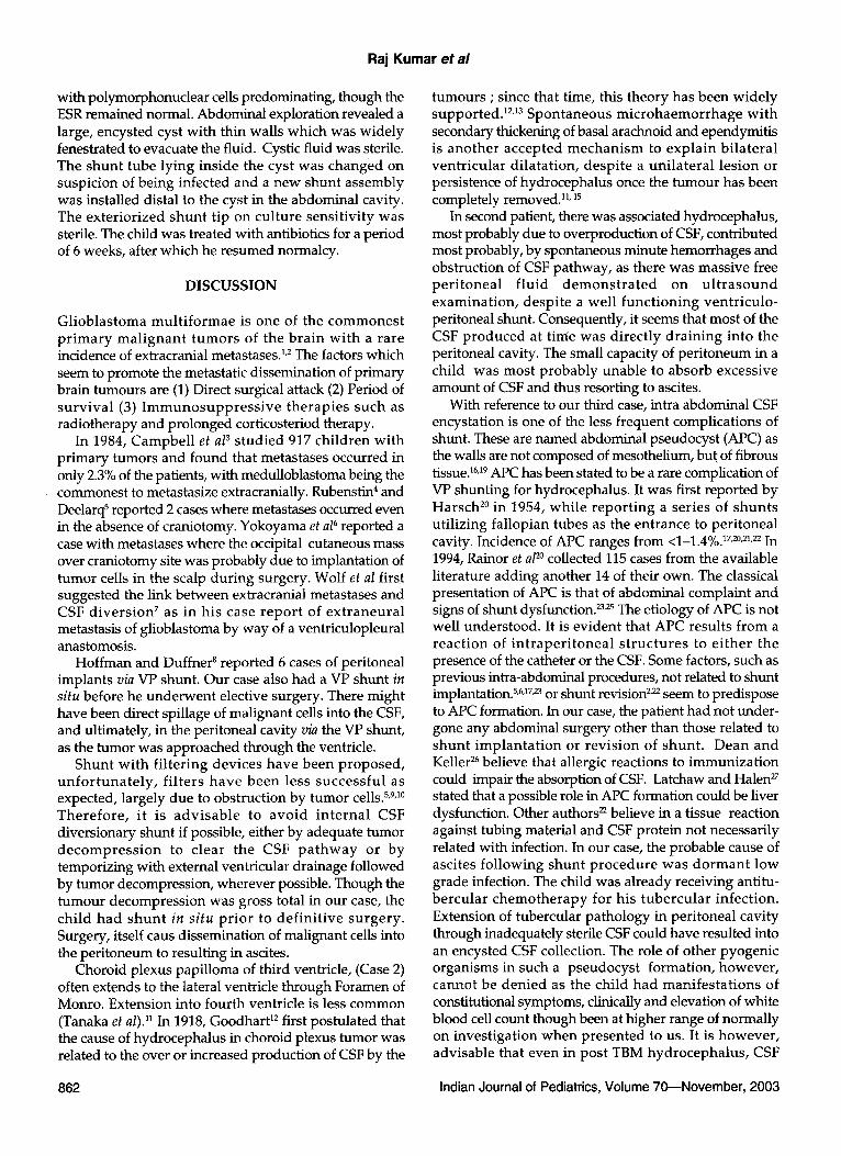
A 4-year-old male child, provisionally diagnosed case of post TBM hydrocephalus at I year of age with VP shunt procedure done at some other hospital presented. He presented with symptoms of progressively increasing abdominal swelling, headache and bodyache for last 15 days and lack of appetite for last 10 days. On exami- nation, higher mental functions and cranial nerves were normal. Pupils were bilaterally normal in size and reaction with full extra occular movements. There was no sensory or motor deficit. Per abdominal examination revealed a tense, large, cystic swelling present infrasternally extending to involve nearly full abdomen (Figs. 2, 3, 4). Hematologically, hemoglobin was 9.4 gm%, hematocrit was 32 cc%, total leukocyte count was 10,200/ mm. 3 Blood urea was 9 mg% while serum Na* was 140.0 mEq/1 and serum K § 5.8 mEq/1. CSF showed lymphocytosis and no other evidence of infection. Culture was sterile after 72 hours. Ultrasound abdomen showed a large, cystic mass in upper abdomen extending below
Fig. 1. Child with peritoneal metastasis and ascites after operated thalamic glioblastoma.
Case 2
A child of 2 years of age, was diagnosed as a case of hydrocephalus I year prior to admission had a right VP shunt followig documentation of ventriculomegaly on cranial ultrasound. Six months later, the child developed incapacitat ing ascites, (verified by abdominal ultrasonography as massive fluid collection) hence, the shunt was converted into ventriculo-atrial shunt. Patient was admitted to our hospital with history of headache, vomiting, gait ataxia and abnormal head movements for the last 15 days. On examination, he was conscious, oriented but irritable, had bilateral papilloedema and mild neck stiffness. Child was ataxic and walked on broad base. Intermittent abnormal head tremors of 1-2Hz in anteroposterior direction were present. Shunt was digitally functioning and head circumference was 56 cm. Enhanced cranial CT showed markedly enhancing and well defined partially lobulated high attenuating, intra third ventricular mass with functioning ventriculo- atrial shunt.
Fig. 2. Child of post TBM hydrocephalus who developed large peritoneal pseudocyst following VP shunt.
860 Indian Journal of Pediatrics, Volume 70--November, 2003

Ascites in Ventriculoperitoneal Shunt
Fig. 3. Ultrasound abdomen of same child (Fig. 2) showing ascites and shunt tube traversing and terminating in cyst.
Vision was bilaterally PL negative and both pupils were 5 mm in diameter. Left eye showed marcus gunn reaction while right eye had no reaction. No other cranial nerve or sensory-motor deficit was demonstrated. Cranial CT scan revealed large suprasel lar iso to hype rdense hetero- geneous mass with signs of calcification. Blockade of both foramen of Monro caused obstructive hydrocephalus.
A biventricular shunt was done initially, followed by right pterional craniotomy and near total excision of the tumor. The tumor was relatively avascular, filling the supra seller cistern and pushing the floor of third ventricle upwards and causing pressure over optic chiasma.
Post operatively, patient developed non concomitant strabismus and ptosis in the right eye. Transient diabeties ins ip idus and SIADH d e v e l o p e d wh ich i m p r o v e d g r adua l l y w i th in a week, whi le v i s ion r e m a i n e d unchanged . Headache and vomi t ing also subsided. Patient improved considerably and was able to walk unaided. He was finally discharged with advice to report if urine output becomes less than 400 cc or more than 3000cc/day or if excessive thirst or drowsiness occurred. At a follow-up of I year and 2 months, the child presented with history of mild, intermittent fever (99-100~ and gradual ly increasing abdominal dis tension for last 5 weeks. Per abdominal examination revealed a large, well - defined, tense, cystic intra-abdominal swelling (Fig. 5). Haematology showed leukocyte count of 9,000 cel ls /mm 3
Fig. 4. Ultrasound abdomen demonstrating cystic abdominal mass of same child (Figs. 2, 3).
umbilicus with catheter traversing the mass and tip seen at umbilicus. No free fluid was present in the peritoneal cavity.
Peroperatively, intraperitoneal cyst was decompressed and marsupalization of cyst was done. The enc3;sted fluid was clear, thin and under tension and surrounding wall was thin, grayish but strong. The abdominal end of shunt was r epos i t i oned be low umbi l icus . Pos tope ra t i ve recovery was uneventful with a resumption of normal abdominal girth. Post-operative ultrasonography did not show any fluid collection and patient was discharged with advice for further follow-up.
Case 4
An 8-years-old male child presented with symptoms of headache associated with vomiting for last 2 years and a progressive diminution of vision for last 6 months with total bl indness . On examinat ion , he was conscious, oriented and was not able to sit or stand without support. Fig. 5. Child with biventricular shunt showing ascites.
Indian Journal of Pediatrics, Volume 70~November, 2003 861
Raj Kumar et al
with polymorphonudear cells predominating, though the ESR remained normal. Abdominal exploration revealed a large, encysted cyst with thin walls which was widely fenestrated to evacuate the fluid. Cystic fluid was sterile. The shunt tube lying inside the cyst was changed on suspicion of being infected and a new shunt assembly was installed distal to the cyst in the abdominal cavity. The exteriorized shunt tip on culture sensitivity was sterile. The child was treated with antibiotics for a period of 6 weeks, after which he resumed normalcy.
DISCUSSION
Glioblastoma mul t i formae is one of the commones t p r imary mal ignan t tumors of the brain wi th a rare incidence of extracranial metastases. 1,2 The factors which seem to promote the metastatic dissemination of primary brain tumours are (1) Direct surgical attack (2) Period of survival (3) I m m u n o s u p p r e s s i v e therapies such as radiotherapy and prolonged corticosteriod therapy.
In 1984, Campbell et al 3 s tudied 917 children with primary tumors and found that metastases occurred in only 2.3% of the patients, with medulloblastoma being the commonest to metastasize extracranially. Rubenstin 4 and Deelarq s reported 2 cases where metastases occurred even in the absence of craniotomy. Yokoyama et al 6 reported a case with metastases where the occipital cutaneous mass over craniotomy site was probably due to implantation of tumor cells in the scalp during surgery. Wolf et al first suggested the link between extracranial metastases and CSF divers ion 7 as in his case repor t of ex t raneura l metastasis of glioblastoma by way of a ventriculopleural anastomosis.
Hoffman and Duffner 8 reported 6 cases of peritoneal implants via VP shunt. Our case also had a VP shunt in situ before he underwent elective surgery. There might have been direct spillage of malignant cells into the CSF, and ultimately, in the peritoneal cavity via the VP shunt, as the tumor was approached through the ventricle.
Shunt wi th filtering devices have been proposed, un fo r tuna t e ly , f i l ters have been less successful as expected, largely due to obstruction by tumor cells. 5,9,1~ Therefore , it is advisable to avoid in ternal CSF diversionary shunt if possible, either by adequate tumor decompress ion to clear the CSF p a t h w a y or by temporizing with external ventricular drainage followed by tumor decompression, wherever possible. Though the tumour decompression was gross total in our case, the child had shun t in s i tu prior to def in i t ive surgery. Surgery, itself caus dissemination of malignant cells into the peritoneum to resulting in ascites.
Choroid plexus papilloma of third ventricle, (Case 2) often extends to the lateral ventricle through Foramen of Monro. Extension into fourth ventricle is less common (Tanaka et al)Y In 1918, Goodhart 12 first postulated that the cause of hydrocephalus in choroid plexus tumor was related to the over or increased production of CSF by the
tumours ; since that time, this theory has been widely supported. 12,13 Spontaneous microhaemorrhage wi th secondary thickening of basal arachnoid and ependymitis is another accepted mechanism to explain bilateral ventr icular dilatation, despite a uni la teral lesion or persistence of hydrocephalus once the tumour has been completely removedY.15
In second patient, there was associated hydrocephalus, most probably due to overproduction of CSF, contributed most probably, by spontaneous minute hemorrhages and obstruction of CSF pathway, as there was massive free per i toneal f luid d e m o n s t r a t e d on u l t r a s o u n d examination, despite a well funct ioning ventriculo- peritoneal shunt. Consequently, it seems that most of the CSF produced at time was directly draining into the peritoneal cavity. The small capacity of peritoneum in a child was most probably unable to absorb excessive amount of CSF and thus resorting to ascites.
With reference to our third case, intra abdominal CSF encystation is one of the less frequent complications of shunt. These are named abdominal pseudocyst (APC) as the walls are not composed of mesothelittrn, but of fibrous tissue. 16,19 APC has been stated to be a rare complication of VP shunting for hydrocephalus. It was first reported by Harsch 2~ in 1954, while report ing a series of shunts utilizing fallopian tubes as the entrance to peritoneal cavity. Incidence of APC ranges from <1-1.4%. 17,2~ In 1994, Rainor et al 2~ collected 115 cases from the available literature adding another 14 of their own. The classical presentation of APC is that of abdominal complaint and signs of shunt dysfunction. 23~5 The etiology of APC is not well understood. It is evident that APC results from a react ion of in t raper i tonea l s t ruc tures to e i ther the presence of the catheter or the CSF. Some factors, such as previous intra-abdominal procedures, not related to shunt implantation. 5,6,17~3 or shunt revision 2,22 seem to predispose to APC formation. In our case, the patient had not under- gone any abdominal surgery other than those related to shunt implanta t ion or revision of shunt . Dean and Keller 26 believe that allergic reactions to immunization could impair the absorption of CSF. Latchaw and Halen 27 stated that a possible role in APC formation could be liver dysfunction. Other authors 22 believe in a tissue reaction against tubing material and CSF protein not necessarily related with infection. In our case, the probable cause of ascites following shunt procedure was dorman t low grade infection. The child was already receiving antitu- bercular chemotherapy for his tubercular infection. Extension of tubercular pathology in peritoneal cavity through inadequately sterile CSF could have resulted into an encysted CSF collection. The role of other pyogenic organisms in such a pseudocyst formation, however, cannot be denied as the child had manifesta t ions of constitutional symptoms, clinically and elevation of white blood cell count though been at higher range of normally on investigation when presented to us. It is however, advisable that even in post TBM hydrocephalus , CSF
862 Indian Journal of Pediatrics, Volume 70~November, 2003
Ascites in Ventriculoperitoneal Shunt
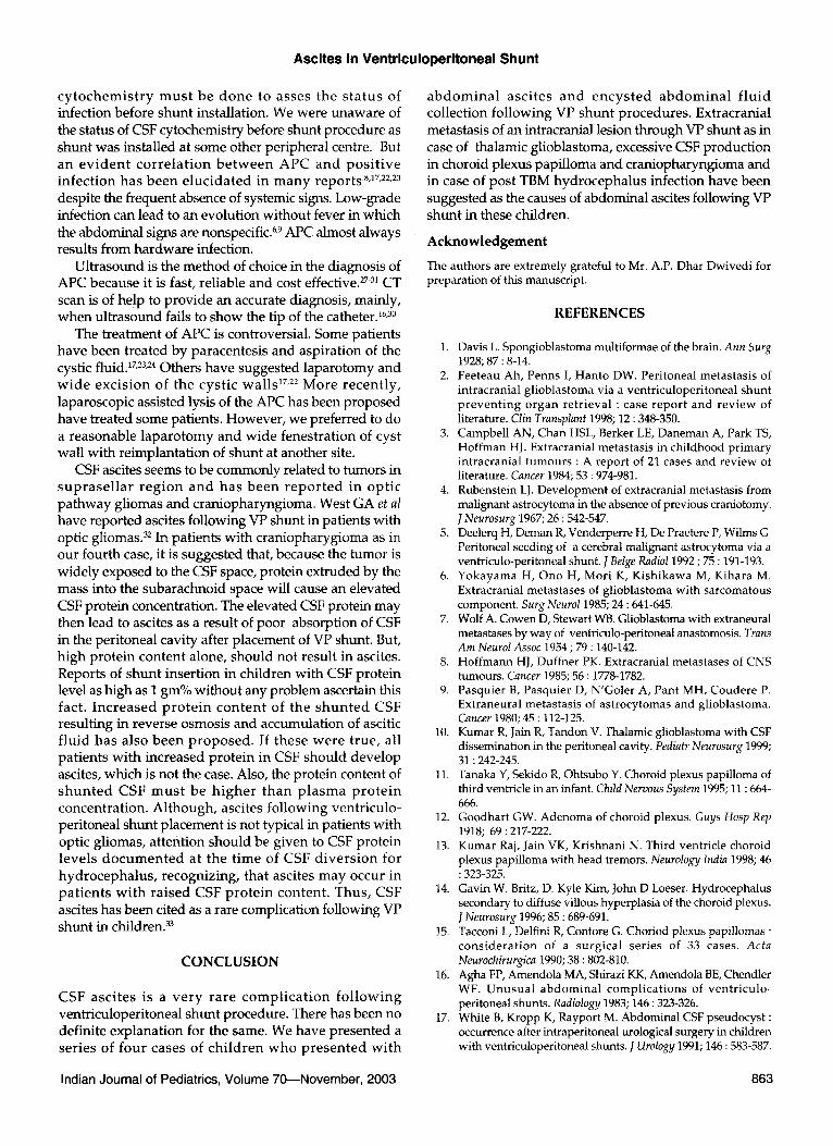
c y t o c h e m i s t r y m u s t be d o n e to asses the s t a t u s of infection before shunt installation. We were unaware of the status of CSF cytochemistry before shunt procedure as shunt was installed at some other peripheral centre. But an e v i d e n t c o r r e l a t i o n b e t w e e n A P C a n d p o s i t i v e in fec t ion has b e e n e luc ida t ed in m a n y repor t s '8,17,22,23 despite the frequent absence of systemic signs. Low-grade infection can lead to an evolut ion wi thout fever in which the abdominal signs are nonspecific. 6,9 APC almost always results f rom ha rdware infection.
Ul t rasound is the method of choice in the diagnosis of APC because it is fast, reliable and cost effective. 27-31 CT scan is of help to provide an accurate diagnosis, mainly, w h e n ul t rasound fails to show the tip of the catheter26,3~
The t reatment of APC is controversial. Some patients have been treated by paracentesis and aspirat ion of the cystic fluid. ~7,23,24 Others have suggested laparo tomy and w i d e exc i s ion of the cys t i c wa l l s 17,22 M o r e r ecen t l y , laparoscopic assisted lysis of the APC has been proposed have treated some patients. However , we preferred to do a reasonable l a p a r o t o m y and wide fenestrat ion of cyst wall with reimplantat ion of shunt at another site.
CSF ascites seems to be commonly related to tumors in s u p r a s e l l a r r e g i o n a n d ha s b e e n r e p o r t e d in op t i c pa thway gliomas and craniopharyngioma. West GA et al have reported ascites following VP shunt in patients with optic gliomas. 32 In patients with c ran iopharygioma as in our fourth case, it is suggested that, because the tumor is widely exposed to the CSF space, protein extruded by the mass into the subarachnoid space will cause an elevated CSF protein concentration. The elevated CSF protein m a y then lead to ascites as a result of poor absorption of CSF in the peritoneal cavity after placement of VP shunt. But, h igh prote in content alone, should not result in ascites. Reports of shun t insert ion in children wi th CSF protein level as high as I gm% without any problem ascertain this fact. I n c r e a s e d p r o t e i n c o n t e n t of the s h u n t e d CSF result ing in reverse osmosis and accumulat ion of ascitic f lu id has a lso b e e n p r o p o s e d . If these w e r e t rue, all pat ients wi th increased pro te in in CSF shou ld deve lop ascites, which is not the case. Also, the protein content of s h u n t e d CSF m u s t be h i g h e r t h a n p l a s m a p r o t e i n concentra t ion. A l though , ascites fo l lowing ventr iculo- peritoneal shunt placement is not typical in patients with optic gliomas, at tention should be given to CSF protein levels d o c u m e n t e d at the t ime of CSF d i v e r s i o n for h y d r o c e p h a l u s , recogniz ing , that ascites m a y occur in pa t i en t s w i th ra i sed CSF p ro t e in content . Thus , CSF ascites has been cited as a rare complication following VP shunt in children. 33
CONCLUSION
CSF asc i tes is a v e r y r a re c o m p l i c a t i o n f o l l o w i n g ventriculoperitoneal shunt procedure. There has been no definite explanat ion for the same. We have presented a ser ies of f o u r cases of c h i l d r e n w h o p r e s e n t e d w i t h
a b d o m i n a l asc i tes and e n c y s t e d a b d o m i n a l f l u id collection fo l lowing VP shunt procedures . Extracranial metastasis of an intracranial lesion through VP shunt as in case of thalamic glioblastoma, excessive CSF product ion in choroid plexus papi l loma and craniopharyngioma and in case of pos t TBM hydrocepha lus infection have been suggested as the causes of abdominal ascites following VP shunt in these children.
Acknowledgement
The authors are extremely grateful to Mr. A.P. Dhar Dwivedi for preparation of this manuscript.
REFERENCES
1. Davis L. Spongioblastoma multiformae of the brain. Ann Surg 1928; 87 : 8-14.
2. Feeteau Ah, Penns I, Hanto DW. Peritoneal metastasis of intracranial glioblastoma via a ventriculoperitoneal shunt preventing organ retrieval : case report and review of literature. Clin Transplant 1998; 12 : 348-350.
3. Campbell AN, Chan HSL, Berker LE, Daneman A, Park TS, Hoffman HJ. Extracranial metastasis in childhood primary intracranial tumours : A report of 21 cases and review of literature. Cancer 1984; 53 : 974-981.
4. Rubenstein LJ. Development of extracranial metastasis from malignant astrocytoma in the absence of previous craniotomy. J Neurosurg 1967; 26 : 542-547.
5. Deelerq H, Deman R, Venderperre H, De Praetere P, Wilms G. Peritoneal seeding of a cerebral malignant astrocytoma via a ventriculo-peritoneal shunt. J Belge Radiol 1992 ; 75 : 191-193.
6. Yokayama H, Ono H, Mori K, Kishikawa M, Kihara M. Extracranial metastases of glioblastoma with sarcomatous component. Surg Neurol 1985; 24 : 641-645.
7. Wolf A. Cowen D, Stewart WB. Glioblastoma with extraneural metastases by way of ventriculo-peritoneal anastomosis. Trans Am Neurol Assoc 1954 ; 79 : 140-142.
8. Hoffmann HJ, Duffner PK. Extracranial metastases of CNS tumours. Cancer 1985; 56 : 1778-1782.
9. Pasquier B, Pasquier D, N'Goler A, Pant MH, Coudere P. Extraneural metastasis of astrocytomas and glioblastoma. Cancer 1980; 45 : 112-125.
10. Kumar R, Jain R, Tandon V. Thalamic glioblastoma with CSF dissemination in the peritoneal cavity. Pediatr Neurosurg 1999; 31 : 242-245.
11. Tanaka Y, Sekido R, Ohtsubo Y. Choroid plexus papilloma of third ventricle in an infant. Child Nervous System 1995; 11 : 664- 666.
12. Goodhart GW. Adenoma of choroid plexus. Guys Hosp Rep 1918; 69 : 217-222.
13. Kumar Raj, Jain VK, Krishnani N. Third ventricle choroid plexus papilloma with head tremors. Neurology India 1998; 46 : 323-325.
14. Gavin W. Britz, D. Kyle Kim, John D Loeser. Hydrocephalus secondary to diffuse villous hyperplasia of the choroid plexus. ] Neurosurg 1996; 85 : 689-691.
15. Tacconi L, Delfini R, Contore G. Choriod plexus papillomas : consideration of a surgical series of 33 cases. Acta Neurochirurgica 1990; 38 : 802-810.
16. Agha FP, Amendola MA, Shirazi KK, Amendola BE, Chendler WF. Unusual abdominal complications of ventriculo- peritoneal shunts. Radiology 1983; 146 : 323-326.
17. White B, Kropp K, Rayport M. Abdominal CSF pseudocyst : occurrence after intraperitoneal urological surgery in children with ventriculoperitoneal shunts. J Urology 1991; 146 : 583-587.
Indian Journal of Pediatrics, Volume 70~November, 2003 863

Raj Kumar et al
18. Baumgartner F, Moor TC, Mitchner J. Recurrent ventriculoperitoneal shunt pseudocyst in a 9 year old gift. Klin Wochenschr 1990; 68 : 485-487.
19. Fischer EG, Shillito J Jr. Large abdominal cyst : A complication of peritoneal shunt. Report of three cases. J Neurosurg 1969; 31 : 4,11-444.
20. Harsch GR. Peritoneal shunt for hydrocephalus : utilizing the fimbria of the fallopian tube for entrance to the peritoneal cavity. J Neurosurg 1954 ; 11 : 248-294.
21. Norfray JF, Henry HM, Gwens JD, Sparberg MS. Abdominal complications from peritoneal shunts. Gastroenterology 1979; 77 : 337-340.
22. Rainor N, Schobess A, Heidecke V, Burkert W. Abdominal CSF pseudocyst in patients with ventriculo-peritoneal shunts. Report of fourteen cases and review of literature. Acta Neurochir (Wien) 1994; 127 : 73-78.
23. Parry SW, Schuhmacher JF, Llewellyn RC. Abdominal pseudocysts and ascites formation after ventriculoperitoneal shunt procedures. Report of four cases. J Neurosurg 1975; 43 : 476-480.
24. Nakagaki H, Matsunagar M, Maeyama R, Mizoguchi R : Intraperitoneal pseudocyst after ventriculoperitoneal shunt. Surg Neuro11979 ; 11 : 447-450.
25. Ersahin Y, Mutluer S, Tekeli G. Abdominal CSF pseudocysts.
Child's Nerv Sys 1996 ; 12 : 755-758. 26. Dean DF, Keller IB. CSF ascites : A complication of
ventriculoperitoneal shunt. J Neurol Neurosurg Psychiatry 1972; 35 : 474-476.
27. Latchaw JP Jr, Hahn JF. Intraperitoneal pseudocyst associated with peritoneal shunt. Neurosurgery 1981; 8 : 469-472.
28. Agha FP, Amendola MA, Shirazi KK, Amendola BE, Chandler WF. Abdominal complications of ventriculo-peritoneal shunt with emphasis on the role of imaging methods. Surg Gyne Obstet 1983; 156 : 473-478.
29. Brenbridge ANAG, Buschi AJ, Lees RF, Sims T. Sonography of CSF pseudocyst. Am J Dis Child 1979; 133 : 646-647.
30. Lee TG, Parsons PM. Ultrasound diagnosis of CSF abdominal cyst. Radiology 1978; 127 : 220.
31. Chuang VP, Fried AM, Oliff M, Ellis GT, Sachatello CR. Abdominal CSF pseudocyst secondary to ventriculoperitoneal shunt : Diagnosis by CT in two cases. J Comput Assist Tomogr 1978; 2 : 88-91.
32. West GA, Berger MS, Geyer JR. Childhood optic pathway tumours associated with ascities following ventriculo-perito- heal shunt placement. Pediatr Neurosurg 1994; 21(4) : 254-258.
33. Chidambaram B, Balasubramaniam V. CSF ascites : A rare complication of ventriculo-peritoneal shunt surgery. Neurology India 2000; 48 : 378-380.
1,
2.
3.
4.
5.
6.
IJP TRAVEL GRANT FOR MEDICAL CONFERENCE 2 0 0 3
(Benefit for Subscribers)
Six grants are available. Only Postgraduate students are eligible to apply.
Grant will cover train journey to and fro, registration fee and limited local expenses. Total expenditure not to exceed Rs. 5000 per candidate.
Applicant should submit a copy of the research paper to be presented at the conference along with the letter of acceptance by the conference organizers.
The application should be forwarded through the Head of the Department along with a letter certifying that the applicant is not being supported by any other source.
Applicant should be a subscriber to the journal.
Applicant should explain in 200 words why he/she would like to be considered for the grant.
For in format ion please wri te to :
Editor-in-Chief, The Indian Journal of Pediatrics, 125 (2nd Floor), Gautam Nagar, N e w Delhi-110049.
Post Box No. 3875, N e w Delh i - 110 049. Phone : 26568098. Telefax : 26857587
E-mail : i jp.journal .vsnl .net@vsnl .net
864 Indian Journal of Pediatrics, Volume 70--November, 2003
Related Documents