Review Article Eur Neurol 2019;82:41–64 Artificial Intelligence Techniques for Automated Diagnosis of Neurological Disorders U. Raghavendra a U. Rajendra Acharya b–d Hojjat Adeli e, f a Department of Instrumentation and Control Engineering, Manipal Institute of Technology, Manipal Academy of Higher Education, Manipal, India; b Department of Electronics and Computer Engineering, Ngee Ann Polytechnic, Clementi, Singapore; c Department of Biomedical Engineering, School of Science and Technology, SUSS University, Clementi, Singapore; d International Research Organization for Advanced Science and Technology (IROAST) Kumamoto University, Kumamoto, Japan; e Department of Neuroscience, The Ohio State University, Columbus, OH, USA; f Departments of Biomedical Informatics and Neuroscience, The Ohio State University, Columbus, OH, USA Received: October 11, 2019 Accepted: October 15, 2019 Published online: November 19, 2019 Hojjat Adeli Department of Neuroscience, The Ohio State University 470 Hitchcock Hall, 2070 Neil Avenue Columbus, OH 43210 (USA) E-Mail adeli.1 @osu.edu © 2019 S. Karger AG, Basel E-Mail [email protected] www.karger.com/ene DOI: 10.1159/000504292 Keywords Neurological disorder · Computer-aided diagnosis · Machine learning · Classification algorithm Abstract Background: Authors have been advocating the research ideology that a computer-aided diagnosis (CAD) system trained using lots of patient data and physiological signals and images based on adroit integration of advanced signal processing and artificial intelligence (AI)/machine learning techniques in an automated fashion can assist neurologists, neurosurgeons, radiologists, and other medical providers to make better clinical decisions. Summary: This paper pres- ents a state-of-the-art review of research on automated di- agnosis of 5 neurological disorders in the past 2 decades us- ing AI techniques: epilepsy, Parkinson’s disease, Alzheimer’s disease, multiple sclerosis, and ischemic brain stroke using physiological signals and images. Recent research articles on different feature extraction methods, dimensionality reduc- tion techniques, feature selection, and classification tech- niques are reviewed. Key Message: CAD systems using AI and advanced signal processing techniques can assist clini- cians in analyzing and interpreting physiological signals and images more effectively. © 2019 S. Karger AG, Basel Introduction Neurological disorders are the diseases connected with peripheral and central nervous systems. The common symptoms include muscle weakness, paralysis, seizures, pain, poor coordination, and loss of consciousness [1]. There are >600 diseases related to the nervous system such as brain tumor, Parkinson’s disease (PD), Alzheim- er’s disease (AD), multiple sclerosis (MS), epilepsy, de- mentia, headache disorders, neuroinfections, stroke, and traumatic brain injuries among others. Various viral in- fections (i.e., HIV, Zika, West Nile Virus, Enteroviruses), bacterial infections (such as Neisseria meningitides and Mycobacterium tuberculosis), fungal-related infections (such as Aspergillus and Cryptococcus), and parasitic in- fections (such as Chagas and malaria) can affect the entire

Welcome message from author
This document is posted to help you gain knowledge. Please leave a comment to let me know what you think about it! Share it to your friends and learn new things together.
Transcript
Review Article
Eur Neurol 2019;82:41–64
Artificial Intelligence Techniques for Automated Diagnosis of Neurological Disorders
U. Raghavendra
a U. Rajendra Acharya
b–d Hojjat Adeli
e, f a
Department of Instrumentation and Control Engineering, Manipal Institute of Technology, Manipal Academy of Higher Education, Manipal, India; b Department of Electronics and Computer Engineering, Ngee Ann Polytechnic, Clementi, Singapore; c Department of Biomedical Engineering, School of Science and Technology, SUSS University, Clementi, Singapore; d International Research Organization for Advanced Science and Technology (IROAST) Kumamoto University, Kumamoto, Japan; e Department of Neuroscience, The Ohio State University, Columbus, OH, USA; f Departments of Biomedical Informatics and Neuroscience, The Ohio State University, Columbus, OH, USA
Received: October 11, 2019Accepted: October 15, 2019Published online: November 19, 2019
Hojjat AdeliDepartment of Neuroscience, The Ohio State University470 Hitchcock Hall, 2070 Neil AvenueColumbus, OH 43210 (USA)E-Mail adeli.1 @ osu.edu
© 2019 S. Karger AG, Basel
E-Mail [email protected]/ene
DOI: 10.1159/000504292
KeywordsNeurological disorder · Computer-aided diagnosis · Machine learning · Classification algorithm
AbstractBackground: Authors have been advocating the research ideology that a computer-aided diagnosis (CAD) system trained using lots of patient data and physiological signals and images based on adroit integration of advanced signal processing and artificial intelligence (AI)/machine learning techniques in an automated fashion can assist neurologists, neurosurgeons, radiologists, and other medical providers to make better clinical decisions. Summary: This paper pres-ents a state-of-the-art review of research on automated di-agnosis of 5 neurological disorders in the past 2 decades us-ing AI techniques: epilepsy, Parkinson’s disease, Alzheimer’s disease, multiple sclerosis, and ischemic brain stroke using physiological signals and images. Recent research articles on different feature extraction methods, dimensionality reduc-tion techniques, feature selection, and classification tech-niques are reviewed. Key Message: CAD systems using AI
and advanced signal processing techniques can assist clini-cians in analyzing and interpreting physiological signals and images more effectively. © 2019 S. Karger AG, Basel
Introduction
Neurological disorders are the diseases connected with peripheral and central nervous systems. The common symptoms include muscle weakness, paralysis, seizures, pain, poor coordination, and loss of consciousness [1]. There are > 600 diseases related to the nervous system such as brain tumor, Parkinson’s disease (PD), Alzheim-er’s disease (AD), multiple sclerosis (MS), epilepsy, de-mentia, headache disorders, neuroinfections, stroke, and traumatic brain injuries among others. Various viral in-fections (i.e., HIV, Zika, West Nile Virus, Enteroviruses), bacterial infections (such as Neisseria meningitides and Mycobacterium tuberculosis), fungal-related infections (such as Aspergillus and Cryptococcus), and parasitic in-fections (such as Chagas and malaria) can affect the entire
Raghavendra/Acharya/AdeliEur Neurol 2019;82:41–6442DOI: 10.1159/000504292
nervous system [1–6]. The aforementioned neurological symptoms possibly occur due to immune response or in-fection itself. Hundreds of millions of people worldwide are affected by neurological disorders [7–10]. More than 6 million people die because of stroke each year; majority of these deaths take place in low- and middle-income countries [1, 11, 12]. It is reported that around 50 million people will have epilepsy [1], and 47.5 million people will suffer from dementia [1, 13–17].
The abnormal or the anomalous neurological condi-tions are commonly identified by a neuropathological ex-amination. Anomalous neurological conditions are found in majority of the population and are not always associ-ated with a neurological disorder [18].
Dementia is usually progressive in nature. The demen-tia syndromes disturb multiple cortical functions, that is, memory, orientation, thinking, calculation, language, comprehension, judgment, and learning capacity. AD is found to be the most common cause of dementia, which is characterized by neurofibrillary and cortical amyloids ac-counting for 3 quarters of the cases [16, 19, 20]. Dementia affects mainly older people, above 65 years, but also 2% of the people under 65 years old. The prevalence of dementia doubles with age every 5 years. A genetic polymorphism increases the risk for 25% of entire population [21, 22].
Epilepsy, a chronic neurological disorder, is defined as, “disorder of brain characterized by enduring predisposition to generate the epileptic seizures.” [23–25]. The epilepsy def-inition requires at least occurrence of one epileptic seizure [26, 27]. It affects both male and female sexes and people of all ages. The diagnosis for epileptic seizures is performed by first determining the event of epilepsy and later differenti-ating between the conditions called provoked or chronic epileptic seizures [28, 29]. The overall incidence of epilepsy is found to be 23–190 per 100,000 of population [30]. The prevalence is lower in early ages and gradually increases with aging [31–34]. Since the pioneering work of Adeli et al. [35], wavelet transform (WT) has been used extensively for electroencephalogram (EEG) analysis, seizure detec-tion, and epilepsy diagnosis [36, 37]. Kugiumtzis et al. [27] investigate the dynamics of epileptiform discharges in-duced by transcranial magnetic stimulation in epilepsy. Yuan et al. [38] present a method for epileptic seizure pre-diction using diffusion distance and Bayesian linear dis-criminate analysis [39] in intracranial EEG.
MS, a disorder caused by a condition called inflamma-tory demyelinating of the nervous system, is the most common among all neurological disorders. MS causes disabilities in young adults and affects nearly 2.5 million people worldwide. The diagnosis of MS is generally per-
formed by magnetic resonance imaging (MRI). There are no treatments available for this disease [40, 41].
PD is a chronic neurodegenerative disorder often characterized by the presence of predominantly motor symptomatology [42, 43], but it can have nonmotor hy-posmia, paresthesia, depression, and pain [44]. PD is a universal disorder with incidence rate of 4.5–19 per 100,000 of population per year [45–47] for both females and males of all ages. The therapy depends on severity, mental status, and age of the patient. Gálvez et al. [48] in-vestigate the short-term effects of Binaural Beats on EEG power, functional connectivity, cognition, gait, and anxi-ety in PD patients.
Stroke is a clinical syndrome of cerebral deficit that lasts for > 24 h with no apparent cause except the vascular one [49]. In the modern developed countries, 75–80% of the strokes are attributed to brain ischemia, and 10–15% are attributed to intracerebral hemorrhage. Stroke diag-nosis is made accurately completely based on clinical grounds by a specialist alone.
Traumatic brain injury is one of the foremost causes of disability and death in young adults and children world-wide. More than five million people suffer from the trau-matic brain injury disability in the United States alone [50–52].
Authors have been advocating the research ideology that a computer-aided diagnosis (CAD) system trained using lots of patient data and physiological signals and im-ages based on adroit integration of advanced signal pro-cessing and artificial intelligence (AI)/machine learning (ML) techniques in an automated fashion can assist neu-rologists, neurosurgeons, radiologists, and other medical providers to make better clinical decisions. Research in this area has been growing at an accelerating rate in the past decade. In this paper, authors explore and review re-cent articles on the applications of AI-based CAD systems for the diagnosis of 5 major neurological disorders: epi-lepsy, AD, PD, MS, and ischemic brain stroke.
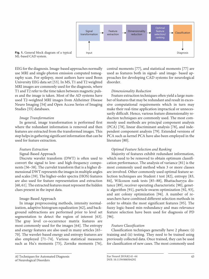
Figure 1 shows the functional block diagram of a typi-cal ML-based CAD system consisting of 5 stages: (1) sig-nal transformation, (2) feature extraction, (3) feature di-mensionality reduction, (4) optimal feature selection/ranking, and (5) classification.
ML-Based CAD
Input Data DescriptionInput data for a CAD system are normally signals and/
or images. For PD, many CAD systems use speech and
AI Techniques for Automated Diagnosis of Neurological Disorders
43Eur Neurol 2019;82:41–64DOI: 10.1159/000504292
EEG for the diagnosis. Image-based approaches normally use MRI and single-photon emission computed tomog-raphy scan. For epilepsy, most authors have used Bonn University EEG data set [53]. In MS, T1 and T2 weighted MRI images are commonly used for the diagnosis, where T1 and T2 refer to the time taken between magnetic puls-es and the image is taken. Most of the AD systems have used T2-weighted MRI images from Alzheimer Disease Neuro Imaging [54] and Open Access Series of Imaging Studies [55] databases.
Image TransformationIn general, image transformation is performed first
where the redundant information is removed and then features are extracted from the transformed images. This step helps in gathering significant information that can be used for feature extraction.
Feature ExtractionSignal-Based ApproachDiscrete wavelet transform (DWT) is often used to
convert the signal to low- and high-frequency compo-nents [56–58]. The curvelet transform that is a higher di-mensional DWT represents the images in multiple angles and scales [59]. The higher-order spectra (HOS) features are also used for feature representation and extraction [60, 61]. The extracted features must represent the hidden clues present in the input data.
Image-Based ApproachIn image preprocessing methods, intensity normal-
ization, adaptive histogram equalization [62], and back-ground subtractions are performed prior to level set segmentation to detect the region of interest [63]. The gray level co-occurrence matrix features are most commonly used for the images [64]. The entropy and energy features are also used in many articles [65–70]. The wavelet-based energy and entropy features are also employed [71–74]. Various statistical measures such as Hu’s moments [75], Zernike moments [76],
central moments [77], and statistical moments [77] are used as features both in signal- and image- based ap-proaches for developing CAD systems for neurological disorder.
Dimensionality ReductionFeature extraction techniques often yield a large num-
ber of features that may be redundant and result in exces-sive computational requirements which in turn may make their real-time application impractical or unneces-sarily difficult. Hence, various feature dimensionality re-duction techniques are commonly used. The most com-monly used methods are principal component analysis (PCA) [78], linear discriminant analysis [78], and inde-pendent component analysis [79]. Extended versions of PCA such as kernel PCA have also been employed in the literature [80].
Optimal Feature Selection and RankingMajority of features exhibit redundant information,
which need to be removed to obtain optimum classifi-cation performance. The analysis of variance [81] is the most commonly used method when 3 or more classes are involved. Other commonly used optimal feature se-lection techniques are Student t test [82], entropy [83, 84], Wilcoxon rank tests [85–88], Bhattacharyya dis-tance [89], receiver operating characteristic [90], genet-ic algorithm [91], particle swarm optimization [92, 93], and ant colony optimization [94]. A number of re-searchers have combined different selection methods in order to obtain the most significant features [95]. The fuzzy logic-based min-redundancy and max-relevance feature selection have been used for diagnosis of PD [96].
Feature ClassificationClassification techniques generally have 2 phases: (i)
training and (ii) testing. They need to be trained using previously collected data. Once trained, they can be used for classification of new cases. The most commonly used
Signaltransformation
Featureextraction
Input Diagnosis
Dimensionalityreduction
Optimalfeature
selection/ranking
Classification
Fig. 1. General block diagram of a typical ML-based CAD system.
Raghavendra/Acharya/AdeliEur Neurol 2019;82:41–6444DOI: 10.1159/000504292
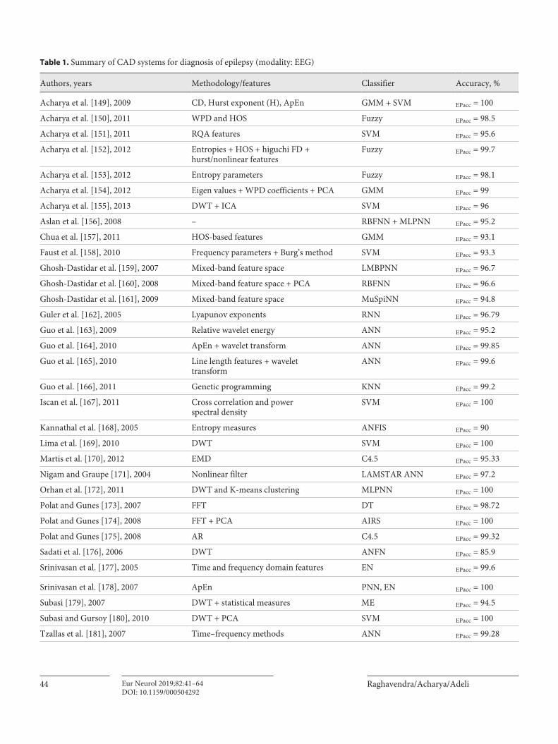
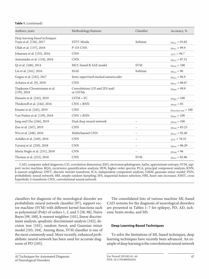
Table 1. Summary of CAD systems for diagnosis of epilepsy (modality: EEG)
Authors, years Methodology/features Classifier Accuracy, %
Acharya et al. [149], 2009 CD, Hurst exponent (H), ApEn GMM + SVM EPacc = 100
Acharya et al. [150], 2011 WPD and HOS Fuzzy EPacc = 98.5
Acharya et al. [151], 2011 RQA features SVM EPacc = 95.6
Acharya et al. [152], 2012 Entropies + HOS + higuchi FD +hurst/nonlinear features
Fuzzy EPacc = 99.7
Acharya et al. [153], 2012 Entropy parameters Fuzzy EPacc = 98.1
Acharya et al. [154], 2012 Eigen values + WPD coefficients + PCA GMM EPacc = 99
Acharya et al. [155], 2013 DWT + ICA SVM EPacc = 96
Aslan et al. [156], 2008 – RBFNN + MLPNN EPacc = 95.2
Chua et al. [157], 2011 HOS-based features GMM EPacc = 93.1
Faust et al. [158], 2010 Frequency parameters + Burg’s method SVM EPacc = 93.3
Ghosh-Dastidar et al. [159], 2007 Mixed-band feature space LMBPNN EPacc = 96.7
Ghosh-Dastidar et al. [160], 2008 Mixed-band feature space + PCA RBFNN EPacc = 96.6
Ghosh-Dastidar et al. [161], 2009 Mixed-band feature space MuSpiNN EPacc = 94.8
Guler et al. [162], 2005 Lyapunov exponents RNN EPacc = 96.79
Guo et al. [163], 2009 Relative wavelet energy ANN EPacc = 95.2
Guo et al. [164], 2010 ApEn + wavelet transform ANN EPacc = 99.85
Guo et al. [165], 2010 Line length features + wavelettransform
ANN EPacc = 99.6
Guo et al. [166], 2011 Genetic programming KNN EPacc = 99.2
Iscan et al. [167], 2011 Cross correlation and powerspectral density
SVM EPacc = 100
Kannathal et al. [168], 2005 Entropy measures ANFIS EPacc = 90
Lima et al. [169], 2010 DWT SVM EPacc = 100
Martis et al. [170], 2012 EMD C4.5 EPacc = 95.33
Nigam and Graupe [171], 2004 Nonlinear filter LAMSTAR ANN EPacc = 97.2
Orhan et al. [172], 2011 DWT and K-means clustering MLPNN EPacc = 100
Polat and Gunes [173], 2007 FFT DT EPacc = 98.72
Polat and Gunes [174], 2008 FFT + PCA AIRS EPacc = 100
Polat and Gunes [175], 2008 AR C4.5 EPacc = 99.32
Sadati et al. [176], 2006 DWT ANFN EPacc = 85.9
Srinivasan et al. [177], 2005 Time and frequency domain features EN EPacc = 99.6
Srinivasan et al. [178], 2007 ApEn PNN, EN EPacc = 100
Subasi [179], 2007 DWT + statistical measures ME EPacc = 94.5
Subasi and Gursoy [180], 2010 DWT + PCA SVM EPacc = 100
Tzallas et al. [181], 2007 Time–frequency methods ANN EPacc = 99.28
AI Techniques for Automated Diagnosis of Neurological Disorders
45Eur Neurol 2019;82:41–64DOI: 10.1159/000504292
Authors, years Methodology/features Classifier Accuracy, %
Jaiswal and Banka [182], 2017 SpPCA and SubXPCA SVM EPacc = 94.60
Ubeyli [183], 2010 AR LS-SVM EPacc = 99.56
Wang et al. [184], 2011 Wavelet + entropy KNN + HKB EPacc = 100
Swami et al. [185], 2016 DTCWT GRNN –
Peker et al. [186], 2016 DTCWT CVNN EPacc = 100
Sharma and Pachori [187], 2017 TQWT LS-SVM + FD EPacc = 100
Patidar et al. [188], 2017 TQWT and kraskov entropy LS-SVM EPacc = 97.75
Gandhi et al. [189], 2011 DWT, energy, SD, entropy PNN EPacc = 99.9
Chen [190], 2014 DTCWT, fourier features NN EPacc = 100
Swami et al. [191], 2014 Energy, SD, entropy features SVM EPacc = 99.53
Pachori and Patidar [192], 2014 EMD and SODP ANN EPacc = 100
Sharma and Pachori [193], 2015 EMD and PSR of IMF LS-SVM EPacc = 98.67
Bhattacharyya et al. [194], 2017 TQWT and KNN entropies SVM EPacc = 100
Bhattacharyya et al. [195], 2016 Empirical wavelet transform LS-SVM EPacc = 90
Bhati et al. [196], 2017 Time–frequency localized three-bandbiorthogonal linear phase waveletfilter bank
MLPNN EPacc = 99.33
Sharma et al. [197], 2017 Orthogonal wavelet filter banks LS-SVM EPacc = 94.25
Kaya et al. [198], 2014 1D – LBP FT EPacc = 99.5
Zhu et al. [199], 2014 FWHVA KNN EPacc = 100
Samiee et al. [200], 2015 DSTFT MLPNN EPacc = 99.8
Riaz et al. [201], 2016 EMD SVM EPacc = 96.2
Diykh et al. [202], 2017 Weighted complex networkcombined with time domain features
LS-SVM EPacc = 98
Li et al. [203], 2017 MODWT and LND RFC EPacc = 100
Acharya et al. [7] – CNN Eac = 88.7
Ghayab et al. [205], 2016 SRS and SFS LS-SVM EPacc = 99.9
Sharma et al. [206], 2018 MMSFL-OWFB based KE SVM EPacc = 100
Tiwari et al. [207], 2017 Pyramid scheme for key-pointlocalisation and LBP
SVM EPacc = 99.89
Bajaj and Pachori [208], 2012 EMD based intrinsic mode functionsand hilbert transform
LS-SVM EPacc = 100
Nicolaou and Georgiou [209], 2012 Permutation entropy SVM EPacc = 93.55
Xie and Krishnan [210], 2013 Wavelet-based sparse functionallinear model
KNN (1NN) EPacc = 100
Mursalin et al. [211], 2017 ICFS RFC EPacc = 100
Upadhyay et al. [212], 2016 DWT based features LS-SVM EPacc = 100
Table 1. (continued)

Raghavendra/Acharya/AdeliEur Neurol 2019;82:41–6446DOI: 10.1159/000504292
Authors, years Methodology/features Classifier Accuracy, %
Kabir et al. [213], 2016 Optimum allocation technique LMT EPacc = 95.33
Murugavel and Ramakrishnan [214], 2016
Wavelet transform based features H-MSVMwith ELM
EPacc = 93.63
Pippa et al. [215], 2016 Time domain and frequency domainfeatures
Bayesian net EPacc = 95
Kumar et al. [216], 2014 LBP kNN (1-NN) EPacc = 98.33
Naser et al. [217], 2019 DWT and approximation andabe entropies
SVM EPacc = 98.75
Tzimourta et al. [218], 2018 Wavelet transform based features Random forestclassifier
EPacc = 95
Lamhiri and Shmuel [219], 2019 Hurst exponent k-NN EPacc = 100
Raghu et al. [220], 2019 Sigmoid entropy SVM EPsen = 100
Wang et al. [221], 2019 Symlet wavelet processing,and grid search optimizer
Gradient boostingmachine
EPacc = 96.5
Bose et al. [222], 2019 Multifractal detrendedfluctuation analysis
SVM EPacc = 100
Dalal et al. [223], 2019 FAWT and FD RELS-TSVM EPacc = 90.2
Sriam et al. [224], 2018 Teager energy feature Supervised backpropagation neural net-work
EPsen = 96.66
Shaikh et al. [225], 2017 EMD ANN EPacc = 96.1
Osman and Alzahrani [226], 2019 SOM RBFNN EPacc = 97.47
Sudalaimani et al. [227], 2018 Sub-frequency band features GRNN EPacc = 91.6
Raghu and Sriram [228], 2018 NCA SVM EPacc = 98.8
Li et al. [37], 2018 GMM and GLCM features,RFE-SVM
SVM EPacc = 100
Cooman et al. [229], 2018 HRI features SVM + AdaptiveHeuristic classifier
EPsen = 83.3
Li et al. [230], 2018 WPT and KDE LS-SVM EPacc = 99.6
Cruz et al. [231], 2018 ACC and EMG SVM on cloudcomputing platform
EPsen = 83.3
Kocadagli and Langari [232], 2017 DWT and fuzzy relations ANN EPacc = 99.9
Zhang et al. [233], 2018 WPD, fDistIn kNN EPsen = 98.33
Feng et al. [234], 2018 WPD SVM EPacc = 98.67
Tanveer et al. [235], 2018 FAWT and entropy-based features RELS-TSVM EPacc = 100
Choudhury et al. [236], 2018 XHST kNN EPacc = 100
Torse et al. [237], 2017 EMD CSM-SVM EPacc = 96.4
Tomanik et al. [238], 2019 Complex networks – EPacc = 98.2
Wani et al. [239], 2018 DWT ANN EPacc = 95
Table 1. (continued)
AI Techniques for Automated Diagnosis of Neurological Disorders
47Eur Neurol 2019;82:41–64DOI: 10.1159/000504292
classifiers for diagnosis of the neurological disorder are probabilistic neural network classifier [97], support vec-tor machine (SVM) with different kernel functions such as polynomial (Poly) of orders 1, 2, and 3 [58, 98], Naive Bayes [99, 100], k-nearest neighbor [101], linear discrim-inant analysis, quadratic discriminant analysis [102], de-cision tree [101], random forest, and Gaussian mixer model [103, 104]. Among these, SVM classifier is one of the most commonly used. More recently, enhanced prob-abilistic neural network has been used for accurate diag-nosis of PD [105].
The consolidated lists of various machine ML-based CAD systems for the diagnosis of neurological disorders are presented in Tables 1–7 for epilepsy, PD, AD, isch-emic brain stroke, and MS.
Deep Learning-Based Techniques
To solve the limitations of ML-based techniques, deep learning techniques have recently been advanced. An ex-ample of deep learning is the convolutional neural network
Table 1. (continued)
Authors, years Methodology/features Classifier Accuracy, %
Deep learning based techniquesYuan et al. [136], 2017 STFT-Mssda Softmax EPacc = 93.82
Ullah et al. [137], 2018 P-1D-CNN EPacc = 99.9
Johansen et al. [135], 2016 CNN AUC = 94.7
Antoniades et al. [134], 2016 CNN EPacc = 87.51
Qi et al. [240], 2014 MCC-based R-SAE model SVM EPsen = 100
Lin et al. [241], 2016 SSAE Softmax EPacc = 96
Gogna et al. [242], 2017 Semi-supervised stacked autoencoder EPacc = 96.9
Acharya et al. [9], 2018 CNN EPacc = 88.67
Tjepkema-Cloostermans et al. [139], 2018
Convolutions (1D and 2D) and/or LSTMs
EPspe = 99.9
Hussein et al. [243], 2019 LSTM + FC EPspe = 100
Thodoroff et al. [244], 2016 CNN + RNN EPsen = 85
Emami et al. [245], 2019 CNN Detection rate = 100
Van Putten et al. [139], 2018 CNN + RNN EPspe = 100
Jang and Cho [246], 2019 Dual deep neural network – EPsen = 100
Zuo et al. [247], 2019 CNN – EPsen = 83.23
Wei et al. [248], 2016 Multichannel CNN – EPacc = 92.40
Achilles et al. [249], 2016 CNN – AUC = 78.33
Yuvaraj et al. [250], 2018 CNN – EPsen = 86.29
Maria Hugle et al. [251], 2018 CNN – EPsen = 96
Thomas et al. [252], 2018 CNN SVM EPacc = 83.86
CAD, computer aided diagnosis; CD, correlation dimension; EEG, electroencephalogram; ApEn, approximate entropy; SVM, sup-port vector machine; RQA, recurrence quantification analysis; HOS, higher order spectra; PCA, principal component analysis; KNN, k-nearest neighbour; DWT, discrete wavelet transform; ICA, independent component analysis; GMM, gaussian mixer model; PNN, probabilistic neural network; SRS, simple random Sampling; SFS, sequential feature selection; HRI, heart rate increases; XHST, cross hyperbolic S-transform; CNN, convolutional neural network.
Raghavendra/Acharya/AdeliEur Neurol 2019;82:41–6448DOI: 10.1159/000504292
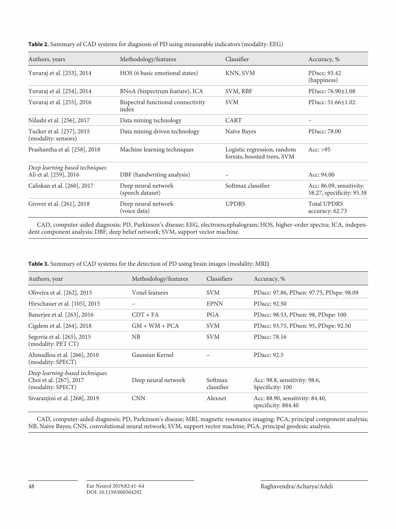
Table 2. Summary of CAD systems for diagnosis of PD using measurable indicators (modality: EEG)
Authors, years Methodology/features Classifier Accuracy, %
Yuvaraj et al. [253], 2014 HOS (6 basic emotional states) KNN, SVM PDacc: 93.42(happiness)
Yuvaraj et al. [254], 2014 BNoA (bispectrum feature), ICA SVM, RBF PDacc: 76.90±1.08
Yuvaraj et al. [255], 2016 Bispectral functional connectivityindex
SVM PDacc: 51.66±1.02
Nilashi et al. [256], 2017 Data mining technology CART –
Tucker et al. [257], 2015(modality: sensors)
Data mining driven technology Naïve Bayes PDacc: 78.00
Prashantha et al. [258], 2018 Machine learning techniques Logistic regression, randomforests, boosted trees, SVM
Acc: >95
Deep learning based techniquesAli et al. [259], 2016 DBF (handwriting analysis) – Acc: 94.00
Caliskan et al. [260], 2017 Deep neural network(speech dataset)
Softmax classifier Acc: 86.09, sensitivity:58.27, specificity: 95.38
Grover et al. [261], 2018 Deep neural network(voice data)
UPDRS Total UPDRSaccuracy: 62.73
CAD, computer-aided diagnosis; PD, Parkinson’s disease; EEG, electroencephalogram; HOS, higher-order spectra; ICA, indepen-dent component analysis; DBF, deep belief network; SVM, support vector machine.
Table 3. Summary of CAD systems for the detection of PD using brain images (modality: MRI)
Authors, year Methodology/features Classifiers Accuracy, %
Oliveira et al. [262], 2015 Voxel features SVM PDacc: 97.86, PDsen: 97.75, PDspe: 98.09
Hirschauer et al. [105], 2015 – EPNN PDacc: 92.50
Banerjee et al. [263], 2016 CDT + FA PGA PDacc: 98.53, PDsen: 98, PDspe: 100
Cigdem et al. [264], 2018 GM + WM + PCA SVM PDacc: 93.75, PDsen: 95, PDspe: 92.50
Segovia et al. [265], 2015(modality: PET CT)
NB SVM PDacc: 78.16
Ahmadlou et al. [266], 2010(modality: SPECT)
Gaussian Kernel – PDacc: 92.5
Deep learning-based techniquesChoi et al. [267], 2017(modality: SPECT)
Deep neural network Softmaxclassifier
Acc: 98.8, sensitivity: 98.6,Specificity: 100
Sivaranjini et al. [268], 2019 CNN Alexnet Acc: 88.90, sensitivity: 84.40,specificity: 884.40
CAD, computer-aided diagnosis; PD, Parkinson’s disease; MRI, magnetic resonance imaging; PCA, principal component analysis; NB, Naive Bayes; CNN, convolutional neural network; SVM, support vector machine; PGA, principal geodesic analysis.
AI Techniques for Automated Diagnosis of Neurological Disorders
49Eur Neurol 2019;82:41–64DOI: 10.1159/000504292
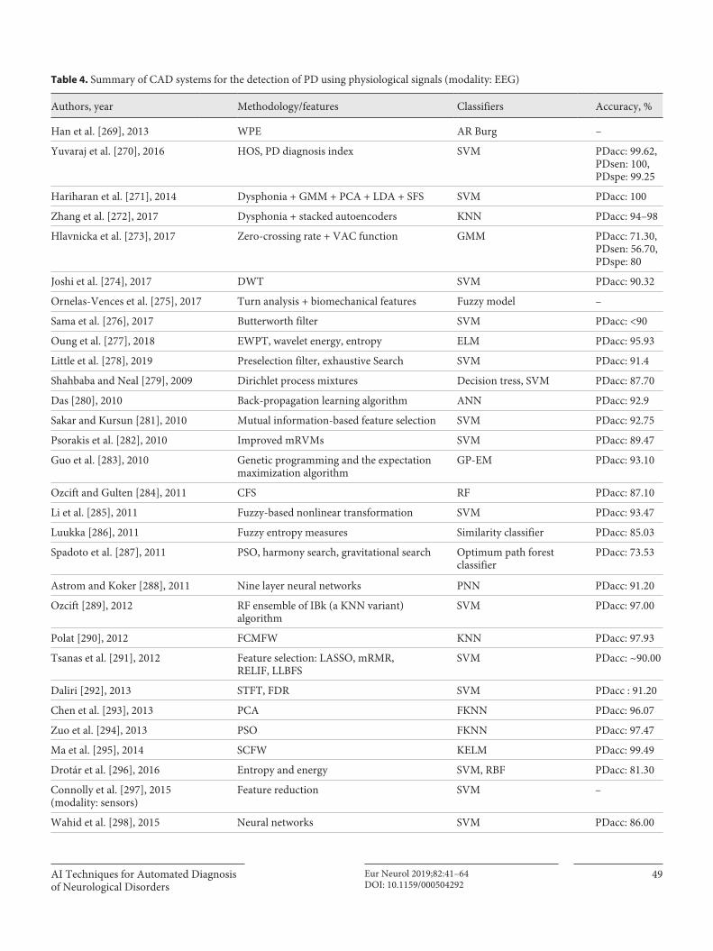
Table 4. Summary of CAD systems for the detection of PD using physiological signals (modality: EEG)
Authors, year Methodology/features Classifiers Accuracy, %
Han et al. [269], 2013 WPE AR Burg –
Yuvaraj et al. [270], 2016 HOS, PD diagnosis index SVM PDacc: 99.62,PDsen: 100,PDspe: 99.25
Hariharan et al. [271], 2014 Dysphonia + GMM + PCA + LDA + SFS SVM PDacc: 100
Zhang et al. [272], 2017 Dysphonia + stacked autoencoders KNN PDacc: 94–98
Hlavnicka et al. [273], 2017 Zero-crossing rate + VAC function GMM PDacc: 71.30,PDsen: 56.70,PDspe: 80
Joshi et al. [274], 2017 DWT SVM PDacc: 90.32
Ornelas-Vences et al. [275], 2017 Turn analysis + biomechanical features Fuzzy model –
Sama et al. [276], 2017 Butterworth filter SVM PDacc: <90
Oung et al. [277], 2018 EWPT, wavelet energy, entropy ELM PDacc: 95.93
Little et al. [278], 2019 Preselection filter, exhaustive Search SVM PDacc: 91.4
Shahbaba and Neal [279], 2009 Dirichlet process mixtures Decision tress, SVM PDacc: 87.70
Das [280], 2010 Back-propagation learning algorithm ANN PDacc: 92.9
Sakar and Kursun [281], 2010 Mutual information-based feature selection SVM PDacc: 92.75
Psorakis et al. [282], 2010 Improved mRVMs SVM PDacc: 89.47
Guo et al. [283], 2010 Genetic programming and the expectationmaximization algorithm
GP-EM PDacc: 93.10
Ozcift and Gulten [284], 2011 CFS RF PDacc: 87.10
Li et al. [285], 2011 Fuzzy-based nonlinear transformation SVM PDacc: 93.47
Luukka [286], 2011 Fuzzy entropy measures Similarity classifier PDacc: 85.03
Spadoto et al. [287], 2011 PSO, harmony search, gravitational search Optimum path forestclassifier
PDacc: 73.53
Astrom and Koker [288], 2011 Nine layer neural networks PNN PDacc: 91.20
Ozcift [289], 2012 RF ensemble of IBk (a KNN variant)algorithm
SVM PDacc: 97.00
Polat [290], 2012 FCMFW KNN PDacc: 97.93
Tsanas et al. [291], 2012 Feature selection: LASSO, mRMR,RELIF, LLBFS
SVM PDacc: ~90.00
Daliri [292], 2013 STFT, FDR SVM PDacc : 91.20
Chen et al. [293], 2013 PCA FKNN PDacc: 96.07
Zuo et al. [294], 2013 PSO FKNN PDacc: 97.47
Ma et al. [295], 2014 SCFW KELM PDacc: 99.49
Drotár et al. [296], 2016 Entropy and energy SVM, RBF PDacc: 81.30
Connolly et al. [297], 2015 (modality: sensors)
Feature reduction SVM –
Wahid et al. [298], 2015 Neural networks SVM PDacc: 86.00

Raghavendra/Acharya/AdeliEur Neurol 2019;82:41–6450DOI: 10.1159/000504292
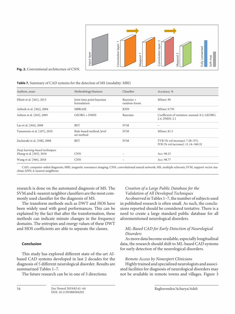
(CNN) with a number of advantages over conventional ML-based CAD systems. First and foremost is that it can handle large data sets using its multilayer architecture. It can also handle imbalanced data samples and execute the results without biasing toward majority class. The CNN ar-chitecture generally includes a convolution layer, a pooling layer, and fully connected layers [106]. These layers can be extended with few additional layers based on the require-ments. Recently, various CNN-based techniques have shown good results in detecting neurological disorders [107–116]. But, detection of such abnormalities in its early stage may require more deeper architecture. Such deeper architecture can pick distinct features from the signals by convolving with kernels defined in each layers [106].
In the past few years, deep neural network learning techniques have been used to solve intractable problems in a variety of disciplines such as image recognition [117–119], pavement crack detection [120], structural damage detection [121, 122], vehicle-type detection [123], trans-portation systems [124, 125], concrete strength estima-tion [126], big data time series forecasting [127], and com-puter-brain interface [128]. Following that trend, a num-ber of researchers have developed CAD systems using
deep learning techniques. These techniques can discover structures or patterns in the biological signals or images. Its effectiveness in signal and image-based CAD systems has been demonstrated recently [117, 127, 129–133].
Deep learning techniques can be broadly classified into 2 types: (1) supervised learning and (2) unsupervised learn-ing. The CNN belongs to the first and auto encoders belong to second type. Antoniades et al. [134] analyze the epileptic intracranial EEG data using deep learning. Johansen et al. [135] use CNN for epileptiform spike detection. Yuan et al. [136] employ a combination of deep learning and short-time Fourier transform for epileptic seizure detection. Ul-lah et al. [137] report application of deep learning for epi-lepsy diagnosis. Martinez-Murcia et al. [138] employ the CNN for analysis of neuroimaging in PD.
The most common architecture of CNN includes a bank of convolution, max-pool, fully connected, and soft-max layers as shown in Figure 2 [129–131]. The 3D CNN and CNN – Recurrent neural network (RNN) are more recent networks used in the diagnosis of epilepsy [139], PD [140] and AD [141]. Other CNN variants are also ex-perimented on PD and ischemic brain stroke detection. The list of approaches used is presented in Tables 1–7. It
Authors, year Methodology/features Classifiers Accuracy, %
Smith et al. [299], 2015 (modality: sensors)
Evolutionary algorithms ROC PDacc: 78.00
Hirschauer et al. [105], 2015 Neural networks EPNN PDacc: 92.5
Shamir et al. [300], 2015 Clinical decision support systems SVM PDacc: 71.00
Procházka et al. [301], 2015 (modality: sensors)
Bayesian Bayesian probabilityclassifiers
PDacc: 94.1
Nilashi et al. [302], 2018 Partial least squares, self organizing map SVM UPDRS acc: 46.56
Deep learning based techniquesTagaris et al. [140], 2017 Deep neural network CNN-RNN Acc: 74
Kim et al. [303], 2018 Neural networks CNN PDacc: 85
Oh [304], 2018 CNN – Acc: 88.25, sensitivity: 84.71,specificity: 91.77
CAD, computer aided diagnosis; ICA, independent component analysis; EEG, electroencephalogram; HOS, higher order spectra; PD, Parkinson’s disease; WPE, wavelet packet entropy; SVM, support vector machine; GMM, gaussian mixture model; KNN, k-nearest neighbour; DWT, discrete wavelet transform; CNN, convolutional neural network; PSO, particle swarm optimization; PNN, probabi-listic neural network; PCA, principal component analysis.
Table 4. (continued)
AI Techniques for Automated Diagnosis of Neurological Disorders
51Eur Neurol 2019;82:41–64DOI: 10.1159/000504292
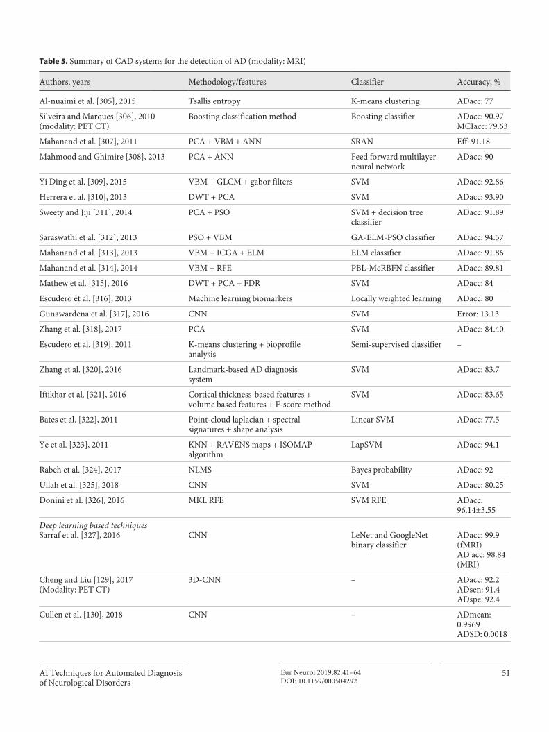
Table 5. Summary of CAD systems for the detection of AD (modality: MRI)
Authors, years Methodology/features Classifier Accuracy, %
Al-nuaimi et al. [305], 2015 Tsallis entropy K-means clustering ADacc: 77
Silveira and Marques [306], 2010 (modality: PET CT)
Boosting classification method Boosting classifier ADacc: 90.97MCIacc: 79.63
Mahanand et al. [307], 2011 PCA + VBM + ANN SRAN Eff: 91.18
Mahmood and Ghimire [308], 2013 PCA + ANN Feed forward multilayerneural network
ADacc: 90
Yi Ding et al. [309], 2015 VBM + GLCM + gabor filters SVM ADacc: 92.86
Herrera et al. [310], 2013 DWT + PCA SVM ADacc: 93.90
Sweety and Jiji [311], 2014 PCA + PSO SVM + decision treeclassifier
ADacc: 91.89
Saraswathi et al. [312], 2013 PSO + VBM GA-ELM-PSO classifier ADacc: 94.57
Mahanand et al. [313], 2013 VBM + ICGA + ELM ELM classifier ADacc: 91.86
Mahanand et al. [314], 2014 VBM + RFE PBL-McRBFN classifier ADacc: 89.81
Mathew et al. [315], 2016 DWT + PCA + FDR SVM ADacc: 84
Escudero et al. [316], 2013 Machine learning biomarkers Locally weighted learning ADacc: 80
Gunawardena et al. [317], 2016 CNN SVM Error: 13.13
Zhang et al. [318], 2017 PCA SVM ADacc: 84.40
Escudero et al. [319], 2011 K-means clustering + bioprofileanalysis
Semi-supervised classifier –
Zhang et al. [320], 2016 Landmark-based AD diagnosissystem
SVM ADacc: 83.7
Iftikhar et al. [321], 2016 Cortical thickness-based features +volume based features + F-score method
SVM ADacc: 83.65
Bates et al. [322], 2011 Point-cloud laplacian + spectralsignatures + shape analysis
Linear SVM ADacc: 77.5
Ye et al. [323], 2011 KNN + RAVENS maps + ISOMAPalgorithm
LapSVM ADacc: 94.1
Rabeh et al. [324], 2017 NLMS Bayes probability ADacc: 92
Ullah et al. [325], 2018 CNN SVM ADacc: 80.25
Donini et al. [326], 2016 MKL RFE SVM RFE ADacc:96.14±3.55
Deep learning based techniquesSarraf et al. [327], 2016 CNN LeNet and GoogleNet
binary classifierADacc: 99.9(fMRI)AD acc: 98.84(MRI)
Cheng and Liu [129], 2017 (Modality: PET CT)
3D-CNN – ADacc: 92.2ADsen: 91.4ADspe: 92.4
Cullen et al. [130], 2018 CNN – ADmean:0.9969ADSD: 0.0018
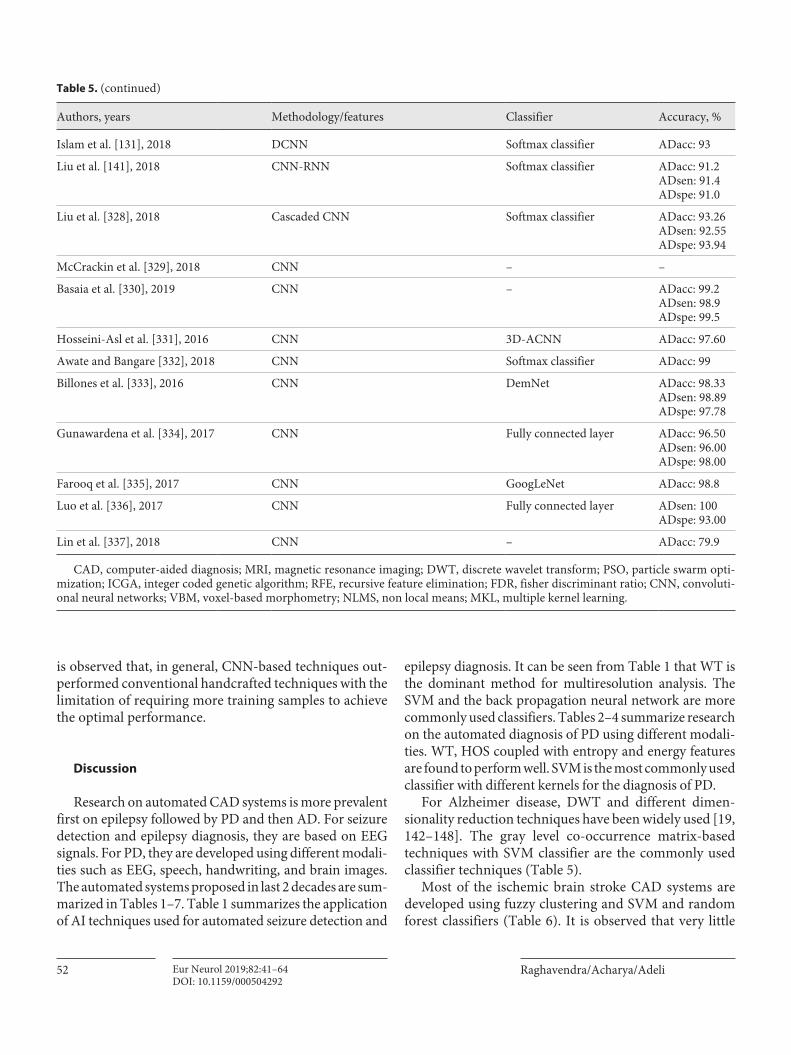
Raghavendra/Acharya/AdeliEur Neurol 2019;82:41–6452DOI: 10.1159/000504292
is observed that, in general, CNN-based techniques out-performed conventional handcrafted techniques with the limitation of requiring more training samples to achieve the optimal performance.
Discussion
Research on automated CAD systems is more prevalent first on epilepsy followed by PD and then AD. For seizure detection and epilepsy diagnosis, they are based on EEG signals. For PD, they are developed using different modali-ties such as EEG, speech, handwriting, and brain images. The automated systems proposed in last 2 decades are sum-marized in Tables 1–7. Table 1 summarizes the application of AI techniques used for automated seizure detection and
epilepsy diagnosis. It can be seen from Table 1 that WT is the dominant method for multiresolution analysis. The SVM and the back propagation neural network are more commonly used classifiers. Tables 2–4 summarize research on the automated diagnosis of PD using different modali-ties. WT, HOS coupled with entropy and energy features are found to perform well. SVM is the most commonly used classifier with different kernels for the diagnosis of PD.
For Alzheimer disease, DWT and different dimen-sionality reduction techniques have been widely used [19, 142–148]. The gray level co-occurrence matrix-based techniques with SVM classifier are the commonly used classifier techniques (Table 5).
Most of the ischemic brain stroke CAD systems are developed using fuzzy clustering and SVM and random forest classifiers (Table 6). It is observed that very little
Authors, years Methodology/features Classifier Accuracy, %
Islam et al. [131], 2018 DCNN Softmax classifier ADacc: 93
Liu et al. [141], 2018 CNN-RNN Softmax classifier ADacc: 91.2ADsen: 91.4ADspe: 91.0
Liu et al. [328], 2018 Cascaded CNN Softmax classifier ADacc: 93.26ADsen: 92.55ADspe: 93.94
McCrackin et al. [329], 2018 CNN – –
Basaia et al. [330], 2019 CNN – ADacc: 99.2ADsen: 98.9ADspe: 99.5
Hosseini-Asl et al. [331], 2016 CNN 3D-ACNN ADacc: 97.60
Awate and Bangare [332], 2018 CNN Softmax classifier ADacc: 99
Billones et al. [333], 2016 CNN DemNet ADacc: 98.33ADsen: 98.89ADspe: 97.78
Gunawardena et al. [334], 2017 CNN Fully connected layer ADacc: 96.50ADsen: 96.00ADspe: 98.00
Farooq et al. [335], 2017 CNN GoogLeNet ADacc: 98.8
Luo et al. [336], 2017 CNN Fully connected layer ADsen: 100ADspe: 93.00
Lin et al. [337], 2018 CNN – ADacc: 79.9
CAD, computer-aided diagnosis; MRI, magnetic resonance imaging; DWT, discrete wavelet transform; PSO, particle swarm opti-mization; ICGA, integer coded genetic algorithm; RFE, recursive feature elimination; FDR, fisher discriminant ratio; CNN, convoluti-onal neural networks; VBM, voxel-based morphometry; NLMS, non local means; MKL, multiple kernel learning.
Table 5. (continued)
AI Techniques for Automated Diagnosis of Neurological Disorders
53Eur Neurol 2019;82:41–64DOI: 10.1159/000504292
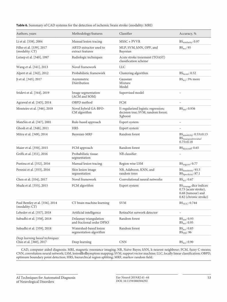
Table 6. Summary of CAD systems for the detection of ischemic brain stroke (modality: MRI)
Authors, years Methodology/features Classifier Accuracy, %
Li et al. [338], 2004 Manual lesion tracing MSSC + PVVR BSsimilarity: 0.97
Filho et al. [339], 2017(modality: CT)
ABTD extractor used toextract features
MLP, SVM, kNN, OPF, andBayesian
BSacc: 95
Lutsep et al. [340], 1997 Radiologic techniques Acute stroke trearment (TOAST)classification scheme
Wang et al. [341], 2013 Novel framework LLC
Alpert et al. [342], 2012 Probabilistic framework Clustering algorithm BSfscore: 0.52
Ji et al. [343], 2017 AsymmetricDistribution
GaussianMixtureModel
BSacc: 5% more
Sridevi et al. [344], 2019 Image segmentation(ACM and SOM)
Supervised model –
Agrawal et al. [345], 2014 OBPD method FCM –
Monteiro et al. [346], 2018 Novel hybrid GA-BFO-CM algorithm
l1 regularized logistic regression;decision tree; SVM; random forest;Xgboost
BSauc: 0.936
MateSin et al. [347], 2001 Rule-based approach Expert system –
Ghosh et al. [348], 2011 HRS Expert system –
Mitra et al. [349], 2014 Bayesian-MRF Random forest BSsensitivity: 0.53±0.13BSmeanpositivevalue:0.75±0.18
Maier et al. [350], 2015 FCM approach Random forest BSdicecoeff: 0.65
Griffs et al. [351], 2016 Probabilistic tissuesegmentation
NB classifier –
Pustina et al. [352], 2016 Manual lesion tracing Region wise LSM BSavgcorr: 0.77
Pennisi et al. [353], 2016 Skin lesion imagesegmentation
NB, Adaboost, KNN, andrandom trees
BSSensitivity: 93.5BSSpecificity: 87.1
Chen et al. [354], 2017 Novel framework Convolutional neural networks BSacc: 0.67
Muda et al. [355], 2015 FCM algorithm Expert system BSAverage dice indices0.73 (acute stroke),0.68 (tumour) and0.82 (chronic stroke)
Paul Bentley et al. [356], 2014(modality: CT)
CT brain machine learning SVM BSAUC: 0.744
Lebedev et al. [357], 2018 Artificial intelligence RetinaNet network detector –
Subudhi et al. [358], 2018 Delaunay triangulationand fractional order DPSO
Random forest BSsen: 0.93BSacc: 0.95
Subudhi et al. [359], 2018 Watershed-based lesionsegmentation algorithm
Random forest BSacc: 0.85BSDSI: 96
Deep learning based techniquesChin et al. [360], 2017 Deep learning CNN BSacc: 0.90
CAD, computer aided diagnosis; MRI, magnetic resonance imaging; NB, Naive Bayes; kNN, k-nearest neighbour; FCM, fuzzy C-means; CNN, convolution neural network; LSM, lesion‐to‐symptom mapping; SVM, support vector machine; LLC, locally linear classification; OBPD, optimum boundary point detection; HRS, hierarchical region splitting; MRF, markov random field.
Raghavendra/Acharya/AdeliEur Neurol 2019;82:41–6454DOI: 10.1159/000504292
research is done on the automated diagnosis of MS. The SVM and k-nearest neighbor classifiers are the most com-monly used classifier for the diagnosis of MS.
The transform methods such as DWT and HOS have been widely used with good performances. This can be explained by the fact that after the transformation, these methods can indicate minute changes in the frequency domains. The entropies and energy values of these DWT and HOS coefficients are able to separate the classes.
Conclusion
This study has explored different state-of-the-art AI-based CAD systems developed in last 2 decades for the diagnosis of 5 different neurological disorder. Results are summarized Tables 1–7.
The future research can be in one of 3 directions:
Creation of a Large Public Database for the Validation of All Developed TechniquesAs observed in Tables 1–7, the number of subjects used
in published research is often small. As such, the conclu-sions reported should be considered tentative. There is a need to create a large standard public database for all aforementioned neurological disorders.
ML-Based CAD for Early Detection of Neurological DisordersAs more data become available, especially longitudinal
data, the research should shift to ML-based CAD systems for early detection of the neurological disorders.
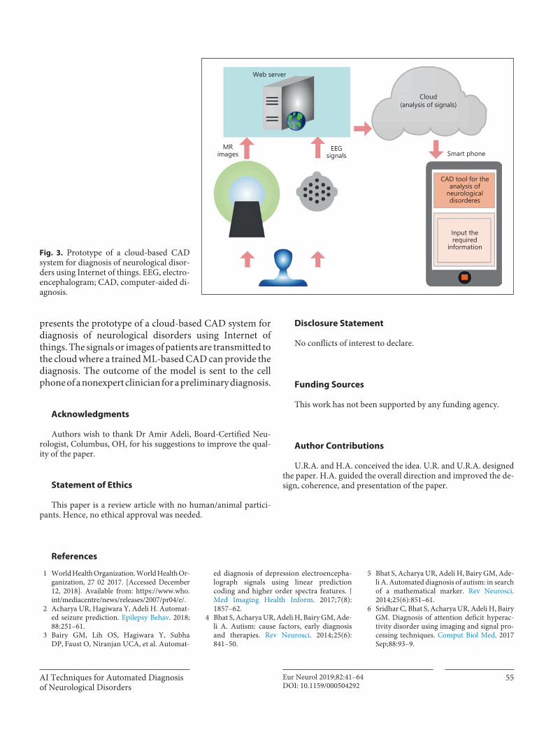
Remote Access by Nonexpert CliniciansHighly trained and specialized neurologists and associ-
ated facilities for diagnosis of neurological disorders may not be available in remote towns and villages. Figure 3
Table 7. Summary of CAD systems for the detection of MS (modality: MRI)
Authors, years Methodology/features Classifier Accuracy, %
Elliott et al. [361], 2013 Joint time point bayesianformulation
Bayesian +random-forest
MSsen: 99
Anbeek et al. [362], 2004 MBRASE KNN MSsen: 0.791
Ashton et al. [363], 2003 GEORG + DMSS Bayesian Coefficient of variation: manual: 8.2, GEORG: 2.4, DMSS: 2.1
Lao et al. [364], 2008 BET SVM –
Yamamoto et al. [107], 2010 Rule-based method, levelset method
SVM MSsen: 81.5
Zacharaki et al. [108], 2008 BET SVM TVR (% vol increase): 7.58–375,IVR (% vol increase): 11.14–340.32
Deep learning-based techniquesZhang et al. [365], 2018 CNN – Acc: 98.23
Wang et al. [366], 2018 CNN – Acc: 98.77
CAD, computer-aided diagnosis; MRI, magnetic resonance imaging; CNN, convolutional neural network; MS, multiple sclerosis; SVM, support vector ma-chine; kNN, k-nearest neighbour.
Inpu
t lay
er
Conv
olut
ion
laye
r: 1
Max
pool
1
Max
pool
2
Fully
con
nect
ed
Soft-
max
Conv
olut
ion
laye
r: 2
Fig. 2. Conventional architecture of CNN.
AI Techniques for Automated Diagnosis of Neurological Disorders
55Eur Neurol 2019;82:41–64DOI: 10.1159/000504292
presents the prototype of a cloud-based CAD system for diagnosis of neurological disorders using Internet of things. The signals or images of patients are transmitted to the cloud where a trained ML-based CAD can provide the diagnosis. The outcome of the model is sent to the cell phone of a nonexpert clinician for a preliminary diagnosis.
Acknowledgments
Authors wish to thank Dr Amir Adeli, Board-Certified Neu-rologist, Columbus, OH, for his suggestions to improve the qual-ity of the paper.
Statement of Ethics
This paper is a review article with no human/animal partici-pants. Hence, no ethical approval was needed.
Disclosure Statement
No conflicts of interest to declare.
Funding Sources
This work has not been supported by any funding agency.
Author Contributions
U.R.A. and H.A. conceived the idea. U.R. and U.R.A. designed the paper. H.A. guided the overall direction and improved the de-sign, coherence, and presentation of the paper.
MRimages
Web server
EEGsignals Smart phone
Cloud(analysis of signals)
CAD tool for theanalysis of
neurologicaldisorderes
Input therequired
informationFig. 3. Prototype of a cloud-based CAD system for diagnosis of neurological disor-ders using Internet of things. EEG, electro-encephalogram; CAD, computer-aided di-agnosis.
References
1 World Health Organization. World Health Or-ganization, 27 02 2017. [Accessed December 12, 2018]. Available from: https://www.who.int/mediacentre/news/releases/2007/pr04/e/.
2 Acharya UR, Hagiwara Y, Adeli H. Automat-ed seizure prediction. Epilepsy Behav. 2018;
88: 251–61. 3 Bairy GM, Lih OS, Hagiwara Y, Subha
DP, Faust O, Niranjan UCA, et al. Automat-
ed diagnosis of depression electroencepha-lograph signals using linear prediction coding and higher order spectra features. J Med Imaging Health Inform. 2017; 7(8):
1857–62. 4 Bhat S, Acharya UR, Adeli H, Bairy GM, Ade-
li A. Autism: cause factors, early diagnosis and therapies. Rev Neurosci. 2014; 25(6):
841–50.
5 Bhat S, Acharya UR, Adeli H, Bairy GM, Ade-li A. Automated diagnosis of autism: in search of a mathematical marker. Rev Neurosci. 2014; 25(6): 851–61.
6 Sridhar C, Bhat S, Acharya UR, Adeli H, Bairy GM. Diagnosis of attention deficit hyperac-tivity disorder using imaging and signal pro-cessing techniques. Comput Biol Med. 2017 Sep; 88: 93–9.
Raghavendra/Acharya/AdeliEur Neurol 2019;82:41–6456DOI: 10.1159/000504292
7 Jahmunah V, Lih Oh S, Rajinikanth V, Ciac-cio EJ, Hao Cheong K, Arunkumar N, et al. Automated detection of schizophrenia using nonlinear signal processing methods. Artif Intell Med. 2019 Sep; 100: 101698.
8 Acharya UR, Oh SL, Hagiwara Y, Tan JH, Adeli H, Subha DP. Automated EEG-based screening of depression using deep convolu-tional neural network. Comput Methods Pro-grams Biomed. 2018 Jul; 161: 103–13.
9 Acharya UR, Oh SL, Hagiwara Y, Tan JH, Adeli H. Deep convolutional neural network for the automated detection and diagnosis of seizure using EEG signals. Comput Biol Med. 2018 Sep; 100: 270–8.
10 Ortega-Zamorano F, Jerez JM, Gómez I, Fran-co L. Layer Multiplexing FPGA Implementa-tion for Deep Back-Propagation Learning. In-tegr Comput Aided Eng. 2017; 24(2): 171–85.
11 Ay B, Yildirim O, Talo M, Baloglu UB, Aydin G, Puthankattil SD, et al. Automated Depres-sion Detection Using Deep Representation and Sequence Learning with EEG Signals. J Med Syst. 2019 May; 43(7): 205.
12 Yıldırım Ö, Baloglu UB, Acharya UR. A deep convolutional neural network model for au-tomated identification of abnormal EEG sig-nals. Neural Comput Appl. 2018: 1–12.
13 Khedher L, Illán IA, Górriz JM, Ramírez J, Brahim A, Meyer-Baese A. Independent Component Analysis-Support Vector Ma-chine-Based Computer-Aided Diagnosis Sys-tem for Alzheimer’s with Visual Support. Int J Neural Syst. 2017 May; 27(3): 1650050.
14 López-Sanz D, Garcés P, Álvarez B, Delgado-Losada ML, López-Higes R, Maestú F. Net-work Disruption in the Preclinical Stages of Alzheimer’s Disease: From Subjective Cogni-tive Decline to Mild Cognitive Impairment. Int J Neural Syst. 2017 Dec; 27(8): 1750041.
15 Fang C, Li C, Cabrerizo M, Barreto A, An-drian J, Rishe N, et al. Gaussian Discriminant Analysis-Based Dual High-Dimensional De-cision Spaces for the Diagnosis of Mild Cogni-tive Impairment in Alzheimer’s Disease. Int J Neural Syst. 2018; 28(8): 1850017.
16 Valenzuela O, Jiang X, Carrillo A, Rojas I. Multi-Objective Genetic Algorithms to Find Most Relevant Volumes of the Brain Related to Alzheimer’s Disease and Mild Cognitive Impairment. Int J Neural Syst. 2018 Nov;
28(9): 1850022.17 Acharya UR, Hagiwara Y, Deshpande SN,
Suren S, Wei Koh JE, Lih Oh S, et al. Charac-terization of focal EEG signals: a review. Fu-ture Gener Comput Syst. 2019; 91: 290–9.
18 Budka H. Neuropathology of human immu-nodeficiency virus infection. Brain Pathol. 1991 Apr; 1(3): 163–75.
19 Mammone N, Ieracitano C, Adeli H, Braman-ti A, Morabito FC. Permutation Jaccard Dis-tance-based Hierarchical Clustering to esti-mate EEG network density modifications in MCI subjects. IEEE Trans Neural Netw Learn Syst. 2018 Feb; 29(10): 5122–35.
20 Acharya UR, Fernandes SL, WeiKoh JE, Ciac-cio EJ, Fabell MK, Tanik UJ, et al. Automated
Detection of Alzheimer’s Disease Using Brain MRI Images- A Study with Various Feature Extraction Techniques. J Med Syst. 2019 Aug;
43(9): 302.21 Saunders AM, Strittmatter WJ, Schmechel D,
St. George-Hyslop PH, Pericak-Vance MA, Joo SH, et al. Association of apolipoprotein E allele e4 with late-onset familial and sporadic AD. Neurology. 1993; 43: 1467–72.
22 Nalbantoglu J, Gilfix BM, Bertrand P, Rob-itaille Y, Gauthier S, Rosenblatt DS, et al. Pre-dictive value of apolipoprotein E genotyping in Alzheimer’s disease: results of an autopsy series and an analysis of several combined studies. Ann Neurol. 1994 Dec; 36(6): 889–95.
23 Fisher RS, van Emde Boas W, Blume W, Elger C, Genton P, Lee P, et al. Epileptic seizures and epilepsy. Defi nitions proposed by the In-ternational League against Epilepsy (ILAE) and the International Bureau for Epilepsy (IBE). Epilepsia. 2005 Apr; 46(4): 470–2.
24 Guo L, Wang Z, Cabrerizo M, Adjouadi M. A Cross-Correlated Delay Shift Supervised Learning Method for Spiking Neurons with Application to Interictal Spike Detection in Epilepsy. Int J Neural Syst. 2017 May; 27(3):
1750002.25 Wostyn S, Staljanssens W, De Taeye L, Strob-
be G, Gadeyne S, Van Roost D, et al. EEG De-rived Brain Activity Reflects Treatment Re-sponse from Vagus Nerve Stimulation in Pa-tients with Epilepsy. Int J Neural Syst. 2017 Jun; 27(4): 1650048.
26 Martín-López D, Jiménez-Jiménez D, Caba-ñés-Martínez L, Selway RP, Valentín A, Alar-cón G. The role of thalamus versus cortex in epilepsy: evidence from human ictal centro-median recordings in patients assessed for deep brain stimulation. Int J Neural Syst. 2017 Nov; 27(7): 1750010.
27 Kugiumtzis D, Koutlis C, Tsimpiris A, Kim-iskidis VK. Dynamics of Epileptiform Dis-charges Induced by Transcranial Magnetic Stimulation in Genetic Generalized Epilepsy. Int J Neural Syst. 2017 Nov; 27(7): 1750037.
28 Varatharajah Y, Iyer RK, Berry BM, Worrell GA, Brinkmann BH. Seizure Forecasting and the Preictal State in Canine Epilepsy. Int J Neural Syst. 2017 Feb; 27(1): 1650046.
29 Shanir PP, Khan KA, Khan YU, Farooq O, Adeli H. Automatic Seizure Detection Based on Morphological Features Using One-Di-mensional Local Binary Pattern on Long-Term EEG. Clin EEG Neurosci. 2018 Sep;
49(5): 351–62.30 Kotsopoulos IA, van Merode T, Kessels FG,
de Krom MC, Knottnerus JA. Systematic re-view and meta-analysis of incidence studies of epilepsy and unprovoked seizures. Epilepsia. 2002 Nov; 43(11): 1402–9.
31 Kotsopoulos I, de Krom M, Kessels F, Lodder J, Troost J, Twellaar M, et al. Incidence of ep-ilepsy and predictive factors of epileptic and non-epileptic seizures. Seizure. 2005 Apr;
14(3): 175–82.32 De Cooman T, Varon C, Hunyadi B, Van
Paesschen W, Lagae L, Van Huffel S. Online
Automated Seizure Detection in Temporal Lobe Epilepsy Patients Using Single-lead ECG. Int J Neural Syst. 2017 Nov; 27(7):
1750022.33 Li R, Ji GJ, Yu Y, Yu Y, Ding MP, Tang YL, et
al. Epileptic discharge related functional con-nectivity within and between networks in be-nign epilepsy with centrotemporal spikes. Int J Neural Syst. 2017 Nov; 27(7): 1750018.
34 Jiang S, Luo C, Gong J, Peng R, Ma S, Tan S, et al. Aberrant thalamocortical connectivity in juvenile myoclonic epilepsy. Int J Neural Syst. 2018 Feb; 28(1): 1750034.
35 Adeli H, Zhou Z, Dadmehr N. Analysis of EEG records in an epileptic patient using wavelet transform. J Neurosci Methods. 2003 Feb; 123(1): 69–87.
36 Yuan Q, Zhou W, Xu F, Leng Y, Wei D. Epi-leptic EEG Identification via LBP Operators on Wavelet Coefficients. Int J Neural Syst. 2018 Oct; 28(8): 1850010.
37 Li Y, Cui W, Luo M, Li K, Wang L. Epileptic Seizure Detection Based on Time-Frequency Images of EEG Signals Using Gaussian Mix-ture Model and Gray Level Co-Occurrence Matrix Features. Int J Neural Syst. 2018 Sep;
28(7): 1850003.38 Yuan S, Zhou W, Chen L. Epileptic Seizure
Prediction Using Diffusion Distance and BLDA in Intracranial EEG. Int J Neural Syst. 2018; 28(1): 1750043.
39 Schetinin V, Jakaite L, Krzanowski W. Bayes-ian Learning of Models for Estimating Uncer-tainty in Alert Systems: Application to Air traffic conflict avoidance. Integr Comput Aided Eng. 2018; 25(3): 1–17.
40 Kobelt G, Pugliatti M. Cost of multiple sclero-sis in Europe. Eur J Neurol. 2005 Jun; 12(s1 Suppl 1): 63–7.
41 Jock Murray T, Allen C. Bowling, Chris Pol-man, Alan Thompson, John Noseworthy: Multiple sclerosis – The guide to treatment and management. London, 6th Ed. Multiple Sclerosis International Federation. 2006.
42 Nutt JG, Wooten GF. Clinical practice. Diag-nosis and initial management of Parkinson’s disease. N Engl J Med. 2005 Sep; 353(10):
1021–7.43 Bhat S, Acharya UR, Hagiwara Y, Dadmehr
N, Adeli H. Parkinson’s disease: cause factors, measurable indicators, and early diagnosis. Comput Biol Med. 2018 Nov; 102: 234–41.
44 Chaudhuri KR, Yates L, Martinez-Martin P. The non-motor symptom complex of Parkin-son’s disease: a comprehensive assessment is essential. Curr Neurol Neurosci Rep. 2005 Jul;
5(4): 275–83.45 Marras C, Tanner CM. Epidemiology of Par-
kinson’s disease. In: Watts RL, Koller WC, editors. Movement disorders, neurologic principles and practice. 2nd ed. New York: McGraw Hill; 2004. pp. 177–96.
46 Shinde S, Prasad S, Saboo Y, Kaushick R, Saini J, Pal PK, et al. Predictive markers for Parkin-son’s disease using deep neural nets on neu-romelanin sensitive MRI. Neuroimage Clin. 2019; 22: 101748.
AI Techniques for Automated Diagnosis of Neurological Disorders
57Eur Neurol 2019;82:41–64DOI: 10.1159/000504292
47 Zhang A, San-Segundo R, Panev S, Tabor G, Stebbins K, Whitford A, et al. Automated Tremor Detection in Parkinson’s Disease Us-ing Accelerometer Signals. IEEE/ACM Inter-national Conference on Connected Health: Applications, Systems and Engineering Tech-nologies (CHASE), Washington, DC, USA, 2018.
48 Gálvez G, Recuero M, Canuet L, Del-Pozo F. Short-term effects of Binaural Beats on EEG power, functional connectivity, cognition, gait and anxiety in Parkinson’s Disease. Int J Neural Syst. 2018 Jun; 28(5): 1750055.
49 Hatano S. Experience from a multicentre stroke register: a preliminary report. Bull World Health Organ. 1976; 54(5): 541–53.
50 Kelly DF, Becker DP. Advances in manage-ment of neurosurgical trauma: USA and Canada. World J Surg. 2001 Sep; 25(9): 1179–85.
51 Fakhry SM, Trask AL, Waller MA, Watts DD; IRTC Neurotrauma Task Force. Management of brain-injured patients by an evidence-based medicine protocol improves outcomes and decreases hospital charges. J Trauma. 2004 Mar; 56(3): 492–9.
52 Pain terms: a list with definitions and notes on usage. Recommended by the IASP Subcom-mittee on Taxonomy. Pain. 1979 Jun; 6(3): 249.
53 http://epileptologiebonn.de/cms/front_con-tent.php?idcat=0&idart=0&client=1&lang=3&error=1.
54 http://adni.loni.usc.edu/.55 https://www.oasis-brains.org/.56 Arne Jensen and Anders la Cour-Harbo. Rip-
ples in mathematics: the discrete wavelet transform. Springer Science & Business Me-dia; 2001.
57 Abbasi H, Bennet L, Gunn AJ, Unsworth CP. Robust Wavelet Stabilized ‘Footprints of Un-certainty’ for Fuzzy System Classifiers to Au-tomatically Detect Sharp Waves in the EEG after Hypoxia Ischemia. Int J Neural Syst. 2017 May; 27(3): 1650051.
58 Dai H, Cao Z. A wavelet support vector ma-chine-based neural network meta model for structural reliability assessment. Comput Aid-ed Civ Infrastruct Eng. 2017; 32(4): 344–57.
59 Candes EJ, Donoho DL. Curvelets: A surpris-ingly effective non adaptive representation for objects with edges. Technical report. DTIC Document; 2000.
60 Shao Y, Celenk M. Higher-order spectra (HOS) invariants for shape recognition. Pat-tern Recognit. 2001; 34(11): 2097–113.
61 Chua KC, Chandran V, Acharya UR, Lim CM. Application of higher order statistics/spectra in biomedical signals—a review. Med Eng Phys. 2010 Sep; 32(7): 679–89.
62 Pizer SM, Amburn EP, Austin JD, Cromartie R, Geselowitz A, Greer T, et al. Adaptive histo-gram equalization and its variations. Comput Vis Graph Image Process. 1987; 39(3): 355–68.
63 Malladi R, Sethian JA, Vemuri B. Shape mod-eling with front propagation: A level set ap-proach. IEEE Trans Pattern Anal Mach Intell. 1995; 17(2): 158–75.
64 Robert M. Haralick, Karthikeyan Shanmu-gam, and Its’ Hak Dinstein: textural features for image classification. Systems. IEEE Trans-actions on Man and Cybernetics. 1973; 6: 610–21.
65 Renyi A. On measures of entropy and infor-mation, in: Proceedings of the Fourth Berke-ley symposium on mathematical statistics and probability. 1961; 1: 547–561.
66 Shannon CE. A mathematical theory of com-munication. The Bell System Technical Jour-nal J. 1948; 27(3): 379–423.
67 Kapur JN. Information of order αand type β. Proc Indiana Acad Sci. 1968; 68(2): 65–75.
68 Ghosh M, Chakraborty C, Ray AK. Yager’s measure based fuzzy divergence for micro-scopic color image segmentation. in: Indian Conference on Medical Informatics and Tele-medicine. Kharagpur, 2013; 13–16.
69 Chen W, Wang Z, Xie H, Yu W. Characteriza-tion of surface EMG signal based on fuzzy en-tropy. IEEE Trans Neural Syst Rehabil Eng. 2007 Jun; 15(2): 266–72.
70 Yin PY. Maximum entropy-based optimal threshold selection using deterministic rein-forcement learning with controlled random-ization. Signal Processing. 2002; 82(7): 993–1006.
71 Sezgin N, Emin Tagluk M. Energy based fea-ture extraction for classification of sleep ap-nea syndrome. Comput Biol Med. 2009 Nov;
39(11): 1043–50.72 Rosso OA, Blanco S, Yordanova J, Kolev V,
Figliola A, Schürmann M, et al. Wavelet en-tropy: a new tool for analysis of short duration brain electrical signals. J Neurosci Methods. 2001 Jan; 105(1): 65–75.
73 Rényi A. On measures of entropy and infor-mation. In: Proceedings of the fourth Berke-ley symposium on mathematical statistics and probability, 1961; 547–561.
74 Chen J, Li G. Tsallis wavelet entropy and its application in power signal analysis. Entropy (Basel). 2014; 16(6): 3009–25.
75 Hu MK. Visual pattern recognition by mo-ment invariants. IRE Trans Inf Theory. 1962;
8(2): 179–87.76 Khotanzad A, Hong YH. Invariant image
recognition by zernike moments. IEEE Trans Pattern Anal Mach Intell. 1990; 12(5):
489–97.77 Nixon M, Nixon MS, Aguado AS. Feature ex-
traction & image processing for computer vi-sion. Academic Press; 2012.
78 Duda RO, Hart PE, Stork DG. Pattern classi-fication. 2nd ed. California, USA: Wiley-In-terscience; 2000.
79 Hyvärinen A, Oja E. Independent component analysis: algorithms and applications. Neural Netw. 2000 May-Jun; 13(4-5): 411–30.
80 Scholkopf B, Smola A, Muller KR. Nonlinear component analysis as a kernel eigenvalue problem. Neural Comput. 1998; 10(5): 1299–319.
81 Gelman A. Analysis of variance –why it is more important than ever. Ann Stat. 2005; 33(1): 1–33.
82 T-test. Student’s t-tests. Information. [date accessed on 04.07.17]. Available from: http://www.physics.csbsju.edu/stats/t-test.html.
83 Dash M, Liu H. Handling large unsupervised data via dimensionality reduction. ACM SIGMOD Workshop on Research Issues in Data Mining and Knowledge Discovery. 1999.
84 Abe N, Kudo M. Entropy criterion for classi-fier-independent feature selection. Knowl-edge-based intelligent information and engi-neering systems. Lect Notes Comput Sci. 2005; 3684: 689–95.
85 Lopes N. Comparing machine learning algo-rithms with the Wilcoxon Signed Rank Test. Information. [date accessed on 04.07.18]. Available from: http://www.uc.pt/fctuc/dei/statisticalHypothesis/noel.
86 Hwang T, Sun CH, Yun T, Yi GS. FiGS: a filter-based gene selection workbench for microar-ray data. BMC Bioinformatics. 2010 Jan; 11: 50.
87 Natarajan S, Lipsitz SR, Fitzmaurice GM, Sinha D, Ibrahim JG, Haas J, et al. An exten-sion of the Wilcoxon Rank-Sum test for com-plex sample survey data. J R Stat Soc Ser C Appl Stat. 2012 Aug; 61(4): 653–64.
88 Yuan Y, Van Allen EM, Omberg L, Wagle N, Amin-Mansour A, Sokolov A, et al. Assessing the clinical utility of cancer genomic and pro-teomic data across tumor types. Nat Biotech-nol. 2014 Jul; 32(7): 644–52.
89 Kailath T. The divergence and Bhattacha-ryya distance measures in signal selection. IEEE Trans Commun Technol. 1967; 15(1):
52–60.90 Obuchowski NA. Receiver operating charac-
teristic curves and their use in radiology. Ra-diology. 2003 Oct; 229(1): 3–8.
91 Mitchell M. An Introduction to Genetic Algo-rithms. USA: MIT Press. Cambridge. 1998.
92 Kennedy J, Eberhart R: Particle swarm opti-mization. IEEE Int. Conf. Neural Netw. 1995.
93 Shi Y, Eberhart R. A modified particle swarm optimizer. In: Proceedings of the IEEE World Congress on Computational Intelligence. An-chorage; 1998.
94 Dorigo M, Birattari M, Stutzle T. Ant colony optimization. IEEE Comput Intell Mag. 2006;
1(4): 28–39.95 Nemati S, Basiri ME, Ghasem-Aghaee N,
Aghdam MH. A novel ACO–GA hybrid algo-rithm for feature selection in protein function prediction. Expert Syst Appl. 2009; 36(10):
12086–94.96 Peng H, Long F, Ding C. Feature selection
based on mutual information: criteria of max-dependency, max-relevance, and min-redun-dancy. IEEE Trans Pattern Anal Mach Intell. 2005 Aug; 27(8): 1226–38.
97 Specht DF. Probabilistic neural networks and the polynomial Adaline as complementary techniques for classification. IEEE Trans Neu-ral Netw. 1990; 1(1): 111–21.
98 Kecman V. Learning and Soft Computing. Cambridge (MA): MIT Press; 2001.
99 Yager RR. An extension of the naive bayesian classifier. Inf Sci. 2006; 176(5): 577–88.

Raghavendra/Acharya/AdeliEur Neurol 2019;82:41–6458DOI: 10.1159/000504292
100 Heckerman D, Geiger D, Chickering DM. Learning Bayesian networks: the combina-tion of knowledge and statistical data. Mach Learn. 1995; 20(3): 197–243.
101 Larose DT. Discovering Knowledge in Data: An Introduction to Data Mining. Wiley-In-terscience; 2004.
102 Cover TM. Geometrical and statistical prop-erties of systems of linear inequalities with applications in pattern recognition. IEEE Trans Electron Comput. 1965; 14(3): 326–34.
103 Amit Y, Geman D. Shape quantization and recognition with randomized trees. Neural Comput. 1997; 9(7): 1545–88.
104 Berge A, Solberg AH. Structured Gaussian components for hyperspectral image classi-fication. IEEE Trans Geosci Remote Sens. 2006; 44(11): 3386–96.
105 Hirschauer TJ, Adeli H, Buford JA. Com-puter-aided diagnosis of Parkinson’s dis-ease using an enhanced probabilistic neural network. J Med Syst. 2015 Nov; 39(11): 179.
106 Tan JH, Hagiwara Y, Pang W, Lim I, Oh SL, Adam M, et al. Application of stacked con-volutional and long short-term memory network for accurate identification of CAD ECG signals. Comput Biol Med. 2018 Mar;
94: 19–26.107 Yamamoto D, Arimura H, Kakeda S, Ma-
gome T, Yamashita Y, Toyofuku F, et al. Computer-aided detection of multiple scle-rosis lesions in brain magnetic resonance images: false positive reduction scheme con-sisted of rule-based, level set method, and support vector machine. Comput Med Im-aging Graph. 2010 Jul; 34(5): 404–13.
108 Zacharaki EI, Kanterakis S, Bryan RN, Da-vatzikos C. Measuring brain lesion progres-sion with a supervised tissue classification system. Proc Int Conf Med Image Comput Comput Assist Interv. 2008; 11: 620–27.
109 Raghavendra U, Shyamasunder Bhat N, Gu-digar A, Acharya UR. Automated system for the detection of thoracolumbar fractures us-ing a CNN architecture. Future Gener Com-put Syst. 2018; 85: 184–9.
110 Raghavendra U, Fujita H, Bhandary SV, Gudigar A, Tan JH, Acharya UR. Deep con-volution neural network for accurate diag-nosis of glaucoma using digital fundus im-ages. Inf Sci. 2018; 441: 41–9.
111 Yildirim O, Talo M, Ay B, Baloglu UB, Aydin G, Acharya UR. Automated detec-tion of diabetic subject using pre-trained 2D-CNN models with frequency spectrum images extracted from heart rate signals. Comput Biol Med. 2019 Aug; 113: 103387.
112 Tan JH, Bhandary SV, Sivaprasad S, Hagi-wara Y, Bagchi A, Raghavendra U. Age-re-lated Macular Degeneration detection using deep convolutional neural network. Future Gener Comput Syst. 2018; 87: 127–35.
113 Acharya UR, Oh SL, Hagiwara Y, Tan JH, Adam M, Gertych A, et al. A deep convolu-tional neural network model to classify heartbeats. Comput Biol Med. 2017 Oct; 89:
389–96.
114 Ker J, Wang L, Rao J, Lim T. Deep Learning Applications in Medical Image Analysis. IEEE Access. 2017; 6: 9375–89.
115 Oh SL, Ng EY, Tan RS, Acharya UR. Auto-mated diagnosis of arrhythmia using com-bination of CNN and LSTM techniques with variable length heart beats. Comput Biol Med. 2018 Nov; 102: 278–87.
116 Yıldırım Ö, Pławiak P, Tan RS, Acharya UR. Arrhythmia detection using deep convolu-tional neural network with long duration ECG signals. Comput Biol Med. 2018 Nov;
102: 411–20.117 Koziarski M, Cyganek B. Image Recogni-
tion with Deep Neural Networks in Pres-ence of Noise – Dealing with and Taking Advantage of Distortions. Integr Comput Aided Eng. 2017; 24(4): 337–49.
118 Wang P, Bai X. Regional Parallel Structure Based CNN for Thermal Infrared Face Iden-tification. Integr Comput Aided Eng. 2018;
25(3): 247–60.119 Chen L, Ye F, Ruan Y, Fan H, Chen Q. An
algorithm for highway vehicle detection based on convolutional neural network. EURASIP J Image Video Process. 2018;
2018(1): 109.120 Zhang A, Wang KC, Li B, Yang E, Dai X,
Peng Y, et al. Automated Pixel-Level Pave-ment Crack Detection on 3D Asphalt Sur-faces Using a Deep-Learning Network. Comput Aided Civ Infrastruct Eng. 2017;
32(10): 805–19.121 Lin YZ, Nie ZH, Ma HW. Structural Dam-
age Detection with Automatic Feature-ex-traction through Deep Learning. Comput Aided Civ Infrastruct Eng. 2017; 32(12):
1025–46.122 Gao Y, Mosalam KM. Deep Transfer Learn-
ing for Image-based Structural Damage Recognition. Comput Aided Civ Infrastruct Eng. 2018; 33(9): 748–68.
123 Molina-Cabello MA, Luque-Baena RM, López-Rubio E, Thurnhofer-Hemsi K. Ve-hicle Type Detection by Ensembles of Con-volutional Neural Networks Operating on Super-resolved Images. Integr Comput Aided Eng. 2018; 25(4): 321–33.
124 Nabian MA, Meidani H. Deep Learning for Accelerated Reliability Analysis of Trans-portation Networks. Comput Aided Civ In-frastruct Eng. 2018; 33(6): 459–80.
125 Hashemi H, Abdelghany K. End-to-end deep learning methodology for real-time traffic network management. Comput Aid-ed Civ Infrastruct Eng. 2018; 33(10): 849–63.
126 Rafiei MH, Khushefati WH, Demirboga R, Adeli H. Supervised Deep Restricted Boltzmann Machine for Estimation of Con-crete Compressive Strength. ACI Mater J. 2017; 114(2): 237–44.
127 Torres JF, Galicia A, Troncoso A, Martínez-Álvarez F. A scalable approach based on deep learning for big data time series fore-casting. Integr Comput Aided Eng. 2018;
25(4): 335–48.
128 Li W, Li M, Zhou H, Chen G, Jin J, Duan F. A Dual Stimuli Approach Combined with Convolutional Neural Network to Improve Information Transfer Rate of Event-Related Potential-Based Brain-Computer Interface. Int J Neural Syst. 2018 Dec; 28(10): 1850034.
129 Cheng D, Liu M. Classification of AD by Cascaded Convolutional Neural Networks Using PET Images. Springer International Publishing AG 2017. Wang Q, et al. (eds). MLMI 2017. LNCS 10541. 2017. pp. 106–113.
130 Cullen NC, Avants BB. Convolutional Neu-ral Networks for Rapid and Simultaneous Brain Extraction and Tissue Segmentation. Brain Morphometry. Neuromethods. 2018;
136: 13–34.131 Islam J, Zhang Y. Brain MRI analysis for Al-
zheimer’s disease diagnosis using an ensem-ble system of deep convolutional neural net-works. Brain Inform. 2018 May; 5(2): 2.
132 Hsu WY. A hybrid approach for brain im-age registration with local constraints. In-tegr Comput Aided Eng. 2017; 24(1): 73–85.
133 Lozano A, Soto-Sánchez C, Garrigós J, Mar-tínez JJ, Ferrández JM, Fernández E. A 3D convolutional neural network to model ret-inal ganglion cell’s responses to light pat-terns in mice. Int J Neural Syst. 2018 Dec;
28(10): 1850043.134 Antoniades A, Spyrou L, Took CC, Sanei S.
Deep learning for epileptic intracranial EEG data. Italy: IEEE 26th International Work-shop on Machine Learning for Signal Pro-cessing (MLSP); 2016.
135 Johansen AR, Jin J, Maszczyk T, Dauwels J, Cash SS, Westover MB. Epileptiform spike detection via convolutional neural networks. China: IEEE International Conference on Acoustics, Speech and Signal Processing (ICASSP); 2016.
136 Yuan Y, Xun G, Jia K, Zhang A. A Multi-view Deep Learning Method for Epileptic Seizure Detection using Short-time Fourier Transform. Proceedings of the 8th ACM In-ternational Conference on Bioinformatics, Computational Biology and Health Infor-matics - ACM-BCB; 2017.
137 Ullah I, Hussain M, Qazi EH, Aboalsamh H. An automated system for epilepsy detection using EEG brain signals based on deep learn-ing approach. Expert Syst Appl. 2018; 107:
61–71.138 Martinez-Murcia FJ, Górriz JM, Ramírez J,
Ortiz A. Convolutional Neural Networks for Neuroimaging in Parkinson’s Disease: Is Preprocessing Needed? Int J Neural Syst. 2018 Dec; 28(10): 1850035.
139 Tjepkema-Cloostermans MC, de Carvalho RC, van Putten MJ. Deep learning for detec-tion of focal epileptiform discharges from scalp EEG recordings. Clin Neurophysiol. 2018 Oct; 129(10): 2191–6.
140 Tagaris A, Kollias D, Stafylopatis A. Assess-ment of Parkinson’s Disease Based on Deep Neural Networks. Commun Comput Inf Sci. 2017; 744: 391–403.
AI Techniques for Automated Diagnosis of Neurological Disorders
59Eur Neurol 2019;82:41–64DOI: 10.1159/000504292
141 Liu M, Cheng D, Yan W; Alzheimer’s Dis-ease Neuroimaging Initiative. Classification of Alzheimer’s Disease by Combination of Convolutional and Recurrent Neural Net-works Using FDG-PET Images. Front Neu-roinform. 2018 Jun; 12: 35.
142 Ahmadlou M, Adeli H, Adeli A. New diag-nostic EEG markers of the Alzheimer’s dis-ease using visibility graph. J Neural Transm (Vienna). 2010 Sep; 117(9): 1099–109.
143 Ahmadlou M, Adeli H, Adeli A. Fractality and a wavelet-chaos-methodology for EEG-based diagnosis of Alzheimer disease. Al-zheimer Dis Assoc Disord. 2011 Jan-Mar;
25(1): 85–92.144 Sankari Z, Adeli H. Probabilistic neural net-
works for diagnosis of Alzheimer’s disease using conventional and wavelet coherence. J Neurosci Methods. 2011 Apr; 197(1): 165–70.
145 Sankari Z, Adeli H, Adeli A. Intrahemi-spheric, interhemispheric, and distal EEG coherence in Alzheimer’s disease. Clin Neu-rophysiol. 2011 May; 122(5): 897–906.
146 Sankari Z, Adeli H, Adeli A. Wavelet coher-ence model for diagnosis of Alzheimer dis-ease. Clin EEG Neurosci. 2012 Oct; 43(4):
268–78.147 Amezquita-Sanchez JP, Adeli A, Adeli H. A
new methodology for automated diagnosis of mild cognitive impairment (MCI) using magnetoencephalography (MEG). Behav Brain Res. 2016 May; 305: 174–80.
148 Amezquita-Sanchez JP, Mammone N, Morabito FC, Marino S, Adeli H. A novel methodology for automated differential di-agnosis of mild cognitive impairment and the Alzheimer’s disease using EEG signals. J Neurosci Methods. 2019 Jul; 322: 88–95.
149 Acharya UR, Chua KC, Lim TC, Dorithy JS, Suri JS. Suri: automatic identification of epileptic EEG signals using nonlinear pa-rameters. J Mech Med Biol. 2009; 9(4): 539–53.
150 Acharya UR, Sree SV, Suri JS. Automatic de-tection of epileptic EEG signals using higher order cumulant features. Int J Neural Syst. 2011 Oct; 21(5): 403–14.
151 Acharya UR, Sree SV, Chattopadhyay S, Yu W, Ang PC. Application of recurrence quantification analysis for the automated identification of epileptic EEG signals. Int J Neural Syst. 2011 Jun; 21(3): 199–211.
152 Acharya UR, Vinitha Sree S, Alvin PC, Yan-ti R, Suri JS. Application of non-linear and wavelet based features for the automated identification of epileptic EEG signals. Int J Neural Syst. 2012;22(2): 1250002.
153 Acharya UR, Molinari F, Vinitha Sree S, Chattopadhyay S, Kwan-Hoong N, Suri JS. Automated diagnosis of epileptic EEG using entropies. Biomed Signal Process Control. 2012; 7(4): 401–8.
154 Acharya UR, Vinitha Sree S, Suri JS. Use of principal component analysis for automatic classification of epileptic EEG activities. Ex-pert Syst Appl. 2012; 39(10): 9072–8.
155 Acharya UR, Yanti R, Swapna G, Sree VS, Martis RJ, Suri JS. Automated diagnosis of epileptic electroencephalogram using independent component analysis and discrete wavelet transform for differ-ent electroencephalogram durations. Proc Inst Mech Eng H. 2013 Mar; 227(3): 234–44.
156 Aslan K, Bozdemir H, Sahin C, Oğulata SN, Erol R. A radial basis function neural net-work model for classification of epilepsy us-ing EEG signals. J Med Syst. 2008 Oct; 32(5):
403–8.157 Chua KC, Chandran V, Acharya UR, Lim
CM. Application of higher order spectra to identify epileptic EEG. J Med Syst. 2011 Dec; 35(6): 1563–71.
158 Faust O, Acharya UR, Min LC, Sputh BH. Automatic identification of epileptic and background EEG signals using frequency domain parameters. Int J Neural Syst. 2010 Apr; 20(2): 159–76.
159 Ghosh-Dastidar S, Adeli H, Dadmehr N. Mixed-band wavelet-chaos-neural network methodology for epilepsy and epileptic sei-zure detection. IEEE Trans Biomed Eng. 2007 Sep; 54(9): 1545–51.
160 Ghosh-Dastidar S, Adeli H, Dadmehr N. Principal component analysis-enhanced cosine radial basis function neural network for robust epilepsy and seizure detection. IEEE Trans Biomed Eng. 2008 Feb; 55(2 Pt 1): 512–8.
161 Ghosh-Dastidar S, Adeli H. A new super-vised learning algorithm for multiple spik-ing neural networks with application in epi-lepsy and seizure detection. Neural Netw. 2009 Dec; 22(10): 1419–31.
162 Guler NF, Ubey ED, Guler I. Recurrent neu-ral network employing Lyapunov expo-nents for EEG signals classification. Expert Syst Appl. 2005; 29(3): 506–14.
163 Guo L, Rivero D, Seoane JA, Pazos A. Clas-sification of EEG signals using relative wavelet energy and artificial neural net-works. In: Conf Proc of the First ACM/SI-GEVO Summit on Genetic and, Evolution-ary Computation. 2009. pp. 177–84.
164 Guo L, Rivero D, Pazos A. Epileptic seizure detection using multiwavelet transform based approximate entropy and artificial neural networks. J Neurosci Methods. 2010 Oct; 193(1): 156–63.
165 Guo L, Rivero D, Dorado J, Rabuñal JR, Pa-zos A. Automatic epileptic seizure detection in EEGs based on line length feature and ar-tificial neural networks. J Neurosci Meth-ods. 2010 Aug; 191(1): 101–9.
166 Guo L, Rivero D, Dorado J, Munteanu CR, Pazos A. Automatic feature extraction using genetic programming: an application to ep-ileptic EEG classification. Expert Syst Appl. 2011; 38(8): 10425–36.
167 Iscan Z, Dokur Z, Demiralp T. Classifica-tion of electroencephalogram signals with combined time and frequency features. Ex-pert Syst Appl. 2011; 38(8): 10499–505.
168 Kannathal N, Choo ML, Acharya UR, Sada-sivan PK. Entropies for detection of epilepsy in EEG. Comput Methods Programs Biomed. 2005 Dec; 80(3): 187–94.
169 Lima CA, Coelho AL, Eisencraft M. Tack-ling EEG signal classification with least squares support vector machines: a sensitiv-ity analysis study. Comput Biol Med. 2010 Aug; 40(8): 705–14.
170 Martis RJ, Acharya UR, Tan JH, Petznick A, Yanti R, Chua CK, et al. Application of em-pirical mode decomposition (emd) for auto-mated detection of epilepsy using EEG sig-nals. Int J Neural Syst. 2012 Dec; 22(6):
1250027.171 Nigam VP, Graupe D. A neural-network-
based detection of epilepsy. Neurol Res. 2004 Jan; 26(1): 55–60.
172 Orhan U, Hekim M, Ozer M. EEG signals classification using the K-means clustering and a multilayer perceptron neural network model. Expert Syst Appl. 2011; 38(10):
13475–81.173 Polat K, Gunes S. Classification of epilepti-
form EEG using a hybrid systems based on decision tree classifier and fast Fourier trans-form. Appl Math Comput. 2007; 187(2):
1017–26.174 Polat K, Gunes S. Artificial immune recog-
nition system with fuzzy resource allocation mechanism classifier, principal component analysis and FFT method based new hybrid automated identification system for classifi-cation of EEG signals. Expert Syst Appl. 2008; 34(3): 2039–48.
175 Polat K, Gunes S. A novel data reduction method: distance based data reduction and its application to classification of epilepti-form EEG signals. Appl Math Comput. 2008; 200(1): 10–27.
176 Sadati N, Mohseni HR, Magshoudi A. Epi-leptic seizure detection using neural fuzzy networks. In: Proceedings of the IEEE Inter-national Conference on Fuzzy Systems. 2006. pp. 596–600.
177 Srinivasan V, Eswaran C, Sriraam N. Artifi-cial neural network based epileptic detection using time-domain and frequency-domain features. J Med Syst. 2005 Dec; 29(6): 647–60.
178 Srinivasan V, Eswaran C, Sriraam N. Ap-proximate entropy-based epileptic EEG de-tection using artificial neural networks. IEEE Trans Inf Technol Biomed. 2007 May;
11(3): 288–95.179 Subasi A. EEG Signal classification using
wavelet feature extraction and a mixture of expert model. Expert Syst Appl. 2007; 32(4):
1084–93.180 Subasi A, Gursoy MI. EEG Signal classifica-
tion using PCA, ICA, LDA and support vec-tor machine. Expert Syst Appl. 2010; 37(12):
8659–66.181 Tzallas AT, Tsipouras MG, Fotiadis DI. Au-
tomatic seizure detection based on time-fre-quency analysis and artificial neural net-works. Comput Intell Neurosci. 2007; 2007:
80510.
Raghavendra/Acharya/AdeliEur Neurol 2019;82:41–6460DOI: 10.1159/000504292
182 Jaiswal AK, Banka H. Epileptic seizure de-tection in EEG signal using machine learn-ing techniques. Australas Phys Eng Sci Med. 2018 Mar; 41(1): 81–94.
183 Ubeyli ED. Least squares support vector machine employing model-based methods coefficients for analysis of EEG signals. Ex-pert Syst Appl. 2010; 37(1): 233–9.
184 Wang D, Miao D, Xie C. Best basis-based wavelet packet entropy feature extraction and hierarchical EEG classification for epi-leptic detection. Expert Syst Appl. 2011;
38(11): 14314–20.185 Swami P, Gandhi TK, Panigrahi BK, Tripa-
thi M, Anand S. A novel robust diagnostic model to detect seizures in lectroencepha-lography. Expert Syst Appl. 2016; 56: 116–30.
186 Peker M, Sen B, Delen D. A novel method for automated diagnosis of epilepsy using complex-valued classifiers. IEEE J Biomed Health Inform. 2016 Jan; 20(1): 108–18.
187 Sharma M, Pachori RB. A novel approach to detect epileptic seizures using a combina-tion of tunable-q wavelet transform and fractal dimension. J Mech Med Biol. 2017;
17(07): 1740003.188 Patidar S, Panigrahi T. Detection of epilep-
tic seizure using Kraskov entropy applied on tunable-q wavelet transform of EEG signals. Biomed Signal Process Control. 2017; 34:
74–80.189 Gandhi T, Panigrahi BK, Anand S. A com-
parative study of wavelet families for EEG signal classification. Neurocomputing. 2011; 74(17): 3051–7.
190 Chen G. Automatic EEG seizure detection using dual-tree complex wavelet-Fourier features. Expert Syst Appl. 2014; 41(5):
2391–4.191 Swami P, Godiyal AK, Santhosh J, Panigrahi
BK, Bhatia M, Anand S. Robust expert sys-tem design for automated detection of epi-leptic seizures using SVM classifier. In: Pro-ceedings of IEEE International Conference on Parallel, Distributed and Grid Comput-ing; 2014. pp. 219–22.
192 Pachori RB, Patidar S. Epileptic seizure clas-sification in EEG signals using second-order difference plot of intrinsic mode functions. Comput Methods Programs Biomed. 2014 Feb; 113(2): 494–502.
193 Sharma R, Pachori RB. Classification of epileptic seizures in EEG signals based on phase space representation of intrinsic mode functions. Expert Syst Appl. 2015;
42(3): 1106–17.194 Bhattacharyya A, Pachori RB, Upadhyay A,
Acharya UR. Tunable-q wavelet transform based multiscale entropy measure for auto-mated classification of epileptic EEG sig-nals. Appl Sci (Basel). 2017; 7(4): 385.
195 Bhattacharyya A, Sharma M, Pachori RB, Sircar P, Acharya UR. A novel approach for automated detection of focal EEG signals using empirical wavelet transform. Neural Comput Appl. 2018; 29(8): 47–57.
196 Bhati D, Sharma M, Pachori RB, Gadre VM. Time-frequency localized threeband bior-thogonal wavelet filter bank using semidefi-nite relaxation and nonlinear least squares with epileptic seizure EEG signal classifica-tion. Digit Signal Process. 2017; 62: 259–73.
197 Sharma M, Dhere A, Pachori RB, Acharya UR. An automatic detection of focal EEG signals using new class of time–frequency localized orthogonal wavelet filter banks. Knowl Base Syst. 2017; 118: 217–27.
198 Kaya Y, Uyar M, Tekin R, Yıldırım S. 1D-local binary pattern based feature extraction for classification of epileptic EEG signals. Appl Math Comput. 2014; 243: 209–19.
199 Zhu G, Li Y, Wen PP. Epileptic seizure de-tection in EEGs signals using a fast weighted horizontal visibility algorithm. Comput Methods Programs Biomed. 2014 Jul; 115(2):
64–75.200 Samiee K, Kovács P, Gabbouj M. Epileptic
seizure classification of EEG time-series us-ing rational discrete short-time fourier transform. IEEE Trans Biomed Eng. 2015 Feb; 62(2): 541–52.
201 Riaz F, Hassan A, Rehman S, Niazi IK, Dremstrup K. Emd-based temporal and spectral features for the classification of EEG signals using supervised learning. IEEE Trans Neural Syst Rehabil Eng. 2016 Jan;
24(1): 28–35.202 Diykh M, Li Y, Wen P. Classify epileptic eeg
signals using weighted complex networks based community structure detection. Ex-pert Syst Appl. 2017; 90: 87–100.
203 Li M, Chen W, Zhang T. Application of MODWT and log-normal distribution model for automatic epilepsy identification. Biocybern Biomed Eng. 2017; 37(4): 679–89.
204 Oh SL, Vicnesh J, Edward JC, Yuvaraj R, Acharya UR. Deep Convolutional Neural Network Model for Automated Diagnosis of Schizophrenia Using EEG Signals. Appl Sci (Basel). 2019; 9(14): 2870.
205 Al Ghayab HR, Li Y, Abdulla S, Diykh M, Wan X. Classification of epileptic EEG sig-nals based on simple random sampling and sequential feature selection. Brain Inform. 2016 Jun; 3(2): 85–91.
206 Sharma M, Bhuraneb AA, AcharyaUR. MMSFL-OWFB: A novel class of orthogo-nal wavelet filters for epileptic seizure detec-tion. Knowl Base Syst. 2018; 160: 265–77.
207 Tiwari AK, Pachori RB, Kanhangad V, Pani-grahi BK, Panigrahi B. Automated diagnosis of epilepsy using key-point based local bi-nary pattern of EEG signals. IEEE J Biomed Health Inform. 2017 Jul; 21(4): 888–96.
208 Bajaj V, Pachori RB. Classification of sei-zure and non-seizure EEG signals using em-pirical mode decomposition. IEEE Trans Inf Technol Biomed. 2012 Nov; 16(6): 1135–42.
209 Nicolaou N, Georgiou J. Detection of epi-leptic electroencephalogram based on Per-mutation Entropy and Support Vector Ma-chines. Expert Syst Appl. 2012; 39(1): 202–9.
210 Xie S, Krishnan S. Wavelet-based sparse functional linear model with applications to EEGs seizure detection and epilepsy diag-nosis. Med Biol Eng Comput. 2013 Feb;
51(1-2): 49–60.211 Mursalin M, Zhang Y, Chen Y, Chawla NV.
Automated epileptic seizure detection using improved correlation-based feature selec-tion with random forest classifier. Neuro-computing. 2017; 241: 204–14.
212 Upadhyay R, Padhy P, Kankar P. A com-parative study of feature ranking techniques for epileptic seizure detection using wavelet transform. Comput Electr Eng. 2016; 53:
163–76.213 Kabir E, Siuly, Zhang Y. Epileptic seizure
detection from EEG signals using logistic model trees. Brain Inform. 2016 Jun; 3(2):
93–100.214 Murugavel AS, Ramakrishnan S. Hierarchi-
cal multi-class SVM with ELM kernel for epileptic EEG signal classification. Med Biol Eng Comput. 2016 Jan; 54(1): 149–61.
215 Pippa E, Zacharaki EI, Mporas I, Tsirka V, Richardson MP, Koutroumanidis M, et al. Improving classification of epileptic and non-epileptic EEG events by feature selec-tion. Neurocomputing. 2016; 171: 576–85.
216 Kumar Y, Dewal M, Anand R. Epileptic sei-zures detection in EEG using DWT-based ApEn and artificial neural network. Signal Image Video Process. 2014; 8(7): 1323–34.
217 Naser A, Tantawi M, Shedeed H, Tolba M. Detecting Epileptic Seizures Using Abe En-tropy, Line Length and SVM Classifier. In-ternational Conference on Advanced Ma-chine Learning Technologies and Applica-tions. 2019; 169–178.
218 Tzimourta K, Tzallas A, Giannakeas N, As-trakas L, Angelidis L, Tsipouras D, et al. A robust methodology for classification of ep-ileptic seizures in EEG signals. Health Tech-nol. 2019; 9(2): 135–42.
219 Lahmiri S, Shmuel A. Accurate Classifica-tion of Seizure and Seizure-Free Intervals of Intracranial EEG Signals From Epileptic Patients. IEEE Trans Instrum Meas. 2019;
68(3): 791–6.220 Raghu S, Sriraam N, Temel Y, Rao SV, Hegde
AS, Kubben PL. Performance evaluation of DWT based sigmoid entropy in time and fre-quency domains for automated detection of epileptic seizures using SVM classifier. Com-put Biol Med. 2019 Jul; 110: 127–43.
221 Wang X, Gong G, Li N. Automated recogni-tion of epileptic EEG states using a combi-nation of symlet wavelet processing, gradi-ent boosting machine, and grid search opti-mizer. Sensors. 2019; 19(2): 219.
222 Bose R, Pratiher S, Chatterjee S. Detection of epileptic seizure employing a novel set of features extracted from multifractal spec-trum of electroencephalogram signals. IET Signal Process. 2019; 13(2): 157–64.
223 Dalal M, Tanveer M, Pachori RB. Machine Intelligence and Signal Analysis. Springer Singapore; 2019.

AI Techniques for Automated Diagnosis of Neurological Disorders
61Eur Neurol 2019;82:41–64DOI: 10.1159/000504292
224 Sriraam N, Tamanna K, Narayan L, Kha-num M, Raghu S, Hegde AS, et al. Multi-channel EEG based inter-ictal seizures de-tection using Teager energy with backprop-agation neural network classifier. Australas Phys Eng Sci Med. 2018 Dec; 41(4): 1047–55.
225 Shaikh M, Farooq O, Chandel G. Lecture Notes in Electrical Engineering 509 Advanc-es in System Optimization and Control; 2017.
226 Osman AH, Alzahrani AA. New Approach for Automated Epileptic Disease Diagnosis Using an Integrated Self-Organization Map and Radial Basis Function Neural Network Algorithm. IEEE Access. 2019; 7(7): 4741–7.
227 Sudalaimani C, Sivakumaran N, Elizabeth TT, Rominus VS. Automated detection of the preseizure state in EEG signal using neu-ral networks. Biocybern Biomed Eng. 2019;
39(1): 160–75.228 Raghu S, Sriraam N. Classification of focal
and non-focal EEG signals using neighbor-hood component analysis and machine learning algorithms. Expert Syst Appl. 2018;
113: 18–32.229 De Cooman T, Varon C, Van de Vel A, Jan-
sen K, Ceulemans B, Lagae L, et al. Adaptive nocturnal seizure detection using heart rate and low-complexity novelty detection. Sei-zure. 2018 Jul; 59: 48–53.
230 Li M, Chen W, Zhang T. A novel seizure di-agnostic model based on kernel density es-timation and least squares support vector ma.chine. Biomed Signal Process Control. 2018; 41: 233–41.
231 Cruz NE, Solarte J, Varghas A. Automated Epileptic Seizure Detection System Based on a Wearable Prototype and Cloud Com-puting to Assist People with Epilepsy. Ap-plied Computer Sciences in Engineering. Springer; 2018. pp. 204–13.
232 Kocadagli O, Langari R. Classification of EEG signals for epileptic seizures using hy-brid artificial neural networks based wavelet transforms and fuzzy relations. Expert Syst Appl. 2017; 88: 419–34.
233 Zhang T, Chen W, Li M. Fuzzy distribution entropy and its application in automated seizure detection technique. Biomed Signal Process Control. 2018; 39: 360–77.
234 Feng B, Zhao J, Fu W. Automated Classifi-cation of Epileptic EEG Signals Based on Multi-Feature Extraction. Beijing: IEEE 9th International Conference on Software Engi-neering and Service Science (ICSESS); 2018. pp. 382–6.
235 Tanveer M, Pachori R, Angami N. Entropy based features in FAWT framework for au-tomated detection of epileptic seizure EEG signals. IEEE Symp Ser Comput Intell; 2018. pp. 1946–52.
236 Choudhury NR, Roy SS, Pal A, Chatterjee S, Bose R. Epileptic Seizure Detection Em-ploying Cross-Hyperbolic Stockwell Trans-form. Kolkata: Fourth International Con-ference on Research in Computational In-telligence and Communication Networks (ICRCICN); 2018. pp. 70–4.
237 Torse D, Desai V, Khanai R. Classification of EEG Signals in Seizure Detection System Using Ellipse Area Features and Support Vector Machine. Proceedings of the 2nd In-ternational Conference on Data Engineer-ing and Communication Technology ICDECT. 2017.
238 Tomanik G, Betting L, Luiz A. Campanharo: Automatic Identification of Interictal Epi-leptiform Discharges with the Use of Com-plex Networks. Gran Canaria: Advances in Computational Intelligence, Proceedings of 15th International Work-Conference on Ar-tificial Neural Networks, IWANN 2019; 2019. pp. 152–61.
239 Wani S, Sabut S, Nalbalwar S. Detection of Epileptic Seizure Using Wavelet Transform and Neural Network Classifier. Proceedings of ICCASP; 2018.
240 Qi Y, Wang Y, Zhang J, Zhu J, Zheng X. Ro-bust deep network with maximum corrent-ropy criterion for seizure detection. BioMed Res Int. 2014; 2014: 703816.
241 Lin Q, Ye S, Huang X, Li S, Zhang M, Xue Y, et al. Classification of Epileptic EEG Sig-nals with Stacked Sparse Autoencoder Based on Deep Learning. In: Huang DS, Han K, Hussain A (eds). Intelligent Com-puting Methodologies. ICIC 2016. Lecture Notes in Computer Science, Springer, Cham; 2016. vol 9773, pp. 802–10.
242 Gogna A, Majumdar A, Ward R. Semi-su-pervised Stacked Label Consistent Autoen-coder for Reconstruction and Analysis of Biomedical Signals. IEEE Trans Biomed Eng. 2017 Sep; 64(9): 2196–205.
243 Hussein R, Palangi H, Ward RK, Wang ZJ. Optimized deep neural network architec-ture for robust detection of epileptic sei-zures using EEG signals. Clin Neurophysiol. 2019 Jan; 130(1): 25–37.
244 Thodoroff P, Pineau J, Lim A. Learning ro-bust features using deep learning for auto-matic seizure detection. In Proceedings of MLHC, 2016. pp. 178–90).
245 Emami A, Kunii N, Matsuo T, Shinozaki T, Kawai K, Takahashi H. Seizure detection by convolutional neural network-based analy-sis of scalp electroencephalography plot im-ages. Neuroimage Clin. 2019; 22: 101684.
246 Jang HJ, Cho KO. Dual deep neural net-work-based classifiers to detect experimen-tal seizures. Korean J Physiol Pharmacol. 2019 Mar; 23(2): 131–9.
247 Zuo R, Wei J, Li X, Li C, Zhao C, Ren Z, et al. Automated Detection of High-Frequen-cy Oscillations in Epilepsy Based on a Con-volutional Neural Network. Front Comput Neurosci. 2019 Feb; 13: 6.
248 Wei X, Zhou L, Chen Z, Zhang L, Zhou Y. Automatic seizure detection using three-di-mensional CNN based on multi-channel EEG. BMC Med Inform Decis Mak. 2018 Dec; 18(5 Suppl 5): 111.
249 Achilles F, Tombari F, Belagiannis V, Loesch A, Noachtar S, Navab N. Convolu-tional neural networks for real-time epilep-
tic seizure detection. Comput Methods Bio-mech Biomed Eng Imaging Vis. 2016; 1163:
264–9.250 Yuvaraj R, Thomas J, Kluge T, Dauwels J. A
deep Learning Scheme for Automatic Sei-zure Detection from Long-Term Scalp EEG. 52nd Asilomar Conf. Signals, Syst. Comput; 2018. pp. 368–72.
251 Hügle M, Heller S, Watter M, Blum M, Manzouri F, Dümpelmann M, et al. Early Seizure Detection with an Energy-Efficient Convolutional Neural Network on an Im-plantable Microcontroller. IEEE; 2018.
252 Thomas J, Comoretto L, Jin J, Dauwels J, Cash S, Westover M. EEG Classification Via Convolutional Neural Network-Based Interictal Epileptiform Event Detection. Proceedings of Annual International Con-ference of the IEEE Engineering in Medi-cine and Biology Society. 2018. pp. 3148–51.
253 Yuvaraj R, Murugappan M, Ibrahim NM, Sundaraj K, Omar MI, et al. Detection of emo-tions in Parkinson’s disease using higher oder spectral features from brain’s electrical activ-ity. Biomed. Signal Process Contr. 2014; 14:
108–16.254 Yuvaraj R, Murugappan M, Ibrahim NM,
Sundaraj K, Omar MI, Mohamad K, et al. Optimal set of EEG features for emotional state classification and trajectory visualiza-tion in Parkinson’s disease. Int J Psycho-physiol. 2014 Dec; 94(3): 482–95.
255 Yuvaraj R, Murugappan M, Acharya UR, Adeli H, Ibrahim NM, Mesquita E. Brain functional connectivity patterns for emo-tional state classification in Parkinson’s dis-ease patients without dementia. Behav Brain Res. 2016 Feb; 298 Pt B: 248–60.
256 Nilashi M, Ibrahim O, Ahmadi H, Shahmo-radi L. An analytical method for diseases prediction using machine learning tech-niques. Comput Chem Eng. 2017; 106: 212–23.
257 Tucker CS, Behoora I, Nembhard HB, Lew-is M, Sterling NW, Huang X. Machine learning classification of medication adher-ence in patients with movement disorders using non-wearable sensors. Comput Biol Med. 2015 Nov; 66: 120–34.
258 Prashantha R., Sumantra Dutta Roy: Early Detection of Parkinson's Disease through Patient Questionnaire and Predictive Mod-elling. Int J Med Inform. 2018; 119: 75–87.
259 Ali H. Al-Fatlawi, Mohammed H. Jabardi, Sai Ho Ling: Efficient Diagnosis System for Parkinson’s Disease Using Deep Belief Net-work. Canada: In 2016 IEEE Congress on Evolutionary Computation (CEC); 2016.
260 Caliskan A, Badem H, Baştürk A, Yüksel ME. Diagnosis of The Parkinson Disease By Using Deep Neural Network Classifier. In Iu-JEEE. 2017; 17(2): 3311–8.
261 Grover S, Bhartia S, Akshama AY, Seeja KR. Predicting Severity Of Parkinson’s Disease Using Deep Learning. Procedia Comput Sci. 2018; 132: 1788–94.
Raghavendra/Acharya/AdeliEur Neurol 2019;82:41–6462DOI: 10.1159/000504292
262 Oliveira FP, Castelo-Branco M. Computer-aided diagnosis of Parkinson’s disease based on [(123)I]FP-CIT SPECT binding poten-tial images, using the voxels-as-features ap-proach and support vector machines. J Neu-ral Eng. 2015 Apr; 12(2): 026008.
263 Banerjee M, Okun MS, Vaillancourt DE, Vemuri BC. A method for automated clas-sification of Parkinson’s disease diagnosis using an ensemble average propagator template brain map estimated from diffu-sion MRI. PLoS One. 2016 Jun; 11(6):
e0155764.264 Cigdem O, Beheshti I, Demirel H. Effects of
different covariates and contrasts on classi-fication of Parkinson’s disease using struc-tural MRI. Comput Biol Med. 2018 Aug; 99:
173–81.265 Segovia F, Illán IA, Górriz JM, Ramírez J,
Rominger A, Levin J. Distinguishing Par-kinson’s disease from atypical parkinsonian syndromes using PET data and a computer system based on support vector machines and Bayesian networks. Front Comput Neurosci. 2015 Nov; 9(137): 137.
266 Ahmadlou M, Adeli H. Enhanced probabi-listic neural network with local decision cir-cles: a robust classifier. Integr Comput Aid-ed Eng. 2010; 17(3): 197–210.
267 Choi H, Ha S, Im HJ, Paek SH, Lee DS. Re-fining diagnosis of Parkinson’s disease with deep learning-based interpretation of dopa-mine transporter imaging. Neuroimage Clin. 2017 Sep; 16: 586–94.
268 Sivaranjini S, Sujatha CM. Deep learning based diagnosis of Parkinsons disease using convolutional neural network. Multimed Tools Appl. 2019: 1–13.
269 Han CX, Wang J, Yi GS, Che YQ. Investiga-tion of EEG abnormalities in the early stage of Parkinson’s disease. Cogn Neurodyn. 2013 Aug; 7(4): 351–9.
270 Yuvaraj R, Acharya UR, Hagiwara Y. A nov-el Parkinson’s disease diagnosis index using higher-order spectra features in EEG sig-nals. Neural Comput Appl. 2018; 30(4):
1225–35.271 Hariharan M, Polat K, Sindhu R. A new hy-
brid intelligent system for accurate detec-tion of Parkinson’s disease. Comput Meth-ods Programs Biomed. 2014 Mar; 113(3):
904–13.272 Zhang YN. Can a Smartphone Diagnose
Parkinson Disease? A Deep Neural Net-work Method and Telediagnosis System Implementation. Parkinsons Dis. 2017;
2017: 6209703.273 Hlavnička J, Čmejla R, Tykalová T, Šonka K,
Růžička E, Rusz J. Automated analysis of connected speech reveals early biomarkers of Parkinson’s disease in patients with rapid eye movement sleep behaviour disorder. Sci Rep. 2017 Feb; 7(1): 12.
274 Joshi D, Khajuria A, Joshi P. An automatic non-invasive method for Parkinson’s dis-ease classification. Comput Methods Pro-grams Biomed. 2017 Jul; 145: 135–45.
275 Ornelas-Vences C, Sanchez-Fernandez LP, Sanchez-Perez LA, Garza-Rodriguez A, Vil-legas-Bastida A. Fuzzy inference model evaluating turn for Parkinson’s disease pa-tients. Comput Biol Med. 2017 Oct; 89: 379–88.
276 Samà A, Pérez-López C, Rodríguez-Martín D, Català A, Moreno-Aróstegui JM, Cabes-tany J, et al. Estimating bradykinesia sever-ity in Parkinson’s disease by analysing gait through a waist-worn sensor. Comput Biol Med. 2017 May; 84: 114–23.
277 Oung QW, Muthusamy H, Basah SN, Lee H, Vijean V. Empirical wavelet transform based features for classification of Parkin-son’s disease severity. J Med Syst. 2017 Dec;
42(2): 29.278 Little MA, McSharry PE, Hunter EJ, Spiel-
man J, Ramig LO. Suitability of dysphonia measurements for telemonitoring of Par-kinson’s disease. IEEE Trans Biomed Eng. 2009 Apr; 56(4): 1015–22.
279 Shahbaba B, Neal R. Nonlinear models us-ing Dirichlet process mixtures. J Mach Learn Res. 2009; 10: 1829–50.
280 Das R. A comparison of multiple classifica-tion methods for diagnosis of Parkinson disease. Expert Syst Appl. 2010; 37(2): 1568–72.
281 Sakar CO, Kursun O. Telediagnosis of Par-kinson’s disease using measurements of dysphonia. J Med Syst. 2010 Aug; 34(4): 591–9.
282 Psorakis I, Damoulas T, Girolami MA. Mul-ticlass relevance vector machines: sparsity and accuracy. IEEE Trans Neural Netw. 2010 Oct; 21(10): 1588–98.
283 Guo PF, Bhattacharya P, Kharma N. Ad-vances in detecting Parkinson’s disease. In: Zhang D, Sonka M, (eds). Berlin: Medical biometrics, Lecture Notes in Computer Sci-ence; 2010. pp. 306–14.
284 Ozcift A, Gulten A. Classifier ensemble con-struction with rotation forest to improve medical diagnosis performance of machine learning algorithms. Comput Methods Pro-grams Biomed. 2011 Dec; 104(3): 443–51.
285 Li DC, Liu CW, Hu SC. A fuzzy-based data transformation for feature extraction to in-crease classification performance with small medical data sets. Artif Intell Med. 2011 May; 52(1): 45–52.
286 Luukka P. Feature selection using fuzzy en-tropy measures with similarity classifier. Expert Syst Appl. 2011; 38(4): 4600–7.
287 Spadoto AA, Guido RC, Carnevali FL, Pagnin AF, Falcao AX, Papa JP. Improving Parkinson’s disease identification through evolutionary-based feature selection. In: Proceedings of the annual international conference of the IEEE engineering in med-icine and biology society (EMBC-11), Bos-ton; 2011. pp. 7857–60.
288 Astrom F, Koker R. A parallel neural net-work approach to prediction of Parkinson’s Disease. Expert Syst Appl. 2011; 38(10):
12470–4.
289 Ozcift A. SVM feature selection based rota-tion forest ensemble classifiers to improve computer-aided diagnosis of Parkinson dis-ease. J Med Syst. 2012 Aug; 36(4): 2141–7.
290 Polat K. Classification of Parkinson’s dis-ease using feature weighting method on the basis of fuzzy C-means clustering. Int J Syst Sci. 2012; 43(4): 597–609.
291 Tsanas A, Little MA, McSharry PE, Spiel-man J, Ramig LO. Novel speech signal pro-cessing algorithms for high-accuracy classi-fication of Parkinson’s disease. IEEE Trans Biomed Eng. 2012 May; 59(5): 1264–71.
292 Daliri MR. Chi square distance kernel of the gaits for the diagnosis of Parkinson’s dis-ease. Biomed Signal Process Control. 2013;
8(1): 66–70.293 Chen HL, Huang CC, Yu XG, Xuc X, Sund
X, Wang G, et al. An efficient diagnosis sys-tem for detection of Parkinson’s disease us-ing fuzzy k-nearest neighbor approach. Ex-pert Syst Appl. 2013; 40(1): 263–71.
294 Zuo WL, Wang ZY, Liu T, Chen HL. Effec-tive detection of Parkinson’s disease using an adaptive fuzzy K-nearest neighbour ap-proach. Biomed Signal Process Control. 2013; 8(4): 364–73.
295 Ma C, Ouyang J, Chen HL, Zhao XH. An efficient diagnosis system for Parkinson’s disease using kernel-based extreme learning machine with subtractive clustering fea-tures weighting approach. Comput Math Methods Med. 2014; 2014: 985789.
296 Drotár P, Mekyska J, Rektorová I, Masarová L, Smékal Z, Faundez-Zanuy M. Evaluation of handwriting kinematics and pressure for differential diagnosis of Parkinson’s disease. Artif Intell Med. 2016 Feb; 67: 39–46.
297 Connolly AT, Kaemmerer WF, Dani S, Stanslaski SR, Panken E, Johnson MD, et al. Guiding deep brain stimulation contact se-lection using local field potentials sensed by a chronically implanted device in Parkin-son's disease patients. Montpellier: 7th in-ternational conference on neural engineer-ing; 2015. pp. 840–3.
298 Wahid F, Begg RK, Hass CJ, Halgamuge S, Ackland DC. Classification of Parkinson’s disease gait using spatial-temporal gait fea-tures. IEEE J Biomed Health Inform. 2015 Nov; 19(6): 1794–802.
299 Smith SL, Lones MA, Bedder M, Alty JE, Cosgrove J, Maguire RJ, et al. Computation-al approaches for understanding the diag-nosis and treatment of Parkinson’s disease. IET Syst Biol. 2015 Dec; 9(6): 226–33.
300 Shamir RR, Dolber T, Noecker AM, Walter BL, McIntyre CC. Machine learning ap-proach to optimizing combined stimulation and medication therapies for Parkinson’s disease. Brain Stimul. 2015 Nov-Dec; 8(6):
1025–32.301 Procházka A, Vysata O, Valis M, Tupa O,
Schätz M, Marík V. Bayesian classification and analysis of gait disorders using image and depth sensors of Microsoft Kinect. Dig-it Signal Process. 2015; 47: 169–77.

AI Techniques for Automated Diagnosis of Neurological Disorders
63Eur Neurol 2019;82:41–64DOI: 10.1159/000504292
302 Nilashi M, Ibrahim O, Ahmadi H, Shahmo-radi L, Farahmand M. A hybrid intelligent system for the prediction of Parkinson’s Disease progression using machine learning techniques. Biocybern Biomed Eng. 2018;
38(1): 1–15.303 Kim HB, Lee WW, Kim A, Lee HJ, Park HY,
Jeon HS, et al. Wrist sensor-based tremor severity quantification in Parkinson’s dis-ease using convolutional neural network. Comput Biol Med. 2018 Apr; 95: 140–6.
304 Oh SL, Hagiwara Y, Raghavendra U, Yu-varaj R, Arunkumar N, Murugappan M, et al. A Deep Learning Approach for Parkin-son’s Disease Diagnosis from EEG Signals. Neural Comput Appl. 2018: 1–7.
305 Al-nuaimi AH, Jammeh E, Sun L, Ifeachor E. Tsallis Entropy as a Biomarker for Detec-tion of AD. Conf Proc IEEE Eng Med Biol Soc. 2015; 2015: 4166–9.
306 Silveira M, Marques J. Boosting Alzheimer Disease Diagnosis using PET images. Tur-key: International Conference on Pattern Recognition; 2010.
307 Mahanand BS, Suresh S, Sundararajan N, Aswatha Kumar M. Alzheimer’s disease de-tection using a Self-adaptive Resource Al-location Network classifier. San Jose: The 2011 International Joint Conference on Neural Networks; 2011. pp. 1930–4.
308 Mahmood R, Ghimire B. Automatic detec-tion and classification of Alzheimer’s Dis-ease from MRI scans using principal com-ponent analysis and artificial neural net-works. Bucharest: 20th International Conference on Systems, Signals and Image Processing (IWSSIP); 2013. pp. 133–7.
309 Ding Y. Cong Zhang, Tian Lan, Zhiguang Qin, Xinjie Zhang and Wei Wang: Classifi-cation of Alzheimer's disease based on the combination of morphometric feature and texture feature. Washington: IEEE Interna-tional Conference on Bioinformatics and Biomedicine; 2015. pp. 409–412.
310 Herrera LJ, Rojas I, Pomares H, Guillén A, Valenzuela O, Baños O. Classification of MRI Images for Alzheimer’s Disease Detec-tion. Alexandria: International Conference on Social Computing; 2013. pp. 846–51.
311 Sweety ME, Jiji GW. Detection of Alzheim-er disease in brain images using PSO and Decision Tree Approach. Ramanatha-puram: IEEE International Conference on Advanced Communications, Control and Computing Technologies; 2014. pp. 1305–9.
312 Saraswathi S, Mahanand BS, Kloczkowski A, Suresh S, Sundararajan N. Detection of onset of Alzheimer’s disease from MRI im-ages using a GA-ELM-PSO classifier. Singa-pore: Fourth International Workshop on Computational Intelligence in Medical Im-aging; 2013. pp. 42–48.
313 Mahanand BS, Suresh S, Sundararajan N, Kumar MA. ICGA-ELM classifier for Al-zheimer’s disease detection. Kharagpur: In-dian Conference on Medical Informatics
and Telemedicine (ICMIT); 2013. pp. 48–52.
314 Mahanand BS, Babu GS, Suresh S, Sundara-rajan N. Identification of imaging biomark-ers responsible for Alzheimer’s Disease us-ing a McRBFN classifier. International Con-ference on Cognitive Computing and Information Processing; 2015.
315 Mathew J, Mekkayil L, Ramasangu H, Karthikeyan BR, Manjunath AG. Robust al-gorithm for early detection of Alzheimer’s disease using multiple feature extractions. Bangalore: IEEE Annual India Conference (INDICON); 2016. pp. 1–6.
316 Escudero J, Ifeachor E, Zajicek JP, Green C, Shearer J, Pearson S; Alzheimer’s Disease Neuroimaging Initiative. Machine learn-ing-based method for personalized and cost-effective detection of Alzheimer’s dis-ease. IEEE Trans Biomed Eng. 2013 Jan;
60(1): 164–8.317 Gunawardena KA, Rajapakse RN, Kodikara
ND, Mudalige IU. Moving from detection to pre-detection of Alzheimer’s Disease from MRI data. Negombo: Sixteenth Inter-national Conference on Advances in ICT for Emerging Regions (ICTer); 2016. p. 324.
318 Zhang J, Liu M, Le An Y, Gao Y, Shen D. Alzheimer’s Disease Diagnosis Using Land-mark-Based Features From Longitudinal Structural MR Images. IEEE J Biomed Health Inform. 2017 Nov; 21(6): 1607–16.
319 Escudero J, Zajicek JP, Ifeachor E. Early de-tection and characterization of Alzheimer's disease in clinical scenarios using Bioprofile concepts and K-means. Conf Proc IEEE Eng Med Biol Soc. 2011; 2011: 6470–3.
320 Zhang J, Gao Y, Gao Y, Munsell BC, Shen D. Detecting Anatomical Landmarks for Fast Alzheimer’s Disease Diagnosis. IEEE Trans Med Imaging. 2016 Dec; 35(12): 2524–33.
321 Iftikhar MA, Idris A. An ensemble classifi-cation approach for automated diagnosis of Alzheimer’s disease and mild cognitive im-pairment. International Conference on Open Source Systems & Technologies (ICOSST); 2016. pp. 78–83.
322 Bates J, Pafundi D, Kanel P, Liu X, Mio W. Spectral signatures of point clouds and ap-plications to detection of Alzheimer’s Dis-ease through Neuroimaging. Chicago: IEEE International Symposium on Biomedical Imaging: From Nano to Macro; 2011. pp. 1851–4.
323 Ye DH, Pohl KM, Davatzikos C. Semi-su-pervised Pattern Classification: Application to Structural MRI of Alzheimer’s Disease. Seoul: International Workshop on Pattern Recognition in NeuroImaging; 2011.
324 Rabeh AB, Benzarti F, Amiri H. New Meth-od of Classification to Detect Alzheimer Disease. 14th International Conference on Computer Graphics, Imaging and Visual-ization; 2017.
325 Ullah HM, Onik Z, Islam R, Nandi D. Al-zheimer's Disease and Dementia Detection from 3D Brain MRI Data Using Deep Con-
volutional Neural Networks. Pune: 3rd In-ternational Conference for Convergence in Technology (I2CT); 2018.
326 Donini M, Monteiro JM, Pontil M, Shawe-Taylor J, Mourao-Miranda J. A multimodal multiple kernel learning approach to Al-zheimer’s disease detection. Vietri sul Mare: IEEE 26th International Workshop on Ma-chine Learning for Signal Processing (MLSP); 2016.
327 Sarraf S, Tofighi G, Deep AD. AD Classifi-cation via Deep Convolutional Neural Networks using MRI and fMRI. bioRxiv. 2016.
328 Liu M, Cheng D, Wang K, Wang Y; Al-zheimer’s Disease Neuroimaging Initiative. Multi-Modality Cascaded Convolutional Neural Networks for AD Diagnosis. Neuro-informatics. 2018 Oct; 16(3-4): 295–308.
329 McCrackin L, Bagheri E, Cheung JCK. Early Detection of AD Using Deep Learning. in Springer International Publishing AG, part of Springer Nature 2018 Canadian AI 2018; 2018. pp. 355–359.
330 Basaia S, Agosta F, Wagner L, Canu E, Mag-nani G, Santangelo R, et al.; Alzheimer’s Disease Neuroimaging Initiative. Automat-ed classification of Alzheimer’s disease and mild cognitive impairment using a single MRI and deep neural networks. Neuroim-age Clin. 2019; 21: 101645.
331 Hosseini-Asl E, Keynton R, El-Baz A. Ad Diagnostics By Adaptation Of 3d Convolu-tional Network. In the IEEE 2016 Interna-tional Conference on Image Processing; 2016.
332 Awate GJ, Bangare SL. Detection of AD from MRI using Convolutional Neural Network with Tensorflow. IEEE Xplore 2018.
333 Billones CD, Demetria OJL, Hostallero DE, Naval PC. DemNet: A Convolutional Neu-ral Network for the Detection of AD and Mild Cognitive Impairment. In IEEE Re-gion 10 Conference. TENCON; 2016.
334 Gunawardena KANN, Rajapaksey RN, Ko-dikara ND. Applying Convolutional Neural Networks for Pre-detection of AD from Structural MRI data. 24th International Conference on Mechatronics and Machine Vision in Practice (M2VIP). IEEE 2017.
335 Farooq A, Anwar SM, Awais M, Rehman S. A deep CNN based Multi-Class classifica-tion of Alzheimers Disease using MRI. IEEE International Conference on Imaging Sys-tems and Techniques (IST); 2017.
336 Luo S, Li X, Li J; for the AD Neuroimaging Initiative. Automatic AD Recognition from MRI Data Using Deep Learning Method. Z Angew Math Phys. 2017; 5: 1892–8.
337 Lin W, Tong T, Gao Q, Guo D, Du X, Yang Y, et al.; Alzheimer’s Disease Neuroimaging Initiative. Convolutional Neural Networks-Based MRI Image Analysis for the Alzheim-er’s Disease Prediction From Mild Cognitive Impairment. Front Neurosci. 2018 Nov; 12:
777.
Raghavendra/Acharya/AdeliEur Neurol 2019;82:41–6464DOI: 10.1159/000504292
338 Li W, Tian J, Li E, Dai J. Robust unsuper-vised segmentation of infarct lesion from diffusion tensor MR images using multi-scale statistical classification and partial vol-ume voxel reclassification. Neuroimage. 2004 Dec; 23(4): 1507–18.
339 Rebouças Filho PP, Sarmento RM, Holanda GB, de Alencar Lima D. New approach to detect and classify stroke in skull CT images via analysis of brain tissue densities. Comput Methods Programs Biomed. 2017 Sep; 148:
27–43.340 Lutsep HL, Albers GW, DeCrespigny A, Ka-
mat GN, Marks MP, Moseley ME. Clinical utility of diffusion-weighted magnetic reso-nance imaging in the assessment of isch-emic stroke. Ann Neurol. 1997 May; 41(5):
574–80.341 Wang Y, Xiang S, Pan C, Wang L, Meng G.
Level set evolution with locally linear clas-sification for image segmentation. Pattern Recognit. 2013; 46(6): 1734–46.
342 Alpert S, Galun M, Brandt A, Basri R. Image segmentation by probabilistic bottom-up aggregation and cue integration. IEEE Trans Pattern Anal Mach Intell. 2012 Feb;
34(2): 315–26.343 Ji Z, Xia Y, Zheng Y. Robust generative
asymmetric GMM for brain MR image seg-mentation. Comput Methods Programs Biomed. 2017 Nov; 151: 123–38.
344 Sridevi M, Mala C. Self-organizing neural networks for image segmentation based on mulitphase active contour. Neural Comput Appl. 2019; 31(2): 865–76.
345 Agrawal S, Panda R, Dora L. A study on fuzzy clustering for magnetic resonance brain image segmentation using soft com-puting approaches. Appl Soft Comput. 2014; 24: 522–33.
346 Monteiro M, Fonseca AC, Freitas AT, Pinho E Melo T, Francisco AP, Ferro JM, et al. Using machine learning to improve the prediction of functional outcome in ischemic stroke pa-tients. IEEE/ACM Trans Comput Biol Bioin-form. 2018 Nov-Dec; 15(6): 1953–9.
347 Matesin M, Loncaric S, Petravic D. A rule based approach to stroke lesion analysis from CT brain images. Proc.of the 2nd Int Symposium on Image and Signal Processing and Analysis. 2001; 219–223.
348 Ghosh N, Recker R, Shah A, Bhanu B, Ash-wal S, Obenaus A. Automated ischemic le-sion detection in a neonatal model of hy-poxic ischemic injury. J Magn Reson Imag-ing. 2011 Apr; 33(4): 772–81.
349 Mitra J, Bourgeat P, Fripp J, Ghose S, Rose S, Salvado O, et al. Lesion segmentation from multimodal MRI using random forest following ischemic stroke. Neuroimage. 2014 Sep; 98: 324–35.
350 Maier O, Wilms M, von der Gablentz J, Krämer UM, Münte TF, Handels H. Extra tree forests for sub-acute ischemic stroke le-sion segmentation in MR sequences. J Neu-rosci Methods. 2015 Jan; 240: 89–100.
351 Griffis JC, Allendorfer JB, Szaflarski JP. Voxel-based Gaussian naïve Bayes classifi-cation of ischemic stroke lesions in individ-ual T1-weighted MRI scans. J Neurosci Methods. 2016 Jan; 257: 97–108.
352 Pustina D, Coslett HB, Turkeltaub PE, Tus-tison N, Schwartz MF, Avants B. Automated segmentation of chronic stroke lesions us-ing LINDA: lesion identification with neighborhood data analysis. Hum Brain Mapp. 2016 Apr; 37(4): 1405–21.
353 Pennisi A, Bloisi DD, Nardi D, Giampetru-zzi AR, Mondino C, Facchiano A. Skin le-sion image segmentation using Delaunay Triangulation for melanoma detection. Comput Med Imaging Graph. 2016 Sep; 52:
89–103.354 Chen L, Bentley P, Rueckert D. Fully auto-
matic acute ischemic lesion segmentation in DWI using convolutional neural net-works. Neuroimage Clin. 2017 Jun; 15: 633–43.
355 Muda AF, Saad NM, Abu-Bakar SAR, Muda S, Abdullah AR. Brain lesion segmentation using fuzzy C-means on diffusion-weighted imaging. ARPN J Eng Appl Sci. 2015; 10:
1138–44.356 Bentley P, Ganesalingam J, Carlton Jones
AL, Mahady K, Epton S, Rinne P, et al. Pre-diction of stroke thrombolysis outcome us-ing CT brain machine learning. Neuroim-age Clin. 2014 Mar; 4(4): 635–40.
357 Lebedev G, Klimenkoa H, Kachkovskiy S, Konushin V, Ryabkov I, Gromov A. Ap-plication of artificial intelligence methods to recognize pathologies on medical im-
ages. Procedia Comput Sci. 2018; 126:
1171–7.358 Subudhi A, Acharya UR, Dash M, Jena S,
Sabut S. Automated approach for detection of ischemic stroke using Delaunay Triangu-lation in brain MRI images. Comput Biol Med. 2018; 103: 116–29.
359 Subudhi A, Jena S, Sabut S. Delineation of the ischemic stroke lesion based on water-shed and relative fuzzy connectedness in brain MRI. Med Biol Eng Comput. 2018 May; 56(5): 795–807.
360 Chin C-L, Lin B-J, Wu G-R, Weng T-C, Yang C-S, Su R-C, Pan Y-J. An Automated Early Ischemic Stroke Detection System us-ing CNN Deep Learning Algorithm. IEEE 8th International Conference on Awareness Science and Technology (iCAST 2017); 2017.
361 Elliott C, Arnold DL, Collins DL, Arbel T. Temporally consistent probabilistic detec-tion of new multiple sclerosis lesions in brain MRI. IEEE Trans Med Imaging. 2013 Aug; 32(8): 1490–503.
362 Anbeek P, Vincken KL, van Osch MJ, Bisschops RH, van der Grond J. Automat-ic segmentation of different-sized white matter lesions by voxel probability estima-tion. Med Image Anal. 2004 Sep; 8(3): 205–15.
363 Ashton EA, Takahashi C, Berg MJ, Good-man A, Totterman S, Ekholm S. Accuracy and reproducibility of manual and semiau-tomated quantification of MS lesions by MRI. J Magn Reson Imaging. 2003 Mar;
17(3): 300–8.364 Lao Z, Shen D, Liu D, Jawad AF, Mel-
hem ER, Launer LJ, et al. Computer-assisted segmentation of white matter lesions in 3D MR images using support vector machine. Acad Radiol. 2008 Mar; 15(3): 300–13.
365 Zhang YD, Pan C, Sun J, Tang C. Multiple sclerosis identification by convolutional neural network with dropout and paramet-ric ReLU. J Comput Sci. 2018; 28: 1–10.
366 Wang SH, Tang C, Sun J, Yang J, Huang C, Phillips P, et al. Multiple Sclerosis Identifi-cation by 14-Layer Convolutional Neural Network With Batch Normalization, Drop-out, and Stochastic Pooling. Front Neurosci. 2018 Nov; 12: 818.
Related Documents