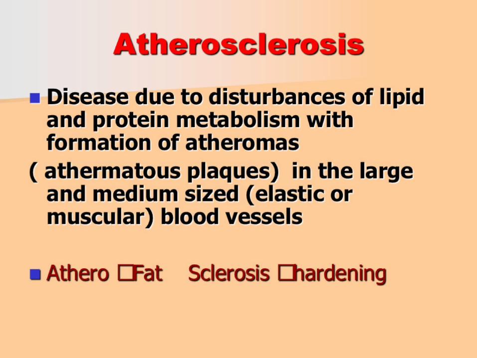
Atherosclerosis

Artherosclerosis
Dec 10, 2014
Artherosclerosis
Welcome message from author
This document is posted to help you gain knowledge. Please leave a comment to let me know what you think about it! Share it to your friends and learn new things together.
Transcript

Atherosclerosis
Atherosclerosis

Common Sites:


The most modern theory


Atherosclerosis. Formation of the atheroma.
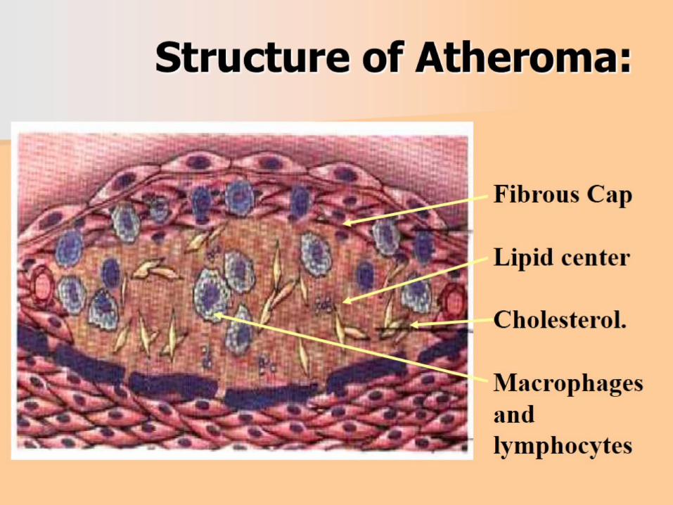
Structure of Atheroma:

Pathogenesis of Atheroma: Stage 1.

Pathogenesis of Atheroma: Stage 2.

Pathogenesis of Atheroma: Stage 3.

Pathogenesis of Atheroma: Stage 4.


Atherosclerosis of aorta
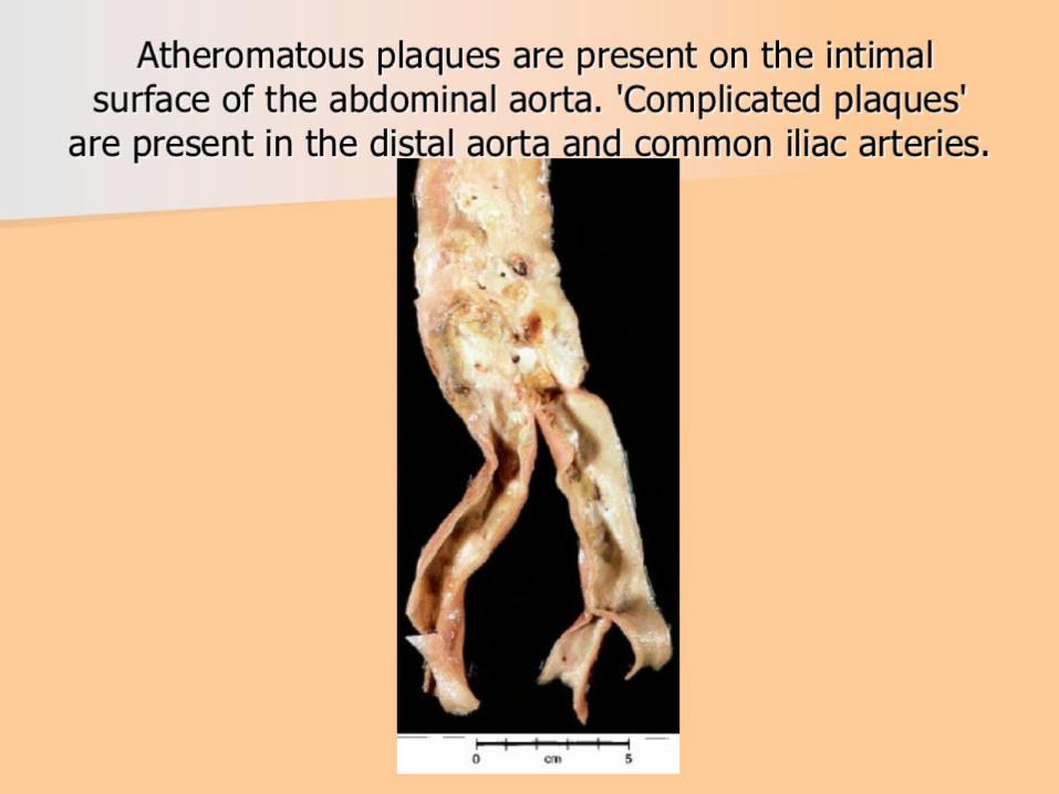
Atheromatous plaques are present on the intimal surface of the abdominal aorta. 'Complicated plaques' are present in the distal aorta and common iliac arteries.

Atherosclerosis of aorta
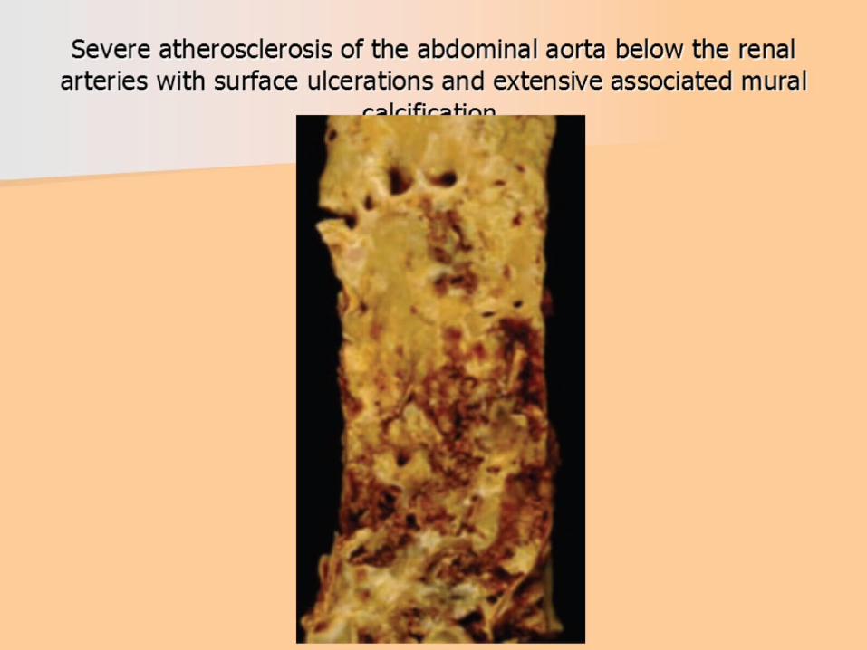
Severe atherosclerosis of the abdominal aorta below the renal arteries with surface ulcerations and extensive associated mural calcification.


Changes in endothelial dysfunction in atherosclerosis

Liposclerosis or fibrous plaques.
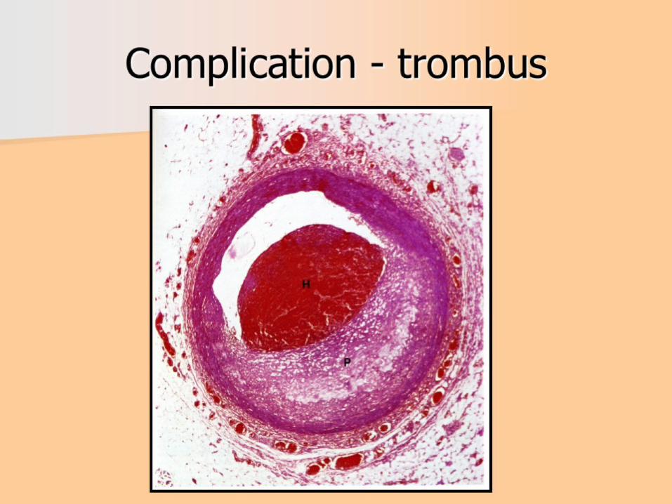
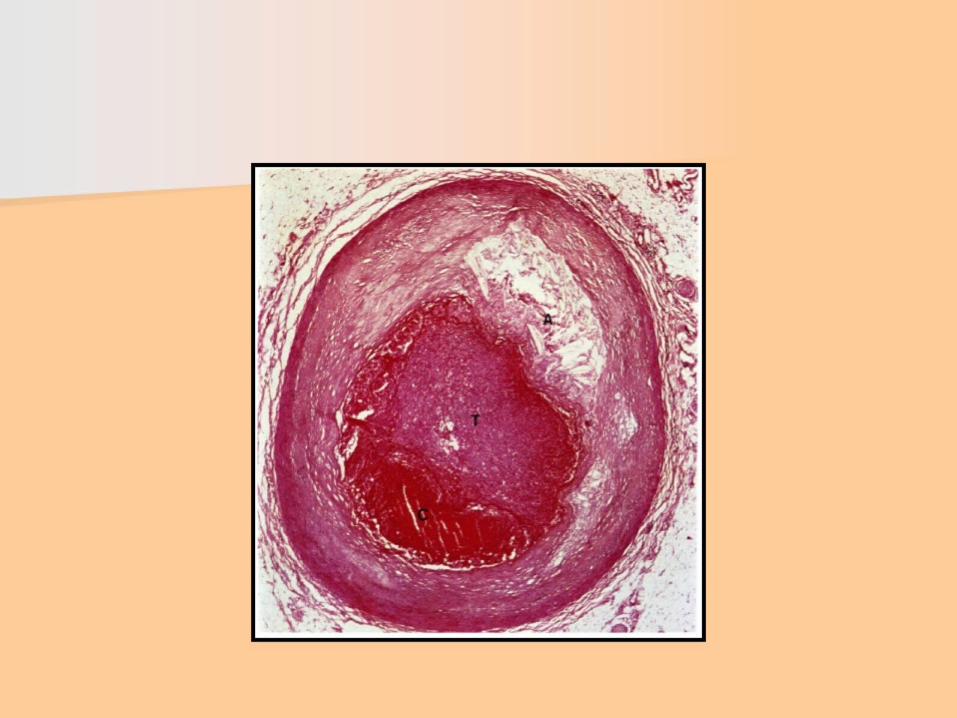
Complication - trombus


Complications: clinical-morphological forms (6)


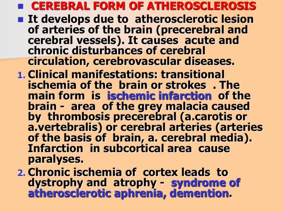


MESENTERIAL FORM

The illustration shows how Peripheral Arterial Disease can affect arteries in the legs. Figure A shows a normal artery with normal blood flow. The inset image shows a cross-section of the normal artery. Figure B shows an artery with plaque buildup that’s partially blocking blood flow. The inset image shows a cross-section of the narrowed artery.

Dry gangrene of two toes in a diabetic patient with severe arterial atherosclerosis. Thickened basement membranes of small blood vessels plus a reduction in the number of small arteries contributes to the ischemia in diabetic individuals.

atherosclerosis of low extremitas

Figure A shows the location of the heart. Figure B shows how vein and artery bypass grafts are attached to the heart.

Angina is a specific type of pain in the chest caused by inadequate blood flow through the blood vessels (coronary vessels) of the heart muscle (myocardium).

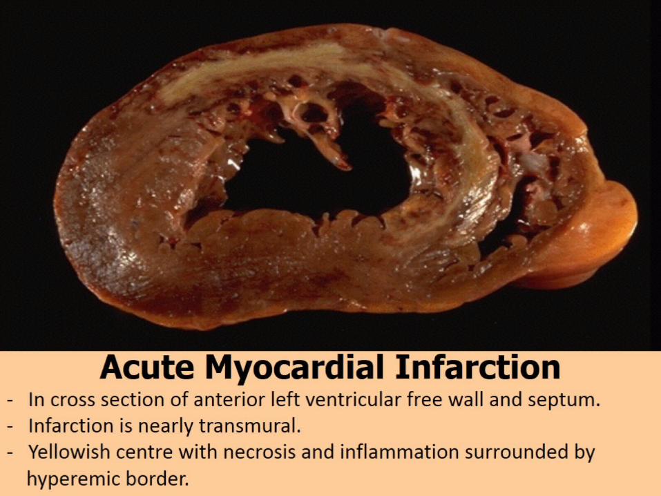
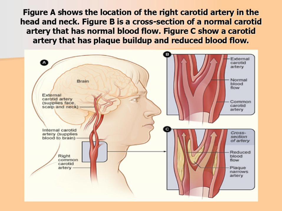
Figure A shows the location of the right carotid artery in the head and neck. Figure B is a cross-section of a normal carotid artery that has normal blood flow. Figure C show a carotid artery that has plaque buildup and reduced blood flow.

Recent thrombosis of the right coronary artery causing complete occlusion of the vessel. Occlusion of this artery causes a posterior myocardial infarction. The coronary artery shows severe atherosclerosis.

Necrotic cells in the brain
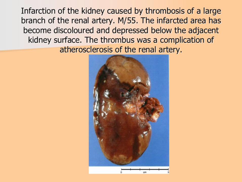
Infarction of the kidney caused by thrombosis of a large branch of the renal artery. M/55. The infarcted area has become discoloured and depressed below the adjacent kidney surface. The thrombus was a complication of atherosclerosis of the renal artery.