ANTIMICROBIAL AGENTS AND CHEMOTHERAPY, Apr. 2011, p. 1383–1390 Vol. 55, No. 4 0066-4804/11/$12.00 doi:10.1128/AAC.01277-10 Copyright © 2011, American Society for Microbiology. All Rights Reserved. Artemether and Artesunate Show the Highest Efficacies in Rescuing Mice with Late-Stage Cerebral Malaria and Rapidly Decrease Leukocyte Accumulation in the Brain L. Clemmer, 1 † Y. C. Martins, 1 † G. M. Zanini, 1,2 J. A. Frangos, 1 and L. J. M. Carvalho 1 * La Jolla Bioengineering Institute, 3535 General Atomics Court, Suite 210, San Diego, California 92121, 1 and IPEC, Fiocruz, Rio de Janeiro, Brazil 2 Received 18 September 2010/Returned for modification 19 November 2010/Accepted 2 January 2011 The murine model of cerebral malaria (ECM) caused by Plasmodium berghei ANKA (PbA) infection in susceptible mice has been extensively used for studies of pathogenesis and identification of potential targets for human CM therapeutics. However, the model has been seldom explored to evaluate adjunctive therapies for this malaria complication. A first step toward this goal is to define a treatment protocol with an effective antimalarial drug able to rescue mice presenting late-stage ECM. We evaluated the efficacy of artemisinin, artemether, artesunate, and quinine given intraperitoneally once a day, and combinations with mefloquine, in suppressing PbA infection in mice with moderate parasitemia. Artemether, artesunate, and quinine were then evaluated for efficacy in rescuing PbA-infected mice with ECM, strictly defined by using objective criteria based on the presentation of clinical signs of neurological involvement, degree of hypothermia, and performance in a set of six motor behavior tests. Artemether at 25 mg/kg presented the fastest parasite killing ability in 24 h and fully avoided recrudescence in a 5-day treatment protocol. Artemether and artesunate were equally effective in rescuing mice with late-stage ECM (46 and 43% survival, respectively), whereas quinine had a poor performance (12.5% survival). Artemether caused a marked decrease in brain leukocyte accumulation 24 h after the first dose. In conclusion, artemether and artesunate are effective in rescuing mice with late-stage ECM and decrease brain inflammation. In addition, the described protocols for more strict clinical evaluation and for rescue treatment provide a framework for studies of CM adjunctive therapies using this mouse model. The Plasmodium berghei ANKA (PbA) murine model of cerebral malaria (ECM) is the most widely used animal model for human CM. This model has been intensely explored to study mechanisms of CM pathogenesis, and the role of several mediators on disease development has been unraveled using different interventions such as mediator blockade by monoclo- nal antibodies and gene knockout (a list of interventions is provided in the supplemental file on reference [7]). However, and surprisingly, the model has seldom been explored to assess the efficacy of potential adjunctive therapies aimed to improve survival in combination with antimalarial drugs. Indeed, al- though nearly 300 papers have been published using this model in the past 3 decades, only a handful have used approaches that can be characterized as adjunctive therapy interventions, with the administration of an ancillary treatment in addition to a primary antimalarial drug to mice presenting clinical signs of ECM (5–7). This shortcoming has helped to fuel criticisms on the relevance of ECM as a model for the human disease (38; debated in references 7, 15, 29, 31, and 33). Assessments of adjunctive therapies for CM in this model will require basic data on the efficacy of different antimalarial drugs themselves in rescuing mice with established clinical signs of ECM, and such data are scarce. Prada et al. treated PbA-infected mice with chloroquine or artemether and re- ported 95% efficacy for both drugs, but the criterion for treatment was the level of parasitemia rather than the presen- tation of neurological signs (27). Reis et al. also showed high efficacy (100%) of chloroquine and artesunate in preventing the syndrome, also at early stages of disease when mice pre- sented slight decreases in SHIRPA scores but apparently no clinical signs of ECM (28). Baptista et al. reported that a single dose of pyrimethamine given 15 to 20 h before the expected onset of ECM (on day 5 of infection with mice presenting a mean parasitemia of ca. 4%) was able to prevent the syndrome (3). Dai et al., on the contrary, showed that chloroquine treat- ment starting at an early stage of ECM (parasitemia 7%, low activity levels, and body weight loss 2%) resulted in 70% mortality and did not prevent cognitive and motor deficits in the survivors (8). Golenser et al. tried several treatment pro- tocols with quinine and dihydroartemisinin (DHA), and basi- cally early treatments prevented the neurological syndrome delaying mortality, whereas later treatments were much less effective (11). Subcurative doses of quinine or DHA given on days 4 to 6 also prevented ECM (22). Artesunate and artemi- sone have also been shown to be effective in preventing the development of ECM in mice when given before, or at early stages of, ECM development (12, 36). To test the efficacy of an ECM-rescuing therapy, it is nec- essary to establish well-defined clinical parameters of neuro- logical involvement in order to clearly determine the stage of the disease at which the treatment is administered. Cognitive, sensorimotor function and behavior evaluations, as provided for instance by the SHIRPA and other well-established proto- * Corresponding author. Mailing address: La Jolla Bioengineering Institute, 3535 General Atomics Court, Suite 210, San Diego, CA 92121. Phone: (858) 456-7500, ext. 103. Fax: (858) 456-7540. E-mail: [email protected]. † L.C. and Y.C.M. contributed equally to this study. Published ahead of print on 10 January 2011. 1383

Welcome message from author
This document is posted to help you gain knowledge. Please leave a comment to let me know what you think about it! Share it to your friends and learn new things together.
Transcript
ANTIMICROBIAL AGENTS AND CHEMOTHERAPY, Apr. 2011, p. 1383–1390 Vol. 55, No. 40066-4804/11/$12.00 doi:10.1128/AAC.01277-10Copyright © 2011, American Society for Microbiology. All Rights Reserved.
Artemether and Artesunate Show the Highest Efficacies in RescuingMice with Late-Stage Cerebral Malaria and Rapidly Decrease
Leukocyte Accumulation in the Brain�
L. Clemmer,1† Y. C. Martins,1† G. M. Zanini,1,2 J. A. Frangos,1 and L. J. M. Carvalho1*La Jolla Bioengineering Institute, 3535 General Atomics Court, Suite 210, San Diego, California 92121,1 and
IPEC, Fiocruz, Rio de Janeiro, Brazil2
Received 18 September 2010/Returned for modification 19 November 2010/Accepted 2 January 2011
The murine model of cerebral malaria (ECM) caused by Plasmodium berghei ANKA (PbA) infection insusceptible mice has been extensively used for studies of pathogenesis and identification of potential targets forhuman CM therapeutics. However, the model has been seldom explored to evaluate adjunctive therapies forthis malaria complication. A first step toward this goal is to define a treatment protocol with an effectiveantimalarial drug able to rescue mice presenting late-stage ECM. We evaluated the efficacy of artemisinin,artemether, artesunate, and quinine given intraperitoneally once a day, and combinations with mefloquine, insuppressing PbA infection in mice with moderate parasitemia. Artemether, artesunate, and quinine were thenevaluated for efficacy in rescuing PbA-infected mice with ECM, strictly defined by using objective criteria basedon the presentation of clinical signs of neurological involvement, degree of hypothermia, and performance ina set of six motor behavior tests. Artemether at 25 mg/kg presented the fastest parasite killing ability in 24 hand fully avoided recrudescence in a 5-day treatment protocol. Artemether and artesunate were equallyeffective in rescuing mice with late-stage ECM (46 and 43% survival, respectively), whereas quinine had a poorperformance (12.5% survival). Artemether caused a marked decrease in brain leukocyte accumulation 24 hafter the first dose. In conclusion, artemether and artesunate are effective in rescuing mice with late-stage ECMand decrease brain inflammation. In addition, the described protocols for more strict clinical evaluation andfor rescue treatment provide a framework for studies of CM adjunctive therapies using this mouse model.
The Plasmodium berghei ANKA (PbA) murine model ofcerebral malaria (ECM) is the most widely used animal modelfor human CM. This model has been intensely explored tostudy mechanisms of CM pathogenesis, and the role of severalmediators on disease development has been unraveled usingdifferent interventions such as mediator blockade by monoclo-nal antibodies and gene knockout (a list of interventions isprovided in the supplemental file on reference [7]). However,and surprisingly, the model has seldom been explored to assessthe efficacy of potential adjunctive therapies aimed to improvesurvival in combination with antimalarial drugs. Indeed, al-though nearly 300 papers have been published using this modelin the past 3 decades, only a handful have used approaches thatcan be characterized as adjunctive therapy interventions, withthe administration of an ancillary treatment in addition to aprimary antimalarial drug to mice presenting clinical signs ofECM (5–7). This shortcoming has helped to fuel criticisms onthe relevance of ECM as a model for the human disease (38;debated in references 7, 15, 29, 31, and 33).
Assessments of adjunctive therapies for CM in this modelwill require basic data on the efficacy of different antimalarialdrugs themselves in rescuing mice with established clinicalsigns of ECM, and such data are scarce. Prada et al. treated
PbA-infected mice with chloroquine or artemether and re-ported �95% efficacy for both drugs, but the criterion fortreatment was the level of parasitemia rather than the presen-tation of neurological signs (27). Reis et al. also showed highefficacy (100%) of chloroquine and artesunate in preventingthe syndrome, also at early stages of disease when mice pre-sented slight decreases in SHIRPA scores but apparently noclinical signs of ECM (28). Baptista et al. reported that a singledose of pyrimethamine given 15 to 20 h before the expectedonset of ECM (on day 5 of infection with mice presenting amean parasitemia of ca. 4%) was able to prevent the syndrome(3). Dai et al., on the contrary, showed that chloroquine treat-ment starting at an early stage of ECM (parasitemia � 7%, lowactivity levels, and body weight loss � 2%) resulted in 70%mortality and did not prevent cognitive and motor deficits inthe survivors (8). Golenser et al. tried several treatment pro-tocols with quinine and dihydroartemisinin (DHA), and basi-cally early treatments prevented the neurological syndromedelaying mortality, whereas later treatments were much lesseffective (11). Subcurative doses of quinine or DHA given ondays 4 to 6 also prevented ECM (22). Artesunate and artemi-sone have also been shown to be effective in preventing thedevelopment of ECM in mice when given before, or at earlystages of, ECM development (12, 36).
To test the efficacy of an ECM-rescuing therapy, it is nec-essary to establish well-defined clinical parameters of neuro-logical involvement in order to clearly determine the stage ofthe disease at which the treatment is administered. Cognitive,sensorimotor function and behavior evaluations, as providedfor instance by the SHIRPA and other well-established proto-
* Corresponding author. Mailing address: La Jolla BioengineeringInstitute, 3535 General Atomics Court, Suite 210, San Diego, CA92121. Phone: (858) 456-7500, ext. 103. Fax: (858) 456-7540. E-mail:[email protected].
† L.C. and Y.C.M. contributed equally to this study.� Published ahead of print on 10 January 2011.
1383

cols (17, 20), are very useful in helping to establish a compre-hensive clinical status of mice. The presentation of well-recog-nized clinical signs of ECM such as ataxia, limb paralysis,convulsions, moderate to severe hypothermia, loss of rightingreflex, and/or coma should in principle be the guide for treat-ment decisions in experimental protocols, however it must beemphasized that these signs usually appear only at very latestages of disease, in mice with full-blown ECM. Bienvenu et al.defined five stages of clinical presentation in mice receivingtreatment: 0, no symptoms; 1, ruffled fur (non-ECM specific);2, motor impairment; 3, respiratory distress; and 4, coma. Inthat study, when the first dose of artesunate or the combinationartesunate-erythropoietin was given on day 6, ca. 70% of themice were at stages 1 or 2, and most survived. We have previ-ously defined a protocol for late-stage (rescue) treatment ofmice with ECM using artemether (6). To treat mice, we usedas the basic criterion the expression of well-established clinicalsigns of full-blown murine ECM: ataxia, limb paralysis, con-vulsions, loss of righting reflex, and/or coma. Because even inthe presence of these classical ECM-associated neurologicalsigns the overall clinical status of the mice may vary consider-ably (e.g., mice with ataxia are obviously in better shape thanmice in coma), we added a composite clinical score based on aset of six simple, fast-to-perform, motor behavior tests (trans-fer arousal, locomotor activity, tail elevation, wire maneuver,contact righting reflex, and righting in arena) derived from theSHIRPA protocol (17, 20), as well as rectal temperature, tobetter define the overall clinical status of each mouse at thetime of treatment. We have shown that artemether at 50 mg/kggiven intraperitoneally (i.p.) was able to rescue 32% of micepresenting late-stage ECM. However, because we were eval-uating the adjunctive effect of the calcium channel blockernimodipine, mice receiving artemether also received thenimodipine vehicle containing ethanol and polyethylene glycol(PEG), which therefore probably influenced the results.
In the present study we defined the efficacy of a number ofantimalarial drugs (quinine, artemisinin, artesunate, artemether,and combinations artemether-mefloquine) in (i) clearing PbAparasitemia with treatments starting on day 5 of infection and(ii) rescuing mice with late-stage ECM. In addition, we evalu-ated the effect of artemether in decreasing leukocyte seques-tration in the brain 24 h after the first dose in mice with ECM.We expect this study to be useful in helping to define standardsfor experiments designed to evaluate adjunctive therapies forCM using the PbA model.
MATERIALS AND METHODS
Mice, parasites, and infection. Eight- to ten-week-old C57BL/6J mice wereobtained from Jackson Laboratories (Bar Harbor, ME). Mice were housed ingroups of no more than five per cage with free access to chow and water. Micewere allowed to adapt to their new environment for 3 days before experimen-tation. All experimental protocols were reviewed and approved by the La JollaBioengineering Institute (LJBI) Institutional Animal Care and Use Committee.The Plasmodium berghei ANKA PbA-GFPcon 259cl2, which is a geneticallymodified parasite of clone cl15cy1 of the ANKA strain that expresses greenfluorescent protein (GFP) constitutively during the whole life cycle, was used (akind donation of MR4, Manassas, VA; deposited by C. J. Janse and A. P. Waters;MR4 reagent number MRA-865). The parasite was propagated in C57BL/6Jmice, and in each experiment a fresh blood sample was obtained from a passagemouse and a suspension containing 106 parasitized red blood cells (pRBC) in 100�l was injected i.p. in each mouse of the experimental groups. Parasitemia, motorbehavior, and rectal temperature were checked beginning on day 5 after infec-
tion. Parasitemia was checked by using flow cytometry and quantified by countingthe number of pRBC in 10,000 RBC. After treatment, thin blood smears weremade from a drop of tail blood and stained with Giemsa to distinguish dead fromviable parasites. We considered as dead parasites those that we could no longerclearly distinguish the typical morphology and presented themselves as a mass ofcondensed matter inside the RBC, many times featured just as a dark dot. Theslides were examined under a light microscope at �1,000 magnification with anoil immersion lens (Nikon Eclipse E200). Parasitemia was calculated by countingthe number of pRBC in at least 1,000 RBC.
Clinical assessment. A set of six simple behavioral tests (transfer arousal,locomotor activity, tail elevation, wire maneuver, contact righting reflex, andrighting in arena) adapted from the SHIRPA protocol (17, 20) was used toprovide a better estimate of the overall clinical status of the mice during infec-tion. The performance in each test was assessed by using a modified scoringsystem (0 to 5 for transfer arousal, 0 to 4 for locomotor activity, 0 to 4 for tailelevation, 0 to 4 for wire maneuver, 0 to 3 for contact righting reflex, and 0 to 3for righting in arena), and a composite score was built ranging from 0 to 23,where 23 indicates maximum performance and 0 indicates complete impairment(usually coma). Body temperature was monitored by using an Accorn SeriesThermocouple thermometer with a mouse rectal probe (Oakton Instruments,Vernon Hills, IL). ECM was defined as the presentation of one or more of thefollowing clinical signs of neurological involvement: ataxia, limb paralysis, poorrighting reflex, seizures, rollover, and coma.
Experimental design. The present study was divided in three parts. (i) The firstpart was the treatment of PbA-infected mice on day 5 of infection. At this timepoint, infected mice show a moderate, rising parasitemia but present no ECMsigns, which usually start to manifest on day 6. Treating mice at this point allowedus to evaluate the efficacy and rapidness of parasite killing, parasite clearance,and recrudescence avoidance by different antimalarial drugs and doses. (ii) Thesecond part of the study was the treatment of PbA-infected mice presentingclinical signs of ECM. In this way, we sought to determine the efficacy of suitabletreatment protocols defined in part i in rescuing mice with late-stage ECM. (iii)The third part of the study was the quantification of leukocyte accumulation inbrain vessels before and after treatment of mice with ECM in order to determinethe effect of treatment on brain inflammation.
Antimalarial drug regimens. (i) Treatment before ECM development. We hadpreviously shown that artemether at 50 mg/kg, coadministered with a solution ofethanol and PEG 400 in saline (1:1:8 [vol/vol], the vehicle for nimodipine), wasable to rescue one-third of the mice with late-stage ECM (6). For the presentstudy, we defined half this dose (25 mg/kg) as our reference for artemether itself.Artemisinin was given at this same dose, and artesunate was given at a molar-equivalent dose (32 mg/kg) and also at twice this amount (64 mg/kg). On day 5postinoculation, infected mice were treated with one of the following drugregimens. (i) Artemether (Artesiane [20 mg/ml], kindly provided by DafraPharma, Belgium) was already prepared in coconut oil and was administered i.p.at 25 mg/kg. (ii) Artesunate (Sigma, St. Louis, MO) was dissolved in 5% sodiumbicarbonate in saline and administered i.p. at 32 or 64 mg/kg. (iii) Artemisinin(Sigma) was dissolved in 10% dimethyl sulfoxide (DMSO) and then 90% saline,mixed thoroughly, and administered i.p. at 25 mg/kg. (iv) Quinine (Sigma) wasdissolved in saline and delivered i.p. at 60 or 120 mg/kg. These dosages are withinthe range of dosages commonly used in rodent studies with P. berghei (5, 32, 40).Mice treated with artemisinin, artemether, artesunate, or quinine received treat-ment once a day for 5 days.
Artemether was also administered in combination with mefloquine for eithera 1- or a 3-day treatment. For the 1-day treatment, artemether at 25 mg/kg wasadministered i.p., together with mefloquine at 40 mg/kg i.p., or per gavage, singledose on day 5 of infection. For the 3-day treatment, artemether was given for 3days (once a day) i.p. starting on day 5 of infection, and mefloquine was admin-istered together with the third dose of artemether by oral gavage. In both cases,mefloquine (Sigma) was dissolved in 10% DMSO and 90% saline.
(ii) Treatment after ECM development (rescue treatment). Treatments withartemether (25 mg/kg), artesunate (32 mg/kg), or quinine (120 mg/kg) wereadministered only after infected mice were diagnosed with ECM, using thefollowing strict criteria: (i) presentation of at least one well-recognized clinicalsign of neurological involvement in ECM, i.e., ataxia, limb paralysis, convulsions,rollover, loss of righting reflex, and coma; (ii) moderate to severe hypothermia(rectal temperature � 33°C); and (iii) poor motor behavior performance (com-posite score � 10). Each injection subsequent to the initial dose was given in themorning.
Quantification of leukocyte accumulation in the brain. Ten mice presentingclinical signs of ECM and with similar parasitemias, body temperatures, andcomposite motor scores were randomly assigned to two groups. In the first group,five mice were euthanized, and the brains were collected and processed for
1384 CLEMMER ET AL. ANTIMICROB. AGENTS CHEMOTHER.
histology. In the second group, five mice were treated with artemether at 25mg/kg; 24 h later the four mice alive were also euthanized, and the brains werecollected and processed for histology. Brains from two uninfected mice were alsocollected and served as controls. Mice were anesthetized (ketamine, 150 mg/kg;xylazine, 10 mg/kg) and perfused with 5 ml of heparinized saline (10 U/ml),followed by 5 ml of 4% paraformaldehyde (PFA), both solutions at a rate of 1ml/min, using a gravity perfusion setup. Each brain was carefully collected im-mediately after perfusion and stored in PFA during 48 h for fixation. Brains werecut in four coronal slices of 2 to 3 mm using a mouse brain blocker (David KopfInstruments, Tujunga, CA), and slices were numbered 1 to 4 (with slice 1 beingthe frontal lobe/olfactory bulb and slice 4 being the cerebellum-brainstem). Eachslice was embedded in paraffin, and 5-�m sections were obtained at approxi-mately 400-�m intervals (four sections per slice). Sections were mounted in glassslides and stained with hematoxylin-eosin (H&E). Leukocyte quantification wasperformed on section 2 of each slice; therefore, a total of four sections werecounted per mouse. The number of leukocytes in each vessel of each section wasquantified by using an ocular grid calibrated with a �400 magnification (fielddimensions, 200 by 180 �m) in a Nikon microscope (Eclipse E200). The wholearea of each section was similarly quantified with the grid calibrated at �40magnification. Quantification was performed by an experienced investigator in ablinded fashion. Pictures were taken with a SPOT RT3 camera (DiagnosticInstruments, Inc.; 1,014 by 721 pixels).
Statistical analyses. Results were expressed as means and standard errors ofthe mean (SEM) unless otherwise stated. Statistical analysis were performedusing one-way or two-way analysis of variance with Tukey’s or Bonferroni’s posttests when comparing whether one or two parameters, respectively, varied amongthe different treatment groups. A log-rank test was used to compare the differentsurvival curves. A P value of �0.05 was considered significant.
RESULTS
Efficacy of antimalarial drugs in clearing PbA parasitemiaon day 5 of infection. Before evaluating the efficacy of differentantimalarial drugs in rescuing mice with ECM, we performeda preliminary trial to evaluate their efficacy and rapidity inclearing PbA parasitemia. We chose to treat mice on day 5,which is a time point when mice present moderate, rising para-sitemia but usually no neurological involvement, and thereforewe expected that all drugs, even the slower-acting ones, wouldbe effective in clearing parasitemia without mortality, allowing
the definition of doses, speediness of parasite clearance, andlength of treatment to avoid recrudescence. We chose qui-nine, artemisinin, and its derivatives artesunate and arte-mether since these are the mainstream drugs used to treathuman CM. On attempting to define a simpler and fastertreatment protocol, we also evaluated combinations of a fast-acting/rapidly cleared drug (artemether) in combination with aslower-acting/slowly cleared drug (mefloquine).
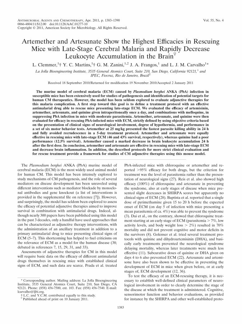
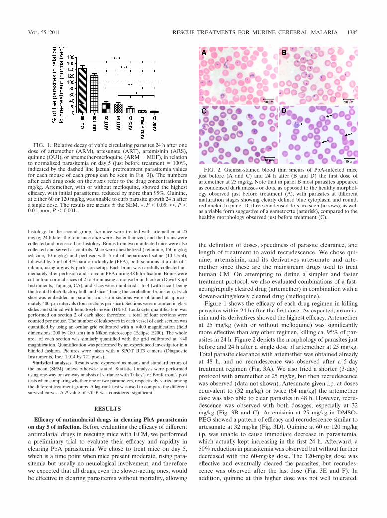
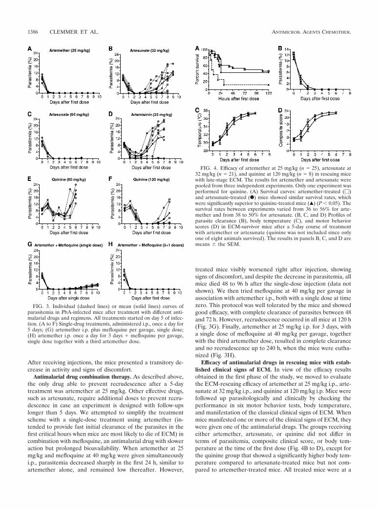
Figure 1 shows the efficacy of each drug regimen in killingparasites within 24 h after the first dose. As expected, artemis-inin and its derivatives showed the highest efficacy. Artemetherat 25 mg/kg (with or without mefloquine) was significantlymore effective than any other regimen, killing ca. 95% of par-asites in 24 h. Figure 2 depicts the morphology of parasites justbefore and 24 h after a single dose of artemether at 25 mg/kg.Total parasite clearance with artemether was obtained alreadyat 48 h, and no recrudescence was observed after a 5-daytreatment regimen (Fig. 3A). We also tried a shorter (3-day)protocol with artemether at 25 mg/kg, but then recrudescencewas observed (data not shown). Artesunate given i.p. at dosesequivalent to (32 mg/kg) or twice (64 mg/kg) the artemetherdose was also able to clear parasites in 48 h. However, recru-descence was observed with both dosages, especially at 32mg/kg (Fig. 3B and C). Artemisinin at 25 mg/kg in DMSO-PEG showed a pattern of efficacy and recrudescence similar toartesunate at 32 mg/kg (Fig. 3D). Quinine at 60 or 120 mg/kgi.p. was unable to cause immediate decrease in parasitemia,which actually kept increasing in the first 24 h. Afterward, a50% reduction in parasitemia was observed but without furtherdecreased with the 60-mg/kg dose. The 120-mg/kg dose waseffective and eventually cleared the parasites, but recrudes-cence was observed after the last dose (Fig. 3E and F). Inaddition, quinine at this higher dose was not well tolerated.
FIG. 1. Relative decay of viable circulating parasites 24 h after onedose of artemether (ARM), artesunate (ART), artemisinin (ARS),quinine (QUI), or artemether-mefloquine (ARM � MEF), in relationto normalized parasitemia on day 5 (just before treatment � 100%,indicated by the dashed line [actual pretreatment parasitemia valuesfor each mouse of each group can be seen in Fig. 3]). The numbersafter each drug code on the x axis refer to the drug concentrations inmg/kg. Artemether, with or without mefloquine, showed the highestefficacy, with initial parasitemia reduced by more than 95%. Quinine,at either 60 or 120 mg/kg, was unable to curb parasite growth 24 h aftera single dose. The results are means � the SEM. *, P � 0.05; **, P �0.01; ***, P � 0.001.
FIG. 2. Giemsa-stained blood thin smears of PbA-infected micejust before (A and C) and 24 h after (B and D) the first dose ofartemether at 25 mg/kg. Note that in panel B most parasites appearedas condensed dark masses or dots, as opposed to the healthy morphol-ogy observed just before treatment (A), with parasites at differentmaturation stages showing clearly defined blue cytoplasm and round,red nuclei. In panel D, three condensed dots are seen (arrows), as wellas a viable form suggestive of a gametocyte (asterisk), compared to thehealthy morphology observed just before treatment (C).
VOL. 55, 2011 RESCUE TREATMENTS FOR MURINE CEREBRAL MALARIA 1385
After receiving injections, the mice presented a transitory de-crease in activity and signs of discomfort.
Antimalarial drug combination therapy. As described above,the only drug able to prevent recrudescence after a 5-daytreatment was artemether at 25 mg/kg. Other effective drugs,such as artesunate, require additional doses to prevent recru-descence in case an experiment is designed with follow-upslonger than 5 days. We attempted to simplify the treatmentscheme with a single-dose treatment using artemether (in-tended to provide fast initial clearance of the parasites in thefirst critical hours when mice are most likely to die of ECM) incombination with mefloquine, an antimalarial drug with sloweraction but prolonged bioavailability. When artemether at 25mg/kg and mefloquine at 40 mg/kg were given simultaneouslyi.p., parasitemia decreased sharply in the first 24 h, similar toartemether alone, and remained low thereafter. However,
treated mice visibly worsened right after injection, showingsigns of discomfort, and despite the decrease in parasitemia, allmice died 48 to 96 h after the single-dose injection (data notshown). We then tried mefloquine at 40 mg/kg per gavage inassociation with artemether i.p., both with a single dose at timezero. This protocol was well tolerated by the mice and showedgood efficacy, with complete clearance of parasites between 48and 72 h. However, recrudescence occurred in all mice at 120 h(Fig. 3G). Finally, artemether at 25 mg/kg i.p. for 3 days, witha single dose of mefloquine at 40 mg/kg per gavage, togetherwith the third artemether dose, resulted in complete clearanceand no recrudescence up to 240 h, when the mice were eutha-nized (Fig. 3H).
Efficacy of antimalarial drugs in rescuing mice with estab-lished clinical signs of ECM. In view of the efficacy resultsobtained in the first phase of the study, we moved to evaluatethe ECM-rescuing efficacy of artemether at 25 mg/kg i.p., arte-sunate at 32 mg/kg i.p., and quinine at 120 mg/kg i.p. Mice werefollowed up parasitologically and clinically by checking theperformance in six motor behavior tests, body temperature,and manifestation of the classical clinical signs of ECM. Whenmice manifested one or more of the clinical signs of ECM, theywere given one of the antimalarial drugs. The groups receivingeither artemether, artesunate, or quinine did not differ interms of parasitemia, composite clinical score, or body tem-perature at the time of the first dose (Fig. 4B to D), except forthe quinine group that showed a significantly higher body tem-perature compared to artesunate-treated mice but not com-pared to artemether-treated mice. All treated mice were at a
FIG. 3. Individual (dashed lines) or mean (solid lines) curves ofparasitemia in PbA-infected mice after treatment with different anti-malarial drugs and regimens. All treatments started on day 5 of infec-tion. (A to F) Single-drug treatments, administered i.p., once a day for5 days; (G) artemether i.p. plus mefloquine per gavage, single dose;(H) artemether i.p. once a day for 3 days � mefloquine per gavage,single dose together with a third artemether dose.
FIG. 4. Efficacy of artemether at 25 mg/kg (n � 25), artesunate at32 mg/kg (n � 21), and quinine at 120 mg/kg (n � 8) in rescuing micewith late-stage ECM. The results for artemether and artesunate werepooled from three independent experiments. Only one experiment wasperformed for quinine. (A) Survival curves: artemether-treated (�)and artesunate-treated (F) mice showed similar survival rates, whichwere significantly superior to quinine-treated mice (Œ) (P � 0.05). Thesurvival rates between experiments varied from 36 to 56% for arte-mether and from 38 to 50% for artesunate. (B, C, and D) Profiles ofparasite clearance (B), body temperature (C), and motor behaviorscores (D) in ECM-survivor mice after a 5-day course of treatmentwith artemether or artesunate (quinine was not included since onlyone of eight animals survived). The results in panels B, C, and D aremeans � the SEM.
1386 CLEMMER ET AL. ANTIMICROB. AGENTS CHEMOTHER.
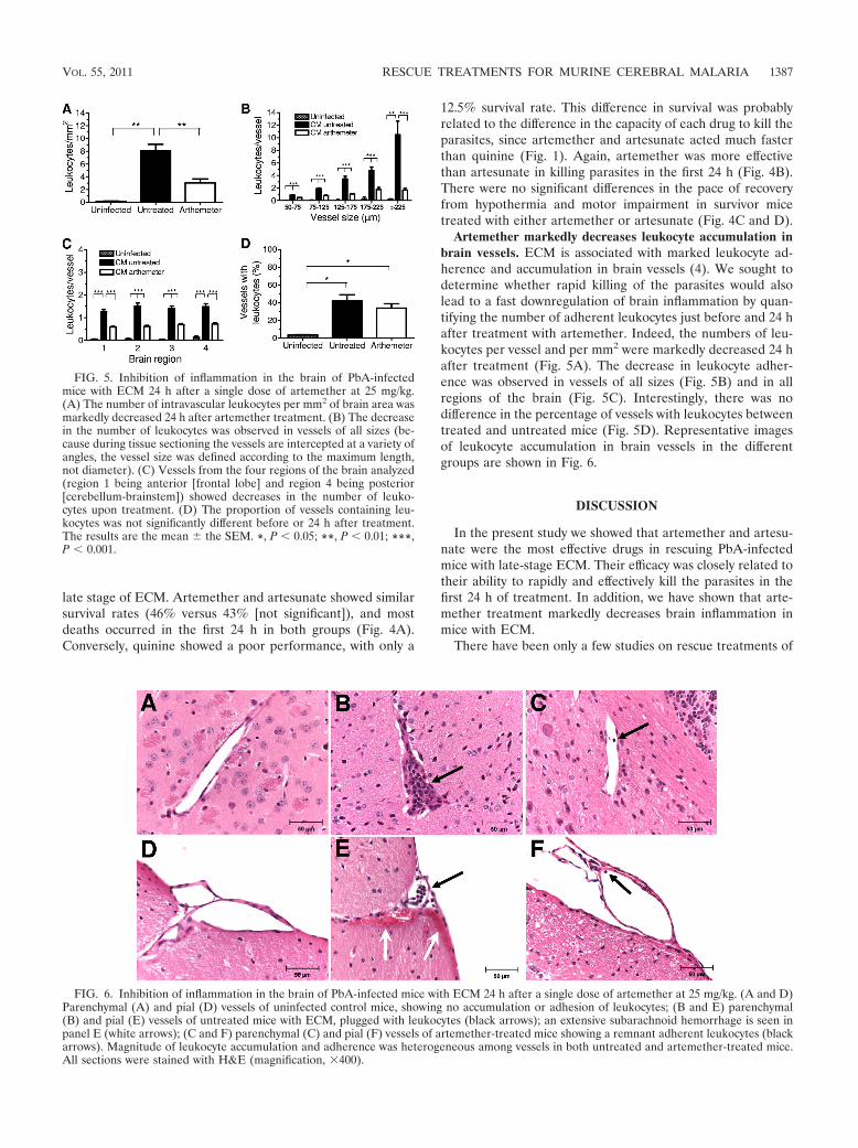
late stage of ECM. Artemether and artesunate showed similarsurvival rates (46% versus 43% [not significant]), and mostdeaths occurred in the first 24 h in both groups (Fig. 4A).Conversely, quinine showed a poor performance, with only a
12.5% survival rate. This difference in survival was probablyrelated to the difference in the capacity of each drug to kill theparasites, since artemether and artesunate acted much fasterthan quinine (Fig. 1). Again, artemether was more effectivethan artesunate in killing parasites in the first 24 h (Fig. 4B).There were no significant differences in the pace of recoveryfrom hypothermia and motor impairment in survivor micetreated with either artemether or artesunate (Fig. 4C and D).
Artemether markedly decreases leukocyte accumulation inbrain vessels. ECM is associated with marked leukocyte ad-herence and accumulation in brain vessels (4). We sought todetermine whether rapid killing of the parasites would alsolead to a fast downregulation of brain inflammation by quan-tifying the number of adherent leukocytes just before and 24 hafter treatment with artemether. Indeed, the numbers of leu-kocytes per vessel and per mm2 were markedly decreased 24 hafter treatment (Fig. 5A). The decrease in leukocyte adher-ence was observed in vessels of all sizes (Fig. 5B) and in allregions of the brain (Fig. 5C). Interestingly, there was nodifference in the percentage of vessels with leukocytes betweentreated and untreated mice (Fig. 5D). Representative imagesof leukocyte accumulation in brain vessels in the differentgroups are shown in Fig. 6.
DISCUSSION
In the present study we showed that artemether and artesu-nate were the most effective drugs in rescuing PbA-infectedmice with late-stage ECM. Their efficacy was closely related totheir ability to rapidly and effectively kill the parasites in thefirst 24 h of treatment. In addition, we have shown that arte-mether treatment markedly decreases brain inflammation inmice with ECM.
There have been only a few studies on rescue treatments of
FIG. 5. Inhibition of inflammation in the brain of PbA-infectedmice with ECM 24 h after a single dose of artemether at 25 mg/kg.(A) The number of intravascular leukocytes per mm2 of brain area wasmarkedly decreased 24 h after artemether treatment. (B) The decreasein the number of leukocytes was observed in vessels of all sizes (be-cause during tissue sectioning the vessels are intercepted at a variety ofangles, the vessel size was defined according to the maximum length,not diameter). (C) Vessels from the four regions of the brain analyzed(region 1 being anterior [frontal lobe] and region 4 being posterior[cerebellum-brainstem]) showed decreases in the number of leuko-cytes upon treatment. (D) The proportion of vessels containing leu-kocytes was not significantly different before or 24 h after treatment.The results are the mean � the SEM. *, P � 0.05; **, P � 0.01; ***,P � 0.001.
FIG. 6. Inhibition of inflammation in the brain of PbA-infected mice with ECM 24 h after a single dose of artemether at 25 mg/kg. (A and D)Parenchymal (A) and pial (D) vessels of uninfected control mice, showing no accumulation or adhesion of leukocytes; (B and E) parenchymal(B) and pial (E) vessels of untreated mice with ECM, plugged with leukocytes (black arrows); an extensive subarachnoid hemorrhage is seen inpanel E (white arrows); (C and F) parenchymal (C) and pial (F) vessels of artemether-treated mice showing a remnant adherent leukocytes (blackarrows). Magnitude of leukocyte accumulation and adherence was heterogeneous among vessels in both untreated and artemether-treated mice.All sections were stained with H&E (magnification, �400).
VOL. 55, 2011 RESCUE TREATMENTS FOR MURINE CEREBRAL MALARIA 1387
ECM using antimalarial drugs in murine models (5, 6, 8, 11, 12,27, 28, 36), and in most such studies treatment was adminis-tered before neurological signs were patent or at an early stageof ECM development. Quinine and artemisinin derivativesare the drugs of choice to treat human CM (16, 21) andtherefore were chosen to be evaluated for ECM-rescuingefficacy in the PbA model. As expected, artemisinin and itsderivatives artemether and artesunate showed the fastest par-asite clearance effect. Artemether presented the best perfor-mance at a lower dose, which is likely related to the formu-lation, as oil-based formulations of artemisinin derivativeshave been shown to provide a slower and more sustaineddrug release due to the “depot” effect than water-basedformulations, which are cleared faster (19, 23).
Artemether was also the only drug able to fully preventrecrudescence following a 5-day treatment protocol. A 3-daytreatment protocol is also possible with artemether as long asan oral dose of mefloquine is given together with the third doseof artemether, and this can be a convenient alternative for ashorter treatment. Our attempts for a single-dose treatmentprotocol failed, since the combination of i.p. artemether-mef-loquine per gavage resulted in late recrudescence, and arte-mether i.p.-mefloquine i.p. turned out to be toxic, resulting ininitial discomfort for the animals and later death despite par-asite clearance. It is possible that the i.p. injection of meflo-quine at 40 mg/kg resulted in overdose and the observed tox-icity, which did not happen when the same dose was given pergavage. Mefloquine can present a number of toxic effects whengiven at high doses, potentially affecting the central nervoussystem and the gastrointestinal tract (2, 34). A similar effect,though less severe, may have occurred with quinine given i.p.,since mice also showed discomfort upon injection of the higherdose (120 mg/kg). It is possible that an effective single dosetreatment can still be attained with different drug combina-tions/dosages and delivery systems. Arteether, for instance,was shown to present better bioavailability, lower toxicity, andhigher efficacy against P. berghei in rats when formulated withcremophore compared to sesame oil formulation (18). The useof a hybrid molecule derived from artesunate and mefloquine,named MEFAS, presented higher efficacy against P. bergheithan the individual drugs given separately or combined (9).Woodard et al. showed that the monomeric trioxane fluoro-anilide 4b (an artemisinin derivative) at a single dose as low as6.8 mg/kg, combined with mefloquine at 20 mg/kg, was able toclear PbA parasitemia and prevent recrudescence in 80% oftreated mice, whereas the combination artemether-mefloquineat the same single doses did not prevent recrudescence andresulted in 100% lethality (39). However, it should be notedthat treatment was given only 24 h after infection, when par-asitemia was presumably very low. Rottmann et al. recentlyreported that spiroindolones can be more effective than theartemisinins at lower doses (10 mg/kg) and were able to com-pletely clear P. berghei parasitemia with a single higher dose(100 mg/kg) (30).
In the present study we did not evaluate the toxicity of thedifferent drugs and dosages, which can be a concern sinceartemisinin derivatives, particularly those formulated in oilsuch as artemether, have been shown to cause neurotoxicity inanimal models when given intramuscularly at relatively highdoses for several days (1, 10, 18, 24). This concern is boosted by
the fact that, in our case, mice being treated already presentaffected brains. Arteether or artemether given at 25 mg/kg(same dose as in the present study) daily for 7 days causedadverse effects in the brains of rats (1, 18). On the other hand,the same dose of artemether given daily for 28 days in miceproduced no evident brain changes, although higher doses did(24). Because we used the i.p. route, a shorter (5-day) treat-ment scheme, and treatments were given to infected mice withECM rather than to uninfected mice, a specific study would benecessary to determine whether these treatment schemes, un-der these circumstances, result in toxicity.
We have previously shown that artemether given i.p. at 50mg/kg was able to rescue 32% of mice with late-stage ECM (6).However, in that study the concomitant injection of a vehiclecontaining ethanol and PEG may have influenced the results.We show here that artemether at half that dose (25 mg/kg) wasable to rescue 46% of mice with late-stage ECM. Artesunate,at an equivalent molar dose (32 mg/kg), presented a similarefficacy (43%). On the other hand, quinine at 120 mg/kg showedvery low efficacy (12.5%). These data suggest that both arte-mether and artesunate are appropriate drugs to be used inECM-rescuing therapy experiments in the PbA model. Thecriteria we defined for starting the treatments were fairly strict,and only mice with late ECM presentations were included inthe study. At this stage, untreated mice are likely to die withina few hours (17). Therefore, it is remarkable that artemetherand artesunate were still able to rescue ca. 40% of the animalsat this stage. This effect is likely due to the fast parasite-killingcapacity of these drugs, which kill 70 to 95% of the parasites in24 h, whereas quinine, unable to decrease parasitemia in thefirst 24 h, was ineffective.
In humans, artemether, artesunate, and quinine are main-stream drugs used to treat CM. Despite the fact that artemis-inin derivatives clear parasitemia much faster, several studiesfailed to detect an obvious benefit of intramuscular artemetherover intravenous quinine to rescue patients with CM (13, 16,21). Nevertheless, intravenous artesunate has been shown tobe superior to intramuscular artemether, with improved sur-vival outcomes (25). The lower efficacy of artemether has beenblamed on pharmacokinetics variability among patients due toslow and erratic absorption with relatively low conversion tothe active metabolite dihydroartemisinin compared to artesu-nate, rather than on intrinsic differences in parasite-killingefficacy (14, 25). Indeed, in some patients, the oil-based arte-mether can remain on the site of injection not reaching properplasma levels, resulting in a lack of efficacy. In rats, arteether insesame oil achieves only a 20% bioavailability after a singleintramuscular dose, and nearly 40% of the dose is retained inthe muscle (18). We have not performed pharmacokineticevaluations in the present study, but when using i.p. injectionsartemether was as effective in rescuing mice with ECM asartesunate; therefore, the shortcomings of intramuscular injec-tions were not apparent with the i.p. injection in mice. On theother hand, this protocol was not appropriate for quinine. Inhumans, plasma levels of quinine are usually attained with anintravenous loading dose and maintained with short-intervalinjections (16). In our case, the 24-h period between the firstand second doses was probably too long. Once-a-day treat-ments with similar doses of quinine have been shown to beeffective when started right after parasite inoculation, when
1388 CLEMMER ET AL. ANTIMICROB. AGENTS CHEMOTHER.
parasitemias are low (32). However, a single daily dose whenthe parasitemia is moderate and rising was shown here to beinsufficient to prevent disease progression. In contrast to arte-misinin derivatives, quinine acts only on mature trophozoitesand schizonts; hence, a discontinued dosage probably killedonly a fraction of all parasites, leaving behind younger para-sites to grow and replicate.
The ability of artemether in rescuing mice with ECM wasassociated with a marked decrease in leukocyte accumulationin brain vessels in the first 24 h, after a single dose. These dataindicate that the inflammatory process associated with ECM isnot sustained without parasite multiplication and is quicklydownregulated as parasites are killed, with leukocyte migrationto the brain (4) probably being inhibited. Similarly, limitingparasite growth with subcurative doses of quinine or DHAgiven on days 4 to 6 decreased the parasite burden and alsodecreased CD8� T-cell accumulation in the brain by 50% onday 6 (22). A different picture was apparent with pyrimethaminetreatment, since a single dose given on day 5 of infection wasreported to inhibit parasite growth without altering CD8� T-cell accumulation in the brain (3). However, it should be notedthat, although no significant difference was detected, the num-ber of CD8� T cells in the brain of pyrimethamine-treatedmice was on average 40% lower than in vehicle-treated micewith ECM on day 7 (3), and therefore it is plausible that thistrend might become evident in a larger sample size. In additionto their antiplasmodial effects, artemisinin derivatives presentintrinsic anti-inflammatory properties (37), and this may alsohelp to explain the reduced leukocyte accumulation in brainvessels in treated mice. Human CM is also associated withinflammation (35), and anti-inflammatory, immunomodulatorystrategies have been proposed as potential alternatives for adjunc-tive treatment of CM (11). Our data also suggest that cautionshould be taken in human postmortem studies when interpretingfindings of minor leukocyte accumulation in brain vessels, sincemost patients that die of CM usually receive antimalarial treat-ment for several hours before death (7, 26, 38).
In conclusion, artemether and artesunate are effective inrescuing mice with late-stage ECM and decrease brain inflam-mation. In addition, the present study describes a simple modelfor experiments designed to evaluate adjunctive therapies inECM caused by PbA, with an accurate and yet simple charac-terization of the clinical status of sick mice by checking theexpression of classical neurological signs, simple motor behav-ior assessment, and body temperature and with simple treat-ment protocols using artemisinin derivatives.
ACKNOWLEDGMENTS
This study was supported by NIH grants R01-HL087290, R01-HL087290-S1, and R01-AI082610 (L.J.M.C.). G.M.Z. was the recipi-ent of a CNPq (Brazil) postdoctoral fellowship.
We thank Dafra Pharma (Turnhout, Belgium) for kindly donatingthe artemether (Artesiane 20) used in this study. We thank John Nolan(LJBI) for granting access to the flow cytometry facilities.
REFERENCES
1. Akinlolu, A. A., and M. T. Shokunbi. 2010. Neurotoxic effects of 25mg/kg ofbody weight of artemether on the histology of the trapezoid nuclei andbehavioral functions in adult male Wistar rats. Acta Histochem. 112:193–198.
2. AlKadi, H. 2007. Antimalarial drug toxicity: a review. Chemotherapy 53:385–391.
3. Baptista, F., et al. 2010. Accumulation of Plasmodium berghei-infected redblood cells in the brain is crucial for the development of cerebral malaria inmice. Infect. Immun. 78:4033–4039.
4. Belnoue, E., et al. 2002. On the pathogenic role of brain-sequestered �CD8� T cells in experimental cerebral malaria. J. Immunol. 169:6369–6375.
5. Bienvenu, A., J. Ferrandiz, K. Kaiser, C. Latour, and S. Picot. 2008. Arte-sunate-erythropoietin combination for murine cerebral malaria treatment.Acta Trop. 106:104–108.
6. Cabrales, P., G. Zanini, D. Meays, J. Frangos, and L. Carvalho. 2010.Murine cerebral malaria is associated with a vasospasm-like microcirculatorydysfunction, and survival upon rescue treatment is markedly increased bynimodipine. Am. J. Pathol. 176:1306–1315.
7. Carvalho, L. 2010. Murine cerebral malaria: how far from human cerebralmalaria? Trends Parasitol. 26:271–272.
8. Dai, M., et al. 2010. Persistent cognitive and motor deficits after successfulantimalarial treatment in murine cerebral malaria. Microbes Infect. 12:1198–1207.
9. de Pilla Varotti, F., et al. 2008. Synthesis, antimalarial activity, and intracel-lular targets of MEFAS, a new hybrid compound derived from mefloquineand artesunate. Antimicrob. Agents Chemother. 52:3868–3874.
10. Efferth, T., and B. Kaina. 2010. Toxicity of the antimalarial artemisinin andits derivatives. Crit. Rev. Toxicol. 40:405–421.
11. Golenser, J., J. McQuillan, L. Hee, A. Mitchell, and N. Hunt. 2006. Con-ventional and experimental treatment of cerebral malaria. Int. J. Parasitol.36:583–593.
12. Gumede, B., P. Folb, and B. Ryffel. 2003. Oral artesunate prevents Plasmo-dium berghei Anka infection in mice. Parasitol. Int. 52:53–59.
13. Hien, T., et al. 1992. Comparison of artemisinin suppositories with intrave-nous artesunate and intravenous quinine in the treatment of cerebral ma-laria. Trans. R. Soc. Trop. Med. Hyg. 86:582–583.
14. Hien, T., et al. 2004. Comparative pharmacokinetics of intramuscular arte-sunate and artemether in patients with severe falciparum malaria. Antimi-crob. Agents Chemother. 48:4234–4239.
15. Hunt, N. H., et al. 2010. Murine cerebral malaria: the whole story. TrendsParasitol. 26:272–274.
16. Jones, K., S. Donegan, and D. Lalloo. 2007. Artesunate versus quinine fortreating severe malaria. Cochrane Database Syst. Rev. 2007:CD005967.
17. Lackner, P., et al. 2006. Behavioral and histopathological alterations in micewith cerebral malaria. Neuropathol. Appl. Neurobiol. 32:177–188.
18. Li, Q., et al. 2002. Neurotoxicity and efficacy of arteether related to itsexposure times and exposure levels in rodents. Am. J. Trop. Med. Hyg.66:516–525.
19. Li, Q., et al. 1998. The pharmacokinetics and bioavailability of dihydroarte-misinin, arteether, artemether, artesunic acid, and artelinic acid in rats.J. Pharm. Pharmacol. 50:173–182.
20. Martins, Y., et al. 2010. Algorithms to predict cerebral malaria in murinemodels using the SHIRPA protocol. Malar. J. 9:85.
21. McIntosh, H., and P. Olliaro. 2000. Artemisinin derivatives for treatingsevere malaria. Cochrane Database Syst. Rev. 2000:CD000527.
22. McQuillan, J., et al. 2010. Reducing parasite burden by anti-plasmodialtreatment is protective against experimental cerebral malaria. Int. J. Parasi-tol. doi:10.1016/j.ijpara.2010.08.003.
23. Navaratnam, V., et al. 2000. Pharmacokinetics of artemisinin-type com-pounds. Clin. Pharmacokinet. 39:255–270.
24. Nontprasert, A., et al. 2002. Neuropathologic toxicity of artemisinin deriva-tives in a mouse model. Am. J. Trop. Med. Hyg. 67:423–429.
25. Phu, N., et al. 2010. Randomized controlled trial of artesunate or artemetherin Vietnamese adults with severe falciparum malaria. Malar. J. 9:97.
26. Pongponratn, E., et al. 2003. An ultrastructural study of the brain in fatalPlasmodium falciparum malaria. Am. J. Trop. Med. Hyg. 69:345–359.
27. Prada, J., S. Muller, U. Bienzle, and P. Kremsner. 1996. Upregulation ofreactive oxygen and nitrogen intermediates in Plasmodium berghei infectedmice after rescue therapy with chloroquine or artemether. J. Antimicrob.Chemother. 38:95–102.
28. Reis, P., et al. 2010. Cognitive dysfunction is sustained after rescue therapyin experimental cerebral malaria, and is reduced by additive antioxidanttherapy. PLoS Pathog. 6:e1000963.
29. Riley, E. M., et al. 2010. Neuropathogenesis of human and murine malaria.Trends Parasitol. 26:277–278.
30. Rottmann, M., et al. 2010. Spiroindolones, a potent compound class for thetreatment of malaria. Science 329:1175–1180.
31. Renia, L., A. C. Gruner, and G. Snounou. 2010. Cerebral malaria: in praiseof epistemes. Trends Parasitol. 26:275–277.
32. Sanchez, B., M. Mota, A. Sultan, and L. Carvalho. 2004. Plasmodium ber-ghei parasite transformed with green fluorescent protein for screening bloodschizontocidal agents. Int. J. Parasitol. 34:485–490.
33. Stevenson, M. M., P. Gros, M. Olivier, A. Fortin, and L. Serghides. 2010.Cerebral malaria: human versus mouse studies. Trends Parasitol. 26:274–275.
34. Taylor, W., and N. White. 2004. Antimalarial drug toxicity: a review. DrugSafety 27:25–61.
35. van der Heyde, H., J. Nolan, V. Combes, I. Gramaglia, and G. Grau. 2006.
VOL. 55, 2011 RESCUE TREATMENTS FOR MURINE CEREBRAL MALARIA 1389

A unified hypothesis for the genesis of cerebral malaria: sequestration,inflammation and hemostasis leading to microcirculatory dysfunction.Trends Parasitol. 22:503–508.
36. Waknine-Grinberg, J., et al. 2010. Artemisone effective against murinecerebral malaria. Malar. J. 9:227.
37. Wang, Z., et al. 2007. Anti-inflammatory properties and regulatory mecha-nism of a novel derivative of artemisinin in experimental autoimmune en-cephalomyelitis. J. Immunol. 179:5958–5965.
38. White, N., G. Turner, I. Medana, A. Dondorp, and N. Day. 2010. The murinecerebral malaria phenomenon. Trends Parasitol. 26:11–15.
39. Woodard, L., et al. 2009. Malaria-infected mice live until at least day 30 aftera new monomeric trioxane combined with mefloquine are administeredtogether in a single low oral dose. J. Med. Chem. 52:7458–7462.
40. Xie, L., et al. 2005. Risk assessment and therapeutic indices of artesunateand artelinate in Plasmodium berghei-infected and uninfected rats. Int. J.Toxicol. 24:251–264.
1390 CLEMMER ET AL. ANTIMICROB. AGENTS CHEMOTHER.
Related Documents


![Artemether-lumefantrine (six-dose regimen) for treating ... · [Intervention Review] Artemether-lumefantrine (six-dose regimen) for treating uncomplicated falciparum malaria Aika](https://static.cupdf.com/doc/110x72/5e6ccf4e93b39a25234cbc15/artemether-lumefantrine-six-dose-regimen-for-treating-intervention-review.jpg)

